Note: Descriptions are shown in the official language in which they were submitted.
:A 02782803 2012-M04
WO 2011/068829 PCT/US2010/058496
BIOMARKERS FOR DETERMINING AN
ALLOGRAFT TOLERANT PHENOTYPE
INTRODUCTION
Transplantation of a graft organ or tissue from a donor to a host patient is a
feature of certain medical procedures and treatment protocols. Despite efforts
to
avoid graft rejection through host-donor tissue type matching, in
transplantation
procedures where a donor organ is introduced into a host, immunosuppressive
therapy is generally required to the maintain viability of the donor organ in
the host.
A variety of immunosuppressive agents have been employed in
transplantation procedures, including azathioprine, methotrexate,
cyclophosphamide, FK-506, rapamycin and corticosteroids. Agents finding
increased use in immunosuppressive therapy due to their preferential effect on
T-
cell mediated reactions are the cyclosporins.
Following transplantation, administration of the immunosuppressive agent
must be continued indefinitely since the benefits of immunosuppressive therapy
are
reversible and graft rejection may occur once administration of the
immunosuppressive agent is discontinued. While use of immunosuppressive
agents, such as Cyclosporin A, has been reported to prolong the survival of
allogeneic transplants involving skin, heart, kidney, pancreas, bone marrow,
small
intestine and lung, use of such agents is not without undesirable side
effects.
Examples of undesirable side effects include increased risk of development of
neoplastic disease conditions, e.g., skin cancer, lymphoma, etc.
While most recipients who discontinue their immunosuppressive treatment
following a graft go on to suffer rejection, not all subjects suffer graft
rejection. In a
few cases, individuals tolerate their graft without immunosuppression,
suggesting
that immune non-responsiveness can be achieved in clinical practice. The
mechanisms of this process are not well understood, but may involve a
combination
of clonal deletion, clonal anergy and the generation of active regulatory T
cells.
Because of the undesirable sides effects and risks of long term
immunosuppressive therapy, it would be desirable to be able identify those
individuals who are tolerant to their graft, i.e., graft tolerant (TOL), so
that
immunosuppression could be reduced or even discontinued in those individuals.
Of
1
:A 02782803 2012-M04
WO 2011/068829 PCT/US2010/058496
particular interest would be the development of a way to identify graft
tolerant
individuals without first discontinuing immunosuppressive therapy, thereby
avoiding
the risk of graft rejection and damage to the graft associated therewith. The
present
invention meets this, and other, needs.
SUMMARY OF THE INVENTION
Methods are provided for determining whether a subject has a graft tolerant
phenotype (TOL). In practicing the subject methods, the expression of at least
one
gene in a sample from the subject, e.g., a blood sample, is assayed to obtain
an
expression evaluation for the at least one gene. The obtained expression
evaluation
is then employed to determine whether the subject has a graft tolerant
phenotype.
Also provided are compositions, systems and kits that find use in practicing
the
subject methods. The subject methods and compositions find use in a variety of
applications, including the determination of an immunosuppressive therapy
regimen.
BRIEF DESCRIPTION OF THE FIGURES
Figure 1. Serum creatinine concentrations, chimerism, and T cell subsets in
patients who stopped anti-rejection immunosuppressive drugs (ISD). Top panels
show serum creatinine concentrations at serial time points in patients #1, 4,
5, 7,
and 8. Arrows show time points at which immunosuppressive drugs were stopped.
The percentages of donor type cells measured by STR analysis among enriched
blood T, B, NK cells and granulocytes (G) are shown for all patients. The two
bottom
panels show the changes in the absolute numbers and ratios of blood T cell
subsets
including 008+ naïve T cells (CD8N), CD4+ naïve T cells (CD4N), and CD4+CD25+
T cells (Treg cells. Ratios of the absolute numbers of Treg cells to CD4+
naïve T
cells (Treg/CD4N) are also shown. Treg cells were >80% FoxP3+ using the CD25+
threshold for Treg identification. The first posttransplant time point for T
cell subsets
was delayed in some patients due to lymphopenia.
Figure 2. Graft function, chimerism, and T cell subsets in patients in the
midst of immunosuppressive drug withdrawal and in those who did not meet
criteria
for withdrawal of immunosuppressive drugs. A, top panels show serial
creatinine
measurements of patients #9 and 10 who met drug withdrawal criteria and are in
the
midst of withdrawal. B, top panels show serial creatinine concentrations of
patients
2
:A 02782803 2012-M04
WO 2011/068829
PCT/US2010/058496
who did not meet withdrawal criteria, and arrows show time points of FSGS
disease
recurrence (DR) in patient #2, and of rejection episodes (RE) confirmed by
biopsy in
patients #3, and 6. Chimerism and T cell subset measurements are shown also.
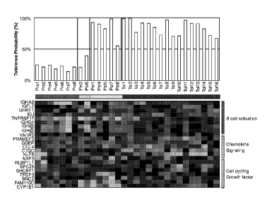
Figure 3. Heat map of gene microarrays and tolerance prediction scores of
.. blood samples from protocol patients and from operationally tolerant
patients. Heat
map shows patterns of expression of 21 unique genes that discriminated
operationally tolerant (TOL) from non-tolerant individuals (e.g., chronic
rejection
(chronic allograft nephropathy; CAN), healthy donors/controls (HD), etc.). Pre
(Pre)
and posttransplant (Pst) blood samples available from patients #1, 2, 4, 5, 6,
7, and
8 were compared to those from operationally tolerant patients (Tol 1-16).
Pretransplant samples are grouped by a horizontal red bar (Pre),
posttransplant
samples are grouped by a yellow bar from patients who did not meet drug
withdrawal criteria (Pst), by a blue bar from patients who stopped
immunosuppressive drugs (Pst), and a black bar from operationally tolerant
patients. Bar graph above heat map shows tolerance prediction scores as
determined by matching with the tolerance signature from a group of
"operationally"
tolerant patients as judged by Probability Analysis of Microarrays.
Posttransplant
samples are the first samples monitored. There were 22 probes used for the 21
unique genes (two probes for IGH@ were used). Gene identifiers are shown on
left
side of map, and gene groupings are shown on right.
Figure 4. Immune monitoring parameters that distinguish patients followed
for more than 1 year who are on or off immunosuppressive drugs. A, shows the
pre
and first posttransplant gene array tolerance prediction scores. The open
symbols
represent patients who are off drugs, and the closed symbols represent
patients
who are on drugs. B, shows the pre and first posttransplant ratios of Treg/
CD4+
naive T cells. C, shows the maximum percentages of donor type cells among NK
cells in protocol patients within the first 50 days after transplantation. Pre
and
posttransplant samples from 8 patients were available for B and C, and from 7
patients for A. Samples in A were obtained from 1 to 5 months posttransplant,
and
samples in B and C within the first 30 or 50 days respectively. Patients are
grouped
by different chimerism patterns as follows: = ¨ primary engraftment failure
(patient
#2); = ¨ loss of chimerism with rejection episodes during drug reductions
(patients
#3 and 6); a ¨ complete chimerism (patient #7); a ¨ stable mixed chimerism
3
:A 02782803 2012-M04
WO 2011/068829 PCT/US2010/058496
(patients #1 and 8); A - loss of chimerism before stopping drugs (patient #4);
0 - loss
of chimerism after stopping drugs (patient #5).
Figure 5 shows tolerance prediction scores using the 21 gene biomarker set
for groups of patients and controls, with 100% representing the closest match.
Agilent microarray analysis on 70 PBL samples were performed and analyzed.
Samples included 35 operationally tolerant (TOL), 29 chronic rejection (or
chronic
allograft nephropathy; CAN), and 6 healthy donors (HD). The training samples
(top
panel; 6 HD, 10 CAN and 16 TOL) and test samples (bottom panel; 19 CAN and 19
TOL) are indicated. As shown in the bottom panel of Figure 5, only two mis-
classifications occurred in the 19 sample CAN test set (CAN_test; 89%
sensitivity)
while only 1 mis-classification occurred in the 19 sample TOL test set (TOL-
test;
95% sensitivity).
DESCRIPTION OF THE SPECIFIC EMBODIMENTS
Methods are provided for determining whether a subject has a graft tolerant
phenotype. In practicing the subject methods, the expression of at least one
gene in
a sample from the subject, e.g., a blood sample, is assayed to obtain an
expression
evaluation for the at least one gene. The obtained expression evaluation is
then
employed to determine whether the subject has a graft tolerant phenotype. Also
provided are compositions, systems and kits that find use in practicing the
subject
methods. The methods and compositions find use in a variety of applications,
including the determination of an immunosuppressive therapy regimen.
Before the present invention is described in greater detail, it is to be
understood that this invention is not limited to particular embodiments
described, as
such may, of course, vary. It is also to be understood that the terminology
used
herein is for the purpose of describing particular embodiments only, and is
not
intended to be limiting, since the scope of the present invention will be
limited only
by the appended claims.
Where a range of values is provided, it is understood that each intervening
value, to the tenth of the unit of the lower limit unless the context clearly
dictates
otherwise, between the upper and lower limit of that range and any other
stated or
intervening value in that stated range, is encompassed within the invention.
The
4
upper and lower limits of these smaller ranges may independently be included
in the
smaller ranges and are also encompassed within the invention, subject to any
specifically excluded limit in the stated range. Where the stated range
includes one
or both of the limits, ranges excluding either or both of those included
limits are also
included in the invention.
Certain ranges are presented herein with numerical values being preceded
by the term "about." The term ''about" is used herein to provide literal
support for the
exact number that it precedes, as well as a number that is near to or
approximately
the number that the term precedes. In determining whether a number is near to
or
approximately a specifically recited number, the near or approximating
unrecited
number may be a number which, in the context in which it is presented,
provides the
substantial equivalent of the specifically recited number.
Unless defined otherwise, all technical and scientific terms used herein have
the
same meaning as commonly understood by one of ordinary skill in the art to
which
this invention belongs. Although any methods and materials similar or
equivalent to
those described herein can also be used in the practice or testing of the
present
invention, representative illustrative methods and materials are now
described.
The citation of any publication is for its disclosure
prior to the filing date and should not be construed as an admission that the
present
invention is not entitled to antedate such publication by virtue of prior
invention.
Further, the dates of publication provided may be different from the actual
= publication dates which may need to be independently confirmed.
It is noted that, as used herein and in the appended claims, the singular
forms "a",
"an", and "the" include plural referents unless the context clearly dictates
otherwise.
It is further noted that the claims may be drafted to exclude any optional
element.
As such, this statement is intended to serve as antecedent basis for use of
such
exclusive terminology as "solely," "only" and the like in connection with the
recitation
of claim elements, or use of a "negative" limitation.
5
CA 2782803 2017-08-23
:A 02782803 2012-M04
WO 2011/068829 PCT/US2010/058496
As will be apparent to those of skill in the art upon reading this disclosure,
each of the individual embodiments described and illustrated herein has
discrete
components and features which may be readily separated from or combined with
the features of any of the other several embodiments without departing from
the
scope or spirit of the present invention. Any recited method can be carried
out in the
order of events recited or in any other order which is logically possible.
As summarized above, the subject invention is directed to methods of
determining whether a subject has a graft tolerant phenotype, as well as
reagents
and kits for use in practicing the subject methods. In further describing the
invention,
the subject methods are described first, followed by a review of the reagents
and
kits for use in practicing the subject methods.
METHODS OF DETERMINING WHETHER A SUBJECT HAS A GRAFT TOLERANT PHENOTYPE
Aspects of the subject invention provide methods of determining whether a
patient or subject has a graft tolerant phenotype. By graft tolerant phenotype
is
meant that the subject does not reject a graft organ, tissue or cell(s) that
has been
introduced into/onto the subject. In other words, the subject tolerates or
maintains
the organ, tissue or cell(s) that has been transplanted to it. As in known in
the
transplantation field, the graft organ, tissue or cell(s) may be allogeneic or
xenogeneic, such that the grafts may be allografts or xenografts. A feature of
the
graft tolerant phenotype detected or identified by the subject methods is that
it is a
phenotype which occurs without immunosuppressive therapy, i.e., it is present
in a
host that is not undergoing immunosuppressive therapy such that
immunosuppressive agents are not being administered to the host.
In practicing the subject methods, a subject or patient sample, e.g., cells or
collections thereof, e.g., tissues, is assayed to determine whether the host
from
which the assayed sample was obtained is graft tolerant, i.e., has a graft
tolerant
phenotype. Accordingly, the first step of the subject methods is to obtain a
suitable
sample from the subject or patient of interest, i.e., a patient on
immunosuppressive
therapy and having at least one graft, e.g., allograft. The sample is derived
from any
initial suitable source, where sample sources of interest include, but are not
limited
to, many different physiological sources, e.g., CSF, urine, saliva, tears,
tissue
derived samples, e.g., homogenates, and blood or derivatives thereof.
6
:A 02782803 2012-M04
WO 2011/068829 PCT/US2010/058496
In certain embodiments, a suitable initial source for the patient sample is
blood. As such, the sample employed in the subject assays of these embodiments
is
generally a blood-derived sample. The blood derived sample may be derived from
whole blood or a fraction thereof, e.g., serum, plasma, etc., where in many
embodiments the sample is derived from blood cells harvested from whole blood.
Of
particular interest as a sample source are peripheral blood lymphocytes (PBL).
Any
convenient protocol for obtaining such samples may be employed, where suitable
protocols are well known in the art.
In practicing the subject methods, the sample is assayed to obtain an
expression evaluation, e.g., expression profile or expression signature, for
one or
more genes, where the term expression profile (or expression signature) is
used
broadly to include a genomic expression profile, e.g., an expression profile
of
nucleic acid transcripts, e.g., mRNAs, of the one or more genes of interest,
or a
proteomic expression profile, e.g., an expression profile of one or more
different
proteins, where the proteins/polypeptides are expression products of the one
or
more genes of interest. As such, in certain embodiments the expression of only
one
gene is evaluated. In yet other embodiments, the expression of two or more,
e.g.,
about 5 or more, about 10 or more, about 15 or more, about 25 or more, about
50 or
more, about 100 or more, about 200 or more, etc., genes is evaluated.
Accordingly,
in the subject methods, the expression of at least one gene in a sample is
evaluated. In certain embodiments, the evaluation that is made may be viewed
as
an evaluation of the transcriptome, as that term is employed in the art. See
e.g.,
Gomes et al., Blood (2001 Jul 1) 98(1):93-9.
In many embodiments, a sample is assayed to generate an expression profile
(or signature) that includes expression data for at least one gene/protein,
usually a
plurality of genes/proteins, where by plurality is meant at least two
different
genes/proteins, and often at least about 5, at least about 10, at least about
20
different genes/proteins or more, such as 50 or more, 100 or more, etc.
In the broadest sense, the expression evaluation may be qualitative or
quantitative. As such, where detection is qualitative, the methods provide a
reading
or evaluation, e.g., assessment, of whether or not the target analyte, e.g.,
nucleic
acid or expression product, is present in the sample being assayed. In yet
other
embodiments, the methods provide a quantitative detection of whether the
target
7
:A 02782803 2012-M04
WO 2011/068829 PCT/US2010/058496
analyte is present in the sample being assayed, i.e., an evaluation or
assessment of
the actual amount or relative abundance of the target analyte, e.g., nucleic
acid in
the sample being assayed. In such embodiments, the quantitative detection may
be
absolute or, if the method is a method of detecting two or more different
analytes,
e.g., target nucleic acids, in a sample, relative. As such, the term
"quantifying" when
used in the context of quantifying a target analyte, e.g., nucleic acid(s), in
a sample
can refer to absolute or to relative quantification. Absolute quantification
may be
accomplished by inclusion of known concentration(s) of one or more control
analytes and referencing the detected level of the target analyte with the
known
control analytes (e.g., through generation of a standard curve).
Alternatively, relative
quantification can be accomplished by comparison of detected levels or amounts
between two or more different target analytes to provide a relative
quantification of
each of the two or more different analytes, e.g., relative to each other.
In certain embodiments, genes/proteins of interest are genes/proteins that
are differentially expressed or present at different levels in graft tolerant
versus non-
graft tolerant individuals who have received a kidney allograft.
Representative
genes/proteins of interest in these embodiments include, but are not limited
to, the
genes/proteins provided in Table 1A, where the Entrez Gene ID number for each
gene is listed. (Note that detailed information for each gene in Table 1A,
including
nucleotide sequence information, can be retrieved through the NCB! Entrez
nucleotide database located at the website http (colon) //www (dot)
ncbi.nlm.nih
(dot) gov/nucleotide by selecting "Gene" as the database and entering the
Entrez
Gene ID number listed into the search window.)
In certain embodiments, at least one of the genes/proteins in the expression
profile is from Table 1A, where the expression profile may include expression
data
for any combination of the genes listed in Table lA (e.g., 1, 2, 3, 4, 5, 6,
7, 8, 9, 10,
11, 12, etc., up to and including all 21 genes in Table 1A).
Table 1A. A list of 21 genes whose expression level can be used to determine a
TOL
phenotype in a subject having a kidney transplant.
Gene Gene Information Entrez
Symbol Gene
ID
BNC2 Name: basonuclin 2 [Homo sapiens] 54796
Other Aliases: RP11-18316.1, BSN2, DKFZp686A01127,
FLJ20043, FLJ34928
Chromosome: 9; Location: 9p22.3-p22.2
8
:A 02782803 2012-M04
WO 2011/068829
PCT/US2010/058496
Annotation: Chromosome 9, NC 000009.11
(16409501..16870786, complement)
MIM: 608669
Cl QC Name: complement component 1, q subcomponent, C chain 714
[Homo sapiens]
Other Aliases: C1Q-C, C1QG, FLJ27103
Other Designations: OTTHUMP00000002933; complement
C1q subcomponent subunit C; complement component 1, q
subcomponent, gamma polypeptide
Chromosome: 1; Location: 1p36.11
Annotation: Chromosome 1, NC 000001.10
(22970118..22974603)
MIM: 120575
CCL4 Name: chemokine (C-C motif) ligand 4 [Homo sapiens] 6351
Other Aliases: ACT2, A1744.1, G-26, LAG1, MGC104418,
MGC126025, MGC126026, MIP-1-beta, MIP1B, MIP1B1,
SCYA2, SCYA4
Other Designations: CC chemokine ligand 4; chemokine C-C
motif ligand 4; lymphocyte-activation gene 1; secreted protein
3-26; small inducible cytokine A4 (homologous to mouse Mip-
1b)
Chromosome: 17; Location: 17q12
Annotation: Chromosome 17, NC 000017.10
(34431220..34433014)
MIM: 182284
CYP1B1 Name: cytochrome P450, family 1, subfamily B, polypeptide 1 1545
[Homo sapiens]
Other Aliases: CP1B, GLC3A, P4501 B1
Other Designations: OTTHUMP00000201401; aryl
hydrocarbon hydroxylase; cytochrome P450, subfamily I
(dioxin-inducible), polypeptide 1 (glaucoma 3, primary
infantile); flavoprotein-linked monooxygenase; microsomal
monooxygenase; xenobiotic monooxygenase
Chromosome: 2; Location: 2p21
Annotation: Chromosome 2, NC 000002.11
(38294746..38303323, complement)
MIM: 601771
FAM110C Name: family with sequence similarity 110, member C [Homo 642273
sapiens]
Other Designations: hypothetical protein L00642273
Chromosome: 2; Location: 2p25.3
Annotation: Chromosome 2, NC 000002.11 (41608..46385,
complement)
MIM: 611395
GDEP Gene description: gene differentially expressed in prostate 11
8425
[Homo sapiens]
Chromosome: 4; Location: 4q21.1
Other Aliases: PCAN1
IGFL2 Name: IGF-like family member 2 [Homo sapiens] 147920
Other Aliases: UNQ645, VPRI645
Other Designations: insulin growth factor-like family member 2
Chromosome: 19; Location: 19q13.32
Annotation: Chromosome 19, NC 000019.9
(46651039..46664561)
9
:A 02782803 2012-M04
WO 2011/068829 PCT/US2010/058496
MIM: 610545
IGH@ Name: immunoglobulin heavy locus [Homo sapiens] 3492
Other Aliases: DKFZp686C15213, IGH, IGH.1@, IGHDY1,
MG072071, MGC88774
Other Designations: immunglobulin heavy chain variable
region
Chromosome: 14; Location: 14q32.33
IGHA2 Name: immunoglobulin heavy constant alpha 2 (A2m marker) 3494
[Homo sapiens]
Chromosome: 14; Location: 14q32.33
Annotation: Chromosome 14, NC 000014.8
(106053244..106054731, complement)
MIM: 147000
IGHG4 Official Symbol IGHG4 and Name: immunoglobulin heavy 3503
constant gamma 4 (G4m marker) [Homo sapiens]
Other Aliases: MGC117419
Chromosome: 14; Location: 14q32.33
Annotation: Chromosome 14, NC 000014.8
(106090707..106092402, complement)
MIM: 147130
IGJ Name: immunoglobulin J polypeptide, linker protein for 351 2
immunoglobulin alpha and mu polypeptides [Homo sapiens]
Other Aliases: IGCJ, JCH
Other Designations: immunoglobulin J chain
Chromosome: 4; Location: 4q21
Annotation: Chromosome 4, NC 000004.11
(71521258..71532348, complement)
MIM: 147790
KLF6 Name: Kruppel-like factor 6 [Homo sapiens] 1316
Other Aliases: RP11-184A2.1, BCD1, CBA1, COPEB, CPBP,
DKFZp686N0199, GBF, PAC, ST12, ZF9
Other Designations: GC-rich binding factor; Kruppel-like zinc
finger protein Zf9; core promoter element binding protein;
protooncogene B-cell derived 1; suppression of tumorigenicity
12 (prostate)
Chromosome: 10; Location: 10p15
Annotation: Chromosome 10, NC 000010.10
(3821234..3827455, complement)
MIM: 602053
NXF3 Name: nuclear RNA export factor 3 [Homo sapiens] 56000
Other Aliases: LLOXNC01-221F2.3
Chromosome: X; Location: Xq22-q23
Annotation: Chromosome X, NC 000023.10
(102330749..102348022, complement)
MIM: 300316
PRAMEF3 Name: PRAME family member 3 [Homo sapiens] 401940
Other Aliases: RP11-248D7.1
Chromosome: 1; Location: 1p36.21
Annotation: Chromosome 1, NC 000001.10
(13328196..13331692, complement)
RLBP1L1 Name: clavesin 1 [Homo sapiens] 157807
Other Aliases: CRALBPL, FLJ37248, MGC34646, RLBP1L1
Other Designations: retinaldehyde binding protein 1-like 1
Chromosome: 8; Location: 8q12.3
:A 02782803 2012-M04
WO 2011/068829
PCT/US2010/058496
Annotation: Chromosome 8, NC 000008.10
(62200525..62414204)
MIM: 611292
SHCBP1 Name: SHC SH2-domain binding protein 1 [Homo sapiens] 79801
Other Aliases: FLJ22009, MGC26900, PAL
Chromosome: 16; Location: 16q11.2
Annotation: Chromosome 16, NC 000016.9
(46614466..46655311, complement)
MIM: 611027
SPC25 Name: SPC25, NDC80 kinetochore complex component, 57405
homolog (S. cerevisiae) [Homo sapiens]
Other Aliases: AD024, MGC22228, SPBC25
Other Designations: 2600017H08Rik; kinetochore protein
5pc25; spindle pole body component 25; spindle pole body
component 25 homolog
Chromosome: 2; Location: 2q31.1
Annotation: Chromosome 2, NC 000002.11
(169727401..169746944, complement)
MIM: 609395
TFDP3 Name: transcription factor Dp family, member 3 [Homo 51270
sapiens]
Other Aliases: RP3-358H7.2, CT30, DP4, E2F-like, HCA661,
MGC161639
Other Designations: OTTHUMP00000024051; cancer/testis
antigen 30
Chromosome: X; Location: Xq26.2
Annotation: Chromosome X, NC 000023.10
(132350697..132352376, complement)
MIM: 300772
TNFRSF1 7 Name: tumor necrosis factor receptor superfamily, member 17 608
[Homo sapiens]
Other Aliases: BCM, BCMA, CD269
Other Designations: B cell maturation antigen; B-cell
maturation factor
Chromosome: 16; Location: 16p13.1
Annotation: Chromosome 16, NC 000016.9
(12058964..12061925)
MIM: 109545
UHRF1 Name: ubiquitin-like with PHD and ring finger domains 1 29128
[Homo sapiens]
Other Aliases: FLJ21925, ICBP90, MGC138707, Np95,
RNF106, hNP95
Other Designations: E3 ubiquitin-protein ligase UHRF1; RING
finger protein 106; inverted CCAAT box-binding protein of 90
kDa; nuclear zinc finger protein Np95; transcription factor
ICBP90; ubiquitin-like, containing PHD and RING finger
domains, 1
Chromosome: 19; Location: 19p13.3
Annotation: Chromosome 19, NC 000019.9
(4909510..4962165)
MIM: 607990
VN1R2 Name: vomeronasal 1 receptor 2 [Homo sapiens] 317701
Other Aliases: V1RL2
Other Designations: V1R-like 2; pheromone receptor
11
:A 02782803 2012-M04
WO 2011/068829 PCT/US2010/058496
Chromosome: 19; Location: 19q13.42
Annotation: Chromosome 19, NC 000019.9
(53761545..53762855)
In certain embodiments, genes/proteins of interest are genes/proteins that
are differentially expressed or present at different levels in graft tolerant
versus non-
graft tolerant individuals who have received a liver allograft (e.g.,
pediatric patients).
Representative genes/proteins of interest in these embodiments include, but
are not
limited to, the genes/proteins provided in Table 1B, where the Entrez Gene ID
number for each gene is listed. (Note that detailed information for each gene
in
Table 1B, including nucleotide sequence information, can be retrieved through
the
NCB! Entrez nucleotide database located at the website http (colon) //www
(dot)
ncbi.nlm.nih (dot) gov/nucleotide by selecting "Gene" as the database and
entering
the Entrez Gene ID number listed into the search window.)
In certain embodiments, at least one of the genes/proteins in the expression
profile is from Table 1B, where the expression profile may include expression
data
for any combination of the genes listed in Table 1B (e.g., 1, 2, 3, 4, 5, 6,
7, 8, 9, 10,
11, up to and including all 12 genes in Table 1B).
Table 1B. A list of 12 genes whose expression level can be used to determine a
TOL
phenotype in a subject having a liver transplant.
Gene Gene Information
Entrez
Symbol Gene
ID
AKR1C3 Official Symbol AKR1C3 and Name: aldo-keto reductase 8644
family 1, member C3 (3-alpha hydroxysteroid dehydrogenase,
type II) [Homo sapiens]
Other Aliases: DD3, DDX, HA1753, HAKRB, HAKRe,
HSD17B5, KIAA0119, hluPGFS
Other Designations: aldo-keto reductase family 1, member C3;
chlordecone reductase; dihydrodiol dehydrogenase 3;
dihydrodiol dehydrogenase X; hydroxysteroid (17-beta)
dehydrogenase 5; prostaglandin F synthase; trans-1,2-
dihydrobenzene-1,2-diol dehydrogenase; type ll 3a-
hydroxysteroid dehydrogenase; type Ilb 3-alpha
hydroxysteroid dehydrogenase
Chromosome: 10; Location: 10p15-p14
Annotation: Chromosome 10, NC 000010.10
(5136568..5149878)
MIM: 603966
ASPH Official Symbol ASPH and Name: aspartate beta-hydroxylase 444
[Homo sapiens]
Other Aliases: AAH, BAH, CASQ2BP1, HAAH, JCTN, junctin
Other Designations: A beta H-J-J; aspartyl/asparaginyl-beta-
12
:A 02782803 2012-M04
WO 2011/068829
PCT/US2010/058496
hydroxylase; cardiac junctin; humbug; junctate; peptide-
aspartate beta-dioxygenase
Chromosome: 8; Location: 8q12.1
Annotation: Chromosome 8, NC 000008.10
(62413115..62627199, complement)
MIM: 600582
ERBB2 Official Symbol ERBB2 and Name: v-erb-b2 erythroblastic 2064
leukemia viral oncogene homolog 2, neuro/glioblastoma
derived oncogene homolog (avian) [Homo sapiens]
Other Aliases: CD340, HER-2, HER-2/neu, HER2, NEU, NGL,
TKR1
Other Designations: c-erb B2/neu protein; erbB-2; herstatin;
neuroblastoma/glioblastoma derived oncogene homolog; v-
erb-b2 avian erythroblastic leukemia viral oncogene homolog
2 (neuro/glioblastoma derived oncogene homolog)
Chromosome: 17; Location: 17q21.1
Annotation: Chromosome 17, NC 000017.10
(37844393..37884915)
MIM: 164870
FEM1C Official Symbol FEM1C and Name: fem-1 homolog c (C. 56929
elegans) [Homo sapiens]
Other Aliases: EUROIMAGE686608, EUROIMAGE783647,
FEM1A, KIAA1785
Other Designations: feminization 1 homolog a
Chromosome: 5; Location: 5q22
Annotation: Chromosome 5, NC 000005.9
(114856608..114880591, complement)
MIM: 608767
MAFG Official Symbol MAFG and Name: v-maf musculoaponeurotic 4097
fibrosarcoma oncogene homolog G (avian) [Homo sapiens]
Other Aliases: MGC13090, MGC20149
Other Designations: basic leucine zipper transcription factor
MafG; transcription factor MafG; v-maf musculoaponeurotic
fibrosarcoma oncogene homolog G
Chromosome: 17; Location: 17q25.3
Annotation: Chromosome 17, NC 000017.10
(79876146..79885588, complement)
MIM: 602020
NFKB1 Official Symbol NFKB1 and Name: nuclear factor of kappa 4790
light polypeptide gene enhancer in B-cells 1 [Homo sapiens]
Other Aliases: DKFZp686C01211, EBP-1, KBF1, MGC54151,
NF-kappa-B, NF-kappaB, NFKB-p105, NFKB-p50, p105, p50
Other Designations: DNA binding factor KBF1; NF-kappabeta;
nuclear factor NF-kappa-B p50 subunit; nuclear factor kappa-
B DNA binding subunit; nuclear factor kappa-B, subunit 1
Chromosome: 4; Location: 4q24
Annotation: Chromosome 4, NC 000004.11
(103422486..103538459)
MIM: 164011
PDE4DIP Official Symbol PDE4DIP and Name: phosphodiesterase 4D 9659
interacting protein [Homo sapiens]
Other Aliases: CMYA2, DKFZp781J054, MGC75440, MMGL
Other Designations: cardiomyopathy associated 2;
myomegalin
13
:A 02782803 2012-M04
WO 2011/068829
PCT/US2010/058496
Chromosome: 1; Location: 1q12
Annotation: Chromosome 1, NC 000001.10
(144851427..145076079, complement)
MIM: 608117
PHLDA2 Official Symbol PHLDA2 and Name: pleckstrin homology-like 7262
domain, family A, member 2 [Homo sapiens]
Other Aliases: BRW1C, BWR1C, HLDA2, IPL, TSSC3
Other Designations: imprinted in placenta and liver; p17-
Beckwith-Wiedemann region 1C; pleckstrin homology-like
domain family A member 2; tumor suppressing
subchromosomal transferable fragment cDNA 3; tumor
suppressing subtransferable candidate 3; tumor-supressing
STF cDNA 3
Chromosome: 11; Location: 11p15.5
Annotation: Chromosome 11, NC 000011.9
(2949503..2950650, complement)
MIM: 602131
PTBP2 Official Symbol PTBP2 and Name: polypyrimidine tract binding 58155
protein 2 [Homo sapiens]
Other Aliases: FLJ34897, PTB, PTBLP, brPTB, nPTB, nPTB5,
nPTB6, nPTB7, nPTB8
Other Designations: PTB-like; neural polypyrimidine tract
binding protein; splicing regulator
Chromosome: 1; Location: 1p22.1-p21.3
Annotation: Chromosome 1, NC 000001.10
(97187175..97280605)
MIM: 608449
SENP6 Official Symbol SENP6 and Name: SUM01/sentrin specific 26054
peptidase 6 [Homo sapiens]
Other Aliases: RP1-134M13.1, FLJ11355, FLJ11887,
KIAA0389, KIAA0797, SSP1, SUSP1
Other Designations: 2810017C20Rik; SUM0-1-specific
protease; SUM01/sentrin specific protease 6
Chromosome: 6; Location: 6q13-q14.3
Annotation: Chromosome 6, NC 000006.11
(76311622..76427997)
MIM: 605003
UBAC2 Official Symbol UBAC2 and Name: UBA domain containing 2 337867
[Homo sapiens]
Other Aliases: RP11-178C10.1, FLJ26351, FLJ30001,
FLJ30548, FLJ42413, MGC90487, PHGDHL1
Other Designations: RP11-178C10.1; phosphoglycerate
dehydrogenase like 1
Chromosome: 13; Location: 13q32.3
Annotation: Chromosome 13, NC 000013.10
(99852679..100038753)
ZNF420 Official Symbol ZNF420 and Name: zinc finger protein 420 147923
[Homo sapiens]
Other Aliases: APAK, FLJ32191
Other Designations: ATM and p53-associated KZNF protein
Chromosome: 19; Location: 19q13.12
Annotation: Chromosome 19, NC 000019.9
(37569382..37620662)
14
:A 02782803 2012-M04
WO 2011/068829 PCT/US2010/058496
In certain embodiments, the expression profile obtained is a genomic or
nucleic acid expression profile, where the amount or level of one or more
nucleic
acids in the sample is determined, e.g., the nucleic acid transcript of the
gene of
interest. In these embodiments, the sample that is assayed to generate the
expression profile employed in the diagnostic methods is one that is a nucleic
acid
sample. The nucleic acid sample includes a plurality or population of distinct
nucleic
acids that includes the expression information of the phenotype determinative
genes
of interest of the cell or tissue being diagnosed. The nucleic acid may
include RNA
or DNA nucleic acids, e.g., mRNA, cRNA, cDNA etc., so long as the sample
retains
the expression information of the host cell or tissue from which it is
obtained. The
sample may be prepared in a number of different ways, as is known in the art,
e.g.,
by mRNA isolation from a cell, where the isolated m RNA is used as is,
amplified,
employed to prepare cDNA, cRNA, etc., as is known in the differential
expression
art. The sample is typically prepared from a cell or tissue harvested from a
subject
to be diagnosed, e.g., via biopsy of tissue, using standard protocols, where
cell
types or tissues from which such nucleic acids may be generated include any
tissue
in which the expression pattern of the to be determined phenotype exists,
including,
but not limited to, peripheral blood lymphocyte cells, etc., as reviewed
above.
The expression profile may be generated from the initial nucleic acid sample
using any convenient protocol. While a variety of different manners of
generating
expression profiles are known, such as those employed in the field of
differential
gene expression analysis, one representative and convenient type of protocol
for
generating expression profiles is array-based gene expression profile
generation
protocols. Such applications are hybridization assays in which a nucleic acid
that
displays "probe" nucleic acids for each of the genes to be assayed/profiled in
the
profile to be generated is employed. In these assays, a sample of target
nucleic
acids is first prepared from the initial nucleic acid sample being assayed,
where
preparation may include labeling of the target nucleic acids with a label,
e.g., a
member of signal producing system. Following target nucleic acid sample
preparation, the sample is contacted with the array under hybridization
conditions,
whereby complexes are formed between target nucleic acids that are
complementary to probe sequences attached to the array surface. The presence
of
hybridized complexes is then detected, either qualitatively or quantitatively.
Specific
hybridization technology which may be practiced to generate the expression
profiles
employed in the subject methods includes the technology described in U.S.
Patent
Nos.: 5,143,854; 5,288,644; 5,324,633; 5,432,049; 5,470,710; 5,492,806;
5,503,980; 5,510,270; 5,525,464; 5,547,839; 5,580,732; 5,661,028; 5,800,992;
as well as WO 95/21265;
WO 96/31622; WO 97/10365; WO 97/27317; EP 373 203; and EP 785 280. In these
methods, an array of "probe" nucleic acids that includes a probe for each of
the
phenotype determinative genes whose expression is being assayed is contacted
with target nucleic acids as described above. Contact is carried out under
hybridization conditions, e.g., stringent hybridization conditions, and
unbound
nucleic acid is then removed.
The term "stringent assay conditions" as used herein refers to conditions that
are compatible to produce binding pairs of nucleic acids, e.g., surface bound
and
solution phase nucleic acids, of sufficient complementarity to provide for the
desired
level of specificity in the assay while being less compatible to the formation
of
binding pairs between binding members of insufficient complementarity to
provide
for the desired specificity. Stringent assay conditions are the summation or
combination (totality) of both hybridization and wash conditions.
"Stringent hybridization conditions" and "stringent hybridization wash
conditions" in the context of nucleic acid hybridization (e.g., as in array,
Southern or
Northern hybridizations) are sequence dependent, and are different under
different
experimental parameters. Stringent hybridization conditions that can be used
to
identify nucleic acids within the scope of the invention can include, e.g.,
hybridization in a buffer comprising 50% formamide, 5xSSC, and 1% SDS at 42 C,
or hybridization in a buffer comprising 5xSSC and 1% SDS at 65 C, both with a
wash of 0.2xSSC and 0.1% SDS at 65 C. Exemplary stringent hybridization
conditions can also include a hybridization in a buffer of 40% formamide, 1 M
NaCl,
and 1% SDS at 37 C, and a wash in 1xSSC at 45 C. Alternatively, hybridization
to
filter-bound DNA in 0.5 M NaHPO4, 7% sodium dodecyl sulfate (SDS), 1 mM EDTA
at 65 C, and washing in 0.1xSSC/0.1% SDS at 68 C can be employed. Yet
additional stringent hybridization conditions include hybridization at 60 C or
higher
and 3xSSC (450 mM sodium chloride/45 mM sodium citrate) or incubation at 429=C
16
CA 2782803 2017-08-23
WO 2011/068829 PCT/US2010/058496
in a solution containing 30% formamide, 1M NaCI, 0.5% sodium sarcosine, 50 mM
MES, pH 6.5. Those of ordinary skill will readily recognize that alternative
but
comparable hybridization and wash conditions can be utilized to provide
conditions
of similar stringency.
In certain embodiments, the stringency of the wash conditions that set forth
the conditions which determine whether a nucleic acid is specifically
hybridized to a
surface bound nucleic acid. Wash conditions used to identify nucleic acids may
include, e.g.: a salt concentration of about 0.02 molar at pH 7 and a
temperature of
at least about 50 C or about 55 C to about 60 C; or, a salt concentration of
about
0.15 M NaCl at 72 C for about 15 minutes; or, a salt concentration of about
0.2xSSC at a temperature of at least about 50 C or about 55 C to about 60 C
for
about 15 to about 20 minutes; or, the hybridization complex is washed twice
with a
solution with a salt concentration of about 2xSSC containing 0.1% SDS at room
temperature for 15 minutes and then washed twice by 0.1xSSC containing 0.1%
SDS at 68 C for 15 minutes; or, equivalent conditions. Stringent conditions
for
washing can also be, e.g., 0.2xSSC/0.1% SDS at 42 C.
A specific example of stringent assay conditions is rotating hybridization at
65 C in a salt based hybridization buffer with a total monovalent cation
concentration of 1.5 M (e.g., as described in U.S. Patent Application No.
09/655,482
filed on September 5, 2000) followed by washes of 0.5xSSC and 0.1xSSC at room
temperature.
Stringent assay conditions are hybridization conditions that are at least as
stringent as the above representative conditions, where a given set of
conditions are
considered to be at least as stringent if substantially no additional binding
complexes that lack sufficient complementarity to provide for the desired
specificity
are produced in the given set of conditions as compared to the above specific
conditions, where by "substantially no more is meant less than about 5-fold
more,
typically less than about 3-fold more. Other stringent hybridization
conditions are
known in the art and may also be employed, as appropriate.
The resultant pattern of hybridized nucleic acid provides information
regarding expression for each of the genes that have been probed, where the
expression information is in terms of whether or not the gene is expressed
and,
17
CA 2 78 2 8 0 3 2 0 1 9-1 0-2 3
:A 02782803 2012-M04
WO 2011/068829 PCT/US2010/058496
typically, at what level, where the expression data, i.e., expression profile
(e.g., in
the form of a transcriptome), may be both qualitative and quantitative.
Alternatively, non-array based methods for quantitating the levels of one or
more nucleic acids in a sample may be employed, including those based on
amplification protocols, e.g., Polymerase Chain Reaction (PCR)-based assays,
including quantitative PCR, reverse-transcription PCR (RT-PCR), real-time PCR,
and the like.
Where the expression profile is a protein expression profile, any convenient
protein quantitation protocol may be employed, where the levels of one or more
proteins in the assayed sample are determined. Representative methods include,
but are not limited to: proteomic arrays, flow cytometry, standard
immunoassays
(e.g., western blot, ELISA assays), Mass spectrometry, etc.
Following obtainment of the expression profile from the sample being
assayed, the expression profile is compared with a reference or control
profile to
determine the particular graft tolerant/intolerant phenotype of the cell or
tissue, and
therefore host, from which the sample was obtained/derived. The terms
"reference"
and "control" as used herein mean a standardized pattern of gene expression or
levels of expression of certain genes to be used to interpret the expression
signature of a given patient and assign a graft tolerant/intolerant phenotype
thereto.
The reference or control profile may be a profile that is obtained from a
cell/tissue
known to have the desired phenotype, e.g., a graft tolerant phenotype, and
therefore
may be a positive reference or control profile. In addition, the
reference/control
profile may be from a cell/tissue known to not have the desired phenotype,
e.g., a
graft intolerant phenotype, and therefore be a negative reference/control
profile.
In certain embodiments, the obtained expression profile is compared to a
single reference/control profile to obtain information regarding the phenotype
of the
cell/tissue being assayed. In yet other embodiments, the obtained expression
profile
is compared to two or more different reference/control profiles to obtain more
in
depth information regarding the phenotype of the assayed cell/tissue. For
example,
the obtained expression profile may be compared to a positive and negative
reference profile to obtain confirmed information regarding whether the
cell/tissue
has the phenotype of interest.
18
The comparison of the obtained expression profile and the one or more
reference/control profiles may be performed using any convenient methodology,
where a variety of methodologies are known to those of skill in the array art,
e.g., by
comparing digital images of the expression profiles, by comparing databases of
expression data, etc. Patents describing ways of comparing expression profiles
include, but are not limited to, U.S. Patent Nos_ 6,308,170 and 6,228,575.
Methods of comparing
expression profiles are also described above.
The comparison step results in information regarding how similar or dissimilar
the obtained expression profile is to the control/reference profile(s), which
similarity/dissimilarity information is employed to determine the phenotype of
the
cell/tissue being assayed. For example, similarity with a positive control
indicates
that the assayed cell/tissue has a graft tolerant phenotype. Likewise,
similarity with a
negative control indicates that the assayed cell/tissue has an intolerant
phenotype.
Depending on the type and nature of the reference/control profile(s) to which
the obtained expression profile is compared, the above comparison step yields
a
variety of different types of information regarding the cell/tissue that is
assayed. As
such, the above comparison step can yield a positive/negative determination of
a
tolerant phenotype of an assayed cell/tissue. In many embodiments, the above-
obtained information about the cell/tissue being assayed is employed to
diagnose a
host, subject or patient with respect to that host's graft tolerance, as
described
above.
The subject methods further find use in pharmacogenomic applications. In
these applications, a subject/host/patient is first diagnosed for the presence
or
absence of the graft tolerant phenotype using a protocol such as the
diagnostic
protocol described in the preceding section. The subject is then treated using
a
protocol whose suitability is determined using the results of the diagnosis
step. More
specifically, where the identified phenotype is tolerant, a protocol that may
include a
reduced level of immunosuppression (i.e., immunosuppression at a level less
than
that which is indicated for patients not known to be graft tolerant), or no
immunosuppression, may be employed to manage/treat the subject. Alternatively,
where a patient is identified as having an intolerant phenotype, full
immunosuppressive protocols may be employed/continued.
19
CA 2782803 2017-08-23
:A 02782803 2012-M04
WO 2011/068829 PCT/US2010/058496
In many embodiments, a host is screened for the presence of a graft tolerant
phenotype following receipt of a graft or transplant. The host may be screened
once
or serially following transplant receipt, e.g., weekly, monthly, bimonthly,
half-yearly,
yearly, etc., as long as the host is on immunosuppressive therapy. In certain
.. embodiments, monitoring of the host expression profile even after
immunosuppressive therapy has been reduced or discontinued is conducted to
determine whether the host has maintained the tolerogenic expression profile
and
may continue for the lifetime of the host.
DATABASES OF EXPRESSION PROFILES OF PHENOTYPE DETERMINATIVE GENES
Also provided are databases of expression profiles of graft tolerant
phenotype determinative genes. Such databases will typically comprise
expression
profiles of various cells/tissues having graft tolerant phenotypes, negative
expression profiles, etc., where such profiles are further described below.
The expression profiles and databases thereof may be provided in a variety
of media to facilitate their use. "Media" refers to a manufacture that
contains the
expression profile information of the present invention. The databases of the
present invention can be recorded on computer readable media, e.g. any medium
that can be read and accessed directly by a user employing a computer. Such
media include, but are not limited to: magnetic storage media, such as floppy
discs,
hard disc storage medium, and magnetic tape; optical storage media such as CD-
ROM; electrical storage media such as RAM and ROM; and hybrids of these
categories such as magnetic/optical storage media. One of skill in the art can
readily appreciate how any of the presently known computer readable mediums
can
be used to create a manufacture comprising a recording of the present database
information. "Recorded" refers to a process for storing information on
computer
readable medium, using any such methods as known in the art. Any convenient
data storage structure may be chosen, based on the means used to access the
stored information. A variety of data processor programs and formats can be
used
for storage, e.g. word processing text file, database format, etc. Thus, the
subject
expression profile databases are accessible by a user, i.e., the database
files are
saved in a user-readable format (e.g., a computer readable format, where a
user
controls the computer).
As used herein, "a computer-based system" refers to the hardware means,
software means, and data storage means used to analyze the information of the
present invention. The minimum hardware of the computer-based systems of the
present invention comprises a central processing unit (CPU), input means,
output
means, and data storage means. A skilled artisan can readily appreciate that
any
one of the currently available computer-based system are suitable for use in
the
present invention. The data storage means may comprise any manufacture
comprising a recording of the present information as described above, or a
memory
access means that can access such a manufacture.
A variety of structural formats for the input and output means can be used to
input and output the information in the computer-based systems of the present
invention, e.g., to and from a user. One format for an output means ranks
expression profiles possessing varying degrees of similarity to a reference
expression profile. Such presentation provides a skilled artisan with a
ranking of
similarities and identifies the degree of similarity contained in the test
expression
profile.
REAGENTS AND KITS
Also provided are reagents and kits thereof for practicing one or more of the
above-described methods. The subject reagents and kits thereof may vary
greatly.
Reagents of interest include reagents specifically designed for use in
production of
the above-described expression profiles of phenotype determinative genes,
i.e., a
gene expression evaluation element made up of one or more reagents.
One type of such reagent is an array of probe nucleic acids in which the
phenotype determinative genes of interest are represented. A variety of
different
array formats are known in the art, with a wide variety of different probe
structures,
substrate compositions and attachment technologies (e.g., dot blot arrays,
microarrays, etc.). Representative array structures of interest include those
described in U.S. Patent Nos.: 5,143,854; 5,288,644; 5,324,633; 5,432,049;
5,470,710; 5,492,806; 5,503,980; 5,510,270; 5,525,464; 5,547,839; 5,580,732;
5,661,028; 5,800,992;
as well as WO 95/21265; WO 96/31622; WO 97/10365; WO 97/27317;
EP 373 203; and EP 785 280.
21
CA 2782803 2017-08-23
In many embodiments, the arrays include probes for at least 1 of the genes
listed in Table lA or Table 1B. The number of genes from Tables lA and/or B
that
are represented on the array can be 1, 2, 3, 4. 5, 6, 7, 8, 9, etc. up to and
including
all 21 genes in Table 1A and/or all 12 genes in Table 1B. In other words, any
combination of genes in Tables lA and B can be represented on arrays of the
subject invention. The subject arrays may include only those genes that are
listed in
Table 1A, only those genes that are listed in Table 1B, only those genes that
are
listed in Tables lA or 1B. Alternatively, the arrays may include additional
genes that
are not listed in either Table lA or 1B.
Another type of reagent that is specifically tailored for generating
expression
profiles of phenotype determinative genes (the genes listed in Table lA and/or
1B)
is a collection of gene specific primers that is designed to selectively
amplify such
genes. Gene specific primers and methods for using the same are described in
U.S.
Patent No. 5,994,076.
Of particular interest are collections of gene specific primers that have
primers for at
least 1 of the genes listed in Table 1A or 1B, often a plurality of these
genes, e.g., at
least 2, 5, 10, and up to and including all 21 genes in Table 1A and/or all 12
genes
in Table 1 B. The subject gene specific primer collections may include only
those
genes that are listed in Table lA and/or 1B, or they may include primers for
additional genes that are not listed in Table lA or 1B.
The kits of the subject invention may include the above-described arrays
and/or gene specific primer collections. The kits may further include one or
more
additional reagents employed in the various methods, such as primers for
generating target nucleic acids, dNTPs and/or rNTPs, which may be either
premixed
or separate, one or more uniquely labeled dNTPs and/or rNTPs, such as
biotinylated or Cy3 or Cy5 tagged dNTPs, gold or silver particles with
different
scattering spectra, or other post synthesis labeling reagent, such as
chemically
active derivatives of fluorescent dyes, enzymes, such as reverse
transcriptases,
DNA polymerases, RNA polymerases, and the like, various buffer mediums, e.g.
hybridization and washing buffers, prefabricated probe arrays, labeled probe
purification reagents and components, like spin columns, etc., signal
generation and
detection reagents, e.g. streptavidin-alkaline phosphatase conjugate,
chemifluorescent or chemiluminescent substrate, and the like.
22
CA 2782803 2017-08-23
The subject kits may also include a phenotype determination element, which
element is, in many embodiments, a reference or control expression profile
that can
be employed, e.g., by a suitable computing means, to make a phenotype
determination based on an "input" expression profile, e.g., that has been
determined
with the above described gene expression evaluation element. Representative
phenotype determination elements include databases of expression profiles,
e.g.,
reference or control profiles, as described above.
In addition to the above components, the subject kits will further include
instructions for practicing the subject methods. These instructions may be
present in
the subject kits in a variety of forms, one or more of which may be present in
the kit.
One form in which these instructions may be present is as printed information
on a
suitable medium or substrate, e.g., a piece or pieces of paper on which the
information is printed, in the packaging of the kit, in a package insert, etc.
Yet
another means would be a computer readable medium, e.g., diskette, CD, etc.,
on
which the information has been recorded. Yet another means that may be present
is
a website address which may be used via the internet to access the information
at a
removed site. Any convenient means may be present in the kits.
SYSTEMS
Also provided are systems for practicing one or more of the above-described
methods. The subject systems may vary greatly, but typically include at least
a gene
expression evaluation element, e.g., one or more reagents, and a phenotype
determination element.
Reagents of interest include reagents specifically designed for use in
production of the above-described expression profiles of phenotype
determinative
genes, i.e., a gene expression evaluation element made up of one or more
reagents. One type of such reagent is an array of probe nOcleic acids in which
the
phenotype determinative genes of interest are represented. A variety of
different
array formats are known in the art, with a wide variety of different probe
structures,
substrate compositions and attachment technologies. Representative array
structures of interest include those described in U.S. Patent Nos.: 5,143,854;
5,288,644; 5,324,633; 5,432,049; 5,470,710; 5,492,806; 5,503,980; 5,510,270;
5,525,464; 5,547,839; 5,580,732; 5,661,028; 5,800,992;
23
CA 2782803 2017-08-23
as well as WO 95/21265; WO 96/31622; WO
97/10365; WO 97/27317; EP 373 203; and EP 785 280.
In many embodiments. the arrays include probes for at least 1 of the genes
listed in Table lA and/or 1B. In certain embodiments, the number of genes that
are
from Table 1A and or 1B that are represented on the array is 1, 2, 3, 4, 5, 6,
7, 8, 9,
etc., up to and including all 21 genes listed in Table 1A and/or up to and
including all
12 genes listed in Table 1B. The subject arrays may include only those genes
that
are listed in Table 1A and/or Table 1B, or they may include additional genes
that are
riot listed in Table 1A or 1B.
Another type of reagent that is specifically tailored for generating
expression
profiles of phenotype determinative genes is a collection of gene specific
primers
that is designed to selectively amplify such genes. Gene specific primers and
methods for using the same are described in U.S. Patent No. 5,994,076
Of particular interest are
collections of gene specific primers that have primers for at least 1 of the
genes
listed in Table lA and/or Table 1B, often a plurality of these genes, e.g., 2,
3, 4, 5, 6,
7, 8, 9, etc., up to and including all 21 genes listed in Table 1A and/or up
to and
including all 12 genes listed in Table 1B. The subject gene specific primer
collections may include only those genes that are listed in Table lA and/or
Table
1B, or they may include primers for additional genes that are not listed in
Table lA
or 1B.
The systems of the subject invention may include the above-described arrays
and/or gene specific primer collections. The systems may further include one
or
more additional reagents employed in the various methods, such as primers for
.. generating target nucleic acids, dNTPs and/or rNTPs, which may be either
premixed
or separate, one or more uniquely labeled dNTPs and/or rNTPs, such as
biotinylated or Cy3 or Cy5 tagged dNTPs, gold or silver particles with
different
scattering spectra, or other post synthesis labeling reagent, such as
chemically
active derivatives of fluorescent dyes, enzymes, such as reverse
transcriptases,
DNA polymerases, RNA polymerases, and the like, various buffer mediums, e.g.
hybridization and washing buffers, prefabricated probe arrays, labeled probe
purification reagents and components, like spin columns, etc., signal
generation and
24
CA 2782803 2017-08-23
:A 02782803 2012-M04
WO 2011/068829 PCT/US2010/058496
detection reagents, e.g. streptavidin-alkaline phosphatase conjugate,
chemifluorescent or chemiluminescent substrate, and the like.
The systems may also include a phenotype determination element, which
element is, in many embodiments, a reference or control expression profile
that can
be employed, e.g., by a suitable computing means, to make a phenotype
determination based on an "input" expression profile, e.g., that has been
determined
with the above described gene expression evaluation element. Representative
phenotype determination elements include databases of expression profiles,
e.g.,
reference or control profiles, as described above.
The following examples are offered by way of illustration and not by way of
limitation.
EXPERIMENTAL
Example 1
Recipients of kidney transplants require the lifelong use of
immunosuppressive drugs to prevent graft rejection (1, 2). The use of these
drugs is
associated with a variety of cumulative side effects including increased risks
of heart
disease, infection, cancer, and diabetes (2-4). Despite the use of these
drugs,
chronic rejection remains an important problem that results in gradual graft
loss (4,
5).
The induction of immune tolerance can prevent the rejection of grafts without
immunosuppressive drugs in a variety of preclinical studies (6-9). A
successful
approach applied to clinical studies combined organ transplantation with the
injection of hematopoietic cells from the donor to achieve stable mixed
chimerism
(10-13). This approach was used in the current study of kidney transplant
recipients
who were given total lymphoid irradiation and anti-thymocyte globulin
conditioning,
and a donor cell injection containing defined doses of highly enriched CD34+
hematopoietic progenitor cells mixed with CD3+ T cells (12). This conditioning
regimen has been shown to protect against GVHD in preclinical models (14-17),
and
in recent clinical trials of 111 patients with leukemia and lymphoma followed
for up
to 8 years (18-19). We used this regimen to avoid the complications of GVHD,
pulmonary capillary leak syndromes, severe neutropenia (<500 cells/mm3),
humoral
:A 02782803 2012-M04
WO 2011/068829
PCT/US2010/058496
rejections, and graft loss that have been reported in previous tolerance
induction
trials (10, 11). The first patient in the kidney transplant cohort was the
subject of a
previous case report (12). In order to improve the safety of the protocol,
monitoring
was performed to identify immune parameters that can predict the tolerant
state,
and guide the withdrawal of immunosuppressive drugs.
Methods
Patients
Ten patients with end stage renal failure who were candidates for kidney
transplantation, and who had donors matched for 6 HLA antigens by standard
genotyping were enrolled in the study. Patients were between 23 and 61 years
old,
and 5 were female. Details of each patient and causes of renal failure are
shown in
Table 2. Donors were all siblings except for the donor of patient #6
(daughter).
Table 2: Patient Characteristics, Conditioning, and Donor Cell Composition
Patients' Age/ ESRD Total Dose CD34
Cell CD3 Cell Serum creatinine
Gender Cause TLI (cGy) Dose Dose at last
ohs.
(x106/kg) (x106/kg) (mg/c1L)
1(41 mo.) 48/M unknown 800 8.0 1 1.2
2 (45 mo.) 39/F FSGS 800 8.4 1 0.8
3(29 mo.) 24/M Dysplasia 800 12.5 1 1.3
4 (25 mo.) 52/M unknown 1,2001s 4.9 1 1.3
5 (21 mo.) 34/M IgA 1,200 12.8 1 1.1
6 (20 ino.) 61T DM 1,200 12.2 1 1.3
7 (16 mo.) 23/F SLE 1,200 16.7 l0' 0.9
8 (14 mo.) 33/M Reflux 1,200 16.7 1 0.8
9 ( 7 mo.) 29T unknown 1,200 17.5 1 1.1
10 ( 6 mo.) 52/F PRD 1,200 14,0 1 0,9
ESRD-end stage renal disease; FSGS-focal segmental glomerulosclerosis; IgA-IgA
nephropathy; DM-
diabetes mellitus; SLE - Systemic lupus erythematosus; PKD - polycystic kidney
disease
'parentheses show duration of follow-up
b dose increased to facilitate persistent chimerisin
'dose increased to achieve complete chimerism
Conditioning
TLI was administered as 10 doses of 80 or 120cGy each to the
supradiaphragmatic lymph nodes, thymus, subdiaphragmatic lymph nodes, and
spleen during the first 11 days post-transplant as described previously (12,
13).
26
:A 02782803 2012-M04
WO 2011/068829 PCT/US2010/058496
Rabbit anti-thymocyte globulin (Thymoglobulin, Genzyme) was given
intravenously
(1.5 mg per kilogram for each of 5 daily doses) starting with an
intraoperative
injection. Patients received prophylactic medications against fungal,
bacterial, and
viral infections. The protocol was approved by the institutional review board
of
Stanford University, and all recipients and donors provided written informed
consent.
Donor Cells
Donors received a 5 day course of granulocyte colony stimulating factor at a
dose of 16 mg per kilogram per day, and mononuclear cells were harvested by 1
apheresis in the first 4 patients and by 2 aphereses in the last 6 patients to
increase
the dose of hematopoietic progenitor cells (Table 2). CD34+ cells were
enriched
with the use of an lsolex column (Baxter), and cryopreserved until infusion
into
recipients. Column flow through cells were added back to CD34+ cells to
achieve a
defined dose of CD3+ T cells in the infusion as shown in Table 2.
Measurement of Chimerism
Serial chimerism measurements were performed using DNA from blood
mononuclear cells enriched for T cells, B cells, natural killer cells, and
granulocytes
.. on immunomagnetic beads (Dyna-beads, Dynal) coated with monoclonal
antibodies
to CD3, CD19, CD56, and CD15 respectively. The percentage of donor type cells
was determined by analysis of polymorphisms in the lengths of short tandem
repeats (STR) (12, 20).
lmmunofluorescent Staining and Analysis of T Cell Subsets
Blood mononuclear cells were stained with fluorochrome conjugated
monoclonal antibodies against CD3, CD4, CD8, CD62L, CD45RA CD45RO, CD25
(BD Pharmingen), and Va24 and V811 (Beckman Coulter) (21). Multi-color flow
cytometry was used to identify T cell subsets with the use of standard
techniques
.. and equipment (LSR and FACS Vantage cytometers, BD Biosciences) (21).
CD4+0D25+ Treg cells were analyzed for the intracellular staining of FoxP3
with an
eBiosciences kit.
27
:A 02782803 2012-M04
WO 2011/068829 PCT/US2010/058496
Gene Microarray Analysis
Recipient blood mononuclear cells were analyzed using gene microarrays to
identify a "tolerant" gene expression pattern using modifications of methods
described previously (22).
T Cell Responses to Antigens
Immune response assays were performed by culturing recipient blood
mononuclear cells with recall antigens, third party mononuclear cells, or
donor
dendritic cells and measuring 3H- thymidine incorporation as described
previously
(12, 13, 23, 24).
Results
Transplantation Protocol and Assessment of Safety
Ten patients were conditioned with total lymphoid irradiation and rabbit
antithymocyte globulin after kidney transplantation. Hospitalization for
transplantation surgery was between 4 to 7 days (median 5 days). Donor 0D34+
selected cells and a defined dose of T cells were injected intravenously on
day 11 in
the outpatient clinic (Table 2). One patient with active systemic lupus
received a T
cell dose of 10x106/kg in order to induce complete chimerism to treat lupus.
All
patients were given mycophenolate mofetil for 1 month (2 grams per day) after
the
donor cell infusion, and therapeutic doses (800-1200 ng per milliliter peak
blood
level) of cyclosporine for 3 months starting on day 0. Cyclosporine was
tapered
starting at 3 months and discontinued after at least 6 months if patients met
immunosuppressive drug withdrawal criteria that included (1) persistent
chimerism
for at least 6 months, (2) no evidence of GVHD, and (3) no rejection episodes.
The
rapidity of complete withdrawal varied from shortly after 6 months to 17
months
depending on whether chimerism was stable or declining and on recurrence of
the
original disease.
The nadir white blood cell counts were above 1x10³ per microliter in 9 of 10
patients, and the median was 1.3x10³ per microliter. None of the patients
developed
acute or chronic GVHD, pulmonary capillary leak syndromes or early humoral
rejections. Three had return hospitalizations for neutropenic fever, ureteral
stricture,
and acute cellular rejection. Infection was diagnosed in 1 patient with
28
:A 02782803 2012-M04
WO 2011/068829 PCT/US2010/058496
cytomegalovirus (fever and malaise), and 2 with varicella zoster, and
treatment was
given without hospitalization. Patient #1, who had a history of coronary
artery
disease, died suddenly 41 months after transplantation during a bicycle tour
in
Europe. All other patients are alive and well.
Serial monitoring of graft function, chimerism, and T cell subsets in patients
who
stopped anti-rejection medications
Of the 10 patients enrolled, 5 who were followed for more than 12 months
had immunosuppressive drugs discontinued after meeting drug withdrawal
criteria.
Figure 1 (top panels) shows the serial serum creatinine measurements of the
latter
patients. There was a rapid decrease in creatinine concentrations shortly
after
transplantation, and concentrations remained stable between 0.8 to 1.4 mg per
deciliter after stopping anti-rejection drugs. The drugs have been
discontinued for
35, 14, 8, 7, and 6 months without evidence of graft dysfunction as judged by
creatinine clearance, urinary protein, and surveillance biopsies.
Figure 1 also shows the serial measurements of the percentages of donor
type cells among blood T cells, B cells, NK cells, and granulocytes. In
patients, #1
and 7, there was a pattern of stable mixed chimerism. Patients # 4 and 5
showed a
declining pattern of mixed chimerism, and donor type cells could no longer be
detected at 400 to 500 days after transplant. Patient #7, who received a high
dose
of donor T cells, developed complete chimerism by 3 months.
The two bottom panels of Figure 1 show serial changes in T cell subsets in
the blood after transplantation including naïve (CD62L+CD45RA+RO-) CD4+, naïve
CD8+ T cells, and CD4+CO25+ Treg cells. Naïve T cells mediate, and Treg and
NKT cells suppress alloimmunity (8, 9, 25-31). Figure 1 shows that all 5
patients had
a marked (about 100 fold) reduction of the absolute number of all T cells
subsets
shortly after conditioning and donor cell injection, and a recovery of these
numbers
close to pretransplant levels in the first several months after
transplantation. The fall
in naive T cells was more severe than that of the Treg cells, such that the
ratio of
the Treg/CD4 naïve T cells rose from about 0.1 to more than 2 in all 5
patients at the
first posttransplant time point, and the high ratios persisted for more than 1
year in 4
patients. The absolute number of naïve CD8+ T cells returned to pretreatment
levels
more rapidly than the naïve CD4+ T cells. A similar pattern was observed with
total
29
:A 02782803 2012-M04
WO 2011/068829 PCT/US2010/058496
CD8+ and CD4+ T cells. The absolute numbers of NKT cells (Valpha24+ Vbeta11+)
and the ratios of NKT cells to naïve total T cells were measured serially
also.
Although NKT cells suppress rejection and GVHD in preclinical models (17, 27,
28,
31), the NKT/naïve total T cell ratios did not increase uniformly or
persistently.
Figure 2A shows that patients #9 and 10, who have been followed for 7 and
6 months respectively, met immunosuppressive drug withdrawal criteria, and are
stable mixed chimeras without rejection episodes or GVHD. Both have stable
creatinine levels below 1.2 mg/dL. In summary, 7 of 10 patients met drug
withdrawal
criteria, 5 were withdrawn and 2 are in the midst of withdrawal.
Serial monitoring of graft function, chimerism, and T cell subsets in patients
who
failed to meet criteria for withdrawal of anti-rejection drugs
Figure 2B (top panels) shows the serial creatinine concentrations of 3
patients who failed to meet drug withdrawal criteria. Patient #2 had a biopsy
confirmed recurrence of her underlying disease, focal segmental
glomerulosclerosis
(FSGS), in the transplanted kidney. She was treated with plasmapheresis, and
chimerism never developed. Patients #3 and #6 developed chimerism in the first
month transplant. Patient #3 developed a mild cellular rejection episode
(Banff IB)
and loss of chimerism during tapering of cyclosporine in month 6 (Figure 2B).
Patient #6 developed a cellular rejection episode (Banff IIA) during the
second
month, and lost chimerism shortly thereafter. Both were treated with
intensified anti-
rejection medications, and the serum creatinine levels returned to the pre-
rejection
values that continue to the present. Maintenance therapy includes cyclosporine
and
mycophenolate mofetil (patient #2), cyclosporine alone (#3), and tacrolimus,
mycophenolate mofetil, and prednisone (#6). Figure 2B (bottom 2 panels) shows
the serial changes in the T cell subsets. The ratios of Tregs/CD4+ naïve T
cells
were all below 1 at the first posttransplant observations.
Specific Unresponsiveness to donor alloantigens in patients off drugs
The immune responses to third party and donor alloantigens and to microbial
recall antigens were determined during the second year in 3 of 5 patients who
discontinued immunosuppressive drugs. In these 3 patients, posttransplant
responses to donor alloantigens were significantly reduced as compared to the
:A 02782803 2012-M04
WO 2011/068829 PCT/US2010/058496
pretransplant values, and posttransplant responses to third party alloantigens
and
microbial antigens were not significantly reduced. Two of 3 patients who were
maintained on immunosuppressive drugs were also tested during the second year.
In contrast to patients off drugs, their pre and posttransplant responses to
donor
.. alloantig ens were not significantly different.
Monitoring changes in gene microarray patterns
Since previous microarray cross sectional studies identified a "tolerant"
gene expression pattern that distinguished "operationally tolerant" patients
from
.. those maintained on conventional immunosuppressive drugs and from healthy
donors (22), we monitored the pre and posttransplant gene array patterns from
patients enrolled in the tolerance induction study.
A new set of 21 unique genes was initially validated to discriminate between
recipients with "operational tolerance" (TOL) versus those with chronic (CAN)
or no
.. rejection on immunosuppressive drugs, and healthy donors (HD). Figure 5
shows
tolerance prediction scores using the 21 gene biomarker set for groups of
patients
and controls, with 100% representing the closest match. For Figure 5, Agilent
microarray analysis on 70 PBL samples were performed and analyzed. Samples
included 35 operationally tolerant (TOL), 29 chronic rejection (or chronic
allograft
.. nephropathy; CAN), and 6 healthy donors (HD). The training samples (top
panel; 6
HD, 10 CAN and 16 TOL) and test samples (bottom panel; 19 CAN and 19 TOL) are
indicated. As shown in the bottom panel of Figure 5, only two mis-
classifications
occurred in the 19 sample CAN test set (CAN_test; 89% sensitivity) while only
1
mis-classification occurred in the 19 sample TOL test set (TOL-test; 95%
.. sensitivity).
Table 3 provides data showing the expression level changes of the 21 gene
biomarker panel in TOL versus CAN training samples. Data is from exemplary
Agilent microarray probes specific for one of the 21 biomarker genes. Agilent
probe
ID numbers are shown in the left column with the gene name of the intended
target
in the following column (note that two probes detect IGHg). CAN and TOL
scores,
their ratios and the fold changes in expression are shown in the following
columns.
Genes with positive fold change are upregulated in TOL vs. CAN samples whereas
genes with negative fold change are downregulated in TOL vs. CAN samples.
31
:A 02782803 2012-M04
WO 2011/068829 PCT/US2010/058496
Table 3: Expression data for 21 biomarker genes for kidney allograft
tolerance.
Probe ID Name CAN score TOL score Ratio Fold*
A _23_P167168 IGJ -0.2745 0.1715 3.453913165
3.453913165
A 23 P37736 TNFRSF17 -0.2145 0.1341 3.201508892
3.201508892
A 32 P200144 IGHtt) -0.2061 0.1288 3.205147016
3.205147016
A132-247643 FAM1100 -0.1477 0.0923 2.24651885 2.24651885
A_24 P24371 IGHG4 -0.1403 0.0877 2.766827498
2.766827498
A_24:P169873 IGHA2 -0.1224 0.0765 2.118149336
2.118149336
A 23 P158817 IGHa -0.1003 0.0627 2.462754139
2.462754139
A 32 P96719 SHCBP1 -0.0867 0.0542 1.793550389
1.793550389
A_ 24 _P204690 PRAMEF3 0.0786 -0.0491 0.373452048 -
2.677719949
A_23_P153571 IGFL2 0.0696 -0.0435 0.244359792 -
4.092326293
A_32_P133916 BNC2 -0.0695 0.0434 2.090806778
2.090806778
A_23_P209625 CYP1 B1 -0.0559 0.0349 1.608468621
1.608468621
A 23 P125977 C1QC 0.0505 -0.0315 0.403376789 -
2.479071746
A_23- P10518 TFDP3 0.0494 -0.0309 0.542111309 -
1.84463962
A_23:P208880 UHRF1 -0.0364 0.0228 1.776832215 1.776832215
A 23_P402279 VN1R2 -0.0192 0.012 1.542618515
1_542618515
A_23_P171336 NXF3 0.0157 -0.0098 0.56763107 -
1.761707653
A_24_P933448 RLBP1L1 -0.0119 0.0074 1.523505215 1.523505215
A_23_P207564 CCL4 -0.0104 0.0065 1.780351103
1.780351103
A_23 P250747 GDEP 0.0097 -0.006 0.607590273 -
1.645845966
-A 23-P51085 SPC25 -0.0082 0.0051 1_734334314
1.734334314
A_24_P932981 KLF6 -0.0033 0.0021 2.008160926
2.008160926
*Genes with positive "fold" change are upregulated in TOL vs. CAN; genes with
negative "fold"
change are downregulated in TOL vs. CAN.
Figure 3 shows a heat map of the expression pattern of the 21 genes using
posttransplant blood samples from 16 "operationally tolerant" patients, first
posttransplant samples from 5 patients who were withdrawn from
immunosuppressive drugs in the current study, first posttransplant samples in
2
patients maintained on drugs in the current study, and 7 available
pretransplant
samples. The bar graph above the heat map shows that the tolerance prediction
scores determined by Predictive Analysis of Microarray testing (32) were well
below
50% in all pretransplant samples, and in the first posttransplant samples
of study
patients maintained on drugs. In contrast, the prediction scores from first
posttransplant samples from all study patients off drugs and from all
"operationally
tolerant" patients were between 55% and 100% (Figure 3).
With the objective of further refining the gene set into a manageable
number of genes for PCR-based analyses, the array data was analyzed by
logistic
regression. In this analysis, a three gene model for kidney tolerant phenotype
was
identified, where the three genes are FAM110C, IGHG4 and KLF6. The equation is
as follows:
32
:A 02782803 2012-M04
WO 2011/068829 PCT/US2010/058496
e [-4.4116 +(6.828* FAM110C) + (158.3* IGHG4) + (6.3819* KLF6)]
0=
[- 4.4116 + (6.828 " FAM1100) + (158.3* IGHG4) + (6.3819 " KLF6)]
1 + e
where gene names indicate the fold-change data measurements at each locus.
Early monitoring parameters that distinguish patients on or off drugs after 12
months
We analyzed the immune monitoring data of the 8 patients who were
followed for more than 12 months. Pre and first posttransplant tolerance
prediction
scores, ratios of Treg/CD4+ naïve T cells, and the maximum levels of chimerism
among NK cells by day 50 were compared using thresholds that have been
reported
to be predictive of tolerance (22), immune suppression (8, 9, 27), or
stability of
chimerism (20) in previous studies. Figure 4A shows that all 5 patients who
stopped
drugs had tolerance prediction scores above 50%, whereas pre transplant scores
of
all patients and posttransplant scores of patients on maintenance drugs were
below
50%. Posttransplant values of patients on and off drugs were statistically
significantly different (p<0.04) using the Fisher exact test. All 5 patients
off drugs
had posttransplant Treg/CD4+ naïve ratios that were more than 1, whereas
pretransplant ratios of all patients and posttransplant ratios of patients on
drugs
were below 1 (Figure 4B). Differences in posttransplant values between the 2
groups were significant (p<0.02). The maximum levels of donor type cells among
NK cells by day 50 were above 35% for all 5 patients on drugs , and were below
35% for patients off drugs (p<0.02) (Figure 4C).
Discussion
The goal of the current study was to achieve persistent mixed chimerism,
tolerance, and complete immunosuppressive drug withdrawal safely in HLA
matched kidney and hematopoietic cell transplant recipients conditioned with
total
lymphoid irradiation and antithymocyte globulin. At the last observation point
all
patients had excellent graft function. Seven of 10 patients developed
persistent
chimerism for at least 6 months, and 5 of these followed for at least 12
months were
withdrawn from all immunosuppressive drugs for 6 to 35 months. Patients from
this
33
:A 02782803 2012-M04
WO 2011/068829 PCT/US2010/058496
group had specific unresponsiveness to donor alloantigens in the mixed
leukocyte
reaction. There were 3 patients without persistent chimersim who were
maintained
on immunosuppressive drugs after rejection episodes or return of underlying
disease (FSGS), and who responded to donor alloantigens. The results show that
complete withdrawal of drugs without subsequent rejection episodes was
accomplished after at least 6 months of persistent chimerism. The intentional
establishment of persistent mixed chimerism in a series of HLA matched or
mismatched patients has not been reported previously. Immune monitoring showed
that patients withdrawn from immunosuppressive drugs had significantly higher
.. ratios of Treg/naIve CD4+ T cells, gene array tolerance prediction scores,
and
chimerism among NK cells at early time points after transplantation as
compared to
the patients who were maintained on drugs. These monitoring tests can help
guide
the rapidity of withdrawal of immunosuppressive drugs, and results suggest
that
drug tapering be delayed until 6 months in patients with low test scores. We
are
applying this strategy to the last 2 patients.
Although the persistence of chimerism for more than 6 months was
associated with the development of tolerance, chimerism per se was not
necessary
or sufficient to ensure tolerance. Some patients with chimerism had rejection
episodes within the first 6 months during drug reduction. Permanent chimerism
is
not necessary to maintain tolerance, since some patients who lost chimerism
after 1
year had continued good graft function without immunosuppressive drugs and had
specific unresponsiveness to donor antigens. Previous clinical studies showed
that
tolerance can be induced without chimerism, but graft loss in non-tolerant
study
patients and a high incidence of rejection episodes were observed (11, 33,
34).
In conclusion, persistent chimerism and tolerance to HLA matched kidney
transplants can be achieved safely. Immune monitoring of Treg/naIve CD4+ T
cell
ratios, levels of early chimerism among NK cells, and gene array testing may
provide assistance in guiding the withdrawal of immunosuppressive drugs.
References
1. Srinivas TR, Schold JD, Guerra G, Eagan A, Bucci CM, Meier-
Kriesche HU. Mycophenolate mofetil/sirolimus compared to other common
34
:A 02782803 2012-M04
WO 2011/068829 PCT/US2010/058496
immunosuppressive regimens in kidney transplantation. Am J Transplant
2007;7:586-94.
2. Srinivas TR, Meier-Kriesche HU. Minimizing immunosuppression, an
alternative approach to reducing side effects: objectives and interim result.
Olin J
Am Soc Nephrol 2008;3 Suppl 2:S101-16.
3. Cosio FG, Kudva Y, van der Velde M, et al. New onset hyperglycemia
and diabetes are associated with increased cardiovascular risk after kidney
transplantation. Kidney Int 2005;67:2415-21.
4. Yang H. Maintenance immunosuppression regimens: conversion,
minimization, withdrawal, and avoidance. Am J Kidney Dis 2006;47:S37-51.
5. Tantravahi J, Womer KL, Kaplan B. Why hasn't eliminating acute
rejection improved graft survival? Annu Rev Med 2007;58:369-85.
6. Field EH, Strober, S. Tolerance, mixed chimerism and protection
against GVHD after TLI. Philos Trans R Soc Lond B Biol Sci 2001;356:1-10.
7. Sykes M. Mixed chimerism and transplant tolerance. Immunity
2001 ;14:417-24.
8. Wood KJ, Sakaguchi S. Regulatory T cells in transplantation
tolerance. Nat Rev Immunol 2003;3:199-210.
9. Kang SM, Tang Q, Bluestone JA. CD4+0D25+ regulatory T cells in
transplantation: progress, challenges and prospects. Am J Transplant
2007;7:1457-
63.
10. Fudaba Y, Spitzer TR, Shaffer J, et al. Myeloma responses and
tolerance following combined kidney and nonmyeloablative marrow
transplantation:
in vivo and in vitro analyses. Am J Transplant 2006;6:2121-33.
11. Kawai T, Cosimi AB, Spitzer TR, et al. HLA-mismatched renal
transplantation without maintenance immunosuppression. N Engl J Med
2008;358:353-61.
12. Scandling JD, Busque S, Dejbakhsh-Jones S, et al. Tolerance and
chimerism after renal and hematopoietic-cell transplantation. N Engl J Med
2008;358:362-8.
13. Milian MT, Shizuru JA, Hoffmann P, et al. Mixed chimerism and
immunosuppressive drug withdrawal after HLA-mismatched kidney and
hematopoietic progenitor transplantation. Transplantation 2002;73:1386-91.
:A 02782803 2012-M04
WO 2011/068829 PCT/US2010/058496
14. Slavin S, Strober S, Fuks Z, Kaplan HS. Long-term survival of skin
allografts in mice treated with fractionated total lymphoid irradiation.
Science
1976;193:1252-4.
15. Slavin S, Strober S, Fuks Z, Kaplan HS. Induction of specific tissue
transplantation tolerance using fractionated total lymphoid irradiation in
adult mice:
long-term survival of allogeneic bone marrow and skin grafts. J Exp Med
1977;146:34-48.
16. Lan F, Zeng D, Higuchi M, Huie P, Higgins JP, Strober S.
Predominance of NK1.1+TCR alpha beta+ or DX5+TCR alpha beta+ T cells in mice
conditioned with fractionated lymphoid irradiation protects against graft-
versus-host
disease: "natural suppressor" cells. J Immunol 2001;167:2087-96.
17. Pillai AB, George TI, Dutt S, Strober S. Host natural killer T cells
induce an interleukin-4-dependent expansion of donor 004+CO25+Foxp3+ T
regulatory cells that protects against graft-versus-host disease. Blood
2009;113:4458-67.
18. Lowsky R, Takahashi T, Liu YP, et al. Protective conditioning for acute
graft-versus-host disease. N Engl J Med 2005;353:1321-31.
19. Kohrt HE, Turnbull BB, Heydari K, et al. TLI and ATG conditioning with
low risk of graft-versus-host disease retains anti-tumor reactions after
allogeneic
hematopoietic cell transplantation from related and unrelated donors. PMID
19423725 Blood 2009.
20. Baron F, Baker JE, Storb R, et al. Kinetics of engraftment in patients
with hematologic malignancies given allogeneic hematopoietic cell
transplantation
after nonmyeloablative conditioning. Blood 2004;104:2254-62.
21. Takahashi T, Dejbakhsh-Jones S, Strober S. Expression of CD161
(NKR-P1A) defines subsets of human CD4 and CD8 T cells with different
functional
activities. J Immunol 2006;176:211-6.
22. Brouard S, Mansfield E, Braud C, et al. Identification of a peripheral
blood transcriptional biomarker panel associated with operational renal
allograft
tolerance. Proc Natl Acad Sci U S A 2007;104:15448-53.
23. Mehta-Damani A, Markowicz S, Engleman EG. Generation of antigen-
specific CD8+ CTLs from naive precursors. J Immunol 1994;153:996-1003.
36
:A 02782803 2012-M04
WO 2011/068829 PCT/US2010/058496
24. Zhang AL, Colmenero P, Purath U, et al. Natural killer cells trigger
differentiation of monocytes into dendritic cells. Blood 2007;110:2484-93.
25. Hoffmann P, Ermann J, Edinger M, Fathman CG, Strober S. Donor-
type CD4(+)CD25(+) regulatory T cells suppress lethal acute graft-versus-host
.. disease after allogeneic bone marrow transplantation. J Exp Med
2002;196:389-99.
26. Higuchi M, Zeng D, Shizuru J, et al. Immune tolerance to combined
organ and bone marrow transplants after fractionated lymphoid irradiation
involves
regulatory NK T cells and clonal deletion. J Immunol 2002;169:5564-70.
27. Zeng D, Lewis D, Dejbakhsh-Jones S, et al. Bone marrow NK1.1(-)
and NK1.1(+) T cells reciprocally regulate acute graft versus host disease. J
Exp
Med 1999;189:1073-81.
28. Joffre 0, Santolaria T, Calise D, et al. Prevention of acute and
chronic
allograft rejection with 004+CO25+Foxp3+ regulatory T lymphocytes. Nat Med
2008;14:88-92.
29. Dutt S, Tseng D, Ermann J, et al. Naive and memory T cells induce
different types of graft-versus-host disease. J Immunol 2007;179:6547-54.
30. Anderson BE, McNiff J, Yan J, et al. Memory CD4+ T cells do not
induce graft-versus-host disease. J Olin Invest 2003;112:101-8.
31. Seino KI, Fukao K, Muramoto K, et al. Requirement for natural killer T
.. (NKT) cells in the induction of allograft tolerance. Proc Natl Acad Sci U S
A
2001;98:2577-81.
32. Tibshirani R, Hastie T, Narasimhan B, Chu G. Diagnosis of multiple
cancer types by shrunken centroids of gene expression. Proc Natl Acad Sci U S
A
2002;99:6567-72.
33. Strober S, Dhillon M, Schubert M, et al. Acquired immune tolerance to
cadaveric renal allografts. A study of three patients treated with total
lymphoid
irradiation. N Engl J Med 1989;321:28-33.
34. Saper V, Chow D, Engleman ED, et al. Clinical and immunological
studies of cadaveric renal transplant recipients given total-lymphoid
irradiation and
.. maintained on low-dose prednisone. Transplantation 1988;45:540-6.
37
:A 02782803 2012-M04
WO 2011/068829 PCT/US2010/058496
Example 2
In this Example, gene expression biomarkers for determining a graft tolerant
phenotype (TOL) in subjects having a liver transplant are identified.
Methods
46 unique whole blood samples from 4 demographically matched patient
phenotypes were run on Agilent Whole Human Genome 44K microarrays. Patients
included: 7 operational tolerant pediatric liver transplant patients who were
on no
medications for between 9.3 to 17.1 years (P-TOL); 13 patients with biopsy
proven
acute rejection (AR); 7 patients on low-dose prograf monotherapy/ minimal
immunosuppression (MIS); and 13 stable patients on dual immunosuppression.
Additionally, samples from 6 healthy donors (HD) were also analyzed.
Standardized
bioinformatic analyses were applied and significant TOL genes were mapped by
AILUN to published data on Affymetrix arrays from an operational tolerance
study in
adult liver transplant recipients (A-TOL; Llordella et al "Multiparameter
immune
profiling of operational tolerance in liver transplantation" Am J Transplant.
2007
Feb;7(2):309-19).
Results
Twelve unique genes were identified (FDR<5 /0) by prediction analysis of
microarrays (PAM) as a minimum gene set to cross-validate and predict P-TOL
with
100% sensitivity and 85% specificity (see Table 1B and Table 4). These genes
are
enriched in liver regeneration and 11/12 genes are regulated by NFkB1 and
SMAD3. The tolerant specific genes are highly expressed in T cells, C034+
endothelial and NK cells. 65% MIS and STA patients were predicted as TOL based
on prediction probability scores >50%. These genes also correctly predicted
76% of
the 17 A-TOL samples and 95% of the 21 non-TOL samples in the adult study. The
most significant 100 genes from the adult tolerance (A-TOL) published study
(Llordella et al, cited above) could not back predict any of the P-TOL samples
in the
tolerance class. There is no association between gene expression and age
either at
the sample time or age at the transplant for the 12 gene set.
38
:A 02782803 2012-M04
WO 2011/068829 PCT/US2010/058496
Table 4: Expression data for 12 biomarker genes for liver allograft tolerance.
Non
ID Name TOL score* GenelD Cell Type
TOL score*
213341 at FEM1C 0.154 -0.1902 56929 E, B, D, T, NK
1554614_a_at PTBP2 0.0776 -0.0959 58155 E, T
209160 at AKR1C3 -0.0693 0.0856 8644 E, NK
239876_at NFKB1 0.045 -0.0556 4790 E, B, D, T, NK
216836 sat ERBB2 -0.0407 0.0503 2064 E,B, T, NK
209803_s_at PHLDA2 0.0222 -0.0274 7262 E, B, D, T, NK
242761 s at ZNF420 -0.0172 0.0213 147923
244511_at PDE4DIP -0.0165 0.0203 9659 E, B, D, T, NK
210896 s at ASPH 0.0057 -0.007 444 D, E
239200_at UBAC2 0.0041 -0.0051 337867 E, D, T, NK
204970 sat MAFG 0.004 -0.0049 4097 E, B, D, NK
240016_at SENP6 -0.0013 0.0016 26054 E, B, D, T, NK
E= endothelial cell, B= B cell, D= dendritic cell, T= T cell, NK= Natural
Killer Cell
*Genes with positive change from Non TOL score to TOL score are upregulated in
TOL
phenotype (in bold), genes with negative change from Non TOL score to TOL
score are
downregulated in TOL phenotype.
Conclusion
Specific peripheral transcriptional programs can be identified in operational
tolerance in pediatric recipients of liver allografts (P-TOL), distinct from
those
previously identified in adult operationally tolerant liver recipients (A-
TOL). This
findings provides a means to non-invasively monitor patients for graft
tolerance in a
serial manner, providing a basis for immunosuppression minimization. While not
being bound by theory, it is noted that the 12 biomarker genes identified in
this study
are highly expressed in specific peripheral blood lymphocyte subsets, and thus
their
coordinated regulation (e.g., by specific cytokines) may support the
maintenance of
operational tolerance in children following liver transplantation.
As detailed above, biomarkers for monitoring induced tolerance in adult renal
and liver transplant patients are provided herein. Gene expression signatures
characteristic of graft tolerance can be detected in whole blood lysates
obviating the
need for more invasive methods of sampling (e.g., from graft biopsy tissue).
39
:A 02782803 2012-M04
WO 2011/068829 PCT/US2010/058496
Although the foregoing invention has been described in some detail by way of
illustration and example for purposes of clarity of understanding, it is
readily
apparent to those of ordinary skill in the art in light of the teachings of
this invention
that certain changes and modifications may be made thereto without departing
from
the spirit or scope of the appended claims.
Accordingly, the preceding merely illustrates the principles of the invention.
It
will be appreciated that those skilled in the art will be able to devise
various
arrangements which, although not explicitly described or shown herein, embody
the
principles of the invention and are included within its spirit and scope.
Furthermore,
all examples and conditional language recited herein are principally intended
to aid
the reader in understanding the principles of the invention and the concepts
contributed by the inventors to furthering the art, and are to be construed as
being
without limitation to such specifically recited examples and conditions.
Moreover, all
statements herein reciting principles, aspects, and embodiments of the
invention as
well as specific examples thereof, are intended to encompass both structural
and
functional equivalents thereof. Additionally, it is intended that such
equivalents
include both currently known equivalents and equivalents developed in the
future,
i.e., any elements developed that perform the same function, regardless of
structure. The scope of the present invention, therefore, is not intended to
be
limited to the exemplary embodiments shown and described herein. Rather, the
scope and spirit of present invention is embodied by the appended claims.