Note: Descriptions are shown in the official language in which they were submitted.
CA 02783366 2016-06-22
- 1 -
UNIFORM FIELD MAGNETIZATION AND
TARGETING OF THERAPEUTIC FORMULATIONS
FIELD OF THE INVENTION
This invention relates generally to the field of biotherapy. More
specifically, the
invention relates to the use of uniform magnetic fields to induce
magnetization of
magnetizable objects and generate magnetic field gradients. The resultant
gradients
can be used for magnetic targeting of magnetized or magnetizable nanoparticle
therapeutic agents within the body of a subject.
BACKGROUND OF THE INVENTION
Various publications, including patents, published applications and scholarly
articles, are cited throughout the specification.
Therapeutic agents delivered in a conventional or non-specific manner often
are distributed to non-designated areas of the body. As a consequence, the
agent
may be metabolized, for example, through first pass metabolism of the liver,
thereby
resulting in diminished bioavailability and the possibility for increased
dosing at a
higher cost and with the risk of adverse side effects. In addition, non-
specific
distribution of therapeutic agents may result in adverse effects and unwanted
pharmacological responses in the subject to which they are administered. As a
result,
certain agents may be contraindicated in certain subjects or under certain
conditions.
Implanting medical devices within a subject may necessitate follow-up
chemotherapy, for example, to lessen the possibility for infection, to reduce
inflammation, to repair tissue, or to prevent further local tissue damage.
Drug-eluting
devices, including stents, are increasingly used in a variety of biomedical
applications
to effectuate targeted delivery of drugs to the area of the implant. Drug-
containing
implants are limited, however, insofar as they generally contain only a small
dose of a
single therapeutic agent, and therefore lack the possibility for re-
administration of the
same or different therapeutic agent through the implanted device.
22941631.1
CA 02783366 2012-06-07
WO 2011/075255 PCT/US2010/056674
- 2 -
Nanoparticles and microparticles have shown potential as carrier systems for a
variety of therapeutic agents, including enzymes for enzyme replacement
therapy,
hormones, cell modifying agents and genetic material as well as for imaging.
Initial
attempts to use nanoparticles and microparticles for site-specific delivery
have shown
potential to lower adverse effects in the patients to which they are
administered,
attributed in part to lower doses of therapeutic agents being required.
The foregoing discussion indicates that carrier systems show promise for
optimizing agent administration, and as a possible vehicle for targeted drug
delivery.
Such technology is limited, however, in its capacity to actually effectuate
optimized
targeted delivery. In this regard, magnet targeting is considered an
attractive way to
achieve optimized targeted delivery of agents, particularly those formulated
as a
nanoparticle carrier. Preliminary attempts to deliver magnetized therapeutic
agents or
agent-containing magnetic carriers to specific locations in the body have
shown promise,
see U.S. Pat. No. 5,921,244. These methodologies, however, suffer from a major
drawback, namely that this approach is restricted to targets that are close to
the surface
of the body.
Thus, a need exists for an optimized and efficacious targeting using magnetic
carriers. It is desired that therapeutic systems allow for peripheral as well
as local
administration, and that the therapeutic system allow practioners to
administer doses of
agents that lessen untoward effects in patients, as well as allow
administration of agents
to patients in situations where they may otherwise be contraindicated due to
the
possibility of non-specific distribution or of high dose requirements. There
is a further
need to be able to remove unused or spent magnetic carriers to further lessen
the
possibility for untoward effects on the patient.
SUMMARY OF THE INVENTION
The invention features systems for magnetically targeting therapeutic
particles.
Generally, the systems comprise a particle comprising at least one therapeutic
agent and
a first magnetic or magnetizable material, an implantable device such as a
stent
comprising a second magnetic or magnetizable material, and a retrieval system
comprising a third magnetic or magnetizable material capable of being
reversibly
connected to a subject. In some aspect, the systems further comprise at least
one
magnetic field generator configured to generate a uniform magnetic field
capable of
magnetizing magnetizable material. The uniform magnetic field can generates at
least
one directable magnetic field gradient. The gradient can direct the particle
to the device
as well as direct any spent particles or particles that are not delivered to
the device to
the retrieval system. The magnetic field gradient can be generated proximal to
the
CA 02783366 2012-06-07
WO 2011/075255 PCT/US2010/056674
- 3 -
device and/or proximal to the retrieval system. The therapeutic agent can be
any agent
suitable to the therapeutic purpose to which it is being used, and can
comprise a
pharmaceutical, biomolecule, or cell, among other things. In some highly
preferred
aspects, the agent is a biomolecule such as a nucleic acid, and in particular
a regulatory
nucleic acid such as siRNA, shRNA, or miRNA. In some highly preferred aspects,
the
agent is a biomolecule such as a protein, preferably an enzyme, and more
preferably an
antioxidant enzyme. In some highly preferred aspects, the agent is a cell such
as an
endothelial cell, and in particular, a vascular endothelium cell.
The invention also features methods for magnetically targeting a therapeutic
particle to an implanted device such as a stent. Generally, the methods can
comprise
administering to a subject having an implanted device a particle comprising at
least one
therapeutic agent and a first magnetic or magnetizable material, generating a
uniform
magnetic field capable of magnetizing magnetizable materials, and, optionally,
removing
particles not delivered to the implanted device. In some aspects, the uniform
magnetic
field generates a magnetic field gradient proximal to the implanted device
comprising a
second magnetic or magnetizable material. In some aspects, the gradient
targets the
particle to the implanted device. The methods can further comprise removing
spent
particles. The therapeutic agent can be any agent suitable to the therapeutic
purpose to
which it is being used, and can comprise a pharmaceutical, biomolecule, or
cell, among
other things. In some highly preferred aspects, the agent is a biomolecule
such as a
nucleic acid, and in particular a regulatory nucleic acid such as siRNA,
shRNA, or miRNA.
In some highly preferred aspects, the agent is a biomolecule such as a
protein,
preferably an enzyme, and more preferably an antioxidant enzyme. In some
highly
preferred aspects, the agent is a cell such as an endothelial cell, and in
particular, a
vascular endothelium cell.
In some aspects of the inventive methods, removing particles not delivered to
the
implanted device comprises reversibly connecting a third magnetic or
magnetizable
material to the subject and generating a second magnetic field gradient
proximal to the
third magnetic or magnetizable material. The second magnetic field gradient
can target
the particles to the third magnetic or magnetizable material. It is highly
preferred that
the third magnetic or magnetizable material is reversibly connected to at
least one blood
vessel of the subject. In other aspects, removing particles not delivered to
the
implanted device can comprise removing the blood of the subject, contacting
the blood
with a third magnetic or magnetizable material, generating a second magnetic
field
gradient proximal to the third magnetic or magnetizable material, and
returning the
blood to the subject. The second magnetic field gradient can target particles
to the third
CA 02783366 2012-06-07
WO 2011/075255 PCT/US2010/056674
- 4 -
magnetic or magnetizable material. Preferably, the returned blood is
substantially free
of particles, and more preferably is free of particles.
In some aspects of the inventive methods, removing spent particles can
comprise
reversibly connecting a third magnetic or magnetizable material to the subject
and
generating a second magnetic field gradient proximal to the third magnetic or
magnetizable material. The second magnetic field gradient targets the spent
particles to
the third magnetic or magnetizable material. In other aspects, removing spent
particles
can comprise removing the blood of the subject, contacting the blood with a
third
magnetic or magnetizable material, generating a second magnetic field gradient
proximal to the third magnetic or magnetizable material, and returning the
blood to the
subject. The second magnetic field gradient targets the spent particles to the
third
magnetic or magnetizable material. Preferably, the returned blood is
substantially free
of the spent particles.
The invention also features methods for preparing nanoparticles. The methods
can comprise providing a first aqueous solution comprising a water soluble
salt of a
mono-carboxylic fatty acid or a lipid mono-phosphate, a stabilizer such as
albumin or
Pluronic F-127 , and at least one therapeutic agent, and adding to the first
aqueous
solution a second aqueous solution comprising a polyvalent biocompatible
cation such as
calcium or zinc. The therapeutic agent can be taxol or all-trans retinoic
acid. The water
soluble salt of the fatty acid or the lipid mono-phosphate can be sodium
oleate. The first
aqueous solution can further comprise magnetic nanocrystals. The second
aqueous
solution can further comprise at least one cationic polypeptide such as poly-L-
arginine.
In some aspects, the methods further comprise forming the magnetic
nanocrystals in the
first aqueous solution.
The invention further provides therapeutic particles. These particles can
comprise
a stabilizer, a magnetic or magnetizable material, a fatty acid or salt
thereof, and a
protein. The magnetic or magnetizable material can be a superparamagnetic
material.
The fatty acid can comprise any number of carbons, and can be, for example
oleic acid
or a salt thereof, for example, a calcium salt of oleic acid. The protein can
be an
enzyme, and more preferably can be an antioxidant enzyme. The antioxidant
enzyme
can be catalase, superoxide dismutase, or glutathione peroxidase. The
stabilizer can be
biotinylated.
The therapeutic particles can further comprise one or more antibodies. The
antibodies can facilitate delivery of the particles to a particular targeted
cell or tissue, in
vitro or, preferably, in vivo. The antibody can be directly incorporated into
the particle,
or can be coated on the surface of the particle. The antibody can be joined to
avidin or
CA 02783366 2012-06-07
WO 2011/075255 PCT/US2010/056674
- 5 -
streptavidin, and incorporated into or onto the particle via one or more
biotinylated
components of the particle, for example, biotinylated stabilizer. The antibody
can
specifically bind to an antigen on the surface of an endothelial cell.
The invention also provides methods for protecting cells from oxidative
damage,
for example, oxidative damage caused by exposure of the cell to a reactive
oxidative
species. In general, the methods comprise contacting the cell with a particle
comprising
a magnetic or magnetizable material and at least one antioxidant enzyme, and
generating a uniform magnetic field capable of magnetizing the magnetic or
magnetizable material proximal to the cell for a period of time sufficient to
permit the
cell to internalize the particle. The antioxidant enzyme can be catalase,
superoxide
dismutase, or glutathione peroxidase. The particle can further comprises an
antibody
that specifically binds to an antigen on the surface of the cell. The methods
can be
carried out in vitro or in vivo. The cell can be any cell, such as an
epithelial cell or an
endothelial cell. Some preferred cells are vascular endothelium cells.
BRIEF DESCRIPTION OF THE DRAWINGS
The patent or application file contains at least one drawing executed in
color.
Copies of this patent or patent application publication with color drawings
will be
provided by the Office upon request and payment of the necessary fee.
Figure 1A shows the relationship between the size of nanoparticles (NP) and
the
concentration of stabilizer. Fig. 1B shows the relationship between the yield
of
nanoparticles (NP) and the concentration of stabilizer.
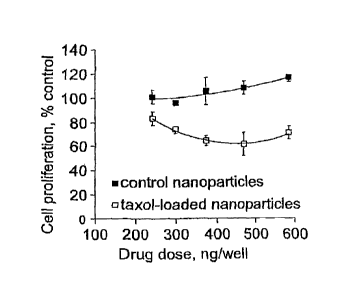
Figure 2 shows the effect of taxol-loaded magnetic nanoparticles on the
proliferation of cultured rat aortic smooth muscle cells as a function of the
nanoparticle
amount.
Figure 3A shows transgene expression in cultured rat aortic smooth muscle
cells
as a function of the poly-L-arginine formulation amount and nanoparticle dose.
Figure 3B shows transgene (Green Fluorescent Protein, GFP) expression in
bovine
aortic endothelial cells as a function of the poly-L-arginine formulation
amount and
nanoparticle dose.
Figure 3C shows transgene expression in cultured endothelial cells as a
function
of magnetic exposure.
Figure 3D shows the kinetics of transgene expression in cultured endothelial
cells
treated with poly-L-arginine modified nanoparticles at a dose equivalent to
285x106 viral
particles per well with or without a magnetic field.
CA 02783366 2012-06-07
WO 2011/075255 PCT/US2010/056674
- 6 -
Figure 4 shows an exemplary magnetically assisted therapeutic system according
to an embodiment of the invention.
Figure 5 shows a flowchart illustrating an exemplary method for administering
a
therapeutic agent to an implanted device and for retrieving magnetic carrier
nanoparticles that do not localize on the implanted device, according to an
embodiment
of the invention.
Figure 6A summarizes an exemplary embodiment of the magnetically assisted
therapeutic system, in which albumin modified magnetic carrier nanoparticles
with a red
fluorescent label were injected into a rat having an intravascularly implanted
steel stent.
Figure 6B summarizes results of the therapeutic agent delivery, for
sequestering
in the implanted device.
Figure 7 summarizes schematically the retrieval system shown in Figure 5 that
is
used to model the retrieval of magnetic carrier nanoparticles or cells from
the
cardiovascular circulation cycle.
Figure 8 summarizes exponential depletion kinetics of carrier nanoparticles
under
the influence of a magnetic field gradient.
Figure 9 summarizes exponential depletion kinetics of carrier cells under the
influence of a magnetic field gradient.
Figure 10 summarizes how different magnetic sequestering configurations, for
performing the exemplary method shown in Figure 5, affect depletion kinetics.
Figures 11A and 11B summarize results of transmission electron microscopy and
magnetic moment versus magnetic field (magnetization curve) for Albumin-
stabilized
superparamagnetic nanoparticles (MNP).
Figures 12A and 12B summarize in vitro MNP cell loading studies with respect
to
the kinetics of MNP uptake and viability of cells loaded with MNP.
Figure 13A shows reporter gene transfer mediated by polyethylenimine-coated
MNP combined with DNA encoding green fluorescent protein (GFP) presented as
GFP
fluorescence at Aem/Aex of 485 nm/535 nm as a function of a nanoparticle
amount in
A10 cells, wherein iron oxide-loaded nanoparticles were prepared using 0 ml
THF (large
nanoparticles, LNP), 3 ml THF (medium nanoparticles, MNP), or 4.5 ml THF
(small
nanoparticles, SNP) in the organic phase, versus large nanoparticles without
iron oxide
(LNP Non Mag, used herein as a control).
CA 02783366 2012-06-07
WO 2011/075255 PCT/US2010/056674
- 7 -
Figure 13B shows the relative fluorescence measured at 485 nm/535 nm as a
function of a nanoparticle amount in BAEC cells.
Figure 13C shows internalization of fluorescent (far red) labeled
nanoparticles
expressed as the relative fluorescence measured at 650 nm/670 nm as a function
of a
nanoparticle amount in A10 cells.
Figure 13D shows the relative fluorescence measured at 650 nm/670 nm as a
function of a nanoparticle amount in BAEC.
Figure 13E shows cell survival as a function of a nanoparticle amount in A10
cells.
Figure 13F shows cell survival as a function of a nanoparticle amount in BAEC
cells.
Figure 14 shows suppression of eGFP expression in lentivirus-transduced smooth
muscle cells (A10) by siRNA delivered with magnetic nanoparticles in the
presence of a
magnetic field (500 Oe).
Figure 15A shows magnetization curves of 304 (left sided Y axis) and 316L
(right
sided Y axis) grade stainless steel stents. The 304 stainless steel stent
exhibits a near
superparamagnetic behavior showing slight hysteresis and a remnant
magnetization on
the order of 7% of the saturation magnetization value. By comparison, the 316L
stent
shows far less magnetic responsiveness.
Figure 15B shows micrographs of BAEC's in culture (magnification of x100) with
bright field and red fluorescent images qualitatively showing the relative
amount of MNP
internalized within cells at different time points at the applied MNP dose of
9 pg/well.
Green fluorescent micrographs show cell viability as assessed by Calcein Green
staining.
Figure 16A shows in vitro capture kinetics of magnetically responsive BAEC
onto a
304 grade stainless steel stent in the presence of a uniform field of 1000
Gauss and a
nonpulsatile flow with a rate of 30 ml/min. The initial capture rate was
estimated to be
1% of cells/min. The data were obtained by measuring the fluorescence of
internalized
MNP.
Figures 16B and 16C show magnetically responsive BAEC captured in vitro onto a
304 stainless steel stent as evidenced by the red fluorescence of internalized
MNP, or by
Calcein Green staining of live cells, respectively.
Figure 16D shows MNP loaded BAEC captured in vivo onto a deployed 304
stainless steel stent in rat carotid artery. BAEC preloaded with fluorescent
MNP were
transthoracically injected into the left ventricular cavity. Animals were
exposed to a
magnetic field of 1000 Gauss for 5 minutes including the period of injection.
The
CA 02783366 2012-06-07
WO 2011/075255 PCT/US2010/056674
- 8 -
animals were sacrificed 5 minutes after delivery, and the explanted stents
were
immediately examined by fluorescence microscopy.
Figure 16E shows control rats subjected to an identical procedure, where no
magnetic field was employed. Micrographs (b-e) were obtained at the
magnification of
x40.
Figure 16F shows in vivo local magnetic cell delivery in a rat carotid
stenting
model under stop-flow conditions. A catheter was introduced via the external
carotid
into the common carotid artery and was positioned distal to a deployed stent.
The cell
suspension was delivered into isolated arterial segments for 15 sec.
Figure 16G shows in vivo cell delivery under uninterrupted blood flow
conditions.
A catheter was introduced via the external carotid into the common carotid and
advanced beyond the stent to the aortic arch. The cells were injected at this
site at the
rate of 1m1/min during one minute. For both delivery protocols (f and g), in
the
magnetic group (Mag+) the injection was carried out with animals placed in a
magnetic
field of 1000 Gauss, and the field was maintained for a total of 5 minutes
following
delivery. In control rats (Mag- group) no magnetic field was applied. In both
settings
BAEC were first transduced in culture with luciferase adenovirus and then
loaded with
MNP. The animals were imaged 48 hours post delivery by local perivascular
administration of luciferin admixed in a Pluronic gel. The signal emitted from
the stented
arterial segment due to the luciferase transgene expression was significantly
higher in
the animals that received cells in the presence of a magnetic field (Mag+
group).
Figure 17 shows that a MRI imager can magnetize a 316L steel stent for cell
targeting. In the presence of a magnetic field (Mag+), BAECs preloaded with
red
fluorescent polylactic acid (PLA) MNP are shown to localize to the magnetized
steel stent.
Controls (Mag-) did not show a significant localization to the stent.
Figure 18A is a TEM image of catalase loaded MNPs.
Figure 18B is a graph showing magnetic behavior of MNPs.
Figures 18C and 18D are bar graphs showing the size distribution of MNPs.
Figure 19A shows % SOD activity retained relative to mass added.
Figure 19B shows % Mass of SOD loaded relative to mass added.
Figure 19C shows the calculated number of molecules per particle based on mass
loading.
CA 02783366 2012-06-07
WO 2011/075255 PCT/US2010/056674
- 9 -
Figure 20A shows catalase loading versus addition measured by radiotracing of
1251-catalase.
Figure 20B shows the activity of loaded catalase as measured by degradation of
H202 absorbance at 242 nm over time.
Figure 21A shows catalase mass added to MNP versus mass protected from
proteolysis from 0.2 wt% Pronase at 37 C shaken for 1 hr.
Figure 21B shows (-=-) activity of catalase loaded into MNP versus time versus
time with exposure to 0.2 wt% Pronase at 37 C and (-o-) activity of free
catalase
versus time versus time with exposure to 0.2 wt% Pronase at 37 C.
Figure 22 shows the stability of MNP in plasma at 37 C over time. Release of
catalase measured by radiotracing of 1251-catalase. MNP incubated with aqueous
glucose
solution (5%) or plasma at 37 C over time. Release of catalase determined by
centrifuging free catalase from particles and measuring activity in
supernantant versus
retained MNP on microcentrifuge concentrator filter. (-=-) Released catalase
from MNPs
diluted in glucose solution. (-o-) Released catalase from MNPs diluted in
whole
heparinized mouse plasma.
Figure 23A shows phase contrast micrograph of 10 min magnetic delivery of
MNPs.
Figure 23B shows fluorescent micrograph of 10 min magnetic delivery of MNP
containing Dylight 488-labeled catalase.
Figure 23C shows 5 min MNP magnetic delivery.
Figures 23D and 23E show MNP delivery for 10 min without magnetic field.
Figure 23F shows the control without MNP.
Figures 24A and 24B show the protection of HUVECs from oxidative stress
through magnetic delivery of catalase loaded MNPs.
Figure 25 illustrates particle formation and synthesis.
Figure 26 shows biotinylated MNP affinity.
Figure 27 shows protection of HUVECs from oxidative stress through magnetic
delivery of catalase loaded MNPs and protection of HUVECs from oxidative
stress through
antibody-targeted delivery.
CA 02783366 2012-06-07
WO 2011/075255 PCT/US2010/056674
- 10 -
Figure 28 shows biodistribution of Ab62-modified MNP vs. control IgG-modified
MNP after tail vein injection in mice. Tail vein injected anti-PECAM labeled
MNP
specifically target the lung endothelium.
DETAILED DESCRIPTION OF THE INVENTION
Various terms relating to the methods and other aspects of the present
invention
are used throughout the specification and claims. Such terms are to be given
their
ordinary meaning in the art unless otherwise indicated. Other specifically
defined terms
are to be construed in a manner consistent with the definition provided
herein.
As used in this specification and the appended claims, the singular forms "a,"
"an," and "the" include plural referents unless the content clearly dictates
otherwise.
Thus, for example, reference to "a particle" includes a combination of two or
more
particles, and the like.
The term "about" as used herein when referring to a measurable value such as
an
amount, a temporal duration, and the like, is meant to encompass variations of
20% or
10%, more preferably 5%, even more preferably 1%, and still more preferably
0.1% from the specified value.
"Polynucleotide," also referred to as "nucleic acid" or "nucleic acid
molecule,"
refers to any polyribonucleotide or polydeoxribonucleotide, which may be
unmodified
RNA or DNA or modified RNA or DNA. Polynucleotides include, without limitation
single-
and double-stranded DNA, DNA that is a mixture of single- and double-stranded
regions,
single- and double-stranded RNA, and RNA that is mixture of single- and double-
stranded regions, hybrid molecules comprising DNA and RNA that may be single-
stranded or, more typically, double-stranded or a mixture of single- and
double-stranded
regions. In addition, polynucleotide refers to triple-stranded regions
comprising RNA or
DNA or both RNA and DNA. The term polynucleotide also includes DNAs or RNAs
containing one or more modified bases and DNAs or RNAs with backbones modified
for
stability or for other reasons. Modified bases include, for example,
tritylated bases and
unusual bases such as inosine. A variety of modifications can be made to DNA
and RNA;
thus, polynucleotide embraces chemically, enzymatically or metabolically
modified forms
of polynucleotides as typically found in nature, as well as the chemical forms
of DNA and
RNA characteristic of viruses and cells. Polynucleotide also embraces
relatively short
polynucleotides, often referred to as oligonucleotides.
"Polypeptide" refers to any peptide or protein comprising two or more amino
acids joined to each other by peptide bonds or modified peptide bonds, i.e.,
peptide
isosteres. Polypeptide refers to both short chains, commonly referred to as
peptides,
CA 02783366 2012-06-07
WO 2011/075255 PCT/US2010/056674
- 11 -
oligopeptides or oligomers, and to longer chains, generally referred to as
proteins.
Polypeptides may contain amino acids other than the 20 gene-encoded amino
acids.
Polypeptides include amino acid sequences modified either by natural
processes, such as
post-translational processing, or by chemical modification techniques which
are well
known in the art. Such modifications are well described in basic texts and in
more
detailed monographs, as well as in a voluminous research literature.
Modifications can
occur anywhere in a polypeptide, including the peptide backbone, the amino
acid side-
chains and the amino or carboxyl termini. It will be appreciated that the same
type of
modification may be present in the same or varying degrees at several sites in
a given
polypeptide. Also, a given polypeptide may contain many types of
modifications.
Except when noted, "subject" or "patient" are used interchangeably and refer
to
any animal, but preferably refer to mammals such as humans and non-human
primates,
as well as companion, farm, or experimental animals such as rabbits, dogs,
cats, rats,
mice, horses, cows, pigs, and the like. Humans are most preferred.
"Effective amount" or "therapeutically effective amount" are used
interchangeably
herein, and refer to an amount of a therapeutic agent, as described herein,
effective to
achieve a particular biological result such as, but not limited to, biological
results
disclosed, described, or exemplified herein, as determined by any means
suitable in the
art.
"Pharmaceutically acceptable" refers to those properties and/or substances
which
are acceptable to the patient from a pharmacological/toxicological point of
view and to
the manufacturing pharmaceutical chemist from a physical/chemical point of
view
regarding composition, formulation, stability, patient acceptance and
bioavailability.
"Pharmaceutically acceptable carrier" refers to a medium that does not
interfere with the
effectiveness of the biological activity of the therapeutic agent(s) and is
not toxic to the
host to which it is administered.
It has been discovered in accordance with the present invention that
therapeutic
agents can be targeted to specific locations in the body through use of
uniform magnetic
fields to induce magnetization of magnetizable objects and to generate a
magnetic field
gradient. It has further been discovered that magnetic targeting can be
utilized for any
type of therapeutic agent, including pharmaceutical or chemical compounds,
biomolecules, and cells. Accordingly, the invention features systems and
methods for
magnetically targeting therapeutic agents to one or more desired locations in
the body.
In one aspect, the systems comprise therapeutic agents provided as part of a
therapeutic formulation. The therapeutic formulation can comprise an effective
amount
CA 02783366 2012-06-07
WO 2011/075255
PCT/US2010/056674
- 12 -
of a therapeutic agent and a particle, which particle can comprise a magnetic
or
magnetizable material. Preferably, the particle is a nanoparticle. The
associated particle
and therapeutic agent are synonymously referred to herein as a therapeutic
particle.
Magnetic nanoparticles include particles that are permanently magnetic and
those that
are magnetizable upon exposure to an external magnetic field, but lose their
magnetization when the field is removed. Materials that are magnetic or
magnetizable
upon exposure to a magnetic field that lose their magnetic properties when the
field is
removed are referred to herein as superparamagnetic material.
Superparamagnetic
particles are preferred to prevent irreversible aggregation of the particles.
Examples of
suitable superparamagnetic materials include, but are not limited to, iron,
mixed iron
oxide (magnetite), or gamma ferric oxide (maghemite) as well as substituted
magnetites
that include additional elements such as zinc. Preferably, the
superparamagnetic
material is in the form of one or more nanocrystals, for example, single-
domain
crystalline systems with at least one dimension 5_ 100nm. A nanocrystal is any
nanomaterial with at least one dimension 100nm and that is
singlecrystalline or
monocrystalline, formed of a single crustal-unit, and so all elements have
identical
crystallographic orientation of c- and a-axes and overgrow as one unit. Any
particle that
exhibits regions of crystalinity can be termed nanoparticle or nanocluster
based on
dimensions.
Ferromagnetic crystals can be comprised of magnetized domains the size of a
micron. Superparamagnetism can occur when the size of the crystals is smaller
than the
ferromagnetic domain (-30nm). Superparamagnetic properties can depend on
temperature. Temperature can, under some conditions, destabilize the
magnetism.
Without intending to be limited to any particular theory or mechanism of
action, it is
believed that thermal energy may prevent the alignment of the magnetic moments
present in superparamagnetic materials. After the removal of an applied
magnetic field,
the magnetic moments of superparamagnetic materials still exist, but are in
rapid
motion, causing a randomly oriented or disordered magnetic moment and, thus,
no net
magnetic field. At the temperatures of biological systems and in the applied
magnetic
fields of MR imagers, superparamagnetic materials are less magnetic than their
ferromagnetic counterparts. For example, it has been noted that magnetism of
small
superparamagnetic iron oxides decreases at elevated temperatures. (Berkowitz
et al.
(1968) J. Appl. Phys. 39:1261).
The superparamagnetic nanocrystals can range in size from about mm to about
20nm, depending on, among other things, the preparation method and medium
composition. Preferably, the nanocrystals are smaller than 10-20 nm to ensure
CA 02783366 2012-06-07
WO 2011/075255 PCT/US2010/056674
- 13 -
superparamagnetic properties of the material. More preferably, the
nanocrystals are
from about 5nm to about 20nm.
In some aspects, the particle is a composite nanocrystal. The composite
nanocrystal can comprise more than one individual magnetic or magnetizable
nanocrystals and one or more water-insoluble biocompatible materials to hold
the
crystals together. The biocompatible materials can be a polymer, which can be
biodegradable or non-biodegradable. Non-limiting examples of such polymers
include
poly(urethane), poly(ester), poly(lactic acid), poly(glycolic acid),
poly(lactide-co-
glycolide), poly(E-caprolactone), poly(ethyleneimine), poly(styrene),
poly(amide),
rubber, silicone rubber, poly(acrylonitrile), poly(acrylate),
poly(methacrylate), poly(a-
hydroxy acid), poly(dioxanone), poly(orthoester), poly(ether-ester),
poly(lactone),
poly(alkylcyanoacrylate), poly(anhydride), poly(ethylenevinyl acetate),
poly(hydroxybutyrate), poly(tetrafluoroethylene), poly(ethylene terephthalate,
polyoxyethylene, polyoxyethlyene-polyoxypropylene block copolymers, mixtures
thereof
and copolymers of corresponding monomers.
Polymeric nanoparticles with incorporated superparamagnetic nanocrystals can
be
prepared according to any means suitable in the art. For example, the
nanoparticles can
be prepared by dispersing the superparamagnetic nanocrystals in an organic
solvent, in
which the polymer and/or the therapeutic agent is dissolved, emulsifying the
organic
phase in water in the presence of a suitable stabilizer, and finally
eliminating the solvent
to obtain solidified nanoparticles. Conditions of nanoparticle preparation
should not be
damaging for the therapeutic agent to be attached. The temperature for
nanoparticle
preparation preferably ranges from about 25 C to about 37 C, although higher
or lower
temperatures can be used. Non-limiting examples of ways to prepare
superparamagnetic nanoparticles for biological applications are described in
U.S. Patent
Nos. 7,175,912 and 7,175,909, and U.S. Publication No. 20050271745. Magnetic
nanoparticles, information for the development of magnetic nanoparticles, and
reagents
for the preparation of magnetic nanoparticles (MNP) are commercially
available.
The particles can be composed of the salts/complexes of anionic lipids, for
example, fatty acids or lipid phosphates with polyvalent biocompatible
cations. The
particles can be formed under mild conditions through combination of the
respective
aqueous solutions in the presence of colloid stabilizers, thus avoiding use of
organic
solvents and without need for external mechanical energy input.
In some preferred aspects, the particles are bioresorbable nanoparticles,
including those prepared without the use of high energy dispersion or organic
solvents.
CA 02783366 2012-06-07
WO 2011/075255 PCT/US2010/056674
- 14 -
Bioresorbable nanoparticles can be comprised of at least one anionic lipid
salt, at least
one therapeutic agents, and at least one magnetic or magnetizable material.
To prepare bioresorbable nanoparticles, a first aqueous solution is provided.
The
first aqueous solution comprises i) a water soluble salt of a mono-carboxylic
fatty acid,
or salt thereof, or a lipid phosphate/phosphonate, ii) a stabilizer, and iii)
a therapeutic
agent. Exemplary soluble salts include, but are not limited to, the lithium,
sodium,
ammonium, and potassium salts. An aqueous solution of the fatty acid salt can
be
prepared, for example, by adding the fatty acid and base, such as sodium
hydroxide, to
water and dissolving the fatty acid.
Fatty acids that can be used include straight and branched chain, saturated
and
unsaturated mono-carboxylic fatty acids having eight or more carbon atoms,
particularly
eight to thirty carbon atoms. Typical mono-carboxylic fatty acids include
caprylic acid
(octanoic acid), 2-ethyl octanoic acid, capric acid (decanoic acid), 2-ethyl-
decanoic acid,
11-undecenoic acid, undecanoic acid, 2-ethyl-dodecanoic acid, cis-5-dodecenoic
acid,
lauroleic acid (cis-9-dodecanoic acid), traumatic acid (2-dodecenoic acid),
lauric acid
(dodecanoic acid), brassylic acid (tridecanoic acid), 2-ethyl-tetradecanoic
acid,
myristoleic acid (cis-9-tetradecanoic acid), tsuzuic acid (cis-4-tetradecenoic
acid),
myristic acid (tetradecanoic acid), pentadecanoic acid, 2-ethyl-hexadecanoic
acid,
palmitoleic acid (cis-9-hexadecanoic acid), palmitic acid (hexadecanoic acid),
heptadecanoic acid, margaric acid (heptadecanoic acid), petroselic acid (cis-6-
octadecenoic acid), 2-ethyl-octadecanoic acid, oleic acid (cis-9-octadecenoic
acid),
elaidic (trans-9-octadecenoic acid), asclepinic acid (cis-11-octadecenoic
acid), vaccenic
acid (trans-11-octadecenoic acid), taxoleic acid (cis, cis-5,9-
octadecadienoic), linoleic
acid (cis, cis-9,12-octadecadienoic acid), linolenic acid (cis, cis, cis-
9,12,15-
octadecatrienoic acid), stearic acid (octadecanoic acid), tuberculostearic
acid (10-methyl
octadecanoic acid), nonadecanoic acid, 2-ethyl-eicosanoic acid, arachidonic
acid
(5,8,11,14-eicosatetraenoic acid), cis-8,11,14-eicosatrienoic acid, gadoleic
acid (cis-9-
eicosenoic acid), gondoic acid (cis-11-eicosenoic acid), arachidic acid
(eicosanoic acid),
2-ocyldodecanoic acid, erucic acid (cis-13-docosenoic acid), behenic acid
(docosanoic
acid), tricosanoic acid, selacholeic acid (cis-15-tetracosanoic acid),
lignoceric acid
(tetracosanoic acid), ximenic acid (cis-17-hexacosenoic acid), and
hexacosanoic acid. A
particularly preferred fatty acid is oleic acid. Salts of the fatty acids,
including for
example, alkaline metal and alkaline earth metal salts, and ammonium salts,
can also be
used.
As an alternative to fatty acids, lipid phosphates, such as the water soluble
mono-
phosphate salts of alcohols having eight or more carbon atoms, more preferably
eight to
CA 02783366 2012-06-07
WO 2011/075255 PCT/US2010/056674
- 15 -
thirty carbon atoms, can also be used. Such phosphates include a-tocopherol
phosphate
disodium salt, ()leyl phosphate disodium salt, and the disodium salts of the
phosphate
esters of straight and branched chain, saturated and unsaturated mono-alcohols
having
eight or more carbon atoms, such as the disodium salts of the phosphate esters
of n-
decanol, n-dodecanol, n-tetradecanol, n-hexadecanol, and n-octadecanol.
Hydroxy acids such as 11-hydroxy-undecanoic acid, ricinoleic acid (12-hydroxy-
cis-9-octadecenoic acid), lesquerolic acid (14-hydroxy-cis-11-eicosenoic acid:
20:1-0H),
densipolic acid (12-hydroxy-cis, cis-9,15-octadecadienoic acid) auricolic acid
(14-
hydroxy-cis, cis-11,17-eicosadienoic acid), 9,10-dihydroxyoctadecanoic acid,
9,14-
dihydroxyoctadecanoic acid, and phellonic acid (22-hydroxydocosanoic acid) and
salts
thereof can also be used.
Lipid phosphates, such as the water soluble mono-phosphate salts of alcohols
having eight or more carbon atoms, more preferably eight to thirty carbon
atoms, can
also be used. Such phosphates include a-tocopherol phosphate disodium salt,
oleyl
phosphate disodium salt, and the disodium salts of the phosphate esters of
straight and
branched chain, saturated and unsaturated mono-alcohols having eight or more
carbon
atoms, such as the disodium salts of the phosphate esters of n-decanol, n-
dodecanol, n-
tetradecanol, n-hexadecanol, and n-octadecanol.
A colloidal stabilizer, or mixture of colloid stabilizers, can be added to the
aqueous
solution of the fatty acid salt or to the aqueous solution containing the
polycation.
Colloidal stabilizers are materials believed to be adsorbed onto the
nanoparticles,
thereby providing charge or steric protection of the particles from
aggregation. Suitable
stabilizers include secondary colloids, such as gelatin, agar-agar, starch,
cellulose
derivatives such as carboxymethyl cellulose and hydroxypropyl cellulose, and
proteins,
such as albumin. Non-ionic surfactants, such as polyethylene oxide, ethylene
oxide/propylene oxide block co-polymers, for example, PLURONIC surfactants,
and
ethoxylated fatty acid esters of esters of sorbitol, such as polyoxyethylene
(20) sorbitan
monolautate (TWEEN 20), polyoxyethylene (20) sorbitan monopalmitate (TWEEN
40),=
polyoxyethylene (20) sorbitan monosterate (TWEEN 60), polyoxyethylene (20)
sorbitan
monooleate (TWEEN 80), or polyoxyethylene (20) sorbitan trioleate (TWEEN 85)
can
also be used. A preferred stabilizer is albumin. PLURONIC is a registered
trademark of
BASF Corporation and TWEEN is a registered trademark of Croda International
PLC.
A second aqueous solution comprising a water-soluble salt of a polyvalent
biocompatible metal or organic cation can be added to the first aqueous
solution. A
cation is biocompatible if it is non-toxic to the recipient in the quantities
used, and also
presents no significant deleterious or untoward effects on the recipient's
body. Useful
CA 02783366 2012-06-07
WO 2011/075255 PCT/US2010/056674
- 16 -
biocompatible polyvalent cations include, without limitation, Al+3, Ca+2,
Mg+2, Zn+2, Ba+2,
Sr+2, Fe+2, and Cu+2, polyarginine, protamine. Preferred biocompatible
polyvalent
cations include Ca+2 and ZN1+2.
The size of the bioresorbable particles can be readily controlled and adapted
for
specific applications by adjusting the amount of the stabilizer, lipid salt,
and polyvalent
biocompatible cation. Although the particles are generally lipophilic, showing
high
affinity for hydrophobic therapeutic agents, ionic water-soluble therapeutic
agents can
also be encapsulated as their water-insoluble salts/complexes in the process
of particle
formation. The nature and amount of the lipid salt and polyvalent
biocompatible cation
can be varied in order to adjust the relative lipophilicity of the resulting
particles.
A cationic peptide, cationic protein, or a mixture of cationic peptides and/or
cationic proteins can also be co-added to the second aqueous solution
containing a metal
polycation. Preferred cationic peptides contain at least about 50%, preferably
at least
about 70%, and more preferably at least about 85% of basic amino acid
residues, such
as arginine, lysine, and guanidine, and contain more then five amino acid
residues,
preferably about 10 to about 1000 residues. More preferred cationic peptides
include
arginine-rich polypeptides, such as poly-L-arginine. More preferred peptides
are
arginine-rich proteins, such as protamine. Guanidinium-rich proteins can also
be used.
Synthetic organic polycations (polypeptide-like substances), such as
polyethyleneimine,
can also be used.
Bioresorbable nanoparticles can be rendered magnetic through inclusion of
magnetically responsive nanocrystals in their structure, for example, by
combining a fine
suspension of such crystals (a ferrofluid) with the anionic lipid solution
prior to the
particle formation. Ferrofluids are composed of nanosacle ferromagnetic
particles
suspended in a carrier fluid, such as water. Preparation of such nanoparticles
is a two-
step process consisting of 1) making the fine suspension of magnetic
nanocrystals
(ferrofluid) in the presence of an anionic lipid, and 2) forming nanoparticles
by controlled
precipitation of the anionic lipid with a polyvalent cation in the presence of
a stabilizer
and a therapeutic agent. In one aspect, the magnetic nanoparticles are
prepared by
controlled aggregation of an oleate-stabilized ferrofluid with Ca+2.
To prepare a ferrofluid, an aqueous solution containing a water soluble ferric
(Fe+3) salt, such as ferric chloride hexahyd rate, and a water soluble ferrous
salt (Fe+2),
such as ferrous chloride tetrahydrate, is precipitated with base, such as an
aqueous
sodium hydroxide solution to form a magnetite precipitate containing magnetic
nanocrystals. A water soluble salt of a fatty acid, such as an aqueous
solution of sodium
oleate, is added, and the magnetic nanocrystals resuspended by heating, for
example, in
CA 02783366 2012-06-07
WO 2011/075255 PCT/US2010/056674
- 17 -
an inert atmosphere, such as under argon. A stabilizer such as albumin can be
added,
along with the therapeutic agent, either to the first aqueous solution, which
comprises
the magnetic nanocrystals, stabilizer, water soluble salt of a mono-carboxylic
fatty acid,
and therapeutic agent, or to the second aqueous solution, which comprises the
polyvalent biocompatible cation. The second solution is then added to form the
magnetic
nanoparticles.
In some aspects, the therapeutic agent can be attached or tethered to the
surface
of a pre-formed nanoparticle. The attachment can be according to any means
suitable
for the therapeutic application to which the agent will be used, or according
to the
chemical properties of the agent or the nanoparticle. For example, attachment
can be
by adsorption, electrostatic interactions, charge complexation, or covalent
binding,
including the use of biomolecule tethers. Non-limiting examples of procedures
for
associating therapeutic agents with nanoparticles are described in US Pat.
Nos.
7,081,489, 6,048,515, 6,576,221, and 6,767,635. The attachment can be by way
of a
linking molecule. Some non-limiting examples of linking molecule pairs include
avidin or
streptavidin and biotin, thiol and Succinimidyl 3-(2-pyridyldithio)-propionate
(SPDP) or
Succinimidyl 4[N-maleimidomethyl]cyclohexane-1-carboxylate (SMCC), or suitable
variants or isoforms thereof, and folate and the folate receptor
The magnetic nanoparticles associated with the therapeutic agent can range in
size from about 50 to about 500 nm. The size can vary according to any
appropriate
variable. Preferably, the nanoparticles range in size from about 50nm to about
200nm,
and more preferably from about 100nm to about 200nm.
The particle can be derivatized, and the surface of the particle can be
modified to
facilitate derivatization. For example, the particles can be coated with a
thiol-reactive
and photoactivatable polymer. Irradiation can facilitate the covalent binding
of the
polymer to the surface, and its thiol-reactive groups can subsequently be used
to attach
agents providing stealth properties in the blood circulation and/or specific
binding to a
target tissue. Photochemical activation of surfaces for attaching biomaterial
is described
in US Publ. No. 20060147413.
Extended circulation time of particles associated with a therapeutic agent can
be
achieved by preventing opsonization and clearance by the subject's immune
system by
coating the particle with a biocompatible hydrophilic polymer such as
polyethyleneglycol
or dextran, or by coating the particle with albumin to inhibit the binding of
opsonins to
the particle surface.
CA 02783366 2012-06-07
WO 2011/075255 PCT/US2010/056674
- 18 -
Preparation of modified particles can proceed according to any means suitable
in
the art. For example, a magnetically responsive agent, iron oxide, can be
produced.
Fine dispersion of iron oxide in a suitable organic solvent is typically
obtained as follows:
an aqueous solution containing ferric and ferrous chlorides is mixed with an
aqueous
solution of sodium hydroxide. The precipitate is coated with oleic acid by
short
incubation at 90 C in ethanol. The precipitate is washed once with ethanol to
remove
free acid and dispersed in chloroform.
The resulting organic dispersion of iron oxide in chloroform is used to
dissolve a
biodegradable polymer, polylactic acid (PLA) or its polyethyleneglycol
conjugate (PLA-
PEG), thus forming an organic phase. The organic phase is emulsified in an
aqueous
albumin solution (1%) by sonication on an ice bath followed by evaporation of
the
organic solvent. The particles are separated from the unbound albumin by
repeated
magnetic sedimentation/resuspension cycles.
In an alternative aspect, a post-formation surface modification can be used.
For
example, particles can be formed using a photoreactive polymer (e.g.,
PBPC/PBMC,
polyallylamine- benzophenone-pyridyldithio/maleimido-carboxylate polymer) as a
stabilizer in the aqueous phase. Subsequent brief ultraviolet irradiation
achieves
covalent binding of the polymer to the magnetic nanoparticle. The resulting
particles are
reacted in suspension with a thiolated polyethyleneglycol, which allows better
control
over the particle size and the extent of surface modification.
Therapeutic agents include any molecules that can be associated with a
particle
and used in the systems and methods of the present invention. Agents can be
purified
molecules, substantially purified molecules, molecules that are one or more
components
of a mixture of compounds, or a mixture of a compound with any other material.
Agents
can be organic or inorganic chemicals, radioisotopes, pharmaceutical
compounds,
pharmaceutical salts, pro-drugs, or biomolecules, and all fragments, analogs,
homologs,
conjugates, and derivatives thereof. Biomolecules include, without limitation,
proteins,
polypeptides, nucleic acids, lipids, polysaccharides, monosaccharides, and all
fragments,
analogs, homologs, conjugates, and derivatives thereof. Agents can also be an
isolated
product of unknown structure, a mixture of several known products, or an
undefined
composition comprising one or more compounds. Examples of undefined
compositions
include cell and tissue extracts, growth medium in which prokaryotic,
eukaryotic, and
archaebacterial cells have been cultured, fermentation broths, protein
expression
libraries, and the like. Agents can also be one or more cells, including
eukaryotic or
prokaryotic cells, or can be one or more viruses. Therapeutic agents can be
provided in
or otherwise associated with a carrier such as a pharmaceutically acceptable
carrier.
CA 02783366 2012-06-07
WO 2011/075255 PCT/US2010/056674
- 19 -
Therapeutic agent also includes viral vector systems, which are used in gene
therapy. A number of viral vector systems under development, such as
adenovirus,
adeno-associated virus, retrovirus and Herpes simplex virus. One of the most
successful
ways of introducing the gene of interest into the appropriate cell line uses
recombinant
adenovirus. Adenoviruses are non-enveloped particles having a diameter of
about 70
nm, and contain a linear double stranded DNA of approximately 36,000 base
pairs. They
are easily prepared with high titers and can infect a wide range of cells,
including non-
dividing cells. Recombinant adenovirus can also be used in vaccination by
expressing a
gene product that triggers an immune response.
Adeno-associated viruses have a particle diameter of 20 nm. Retroviruses are
spherical, enveloped particles having a particle diameter of between about
80nm to
about 100 nm in diameter. Retroviruses have been widely used as vectors for
DNA
delivery. Herpes simplex viruses have a particle diameter of about 100 nm, and
contain
enveloped, double-stranded DNA virus of approximately 150,000 base pairs.
These
viruses have a large loading capacity for foreign genes and are able to infect
a wide
range of cells. In addition, the virus genome remains episomal after
infection, thus
eliminating the possibility of opportunistic malignant insertional mutagenesis
of the host
genome. Herpes viruses have been exploited for specific gene transfer trials
into the
central nervous system.
Multiple agents can be included in a particle. Those of skill in the art can
determine the particular combination of agents, based, for example, on the
condition
being treated, or on the needs of the particular subject. For example,
additional agents
that modulate the activity of a primary agent, reduce pain, support growth of
therapeutic
cells, are antithrombogenic, anti-apoptotic, anti-inflammatory,
immunosuppressants, or
antioxidants, or other agents ordinarily used in the art to treat the disease
of interest
can be used.
The therapeutic agents can also be formulated in sustained release vehicles or
depot preparations. For example, the agents can be formulated with suitable
polymeric
or hydrophobic materials (for example, as an emulsion in an acceptable oil) or
ion
exchange resins, or as sparingly soluble derivatives, for example, as a
sparingly soluble
salt. Liposomes and emulsions are well-known examples suitable for use as
carriers for
hydrophobic drugs.
In some preferred aspects, the therapeutic agents are enzymes. For example,
antioxidant enzymes can be used. Antioxidant enzymes include, without
limitation,
catalase, superoxide dismutase, and glutathione peroxidase. Other examples of
proteins
include antibodies. Any antibody suitable for the purpose to which the
particle is being
CA 02783366 2012-06-07
WO 2011/075255 PCT/US2010/056674
- 20 -
used can be included. The antibodies can be the therapeutic agent, or can be
used to
help guide the particle to targeted tissue.
In some preferred aspects, the therapeutic agents are regulatory nucleic
acids.
For example, regulatory nucleic acids can be used to facilitate post-
transcriptional gene
silencing (RNA silencing). RNA silencing involves the processing of double-
stranded RNA
(dsRNA) into small 21-28 nucleotide fragments by an RNase H-based enzyme. The
cleavage products are siRNA (small interfering RNA) or miRNA (micro-RNA),
which
regulate gene expression in a sequence-specific manner. Regulatory nucleic
acids can
be part of a plasmid or other suitable vector, for example, administered as
DNA that is
transcribed and processed to regulatory RNA.
siRNAs may thus comprise RNA molecules having a double-stranded region
approximately 19 nucleotides in length with 1-2 nucleotide 3' overhangs on
each strand,
resulting in a total length of between approximately 21 and 23 nucleotides. As
used
herein, siRNAs also include various RNA structures that may be processed in
vivo to
generate such molecules. Such structures include RNA strands containing two
complementary elements that hybridize to one another to form a stem, a loop,
and
optionally an overhang, preferably a 3' overhang. Preferably, the stem is
approximately
19 bp long, the loop is about 1-20, more preferably about 4-10, and most
preferably
about 6-8 nt long and/or the overhang is about 1-20, and more preferably about
2-15 nt
long. In certain embodiments of the invention the stem is minimally 19
nucleotides in
length and may be up to approximately 29 nucleotides in length. Loops of 4
nucleotides
or greater are less likely subject to steric constraints than are shorter
loops and
therefore may be preferred. The overhang may include a 5' phosphate and a 3'
hydroxyl. The overhang may, but need not comprise a plurality of U residues,
e.g.,
between 1 and 5 U residues.
miRNAs are typically between approximately 20 and 26 nucleotides in length,
e.g., 22 nt in length. It is believed that they are derived from larger
precursors known
as small temporal RNAs (stRNAs) or mRNA precursors, which are typically
approximately
70 nt long with an approximately 4-15 nt loop.
Viral vectors or DNA vectors encoding short hairpin RNA (shRNA) which are
processed in the cell cytoplasm to short interfering RNA (siRNA) can also be
used. A
plasmid containing a DNA sequence encoding for a particular desired siRNA
sequence
can be delivered to a target cell, and subsequently internalized, for example,
by virally-
mediated infection. Once in the cell, the DNA sequence is continuously
transcribed into
RNA molecules that loop back on themselves and form hairpin structures through
intramolecular base pairing. These hairpin structures, once processed by the
cell, are
CA 02783366 2012-06-07
WO 2011/075255 PCT/US2010/056674
- 21 -
equivalent to transfected siRNA molecules and are used by the cell to mediate
RNAi of
the desired protein. The use of shRNA has an advantage over siRNA as the
former can
lead to stable, long-term inhibition of protein expression. In cases where
longer periods
of protein inhibition are necessary, shRNA as the therapeutic agent is
preferable.
In some preferred aspects, the therapeutic agent is one or more cells. For
example, the cell can be a stem cell such as a postpartum derived cell or bone
marrow
derived cell or can be a progenitor cell, Blood Outgrowth Endothelial Cell
(BOED), adult
and cord blood stem cells (CBSC), Induced Pluripotent Stem Cells, e.g., skin
cells that
are programmed to transform into pluripotent stem cells with further potential
to
differentiate them into cell with at least one endothelial phenotype. In some
exemplary
aspects, the cell is a vascular cell such as a vascular endothelial cell.
Endothelial cells
can be autologous, heterologous, or derived from either blood, bone marrow, or
tissue
biopsy. Tissue biopsied endothelial cells can be derived from arteries, veins,
adipose
tissue, or any other tissue with the potential to contain endothelial cells or
their
progenitors.
In some aspects, the systems comprise an implantable device. Any implantable
device known or used in the art can be utilized in the inventive systems. Non-
limiting
examples of suitable implantable device include stents, heart valves, wire
sutures,
temporary joint replacements and urinary dilators, orthopedic implants such as
joint
prostheses, screws, staples, nails, nuts, bolts, plates, rods, pins, wires,
inserters,
osteoports, halo systems and other orthopedic devices used for stabilization
or fixation of
spinal and long bone fractures or disarticulations. Other devices include non-
orthopedic
devices, temporary placements and permanent implants, such as traceostomy
devices,
drainage ducts, jejunostomy and gastrostomy tubes, intraurethral and other
genitourinary implants, stylets, dilators, vascular clips and filters,
pacemakers, wire
guides and access ports of subcutaneously implanted vascular catheters,
electronic
chips, transmitting/receiving micro-electronic implants, implantable drug
delivery micro-
devices, implantable biosensors, implantable micro-video devices, and
implantable
microbattery devices. A highly preferred implantable device is a stent. The
stent can be
a drug-eluting stent.
Preferably, the implantable device comprises a magnetic or magnetizable
material. More preferably, such magnetic or magnetizable materials are
biocompatible.
The device can be modified to be biocompatible. For example, surface
modifications of
metal supports to improve biocompatibility are described in US Publ. No.
2003/0044408.
Stainless steel, for example, Grade 304 Stainless Steel is one preferred non-
limiting example of a material that can be used in the implantable device.
Other
CA 02783366 2012-06-07
WO 2011/075255 PCT/US2010/056674
- 22 -
examples includes the 200-series austenitic chromium-nickel-manganese alloys
and 300-
series austenitic chromium-nickel alloys.
The device can be implanted anywhere in the body of the subject. In some
preferred aspects, the device is implanted in the vascular system of the
subject, for
example, in a blood vessel such as a vein, artery, or capillary. Where stents
are used,
the stents can be implanted in any duct such as a hepatic duct, bile duct,
parts of the
digestive system such as the esophagus, stomach, intestines, or colon, parts
of the
respiratory system such as the trachea or bronchi, or parts of the excretory
system such
as the ureter, urethra, or renal excretory duct. Other implants include ocular
implants
and radioactive seeds.
In some aspects, the systems can comprise a retrieval system. In general, the
retrieval system can be used to capture and contain therapeutic particles that
do not
reach the target site, or to capture and contain particles after the
therapeutic agent has
been delivered to the target site, synonymously referred to herein as spent
particles.
Unused or spent particles may enter or remain in the blood of the subject, and
may
produce untoward effects in the subject. To minimize risks to the subject, it
is
preferable to remove such unused and/or spent particles. The retrieval system
preferably captures and contains most, and more preferably substantially all
unused
and/or spent particles such that the body or particular fluid, organ,
appendage, and the
like within the body is substantially free of spent or unused particles.
In some preferred aspects, the retrieval system comprises a magnetic or
magnetizable material. The material can be provided in any form suitable in
the art. For
example, the material can be a rod, plate, bead, tube, wire, panel, filter,
screen, mesh,
and the like. The particular form (geometry) is not critical, and can vary
according to
any number of variables.
The retrieval system is preferably biocompatible. Suitable materials that can
be
used to comprise the retrieval system include, but are not limited to
stainless steel such
as 316L stainless steel. 400-series stainless steel, including 430-grade can
also be used
in some instances, such as those for short term use where potential long-term
corrosive
properties of retrieval system materials is not of concern.
Preferably, the retrieval system is configured such that it is capable of
being
reversibly connected to the subject. The retrieval system can be reversibly
connected to
a subject at any location on the body suitable according to the therapeutic
use to which
the overall system is being used. For example, the retrieval system can be
reversibly
connected to the body surface, or to a particular interior organ, bone, or
system.
CA 02783366 2012-06-07
WO 2011/075255 PCT/US2010/056674
- 23 -
Preferably, the retrieval system is reversibly connected to at least one blood
vessel, and
more preferably to the lumen of the blood vessel to allow blood to directly
flow into the
retrieval system. The circulatory system is a high-flow, well-accessed system,
and is
thus highly preferred for connection to a retrieval system.
In some aspects, the retrieval system is indirectly connected to the subject.
For
example, a biological fluid of the subject can be removed and contacted with
the
retrieval system. Biological fluids can include, but are not limited to blood,
cerebrospinal
fluid, ascites fluid, bile, amniotic fluid, milk, saliva, gingival crevicular
fluid, urine,
mucosal fluid, renal fluid, and the like. After the particles are sequestered
from the
biological fluid, the biological fluid can be returned to the subject. Blood
is a particularly
preferred biological fluid. Thus, for example, the subject's blood may be
removed from
the body, contacted with the retrieval system, and returned to the body by
transfusion.
The blood can be separated into its components or can remain as whole blood.
Particles directed to the retrieval system can be disposed of according to any
means suitable in the art. After the particles are directed to the retrieval
system, the
retrieval system can be removed from the subject.
The particles comprising at least one therapeutic agent and at least one
magnetic
or magnetizable material are targeted to one or more desired locations in the
body, for
example, to one or more implanted devices in the body, through a magnetic
field. Thus,
the inventive systems can comprise a magnetic field generator. The magnetic
field
generator can include an external magnet, including a magnetic resonance
imaging
device. In some aspects, the magnetic field generator is configured to
generate at least
one directable magnetic field gradient. The magnetic field gradient can direct
the
particle to the implantable device. The magnetic field gradient can direct
particles not
delivered to the device to the retrieval system, or can direct spent
particles, that is,
particles that have successfully delivered and are depleted of the therapeutic
agent to
the retrieval system. A single magnetic field gradient can be used to direct
the particles
to the implantable device, and then reconfigured to direct extant unused or
spent
particles to the retrieval system. Alternatively, multiple gradients can be
produced and
used, with at least one gradient directing particles to the implanted device,
and at least
one additional gradient directing particles to the retrieval system. The
gradient can be
generated proximal to the implanted device, and can be generated proximal to
the
retrieval system.
Referring to Figures 4 and 5, an exemplary magnetically assisted therapeutic
system 100 is illustrated. The therapeutic system 100 comprises a device 104
that has
been implanted in a mammalian subject (not shown), a magnetic field generator
106,
CA 02783366 2012-06-07
WO 2011/075255 PCT/US2010/056674
- 24 -
configured to generate a magnetic field gradient external to the subject that
is directed
proximally to the implanted device 104, and a therapeutic particle 102 that
has been
administered to the subject. Device 104 can be a vascular device that has been
implanted in the vascular system of the mammalian subject.
Also featured in accordance with the present invention are methods for
magnetically targeting a therapeutic particle to one or more desired locations
in or on a
subject. In some preferred aspects, the methods are applicable to magnetically
target a
therapeutic particle to a device implanted in a subject.
The methods can comprise administering to a subject having an implanted device
comprising at least one biocompatible magnetic or magnetizable material a
therapeutic
particle comprising at least one therapeutic agent and at least one magnetic
or
magnetizable material, generating a magnetic field gradient proximal to the
implanted
device, wherein the gradient targets the particle to the implantable device,
and removing
particles not delivered to the implantable device, or removing spent
particles.
The therapeutic agent can be any molecule as described or exemplified herein,
including without limitation, a pharmaceutical, biomolecule, or cell. The
implanted
device can be any device used, known, or otherwise suitable in the art such as
those
described or exemplified herein.
In some preferred aspects, the methods are applicable to magnetically target a
therapeutic particle to a particular cell or tissue. These methods can be
carried out in
vitro, and preferably can be carried out in vivo. Thus, for example,
therapeutic particles
can be magnetically targeted to a particular location in the body of a
subject. The
location need not have an implanted device. A magnetic field can be used to
guide the
particle to the desired location, and can be used to facilitate
internalization of the
therapeutic particle by particular cells at the desired location.
In some detailed aspects, these methods can be used to protect cells from
oxidative damage. Thus, for example, methods for protecting a cell from
oxidative
damage comprise contacting the cell with a particle comprising a magnetic or
magnetizable material and at least one antioxidant enzyme, and generating a
uniform
magnetic field capable of magnetizing the magnetic or magnetizable material
proximal to
the cell for a period of time sufficient to permit the cell to internalize the
particle. The
cell can be in vitro or in vivo. The cell can be an endothelial cell, for
example, a vascular
endothelium cell. The antioxidant enzyme can be catalase, superoxide
dismutase, or
glutathione peroxidase. The particle can further comprise an antibody that
specifically
CA 02783366 2012-06-07
WO 2011/075255 PCT/US2010/056674
- 25 -
binds to an antigen on the surface of the cell. Thus, the antibody can be used
to
facilitate targeting to the proper target cell.
Administration of the therapeutic particles to a subject can be by infusion or
injection (intravenously, intramuscularly, intracutaneously, subcutaneously,
intrathecal,
intraduodenally, intraperitoneally, and the like). The particles can also be
administered
intranasally, vaginally, rectally, buccally, orally, or transdermally.
Preferably, the
compositions are administered intravenously. Administration can be at the
direction of a
physician. The particles can be administered proximally or distally to the
implanted
device.
For buccal administration, the compositions may take the form of tablets,
troche
or lozenge formulated in conventional manner. Compositions for oral or buccal
administration, may be formulated to give controlled release of the particles.
Such
formulations may include one or more sustained-release agents known in the
art, such
as glyceryl mono-stearate, glyceryl distearate and wax.
Particles may be applied topically. Such administrations include applying the
particles externally to the epidermis, the mouth cavity, eye, ear and nose.
Particles for
use in topical administration include, e.g., liquid or gel preparations
suitable for
penetration through the skin such as creams, liniments, lotions, ointments or
pastes,
and drops suitable for delivery to the eye, ear or nose.
Various alternative pharmaceutical delivery systems may be employed. Non-
limiting examples of such systems include liposomes and emulsions. Certain
organic
solvents such as dimethylsulfoxide also may be employed. Additionally, the
particles
may be delivered using a sustained-release system, such as semipermeable
matrices of
solid polymers containing the therapeutic agent. The various sustained-release
materials available are well known by those skilled in the art. Sustained-
release
capsules may, depending on their chemical nature, release the particles over a
range of
several days to several weeks to several months.
The particles may also be co-administered with other well known therapeutic
agents that are selected for their particular usefulness against the condition
that is being
treated. For example, such therapeutic agents can be pain relievers, blood
thinners/anticoagulants, clot busters, stomach antacids, or compounds which
lessen
untoward effects of the particles.
The administration of these additional compounds may be simultaneous with the
administration of the particles, or may be administered in tandem, either
before or after
the administration of the particles, as necessary. Any suitable protocol may
be devised
CA 02783366 2012-06-07
WO 2011/075255 PCT/US2010/056674
- 26 -
whereby the various compounds to be included in the combination treatment are
administered within minutes, hours, days, or weeks of each other. Repeated
administration in a cyclic protocol is also contemplated to be within the
scope of the
present invention.
Following administration of the particles, capture of the therapeutic
particles by
the implanted device is effectuated for a period of time. The duration of the
magnetic
particle delivery may be dependent on any number of variables, including
without
limitation, the species, breed, size, height, weight, age, overall health of
the subject, the
type of therapeutic agent, the composition of the particle, the mode or manner
or
administration, or the type or severity of the condition being treated. The
appropriate
effective amount of particles to use can be routinely determined by those of
skill in the
art using routine optimization techniques and the skilled and informed
judgment of the
practitioner and other factors evident to those skilled in the art.
Preferably, a
therapeutically effective dose of the particles described herein will provide
therapeutic
benefit without causing substantial toxicity to the subject.
The particle therapeutic regimen can be initiated with smaller dosages of
particles, followed by an increase in dosage over the course of the treatment
until the
optimum effect under the circumstances is reached. If needed, the total daily
dosage
may be divided and administered in portions throughout the day.
For effective treatment of a particular condition, one skilled in the art may
recommend a dosage schedule and dosage amount adequate for the subject being
treated. It may be preferred that dosing occur one to four or more times daily
for as
long as needed. The dosage schedule may also vary depending on the active
agent
concentration, which may depend on the needs of the subject.
The inventive methods can be used to treat any condition that is amenable to
targeting therapeutics. The methods are particularly well suited to treat
vascular
conditions, including follow up care for a metallic stent angioplasty.
The following examples are provided to describe the invention in greater
detail.
They are intended to illustrate, not to limit, the invention.
Example 1
Preparation of Non-Magnetic and Magnetic, Stabilized Nanoparticles Comprised
of Anionic Lipid Salt
To prepare non-magnetic calcium oleate-based nanoparticles, sodium oleate was
formed by dissolving oleic acid (100 mg) in 5 ml aqueous solution containing
14.8 mg
CA 02783366 2012-06-07
WO 2011/075255 PCT/US2010/056674
- 27 -
sodium hydroxide with gentle heating to 40 C. Albumin solution (10%, 0.4 ml)
was
added to the aqueous sodium oleate. Calcium chloride aqueous solution (111 mg
in 5
ml) was added dropwise resulting in formation of nanoparticles exhibiting a
characteristic
bluish opalescence due to the Tyndall effect. The nanoparticle size was 230 20
nm as
determined by photon correlation spectroscopy.
Magnetic calcium oleate-based nanoparticles were prepared by a two-step
procedure as follows. To prepare a stable aqueous suspension of mixed iron
oxide
(magnetite), ferric chloride hexahydrate and ferrous chloride tetrahyd rate
(100 mg and
50 mg, respectively) are dissolved in 4 ml deionized water and precipitated by
1.6 ml of
aqueous sodium hydroxide (1 M). Sodium oleate (150 mg in 5 ml deionized water)
is
then added to the resulting precipitate. The magnetite precipitate is
resuspended in the
form of nanocrystals by heating under argon to 90 C and ultrasonication (5 min
each
step) repeated twice to form a ferrofluid. Bovine serum albumin (200 pl, 10%
w/v) is
added as a stabilizer to 1 ml of the obtained sodium oleate solution
containing oleate-
stabilized colloidal magnetite, and nanoparticles are formed by dropwise
adding calcium
chloride (33.3 mM, 1.5 ml) upon gentle stirring. The particles are washed by
sedimentation on a magnet and subsequently resuspended in water. The particles
can
also be stored lyophilized after freeze drying in 10% w/v trehalose solution
as a
cryoprotectant.
Figure 1A shows that the size of the resultant nanoparticles can be adjusted
by
varying the concentration of the stabilizer. Figure 1B shows that the yield of
the
resultant nanoparticles can be adjusted by varying the concentration of the
stabilizer.
Example 2
Incorporation of a Neutral Lipophilic Agent into Magnetic Nanoparticles
This example shows the incorporation of a neutral lipophilic agent, such as
taxol,
into magnetic nanoparticles, by adding its concentrated solution in a small
volume of a
biocompatible water-miscible solvent, to the ferrofluid containing the anionic
lipid and
the stabilizer.
Taxol-loaded nanoparticles were prepared as in Example 1. Taxol, 1.0 mg in 10
pl dimethylformamide, was added to the ferrofluid containing the anionic lipid
and the
stabilizer and the nanoparticles isolated as in Example 1.
The effect of taxol-loaded magnetic nanoparticles in comparison to an
equivalent
dose of nanoparticles containing no drug on proliferation of cultured rat
aortic smooth
muscle cells following 15 min incubation under magnetic field (500 G) was
determined
using Alamar Blue assay (A
...excitation = 540 nm,
¨emission = 575 nm). The cells were seeded
CA 02783366 2012-06-07
WO 2011/075255
PCT/US2010/056674
- 28 -
on a 96-well plate at 10% confluency, and the cell proliferation was measured
after
three days as a function of the drug formulation amount.
Figure 2 shows the effect of taxol-loaded magnetic nanoparticles on the
proliferation of cultured rat aortic smooth muscle cells as a function of the
nanoparticle
amount. As can be seen from this Figure, although the control nanoparticles
have a
weak stimulatory effect on the growth of smooth muscle cells, the
proliferation of cells
treated with taxol nanoparticles was inhibited in a dose dependent manner and
decreased by ¨30% at the highest dose applied compared to untreated cells.
Example 3
Preparation of All-Trans Retinoic Acid-Loaded Magnetic Nanoparticles
The formulation method described above is suitable for preparing nanoparticles
with ionic compounds through their inclusion as their water-insoluble
complexes. All-
trans retinoic acid (atRA) sodium salt, an anticancer agent, is an anionic
lipid that readily
complexes with calcium, and provides an example of an ionic substance that can
be
incorporated into nanoparticles.
A formulation containing atRA was prepared as described in Example 2 with the
following modifications. Magnetite aqueous dispersion was prepared in 4 ml
water
containing 100 mg sodium oleate. All-trans retinoic acid (50 mg) was dissolved
in 1 ml
aqueous of sodium hydroxide (6.7 mg) and added to the magnetite dispersion
prior to
the dropwise addition of calcium chloride in the presence of albumin.
Example 4
Preparation of Adenovirus containing magnetic nanoparticles
Magnetic nanoparticles encapsulating adenovirus can be produced by adding
adenovirus prior to the nanoparticle formation step either in the sodium
oleate solution
containing ferrofluid, or in the Ca2+ or Zn2+ solution. In the following
examples 50 pl of
adenovirus (5x10¹² particles per ml) encoding for green fluorescent protein
(GFP) were
added to the ferrofluid to accomplish adenovirus entrapment in the particles.
Adenovirus-impregnated nanoparticles were applied to confluent smooth muscle
cells seeded on 96-well plates at doses equivalent to 60-285 million viral
particles per
well for 30 min with or without a magnetic field. The gene expression was
assayed by
measuring GFP fluorescence (A
-excitation = 485 nm,
¨emission = 535 nm) in cells 3 days post
treatment.
CA 02783366 2012-06-07
WO 2011/075255 PCT/US2010/056674
- 29 -
Example 5
Formation of Magnetic Nanoparticles in the Presence of a Cationic Peptide
A further improvement in the transduction efficiency can be achieved by co-
addition of one or more cationic peptides or proteins, in particular arginine-
rich
polypeptides and proteins, such as poly-L-arginine or protamine, to the
calcium chloride
or zinc chloride solution. Nanoparticle modification with these peptides and
proteins
facilitates the cellular uptake and therefore results in a more efficient
internalization of
the encapsulated adenovirus with a resultant increase in transgene expression.
Incorporating poly-L-arginine hydrochloride (molecular weight ¨ 70,000 Da) in
the
nanoparticles results in a dose dependent increase in adenoviral gene transfer
both in
cultured rat aortic smooth muscle cells and bovine aortic endothelial cells.
The cells were seeded at confluence on 96-well plates and treated for 15 min
in
the presence of a magnetic field with increasing doses of adenovirus-
impregnated
nanoparticles (290 20 nm) prepared with addition of 0-2.25 mg poly-L-
arginine
hydrochloride. The expression of GFP was measured fluorimetrically on day
three.
Figure 3 shows transgene expression in cultured rat aortic smooth muscle cells
and bovine aortic endothelial cells (Figure 3A and Figure 3B, respectively) as
a function
of the poly-L-arginine formulation amount and nanoparticle dose. Transgene
expression
in cultured endothelial cells with and without magnetic exposure is shown in
Figure 3C.
The kinetics of transgene expression in cultured endothelial cells treated
with poly-L-
arginine modified nanoparticles at a dose equivalent to 285 x 106 viral
particles per well
with or without a magnetic field is shown in Figure 3D. In the absence of a
magnetic
field substantially lower gene transfer rates were observed as shown here for
cultured
endothelial cells (Figure 3C).
Example 6
Magnetic Gradient Targeting of Nanoparticles
Albumin modified magnetic nanoparticles with a red fluorescent label were
injected into the tail vein of a rat with an already deployed 6 mm-long Grade
304
Stainless Steel stent (Figure 6A). Grade 304 Stainless Steel ("304 steel") has
a history
of use in implantable devices. Although there are no commercially available
stents made
out of 304 steel, a stent design was created and contracted to a medical
device company
to fabricate a set of these stents for use in the experiments. Thus, all of
the studies
reported here did not use any of the currently commercially used stents.
CA 02783366 2012-06-07
WO 2011/075255 PCT/US2010/056674
- 30 -
The 304 stent in these rat studies was investigated both with and without a
magnetic field across the stent. In addition, magnetic nanoparticles without a
stent were
also injected into animals, with investigations to see if there was any
localization that
took place without stent deployment.
Methods: Paclitaxel was dispersed within the polylactic acid (PLA) matrix of
magnetite-loaded nanoparticles (MNP). Adenovirus-tethered MNP were prepared
using
photochemical surface activation with the subsequent attachment of a
recombinant
adenovirus binding protein, D1, followed by formation of nanoparticle-
adenovirus
complexes. Plasmid vectors were charge-associated with PEI-functionalized MNP.
Magnetic trapping of MNP on the steel meshes and stents under different field
strength
and flow conditions was studied in a closed circuit flow system.
Transfection/transduction using gene vectors associated with magnetic
nanoparticles was
studied in smooth muscle (SMC) and endothelial cells. Magnetic force-driven
localization
of reporter gene-associated MNP and MNP-loaded cells on pre-deployed stents
and
resulting transgene expression were studied a rat carotid stent model.
Protocol (Figure 6A): Four hundred 1.1.1 of magnetically responsive
fluorescent
labeled, polylactic acid based magnetite-loaded nanoparticles were
intravenously-
injected (through the tail vein) upon induction of anesthesia in 480-510 g
rats (Sprague-
Dawley rats (n=6)). The magnetite-loaded nanoparticles were 350 nm, consisting
of 7.2
mg per injection. This injection was carried out to saturate the reticulo-
endothelial
system of the animal to prevent excessive capturing of the second main dose of
nanoparticles in liver and spleen.
Within 30 minutes of the first injection, a 304 steel stent was deployed in
the left
common carotid artery. Immediately after that, another 400 pi dose of the
nanoparticles
was injected intravenously, either with or without 300 G magnetic field
created by 2
electromagnets placed adjacent to the neck of the animal. The field was
maintained for
5 min after injection, after which the arteries were harvested. The stents
were removed
and nanoparticles deposition on stents and luminal aspects of arteries was
examined by
fluorescence microscopy. After acquisition of respective images BODIPY-labeled
(red
fluorescent) PLA was extracted in acetonitrile and its concentration was
determined
fluorimetrically against a calibration curve. For fluorescence
control/background
purposes in one additional rat no nanoparticles were injected and the stented
arteries
were removed and similarly processed to obtain background fluorescence values.
Results: In a closed circuit flow system MNP and cells loaded with MNP were
trapped on magnetic meshes with exponential kinetics. Rat aortic SMC (A10)
cultured
on 316L stainless steel grids showed 100-fold increased gene transduction when
exposed
CA 02783366 2012-06-07
WO 2011/075255 PCT/US2010/056674
- 31 -
to the MNP-AdGFp compared to controls. Paclitaxel MNP demonstrated inhibition
of A10
cells growth in culture. Systemic intravenous injection in rats of MNP
resulted in 7-fold
higher localization of MNP on intra-arterial stents compared to controls when
carried out
in the presence of external magnetic field (300-G).
The results of these studies are shown in Figure 6B (fluorimetry, 540/575 nm),
as
well as with fluorescent microscopy (not shown), demonstrating intense
localization of
magnetic nanoparticles to the deployed 304 stent, and also localization of
magnetic
nanoparticles to the arterial wall directly proximal to the stent. In
addition, using a
specific fluorescent assay, the significant localization of magnetic
nanoparticles following
intravenous injection using this methodology was quantified.
Conclusion: Magnetically targeted drug/gene delivery using high field
gradients
to stented arteries offers great promise because of the potential for not only
initial
dosing, but repeated administration utilizing magnetic field-mediated
localization of
vectors to the stented arterial wall. These results clearly demonstrate a
significantly
higher nanoparticles deposition on stents and adjacent arterial tissue in the
group where
systemic intravenous delivery was carried out in conjunction with an
electromagnetic
field compared to "no field" controls. Non-stented arteries demonstrated no
nanoparticle
localization with or without a magnetic field.
Example 7
Magnetic Trapping and Removal of Residual Nanoparticles and Cells
This Example illustrates removal of residual nanoparticles and cells with an
external magnetically responsive steel filter ("magnetic trap"). Figure 7
illustrates a flow
system 400 that schematically summarizes the retrieval system 108 (Figure 4)
that is
used to model the retrieval of magnetic nanoparticles or cells from the
circulation. As
shown in Figure 7, flow system 400 includes a magnetic trap 402 (an Eppendorf
with
430 stainless steel mesh for capturing of the residual nanoparticles),
electromagnets 404
for generating a magnetic field, a peristaltic pump 406, a stirrer 408, and
faucets 410 for
directing flow to cycle A or cycle B. A suitable peristaltic pump 406, stirrer
408, and
faucets 410, as commonly for an apheresis apparatus, will be understood by the
skilled
person from the description herein.
The following experimental protocol was used to determine the kinetics of
magnetic nanoparticles and cell capture, respectively, using the "Magnetic
Trap"
apparatus.
PLA-PEG based magnetic nanoparticles were diluted in 50 ml of 5% glucose
solution and filtered (5 j.tm cut-off) to ensure uniform particle size.
Alternatively, bovine
CA 02783366 2012-06-07
WO 2011/075255 PCT/US2010/056674
- 32 -
aortic endothelial cells (BAEC) were grown to confluence and incubated with
fluorescently labeled magnetic nanoparticles on a cell culture magnet (Dexter
Magnet
Technologies, Elk Grove Village, IL) producing a strong magnetic field (500
Gauss) for 24
hours, followed by cell washing and resuspension in fresh cell culture medium.
Untreated cells were used as a control.
The flow system 400 was purged with 5% glucose or cell culture medium,
respectively, (washing step) followed by one cycle of nanoparticle/cell
suspension in the
loop A to equilibrate the system (priming step). Next, nanoparticle/cell
suspension was
redirected to the loop B including the trapping device 402 equipped with one
or three
430 stainless steel mesh pieces (total weight of 0.30 0.01 and 0.83 0.05 g,
respectively) and an external magnetic field of 800 Gauss generated by two
solenoid
electromagnets 404. A to sample was withdrawn and further used as a reference
(100%
of NP/cells). Additional samples were collected at predetermined time points
during 2.5
hours and 35 min in the nanoparticles and cell retrieval experiments,
respectively. The
effect of the magnetic field exposure was investigated in comparison to "no
field"
conditions employed during the first 25 and at 3 minutes into the experiment
for the
nanoparticles and cells, respectively, after which the field was applied. A
NP/cell fraction
remaining in the circulation at a given time point was determined
fluorimetrically (?ex =
540 nm, Xern = 575 nm) in relation to the reference sample. The mesh samples
were
visualized under the fluorescent microscope using red fluorescence filter set
(540/575
nm) immediately and 24 hours after completing the experiment. Collected cells
were
incubated overnight at 37 C and their morphology was examined microscopically.
Figure 8 and Figure 9 depict exponential depletion kinetics of nanoparticles
and
BAEC cells, respectively, over time under the influence of a magnetic field. A
significantly less pronounced decrease in both nanoparticles and BAEC cells is
also
observed in "no field" conditions. Under the magnetic field exposure, the
depletion
kinetics of both nanoparticles and cells was very fast with two/. (i.e., time
required to
eliminate 90% of the circulating nanoparticles or cells) equaling 75 min and
16 min for
nanoparticles and cells, respectively. The five-fold lower tgo% for cell
capture is
apparently due to their higher magnetic responsiveness due to the cells
containing a
large number of nanoparticles/cell compared to that of the smaller sized NP.
Referring to Figure 10, different magnetic trap configurations and
corresponding
depletion kinetics are shown. Increasing the amount and surface area of the
430
stainless steel in the "Magnetic Trap" from 0.3 to 0.83 g, caused a
significant decrease in
the circulation tv2 of the nanoparticles (27 vs. 50 min). Thus, optimization
of the
"Magnetic Trap" design could potentially allow for nanoparticles and cell
retrieval kinetics
CA 02783366 2012-06-07
WO 2011/075255 PCT/US2010/056674
- 33 -
sufficiently fast for its clinical use. Spreading of cells was also
demonstrated where the
cells were removed from the circulation for measurement of cell depletion.
Cells were
grown overnight on the cell culture plate at the 37 C and in the atmosphere of
5% of
CO2. Micrographs of the mesh taken post experiment demonstrated nanoparticles
deposited on the "Magnetic Trap."
Magnetically responsive cells captured at the end of the experiment and
spreading of the cells 24 hours later were also demonstrated. Cells sampled
from the
circulation during the cell capture experiment demonstrate normal morphology
characteristic of BAEC. The growth conditions are 10% FBS supplemented DMEM at
37 C and 5% CO2. The meshes used in the magnetic trap in this experiment were
visualized under the fluorescent microscope immediately and 24 hours post
experiment
in order to evaluate the morphology of the captured cells. A high number of
cells are
shown to be initially captured by the edges of the mesh, of which those
located most
adjacent to the mesh surface form a layer of uniformly spread cells after 24
hours over
the expanse of the entire surface of the mesh framework thus showing the
viability of
the magnetically targeted cells. Capture of magnetic carrier nanoparticles at
the end of
experiment was demonstrated on the surface of the 430 stainless steel mesh
under the
field of 800 Gauss ("The Magnetic Trap"), as compared with a control mesh at
the
beginning of the experiment before application of magnetic field.
Example 8
TEM and Magnetization Curve of Albumin-Stabilized Magnetic Nanoparticles
Referring now to Figures 11A and 11B, results from transmission electron
microscopy and a magnetization curve (magnetic moment versus magnetic field)
are
shown, respectively for Albumin-stabilized magnetic nanoparticles (MNP),
described
above with respect to Example 6. Note the small size and the large number of
individual
oleic acid coated magnetite grains distributed in the MNP polymeric matrix
(Figure 11A).
MNP exhibits a superparamagnetic behavior, showing no significant hysteresis,
and a
remnant magnetization on the order of 0.5% of the respective saturation
magnetization
value (Figure 11B).
Example 9
MNP Cell Loading
Referring now to Figures 12A-12B, in vitro MNP cell loading studies are
illustrated.
In particular, Figure 12A illustrates kinetics of the MNP uptake by bovine
aortic
endothelial cells (BAEC) as a function of MNP dose and incubation time; Figure
12B
illustrates cell viability as a function of MNP dose and incubation time; and
Figure 12C
CA 02783366 2012-06-07
WO 2011/075255 PCT/US2010/056674
- 34 -
illustrates a magnetization curve of cells loaded with MNP demonstrating
superparamagnetic behavior as was observed with MNPs per se. The nanoparticles
uptake was determined by fluorescence of internalized MNPs. Cell survival was
determined by Ala mar Blue assay.
BAEC (bovine aortic endothelial cells) were incubated with various doses of
MNP
on a magnet. As shown in Figure 12A, the MNP uptake was determined at
different time
points by fluorescence of internalized nanoparticles. The amount of
internalized MNP
was near linearly dependent on the nanoparticle dose. Approximately 30% of
internalization was observed after 8 hours and the uptake was practically
complete after
24 hours, whereas no significant uptake was achieved in the absence of a
magnetic field
at 24 hr. As shown in Figure 12B, cell viability at different experimental
conditions
(incubation time and MNP dose) was not adversely affected by MNP loading.
Greater
than 85% of cell survival was observed at all studied MNP doses and incubation
times
relatively to untreated cells. As shown in Figure 12C, the magnetization curve
of cells
loaded with MNPs demonstrating super-paramagnetic behavior showing no
significant
hysteresis and a remnant magnetization on the order of 0.5% of the respective
saturation magnetization value.
Example 10
Gene Transfer Efficiency and Cell Toxicity
Particles were studied for transfection of cells in culture. Three kinds of
magnetically responsive particles were prepared and complexed with DNA at
different
PEI:DNA ratios. In all experiments, nanoparticles were complexed with 0.25 pg
GFP-
encoding DNA plasmid per well in 5% glucose for 30 min, then mixed 1:4 with
cell
culture medium supplemented with 10% fetal bovine serum (FBS) and applied to
cells
for 10 min with magnetic field. Their transfection efficiency, as well as
nanoparticles
uptake and toxicity, was studied in cultured rat aortic smooth muscle and
bovine aortic
endothelial cells (A10 and BAEC, respectively) using non-magnetic particles as
a control.
Gene expression, NP uptake and cell survival were determined by measuring
fluorescence at 485/535 nm, 620/670 nm and with the Alamar Blue assay (540/575
nm), respectively, at 2 day time point. The results are presented in Figure
13A-F.
Magnetically responsive formulations resulted in high levels of gene product
as opposed
to non-magnetic nanoparticles (Figures 13A-F) in correlation with their
cellular uptake
(Figure 13C and D). All formulations exhibited low toxicity in cell culture in
the
examined amount range (Figure 13 E and F).
CA 02783366 2012-06-07
WO 2011/075255 PCT/US2010/056674
- 35 -
In another experiment, bovine aortic endothelial cells (BAEC) were seeded on
day-1 (2 X 10⁴/well, on four 24-well plate). The cells were washed 2 times (2
X 1 hr)
with the 10% FBS-supplemented Dulbecco's Modified Eagle's Medium (DMEM) on day
0
prior to transfection. Plasmid DNA was incubated for 30 minutes with magnetic
nanoparticles (the complexants) at various ratios in 5% w/v glucose solution,
serially
diluted 1.25-fold with FBS-containing DMEM to provide a final serum
concentration of
10% or 80% and applied to cells at 0.25pg DNA/well. The cells were incubated
at 37 C,
while one plate was placed at a time on the magnet (15 min), and another kept
at a
distance from it. The medium was then replaced with fresh pre-warmed DMEM
supplemented with 10% FBS. The cells were observed for transfection after 24
hr.
The nanoparticles and PEI showed comparable transfection of BAEC cells in
culture with DNA encoding for green fluorescent protein when applied without
use of
external magnetic field, whereas the transfection efficacy of the magnetic
nanoparticles
applied in the presence of a permanent magnet was substantially increased as
compared
to both control formulations and the magnetic nanoparticles applied in the
absence of
the magnet.
Similar effect of the magnetic field on the transfection efficacy was also
observed
in A10 cells in culture.
Notably, the magnetic NP were able to effectively transfect cells in presence
of
10% and 80% serum apparently due to their protective effect against DNA
enzymatic
degradation, whereas practically no transfection was found when DNA:PEI
complex was
added to the cells in the presence of serum for the same time period.
Example 11
Magnetic Targeting of siRNA-Containing Nanoparticles
Magnetic nanoparticles were formulated using following protocol: 5.5ml of an
aqueous solution containing 300 mg FeCI3 hexahydrate and 150 mg FeCl2
tetrahydrate
was rapidly mixed with 4.75 ml aqueous solution of NaOH (1.0 M). The obtained
precipitate was separated on a magnet. Oleic acid (150 mg) was added dropwise,
the
precipitate was suspended in 2 ml ethanol, and the mixture was degassed in
argon. The
contents were heated to 90 C in a water bath for 5 min with several stirrings.
4 ml
water was added dropwise upon gentle stirring, the oleic acid-coated iron
oxide was
precipitated on a magnet, and the liquid phase was carefully aspirated. The
precipitate
was washed with 4 ml ethanol to remove excess oleic acid; ethanol was
aspirated
following sedimentation on a magnet. The precipitate was resuspended in 5 ml
chloroform.
CA 02783366 2012-06-07
WO 2011/075255 PCT/US2010/056674
- 36 -
An organic phase consisting of 200 mg polylactic acid (PLA) (D,L-PLA 70-120 K
Sigma), 100 mg polyethyleneimine (PEI) (branched PEI, Aldrich 25 K) or 200 mg
of
linear PEI 200K (pH 7) and 100 mg oleic acid dissolved in the chloroformic
suspension of
magnetite was added to 15 ml deionized water pre-cooled in an ice bath, and
the
mixture was emulsified by sonication. The organic solvent was removed by
rotavaporation at 25 C. The particles were filtered through 1.0 pm glass fiber
filter and
dialyzed against deionized water at 40C for 24 hr with several water
replacements using
300,000 Da cut-off dialysis membrane. Trehalose (10% w/v) was added to the
obtained
nanoparticle suspension, and nanoparticles were lyophilized and stored at -
200C.
The NP formulation (Figure 14) used in the present example, diameter 360nm,
exhibited superparamagnetic behavior showing no significant hysteresis, and a
remnant
magnetization in the order of 0.5% of the respective saturation magnetization
values.
The magnetic moment depended near-linearly on magnetic field up to 1000 Oe
reaching
66-68% of the saturation value, while a comparatively low increment in
magnetization
was observed upon further increasing the field to 5000 Oe (Figure 14). The
specific
magnetic susceptibility of the NP was found to be about 5.03+0.04 emu/cm3 x
k0e.
The linear PEI was prepared as described in: Thomas, M etal. (2005). Proc.
Natl.
Acad. Sci. USA 102:5679-84. In brief, fully deacylated linear PEI 200K was
prepared by
the acid-catalyzed hydrolysis of the commercially available 200K PEOZ poly(2-
ethy1-2-
oxazoline) (Sigma-Aldrich). Typically, 3.0 g of the PEOZ was added to 120 ml
of 24%
(w/v) HC1, followed by refluxing for 96 h. the first reaction mixture
contained a white
precipitate throughout the reaction. The PEOZ crystals dissolved completely in
¨2hr,
washed once with 2-propanol. The powder was redispersed in 2-propanol for 2
hours,
isolated by filtration and dried under reduced pressure. The resultant white
powder was
confirmed by 1H-NMR to be pure PEI hydrochloride. The fully deacylated PEI 200
exhibited a singlet at 3.57 ppm by NMR corresponding to -CH2-CH2-NH2+ but no
signal
corresponding to the N-propionyl moieties, confirming their complex removal.
Suppression of eGFP expression in lentivirus transduced smooth muscle cells by
siRNA using magnetic NP was achieved in the following experiment: Rat aortic
smooth
muscle cells (A10) were cultivated for several passages after their
transduction with GFP
encoding lentivirus. Cells were grown in DMEM medium supplemented with 105
fetal
bovine serum (FBS). Lyophilized NP were resuspended in 100 pl of deionized
water and
diluted serially in triplicates to achieve following NP amount range: 0-14 pg
and 2.2 pg
for branched and linear PEI-NP per well (96-well plate format) respectively.
siRNA was
incubated for 30 min with NP at various ratios in 5% w/v glucose solution and
added to
cells after 5-fold dilution with serum supplemented DMEM at 0.15, 0.25, and
0.35 pg
CA 02783366 2012-06-07
WO 2011/075255 PCT/US2010/056674
- 37 -
RNA/well followed by 20 min exposure to magnetic field (500 Gauss produced by
a cell
culture magnet, Dexter Magnet Technologies, Elk Grove Village, IL).
Fluorimetric
measurements of GFP expression (Aem Aex = 485 nm/535 nm) and cell viability
(AlamarBlue assay [Biosource, Camarillo, CA USA], (Aem Aex = 540 nm/575 nm)
were
performed in live cells 5 days post treatment.
Efficient eGFP suppression was achieved using magnetic NP formulated with
either branched or linear PEI (Figure 14). For both formulation types the eGFP
suppression depended directly on the NP dose, reaching maximum of 20-40%
(Figure
14). However, the eGFP suppression achieved using linear PEI formulation
exhibited
saturation with increasing NP dose, while for branched PEI-NP formulation the
eGFP
suppression exhibited near-linear dose dependence in the entire studied NP
range. The
suppression of eGFP was directly siRNA dose dependent in the case of the
linear PEI
formulation and inversely dependent for branched PEI-NP formulation resulting
in a
maximal suppression of 38 and 40% for linear and branched PEI formulations at
a dose
of 0.35 and 0.15pg of siRNA, respectively (Figure 14A and C). The NP/siRNA
formulations did not significantly compromise cell survival showing more than
90% of
viable cells 5 days post treatment at maximal NP dosages (Figure 14 B and D).
Example 12
High Field Gradient Targeting of Magnetic Nanoparticle-Loaded Endothelial
Cells to the Surface of Steel Stents
Nanoparticle Formulation and Characterization. Magnetite prepared from ferric
and ferrous chloride (300 mg and 150 mg, respectively) by alkaline
precipitation with
aqueous sodium hydroxide was magnetically separated, resuspended in 2 ml of
ethanol
and coated with oleic acid (200 mg) with heating under argon to 90 C in a
water bath
for 5 min. Excess oleic acid was phase-separated by dropwise addition of 4 ml
of water
and the lipid-coated magnetite was washed twice with ethanol. Lipophilic
magnetite was
dispersed in 6 ml chloroform, forming a stable ferrofluid. The resulting
organic
dispersion of iron oxide was used to dissolve PLA, thus forming an organic
phase. The
organic phase was emulsified in an aqueous albumin solution (1%) by sonication
in an
ice bath followed by organic solvent evaporation. The particles were separated
from the
unbound albumin by repeated magnetic sedimentation/resuspension cycles, and
lyophilized with 10% (w/v) glucose as a cryoprotectant. Lyophilized MNP were
kept at -
20 C and resuspended in deionized water before use.
Particle size measurements were performed using the 90 Plus Particle Size
Analyzer (Brookhaven Instruments, Holtville, NY USA). The magnetic properties
of MNP
CA 02783366 2012-06-07
WO 2011/075255 PCT/US2010/056674
- 38 -
and cells loaded with MNP were estimated from the hysteresis curves of either
MNP or
MNP-loaded cells 5 pl samples air-dried on a 4x4 mm2 cover-glass slide using
an
alternating gradient magnetometer (Princeton Instruments Corporation,
Princeton, NJ,
USA).
Cell Preparation. Bovine aortic endothelial cells (BAEC) were seeded on clear-
bottom 96-well plates at a density of 1.5x10⁴ cells/well using DMEM
supplemented with
10% fetal bovine serum (FBS) for the cell loading and cell viability
experiments. To
synchronize cell cultures with respect to MNP uptake, the cells were incubated
at 4 C for
30 min. Then MNP were added to cells at different doses and cell cultures were
incubated on a magnetic separator adapted for cell culture plates using a
magnetic field
source of 500 Gauss (LifeSepTM 96F, Dexter Magnetic Technologies, Fremont, CA,
USA).
Further, at predetermined time points cells were washed with phosphate
buffered saline
and the amount of internalized MNP was measured fluorimetrically
(Aem/Aex=540/575
nm). Cell viability was determined at all time points using Calcein Green
staining and
the AlamarBlue assay as described by the manufacturer (Biosource, Camarillo,
CA USA).
For the studies of in vitro cell capture on stents, BAEC were seeded on clear-
bottom 12-well plates. On the next day 72 pg of MNP were added to each well of
cells
(4.5 1.0) x104. Cells and MNP were incubated on a magnetic source (Life SepTM
96F)
for 24 h to allow nearly complete (-95%) internalization of MNP. Then cells
were
trypsinized and resuspended in a cell culture medium for further capturing
experiments.
Cells used for in vivo delivery experiments were first transduced with
replication
defective type 5 (El, E3 deleted) adenoviruses expressing luciferase (Ad luc)
under the
control of the human cytomegalovirus promoter (Gene Vector Core, University of
Pennsylvania, Philadelphia, PA) for 10 hours (MOI=500) and then loaded with
MNP for
24 hours.
In Vitro and In Vivo Short-Term Cell-Capture Experiments. In an in vitro cell
capture experiment, MNP-loaded BAEC (ca. 2.5x10⁶) circulated in a closed-loop
system
at a flow rate of 30 ml/min (0.015 m/s fluid velocity across the stent
surface) while a
homogeneous magnetic field of 1000 Gauss was applied. A homogeneous magnetic
field
was produced by passing an electrical current through serially connected
solenoid coils
with iron cores (40 mm in diameter) placed at both sides of either a stent
positioned in a
flow chamber of a model loop-circulatory system or a stented animal within a
distance of
mm between the electromagnets' cores. An electrical current of 9.4A was
generated
by a HP 6034A (Hewlett Packard, Palo Alto, CA) power supply by applying a
voltage of
35 28V. The magnetic field strength was measured by a Hall Probe purchased
from Lake
CA 02783366 2012-06-07
WO 2011/075255 PCT/US2010/056674
- 39 -
Shore Cryotronics (Westerville, OH). Cell depletion was monitored by measuring
MNP
fluorescence and the results presented as a percent of captured cells.
In in vivo cell capture experiments 304 grade stainless steel stents were
deployed
in rat carotid arteries. For acute studies, BAEC cells preloaded with
fluorescent MNP
were transthoracically injected into the left ventricular cavity. Animals were
exposed to
a magnetic field of 1000 Gauss for 5 minutes, using the system described
above,
including the period of injection. Control rats underwent an identical
procedure, where
no magnetic field was employed. The animals were sacrificed 5 min after
delivery, and
the explanted stents were examined by fluorescence microscopy.
Angioplasty and In Vivo Delivery Procedure: 48-Hour Studies. The left common
carotids of 450-500 g Sprague-Dawley male rats were injured by 4 passages of a
Fogarty catheter prior to deployment (16 atm) of multilink stents made of 304
grade
stainless steel (Circle Medical Devices, Los Gatos, CA). In the model studies
of cell
delivery under stop-flow conditions, a 23 G tubing was introduced via the
external
carotid into the common carotid artery and was positioned distal to the
deployed stent.
Thus, for these studies with temporary interruption of carotid blood flow, a
15 mm
segment of common carotid artery encompassing the stented site was isolated by
ligatures. The cell suspension (50 pl) was delivered into the isolated
arterial segment for
15 sec, after which the excess cells that were not retained in the artery were
evacuated
by syringe retrieval. In the model studies of cell delivery under
uninterrupted flow a 26
G tubing was introduced via the external carotid into the common carotid and
advanced
beyond the stent to the aortic arch (total 3.5 cm proximal from carotid
bifurcation). The
cells were injected at the rate 1m1/min for one minute. For both delivery
protocols, in the
Mag+ group the injection was carried out with animals placed in a magnetic
field of 1000
Gauss, as described above, and the field was maintained for a total of 5
minutes
following delivery. In control rats (Mag- group) no magnetic field was
applied. The
animals were imaged 48 hours post delivery by local perivascular
administration of 2.5
mg of luciferin admixed in 250 pl of 25% Pluronic F127 dissolved in PBS. This
formulation undergoes phase transition between solution and gel at
temperatures higher
than 30 C and thus immediately solidifies upon contact with tissue, forming a
drug depot
with well defined (24) rapid release kinetics. The imaging was initiated 5
minutes after
delivery. The integration time was 10 min. For immunohistochemical detection
of
luciferase, paraformaldehyde-fixed cryoembedded arterial sections were stained
with
anti-luciferase mouse monoclonal antibody (Upstate-Millipore, Temacula, CA,
clone
mAb21, 1:100) using a peroxidase/DAB method.
CA 02783366 2012-06-07
WO 2011/075255 PCT/US2010/056674
- 40 -
Statistics. Experimental data were presented as means standard errors (SE).
The results were evaluated by regression analysis. The Student t test was used
to
analyze the significance of differences in data sets. Differences were termed
significant
at p<0.05.
Results. Polylactide MNPs were prepared by a modified emulsification-solvent
evaporation methodology with the incorporation of oleate-coated iron oxide
nanocrystals
(Quintanar-Guerrero D etal. (1998) Drug Dev. Ind. Pharm. 24:1113-28) using
bovine
serum albumin (BSA) as a surface stabilizing agent. Albumin-stabilized MNP
displayed a
narrow size distribution with an average diameter of 290 15 nm (Figure 11A)
and
exhibited superparamagnetic behavior showing no significant hysteresis, and a
remnant
magnetization on the order of 0.5% of the saturation magnetization value. The
stent
material used was 304 grade stainless steel, which was chosen for its
combination of
suitable magnetic properties and its corrosion resistance to aqueous
environments. The
304 stents also exhibited nearly superparamagnetic behavior showing only
slight
magnetic hysteresis and displaying a remnant magnetization on the order of 7%
of the
saturation magnetization value (Figure 15A). 316L stainless steel stents were
also
tested but were not included in targeting studies due to their less responsive
magnetic
properties (Figure 15A).
A subset of MNP were formulated with polylactic acid (PLA) that was covalently
modified with BODIPY 564/570 thereby resulting in MNP that could be used in
fluorescent microscopy experiments and fluorometry based quantitation studies
(Chorny
M etal. (2006) Mol. Ther. 14:382-91). This formulation was used to
characterize the
kinetics of cell loading with MNP. Bovine aortic endothelial cells (BAEC) in
confluent cell
cultures were incubated with various doses of BODIPY 564/570-MNP on a cell
culture
magnet (see Materials and Methods). The MNP uptake was determined at different
time
points by measuring the fluorescence of internalized nanoparticles. The amount
of the
internalized MNP was linearly dependent upon the MNP dose in the tested range
(Figure
12A). Approximately 30% of internalization was observed after 8 hours and the
uptake
was essentially complete after 24 hours (Figures 12A and 15B). Cell viability
was not
adversely affected by internalized MNP, as assessed by the results of Calcein
Green
staining and Alamar Blue assays (Figure 15B and 12B respectively). Cell
survival of
83 3% relative to untreated cells was observed at the highest applied MNP
dose, 9
pg/well (corresponding to a MNP loading of 0.3 ng/cell), and at the maximal
incubation
time of 24 hours (Figure 12B). Based on these results, a MNP dose of 0.2
ng/cell was
chosen for subsequent experiments (922% cell survival, a dose of 5.8 pg/well
per
Figure 12B). As expected, BAEC laden with MNP demonstrated superparamagnetic
CA 02783366 2012-06-07
WO 2011/075255 PCT/US2010/056674
- 41 -
behavior, showing hysteretic properties similar to free (non-cell associated)
MNP (i.e., a
remnant magnetization of less than 1% of the saturation value) (Figure 15A).
MNP-loaded endothelial cell targeting to 304 grade stainless steel stents was
first
studied in a model flow-loop system. In the absence of an externally applied
uniform
magnetic field, almost no cell capture was observed (Figure 16A). However when
a
uniform magnetic field was applied across the stent within the flow loop
system, the
stent captured a significant percentage of the circulating cells,
demonstrating an initial
rate of 1% of cells captured per minute (Figure 16A). Saturation in cell
capture was
observed within 50 min after the magnetic field was applied, resulting in the
targeting of
20% of the circulating cells (ca. 0.5x10⁶). Approximately 50% of the captured
cells (ca.
0.25x10⁶) accumulated on the stent surface within the first 6 minutes. Figure
1613
shows the stent surface at the end of the experiment with adherent cells
completely
covering the stent wire surfaces, as demonstrated by red MNP fluorescence
microscopy
results and Calcein Green staining indicating viability of the captured cells
(Figure 16C).
Acute rat carotid stenting studies were carried out by transthoracic injection
of
BAEC loaded with MNP into the left ventricular cavity in the presence of a
uniform
magnetic field (1000 Gauss) across the region of the stented artery (Figures
16D and E).
The animals were euthanized 5 minutes after magnetic targeting, and the stents
retrieved, revealing targeting of MNP to 304 grade stainless steel stent
surfaces in the
presence of a magnetic field (Figure 16D), again with complete uniform
coverage of the
stent wires with cells containing fluorescent MNP. However, in the absence of
a
magnetic field, no detectable MNP-loaded BAEC were demonstrable (Figure 16E).
Thus,
these short-term in vivo results were comparable to the in vitro targeting
studies (Figure
16B).
Experiments using BAEC both loaded with MNP and transduced with luciferase
encoding replication defective adenoviruses (AdLuc) were carried out next.
Initial
studies examined magnetic targeting of MNP-loaded Luc modified BAEC with local
delivery in a stop-flow setting (Figure 16F), using Luc transgene activity as
an endpoint,
detected with intravital bioluminescence imaging (Figure 16F). After in vitro
AdLuc
transduction and preloading with MNP, BAEC were harvested and locally
delivered to an
isolated stented segment of each rat's carotid artery in the presence or
absence of a
magnetic field (Figure 16F). The stented vessel was temporarily tied off at
both ends
(stop-flow delivery technique) while the MNP-loaded BAEC were delivered to the
stented
section for a brief period of approximately 15 seconds (Figure 16F). The cell
suspension
was then evacuated from the artery, and the magnetic field was maintained for
an
additional 5 minutes before the circulation was allowed to resume. The animals
were
CA 02783366 2012-06-07
WO 2011/075255 PCT/US2010/056674
- 42 -
recovered and studied 48 hours later, and as expected, significantly greater
transgene
expression (p=0.045) was demonstrated using local perivascular luciferin
administration
followed by bioluminescent in vivo optical imaging, in animals that had been
administered with MNP-loaded BAEC in the presence of a uniform magnetic field
compared to a control group not subjected to magnetic delivery conditions
(Figure 16F).
It is also noteworthy that bioluminescent whole body imaging scans of the
animals in
these studies revealed an absence of transgene activity except within the
stented carotid
segments (data not shown).
Magnetically targeted delivery of MNP-loaded BAEC without interruption of the
stented carotid blood flow was also investigated by injecting the MNP-loaded
BAEC
expressing Luc over the course of 1 minute through a catheter positioned in
the aortic
arch with exposure to a uniform magnetic field for 5 minutes (Figure 16G).
Specific
targeting to the deployed stents due to the applied field was demonstrated
after 48
hours by bioluminescent imaging (Figure 16G), with no detectable Luc activity
present in
stented arterial segments that were not exposed to a magnetic field (p=0.005).
Furthermore, there was an absence of bioluminescence in distal sites (data not
shown) in
these studies as well. Luc-positive immunostaining confirmed the presence of
transgene
activity in the intimal and medial regions of the stented arterial segments
that were
exposed to magnetic cell targeting (data not shown).
Example 13
Use of a MRI Homogenous Magnetic Field to Target Magnetic Nanoparticle-
Loaded Cells to a 316L Stent
A model experiment was carried out to demonstrate the feasibility of targeting
cells loaded with magnetic nanoparticles (MNP) to steel stents using the
homogeneous
magnetic field present in a magnetic resonance imaging (MRI) system. The field
present
in the imager, 1.5 Tesla (T) is 15-fold greater than the highest fields used
in other
studies (1000 Gauss, or 0.1 Tesla). Furthermore, prior studies used 304 steel
stents
that were magnetized in a 0.1 Tesla field, whereas 316L steel stents were not
magnetized at this level of field strength. Furthermore, it should be noted
that the
majority of steel stents used today clinically are 316L steel, and thus this
model
experiment is particularly clinically relevant.
The experiment was carried out as follows: Bovine arterial endothelial cells
(BAEC) were preloaded with red fluorescent polylactic acid (PLA) MNP prepared
as
described above on day -1. The cells were trypsinized, resuspended in 5 ml of
cell
culture medium (DMEM supplemented with 10% FBS) before the experiment, and
CA 02783366 2012-06-07
WO 2011/075255 PCT/US2010/056674
- 43 -
divided in two halves. Stainless steel 316 grade stents (Crown, Cordis,
Warrenton, NJ)
were deployed in flexible polyvinylchloride tubes, to simulate arterial
deployment,
attached to syringes. The cell suspension was extruded from the syringe
through the
tube with the stent within it over a 1 min period (collecting the cell
suspension in the 2nd
syringe) with or without an exposure to a homogenous magnetic field (1.5
Tesla) within
the core of a MRI imager. The stents were imaged by fluorescent microscopy
using an
inverted fluorescent microscope immediately after the experiment before and
after stent
removal from the tubes. The results shown in Figure 17 demonstrate red
fluorescent
cells present on all of the 316L stent struts of the stent samples that were
exposed to
the 1.5T field. However, the control stent, which had cells (plus fluorescent
MNP)
injected through the stented polyvinylchloride tubing demonstrated only rare
red
fluorescent cells. Thus, these studies demonstrate that the homogenous field
present
within a MR' imager can magnetize a 316L stent and thereby enable magnetic
cell
targeting. These results show that MNP targeting by simply injecting a
suspension of
MNP (without loading them into cells), would also target a 316L stent in the
1.5T field.
Example 14
Superparamagnetic Polymeric Nanoparticles Efficiently Enhance Non-Viral
Nucleic Acid Delivery
Non-viral delivery of nucleic acids for therapeutic purposes remains a
challenge
mainly due to a comparatively low efficacy. We addressed this problem by using
non-
viral gene carriers possessing magnetic targeting properties. The achievable
via
magnetic force targeted delivery of nucleic acids may provide a clinically
viable solution
for effective and non-toxic gene transfer. In our previous work we developed
formulations of polylactide (PLA)-based biodegradable nanoparticles (NP)
surface
modified with branched polyethylenimine (PEI 25K). Recent scientific
literature showed
that deacylation of commercial preparations of linear PEI dramatically boosted
its gene
delivery efficiency due to increase in the number of protonatable nitrogens,
which
presumably results in a tighter condensation of nucleic acid and a better
endosomal
escape of the PEI/nucleic acid complexes. The present studies investigated the
hypothesis that non-viral gene transfer can be enhanced via magnetically
driven delivery
of superparamagnetic NP formulated with deacylated linear PEI. The linear PEI
synthesized by acid-catalyzed hydrolysis of 200-kDa poly(2-ethyl-2-oxazoline)
and
adjusted to pH7 was used to formulate iron oxide laden NP by means of modified
emulsification-solvent evaporation methodology. NP containing 35% iron oxide
by weight
had an average size of 360 25 nm, zeta-potential of 43 3 mV and exhibited
superparamagnetic properties (magnetic remnance less than 0.5% of their
magnetic
CA 02783366 2012-06-07
WO 2011/075255 PCT/US2010/056674
- 44 -
saturation value). The ability of linear PEI-NP formulation to deliver nucleic
acids was
examined in vitro in cultured A10, rat aortic smooth muscle cells (SMC) and
bovine
aortic endothelial cells (BAEC), using green fluorescent protein (GFP) as a
reporter gene.
NP formulated with branched PEI were used for comparison. NP complexed with
nucleic
acids were applied to cells for 15 min under magnetic field (500G) in serum-
containing
cell culture medium. In one set of experiments, GFP encoding plasmid DNA was
delivered to the cells and the transfection efficiency was measured
fluorimetrically 2, 4
and 8 days post treatment. Intracellular NP levels were directly dose
dependent in
examined NP concentration range for both PEI formulations. The GFP expression
reached
its maximal level for both PEI formulations at day 4 resulting in 2.5-3 times
higher GFP
levels for both cell types transfected with linear PEI-NP formulation. In
another set of
experiments, enhanced GFP (eGFP) short interfering RNA (siRNA) was delivered
to the
cells and the suppression of eGFP expression in lentivirus transduced smooth
muscle and
endothelial cells as well as cell viability (by AlamarBlue) were measured
fluorimetrically 5
days post treatment. In GFP silencing experiments, efficient eGFP suppression
was
achieved using magnetic NP formulated with either branched or linear PEI. The
eGFP
suppression in A10 cells depended directly on the NP dose for both formulation
types.
The suppression of eGFP was directly siRNA dose dependent in the case of
linear PEI-NP
and inversely dependent for branched PEI-NP resulting in a maximal suppression
of 40%
for both NP types. In BAEC, the eGFP suppression depended directly on the NP
dose for
the branched PEI-NP only. The eGFP suppression was not dependent on the siRNA
doses
for both NP types resulting in a maximal suppression of 50% for both NP
formulations.
Studied NP/siRNA complexes did not significantly compromise cell survival
showing more
than 90% of viable cells 5 days post treatment at maximal NP dosages for both
NP and
cell types. It is concluded that magnetically responsive linear PEI-NP
demonstrated
increased delivery efficiency of plasmid DNA versus branched PEI-NP while in
the gene
silencing experiments no difference in capacity to suppress genes for both
types of NP
was observed.
Example 15
Magnetically Directed Catalase-Containing Calcium Oleate Nanoparticles
Efficiently Protect Endothelial Cells from Oxidative Stress in vitro
Considerations of large enzyme nanocarrier design are multifold. Conditions of
particle synthesis and subsequent purification should allow efficient loading
of protein
mass while sustaining enzymatic activity. In order for nanocarriers of large
enzymes
such as catalase to be effective targeted antioxidant therapeutics, the
carriers should
have the capacity to not only load active enzyme, but also to provide
protection from
CA 02783366 2012-06-07
WO 2011/075255 PCT/US2010/056674
- 45 -
proteolysis. It is desirable to devise a stable, biocompatible nanocarrier
that would load
protein efficiently without organic solvents or high sheer emulsion methods.
In order to
preserve protein activity, water soluble constituents were used that would
form calcium
oleate-based MNP in aqueous media, and eliminate exposure of protein to damage
by
sheer, solvent exposure, and oil and water interfaces.
These studies were carried out to assess the capacity of MNP to load ample and
active enzymes, and to characterize the effect of variables on the properties
of catalase-
loaded MNP, including their size, magnetic behavior, protein loading
efficiency and
functionality. Protection from proteolytic degradation and release of the
protein over
time in vitro were examined. Finally, the magnetically guided internalization
of MNP,
their antioxidant activity and capacity to prevent ROS-mediated cell death
were studied
in cell culture in comparison to non-magnetic controls or free catalase.
Reagents. Ferric chloride hexahydrate, ferrous chloride tetrahydrate, sodium
oleate (99% pure), Pluronic F127, xanthine, xanthine oxidase, and pronase
were all
purchased from Sigma-Aldrich (St Louis, MO). Uranyl acetate was from Electron
Microscopy Sciences. Catalase and Cu, Zn superoxide dismutase, both from
bovine liver,
were purchased from Calbiochem (La Jolla, CA). Iodogen and Dylight 488 were
purchased from Pierce Biotechnology (Rockford, IL). Other reagents were
purchased
from Fisher Scientific (Pittsburgh, PA).
Enzyme preparation and iodination. Solid bovine liver catalase was dissolved
in
DI water and dialyzed (Slide-a-lyzer dialysis cassette, Thermo Scientific,
Rockford IL) in
sodium free phosphate buffer. Superoxide dismutase (SOD) was dissolved in PBS
to
desired concentrations. Final protein concentrations were determined from
calibration
curves of BSA using a standard Bradford assay measured by UV absorbance at 595
nm
(Cary 50 UV-vis, Varian, Palo Alto, CA).
Catalase and SOD were radiolabeled with Na-125I (Perkin Elmer, Boston, MA)
using the Iodogen (Pierce Biotech., Rockford, IL) method as described by the
manufacturer, and purified from unbound iodine using gel permeation
chromatography
(Biospin 6 Columns, Bio-Rad Labs, Hercules, CA). One modification was
necessary for
labeling SOD; the enzyme contains only two tyrosine residues, consequently the
extent
of radiolabeling at pH 7.4 is poor. By using Tris buffer at pH 8.4 histidine
residues were
tagged (pKA 6.5) and radiolabeling was increased by over 4x. The extent of
labeling and
amount of free iodine was determined for both proteins using a standard
trichloroacetic
acid (TCA) assay. A 2 pl aliquot of labeled enzyme, 1.0 ml 3% BSA and 0.2 ml
1000/0
TCA were vortexed and incubated at RT for 15 mins. Precipitated protein was
separated
CA 02783366 2012-06-07
WO 2011/075255 PCT/US2010/056674
- 46 -
from free iodine supernatant by centrifugation (15 mins, 4 C, 2100 g) and
measured
using a Wizard 1470 gamma counter (Wallac Oy, Turku, Finland).
Fluorescent labeling of catalase was carried out as described by the
manufacturer
through amine reactive labeling using N-hydroxysuccinimide (NHS) ester moiety
to form
a reactive acylating reagent.
MNP formulation and characterization. Nanocrystalline iron oxide was prepared
by co-precipitation of ferrous and ferric chlorides (62.5 and 170 mg,
respectively) from
ethanol solution (2.5 ml) with an equivalent amount of aqueous sodium
hydroxide (0.5
N, 5.0 ml). Iron oxide was maturated by incubating it for 1 min at 90 C, then
washed
twice with deionized (DI) water (Direct-Q 5 System, Millipore, Billerica, MA)
by magnetic
decantation on ice, and finally resuspended in 5 ml of an aqueous solution
containing
225 mg of sodium oleate by heating to 90 C under argon followed by bath
sonication (5
min each) repeated in two cycles. The obtained ferrofluid was filtered through
a sterile
PVDF membrane with a 5 pm cut-off.
To prepare enzyme-loaded MNP, protein was added to the ferrofluid at a
specified
amount. A controlled aggregation of the ferrofluid was carried out in the
presence of
Pluronic F-127 (20 mg) as a stabilizer by dropwise addition of an equal
volume of
aqueous calcium oleate (0.1 M). MNP were washed twice by magnetic decantation
and
finally resuspended in aqueous solution of glucose (5% w/v). Radioactive-
labeled
formulations were prepared by admixing a fraction of 1251 labeled catalase to
the protein
prior to the controlled aggregation step. Formulations of enzyme-impregnated
non-
magnetic nanoparticles or blank MNP used as controls were prepared as
described above
without incorporation of iron oxide or enzyme, respectively.
Particle size, concentration and magnetic behavior. Particle size was measured
by
dynamic light scattering (DLS, 90Plus Particle Sizer, Brookhaven Instruments,
Holtsville,
NY) and transmission electron microscopy (JEOL JEM-100CX TEM West Chester,
PA).
Particle size distributions and mean hydrodynamic radii of samples diluted 200-
400x,
were derived from the second order diffusion coefficient from the Stokes
Einstein
equation. Particle number concentration was derived by mass balance of
experimentally
determined density of the sample solution, dry weight, and size of the
particles.
For transmission electron microscopy (TEM), 2-5 pl of MNP sample diluted 20x
in
0.2 pm filtered DI water was added to individual TEM mesh grids (Formvar Film
200
mesh, Electron Microscopy Sciences, Hatfield, PA), excess sample was wicked
with filter
paper. Grids were dried in a vacuum desiccator for at least 1 hr before they
were
imaged with an accelerating voltage of 80 keV.
CA 02783366 2012-06-07
WO 2011/075255 PCT/US2010/056674
- 47 -
To determine the magnetic behavior of MNP 5 pl of the suspension were air-
dried
on a cover-glass slide, and hysteresis measurements were made using an
alternating
gradient magnetometer (Princeton Measurements Corp., NJ).
Iron concentration. The iron retained in the particles was measured in
triplicate
samples against a calibration curve constructed from a 2:1 molar mixture of
ferrous and
ferric chlorides in a range of 0.1-25.0 mg/ml. Each dilution of 10 pl was
added to 1.0 ml
HCI (6 M) and 10 pl H202(3 wt%) and allowed to react in the dark for 1 hr.
Light
absorption was read at 410 nm.
Enzyme loading. The incorporation of protein in MNP was determined by
measuring the distribution of the radiolabeled SOD or catalase between
magnetically
separated MNP and the external aqueous phase using a gamma counter. Percentage
retained is defined as the quotient of the activity in the final sample to
that of the
original suspension adjusted for volume changes.
Enzyme activity. The catalase activity was determined using a standard
hydrogen peroxide degradation assay. PBS-buffered 5 mM H202 solution (990-998
pl)
was added to a quartz cuvette and the absorbance at 242 nm was read at room
temperature. Catalase containing particles were diluted to a final catalase
concentration
of r-0.01-0.50 pg/ml corresponding to the linear region of the calibration
plot where the
slope of the decay curve was proportional to the concentration of the catalase
added.
Two to ten pl of MNPs were typically diluted to make a total volume of 1.0 ml.
The
concentration of the H202 was monitored versus time and the activity of the
catalase
was calculated from the slope of the decay curve where 1 unit activity = 23
(6. Abs/t).
SOD activity was determined using the ferricytochrome C assay. The cytochrome
C assay uses xanthine and xanthine oxidase to generate superoxide anion with
cytochrome C acting as an indicating scavenger which competes with SOD. A
solution
containing 50 mM phosphate buffer, pH 7.8, 0.1 pM EDTA, 50 pM xanthine, 20 pM
cytochrome C and 10 pl sample. The reaction was initiated by the addition of
10 pl of
0.2 U/ml xanthine oxidase. The absorbance was monitored at 550 nm. One unit of
SOD is defined as the amount of enzyme that inhibits the rate of cytochrome C
reduction
by 50% at pH 7.8 and 25 C.
Protection and activity of MNP-encapsulated catalase. The capacity of MNP to
protect the catalase cargo was measured using a proteolysis assay. Samples
were
incubated for 60 min at 37 C with shaking in a 0.2 wt% buffered Pronase, a
robust
mixture of proteinases which completely digests proteins into individual amino
acids.
The amount of catalase retained by MNP post proteolysis was determined
following MNP
CA 02783366 2012-06-07
WO 2011/075255 PCT/US2010/056674
- 48 -
separation by centrifugation (20 min, 4 C at 16.1 g) using the radioactivity
assay
described above. The enzymatic activity of MNP-bound catalase retained over
time was
measured in comparison with free catalase.
Catalase in vitro release from MNP. The release kinetics of catalase from MNP
in
biological fluids at relevant physiological temperature was measured by
monitoring free
catalase in the release medium using the radioactivity assay. Equal volumes of
particle
suspensions and mouse plasma were combined and placed in a 37 C shaker bath.
Aliquots were taken over a 48 hour period and MNP were separated from the
medium
using 0.2 pm centrifugal filter units (Millipore, Billerica MA.). A correction
was made for
the fraction of MNP determined in the filtrate (< 10%). The radioactivity of
MNP-bound
catalase retained by the filters and free protein in the medium were measured
using a
gamma counter.
Cell culture. Bovine aortic endothelial cells (BAECs) were obtained and
cultured
in DMEM (Mediatech, Inc., Herndon, VA, USA) supplemented with 10% fetal calf
serum.
Primary human umbilical vein endothelial cells, 4th passage, (HUVECs,
Clonectics, San
Diego, CA) grown to near confluence were cultured on 1% gelatin-coated 24 or
96 well
plates. Cells were maintained in M199 media (Gibco, Grand Island, NY)
supplemented
with 15% fetal bovine serum and 100 ug/ml heparin (Sigma), 0.1% endothelial
cell
growth supplement (Upstate, Lake Placid, NY), 0.1 pg/ml Glutamax, and 1.0 %
antibiotic-antimycotic (Gibco).
Magnetically guided delivery of MNP to BAECs. BAECs seeded at confluence on a
96-well plate were incubated with MNP for 5 min with/without the presence of a
high
gradient magnetic field using a 96-well magnetic separator with an average
cross-
sectional force density of 5.6 T2/m as a magnetic field source (LifeSepTM 96F,
Dexter
Magnetic Technologies, Fremont, CA, USA). The cells were then washed twice,
incubated with fresh cell culture medium and examined microscopically for MNP
internalization 4 hr post treatment.
Magnetically enhanced protection of HUVECs from oxidative stress by MNP.
Confluent HUVECs seeded on 24-well plates were incubated with MNP at 37 C with
or
without a magnetic exposure for 15 min. MNP were then aspirated and cells
rinsed with
fresh cell culture medium. Non-magnetic catalase-loaded nanoparticles or blank
MNP
(no catalase) were used as controls. Cells were treated with 10 mM H202
diluted in cell
culture media for 5 hours, rinsed with fresh media, then stained for 15 min
with a 2 pM
solution of Calcein AM (Invitrogen, CA) in PBS supplemented with Ca2+ and
Mg2+. The
fluorescence of viable cells was measured after washing at Aem / Aex of 485
nm/535
nm.
CA 02783366 2012-06-07
WO 2011/075255 PCT/US2010/056674
- 49 -
Results
MNP physiochemical properties (Particle size). Particle size of the catalase
loaded MNPs determined by digital analysis of multiple TEM images (example in
Figure
18A) showed an average size of 303 +/- 38 nm. Dynamic light scattering (DLS)
of
equivalent formulations showed a mean particle size of 340 +/- 29 nm (Figures
18C and
18D). Per dynamic light scattering measurements, SOD loaded particles had a
hydrodynamic diameter of 350 +/- 10 nm.
MNP physicochemical properties (Magnetic properties). The MNP iron
concentration was determined to be -21 wt% as measured by colorimetric assay.
The
magnetic behavior of the MNPs is shown in Figure 18B. The magnetic moment at
saturation (Ms) is 14.3 emu/g. The magnetic remanence (Mr), the magnetization
retained after the magnetic field is removed, is 0.65 emu/g. The retention of
magnetization as a percentage, Mr/Ms, is therefore 4.5%. The closed hysteresis
loop in
Figure 18B shows that the particles immediately return to equilibrium with the
removal
of the magnetic field.
Enzyme mass and activity loading. As a percentage of added protein retained by
the purified particles, the mass loading of SOD in MNP was 39 +/- 1%, 34 +/-
1% for
0.5 and 1.0 mg addition of protein respectively (Figure 19A). This corresponds
to
approximately 7500 and 15000 SOD molecules per particle as calculated from
mass
loaded (Figure 19B). Loading of catalase over different mass additions is
shown in
Table 1.
Table 1. MNP properties versus catalase addition.
Catalase mass Diameter % Catalase % Catalase % Catalase
added (mg) (nm) mass loaded mass protected activity
retained
0 336 15
0.2 341 29 24.1 9.8% 29 1.0 % 29.4 1.0%
2 365 29 20.4 0.6% 11.4 0.4% 23.1 0.6%
4 399 14* 33.0 5.0% 22.7 2.1% 33.9 4.8%
6 385 3.0** 28.0 3.0% 25.9 2.3%
20.2 4.2 %
Error is standard deviation; n 3. Size difference *p=0.0145, "*p=0.0017
compared to blank MNPs.
Retention of activity of SOD in MNPs at both mass loadings averaged 17.2 +/-
2% of added, which corresponds to an average of 47% activity retained of mass
loaded
(Figure 19C). Catalase activity in MNPs retained reaches a maximum of about 12
kUnits
CA 02783366 2012-06-07
WO 2011/075255 PCT/US2010/056674
- 50 -
at 4 mg mass addition (Figure 19B) which corresponds to about 34% of activity
retained
(Table 1). Other mass additions ranged from 20-29% with the minimum activity
loaded
found with the maximum mass addition 6 mg (Table 1).
MNPs shield catalase mass and activity in vitro. The mass of catalase
protected
from proteolysis versus mass addition ranged from near 12% to 29% as shown in
Table
1. The relationship between catalase mass loaded and protected is shown in
Figure 21A.
MNPs retained 20% of starting activity by 24 hours of exposure to proteolytic
enzymes
compared to total deactivation of free enzyme by 30 mins (Figure 21B).
The stability of MNPs as defined by retention of catalase with exposure to
plasma
is shown in Figure 22. Particles released near to 15% of catalase at 48 hours
of
exposure to plasma at 37 C compared to a nearly equivalent release with the
control
solution.
Magnetically driven MNP delivery to endothelial cells in vitro. MNPs incubated
with endothelial cells exposed to a magnetic field for 10 min are shown in
Figure 23A
and 23B. The contrast micrograph in 23A shows the MNPs within the cytosol of
the
endothelial cells surrounding the nuclei. The green fluorescence in Figure 23B
indicates
the protein cargo of the particles delivered to the cells. Figure 23C shows
the catalase of
the MNPs within the cells with a 5 min exposure. Association of the
nanocarriers with
cells without exposure to a magnetic field is shown in Figure 23D and 23E.
Protection of endothelial cells from oxidative stress. Fluorescence intensity
was
used to quantify the percentage of protection by various treatments (Figure
24A).
Magnetically delivered catalase within MNPs showed a protective effect of 62
12% cell
viability relative to untreated cells when exposed to 10 mM hydrogen peroxide
for 5
hours as shown in Figure 24B. No statistical difference existed between the
protective
effects of the MNPs without magnetic guidance, the carriers without catalase,
or with
free catalase added with carriers.
Summary. This study describes a unique biocompatible, large protein
nanocarrier
system which loads active enzyme efficiently, without excessive lose of
activity for both
SOD and catalase, and provides the enzymatic activity protection from
proteolysis in
with the catalase formulation, a larger and more labile enzyme, and therefore
more
challenging to protect. Furthermore, via magnetic targeting the carrier system
demonstrated a therapeutic potential by combating a severe oxidative insult in
vitro.
This novel approach to targeted enzyme delivery can not only be translated to
other
therapeutics, large molecules, peptides, and nucleotide applications, but as
demonstrated by the capacity of nanocarriers to retain cargo in plasma, shows
the
CA 02783366 2012-06-07
WO 2011/075255 PCT/US2010/056674
- 51 -
viability and robustness to explore surface modification for potential
immunotargeting.
The attributes of this carrier system described could be translated to
numerous
pathologies related by oxidative stress.
Example 16
Antibody Targeting of Catalase Containing Magnetic Nanoparticles in vitro and
in vivo
These studies were undertaken to test the capacity of these magnetic
nanocarriers (MNP) to load and deliver active antioxidant enzymes to
endothelial targets
in vitro and in vivo.
While antioxidant enzymes such as catalase can alleviate oxidative stress,
they
have little intrinsic affinity for target sites in vivo and are vulnerable to
proteolysis and
reticuloendothelial (RES) clearance. To solve these problems, catalase (MW-
250kDa)
was encapsulated into biocompatible, calcium oleate-based magnetic
nanoparticles using
a controlled precipitation of aqueous phases as described in detail in Example
15 (See
"MNP formulation and characterization"). The two-step process of MNP
formulation is
shown schematically in Figure 25.
Calcium cations complex oleate anions, forming particles (an average diameter
¨400 nm by DLS) that are surface stabilized by Pluronic F127 surfactant.
Catalase
associates with forming complexes presumably through hydrophobic and charge
interactions. The magnetic responsiveness of the nanoparticles enables their
rapid
separation from unincorporated substances, thereby facilitating purification
of the
nanoparticle formulation. Loading efficiency is 20-30% with 80% of that
activity
retained. Approximately 20% of MNP-loaded catalase was resistant to
proteolysis in
vitro by a non specific protease mixture, pronase.
For targeting MNPs to endothelial cells, Pluronic F127 surfactant was dual
biotinylated to incorporate streptavidin-conjugated targeting antibody to
endothelial
antigen, PECAM (Ab62) (Table 2). The affinity of streptavidin (SA) to surface
biotinylated MNP was measured vs. control (Figure 26A).
CA 02783366 2012-06-07
WO 2011/075255 PCT/US2010/056674
- 52 -
Table 2. MNP properties versus extent of biotinylation.
% Diameter % Catalase % Catalase % Catalase % Pluronic
biotinylated (nm) mass mass activity retained
Pluronic loaded protected retained overall
0 399 14 33 5.0% 22.7 2.1% 33.9 4.8% 19%
2 420 34 18.3 25.1 15%
475 33.4 16.2 20.7
To test targeting and therapeutic potential anti-PECAM vs. IgG decorated
particles
were incubated with human umbilical endothelial cells (HUVECs). Isotope and
5 fluorescence tracing showed that anti-PECAM(Ab62)/MNP, but not IgG/MNP
(control)
bind to HUVEC (Figure 26) and protect cells from hydrogen peroxide inflicted
oxidative
stress (76% 5% protection as measured by release of 51Cr) (Figure 27).
Intravenous
injection of radiolabeled anti-PECAM(Ab62)/MNP, but not IgG/MNP (control) in
C57BL/6
mice led to a 28-fold higher pulmonary retention of anti-PECAM/MNPs (212 25%
ID/g
i.e., 27.6 times higher than IgG/MNP), indicating efficient targeting to
vascular
endothelium (Figure 28).
These data show the following: (1) Calcium oleate based magnetic nanocarriers
form and incorporate catalase stably; (2) Magnetic nanocarriers load and
protect
catalase in terms of mass and activity; (3) Including biotinylated Pluronic F-
127 allows
for SA-antibody attachment to MNPs; (4) Surface coating of anti-PECAM
antibodies on
MNPS allows for targeted delivery of MNPs to ECS; (5) Anti-PECAM antibody
coated MNPs
are endocytosed by endothelial cells at 37 C; (6) Both magnetic and antibody
guided
MNP delivery provide protection of ECs from oxidative damage; and, (7) Anti-
PECAM
antibody coated MNPs specifically target of lung endothelium. These data
suggest a high
potential efficacy of targeting antioxidant enzymes therapies with catalase-
bearing
calcium oleate-based nanoparticles.
The present invention is not limited to the embodiments described and
exemplified above, but is capable of variation and modification within the
scope of the
appended claims.