Note: Descriptions are shown in the official language in which they were submitted.
CA 02783566 2012-06-07
WO 2011/087539 PCT/US2010/053351
PROGRAMMABLE ELECTRICAL STIMULATION OF THE FOOT MUSCLES
BACKGROUND OF THE INVENTION
1-FIELD OF THE INVENTION
[0001]
The present invention pertains generally to the field of the electrical
stimulation of muscles for prevention of thrombosis and for pain management
and, more
particularly, to electrical stimulation of muscles of the foot.
2- DESCRIPTION OF RELATED ART
[0002]
Electrical stimulation of muscles and nerves by applying electrodes over
the skin is currently used for enhancing blood circulation and reducing blood
clots and
for scrambling the pain signal that reach the brain in order to manage pain.
[0003]
Patients undergoing surgery, anesthesia and extended periods of bed
rest or other inactivity are often susceptible to a condition known as deep
vein
thrombosis, or DVT. DVT is a clotting of venous blood in the lower extremities
or pelvis.
This clotting occurs due to the absence of muscular activity required to pump
the
venous blood in the lower extremities, local vascular injury or a
hypercoaguble state.
The condition can be life-threatening if a blood clot migrates to the lung,
resulting in
pulmonary embolism (PE), or otherwise interferes with cardiovascular
circulation. More
generally, venous thromboembolic disease (VTED) is a cause of significant
morbidity
and mortality for individuals immobilized after orthopedic surgery, due to
neurologic
disorders, even during prolonged travel, and a variety of other conditions.
[0004]
Since 1954 it has been known that prolonged dependency stasis, a state
imposed by airplane flights, automobiles trips and even attendance at the
theater may
bring on thrombosis. In 1977, it was shown that trips as short as three to
four hours
could induce DVT and PE.
[0005]
DVT and related conditions may be controlled or alleviated by assisting
blood circulation (venous return) in the muscles.
[0006]
Current approaches to prophylaxis include mechanical compression
using pneumatic compression devices, anticoagulation therapy and electrical
stimulation of the muscles.
Pneumatic compression equipment is often too
1
CA 02783566 2012-06-07
WO 2011/087539 PCT/US2010/053351
cumbersome for mobile patients, or during prolonged travel. Anticoagulation
therapy
carries the risk of bleeding complications and must be started several days in
advance
to be effective. Electric stimulation has advantages over the other two
methods in that it
can be started at the time prophylaxis is needed and can be portable using DC
current
sources.
[0007] A number of U.S. patents teach various methods of applying
electrical
stimulation to the calf muscle for the prevention of DVT. These include
Powell, III, U.S.
Patent No. 5,358,513, Tumey, U.S. Patent No. 5,674,262, Dennis, III, U.S.
Patent No.
5,782,893, Katz, U.S. Patent No. 5,643,331, and Katz, U.S. Patent No.
6,002,965.
[0008] U.S. Patent No. 6,615,080 to Unsworth et al. provides a method for
preventing DVT, PE, ankle edema and venostasis and a device that includes a
single
channel sequential neuromuscular electrical stimulation (NMES) unit. The NMES
unit is
battery powered and can be programmed to deliver a particular stimulus
profile. In
order to simplify the patient's ability to properly apply the NMES device, the
stimulator
generates biphasic symmetrical square wave pulses with stimulus parameters
demonstrated to result in optimum venous blood flow. The stimulus profile
included a
stimulus frequency fixed at 50 pulses per second, a stimulus duration of 300
microseconds, a ramp up time of 2 seconds, a ramp down time of 2 seconds, and
a
stimulus cycle set at 12 seconds on and 48 seconds off. Once set in advance by
the
doctor, manufacturer or user, the patient adjusts the intensity, using a
stimulus intensity
dial, to the point needed to produce a minimally visible or palpable muscle
contraction.
The output leads of the stimulator are attached through a conductor to
electrodes of
various types including, self-adherent surface electrodes. These electrodes
are of
opposite polarity and create an electrical potential difference between
themselves and
the tissue that separates them. The frequency and electrical characteristics
of electrical
impulses applied to the patient is referred to as the electrical stimulation
routine.
[0009] In published but abandoned U.S. Patent Application Publication No.
2006/0085047A, a variation of Unsworth et al. provided a method of
automatically
controlling the delivery of, single channel NMES of the plantar muscle, in
response to
the sensing of motion of the foot or leg. In the published application, the
stimulation is
2
CA 02783566 2012-06-07
WO 2011/087539 PCT/US2010/053351
turned off during walking or running to prevent slips or falls and to reduce
power
consumption of the unit that provides the stimulation.
[0010] Figure 1A, Figure 1B, Figure 1C, Figure 1D and Figure 1E show
muscles
of the sole of a foot.
[0011] There are four layers of muscles in the sole of the foot. After
the skin of
the plantar region and the fatty tissue have been removed, an expansion of
fibrous
tissue known as the plantar fascia is visible. If this is also taken away, the
first layer of
muscles is exposed, consisting of abductor pollicis (14), flexor brevis
digitorum (18), and
the abductor minimi digiti (16) (Figure 1A). The second layer, situated under
the first,
consists of the tendons of the flexors longus digitorum,(11) proprius, and
pollicis. (12).
On the outer side of the foot, the tendon of the peroneus longus (5) passes
beneath the
flexor accessories (20). To complete the layer the muscles flexor accessorius
(20) and
the lumbricales (19) must be named (Figure 1B). The third plantar layer
consists of the
tendon of tibialis posticus (10), the flexor brevis pollicis (15), the
adductor pollicis (21),
the flexor brevis minimi digiti (17), and, running across the foot, the
transversus pedis
(22). The sheath of the peroneus longus (5), and the plantar ligament, are
also found in
this layer (Figure 1C). The fourth layer (Figure 1F), consists of three
interossei (23),
one on the inner side of the second toe, and the others one each on the inner
side of
the third and fourth toes.
[0012] They draw to the central line XY, called the "central muscular
action line,"
or the "line of muscular action." The first layer (Figure 1E) on the dorsal
surface
consists of the tendons of the tibialis anticus (1), extensor proprius
pollicis (2), extensor
longus digitorum (3), and the tertius peroneus (4). The muscles of the
extensor brevis
digitorum (13), after passing under the extensor longus digitorum (3), divide
into four
tendons, and aid in the extension of the toes. The second layer (Figure 1D)
consists of
four interossei (23a), fixed on the outer side of the second, third, and
fourth toes, and
draw from the" central muscular action line " XY, and one on inner side of
second toe
drawing to line XY.
[0013] Muscles of the foot are also divided into two groups of plantar
(internal,
external, central), which pertains to the sole of the foot, and dorsal which
indicates the
back muscles behind the plantar muscles.
3
CA 02783566 2012-06-07
WO 2011/087539 PCT/US2010/053351
[0014] The dorsal group includes:
[0015] 13. Extensor brevis digitorum. First layer.
[0016] 23a. Interossei dorsal (4). Second layer.
[0017] The plantar group includes:
[0018] 14. Abductor pollicis. Internal first layer.
[0019] 15. Flexor brevis pollicis. Internal third layer.
[0020] 16. Abductor minimi digiti. External first layer.
[0021] 17. Flexor brevis minimi digiti. External third layer.
[0022] 18. Flexor brevis digitorum. Central first layer.
[0023] 19. Lumbricales. Central second layer.
[0024] 20. Flexor accessorius. Central second layer.
[0025] 21. Adductor pollicis. Central third layer.
[0026] 22. Transversus pedis. Central third layer.
[0027] 23. Interossei plantar (3). Fourth layer.
[0028] The location and function of each muscle is further described
below.
[0029] 13. The extensor brevis digitorum arises in the upper outer side
of the
heel-bone, and, broadening out, it passes under the extensor longus digitorum,
when it
divides into four tendons that go forward and are inserted in the bases of the
first
phalanges. Its action is to aid the extension of the toes and to counteract
the tendency
of obliquity of the extensor longus digitorum.
[0030] 14. The abductor pollicis rises on the inner posterior region of
the os
calcis, and is inserted in the first phalanx of the great toe. Its action is
to abduct the big
toe away from the central line of the foot to the imaginary line that forms
the centre of
the body. By this action the great toes would be brought closer together.
[0031] 15. The flexor brevis pollicis comes from the second row of the
tarsus,
and is inserted to the base of the first phalanx.
[0032] 16. The abductor minimi digiti arises from the outside of the os
calcis,
and goes forwards to the external side of the first phalanx of the little toe.
Its action is to
draw the little toe away from the middle line of the foot.
4
CA 02783566 2012-06-07
WO 2011/087539 PCT/US2010/053351
[0033] 17. The flexor brevis minimi digiti has origin in the sheath of
the
Peroneus longus and the base of the fifth metatarsal bone, and is inserted in
the first
phalanx of the little toe. Its action is to flex the little toe.
[0034] 18. The flexor brevis digitorum, from the heel-bone and the
plantar
fascia, draws down the toes, and is inserted in the second phalanges of the
four toes.
[0035] 19. The four lumbricales are affixed to the inner side of the
four toes.
Their action is to draw the toes in to the inner side of the foot.
[0036] 20. The flexor accessorius extends from the os calcis to the
second,
third, and fourth toes. In contraction it counteracts the obliquity of the
flexor longus
digitorum, hence its name.
[0037] 21. The adductor pollicis arises from the sheath of the peroneus
longus
and the third and fourth metatarsals, and is inserted in the first phalanx of
the great toe
on the outer side. Its action is to adduct, or draw, the great toe to the
central line of the
foot.
[0038] 22. The transversus pedis goes across the foot, and is inserted
in the
phalanx of the great toe. Its office is to adduct, or draw, the big toe to the
line of the foot
termed the "line of muscular action."
[0039] 23. The three plantar interossei are situated between the bones
of the
toes on the inner side, and draw to the central line the three outer toes.
[0040] 23a. The four interossei, on the dorsal surface of the foot, are
situated on
the outer side of the bones of the toes, and draw the third and fourth toes
away from the
central line of muscular action. The two interossei on either side of the
second toe draw
away from the axis of the toe either to the outer or inner side of the foot
respectively.
[0041] The foot is provided with two kinds of nerves - - those that
supply the
skin with sensory branches, and the other sort that give motor impressions to
the
muscles. The posterior tibial and the anterior tibial nerves come from the
sciatic nerve,
the former giving branches to the muscles in passing down to the inner side of
the
ankle. The posterior tibial then divides into external plantar nerves and
internal plantar
nerves, that supply the toes and sole of the foot. The anterior tibial nerves
supply the
dorsum of the foot as well as the outer side of the leg.
[0042] Under the skin are found pads of fat, at the heel and toes
especially.
CA 02783566 2012-06-07
WO 2011/087539 PCT/US2010/053351
[0043] The muscles of the foot are further classified as either
intrinsic or
extrinsic. The intrinsic muscles are located within the foot and cause
movement of the
toes. These muscles are flexors (plantar flexors), extensors (dorsiflexors),
abductors,
and adductors of the toes. Several intrinsic muscles also help support the
arches of the
foot. The extrinsic muscles are located outside the foot, in the lower leg.
The powerful
calf muscle is among them. Most of these muscles have long tendons that cross
the
ankle, to attach on the bones of the foot and assist in movement.
[0044] Figure 2 shows the flexor digitorum brevis muscle.
[0045] This muscle is responsible for flexing the four smaller toes. It
lies in the
middle of the sole of the foot, immediately above the central part of the
plantar
aponeurosis, with which it is firmly united. Its deep surface is separated
from the lateral
plantar vessels and nerves by a thin layer of fascia. It arises by a narrow
tendon, from
the medial process of the tuberosity of the calcaneus, from the central part
of the plantar
aponeurosis, and from the intermuscular septa between it and the adjacent
muscles. It
passes forward, and divides into four tendons, one for each of the four lesser
toes.
[0046] Of the other muscle of the first layer, the abductor digiti
minimi (abductor
minimi digiti, abductor digiti quinti) is a muscle which lies along the
lateral border of the
foot, and is in relation by its medial margin with the lateral plantar vessels
and nerves.
Its function is to flex and abduct the fifth (little) toe. The last muscle of
the first layer,
abductor pollicis is like the abductor digiti minimi except that it lies along
the lateral
inside border of the foot and connects to the big toe.
[0047] Figure 3A and Figure 3B show placement of electrodes as disclosed
by
Unsworth et al., U.S. Patent No. 6,615,080.
[0048] Figure 3A illustrates a sole of a foot 31. Toes 32, ball 33, arch
34, and
heel 35 are shown in the drawing. Electrodes 36a, 36b are located in an area
over
intrinsic muscles on the plantar surface of the foot, or proximal to them, for
example on
or around the ball of the foot 33, and over or proximal to the heel 35. In
Figure 3A
electrodes 36a and 36b are placed that deliver the electrical impulses
generated by the
NMES device 30. Figure 3B shows an alternate area 36a' at which an electrical
impulse
can be delivered. In some embodiments of the Unsworth invention, the electrode
36a
6
CA 02783566 2012-06-07
WO 2011/087539 PCT/US2010/053351
occupies only the area of the ball of the foot, while other embodiments
include elliptical
electrodes having their major axis normal to the longitudinal axis of the foot
31.
[0049] As shown in Figure 3A and Figure 3B, the Unsworth issued patent
applies one electrode over or proximal to the heel and the other over the
intrinsic
muscles on the plantar surface of the foot, for example, on or around the ball
of the foot.
In Unsworth the intensity of the electrical stimulation required is only that
necessary to
create a slight visible muscle twitch of the foot muscles, or a minimally
visible or
palpable muscle contraction. By stimulating in this manner, blood pooling in
the calf
veins was prevented.
[0050] Electrical stimulation is also utilized for pain management. The
most
common form of electrical stimulation used for pain management is
transcutaneous
electrical nerve stimulation (TENS) therapy, which provides short-term pain
relief.
Electrical nerve stimulation and electrothermal therapy are used to relieve
pain
associated with various conditions, including back pain. For example,
intradiscal
electrothermal therapy (IDET) is a treatment option for people with low back
pain
resulting from intervertebral disc problems. In TENS therapy for pain
management, a
small, battery-operated device delivers low-voltage electrical current through
the skin
via electrodes placed near the source of pain. The electricity from the
electrodes
stimulates nerves in the affected area and sends signals to the brain that
"scramble"
normal pain perception. TENS is not painful and has proven to be an effective
therapy
to mask pain.
SUMMARY OF THE INVENTION
[0051] Aspects of the present invention provide systems, devices and
methods
for providing neuromuscular electrical stimulation (NMES) to muscles of the
foot. One
aspect provides a single channel stimulator device that includes an electrical
signal
generator for producing a wave pattern of variable frequency, duration,
intensity, ramp
time and on-off cycle. The stimulator device further includes surface
electrodes for
being positioned over the foot muscles and attached to the signal generator.
The signal
generator is programmed to stimulate the foot muscles. The programming is
adjusted
to reduce pooling of the blood in the soleal veins of the calf and enhance
venous blood
flow to prevent DVT, to enhance venous blood flow for the post-thrombotic
syndrome
7
CA 02783566 2012-06-07
WO 2011/087539 PCT/US2010/053351
patient, to expedite wound healing, to reduce neuropathic pain of the foot and
ankle,
chronic musculoskeletal pain of the ankle and foot, and acute post-operative
foot and
ankle pain, and to prevent muscular atrophy of the foot muscles.
[0052] In some aspects of the present invention, the electrodes are
arranged on
the heel and the mid-section or arch of the foot. This arrangement is
appropriate for
systems, devices and methods of the present invention that contribute to (1)
enhanced
venous blood flow to prevent DVT, (2) enhanced venous blood flow for the post-
thrombotic syndrome patient and (3) prevention of muscular atrophy of the foot
muscles.
[0053] As Figure 3A and Figure 3B of the drawings show, in Unsworth, one
electrode is located on the heel while the second electrode targets the ball
of the foot.
[0054] Aspects of the present invention place the second electrode in
the arch
of the foot. This location targets the flexor digitorum brevis muscle. This
muscle is the
largest muscle; it is close to the skin and is separated from the lateral
plantar vessels
and nerves by a thin layer of fascia, and it is responsible for flexing the
four smaller
toes. Because it is a larger muscle, it generates more circulation when it is
stimulated
and because it is closer to the skin it is more accessible by the electrode.
Moreover,
one end of this muscle is located at the heel and the electrical pulse may be
conducted
through the length of the muscle and the nerves that control it.
[0055] The ball of the foot and its vicinity are separated from the skin
with a
thicker layer of fat and the skin is generally more calloused in that area.
The arch of a
normal foot is seldom calloused and has a relatively thin skin. Moreover, the
lumbricals,
which are located under the ball lie in the second layer of foot muscles which
is located
deeper and further from the skin. Lumbricals are much smaller than the flexor
digitorum
brevis and control the same 4 small toes. Except, the motion generated by the
lumbricals is an adduction motion, which is not as extensive as a flexing
motion, and
generally would not generate as much circulation.
[0056] The electrodes are located on the heel and the bottom of the mid-
foot
region or the arch. The active electrode is located at the mid-foot region and
the ground
electrode is located at the heel.
8
CA 02783566 2012-06-07
WO 2011/087539 PCT/US2010/053351
[0057]
Aspects of the present invention further provide systems, devices and
methods that contribute to (1) enhanced wound healing, (2) reduction of the
neuropathic
pain of the foot and ankle, (4) reduction of the chronic musculoskeletal pain
of the ankle
and foot, and (5) reduction of the acute post-operative foot and ankle pain.
These
aspects of the present invention provide pain relief by generating a tapping
feeling that
results from intermittent electrical stimulation of the muscle.
For reduction of
neuropathic pain, chronic musculoskeletal pain, acute post-op pain, and wound
healing,
the electrodes are placed at the level of the main ankle bones called the
medial
malleolus and the lateral malleolus. For both electrodes, the connection site
would be
just below the malleolus. For other indications, the electrodes are located on
the sole of
the foot.
[0058]
Aspects of the present invention provide a device for delivering electrical
stimulation to muscles of a foot of a patient. The device includes one or more
power
sources, a signal generator for generating electrical current, and electrodes
in
communication with the signal generator for delivery of the electrical current
to the foot.
The electrical current is for causing the muscles to contract, and the
electrodes are
adapted to be located on a heel of the foot and on an arch of the foot.
[0059]
Aspects of the present invention provide a device for delivering electrical
stimulation to muscles of a foot of a patient. The device includes one or more
power
sources, a signal generator for generating electrical current, and electrodes
in
communication with the signal generator for delivery of the electrical current
to the foot.
The electrical current is for disturbing pain signals communicated by the
muscles to
brain, and the electrodes are connected anteriorly to ankle to stimulate
peroneal nerve
of the foot. The electrodes may be adapted to be located at two or more of
medial
ankle at location of posterior tibial nerve, lateral ankle at location of
sural nerve, and
anterior ankle at e location of anterior tibial nerve.
[0060]
Aspects of the present invention provide a method for enhancing venous
blood flow to prevent deep vein thrombosis, enhancing venous blood flow for
post-
thrombotic syndrome patients, and preventing muscular atrophy of foot muscles.
The
method includes connecting electrodes to a foot of the patient, and applying
electrical
current of a programmable waveform, intensity, frequency and duration to the
foot
9
CA 02783566 2012-06-07
WO 2011/087539 PCT/US2010/053351
muscles through the electrodes. A ground electrode is connected to a heel of
the foot,
and a positive electrode is connected to an arch of the foot.
[0061] Aspects of the present invention provide a method for enhancing
wound
healing, reducing neuropathic pain of the foot and ankle, reducing chronic
musculoskeletal pain of the ankle and foot, and reducing acute post-operative
foot and
ankle pain. The method includes connecting electrodes to a foot of the
patient, and
applying electrical current of a programmable waveform, intensity, frequency
and
duration to the foot muscles through the electrodes. The electrodes are
connected
anteriorly to the ankle to stimulate peroneal nerve of the foot. The
electrodes may be
connected at two or more of just below the medial malleolus at posterior
tibial nerve, at
lateral malleolus at sural nerve, and at anterior ankle at anterior tibial
nerve.
BRIEF DESCRIPTION OF THE DRAWINGS
[0062] Figure 1A, Figure 1B, Figure 1C, Figure 1D, Figure 1E and Figure
1F
show muscles of the sole of a foot.
[0063] Figure 2 shows the flexor digitorum brevis muscle.
[0064] Figure 3A and Figure 3B show placement of electrodes as disclosed
by
Unsworth et al., U.S. Patent No. 6,615,080.
[0065] Figure 4 shows a device for providing electrical stimulation to
the foot,
according to aspects of the present invention.
[0066] Figure 5 shows placement of electrodes on the foot, according to
aspects of the present invention.
[0067] Figures 6A, 6B and 6C show placement of electrodes on the foot for
pain
management, according to further aspects of the present invention.
[0068] Figure 7 shows a flowchart of a method of increasing circulation,
according to aspects of the present invention.
[0069] Figure 8 shows a flowchart of a method of pain management,
according
to aspects of the present invention.
[0070] Figure 9 shows a device for providing electrical stimulation to
the foot,
according to aspects of the present invention.
DETAILED DESCRIPTION OF THE INVENTION
CA 02783566 2012-06-07
WO 2011/087539 PCT/US2010/053351
[0071] Aspects of the present invention provide an improved system,
device
and method of administering electrical stimulation to the muscles of the foot.
[0072] Aspects of the present invention provide a programmable
electrical pulse
generator for delivering an electrical current of mild and tolerable intensity
to the
muscles of the foot that results in a mild contraction of the muscles. In
various aspects
of the present invention, the contraction may be accomplished by placing
surface
electrodes on the soles of the feet or at the ankles. When placed on the
soles, the
active surface electrodes are placed over the larger muscles of the first
layer that are
closer to the surface of the skin and in an area where callousing of the skin
and the fat
layer are minimal such as the mid-foot and arch area. The ground electrodes
may be
placed over or proximal to the heel. By stimulating the foot muscles in this
manner,
blood pooling in the calf veins is prevented. When placed on the side or top
of the
ankles, the surface electrodes stimulate the posterior tibial, anterior
tibial, or the sural
nerves. By stimulating the peripheral nerves with the arrangement of
electrodes around
the ankles, pain management and improved wound healing may be achieved.
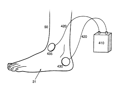
[0073] Figure 4 and Figure 9 show a device for providing electrical
stimulation to
the foot, according to aspects of the present invention.
[0074] The device 400 includes a generator 410, connecting wires 410 and
electrodes 430 and 435. The electrodes are connected to the generator via the
connecting wires. The generator 410 is a programmable electrical stimulation
signal
generation device. The electrodes 430 and 435 may be interchangeable and their
polarity is determined according to their connection to the generator 410. The
electrodes are self adhesive or otherwise attachable to skin.
[0075] Various aspects of the present invention may be implemented in
footwear and accessories to footwear such as shoes, socks and stockings. They
may
be carried in a pocket or pouch in a item of footwear, with conductors
connecting the a
stimulus generating portion of the device to electrodes placed on the skin.
The
electrodes may vary in shape and size and may be self-adhering of the type
utilized for
TENS devices. Moreover, if at least one of the electrodes includes a power
source,
then the electrodes may be wirelessly in communication with the signal
generator. In
that situation, the signal generator may be located closer to the hands and
head of the
11
CA 02783566 2012-06-07
WO 2011/087539 PCT/US2010/053351
user allowing him to more easily adjust the intensity and other parameters of
the
stimulation. In the case of wireless control, the electrodes must be connected
together,
outside the body, to create a closed circuit with the passage through the
muscles.
Further, the signal generator may be remotely programmable by a physician
monitoring
the patient.
[0076] Figure 5 shows placement of electrodes on the foot, according to
aspects of the present invention.
[0077] The electrodes 430 and 435 are located on the foot 31 such that
one
electrode attaches to the heel and the other is attached to the mid-section or
the big
arch of the foot. In the arch area the skin is not calloused and the fat layer
under the
skin is minimal. In one aspect, the heel electrode 430 is the ground electrode
and the
arch electrode is the active or positive electrode.
[0078] Figures 6A, 6B and 6C show placement of electrodes on the ankles,
according to aspects of the present invention.
[0079] The placement of the ankle electrodes is chosen to optimally
stimulate
the posterior tibial, anterior tibial, and sural nerves of the leg 50. This in
turn will provide
the maximum therapeutic effect for pain management, enhancing wound healing,
and
preventing muscle atrophy. These electrodes may be located at the area of the
peroneal motor nerve which is also referred to as the anterior tibial nerve.
In one
aspect, the electrodes would be placed just lateral to the tendon of tibialis
anterior and just proximal to the malleoli. Figure 6B provides the ankle
showing
anterior electrode placement (435) and lateral electrode placement (430).
Figure 6C
provides a line drawing of the ankle showing anterior electrode placement
(435) and
medial electrode placement (430).
[0080] Figure 7 shows a flowchart of a method of increasing circulation,
according to aspects of the present invention.
[0081] The method begins at 701. At 702, one electrode, for example the
ground electrode, is connected to heel of a foot. At 703, the other electrode,
for
example the active electrode, is connected to a mid-section or arch of the
foot. At 704,
electrical stimulation is applied to the muscles of the foot through the
attached
electrodes. At 705, the method ends.
12
CA 02783566 2012-06-07
WO 2011/087539 PCT/US2010/053351
[0082] In variations of this method, the electrical stimulation may be
periodically
or continuously adjusted according to readout of parameters from the patient
or
according to decision of a physician or the patient himself.
[0083] Figure 8 shows a flowchart of a method of pain management,
according
to aspects of the present invention.
[0084] The method begins at 801. At 802, one electrode, for example the
ground electrode, is connected above the ankle of a foot. At 803, the other
electrode,
for example the active electrode, is connected to below the ankle of the foot.
At 804,
electrical stimulation is applied to the muscles of the foot through the
attached
electrodes. At 805, the electrical stimulation is adjusted. At 806, the method
ends.
[0085] The present invention has been described in relation to
particular
examples, which are intended to be illustrative rather than restrictive, with
the scope
and spirit of the invention being indicated by the following claims and their
equivalents.
13