Note: Descriptions are shown in the official language in which they were submitted.
CA 02783656 2012-06-07
WO 2011/109572 PCT/US2011/026943
TITLE OF THE INVENTION
BIOLOGICAL MARKERS PREDICTIVE OF ANTI-CANCER RESPONSE TO INSULIN-
LIKE GROWTH FACTOR-1 RECEPTOR KINASE INHIBITORS
CROSS-REFERENCE TO RELATED APPLICATIONS
[1] This application claims the benefit of U.S. Provisional Application No.
61/310,031, filed
March 3, 2010, which is herein incorporated by reference in its entirety.
BACKGROUND OF THE INVENTION
[2] Cancer is a generic name for a wide range of cellular malignancies
characterized by
unregulated growth, lack of differentiation, and the ability to invade local
tissues and metastasize.
These neoplastic malignancies affect, with various degrees of prevalence,
every tissue and organ in
the body. The present invention is directed to methods for diagnosing and
treating cancer patients. In
particular, the present invention is directed to methods for determining which
patients will most
benefit from treatment with an insulin-like growth factor-1 receptor (IGF-1 R)
kinase inhibitor.
[3] IGF-1R belongs to the insulin receptor family that includes the Insulin
Receptor (IR), IGF-1R
(homodimer), IGF-IR/IR (hybrid receptor), and IGF-2R (mannose 6-phosphate
receptor). IGF-IR/IR
hybrids act as homodimers, preferentially binding and signaling with IGFs. IR
exists in two isoforms:
IR-B (traditional insulin receptor) and IR-A (a fetal form which is re-
expressed in selected tumors and
preferentially binds IGF-II). IGF-2R is a non-signaling receptor that acts as
a "sink" for IGF-II
(Pollak M.N., et al. Nat Rev Cancer 2004 4:505-18). Six well-characterized
insulin-like growth factor
binding proteins (IGFBP-1 through -6) associate with IGF ligands to stabilize
the IGFs and modulate
their ability to bind the IGF-IR.
[4] IGF-1R is a transmembrane RTK that binds primarily to IGF-1 but also to
1GF-II and insulin
with lower affinity. Binding of IGF-1 to its receptor results in activation of
it's tyrosine kinase
activity, intermolecular receptor autophosphorylation, and phosphorylation of
cellular substrates,
including IRS 1 and She, leading to activation of the PI3K/Akt and mitogen-
activated protein kinase
(MAPK) pathways (Adams T.E., et al. Cell Mol Life Sci 2000 57:1050-93; Pollak
M.N., et al. Nat
Rev Cancer 2004 4:505-18; Baserga R., Exp Cell Res 1999 253:1-6). The ligand-
activated IGF-1R
induces mitogenic activity in normal cells and plays an important role in
abnormal growth. A major
physiological role of the IGF-1 system is the promotion of normal growth and
regeneration.
-1-
CA 02783656 2012-06-07
WO 2011/109572 PCT/US2011/026943
Overexpressed IGF-1R (type 1 insulin-like growth factor receptor) can initiate
mitogenesis and
promote ligand-dependent neoplastic transformation. Furthermore, IGF-1R plays
an important role in
the establishment and maintenance of the malignant phenotype. Unlike the
epidermal growth factor
(EGF) receptor, no mutant oncogenic forms of the IGF-1R have been identified.
However, several
oncogenes have been demonstrated to affect IGF-1 and IGF-1R expression. A
correlation between a
reduction of IGF-1R expression and resistance to transformation has been seen.
Exposure of cells to
mRNA antisense to IGF-1R RNA prevents soft agar growth of several human tumor
cell lines. IGF-
1R abrogates progression into apoptosis, both in vivo and in vitro. It has
also been shown that a
decrease in the level of IGF-1R below wild-type levels causes apoptosis of
tumor cells in vivo. The
ability of IGF-1R disruption to cause apoptosis appears to be diminished in
normal, non-tumorigenic
cells.
[5] The IGF-1 pathway has an important role in human tumor development. IGF-1R
overexpression is frequently found in various tumors (breast, colon, lung,
sarcoma) and is often
associated with an aggressive phenotype. High circulating IGF1 concentrations
are strongly
correlated with prostate, lung and breast cancer risk. Furthermore, IGF-1R is
required for
establishment and maintenance of the transformed phenotype in vitro and in
vivo (Baserga R. Exp.
Cell. Res., 1999, 253, 1-6). The kinase activity of IGF-1R is essential for
the transforming activity of
several oncogenes: EGFR, PDGFR, SV40 T antigen, activated Ras, Raf, and v-Src.
The expression
of IGF-1R in normal fibroblasts induces neoplastic phenotypes, which can then
form tumors in vivo.
IGF-1R expression plays an important role in anchorage-independent growth. IGF-
1R has also been
shown to protect cells from chemotherapy-, radiation-, and cytokine-induced
apoptosis. Conversely,
inhibition of endogenous IGF-1 R by dominant negative IGF-1R, triple helix
formation or antisense
expression vector has been shown to repress transforming activity in vitro and
tumor growth in animal
models. The IGF-1R signaling pathway also appears to be a robust target in
colorectal cancer (CRC),
based upon data demonstrating overexpression of the receptor and ligands in
CRC, association with a
more malignant phenotype, chemotherapy resistance, and correlation with a poor
prognosis (Saltz,
L.B., et al. J Clin Oncol 2007;25(30): 4793-4799; Tripkovic I., et al. Med
Res. 2007 Jul;38(5):519-25.
Epub 2007 Apr 26; Miyamoto S., et al. Clin Cancer Res. 2005 May 1;11(9):3494-
502; Nakamura M.,
et al. Clin Cancer Res. 2004 Dec 15;10(24):8434-41; Grothey A, et al. J Cancer
Res Clin Oncol.
1999;125(3-4):166-73).
[6] It has been recognized that inhibitors of protein-tyrosine kinases are
useful as selective
inhibitors of the growth of mammalian cancer cells. For example, GleevecTM
(also known as imatinib
mesylate), a 2-phenylpyrimidine tyrosine kinase inhibitor that inhibits the
kinase activity of the BCR-
ABL fusion gene product, has been approved by the U. S. Food and Drug
Administration for the
treatment of CML. The 4-anilinoquinazoline compound TarcevaTM (erlotinib HCl)
has also been
-2-
CA 02783656 2012-06-07
WO 2011/109572 PCT/US2011/026943
approved by the FDA, and selectively inhibits EGF receptor kinase with high
potency. The
development for use as anti-tumor agents of compounds that directly inhibit
the kinase activity of
IGF-1R, as well as antibodies that reduce IGF-1R kinase activity by blocking
IGF-1R activation or
antisense oligonucleotides that block IGF-1R expression, are areas of intense
research effort (e.g. see
Larsson, O. et al (2005) Brit. J. Cancer 92:2097-2101; Ibrahim, Y.H. and Yee,
D. (2005) Clin. Cancer
Res. 11:944s-950s; Mitsiades, C.S. et al. (2004) Cancer Cell 5:221-230;
Camirand, A. et al. (2005)
Breast Cancer Research 7:R570-R579 (DOI 10. 1 186/bcrl 028); Camirand, A. and
Pollak, M. (2004)
Brit. J. Cancer 90:1825-1829; Garcia-Echeverria, C. et al. (2004) Cancer Cell
5:231-239; Sachdev D,
and Yee D., Mol Cancer Ther. 2007 Jan;6(1):1-12; Hofmann F., and Garcia-
Echeverria C., Drug
Discov Today 2005 10:1041-7). Agents inhibiting the IGF-1R pathway have
demonstrated anti-tumor
efficacy in multiple human cancer models both in vitro and in vivo,
particularly in pediatric models of
Ewing's sarcoma and rhabdomyosarcoma (Manara MC, et al. Int J Oncol 2005
27:1605-16). Despite
early hints of efficacy in patients with sarcoma, results to date of IGF-1R
inhibitors in early clinical
trials have not been impressive, indicating that patient selection strategies
and rational combinations
may be needed to move forward with this approach (Tolcher A.W., et al. Journal
of Clinical
Oncology, 2007 ASCO Annual Meeting Proceedings Part I. Vol 25, No. 18S (June
20 Supplement),
2007: 3002). Data acquired this far, has not indicated that activation,
overexpression, or amplification
of members of the IGF-1R pathway will predict responsiveness.
[7] There is a need for both more efficacious treatment for neoplasia and
other proliferative
disorders, and for more effective means for determining which tumors will
respond to which
treatment. Several groups have investigated or disclosed potential biomarkers
to predict a patient's
response to protein-tyrosine kinase inhibitors (see for example, PCT
publications: WO 2004/063709,
WO 2005/017493, WO 2004/111273, WO 2008/108986, WO 2007/001868 and WO
2004/071572;
and US published patent applications: US 2005/0019785, US 2007/0065858, US
2009/0092596, US
2009/0093488, US 2006/0140960 and US 2004/0132097). Several biomarkers have
been proposed for
predicting the response to EGFR kinase inhibitors, including mutant KRAS as a
predictor of non-
responsiveness in colorectal cancer (e.g. see Brugger, W. et al. (2009) J Clin
Oncol 27:15s, (suppl;
abstr 8020); Siena, S et al (2009) JNCI 101(19):1308-1324; Riely and Ladanyi
(2008) J Mol
Diagnostics 10(6):493; Jimeno, A. et al. (2009) Cancer J. 15(2):110-13). In
addition, several
biomarkers, including mutant KRAS, have been disclosed that have potential in
predicting a patient's
response to IGF-1R kinase inhibitors (e.g. see Rodon, J. et al (2008) Mol
Cancer Ther. 7:2575-2588;
T. Pitts et al. (2009) EORTC Conference, Boston, MA, abstract #2141; Huang, F.
et al. (2009) Cancer
Res. 69(1):161-170; Rodon, J. et al., (2008) Mol. Cancer Ther. 7:2575-2588).
However, in most
instances no FDA-approved diagnostic tests have yet emerged that can
effectively guide practicing
physicians in the treatment of their patients with such inhibitors, or can
indicate to the physician
-3-
CA 02783656 2012-06-07
WO 2011/109572 PCT/US2011/026943
which tumors will respond most favorable to a combination of such an inhibitor
with a standard
chenmotherapy agent.
[8] Thus, there remains a critical need for improved methods for determining
the best mode of
treatment for any given cancer patient. The present invention provides methods
for determining which
tumors will respond most effectively to treatment with IGF-1R kinase
inhibitors that inhibit both IGF-
1R and IR kinases, based on whether the tumor cells express certain levels of
mRNA transcripts that
are predictive of sensitivity to such IGF-1R kinase inhibitors, and for the
incorporation of such
biomarker determinations into more effective treatment regimens for cancer
patients, whether such
inhibitors are used as single agents or combined with other anti-cancer
agents.
SUMMARY OF THE INVENTION
[9] The present invention provides diagnostic methods for predicting the
effectiveness of
treatment of a cancer patient with an IGF-1R kinase inhibitor that inhibits
both IGF-1R and IR
kinases. These methods are based on the surprising discovery that the
sensitivity of tumor cell growth
to inhibition by such IGF-1R kinase inhibitors is predicted by whether such
tumor cells have a
sufficiently high value of a gene expression level index comprising the sum of
the expression levels of
the five gene transcripts IGF-1 R, IR, IR-A, IGF-1 and IGF-2. Whether the
tumor cells have a
sufficiently high value of the expression level index that is predictive of
sensitivity is determined by
assessing whether the index value is equal to or greater than a value of the
expression level index
determined to be a minimum value required to predict inhibitor sensitivity.
The latter minimum value
was determined by a study that established the relationship between tumor cell
sensitivity to inhibitor
and the expression level index, and provides reference tumor cell lines that
can be used for
comparison purposes to indicate the magnitude of this minimum value, e.g. RDES
or SK-N-AS tumor
cells.
[10] Accordingly, the present invention provides a method of identifying
patients with cancer who
are likely to benefit from treatment with an IGF-1R kinase inhibitor that
inhibits both IGF-1R and IR
kinases, comprising: obtaining a sample of a patient's tumor, assessing the
expression level of the five
gene transcripts IGF-1R, IR, IR-A, IGF-1 and IGF-2 in the tumor cells of the
sample; determining an
expression level index for the five gene transcripts by adding the expression
level values for each of
the five transcripts; determining that if the value of the expression level
index for the tumor cells of
the sample is equal to or greater than the value of the expression level index
for RDES or SK-N-AS
tumor cells determined by identical methods, the patient is likely to benefit
from treatment with an
IGF-1R kinase inhibitor that inhibits both IGF-1R and IR kinases.
-4-
CA 02783656 2012-06-07
WO 2011/109572 PCT/US2011/026943
[11] Improved methods for treating cancer patients with IGF-1R kinase
inhibitors that inhibit both
IGF-1R and IR kinases that incorporate the above methodology are also
provided. Thus, the present
invention further provides a method for treating tumors or tumor metastases in
a patient, comprising
the steps of diagnosing a patient's likely responsiveness to an IGF-1R kinase
inhibitor that inhibits
both IGF-1R and IR kinases by assessing whether the tumor cells have a
sufficiently high value of a
gene expression level index comprising the sum of the expression levels of the
five gene transcripts
IGF-1 R, IR, IR-A, IGF-1 and IGF-2, and administering to said patient a
therapeutically effective
amount of an IGF-1R kinase inhibitor that inhibits both IGF-1R and IR kinases
(e.g. OSI-906) where
responsiveness to the inhibitor is predicted.
[12] The present invention also provides diagnostic methods for identifying
patients with cancer
who are likely to benefit from treatment with an IGF-1R kinase inhibitor that
inhibits both IGF-1R
and IR kinases, but would likely not respond to therapy with an anti-IGF-1R
antibody, by combining
the above described methodology with a determination of whether the tumor
cells have a sufficiently
high value of a gene expression level index comprising the sum of the
expression levels of the gene
transcripts IR and IR-A that is predictive of resistance to growth inhibition
by an anti-IGF-1R
antibody. Improved methods for treating cancer patients with IGF-1R kinase
inhibitors that inhibit
both IGF-1R and IR kinases that incorporate this methodology are also
provided.
[13] The present invention thus provides a method of identifying patients with
cancer who are
likely to benefit from treatment with an IGF-1R kinase inhibitor that inhibits
both IGF-1R and IR
kinases, but who would likely not respond to therapy with an anti-IGF-1R
antibody, comprising:
obtaining a sample of a patient's tumor, assessing the expression level of the
five gene transcripts
IGF-1R, IR, IR-A, IGF-1 and IGF-2 in the tumor cells of the sample;
determining an expression level
index for the five gene transcripts by adding the expression level values for
each of the five
transcripts; determining that if the value of the expression level index for
the tumor cells of the
sample is equal to or greater than the value of the expression level index for
RDES tumor cells as
determined by identical methods, the patient is likely to benefit from
treatment with an IGF-1R kinase
inhibitor that inhibits both IGF-1R and IR kinases; and determining that if
the value of the sum of
expression levels for IR and IR-A for the tumor cells of the sample is equal
to or greater than the sum
of expression levels for IR and IR-A for GEO or A673 tumor cells as determined
by identical
methods, the patient is not likely to benefit from treatment with an anti-IGF-
1R antibody, thus
identifying patients with cancer who are likely to benefit from treatment with
an IGF-1R kinase
inhibitor that inhibits both IGF-1R and IR kinases, but would likely not
respond to therapy with an
anti-IGF-1R antibody.
-5-
CA 02783656 2012-06-07
WO 2011/109572 PCT/US2011/026943
[14] The present invention also provides diagnostic methods for identifying
patients with cancer
who are not likely to benefit from treatment with anti-IGF-1R antibody,
comprising
determining whether the tumor cells of the patient express insulin receptor or
phospho-IR,
wherein if insulin receptor or phospho-IR is expressed, the tumor cells will
be resistant to
inhibition by the antibody. Improved methods for treating cancer patients with
IGF-1R kinase
inhibitors that incorporate these methods are also provided.
BRIEF DESCRIPTION OF THE FIGURES
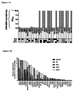
[15] Figure 1. Elevated expression of IGF receptor/ligand pairs is observed
among tumor cell
lines sensitive to OSI-906. A. Sensitivity to OSI-906 for a panel of 32 tumor
cell lines derived from
tumor types, expressed as EC50 values. Cell lines were categorized as either
sensitive (EC50<1
M) or insensitive (EC50>10 M) to OSI-906. Mutational status for KRAS, BRAF,
and PIK3CA is
indicated, as reported by Sanger Wellcome database. Those mutation statuses
that are not reported
are shaded grey. B. Expression of IGFI, IGF2, IGFIR, IR, and IRA mRNA by qPCR
for the panel of
32 tumor cell lines. Gene expression was normalized to the fourth quartile
expression for a given
gene within the 32 cell line panel.
[16] Figure 2. Effect of varying concentrations of OSI-906 on cell growth for
a representative
panel of 5 sensitive tumor cell lines.
[17] Figure 3. The IGF-1R neutralizing antibody MAB391 confers a compensatory
increase in IR
phosphorylation, and co-targeting IGF-1R and IR achieves enhanced inhibition
of the IRS1-AKT
pathway for select tumor cells. A. Phosphorylation of IR and IGF-1R for a
representative group of 8
human tumor cell lines (top panel, 1st page). Effect of OSI-906 (3 M) or
MAB391 (3 g/ml) on the
phosphorylation of IR and IGF-1R for a panel of 9 tumor cell lines categorized
as sensitive to OSI-
906 (2 d page). Data are captured 16 hours after dosing and expressed as % of
basal phosphorylation
signal. A set of representative array images are shown for A673 Ewings sarcoma
tumor cells (lower
panel, 1st page). B. Effect of 16 hour treatment with OSI-906 (3 M) or
NIAB391 (3 g/ml) on the
phosphorylation of IR or IGF-1 R, total IGF-1R expression, and phospho-AKTS437
or phospho-ERK
for a panel of 4 tumor cell lines (1st page: H322 and SK-N-AS; 2"d page: H295R
and A673). C.
Effect of OSI-906 (3 M) or MAB391 (3 g/ml) on phospho-IRS- I Y612 for H295R,
A673, and H322
cells. Also shown is phospho-AKTs473 phospho-PRAS40, and total IGF-1R and IR
levels under basal
conditions or upon treatment with OSI-906 or NIAB391 for H295R cells. Results
shown are typical of
3 or more independent experiments.
-6-
CA 02783656 2012-06-07
WO 2011/109572 PCT/US2011/026943
[18] Figure 4. Xenograft tumors co-expressing pIGF-1R and pIR are sensitive to
OSI-906 but not
MAB391, while tumors expressing IGF-1R and not IR are sensitive to both OSI-
906 and MAB391.
A. Expression of IGF1, IGF2, IGF-1R, and IRA as determined by quantitative PCR
and expression of
phospho-IR and phospho-IGF-1R as determined by capture array (top panel, 1st
page). Mice bearing
SK-N-AS or GEO tumors were dosed with either OSI-906 (50 mg/kg qd) or MAB391
(1 mg/mouse
q3d), as indicated (lower panel, 1st page), and TGI was determined over a 14
day period (2nd page).
Effect of single dose OSI-906 or NIAB391 on the phosphorylation of AKT for GEO
and SK-N-AS
tumors (3rd page). B. Effect of OSI-906 or MAB391 on the phosphorylation
states for IR and IGF-1R
in vivo for GEO tumors over the dosing period (i.e. 24 hours for OSI-906, or
72 hours for MAB391)
(upper panel). Representative images are shown. Effects of MAB391 or OSI-906
on tumoral
phospho-AKTS473 over the dosing period (lower panel).
[19] Figure 5. Insulin activation of tumor cell IR-AKT signaling is inhibited
by OSI-906 but not
MAB391. A. Effects on phosphorylation of IGF-1R and IR for HT-29 tumor cells
treated with either
OSI-906 (3 M) or MAB391 (3 g/ml) for 16 hours followed by stimulation with
50 IU/ml insulin
for 5 minutes prior to cell lysis. B. Effect of OSI-906, MAB391, or IGFBP3 on
phospho-AKTS473 for
HT-29 cells under basal conditions or following stimulation with 5 or 50
IU/ml insulin.
[20] Figure 6. MAB391 inhibits IGF-1, but not IGF-2 or insulin mediated
stimulation of pIR. A.
Effect of OSI-906 (3 M) or MAB391 (3 g/ml) on phospho-IR and phospho-IGF-1R
for control
cells or cells treated with insulin (50 IU/ml), IGF-1 (40ng/ml), or IGF-2
(40ng/ml) for 5 minutes
prior to lysis. Cartoon illustration of ligand-receptor binding pairs (right
panel). B. Effect of OSI-906
or MAB391 on phospho-AktS473 in the presence of IGF-1 or IGF-2. C. Effect of
OSI-906 (3 M),
MAB391 (3 g/ml), or an IGF-2 neutralizing antibody (10 g/ml) on
phosphorylation of IGF-1R or IR
(left panel) or pPRAS40 (right panel) for MDAH-2774 cells following 16 hour
treatment. Results
shown are typical of 3 or more independent experiments. D. Cartoon
illustrating the compensatory
signaling through IR that can occur upon specific inhibition of IGF-1R.
[21] Figure 7: OSI-906 exhibits enhanced inhibition of AKT phosphorylation,
compared to
MAB391, in tumors that co-express phospho-IGF-1R and phospho-IR. A. Effect of
a single dose of
OSI-906 (50mg/kg) or NIAB391 (1mg/mouse) on tumor AKT phosphorylation
following 4 hours of
treatment for SK-N-AS (A) and GEO (B) tumors. pAKT was determined by
immunoblotting.
DETAILED DESCRIPTION OF THE INVENTION
-7-
CA 02783656 2012-06-07
WO 2011/109572 PCT/US2011/026943
[22] The term "cancer" in an animal refers to the presence of cells possessing
characteristics
typical of cancer-causing cells, such as uncontrolled proliferation,
immortality, metastatic potential,
rapid growth and proliferation rate, and certain characteristic morphological
features. Often, cancer
cells will be in the form of a tumor, but such cells may exist alone within an
animal, or may circulate
in the blood stream as independent cells, such as leukemic cells.
[23] "Cell growth", as used herein, for example in the context of "tumor cell
growth", unless
otherwise indicated, is used as commonly used in oncology, where the term is
principally associated
with growth in cell numbers, which occurs by means of cell reproduction (i.e.
proliferation) when the
rate of the latter is greater than the rate of cell death (e.g. by apoptosis
or necrosis), to produce an
increase in the size of a population of cells, although a small component of
that growth may in certain
circumstances be due also to an increase in cell size or cytoplasmic volume of
individual cells. An
agent that inhibits cell growth can thus do so by either inhibiting
proliferation or stimulating cell
death, or both, such that the equilibrium between these two opposing processes
is altered.
[24] "Tumor growth" or "tumor metastases growth", as used herein, unless
otherwise indicated, is
used as commonly used in oncology, where the term is principally associated
with an increased mass
or volume of the tumor or tumor metastases, primarily as a result of tumor
cell growth.
[25] "Abnormal cell growth", as used herein, unless otherwise indicated,
refers to cell growth that
is independent of normal regulatory mechanisms (e.g., loss of contact
inhibition). This includes, for
example, the abnormal growth of. (1) tumor cells (tumors) that proliferate by
expressing a mutated
tyrosine kinase or overexpression of a receptor tyrosine kinase; (2) benign
and malignant cells of
other proliferative diseases in which aberrant tyrosine kinase activation
occurs; (3) any tumors that
proliferate by receptor tyrosine kinases; (4 any tumors that proliferate by
aberrant serine/threonine
kinase activation; and (5) benign and malignant cells of other proliferative
diseases in which aberrant
serine/threonine kinase activation occurs.
[26] The term "treating" as used herein, unless otherwise indicated, means to
give medical aid to
counteract a disease or condition. The phrase "a method of treating" or its
equivalent, when applied to
cancer refers to a procedure or course of action that is designed to reduce or
eliminate the number of
cancer cells in a patient, or to alleviate the symptoms of a cancer. "A method
of treating" cancer or
another proliferative disorder does not necessarily mean that the cancer cells
or other disorder will, in
fact, be eliminated, that the number of cells or disorder will, in fact, be
reduced, or that the symptoms
of a cancer or other disorder will, in fact, be alleviated. Often, a method of
treating cancer will be
performed even with a low likelihood of success, but which, given the medical
history and estimated
survival expectancy of a patient, is nevertheless deemed an overall beneficial
course of action.
-8-
CA 02783656 2012-06-07
WO 2011/109572 PCT/US2011/026943
[27] The term "therapeutically effective agent" means a composition that will
elicit the biological
or medical response of a tissue, system, animal or human that is being sought
by the researcher,
veterinarian, medical doctor or other clinician.
[28] The term "therapeutically effective amount" or "effective amount" means
the amount of the
subject compound or combination that will elicit the biological or medical
response of a tissue,
system, animal or human that is being sought by the researcher, veterinarian,
medical doctor or other
clinician.
[29] The terms "responsive"or "responsiveness" when used herein in referring
to a patient's
reaction to administration of an IGF-1R kinase inhibitor that inhibits both
IGF-1R and IR kinases,
refer to a response that is positive or effective, from which the patient is
likely to benefit.
[30] The data presented in the Experimental Details section herein below
demonstrate that tumor
cells have varying sensitivities to growth inhibition by an IGF-1R kinase
inhibitor that inhibits both
IGF-1R and IR kinases (e.g. OSI-906), with some tumor cells being relatively
resistant to inhibition. It
is demonstrated that the degree of sensitivity of tumor cells to such an IGF-
1R kinase inhibitor can be
assessed by determining whether tumor cells have a sufficiently high value of
a gene expression level
index comprising the sum of the expression level values of the five gene
transcripts IGF-1 R, IR, IR-A,
IGF-1 and IGF-2. Whether the tumor cells have a sufficiently high value of the
expression level index
that is predictive of sensitivity is determined by assessing whether the index
value is equal to or
greater than a value of the expression level index determined to be a minimum
value required to
predict inhibitor sensitivity. This minimum value is the expression level
index value associated with
tumor cells such as RDES or SK-N-AS tumor cells. All tumor cells with an
expression level index at
or above this value are sensitive to an IGF-1R kinase inhibitor that inhibits
both IGF-1R and IR
kinases (e.g. see Figures IA and 1B, which demonstrates this with a variety of
tumor cell types with
the IGF-1R kinase inhibitor OSI-906).
[31] The data presented in the Experimental Details section also indicates
that tumor cells that
express insulin receptor protein above a given level are relatively resistant
to inhibition by an anti-
IGF-1R antibody. Thus, it was found that a sufficiently high value of a gene
expression level index
comprising the sum of the expression levels of the gene transcripts IR and IR-
A is predictive of
resistance to growth inhibition by an anti-IGF-1R antibody. For example, tumor
cells with an IR plus
IR-A gene expression level index at or above that associated with either GEO
or SK-N-AS tumor
cells were found to be relatively resistant to inhibition by an anti-IGF-1R
antibody.
-9-
CA 02783656 2012-06-07
WO 2011/109572 PCT/US2011/026943
[32] These observations can thus be used to successfully predict which
patients will be effectively
treated with an IGF-1R kinase inhibitor that inhibits both IGF-1R and IR
kinases, such as OSI-906,
and of those patients, identify those who would not be effectively treated
with an anti-IGF-1R
antibody, as an alternative therapy. Thus, these observations can form the
basis of valuable new
diagnostic methods for predicting the effects of IGF-1R kinase inhibitors on
tumor growth, and give
oncologists additional tools to assist them in choosing the most appropriate
treatment for their
patients.
[33] Accordingly, the present invention provides a method of identifying
patients with cancer who
are likely to benefit from treatment with an IGF-1R kinase inhibitor that
inhibits both IGF-1R and IR
kinases, comprising: obtaining a sample of a patient's tumor, assessing the
expression level of the five
gene transcripts IGF-1R, IR, IR-A, IGF-1 and IGF-2 in the tumor cells of the
sample; determining an
expression level index for the five gene transcripts by adding the expression
level values for each of
the five transcripts; determining that if the value of the expression level
index for the tumor cells of
the sample is equal to or greater than the value of the expression level index
for RDES tumor cells
determined by identical methods, the patient is likely to benefit from
treatment with an IGF-1R kinase
inhibitor that inhibits both IGF-1R and IR kinases.
[34] The present invention also provides a method of identifying patients with
cancer who are
likely to benefit from treatment with an IGF-1R kinase inhibitor that inhibits
both IGF-1R and IR
kinases, comprising: obtaining a sample of a patient's tumor, assessing the
expression level of the five
gene transcripts IGF-1R, IR, IR-A, IGF-1 and IGF-2 in the tumor cells of the
sample; determining an
expression level index for the five gene transcripts by adding the expression
level values for each of
the five transcripts; determining that if the value of the expression level
index for the tumor cells of
the sample is equal to or greater than the value of the expression level index
for SK-N-AS tumor cells
determined by identical methods, the patient is likely to benefit from
treatment with an IGF-1R kinase
inhibitor that inhibits both IGF-1R and IR kinases.
[35] The present invention also provides a method of identifying patients with
cancer who are
likely to benefit from treatment with an IGF-1R kinase inhibitor that inhibits
both IGF-1R and IR
kinases, but would likely not respond to therapy with an anti-IGF-1R antibody,
comprising: obtaining
a sample of a patient's tumor, assessing the expression level of the five gene
transcripts IGF-1R, IR,
IR-A, IGF-1 and IGF-2 in the tumor cells of the sample; determining an
expression level index for the
five gene transcripts by adding the expression level values for each of the
five transcripts; determining
that if the value of the expression level index for the tumor cells of the
sample is equal to or greater
than that value of the expression level index for RDES tumor cells as
determined by identical
methods, the patient is likely to benefit from treatment with an IGF-1R kinase
inhibitor that inhibits
-10-
CA 02783656 2012-06-07
WO 2011/109572 PCT/US2011/026943
both IGF-1R and IR kinases; determining that if the value of the sum of
expression levels for IR and
IR-A for the tumor cells of the sample is equal to or greater than the sum of
expression levels for IR
and IR-A for GEO tumor cells as determined by identical methods, the patient
is not likely to benefit
from treatment with an anti-IGF-1R antibody, thus identifying patients with
cancer who are likely to
benefit from treatment with an IGF-1R kinase inhibitor that inhibits both IGF-
1R and IR kinases, but
would likely not respond to therapy with an anti-IGF-1R antibody.
[36] The present invention also provides a method of identifying patients with
cancer who are
likely to benefit from treatment with an IGF-1R kinase inhibitor that inhibits
both IGF-1R and IR
kinases, but would likely not respond to therapy with an anti-IGF-1R antibody,
comprising: obtaining
a sample of a patient's tumor, assessing the expression level of the five gene
transcripts IGF-1R, IR,
IR-A, IGF-1 and IGF-2 in the tumor cells of the sample; determining an
expression level index for the
five gene transcripts by adding the expression level values for each of the
five transcripts; determining
that if the value of the expression level index for the tumor cells of the
sample is equal to or greater
than that value of the expression level index for SK-N-AS tumor cells as
determined by identical
methods, the patient is likely to benefit from treatment with an IGF-1R kinase
inhibitor that inhibits
both IGF-1R and IR kinases; determining that if the value of the sum of
expression levels for IR and
IR-A for the tumor cells of the sample is equal to or greater than the sum of
expression levels for IR
and IR-A for GEO tumor cells as determined by identical methods, the patient
is not likely to benefit
from treatment with an anti-IGF-1R antibody, thus identifying patients with
cancer who are likely to
benefit from treatment with an IGF-1R kinase inhibitor that inhibits both IGF-
1R and IR kinases, but
would likely not respond to therapy with an anti-IGF-1R antibody.
[37] The present invention also provides a method of identifying patients with
cancer who are
likely to benefit from treatment with an IGF-1R kinase inhibitor that inhibits
both IGF-1R and IR
kinases, but would likely not respond to therapy with an anti-IGF-1R antibody,
comprising: obtaining
a sample of a patient's tumor, assessing the expression level of the five gene
transcripts IGF-1R, IR,
IR-A, IGF-1 and IGF-2 in the tumor cells of the sample; determining an
expression level index for the
five gene transcripts by adding the expression level values for each of the
five transcripts; determining
that if the value of the expression level index for the tumor cells of the
sample is equal to or greater
than that value of the expression level index for RDES tumor cells as
determined by identical
methods, the patient is likely to benefit from treatment with an IGF-1R kinase
inhibitor that inhibits
both IGF-1R and IR kinases; determining that if the value of the sum of
expression levels for IR and
IR-A for the tumor cells of the sample is equal to or greater than the sum of
expression levels for IR
and IR-A for A673 tumor cells as determined by identical methods, the patient
is not likely to benefit
from treatment with an anti-IGF-1R antibody, thus identifying patients with
cancer who are likely to
-11-
CA 02783656 2012-06-07
WO 2011/109572 PCT/US2011/026943
benefit from treatment with an IGF-1R kinase inhibitor that inhibits both IGF-
1R and IR kinases, but
would likely not respond to therapy with an anti-IGF-1R antibody.
[38] The present invention also provides a method of identifying patients with
cancer who are
likely to benefit from treatment with an IGF-1R kinase inhibitor that inhibits
both IGF-1R and IR
kinases, but would likely not respond to therapy with an anti-IGF-1R antibody,
comprising: obtaining
a sample of a patient's tumor, assessing the expression level of the five gene
transcripts IGF-1R, IR,
IR-A, IGF-1 and IGF-2 in the tumor cells of the sample; determining an
expression level index for the
five gene transcripts by adding the expression level values for each of the
five transcripts; determining
that if the value of the expression level index for the tumor cells of the
sample is equal to or greater
than that value of the expression level index for SK-N-AS tumor cells as
determined by identical
methods, the patient is likely to benefit from treatment with an IGF-1R kinase
inhibitor that inhibits
both IGF-1R and IR kinases; determining that if the value of the sum of
expression levels for IR and
IR-A for the tumor cells of the sample is equal to or greater than the sum of
expression levels for IR
and IR-A for A673 tumor cells as determined by identical methods, the patient
is not likely to benefit
from treatment with an anti-IGF-1R antibody, thus identifying patients with
cancer who are likely to
benefit from treatment with an IGF-1R kinase inhibitor that inhibits both IGF-
1R and IR kinases, but
would likely not respond to therapy with an anti-IGF-1R antibody.
[39] The invention also provides a method for treating cancer in a patient,
comprising the steps of:
(A) diagnosing a patient's likely responsiveness to an IGF-1R kinase inhibitor
that inhibits both IGF-
1R and IR kinases by determining if the patient has a tumor that is likely to
respond to treatment with
such an IGF-1R kinase inhibitor by: obtaining a sample of a patient's tumor,
assessing the expression
level of the five gene transcripts IGF-1R, IR, IR-A, IGF-1 and IGF-2 in the
tumor cells of the sample;
determining an expression level index for the five gene transcripts by adding
the expression level
values for each of the five transcripts; determining that if the value of the
expression level index for
the tumor cells of the sample is equal to or greater than the value of the
expression level index for
RDES tumor cells determined by identical methods, the patient is likely to
benefit from treatment
with an IGF-1R kinase inhibitor that inhibits both IGF-1R and IR kinases; and
(B) administering to
said patient a therapeutically effective amount of an IGF-1R kinase inhibitor
that inhibits both IGF-1R
and IR kinases if the patient is diagnosed to be potentially responsive to
such an IGF-1R kinase
inhibitor.
[40] The invention also provides a method for treating cancer in a patient,
comprising the steps of:
(A) diagnosing a patient's likely responsiveness to an IGF-1R kinase inhibitor
that inhibits both IGF-
1R and IR kinases by determining if the patient has a tumor that is likely to
respond to treatment with
such an IGF-1R kinase inhibitor by: obtaining a sample of a patient's tumor,
assessing the expression
-12-
CA 02783656 2012-06-07
WO 2011/109572 PCT/US2011/026943
level of the five gene transcripts IGF-1R, IR, IR-A, IGF-1 and IGF-2 in the
tumor cells of the sample;
determining an expression level index for the five gene transcripts by adding
the expression level
values for each of the five transcripts; determining that if the value of the
expression level index for
the tumor cells of the sample is equal to or greater than the value of the
expression level index for SK-
N-AS tumor cells determined by identical methods, the patient is likely to
benefit from treatment with
an IGF-1R kinase inhibitor that inhibits both IGF-1R and IR kinases; and (B)
administering to said
patient a therapeutically effective amount of an IGF-1R kinase inhibitor that
inhibits both IGF-1R and
IR kinases if the patient is diagnosed to be potentially responsive to such an
IGF-1R kinase inhibitor.
[41] The invention also provides a method for treating cancer in a patient,
comprising the steps of:
(A) diagnosing a patient's likely responsiveness to an IGF-1R kinase inhibitor
that inhibits both IGF-
1R and IR kinases by determining if the patient has a tumor that is likely to
respond to treatment with
such an IGF-1R kinase inhibitor, but would likely not respond to therapy with
an anti-IGF-1R
antibody, by: obtaining a sample of a patient's tumor, assessing the
expression level of the five gene
transcripts IGF-1R, IR, IR-A, IGF-1 and IGF-2 in the tumor cells of the
sample; determining an
expression level index for the five gene transcripts by adding the expression
level values for each of
the five transcripts; determining that if the value of the expression level
index for the tumor cells of
the sample is equal to or greater than that value of the expression level
index for RDES tumor cells as
determined by identical methods, the patient is likely to benefit from
treatment with an IGF-1R kinase
inhibitor that inhibits both IGF-1R and IR kinases; determining that if the
value of the sum of
expression levels for IR and IR-A for the tumor cells of the sample is equal
to or greater than the sum
of expression levels for IR and IR-A for GEO tumor cells as determined by
identical methods, the
patient is not likely to benefit from treatment with an anti-IGF-1R antibody,
thus identifying patients
with cancer who are likely to benefit from treatment with an IGF-1R kinase
inhibitor that inhibits both
IGF-1R and IR kinases, but would likely not respond to therapy with an anti-
IGF-1R antibody; and
(B) administering to said patient a therapeutically effective amount of an IGF-
1R kinase inhibitor that
inhibits both IGF-1R and IR kinases if the patient is diagnosed to be
potentially responsive to such an
IGF-1 R kinase inhibitor.
[42] The invention also provides a method for treating cancer in a patient,
comprising the steps of:
(A) diagnosing a patient's likely responsiveness to an IGF-1R kinase inhibitor
that inhibits both IGF-
1R and IR kinases by determining if the patient has a tumor that is likely to
respond to treatment with
such an IGF-1R kinase inhibitor, but would likely not respond to therapy with
an anti-IGF-1R
antibody, by: obtaining a sample of a patient's tumor, assessing the
expression level of the five gene
transcripts IGF-1R, IR, IR-A, IGF-1 and IGF-2 in the tumor cells of the
sample; determining an
expression level index for the five gene transcripts by adding the expression
level values for each of
the five transcripts; determining that if the value of the expression level
index for the tumor cells of
- 13 -
CA 02783656 2012-06-07
WO 2011/109572 PCT/US2011/026943
the sample is equal to or greater than that value of the expression level
index for SK-N-AS tumor cells
as determined by identical methods, the patient is likely to benefit from
treatment with an IGF-1R
kinase inhibitor that inhibits both IGF-1R and IR kinases; determining that if
the value of the sum of
expression levels for IR and IR-A for the tumor cells of the sample is equal
to or greater than the sum
of expression levels for IR and IR-A for GEO tumor cells as determined by
identical methods, the
patient is not likely to benefit from treatment with an anti-IGF-1R antibody,
thus identifying patients
with cancer who are likely to benefit from treatment with an IGF-1R kinase
inhibitor that inhibits both
IGF-1R and IR kinases, but would likely not respond to therapy with an anti-
IGF-1R antibody; and
(B) administering to said patient a therapeutically effective amount of an IGF-
1R kinase inhibitor that
inhibits both IGF-1R and IR kinases if the patient is diagnosed to be
potentially responsive to such an
IGF-1 R kinase inhibitor.
[43] The invention also provides a method for treating cancer in a patient,
comprising the steps of:
(A) diagnosing a patient's likely responsiveness to an IGF-1R kinase inhibitor
that inhibits both IGF-
1R and IR kinases by determining if the patient has a tumor that is likely to
respond to treatment with
such an IGF-1R kinase inhibitor, but would likely not respond to therapy with
an anti-IGF-1R
antibody, by: obtaining a sample of a patient's tumor, assessing the
expression level of the five gene
transcripts IGF-1R, IR, IR-A, IGF-1 and IGF-2 in the tumor cells of the
sample; determining an
expression level index for the five gene transcripts by adding the expression
level values for each of
the five transcripts; determining that if the value of the expression level
index for the tumor cells of
the sample is equal to or greater than that value of the expression level
index for RDES tumor cells as
determined by identical methods, the patient is likely to benefit from
treatment with an IGF-1R kinase
inhibitor that inhibits both IGF-1R and IR kinases; determining that if the
value of the sum of
expression levels for IR and IR-A for the tumor cells of the sample is equal
to or greater than the sum
of expression levels for IR and IR-A for A673 tumor cells as determined by
identical methods, the
patient is not likely to benefit from treatment with an anti-IGF-1R antibody,
thus identifying patients
with cancer who are likely to benefit from treatment with an IGF-1R kinase
inhibitor that inhibits both
IGF-1R and IR kinases, but would likely not respond to therapy with an anti-
IGF-1R antibody; and
(B) administering to said patient a therapeutically effective amount of an IGF-
1R kinase inhibitor that
inhibits both IGF-1R and IR kinases if the patient is diagnosed to be
potentially responsive to such an
IGF-1 R kinase inhibitor.
[44] The invention also provides a method for treating cancer in a patient,
comprising the steps of:
(A) diagnosing a patient's likely responsiveness to an IGF-1R kinase inhibitor
that inhibits both IGF-
1R and IR kinases by determining if the patient has a tumor that is likely to
respond to treatment with
such an IGF-1R kinase inhibitor, but would likely not respond to therapy with
an anti-IGF-1R
antibody, by: obtaining a sample of a patient's tumor, assessing the
expression level of the five gene
-14-
CA 02783656 2012-06-07
WO 2011/109572 PCT/US2011/026943
transcripts IGF-1R, IR, IR-A, IGF-1 and IGF-2 in the tumor cells of the
sample; determining an
expression level index for the five gene transcripts by adding the expression
level values for each of
the five transcripts; determining that if the value of the expression level
index for the tumor cells of
the sample is equal to or greater than that value of the expression level
index for SK-N-AS tumor cells
as determined by identical methods, the patient is likely to benefit from
treatment with an IGF-1R
kinase inhibitor that inhibits both IGF-1R and IR kinases; determining that if
the value of the sum of
expression levels for IR and IR-A for the tumor cells of the sample is equal
to or greater than the sum
of expression levels for IR and IR-A for A673 tumor cells as determined by
identical methods, the
patient is not likely to benefit from treatment with an anti-IGF-1R antibody,
thus identifying patients
with cancer who are likely to benefit from treatment with an IGF-1R kinase
inhibitor that inhibits both
IGF-1R and IR kinases, but would likely not respond to therapy with an anti-
IGF-1R antibody; and
(B) administering to said patient a therapeutically effective amount of an IGF-
1R kinase inhibitor that
inhibits both IGF-1R and IR kinases if the patient is diagnosed to be
potentially responsive to such an
IGF-1 R kinase inhibitor.
[45] The invention also provides a method for treating cancer in a patient,
comprising
administering to said patient a therapeutically effective amount of an IGF-1R
kinase inhibitor that
inhibits both IGF-1R and IR kinases, if the patient has been diagnosed to be
potentially responsive to
such an IGF-1R kinase inhibitor, by assessing the expression level of the five
gene transcripts IGF-
1R, IR, IR-A, IGF-1 and IGF-2 in the tumor cells of the cancer; determining an
expression level index
for the five gene transcripts by adding the expression level values for each
of the five transcripts; and
determining that the value of the expression level index for the tumor cells
is equal to or greater than
the value of the expression level index for RDES tumor cells or SK-N-AS tumor
cells determined by
identical methods. This method is thus a method of treatment targeted at a
specific patient population
previously identified or characterized as having a tumor susceptible to
effective treatment with an
IGF-1R kinase inhibitor that inhibits both IGF-1R and IR kinases.
[46] The invention also provides a method for treating cancer in a patient,
comprising
administering to said patient a therapeutically effective amount of an IGF-1R
kinase inhibitor that
inhibits both IGF-1R and IR kinases, if the patient has been diagnosed to be
potentially responsive to
such an IGF-1R kinase inhibitor, by assessing the expression level of the five
gene transcripts IGF-
1R, IR, IR-A, IGF-1 and IGF-2 in the tumor cells of the cancer; determining an
expression level index
for the five gene transcripts by adding the expression level values for each
of the five transcripts; and
determining that the value of the expression level index for the tumor cells
is equal to or greater than
the value of the expression level index for RDES tumor cells or SK-N-AS tumor
cells determined by
identical methods, and if the patient is diagnosed to be potentially
unresponsive to treatment an anti-
IGF-1R antibody by determining that the value of the sum of expression levels
for IR and IR-A for
-15-
CA 02783656 2012-06-07
WO 2011/109572 PCT/US2011/026943
the tumor cells of the cancer is equal to or greater than the sum of
expression levels for IR and IR-A
for GEO or A673 tumor cells as determined by identical methods. This method is
thus a method of
treatment targeted at a specific patient population previously identified or
characterized as having a
tumor susceptible to effective treatment with an IGF-1R kinase inhibitor that
inhibits both IGF-1R
and IR kinases.
[47] In addition, the present invention provides a method of predicting the
sensitivity of tumor cell
growth to inhibition by an IGF-1R kinase inhibitor that inhibits both IGF-1R
and IR kinases,
comprising: assessing the expression level of the five gene transcripts IGF-
1R, IR, IR-A, IGF-1 and
IGF-2 in the tumor cells; determining an expression level index for the five
gene transcripts by adding
the expression level values for each of the five transcripts; determining that
if the value of the
expression level index for the tumor cells is equal to or greater than the
value of the expression level
index for RDES tumor cells determined by identical methods, the tumor cells
will exhibit high
sensitivity to an IGF-1R kinase inhibitor that inhibits both IGF-1R and IR
kinases.
[48] In addition, the present invention provides a method of identifying
patients with cancer who
are likely to benefit from treatment with an IGF-1R kinase inhibitor that
inhibits both IGF-1R and IR
kinases, but would likely not respond to therapy with an anti-IGF-1R antibody,
comprising: assessing
the expression level of the five gene transcripts IGF-1 R, IR, IR-A, IGF-1 and
IGF-2 in tumor cells
from a sample of a patient's tumor; determining an expression level index for
the five gene transcripts
by adding the expression level values for each of the five transcripts;
determining that if the value of
the expression level index for the tumor cells of the sample is equal to or
greater than the value of the
expression level index for RDES or SK-N-AS tumor cells as determined by
identical methods, the
patient is likely to benefit from treatment with an IGF-1R kinase inhibitor
that inhibits both IGF-1R
and IR kinases; determining that if the value of the sum of expression levels
for IR and IR-A for the
tumor cells of the sample is equal to or greater than the sum of expression
levels for IR and IR-A for
GEO or A673 tumor cells as determined by identical methods, the patient is not
likely to benefit from
treatment with an anti-IGF-1R antibody, thus identifying patients with cancer
who are likely to benefit
from treatment with an IGF-1R kinase inhibitor that inhibits both IGF-1R and
IR kinases, but would
likely not respond to therapy with an anti-IGF-1R antibody.
[49] The present invention also provides a method of predicting the
sensitivity of tumor cell
growth to inhibition by an IGF-1R kinase inhibitor that inhibits both IGF-1R
and IR kinases,
comprising: assessing the expression level of the five gene transcripts IGF-
1R, IR, IR-A, IGF-1 and
IGF-2 in the tumor cells; determining an expression level index for the five
gene transcripts by adding
the expression level values for each of the five transcripts; determining that
if the value of the
expression level index for the tumor cells is equal to or greater than the
value of the expression level
-16-
CA 02783656 2012-06-07
WO 2011/109572 PCT/US2011/026943
index for SK-N-AS tumor cells determined by identical methods, the tumor cells
will exhibit high
sensitivity to an IGF-1R kinase inhibitor that inhibits both IGF-1R and IR
kinases.
[50] The present invention also provides a method of identifying tumor cells
that would be
sensitive to growth inhibition by an IGF-1R kinase inhibitor that inhibits
both IGF-1R and IR kinases,
but would not be sensitive to inhibition by an anti-IGF-1R antibody,
comprising: assessing the
expression level of the five gene transcripts IGF-1R, IR, IR-A, IGF-1 and IGF-
2 in the tumor cells;
determining an expression level index for the five gene transcripts by adding
the expression level
values for each of the five transcripts; determining that if the value of the
expression level index for
the tumor cells is equal to or greater than the value of the expression level
index for RDES tumor cells
determined by identical methods, the tumor cells will exhibit high sensitivity
to an IGF-1R kinase
inhibitor that inhibits both IGF-1R and IR kinases, and determining that if
the value of the sum of
expression levels for IR and IR-A for the tumor cells is equal to or greater
than the sum of expression
levels for IR and IR-A for GEO tumor cells as determined by identical methods,
the tumor cells will
not be sensitive to inhibition by an anti-IGF-1R antibody.
[51] The present invention also provides a method of identifying tumor cells
that would be
sensitive to growth inhibition by an IGF-1R kinase inhibitor that inhibits
both IGF-1R and IR kinases,
but would not be sensitive to inhibition by an anti-IGF-1R antibody,
comprising: assessing the
expression level of the five gene transcripts IGF-1R, IR, IR-A, IGF-1 and IGF-
2 in the tumor cells;
determining an expression level index for the five gene transcripts by adding
the expression level
values for each of the five transcripts; determining that if the value of the
expression level index for
the tumor cells is equal to or greater than the value of the expression level
index for SK-N-AS tumor
cells determined by identical methods, the tumor cells will exhibit high
sensitivity to an IGF-1R
kinase inhibitor that inhibits both IGF-1R and IR kinases, and determining
that if the value of the sum
of expression levels for IR and IR-A for the tumor cells is equal to or
greater than the sum of
expression levels for IR and IR-A for GEO tumor cells as determined by
identical methods, the tumor
cells will not be sensitive to inhibition by an anti-IGF-1R antibody.
[52] The present invention also provides a method of identifying tumor cells
that would be
sensitive to growth inhibition by an IGF-1R kinase inhibitor that inhibits
both IGF-1R and IR kinases,
but would not be sensitive to inhibition by an anti-IGF-1R antibody,
comprising: assessing the
expression level of the five gene transcripts IGF-1R, IR, IR-A, IGF-1 and IGF-
2 in the tumor cells;
determining an expression level index for the five gene transcripts by adding
the expression level
values for each of the five transcripts; determining that if the value of the
expression level index for
the tumor cells is equal to or greater than the value of the expression level
index for RDES tumor cells
determined by identical methods, the tumor cells will exhibit high sensitivity
to an IGF-1R kinase
-17-
CA 02783656 2012-06-07
WO 2011/109572 PCT/US2011/026943
inhibitor that inhibits both IGF-1R and IR kinases, and determining that if
the value of the sum of
expression levels for IR and IR-A for the tumor cells is equal to or greater
than the sum of expression
levels for IR and IR-A for A673tumor cells as determined by identical methods,
the tumor cells will
not be sensitive to inhibition by an anti-IGF-1R antibody.
[53] The present invention also provides a method of identifying tumor cells
that would be
sensitive to growth inhibition by an IGF-1R kinase inhibitor that inhibits
both IGF-1R and IR kinases,
but would not be sensitive to inhibition by an anti-IGF-1R antibody,
comprising: assessing the
expression level of the five gene transcripts IGF-1R, IR, IR-A, IGF-1 and IGF-
2 in the tumor cells;
determining an expression level index for the five gene transcripts by adding
the expression level
values for each of the five transcripts; determining that if the value of the
expression level index for
the tumor cells is equal to or greater than the value of the expression level
index for SK-N-AS tumor
cells determined by identical methods, the tumor cells will exhibit high
sensitivity to an IGF-1R
kinase inhibitor that inhibits both IGF-1R and IR kinases, and determining
that if the value of the sum
of expression levels for IR and IR-A for the tumor cells is equal to or
greater than the sum of
expression levels for IR and IR-A for A673 tumor cells as determined by
identical methods, the tumor
cells will not be sensitive to inhibition by an anti-IGF-1R antibody.
[54] Accordingly, the present invention provides a method of identifying
patients with cancer who
are likely to benefit from treatment with an IGF-1R kinase inhibitor that
inhibits both IGF-1R and IR
kinases, comprising: obtaining a sample of a patient's tumor, assessing the
expression level of the five
gene transcripts IGF-1R, IR, IR-A, IGF-1 and IGF-2 in the cells of the sample;
determining an
expression level index for the five gene transcripts by adding the expression
level values for each of
the five transcripts; determining that if the value of the expression level
index for the cells of the
sample is equal to or greater than the value of the expression level index for
RDES tumor cells
determined by identical methods, the patient is likely to benefit from
treatment with an IGF-1R kinase
inhibitor that inhibits both IGF-1R and IR kinases.
[55] The present invention also provides a method of identifying patients with
cancer who are
likely to benefit from treatment with an IGF-1R kinase inhibitor that inhibits
both IGF-1R and IR
kinases, comprising: obtaining a sample of a patient's tumor, assessing the
expression level of the five
gene transcripts IGF-1R, IR, IR-A, IGF-1 and IGF-2 in the cells of the sample;
determining an
expression level index for the five gene transcripts by adding the expression
level values for each of
the five transcripts; determining that if the value of the expression level
index for the cells of the
sample is equal to or greater than the value of the expression level index for
SK-N-AS tumor cells
determined by identical methods, the patient is likely to benefit from
treatment with an IGF-1R kinase
inhibitor that inhibits both IGF-1R and IR kinases.
-18-
CA 02783656 2012-06-07
WO 2011/109572 PCT/US2011/026943
[56] The present invention also provides a method of identifying patients with
cancer who are
likely to benefit from treatment with an IGF-1R kinase inhibitor that inhibits
both IGF-1R and IR
kinases, but would likely not respond to therapy with an anti-IGF-1R antibody,
comprising: obtaining
a sample of a patient's tumor, assessing the expression level of the five gene
transcripts IGF-1R, IR,
IR-A, IGF-1 and IGF-2 in the cells of the sample; determining an expression
level index for the five
gene transcripts by adding the expression level values for each of the five
transcripts; determining that
if the value of the expression level index for the cells of the sample is
equal to or greater than that
value of the expression level index for RDES tumor cells as determined by
identical methods, the
patient is likely to benefit from treatment with an IGF-1R kinase inhibitor
that inhibits both IGF-1R
and IR kinases; determining that if the value of the sum of expression levels
for IR and IR-A for the
cells of the sample is equal to or greater than the sum of expression levels
for IR and IR-A for GEO
tumor cells as determined by identical methods, the patient is not likely to
benefit from treatment with
an anti-IGF-1R antibody, thus identifying patients with cancer who are likely
to benefit from
treatment with an IGF-1R kinase inhibitor that inhibits both IGF-1R and IR
kinases, but would likely
not respond to therapy with an anti-IGF-1R antibody.
[57] The present invention also provides a method of identifying patients with
cancer who are
likely to benefit from treatment with an IGF-1R kinase inhibitor that inhibits
both IGF-1R and IR
kinases, but would likely not respond to therapy with an anti-IGF-1R antibody,
comprising: obtaining
a sample of a patient's tumor, assessing the expression level of the five gene
transcripts IGF-1R, IR,
IR-A, IGF-1 and IGF-2 in the cells of the sample; determining an expression
level index for the five
gene transcripts by adding the expression level values for each of the five
transcripts; determining that
if the value of the expression level index for the cells of the sample is
equal to or greater than that
value of the expression level index for SK-N-AS tumor cells as determined by
identical methods, the
patient is likely to benefit from treatment with an IGF-1R kinase inhibitor
that inhibits both IGF-1R
and IR kinases; determining that if the value of the sum of expression levels
for IR and IR-A for the
cells of the sample is equal to or greater than the sum of expression levels
for IR and IR-A for GEO
tumor cells as determined by identical methods, the patient is not likely to
benefit from treatment with
an anti-IGF-1R antibody, thus identifying patients with cancer who are likely
to benefit from
treatment with an IGF-1R kinase inhibitor that inhibits both IGF-1R and IR
kinases, but would likely
not respond to therapy with an anti-IGF-1R antibody.
[58] The present invention also provides a method of identifying patients with
cancer who are
likely to benefit from treatment with an IGF-1R kinase inhibitor that inhibits
both IGF-1R and IR
kinases, but would likely not respond to therapy with an anti-IGF-1R antibody,
comprising: obtaining
a sample of a patient's tumor, assessing the expression level of the five gene
transcripts IGF-1R, IR,
-19-
CA 02783656 2012-06-07
WO 2011/109572 PCT/US2011/026943
IR-A, IGF-1 and IGF-2 in the cells of the sample; determining an expression
level index for the five
gene transcripts by adding the expression level values for each of the five
transcripts; determining that
if the value of the expression level index for the cells of the sample is
equal to or greater than that
value of the expression level index for RDES tumor cells as determined by
identical methods, the
patient is likely to benefit from treatment with an IGF-1R kinase inhibitor
that inhibits both IGF-1R
and IR kinases; determining that if the value of the sum of expression levels
for IR and IR-A for the
cells of the sample is equal to or greater than the sum of expression levels
for IR and IR-A for A673
tumor cells as determined by identical methods, the patient is not likely to
benefit from treatment with
an anti-IGF-1R antibody, thus identifying patients with cancer who are likely
to benefit from
treatment with an IGF-1R kinase inhibitor that inhibits both IGF-1R and IR
kinases, but would likely
not respond to therapy with an anti-IGF-1R antibody.
[59] The present invention also provides a method of identifying patients with
cancer who are
likely to benefit from treatment with an IGF-1R kinase inhibitor that inhibits
both IGF-1R and IR
kinases, but would likely not respond to therapy with an anti-IGF-1R antibody,
comprising: obtaining
a sample of a patient's tumor, assessing the expression level of the five gene
transcripts IGF-1R, IR,
IR-A, IGF-1 and IGF-2 in the cells of the sample; determining an expression
level index for the five
gene transcripts by adding the expression level values for each of the five
transcripts; determining that
if the value of the expression level index for the cells of the sample is
equal to or greater than that
value of the expression level index for SK-N-AS tumor cells as determined by
identical methods, the
patient is likely to benefit from treatment with an IGF-1R kinase inhibitor
that inhibits both IGF-1R
and IR kinases; determining that if the value of the sum of expression levels
for IR and IR-A for the
cells of the sample is equal to or greater than the sum of expression levels
for IR and IR-A for A673
tumor cells as determined by identical methods, the patient is not likely to
benefit from treatment with
an anti-IGF-1R antibody, thus identifying patients with cancer who are likely
to benefit from
treatment with an IGF-1R kinase inhibitor that inhibits both IGF-1R and IR
kinases, but would likely
not respond to therapy with an anti-IGF-1R antibody.
[60] The invention also provides a method for treating cancer in a patient,
comprising the steps of:
(A) diagnosing a patient's likely responsiveness to an IGF-1R kinase inhibitor
that inhibits both IGF-
1R and IR kinases by determining if the patient has a tumor that is likely to
respond to treatment with
such an IGF-1R kinase inhibitor by: obtaining a sample of a patient's tumor,
assessing the expression
level of the five gene transcripts IGF-1R, IR, IR-A, IGF-1 and IGF-2 in the
cells of the sample;
determining an expression level index for the five gene transcripts by adding
the expression level
values for each of the five transcripts; determining that if the value of the
expression level index for
the cells of the sample is equal to or greater than the value of the
expression level index for RDES
tumor cells determined by identical methods, the patient is likely to benefit
from treatment with an
-20-
CA 02783656 2012-06-07
WO 2011/109572 PCT/US2011/026943
IGF-1R kinase inhibitor that inhibits both IGF-1R and IR kinases; and (B)
administering to said
patient a therapeutically effective amount of an IGF-1R kinase inhibitor that
inhibits both IGF-1R and
IR kinases if the patient is diagnosed to be potentially responsive to such an
IGF-1R kinase inhibitor.
[61] The invention also provides a method for treating cancer in a patient,
comprising the steps of:
(A) diagnosing a patient's likely responsiveness to an IGF-1R kinase inhibitor
that inhibits both IGF-
1R and IR kinases by determining if the patient has a tumor that is likely to
respond to treatment with
such an IGF-1R kinase inhibitor by: obtaining a sample of a patient's tumor,
assessing the expression
level of the five gene transcripts IGF-1R, IR, IR-A, IGF-1 and IGF-2 in the
cells of the sample;
determining an expression level index for the five gene transcripts by adding
the expression level
values for each of the five transcripts; determining that if the value of the
expression level index for
the cells of the sample is equal to or greater than the value of the
expression level index for SK-N-AS
tumor cells determined by identical methods, the patient is likely to benefit
from treatment with an
IGF-1R kinase inhibitor that inhibits both IGF-1R and IR kinases; and (B)
administering to said
patient a therapeutically effective amount of an IGF-1R kinase inhibitor that
inhibits both IGF-1R and
IR kinases if the patient is diagnosed to be potentially responsive to such an
IGF-1R kinase inhibitor.
[62] The invention also provides a method for treating cancer in a patient,
comprising the steps of:
(A) diagnosing a patient's likely responsiveness to an IGF-1R kinase inhibitor
that inhibits both IGF-
1R and IR kinases by determining if the patient has a tumor that is likely to
respond to treatment with
such an IGF-1R kinase inhibitor, but would likely not respond to therapy with
an anti-IGF-1R
antibody, by: obtaining a sample of a patient's tumor, assessing the
expression level of the five gene
transcripts IGF-1 R, IR, IR-A, IGF-1 and IGF-2 in the cells of the sample;
determining an expression
level index for the five gene transcripts by adding the expression level
values for each of the five
transcripts; determining that if the value of the expression level index for
the cells of the sample is
equal to or greater than that value of the expression level index for RDES
tumor cells as determined
by identical methods, the patient is likely to benefit from treatment with an
IGF-1R kinase inhibitor
that inhibits both IGF-1R and IR kinases; determining that if the value of the
sum of expression levels
for IR and IR-A for the cells of the sample is equal to or greater than the
sum of expression levels for
IR and IR-A for GEO tumor cells as determined by identical methods, the
patient is not likely to
benefit from treatment with an anti-IGF-1R antibody, thus identifying patients
with cancer who are
likely to benefit from treatment with an IGF-1R kinase inhibitor that inhibits
both IGF-1R and IR
kinases, but would likely not respond to therapy with an anti-IGF-1R antibody;
and (B) administering
to said patient a therapeutically effective amount of an IGF-1R kinase
inhibitor that inhibits both IGF-
1R and IR kinases if the patient is diagnosed to be potentially responsive to
such an IGF-1R kinase
inhibitor.
-21-
CA 02783656 2012-06-07
WO 2011/109572 PCT/US2011/026943
[63] The invention also provides a method for treating cancer in a patient,
comprising the steps of:
(A) diagnosing a patient's likely responsiveness to an IGF-1R kinase inhibitor
that inhibits both IGF-
1R and IR kinases by determining if the patient has a tumor that is likely to
respond to treatment with
such an IGF-1R kinase inhibitor, but would likely not respond to therapy with
an anti-IGF-1R
antibody, by: obtaining a sample of a patient's tumor, assessing the
expression level of the five gene
transcripts IGF-1 R, IR, IR-A, IGF-1 and IGF-2 in the cells of the sample;
determining an expression
level index for the five gene transcripts by adding the expression level
values for each of the five
transcripts; determining that if the value of the expression level index for
the cells of the sample is
equal to or greater than that value of the expression level index for SK-N-AS
tumor cells as
determined by identical methods, the patient is likely to benefit from
treatment with an IGF-1R kinase
inhibitor that inhibits both IGF-1R and IR kinases; determining that if the
value of the sum of
expression levels for IR and IR-A for the cells of the sample is equal to or
greater than the sum of
expression levels for IR and IR-A for GEO tumor cells as determined by
identical methods, the
patient is not likely to benefit from treatment with an anti-IGF-1R antibody,
thus identifying patients
with cancer who are likely to benefit from treatment with an IGF-1R kinase
inhibitor that inhibits both
IGF-1R and IR kinases, but would likely not respond to therapy with an anti-
IGF-1R antibody; and
(B) administering to said patient a therapeutically effective amount of an IGF-
1R kinase inhibitor that
inhibits both IGF-1R and IR kinases if the patient is diagnosed to be
potentially responsive to such an
IGF-1 R kinase inhibitor.
[64] The invention also provides a method for treating cancer in a patient,
comprising the steps of:
(A) diagnosing a patient's likely responsiveness to an IGF-1R kinase inhibitor
that inhibits both IGF-
1R and IR kinases by determining if the patient has a tumor that is likely to
respond to treatment with
such an IGF-1R kinase inhibitor, but would likely not respond to therapy with
an anti-IGF-1R
antibody, by: obtaining a sample of a patient's tumor, assessing the
expression level of the five gene
transcripts IGF-1 R, IR, IR-A, IGF-1 and IGF-2 in the cells of the sample;
determining an expression
level index for the five gene transcripts by adding the expression level
values for each of the five
transcripts; determining that if the value of the expression level index for
the cells of the sample is
equal to or greater than that value of the expression level index for RDES
tumor cells as determined
by identical methods, the patient is likely to benefit from treatment with an
IGF-1R kinase inhibitor
that inhibits both IGF-1R and IR kinases; determining that if the value of the
sum of expression levels
for IR and IR-A for the cells of the sample is equal to or greater than the
sum of expression levels for
IR and IR-A for A673 tumor cells as determined by identical methods, the
patient is not likely to
benefit from treatment with an anti-IGF-1R antibody, thus identifying patients
with cancer who are
likely to benefit from treatment with an IGF-1R kinase inhibitor that inhibits
both IGF-1R and IR
kinases, but would likely not respond to therapy with an anti-IGF-1R antibody;
and (B) administering
to said patient a therapeutically effective amount of an IGF-1R kinase
inhibitor that inhibits both IGF-
-22-
CA 02783656 2012-06-07
WO 2011/109572 PCT/US2011/026943
1R and IR kinases if the patient is diagnosed to be potentially responsive to
such an IGF-1R kinase
inhibitor.
[65] The invention also provides a method for treating cancer in a patient,
comprising the steps of:
(A) diagnosing a patient's likely responsiveness to an IGF-1R kinase inhibitor
that inhibits both IGF-
1R and IR kinases by determining if the patient has a tumor that is likely to
respond to treatment with
such an IGF-1R kinase inhibitor, but would likely not respond to therapy with
an anti-IGF-1R
antibody, by: obtaining a sample of a patient's tumor, assessing the
expression level of the five gene
transcripts IGF-1 R, IR, IR-A, IGF-1 and IGF-2 in the cells of the sample;
determining an expression
level index for the five gene transcripts by adding the expression level
values for each of the five
transcripts; determining that if the value of the expression level index for
the cells of the sample is
equal to or greater than that value of the expression level index for SK-N-AS
tumor cells as
determined by identical methods, the patient is likely to benefit from
treatment with an IGF-1R kinase
inhibitor that inhibits both IGF-1R and IR kinases; determining that if the
value of the sum of
expression levels for IR and IR-A for the cells of the sample is equal to or
greater than the sum of
expression levels for IR and IR-A for A673 tumor cells as determined by
identical methods, the
patient is not likely to benefit from treatment with an anti-IGF-1R antibody,
thus identifying patients
with cancer who are likely to benefit from treatment with an IGF-1R kinase
inhibitor that inhibits both
IGF-1R and IR kinases, but would likely not respond to therapy with an anti-
IGF-1R antibody; and
(B) administering to said patient a therapeutically effective amount of an IGF-
1R kinase inhibitor that
inhibits both IGF-1R and IR kinases if the patient is diagnosed to be
potentially responsive to such an
IGF-1 R kinase inhibitor.
[66] The invention also provides a method for treating a patient with a tumor,
comprising
administering to said patient a therapeutically effective amount of an IGF-1R
kinase inhibitor that
inhibits both IGF-1R and IR kinases, if the patient is diagnosed to be
potentially responsive to such an
IGF-1R kinase inhibitor, by assessing the expression level of the five gene
transcripts IGF-1R, IR, IR-
A, IGF-1 and IGF-2 in the cells of the tumor; determining an expression level
index for the five gene
transcripts by adding the expression level values for each of the five
transcripts; and determining that
the value of the expression level index for the cells of the tumor is equal to
or greater than the value of
the expression level index for RDES tumor cells or SK-N-AS tumor cells
determined by identical
methods.
[67] The invention also provides a method for treating a patient with a tumor,
comprising
administering to said patient a therapeutically effective amount of an IGF-1R
kinase inhibitor that
inhibits both IGF-1R and IR kinases, if the patient is diagnosed to be
potentially responsive to such an
IGF-1R kinase inhibitor, by assessing the expression level of the five gene
transcripts IGF-1R, IR, IR-
-23-
CA 02783656 2012-06-07
WO 2011/109572 PCT/US2011/026943
A, IGF-1 and IGF-2 in the cells of the tumor; determining an expression level
index for the five gene
transcripts by adding the expression level values for each of the five
transcripts; and determining that
the value of the expression level index for the cells of the tumor is equal to
or greater than the value of
the expression level index for RDES tumor cells or SK-N-AS tumor cells
determined by identical
methods, and if the patient is diagnosed to be potentially unresponsive to
treatment an anti-IGF-1R
antibody by determining that the value of the sum of expression levels for IR
and IR-A for the cells of
the tumor is equal to or greater than the sum of expression levels for IR and
IR-A for GEO or A673
tumor cells as determined by identical methods.
[68] In addition, the present invention provides a method of predicting the
sensitivity of tumor
growth to inhibition by an IGF-1R kinase inhibitor that inhibits both IGF-1R
and IR kinases,
comprising: assessing the expression level of the five gene transcripts IGF-
1R, IR, IR-A, IGF-1 and
IGF-2 in the cells of the tumor; determining an expression level index for the
five gene transcripts by
adding the expression level values for each of the five transcripts;
determining that if the value of the
expression level index for the cells of the tumor is equal to or greater than
the value of the expression
level index for RDES tumor cells determined by identical methods, tumor growth
will exhibit high
sensitivity to an IGF-1R kinase inhibitor that inhibits both IGF-1R and IR
kinases.
[69] The present invention also provides a method of predicting the
sensitivity of tumor growth to
inhibition by an IGF-1R kinase inhibitor that inhibits both IGF-1R and IR
kinases, comprising:
assessing the expression level of the five gene transcripts IGF-1R, IR, IR-A,
IGF-1 and IGF-2 in the
cells of the tumor; determining an expression level index for the five gene
transcripts by adding the
expression level values for each of the five transcripts; determining that if
the value of the expression
level index for the cells of the tumor is equal to or greater than the value
of the expression level index
for SK-N-AS tumor cells determined by identical methods, tumor growth will
exhibit high sensitivity
to an IGF-1R kinase inhibitor that inhibits both IGF-1R and IR kinases.
[70] The present invention also provides a method of identifying tumors that
would be sensitive to
growth inhibition by an IGF-1R kinase inhibitor that inhibits both IGF-1R and
IR kinases, but would
not be sensitive to inhibition by an anti-IGF-1R antibody, comprising:
assessing the expression level
of the five gene transcripts IGF-1 R, IR, IR-A, IGF-1 and IGF-2 in cells of a
tumor; determining an
expression level index for the five gene transcripts by adding the expression
level values for each of
the five transcripts; determining that if the value of the expression level
index for the cells of the
tumor is equal to or greater than the value of the expression level index for
RDES tumor cells
determined by identical methods, tumor growth will exhibit high sensitivity to
an IGF-1R kinase
inhibitor that inhibits both IGF-1R and IR kinases, and determining that if
the value of the sum of
expression levels for IR and IR-A for the cells of the tumor is equal to or
greater than the sum of
-24-
CA 02783656 2012-06-07
WO 2011/109572 PCT/US2011/026943
expression levels for IR and IR-A for GEO tumor cells as determined by
identical methods, tumor
growth will not be sensitive to inhibition by an anti-IGF-1R antibody.
[71] The present invention also provides a method of identifying tumors that
would be sensitive to
growth inhibition by an IGF-1R kinase inhibitor that inhibits both IGF-1R and
IR kinases, but would
not be sensitive to inhibition by an anti-IGF-1R antibody, comprising:
assessing the expression level
of the five gene transcripts IGF-1 R, IR, IR-A, IGF-1 and IGF-2 in the cells
of a tumor; determining an
expression level index for the five gene transcripts by adding the expression
level values for each of
the five transcripts; determining that if the value of the expression level
index for the cells of the
tumor is equal to or greater than the value of the expression level index for
SK-N-AS tumor cells
determined by identical methods, tumor growth will exhibit high sensitivity to
an IGF-1R kinase
inhibitor that inhibits both IGF-1R and IR kinases, and determining that if
the value of the sum of
expression levels for IR and IR-A for the cells of the tumor is equal to or
greater than the sum of
expression levels for IR and IR-A for GEO tumor cells as determined by
identical methods, tumor
growth will not be sensitive to inhibition by an anti-IGF-1R antibody.
[72] The present invention also provides a method of identifying tumors that
would be sensitive to
growth inhibition by an IGF-1R kinase inhibitor that inhibits both IGF-1R and
IR kinases, but would
not be sensitive to inhibition by an anti-IGF-1R antibody, comprising:
assessing the expression level
of the five gene transcripts IGF-1 R, IR, IR-A, IGF-1 and IGF-2 in the cells
of a tumor; determining an
expression level index for the five gene transcripts by adding the expression
level values for each of
the five transcripts; determining that if the value of the expression level
index for the cells of the
tumor is equal to or greater than the value of the expression level index for
RDES tumor cells
determined by identical methods, tumor growth will exhibit high sensitivity to
an IGF-1R kinase
inhibitor that inhibits both IGF-1R and IR kinases, and determining that if
the value of the sum of
expression levels for IR and IR-A for the cells of the tumor is equal to or
greater than the sum of
expression levels for IR and IR-A for A673 tumor cells as determined by
identical methods, tumor
growth will not be sensitive to inhibition by an anti-IGF-1R antibody.
[73] The present invention also provides a method of identifying tumors that
would be sensitive to
growth inhibition by an IGF-1R kinase inhibitor that inhibits both IGF-1R and
IR kinases, but would
not be sensitive to inhibition by an anti-IGF-1R antibody, comprising:
assessing the expression level
of the five gene transcripts IGF-1 R, IR, IR-A, IGF-1 and IGF-2 in the cells
of a tumor; determining an
expression level index for the five gene transcripts by adding the expression
level values for each of
the five transcripts; determining that if the value of the expression level
index for the cells of the
tumor is equal to or greater than the value of the expression level index for
SK-N-AS tumor cells
determined by identical methods, tumor growth will exhibit high sensitivity to
an IGF-1R kinase
-25-
CA 02783656 2012-06-07
WO 2011/109572 PCT/US2011/026943
inhibitor that inhibits both IGF-1R and IR kinases, and determining that if
the value of the sum of
expression levels for IR and IR-A for the cells of the tumor is equal to or
greater than the sum of
expression levels for IR and IR-A for A673 tumor cells as determined by
identical methods, tumor
growth will not be sensitive to inhibition by an anti-IGF-1R antibody.
[74] The present invention also provides a method of identifying patients with
cancer in need of
treatment with an IGF-1R kinase inhibitor who would likely not respond to
therapy with an anti-IGF-
1R antibody, comprising: obtaining a sample of a patient's tumor, assessing
the expression level of
the two gene transcripts IR and IR-A in the tumor cells of the sample; and
determining that if the
value of the sum of expression levels for IR and IR-A for the tumor cells of
the sample is equal to or
greater than the sum of expression levels for IR and IR-A for GEO or A673
tumor cells as determined
by identical methods, the patient is not likely to benefit from treatment with
an anti-IGF-1R antibody.
The present invention also provides this method where instead of assessing the
levels of the two gene
transcripts IR and IR-A, the levels of the two proteins encoded by these
transcripts are assessed, i.e.
IR-B and IR-A proteins (e.g. by immunohistochemical (IHC) analysis).
[75] The present invention also provides a method of identifying patients with
cancer in need of
treatment with an IGF-1R kinase inhibitor who would likely not respond to
therapy with an anti-IGF-
1R antibody, comprising: obtaining a sample of a patient's tumor, assessing
the level of phospho-IR
in the tumor cells of the sample; and determining that if the level of phospho-
IR in the tumor cells of
the sample is equal to or greater than the level of phospho-IR for GEO or A673
tumor cells as
determined by identical methods, the patient is not likely to benefit from
treatment with an anti-IGF-
1R antibody.
[76] The present invention also provides a method of identifying tumor cells
that would not be
sensitive to inhibition by an anti-IGF-1R antibody, comprising: assessing the
expression level of the
two gene transcripts IR and IR-A in the tumor cells; and determining that if
the value of the sum of
expression levels for IR and IR-A for the tumor cells is equal to or greater
than the sum of expression
levels for IR and IR-A for GEO or A673 tumor cells as determined by identical
methods, the tumor
cells will not be sensitive to inhibition by an anti-IGF-1R antibody. The
present invention also
provides this method where instead of assessing the levels of the two gene
transcripts IR and IR-A,
the levels of the two proteins encoded by these transcripts are assessed, i.e.
IR-B and IR-A proteins
(e.g. by immunohistochemical (IHC) analysis).
[77] The present invention also provides a method of identifying tumor cells
that would not be
sensitive to inhibition by an anti-IGF-1R antibody, comprising: assessing the
level of phospho-IR in
the tumor cells; and determining that if the level of phospho-IR in the tumor
cells is equal to or greater
-26-
CA 02783656 2012-06-07
WO 2011/109572 PCT/US2011/026943
than the level of phospho-IR for GEO or A673 tumor cells as determined by
identical methods, the
tumor cells will not be sensitive to inhibition by an anti-IGF-1R antibody.
[78] The present invention also provides a method of identifying patients with
cancer in need of
treatment with an IGF-1R kinase inhibitor who would likely respond to therapy
with an anti-IGF-1R
antibody, comprising: obtaining a sample of a patient's tumor, assessing the
expression level of the
two gene transcripts IR and IR-A in the tumor cells of the sample; and
determining that if the value of
the sum of expression levels for IR and IR-A for the tumor cells of the sample
is equal to or less than
the sum of expression levels for IR and IR-A for SK-N-AS tumor cells as
determined by identical
methods, the patient is likely to benefit from treatment with an anti-IGF-1R
antibody. In one
embodiment of this method the sum of expression levels for IR and IR-A for the
tumor cells of the
sample is zero or undetectable. The present invention also provides this
method where instead of
assessing the levels of the two gene transcripts IR and IR-A, the levels of
the two proteins encoded by
these transcripts are assessed, i.e. IR-B and IR-A proteins (e.g. by
immunohistochemical (IHC)
analysis).
[79] The present invention also provides a method of identifying patients with
cancer in need of
treatment with an IGF-1R kinase inhibitor who would likely respond to therapy
with an anti-IGF-1R
antibody, comprising: obtaining a sample of a patient's tumor, assessing the
level of phospho-IR in
the tumor cells of the sample; and determining that if the level of phospho-IR
in the tumor cells of the
sample is equal to or less than the level of phospho-IR for SK-N-AS tumor
cells as determined by
identical methods, the patient is likely to benefit from treatment with an
anti-IGF-1R antibody. In one
embodiment of this method the level of phospho-IR in the tumor cells of the
sample is zero or
undetectable.
[80] The present invention also provides a method of identifying tumor cells
that would be
sensitive to inhibition by an anti-IGF-1R antibody, comprising: assessing the
expression level of the
two gene transcripts IR and IR-A in the tumor cells; and determining that if
the value of the sum of
expression levels for IR and IR-A for the tumor cells is equal to or less than
the sum of expression
levels for IR and IR-A for SK-N-AS tumor cells as determined by identical
methods, the tumor cells
will be sensitive to inhibition by an anti-IGF-1R antibody. In one embodiment
of this method the sum
of expression levels for IR and IR-A for the tumor cells of the sample is zero
or undetectable. The
present invention also provides this method where instead of assessing the
levels of the two gene
transcripts IR and IR-A, the levels of the two proteins encoded by these
transcripts are assessed, i.e.
IR-B and IR-A proteins (e.g. by immunohistochemical (IHC) analysis).
-27-
CA 02783656 2012-06-07
WO 2011/109572 PCT/US2011/026943
[81] The present invention also provides a method of identifying tumor cells
that would be
sensitive to inhibition by an anti-IGF-1R antibody, comprising: assessing the
level of phospho-IR in
the tumor cells; and determining that if the level of phospho-IR in the tumor
cells is equal to or less
than the level of phospho-IR for SK-N-AS tumor cells as determined by
identical methods, the tumor
cells will be sensitive to inhibition by an anti-IGF-1R antibody. In one
embodiment of this method the
level of phospho-IR in the tumor cells of the sample is zero or undetectable.
[82] The present invention also provides a method for treating cancer in a
patient, comprising
administering to said patient a therapeutically effective amount of an anti-
IGF-1R antibody if the
patient is determined to be likely to benefit from treatment with an anti-IGF-
1R antibody by
determining that the value of the sum of expression levels for IR and IR-A for
the tumor cells of the
patient's tumor is equal to or less than the sum of expression levels for IR
and IR-A for SK-N-AS
tumor cells as determined by identical methods. The present invention also
provides this method
where instead of assessing the levels of the two gene transcripts IR and IR-A,
the levels of the two
proteins encoded by these transcripts are assessed, i.e. IR-B and IR-A
proteins (e.g. by
immunohistochemical (IHC) analysis).
[83] The present invention also provides a method for treating cancer in a
patient, comprising
administering to said patient a therapeutically effective amount of an anti-
IGF-1R antibody if the
patient is determined to be likely to benefit from treatment with an anti-IGF-
1R antibody by
determining that level of phospho-IR in the tumor cells of of the patient's
tumor is equal to or less
than the level of phospho-IR for SK-N-AS tumor cells as determined by
identical methods.
[84] In the methods of this invention, levels of tyrosine phosphorylated
proteins, such as
phosphorylated RTKs, for example phospho-IR or phospho-IGF-1 R, are determined
by any method
known to one of skill in the art. In one embodiment an anti-phospho-tyrosine
antibody is used to
assess levels of tyrosine phosphorylated proteins such as phospho-IR or
phosphor-IGF-1R. For
example, an HRP-conjugated pan anti-phospho-tyrosine antibody may be used.
[85] The present invention also provides a method of identifying patients with
cancer who are
likely to benefit from treatment with an IGF-1R kinase inhibitor that inhibits
both IGF-1R and IR
kinases, comprising: obtaining a sample of a patient's tumor, assessing the
expression level of the
gene transcripts IGF-1 R, IGF-1 and IGF-2 in the tumor cells of the sample;
determining that if the
tumor cells of the sample express IGF-1 R, and if the value of the sum of
expression levels for IGF-1
and IGF-2 for the tumor cells of the sample greater than the sum of expression
levels for IGF-1 and
IGF-2 for RD tumor cells as determined by identical methods, the patient is
likely to benefit from
treatment with an IGF-1R kinase inhibitor that inhibits both IGF-1R and IR
kinases. The present
-28-
CA 02783656 2012-06-07
WO 2011/109572 PCT/US2011/026943
invention also provides this method where instead of assessing the levels of
the gene transcript IGF-
1R, the level of the the protein encoded by this transcript is assessed, i.e.
IGF-1R protein (e.g. by
immunohistochemical (IHC) analysis).
[86] The present invention also provides a method of identifying patients with
cancer who are
likely to benefit from treatment with an IGF-1R kinase inhibitor that inhibits
both IGF-1R and IR
kinases, comprising: obtaining a sample of a patient's tumor, assessing the
expression level of the
gene transcripts IGF-1R, IR, IR-A, IGF-1 and IGF-2 in the tumor cells of the
sample; determining that
if the tumor cells of the sample express IGF-1 R, express IR and/or IR-A, and
if the value of the sum
of expression levels for IGF-1 and IGF-2 for the tumor cells of the sample
greater than the sum of
expression levels for IGF-1 and IGF-2 for RD tumor cells as determined by
identical methods, the
patient is likely to benefit from treatment with an IGF-1R kinase inhibitor
that inhibits both IGF-1R
and IR kinases. The present invention also provides this method where instead
of assessing the levels
of the three gene transcripts IGF-1R, IR and IR-A, the levels of the three
proteins encoded by these
transcripts are assessed, i.e. IGF-1R, IR-B and IR-A proteins (e.g. by
immunohistochemical (IHC)
analysis).
[87] The present invention also provides a method of predicting the
sensitivity of tumor cell
growth to inhibition by an IGF-1R kinase inhibitor that inhibits both IGF-1R
and IR kinases,
comprising: assessing the expression level of the gene transcripts IGF-1R, IGF-
1 and IGF-2 in the
tumor cells; determining that if the tumor cells express IGF-1R, and if the
value of the sum of
expression levels for IGF-1 and IGF-2 for the tumor cells is greater than the
sum of expression levels
for IGF-1 and IGF-2 for RD tumor cells as determined by identical methods, the
tumor cells will
exhibit high sensitivity to an IGF-1R kinase inhibitor that inhibits both IGF-
1R and IR kinases. The
present invention also provides this method where instead of assessing the
levels of the gene transcript
IGF-1R, the level of the the protein encoded by this transcript is assessed,
i.e. IGF-1R protein (e.g. by
immunohistochemical (IHC) analysis).
[88] The present invention also provides a method of predicting the
sensitivity of tumor cell
growth to inhibition by an IGF-1R kinase inhibitor that inhibits both IGF-1R
and IR kinases,
comprising: assessing the expression level of the gene transcripts IGF-1R, IR,
IR-A, IGF-1 and IGF-2
in the tumor cells; determining that if the tumor cells express IGF-1R,
express IR and/or IR-A, and if
the value of the sum of expression levels for IGF-1 and IGF-2 for the tumor
cells is greater than the
sum of expression levels for IGF-1 and IGF-2 for RD tumor cells as determined
by identical methods,
the tumor cells will exhibit high sensitivity to an IGF-1 R kinase inhibitor
that inhibits both IGF-1 R
and IR kinases. The present invention also provides this method where instead
of assessing the levels
of the three gene transcripts IGF-1R, IR and IR-A, the levels of the three
proteins encoded by these
-29-
CA 02783656 2012-06-07
WO 2011/109572 PCT/US2011/026943
transcripts are assessed, i.e. IGF-1R, IR-B and IR-A proteins (e.g. by
immunohistochemical (IHC)
analysis).
[89] The present invention also provides a method for treating cancer in a
patient, comprising
administering to said patient a therapeutically effective amount of an IGF-1R
kinase inhibitor that
inhibits both IGF-1R and IR kinases if the patient is determined to be likely
to benefit from treatment
with such an inhibitor by assessing the expression level of the gene
transcripts IGF-1 R, IGF-1 and
IGF-2 in the tumor cells of the patient's tumor; and determining that the
tumor cells of the patient's
tumor express IGF-1R, and the value of the sum of expression levels for IGF-1
and IGF-2 for the
tumor cells of the patient's tumor is greater than the sum of expression
levels for IGF-1 and IGF-2 for
RD tumor cells as determined by identical methods. The present invention also
provides this method
where instead of assessing the levels of the gene transcript IGF-1 R, the
level of the the protein
encoded by this transcript is assessed, i.e. IGF-1R protein (e.g. by
immunohistochemical (IHC)
analysis).
[90] The present invention also provides a method for treating cancer in a
patient, comprising
administering to said patient a therapeutically effective amount of an IGF-1R
kinase inhibitor that
inhibits both IGF-1R and IR kinases if the patient has been determined to be
likely to benefit from
treatment with such an inhibitor by assessing the expression level of the gene
transcripts IGF-1 R, IR,
IR-A, IGF-1 and IGF-2 in the tumor cells of the patient's tumor; and
determining that the tumor cells
of the patient's tumor express IGF-1R, express IR and/or IR-A, and the value
of the sum of expression
levels for IGF-1 and IGF-2 for the tumor cells of the patient's tumor is
greater than the sum of
expression levels for IGF-1 and IGF-2 for RD tumor cells as determined by
identical methods. The
present invention also provides this method where instead of assessing the
levels of the three gene
transcripts IGF-1 R, IR and IR-A, the levels of the three proteins encoded by
these transcripts are
assessed, i.e. IGF-1R, IR-B and IR-A proteins (e.g. by immunohistochemical
(IHC) analysis). This
method is thus a method of treatment targeted at a specific patient population
previously identified or
characterized as having a tumor susceptible to effective treatment with an IGF-
1R kinase inhibitor
that inhibits both IGF-1R and IR kinases.
[91] The present invention also provides a method of identifying a patient
with a carcinoma who is
likely to benefit from treatment with an IGF-1R kinase inhibitor that inhibits
both IGF-1R and IR
kinases, comprising: obtaining a sample of a patient's tumor, assessing the
expression level of the
gene transcripts IGF-1R and IGF-2 in the tumor cells of the sample;
determining that if the tumor
cells of the sample express IGF-1R, and if the expression level of IGF-2 for
the tumor cells of the
sample is greater than the expression level of IGF-2 for MDAH-2774 tumor cells
as determined by
identical methods, the patient is likely to benefit from treatment with an IGF-
1R kinase inhibitor that
-30-
CA 02783656 2012-06-07
WO 2011/109572 PCT/US2011/026943
inhibits both IGF-1R and IR kinases. The present invention also provides this
method where instead
of assessing the levels of the gene transcript IGF-1R, the level of the the
protein encoded by this
transcript is assessed, i.e. IGF-1R protein (e.g. by immunohistochemical (IHC)
analysis).
[92] The present invention also provides a method of identifying a patient
with a myeloma who is
likely to benefit from treatment with an IGF-1R kinase inhibitor that inhibits
both IGF-1R and IR
kinases, comprising: obtaining a sample of a patient's tumor, assessing the
expression level of the
gene transcripts IGF-1R and IGF-1 in the tumor cells of the sample;
determining that if the tumor
cells of the sample express IGF-1R, and if the expression level of IGF-1 for
the tumor cells of the
sample is greater than the expression level of IGF-1 for U266 tumor cells as
determined by identical
methods, the patient is likely to benefit from treatment with an IGF-1R kinase
inhibitor that inhibits
both IGF-1R and IR kinases. The present invention also provides this method
where instead of
assessing the levels of the gene transcript IGF-1R, the level of the the
protein encoded by this
transcript is assessed, i.e. IGF-1R protein (e.g. by immunohistochemical (IHC)
analysis).
[93] The present invention also provides a method of identifying a patient
with a sarcoma who is
likely to benefit from treatment with an IGF-1R kinase inhibitor that inhibits
both IGF-1R and IR
kinases, comprising: obtaining a sample of a patient's tumor, assessing the
expression level of the
gene transcripts IGF-1R and IGF-1 in the tumor cells of the sample;
determining that if the tumor
cells of the sample express IGF-1R, and if the expression level of IGF-1 for
the tumor cells of the
sample is greater than the expression level of IGF-1 for A673 tumor cells as
determined by identical
methods, the patient is likely to benefit from treatment with an IGF-1R kinase
inhibitor that inhibits
both IGF-1R and IR kinases. The present invention also provides this method
where instead of
assessing the levels of the gene transcript IGF-1R, the level of the the
protein encoded by this
transcript is assessed, i.e. IGF-1R protein (e.g. by immunohistochemical (IHC)
analysis).
[94] The present invention also provides a method of identifying patients with
cancer who are
likely to benefit from treatment with an IGF-1R kinase inhibitor that inhibits
both IGF-1R and IR
kinases, comprising: obtaining a sample of a patient's tumor, assessing the
expression level of the five
gene transcripts IGF-1R, IR, IR-A, IGF-1 and IGF-2 in the tumor cells of the
sample; determining an
expression level index for the five gene transcripts by adding the expression
level values for each of
the five transcripts; determining that if the value of the expression level
index for the tumor cells of
the sample is equal to or greater than a predetermined minimum expression
level index value below
which tumor cells are resistant to IGF-1R kinase inhibitors that inhibits both
IGF-1R and IR kinases,
the patient is likely to benefit from treatment with an IGF-1R kinase
inhibitor that inhibits both IGF-
1R and IR kinases. In one embodiment of this method, the predetermined minimum
expression level
index is the value of the expression level index for RDES or SK-N-AS tumor
cells, determined under
-31-
CA 02783656 2012-06-07
WO 2011/109572 PCT/US2011/026943
identical conditions as used for determining the value of the expression level
index for the tumor cells
of the patient sample. In another embodiment of this method, in an additional
step it is also
determined if the value of the sum of expression levels for IR and IR-A for
the tumor cells of the
sample is equal to or greater than a predetermined minimum level for said sum,
above which tumor
cells are resistant to inhibition by an anti-IGF-1R antibody, thus indicating
whether the patient is also
likely to benefit from treatment with an anti-IGF-1R antibody. In an
embodiment of this additional
step, the predetermined minimum level for said sum is the value of the sum for
GEO or A673 tumor
cells, determined under identical conditions as used for determining the value
of the sum for the tumor
cells of the patient sample. The present invention also provides a method of
treatment of patients with
cancer comprising a step of identifying patients with cancer who are likely to
benefit from treatment
with an IGF-1R kinase inhibitor that inhibits both IGF-1R and IR kinases,
using any of the methods
described above, followed by a step of administration of an IGF-1R kinase
inhibitor that inhibits both
IGF-1R and IR kinases if the patient is identified as being potentially
responsive.
[95] The present invention also provides a method of identifying patients with
cancer who are
likely to benefit from treatment with an IGF-1R kinase inhibitor that inhibits
both IGF-1R and IR
kinases, comprising: obtaining a sample of a patient's tumor, assessing the
expression level of the
genes IGF-1R, IR, IGF-1 and IGF-2 in the tumor cells of the sample;
determining an expression level
index for the genes by adding the expression level values for each of the
genes; determining that if the
value of the expression level index for the tumor cells of the sample is equal
to or greater than a
predetermined minimum expression level index value below which tumor cells are
resistant to IGF-
1R kinase inhibitors that inhibits both IGF-1R and IR kinases, the patient is
likely to benefit from
treatment with an IGF-1R kinase inhibitor that inhibits both IGF-1R and IR
kinases. In one
embodiment of this method, the predetermined minimum expression level index is
the value of the
expression level index for RDES or SK-N-AS tumor cells, determined under
identical conditions as
used for determining the value of the expression level index for the tumor
cells of the patient sample.
In another embodiment of this method, in an additional step it is also
determined if the value of the
sum of expression levels for IR and IR-A for the tumor cells of the sample is
equal to or greater than a
predetermined minimum level for said sum, above which tumor cells are
resistant to inhibition by an
anti-IGF-1R antibody, thus indicating whether the patient is also likely to
benefit from treatment with
an anti-IGF-1R antibody. In an embodiment of this additional step, the
predetermined minimum level
for said sum is the value of the sum for GEO or A673 tumor cells, determined
under identical
conditions as used for determining the value of the sum for the tumor cells of
the patient sample. In
one embodiment of these methods assessing the expression level of the genes
IGF-1 R, IR, IGF-1 and
IGF-2 in tumor cells is by determination of mRNA transcript levels for each of
the genes, as described
elsewhere herein. In an alternative embodiment of these methods assessing the
expression level of the
genes IGF-1R, IR, IGF-1 and IGF-2 in tumor cells is by determination of
protein levels for each of the
-32-
CA 02783656 2012-06-07
WO 2011/109572 PCT/US2011/026943
genes, e.g. by IHC. The present invention also provides a method of treatment
of patients with cancer
comprising a step of identifying patients with cancer who are likely to
benefit from treatment with an
IGF-1R kinase inhibitor that inhibits both IGF-1R and IR kinases, using any of
the methods described
above, followed by a step of administration of an IGF-1R kinase inhibitor that
inhibits both IGF-1R
and IR kinases if the patient is identified as being potentially responsive.
[96] The present invention also provides a method of identifying patients with
cancer who are
likely to benefit from treatment with an IGF-1R kinase inhibitor that inhibits
both IGF-1R and IR
kinases, but would likely not respond to therapy with an anti-IGF-1R antibody,
comprising: obtaining
a sample of a patient's tumor, assessing the level of phospho-IR and phospho-
IGF-1R in the tumor
cells of the sample; and determining that if the tumor cells express both
phospho-IR and phosphor-
IGF-1 R, the patient is likely to benefit from treatment with an IGF-1R kinase
inhibitor that inhibits
both IGF-1R and IR kinases, but would likely not respond to therapy with an
anti-IGF-1R antibody.
[97] A method of treatment of patients with cancer comprising administration
of an IGF-1R kinase
inhibitor that inhibits both IGF-1R and IR kinases to the patient if they are
is identified as being
potentially responsive to such an inhibitor, but would likely not respond to
therapy with an anti-IGF-
1R antibody, by determining that the tumor cells of the patient's tumor
express both phospho-IR and
phosphor-IGF-1 R. In one embodiment, the IGF-1R kinase inhibitor that inhibits
both IGF-1R and IR
kinases is OSI-906.
[98] The present invention also provides a method for treating cancer in a
patient, comprising
administering to said patient a therapeutically effective combination of an
IGF-1R kinase inhibitor
that inhibits both IGF-1R and IR kinases and a chemotherapeutic agent, if the
chemotherapeutic agent
has been determined to upregulate phosphorylation of both IR and IGF-1R in
tumor cells. In one
embodiment of this method, the chemotherapeutic agent that has been determined
to upregulate
phosphorylation of both IR and IGF-1R in tumor cells is doxorubicin.
[99] This invention also encompasses any of the methods of the invention
described herein,
wherein the step of "obtaining a sample of a patient's tumor" is omitted. In
such cases, the step of
determining tumor biomarker expression (e.g. gene transcript level for IGF-1R,
IR, IR-A, IGF-1 or
IGF-2) may for example be performed on a previously processed or prepared
tumor sample, e.g. a
frozen tumor sample, a fixed tumor preparation, a cell extract, an RNA
preparation, a protein
preparation, or the like, from which biomarker expression can be assessed, or
a biological fluid where
the tumor biomarker can be found, as an alternative to the tumor sample itself
(e.g. a biopsy).
-33-
CA 02783656 2012-06-07
WO 2011/109572 PCT/US2011/026943
[100] In the methods of this invention the term "expression level index" means
a sum of the
expression level values of a number of mRNA transcripts. Thus, for example,
one expression level
index used in the methods of this invention comprises the sum of the
expression level values for the
five gene transcripts IGF-1R, IR, IR-A, IGF-1 and IGF-2. A second expression
level index used in the
methods of this invention comprises the sum of the expression level values for
the two gene
transcripts IR and IR-A. In a preferred embodiment the expression level values
of each of the gene
transcripts used in determining the value of an expression level index are
determined using the same
experimental method.
[101] In the methods of the instant invention involving a step of determining
whether a test tumor
cell, or tumor cell sample, has a value of an expression level index, or sum
of gene expression values
for a given group of transcripts, greater than or equal to a value or sum in a
specific reference tumor
cell (e.g. RDES, SK-N-AS, GEO, A673), it will be appreciated by one of skill
in the art that one
practicing the method is not constrained by having to always make a direct
side-by-side comparison
between the test tumor cells and a reference tumor cell. The specific
reference tumor cells indicated
are merely listed to exemplify the minimum cutoff value of expression level
index value above which
high sensitivity (or a beneficial effect) is predicted, and may be used, for
example, to calibrate an
assay system for the determination of transcript levels, after which a ditect
comparison to a reference
tumor cell is not necessary to practice the method. Other tumor cells with
similar expression level
index values may be used in place of the tumor cells indicated. It will be
appreciated by those of skill
in the art that a reference tumor cell sample need not be established for each
assay, while the assay is
being performed, but rather, a baseline or reference can be established by
referring to a form of stored
information regarding a previously determined cutoff level to discriminate
between sensitive and
resistant tumor cells (or patient responders and non-responders). Such a form
of stored information
can include, for example, but is not limited to, a reference chart, listing or
electronic file of population
or individual data regarding sensitive and resistant tumors or patients, or
any other source of data
regarding a cutoff level of expression level index value for tumor cell
sensitivity or resistance that is
useful for the patient or tumor cell to be evaluated.
[102] In practicing the methods of the invention, use of tumor cells with
other expression level
index values may also be used as reference tumor cells, or to calibrate a
transcript assay system. For
example, in the methods of the instant invention, where RDES or SK-N-AS tumor
cells are used to
indicate a value of an expression level index, any of GEO, H929, 8226, 2650,
or H295R tumor cells,
each of which has approximately double the value of expression level index
(i.e. that using the sum of
transcripts IGF-1R, IR, IR-A, IGF-1 and IGF-2), may be used instead. However,
since they have
double the value of expression level index of RDES or SK-N-AS tumor cells, the
step of "determining
that if the value of the expression level index for the tumor cells of the
sample is equal to or greater
-34-
CA 02783656 2012-06-07
WO 2011/109572 PCT/US2011/026943
than that value of the expression level index for RDES (or SK-N-AS) tumor
cells determined by
identical methods" is replaced by a step of "determining that if the value of
the expression level index
for the tumor cells of the sample is equal to or greater than half the value
of the expression level
index for GEO, H929, 8226, 2650, or H295R tumor cells determined by identical
methods". Many of
the other tumor cell lines disclosed herein may be similarly used by
incorporating a different
multiplier into the method to adjust the the expression level index value to
that of RDES or SK-N-AS
tumor cells, which indicate a minimum value of expression level index above
which tumor cells are
sensitive to an IGF-1R kinase inhibitor that inhibits both IGF-1R and IR
kinases.
[103] Determination of gene expression transcript levels can be by any method
known in the art
(e.g. RT-PCR), but the test, or sample, tumor cell determination must be by
the identical method as
used for any reference tumor cell (e.g. RDES, SK-N-AS, GEO, A673), or that
used to calibrate the
assay method, in order for a valid comparison to be made between the
calculated expression level
index value of the test or sample tumor cells, and either a reference tumor
cell expression level index
value, or an assay standard curve. The resulting gene expression transcript
level values may, for
example, be in the form of absolute values (e.g. molecules/cell), relative
levels (e.g. the transcript
level relative to a housekeeping gene transcript level, e.g. GAPDH, (3-actin,
tubulin, 28S copy
number, or the like), or in a normalized form (e.g. in the form of a gene
transcript level relative to the
4th (upper) quartile, or median expression value, for a given gene transcript
for the tumor cells in
which the transcript is measured; or normalized to a given percentile value
(e.g. 75th percentile)). For
normalization, the test or sample tumor cell may be included in a panel of
cells with reference tumor
cells, for example having a range of sensitivities to an an IGF-1R kinase
inhibitor that inhibits both
IGF-1R and IR kinases, or the data from the test or sample tumor cell may be
analysed with data from
such a panel.
[104] The NCBI GeneID numbers listed herein are unique identifiers of the
genes described herein
from the NCBI Entrez Gene database record (National Center for Biotechnology
Information (NCBI),
U.S. National Library of Medicine, 8600 Rockville Pike, Building 38A,
Bethesda, MD 20894;
Internet address http://www.ncbi.nlm.nih.gov/). Accession numbers of
representative mRNAs
expressed from the genes are also listed herein.
[105] "RDES tumor cells" as used herein, refers to cells of the cell line RD-
ES, available from the
American Tissue Culture Collection (ATCC) as HTB-166TM, derived from a human
Ewing's sarcoma.
The cell line was initiated by G. Marshall and M. Kirchen from a primary
osseous Ewings sarcoma of
the humerus. It shows epithelial morphology.
-35-
CA 02783656 2012-06-07
WO 2011/109572 PCT/US2011/026943
[106] "SK-N-AS tumor cells" as used herein, refers to cells of the cell line
SK-N-AS, available
from the American Tissue Culture Collection (ATCC) as CRL-2137TM, and derived
from a human
neuroblastoma at a bone marrow metastatic site. It shows epithelial
morphology.
[107] "GEO tumor cells" as used herein, refers to cells of the cell line GEO,
available from the
Roswell Park Cancer Institute (RPCC; Buffalo, NY). GEO was derived from a
human colon tumor.
[108] "A673 tumor cells" as used herein, refers to cells of the cell line A-
673, available from the
American Tissue Culture Collection (ATCC) as CRL-1598TM, derived from a human
rhabdomyosarcoma, and showing polygonal morphology.
[109] "RD tumor cells" as used herein, refers to cells of the cell line RD,
available from the
American Tissue Culture Collection (ATCC) as CCL-136TM, derived from a human
rhabdomyosarcoma, with a morphology of spindle cells and large multinucleated
cells.
[110] "MDAH-2774 tumor cells" as used herein, refers to cells of the ovarian
tumor cell line
MDAH-2774, derived was derived from ascitic fluid of a 38-year-old patient
with metastatic serous
cystadenocarcinom (Freedman, R., et al. (1978) Characterization of an ovarian
carcinoma cell line,
Cancer 42, 2352-2359), and shows carcinoma morphology.
[111] "U266 tumor cells" as used herein, refers to cells of the cell line
U266B1 [U266], available
from the American Tissue Culture Collection (ATCC) as TIB- 196TM, derived from
a human
myeloma, and showing lymphoblast morphology.
[112] In the context of this invention, the sensitivity of tumor cell growth
to the IGF-1R kinase
inhibitor OSI-906 is defined as high if the tumor cell is inhibited with an
EC50 (half-maximal
effective concentration) of less than 1 M, and low (i.e. relatively
resistant) if the tumor cell is
inhibited with an EC50 of greater than 10 M. Sensitivies between these values
are considered
intermediate. With other IGF-1R kinase inhibitors that inhibits both IGF-1R
and IR kinases,
particularly compounds of Formula I as described herein below, a qualitatively
similar result is
expected since they inhibit tumor cell growth by inhibiting the same signal
transduction pathway,
although quantitatively the EC50 values may differ depending on the relative
cellular potency of the
other inhibitor versus OSI-906. Thus, for example, the sensitivity of tumor
cell growth to a more
potent IGF-1R kinase inhibitor than OSI-906 would be defined as high when the
tumor cell is
inhibited with an EC50 that is correspondingly lower. In tumor xenograft
studies, using tumor cells of
a variety of tumor cell types that all have high sensitivity to OSI-906 in
culture in vitro, the tumors are
consistently inhibited in vivo with a high pencentage tumor growth inhibition
(TGI) (see
-36-
CA 02783656 2012-06-07
WO 2011/109572 PCT/US2011/026943
Experimental section herein). In contast, in similar studies, using tumor
cells that have low sensitivity
to OSI-906 in culture in vitro, the tumors are inhibited in vivo with only a
low pencentage tumor
growth inhibition (TGI). These data indicate that sensitivity to IGF-1R kinase
inhibitors such as OSI-
906 in tumor cell culture is predictive of tumor sensitivity in vivo.
[113] The term EC50 (half maximal effective concentration) refers to the
concentration of
compound which induces a response halfway between the baseline and maximum for
the specified
exposure time, and is used as a measure of the compound's potency.
[114] Although the experimental examples provided herein involve the IGF-1R
kinase inhibitor,
OSI-906, the methods of the present invention are not limited to the
prediction of patients or tumors
that will respond or not respond to any particular IGF-1R kinase inhibitor,
but rather, can be used to
predict patient's outcome to any IGF-1R kinase inhibitor that inhibits both
IGF-1R and IR kinases.
Similarly, the methods of treatment with an IGF-1R kinase inhibitor that
inhibits both IGF-1R and IR
kinases as described herein may use any of this type of IGF-1R kinase
inhibitor. Furthermore, in
another embodiment of any of the methods described herein the IGF-1R kinase
inhibitor may be an
IGF-1R kinase inhibitor approved by a government regulatory authority (e.g. US
Food and Drug
Administration (FDA); European Medicines Agency; Japanese Ministry of Health,
Labour &
Welfare; UK Medicines and Healthcare Products Regulatory Agency (MHRA)) (e.g.
any of the IGF-
1R kinase inhibitors disclosed herein that have been so approved).
[115] In the methods of this invention, an IGF-1R kinase inhibitor that
inhibits both IGF-1R and IR
kinases may be any IGF-1R kinase inhibitor that inhibits both of these
receptor-tyrosine kinases,
including pharmacologically acceptable salts or polymorphs thereof. In a
preferred embodiment the
IGF-1R kinase inhibitor that inhibits both IGF-1R and IR kinases is a small
molecule IGF-1R kinase
inhibitor. In another embodiment the IGF-1R kinase inhibitor that inhibits
both IGF-1R and IR
kinases is a small molecule IGF-1R kinase inhibitor that is ATP-competitive at
the kinase calalytic
site. In one embodiment, the ratio of the inhibitor's IC50 (as determined
using an in vitro biochemical
kinase assay, e.g. see Mulvihill, M.J. et al. (2008) Bioorganic & Medicinal
Chemistry, Volume 16,
Issue 3, 1359-1375) for IGF-1R kinase versus IR kinase (i.e. IC50 IGF-IR:IC50
IR) is within the
range 1:10 to 10:1. In other embodiments, the ratio of the inhibitor's IC50
for IGF-1R kinase versus
IR kinase are within a range selected f r o m 1:5 to 5:1; 1:3 to 3:1; 1:2 to
1:3; 1:2 to 1:5; or 1:2 to 1:10.
In an additional embodiment, the IGF-1R kinase inhibitor inhibits both IGF-1R
and IR kinases, but
has no significant inhibitory activitiy against any other kinases in an in
vitro biochemical assay.
Examples of IGF-1R kinase inhibitors that inhibit both IGF-1R and IR kinases
include, but are not
limited to: OSI-906 (cis-3-[8-amino-l-(2-phenyl-quinolin-7-yl)-imidazo[1,5-
a]pyrazin-3-yl]-1-
methyl-cyclobutanol); PQIP (cis-3-[3-(4-Methyl-piperazin-l-yl)-cyclobutyl]1-(2-
phenyl-quinolin-7-
-37-
CA 02783656 2012-06-07
WO 2011/109572 PCT/US2011/026943
yl)-imidazo[1 ,5-a]pyrazin-8-ylamine); BMS-554417 (Haluska P, et al. Cancer
Res 2006;66(1):362-
71); BMS 536924 (Huang, F. et al. (2009) Cancer Res. 69(1):161-170); BMS-
754807 (Carboni et al.
(2009) Molecular Cancer Therapeutics 8(12)).
[116] In any of the methods, compositions or kits of the invention described
herein, the term "small
molecule IGF-1R kinase inhibitor" refers to a low molecular weight (i.e. less
than 5000 Daltons;
preferably less than 1000, and more preferably between 300 and 700 Daltons)
organic compound that
inhibits IGF-1R kinase by binding to the kinase domain of the enzyme. Examples
of such compounds
include IGF-1R kinase inhibitors of Formula (I) as described herein. The IGF-
1R kinase inhibitor of
Formula (I) can be any IGF-1R kinase inhibitor compound encompassed by Formula
(I) that inhibits
IGF-1R kinase upon administration to a patient. Examples of such inhibitors
have been published in
US Published Patent Application US 2006/0235031, which is incorporated herein
in its entirety, and
include OSI-906 (cis-3-[8-amino- l-(2-phenyl-quinolin-7-yl)-imidazo[1,5-
a]pyrazin-3-yl]-1-methyl-
cyclobutanol), as used in the experiments described herein.
[117] One of skill in the medical arts, particularly pertaining to the
application of diagnostic tests
and treatment with therapeutics, will recognize that biological systems are
somewhat variable and not
always entirely predictable, and thus many good diagnostic tests or
therapeutics are occasionally
ineffective. Thus, it is ultimately up to the judgement of the attending
physician to determine the most
appropriate course of treatment for an individual patient, based upon test
results, patient condition and
history, and his own experience. There may even be occasions, for example,
when a physician will
choose to treat a patient with an IGF-1R kinase inhibitor even when a tumor is
not predicted to be
particularly sensitive to IGF-1R kinase inhibitors, based on data from
diagnostic tests or from other
criteria, particularly if all or most of the other obvious treatment options
have failed, or if some
synergy is anticipated when given with another treatment. The fact that the
IGF-1R kinase inhibitors
as a class of compounds are relatively well tolerated compared to many other
anti-cancer compounds,
such as more traditional chemotherapy or cytotoxic agents used in the
treatment of cancer, makes this
a more viable option. Also, it should be noted that while the methods
disclosed herein predict which
patients with tumors are likely to receive the most benefit from an IGF-1R
kinase inhibitor that
inhibits both IGF-1R and IR kinases, it does not necessarily mean that
patients with tumors which do
not possess the optimal gene transcript signature will receive no benefit,
just that a more modest effect
is to be anticipated.
[118] Since diagnostic assays in biological systems are rarely infallible,
this invention also provides
additional embodiments wherein simultaneous employment of more than one
diagnostic method for
the determination of susceptibility of tumor cell inhibition to IGF-1R kinase
inhibitors is utilized. In
such embodiments there is likely to be a lower chance of a false prediction,
compared to methods
-38-
CA 02783656 2012-06-07
WO 2011/109572 PCT/US2011/026943
employing just a single method for such determination. All diagnostic methods
have potential
advantages and disadvantages, and while the preferred method will ultimately
depend on individual
patient circumstances, the use of multiple diagnostic methods will likely
improve one's ability to
accurately predict the likely outcome of a therapeutic regimen comprising use
of an IGF-1R kinase
inhibitor. Therefore, this invention also provides methods for diagnosing or
for treating a patient with
cancer, comprising the use of two or more diagnostic methods for predicting
sensitivity to inhibition
by IGF-1R kinase inhibitors, followed in the case of a treatment method by
administering to said
patient of a therapeutically effective amount of an IGF-1R kinase inhibitor if
two or more of the
diagnostic methods indicate that the patient is potentially responsive to an
IGF-1R kinase inhibitor.
One of the diagnostic methods for predicting sensitivity to inhibition by IGF-
1R kinase inhibitors may
be a method as described herein to predict tumor sensitivity to inhibition by
an IGF-1R kinase
inhibitor that inhibits both IGF-1R and IR kinases. The other diagnostic
method(s) may be any
method already known in the art for using biomarkers to predict sensitivity to
inhibition by IGF-1R
kinase inhibitors, e.g. determination of epithelial or mesenchymal biomarker
expression level to
assess tumor cell EMT status (e.g. E-cadherin; US 2007/0065858; US
20090092596); biomarkers
predicting sensitivity or resistance to IGF-1R kinase inhibitors as described
in T. Pitts et al. (2009)
EORTC Conference, Boston, MA, abstract #2141; pERK, pHER3 or HER3 (US
2009/0093488); IGF-
1, IGF-2, or other biomarkers reported to predict sensitivity to IGF-1R kinase
inhibitors (e.g. see
Huang F. H.W., et al. Identification of sensitivity markers for BMS-536924, an
inhibitor for insulin-
like growth factor-1 receptor. J Clin Oncol ASCO Ann Meet Proc Part
12007;25:3506).
[119] The gene expression transcript levels assessed for the IGF-1 R, IGF-2
and IGF-1 transcripts in
the methods of the instant invention includes any mRNA expressed by the the
IGF-1 R, IGF-2 and
IGF-1 genes in a tumor cell, i.e. any mRNA naturally expressed by the tumor
cell, including for
example, naturally occurring allelic variants, splice variants, etc. Thus, in
one embodiment, the
transcipts include mRNAs expressed by the human genes IGF-1R (GeneID: 3480,
insulin-like growth
factor 1 receptor), IGF-1 (GeneID: 3479, insulin-like growth factor 1
(somatomedin C)), and IGF-2
(GeneID: 3481, insulin-like growth factor 2 (somatomedin A)), or mRNAs that
hybridize under
stringent conditions to the complement of these nucleic acids, wherein the
stringent conditions
comprise, for example, incubating at 42 C in a solution comprising 50%
formamide, 5x SSC, and 1%
SDS and washing at 65 C in a solution comprising 0.2xSSC and 0.1% SDS. Thus,
the "IGF-1
transcript" includes, for example, one or more the following transcripts as
described in NCBI
databases: Insulin-like growth factor 1 isoform 4 preproprotein transcript
NM_000618.3; Insulin-like
growth factor 1 isoform 1 transcript NM_001111283.1; Insulin-like growth
factor 1 isoform 2
transcript NM_001111284.1; and Insulin-like growth factor 1 isoform 3
transcript NM_001111285.1.
The "IGF-2 transcript" includes, for example, one or more the following
transcripts as described in
NCBI databases: Insulin-like growth factor 1 isoform 1 precursor transcript
NM_000612.4; Insulin-
-39-
CA 02783656 2012-06-07
WO 2011/109572 PCT/US2011/026943
like growth factor 1 isoform 1 transcript NM_001007139.4; and Insulin-like
growth factor 1 isoform 2
transcript NM_001127598.1. The "IGF-1R transcript" includes, for example, the
following transcript
as described in NCBI databases: Insulin-like growth factor 1 receptor
precursor transcript
NM000875.3.
[120] In the methods of the instant invention, in a preferred embodiment, the
gene expression
transcripts IR and IR-A, resulting from human insulin receptor (GenelD: 3643;
INSR) expression, are
as follows: (A) The "IR transcript" refers to transcripts measured with assays
that detect IR-B
transcripts, i.e. Insulin receptor isoform, long precursor transcripts e.g.
transcript NM 000208.2 (IR-
B; Exon 11+)), including naturally occurring allelic variants; and (B) The "IR-
A transcript" refers to
transcripts measured with assays that detect IR-A transcripts, i.e. Insulin
receptor isoform short
precursor transcripts, e.g. transcript NM_001079817.1 (IR-A; Exon 11-),
including naturally
occurring allelic variants. Assessment of the levels of transcripts IR and IR-
A may be performed, for
example, by using a combination of one or more PCR primer pairs selected from
the following: PCR
primers that specifically detect IR-B (e.g. overlapping exon 10-11 boundary);
PCR primers that
specifically detect IR-A (e.g. overlapping exon 10-12 boundary); and PCR
primers that detect both
IR-A and IR-B simultaneously (e.g. overlapping exon 5-6 boundary).
[121] In an alternative embodiment of any of the methods of this invention,
where the tumor is
present in a non-human patient, the transcripts are animal homologues of the
human gene transcripts
(e.g. from dog, mouse, rat, rabbit, cat, monkey, ape, etc.).
[122] In the methods of the invention, the level of expression of gene
transcripts can be assessed by
assessing the amount (e.g. absolute amount or concentration) of the transcript
in a tumor cell sample,
e.g., a tumor biopsy obtained from a patient, or other patient sample
containing tumor cells derived
from the tumor (e.g. blood, serum, urine, or other bodily fluids or
excretions. Samples of a tumor from
a patient may be obtained by procedures such as FNA (fine needle aspiration),
or core biopsies, which
provide larger amounts of tissue. The cell sample may be subjected to a
variety of well-known post-
collection preparative and storage techniques (e.g., nucleic acid and/or
protein extraction, fixation,
storage, freezing, ultrafiltration, concentration, evaporation,
centrifugation, etc.) prior to assessing the
amount of the transcript in the sample. Macrodissection and/or microdisection
methods (e.g. Laser
Microdissection and Pressure Catapulting (LMPC), for example, using the PALM
Micro Beam
microscope (P.A.L.M. Microlaser Technologies AG, Bernried, Germany); SL-
Microtest UV laser
microdissection system (Molecular Machines & Industries, Glattbrugg,
Switzerland)) may be used to
enrich the tumor cell population of a tumor sample by removing normal tissue
cells or stromal cells
(e.g. de Bruin EC. et al. BMC Genomics. 2005 Oct 14;6:142; Dhal, E. et al.
Clinical Cancer Research
July 2006 12; 3950; Funel, N. et al. Laboratory Investigation (2008) 88, 773-
784,
-40-
CA 02783656 2012-06-07
WO 2011/109572 PCT/US2011/026943
doi:10.1038/labinvest.2008.40, published online 19 May 2008). Primary tumor
cell cultures may also
be prepared in order to produce a pure tumor cell population.
[123] Expression of a transcripts described in this invention may be assessed
by any of a wide
variety of well known methods for detecting expression of a transcribed
nucleic acid. Non-limiting
examples of such methods include nucleic acid hybridization methods, nucleic
acid reverse
transcription methods, and nucleic acid amplification methods.
[124] In another embodiment, expression of a transcript is assessed by
preparing mRNA/cDNA (i.e.
a transcribed polynucleotide) from cells in a patient sample, and by
hybridizing the mRNA/cDNA
with a reference polynucleotide which is a complement of a transcript nucleic
acid, or a fragment
thereof. cDNA can, optionally, be amplified using any of a variety of
polymerase chain reaction
methods prior to hybridization with the reference polynucleotide. Expression
of transcripts can
likewise be detected using quantitative PCR to assess the level of expression
of the transcripts.
[125] Ina related embodiment, a mixture of transcribed polynucleotides
obtained from the sample
is contacted with a substrate having fixed thereto a polynucleotide
complementary to or homologous
with at least a portion (e.g. at least 7, 10, 15, 20, 25, 30, 40, 50, 100,
500, or more nucleotide residues)
of a transcript nucleic acid. If polynucleotides complementary to or
homologous with are
differentially detectable on the substrate (e.g. detectable using different
chromophores or
fluorophores, or fixed to different selected positions), then the levels of
expression of a plurality of
transcripts can be assessed simultaneously using a single substrate (e.g. a
"gene chip" microarray of
polynucleotides fixed at selected positions). When a method of assessing
transcript expression is used
which involves hybridization of one nucleic acid with another, it is preferred
that the hybridization be
performed under stringent hybridization conditions.
[126] An exemplary method for detecting the presence or absence of a nucleic
acid transcript in a
biological sample involves obtaining a biological sample (e.g. a tumor biopsy;
a tumor-associated
body fluid containing tumor cells) from a test subject and contacting the
biological sample with a
compound or an agent capable of detecting the polypeptide or nucleic acid
(e.g., mRNA, cDNA). The
detection methods of the invention can thus be used to detect mRNA, or cDNA,
for example, in a
biological sample. For example, in vitro techniques for detection of mRNA
include Northern
hybridizations, in situ hybridizations, polymerase chain reaction (PCR),
Quantitative, real-time PCR,
in vitro transcription, Northern hybridizations and in situ hybridizations.
[127] Many expression detection methods use isolated RNA. For in vitro
methods, any RNA
isolation technique that does not select against the isolation of mRNA can be
utilized for the
-41-
CA 02783656 2012-06-07
WO 2011/109572 PCT/US2011/026943
purification of RNA from tumor cells (see, e.g., Ausubel et al., ed., Current
Protocols in Molecular
Biology, John Wiley & Sons, New York 1987-1999). Additionally, large numbers
of tissue samples
can readily be processed using techniques well known to those of skill in the
art, such as, for example,
the single-step RNA isolation process of Chomczynski (1989, U.S. Pat. No.
4,843,155).
[128] The isolated mRNA can be used in hybridization or amplification assays
that include, but are
not limited to, Northern analyses, polymerase chain reaction analyses and
probe arrays. One method
for the detection of mRNA levels involves contacting the isolated mRNA with a
nucleic acid
molecule (probe) that can hybridize to the mRNA encoded by the gene being
detected. The nucleic
acid probe can be, for example, a full-length cDNA, or a portion thereof, such
as an oligonucleotide of
at least 7, 15, 30, 50, 100, 250 or 500 nucleotides in length and sufficient
to specifically hybridize
under stringent conditions to a mRNA transcript of the present invention, or
homologous cDNA
prepared from the transcript. Other suitable probes for use in the methods of
the invention are
described herein. Hybridization of an mRNA or cDNA with the probe indicates
that the transcript in
question is being expressed.
[129] In one format, the mRNA is immobilized on a solid surface and contacted
with a probe, for
example by running the isolated mRNA on an agarose gel and transferring the
mRNA from the gel to
a membrane, such as nitrocellulose. In an alternative format, the probe(s) are
immobilized on a solid
surface and the mRNA is contacted with the probe(s), for example, in an
Affymetrix gene chip array.
A skilled artisan can readily adapt known mRNA detection methods for use in
detecting the level of
transcripts of the present invention.
[130] An alternative method for determining the level of mRNA transcripts in a
sample involves the
process of nucleic acid amplification, e.g., by RT-PCR (the experimental
embodiment set forth in
Mullis, 1987, U.S. Pat. No. 4,683,202), ligase chain reaction (Barany, 1991,
Proc. Natl. Acad. Sci.
USA, 88:189-193), self sustained sequence replication (Guatelli et al., 1990,
Proc. Natl. Acad. Sci.
USA 87:1874-1878), transcriptional amplification system (Kwoh et al., 1989,
Proc. Natl. Acad. Sci.
USA 86:1173-1177), Q-Beta Replicase (Lizardi et al., 1988, Bio/Technology
6:1197), rolling circle
replication (Lizardi et al., U.S. Pat. No. 5,854,033) or any other nucleic
acid amplification method,
followed by the detection of the amplified molecules using techniques well
known to those of skill in
the art. These detection schemes are especially useful for the detection of
nucleic acid molecules if
such molecules are present in very low numbers. As used herein, amplification
primers are defined as
being a pair of nucleic acid molecules that can anneal to 5' or 3' regions of
a gene (plus and minus
strands, respectively, or vice-versa) and contain a short region in between.
In general, amplification
primers are from about 10 to 30 nucleotides in length and flank a region from
about 50 to 200
nucleotides in length. Under appropriate conditions and with appropriate
reagents, such primers
-42-
CA 02783656 2012-06-07
WO 2011/109572 PCT/US2011/026943
permit the amplification of a nucleic acid molecule comprising the nucleotide
sequence flanked by the
primers.
[131] For in situ methods, mRNA does not need to be isolated from the tumor
cells prior to
detection. In such methods, a cell or tissue sample is prepared/processed
using known histological
methods. The sample is then immobilized on a support, typically a glass slide,
and then contacted with
a probe that can hybridize to mRNA transcripts.
[132] As an alternative to making determinations based on the absolute
expression level of the
transcript, determinations may be based on the normalized expression level of
the transcript.
Expression levels are normalized, for example, by correcting the absolute
expression level of a gene
by comparing its expression to the expression of another gene e.g., a
housekeeping gene that is
constitutively expressed. Suitable genes for normalization include
housekeeping genes such as the
actin gene, or a tumor cell-specific gene that is expressed at a constant
level in the tumor cell type of
interest. Such normalization allows the comparison of the expression level in
one sample, e.g., a
patient sample, to another sample, e.g., a non-tumor sample, a control sample,
or between samples
from different sources.
[133] The invention also encompasses kits for detecting the presence of a
transcript in a biological
sample, using any of the methods of the invention. Such kits can be used to
determine if a subject is
suffering from a tumor that is susceptible to inhibition by an IGF-1R kinase
inhibitor that inhibits both
IGF-1R and IR kinases. For example, the kit can comprise a labeled compound or
agent capable of
detecting a nucleic acid in a biological sample and means for determining the
amount of the mRNA in
the sample (e.g. an oligonucleotide probe which binds to DNA or mRNA encoding
the protein, PCR
primers). Kits can also include reference or control samples, and instructions
for interpreting the
results obtained using the kit.
[134] For oligonucleotide-based kits, the kit can comprise, for example: (1)
an oligonucleotide, e.g.,
a detestably labeled oligonucleotide, which hybridizes to a nucleic acid
sequence or (2) a pair of
primers useful for amplifying a transcript nucleic acid molecule, or cDNA. The
kit can also comprise,
e.g., a buffering agent, a preservative, or a protein stabilizing agent. The
kit can further comprise
components necessary for detecting the detectable label (e.g., an enzyme or a
substrate). The kit can
also contain a control sample or a series of control samples which can be
assayed and compared to the
test sample. Each component of the kit can be enclosed within an individual
container and all of the
various containers can be within a single package, along with instructions for
interpreting the results
of the assays performed using the kit.
-43-
CA 02783656 2012-06-07
WO 2011/109572 PCT/US2011/026943
[135] In several embodiment of the present invention, the level of an
expressed protein is detected
in tumor cells. A preferred agent for detecting proteins of the invention is
an antibody capable of
binding to such a protein or a fragment thereof, preferably an antibody with a
detectable label.
Antibodies can be polyclonal, or more preferably, monoclonal. An intact
antibody, or a fragment or
derivative thereof (e.g., Fab or F(ab')2) can be used. The term "labeled",
with regard to an antibody, is
intended to encompass direct labeling of the antibody by coupling (i.e.,
physically linking) a
detectable substance to the probe or antibody, as well as indirect labeling of
the probe or antibody by
reactivity with another reagent that is directly labeled. Examples of indirect
labeling include detection
of a primary antibody using a fluorescently labeled secondary antibody.
[136] Proteins from tumor cells can be isolated prior to detection using
techniques that are well
known to those of skill in the art. The protein isolation methods employed
can, for example, be such
as those described in Harlow and Lane (Harlow and Lane, 1988, Antibodies: A
Laboratory Manual,
Cold Spring Harbor Laboratory Press, Cold Spring Harbor, N.Y.).
[137] A variety of formats can be employed to determine whether a tumor cell
sample contains a
protein that binds to a given antibody. Examples of such formats include, but
are not limited to,
enzyme immunoassay (EIA), radioimmunoassay (RIA), immunohistochemistry (IHC),
Western blot
analysis and enzyme linked immunoabsorbant assay (ELISA). A skilled artisan
can readily adapt
known protein/antibody detection methods for use in determining whether tumor
cells express a
protein of the present invention.
[138] In one format, antibodies, or antibody fragments or derivatives, can be
used in methods such
as Western blots or immunofluorescence techniques to detect the expressed
proteins. In such uses, it is
generally preferable to immobilize either the antibody or proteins on a solid
support. Suitable solid
phase supports or carriers include any support capable of binding an antigen
or an antibody. Well-
known supports or carriers include glass, polystyrene, polypropylene,
polyethylene, dextran, nylon,
amylases, natural and modified celluloses, polyacrylamides, gabbros, and
magnetite.
[139] One skilled in the art will know many other suitable carriers for
binding antibody or antigen,
and will be able to adapt such support for use with the present invention. For
example, protein isolated
from tumor cells can be run on polyacrylamide gel electrophoresis and
immobilized onto a solid phase
support such as nitrocellulose. The support can then be washed with suitable
buffers followed by
treatment with the detectably labeled antibody. The solid phase support can
then be washed with the
buffer a second time to remove unbound antibody. The amount of bound label on
the solid support
can then be detected by conventional means.
-44-
CA 02783656 2012-06-07
WO 2011/109572 PCT/US2011/026943
[140] For ELISA assays, specific binding pairs can be of the immune or non-
immune type. Immune
specific binding pairs are exemplified by antigen-antibody systems or
hapten/anti-hapten systems.
There can be mentioned fluorescein/anti-fluorescein, dinitrophenyl/anti-
dinitrophenyl, biotin/anti-
biotin, peptide/anti-peptide and the like. The antibody member of the specific
binding pair can be
produced by customary methods familiar to those skilled in the art. Such
methods can involve
immunizing an animal with the antigen member of the specific binding pair. If
the antigen member of
the specific binding pair is not immunogenic, e.g., a hapten, it can be
covalently coupled to a carrier
protein to render it immunogenic. Non-immune binding pairs include systems
wherein the two
components share a natural affinity for each other but are not antibodies.
Exemplary non-immune
pairs are biotin-streptavidin, intrinsic factor-vitamin B12, folic acid-folate
binding protein and the like.
[141] A variety of methods are available to covalently label antibodies with
members of specific
binding pairs. Methods are selected based upon the nature of the member of the
specific binding pair,
the type of linkage desired, and the tolerance of the antibody to various
conjugation chemistries.
Biotin can be covalently coupled to antibodies by utilizing commercially
available active derivatives.
Some of these are biotin-N-hydroxy-succinimide which binds to amine groups on
proteins; biotin
hydrazide which binds to carbohydrate moieties, aldehydes and carboxyl groups
via a carbodiimide
coupling; and biotin maleimide and iodoacetyl biotin which bind to sulfhydryl
groups. Fluorescein
can be coupled to protein amine groups using fluorescein isothiocyanate.
Dinitrophenyl groups can be
coupled to protein amine groups using 2,4-dinitrobenzene sulfate or 2,4-
dinitrofluorobenzene. Other
standard methods of conjugation can be employed to couple monoclonal
antibodies to a member of a
specific binding pair including dialdehyde, carbodiimide coupling,
homofunctional crosslinking, and
heterobifunctional crosslinking. Carbodiimide coupling is an effective method
of coupling carboxyl
groups on one substance to amine groups on another. Carbodiimide coupling is
facilitated by using
the commercially available reagent 1-ethyl-3-(dimethyl-aminopropyl)-
carbodiimide (EDAC).
[142] Homobifunctional crosslinkers, including the bifunctional imidoesters
and bifunctional N-
hydroxysuccinimide esters, are commercially available and are employed for
coupling amine groups
on one substance to amine groups on another. Heterobifunctional crosslinkers
are reagents which
possess different functional groups. The most common commercially available
heterobifunctional
crosslinkers have an amine reactive N-hydroxysuccinimide ester as one
functional group, and a
sulfhydryl reactive group as the second functional group. The most common
sulfhydryl reactive
groups are maleimides, pyridyl disulfides and active halogens. One of the
functional groups can be a
photoactive aryl nitrene, which upon irradiation reacts with a variety of
groups.
[143] The detestably-labeled antibody or detestably-labeled member of the
specific binding pair is
prepared by coupling to a reporter, which can be a radioactive isotope,
enzyme, fluorogenic,
-45-
CA 02783656 2012-06-07
WO 2011/109572 PCT/US2011/026943
chemiluminescent or electrochemical materials. Two commonly used radioactive
isotopes are 125I and
3H. Standard radioactive isotopic labeling procedures include the chloramine
T, lactoperoxidase and
Bolton-Hunter methods for 1251 and reductive methylation for 3H. The term
"detestably-labeled"
refers to a molecule labeled in such a way that it can be readily detected by
the intrinsic enzymic
activity of the label or by the binding to the label of another component,
which can itself be readily
detected.
[144] Enzymes suitable for use in this invention include, but are not limited
to, horseradish
peroxidase, alkaline phosphatase, (3-galactosidase, glucose oxidase,
luciferases, including firefly and
renilla, 3-lactamase, urease, green fluorescent protein (GFP) and lysozyme.
Enzyme labeling is
facilitated by using dialdehyde, carbodiimide coupling, homobifunctional
crosslinkers and
heterobifunctional crosslinkers as described above for coupling an antibody
with a member of a
specific binding pair.
[145] The labeling method chosen depends on the functional groups available on
the enzyme and
the material to be labeled, and the tolerance of both to the conjugation
conditions. The labeling
method used in the present invention can be one of, but not limited to, any
conventional methods
currently employed including those described by Engvall and Pearlmann,
Immunochemistry 8, 871
(1971), Avrameas and Ternynck, Immunochemistry 8, 1175 (1975), Ishikawa et
al., J. Immunoassay
4(3):209-327 (1983) and Jablonski, Anal. Biochem. 148:199 (1985).
[146] Labeling can be accomplished by indirect methods such as using spacers
or other members of
specific binding pairs. An example of this is the detection of a biotinylated
antibody with unlabeled
streptavidin and biotinylated enzyme, with streptavidin and biotinylated
enzyme being added either
sequentially or simultaneously. Thus, according to the present invention, the
antibody used to detect
can be detestably-labeled directly with a reporter or indirectly with a first
member of a specific
binding pair. When the antibody is coupled to a first member of a specific
binding pair, then detection
is effected by reacting the antibody-first member of a specific binding
complex with the second
member of the binding pair that is labeled or unlabeled as mentioned above.
[147] Moreover, the unlabeled detector antibody can be detected by reacting
the unlabeled antibody
with a labeled antibody specific for the unlabeled antibody. In this instance
"detestably-labeled" as
used above is taken to mean containing an epitope by which an antibody
specific for the unlabeled
antibody can bind. Such an anti-antibody can be labeled directly or indirectly
using any of the
approaches discussed above. For example, the anti-antibody can be coupled to
biotin which is
detected by reacting with the streptavidin-horseradish peroxidase system
discussed above.
-46-
CA 02783656 2012-06-07
WO 2011/109572 PCT/US2011/026943
[148] In one embodiment of this invention biotin is utilized. The biotinylated
antibody is in turn
reacted with streptavidin-horseradish peroxidase complex.
Orthophenylenediamine, 4-chloro-
naphthol, tetramethylbenzidine (TMB), ABTS, BTS or ASA can be used to effect
chromogenic
detection.
[149] In one immunoassay format for practicing this invention, a forward
sandwich assay is used in
which the capture reagent has been immobilized, using conventional techniques,
on the surface of a
support. Suitable supports used in assays include synthetic polymer supports,
such as polypropylene,
polystyrene, substituted polystyrene, e.g. aminated or carboxylated
polystyrene, polyacrylamides,
polyamides, polyvinylchloride, glass beads, agarose, or nitrocellulose.
[150] IHC maybe used to localize and quantify tumor proteins in cells of a
tissue section, using
antibodies specific to the proteins of the invention. In one embodiment, IHC
double staining may be
used to evaluate the expression of two distinct proteins in the same tumor
sample, e.g. using rabbit
monoclonal antibodies for dual IHC staining of formalin fixed, paraffin-
embedded tissue samples.
[151] The invention also encompasses kits for detecting the presence of a
tumor protein in a
biological sample. Such kits can be used to determine if a subject is
suffering from or is at increased
risk of developing a tumor that is less susceptible to inhibition by IGF-1R
kinase inhibitors. For
example, the kit can comprise a labeled compound or agent capable of detecting
a protein in a
biological sample and means for determining the amount of the protein in the
sample (e.g., an
antibody which binds the protein or a fragment thereof). Kits can also include
instructions for
interpreting the results obtained using the kit. For antibody-based kits, the
kit can comprise, for
example: (1) a first antibody (e.g., attached to a solid support) which binds
to a tumor protein; and,
optionally, (2) a second, different antibody which binds to either the protein
or the first antibody and
is conjugated to a detectable label.
[152] The present invention further provides a method for treating tumors or
tumor metastases in a
patient, comprising the steps of diagnosing a patient's likely responsiveness
to an IGF-1R kinase
inhibitor that inhibits both IGF-1R and IR kinases by assessing whether the
tumor cells are sensitive
to inhibition by an IGF-1R kinase inhibitor that inhibits both IGF-1R and IR
kinases, by for example
any of the methods described herein, identifying the patient as one who is
likely to demonstrate an
effective response to treatment with an IGF-1R kinase inhibitor that inhibits
both IGF-1R and IR
kinases, and administering to said patient a therapeutically effective amount
of an IGF-1R kinase
inhibitor that inhibits both IGF-1R and IR kinases. In one embodiment the IGF-
1R kinase inhibitor
used for treatment comprises OSI-906.
-47-
CA 02783656 2012-06-07
WO 2011/109572 PCT/US2011/026943
[153] It will be appreciated by one of skill in the medical arts that the
exact manner of administering
to said patient of a therapeutically effective amount of an IGF-1R kinase
inhibitor following a
diagnosis of a patient's likely responsiveness to an IGF-1R kinase inhibitor
that inhibits both IGF-1R
and IR kinases will be at the discretion of the attending physician. The mode
of administration,
including dosage, combination with other anti-cancer agents, timing and
frequency of administration,
and the like, may be affected by the diagnosis of a patient's likely
responsiveness to an IGF-1R kinase
inhibitor, as well as the patient's condition and history. Thus, even patients
diagnosed with tumors
predicted to be relatively insensitive to IGF-1R kinase inhibitors may still
benefit from treatment with
such inhibitors, particularly in combination with other anti-cancer agents, or
agents that may alter a
tumor's sensitivity to IGF-1R kinase inhibitors.
[154] The effectiveness of treatment of any of the methods of treatment
described herein can, be
determined, for example, by measuring the decrease in size of tumors present
in the patients with the
neoplastic condition, or by assaying a molecular determinant of the degree of
proliferation of the
tumor cells.
[155] The present invention further provides any of the methods described
herein for treating
tumors or tumor metastases, or cancer, in a patient comprising administering
to the patient a
therapeutically effective amount of an IGF-1R kinase inhibitor that inhibits
both IGF-1R and IR
kinases, and in addition, simultaneously or sequentially, one or more other
cytotoxic,
chemotherapeutic or anti-cancer agents, or compounds that enhance the effects
of such agents. In the
context of this invention, other anti-cancer agents includes, for example,
other cytotoxic,
chemotherapeutic or anti-cancer agents, or compounds that enhance the effects
of such agents, anti-
hormonal agents, angiogenesis inhibitors, agents that inhibit or reverse EMT
(e.g. TGF-beta receptor
inhibitors), tumor cell pro-apoptotic or apoptosis-stimulating agents, histone
deacetylase (HDAC)
inhibitors, histone demethylase inhibitors, DNA methyltransferase inhibitors,
signal transduction
inhibitors, anti-proliferative agents, anti-HER2 antibody or an
immunotherapeutically active fragment
thereof, anti-proliferative agents, "another IGF-1R kinase inhibitor" (i.e.
other than the IGF-1R kinase
inhibitor of the invention that inhibits both IGF-1R and IR kinases), COX II
(cyclooxygenase II )
inhibitors, and agents capable of enhancing antitumor immune responses, as
described herein.
[156] In the context of this invention, additional other cytotoxic,
chemotherapeutic or anti-cancer
agents, or compounds that enhance the effects of such agents, include, for
example: alkylating agents
or agents with an alkylating action, such as cyclophosphamide (CTX; e.g.
CYTOXAN ),
chlorambucil (CHL; e.g. LEUKERAN ), cisplatin (CisP; e.g. PLATINOL ) busulfan
(e.g.
MYLERAN ), melphalan, carmustine (BCNU), streptozotocin, triethylenemelamine
(TEM),
mitomycin C, and the like; anti-metabolites, such as methotrexate (MTX),
etoposide (VP 16; e.g.
-48-
CA 02783656 2012-06-07
WO 2011/109572 PCT/US2011/026943
VEPESID ), 6-mercaptopurine (6MP), 6-thiocguanine (6TG), cytarabine (Ara-C), 5-
fluorouracil (5-
FU), capecitabine (e.g.XELODA ), dacarbazine (DTIC), and the like;
antibiotics, such as
actinomycin D, doxorubicin (DXR; e.g. ADRIAMYCIN ), daunorubicin (daunomycin),
bleomycin,
mithramycin and the like; alkaloids, such as vinca alkaloids such as
vincristine (VCR), vinblastine,
and the like; and other antitumor agents, such as paclitaxel (e.g. TAXOL ) and
pactitaxel derivatives,
the cytostatic agents, glucocorticoids such as dexamethasone (DEX; e.g.
DECADRON ) and
corticosteroids such as prednisone, nucleoside enzyme inhibitors such as
hydroxyurea, amino acid
depleting enzymes such as asparaginase, leucovorin and other folic acid
derivatives, and similar,
diverse antitumor agents. The following agents may also be used as additional
agents: arnifostine (e.g.
ETHYOL ), dactinomycin, mechlorethamine (nitrogen mustard), streptozocin,
cyclophosphamide,
lomustine (CCNU), doxorubicin lipo (e.g. DOXIL ), gemcitabine (e.g. GEMZAR ),
daunorubicin
lipo (e.g. DAUNOXOME ), procarbazine, mitomycin, docetaxel (e.g. TAXOTERE ),
aldesleukin,
carboplatin, oxaliplatin, cladribine, camptothecin, CPT 11 (irinotecan), 10-
hydroxy 7-ethyl-
camptothecin (SN38), floxuridine, fludarabine, ifosfamide, idarubicin, mesna,
interferon beta,
interferon alpha, mitoxantrone, topotecan, leuprolide, megestrol, melphalan,
mercaptopurine,
plicamycin, mitotane, pegaspargase, pentostatin, pipobroman, plicamycin,
tamoxifen, teniposide,
testolactone, thioguanine, thiotepa, uracil mustard, vinorelbine,
chlorambucil, and pemetrexed.
[157] The present invention further provides any of the methods described
herein for treating
tumors or tumor metastases, or cancer, in a patient comprising administering
to the patient a
therapeutically effective amount of an IGF-1R kinase inhibitor that inhibits
both IGF-1R and IR
kinases, and in addition, simultaneously or sequentially, one or more other
cytotoxic,
chemotherapeutic or anti-cancer agents, or compounds that enhance the effects
of such agents. In the
context of this invention, other anti-cancer agents includes, for example,
other cytotoxic,
chemotherapeutic or anti-cancer agents, or compounds that enhance the effects
of such agents, anti-
hormonal agents, angiogenesis inhibitors, agents that inhibit or reverse EMT
(e.g. TGF-beta receptor
inhibitors), tumor cell pro-apoptotic or apoptosis-stimulating agents, histone
deacetylase (HDAC)
inhibitors, histone demethylase inhibitors, DNA methyltransferase inhibitors,
signal transduction
inhibitors, anti-proliferative agents, anti-HER2 antibody or an
immunotherapeutically active fragment
thereof, anti-proliferative agents, COX II (cyclooxygenase II ) inhibitors,
and agents capable of
enhancing antitumor immune responses, as described herein.
[158] The present invention further provides any of the methods described
herein for treating
tumors or tumor metastases in a patient comprising administering to the
patient a therapeutically
effective amount of an IGF-1R kinase inhibitor that inhibits both IGF-1R and
IR kinases, and in
addition, simultaneously or sequentially, one or more anti-hormonal agents. As
used herein, the term
-49-
CA 02783656 2012-06-07
WO 2011/109572 PCT/US2011/026943
"anti-hormonal agent" includes natural or synthetic organic or peptidic
compounds that act to regulate
or inhibit hormone action on tumors.
[159] Antihormonal agents include, for example: steroid receptor antagonists,
anti-estrogens such as
tamoxifen, raloxifene, aromatase inhibiting 4(5)-imidazoles, other aromatase
inhibitors, 42-
hydroxytamoxifen, trioxifene, keoxifene, LY 117018, onapristone, and
toremifene (e.g.
FARESTON ); anti-androgens such as flutamide, nilutamide, bicalutamide,
leuprolide, and
goserelin; and pharmaceutically acceptable salts, acids or derivatives of any
of the above; agonists
and/or antagonists of glycoprotein hormones such as follicle stimulating
hormone (FSH), thyroid
stimulating hormone (TSH), and luteinizing hormone (LH) and LHRH (leuteinizing
hormone-
releasing hormone); the LHRH agonist goserelin acetate, commercially available
as ZOLADEX
(AstraZeneca); the LHRH antagonist D-alaninamide N-acetyl-3-(2-naphthalenyl)-D-
alanyl-4-chloro-
D-phenylalanyl-3-(3-pyridinyl)-D-alanyl-L-seryl-N6-( 3-pyridinylcarbonyl)-L-
lysyl-N6-(3-
pyridinylcarbonyl)-D-lysyl-L-leucyl-N6- (1-methylethyl)-L-lysyl -L-proline
(e.g ANTIDE , Ares-
Serono); the LHRH antagonist ganirelix acetate; the steroidal anti-androgens
cyproterone acetate
(CPA) and megestrol acetate, commercially available as MEGACE (Bristol-Myers
Oncology); the
nonsteroidal anti-androgen flutamide (2-methyl-N-[4, 20-nitro-3-
(trifluoromethyl)
phenylpropanamide), commercially available as EULEXIN (Schering Corp.); the
non-steroidal anti-
androgen nilutamide, (5,5-dimethyl-3-[4-nitro-3-(trifluoromethyl-4'-
nitrophenyl)-4,4-dimethyl-
imidazolidine-dione); and antagonists for other non-permissive receptors, such
as antagonists for
RAR, RXR, TR, VDR, and the like.
[160] The use of the cytotoxic and other anticancer agents described above in
chemotherapeutic
regimens is generally well characterized in the cancer therapy arts, and their
use herein falls under the
same considerations for monitoring tolerance and effectiveness and for
controlling administration
routes and dosages, with some adjustments. For example, the actual dosages of
the cytotoxic agents
may vary depending upon the patient's cultured cell response determined by
using histoculture
methods. Generally, the dosage will be reduced compared to the amount used in
the absence of
additional other agents.
[161] Typical dosages of an effective cytotoxic agent can be in the ranges
recommended by the
manufacturer, and where indicated by in vitro responses or responses in animal
models, can be
reduced by up to about one order of magnitude concentration or amount. Thus,
the actual dosage will
depend upon the judgment of the physician, the condition of the patient, and
the effectiveness of the
therapeutic method based on the in vitro responsiveness of the primary
cultured malignant cells or
histocultured tissue sample, or the responses observed in the appropriate
animal models.
-50-
CA 02783656 2012-06-07
WO 2011/109572 PCT/US2011/026943
[162] The present invention further provides any of the methods described
herein for treating
tumors or tumor metastases in a patient comprising administering to the
patient a therapeutically
effective amount of an IGF-1R kinase inhibitor that inhibits both IGF-1R and
IR kinases, and in
addition, simultaneously or sequentially, one or more angiogenesis inhibitors.
[163] Anti-angiogenic agents include, for example: VEGFR inhibitors, such as
SU-5416 and SU-
6668 (Sugen Inc. of South San Francisco, Calif., USA), or as described in, for
example International
Application Nos. WO 99/24440, WO 99/62890, WO 95/21613, WO 99/61422, WO
98/50356, WO
99/10349, WO 97/32856, WO 97/22596, WO 98/54093, WO 98/02438, WO 99/16755, and
WO
98/02437, and U.S. Patent Nos. 5,883,113, 5,886,020, 5,792,783, 5,834,504 and
6,235,764; VEGF
inhibitors such as IM862 (Cytran Inc. of Kirkland, Wash., USA); sunitinib
(Pfizer); angiozyme, a
synthetic ribozyme from Ribozyme (Boulder, Colo.) and Chiron (Emeryville,
Calif.); and antibodies
to VEGF, such as bevacizumab (e.g. AVASTINTM, Genentech, South San Francisco,
CA), a
recombinant humanized antibody to VEGF; integrin receptor antagonists and
integrin antagonists,
such as to av(33, av(3s and aõ(36 integrins, and subtypes thereof, e.g.
cilengitide (EMD 121974), or the
anti-integrin antibodies, such as for example aõ(33 specific humanized
antibodies (e.g. VITAXIN );
factors such as IFN-alpha (U.S. Patent Nos. 41530,901, 4,503,035, and
5,231,176); angiostatin and
plasminogen fragments (e.g. kringle 1-4, kringle 5, kringle 1-3 (O'Reilly, M.
S. et al. (1994) Cell
79:315-328; Cao et al. (1996) J. Biol. Chem. 271: 29461-29467; Cao et al.
(1997) J. Biol. Chem.
272:22924-22928); endostatin (O'Reilly, M. S. et al. (1997) Cell 88:277; and
International Patent
Publication No. WO 97/15666); thrombospondin (TSP-1; Frazier, (1991) Curr.
Opin. Cell Biol.
3:792); platelet factor 4 (PF4); plasminogen activator/urokinase inhibitors;
urokinase receptor
antagonists; heparinases; fumagillin analogs such as TNP-4701; suramin and
suramin analogs;
angiostatic steroids; bFGF antagonists; flk-1 and flt-1 antagonists; anti-
angiogenesis agents such as
MMP-2 (matrix-metalloproteinase 2) inhibitors and MMP-9 (matrix-
metalloproteinase 9) inhibitors.
Examples of useful matrix metalloproteinase inhibitors are described in
International Patent
Publication Nos. WO 96/33172, WO 96/27583, WO 98/07697, WO 98/03516, WO
98/34918, WO
98/34915, WO 98/33768, WO 98/30566, WO 90/05719, WO 99/52910, WO 99/52889, WO
99/29667, and WO 99/07675, European Patent Publication Nos. 818,442, 780,386,
1,004,578,
606,046, and 931,788; Great Britain Patent Publication No. 9912961, and U.S.
patent Nos. 5,863,949
and 5,861,510. Preferred MMP-2 and MMP-9 inhibitors are those that have little
or no activity
inhibiting MMP- 1. More preferred, are those that selectively inhibit MMP-2
and/or MMP-9 relative to
the other matrix-metalloproteinases (i.e. MMP-1, MMP-3, MMP-4, MMP-5, MMP-6,
MMP-7, MMP-
8, MMP-10, MMP-11, MMP-12, and MMP-13).
[164] The present invention further provides any of the methods described
herein for treating
tumors or tumor metastases in a patient comprising administering to the
patient a therapeutically
-51-
CA 02783656 2012-06-07
WO 2011/109572 PCT/US2011/026943
effective amount of an IGF-1R kinase inhibitor that inhibits both IGF-1R and
IR kinases, and in
addition, simultaneously or sequentially, one or more tumor cell pro-apoptotic
or apoptosis-
stimulating agents.
[165] The present invention further provides any of the methods described
herein for treating
tumors or tumor metastases in a patient comprising administering to the
patient a therapeutically
effective amount of an IGF-1R kinase inhibitor that inhibits both IGF-1R and
IR kinases, and in
addition, simultaneously or sequentially, one or more signal transduction
inhibitors.
[166] Signal transduction inhibitors include, for example: erbB2 receptor
inhibitors, such as organic
molecules, or antibodies that bind to the erbB2 receptor, for example,
trastuzumab (e.g.
HERCEPTIN ); inhibitors of other protein tyrosine-kinases, e.g. imitinib (e.g.
GLEEVEC ); EGFR
kinase inhibitors (see herein below); Met kinase inhibitors (e.g. PF-2341066);
ras inhibitors; raf
inhibitors; MEK inhibitors; mTOR inhibitors, including mTOR inhibitors that
bind to and directly
inhibits both mTORC1 and mTORC2 kinases (e.g. OSI-027, OSI Pharmaceuticals);
mTOR inhibitors
that are dual PI3K/mTOR kinase inhibitors, such as for example the compound PI-
103 as described in
Fan, Q-W et al (2006) Cancer Cell 9:341-349 and Knight, Z.A. et al. (2006)
Cell 125:733-747; mTOR
inhibitors that are dual inhibitors of mTOR kinase and one or more other PIKK
(or PIK-related)
kinase family members. Such members include MEC1, TEL 1, RAD3, MEI-41, DNA-PK,
ATM,
ATR, TRRAP, P13K, and P14K kinases; cyclin dependent kinase inhibitors;
protein kinase C
inhibitors; PI-3 kinase inhibitors; and PDK-1 inhibitors (see Dancey, J. and
Sausville, E.A. (2003)
Nature Rev. Drug Discovery 2:92-313, for a description of several examples of
such inhibitors, and
their use in clinical trials for the treatment of cancer).
[167] EGFR kinase inhibitors include, for example: [6,7-bis(2-methoxyethoxy)-4-
quinazolin-4-yl]-
(3-ethynylphenyl) amine (also known as OSI-774, erlotinib, or TARCEVATM
(erlotinib HC1); OSI
Pharmaceuticals/Genentech/Roche) (U.S. Pat. No. 5,747,498; International
Patent Publication No.
WO 01/34574, and Moyer, J.D. et al. (1997) Cancer Res. 57:4838-4848); CI-1033
(formerly known
as PD183805; Pfizer) (Sherwood et al., 1999, Proc. Am. Assoc. Cancer Res.
40:723); PD-158780
(Pfizer); AG-1478 (University of California); CGP-59326 (Novartis); PKI-166
(Novartis); EKB-569
(Wyeth); GW-2016 (also known as GW-572016 or lapatinib ditosylate ; GSK);
gefitinib (also known
as ZD1839 or IRESSATM; Astrazeneca) (Woodburn et al., 1997, Proc. Am. Assoc.
Cancer Res.
38:633); and antibody-based EGFR kinase inhibitors. A particularly preferred
low molecular weight
EGFR kinase inhibitor that can be used according to the present invention is
[6,7-bis(2-
methoxyethoxy)-4-quinazolin-4-yl]-(3-ethynylphenyl) amine (i.e. erlotinib),
its hydrochloride salt (i.e.
erlotinib HC1, TARCEVATM), or other salt forms (e.g. erlotinib mesylate).
Antibody-based EGFR
kinase inhibitors include any anti-EGFR antibody or antibody fragment that can
partially or
-52-
CA 02783656 2012-06-07
WO 2011/109572 PCT/US2011/026943
completely block EGFR activation by its natural ligand. Non-limiting examples
of antibody-based
EGFR kinase inhibitors include those described in Modjtahedi, H., et al.,
1993, Br. J. Cancer 67:247-
253; Teramoto, T., et al., 1996, Cancer 77:639-645; Goldstein et al., 1995,
Clin. Cancer Res. 1:1311-
1318; Huang, S. M., et al., 1999, Cancer Res. 15:59(8):1935-40; and Yang, X.,
et al., 1999, Cancer
Res. 59:1236-1243. Thus, the EGFR kinase inhibitor can be the monoclonal
antibody Mab E7.6.3
(Yang, X.D. et al. (1999) Cancer Res. 59:1236-43), or Mab C225 (ATCC Accession
No. HB-8508),
or an antibody or antibody fragment having the binding specificity thereof.
Suitable monoclonal
antibody EGFR kinase inhibitors include, but are not limited to, IMC-C225
(also known as cetuximab
or ERBITUXTM; Imclone Systems), ABX-EGF (Abgenix), EMD 72000 (Merck KgaA,
Darmstadt),
RH3 (York Medical Bioscience Inc.), and MDX-447 (Medarex/ Merck KgaA).
[168] EGFR kinase inhibitors also include, for example multi-kinase inhibitors
that have activity on
EGFR kinase, i.e. inhibitors that inhibit EGFR kinase and one or more
additional kinases. Examples
of such compounds include the EGFR and HER2 inhibitor CI-1033 (formerly known
as PD183805;
Pfizer); the EGFR and HER2 inhibitor GW-2016 (also known as GW-572016 or
lapatinib ditosylate;
GSK); the EGFR and JAK 2/3 inhibitor AG490 (a tyrphostin); the EGFR and HER2
inhibitor ARRY-
334543 (Array BioPharma); BIBW-2992, an irreversible dual EGFR/HER2 kinase
inhibitor
(Boehringer Ingelheim Corp.); the EGFR and HER2 inhibitor EKB-569 (Wyeth); the
VEGF-R2 and
EGFR inhibitor ZD6474 (also known as ZACTIMATM; AstraZeneca Pharmaceuticals),
and the EGFR
and HER2 inhibitor BMS-599626 (Bristol-Myers Squibb).
[169] ErbB2 receptor inhibitors include, for example: ErbB2 receptor
inhibitors, such as lapatinib or
GW-282974 (both Glaxo Wellcome plc), monoclonal antibodies such as AR-209
(Aronex
Pharmaceuticals Inc. of The Woodlands, Tex., USA) and 2B-1 (Chiron), and erbB2
inhibitors such as
those described in International Publication Nos. WO 98/02434, WO 99/35146, WO
99/35132, WO
98/02437, WO 97/13760, and WO 95/19970, and U.S. Patent Nos. 5,587,458,
5,877,305, 6,465,449
and 6,541,481.
[170] The present invention further provides any of the methods described
herein for treating
tumors or tumor metastases in a patient comprising administering to the
patient a therapeutically
effective amount of an IGF-1R kinase inhibitor that inhibits both IGF-1R and
IR kinases, and in
addition, simultaneously or sequentially, an anti-HER2 antibody (e.g.
trastuzumab, Genentech) or an
immunotherapeutically active fragment thereof.
[171] The present invention further provides any of the methods described
herein for treating
tumors or tumor metastases in a patient comprising administering to the
patient a therapeutically
-53-
CA 02783656 2012-06-07
WO 2011/109572 PCT/US2011/026943
effective amount of an IGF-1R kinase inhibitor that inhibits both IGF-1R and
IR kinases, and in
addition, simultaneously or sequentially, one or more additional anti-
proliferative agents.
[172] Additional antiproliferative agents include, for example: Inhibitors of
the enzyme farnesyl
protein transferase and inhibitors of the receptor tyrosine kinase PDGFR,
including the compounds
disclosed and claimed in U.S. patent Nos. 6,080,769, 6,194,438, 6,258,824,
6,586,447, 6,071,935,
6,495,564, 6,150,377, 6,596,735 and 6,479,513, and International Patent
Publication WO 01/40217,
and FGFR kinase inhibitors.
[173] Examples of PDGFR kinase inhibitors that can be used according to the
present invention
include Imatinib (GLEEVEC ; Novartis); SU- 12248 (sunitinib malate, SUTENT ;
Pfizer); Dasatinib
(SPRYCEL ; BMS; also known as BMS-354825); Sorafenib (NEXAVAR ; Bayer; also
known as
Bay-43-9006); AG-13736 (Axitinib; Pfizer); RPR127963 (Sanofi-Aventis); CP-
868596 (Pfizer/OSI
Pharmaceuticals); MLN-518 (tandutinib; Millennium Pharmaceuticals); AMG-706
(Motesanib;
Amgen); ARAVA (leflunomide; Sanofi-Aventis; also known as SU101), and OSI-930
(OSI
Pharmaceuticals); Additional preferred examples of low molecular weight PDGFR
kinase inhibitors
that are also FGFR kinase inhibitors that can be used according to the present
invention include XL-
999 (Exelixis); SU6668 (Pfizer); CHIR-258/TKI-258 (Chiron); R04383596
(Hoffmann-La Roche)
and BIBF-1120 (Boehringer Ingelheim).
[174] Examples of FGFR kinase inhibitors that can be used according to the
present invention
include RO-4396686 (Hoffmann-La Roche); CHIR-258 (Chiron; also known as TKI-
258); PD
173074 (Pfizer); PD 166866 (Pfizer); ENK-834 and ENK-835 (both Enkam
Pharmaceuticals A/S);
and SU5402 (Pfizer). Additional preferred examples of low molecular weight
FGFR kinase inhibitors
that are also PDGFR kinase inhibitors that can be used according to the
present invention include XL-
999 (Exelixis); SU6668 (Pfizer); CHIR-258/TKI-258 (Chiron); R04383596
(Hoffmann-La Roche),
and BIBF-1120 (Boehringer Ingelheim).
[175] The present invention further provides any of the methods described
herein for treating
tumors or tumor metastases in a patient comprising administering to the
patient a therapeutically
effective amount of an IGF-1R kinase inhibitor that inhibits both IGF-1R and
IR kinases, and in
addition, simultaneously or sequentially, a COX II (cyclooxygenase II )
inhibitor. Examples of useful
COX-II inhibitors include alecoxib (e.g. CELEBREXTM), valdecoxib, and
rofecoxib.
[176] The present invention further provides any of the methods described
herein for treating
tumors or tumor metastases in a patient comprising administering to the
patient a therapeutically
-54-
CA 02783656 2012-06-07
WO 2011/109572 PCT/US2011/026943
effective amount of an an IGF-1R kinase inhibitor that inhibits both IGF-1R
and IR kinases, and in
addition, simultaneously or sequentially, treatment with radiation or a
radiopharmaceutical.
[177] The source of radiation can be either external or internal to the
patient being treated. When
the source is external to the patient, the therapy is known as external beam
radiation therapy (EBRT).
When the source of radiation is internal to the patient, the treatment is
called brachytherapy (BT).
Radioactive atoms for use in the context of this invention can be selected
from the group including,
but not limited to, radium, cesium-137, iridium-192, americium-241, gold-198,
cobalt-57, copper-67,
technetium-99, iodine-123, iodine-131, and indium-111.
[178] Radiation therapy is a standard treatment for controlling unresectable
or inoperable tumors
and/or tumor metastases. Improved results have been seen when radiation
therapy has been combined
with chemotherapy. Radiation therapy is based on the principle that high-dose
radiation delivered to a
target area will result in the death of reproductive cells in both tumor and
normal tissues. The
radiation dosage regimen is generally defined in terms of radiation absorbed
dose (Gy), time and
fractionation, and must be carefully defined by the oncologist. The amount of
radiation a patient
receives will depend on various considerations, but the two most important are
the location of the
tumor in relation to other critical structures or organs of the body, and the
extent to which the tumor
has spread. A typical course of treatment for a patient undergoing radiation
therapy will be a treatment
schedule over a 1 to 6 week period, with a total dose of between 10 and 80 Gy
administered to the
patient in a single daily fraction of about 1.8 to 2.0 Gy, 5 days a week. In a
preferred embodiment of
this invention there is synergy when tumors in human patients are treated with
the combination
treatment of the invention and radiation. In other words, the inhibition of
tumor growth by means of
the agents comprising the combination of the invention is enhanced when
combined with radiation,
optionally with additional chemotherapeutic or anticancer agents. Parameters
of adjuvant radiation
therapies are, for example, contained in International Patent Publication WO
99/60023.
[179] The present invention further provides any of the methods described
herein for treating
tumors or tumor metastases in a patient comprising administering to the
patient a therapeutically
effective amount of an IGF-1R kinase inhibitor that inhibits both IGF-1R and
IR kinases, and in
addition, simultaneously or sequentially, treatment with one or more agents
capable of enhancing
antitumor immune responses.
[180] Agents capable of enhancing antitumor immune responses include, for
example: CTLA4
(cytotoxic lymphocyte antigen 4) antibodies (e.g. MDX-CTLA4, ipilimumab, MDX-0
10), and other
agents capable of blocking CTLA4. Specific CTLA4 antibodies that can be used
in the present
invention include those described in U.S. Patent No. 6,682,736.
-55-
CA 02783656 2012-06-07
WO 2011/109572 PCT/US2011/026943
[181] In the context of this invention, an "effective amount" of an agent or
therapy is as defined
above. A "sub-therapeutic amount" of an agent or therapy is an amount less
than the effective amount
for that agent or therapy, but when combined with an effective or sub-
therapeutic amount of another
agent or therapy can produce a result desired by the physician, due to, for
example, synergy in the
resulting efficacious effects, or reduced side effects.
[182] As used herein, the term "patient" preferably refers to a human in need
of treatment with an
IGF-1R kinase inhibitor that inhibits both IGF-1R and IR kinases for cancer or
a pre-cancerous
condition or lesion, including refractory versions of such cancers that have
failed to respond to other
treatments. However, the term "patient" can also refer to non-human animals,
preferably mammals
such as dogs, cats, horses, cows, pigs, sheep and non-human primates, among
others, that are in need
of treatment with an IGF-1R kinase inhibitor that inhibits both IGF-1R and IR
kinases.
[183] The cancers, or tumors and tumor metastases, of this invention include,
but are not limited to,
carcinoma, lymphoma, blastoma, sarcoma, and leukemia or lymphoid malignancies,
including NSCL
(non-small cell lung), pancreatic, head and neck, oral or nasal squamous cell
carcinoma, colon,
ovarian or breast cancers, lung cancer, bronchioloalveolar cell lung cancer,
bone cancer, skin cancer,
cancer of the head or neck, HNSCC, cutaneous or intraocular melanoma, uterine
cancer, ovarian
cancer, rectal cancer, cancer of the anal region, stomach cancer, gastric
cancer, uterine cancer,
carcinoma of the fallopian tubes, carcinoma of the endometrium, carcinoma of
the vagina, carcinoma
of the vulva, Hodgkin's Disease, cancer of the esophagus, cancer of the small
intestine, colorectal
cancer, cancer of the endocrine system, cancer of the thyroid gland, cancer of
the parathyroid gland,
cancer of the adrenal gland, adrenocortical carcinoma (ACC), sarcoma of soft
tissue, Ewing's
sarcoma, rhabdomyosarcoma, myeloma, multiple meeloma, cancer of the urethra,
cancer of the penis,
prostate cancer, cancer of the bladder, cancer of the ureter, carcinoma of the
renal pelvis,
mesothelioma, hepatocellular cancer, biliary cancer, cancer of the kidney,
renal cell carcinoma,
chronic or acute leukemia, lymphocytic lymphomas, neuroblastoma, neoplasms of
the central nervous
system (CNS), spinal axis tumors, brain stem glioma, glioblastoma multiforme,
astrocytomas,
schwannomas, ependymomas, medulloblastomas, meningiomas, squamous cell
carcinomas, pituitary
adenomas, including refractory versions of any of the above cancers, or a
combination of one or more
of the above cancers. In addition to cancer, the methods of this invention may
also be used for
precancerous conditions or lesions, including, for example, oral leukoplakia,
actinic keratosis (solar
keratosis), precancerous polyps of the colon or rectum, gastric epithelial
dysplasia, adenomatous
dysplasia, hereditary nonpolyposis colon cancer syndrome (HNPCC), Barrett's
esophagus, bladder
dysplasia, liver cirrhosis or scarring, and precancerous cervical conditions.
-56-
CA 02783656 2012-06-07
WO 2011/109572 PCT/US2011/026943
[184] The term "refractory" as used herein is used to define a cancer for
which treatment (e.g.
chemotherapy drugs, biological agents, and/or radiation therapy) has proven to
be ineffective. A
refractory cancer tumor may shrink, but not to the point where the treatment
is determined to be
effective. Typically however, the tumor stays the same size as it was before
treatment (stable disease),
or it grows (progressive disease). As used herein the term can apply to any of
the treatments or agents
described herein, when used as single agents or combinations.
[185] For purposes of the present invention, "co-administration of' and "co-
administering" an IGF-
1R kinase inhibitor that inhibits both IGF-1R and IR kinases with an
additional anti-cancer agent
(both components referred to hereinafter as the "two active agents") refer to
any administration of the
two active agents, either separately or together, where the two active agents
are administered as part
of an appropriate dose regimen designed to obtain the benefit of the
combination therapy. Thus, the
two active agents can be administered either as part of the same
pharmaceutical composition or in
separate pharmaceutical compositions. The additional agent can be administered
prior to, at the same
time as, or subsequent to administration of the IGF-1R kinase inhibitor that
inhibits both IGF-1R and
IR kinases, or in some combination thereof. Where the IGF-1R kinase inhibitor
that inhibits both IGF-
1R and IR kinases is administered to the patient at repeated intervals, e.g.,
during a standard course of
treatment, the additional agent can be administered prior to, at the same time
as, or subsequent to,
each administration of the IGF-1R kinase inhibitor that inhibits both IGF-1R
and IR kinases, or some
combination thereof, or at different intervals in relation to the IGF-1R
kinase inhibitor that inhibits
both IGF-1R and IR kinases treatment, or in a single dose prior to, at any
time during, or subsequent
to the course of treatment with the IGF-1R kinase inhibitor that inhibits both
IGF-1R and IR kinases.
[186] The IGF-1R kinase inhibitor that inhibits both IGF-1R and IR kinases
will typically be
administered to the patient in a dose regimen that provides for the most
effective treatment of the
cancer (from both efficacy and safety perspectives) for which the patient is
being treated, as known in
the art, and as disclosed, e.g. in International Patent Publication No. WO
01/34574. In conducting the
treatment method of the present invention, the IGF-1R kinase inhibitor that
inhibits both IGF-1R and
IR kinases can be administered in any effective manner known in the art, such
as by oral, topical,
intravenous, intra-peritoneal, intramuscular, intra-articular, subcutaneous,
intranasal, intra-ocular,
vaginal, rectal, or intradermal routes, depending upon the type of cancer
being treated, the particular
IGF-1R kinase inhibitor being used, and the medical judgement of the
prescribing physician as based,
e.g., on the results of published clinical studies.
[187] The amount of IGF-1R kinase inhibitor that inhibits both IGF-1R and IR
kinases administered
and the timing of IGF-1R kinase inhibitor administration will depend on the
type (species, gender,
age, weight, etc.) and condition of the patient being treated, the severity of
the disease or condition
-57-
CA 02783656 2012-06-07
WO 2011/109572 PCT/US2011/026943
being treated, and on the route of administration. For example, a small
molecule IGF-1R kinase
inhibitor that inhibits both IGF-1R and IR kinasess can be administered to a
patient in doses ranging
from 0.001 to 100 mg/kg of body weight per day or per week in single or
divided doses, or by
continuous infusion (see for example, International Patent Publication No. WO
0 1/34574). In
particular, compounds such as OSI-906, or similar compounds, can be
administered to a patient in
doses ranging from 5-200 mg per day, or 100-1600 mg per week, in single or
divided doses, or by
continuous infusion. In some instances, dosage levels below the lower limit of
the aforesaid range
may be more than adequate, while in other cases still larger doses may be
employed without causing
any harmful side effect, provided that such larger doses are first divided
into several small doses for
administration throughout the day.
[188] The IGF-1R kinase inhibitor that inhibits both IGF-1R and IR kinasess
and other additional
agents can be administered either separately or together by the same or
different routes, and in a wide
variety of different dosage forms. For example, the IGF-1R kinase inhibitor
that inhibits both IGF-1R
and IR kinases is preferably administered orally or parenterally. Where the
IGF-1R kinase inhibitor is
OSI-906, or a similar such compound, oral administration is preferable. Both
the IGF-1R kinase
inhibitor that inhibits both IGF-1R and IR kinases and other additional agents
can be administered in
single or multiple doses.
[189] The IGF-1R kinase inhibitor that inhibits both IGF-1R and IR kinases can
be administered
with various pharmaceutically acceptable inert carriers in the form of
tablets, capsules, lozenges,
troches, hard candies, powders, sprays, creams, salves, suppositories,
jellies, gels, pastes, lotions,
ointments, elixirs, syrups, and the like. Administration of such dosage forms
can be carried out in
single or multiple doses. Carriers include solid diluents or fillers, sterile
aqueous media and various
non-toxic organic solvents, etc. Oral pharmaceutical compositions can be
suitably sweetened and/or
flavored.
[190] The IGF-1R kinase inhibitor that inhibits both IGF-1R and IR kinases can
be combined
together with various pharmaceutically acceptable inert carriers in the form
of sprays, creams, salves,
suppositories, jellies, gels, pastes, lotions, ointments, and the like.
Administration of such dosage
forms can be carried out in single or multiple doses. Carriers include solid
diluents or fillers, sterile
aqueous media, and various non-toxic organic solvents, etc.
[191] Methods of preparing pharmaceutical compositions comprising an IGF-1R
kinase inhibitor
that inhibits both IGF-1R and IR kinases are known in the art, and are
described, e.g. in International
Patent Publication No. WO 0 1/34574. In view of the teaching of the present
invention, methods of
preparing pharmaceutical compositions comprising an IGF-1R kinase inhibitor
that inhibits both IGF-
-58-
CA 02783656 2012-06-07
WO 2011/109572 PCT/US2011/026943
1R and IR kinases will be apparent from the above-cited publications and from
other known
references, such as Remington's Pharmaceutical Sciences, Mack Publishing
Company, Easton, Pa.,
18th edition (1990).
[192] For oral administration of an IGF-1R kinase inhibitor that inhibits both
IGF-1R and IR
kinases, tablets containing one or both of the active agents are combined with
any of various
excipients such as, for example, micro-crystalline cellulose, sodium citrate,
calcium carbonate,
dicalcium phosphate and glycine, along with various disintegrants such as
starch (and preferably corn,
potato or tapioca starch), alginic acid and certain complex silicates,
together with granulation binders
like polyvinyl pyrrolidone, sucrose, gelatin and acacia. Additionally,
lubricating agents such as
magnesium stearate, sodium lauryl sulfate and talc are often very useful for
tableting purposes. Solid
compositions of a similar type may also be employed as fillers in gelatin
capsules; preferred materials
in this connection also include lactose or milk sugar as well as high
molecular weight polyethylene
glycols. When aqueous suspensions and/or elixirs are desired for oral
administration, the IGF-1R
kinase inhibitor that inhibits both IGF-1R and IR kinases may be combined with
various sweetening
or flavoring agents, coloring matter or dyes, and, if so desired, emulsifying
and/or suspending agents
as well, together with such diluents as water, ethanol, propylene glycol,
glycerin and various like
combinations thereof.
[193] For parenteral administration of either or both of the active agents,
solutions in either sesame
or peanut oil or in aqueous propylene glycol may be employed, as well as
sterile aqueous solutions
comprising the active agent or a corresponding water-soluble salt thereof.
Such sterile aqueous
solutions are preferably suitably buffered, and are also preferably rendered
isotonic, e.g., with
sufficient saline or glucose. These particular aqueous solutions are
especially suitable for intravenous,
intramuscular, subcutaneous and intraperitoneal injection purposes. The oily
solutions are suitable for
intra-articular, intramuscular and subcutaneous injection purposes. The
preparation of all these
solutions under sterile conditions is readily accomplished by standard
pharmaceutical techniques well
known to those skilled in the art.
[194] Additionally, it is possible to topically administer either or both of
the active agents, by way
of, for example, creams, lotions, jellies, gels, pastes, ointments, salves and
the like, in accordance with
standard pharmaceutical practice. For example, a topical formulation
comprising an IGF-1R kinase
inhibitor that inhibits both IGF-1R and IR kinases in about 0.1% (w/v) to
about 5% (w/v)
concentration can be prepared.
[195] For veterinary purposes, the active agents can be administered
separately or together to
animals using any of the forms and by any of the routes described above. In a
preferred embodiment,
-59-
CA 02783656 2012-06-07
WO 2011/109572 PCT/US2011/026943
the IGF-1R kinase inhibitor that inhibits both IGF-1Rand IR kinases is
administered in the form of a
capsule, bolus, tablet, liquid drench, by injection or as an implant. As an
alternative, the IGF-1R
kinase inhibitor that inhibits both IGF-1R and IR kinases can be administered
with the animal
feedstuff, and for this purpose a concentrated feed additive or premix may be
prepared for a normal
animal feed. Such formulations are prepared in a conventional manner in
accordance with standard
veterinary practice.
[196] As used herein, the term "another IGF-1R kinase inhibitor", when
referring to an additional
IGF-1R kinase inhibitor that is added for combination treatment to the IGF-1R
kinase inhibitor of the
invention that inhibits both IGF-1R and IR kinases, refers to any IGF-1R
kinase inhibitor that is
currently known in the art, and includes any chemical entity that, upon
administration to a patient,
results in inhibition of a biological activity specifically associated with
activation of the IGF-1
receptor in the patient, and resulting from the binding to IGF-1R of its
natural ligand(s). Such IGF-1R
kinase inhibitors include any agent that can block IGF-1R activation and the
downstream biological
effects of IGF-1R activation that are relevant to treating cancer in a
patient. Such an inhibitor can act
by binding directly to the intracellular domain of the receptor and inhibiting
its kinase activity.
Alternatively, such an inhibitor can act by occupying the ligand binding site
or a portion thereof of the
IGF-1 receptor, thereby making the receptor inaccessible to its natural ligand
so that its normal
biological activity is prevented or reduced. Alternatively, such an inhibitor
can act by modulating the
dimerization of IGF-1R polypeptides, or interaction of IGF-1R polypeptide with
other proteins, or
enhance ubiquitination and endocytotic degradation of IGF-1R. An IGF-1R kinase
inhibitor can also
act by reducing the amount of IGF-1 available to activate IGF-1 R, by for
example antagonizing the
binding of IGF-1 to its receptor, by reducing the level of IGF-1, or by
promoting the association of
IGF-1 with proteins other than IGF-1R such as IGF binding proteins (e.g.
IGFBP3). IGF-1R kinase
inhibitors include but are not limited to low molecular weight inhibitors,
antibodies or antibody
fragments, antisense constructs, small inhibitory RNAs (i.e. RNA interference
by dsRNA; RNAi), and
ribozymes. In a preferred embodiment, the IGF-1R kinase inhibitor is a small
organic molecule or an
antibody that binds specifically to the human IGF-1 R.
[197] IGF-1R kinase inhibitors include, for example imidazopyrazine IGF-1R
kinase inhibitors,
quinazoline IGF-1 R kinase inhibitors, pyrido-pyrimidine IGF-1 R kinase
inhibitors, pyrimido-
pyrimidine IGF-1R kinase inhibitors, pyrrolo-pyrimidine IGF-1R kinase
inhibitors, pyrazolo-
pyrimidine IGF-1 R kinase inhibitors, phenylamino-pyrimidine IGF-1 R kinase
inhibitors, oxindole
IGF-1R kinase inhibitors, indolocarbazole IGF-1R kinase inhibitors,
phthalazine IGF-1R kinase
inhibitors, isoflavone IGF-1R kinase inhibitors, quinalone IGF-1R kinase
inhibitors, and tyrphostin
IGF-1R kinase inhibitors, and all pharmaceutically acceptable salts and
solvates of such IGF-1R
kinase inhibitors.
-60-
CA 02783656 2012-06-07
WO 2011/109572 PCT/US2011/026943
Additional examples of IGF-1R kinase inhibitors include those in International
Patent Publication
No.WO 05/097800, that describes 6,6-bicyclic ring substituted heterobicyclic
protein kinase
inhibitors, International Patent Publication No. WO 05/037836, that describes
imidazopyrazine IGF-
1R kinase inhibitors, International Patent Publication Nos. WO 03/018021 and
WO 03/018022, that
describe pyrimidines for treating IGF-1R related disorders, International
Patent Publication Nos. WO
02/102804 and WO 02/102805, that describe cyclolignans and cyclolignans as IGF-
1R inhibitors,
International Patent Publication No. WO 02/092599, that describes
pyrrolopyrimidines for the
treatment of a disease which responds to an inhibition of the IGF-1R tyrosine
kinase, International
Patent Publication No. WO 01/72751, that describes pyrrolopyrimidines as
tyrosine kinase inhibitors,
and in International Patent Publication No. WO 00/71129, that describes
pyrrolotriazine inhibitors of
kinases, and in International Patent Publication No. WO 97/28161, that
describes pyrrolo [2,3-
d]pyrimidines and their use as tyrosine kinase inhibitors, Parrizas, et al.,
which describes tyrphostins
with in vitro and in vivo IGF-1R inhibitory activity (Endocrinology, 138:1427-
1433 (1997)),
International Patent Publication No. WO 00/35455, that describes heteroaryl-
aryl ureas as IGF-1R
inhibitors, International Patent Publication No. WO 03/048133, that describes
pyrimidine derivatives
as modulators of IGF-1R, International Patent Publication No. WO 03/024967, WO
03/035614, WO
03/035615, WO 03/035616, and WO 03/035619, that describe chemical compounds
with inhibitory
effects towards kinase proteins, International Patent Publication No. WO
03/068265, that describes
methods and compositions for treating hyperproliferative conditions,
International Patent Publication
No. WO 00/17203, that describes pyrrolopyrimidines as protein kinase
inhibitors, Japanese Patent
Publication No. JP 07/133280, that describes a cephem compound, its production
and antimicrobial
composition, Albert, A. et al., Journal of the Chemical Society, 11: 1540-1547
(1970), which
describes pteridine studies and pteridines unsubstituted in the 4-position,
and A. Albert et al., Chem.
Biol. Pteridines Proc. Int. Symp., 4th, 4: 1-5 (1969) which describes a
synthesis of pteridines
(unsubstituted in the 4-position) from pyrazines, via 3-4-dihydropteridines.
[198] IGF-1R kinase inhibitors that inhibits both IGF-1R and IR kinasess that
are useful in this
invention include compounds represented by Formula (I) (see below), as
described in US Published
Patent Application US 2006/0235031, where their preparation is described in
detail. PQIP (cis-3-[3-
(4-Methyl-piperazin-l-yl)-cyclobutyl] 1-(2-phenyl-quinolin-7-yl)-imidazo [ 1
,5-a]pyrazin-8-ylamine)
and OSI-906 (cis-3-[8-amino-1-(2-phenyl-quinolin-7-yl)-imidazo[1,5-a]pyrazin-3-
yl]-1-methyl-
cyclobutanol) represents IGF-1R kinase inhibitors according to Formula (I).
[199] OSI-906 has the structure as follows:
-61-
CA 02783656 2012-06-07
WO 2011/109572 PCT/US2011/026943
'N
NH2
N i
L/N
N
HO CH3
[200] PQIP has the structure as follows:
NH2
N~ i N
LN /
PQ I P _N
[201] CH3
[202] An IGF-1R kinase inhibitor of Formula (I), as described in US Published
Patent Application
US 2006/0235031, is represented by the formula:
NH2 Q1
N -x 6
I X
X "X, / 5
\
X 3 Xq
2 R1
[203] or a pharmaceutically acceptable salt thereof, wherein:
[204] X1, and X2 are each independently N or C-(E')aa;
-62-
CA 02783656 2012-06-07
WO 2011/109572 PCT/US2011/026943
[205] X5 is N, C-(E')aa, or N-(E')aa;
[206] X3, X4, X6, and X7 are each independently N or C;
[207] wherein at least one of X3, X4, X5, X6, and X7 is independently N or N-
(E')aa;
[208] Ql is
X1 X15
X >_G1
X121 v X16
X
[209] X11, X12, X13, X14, X15, and X16 are each independently N, C-(El 1)bb,
or N+-O-;
[210] wherein at least one of X11, X12, X13, X14, X15, and X16 is N or N+-O-;
[211] Rl is absent, Co_loalkyl, cycloC3_loalkyl, bicycloC5_loalkyl, aryl,
heteroaryl, aralkyl,
heteroaralkyl, heterocyclyl, heterobicycloC5_loalkyl, spiroalkyl, or
heterospiroalkyl, any of which is
optionally substituted by one or more independent G" substituents;
[212] E1, Ell, G', and G41 are each independently halo, -CF3, -OCF3, -OR2, -NR
2R3(R2a)j1,
-C(=O)R2, -C02R2, -CONR2R3, -NO2, -CN, -S(O),1R2, -SO2NR2R3, -NR2C(=O)R3,
-NR 2C(=O)OR3, -NR2C(=O)NR3R2a, -NR 2S(O)j1R3, -C(=S)OR2, -C(=O)SR2,
-NR 2C(=NR3)NR2aR3a, -NR 2C(=NR3)OR2a, -NR2C(=NR3)SR2a, -OC(=O)OR2, -
OC(=O)NR2R3,
-OC(=O)SR2, -SC(=O)OR2, -SC(=O)NR2R3, Co_loalkyl, C2_loalkenyl, C2_loaIkynyl,
Cl_loalkoxyCi_
loalkyl, Cl_loalkoxyC2_loalkenyl, Cl_loalkoxyC2_loaIkynyl,
Cl_loalkylthioCi_loalkyl, Cl_loalkylthioC2
loalkenyl, Cl_loalkylthioC2loalkynyl, cycloC3_galkyl, cycloC3_galkenyl,
cycloC3_galkylCl_loalkyl,
cycloC3_galkenylCl_loalkyl, cycloC3_galkylC2_loalkenyl,
cycloC3_galkenylC2loalkenyl, cycloC3_galkylC2
loalkynyl, cycloC3_galkenylC2loaIkynyl, heterocyclyl-Co_loalkyl, heterocyclyl-
C2_loalkenyl, or
heterocyclyl-C2_loalkynyl, any of which is optionally substituted with one or
more independent halo,
oxo, -CF3, -OCF3, -OR222> -NR 222R333(R222a)jla, -C(=0)R222> -C02R222> C(-
0)NR222R333, -NO2,
-CN, -S(=O),1aR222 _S02NR222R333 -NR222C(=O)R333 -NR222C(=O)OR333 -NR
222C(=O)NR333R222a
-NR 222S(O)jiaR333 -C(=S)OR222, -C(=O)SR222, -NR 222C(=NR333)NR222aR333a
-NR 222C(=NR333)OR222a, -NR 222C(=NR333)SR222a, -OC(=O)OR222, -OC(=O)NR222R333
-OC(=O)SR222, -SC(=O)OR222, or -SC(=O)NR222R333 substituents;
[213] or El, Ell, or Gl optionally is -(W'),(Yl)m R4;
[214] or El, Ell, G', or G41 optionally independently is aryl-Co_loalkyl, aryl-
C2_loalkenyl,
aryl-C2_loalkynyl, hetaryl-Co_loalkyl, hetaryl-C2_loalkenyl, or hetaryl-
C2_loalkynyl, any of which is
optionally substituted with one or more independent halo, -CF3, -OCF3, -OR 222
_NR222R333(R222a)12a
-C(O)R222, -C02R222, -C(=O)NR222R333 -N02, -CN, -S(O)j2aR222 -S02NR222R333
-NR 222C(=O)R333 _NR222C(=O)OR333 -NR 222C(=O)NR333R222a, -NR222S(O)j2aR333 -
C(=S)OR222,
-63-
CA 02783656 2012-06-07
WO 2011/109572 PCT/US2011/026943
-C(=O)SR22z _NR222C(=NR333)NR222aR333a _NR222C(=NR333)OR222a -NR 222C(=NR333)
SR 222a,
-OC(=O)0R222, -OC(=O)NR222R333 _OC(=O)SR222, -SC(=O)0R222, or -SC(=O)NR222R333
substituents;
[215] G11 is halo, oxo, -CF3, -OCF3, -OR21, _NR21R31(R2a1);4, _C(O)R21, -
C02R21,
-C(=O)NR21R31 -NO2, -CN, -S(O)j4R21, -SO2NR21R31 NR21(C=O)R31, NR21C(=O)OR31,
NR21C(=O)NR31R2a1 NR21S(O)j4R31, -C(=S)OR21, -C(=O)SR21, -NR21C(=NR31)NR2a
R3ai
-NR 21C(=NR31)OR2a1 , -NR 21C(=NR31)SR2a1 -OC(=O)OR21, -OC(=O)NR21R31 -
OC(=O)SR21,
-SC(=O)OR21, -SC(=O)NR21R31, -P(O)OR 21OR31, Ci_ioalkylidene, Co_ioalkyl, C2
ioalkenyl, C2
ioalkynyl, Ci_ioalkoxyCi_ioalkyl, Ci_ioalkoxyC2 ioalkenyl,
Ci_ioalkoxyC2_1oalkynyl, Ci_ioalkylthioCi_
ioalkyl, Ci_ioalkylthioC2_ioalkenyl, Ci_ioalkylthioC2_ioalkynyl,
cycloC3_galkyl, cycloC3_galkenyl,
cycloC3_galkylCi_ioalkyl, cycloC3_galkenylCi_ioalkyl, cycloC3_galkylC2
ioalkenyl, cycloC3_galkenylC2_
ioalkenyl, cycloC3_galkylC2 ioalkynyl, cycloC3_galkenylC2 ioalkynyl,
heterocyclyl-Co_ioalkyl,
heterocyclyl-C2_ioalkenyl, or heterocyclyl-C2 ioalkynyl, any of which is
optionally substituted with
one or more independent halo, oxo, -CF3, -OCF3, -OR2221v l _NR2221R3331(R222a1
) R2221
J4av -Cl//O) v
-C02R2221 -C(=O)NR2221R3331 -NO2, -CN, -S(O)j4aR2221 _SO2NR2221R3331
_NR2221C(=O)R3331
-NR 2221C(=O)OR3331 -NR 2221C(=O)NR3331R222a1 _NR2221S(O)j4aR3331 -C(=S)0R2221
_C(=O)SR2221
-NR 2221C(=NR3331)NR222a1R333a1 _NR2221C(=NR3331)OR222a1
_NR2221C(=NR3331)SR222a1
-OC(=O)OR2221, -OC(=O)NR2221R3331 -OC(=O)SR2221, -SC(=O)OR2221, _p(O)OR2221 OR
3331, or
-SC(=O)NR2221R3331 substituents;
[216] or G11 is aryl-Co_loalkyl, aryl-C2loalkenyl, aryl-C2loalkynyl, hetaryl-
Co_loalkyl,
hetaryl-C2.loalkenyl, or hetaryl-C2.loalkynyl, any of which is optionally
substituted with one or more
,
independent halo, -CF3, OCF3> OR2221> NR2221R3331(R222a1 )JSa> -C(O)R2221> -
CO2 R2221
-C(=O)NR2221R3331 _NO2, -CN, -S(O)j5aR2221 _SO2NR2221R3331 _NR2221C(=O)R3331
-NR 2221C(=O)OR3331 -NR 2221C(=O)NR3331R222a1 _NR2221S(O)j5aR3331 -C(=S)0R2221
_C(=O)SR2221
-NR 2221C(=NR3331)NR222a1R333a1 _NR2221C(=NR3331)OR222a1
_NR2221C(=NR3331)SR222a1
-OC(=O)OR2221, -OC(=O)NR2221R3331 -OC(=O)SR2221, -SC(=O)OR2221, _p(O)OR2221 OR
3331, or
-SC(=O)NR2221R3331 substituents;
[217] or G11 is C, taken together with the carbon to which it is attached
forms a C=C double
bond which is substituted with R5 and G111;
[218] R2 R2a R3 R3a R222 R222a R333 R333a R21 R2a1 R31 R3a1 R2221 R222a1 R3331
and
> > > > > > > > > > > > > > >
R333a1 are each independently Co_1oalky1, C2 ioalkeny1, C2.loalkYny1,
C1.loalkoxyC1.loalky1, C1_
loalkoxyC2loalkenyl, C1.1oalkoxyC2.1oalkynyl, C1.1oalkylthioCl_loalkyl,
C1.1oalkylthioC2.loalkenyl, C1_
1oalkylthioC2loalkynyl, cycloC3_galkyl, cycloC3_galkenyl,
cycloC3_galkylCl_loalkyl, cycloC3_galkenylCl_
ioalkyl, cycloC3_galkylC2 ioalkenyl, cycloC3_galkenylC2.1oalkenyl,
cycloC3_galkylC2.1oalkynyl, cycloC3_
galkenylC2.loalkynyl, heterocyclyl-Co_loalkyl, heterocyclyl-C2.loalkenyl,
heterocyclyl-C2loaIkynyl,
-64-
CA 02783656 2012-06-07
WO 2011/109572 PCT/US2011/026943
aryl-Co_loalkyl, aryl-C2_loalkenyl, or aryl-C2_loaIkynyl, hetaryl-Co_loalkyl,
hetaryl-C2210alkenyl, or
hetaryl-C2_ioalkynyl, any of which is optionally substituted by one or more
independent G111
substituents;
[219] or in the case of -NR 2R3(R2a);1 or -NR 222R333(R222a)j la or -NR
222R333(R222a)j2a or
-NR 21R31(R2a1)j4 or -NR2221R3331(R222a1)j4a or -NR 2221 R 3331(R 22231)j5a,
then R 2 and R 3, or R 222 and R 333
,
or R2221 and R3331 respectfully, are optionally taken together with the
nitrogen atom to which they are
attached to form a 3-10 membered saturated or unsaturated ring, wherein said
ring is optionally
substituted by one or more independent G1111 substituents and wherein said
ring optionally includes
one or more heteroatoms other than the nitrogen to which R2 and R3, or R222
and R333 or R2221 and
R3331 are attached;
[220] W1 and Y' are each independently -0-, -NR'-, -S(O)7-7-, -CR5R6-, -
N(C(O)OR7)-,
-N(C(O)R7)-, -N(S02R7)-, -CH2O-, -CH2S-, -CH2N(R7)-, -CH(NR7)-, -CH2N(C(O)R7)-
,
-CH2N(C(O)OR7)-, -CH2N(S02R7)-, -CH(NHR7)-, -CH(NHC(O)R7)-, -CH(NHS02R7)-,
-CH(NHC(O)OR7)-, -CH(OC(O)R7)-, -CH(OC(O)NHR7)-, -CH=CH-, -C-C-, -C(=NOR')-,
-C(O)-, -CH(OR7)-, -C(O)N(R7)-, -N(R7)C(O)-, -N(R7)S(O)-, -N(R7)S(O)2- -
OC(O)N(R7)-,
-N(R7)C(O)N(R8)-, -NR7C(O)O-, -S(O)N(R7)-, -S(O)2N(R7)-, -N(C(O)R7)S(O)-,
-N(C(O)R7)S(O)2-, -N(R7)S(O)N(R8)-, -N(R7)S(O)2N(R8)-, -C(O)N(R7)C(O)-, -
S(O)N(R7)C(O)-,
-S(O)2N(R7)C(O)-, -OS(O)N(R7)-, -OS(O)2N(R7)-, -N(R7)S(O)O-, -N(R7)S(O)20-,
-N(R7)S(O)C(O)-, -N(R7)S(O)2C(O)-, -SON(C(O)R7)-, -SO2N(C(O)R7)-, -
N(R7)SON(R8)-,
-N(R7)SO2N(R8)-, -C(0)0-, -N(R7)P(OR8)O-, -N(R7)P(OR8)-, -N(R7)P(O)(OR8)O-,
-N(R7)P(O)(OR8)-, -N(C(O)R7)P(OR8)O-, -N(C(O)R7)P(OR8)-, -N(C(O)R7)P(O)(OR8)O-
,
-N(C(O)R7)P(OR8)-, -CH(R7)S(O)-, -CH(R7)S(O)2-, -CH(R7)N(C(O)OR8)-,
-CH(R7)N(C(O)R8)-, -CH(R7)N(S02R')-, -CH(R7)O-, -CH(RI)S-, -CH(R7)N(R8)-,
-CH(R7)N(C(O)R8)-, -CH(R7)N(C(O)OR8)-, -CH(R7)N(S02R')-, -CH(R7)C(=NOR8)-,
-CH(R7)C(O)-, -CH(R7)CH(OR8)-, -CH(R7)C(O)N(R8)-, -CH(R7)N(R8)C(O)-,
-CH(R7)N(R8)S(O)-, -CH(R7)N(R8)S(O)2-, -CH(R7)OC(O)N(R8)-, -
CH(R7)N(R8)C(O)N(R7a)-,
-CH(R7)NR8C(O)O-, -CH(R7)S(O)N(R8)-, -CH(R7)S(O)2N(R8)-, -CH(R7)N(C(O)R8)S(O)-
,
-CH(R7)N(C(O)R8)S(O)-, -CH(R7)N(R8)S(O)N(R7a)-, -CH(R7)N(R8)S(0)2N(R7a)-,
-CH(R7)C(O)N(R8)C(O)-, -CH(R7)S(O)N(R8)C(O)-, -CH(R7)S(O)2N(R8)C(O)-,
-CH(R7)OS(O)N(R8)-, -CH(R7)OS(O)2N(R8)-, -CH(R7)N(R8)S(O)O-, -
CH(R7)N(R8)S(O)20-,
-CH(R7)N(R8)S(O)C(O)-, -CH(R7)N(R8)S(O)2C(O)-, -CH(R7)SON(C(O)R8)-,
-CH(R7)SO2N(C(O)R8)-, -CH(R7)N(R8)SON(R7a)-, -CH(R7)N(R8)SO2N(R7a)-, -
CH(R7)C(O)O-,
-CH(R7)N(R8)P(OR7a)O-, -CH(R7)N(R8)P(OR7a)-, -CH(R7)N(R8)P(O)(OR7a)O-,
-CH(R7)N(R8)P(O)(OR7a)-, -CH(R7)N(C(O)R8)P(OR7a)0-, -CH(R7)N(C(O)R8)P(OR7a)-,
-CH(R7)N(C(O)R8)P(O)(OR7a)O-, or -CH(R7)N(C(O)R8)P(OR7a)-;
-65-
CA 02783656 2012-06-07
WO 2011/109572 PCT/US2011/026943
[221] Rs R6 G111, and G" are each independently Co_ioalkyl, C2ioalkenyl,
C2_ioalkynyl,
Ci_ioalkoxyCi_ioalkyl, Ci_ioalkoxyC2_ioalkenyl, Ci_ioalkoxyC2_1oalkynyl,
Ci_ioalkylthioCi_ioalkyl, C1_
ioalkylthioC2ioalkenyl, Ci_ioalkylthioC2ioalkynyl, cycloC3_galkyl,
cycloC3_galkenyl, cycloC3_galkylCi_
ioalkyl, cycloC3_galkenylCi_ioalkyl, cycloC3_galkylC2_ioalkenyl,
cycloC3_galkenylC2_ioalkenyl, cycloC3_
galkylC2_ioaIkynyl, cycloC3_galkenylC2ioaIkynyl, heterocyclyl-Co_ioalkyl,
heterocyclyl-C2_ioalkenyl,
heterocyclyl-C2_ioalkynyl, aryl-Co_ioalkyl, aryl-C2_ioalkenyl, aryl-
C2loaIkynyl, hetaryl-Co_ioalkyl,
hetaryl-C2_ioalkenyl, or hetaryl-C2_ioalkynyl, any of which is optionally
substituted with one or more
independent halo, -CF3, -OCF3, OR" -NR 77 R1' C(O)R" -COzR", -CONR" R 17, -
NO2, -CN,
-S(O)j5aR77, -SO2NR77 R17, -NR "C(=O)R17, -NR "C(=O)OR17, -NR "C(=O)NR78R87,
-NR 77 S(O)j5aR17, -C(=S)OR", -C(=O)SR", -NR 77 C(=NR 17 )NR 7'R", -NR 77
C(=NR I)OR 78,
-NR "C(=NR87)SR71, -OC(=O)OR", -OC(=O)NR77 R17, -OC(=O)SR", -SC(=O)OR",
-P(O)OR 77OR17, or -SC(=O)NR77R17 substituents;
[222] or R5 with R6 are optionally taken together with the carbon atom to
which they are
attached to form a 3-10 membered saturated or unsaturated ring, wherein said
ring is optionally
substituted with one or more independent R69 substituents and wherein said
ring optionally includes
one or more heteroatoms;
[223] R7, R7 , and R8 are each independently acyl, Co_ioalkyl, C2ioalkenyl,
aryl, heteroaryl,
heterocyclyl or cycloC3_ioalkyl, any of which is optionally substituted by one
or more independent
G" substituents;
[224] R4 is Co_ioalkyl, C2_ioalkenyl, C2ioalkynyl, aryl, heteroaryl,
cycloC3_ioalkyl,
heterocyclyl, cycloC3_galkenyl, or heterocycloalkenyl, any of which is
optionally substituted by one or
more independent G41 substituents;
[225] R69 is halo, -OR 71, -SH, -NR78R88, -C02R78, -C(=O)NR78R88, -NO2, -CN,
-S(O),gR78, -SO2NR78R88, Co_ioalkyl, C2ioalkenyl, C2ioaIkynyl,
Ci_ioalkoxyCi_ioalkyl, Ci_ioalkoxyC2_
ioalkenyl, Ci_ioalkoxyC2_ioalkynyl, Ci_ioalkylthioCi_ioalkyl,
Ci_ioalkylthioC2_ioalkenyl, Ci_ioalkylthioC2_
ioalkynyl, cycloC3_galkyl, cycloC3_galkenyl, cycloC3_galkylCi_ioalkyl,
cycloC3_galkenylCi_ioalkyl,
cycloC3_galkylC2ioalkenyl, cycloC3_galkenylC2_ioalkenyl,
cycloC3_galkylC2_ioalkynyl, cycloC3_
galkenylC2_ioalkynyl, heterocyclyl-Co_ioalkyl, heterocyclyl-C2_ioalkenyl, or
heterocyclyl-C2loalkynyl,
any of which is optionally substituted with one or more independent halo,
cyano, nitro, -OR"g,
-SO2NR778R888, or -NR 77'R... substituents;
[226] or R69 is aryl-Co_ioalkyl, aryl-C2loalkenyl, aryl-C2loaIkynyl, hetaryl-
Co_ioalkyl,
hetaryl-CZ_loalkenyl, hetaryl-C2loalkynyl, mono(C1_6alkyl)aminoC1_6alkyl,
di(C1_6alkyl)aminoCl_
6alkyl, mono(aryl)aminoCl_6alkyl, di(aryl)aminoCl_6alkyl, or -N(Cl_6alkyl)-
C1.6alkyl-aryl, any of
which is optionally substituted with one or more independent halo, cyano,
nitro, -OR"g, Cl_loalkyl,
-66-
CA 02783656 2012-06-07
WO 2011/109572 PCT/US2011/026943
C2ioalkenyl, C2_ioalkynyl, haloCi_ioalkyl, haloC2_ioalkenyl, haloC2_ioalkynyl,
-COOH, Ci_
4alkoxycarbonyl, -C(=O)NR"$Rggg, -SO2NR77$Rggg, or -NR 77'R... substituents;
[227] or in the case of -NR 7'R", R78 and Rgg are optionally taken together
with the nitrogen
atom to which they are attached to form a 3-10 membered saturated or
unsaturated ring, wherein said
ring is optionally substituted with one or more independent halo, cyan,
hydroxy, nitro, Ci_ioalkoxy,
-SO2NR77$Rggg, or -NR"$Rggg substituents, and wherein said ring optionally
includes one or more
heteroatoms other than the nitrogen to which R78 and R88 are attached;
[228] R77, R78, R87, Rgg, R"g, and R888 are each independently Co_ioalkyl,
C2_ioalkenyl, Cz_
ioalkynyl, Ci_ioalkoxyCi_ioalkyl, Ci_ioalkoxyC2ioalkenyl,
Ci_ioalkoxyC2_ioalkynyl, Ci_ioalkylthioCi_
ioalkyl, Ci_ioalkylthioC2_ioalkenyl, Ci_ioalkylthioC2_ioalkynyl,
cycloC3_galkyl, cycloC3_galkenyl,
cycloC3_galkylCi_ioalkyl, cycloC3_galkenylCi_ioalkyl,
cycloC3_galkylC2ioalkenyl, cycloC3_galkenylC2_
ioalkenyl, cycloC3_galkylC2ioalkynyl, cycloC3_galkenylC2ioalkynyl,
heterocyclyl-Co_ioalkyl,
heterocyclyl-C2_ioalkenyl, heterocyclyl-C2ioaIkynyl, Ci_ioalkylcarbonyl,
C2_ioalkenylcarbonyl, C2
loalkynylcarbonyl, C1_loalkoxycarbonyl, C1_loalkoxycarbonylC1_loalkyl,
monoCl_6alkylaminocarbonyl,
diCi_6alkylaminocarbonyl, mono(aryl)aminocarbonyl, di(aryl)aminocarbonyl, or
Ci_ioalkyl(aryl)aminocarbonyl, any of which is optionally substituted with one
or more independent
halo, cyano, hydroxy, nitro, Ci_ioalkoxy, -SO2N(Co-4alkyl)(C0_4alkyl), or -
N(C0_4alkyl)(C0_4alkyl)
substituents;
[229] or R", R78, R87, R88, R"g, and Rggg are each independently aryl-
Co_ioalkyl, aryl-C2
ioalkenyl, aryl-C2ioalkynyl, hetaryl-Co_ioalkyl, hetaryl-C2_ioalkenyl, hetaryl-
C2_ioalkynyl,
mono(C1.6alkyl)aminoCl_6alkyl, di(Cl_6alkyl)aminoCl_6alkyl,
mono(aryl)aminoC1_6alkyl,
di(aryl)aminoCi_6alkyl, or -N(Ci_6alkyl)-Ci_6alkyl-aryl, any of which is
optionally substituted with
one or more independent halo, cyano, nitro, -O(C0_4alkyl), Ci_ioalkyl,
C2_ioalkenyl, C2_ioaIkynyl,
haloCi_ioalkyl, haloC2_ioalkenyl, haloC2ioaIkynyl, -COOH, Ci_4alkoxycarbonyl, -
CON(Co_4alkyl)(Co_
ioalkyl), -SO2N(Co_4alkyl)(Co_4alkyl), or -N(Co-4alkyl)(Co_4alkyl)
substituents;
[230] n, m, j 1, jla, j2a, j4, j4a, j5a, j7, and j8 are each independently 0,
1, or 2; and as and
bb are each independently 0 or 1.
[231] Additional, specific examples of IGF-1R kinase inhibitors include h7C10
(Centre de
Recherche Pierre Fabre), an IGF-1 antagonist; EM-164 (ImmunoGen Inc.), an IGF-
1R modulator;
CP-751871 (figitumumab; Pfizer Inc.), an IGF-1 antagonist; lanreotide (Ipsen),
an IGF-1 antagonist;
IGF-1R oligonucleotides (Lynx Therapeutics Inc.); IGF-1 oligonucleotides
(National Cancer
Institute); IGF-1R protein-tyrosine kinase inhibitors in development by
Novartis (e.g. NVP-AEW54 1,
Garcia-Echeverria, C. et al. (2004) Cancer Cell 5:231-23 9; or NVP-ADW742,
Mitsiades, C.S. et al.
(2004) Cancer Cell 5:221-23 0); IGF-1R protein-tyrosine kinase inhibitors
(Ontogen Corp); AG-1024
(Camirand, A. et al. (2005) Breast Cancer Research 7:R570-R579 (DOI
10.1186/bcr1028); Camirand,
-67-
CA 02783656 2012-06-07
WO 2011/109572 PCT/US2011/026943
A. and Pollak, M. (2004) Brit. J. Cancer 90:1825-1829; Pfizer Inc.), an IGF-1
antagonist; the
tyrphostins AG-538 and I-OMe-AG 538; BMS-536924, a small molecule inhibitor of
IGF-1R; PNU-
145156E (Pharmacia & Upjohn SpA), an IGF-1 antagonist; BMS 536924, a dual IGF-
1R and IR
kinase inhibitor (Bristol-Myers Squibb); AEW541 (Novartis); GSK621659A (Glaxo
Smith-Kline);
INSM-18 (Insmed); and XL-228 (Exelixis).
[232] Antibody-based IGF-1R kinase inhibitors include any anti-IGF-1R antibody
or antibody
fragment that can partially or completely block IGF-1R activation by its
natural ligand. Antibody-
based IGF-1R kinase inhibitors also include any anti-IGF-1 antibody or
antibody fragment that can
partially or completely block IGF-1R activation. Non-limiting examples of
antibody-based IGF-1R
kinase inhibitors include those described in Larsson, O. et al (2005) Brit. J.
Cancer 92:2097-2 101 and
Ibrahim, Y.H. and Yee, D. (2005) Clin. Cancer Res. 11:944s-950s, or being
developed by Imclone
(e.g. A12) or Schering-Plough Research Institute (e.g. 19D12; or as described
in US Patent
Application Publication Nos. US 2005/0136063 Al and US 2004/0018191 Al). The
IGF-1R kinase
inhibitor can be a monoclonal antibody, or an antibody or antibody fragment
having the binding
specificity thereof.
[233] Additional antibody-based IGF-1R kinase inhibitors can be raised
according to known
methods by administering the appropriate antigen or epitope to a host animal
selected, e.g., from pigs,
cows, horses, rabbits, goats, sheep, and mice, among others. Various adjuvants
known in the art can
be used to enhance antibody production.
[234] Although antibodies useful in practicing the invention can be
polyclonal, monoclonal
antibodies are preferred. Monoclonal antibodies against IGF-1R can be prepared
and isolated using
any technique that provides for the production of antibody molecules by
continuous cell lines in
culture. Techniques for production and isolation include but are not limited
to the hybridoma
technique originally described by Kohler and Milstein (Nature, 1975, 256: 495-
497); the human B-
cell hybridoma technique (Kosbor et al., 1983, Immunology Today 4:72; Cote et
al., 1983, Proc. Nati.
Acad. Sci. USA 80: 2026-2030); and the EBV-hybridoma technique (Cole et al,
1985, Monoclonal
Antibodies and Cancer Therapy, Alan R. Liss, Inc., pp. 77-96).
[235] Alternatively, techniques described for the production of single chain
antibodies (see, e.g.,
U.S. Patent No. 4,946,778) can be adapted to produce anti-IGF-1R single chain
antibodies. Antibody-
based IGF-1R kinase inhibitors useful in practicing the present invention also
include anti-IGF-1R
antibody fragments including but not limited to F(ab')2 fragments, which
can be generated by
pepsin digestion of an intact antibody molecule, and Fab fragments, which can
be generated by
reducing the disulfide bridges of the F(ab')2 fragments. Alternatively,
Fab and/or scFv expression
-68-
CA 02783656 2012-06-07
WO 2011/109572 PCT/US2011/026943
libraries can be constructed (see, e.g., Huse et al., 1989, Science 246: 1275-
128 1) to allow rapid
identification of fragments having the desired specificity to IGF-1 R.
[236] Techniques for the production and isolation of monoclonal antibodies and
antibody fragments
are well-known in the art, and are described in Harlow and Lane, 1988,
Antibodies: A Laboratory
Manual, Cold Spring Harbor Laboratory, and in J. W. Goding, 1986, Monoclonal
Antibodies:
Principles and Practice, Academic Press, London. Humanized anti-IGF-1R
antibodies and antibody
fragments can also be prepared according to known techniques such as those
described in Vaughn, T.
J. et al., 1998, Nature Biotech. 16:535-539 and references cited therein, and
such antibodies or
fragments thereof are also useful in practicing the present invention.
[237] IGF-1R kinase inhibitors can alternatively be based on antisense
oligonucleotide constructs.
Anti-sense oligonucleotides, including anti-sense RNA molecules and anti-sense
DNA molecules,
would act to directly block the translation of IGF-1R mRNA by binding thereto
and thus preventing
protein translation or increasing mRNA degradation, thus decreasing the level
of IGF-1R kinase
protein, and thus activity, in a cell. For example, antisense oligonucleotides
of at least about 15 bases
and complementary to unique regions of the mRNA transcript sequence encoding
IGF-1R can be
synthesized, e.g., by conventional phosphodiester techniques and administered
by e.g., intravenous
injection or infusion. Methods for using antisense techniques for specifically
inhibiting gene
expression of genes whose sequence is known are well known in the art (e.g.
see U. S. Patent Nos.
6,566,135; 6,566,131; 6,365,354; 6,410,323; 6,107,091; 6,046,321; and
5,981,732).
[238] Small inhibitory RNAs (siRNAs) can also function as IGF-1R kinase
inhibitors. IGF-1R gene
expression can be reduced by contacting the tumor, subject or cell with a
small double stranded RNA
(dsRNA), or a vector or construct causing the production of a small double
stranded RNA, such that
expression of IGF-1R is specifically inhibited (i.e. RNA interference or
RNAi). Methods for selecting
an appropriate dsRNA or dsRNA-encoding vector are well known in the art for
genes whose sequence
is known (e.g. see Tuschi, T., et al. (1999) Genes Dev. 13(24):3191-3197;
Elbashir, S.M. et al. (2001)
Nature 411:494-498; Hannon, G.J. (2002) Nature 418:244-251; McManus, M.T. and
Sharp, P. A.
(2002) Nature Reviews Genetics 3:737-747; Bremmelkamp, T.R. et al. (2002)
Science 296:550-553;
U.S. Patent Nos. 6,573,099 and 6,506,559; and International Patent Publication
Nos. WO 01/36646,
WO 99/32619, and WO 01/68836).
[239] Ribozymes can also function as IGF-1R kinase inhibitors. Ribozymes are
enzymatic RNA
molecules capable of catalyzing the specific cleavage of RNA. The mechanism of
ribozyme action
involves sequence specific hybridization of the ribozyme molecule to
complementary target RNA,
followed by endonucleolytic cleavage. Engineered hairpin or hammerhead motif
ribozyme molecules
-69-
CA 02783656 2012-06-07
WO 2011/109572 PCT/US2011/026943
that specifically and efficiently catalyze endonucleolytic cleavage of IGF-IR
mRNA sequences are
thereby useful within the scope of the present invention. Specific ribozyme
cleavage sites within any
potential RNA target are initially identified by scanning the target molecule
for ribozyme cleavage
sites, which typically include the following sequences, GUA, GUU, and GUC.
Once identified, short
RNA sequences of between about 15 and 20 ribonucleotides corresponding to the
region of the target
gene containing the cleavage site can be evaluated for predicted structural
features, such as secondary
structure, that can render the oligonucleotide sequence unsuitable. The
suitability of candidate targets
can also be evaluated by testing their accessibility to hybridization with
complementary
oligonucleotides, using, e.g., ribonuclease protection assays.
[240] Both antisense oligonucleotides and ribozymes useful as IGF-1R kinase
inhibitors can be
prepared by known methods. These include techniques for chemical synthesis
such as, e.g., by solid
phase phosphoramadite chemical synthesis. Alternatively, anti-sense RNA
molecules can be
generated by in vitro or in vivo transcription of DNA sequences encoding the
RNA molecule. Such
DNA sequences can be incorporated into a wide variety of vectors that
incorporate suitable RNA
polymerase promoters such as the T7 or SP6 polymerase promoters. Various
modifications to the
oligonucleotides of the invention can be introduced as a means of increasing
intracellular stability and
half-life. Possible modifications include but are not limited to the addition
of flanking sequences of
ribonucleotides or deoxyribonucleotides to the 5' and/or 3' ends of the
molecule, or the use of
phosphorothioate or 2'-O-methyl rather than phosphodiesterase linkages within
the oligonucleotide
backbone.
[241] In the context of the methods of treatment of this invention, IGF-1R
kinase inhibitors that
inhibit both IGF-1R and IR kinases are used as a composition comprised of a
pharmaceutically
acceptable carrier and a non-toxic therapeutically effective amount of an IGF-
1R kinase inhibitor that
inhibits both IGF-1R and IR kinases compound (including pharmaceutically
acceptable salts thereof).
[242] The term "pharmaceutically acceptable salts" refers to salts prepared
from pharmaceutically
acceptable non-toxic bases or acids. When a compound of the present invention
is acidic, its
corresponding salt can be conveniently prepared from pharmaceutically
acceptable non-toxic bases,
including inorganic bases and organic bases. Salts derived from such inorganic
bases include
aluminum, ammonium, calcium, copper (cupric and cuprous), ferric, ferrous,
lithium, magnesium,
manganese (manganic and manganous), potassium, sodium, zinc and the like
salts. Particularly
preferred are the ammonium, calcium, magnesium, potassium and sodium salts.
Salts derived from
pharmaceutically acceptable organic non-toxic bases include salts of primary,
secondary, and tertiary
amines, as well as cyclic amines and substituted amines such as naturally
occurring and synthesized
substituted amines. Other pharmaceutically acceptable organic non-toxic bases
from which salts can
be formed include ion exchange resins such as, for example, arginine, betaine,
caffeine, choline,
-70-
CA 02783656 2012-06-07
WO 2011/109572 PCT/US2011/026943
N',N'-dibenzylethylenediamine, diethylamine, 2-diethylaminoethanol, 2-
dimethylaminoethanol,
ethanolamine, ethylenediamine, N-ethylmorpholine, N-ethylpiperidine,
glucamine, glucosamine,
histidine, hydrabamine, isopropylamine, lysine, methylglucamine, morpholine,
piperazine, piperidine,
polyamine resins, procaine, purines, theobromine, triethylameine,
trimethylamine, tripropylamine,
tromethamine and the like.
[243] When a compound used in the present invention is basic, its
corresponding salt can be
conveniently prepared from pharmaceutically acceptable non-toxic acids,
including inorganic and
organic acids. Such acids include, for example, acetic, benzenesulfonic,
benzoic, camphorsulfonic,
citric, ethanesulfonic, fumaric, gluconic, glutamic, hydrobromic,
hydrochloric, isethionic, lactic,
maleic, malic, mandelic, methanesulfonic, mucic, nitric, pamoic, pantothenic,
phosphoric, succinic,
sulfuric, tartaric, p-toluenesulfonic acid and the like. Particularly
preferred are citric, hydrobromic,
hydrochloric, maleic, phosphoric, sulfuric and tartaric acids.
[244] Pharmaceutical compositions used in the present invention comprising an
IGF-1R kinase
inhibitor that inhibits both IGF-1R and IR kinases (including pharmaceutically
acceptable salts
thereof) as active ingredient, can include a pharmaceutically acceptable
carrier and optionally other
therapeutic ingredients or adjuvants. Other therapeutic agents may include
those cytotoxic,
chemotherapeutic or anti-cancer agents, or agents which enhance the effects of
such agents, as listed
above. The compositions include compositions suitable for oral, rectal,
topical, and parenteral
(including subcutaneous, intramuscular, and intravenous) administration,
although the most suitable
route in any given case will depend on the particular host, and nature and
severity of the conditions
for which the active ingredient is being administered. The pharmaceutical
compositions may be
conveniently presented in unit dosage form and prepared by any of the methods
well known in the art
of pharmacy.
[245] In practice, the IGF-1R kinase inhibitors that inhibit both IGF-1R and
IR kinases (including
pharmaceutically acceptable salts thereof) of this invention can be combined
as the active ingredient
in intimate admixture with a pharmaceutical carrier according to conventional
pharmaceutical
compounding techniques. The carrier may take a wide variety of forms depending
on the form of
preparation desired for administration, e.g. oral or parenteral (including
intravenous). Thus, the
pharmaceutical compositions of the present invention can be presented as
discrete units suitable for
oral administration such as capsules, cachets or tablets each containing a
predetermined amount of the
active ingredient. Further, the compositions can be presented as a powder, as
granules, as a solution,
as a suspension in an aqueous liquid, as a non-aqueous liquid, as an oil-in-
water emulsion, or as a
water-in-oil liquid emulsion. In addition to the common dosage forms set out
above, an IGF-1R
kinase inhibitor that inhibits both IGF-1R and IR kinases (including
pharmaceutically acceptable salts
-71-
CA 02783656 2012-06-07
WO 2011/109572 PCT/US2011/026943
of each component thereof) may also be administered by controlled release
means and/or delivery
devices. The combination compositions may be prepared by any of the methods of
pharmacy. In
general, such methods include a step of bringing into association the active
ingredients with the
carrier that constitutes one or more necessary ingredients. In general, the
compositions are prepared
by uniformly and intimately admixing the active ingredient with liquid
carriers or finely divided solid
carriers or both. The product can then be conveniently shaped into the desired
presentation.
[246] An IGF-1R kinase inhibitor that inhibits both IGF-1R and IR kinases
(including
pharmaceutically acceptable salts thereof) used in this invention, can also be
included in
pharmaceutical compositions in combination with one or more other
therapeutically active
compounds. Other therapeutically active compounds may include those cytotoxic,
chemotherapeutic
or anti-cancer agents, or agents which enhance the effects of such agents, as
listed above.
[247] Thus in one embodiment of this invention, the pharmaceutical composition
can comprise an
IGF-1R kinase inhibitor that inhibits both IGF-1R and IR kinases in
combination with an anticancer
agent, wherein said anti-cancer agent is a member selected from the group
consisting of alkylating
drugs, antimetabolites, microtubule inhibitors, podophyllotoxins, antibiotics,
nitrosoureas, hormone
therapies, kinase inhibitors, activators of tumor cell apoptosis, and
antiangiogenic agents.
[248] The pharmaceutical carrier employed can be, for example, a solid,
liquid, or gas. Examples
of solid carriers include lactose, terra alba, sucrose, talc, gelatin, agar,
pectin, acacia, magnesium
stearate, and stearic acid. Examples of liquid carriers are sugar syrup,
peanut oil, olive oil, and water.
Examples of gaseous carriers include carbon dioxide and nitrogen.
[249] In preparing the compositions for oral dosage form, any convenient
pharmaceutical media
may be employed. For example, water, glycols, oils, alcohols, flavoring
agents, preservatives,
coloring agents, and the like may be used to form oral liquid preparations
such as suspensions, elixirs
and solutions; while carriers such as starches, sugars, microcrystalline
cellulose, diluents, granulating
agents, lubricants, binders, disintegrating agents, and the like may be used
to form oral solid
preparations such as powders, capsules and tablets. Because of their ease of
administration, tablets
and capsules are the preferred oral dosage units whereby solid pharmaceutical
carriers are employed.
Optionally, tablets may be coated by standard aqueous or nonaqueous
techniques.
[250] A tablet containing the composition used fot this invention may be
prepared by compression
or molding, optionally with one or more accessory ingredients or adjuvants.
Compressed tablets may
be prepared by compressing, in a suitable machine, the active ingredient in a
free-flowing form such
as powder or granules, optionally mixed with a binder, lubricant, inert
diluent, surface active or
-72-
CA 02783656 2012-06-07
WO 2011/109572 PCT/US2011/026943
dispersing agent. Molded tablets may be made by molding in a suitable machine,
a mixture of the
powdered compound moistened with an inert liquid diluent. Each tablet
preferably contains from
about 0.05mg to about 5g of the active ingredient and each cachet or capsule
preferably contains from
about 0.05mg to about 5g of the active ingredient.
[251] For example, a formulation intended for the oral administration to
humans may contain from
about 0.5mg to about 5g of active agent, compounded with an appropriate and
convenient amount of
carrier material that may vary from about 5 to about 95 percent of the total
composition. Unit dosage
forms will generally contain between from about lmg to about 2g of the active
ingredient, typically
25mg, 50mg, 100mg, 200mg, 300mg, 400mg, 500mg, 600mg, 800mg, or 1000mg.
[252] Pharmaceutical compositions used in the present invention suitable for
parenteral
administration may be prepared as solutions or suspensions of the active
compounds in water. A
suitable surfactant can be included such as, for example,
hydroxypropylcellulose. Dispersions can
also be prepared in glycerol, liquid polyethylene glycols, and mixtures
thereof in oils. Further, a
preservative can be included to prevent the detrimental growth of
microorganisms.
[253] Pharmaceutical compositions used in the present invention suitable for
injectable use include
sterile aqueous solutions or dispersions. Furthermore, the compositions can be
in the form of sterile
powders for the extemporaneous preparation of such sterile injectable
solutions or dispersions. In all
cases, the final injectable form must be sterile and must be effectively fluid
for easy syringability.
The pharmaceutical compositions must be stable under the conditions of
manufacture and storage;
thus, preferably should be preserved against the contaminating action of
microorganisms such as
bacteria and fungi. The carrier can be a solvent or dispersion medium
containing, for example, water,
ethanol, polyol (e.g., glycerol, propylene glycol and liquid polyethylene
glycol), vegetable oils, and
suitable mixtures thereof.
[254] Pharmaceutical compositions for the present invention can be in a form
suitable for topical
sue such as, for example, an aerosol, cream, ointment, lotion, dusting powder,
or the like. Further, the
compositions can be in a form suitable for use in transdermal devices. These
formulations may be
prepared, utilizing an IGF-1R kinase inhibitor that inhibits both IGF-1R and
IR kinases (including
pharmaceutically acceptable salts thereof), via conventional processing
methods. As an example, a
cream or ointment is prepared by admixing hydrophilic material and water,
together with about 5wt%
to about l Owt% of the compound, to produce a cream or ointment having a
desired consistency.
[255] Pharmaceutical compositions for this invention can be in a form suitable
for rectal
administration wherein the carrier is a solid. It is preferable that the
mixture forms unit dose
-73-
CA 02783656 2012-06-07
WO 2011/109572 PCT/US2011/026943
suppositories. Suitable carriers include cocoa butter and other materials
commonly used in the art.
The suppositories may be conveniently formed by first admixing the composition
with the softened or
melted carrier(s) followed by chilling and shaping in molds.
[256] In addition to the aforementioned carrier ingredients, the
pharmaceutical formulations
described above may include, as appropriate, one or more additional carrier
ingredients such as
diluents, buffers, flavoring agents, binders, surface-active agents,
thickeners, lubricants, preservatives
(including anti-oxidants) and the like. Furthermore, other adjuvants can be
included to render the
formulation isotonic with the blood of the intended recipient. Compositions
containing an IGF-1R
kinase inhibitor that inhibits both IGF-1R and IR kinases (including
pharmaceutically acceptable salts
thereof) may also be prepared in powder or liquid concentrate form.
[257] Dosage levels for the compounds used for practicing this invention will
be approximately as
described herein, or as described in the art for these compounds. It is
understood, however, that the
specific dose level for any particular patient will depend upon a variety of
factors including the age,
body weight, general health, sex, diet, time of administration, route of
administration, rate of
excretion, drug combination and the severity of the particular disease
undergoing therapy.
[258] The present invention further provides for any of the "methods of
treatment" described
herein, a corresponding "method for manufacturing a medicament" for use with
the same indications
and under identical conditions or modalities described for the method of
treatment, characterized in
that an IGF-1R kinase inhibitor that inhibits both IGF-1R and IR kinases is
used, such that where any
additional agents, inhibitors or conditions are specified in alternative
embodiments of the method of
treatment they are also included in the corresponding alternative embodiment
for the method for
manufacturing a medicament. The present invention also provides an IGF-1R
kinase inhibitor that
inhibits both IGF-1R and IR kinases for use in any of the methods of treatment
for cancer described
herein.
[259] Many alternative experimental methods known in the art may be
successfully substituted for
those specifically described herein in the practice of this invention, as for
example described in many
of the excellent manuals and textbooks available in the areas of technology
relevant to this invention
(e.g. Using Antibodies, A Laboratory Manual, edited by Harlow, E. and Lane,
D., 1999, Cold Spring
Harbor Laboratory Press, (e.g. ISBN 0-87969-544-7); Roe B.A. et. al. 1996, DNA
Isolation and
Sequencing (Essential Techniques Series), John Wiley & Sons.(e.g. ISBN 0-471-
97324-0); Methods
in Enzymology: Chimeric Genes and Proteins", 2000, ed. J.Abelson, M.Simon,
S.Emr, J.Thorner.
Academic Press; Molecular Cloning: a Laboratory Manual, 2001, 3d Edition, by
Joseph Sambrook
and Peter MacCallum, (the former Maniatis Cloning manual) (e.g. ISBN 0-87969-
577-3); Current
-74-
CA 02783656 2012-06-07
WO 2011/109572 PCT/US2011/026943
Protocols in Molecular Biology, Ed. Fred M. Ausubel, et. al. John Wiley & Sons
(e.g. ISBN 0-471-
50338-X); Current Protocols in Protein Science, Ed. John E. Coligan, John
Wiley & Sons (e.g. ISBN
0-471-11184-8); and Methods in Enzymology: Guide to protein Purification,
1990, Vol. 182, Ed.
Deutscher, M.P., Acedemic Press, Inc. (e.g. ISBN 0-12-213585-7)), or as
described in the many
university and commercial websites devoted to describing experimental methods
in molecular
biology.
[260] This invention will be better understood from the Experimental Details
that follow. However,
one skilled in the art will readily appreciate that the specific methods and
results discussed are merely
illustrative of the invention as described more fully in the claims which
follow thereafter, and are not
to be considered in any way limited thereto.
[261] Experimental Details:
[262] Introduction
[263] The role of insulin-like growth factor receptor (IGF-1R) in tumor cell
proliferation and
survival is well established (1-3). IGF-1R is a receptor tyrosine kinase (RTK)
with a di-dimeric
structure, coordinated by disulfide bonding, and is activated upon binding the
growth factor ligands
IGF-1 and IGF-2 (4). IGF-1R couples to the PI3K-AKT signaling pathway, via
interactions with the
adaptor protein insulin receptor substrate (IRS). IGF-1R is required for
oncogenic transformation and
tumorigenesis (5, 6), and disruption of IGF-1R activity by either genetic (7,
8) or pharmacological (9-
15) approaches can reduce tumor cell proliferation and promote apoptosis.
Increased expression of
IGF-1R and its ligands is associated with etiology, progression, and prognosis
for many human cancer
types (16, 17). IGF-1R signaling is a key contributor of resistance to
cytotoxic chemotherapeutics,
ionizing radiation, and certain targeted agents, including inhibitors of EGFR,
HER2, and mTOR (16-
22). IGF-1R has been intensely pursued as a cancer target, and both biologic
and small molecule
tyrosine kinase domain inhibitors (TKIs) of IGF-1R are under investigation in
oncology clinical trials
(23-26). Given the important role for IGF-1R signaling as an adaptive survival
mechanism against a
diverse array of anti-tumor agents, combination therapies centered on IGF-1R
inhibitors are being
widely explored.
[264] IGF-1R is closely related to the insulin receptor (IR); sharing 70%
amino acid identity overall
and 84% identity within the catalytic (tyrosine kinase) domains (27, 28). IGF-
1R and IR can homo-
or hetero-dimerize, and dimers are differentially activated by the ligands,
insulin, IGF-1 and IGF-2.
Insulin is the classical ligand for IR and most potently activates IR
homodimers, however the ability
of IGF-2 to activate IR is also well established (29-31). In addition to IR's
role in metabolic signaling
-75-
CA 02783656 2012-06-07
WO 2011/109572 PCT/US2011/026943
for tissues that regulate glucose homeostasis, IR can also promote cell
proliferation and survival.
Increased IGF-2-mediated IR signaling can rescue mouse embryonic development
to prevent
dwarfism in mice caused by knockout of the IGFIR gene (30). A growing body of
data indicates that
tumor cells can also exploit IR to promote proliferation and survival (31-33).
Ectopic expression of
IR oncogenically transforms NIH3T3 fibroblasts and 184B5 mammary epithelial
cells (34, 35).
Ablation of pancreatic islet cells in mice reduces the growth rates of
implanted xenograft tumors
suggesting that insulin-mediated IR signaling in tumor cells can promote tumor
growth (44-46).
Epidemiological studies have shown that elevated levels of insulin and C-
peptide are associated with
poor prognosis and accelerated tumor growth for a number of tumor types
including carcinomas of the
breast, prostate, colon, endometrium, liver and ovary (1, 36, 37).
Furthermore, clinical studies of an
inhaled form of insulin for the treatment of Type I diabetes were recently
halted due to an increased
risk of developing lung cancer (38).
[265] Compensatory RTK signaling is emerging as a major mode of resistance to
anti-tumor agents
that selectively target a single RTK in tumor cells. Resistance to inhibition
of EGFR or HER2 can be
mediated by an adaptive increase in MET or IGF-1R activity (39, 40). There are
also data showing
reciprocal crosstalk between IGF-1R and IR. In mouse embryogenesis,
compensatory IR signaling,
driven by IGF-2 can fully maintain normal embryonic growth in IGF-1 R _i_
mice, while double
knockouts, IGF-1R _i_ IR -1-, are non-viable (30). In osteoblasts, where IGF-
1R signaling stimulates
growth and differentiation, genetic ablation of IGFIR results in increased IR
activation that is
associated with enhanced insulin-driven AKT and ERK signaling (41). Upon loss
of IGF-1R
function, osteoblasts shift from IGF- to insulin-mediated growth and
differentiation. In a reciprocal
manner, knockout of IR in keratinocytes is associated with a compensatory
increase in IGF-driven
IGF-1R signaling (42). Therefore, upregulated IR signaling can compensate for
loss of IGF-1R, and
vice versa, to maintain cellular function in a number of biological systems.
More recent data have
indicated that crosstalk between IR and IGF-1R may also occur in tumor cells
as increased insulin
signaling is observed upon downregulation of IGF-1R (43).
[266] Although mitogenic signaling by IR has been demonstrated in some tumor
cell models, co-
dependence on IGF-1R and IR has not been extensively studied. We sought to
determine whether IR
can drive tumor cell survival signaling and mediate resistance to selective
inhibition of IGF-1R and if
co-inhibition of IR and IGF-1R could provide superior inhibition of AKT
signaling as well as
inhibition of tumor cell proliferation and survival compared to selective
inhibition of IGF-1R. IGF-
1R and IR are co-expressed in a wide range of human tumor cell lines, and
treatment with a
neutralizing monoclonal antibody (MAb) specifically directed against IGF-1R
(MAB391) resulted in
increased phosphorylation of IR. Furthermore, treatment of tumor cells and
xenografts with the anti-
IGF-1R MAb alone resulted in only partial reduction of phospho-IRS 1 and
phospho-AKT, whereas
-76-
CA 02783656 2012-06-07
WO 2011/109572 PCT/US2011/026943
OSI-906, a selective dual inhibitor of IGF-1R and IR, more effectively reduced
phospho-IRS 1 and
phospho-AKT in several human tumor cell lines. In xenograft tumors with
readily detectable basal
levels of phospho-IGF-1R and phospho-IR, dual receptor inhibition by OSI-906
resulted in enhanced
anti-tumor activity compared to a selective anti-IGF-1R MAb. Either insulin or
IGF-2 was able to
activate the IR-AKT pathway and decrease the sensitivity of tumor cells to
selective inhibition of
IGF-1R by the anti-IGF-1R MAb. In contrast, activation of the IR-AKT pathway
by insulin or IGF-2
was fully blocked by OSI-906. Collectively, these data support the hypothesis
that drugs co-targeting
IGF-1R and IR, such as OSI-906, may provide superior efficacy compared to MAbs
selective for IGF-
1R by preventing IR:IGF-1R mediated compensatory signaling.
[267] Materials and Methods
[268] IGF-IR/IR inhibitors: OSI-906 was synthesized as previously described
(13) and dissolved in
DMSO at 10 mmol/L for use in in vitro cellular assays. MAB391, IGFBP3, and the
IGF-2
neutralizing antibody were from R&D Systems (Minneapolis, MN).
[269] Cell Lines and Culture. Human cancer cell lines, were obtained from
American Type Culture
Collection (ATCC, Manassas, Va), or the following additional indicated
sources, and cultured in
media as described. Tumor types are also indicated: H295R (adrenocortical
carcinoma; ATCC), NCI-
H322 (NSCLC; ECACC), NCI-H460 (NSCLC; ATCC), SW1573 (NSCLC ; ATCC), H1703
(NSCLC; ATCC), BxPC3 (pancreatic; ATCC), OVCAR5 (ovarian; NCI;), MDAH-2774
(ovarian;
ATCC), Igrovl (ovarian; NCI), GEO (colon; Roswell Park Cancer Institute
(RPCC)), HT-29 (colon;
ATCC), RKO (colon; ATCC), H226 (NSCLC; ATCC), 8226 (myeloma; ATCC), H929
(myeloma;
ATCC), U266 (myeloma; ATCC), SKES1 (Ewings sarcoma; ATCC), RDES (Ewings
sarcoma;
ATCC), RD (rhabdomyosarcoma; ATCC), DU4475 (breast; ATCC), SKNAS
(neuroblastoma;
ATCC), 2650 (nasal SCC; ATCC), OVCAR4 (ovarian; NCI), A673 (Ewings sarcoma;
ATCC),
BT474 (breast; ATCC), 1386 (oral SCC; MSKCC, NY), 1186 (SCCHN; MSKCC, NY),
Colo205
(colon; ATCC), HCT-15 (colon; ATCC), Fadu (oral SCC; ATCC), SKBR3 (breast;
ATCC), 1483
(HNSCC; MSKCC, NY), HSC-2 (HNSCC; RIKEN BioResource Center, Tsukuba, Ibaraki,
305-0074,
Japan). Cells were maintained at 37 C in an incubator under an atmosphere
containing 5% CO2. The
cells were routinely screened for the presence of mycoplasma (MycoAlert,
Cambrex Bio Science,
Baltimore, MD). For growth inhibition assays, cells were plated and allowed to
proliferate for 24
hours. After 24 hours, cells had reached approximately 15% confluency, at
which time serial
dilutions of OSI-906 were added and the cells grown for a further 72 hours.
Cell viability was assayed
using the Cell Titer-Glo reagent (Promega Corp., Madison, WI).
-77-
CA 02783656 2012-06-07
WO 2011/109572 PCT/US2011/026943
[270] Preparation of Protein Lysates and Western Blotting: Lysates for Western
blotting were
prepared as previously described (44). Antibodies included: IGF-1R (Santa
Cruz), IR (Santa Cruz),
phospho-p42/p44 (Cell Signaling Technologies), phospho-Akt (S473) (Cell
Signaling Technologies),
phospho-Akt (T308) (Cell Signaling Technologies), phosho-S6 (235/236) (Cell
Signaling
Technologies), phosphor-PRAS40 (Cell Signaling Technologies), and phosphor-IRS-
I -Y612
(Biosource). Where indicated, 40 ng/ml IGF1/2 ligands or insulin (5[dU/ml or
50 IU/ml) were
added for 5 minutes prior to lysis.
[271] Analysis of RTK phosphorylation via aproteome array: Proteome Profiler
arrays containing
capture antibodies for 42 RTKs were from R&D systems (RTK Proteome Profile;
R&D Systems,
Minneapolis, MN) and processed according to manufacturer's protocol. pIGF-1R
and pIR were
determined by RTK capture array. Proteome profiler arrays housed 42 different
RTKs. RTKs
included on the array include: HER1, HER2, HER3, HER4, FGFR1, FGFR2a, FGFR3,
FGFR4, IR,
IGF-1R, Axl, Dtk, Mer, HGFR, MSPR, PDGFRo., PDGFR(3, SCFR, Flt-3, M-CSFR, c-
Ret, ROR1,
ROR2, Tie-1, Tie-2, TrkA, TrkB, TrkC, VEGFRI, VEGFR2, VEGFR3, MuSK, EphA1,
EphA2,
EphA3, EphA4, EphA6, EphA7, EphB 1, Eph132, Eph134, Eph136. This array was
used as an RTK
capture assay for determining pIGF-1R and pIR levels. Capture antibodies
specific to each RTK are
used to bind RTKs in tumor cell extracts. An HRP-conjugated pan anti-phospho-
tyrosine antibody is
used to specifically detect phosphorylated RTKs.
[272] Taqman Assays: The Gene Expression Assays for the genes IGF2, IGFI, IGF-
IR, and IR
were obtained from Applied Biosystems, Foster City, CA. Quantitation of
relative gene expression
was conducted as described by the manufacturer using 50 ng of template. In
order to determine
relative expression across cell lines, amplification of IGF axis gene was
compared to amplification of
the gene for (3-actin. All data were normalized to the 4t' quartile expression
for a given gene within
the 32 cell line panel. Gene expression assays were obtained from Applied
Biosystems. Gene
expression assays for IGF1 (HsO 1547656), IGF2 (HsO 1005963), IR (Hs00961557),
and IGF-1R
(Hs99999020) were inventoried. The gene expression assay for IRA was custom
prepared for the
sequences: INSRA probe (6FAM- CCC AGG CCA TCT CGG AAA CGC -TAMRA), INSRA
forward primer (CTG CAC CAC AAC GTG GTT TTC GT), and INSRA reverse primer (ACG
GCC
ACC GTC ACA TTC). The IRA gene expression assays were previously described, K.
Kalli et al.
(2002) Endocrinology, 143(9), 3259-67. N.B. TAMRA is
6'carboxytetramethylrhodamine; 6-FAM is
6'carboxyfluorescein.
[273] Animals: Female nu/nu CD-1 mice (6-8 weeks, 22-25 g) were purchased from
Charles River
Laboratories (Wilmington, MA) and maintained in an AAALAC-accredited facility
at OSI
Pharmaceuticals as described previously (12).
-78-
CA 02783656 2012-06-07
WO 2011/109572 PCT/US2011/026943
[274] In vivo pharmacodynamic analysis: To assess the ability of OSI-906 or
MAB391 to inhibit
IGF-1R or IR phosphorylation in tumor tissue, female nu/nu CD-1 mice were
implanted s.c. with
tumor cells as described previously (12). Animals with established tumors of
300 50 mm3 size were
dosed orally with OSI-906 dissolved in 25 mM tartaric acid or i.p. with
NIAB391 diluted in PBS at
indicated doses. Tumor samples were collected at specified time points and
snap frozen in liquid
nitrogen. Tumor lysates were prepared by homogenizing samples in Precellys 24
homogenizer (MO
Bio Laboratories, Inc., CA) with tumor lysis buffer (1% Triton X-100, 10%
glycerol, 50 mM HEPES
(ph 7.4), 150 mM NaCl, 1.5 mM MgC12, 1mM EDTA supplemented with fresh protease
inhibitor
cocktail (Sigma, MO), phosphatase inhibitor cocktail (Sigma, MO), 10mM NaF and
1 mM sodium
orthovanadate). Tissue homogenates were clarified by centrifugation at 14,000g
for 5 min at 4 C and
supernatants were then analyzed by Western blot or phospho-RTK array as
indicated.
[275] In vivo anti-tumor efficacy studies: Cells were harvested and implanted
s.c. in the right flank
of nu/nu CD-1 mice as described previously (12). Tumors were allowed to
establish to 200 50 mm3
in size before randomization into various treatment groups. OSI-906 and MAB391
were administered
as indicated. Tumor volumes were determined from caliper measurements using
the formula V =
(length x width )/2. Tumor sizes and body weights were measured twice weekly.
Tumor growth
inhibition (TGI) was determined at different time points by the following
formula: %TGI = {1 -
[(Tt/To) / (Ct/Co)] / 1 - [Co/Cr]} x 100, where Tt = median tumor volume of
treated at time t, To =
median tumor volume of treated at time 0, Ct = median tumor volume of control
at time t, and Co =
median tumor volume of control at time 0. Mean TGI was calculated for the
entire dosing period, with
a mean TGI of 50% considered to be minimal response required for efficacy.
[276] Results and Discussion
[277] Tumor cells with elevated expression of genes associated with the IGF-
IR/IR signaling axis
are sensitive to OSI-906.
[278] We sought to determine if gene expression or mutations within the IGF-
1R/IR axis were
predictive of sensitivity to OSI-906, a small molecule dual inhibitor of IGF-
1R and IR. OSI-906
selectively inhibits both IGF-1R (ICso = 35 nM) and IR (ICso = 75 nM) and is
far less potent (<50%
inhibition at 1 M) against a broad panel (n=116) of additional RTKs and other
protein kinases (45).
A panel of 32 tumor cell lines representing ten tumor types was selected based
on differential
sensitivity to OSI-906 in cell proliferation assays. Cell lines were
categorized as either sensitive
(EC50<1 M) or insensitive (EC50>10 M) to OSI-906 (Fig. IA). For sensitive
tumor cell lines,
-79-
CA 02783656 2012-06-07
WO 2011/109572 PCT/US2011/026943
growth inhibition by OSI-906 was dose-dependent (Fig. 1B). Mutations in KRAS
or BRAF are
reported to decrease sensitivity to the anti-EGFR antibody cetuximab, however,
it was found herein
that these mutations occurred frequently in OSI-906-sensitive tumor cell
lines. Greater than 50% of
the OSI-906-sensitive tumor cells harbored mutations in either KRAS or BRAF,
while these mutations
were less frequent (<25%) in OSI-906-insensitive tumor cells, (Fig IA). In
contrast, mutations in
PIK3CA were observed in nearly half (6/13) of the OSI-906-insensitive tumor
cell lines, but did not
occur in any cell line that was sensitive to OSI-906. IGF-1R and IR couple
very strongly to the P13K-
AKT pathway, and therefore mutations resulting in constitutive downstream
signaling may mitigate
the activity of IGF-1R/IR RTK inhibitors. Expression of IGFI, IGF2, IGF-IR and
IR mRNAs, was
measured by quantitative RT-PCR. For each gene, expression was normalized to
the fourth quartile
of expression for that gene within the 32-cell line panel. The cell lines were
then ranked according to
collective expression of ligands and receptors. Cell lines exhibiting the
highest expression of genes in
the IGF axis were sensitive to OSI-906 (Fig. 2). Among 19 OSI-906-sensitive
cell lines, 14 exhibited
expression of IGFI or IGF2 mRNAs at levels that fell within the top quartile
of expression across the
entire 32-cell line panel. Interestingly, expression of IGFI and IGF2 mRNAs
were nearly mutually
exclusive, with autocrine IGFI mRNA expression frequent in tumor cells derived
from hematologic
malignancies (U266, H929, 822) or sarcomatoid tumor types (A673, RDES, SKES,
RDES), and IGF2
mRNA expression frequent in tumor cells of epithelial derivation (GEO, HT29,
MDAH-2774,
DU4475, H322). In 9/19 of the OSI-906 sensitive cell lines, high (top
quartile) co-expression of
mRNAs encoding ligand (either IGFI or IGF2) was observed along with mRNAs
encoding receptor
(either IGF-IR or IRA). In contrast, elevated co-expression of ligand and
receptor mRNAs was not
observed for any of the 13 OSI-906-insensitive cell lines evaluated. These
data support a model in
which elevated co-expression of receptor-ligand pairs in the IGF-1R/IR axis,
consistent with tumor
cell autocrine signaling, may be predictive for response to OSI-906.
[279] Thus the sum of the measured expression levels for the transcripts
expressed from the IGF- 1,
IGF-2, IGF-1R and IR genes (i.e. expression level index) is predictive of
tumor cell sensitivity to IGF-
1R kinase inhibitors such as OSI-906, with tumor cells having values for such
an index equal to or
greater than that for RDES or SK-N-AS tumor cells having high sensitivity, and
thus patients with
tumors comprising tumor cells with such index values are likely to be
responsive to IGF-1R kinase
inhibitors such as OSI-906.
[280] In tumor xenograft studies, using tumor cells of a variety of tumor cell
types that all have high
sensitivity to OSI-906 in culture in vitro (<1 M EC50), the tumors are also
consistently inhibited in
vivo with a high pencentage tumor growth inhibition (TGI) (e.g. For the
following tumor cells, the
indicated %TGI was obtained after treatment with OSI-906 in vivo for 10-14
days: H295R: 85%;
SKNAS: 71%; BxPC3: 56%; Colo205: 90% ). In contast, in similar studies, using
tumor cells that
-80-
CA 02783656 2012-06-07
WO 2011/109572 PCT/US2011/026943
have low sensitivity to OSI-906 in culture in vitro (>10 M EC50), the tumors
are inhibited in vivo
with only a low pencentage tumor growth inhibition (TGI) (e.g. For the
following tumor cells, the
indicated %TGI was obtained after treatment with OSI-906 in vivo for 10-14
days: FaDu: <30%;
H460: <30%). These data indicate that sensitivity to IGF-1R kinase inhibitors
such as OSI-906 in
tumor cell culture is predictive of tumor sensitivity in vivo.
[281] Inhibition ofIGF-IR is associated with a compensatory increase in IR
signaling.
[282] Phospho-IGF-1R and phospho-IR are often simultaneously detectable
inhuman tumor cell
lines (Fig. 3A, left panel). We sought to determine whether co-inhibition of
IGF-1R and IR was
required for maximal inhibition of downstream survival signaling through IRS 1
and AKT in human
tumor cell lines. OSI-906 was compared to the selective anti-IGF-1R MAb,
MAB391, in tumor cell
signaling assays measuring the phosphorylation of IGF-1R and IR as well as
cytoplasmic signaling
intermediates, including phospho-IRS I Y612, phospho-AKT, and phospho-ERK.
MAB391 is believed
to exhibit pharmacological properties similar to many anti-IGF-1R MAb drug
candidates currently in
clinical development by inhibiting signaling from both IGF-1R homodimers and
IGF-1R/IR
heterodimers but not from IR/IR homodimers. The effects of OSI-906 (3 [LM) or
NIAB391 (3
g/mL) on the phosphorylation of IGF-1R and IR was determined across a panel of
nine tumor cell
lines representing several tumor types. OSI-906 decreased phospho-IGF-1R by
>90% and phospho-
IR by >50% in each cell line tested (Fig. 3A). NIAB391 was similarly effective
at decreasing
phospho-IGF-1R, but only moderately inhibited (50%) phospho-IR in one of the
nine tumor cell lines
tested, Colo205. Interestingly, NIAB391 treatment resulted in a substantial
increase in detectable
phospho-IR in 7/9 cell lines evaluated, supporting a model of compensatory IGF-
IR/IR signaling.
[283] The ability of IGF-1R inhibitors to block downstream AKT and ERK
signaling is associated
with their ability to decrease tumor cell proliferation and survival. In SK-N-
AS (neuroblastoma)
tumor cells, phospho-IGF-1R but not phospho-IR was detectable and associated
with ability of either
OSI-906 or NIAB391 to decrease phospho-AKT levels (Fig. 3B). However, in 3/4
cell lines with
detectable basal phospho-IR and phospho-IGF-1R (H322, H295R, and A673), OSI-
906 decreased
phospho-AKT or phospho-ERK levels to a greater extent than did MAB391. This
was especially
striking in the H295R ACC cell line, which showed the highest phospho-
IR/phospho-IGF-1R ratio.
Despite its ability to promote a 70-90% decrease in IGF-1R expression
(presumably by internalization
and degradation), MAB391 was still unable to maximally decrease phospho-AKT.
These data
support a role for IR in maintaining downstream signaling when IGF-1R is
selectively inhibited.
[284] IRSI is a substrate of IGF-IR/IR and serves as a signaling intermediary
for the PI3K-AKT
pathway. Inhibition of pIRS IY612 is associated with activity of IGF-1R
inhibitors (47). In A673,
-81-
CA 02783656 2012-06-07
WO 2011/109572 PCT/US2011/026943
H322, and H295R tumor cell lines OSI-906, but not MAB391, strongly inhibited
phosphorylation of
IRS IY612 (Fig. 3C). In H295R cells, inhibition of the phosphorylation of IRS
1 and AKT by OSI-906,
but not MAB391, was associated decreased phospho-PRAS40, a direct substrate of
AKT. These data
indicate that the IR can contribute to activation of downstream AKT signal in
tumor cells at the level
of IRS 1.
[285] Data indicate that IR may also play a pro-survival role upon treatment
of tumor cells with a
chemotherapeutic agent. We find that following treatment of A673 tumor cells
with doxorubicin
(300nM) there is upregulated phosphorylation of both IR and IGF-1R, and this
correlates with
increased phosphorylation of downstream signaling at the level of pERK. OSI-
906 inhibits both IR
and IGF-1R, while NIAB391 does not fully inhibit pIR. This results in greater
inhibition of pERK by
OSI-906, compared with MAB391.
[286] Dual inhibition of IR and IGF-IR is associated with enhanced anti-tumor
activity in vivo.
[287] Dual inhibition of IR and IGF-1R was investigated in vivo in two
xenograft tumor models,
GEO (CRC) and SK-N-AS (neuroblastoma). Both GEO and SK-N-AS tumor cells
express IGF2
mRNA Both cell lines express similar levels of IGFIR mRNA, however GEO cells,
but not SK-N-
AS cells, also express IR mRNA (Fig. 4A). SK-N-AS tumor cells have readily
detectable levels of
basal phospho-IGF-1R, but not phospho-IR, whereas GEO cells contain high
levels of both phospho-
IGF-1R and phospho-IR (Fig. 3B and 4A). In the SK-N-AS tumor model, OSI-906,
administered at
50 mg/kg once-daily for 14 days, resulted in significant mean tumor growth
inhibition (TGI) of 100%
over the dosing period. MAB391 administered at 1 mg every three days
intraperitoneally was also
efficacious (68% mean TGI) in this model. Treatment with a single dose of
either OSI-906 or
MAB391 resulted in decreased phospho-AKT (>60% compared to vehicle control)
with partial
recovery at later timepoints (Fig. 4A and Fig. 7). Similar effects on phospho-
PRAS40, a substrate of
AKT, were also observed (data not shown). In the GEO xenograft model,
treatment with OSI-906 at
50 mg/kg once-daily for 14 days resulted in significant inhibition of tumor
growth (mean TGI of 79%
over the dosing period), while MAB391, administered every three days for a
total of 5 doses was
completely inactive in this model (Fig. 4A). Both drugs were well tolerated,
with minimal (<10%)
body weight loss. The efficacy of OSI-906 in the GEO model was reflected by
decreased phospho-
AKT in tumors, whereas treatment with MAB391 did not result in decreased
phospho-AKT (Fig. 4A,
and data not shown). Differential effects of OSI-906 and MAB391 on phospho-AKT
correlated with
their effects on phospho-IR (Fig. 4B). Although treatment with either OSI-906
or MAB391 resulted
in decreased phospho-IGF-1R (>50% inhibition throughout the dosing period),
only treatment with
OSI-906 resulted in a significant decrease in phospho-IR (> 50% for at least
16 hours). In contrast,
treatment with MAB391 had no significant effect on phospho-IR for the first 48
hours after dosing,
-82-
CA 02783656 2012-06-07
WO 2011/109572 PCT/US2011/026943
and by 72 hours after dosing, phospho-IR levels increased by greater than two-
fold compared to
vehicle treated control tumors (Fig. 4B). These data are consistent with our
in vitro observations,
where treatment with MAB391 resulted in a compensatory increase in phospho-IR.
Therefore, in
GEO tumors co-targeting of IGF-1R and IR resulted in enhanced inhibition of
phospho-AKT,
corresponding with improved tumor growth inhibition. Taken together, the
pharmacodynamic and
efficacy studies in the GEO and SK-N-AS tumor models indicate that inhibition
of both IGF-1R and
IR may be required for optimal efficacy in cancers where both receptors are
present and activated.
The data also indicate that tumor cells with insulin receptor levels (e.g. IR
transcript levels (i.e. IR-A
and/or IR-B)) equal to or greater than GEO tumor cells will be insensitive to
inhibition by an anti-
IGF-1R antibody, and thus patients with tumors comprising tumor cells with
such levels are likely to
be unresponsive to anti-IGF-1R antibody therapy.
[288] The data also indicates that certain tumor cells with high phospho-
IR/phospho-IGF-1R ratio
(e.g. A673 cells, Figure 3B), indicative of a high level of active insulin
receptor, will be insensitive to
anti-IGF-1R antibodies. This data indicates that tumor cells with insulin
receptor levels (e.g. IR
transcript levels (i.e. IR-A and/or IR-B)) equal to or greater than A673 tumor
cells will be insensitive
to inhibition by an anti-IGF-1R antibody, and thus patients with tumors
comprising tumor cells with
such levels are likely to be unresponsive to anti-IGF-1R antibody therapy.
[289] OSI-906 inhibits insulin-driven AKT signaling.
[290] Elevated insulin is associated with poor prognosis in a number of tumor
types (1, 36, 37). It
was confirmed that insulin at 50 IU/mL, a level corresponding to mild fasting
hyperinsulinemia in
humans, increased both phospho-IR and phospho-AKT, but not phospho-IGF-1R, in
HT-29 CRC cells
(Fig. 5A and B). Only OSI-906 fully inhibited phospho-IGF-1R, phospho-IR and
phospho-AKT in
HT-29 cells treated with either 5 or 50 IU/mL insulin, corresponding to
normal fasting insulin levels
and mild hyperinsulemic levels, respectively. In contrast, MAB391 only
significantly reduced
phospho-IGF-1R content in HT-29 and had minimal to no effects on phoshpo-IR
and phospho-AKT
under all conditions tested (Fig. 5A and B). Treatment with IGFBP3, which can
neutralize IGF-1 or
IGF-2 ligands, but not insulin, resulted in effects on phospho-AKT similar to
those oberseved for
MAB391 and far less significant than those caused by OSI-906 (Fig. 5B). These
data indicate that
even mild increases in insulin levels may provide survival signals to tumor
cells which may mitigate
the activity of IGF-1R-selective therapies.
[291] IGF-2 can drive IR-AKT signaling.
83-
CA 02783656 2012-06-07
WO 2011/109572 PCT/US2011/026943
[292] Increased expression of IGF-2 has been observed in a number of tumor
types, caused in some
instances by loss of imprinting (LOI) at the IGF2 locus (41-53) LOI for IGF2
occurs in subsets of a
number of human cancers including colorectal carcinomas (CRC) and
adrenocortical carcinomas
(ACC). LOI for IGF2 and increased IGF2 mRNA expression are observed in greater
than 90% of
ACC tumors (54). Since IGF-2 can activate IR, we asked whether it also signals
through AKT in an
autocrine loop independently of IGF-1R. MDAH-2774 OvCa tumor cells use an IGF-
2 autocrine
loop, and are sensitive to OSI-906 in vitro. MDAH-2774 cells were treated with
OSI-906 or
MAB391 alone, or in the presence of insulin, IGF-1, or IGF-2. Insulin (50
IU/mL) activated IR, but
not IGF-1R, as reflected by increased receptor phosphorylation (Fig. 6A).
Treatment with 40 ng/mL
IGF-1 or IGF-2 increased IR and IGF-1R phosphorylation. IGF-1 presumably
increased phospho-IR
within the context of IGF-1R/IR heterodimers, while IGF-2 presumably increased
phospho-IR within
the context of either IGF-IR/IR heterodimers or IR/IR homodimers. OSI-906
fully inhibited IGF-1R
and IR phosphorylation in all cases. While MAB391 also inhibited phospho-IGF-
1R under all
conditions, it had varied effects on phospho-IR, which were dependent on the
stimulating ligand.
Under basal conditions, MAB391 activated phospho-IR by approximately two-fold.
50 IU/ml
insulin promoted a 7-fold increase in phospho-IR, and this was potentiated to
greater than 12-fold
when cells were co-treated with MAB391. Both IGF-1 and IGF-2 promoted
increased phospho-IR,
however, while MAB391 completely inhibited phospho-IR driven by IGF-1, it did
not fully inhibit
phospho-IR driven by IGF-2. Both ligands promoted downstream AKT signaling.
MAB391 fully
inhibited IGF-1 stimulation of phospho-AKT (Fig. 6B). However, in cells
pretreated with MAB391,
IGF-2 could partially rescue AKT phosphorylation. These data indicate that the
potential for
differential efficacy for agents which specifically inhibit IGF-1 R, compared
to those that co-inhibit
IGF-1R and IR, may be affected by the levels of various ligands available
within the intratumoral
compartment. High intratumoral levels of IGF-2 and/or insulin may indicate
that co-targeting of IGF-
1R and IR is required for maximal efficacy, since both of these ligands can
activate IR homodimers.
[293] To further validate IGF-2-driven IR-AKT signaling, the ability of an IGF-
2 neutralizing
antibody to decrease phospho-IR and phospho-AKT was evaluated. Under basal
culture conditions,
MAB391 activated IR in a compensatory manner. However, neutralization of IGF-2
achieved near
complete inhibition of the phosphorylation states for both IGF-1R and IR (Fig.
6C). Furthermore,
greater inhibition phospho-PRAS40 was caused by the IGF-2 neutralizing
antibody, compared to
MAB391. These data indicate that the enhanced activity for OSI-906 against the
IR-AKT pathway is
specific, and indicate that IGF-2, in addition to insulin can activate IR
signaling in tumor cells in order
to maintain survival signaling.
[294] Discussion/Conclusions
-84-
CA 02783656 2012-06-07
WO 2011/109572 PCT/US2011/026943
[295] The observation that a range of RTKs can function to drive tumorigenesis
has revolutionized
drug discovery and development efforts in recent decades. However, tumor cells
exhibit a high
degree of signaling plasticity, which can contribute to adaptive survival in
the presence of RTK
inhibitors, and identifying the mechanisms of acquired resistance for these
agents is a major goal
toward optimizing their design and individualizing their use in the clinic.
Multiple RTKs can be
activated simultaneously within a single cell, and crosstalk can exist between
them. Crosstalk
between EGFR and either IGF-1R or MET can provide adaptive survival for tumor
cells when EGFR
is targeted individually (39, 40). Preclinical data highlighting reciprocity
for these receptor pairs has
spurred the evaluation of combinatorial RTK targeting in the clinic for EGFR
inhibitors.
[296] There is growing support for IR as a mitogenic driver for tumor cells,
and there are several
examples in which IGF-1R or IR can compensate for inhibition of the other in
non-transformed cells.
Indeed the activity of IGF-2 on IR was first discovered in studying mouse
development where it was
found that IR, activated by IGF-2, can compensate for IGF-1R disruption to
rescue embryonic growth
(30). Other studies have described enhanced signaling by insulin when IGF-1R
is disrupted in tumor
cells (43). Elevated phosphorylation of both IGF-1R and IR is observed in many
human tumor cell
lines, and it was shown herein that IGF-1R/IR crosstalk is another means
exploited by tumor cells to
maintain activation of cell survival pathways when IGF-1R is specifically
targeted (Fig. 6D). Of
particular relevance is our observation that treatment of tumor cell lines
with a selective anti-IGF-1R
MAb, MAB391, promoted a compensatory increase in phospho-IR in select tumor
cell lines. In
contrast to observations with the IGF-1R MAb, it was demonstrated that co-
targeting IGF-1R and IR
with OSI-906 resulted in enhanced inhibition of the IRS1-AKT signaling
pathway. Finally, while
both OSI-906 and MAB391 achieved efficacy in a human tumor xenograft model
expressing only
detectable phospho-IGF-1R, only OSI-906 was efficacious in a human tumor
xenograft model in
which both phospho-IR and phospho-IGF-1R were detectable. In such a setting,
it is likely that both
IGF-1 R and IR are required in tumor cells to mediate growth and or survival
signals.
[297] Hyperinsulinemia has been implicated as an increased risk and poor
prognosis factor for
certain cancers, and one hypothesis is that insulin is driving tumor cell
survival through IR-AKT
signaling. It was determined herein that treatment with either insulin or IGF-
2 could maintain
activation of the AKT pathway when IGF-1R was selectively targeted. Insulin
concentrations
corresponding to mild hyperinsulinemia promoted an increase in phosphorylation
of IR and AKT,
independent of IGF-1 R, and insulin treatment promoted resistance toward
inhibition of phospho-AKT
by MAB391. Under basal conditions MAB391 promoted a compensatory increase in
phospho-IR in
tumor cells by approximately two-fold, which was increased further to 12-fold
by addition of insulin.
IGF-1R-selective drug candidates in clinical development can provoke an
increase in systemic insulin
-85-
CA 02783656 2012-06-07
WO 2011/109572 PCT/US2011/026943
levels, therefore the compensatory increase in phospho-IR in response to an
anti-IGF-1R antibody in
tumor cells may be further enhanced by increased supplies of endocrine insulin
ligand (55).
[298] IR, in addition to IGF-1R, can also be activated by IGF-2. MAB391
inhibited IGF-1- or IGF-
2-stimulated phospho-IGF-1R. However, while MAB391 inhibited IR when activated
by IGF-1,
presumably mediated by trans-phosphorylation by IGF-1R within the context of
IGF-1R/IR
heterodimers, NIAB391 had little effect on IGF-2-activated IR signaling.
Further, for tumor cells
pretreated with MAB391, IGF-2 but not IGF-1, could partially rescue AKT
signaling. These data
indicate that IGF-2-mediated activation of IR homodimers may compensate for
activation of the AKT
pathway when IGF-1R is individually targeted. Finally, tumor cell lines with
IGF-2 autocrine loops
appeared to be especially sensitive to OSI-906 compared to MAB391.
[299] Collectively, these data suggest that co-targeting IGF-1R and IR may
deliver enhanced and
sustained anti-tumor activity for tumors that are dually reliant on signaling
through both of these
receptors. Moreover, since rapid resistance to IGF-1R specific antibodies can
emerge via increased
signaling through IR, dual targeting of IGF-1R and IR by TKIs alone may be
efficacious following
failure of an anti-IGF-1R antibody. Identifying markers that indicate
differential use of these
receptors will be important to personalize the use of IGF-1R/IR therapeutics.
[300] References
1. Pollak M. Insulin and insulin-like growth factor signalling in neoplasia.
Nat Rev Cancer
2008;8(12):915-28.
2. Knowlden JM, Hutcheson IR, Barrow D, Gee JM, Nicholson RI. Insulin-like
growth factor-I
receptor signaling in tamoxifen-resistant breast cancer: a supporting role to
the epidermal growth
factor receptor. Endocrinology 2005;146(11):4609-18.
3. Laviola L, Natalicchio A, Giorgino F. The IGF-I signaling pathway. Current
pharmaceutical
design 2007;13(7):663-9.
4. De Meyts P, Whittaker J. Structural biology of insulin and IGF 1 receptors:
implications for
drug design. Nat Rev Drug Discov 2002;1(10):769-83.
5. Peruzzi F, Prisco M, Dews M, et at. Multiple signaling pathways of the
insulin-like growth
factor 1 receptor in protection from apoptosis. Mol Cell Biol 1999;19(10):7203-
15.
6. Sell C, Rubini M, Rubin R, Liu JP, Efstratiadis A, Baserga R. Simian virus
40 large tumor
antigen is unable to transform mouse embryonic fibroblasts lacking type 1
insulin-like growth factor
receptor. Proc Natl Acad Sci U S A 1993;90(23):11217-21.
7. Chernicky CL, Yi L, Tan H, Gan SU, Ilan J. Treatment of human breast cancer
cells with
antisense RNA to the type I insulin-like growth factor receptor inhibits cell
growth, suppresses
-86-
CA 02783656 2012-06-07
WO 2011/109572 PCT/US2011/026943
tumorigenesis, alters the metastatic potential, and prolongs survival in vivo.
Cancer Gene Ther
2000;7(3):384-95.
8. Resnicoff M, Coppola D, Sell C, Rubin R, Ferrone S, Baserga R. Growth
inhibition of human
melanoma cells in nude mice by antisense strategies to the type 1 insulin-like
growth factor receptor.
Cancer Res 1994;54(18):4848-50.
9. Cohen BD, Baker DA, Soderstrom C, et al. Combination therapy enhances the
inhibition of
tumor growth with the fully human anti-type 1 insulin-like growth factor
receptor monoclonal
antibody CP-751,871. Clin Cancer Res 2005;11(5):2063-73.
10. Garcia-Echeverria C. Medicinal chemistry approaches to target the kinase
activity of IGF-1R.
IDrugs 2006;9(6):415-9.
11. Garcia-Echeverria C, Pearson MA, Marti A, et al. In vivo antitumor
activity of NVP-
AEW541-A novel, potent, and selective inhibitor of the IGF-IR kinase. Cancer
Cell 2004;5(3):231-9.
12. Haluska P, Carboni JM, Loegering DA, et at. In vitro and in vivo antitumor
effects of the dual
insulin-like growth factor-I/insulin receptor inhibitor, BMS-554417. Cancer
Res 2006;66(1):362-71.
13. Ji QS, Mulvihill MJ, Rosenfeld-Franklin M, et al. A novel, potent, and
selective insulin-like
growth factor-I receptor kinase inhibitor blocks insulin-like growth factor-I
receptor signaling in vitro
and inhibits insulin-like growth factor-I receptor dependent tumor growth in
vivo. Mol Cancer Ther
2007;6(8):2158-67.
14. Maloney EK, McLaughlin JL, Dagdigian NE, et at. An anti-insulin-like
growth factor I
receptor antibody that is a potent inhibitor of cancer cell proliferation.
Cancer Res 2003;63(16):5073-
83.
15. Wang Y, Hailey J, Williams D, et at. Inhibition of insulin-like growth
factor-I receptor (IGF-
IR) signaling and tumor cell growth by a fully human neutralizing anti-IGF-IR
antibody. Mol Cancer
Ther 2005;4(8):1214-21.
16. LeRoith D, Roberts CT, Jr. The insulin-like growth factor system and
cancer. Cancer Lett
2003;195(2):127-37.
17. Rubin R, Baserga R. Insulin-like growth factor-I receptor. Its role in
cell proliferation,
apoptosis, and tumorigenicity. Lab Invest 1995;73(3):311-31.
18. Buck E, Eyzaguirre A, Rosenfeld-Franklin M, et at. Feedback mechanisms
promote
cooperativity for small molecule inhibitors of epidermal and insulin-like
growth factor receptors.
Cancer Research 2008;in press.
19. Chakravarti A, Loeffler JS, Dyson NJ. Insulin-like growth factor receptor
I mediates
resistance to anti-epidermal growth factor receptor therapy in primary human
glioblastoma cells
through continued activation of phosphoinositide 3-kinase signaling. Cancer
research 2002;62(1):200-
7.
20. Guix M, Faber AC, Wang SE, et at. Acquired resistance to EGFR tyrosine
kinase inhibitors in
cancer cells is mediated by loss of IGF-binding proteins. The Journal of
clinical investigation 2008.
-87-
CA 02783656 2012-06-07
WO 2011/109572 PCT/US2011/026943
21. Nahta R, Yuan LX, Zhang B, Kobayashi R, Esteva FJ. Insulin-like growth
factor-I
receptor/human epidermal growth factor receptor 2 heterodimerization
contributes to trastuzumab
resistance of breast cancer cells. Cancer research 2005;65(23):11118-28.
22. O'Reilly KE, Rojo F, She QB, et at. mTOR inhibition induces upstream
receptor tyrosine
kinase signaling and activates Akt. Cancer Res 2006;66(3):1500-8.
23. Baserga R. Customizing the targeting of IGF-1 receptor. Future oncology
(London, England)
2009;5(1):43-50.
24. Yuen JS, Macaulay VM. Targeting the type 1 insulin-like growth factor
receptor as a
treatment for cancer. Expert opinion on therapeutic targets 2008;12(5):589-
603.
25. Gualberto A, Pollak M. Emerging role of insulin-like growth factor
receptor inhibitors in
oncology: early clinical trial results and future directions. Oncogene 2009.
26. Rodon J, DeSantos V, Ferry RJ, Jr., Kurzrock R. Early drug development of
inhibitors of the
insulin-like growth factor-I receptor pathway: lessons from the first clinical
trials. Mol Cancer Ther
2008;7(9):2575-88.
27. Lawrence MC, McKern NM, Ward CW. Insulin receptor structure and its
implications for the
IGF-1 receptor. Current opinion in structural biology 2007;17(6):699-705.
28. Ward CW, Garrett TP, McKern NM, et at. The three dimensional structure of
the type I
insulin-like growth factor receptor. Mol Pathol 2001;54(3):125-32.
29. Baker J, Liu JP, Robertson EJ, Efstratiadis A. Role of insulin-like growth
factors in
embryonic and postnatal growth. Cell 1993;75(1):73-82.
30. Louvi A, Accili D, Efstratiadis A. Growth-promoting interaction of IGF-II
with the insulin
receptor during mouse embryonic development. Developmental biology
1997;189(1):33-48.
31. Morrione A, Valentinis B, Xu SQ, et at. Insulin-like growth factor II
stimulates cell
proliferation through the insulin receptor. Proc Natl Acad Sci U S A
1997;94(8):3777-82.
32. Vella V, Sciacca L, Pandini G, et at. The IGF system in thyroid cancer:
new concepts. Mol
Pathol 2001;54(3):121-4.
33. Kalli KR, Falowo 01, Bale LK, Zschunke MA, Roche PC, Conover CA.
Functional insulin
receptors on human epithelial ovarian carcinoma cells: implications for IGF-II
mitogenic signaling.
Endocrinology 2002; 143(9):3259-67.
34. Giorgino F, Belfiore A, Milazzo G, et at. Overexpression of insulin
receptors in fibroblast and
ovary cells induces a ligand-mediated transformed phenotype. Mol Endocrinol
199 1;5(3):452-9.
35. Frittitta L, Vigneri R, Stampfer MR, Goldfine ID. Insulin receptor
overexpression in 184B5
human mammary epithelial cells induces a ligand-dependent transformed
phenotype. J Cell Biochem
1995;57(4):666-9.
36. Ma J, Giovannucci E, Pollak M, et at. A prospective study of plasma C-
peptide and colorectal
cancer risk in men. J Natl Cancer Inst 2004;96(7):546-53.
-88-
CA 02783656 2012-06-07
WO 2011/109572 PCT/US2011/026943
37. Jenab M, Riboli E, Cleveland RJ, et al. Serum C-peptide, IGFBP-1 and IGFBP-
2 and risk of
colon and rectal cancers in the European Prospective Investigation into Cancer
and Nutrition. Int J
Cancer 2007;121(2):368-76.
38. Kling J. Inhaled insulin's last gasp? Nature biotechnology 2008;26(5):479-
80.
39. Bean J, Brennan C, Shih JY, et al. MET amplification occurs with or
without T790M
mutations in EGFR mutant lung tumors with acquired resistance to gefitinib or
erlotinib. Proc Natl
Acad Sci U S A 2007;104(52):20932-7.
40. Engelman JA, Zejnullahu K, Mitsudomi T, et al. MET amplification leads to
gefitinib
resistance in lung cancer by activating ERBB3 signaling. Science
2007;316(5827):1039-43.
41. Fulzele K, DiGirolamo DJ, Liu Z, Xu J, Messina JL, Clemens TL. Disruption
of the insulin-
like growth factor type 1 receptor in osteoblasts enhances insulin signaling
and action. J Biol Chem
2007;282(35):25649-58.
42. Wertheimer E, Spravchikov N, Trebicz M, et at. The regulation of skin
proliferation and
differentiation in the IR null mouse: implications for skin complications of
diabetes. Endocrinology
2001;142(3):1234-41.
43. Zhang H, Pelzer AM, Kiang DT, Yee D. Down-regulation of type I insulin-
like growth factor
receptor increases sensitivity of breast cancer cells to insulin. Cancer Res
2007;67(1):391-7.
44. Buck E, Eyzaguirre A, Haley JD, Gibson NW, Cagnoni P, Iwata KK.
Inactivation of Akt by
the epidermal growth factor receptor inhibitor erlotinib is mediated by HER-3
in pancreatic and
colorectal tumor cell lines and contributes to erlotinib sensitivity. Mol
Cancer Ther 2006;5(8):2051-9.
45. Mulvihill MJ, Cooke A, Rosenfeld-Franklin M, et al. Discovery of OSI-906:
a selective and
orally efficacious dual inhibitor of the IGF-1 receptor and insulin receptor.
Future Medicinal
Chemistry 2009;1(6):1153-71.
46. Watters JW, Cheng C, Majumder PK, et al. De novo discovery of a gamma-
secretase
inhibitor response signature using a novel in vivo breast tumor model. Cancer
Res 2009;69(23):8949-
57.
47. Buck E, Eyzaguirre A, Rosenfeld-Franklin M, et al. Feedback mechanisms
promote
cooperativity for small molecule inhibitors of epidermal and insulin-like
growth factor receptors.
Cancer Res 2008;68(20):8322-32.
48. Chen CL, Ip SM, Cheng D, Wong LC, Ngan HY. Loss of imprinting of the IGF-
II and H19
genes in epithelial ovarian cancer. Clin Cancer Res 2000;6(2):474-9.
49. Cui H, Cruz-Correa M, Giardiello FM, et al. Loss of IGF2 imprinting: a
potential marker of
colorectal cancer risk. Science 2003;299(5613):1753-5.
50. Fottner C, Hoeflich A, Wolf E, Weber MM. Role of the insulin-like growth
factor system in
adrenocortical growth control and carcinogenesis. Hormone and metabolic
research Hormon- and
Stoffwechselforschung 2004;36(6):397-405.
-89-
CA 02783656 2012-06-07
WO 2011/109572 PCT/US2011/026943
51. Giordano TJ, Kuick R, Else T, et al. Molecular classification and
prognostication of
adrenocortical tumors by transcriptome profiling. Clin Cancer Res
2009;15(2):668-76.
52. Nonomura N, Nishimura K, Miki T, et al. Loss of imprinting of the insulin-
like growth factor
II gene in renal cell carcinoma. Cancer Res 1997;57(13):2575-7.
53. Wang Z, Ruan YB, Guan Y, Liu SH. Expression of IGF-II in early
experimental
hepatocellular carcinomas and its significance in early diagnosis. World J
Gastroenterol
2003;9(2):267-70.
54. Barlaskar FM, Hammer GD. The molecular genetics of adrenocortical
carcinoma. Reviews in
endocrine & metabolic disorders 2007;8(4):343-8.
55. Haluska P, Shaw HM, Batzel GN, et at. Phase I dose escalation study of the
anti insulin-like
growth factor-I receptor monoclonal antibody CP-751,871 in patients with
refractory solid tumors.
Clin Cancer Res 2007;13(19):5834-40.
[301] Abbreviations
[302] EGF, epidermal growth factor; EMT, epithelial to mesenchymal transition;
NSCLC, non-
small cell lung carcinoma; SCC, squamous cell carcinoma ; HNSCC, head and neck
squamous cell
carcinoma; CRC, colorectal cancer; MBC, metastatic breast cancer; INSR or IR,
insulin receptor;
EGFR, epidermal growth factor receptor; ErbB3, "v-erb-b2 erythroblastic
leukemia viral oncogene
homolog 3", also known as HER-3; pHER3, phosphorylated HER3; Erk kinase,
Extracellular signal-
regulated protein kinase, also known as mitogen-activated protein kinase;
pErk, phosphorylated Erk;
Brk, Breast tumor kinase (also known as protein tyrosine kinase 6 (PTK6)); LC,
liquid
chromatography; MS, mass spectrometry; IGF- 1, insulin-like growth factor-1;
IGF-2, insulin-like
growth factor-2; IGF-1R or IGFR, insulin-like growth factor-1 receptor; RTK,
receptor-tyrosine
kinase; TGFa, transforming growth factor alpha; HB-EGF, heparin-binding
epidermal growth factor;
LPA, lysophosphatidic acid; TGFa, transforming growth factor alpha; IC50, half
maximal inhibitory
concentration; RT, room temperature; pY, phosphotyrosine; pPROTEIN, phospho-
PROTEIN,
"PROTEIN" can be any protein that can be phosphorylated, e.g. EGFR, ERK, HER3,
S6 etc; wt,
wild-type; P13K, phosphatidyl inositol-3 kinase; GAPDH, Glyceraldehyde 3-
phosphate
dehydrogenase, PMID, PubMed Unique Identifier; NCBI, National Center for
Biotechnology
Information; NCI, National Cancer Institute; MSKCC, Memorial Sloan Kettering
Cancer Center;
ECACC, European Collection of Cell Cultures; ATCC, American Type Culture
Collection.
[303] Incorporation by Reference
-90-
CA 02783656 2012-06-07
WO 2011/109572 PCT/US2011/026943
[304] All patents, published patent applications and other references
disclosed herein are hereby
expressly incorporated herein by reference.
[305] Equivalents
Those skilled in the art will recognize, or be able to ascertain, using no
more than routine
experimentation, many equivalents to specific embodiments of the invention
described specifically
herein. Such equivalents are intended to be encompassed in the scope of the
following claims.
-91-