Note: Descriptions are shown in the official language in which they were submitted.
CA 02783680 2017-01-18
1
INTRAOCULAR LENS AND ASSOCIATED METHODS AND APPARATUS
BACKGROUND OF THE INVENTION
The present disclosure relates to a vision aid for patients with age-related
macular degeneration (AMD) and other low vision conditions, including
amblyopic
patients. AMD patients usually have impaired central visual fields and often
rely
heavily on peripheral vision for daily tasks. Generally, the peripheral retina
has low
receptors densities relative to the central retina, which leads to a lower
resolution
ability. Low vision patients, such as those with AMD, also have poor central
retina
resolutions. In that regard, AMD patients often have compromised fovea.
However,
there are typically still functional retina receptors surrounding the
compromised
central receptors. These functional retina receptors are often peripherally
located
and have larger spacing between each other. The increased spacing results in a
decreased image resolution. For example, at 3 degrees nasal retina, the visual
acuity is reduced to 0.4 compared to the 1.0 visual acuity at 0 degrees, and
at 5
degrees nasal retina, the visual acuity is reduced to 0.34 compared to the 1.0
visual
acuity at 0 degrees.
Currently, there are three basic types of vision aids available for patients
with
low vision conditions. The first type is a single telescope. The single
telescope is
often mounted on the spectacles, which are heavy and are not appealing
cosmetically. Implanted telescopes often require very large incisions during
surgery
to implant. The main disadvantages of using a telescope system are that the
resultant visual field of view is narrowed and the overall image quality is
poor. The
narrow field of vision, or tunnel vision, creates a safety concern for the
patient. In
that regard, the narrow field of vision prevents the patient from recognizing
movements that would normally be seen in the peripheral vision. Since the
patient
cannot see the peripheral movements, the patient will not react to those
movements, which can potentially lead to patient injury.
The second type of vision aid is a prism. The prism is utilized to realign the
line of sight to the peripheral retina. However, the prism must overcome a
CA 02783680 2012 06 08
WO 2011/075331
PCT/US2010/059026
2
binocular fusion problem in order to avoid double imagery. Further, the prism
does not magnify retinal images. So, the problem of low visual resolution
caused by the larger peripheral retina receptor spacing cannot resolved with
the prism.
The third type of vision aid is a magnifying glass. In some instances, the
magnifying glass is combined with a prism. The magnifying glass is often
used as a desk mount device, which limits the useful range of the device for
patients. A handheld magnifying glass, while being portable, is not suitable
for many elderly patients that have hand tremors because of the resulting
vision instability and focus problems.
Therefore, there remains a need for improved vision aids for the low
vision population, including patients with AMD.
CA 02783680 2017-01-18
3
SUMMARY
The present disclosure provides visual aids and associated methods for low
vision patients, including AMD patients.
Certain exemplary embodiments can provide an intra-ocular lens system,
comprising: a first lens sized and shaped for implantation into a posterior
chamber of
an eye, the first lens having a positive power optic with a first optical
axis; and a
second lens sized and shaped for implantation into the posterior chamber of
the eye
and configured for engagement with the first lens, the second lens having an
anterior
surface and an opposing posterior surface, wherein a central portion of the
second
lens defines a negative surface power optic with a second optical axis and
wherein a
peripheral portion of the anterior surface defines a first positive surface
power optic;
wherein the first lens and second lens are mounted to one another in a fixed
position
to prevent motion of the first lens and the second lens relative to one
another and
wherein the first optical axis and the second optical axis are spaced at a
fixed
distance and laterally and/or angularly offset with respect to one another
when the
first and second lenses are engaged to produce a magnified off-center image.
Certain exemplary embodiments can provide an apparatus comprising: an anterior
lens sized and shaped for implantation into a posterior chamber of an eye, the
anterior lens defining a positive power optic having a first optical axis such
that, in
combination with a predicted optical power for a cornea of an eye based on
patient
measurement information, the anterior lens provides a back focal length
between
about 3.0 mm and about 5.0 mm; and a posterior lens sized and shaped for
implantation into the posterior chamber of the eye in a position posterior to
the
anterior lens, the posterior lens having an anterior surface and an opposing
posterior
surface, wherein a central portion of the anterior surface defines a negative
power
optic surface having a second optical axis, wherein a peripheral portion of
the
CA 02783680 2017-01-18
3a
anterior surface defines a first positive power optic surface, wherein a
central portion
of the posterior surface defines a second positive power optic surface, and
wherein a
peripheral portion of the posterior surface defines a third positive power
optic surface,
the first and third positive power optic surfaces of the peripheral portions
of the
anterior and posterior surfaces forming a single focal optic with a power
range
between 6 diopters and 34 diopters; wherein the anterior and posterior lenses
include
haptics configured to space the anterior and posterior lenses by a fixed
distance and
to prevent the anterior and posterior lenses from moving relative to one
another and
to laterally offset the first optical axis relative to the second optical axis
by between
about 0.05 mm and about 0.75 mm when the anterior and posterior lenses are
implanted into the posterior chamber of the eye to produce a magnified off-
center
image.
In one embodiment, an intra-ocular lens system is provided. The intraocular
lens system includes a first lens sized and shaped for implantation into a
posterior
chamber of an eye and a second lens sized and shaped for implantation into the
posterior chamber of the eye and configured for engagement with the first
lens. The
first lens has a positive power optic with a first optical axis. The second
lens has an
anterior surface and an opposing posterior surface. A central portion of the
second
lens defines a negative surface power optic with a second optical axis, while
a
peripheral portion of the anterior surface defines a positive surface power
optic. The
first optical axis and the second optical axis are offset with respect to one
another
when the first and second lenses are engaged. In some instances, the first
optical
axis and the second optical axis extend substantially parallel to one another,
but are
offset by a distance between about 0.05 mm and about 0.75 mm. In some
instances,
the first optical axis and the second optical axis are offset by an oblique
angle
between about 1 degree and about 15 degrees.
In some instances, the central portion of the second lens defining the
negative
surface power optic includes a portion of the anterior surface. In some
instances, the
central portion of the second lens defining the negative surface power optic
includes
a portion of the posterior surface. In some instances, central portions of
both the
CA 02783680 2017-01-18
3b
anterior and posterior surfaces define the negative surface power optic. In
some
instances, a peripheral portion of the posterior surface also has a positive
surface
power optic such that the peripheral portions of the anterior and posterior
surfaces of
the second lens form a single focal optic. In some instances, the power range
of the
single focal optic formed by the peripheral portions of the second lens is
between 6
diopters and 34 diopters. In that regard, in some embodiments the positive
power
optic of the first lens has a first diameter and the second lens has a second
diameter greater than the first diameter such that, when the first and second
lenses are engaged, light passing around the positive power optic of the first
lens
passes through the single focal optic formed by the peripheral
CA 02783680 2012 06 08
WO 2011/075331
PCT/US2010/059026
4
portions of the anterior and posterior surfaces of the second lens. The
positive power optic of the first lens and the negative surface power optic of
the anterior surface of the second lens provide an angular magnification
between about 1.5X and about 4.0X in some configurations. In that regard,
the positive power optic of the first lens and the negative surface power
optic
of the anterior surface of the second lens can produce a substantially
collimated light beam within the second lens that is projected onto a central
portion of the posterior surface of the second lens defining a positive
surface
power optic.
In some instances, the first lens includes a first haptic system and the
second lens includes a second haptic system, where the first and second
haptic systems are configured to produce the offset between the first optical
axis and the second optical axis. In some configurations, the first and second
lenses are configured for implantation into a capsular bag. In that regard,
the
first and second haptic systems may be configured such that at least a portion
of the first lens protrudes through a capsular rhexis after the capsular bag
is
shrink-wrapped around the first and second haptic systems. The first and
second haptic systems are configured in some embodiments such that the
positive power optic of the first lens is spaced from the central portion of
the
anterior surface of the second lens by a distance between about 2.0 mm and
about 4.0 mm when the first and second lenses are engaged. The first and
second lenses are foldable to facilitate implantation of the lenses through an
incision less than about 4.0 mm in length. In that regard, the first and
second
lenses are configured for insertion utilizing a cartridge system in some
embodiments.
In another embodiment, an implantable apparatus that includes an
anterior lens and a posterior lens is provided. The anterior lens is sized and
shaped for implantation into a posterior chamber of an eye. The anterior lens
defining a positive power optic having a first optical axis such that, in
combination with a cornea of the eye, the anterior lens provides a back focal
length between about 3.0 mm and about 5.0 mm. The posterior lens is sized
and shaped for implantation into the posterior chamber of the eye in a
position
posterior to the anterior lens. The posterior lens has an anterior surface and
an opposing posterior surface. A central portion of the anterior surface
CA 02783680 2012 06 08
WO 2011/075331
PCT/US2010/059026
defines a negative power optic surface having a second optical axis and a
peripheral portion of the anterior surface defines a first positive power
optic
surface. A central portion of the posterior surface defines a second positive
power optic surface and a peripheral portion of the posterior surface defines
a
5 third positive power optic surface. The first and third positive power
optic
surfaces of the peripheral portions of the anterior and posterior surfaces
form
a single focal optic with a power range between 6 diopters and 34 diopters.
The anterior and posterior lenses include haptics configured to offset the
first
optical axis relative to the second optical axis by between about 0.05 mm and
about 0.75 mm when the anterior and posterior lenses are implanted into the
posterior chamber of the eye.
In some instances, the anterior and posterior lenses are configured for
implantation into a capsular bag. In some instances, the haptics of the
anterior and posterior lenses are configured such that at least a portion of
the
anterior lens protrudes through a capsular rhexis after the capsular bag is
shrink-wrapped around the anterior and posterior lenses. Further, the haptics
of the anterior and posterior lenses may be configured such that the anterior
lens is spaced from the posterior lens by a distance between about 2.0 mm
and about 4.0 mm when the anterior and posterior lenses are implanted into
the posterior chamber of the eye. The anterior and posterior lenses are
foldable to facilitate implantation through an incision less than about 4.0 mm
in length, in some instances.
In another embodiment, a method for improving vision of a patient
affected by age-related macular degeneration (AMD) and other vision
problems is provided. The method includes implanting an intraocular lens
system into a capsular bag such that a first optical axis of a first lens is
offset
with respect to a second optical axis of a second lens. The system is
implanted such that the first lens and a central portion of the second lens
project a magnified image onto an off-center portion of a retina and such that
a peripheral portion of the second lens acts as a single focal optic with a
power range between 6 diopters and 34 diopters and projects peripheral
images onto the retina. In some instances, the method further includes
identifying a damaged portion of the retina and orienting the first and second
lenses within the capsular bag such that the offset of the first optical axis
and
CA 02783680 2012 06 08
WO 2011/075331
PCT/US2010/059026
6
the second optical axis directs the magnified image away from the damaged
portion of the retina. In some embodiments, the offset of the first optical
axis
and the second optical axis directs the magnified image away from at least a
portion of a fovea of the retina and towards a peripheral portion of the
retina.
The first and second lenses are inserted into the posterior chamber of the eye
separately in some instances. In some embodiments, the first and second
lenses are inserted into the posterior chamber of the eye utilizing a
cartridge
system. Further, in some embodiments the capsular bag is shrink-wrapped
around the first and second lenses. In that regard, the first lens is
implanted
such that at least a portion of the first lens protrudes out of a capsular
rhexis
after the first and second lenses are shrink-wrapped by the capsular bag.
Generally, the devices of the present disclosure address the needs of
AMD and other low vision patients by providing a magnified retinal image
while keeping a large visual field of view. Further, the devices of the
present
disclosure allow direction of the magnified retinal image away from damaged
portions of the retina and towards healthy, or at least healthier, portions of
the
retina. The devices of the present disclosure are also configured for
implantation within the eye using minimally invasive surgical procedures.
Other aspects, features, and advantages of the present disclosure will
become apparent from the following detailed description.
CA 02783680 2012 06 08
WO 2011/075331
PCT/US2010/059026
7
BRIEF DESCRIPTION OF THE DRAWINGS
Illustrative embodiments of the present disclosure will be described
with reference to the accompanying drawings, of which:
Fig. 1 is a diagrammatic cross-sectional side view of an eye with an
implanted intra-ocular lens system according to one aspect of the present
disclosure.
Fig. 2 is a cross-sectional side view of the intra-ocular lens system of
Fig. 1.
Fig. 3 is perspective top view of a lens of the intra-ocular lens system
of Figs. 1 and 2.
Fig. 4 is a perspective bottom view of the lens of Fig. 3.
Fig. 5 is a top view of the lens of Figs. 3 and 4.
Fig. 6 is a side view of the lens of Figs. 3, 4, and 5.
Fig. 7 is a perspective top view of another lens of the intra-ocular lens
system of Figs. 1 and 2.
Fig. 8 is a perspective bottom view of the lens of Fig. 7.
Fig. 9 is a side view of the lens of Figs. 7 and 8.
Fig. 10 is a top view of the lens of Figs. 7, 8, and 9.
Fig. 11 is a bottom view of the lens of Figs. 7, 8, 9, and 10.
Fig. 12 is a diagrammatic cross-sectional side view of the eye with the
implanted intra-ocular lens system of Fig. 1 illustrating the projection of a
magnified retinal image to an off-center location of the retina according to
one
aspect of the present disclosure.
Fig. 13 is a diagrammatic cross-sectional side view of an eye with an
implanted intra-ocular lens system according to another aspect of the present
disclosure.
Fig. 14 is a perspective top view of the intra-ocular lens system of Fig.
13.
Fig. 15 is a perspective top view of a lens of the intra-ocular lens
system of Figs. 13 and 14.
Fig. 16 is a perspective bottom view of the lens of Fig. 15.
Fig. 17 is a side view of the lens of Figs. 15 and 16.
Fig. 18 is a front view of the lens of Figs. 15, 16, and 17.
CA 02783680 2012 06 08
WO 2011/075331
PCT/US2010/059026
8
Fig. 19 is a top view of the lens of Figs. 15, 16, 17, and 18.
Fig. 20 is a diagrammatic cross-sectional side view of the eye with the
implanted intra-ocular lens system of Fig. 13 illustrating the projection of a
magnified retinal image to an off-center location of the retina according to
one
aspect of the present disclosure.
Fig. 21 is a cross-sectional perspective view of an intra-ocular lens
system according to another aspect of the present disclosure.
Fig. 22 is a perspective top view of a lens for use in an intra-ocular lens
system according to another aspect of the present disclosure.
Fig. 23 is a perspective top view of a lens for use in an intra-ocular lens
system according to yet another aspect to the present disclosure.
CA 02783680 2012 06 08
WO 2011/075331
PCT/US2010/059026
9
DETAILED DESCRIPTION
For the purposes of promoting an understanding of the principles of the
present disclosure, reference will now be made to the embodiments illustrated
in the drawings, and specific language will be used to describe the same. It
will nevertheless be understood that no limitation of the scope of the
disclosure is intended. Any alterations and further modifications to the
described devices, instruments, methods, and any further application of the
principles of the present disclosure are fully contemplated as would normally
occur to one skilled in the art to which the disclosure relates. In
particular, it is
fully contemplated that the features, components, and/or steps described with
respect to one embodiment may be combined with the features, components,
and/or steps described with respect to other embodiments of the present
disclosure.
Referring to Fig. 1, shown therein is an arrangement 100 illustrating
aspects of the present disclosure. In that regard, Fig. 1 is a diagrammatic
cross-sectional side view of an eye 102. The eye 102 includes a cornea 104,
an anterior chamber 106, and a posterior chamber 108. A capsular bag 110
is illustrated in the posterior chamber 108. The eye 102 further includes a
retina 112, including macula 114 and fovea 116. In general, the eye 102
represents the eye of an AMD or other low vision patient to which the present
disclosure relates. An intra-ocular lens system 120 is implanted in the
posterior chamber 108. In particular, the intra-ocular lens 120 is implanted
within the capsular bag 110. As shown, the intra-ocular lens system 120
includes an anterior lens 122 and a posterior lens 124.
Referring now to Figs. 2, 3,4, 5, 6, 7, 8,9, 10, and 11, aspects of the
intra-ocular lens system will be discussed in greater detail. In that regard,
Fig.
2 is a cross-sectional side view of the anterior and posterior lenses 122, 124
of the intra-ocular lens system 120, Figs. 3, 4, 5, and 6 are, respectively,
perspective top, perspective bottom, top, and side views of the anterior lens
122, and Figs. 7, 8, 9, 10, and 11 are, respectively, perspective top,
perspective bottom, side, top, and bottom views of the posterior lens 124.
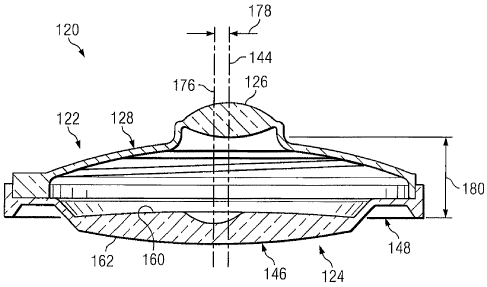
Referring more particularly to Figs. 2, 3, 4, 5, and 6, the anterior lens
122 includes an optic 126. The optic 126 is a power positive optic. In the
CA 02783680 2012 06 08
WO 2011/075331
PCT/US2010/059026
illustrated embodiment, the optic 126 is biconvex. That is, the anterior and
posterior surfaces of the optic 126 are convex. In some embodiments, the
optic 126 has a focal length between about 3.0 mm and about 7.0 mm and, in
some instances, between about 5.0 mm and about 6.0 mm.
5 The anterior lens 122 also includes haptics 128. As a general matter,
the haptics 128 are configured to offset the optic 126 as will be discussed in
greater detail below. In some instances, the haptics 128 are clear or
translucent and provide substantially no optical power. In the illustrated
embodiment, the haptics 128 have a rim 130 that defines an outer boundary
10 131. In
the illustrated embodiment, the outer boundary 131 has a
substantially circular profile centered about a center point 132, as best seen
in
Fig. 5. The outer boundary 131 is defined by a radius 133 extending from the
center point. Generally, the radius 133 is between about 3.0 mm and about
5.5 mm and, in some instances, is between about 4.2 mm and about 4.8 mm.
Extending inwardly from the rim 130 are arms 134 and 136. The arms
134, 136 connect the rim 130 to a mounting area 138. The mounting area
138 is configured to mount the optic 126 in a proper orientation. In that
regard, the haptics 128 are configured to position the optic 126 such that it
is
offset relative to the center point 132. In particular, the optic 126 is
centered
about a center point 140 that is offset from the center point 132 by a
distance
142. In some embodiments, the distance 142 is between about 0.05 mm and
about 0.75 mm. As the optic 126 is centered about the center point 140, an
optical axis 144 of the optic 126 extends through the center point 140, as
shown in Figs. 2 and 6.
Referring again to Fig. 5, in the illustrated
embodiment the mounting area 138 has a generally circular outer profile
centered about the center point 140. Accordingly, mounting area 138 is offset
relative to the center point 132. In that regard, the arms 134, 136 have
different lengths to accommodate the offset position of the mounting area 138
and optic 126. In the illustrated embodiment, the arm 134 is shorter than the
arm 136. While the two arms 134, 136 are illustrated, it is understood that
any number of connections between the rim 130 and the mounting area 138
may be utilized.
Referring more particularly to Figs. 2, 7, 8, 9, 10, and 11, aspects of the
posterior lens 124 will be discussed. As a general matter, the posterior lens
CA 02783680 2012 06 08
WO 2011/075331
PCT/US2010/059026
11
124 includes optics 146 and haptics 148. In the illustrated embodiment, the
haptics 148 include a rim 150 that defines an outer boundary 151 and an
inner boundary 152. In the illustrated embodiment, the outer boundary 151
and the inner boundary 152 have substantially circular profiles centered about
a center point 154, as best seen in Fig. 10. As shown in Fig. 10, the inner
boundary 152 is generally defined by a radius 156 extending from the center
point 154. In that regard, the radius 156 is substantially equal to the radius
133 of the anterior lens 122 to allow mounting of the anterior lens 122 within
the rim 150. The haptics 148 of the posterior lens 124 also include a surface
158 extending inwardly from the rim 150. In some instances, the surface 158
is configured to mate with a bottom surface of the rim 130 of the anterior
lens
122. In
that regard, the surface 158 is substantially planar in some
embodiments. The surface 158 extends substantially perpendicular to the
inner boundary 152 in the illustrated embodiment. In
other instances,
however, the surface 158 extends at an oblique angle relative to the inner
boundary 152.
As shown in Fig. 2, the optics 146 include an anterior surface 160 and
a posterior surface 162. Referring to Figs. 7 and 10, the anterior surface 160
includes a central portion 164 surrounded by a peripheral portion 166. In the
illustrated embodiment, the central portion 164 has a generally circular
profile
defined by a radius 168 extending from the center point 154. In that regard,
the radius 168 is generally between about 0.5 mm and about 4.0 mm.
Relative to the anterior surface 160 as a whole, the central portion 164 is
generally between about 10% and about 70% of the total surface area of the
anterior surface 160. The central portion 164 defines a negative power
surface optic. Accordingly, in the illustrated embodiment the central portion
164 of the anterior surface 160 is concave. The peripheral portion 166
defines a positive power surface optic. Accordingly, in the illustrated
embodiment the peripheral portion 166 is convex. The transition between the
central portion 164 and the peripheral portion 166 may be a smoothed or
rounded transition, an abrupt transition (e.g., such that the transition
defines
an edge), and/or combinations thereof.
Referring more particular to Fig. 11, the posterior surface 162 similarly
includes a central portion 170 surrounded by a peripheral portion 172. In the
CA 02783680 2012 06 08
WO 2011/075331
PCT/US2010/059026
12
illustrated embodiment, the central portion 170 has a generally circular
profile
defined by a radius 174 extending from the center point 154. In that regard,
the radius 174 is generally between about 0.5 mm and about 4.0 mm.
Relative to the posterior surface 162 as a whole, the central portion 170 is
generally between about 10% and about 70% of the total surface area of the
posterior surface 162. In some instances, the radius 174 defining the central
portion 170 of the posterior surface 162 is substantially equal to the radius
168 defining the central portion 164 of the anterior surface 160. In other
instances, the radius 174 is larger or smaller than the radius 168 such that
the
central portion 170 of the posterior surface 162 is correspondingly larger or
smaller than the central portion 164 of the anterior surface 160.
The central portion 170 of the posterior surface 162 defines a positive
power surface optic. Accordingly, in the illustrated embodiment the central
portion 170 of the posterior surface 162 is convex. Similarly, the peripheral
portion 172 of the posterior surface 162 also defines a positive power surface
optic. Accordingly, in the illustrated embodiment the peripheral portion 172
is
convex as well. The transition between the central portion 170 and the
peripheral portion 172 may be a smoothed or rounded transition, an abrupt
transition (e.g., such that the transition defines an edge), and/or
combinations
thereof. The central portion 170 is demarcated in phantom to illustrate the
fact that the central portion 170 and the peripheral portion 172 are parts of
a
single continuous optical surface in some instances. In that regard, there is
not a visible transition between the central portion 170 and the peripheral
portion 172 in some instances. Further, in some instances, the central portion
170 and the peripheral portion 172 have the same positive optical power.
Generally, the central portion 164 of the anterior surface 160 and the
central portion 170 of the posterior surface 162 project a magnified image
towards the retina 112. As discussed below, in some instances the central
portion 164 projects a substantially collimated beam of light towards the
central portion 170, which then projects a resulting magnified image towards
the retina 112. Further, in some embodiments the peripheral portions 166,
172 of the anterior and posterior surfaces 160, 162 together form a single
focal optic. In that regard, the peripheral portions 166, 172 provide a power
range between about 6 diopters and about 34 diopters in some instances.
CA 02783680 2012 06 08
WO 2011/075331
PCT/US2010/059026
13
The particular strength of the single focal optic formed by the peripheral
portions 166, 172 may be selected based on patient need. In that regard, the
peripheral portions 166, 172 of the posterior lens 124 are utilized to project
images of the peripheral field of vision onto the retina in some instances.
Generally, the optics 146 defined by the anterior surface 160 and the
posterior surface 162 share a common optical axis 176, as shown in Fig. 9.
The optical axis 176 generally extends through the center point 154 of the
posterior lens 124. As shown in Figs. 1 and 2, when the anterior lens 122 is
engaged with the posterior lens 124, the optical axis 144 of the anterior lens
is
offset with respect to the optical axis 176 of the posterior lens by a
distance
178. In that regard, engagement of the outer boundary 131 of the rim 130 of
the anterior lens 122 with the interior boundary 152 of the rim 150 of the
posterior lens 124 substantially aligns the center point 132 of the anterior
lens
with the center point 154 of the posterior lens. Accordingly, the optic 126 of
the anterior lens 122 is offset with respect to the optics 146 of the
posterior
lens by a distance equal to the offset distance of the optic 126 relative to
the
center point 132. Since the optical axis 176 of the posterior lens extends
from
the center point 154, the offset distance 178 between the optical axes 144,
176 is substantially equal to the offset distance 142. Accordingly, in some
instances the offset distance 178 is between about 0.05 mm and about 0.75
mm. As shown in Fig. 2, when the anterior lens 122 is engaged with the
posterior lens 124, the optic 126 of the anterior lens is spaced from the
optics
146 of the posterior lens by a distance 180. In that regard, the distance 180
represents the distance between the posterior-most portion of the optic 126
and the anterior-most portion of the optics 146. In some instances, the
distance 180 is between about 2.0 mm and about 4.0 mm, but may be outside
of this range in some instances. In some instances, the distance 180 is
determined based on the focal length of the optic 126. In that regard, the
distance 180 may be selected such that the focal point of the optic 126 falls
within the optics 146 of the posterior lens 124.
Referring now to Fig. 12, the offset between the optic 126 of the
anterior lens 122 and the optics 148, in particular the central portions 164,
170, of the posterior lens 124 results in an a corresponding offset in the
image
projected onto the retina 112. In particular, light 182 representing a central
CA 02783680 2012 06 08
WO 2011/075331
PCT/US2010/059026
14
field of vision comes into the eye 102 and passes through the cornea 104 and
into optic 126 of the anterior lens 122. The optic 126 focuses the light 182
towards the central portion 164 of the anterior surface 160 of the posterior
lens. In some instances, the cornea 104, optic 126, and central portion 164
form an afocal Galilean telescope having an angular magnification in the
range of 1.5X to 4.0X. In that regard, the cornea, optic 126, and central
portion 164 produce a substantially collimated light beam within the posterior
lens 124 that is directed towards the central portion 170 of the posterior
surface 162, in some embodiments. The light passes through the central
portion 170 of the posterior surface 162 and is projected onto the retina 112.
In that regard, offset distance 178 between the optical axes 144 and 176
determines the amount of offset of the resulting magnified image 184 relative
to a center point of the fovea. In general, the greater the offset distance
178,
the greater the amount of offset of the resulting magnified image 184. In that
regard, it is contemplated that a surgical kit for the intra-ocular lens
system
120 may include a plurality of anterior lenses 122 having different offsets
such
that an anterior lens with the appropriate amount of offset for a particular
patient may be selected.
Further, in addition to the amount of offset of the resulting image 184,
the direction of the offset may also be selected. In that regard, in some
instances the anterior lens 122 is oriented relative to the posterior lens 124
such that the magnified image 184 produced by the intra-ocular lens system
120 is directed away from a damaged portion of the macular 114, such as all
or a portion of the fovea 116, and towards a healthier portion of the retina
112.
In that regard, the anterior lens 122 may be rotated relative to the posterior
lens 124 to adjust the direction of the offset. The anterior lens 122 may be
rotated 360 degrees relative to the posterior lens 124 such that the magnified
image 184 may be directed up, down, left, right, and/or combinations thereof.
In the illustrated embodiment, the circular profiles of the rims 130 and 150
result in the amount of offset being substantially constant. However, by
providing a plurality of anterior lenses with different amounts of offset, as
discussed above, and the fact that the direction of offset is selectable via
rotation of the anterior lens relative to the posterior lens, the direction
and
magnitude of the offset can generally be tailored to fit the needs of any AMD
CA 02783680 2012 06 08
WO 2011/075331
PCT/US2010/059026
or other low vision patient.
To facilitate proper orientation of the lenses 122, 124 and, in particular,
the optic 126 of the anterior lens 122, one or both of the lenses 122, 124 may
include markings, an index, and/or other feature(s) to indicate a relative
5 position of the lenses. In that regard, the markings, index, and/or other
feature(s) can signify to a surgeon the direction of offset of the optic 126
and,
thereby, the direction in which the resulting magnified image 184 of the intra-
ocular system will be directed relative to a center point of the fovea.
Accordingly, if, for example, the patient has damage in a lower left quadrant
of
10 the fovea, the lenses 122, 124 can be oriented to direct the magnified
image
184 towards the upper right quadrant of the fovea and surrounding portions of
the macular and retina. In some instances, the markings, index, and/or other
feature(s) are part of the rim 130 of the anterior lens 122. In some
instances,
the structure of the haptics 128 of the anterior lens 122 is utilized to
identify to
15 the surgeon or caregiver the direction of offset of the optic 126.
Identifying the
portions of the fovea, macular, and/or retina that are damaged and, therefore,
the appropriate direction for offsetting the magnified image 184 may be
determined utilizing standard techniques (e.g., retinal scope) prior to
implantation of the intra-ocular lens system 120. In that regard, a calculator
program can propose a suggested position for the magnified image 184 and
provide the corresponding orientation of the lenses 122, 124 based on data
received from pre-implantation testing. Alternatively, the intra-ocular lens
system 120 may be implanted and then tuned or adjusted to provide the best
vision for the patient. In that regard, the orientation of the lenses 122, 124
may be adjusted after implantation to accommodate for future changes in the
patient's eyesight. For example, if the area initially selected to receive the
magnified image 184 itself becomes damaged, then the another suitable area
can be identified and the orientation of the lenses 122, 124 adjusted to
direct
the magnified image there. In this manner, the intra-ocular lens system 120
may be tailored to a patient's needs even long after initial implantation.
The magnified image 184 discussed above is generally produced by
the optic 126 of the anterior lens 122 and the central portions 164, 170 of
the
posterior lens 124. In that regard, the magnified image 184 is of a central
field
of vision and, importantly, the resulting magnified image 184 does not occupy
CA 02783680 2012 06 08
WO 2011/075331
PCT/US2010/059026
16
the entire field of vision of the patient. Rather, magnified image 184 is
projected only over a portion of the retina 112 such that images from the
peripheral field of vision are also projected onto the retina. In that regard,
light
passing into the eye representing the peripheral field of vision misses the
optic 126 of the anterior lens 122 and passes through to the peripheral
portions 166, 172 of the posterior lens. As discussed above, the peripheral
portions 166, 172 together form a single focal optic that is utilized to
project
the light representative of the peripheral field of vision onto the retina. In
that
regard, the peripheral portions 166, 172 provide a power range between
about 6 diopters and about 34 diopters in some instances. The particular
strength of the single focal optic formed by the peripheral portions 166, 172
may be selected based on patient need. Accordingly, the intra-ocular lens
system 120 provides the patient with both an improved magnified image 184
of the central field of vision without causing tunnel vision by still
providing the
peripheral field of vision to the surrounding portions of the retina.
In some instances, the deflection of the magnified image 184 is utilized
to avoid scotoma in the visual field. For example, deflection of the image 184
is particularly useful for AMD patients who have undergone macular
translocation surgeries. In that regard, macular translocation is a surgical
technique designed to move the area of the retina responsible for fine vision
(macula) away from the diseased underlying layers (the retinal pigment
epithelium and choroid). Generally, the macula is moved to an area where
these underlying tissues are healthier. For patients who have undergone
macular translocation surgeries, their normal line of sight is no longer
aligned
with their macula. Consequently, the macular translocation treated eye could
show the undesirable "tropia" appearances, such as "esotropia" or
"exotropia". Further, in cases where the patient has both eyes treated with
macular translocation surgeries, there can be negative impact to the intended
vision function. For example, if the left eye needs to look up to see better
and
the right eye needs to look down to see better, then the patient will have a
difficult time seeing clearly with both eyes because such binocular eye
movements are very difficult to perform. Redirecting the retinal image
location
can reduce or correct the "tropia" appearances by relocating the line of sight
to the new macular location. Further, the intra-ocular lens systems of the
CA 02783680 2012 06 08
WO 2011/075331
PCT/US2010/059026
17
present disclosure allow redirecting the retinal image location for each eye,
such that in the case of dual macular translocation the need for binocular eye
movements is eliminated or greatly reduced.
The lenses 122, 124 of the intra-ocular lens system 120 are configured
for implantation into the capsular bag 110 in the posterior chamber 108 of the
eye 102 utilizing minimally invasive techniques. Accordingly, the intra-ocular
lens system avoids the complications associated with a combination anterior
chamber and posterior chamber system, while still providing the benefits of
minimally invasive surgical techniques. In that regard, the lenses 122, 124
are configured for implantation through an incision or capsular rhexis having
a
length less than about 4.0 mm and, typically, less than 3.5 mm. In some
instances, the lenses 122, 124 are configured for implantation utilizing a
cartridge system, including cartridge systems commercially available from
Alcon. In some instances, the lenses 122, 124 are engaged with one another
prior to implantation. In other instances, the lenses 122, 124 are inserted
into
the capsular bag 110 separately. For example, in some embodiments, the
posterior lens 124 is inserted into the capsular bag 110. Then the anterior
lens 122 is inserted into the capsular bag 110 and engaged with the posterior
lens 124. In some instances, the capsular bag 110 is shrink-wrapped around
the lenses 122, 124 after implantation to securely engage the lenses. Further,
in some embodiments at least a portion of the optic 126 of the anterior lens
122 is sized and shaped to extend through the incision or capsular rhexis in
the capsular bag 110 after the capsular bag has been shrink-wrapped around
the lenses. Further, in some embodiments, the size and shape of the lenses
122, 124 helps prevent interlenticular cell growth. In that regard, the
structure
of at least the anterior lens facilitates easier contact between the anterior
capsular leaflets and the posterior capsule. In some instances, the diameter
of the optic 126 being smaller than the capsular rhexis opening combined with
the central leg spacing of the haptics results in easier contact with the
anterior
capsular leaflets, thereby limiting or preventing unwanted interlenticular
cell
growth. In some instances, shrink-wrapping of the capsular bag 110 around
the lenses 122, 124 seals off the circumferential space around the optics of
the lenses to prevent interlenticular cell growth.
Referring to Fig. 13, shown therein is an arrangement 200 illustrating
CA 02783680 2012 06 08
WO 2011/075331
PCT/US2010/059026
18
an alternative embodiment of the present disclosure. Specifically, an intra-
ocular lens system 220 is implanted within the capsular bag 110 in the
posterior chamber 108 of the eye 102. As shown, the intra-ocular lens system
220 includes an anterior lens 222 and a posterior lens 224. As a general
matter, the intra-ocular lens system 220 provides functionality similar to
that of
intra-ocular lens system 120 described above. For example, the intra-ocular
lens system 220 provides a magnified retinal image that is directed away from
a damaged portion of the macular 114, such as all or a portion of the fovea
116, while still providing peripheral images to the retina. However, instead
of
the having optical axis of the anterior lens 222 offset relative to the
optical axis
of the posterior lens 224 by a particular distance (with the optical axes
extending substantially parallel to one another), the optical axis of the
anterior
lens is at an oblique angle relative to the optical axis of the posterior lens
in
the intra-ocular lens system 220.
Referring now to Figs. 14, 15, 16, 17, 18, and 19, aspects of the intra-
ocular lens system 220 will be discussed in greater detail. In that regard, in
the illustrated embodiment the posterior lens 224 is substantially similar to
the
posterior lens 124 discussed above and, therefore, will not be discussed in
detail here. Accordingly, the current focus will be on the features of the
anterior lens 224. In that regard, Fig. 14 is a perspective top view of the
anterior and posterior lenses 222, 224 of the intra-ocular lens system 220,
while Figs. 15, 16, 17, 18, and 19 are, respectively, perspective top,
perspective bottom, side, front, and top views of the anterior lens 222.
As shown, the anterior lens 222 includes an optic 226. The optic 226 is
a power positive optic. In the illustrated embodiment, the optic 226 is
biconvex. That is, the anterior and posterior surfaces of the optic 226 are
convex. In some embodiments, the optic 226 has a focal length between
about 3.0 mm and about 7.0 mm and, in some instances, the focal length is
between about 5.0 mm and about 6.0 mm.
The anterior lens 222 also includes haptics 228. As a general matter,
the haptics 228 are configured to angularly offset the optic 226, as will be
discussed in greater detail below. In some instances, the haptics 228 are
clear or translucent and provide substantially no optical power. In the
illustrated embodiment, the haptics 228 have a rim 230 that defines an outer
CA 02783680 2012 06 08
WO 2011/075331
PCT/US2010/059026
19
boundary 232 and an inner boundary 234. In the illustrated embodiment, the
rim 230 has a substantially constant thickness 236 between the outer
boundary 232 and the inner boundary 234. In that regard, the outer boundary
232 and the inner boundary 234 have a substantially circular profiles centered
about a center point 238, as best seen in Fig. 19. In some instances, the
outer boundary has a radius between about 3.0 mm and about 5.5 mm and, in
some instances, is between about 4.2 mm and about 4.8 mm. However, in
other embodiments, the rim 230 has other profiles. For example, Fig. 21
illustrates an embodiment of an anterior lens 400 according to another aspect
of the present disclosure. In that regard, the lens 400 is similar to anterior
lens 222, except that portions of opposing sides of the lens have been
removed such that the outer boundary of the lens defines a generally
rectangular profile with rounded ends. In some embodiments, the rounded
end portions have a partially circular profile, similar that of rim 230, such
that
the lens 400 can interface with a posterior lens (such as lenses 124 and 224)
in a similar manner.
Extending inwardly from the rim 230 are arms 240, 242, and 244. The
arms 240, 242, 244 connect the rim 230 to a mounting area 246. In the
illustrated embodiment, the arms 240, 242, 244 have substantially equal
lengths. While the three arms 240, 242, and 244 are illustrated, it is
understood that any number of connections between the rim 230 and the
mounting area 246 may be utilized. For example, Fig. 22 shows an
embodiment of an anterior lens 500 according to another aspect of the
present disclosure. In that regard, the lens 500 is substantially similar to
lens
222, except that the lens 500 only has two arms connecting the rim to the
mounting area where the optics are positioned. Referring again to, Fig. 19,
the mounting area 246 is configured to mount the optic 226 in a proper
orientation. In that regard, the haptics 228, including mounting area 246, are
configured to position the optic 126 such that it will be angular offset
relative
to the optics of the posterior lens when the anterior and posterior lenses are
engaged with one another.
As best seen in Fig. 17, in the illustrated embodiment, the haptics 228
of the anterior lens 222 define an end 248 of the rim 230 having a height or
thickness 250 and an opposing end 252 having a height or thickness 254. In
CA 02783680 2012 06 08
WO 2011/075331
PCT/US2010/059026
that regard, the height 250 is greater than the height 254 such that the rim
230 tapers between the end 248 and the end 252. As shown, the rim 230 has
a continuous and constant taper between the ends 248, 252 in the illustrated
embodiment. As the arms 240, 242, 244 are spaced about the circumference
5 of the
rim 230 and are substantially equal in length, the mounting area 246 is
angled by an amount matching the taper of the rim 230. Accordingly, the
amount of angle of the mounting area 246 can be adjusted by changing the
relative heights between the ends 248 and 252. In
the illustrated
embodiment, the optic 226 is mounted on the mounting area 246 such that it
10 is also
angled to match the taper of the rim 230. In that regard, the optic 226
defines an optical axis 256 that extends at an oblique angle 258 relative to
an
axis 260 extending substantially perpendicular to a lower surface 262 of the
anterior lens 222, as best seen in Figs. 13 and 17. In that regard, the lower
surface 262 is a generally planar surface configured to mate with a surface of
15 the
posterior lens 224 similar to surface 158 of posterior lens 124 discussed
above. Generally, the oblique angle 258 is between about 0.5 degrees and
about 15 degrees, but may be outside of this range in some instances.
In some instances, the axis 260 is substantially aligned with an optical
axis of the optics of the posterior lens 224 when the anterior lens 222 and
the
20
posterior lens are engaged. In other instances, the axis 260 and the optical
axis of the optics of the posterior lens 224 extend substantially parallel to
one
another, but are separated by a distance between about 0.05 mm and about
1.5 mm. In such embodiments, the optical axis 256 of the optic 226 is offset
with respect to the optical axis of the optics of the posterior lens in both
angular and distance orientations. Generally, the particular angular and/or
distance offset between the optical axes of the anterior and posterior lenses
222, 224 is selected in order to project a magnified image to a desired
portion
of the retina 112.
Referring now to Fig. 20, the angular offset of the optic 226 of the
anterior lens 222 relative to an optical axis of the optics of the posterior
lens
124 results in an a corresponding offset in the image projected onto the
retina
112. In particular, light 264 representing a central field of vision comes
into
the eye 102 and passes through the cornea 104 and into optic 226 of the
anterior lens 222. The optic 226 focuses the light 264 towards the posterior
CA 02783680 2012 06 08
WO 2011/075331
PCT/US2010/059026
21
lens 224, which projects a magnified image 266 onto the retina 112. In that
regard, angle 258 of the offset between the optical axis 256 of the optic 226
and the optical axis of the posterior lens 224 determines the amount of offset
of the resulting magnified image 266 relative to a center point of the fovea.
In
general, the greater angle 258, the greater the amount of offset of the
resulting magnified image 266, assuming the anterior lens 222 is centered
about the posterior lens 224 such that the optical axis of the posterior lens
224 substantially coincides with the axis 260. It is contemplated that a
surgical kit for the intra-ocular lens system 220 may include a plurality of
anterior lenses 222 having different angular offsets such that an anterior
lens
with the appropriate amount of offset for a particular patient may be
selected.
Generally, the lenses 222, 224 may be manipulated in a similar manner to
lenses 122 and 124, discussed above, in order to adjust the position of the
magnified image 266 on the retina.
Referring now to Fig. 21, shown therein is perspective cross-sectional
view of an intra-ocular lens system 320 according to another embodiment of
the present disclosure. In that regard, the intra-ocular lens system 320
includes an anterior lens 322 and a posterior lens 324. The anterior lens 322
includes a power positive optic 326 similar to optics 126 and 226 above. The
anterior lens 322 further includes haptics 328. The haptics 328 include an
arm 330, as shown. It is understood that the anterior lens 322 includes
another arm (not shown) similar to arm 330 on the other half of the anterior
lens 322 not illustrated in Fig. 21. The posterior lens 324 includes optics
332
that are similar to the optics of posterior lens 124 discussed above. The
posterior lens 324 also includes haptics 334. The haptics 334 include an arm
336, as shown. It is understood that the posterior lens 324 includes another
arm (not shown) similar to arm 336 on the other half of the posterior lens 324
not illustrated in Fig. 21. The haptics 328, 334 and, in particular, the arms
330, 336 of the anterior and posterior lenses 322, 324 have properties that
result in a desired offset (either distance or angle) of the optical axes of
the
optics 326, 332 of the anterior and posterior lenses. In that regard, the
material properties of the haptics 328, 334, the geometrical structures of the
haptics 328, 334, and/or combinations thereof are adjusted to achieve the
desired offset. In some instances, a plurality of anterior lenses 322 and a
CA 02783680 2017-01-18
22
plurality of posterior lenses 324 are provided in a kit to allow treating
medical
personnel to select the appropriate combination of the lenses to achieve a
desired
offset.
Generally, the lenses of the intra-ocular lens systems of the present
disclosure
may be formed of any suitable material. For example, in some instances the
lenses
are formed of a soft acrylic polymer (e.g., a material used to form
commercially
available lenses sold by Alcon under the trademark Acrysof0). In other
embodiments, the lenses are formed of other suitable biocompatible materials,
such
as a silicone or hydrogel. In some instances, the haptics of the lenses are
form of a
different material than the optics. In such instances, the haptics may be
formed of
suitable polymeric materials, such as polymethylmethacrylate, polypropylene
and the
like. The lenses of the intra-ocular lens systems of the present disclosure
may also
be formed of the materials disclosed in U.S. Pat. No. 6,416,550. In some
instances,
the lenses are foldable to facilitate insertion using minimally invasive
surgical
techniques. In particular, the lenses may be configured to be inserted through
an
incision having a length less than 4.0 mm and, in some instances, less than
3.5 mm.
In some embodiments, the lenses are configured for insertion using an intra-
ocular
lens cartridge system. Further, the lenses may be inserted separately or
together. For
example, in one embodiment the posterior lens is first inserted into the
capsular bag
and then the anterior lens is inserted into the capsular bag and engaged with
the
posterior lens.
The intra-ocular lens systems of the present disclosure are used in
combination with other treatments in some instances. For example, when
treating
patients with AMD, any of the disclosed intra-ocular lens systems may be used
in
conjunction with administration of an AMD drug to stop and deter further
development of AMD. In some instances, the AMD drug is an ophthalmic
pharmaceutical preparation for the treatment of advanced macular degeneration.
The
AMD drug can steady and stabilize the vision to help the intra-ocular lens
systems
better improve the patient vision. Also, the intraocular lens systems are used
with
contact lenses, refractive ablations, and/or other treatments in some
instances.
CA 02783680 2012 06 08
WO 2011/075331
PCT/US2010/059026
23
Further, while anterior surfaces of the posterior lenses have generally
been illustrated as forming the negative optics of the posterior lens, this is
for
illustrative purposes of the operation principles of the devices and no
limitation
is intended thereby. Rather, it is understood that the anterior surface, the
posterior surface, and/or combinations of the anterior and posterior surfaces
of the posterior lens are utilized to form the negative optics in some
embodiments. For example, in some instances the central portion of the
anterior surface of the posterior lens is a positive optic and the central
portion
of the posterior surface is a negative optic. In other instances, central
portions of both the anterior surface and the posterior surface are negative
optics. In that regard, in some embodiments where central portions of both
the anterior surface and the posterior surface are negative optics, the degree
of the optics is decreased such that the total of effect of the negative
optics of
the anterior and posterior surfaces is substantially equal to the negative
optics
when only one of the surfaces is utilized.
While the embodiments described above focused on offsetting the
optics of the anterior lens utilizing various methods (e.g., distance and
angle),
it is understood that no limitation is intended thereby. Generally, any means
of producing a deflected, magnified image may be utilized. Further, it is
understood that the same principles discussed with respect to the anterior
lenses above may similarly be applied to offset the optics of the posterior
lens.
Accordingly, in some embodiments, the optics of the posterior lens are offset
utilizing the features and methods described above. Further still, in some
embodiments the optics of both the anterior and posterior lenses are offset
utilizing the features and methods described above. Generally, the intra-
ocular lens systems of the present disclosure may utilize any combination of
offsets (e.g., distance and/or angle) in the optics of one or both of the
anterior
and posterior lenses.
Although illustrative embodiments have been shown and described, a
wide range of modification, change, and substitution is contemplated in the
foregoing disclosure. It is understood that such variations may be made to
the foregoing without departing from the scope of the present disclosure.
Accordingly, it is appropriate that the appended claims be construed broadly
and in a manner consistent with the present disclosure