Note: Descriptions are shown in the official language in which they were submitted.
CA 02784137 2012-06-12
WO 2011/085369 PCT/US2011/020832
ASSAY FOR JC VIRUS ANTIBODIES
RELATED APPLICATIONS
This application claims the benefit of U.S. Provisional Application
No. 61/294,048, filed January 11, 2010, and U.S. Provisional Application No.
61/316,193,
March 22, 2010, both of which are incorporated herein by reference in their
entirety.
FIELD OF THE INVENTION
The invention relates to methods and reagents for analyzing samples for the
presence of JC virus antibodies.
BACKGROUND
Progressive Multifocal Leukoencephalopathy (PML) is an opportunistic infection
of the central nervous system (CNS) that is associated with exposure to the JC
virus
(JCV), a polyoma virus that is believed to be pathogenic in humans only under
conditions
of persistent immune suppression or immune modulation. While the presence of
JCV is
required for development of PML, PML risk is considered, in a not well-
understood way,
to be associated with the convergence of multiple viral and host-related
factors that cause
the virus to become pathogenic (Major, "Progressive Multifocal
Leukoencephalopathy in
Patients on Immunomodulatory Therapies" Annu. Rev. Med. 61:35-47 (2010) [2009
Aug. 31, Epub ahead of print]). Published studies reporting the prevalence of
JCV
infection in the human population are varied. This information is based on
various types
of studies including PCR analysis for viral DNAanddetectionof antibodiesto
JCV.
Despite the prevalence of JCV in the population, infection with JCV rarely
results in
PML, even in individuals with documented immunosuppression.
Published reports on JCV DNA detection suggest the method to be insensitive
and
of limited use for assessing exposure to JCV because JCV DNA has been rarely
and
inconsistently detected in the plasma, serum or peripheral blood mononuclear
cells of
JCV-infected PML patients. Detection of anti-JCV antibodies appears to be a
more
sensitive marker of JCV infection; however the reported results are variable.
In 1973,
1
CA 02784137 2012-06-12
WO 2011/085369 PCT/US2011/020832
Padgett and Walker published a study reporting a JCV seroprevalence of 65-84%
using a
haemagglutination inhibition (HI) assay (Padgett and Walker, "Prevalence of
antibodies
in human sera agains JC virus, an isolate from a case of progressive
multifocal
leukoencephalopathy" J. Infect. Dis. 127:467-70, 1973). Later reports of JCV
seroprevalence rates using the HI assay or ELISA have varied between 33-91%.
The
variable seroprevalence rates among these studies are likely due to marked
differences in
the size and demographics of the studies, and, perhaps most importantly,
differences in
assay methods.
It is therefore desirable to implement a reliable and sensitive assay for
determining the presence of JCV antibodies that can be used, for example, for
assessing
whether an individual has been exposed to JCV.
SUMMARY OF THE INVENTION
The invention relates to the development of an analytically validated,
sensitive
assay for detecting the presence of JCV antibodies in a biological fluid,
e.g., serum or
plasma.
Accordingly, the invention relates to a method that includes obtaining a
biological
sample from a subject (e.g., plasma, serum, blood, urine, or cerebrospinal
fluid (CSF));
contacting the sample with highly purified viral-like particles (HPVLPs) under
conditions
suitable for binding of a JCV antibody in the sample to an HPVLP; detecting
the level of
JCV antibody binding in the sample to HPVLP; and correlating the detected
level with a
reference, such that the reference is selected to indicate a false negative
rate not greater
than 3% and minimal cross reactivity to other components of the sample such as
antibodies against other polyoma viruses, e.g., BK virus (BKV). In some
embodiments,
the reference, derived from a control sample or set of samples, is processed
with the
sample from the subject. In some embodiments, the reference is selected such
that the
false negative rate of the assay is not greater than 1 %.
In one embodiment, at least about 10% of the HPVLPs in a preparation of
purified HPVLPs contain more than five VP1 polypeptides per HPVLP. In other
embodiments, at least about 15%, about 20%, about 25%, about 30%, about 40%,
about
2
CA 02784137 2012-06-12
WO 2011/085369 PCT/US2011/020832
50%, about 60%, about 65%, about 70%, about 80% or about 90% of the HPVLPs in
a
preparation of purified HPVLPs contain more than five VPl polypeptides per
HPVLP.
The assay can be performed such that the HPVLP is immobilized on a solid
substrate such as a microtiter plate or slide. In some embodiments, the HPVLP
consists
essentially of VP1 viral protein. The HPVLP can further include other viral
proteins, for
example at least one of a VP2 or a VP3. The viral protein(s) in the HPVLP can
be
recombinantly derived (e.g., a MAD1 strain VP1) or can be a naturally-
occurring viral
protein (e.g., derived from a naturally-occurring source). The method can be
performed
using, for example, a biological sample obtained from a subject currently
being treated
with an immunomodulatory drug, a subject considering initiating treatment with
an
immunomodulatory drug, or a subject suspected of having Progressive Multifocal
Leukoencephalopathy (PML).
In some aspects, the assay method is a two-step assay that further includes a
secondary confirmation assay process that includes contacting a portion of the
biological
sample from the subject with HPVLP in solution (prior to incubating the sample
with the
HPVLP attached to a solid substrate), thereby providing a secondary sample;
contacting
the secondary sample with HPVLP under the same conditions used for the primary
assay;
detecting the level of JCV antibody binding to HPVLP in the secondary sample;
and
comparing the detected level of JCV antibody in the secondary sample to the
level of
JCV antibody in the sample that was not preincubated with soluble HPVLP, such
that a
decrease in the detected level in the secondary assay sample compared to the
sample that
was not preincubated indicates the sample is positive for JCV antibody, and a
change in
the detected level below a specified percentage indicates that there is no JCV-
specific
antibody present in the sample.
An assay described herein can be used to assay for the presence of JCV
antibodies
in a subject who has never received treatment with an immunomodulator; or in a
subject
who has previously received an immunomodulator, but who is no longer receiving
treatment with the immunomodulator; or in subject who is presently undergoing
treatment with an immunomodulator.
Detection of JCV antibodies binding to the HPVLPs in an assay featured in the
invention can indicate that a subject is at an increased risk for PML.
Detection of JCV
3
CA 02784137 2012-06-12
WO 2011/085369 PCT/US2011/020832
antibodies can also indicate that the subject is at an increased risk for
adverse symptoms,
such as the development of PML, upon administration of certain therapeutic
agents, such
as certain immunomodulators, and therefore the subject is not a candidate for
treatment
with these agent. For example, detection of JCV antibodies in a sample from a
subject
can indicate that the subject is not a candidate for treatment with an anti-
VLA-4
therapeutic, such as natalizumab. In certain embodiments, detection of JCV
antibodies in
a biological sample can indicate that the subject is a candidate for treatment
with an
immunomodulator, such as natalizumab, except that the subject will undergo
enhanced
monitoring during treatment than a subject who does not have detectable JVC
antibodies.
For example, the enhanced monitoring can include observation for adverse
symptoms,
such as symptoms that may indicate the development of PML.
Failure to detect JCV antibodies binding to HPVLPs in an assay featured in the
invention can indicate that the subject is a candidate to receive treatment
with an
immunomodulator, such as natalizumab, and in one embodiment, the subject is
further
administered the immunomodulator. A subject determined not to have JCV
antibodies
can be re-tested at least annually (e.g., at least every 3 months, every 6
months, every 9
months, or every 12 months) to determine whether the subject has developed JCV
antibodies, which may indicate that the subject has been infected with JCV. A
subject
who previously did not have detectable JCV antibodies in a biological sample,
and who
subsequently develops JCV antibodies in a biological sample, can stop
receiving
treatment with an immunomodulator.
In some embodiments, a subject who was previously identified as having JCV
antibodies, can be subsequently tested at a later date and determined not to
have JCV
antibodies. These subjects can be determined to be candidates to receive
treatment with
an immunomodulator, such as natalizumab. In one embodiment, a subject who
previously tested positive for the presence of JCV antibodies and who
subsequently
tested negative for JCV antibodies can be administered the immunomodulator,
and
undergo enhanced monitoring as compared to a subject who never tested positive
for JCV
antibodies, such as to monitor for symptoms that may indicate the development
of PML.
An assay featured in the invention is useful to treat a subject having an
immunological disease or disorder, such as multiple sclerosis (MS) or Crohn's
Disease
4
CA 02784137 2012-06-12
WO 2011/085369 PCT/US2011/020832
(CD). In one embodiment, an assay described herein has been validated for use
in MS
and CD patients, such as by showing that the assay is effective to detect JCV
antibodies
in MS and CD patients in a controlled test environment, such as in a clinical
trial.
In another aspect, the invention relates to a kit comprising an HPVLP and at
least
one reagent for performing an assay to identify a JCV antibody level in a
sample, such as
a biological sample.
In other aspects, the invention relates to a solution comprising HPVLP
particles
consisting essentially of VP 1-containing particles that are greater in size
than a VP 1
pentamer, e.g., containing about 5, 10, 20, 30, 40, 50, 60, 70 or 72 pentamers
or
containing about 25 VPI molecules, about 50 VP1 molecules, about 100 VP1
molecules,
about 150 VP1 molecules, about 200 VP1 molecules, about 300 VP1 molecules,
about
350 VP1 molecules or about 360 VP1 molecules.
Another aspect featured in the invention is a method of preparing a solution
of
HPVLPs, the method comprising removing VP 1 -containing particles from the
solution
that are the size of a VPI pentamer or less. In one method VP1 polypeptides
are
expressed in cells, e.g., in insect cells or mammalian cells. The cells are
lysed, and then
the cells are treated with a nuclease, such as benzonase. Cell debris is
removed by
precipitation, such as by salt (e.g., ammonium sulfate) precipitation, and
then the VPl-
containing supernatant is concentrated and further purified using
diafiltration, such as by
one or two passages through a membrane, e.g., a tangential flow filtration
(TFF)
membrane. The solution containing the VP 1-containing particles, e.g., HPVLPs,
is then
further purified through an ion-exchange step, and elution of the HPVLPs is
performed,
e.g., with a buffer. VP1 purity can be assessed, e.g., electrophoresis (e.g.,
SDS-PAGE)
or mass spetometry. The presence of HPVLPs can be confirmed by microscopy,
e.g.,
electron microscopy. The percentage of total protein in the form of HPVLPs can
be
determined by sedimentation velocity analytical ultracentrifugation.
In one aspect, the invention features a method of identifying a subject at
risk of
developing PML, such as by obtaining a biological sample from the subject;
contacting
the biological sample with HPVLPs under conditions suitable for binding of a
JC Virus
(JCV) antibody in the sample to an HPVLP; detecting the level of JCV antibody
binding
in the sample to HPVLPs; and correlating the detected level with a reference
set, wherein
5
CA 02784137 2012-06-12
WO 2011/085369 PCT/US2011/020832
the subject is at increased risk of PML if JCV antibody binding is detected.
The
reference set is selected to indicate a false negative rate of about 5%, about
3%, about 1%
or less.
In another aspect, the invention features a method of identifying PML risk in
a
subject by determining the level of anti-JCV antibodies in a sample from the
subject,
such as from a plasma, blood or serum sample; and assigning a risk level to
the subject
according to the level of anti-JCV antibodies in the sample. The subject may
be
receiving an immunomodulatory therapy, such as an anti-VLA4 treatment, e.g.,
natalizumab, or may be a candidate for receiving an immunomodulatory thereapy.
In
some embodiments, the subject has been diagnosed with an immunological disease
or
disorder, such as multiple sclerosis or Crohn's disease. In one embodiment,
the level of
anti-JCV antibodies is determined using a one-step assay, and in another
embodiment, the
level of anti-JCV antibodies is determined using a two-step assay. Either the
one-step
assay or the two-step assay may include an ELISA assay.
In one embodiment, the method of identifying PML risk in a subject further
includes determining the level of anti-JCV antibodies in the subject in a
sample from a
date subsequent to the initial sample; comparing the level of anti-JCV
antibodies in the
sample from the subsequent date to the level in the sample from the initial
sample; and
determining whether the subject is at increased risk of PML at the subsequent
date
compared to the time of the initial sample.
In one aspect, the invention features a method of monitoring PML risk in a
subject, the method comprising determining the level of anti-JCV antibodies in
a subject
using a sample from a first date; assigning a risk of PML (e:g., high, or
moderate or low
risk) based on the level of anti-JCV antibodies in the subject on the first
date;
determining the level of anti-JCV antibodies in the subject using a sample
from a second
date; and assigning a risk of PML (e.g., high, or moderate or low risk) based
on the level
of anti-JCV antibodies in the subject on the second date.
As used herein, an "HPVLP" is a highly purified VLP ("virus-like particle")
consisting predominantly of the VP1 protein. An "HPVLP" featured in the
invention is
composed mainly of the major capsid protein "VP 1," which can be a naturally-
occurring
VP1 or a recombinant VP1, from the polyomavirus, JC Virus (JCV). An HPVLP can
be
6
CA 02784137 2012-06-12
WO 2011/085369 PCT/US2011/020832
composed of, e.g., more than one pentameric subunit, at least 10 pentameric
subunits, at
least 20 pentameric subunits, at least 30 pentameric subunits, at least 50
pentameric
subunits, at least seventy-two pentameric subunits or more of VPl. An HPVLP
may
contain VPI polypeptides in an undetermined configuration (e.g., the
polypeptides may
or may not be organized in pentamers), in which case an HPVLP can be composed
of
more than 5 VP1 polypeptides, at least 50 VP1 polypeptides, at least 150 VP1
polypeptides, at least 360 VP1 polypeptides or more. HPVLPs include
capsomeres,
which contain about 10 to 24 pentamers. An HPVLP featured in the invention can
bind
antibodies against naturally-occurring, intact JC virus. In some embodiments,
an HPVLP
includes a second, and optionally a third, type of polypeptide that is a minor
capsid
protein of JC virus, e.g., at least one VP2 or VP3 polypeptide. The VP2 or VP3
can be
recombinant or naturally-occurring or naturally-derived polypeptides.
Such "highly purified" particles contain more than one VP1 pentamer, e.g., at
least 5, 10, 20, 30, 40, 50, 60, 70, 72 VPl pentamers, or less than 100 VP1
pentamers.
Such highly purified particles can be obtained, for example, by a method that
involves
double filtration. For example, in one embodiment, a highly purified
preparation of
VLPs is obtained by purifying the particles at least twice by centrifugation,
e.g., through
a sucrose cushion. In general, an HPVLP preparation can be identified by its
activity in
an ELISA assay using defined control samples. In some cases, such control
samples are
negative controls and/or control samples containing low levels of JCV
antibodies.
Unless otherwise defined, all technical and scientific terms used herein have
the
same meaning as commonly understood by one of ordinary skill in the art to
which this
invention belongs. Although methods and materials similar or equivalent to
those
described herein can be used in the practice or testing of the invention,
suitable methods
and materials are described below. All publications, patent applications,
patents, and
other references mentioned herein are incorporated by reference in their
entirety. In case
of conflict, the present specification, including definitions, will control.
In addition, the
materials, methods, and examples are illustrative only and not intended to be
limiting.
The details of one or more embodiments of the invention are set forth in the
accompanying drawings and the description below. Other features, objects, and
7
CA 02784137 2012-06-12
WO 2011/085369 PCT/US2011/020832
advantages of the invention will be apparent from the description and
drawings, and from
the claims.
DESCRIPTION OF THE DRAWINGS
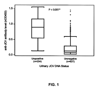
FIG. 1 is a graph depicting the results of an HPVLP ELISA on samples from
subjects positive for JCV DNA in their urine (Uropositive) and negative for
JCV DNA in
their urine (Uronegative). The box represents the interquartile (IQR) range
with the
median line in the center; brackets represent observations within 1.5 times
the IQR. "+"
signs represent observations beyond 1.5 times the IQR (outliers). *Mann-
Whitney U test.
FIG. 2 is a graph depicting anti-JCV antibody levels as measured by ELISA
against urinary JCV DNA level as measured by qPCR (n=204). Open circles
represent
urine and serum samples collected at matched STRATA time points. Closed
circles
represent samples collected at different time points. For 17 samples with DNA
test
results below the level of quantitation (<500 copies/mL) the level was set to
the detection
limit.
FIG. 3 is a graph depicting BKV-JCV cross-reactivity data from one rabbit
immunized with BKV. Antisera from the BKV-immunized rabbit bound BKV VLPs
with high affinity (EC50 = 1:100,000) and cross-reacted with JCV VLPs (EC50 =
1:5,000).
FIGs. 4A and 4B depict the anti-JCV assay reactivity of serum samples from
uronegative (n=311) (FIG. 4A) and uropositive (n=204) (FIG. 4B) patients in
the
screening and confirmation ELISAs. Distribution of serological reactivity of
the samples
in the screening ELISA are shown, with lower (nOD450 = 0.10) and upper
(nOD450 = 0.25) cut points highlighted (left panels). In the supplemental
confirmation
ELISA (right panels), a 40% inhibition cut point is highlighted (vertical
line) with shaded
regions denoting samples that did not confirm to have anti-JCV specific
antibodies
(nOD45o < 0.25 and percent inhibition < 40%).
FIGs. 5A and 5B are histograms depicting the frequency of observations within
each 10% inhibition range for all patients (n=515) (FIG. 5A) and uropositive
patients
(n=204) (FIG. 5B). The distribution consisted of two clearly defined peaks,
most
optimally separated at 40% inhibition. A 40% inhibition level corresponded to
8
CA 02784137 2012-06-12
WO 2011/085369 PCT/US2011/020832
approximately the lower fifth percentile of the response distribution of
uropositive
samples.
FIGs. 6A and 6B are plots of nOD450 values from the screening ELISA (FIG. 6A)
versus percent inhibition values from the confirmation ELISA (FIG. 5B) for the
11 pre-
PML samples. Horizontal lines represent nOD450 values of 0.10 and 0.25, the
vertical
line represents percent inhibition of 40%.
DETAILED DESCRIPTION OF THE INVENTION
A sensitive assay for JCV antibodies that minimizes false negatives and
minimizes detection of cross-reacting antibodies is useful for identification
of individuals
that have been exposed to JCV. Deployment of such a test may be useful in the
identification of individuals who have a current JCV infection or have had
sufficient past
exposure to JCV to develop antibodies against the virus. Such an assay may
also provide
a tool to assist clinicians with PML clinical vigilance and risk
stratification. For example,
such a test may be useful for practitioners and patients as part of an
evaluation of a
patient's risk of developing PML by accurately assessing whether a subject has
been
exposed to JCV. In some cases, the analysis may include determining JCV
antibody
levels in a biological sample from the patient.
Certain difficulties lie in development of a useful assay for JCV antibodies,
for
example, the establishment of validated cut points. Applicants have solved
this problem
using data derived from assays of urine and plasma samples from patients that
are
uropositive or uronegative for JCV DNA. Another problem is developing an assay
with
specificity and reproducibility. Applicants have solved this problem by using
a highly
purified viral protein-containing particle in an antibody assay. In addition,
applicants
have discovered that the use of a secondary assay to resolve samples with
ambiguous
results in the primary assay improves the utility of the assay for providing a
useful result
for such samples.
Accordingly, an analytically validated assay that uses a highly purified
VP 1-containing virus-like particle (VLP) has been developed to detect the
presence of
JCV antibody in a body fluid, such as serum, plasma, urine, CSF, or other body
fluid that
contains antibodies. In experiments to validate the new assay, an
approximately 54%
9
CA 02784137 2012-06-12
WO 2011/085369 PCT/US2011/020832
prevalence of JCV antibodies in a population of MS patients enrolled in a
clinical study
was identified. A key feature of the assay described herein is the use of a
highly purified
viral-like particle (HPVLP).
One advantage of the assay described herein is that it has a relatively low
false
negative rate, e.g., a false negative rate of about 10%, about 8%, about 6%,
about 4%,
about 3%, about 1% or less for the detection of antibodies to JCV. In general,
the assay
has a false negative rate of only about 3% or less for the detection of
antibodies to JCV.
As described herein the new assay can be used to monitor the serconversion
rate for JCV.
For example, the assay has been used to discover an annual seroconversion rate
of no
more than about 2% in a tested cohort of subjects who were initially negative
for JCV
antibody. This demonstrates that the assay can be useful for monitoring the
JCV
exposure status of an individual over time.
The assay can be used for the detection of JCV antibodies in any human
subject,
including a subject considering treatment with an immunomodulator, for example
an
anti-VLA-4 therapy (e.g., natalizumab), an anti-CD20 therapy (e.g.,
rituximab), an
anti-CD 11 a therapy (e.g., efalizumab), or mycophenolate mofetil; in a
subject currently
being treated with an immunomodulator; or a subject that has ceased treatment
with an
immunomodulator. The assay may be useful to others who may be susceptible to
PML,
such as individuals having lymphoproliferative disorders, such as multiple
myeloma or a
lymphoma; individuals infected with human immunodeficiency virus (HIV), or
having
acquired immune deficiency syndrome (AIDS), hematologic malignancies, or an
autoimmune disease such as systemic lupus erythematosus (SLE), an inflammatory
bowel disease, such as Crohn's Disease (CD) or ulcerative colitis, multiple
sclerosis (MS)
or arthritis, e.g., rheumatoid arthritis (RA). The assay may also be useful to
subjects
receiving immunosuppressive or immunomodulatory therapies, such as transplant
patients. Exemplary immunosuppressive or immunomodulatory therapies include
natalizumab, rituximab, efalizumab, and mycophenolate mofetil. The assay can
be useful
for detection of JCV antibodies in a subject having a disorder, or being
treated with a
drug, disclosed in Piccinni et al. "Stronger association of drug-induced
progressive
multifocal leukoencephalopathy (PML) with biological immunomodulating agents"
Eur.
CA 02784137 2012-06-12
WO 2011/085369 PCT/US2011/020832
J Clin. Pharmacol. 66:199-206, 2010, the contents of which are incorporated
herein by
reference.
VP1
It was discovered that the use of HPVLPs in an assay for JCV antibodies can
improve the accuracy of the assay and is useful in an assay suitable for
analytic and
diagnostic purposes. VP1 for use in producing HPVLPs can be generated using
methods
known in the art and can be either naturally-occurring VP1 or recombinantly
produced
VP1, e.g., a VP1 from a JCV virus. In general, the VP1 used is VP1 from MAD1
strain
of JCV. In some embodiments, the VP1 used in the assay comprises VPI from more
than
one JCV strain, for example, from one or more of strains 1 A, 1 B, 2A, 2B, 3,
4, and 7.
After preparation of VP1, e.g., recombinantly synthesized VP1, the VP1 for use
in the
assays described herein is then further purified through standard biochemical
methods
including density-gradient/ultracentrifugation methods, or a series of
chemical
precipitation steps, concentration/diafiltration and ion-exchange
chromatography. The
purification methods typically include a step to remove smaller proteins
including
monomer VP1 polypeptides, or pentamer VP 1. The removal of these smaller
particles
can be done in, for example, in one step or in two steps (e.g., a first
filtration step to
remove VP1 monomers, and then a second filtration step to remove pentamer VP1
particles). Such biochemical purification methods are known to those in the
art.
Examples 1 and 7 provide two different methods of JCV VP1-VLP purification.
An HPVLP preparation (HPVLP5) according to one aspect of the present
invention does not contain significant amounts of VP1 monomer (e.g., has been
purified
to remove monomers). An HPVLP preparation according to another aspect of the
present
invention does not contain significant amounts of VP 1 molecules in
configurations the
size of a VP1 pentamer, or smaller (including monomer). The HPVLP can be
prepared
from recombinant VP1 or naturally-occurring VP1 (e.g., isolated from virus or
virus
capsid). In some embodiments, additional JCV components, such as one or both
of the
minor coat proteins from JC virus, e.g., VP2 or VP3, are included in the HPVLP
particle
or are associated with the substrate.
11
CA 02784137 2012-06-12
WO 2011/085369 PCT/US2011/020832
In some cases, recombinantly expressed VPI may not assemble into pentamers or
HPVLPs that resemble naturally-occurring viral capsids, for example,
recombinantly
expressed VP1 may assemble into tubes or other non-spherical geometries.
Accordingly,
the invention relates to methods of producing HPVLPs that are substantially
spherical in
geometry. The invention includes HPVLP preparations where at least about 10%,
about 15%, about 20%, about 25%, about 50%, about 60%, about 65%, about 70%,
about
80%, about 90%, about 95%, or about 99% of the HPVLPs in the preparation
resemble
the naturally-occurring JCV capsid (e.g., are in an icosahedral or
substantially spherical
configuration). In some embodiments, an HPVLP preparation contains at least
10%, 15%,20%,50%,60%,70%,80%,90%,95%, or 99% of the HPVLPs in the
preparation resemble the naturally-occurring JCV capsid. Such methods can
include
expressing viral proteins under conditions that result in such a preparation
and/or
isolating and purifying expressed viral proteins as described herein to
produce such a
preparation.
Methods of making HPVLP
HPVLPs can be made, for example, by transforming a baculovirus with a vector
expressing a VP1 gene, such as a VP1 gene from a JC virus. The baculovirus is
used to
infect a cell culture, such as an insect cell culture (e.g., SF9 cells) or a
mammalian cell
culture, and the cells express the VP1 protein. HPVLPs are isolated by lysing
the cells,
and purifying the particles through a series of centrifugation and
ultrafiltration steps. In
general, the purification is performed using methods such as sucrose cushion
sedimentation, isopycnic ultracentrifugation and extensive ultrafiltration or
other methods
known to those in the art. In certain embodiments, the purification will
include twice
centrifuging the particles through a sucrose cushion. In an alternative
purification
method, cells are lysed, and particles are isolated by a series of
precipitation and
concentration/diafiltration steps with a final ion-exchange step.
Purity can be assessed using any suitable techniques known in the art, for
example, analytical ultracentrifugation, electron microscopy, PAGE analysis,
mass
spectrometry, protein concentration, or activity in an ELISA with control
sera.
12
CA 02784137 2012-06-12
WO 2011/085369 PCT/US2011/020832
Insufficiently purified VLPs result in a high background yielding falsely high
JCV
antibody levels or calculated exposure rates.
In some embodiments, the HPVLPs contain VP1 as the sole JC virus protein.
In some embodiments, the HPVLPs are heterogeneous particles, and therefore
include VP1 protein, and at least one of the minor coat proteins of JC virus,
e.g., VP2 or
VP3. In another, embodiment, the HPVLP includes VPl, VP2 and VP3 proteins. An
HPVLP that includes VP1 and VP2 can be produced using methods known in the
art, for
example, by transforming a baculovirus with a nucleic acid including a VP1 and
a VP2
gene, such as under the control of the same or different promoters. A cell
culture is
infected with the baculovirus, and the cells express. VP1 and VP2, and HPVLPs
form
which include both types of proteins. In one embodiment, the VP1 and VP2 genes
are on
different DNA molecules, the DNA molecules are transformed into different
baculoviruses and the baculoviruses are used to transfect cells in the same
culture. The
cells express the VP1 and VP2 proteins, and HPVLPs form which include both
types of
protein. In some cases, a heterogeneous HPVLP will include, e.g, one or two
VP2
polypeptides for every five VP1 polypeptides. In general, an HPVLP will
contain more
VP1 polypeptides than VP2 polypeptides, as is the case in naturally-occurring
JC virus.
An HPVLP that includes both VPl and VP3 or both VP1 and VP2 molecules can
be produced, for example, by transforming a baculovirus with a nucleic acid
including a
VP1 and a VP3 gene or a VP1 and VP2 gene, respectively, under the control of
the same
or different promoters. A cell culture is infected with the baculovirus, and
the cells
express VP1 and VP3 or VP1 and VP2, and HPVLPs form which include both types
of
proteins. In some embodiments, the VP1 and VP3 or VP1 and VP2 genes are on
different DNA molecules, the DNA molecules are transformed into different
baculoviruses, and the baculoviruses are used to transfect cells in the same
culture. The
cells express the VP1 and VP3 proteins or VP1 and VP2 genes, respectively, and
HPVLPs form which include both types of protein. HPVLP particles can be
isolated
from such preparations using methods known in the art such as those used to
isolate JCV
capsids.
Typically, a VP1 pentamer that is in a heterogeneous HPVLP will include, e.g,
five VP 1 polypeptides and one VP3 polypeptide and/or one VP2 polypeptide,
depending
13
CA 02784137 2012-06-12
WO 2011/085369 PCT/US2011/020832
on whether a VP3 gene or VP2 gene was used to make the constructs. There will
typically be more VPI polypeptides than VP3 or VP2 polypeptides in an HPVLP.
In
some embodiments, the VP2 or VP3 is from a polyoma virus that is not a JC
virus, e.g., a
BK virus polypeptide.
An HPVLP that includes all three of VP1 and VP2 and VP3 molecules can be
produced by transforming a baculovirus with a nucleic acid (e.g., a circular
DNA, e.g.,
< 5.5 kb) including a VP 1, VP2 and VP3 gene, such as under the control of the
same or
different promoters. A cell culture, such as a mammalian cell culture, is
infected with the
baculovirus, and the cells express VP 1, VP2 and VP3 proteins. HPVLPs
consequently
form which include all three types of proteins. In one embodiment, the VP I,
and either
or both of the VP2 and VP3 genes are on different DNA molecules, the DNA
molecules
are transformed into the same or different baculovirus, and the baculovirus
are used to
infect cells in the same or separate cultures. The cells express the VP 1, VP2
and VP3
proteins, and HPVLPs form which include both types of protein. A heterogeneous
HPVLP can include, e.g, five VP1 polypeptides and one each of VP2 and VP3
polypeptides, although the ratios may vary within a preparation. There will
typically be
more VP1 polypeptides than VP2 and VP3 polypeptides in an HPVLP.
In some embodiments, the HPVLP will be greater in size than a VP1 pentamer.
By greater in size, it is meant that the mass of protein contained in an HPVLP
particle is
greater than a pentamer containing solely VP 1.
In other embodiments, the method of preparing a solution of HPVLP can include
removing from the solution particles (e.g., VP1 monomers or small VP1
containing
particles) that are the size of a VP1 pentamer or smaller. Methods such as
centrifugation
and size-exclusion chromatography can be used to perform this purification
step. In
some embodiments, other methods known in the art, e.g., ion exchange
chromatography,
can be used in the preparation of HPVLPs that are larger than a VP1 pentamer.
In
general, an HPVLP preparation suitable for use in an assay will contain at
least 20%
HPVLPs, at least 25% HPVLPs, at least 40% HPVLPs, at least 60% HPVLPs, at
least
65% HPVLPs, at least 70% HPVLPs, at least 80% HPVLPs, at least 85% HPVLPs, at
least 90% HPVLPs, at least 95% HPVLPs, or at least 99% HPVPLs compared to non-
14
CA 02784137 2012-06-12
WO 2011/085369 PCT/US2011/020832
HLVLP particles (e.g., by percent of pentamers compared to VP1 monomers and
aggregates containing fewer than five VP 1 molecules).
Cut point
The invention provides methods of analysis that employ "cut points" to reduce
false negative and false positive rates. The cut points are established based
on data from
the HPVLP assays (e.g., to detect JCV antibodies in a biological sample),
averaged, for
example, between duplicate test samples and multiple replicates (for example,
at least
two, at least four, or at least eight replicates of control samples).
In one version of an assay according to the present invention, results from
initial
HPVLP screening assays, e.g., ELISA assays, will cause a test sample to be
classified as
having or not having JCV-specific antibodies, or, if the sample does not fall
under one of
these two classifications, then the sample will be subjected to a supplemental
confirmation assay. For example, samples that produce a result in an HPVLP
ELISA
assay featured in the invention less than an established level (e.g., an
nOD450 < 0.1) will
be classified as lacking JCV-specific antibodies, and samples that provide a
result in the
ELISA greater than an established level (e.g., an nOD450 > 0.25) will be
classified as
positive for JCV-specific antibodies. Samples that do not clearly fall into
one of these
classifications (e.g., 0.1 < OD450 < 0.25) can be tested in a confirmatory
assay.
In one embodiment, the confirmatory assay requires a pre-incubation step,
where
the test sample is pre-incubated with buffer (or other suitable solution)
control or with
HPVLPs (in buffer or other suitable solution) to pre-adsorb JCV-specific
antibodies prior
to analysis in an HPVLP ELISA, as described in further detail below. After
pre-incubation with HPVLP if the reaction in the primary assay decreases by
less than
40% compared to buffer control, then the sample is interpreted to be negative
for the
presence of JCV-specific antibodies. If the results show a >40% reduction in
reaction
compared to buffer control in the primary assay after pre-incubation with
HPVLP then
the sample is interpreted to contain JCV specific antibodies. In some
embodiments, only
the confirmatory assay is performed.
An example of a method for selecting and verifying suitable cut points is
provided
in Example 4.
CA 02784137 2012-06-12
WO 2011/085369 PCT/US2011/020832
Substrate
Any suitable solid substrate can be used for the HPVLP assay format. In some
embodiments, the substrate is a microtiter plate (e.g., a 96-well plate) a
slide, a bead, or a
column. The substrate can be suitable for chromogenic or chemiluminescent
detection
methods.
Assay
Assays are conducted by adding a biological sample to a substrate that has
been
coated with an HPVLP and detected using methods known in the art. In general,
a solid
base platform is used such as a microtiter plate (for example, a 96 well
plate); although
other formats known in the art can be used. In some embodiments, the
biological sample
is diluted prior to use in an assay.
In certain embodiments, the assay format is an enzyme-linked immunoassay
(ELISA). Broadly, the method typically includes coating the substrate with
capture
antigen such as HPVLP, incubating sample containing binding antibodies
directed to
capture reagent, washing to remove non-specifically bound species, and
detecting the
bound immune complexes, e.g., by a chromogenic or chemiluminescent assay.
Chromogenic substrates produce a colored end product, which can be detected
and
measured visually or with the use of a spectrophotometer. Chemiluminescent
substrates
produce light, which can be measured using a luminometer.
Coating a plate with HPVLP generally includes incubating the solid substrate
(such as wells of a microtiter plate) with a solution of HPVLP at a suitable
concentration
(e.g., 1 gg/ml), either overnight or for a specified number of hours. The
HPVLP can
include VP1 as the only JCV viral component, or the HPVLP can be a
heterologous
particle, that contains at least one of VP2 or VP3 per particle or at least
one each of VP2
and VP3 per particle. After coating with the HPVLP, the wells of the plate are
washed.
The substrate is then "coated" with a nonspecific protein that is
antigenically neutral with
regard to the samples to be tested. Suitable coating materials are known in
the art and
include bovine serum albumin (BSA), casein or solutions of milk powder.
16
CA 02784137 2012-06-12
WO 2011/085369 PCT/US2011/020832
The sample or reference is incubated on the prepared substrate under
conditions
effective to permit complex formation (HPVLP/JCV antibody), thus forming a
bound
complex. Detection of the bound complex is performed using a labeled antibody
that can
bind to human antibody. In general, the labeled antibody can detect human IgG
or
human IgG and IgM. In some cases, the assay can be performed using secondary
or
tertiary detection methods.
A reference sample can be of the same biological material (e.g., plasma,
serum,
urine, or CSF) isolated from an individual known to be infected with JC virus
based on
the presence of JCV DNA in urine of the individual (uropositive). A reference
sample is
used to establish the assay cut point such that the false negative rate of the
assay is not
greater than 1%-3%.
"Under conditions effective to permit complex formation" generally means
conditions in which the reagents have been diluted to reduce background and
provide
readouts of results that lie within a specified range. Diluents can include,
in non-limiting
examples, solutions that include BSA, phosphate buffered saline (PBS), or PBS
containing Tween.
"Suitable" conditions also include conditions that are at a temperature and/or
for a
period of time sufficient to allow effective binding. Incubations are
typically from about
one to two hours or one to four hours, at temperatures of approximately 25 C
to 27 C, or
may be overnight at about 4 C. However, those in the art will understand that
other
conditions may be suitable.
In general, one or more washes are conducted between the incubations of the
assay. Appropriate wash solutions include diluent buffer (e.g., PBS or
PBS/Tween) or
borate buffer.
In general, the detection of antibody bound to HPVLP is performed using
methods well known in the art. In general, such methods are based on the
detection of a
label or marker, such as a radioactive, fluorescent, biological or enzymatic
tag. U.S.
patents concerning the use of such labels include, for example, U.S. Pat. Nos.
3,817,837;
3,850,752; 3,939,350; 3,996,345; 4,277,437; 4,275,149 and 4,366,241. In
general, the
detection of JCV antibody binding is detected using a secondary antibody that
is labeled.
In general, the secondary antibody is specific for detecting human IgG.
Quantification is
17
CA 02784137 2012-06-12
WO 2011/085369 PCT/US2011/020832
achieved by measuring the degree of color generated, e.g., using a visible
spectra
spectrophotometer.
Example 2 illustrates a method of performing the assay and those in the art
will
understand that suitable modifications can be made.
In one embodiment, the assay is performed in a medical office, such as by a
healthcare provider, e.g., a doctor, a nurse or a technician, working in a
facility where the
biological sample is obtained from a patient. In another embodiment, the
biological
sample obtained from a patient is transported to another facility, e.g., to a
third party
facility, where the assay is performed. In this latter case, the results of
the assay can be
reported back to the healthcare provider, such as through a form, which can be
submitted
by mail or electronically (e.g., through facsimile or e-mail) or through an on-
line
database. In one embodiment, the results of the assay (including the screening
assay and,
optionally, a confirmatory assay) can be stored in a database and can be
accessed by a
healthcare provider, such as through the worldwide web.
Secondary Test
In some cases, for example, when the level of JCV antibody in a sample falls
into
a designated "equivocal zone" or "indeterminate zone," e.g., where it is
determined that
there is limited certainty regarding the presence or absence of JCV antibody,
a secondary
test (also referred to herein as a "confirmatory assay") of the sample is
employed. For
the secondary test, two aliquots of a biological sample are used. The first is
prepared
prior to use in the assay by preincubating the sample in the presence of assay
buffer in
solution for a period of time (e.g., for 30 minutes, one hour, or longer such
as overnight at
4 C): The second aliquot is prepared prior to use in the assay by
preincubating the
sample in the presence of HPVLP in solution for a period of time (e.g., for 30
minutes, or
one hour or longer). The two aliquots are then used in the HPVLP assay as
described
herein, and the assignment of the sample to JCV antibody positive or antibody
negative is
made. If the assay results for the aliquot incubated with HPVLP in solution is
the same
as for the first aliquot incubated with buffer in the primary assay (i.e.,
approximately the
same OD), then the sample is interpreted to be negative for the presence of
JCV-specific
18
CA 02784137 2012-06-12
WO 2011/085369 PCT/US2011/020832
antibodies. If the assay results are lower after pre-incubation (i. e., in the
secondary
assay), then the sample is interpreted to contain JCV specific antibodies.
An assay featured in the invention that utilizes a secondary test is also
referred to
herein as a "two-step test" or a "two-step assay."
Reporting of Assay Results
In some embodiments, the assay includes a read out that can be a level (e.g.,
OD)
relative to a reference or a read out that is an evaluation of whether the
sample is positive,
negative, or indeterminate for the presence of JCV antibodies.. In some
embodiments, a
kit is provided that includes at least HPVLP and optionally, other components
for an
assay. For example, the kit can include assay positive and negative controls,
buffers and
substrates (e.g., microtiter plates) for preparing the tools to perform the
primary ELISA
assay, and the secondary confirmation assay. The kit can include, e.g.,
solvents or
buffers, controls, a stabilizer, a preservative, a secondary antibody, e.g.,
an anti-HRP
antibody (IgG) and a detection reagent.
The HPVLP can be provided in any form, e.g., liquid, dried, semi-dried, or
lyophilized form, or in a form for storage in a frozen condition. In some
embodiments,
prepared HPVLPs are pelleted and stored in a semi-solid form.
Typically, HPVLPs are provided in a form that is sterile. When HPVLP is
provided in a liquid solution, the liquid solution generally is an aqueous
solution, e.g., a
sterile aqueous solution. When the HPVLP is provided as a dried form,
reconstitution
generally is accomplished by the addition of a suitable solvent. The solvent,
e.g., sterile
buffer, can optionally be provided in the kit.
The kit can include one or more containers for the composition containing
HPVLPs in a concentration suitable for use in the assay or with instructions
for dilution
for use in the assay. In some embodiments, the kit contains separate
containers, dividers
or compartments for the HPVLP and assay components, and the informational
material.
For example, the HPVLPs can be contained in a bottle or vial, and the
informational
material can be contained in a plastic sleeve or packet. In other embodiments,
the
separate elements of the kit are contained within a single, undivided
container. For
example, an HPVLP composition is contained in a bottle or vial that has
attached thereto
19
CA 02784137 2012-06-12
WO 2011/085369 PCT/US2011/020832
the informational material in the form of a label. In some embodiments, the
kit includes a
plurality (e.g., a pack) of individual containers, each containing one or more
unit forms
(e.g., for use with one assay) of HPVLP. For example, the kit includes a
plurality of
ampoules, foil packets, or blister packs, each containing a single unit of
HPVLP for use
in a screening or confirmatory assay. The containers of the kits can be air
tight and/or
waterproof. The container can be labeled for use.
In one embodiment, the kit can include informational material for performing
and
interpreting the assay. In another embodiment, the kit can provide guidance as
to where
to report the results of the assay, e.g., to a treatment center or healthcare
provider. The
kit can include forms for reporting the results of an HPVLP assay described
herein, and
address and contact information regarding where to send such forms or other
related
information; or a URL (Uniform Resource Locator) address for reporting the
results in an
online database or an online application (e.g., an app). In another
embodiment, the
informational material can include guidance regarding whether a patient should
receive
treatment with an immunomodulatory drug, depending on the results of the
assay.
The informational material of the kits is not limited in its form. In many
cases,
the informational material, e.g., instructions, is provided in printed matter,
e.g., a printed
text, drawing, and/or photograph, e.g., a label or printed sheet. However, the
informational material can also be provided in other formats, such as computer
readable
material, video recording, or audio recording. In another embodiment, the
informational
material of the kit is contact information, e.g., a physical address, email
address, website,
or telephone number, where a user of the kit can obtain substantive
information about
HPVP assay and/or its use in the methods described herein. Of course, the
informational
material can also be provided in any combination of formats.
In some embodiments, a biological sample is provided to an assay provider,
e.g., a
service provider (such as a third party facility) or a healthcare provider,
who evaluates the
sample in an assay and provides a read out. For example, in one embodiment, an
assay
provider receives a biological sample from a subject, such as a plasma, blood
or serum
sample, and evaluates the sample using an assay described herein, and
determines that the
sample contains JCV antibodies. The assay provider, e.g., a service provider
or
healthcare provider, can then conclude that the subject is at increased risk
for PML. The
CA 02784137 2012-06-12
WO 2011/085369 PCT/US2011/020832
assay provider can further determine that the subject is not a candidate to
receive
treatment with an immunomodulator, such as an anti-VLA therapy, such as
natalizumab,
or that the subject is a candidate to receive treatment with an
immunomodulator, but the
candidate will have enhanced monitoring as compared to a subject who is
determined not
to have JCV antibodies. For example, the candidate will be examined more
frequently
for the development of adverse symptoms, such as symptoms that may indicate
the
development of PML.
In one embodiment, the assay provider performs an assay described herein and
determines that a subject does not have detectable JCV antibodies. The assay
provider
further determines that the subject is a candidate to receive treatment with
an
immunomodulator, such as natalizumab. In one embodiment, the assay provider
informs
a healthcare provider that the subject is a candidate for treatment with the
immunomodulator, and the candidate is administered the immunomodulator.
The assay provider can provide the results of the evaluation, and optionally,
conclusions regarding one or more of diagnosis, prognosis, or appropriate
therapy options
to, for example, a healthcare provider, or patient, or an insurance company,
in any
suitable format, such as by mail or electronically, or through an online
database. The
information collected and provided by the assay provider can be stored in a
database.
The invention is further illustrated by the following examples, which should
not
be construed as further limiting.
EXAMPLES
Example 1: Synthesis and Purification of Highly Purified VPI Particles
HPVLPs consisting of JCV or BKV capsid protein VP1 were produced in SF9
insect cells transfected with a recombinant baculovirus. In the case of JCV
VP1
containing particles, recombinant baculovirus was transformed with a nucleic
acid
expressing VP1 from the Mad-1 strain of JCV. The recombinant VLP was harvested
prior to cell lysis and was purified by differential ultracentrifugation,
detergent washing
and ultrafiltration.
21
CA 02784137 2012-06-12
WO 2011/085369 PCT/US2011/020832
Briefly, baculovirus infected cells were harvested about three days post
infection
by centrifugation at 3000 x G and stored frozen until purification of HPVLPs.
Purification was performed using about 100 grams of frozen cell pellets.
Thawed cells
were lysed in 500 ml of PBS supplemented with 0.1 mM CaC12 (PBS-C). The cells
were
disrupted by passing the cell suspension twice through a Microfluidics
Microfluidizer .
Cell debris was removed by pelleting at 8000 x G for 15 minutes. The
supernatant
volume was adjusted to 720 ml with PBS-C and loaded onto 5 ml 40% sucrose
cushions.
HPVLPs were twice pelleted through the sucrose cushions in a SW28 rotor at
100,000XG
for 5 hours. The HPVLP pellets were resuspended in PBS-CaC12 and then treated
with
0.25% deoxycholate for 1 hour at 37 C followed by the addition of 4 M NaCI
supplemented with 0.1 mM CaC12 for 1 hour at 4 C. Precipitated material was
removed
by centrifugation at 8000 x G for 15 minutes. The resulting supernatant was
concentrated
and buffer exchanged by ultrafiltration through a Pelicon-2 500,000 MWCO
membrane
(Millipore). The concentrated VLPs were applied to the center of a 25-40% step
gradient
of OptiprepTM (Sigma, St. Louis, MO) and banded at 190,000 g for 17 hours in a
Type 50.2 rotor. VLP bands were collected and then concentrated and buffer
exchanged
in an Amicon stirred cell (Millipore) with a 300,000 MWCO (molecular weight
cut-off)
membrane. The concentrated material was filtered through a 0.22 g PES
(polyethersulfone) filter and stored at 4 C. VLPs prepared in this way are
termed
HPVLPs herein. VLP quality is generally determined by gel electrophoresis and
electron
microscopy.
To denature the VLPs for protein determination, EDTA, DTT and SDS were
added to final concentrations of 2mM, 2mM and 2% respectively. The
concentration of
the fully denatured protein was determined by using the Pierce BCA
(bicinchoninic acid)
assay.
For analysis by gel electrophoresis, a sufficient volume to give 2 g to 5 g
of
total protein was loaded on precast 4% to 20% polyacrylamide gels (NOVEX, San
Diego,
CA) by using a NuPAGE morpholineethanesulfonic acid-SDS buffer system
(Invitrogen, Carlsbad, CA). The gels were electrophoresed at a constant
current of
70 mA/gel to 80 mA/gel for 30 minutes. Protein bands were fixed with 50%
methanol
and 10% acetic acid in distilled water and visualized with a commercial
colloidal
22
CA 02784137 2012-06-12
WO 2011/085369 PCT/US2011/020832
Coomassie blue reagent (Invitrogen) according to the recommendations of the
manufacturer.
VLPs were evaluated using electron microscopy. VLP samples were placed on
carbon grids, briefly washed in water and negatively stained with uranyl
acetate and
allowed to dry. The grids were viewed and imaged on a TecnaiTM G2 Spirit
BioTWIN
TEM.
An alternative JCV VP1-VLP purification method is presented below, at
Example 7.
Example 2: HPVLP Antibody
A sensitive assay for anti-JCV antibodies was developed using the HPVLPs
described herein and is referred to herein in its various embodiments as an
HPVLP assay.
In an example of the assay, 96 well microtiter plates were prepared by adding
a solution
containing HPVLP at a concentration of 1 gg/ml and incubating the plate
overnight at
4 C. The wells were rinsed with diluent buffer and then blocked for one hour
at room
temperature with Casein Blocking Buffer and rinsed with diluent buffer. The
assay
controls and serum or plasma samples were diluted 1:200 in assay diluent. The
diluted
samples and controls were added to wells and incubated for one hour at room
temperature
and washed with diluent buffer. Detection was performed using donkey anti-
human-HRP
antibody (IgG), which was added to the wells and incubated at room temperature
for one
hour. Plates were then washed and TMB (3,3',5,5'-tetramethylbenzidine) buffer
(Chromagen, Inc., San Diego, CA) was added. After a development for a time
suitable to
permit color to develop (about 20 minutes), the reaction was stopped with 1 N
H2S04,
and the absorbance at 450 nm was read. Levels of anti-JCV antibody in the
samples were
expressed as OD units.
The assay was interpreted as described below using the OD units to determine
levels.
In secondary testing, if unknown samples produced greater than 40% competitive
inhibition of binding with HPVLP in solution, the sample was considered JCV+
(JCV
positive), with <40% inhibition being scored as JCV- (JCV negative).
23
CA 02784137 2012-06-12
WO 2011/085369 PCT/US2011/020832
Initially, samples with OD values greater than the cut point OD (mean Negative
Control OD x 1.23) were defined as positive for the presence of JCV
antibodies, whereas
samples with OD values equal to or less than the cut point OD were defined as
negative.
Controls used in the assay were selected based on target OD and specificity
(as
determined in the secondary confirmation assay for specificity (described
infra) and
included Positive Control 1, which was pooled donor sera with high reactivity
in the
assay defined as having target OD value of about 1.0 and for specificity,
competed with
JCV >80%; Positive Control 2, which contained pooled donor sera with lower
reactivity
in assay defined as having a target OD value of about 0.25 in the assay; and
for
specificity competed with JCV >80%; and Negative Control, which was pooled
donor
sera with reactivity similar to buffer control in assay having a target OD
value of
approximately 0.07 (note that the assay buffer has an O.D. value of
approximately 0.045).
In some cases, a titration assay was conducted in which positive samples were
tested at multiple dilutions, and the highest dilution giving an OD value
greater than the
cut point OD was defined as the JCV IgG titer.
The assays have been validated from the perspective of specificity, precision,
matrix interference, robustness, and reagent stability.
Example 3: Secondary Confirmation Assay
In some cases, a secondary confirmation assay (secondary assay) was carried
out
in addition to the test described supra. In the confirmation assay, samples
(plasma or
serum) were incubated with HPVLP (final VLP concentration = 1 gg/mL; final
sample
dilution = 1:200) for one hour at room temperature prior to use in the assay.
Control
samples were incubated in assay buffer, and not in the presence of HPVLP. The
assay
was then conducted as described above. A percent nOD450 inhibition was
calculated as:
% inhibition = 100 x [1- (average nOD45o) (JCV MAD-1 VLP pre-incubated
samples)
(average nOD45o) (buffer incubated samples)].
If the assay results were the same after pre-incubation with buffer as in the
primary assay (i.e., approximately the same O.D.), then the sample was
interpreted to be
negative for the presence of JCV-specific antibodies. If the assay results
were lower after
24
CA 02784137 2012-06-12
WO 2011/085369 PCT/US2011/020832
pre-incubation with HPVLPs (i.e., in the secondary assay), then the sample was
interpreted to contain JCV-specific antibodies.
Example 4: Screening/Confirmation Assay Cut Point Algorithm
The serological test (JCV antibody test) was configured as a two-step assay: a
screening ELISA and a supplemental confirmation ELISA (secondary assay).
For comparison of results between assay plates, assay runs, and analysts,
sample
results were normalized to the optical density (OD450) value of the positive
control on the
plate and reported as normalized OD450 as described below.
To implement the utility of the HPVP assay, cut points were derived using a
Weibull three component mixture-distribution model. In these determinations,
the
following definitions were used:
Screening assay normalized OD (nOD)= avg(sample _ OD _ duplicates)
avg(PCl _ OD _ replicates)
For example:
Average (sample_OD_duplicates) = 0.60
Average (Positive Control 1 OD replicates) = 1.20
Normalized OD=0.60/1.20=0.50.
For the Confirmation Assay
Confirmation assay % inhibition= 100% x (1- competition _ sample _ OD
noncompetition _ sample _ OD
In the supplemental confirmation ELISA, soluble HPVLP was used to pre-adsorb
high affinity antibodies against JCV in samples prior to evaluation of the
samples in the
screening ELISA. Results were calculated as percent inhibition to determine
decreases in
reactivity in the screening ELISA after the samples were pre-adsorbed with
HPVLP
[% inhibition = 100 x [1- (average nOD450 HPVLP pre-incubated samples) _
(average
nOD450 buffer incubated samples)].
False positive and false negative rates were defined as follows. The false
negative
rate is the proportion of true JC virus positive samples that are determined
to be antibody
CA 02784137 2012-06-12
WO 2011/085369 PCT/US2011/020832
negative by the assay. The sero-positive rate is the proportion of samples
determined to
be sero-positive (i.e., have JCV antibodies as determined using the anti-JCV
screening/confirmation cut point algorithm).
Data were analyzed using SAS v9. Data not demonstrating a normal distribution
were analyzed by the Mann-Whitney U test. Categorical data were analyzed using
Pearson's x2 test or Fisher's exact test depending on the sample size.
Pearson's
correlation coefficient was used to asses the relationship between nOD450 and
urinary
JCV DNA levels. All tests were two-sided at an alpha level of 0.05. Confidence
limits
for the seroprevalence and false-negative rates were obtained by the bootstrap
percentile
method (6) using 10,000 bootstraps.
Example 4(a): Serological Reactivity to JCV
A study was conducted to establish an assay to detect anti-JCV antibodies in
MS
patients and to conduct a preliminary evaluation of the potential clinical
utility of the
assay for PML risk stratification. To characterize antibody responses against
infectious
agents in humans, it was critical to have reference sera from both infected
and non-
infected individuals. While the asymptomatic nature of JCV infection makes it
impossible to identify "true" negative individuals, Applicants were able to
identify a
population of "true" positive individuals by measuring. JCV DNA in the urine
of
"uropositive" individuals.
Urinary JCV DNA levels (collected in the STRATA (natalizumab reinitiation of
dosing) clinical trial protocol) were determined by a quantitative real-time
polymerase
chain reaction (q-PCR) assay (ViraCor Laboratories, Lee's Summit, MO) with a
limit of
quantitation of 500 copies/mL and a limit of detection of 50 copies/mL.
The anti-JCV antibody status of 831 MS patient serum samples, which included
samples from 204 JCV uropositive patients, was initially evaluated for anti-
JCV
antibodies in a screening ELISA to determine the distribution of serological
responses.
The assay results by urinary DNA status showed the presence of two overlapping
yet
distinct populations of JCV IgG reactivity (FIG. 1). The median level of
reactivity for
JCV DNA uropositive MS patients (nOD45o = 0.895) was significantly higher than
for
JCV DNA uronegative MS patients (nOD450 = 0.131; p<0.001), and no uropositive
26
CA 02784137 2012-06-12
WO 2011/085369 PCT/US2011/020832
patient showed assay reactivity below a nOD450 of 0.10. Therefore, a lower
assay cut
point was established at nOD450 0.10, wherein the empirical false-negative
rate in the
negative zone was 0%.
Many patients with no detectable JCV DNA in the urine (uronegatives) had
serological reactivity similar to that of uropositive patients. These results
are consistent
with the assumption that a urine JCV DNA test is likely to fail to detect all
JCV infected
individuals.
Example 4(bl: Urinary JCV DNA Load and Serological Activity
To address the potential concern that JCV infected patients with low levels of
viral replication may have low serum antibody levels that are not detected in
the
serological assay (potential false negatives) the correlation between viral
levels and
antibody reactivity were examined. FIG. 2 shows data from the 204 JCV DNA
uropositive STRATA patients, and illustrates that there is no detectable
relationship
between urinary JCV DNA levels and anti-JCV antibody levels in samples with
nOD450
below 0.60 (Pearson's correlation coefficient = 0.048,p=0.751). This result
holds true
even if the urine and serum were collected at the same STRATA study time point
(Pearson's correlation coefficient = 0.002,p=0.993). At nOD450> 0.60, a
stronger
correlation was observed with a higher proportion of serum samples from
individuals
with high JCV DNA copies/mL exhibiting higher nOD450 values, consistent with
literature reports (e.g., Egli et al., J. Infect. Dis. 199:837-846, 2009).
These data suggest
that seronegative results are likely due to an absence of JCV infection,
rather than to very
low viral levels.
Example 4(c): Assessment of BKV-JCV Cross Reactivity
Assignment of a single conservative cut-point that controls the false-negative
rate
at 0% is unlikely to exclude detection of antibodies that cross-react to other
common
polyoma viruses (false positives), such as anti-BKV antibodies, which share
high identity
to JCV in the VP1 capsid protein. Additionally, such antibody cross-reactivity
may occur
through exposure of conserved viral epitopes when the HPVLP is directly coated
onto the
ELISA plate. Because dual infections with BKV and JCV may occur in humans and
it is
27
CA 02784137 2012-06-12
WO 2011/085369 PCT/US2011/020832
not possible to reliably identify patients who have been infected with BKV and
not JCV,
the issue of cross-reactivity was examined in rabbits, a species in which
natural infection
with either BKV or JCV cannot occur.
Rabbits were immunized with BKV by subcutaneous injection of proteins in
phosphate-buffered saline without adjuvant, followed by three booster
injections over a
three month period. Serum samples were assayed for direct binding to JCV or
BKV by
ELISA. Antisera from BKV-immunized rabbits bound BKV VLPs with high affinity
(EC50 = 1:100,000) and cross-reacted with HPVLPs with lower affinity (EC50 =
1:5,000). Pre-immune sera showed no reactivity. Representative. data from one
rabbit
are shown in FIG. 3.
Because BKV antibodies cross-reacted with JCV, thus producing a false positive
signal in the anti-JCV assay (FIG. 3), low level reactivity against JCV in
humans could
represent low affinity anti-BKV antibodies that cross-react with JCV to
produce false-
positive signals.
Example 4(d): Measuring CV-Specific Antibody Response (Supplemental
Confirmation ELISA)
To distinguish patients with JCV-specific antibodies from those with
potentially
low affinity, cross reactive antibodies, a competition ELISA was developed
using soluble
HPVLP (secondary assay). JCV-specific higher affinity antibodies were expected
to be
more effectively competed by the soluble antigen, whereas lower affinity
antibodies may
detach from the complexes formed with the JCV antigen in solution and bind to
the JCV
VLP coated on the ELISA plate. A subset of 515 serum samples from uropositive
(n=204) and uronegative (n=311) patients was systematically and non-
proportionally
sampled for evaluation in the ELISA after pre-adsorption with either soluble
JCV VLP or
assay buffer. In FIGs. 4A and 4B, the reactivity of serum samples from
uronegative or
uropositive patients in the screening and confirmation assays are shown side
by side.
Samples with strong JCV reactivity were highly inhibited by pre-adsorption of
antibodies
with soluble JCV, while samples with low levels of JCV antibodies showed
differential
competition. The antibody responses in most uropositive patients were strongly
competed (FIG. 4B). These results support the idea that a significant
proportion of the
28
CA 02784137 2012-06-12
WO 2011/085369 PCT/US2011/020832
low serum reactivity to JCV may be due to cross-reactivity of antibodies not
specific to
JCV.
The distribution of the serum responses in the confirmation ELISA consisted of
two defined peaks, most optimally separated at 40% inhibition (FIG. 5A)
corresponding
approximately to the lower 5th percentile of the response distribution of
uropositive
samples (FIG. 5B). Therefore, the 40% inhibition level was selected as the cut
point for
the confirmation ELISA.
Example 4(e): Finalized Two-Step Anti-JCV Serological Assay
By combining the screening and confirmation assays, the chance of detecting
samples with "true" JCV-specific antibodies is greatly enhanced. In the final
analysis,
samples with nOD450 values <0.10 in the screening ELISA are considered
negative for
JCV antibodies, and those with nOD450 values >0.25 in the screening ELISA are
considered positive for JCV antibodies. Samples with reactivity between nOD
values
0.10 to 0.25 were further tested in the confirmation ELISA. In the
confirmation ELISA,
all samples exhibiting > 40% inhibition are classified as positive (FIG. 4).
At nOD450
values >0.25 the probability of observing >40% inhibition was approximately
95%.
Example 4(f): JCV Seropositivity in the STRATA Cohort and False-Negative
Rate
Based on the above algorithm, the seroprevalence rate in STRATA population
was estimated as 53.6% with bootstrap determined 95% confidence limits ranging
from
49.9% to 57.3% [0.536 = 0.451 (probability of the screening ELISA nOD450
>0.25) +
0.085 (probability of screening ELISA nOD45o falling between 0.10 and 0.25,
and the
supplemental confirmation ELISA %-inhibition >40%)]. This seroprevalence
calculation
assumed confirmation of anti-JCV antibodies in equal proportions of samples
from
uropositive and uronegative subjects in the nOD region between 0.10 and 0.25.
(percent
inhibition >40%); this assumption was supported by a 2-sided Fisher's exact
test with a
p-value of 0.702.
Of the 204 uropositive patients, five had nOD450 between 0.10 and 0.25 and did
not confirm as having anti-JCV specific antibodies (percent inhibition <40%;
FIG. 4B).
29
CA 02784137 2012-06-12
WO 2011/085369 PCT/US2011/020832
Example 5: Assay Validation
Assay validation was performed by Focus Diagnostics, Inc. (Cypress, CA), where
performance parameters including inter- and intra-assay precision,
specificity, sensitivity
and stability of assay reagents and controls were demonstrated. Assay
performance
parameters including inter- and intra-assay precision, specificity,
sensitivity and stability
of assay reagents and controls was demonstrated. Precision parameters were
evaluated
by three independent analysts in both plasma and serum on four different days
using
independent preparations of assay controls. For demonstration of assay
specificity,
ten individual serum and plasma samples from healthy volunteers or MS patients
(TYSABRI (natalizumab) naive) were pre-incubated with either assay buffer or
a
defined concentration of HPVLP or BKV VLP in solution. Robustness was
evaluated by
varying the upper and lower limits of incubation times for sample, conjugate,
and
substrate addition steps and different lots of HPVLP coating reagent were
evaluated to
demonstrate consistent assay control performance. Matrix interference was
evaluated by
determining percent recovery in samples spiked with pre-defined concentrations
of anti-
JCV antibodies and by spiking samples containing JCV-specific antibodies with
varying
concentrations of irrelevant human monoclonal antibodies.
Example 6: Determination of JCV Antibody Status in PML Patients
Plasma and serum samples (single time-points randomly selected from serial
collections) were obtained from a total of 831 patients from the Safety of
TYSABRI Re-
dosing And Treatment (STRATA) study. STRATA is an open-label, single-arm,
multinational study (North America, Europe, Australia, and New Zealand) in
which all
patients receive natalizumab 300 mg by intravenous infusion every 4 weeks for
48 weeks.
Urine samples collected according to the STRATA protocol were analyzed for the
presence of JCV DNA.
From the marketing approval of TYSABRI in June 2006 to February 9, 2010,
there were 35 reported cases of PML on natalizumab treatment. In addition,
there were
three PML cases in the pre-approval clinical trials of natalizumab (10, 13,
25). Stored
samples were obtained from as many PML cases as possible from time points
prior to
CA 02784137 2012-06-12
WO 2011/085369 PCT/US2011/020832
PML diagnosis (pre-PML). Plasma or serum samples were only available from
11 natalizumab-treated PML patients (10 MS patients and 1 Crohn's patient:
Table 1).
Serum samples were tested that were obtained one to three years prior to PML
diagnosis.
Nearly all of these samples had been collected from patients participating in
registries or
clinical studies and were stored at -70C until analysis. Notably, anti-JCV
antibodies
were detected in all 11 patients (100%) via the combination of the serological
status
screening ELISA and the supplemental confirmation ELISA (FIGs. 6A and 6B)
described
above. Using a one-sample Fisher's exact test, this result was significantly
different from
the expected proportion (53.6%) with a p-value of 0.002.
These data indicate that the assay of the present invention can be used to
determine the presence or absence of JCV antibody in subjects as part of an
overall
evaluation of risk for contracting PML.
Table 1. Samples from 11 natalizumab-treated PML patients who had available
blood samples
prior o diagnosis.
Natalizumab Exposure Immunosuppressant Use
PML
Diagnosis No. of doses
Subject Source Geography (date) or months Final dose Type Duration
Infliximab 32 months
1 Clinical Study* Belgium Mar 2005 5 doses Jun 2003
Azathioprine 73 months
2 Clinical Study United States Feb 2005 28 doses Dec 2004 None
(SENTINEL)
3 Clinical Study United States Feb 2005 37 doses Jan 2005 None
(SENTINEL)
4 Post-Marketing Sweden Jul 2008 17 months Jun 2008 None
5 Clinical Study Germany Jun 2009 34 doses Apr 2009 Mitoxantrone 11 months
(STRATA)
6 Clinical Study France Jun 2009 35 doses May 2009 Mitoxantrone 10 months
(STRATA)
7 Post-Marketing Sweden Jun 2009 29 months Jun 2009 None
8 Post-Marketing Switzerland Aug 2009 28 doses/25 Jun 2009 Mitoxantrone 18
months
months Azathioprine 21 months
9 Post-Marketing Switzerland Oct 2009 36 months Sep 2009 Mitoxantrone 4 years
10 Clinical Study Czech Republic Oct 2009 44 doses Sep 2009 Azathioprine 3
months
(STRATA)
31
CA 02784137 2012-06-12
WO 2011/085369 PCT/US2011/020832
11 Post-Marketing United States Oct 2009 33 doses Sep 2009 Methotrexate
Unknown
*Crohn's Disease; SENTINEL = Safety and Efficacy of Natalizumab in Combination
with Interferon Beta-
I a in Patients with Relapsing Remitting Multiple Sclerosis; STRATA = Safety
of TYSABRI Re-dosing and
Treatment; qd=4 x day; qwk = 1 x week
SENTINEL = Safety and Efficacy ofNatalizumab in Combination with Interferon
Beta-la in Patients with
Relapsing Remitting Multiple Sclerosis; STRATA = Safety of TYSABRI Re-dosing
and Treatment;
ROW = Rest of World; qd=4 x day; qwk = 1 x week; *Both prior and concurrent
treatment with
natalizumab
Longitudinal data from other subjects taking an immunomodulator were also
evaluated (i. e., multiple samples collected at different times from a single
individual).
The longitudinal data indicated that, unlike testing intermittent urinary DNA
shedding,
the HPVLP assay can reliably be used to evaluate anti-JCV antibody status, and
that JCV
antibody status remains relatively stable (in the absence of de novo
infection).
Example 7: Alternate JCV VP1-VLP Purification Method
This method is an example of an alternative to the
density-gradient/ultracentrifugation method described above for the
purification of JCV
VP1-VLP's from insect cells. The general steps in the protocol are lysis,
benzonase
treatment, deoxycholate precipitation, ammonium sulfate precipitation and
concentration/diafiltration, with a final ion-exchange step using TMAE
fractogel.
Sf9 cells infected with JCV-VP1 baculovirus were lysed in PBS, 0.1 mM CaCl2
by passing twice through a microfluidizer cell disrupter at 5,000 psi. Cell
debris was
removed by low speed centrifugation and the supernatant treated with 40
units/ml
Benzonase (EMD Biosciences 71206-3) for 1 hour at room temperature. For the
deoxycholate precipitation step, one tenth volume 2.5 % deoxycholate was added
to the
lysate (0.25% final deoxycholate), and the lysate was incubated at 37 C for 1
hour with
gentle stirring. An equal volume of 4 M NaCl, 0.1 mM NaCl was added to the
lysate and
the lysate was incubated on ice for 1 hour. Precipitate was removed by low
speed
centrifugation. The supernatant was then precipitated with 40% ammonium
sulfate to
remove contaminating proteins. The final 40% was achieved by using 232 g solid
ammonium sulfate per liter of solution. While mixing the solution gently at 4
C,
ammonium sulfate was added one fifth at a time, allowing each addition to
dissolve for
32
CA 02784137 2012-06-12
WO 2011/085369 PCT/US2011/020832
to 15 minutes before adding the next fraction. The solution was stirred gently
overnight at 4 C. The ammonium sulfate precipitate was removed by low speed
centrifugation and the VP1-containing supernatant was filtered using a 0.45 m
filter and
carried on to the next step. The solution was concentrated 5 to 10 fold using
a 100 kDa
5 NMWL TFF membrane (Pellicon 2 Mini UF Mod Biomax-100 C O.lm2, P2B100C01)
and exchanged into assembly buffer (25 mM tris, 150 mM NaCl, 1 mM CaC12, pH
7.5)
by diluting 5 fold and concentrating back to the starting volume twice. The
solution was
stored at 4 C for >/= 36 hours. The solution was then diafiltered using a 500
kDa
NMWL TFF membrane (Pellicon 2 Mini UF Mod Biomax-500 V. Millipore part
10 # P2B500V01) using 40 volumes TMA chromatography buffer (25 mM tris, 150 mM
NaCl, 0.1 mM CaC12, pH 8.0). For the chromatography, approximately 1 ml resin
is
required per 2 g starting cell mass. The protein was loaded onto the
appropriately sized
TMAE column (Fractogel(V EMD TMAE HiCap (M) - EMD Biosciences cat. 1.10316)
and washed with 3 column volumes chromatography buffer. The VLPs were eluted
with
25 mM tris, 600 mM NaCl, 0.1 mM CaCl2, pH 8Ø VP1 purity was assessed by
SDS-PAGE and mass spectrometry, presence of VLPs was confirmed by electron
microscopy, and the percentage of total protein in the form of VLPs was
determined by
sedimentation velocity analytical ultracentrifugation. This method resulted in
HPVLP
preparations of about 80% HPVLPs.
OTHER EMBODIMENTS
A number of embodiments of the invention have been described. Nevertheless, it
will be understood that various modifications may be made without departing
from the
spirit and scope of the invention. Accordingly, other embodiments are within
the scope
of the following claims.
33