Note: Descriptions are shown in the official language in which they were submitted.
CA 02784145 2012-06-12
WO 2011/075725 PCT/US2010/061302
METHODS AND COMPOSITIONS RELATED TO CLOT-BINDING
COMPOUNDS
CROSS-REFERENCE TO RELATED APPLICATIONS
This application claims benefit of U.S. Provisional Application No.
61/288,083,
filed December 18, 2009. Application No. 61/288,083, filed December 18, 2009,
is
hereby incorporated herein by reference in its entirety.
STATEMENT REGARDING FEDERALLY SPONSORED RESEARCH
This invention was made with government support under grants P01-CA104898,
P01-CA-124427, and P01-CAl 19335 awarded by the National Institutes of Health
(NIH),
grant HL070818 awarded by the National Heart, Lung and Blood Institute, grant
1 S 10RRO 17753 awarded by the National Center for Research Resources, and
DMR05-
20415 award by the National Science Foundation. The government has certain
rights in
this invention.
REFERENCE TO SEQUENCE LISTING
The Sequence Listing submitted December 20, 2010 as a text file named
"24520459001 _ 2010_12_06_AMD_AFD_Sequence_Listing_TextFile.txt," created on
December 6, 2010, and having a size of 3,481 bytes is hereby incorporated by
reference
pursuant to 37 C.F.R. 1.52(e)(5).
FIELD OF THE INVENTION
The present invention relates generally to the fields of molecular medicine,
cancer
biology, and cardiovascular disease, and, more specifically, to clot-binding
compounds
that selectively home to tumor vasculature and atherosclerotic plaques.
BACKGROUND OF THE INVENTION
A major hurdle to advances in treating cancer is the relative lack of agents
that can
selectively target the cancer while sparing normal tissue. For example,
radiation therapy
and surgery, which generally are localized treatments, can cause substantial
damage to
normal tissue in the treatment field, resulting in scarring and loss of normal
tissue.
Chemotherapy, in comparison, which generally is administered systemically, can
cause
substantial damage to organs such as the bone marrow, mucosae, skin and small
intestine,
which undergo rapid cell turnover and continuous cell division. As a result,
undesirable
side effects such as nausea, loss of hair and drop in blood cell count often
occur when a
cancer patient is treated intravenously with a chemotherapeutic drug. Such
undesirable
1
CA 02784145 2012-06-12
WO 2011/075725 PCT/US2010/061302
side effects can limit the amount of a drug that can be safely administered,
thereby
hampering survival rate and impacting the quality of patient life.
Nanomedicine is an emerging field that uses nanoparticles to facilitate the
diagnosis and treatment of diseases. Notable early successes in the clinic
include the use
of superparamagnetic nanoparticles as a contrast agent in MRI and nanoparticle-
based
treatment systems (Desai 2006; Weissleder 1995). The first generation of
nanoparticles
used in tumor treatments rely on "leakiness" of tumor vessels for preferential
accumulation in tumors; however, this enhanced permeability and retention
(EPR) is not a
constant feature of tumor vessels (Sinek 2004) and even when present, still
leaves the
nanoparticles to negotiate the high interstitial fluid pressure in tumors
(Sinek 2004;
Boucher 1990). An attractive alternative is to target nanoparticles to
specific molecular
receptors in the blood vessels because they are readily available for binding
from the
blood stream and because tumor vessels express a wealth of molecules that are
not
significantly expressed in the vessels of normal tissues (Hoffman 2003; Oh
2004;
Ruoslahti 2002).
Specific targeting of nanoparticles to tumors has been accomplished in various
experimental systems (DeNardo 2005; Akerman 2002; Cai 2006), but the
efficiency of
delivery is generally low. In nature, amplified homing is an important
mechanism ensuring
sufficient platelet accumulation at sites of vascular injury. It involves
target binding,
activation, platelet-platelet binding, and formation of a blood clot. What is
needed in the
art is a nanoparticle delivery system in which the particles amplify their own
homing.
BRIEF SUMMARY OF THE INVENTION
Disclosed are compositions comprising a surface molecule and at least one
modified clot-binding compound. The modified clot-binding compound can
selectively
bind to clotted plasma protein, wherein the composition causes clotting and
amplifies the
accumulation of the composition in tumors. The modified clot-binding compound
can
enhance the clotting in tumors compared to its unmodified derivative.
Also disclosed are methods comprising administering to a subject any of the
disclosed compositions. The composition selectively homes to clotted plasma
protein,
wherein the composition causes clotting and amplifies the accumulation of the
composition at the site of the clotted plasma protein.
Also disclosed are methods comprising administering to a subject a plurality
of
different of the disclosed compositions. In some forms, each of the plurality
of different
compositions comprises a surface molecule and at least one modified clot-
binding
2
CA 02784145 2012-06-12
WO 2011/075725 PCT/US2010/061302
compound. In some forms, at least one of the plurality of different
compositions
comprises a surface molecule and at least one modified clot-binding compound.
In some
forms, each of the plurality of different compositions selectively homes to
clotted plasma
protein. In some forms, at least one of the plurality of compositions
selectively homes to
clotted plasma protein. In some forms, each of the compositions causes
clotting and
amplifies the accumulation of the composition at the site of the clotted
plasma protein. In
some forms, at least one of the compositions causes clotting and amplifies the
accumulation of the composition at the site of the clotted plasma protein.
The modified clot-binding compound can comprise a methylated clot-binding
compound. The methylated clot-binding compound can comprise a methylated amino
acid segment. The methylated amino acid segment can be selected from amino
acid
segments comprising a methylated derivative of amino acid sequence CREKA (SEQ
ID
NO: 1) or a conservative variant thereof, amino acid segments comprising a
methylated
derivative of amino acid sequence CREKA (SEQ ID NO:1), amino acid segments
consisting of a methylated derivative of amino acid sequence CREKA (SEQ ID NO:
1),
and amino acid segments consisting of a methylated derivative amino acid
sequence REK.
The methylated amino acid segment can comprise a methylated derivative of
amino acid
sequence CREKA (SEQ ID NO: 1) or a conservative variant thereof. The
methylated
amino acid segment can comprise a methylated derivative of amino acid sequence
CREKA (SEQ ID NO: 1). The methylated amino acid segment can consist of a
methylated
derivative of amino acid sequence CREKA (SEQ ID NO:1). The methylated amino
acid
segment can consist of a methylated derivative of amino acid sequence REK.
The amino acid sequence can be N- or C-methylated in at least one position.
The
amino acid sequence can be C(NMe)REKA (SEQ ID NO:8), CR(NMe)EKA (SEQ ID
NO:9), CR(CMe)EKA (SEQ ID NO:10), CRE(NMe)KA (SEQ ID NO:11),
CRE(CMe)KA (SEQ ID NO:12), or CR(NMe)E(NMe)KA (SEQ ID NO:13). The amino
acid sequence can be CR(NMe)EKA (SEQ ID NO:9), CRE(CMe)KA (SEQ ID NO: 11), or
CR(NMe)E(NMe)KA (SEQ ID NO:13).
The composition can further comprise a plurality of clot-binding compounds,
wherein the clot-binding compounds selectively bind to clotted plasma protein,
wherein
the plurality of clot-binding compounds causes clotting and amplifies the
accumulation of
the composition in tumors. The plurality of clot-binding compounds can each
and/or
collectively selectively bind to clotted plasma protein. For example, some or
all of the
plurality of clot-binding compounds can selectively bind to clotted plasma
protein. One or
3
CA 02784145 2012-06-12
WO 2011/075725 PCT/US2010/061302
more of the plurality of clot-binding compounds can be modified clot-binding
compounds,
wherein the modified clot-binding compounds enhance the clotting in tumors
compared to
their unmodified derivatives. One or more of the modified clot-binding
compounds of the
plurality of clot-binding compounds can comprise a methylated clot-binding
compound.
One or more of the methylated clot-binding compounds of the plurality of clot-
binding
compounds can comprise a methylated amino acid segment.
Each of the methylated amino acid segments of the plurality of clot-binding
compounds can be independently selected from amino acid segments comprising a
methylated derivative of amino acid sequence CREKA (SEQ ID NO: 1) or a
conservative
variant thereof, amino acid segments comprising a methylated derivative of
amino acid
sequence CREKA (SEQ ID NO: 1), amino acid segments consisting of a methylated
derivative of amino acid sequence CREKA (SEQ ID NO:1), and amino acid segments
consisting of a methylated derivative amino acid sequence REK. The methylated
amino
acid segments of the plurality of clot-binding compounds can each
independently
comprise a methylated derivative of amino acid sequence CREKA (SEQ ID NO: 1)
or a
conservative variant thereof. The methylated amino acid segments of the
plurality of clot-
binding compounds can each independently comprise a methylated derivative of
amino
acid sequence CREKA (SEQ ID NO: 1). The methylated amino acid segments of the
plurality of clot-binding compounds can each independently consist of a
methylated
derivative of amino acid sequence CREKA (SEQ ID NO:1). The methylated amino
acid
segments can each independently consist of a methylated derivative of amino
acid
sequence REK.
The methylated amino acid segments of the plurality of clot-binding compounds
can each comprise a methylated derivative of amino acid sequence CREKA (SEQ ID
NO:
1) or a conservative variant thereof. The methylated amino acid segments of
the plurality
of clot-binding compounds can each comprise a methylated derivative of amino
acid
sequence CREKA (SEQ ID NO: 1). The methylated amino acid segments of the
plurality
of clot-binding compounds can each consist of a methylated derivative of amino
acid
sequence CREKA (SEQ ID NO: 1). The methylated amino acid segments of the
plurality
of clot-binding compounds can each consist of a methylated derivative of amino
acid
sequence REK.
In some forms, the surface molecule can be thrombogenic. In some forms, the
modified clot-binding compound can be thrombogenic.
4
CA 02784145 2012-06-12
WO 2011/075725 PCT/US2010/061302
The composition can further comprising one or more tumor-homing compounds.
One or more of the tumor-homing compounds can comprise an amino acid segment.
One
or more of the amino acid segments of the tumor-homing compounds can comprise
the
amino acid sequence CRKDKC (SEQ ID NO:5) or a conservative derivative thereof
or the
amino acid sequence CGKRK (SEQ ID NO:7) or a conservative derivative thereof.
One
or more of the tumor-homing compounds can be thrombogenic.
The composition can bind inside tumor blood vessels. The composition can
reduce
tumor growth. The surface molecule can comprise an iron oxide nanoworm. The
surface
molecule can comprise an iron oxide nanoparticle. The surface molecule can
comprise an
albumin nanoparticle. The surface molecule can comprise a liposome. The
surface
molecule can comprise a microparticle. The surface molecule can comprise a
fluorocarbon microbubble.
The composition can comprise at least 100 clot-binding compounds. The
composition can comprise at least 1000 clot-binding compounds. The composition
can
comprise at least 10,000 clot-binding compounds.
The composition can further comprise one or more moieties. The moieties can be
independently selected from the group consisting of an anti-angiogenic agent,
a pro-
angiogenic agent, a cancer chemotherapeutic agent, a cytotoxic agent, an anti-
inflammatory agent, an anti-arthritic agent, a polypeptide, a nucleic acid
molecule, a small
molecule, an image contrast agent, a fluorophore, fluorescein, rhodamine, a
radionuclide,
indium-111, technetium-99, carbon- 11, and carbon-13. At least one of the
moieties can be
a therapeutic agent. The therapeutic agent can comprise a compound or
composition for
treating cancer. The therapeutic agent can comprise a compound or composition
to induce
programmed cell death or apoptosis. The therapeutic agent can be Abraxane. The
therapeutic agent can be paclitaxel. The therapeutic agent can be taxol. In
some forms, at
least one of the moieties can be thrombogenic. In some forms, at least one of
the moieties
can not a clot-binding compound. In some forms, none of the moieties are clot-
binding
compounds. In some forms, at least one of the moieties is a homing compound,
wherein
the homing compound is not a clot-binding compound. At least one of the
moieties can be
a detectable agent. The detectable agent can be FAM.
The composition can selectively homes to tumor vasculature, wound sites, or
both.
The composition can have a therapeutic effect. The therapeutic effect can be a
slowing in
the increase of or a reduction of tumor burden. The therapeutic effect can be
a slowing of
the increase of or reduction of tumor size. The therapeutic effect can be a
reduction or
5
CA 02784145 2012-06-12
WO 2011/075725 PCT/US2010/061302
blocking of blood circulation in a tumor. The therapeutic effect can be a
reduction or
cessation of bleeding at a wound site. The therapeutic effect can be a
decrease in the time
for bleeding to stop at a wound site. The therapeutic effect can comprise a
reduction in
inflammation, an increase in speed of wound healing, reduction in amounts of
scar tissue,
decrease in pain, decrease in swelling, decrease in necrosis, or a
combination.
The clotting can have a therapeutic effect. The subject can have one or more
sites
to be targeted, wherein the composition homes to one or more of the sites to
be targeted.
The subject can have a tumor, wherein the composition has a therapeutic effect
on the
tumor.
In some forms, the composition can comprise a sufficient number and
composition
of clot-binding compounds such that the composition causes clotting and
amplifies the
accumulation of the composition in tumors. Sufficiency of the number and
composition of
clot-binding compounds (modified or otherwise) can be determined by assessing
clotting
and amplification of the accumulation of the composition in tumors in a non-
human
animal.
The composition can comprise a sufficient density and composition of clot-
binding
compounds such that the composition causes clotting and amplifies the
accumulation of
the composition in tumors. Sufficiency of the density and composition of clot-
binding
compounds can be determined by assessing clotting and amplification of the
accumulation
of the composition in tumors in a non-human animal.
A plurality of the clot-binding compounds can each be independently selected
from
an amino acid segment comprising the amino acid sequence REK, a fibrin-binding
peptide, a clot-binding antibody, and a clot-binding small organic molecule. A
plurality of
the clot-binding compounds can each independently comprise an amino acid
segment
comprising the amino acid sequence REK. Modified clot-binding compounds can be
independently selected from an amino acid segment comprising a modified form
of the
amino acid sequence REK, a modified form of a fibrin-binding peptide, a
modified form
of a clot-binding antibody, and a modified form of a clot-binding small
organic molecule.
The modified clot-binding compounds can each independently comprise an amino
acid
segment comprising a modified form of the amino acid sequence REK. A
particularly
useful modification is methylation.
The amino acid segments of clot-binding compounds can each be independently
selected from amino acid segments comprising the amino acid sequence CREKA
(SEQ ID
NO: 1) or a conservative variant thereof, amino acid segments comprising the
amino acid
6
CA 02784145 2012-06-12
WO 2011/075725 PCT/US2010/061302
sequence CREKA (SEQ ID NO: 1), amino acid segments consisting of the amino
acid
sequence CREKA (SEQ ID NO:1), and amino acid segments consisting of the amino
acid
sequence REK. The amino acid segments can each independently comprise the
amino acid
sequence CREKA (SEQ ID NO: 1) or a conservative variant thereof.
The amino acid segments can also each independently comprise the amino acid
sequence CREKA (SEQ ID NO:1). The amino acid segment can also consist of the
amino
acid sequence CREKA (SEQ ID NO: 1). The amino acid segment can consist of the
amino
acid sequence REK.
A plurality of the clot-binding compounds can each comprise a fibrin-binding
peptide. The fibrin-binding peptides can independently be selected from the
group
consisting of fibrin binding proteins and fibrin-binding derivatives thereof.
In another
example, a plurality of the clot-binding compounds can each comprise a clot-
binding
antibody. Furthermore, a plurality of the clot-binding compounds can each
comprise a
clot-binding small organic molecule.
In some forms, each of the at least one of the plurality of different
compositions
selectively homes to clotted plasma protein, wherein each of the at least one
of the
plurality of compositions causes clotting and amplifies the accumulation of
the
compositions at the site of the clotted plasma protein. In some forms, at
least one of the
plurality of different compositions comprises a surface molecule and at least
one
unmodified clot-binding compound, wherein the unmodified clot-binding compound
selectively binds to clotted plasma protein. In some forms, at least one of
the plurality of
different compositions comprises a surface molecule and at least one homing
compound,
wherein the homing compound is not a clot-binding compound. In some forms, the
homing compound can selectively bind to tumor vasculature. In some forms, the
homing
compound can be a tumor-homing compound. In some forms, the tumor-homing
compound can comprises an amino acid segment. In some forms, the amino acid
segment
of the tumor-homing compound can comprise the amino acid sequence CRKDKC (SEQ
ID NO:5) or a conservative derivative thereof, or the amino acid sequence
CGKRK (SEQ
ID NO:7) or a conservative derivative thereof. In some forms, at least two of
the plurality
of different compositions can differ in the homing compounds of which the
compositions
are comprised. In some forms, at least two of the plurality of different
compositions can
differ in the clot-binding compounds of which the compositions are comprised.
Additional advantages of the disclosed method and compositions will be set
forth
in part in the description which follows, and in part will be understood from
the
7
CA 02784145 2012-06-12
WO 2011/075725 PCT/US2010/061302
description, or may be learned by practice of the disclosed method and
compositions. The
advantages of the disclosed method and compositions will be realized and
attained by
means of the elements and combinations particularly pointed out in the
appended claims.
It is to be understood that both the foregoing general description and the
following
detailed description are exemplary and explanatory only and are not
restrictive of the
invention as claimed.
BRIEF DESCRIPTION OF THE DRAWINGS
The accompanying drawings, which are incorporated in and constitute a part of
this
specification, illustrate several embodiments of the disclosed method and
compositions
and together with the description, serve to explain the principles of the
disclosed method
and compositions.
Figures IA-1D show tumor homing of CREKA pentapeptide. Fluorescein-
conjugated CREKA peptide (200 gg per mouse) was injected into mice bearing
syngeneic
B 16 melanoma tumors. Representative microscopic fields are shown to
illustrate homing
of fluorescein- CREKA to fibrin-like structures in tumors in wild type mice
(A, arrow) and
lack of homing in fibrinogen null mice (B). (C) The CREKA phage binds to
clotted
plasma proteins in the tube, while non-recombinant control phage shows little
binding. (D)
Dextran-coated iron oxide nanoparticles conjugated with fluorescein-CREKA bind
to
clotted plasma proteins, and the binding is inhibited by free CREKA peptide.
The inset in
(D) shows the microscopic appearance of the clot-bound CREKA-SPIO.
Magnification:
A-B, 200x; D, 600x.
Figures 2A-2D show tumor homing of CREKA-conjugated iron oxide particles.
CREKA-SPIO particles were intravenously injected (4mg Fe/kg) into Balb/c nude
mice
bearing MDA-MB-435 human breast cancer xenograft tumors measuring 1-1.5 cm in
diameter. The mice were sacrificed by perfusion 5-6 hours later and tissues
were examined
for CREKA-SPIO fluorescence (arrowhead). Nuclei were stained with DAPI (gray
spots
seen all over image). (A) Distribution of CREKA-SPIO in tissues from MDA-MB-
435
tumor mice that received 2 hours earlier an injection of PBS (A, upper panels)
or
Ni/DSPC/CHOL liposomes (Ni-liposomes) containing 0.2 gmol Ni in 200 gl of PBS
(A,
lower panels). (B) Plasma circulation half-life of CREKA-SPIO following
different
treatments. At least 4 time points were collected. Data were fitted to mono-
exponential
decay using Prizm software (GraphPad, San Diego, CA), and the half-life values
were
compared in unpaired t-test (***p<0.0001, n=10). (C) Accumulation of CREKA-
SPIO
nanoparticles in tumor vessels. Mice were injected and tissues collected as in
panel A.
8
CA 02784145 2012-06-12
WO 2011/075725 PCT/US2010/061302
Fluorescent intravascular CREKA-SPIO particles overlap with iron oxide viewed
in
transmitted light. Magnification: 600x. (D) Control organs of Ni-
liposome/CREKA-
SPIO-injected mice. Occasional spots of fluorescence are seen in the kidneys
and lungs.
The fluorescence seen in the heart did not differ from uninjected controls,
indicating that it
is autofluorescence. Representative results from at least 3 independent
experiments are
shown. Magnification A and D, 200x; C, 600x.
Figures 3A, 3B and 3C show the accumulation of CREKA-SPIO nanoparticles in
tumor vessels. Mice bearing MDA-MB-435 xenografts were injected with Ni-
liposomes
and CREKA-SPIO nanoparticles as described in the legend to Figure 2. The mice
were
perfused 6 hours after the nanoparticle injection and tissues were collected.
(A) Upper
panels: Co-localization (*) of nanoparticle fluorescence with CD31 staining in
blood
vessels; Middle panels: Co-localization (*) of nanoparticle fluorescence and
anti-
fibrin(ogen) staining in tumor blood vessels. Inset - an image showing CREKA-
SPIO
distributed along fibrils in a tumor blood vessel; Lower panels: Lack of co-
localization of
nanoparticle fluorescence with anti-CD41 staining for platelets. (B)
Intravital confocal
microscopy of tumors using Dil-stained red blood cells as a marker of blood
flow. The
arrow points to a vessel in which stationary erythrocytes indicate obstruction
of blood
flow. Blood flow in the vessel above is not obstructed. Six successive frames
from a 1-min
movie (Movie 2 in Supplementary Material) are shown. (C) CREKA-coated
liposomes co-
localize with fibrin in tumor vessels. The results are representative of 3
independent
experiments. Magnification: A and C, 600x, B, 200x.
Figures 4A-4D show the effect of blood clotting on nanoparticle accumulation
in
tumors. Mice bearing MDA-MB-435 human breast cancer xenografts were
intravenously
injected with PBS or a bolus of 800U/kg of heparin followed 120 min later by
Ni-
liposomes (or PBS) and CREKA-SPIO (or control nanoparticles). The mice
received
additional heparin by intraperitoneal injections (a total of 1000 U/kg) or PBS
throughout
the experiment. (A) Tumors were removed 6 hours after the nanoparticle
injection, and
magnetic signal in the tumor after different treatments was determined with
SQUID.
Aminated dextran SPIO served as a particle control (control SPIO). SPIO
nanoparticle
concentration in tissues is represented by the saturation magnetization value
(electromagnetic unit, emu) of the tissue at IT magnetic field after the
subtraction of the
diamagnetic and the paramagnetic background of blank tissue. The magnetization
values
were normalized to dry weight of the tissue. Results from 3 experiments are
shown. (B) A
representative example of the appearance of CREKA-SPIO particles in tumor
vessels of
9
CA 02784145 2012-06-12
WO 2011/075725 PCT/US2010/061302
mice treated with heparin. (C) Quantification of heparin effect on clotting in
blood
vessels. Mice were pretreated with PBS (white bars) or heparin (black bars) as
described
above, followed by Ni liposomes/CREKA-SPIO nanoparticles. Three sections from
two
tumors representing each treatment were stained with anti-CD31 for blood
vessels, and the
percentage of vessels positive for fluorescence and fluorescent clots was
determined. Note
that heparin did not significantly change the percentage of blood vessels
containing
particles, but dramatically decreased the incidence of the lumens that are
filled with
fluorescence. (D) Near-infrared imaging of mice that received Ni-liposomes
followed by
Cy7-labeled CREKA-SPIO with or without heparin pretreatment. The images were
acquired 8 hours after the injection of the CREKA-SPIO particles using an
Odyssey 2 NIR
scanner (Li-COR Biosciences, Lincoln, NE). The images shown are composites of
2
colors, gray (700 nm channel, body and chow autofluorescence) and white (800
nm
channel, Cy7). Arrows point to the tumors, arrowheads to the liver. Note the
strong
decrease in signal from the tumor in the heparin-pretreated mouse. A
representative
experiment out of 3 is shown.
Figures 5A and 5B show tumor homing of CREKA peptide. (A). Balb/c nude
mice bearing MDA-MB-435 human breast cancer xenograft tumors or transgenic
MMTV
PyMT mice with breast tumors were intravenously injected with 0.1 mg of
fluorescein-
CREKA. The animals were sacrificed by perfusion 24 hours post-injection and
tissue
sections were examined by fluorescent microscopy. Right panel, control organs
of MDA-
MB 435 tumor mice. Magnification 200x. (B). Whole animal imaging of MDA-MB-435
tumor mouse injected 6 hours earlier with 30 gg of Alexa Fluor 647-labeled
CREKA.
Maestro imaging system (Cambridge Research Inc., Woburn, MA) was used to
acquire
and process the image. The arrow points to the tumor and the arrowhead to the
urinary
bladder. Note that the peptide is excreted into the urine and does not
accumulate in the
liver.
Figure 6 shows fluorescence intensity of iron oxide nanoparticles (CREKA-SPIO)
coupled to various levels of substitution with fluorescein-labeled CREKA
peptide.
Fluorescence emitted by the conjugated particles is linearly related to the
level of
substitution. A.U. = Arbitrary Units.
Figure 7 shows CREKA-SPIO nanoparticles accumulate in tumor tissue, but not in
non-RES normal tissues. The low magnification (40x) was used to produce these
images
because only blood vessels in which clotting had concentrated the CREKA-SPIO
fluorescence (white spots) are visible at this magnification. Note the
entrapment of
CA 02784145 2012-06-12
WO 2011/075725 PCT/US2010/061302
nanoparticles in clots in tumor tissue (arrow), but not in non-RES normal
tissues. The
injections were carried out and the tissues prepared for analysis as in Figure
2. A
representative experiment out of 10 is shown.
Figures 8A and 8B show lack of colocalization of fibrin(ogen) staining and
CREKA-SPIO in the liver. The fibrin(ogen)-positive structures can be
background from
fibrinogen production by the liver, as it does not co-localize with the
nanoparticles (A),
and the liver from a non-injected mouse showed similar fibrin(ogen) staining
(B).
Magnification 600x.
Figures 9A and 9B show the role of platelets in nanoparticle homing. (A).
Blood
was drawn 5 min post- injection of 4 mg/kg of CREKA-SPIO into mice and a 50 gl
aliquot was run through a magnetic column. Bound CREKA-SPIO particles were
eluted
from the column, concentrated on a slide, and stained with anti-CD41 antibody.
Some of
the particles appear to be associated with platelets. (B). A low-magnification
image (40x)
showing CREKA-SPIO homing and clot formation in a tumor from a platelet-
depleted
mouse. Platelet depletion was accomplished by treating mice with 0.1 mg of an
anti-CD41
monoclonal antibody as described (Van der Heyde and Gramaglia (2005)). The
mice
subsequently received Ni-liposomes/CREKA-SPIO as described in the legend of
Figure 2.
The anti-platelet treatment did not decrease the incidence of fluorescent
clots (compare
with the tumor panel in Figure 7).
Figures 1 OA and I OB show tumor accumulation of CREKA peptide and its N-
methylated and C-methylated variants. Mice bearing orthotropic 22Rv-1
xenograft tumors
were intravenously injected with 200 g of FAM-conjugated CREKA or methylated
CREKA peptides. The peptides were allowed to circulate for 3 hrs. The mice
were then
perfused through the heart with PBS, and organs were collected and viewed
under UV
light. Figure 10A: Quantification of fluorescence with Image J software.
Several N/C-
methylated CREKA analogs produced stronger fluorescence than unmodified CREKA.
Error bars show mean SEM (n=3-4). Figure I OB: Representative images from mice
injected with CREKA, CR(NMe)EKA or C(NMe)REKA peptide. Dotted lines show
where the organs were placed, and a line outlines the tumor. The middle and
right panels
show confocal images from mice injected with the indicated peptides. FAM-
labeled
CREKA peptide in which the glutamic acid is N-methylated accumulates in tumor
tissue
more strongly than unmodified CREKA. Nuclei were stained with DAPI, and blood
vessels were visualized with CD31 staining. Magnification x200.
11
CA 02784145 2012-06-12
WO 2011/075725 PCT/US2010/061302
Figure 11 shows N-methylated CREKA peptide improves the homing of iron oxide
nanoworms to blood vessels in tumor interior. Nanoworms coated with FAM-
labeled
CREKA peptide or its N-methylated variant, were intravenously injected into
nude mice
bearing 22Rv-1 orthotropic human prostate cancer tumors. Tumors were harvested
5 hrs
later, and tumor sections were stained with anti-CD-31 or anti-fibrino(gen)
and examined
by confocal microscopy. CREKA-coated nanoworms cause clotting in tumor vessels
and
amplify their own homing (Simberg et al., 2006). The N-methyl compound is more
effective than the unmodified peptide, particularly in accumulating in the
interior of
tumors and inducing clotting. Magnification x200. The lower right panel shows
a high
magnification (x 400) view of the dotted area in the nearby panel. Peptide-
conjugated
particles are visible; blood vessels and clotting were separately visualized
with anti-CD31
or anti-fibrin(ogen) staining.
Figures 12A-12C show that combination of CREKA with another tumor-homing
peptide enhances the efficiency of homing and CREKA-induced clotting. Figure
12A: Iron
oxide nanoworms coated with FAM-labeled CRKDKC or CGKRK were intravenously
injected into nude mice bearing 22Rv-1 orthotopic human prostate cancer
tumors. Tumors
were harvested 5 hrs later, and tumor sections were stained with anti-CD-31
and examined
by confocal microscopy. Figure 12B: A mixture of nanoworms coated with FAM-
labeled
CRKDKC peptide (light gray) and rhodamine-labeled CREKA peptide (gray) was
intravenously injected (2.5 mg Fe/kg of each nanoworm preparation) into nude
mice
bearing 22Rv-1 tumors, and tissues were harvested 5 hrs later. Tumor sections
were
stained with anti-CD-31 and anti-fibrino(gen), and examined by confocal
microscopy. The
magnification is x100 (or 400x). Nuclei were stained with DAPI. Figure 12C:
Cryo-
sections of 22Rv-1 orthotopic tumor from mice injected with PBS, or nanoworms
coated
with CREKA or CR(NMe)EKA, or a mixture of CRKDKC and CREKA nanoworms were
immunohistochemically stained with an anti-fibrino(gen) antibody (dark shaded
areas).
The sample were subjected to image analysis with Scanscope to quantify
fibrino(gen)-
positive areas. The insets show examples of the positive staining.
Figures 13A and 13B show MR imaging of 22Rv-1 human prostate cancer
orthotopic xenografts using peptide-coated iron oxide nanoworms. Figure 13A:
T2-
weighted MR images. A mixture of equal proportions of CRKDKC-coated and
CR(NMe)EKA-coated nanoworms (total dose 5 mg/kg) were intravenously injected
into
tumor-bearing mice. The particles were allowed to circulate for the indicated
period of
time. Gray scales and pseudo-colored images of axial plains through the tumors
are
12
CA 02784145 2012-06-12
WO 2011/075725 PCT/US2010/061302
shown. Gadolinium (Gd) and Feridex (Fe) were used as reference standards. The
nanoworms highlight the blood vessels in the tumors. Figure 13B: Histograms
showing the
quantitative changes in tumor iron content at different time points. Only the
targeted
nanoworms show significant accumulation in the tumors with time. Note that the
scale is
different in panels 1 and 2.
Figures 14A and 14B show tumor treatment with targeted nanoworms. Mice
bearing 2 week-old orthotopic xenografts of 22Rv-1 human prostate cancer were
intravenously injected CRKDKC coated nanoworms, or CR(NMe)EKA-coated, or a
combination of both particles in equal amounts. All nanoworms were also coated
with 5K
polyethylene glycol. The particles were given every other day for 14 days (5
mg/kg/day,
total cumulative dose 35 mg/kg). Figure 14A: Tumor volume one day after the
last
injection. Similar results were obtained in two independent experiments (n= 10
(PBS,
CRKDKC), 12 (CR(NMe)EK and, 12 CRKDKC+CR(NMe)EKA). The black line
indicates average of tumor volume. Statistical analysis was performed with
Student's t-
test. Double asterisk, p<0.01. Figure 14B: H&E staining showing a large
necrotic area in
the middle of a treated tumor and blocked blood vessels in the viable tumor
rim. Similarly
sized tumors in the group that received CRKDKC particles, which home to tumor
vessels
but do not cause clotting, show no necrosis or blocked blood vessels.
Figures 15A-15C show tumor treatment study with 5K-PEG-SPIO coated with
CRK, CR(NMe)EKA, or combination (mixture of CRK and CR(NMe)EKA particle).
Figures 15A and 15B: Fluorescence images of tumor-homing CRK- mixed with
CR(NMe)EKA-conjugated SPIO nanoparticles day after last injection. Tumor
sections
were stained with anti-CD-31 (Figure 15A) or anti-fibrino(gen) (Figure 15B)
and
examined by confocal microscopy. Scale bars 200 m (top) and 50 m (bottom).
Nuclei
were stained with DAPI (small circular specs), mixture peptides conjugated
SPIO (bright
areas), and blood vessels were visualized with CD31 (Figure 15A) Fibrinogen
(light gray)
(Figure 15B) staining. No damage or fibrin-filled blood vessels were detected
in normal
organs of the treated tumor mice in histological examination and fibrin(ogen)
staining.
Figures 16A, 16B, 16C and 16D show that combining CREKA-NWs with
nanoworms coated with another tumor homing peptide enhances homing efficiency.
(A)
Iron oxide nanoworms coated with FAM-labeled CREKA peptide were injected
intravenously (5 mg of iron per kilogram of body weight) into nude mice
bearing
orthotropic 22Rv1 human prostate tumors. The mice had been preinjected with Ni-
liposomes to reduce uptake by the reticuloendothelial system (Sirsmberg t) et
a(. Biorsminetic
13
CA 02784145 2012-06-12
WO 2011/075725 PCT/US2010/061302
amplification_ of lianoparLicir lhor ~ij_ng to tuimmois. Pr=oc A`a Acad Sd USA
2007;104(31):932-936). Tumors were harvested 5 hours later, and tumor sections
were
stained with antibodies and examined by confocal microscopy (the 5-hour time
point was
found to be optimal for nanoworm homing with regard to accumulation of the
nanoworms
in the tumor and clearance of nanoworms from the blood). The CREKA-coated
particles
are shown with bright staining; blood vessels and clotting were visualized
separately with
anti-CD31 (example shown with arrowhead) or antifibrin(ogen) staining (example
shown
with arrow); nuclei were stained with DAPI (dark staining). Bars represent 200
pm. (B)
Nanoworms coated with FAM-labeled CRKDKC or CGKRK were injected intravenously,
and the tissues were collected and processed as in A. CRKDKC- or CGKRK-coated
particles are shown in light gray; blood vessels visualized with anti-CD31 are
magenta
(white indicates colocalization of CD31 and coated particles) and those
visualized with anti-
fibrin(ogen) staining are dark gray (asterisks indicates colocalization of
fibrin(ogen) and
coated particles); nuclei were stained with DAPI (dark stained circles). Large
vessels were
selected for the panels on the right because intravascular clotting (which is
not promoted
by CRKDKC-NWs or CGKRK-NWs) is most apparent in larger vessels. Bars represent
200 m (left and middle panels) and 100 m (right panels). (C) A mixture of
nanoworms coated with rhodamine-labeled CREKA (light gray staining in first
panel) and
FAM-labeled CRKDKC (light gray staining in second panel) was injected
intravenously
(2.5 mg of iron per kilogram of each nanoworm preparation), and the tissues
were
collected and processed as in A and stained for fibrin(ogen) (light gray in
third panel);
nuclei were stained with DAPI (dark stained circles). Bars represent 200 m.
(D) Mice
were injected with the indicated materials as in panels A, B, or C. The
sections stained with
anti-fibrin(ogen) antibody were subjected to image analysis with Scanscope to
quantify
fibrin(ogen)-positive areas. The insets show examples of anti-fibrin(ogen)
immunostaining
in the tumor rim (left) and interior (right) from mice injected with the
nanoworm mixture.
Bars represent 50 pm. Statistical analyses were performed with analysis of
variance. Error
bars represent SEM (n = 5-6); * *P < .01
Figures 17 A and 17 B shows CEUS imaging of blood circulation in tumors of
mice treated with peptide-coated nanoworms. (A) Mice preinjected with Ni-
liposomes
were subsequently injected with a mixture of CREKA-NWs and CRKDKC-NWs and,
after the indicated periods of time, injected with an ultrasound contrast
agent. CEUS and
conventional ultrasound (US) images obtained at the different time points are
shown. The
14
CA 02784145 2012-06-12
WO 2011/075725 PCT/US2010/061302
images are representative of 3 tumors imaged. (B) Enhancement-analysis curves
of blood
flow in different tumor regions and the surrounding tissue from experiments
described in
panel A. The orientation of the tumors is slightly different between the time
points because
the mice were anesthetized for each scan and reintroduced to the ultrasound
instrument. n
=3.
Figures 18A, 18B, 18C and 18D show tumor accumulation of the CREKA
peptide and its N/Ca-methylated variants. Mice bearing orthotropic 22Rv1
xenograft
tumors were injected intravenously with 200 g of FAM-labeled CREKA or N/Ca-
methylated CREKA peptides, which were allowed to circulate for 3 hours. This
time
point highlights the differences between nonmodified CREKA and some of the
methylated variants (D). The mice were perfused through the heart with PBS,
and the
organs shown were collected and viewed under ultraviolet light. (A)
Quantification of
fluorescence with ImageJ software. Several N/Ca-methylated CREKA analogs
produced
stronger fluorescence than unmodified CREKA. Statistical analyses were
performed with
analysis of variance. Error bars show SEM (n = 3-4); * *P < .01; * * *P <
.001. (B-C)
Representative images from mice injected with the CREKA or CR(NMe)EKA peptides
(B,
22Rv1 xenografts; C, LAPC9 xenografts). In the top panels, white dotted lines
show
where the organs were placed in a macroscopic examination, and the yellow
lines outline
the tumor. The bottom panels show confocal images of tumor sections from mice
injected
with the peptides (light gray staining) indicated above. Blood vessels were
visualized with
anti-CD31 (shown with asterisks); nuclei were stained with DAPI (dark stained
circles).
Bars represent 200 pm. (B right panels) Representative confocal image fields
illustrate
the localization of the CR(NMe)EKA peptide (bright staining) in relation to
anti-
fibrin(ogen) (dark gray staining) and anti-fibronectin (dark gray staining)
staining used as
markers of tumor stroma; nuclei were stained with DAPI (blue). Bar represents
50 pm.
(C right panels) Quantification of fluorescence with ImageJ software.
Statistical analysis
was performed with Student t test. Error bars show SEM (n = 3); **P < .01. (D)
Quantification of fluorescence with ImageJ software 15 minutes or 3 hours
after peptide
injection into 22Rv1 tumor-bearing mice. CR(NMe)EKA produced stronger
fluorescence
over time than unmodified CREKA. Statistical analysis was performed with
Student t test.
Error bars show SEM (n = 3-4); * * *P < .001. (NMe) and (CMe) indicate an N-
or Ca-
methylated residue, respectively.
CA 02784145 2012-06-12
WO 2011/075725 PCT/US2010/061302
Figure 19 shows CRE(CMe)KA-NW homing to 22Rv1 tumors. Mice bearing
orthotropic 22Rv1 xenograft tumors were injected intravenously with 200 g of
FAM-
labeled CRE(CMe)KA or 5 mg of iron per kilogram of nanoworms coated with FAM-
CRE(CMe)KA. The peptide was allowed to circulate for 3 hours, and nanoworms
were allowed to circulate for 5 hours. The mice were then perfused through the
heart
with PBS, and the tumors were collected. Tumor sections were stained with CD31
or
anti-fibrin(ogen) (light gray staining) and examined by confocal microscopy.
Nanoworms
are brightly stained; nuclei were stained with DAPI (dark stained circles).
Bars represent
200pm.n=3.
Figures 20A and 20B show improved tumor homing of nanoworms coated with
an N-methylated CREKA peptide analog. Nanoworms coated with FAM-labeled CREKA
peptide or its N-methylated variant, CR(NMe)EKA, were injected intravenously
into
mice bearing 22Rv1 tumors (total dose 5 mg of iron per 1 kg). (A) Tumors were
harvested
5 hours later, and tumor sections were stained with antibodies and examined by
confocal microscopy. CR(NMe)EKA- NWs are brightly stained; blood vessels and
clotting were visualized separately with anti-CD31 or anti-fibrin(ogen)
staining (dark gray
staining). Nuclei were stained with DAPI (dark stained circles). Bars
represent 100 m
(50 m in the inset). (B) T2-weighted magnetic resonance images (fast spin
echo,
repetition time = 6.4seconds, echo time = 69 ms). CREKA-NWs or CR(NMe)EKA-NWs
were injected intravenously into tumor-bearing mice. The particles were
allowed to
circulate for 7-8 hours (the time determined in preliminary experiments to be
optimal
for differential homing). Gray- scale images of axial planes through the
tumors are
shown. Gadolinium (Gd) and Feridex (iron) were used as reference standards. n
= 3-4.
Figures 21 A, 21 B, 21 C and 21 D show nanoworm distribution and effects on
intravascular clotting, tumor apoptosis, and tumor therapy. Mice bearing 2-
week-old
orthotopic xenografts of 22Rv1 human prostate cancer were injected
intravenously with
nanoworms coated with peptides through a 5-kDa PEG spacer. The nanoworms were
administered every other day for 14 days (5 mg of iron per kilogram per day,
total
cumulative dose 35 mg/kg). (A) Tumor sections were stained with anti-CD31
(light gray
staining); CR(NMe)EKA-NW/CRKDKC-NW combination is shown as bright staining;
nuclei were stained with DAPI (dark stained circles). Bars represent 200 m.
The necrotic
area at the center of the tumor is autofluorescent. (B) CEUS imaging and
analysis showed
reduction in tumor blood flow at the end of treatment. The images are
representative of n
16
CA 02784145 2012-06-12
WO 2011/075725 PCT/US2010/061302
= 3. (C) Staining with hematoxylin and eosin showed a large necrotic area
(arrow) in the
middle of a typical tumor treated with the CR(NMe)EKA-NW/CRKDKC-NW
combination and occluded vessels in the viable rim of these tumors (broken
arrows). A
tumor of a similar size from a mouse treated with CRKDKC-NWs alone is shown
for
comparison. (D) Apoptosis analysis by TUNEL staining is shown as light gray
staining;
nanoworm combination is shown as brightly stained spots; nuclei were stained
with DAPI.
Bars represent 200 m.
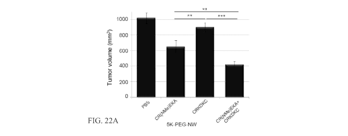
Figures 22A and 22B show tumor treatment with targeted nanoworms. Mice
bearing orthotopic xenografts of 22Rv1 or LAPC9 human prostate cancer (2 weeks
or 10
days after inoculation, respectively) were injected intravenously with
nanoworms coated
with peptides through a 5-kDa PEG spacer. The particles were administered
every other
day for 14 days (5 mg of iron per kilogram per day, total cumulative dose 35
mg/kg).
(A) Tumor volume 1 day after the last injection in the 22Rv1 model is shown.
Statistical
analyses were performed with analysis of variance. Error bars show SEM (n = 10-
12);
* *P < .01; * * *P < .001. Similar results were obtained in 2 independent
experiments.
(B) Mice bearing LAPC9 tumors were treated as described in panel A, and
survival was
monitored over time (n = 8 per group). The arrow indicates the day the
nanoworm
treatment was stopped.
DETAILED DESCRIPTION OF THE INVENTION
The disclosed method and compositions can be understood more readily by
reference to the following detailed description of particular embodiments and
the Example
included therein and to the Figures and their previous and following
description.
Before the present compounds, compositions, articles, devices, and/or methods
are
disclosed and described, it is to be understood that they are not limited to
specific
synthetic methods or specific recombinant biotechnology methods unless
otherwise
specified, or to particular reagents unless otherwise specified, as such may,
of course,
vary. It is also to be understood that the terminology used herein is for the
purpose of
describing particular embodiments only and is not intended to be limiting.
Definitions
As used in the specification and the appended claims, the singular forms "a,"
"an"
and "the" include plural referents unless the context clearly dictates
otherwise. Thus, for
example, reference to "a pharmaceutical carrier" includes mixtures of two or
more such
carriers, and the like.
17
CA 02784145 2012-06-12
WO 2011/075725 PCT/US2010/061302
Ranges can be expressed herein as from "about" one particular value, and/or to
"about" another particular value. When such a range is expressed, another
embodiment
includes from the one particular value and/or to the other particular value.
Similarly,
when values are expressed as approximations, by use of the antecedent "about,"
it will be
understood that the particular value forms another embodiment. It will be
further
understood that the endpoints of each of the ranges are significant both in
relation to the
other endpoint, and independently of the other endpoint. It is also understood
that there
are a number of values disclosed herein, and that each value is also herein
disclosed as
"about" that particular value in addition to the value itself. For example, if
the value "10"
is disclosed, then "about 10" is also disclosed. It is also understood that
when a value is
disclosed that "less than or equal to" the value, "greater than or equal to
the value" and
possible ranges between values are also disclosed, as appropriately understood
by the
skilled artisan. For example, if the value "10" is disclosed the "less than or
equal to 10"as
well as "greater than or equal to 10" is also disclosed. It is also understood
that the
throughout the application, data is provided in a number of different formats,
and that this
data, represents endpoints and starting points, and ranges for any combination
of the data
points. For example, if a particular data point "10" and a particular data
point 15 are
disclosed, it is understood that greater than, greater than or equal to, less
than, less than or
equal to, and equal to 10 and 15 are considered disclosed as well as between
10 and 15. It
is also understood that each unit between two particular units are also
disclosed. For
example, if 10 and 15 are disclosed, then 11, 12, 13, and 14 are also
disclosed.
In this specification and in the claims which follow, reference will be made
to a
number of terms which shall be defined to have the following meanings:
"Optional" or "optionally" means that the subsequently described event or
circumstance may or may not occur, and that the description includes instances
where said
event or circumstance occurs and instances where it does not.
Throughout this application, various publications are referenced. The
disclosures
of these publications in their entireties are hereby incorporated by reference
into this
application in order to more fully describe the state of the art to which this
pertains. The
references disclosed are also individually and specifically incorporated by
reference herein
for the material contained in them that is discussed in the sentence in which
the reference
is relied upon.
It is to be understood that the disclosed method and compositions are not
limited to
specific synthetic methods, specific analytical techniques, or to particular
reagents unless
18
CA 02784145 2012-06-12
WO 2011/075725 PCT/US2010/061302
otherwise specified, and, as such, may vary. It is also to be understood that
the
terminology used herein is for the purpose of describing particular
embodiments only and
is not intended to be limiting.
Materials
Disclosed are the components to be used to prepare the disclosed compositions
as
well as the compositions themselves to be used within the methods disclosed
herein.
These and other materials are disclosed herein, and it is understood that when
combinations, subsets, interactions, groups, etc. of these materials are
disclosed that while
specific reference of each various individual and collective combinations and
permutation
of these compounds may not be explicitly disclosed, each is specifically
contemplated and
described herein. For example, if a particular peptide is disclosed and
discussed and a
number of modifications that can be made to a number of molecules including
the peptide
are discussed, specifically contemplated is each and every combination and
permutation of
the peptides and the modifications that are possible unless specifically
indicated to the
contrary. Thus, if a class of molecules A, B, and C are disclosed as well as a
class of
molecules D, E, and F and an example of a combination molecule, A-D is
disclosed, then
even if each is not individually recited each is individually and collectively
contemplated
meaning combinations, A-E, A-F, B-D, B-E, B-F, C-D, C-E, and C-F are
considered
disclosed. Likewise, any subset or combination of these is also disclosed.
Thus, for
example, the sub-group of A-E, B-F, and C-E would be considered disclosed.
This
concept applies to all aspects of this application including, but not limited
to, steps in
methods of making and using the disclosed compositions. Thus, if there are a
variety of
additional steps that can be performed it is understood that each of these
additional steps
can be performed with any specific embodiment or combination of embodiments of
the
disclosed methods.
Disclosed are compositions comprising a surface molecule and at least one
modified clot-binding compound. The modified clot-binding compound can
selectively
bind to clotted plasma protein, wherein the composition causes clotting and
amplifies the
accumulation of the composition in tumors. The modified clot-binding compound
can
enhance the clotting in tumors compared to its unmodified derivative.
Also disclosed are methods comprising administering to a subject any of the
disclosed compositions. The composition selectively homes to clotted plasma
protein,
wherein the composition causes clotting and amplifies the accumulation of the
composition at the site of the clotted plasma protein.
19
CA 02784145 2012-06-12
WO 2011/075725 PCT/US2010/061302
Also disclosed are methods comprising administering to a subject a plurality
of
different of the disclosed compositions. In some forms, each of the plurality
of different
compositions comprises a surface molecule and at least one modified clot-
binding
compound. In some forms, at least one of the plurality of different
compositions
comprises a surface molecule and at least one modified clot-binding compound.
In some
forms, each of the plurality of different compositions selectively homes to
clotted plasma
protein. In some forms, at least one of the plurality of compositions
selectively homes to
clotted plasma protein. In some forms, each of the compositions causes
clotting and
amplifies the accumulation of the composition at the site of the clotted
plasma protein. In
some forms, at least one of the compositions causes clotting and amplifies the
accumulation of the composition at the site of the clotted plasma protein.
The modified clot-binding compound can comprise a methylated clot-binding
compound. The methylated clot-binding compound can comprise a methylated amino
acid segment. The methylated amino acid segment can be selected from amino
acid
segments comprising a methylated derivative of amino acid sequence CREKA (SEQ
ID
NO: 1) or a conservative variant thereof, amino acid segments comprising a
methylated
derivative of amino acid sequence CREKA (SEQ ID NO:1), amino acid segments
consisting of a methylated derivative of amino acid sequence CREKA (SEQ ID NO:
1),
and amino acid segments consisting of a methylated derivative amino acid
sequence REK.
The methylated amino acid segment can comprise a methylated derivative of
amino acid
sequence CREKA (SEQ ID NO: 1) or a conservative variant thereof. The
methylated
amino acid segment can comprise a methylated derivative of amino acid sequence
CREKA (SEQ ID NO: 1). The methylated amino acid segment can consist of a
methylated
derivative of amino acid sequence CREKA (SEQ ID NO:1). The methylated amino
acid
segment can consist of a methylated derivative of amino acid sequence REK.
The amino acid sequence can be N- or C-methylated in at least one position.
The
amino acid sequence can be C(NMe)REKA (SEQ ID NO:8), CR(NMe)EKA (SEQ ID
NO:9), CR(CMe)EKA (SEQ ID NO:10), CRE(NMe)KA (SEQ ID NO:11),
CRE(CMe)KA (SEQ ID NO:12), or CR(NMe)E(NMe)KA (SEQ ID NO:13). The amino
acid sequence can be CR(NMe)EKA (SEQ ID NO:9), CRE(CMe)KA (SEQ ID NO: 11), or
CR(NMe)E(NMe)KA (SEQ ID NO:13).
The composition can further comprise a plurality of clot-binding compounds,
wherein the clot-binding compounds selectively bind to clotted plasma protein,
wherein
the plurality of clot-binding compounds causes clotting and amplifies the
accumulation of
CA 02784145 2012-06-12
WO 2011/075725 PCT/US2010/061302
the composition in tumors. One or more of the plurality of clot-binding
compounds can be
modified clot-binding compounds, wherein the modified clot-binding compounds
enhance
the clotting in tumors compared to their unmodified derivatives. One or more
of the
modified clot-binding compounds of the plurality of clot-binding compounds can
comprise a methylated clot-binding compound. One or more of the methylated
clot-
binding compounds of the plurality of clot-binding compounds can comprise a
methylated
amino acid segment.
Each of the methylated amino acid segments of the plurality of clot-binding
compounds can be independently selected from amino acid segments comprising a
methylated derivative of amino acid sequence CREKA (SEQ ID NO: 1) or a
conservative
variant thereof, amino acid segments comprising a methylated derivative of
amino acid
sequence CREKA (SEQ ID NO: 1), amino acid segments consisting of a methylated
derivative of amino acid sequence CREKA (SEQ ID NO:1), and amino acid segments
consisting of a methylated derivative amino acid sequence REK. The methylated
amino
acid segments of the plurality of clot-binding compounds can each
independently
comprise a methylated derivative of amino acid sequence CREKA (SEQ ID NO: 1)
or a
conservative variant thereof. The methylated amino acid segments of the
plurality of clot-
binding compounds can each independently comprise a methylated derivative of
amino
acid sequence CREKA (SEQ ID NO: 1). The methylated amino acid segments of the
plurality of clot-binding compounds can each independently consist of a
methylated
derivative of amino acid sequence CREKA (SEQ ID NO:1). The methylated amino
acid
segments can each independently consist of a methylated derivative of amino
acid
sequence REK.
The methylated amino acid segments of the plurality of clot-binding compounds
can each comprise a methylated derivative of amino acid sequence CREKA (SEQ ID
NO:
1) or a conservative variant thereof. The methylated amino acid segments of
the plurality
of clot-binding compounds can each comprise a methylated derivative of amino
acid
sequence CREKA (SEQ ID NO: 1). The methylated amino acid segments of the
plurality
of clot-binding compounds can each consist of a methylated derivative of amino
acid
sequence CREKA (SEQ ID NO: 1). The methylated amino acid segments of the
plurality
of clot-binding compounds can each consist of a methylated derivative of amino
acid
sequence REK.
In some forms, the surface molecule can be thrombogenic. In some forms, the
modified clot-binding compound can be thrombogenic.
21
CA 02784145 2012-06-12
WO 2011/075725 PCT/US2010/061302
The composition can further comprising one or more tumor-homing compounds.
One or more of the tumor-homing compounds can comprise an amino acid segment.
One
or more of the amino acid segments of the tumor-homing compounds can comprise
the
amino acid sequence CRKDKC (SEQ ID NO:5) or a conservative derivative thereof
or the
amino acid sequence CGKRK (SEQ ID NO:7) or a conservative derivative thereof.
One
or more of the tumor-homing compounds can be thrombogenic.
The composition can bind inside tumor blood vessels. The composition can
reduce
tumor growth. The surface molecule can comprise an iron oxide nanoworm. The
surface
molecule can comprise an iron oxide nanoparticle. The surface molecule can
comprise an
albumin nanoparticle. The surface molecule can comprise a liposome. The
surface
molecule can comprise a microparticle. The surface molecule can comprise a
fluorocarbon microbubble.
The composition can comprise at least 100 clot-binding compounds. The
composition can comprise at least 1000 clot-binding compounds. The composition
can
comprise at least 10,000 clot-binding compounds.
The composition can further comprise one or more moieties. The moieties can be
independently selected from the group consisting of an anti-angiogenic agent,
a pro-
angiogenic agent, a cancer chemotherapeutic agent, a cytotoxic agent, an anti-
inflammatory agent, an anti-arthritic agent, a polypeptide, a nucleic acid
molecule, a small
molecule, an image contrast agent, a fluorophore, fluorescein, rhodamine, a
radionuclide,
indium-111, technetium-99, carbon- 11, and carbon-13. At least one of the
moieties can be
a therapeutic agent. The therapeutic agent can comprise a compound or
composition for
treating cancer. The therapeutic agent can comprise a compound or composition
to induce
programmed cell death or apoptosis. The therapeutic agent can be Abraxane. The
therapeutic agent can be paclitaxel. The therapeutic agent can be taxol. In
some forms, at
least one of the moieties can be thrombogenic. In some forms, at least one of
the moieties
is not a clot-binding compound. In some forms, none of the moieties are clot-
binding
compounds. In some forms, at least one of the moieties is a homing compound,
wherein
the homing compound is not a clot-binding compound. At least one of the
moieties can be
a detectable agent. The detectable agent can be FAM.
The composition can selectively homes to tumor vasculature, wound sites, or
both.
The composition can have a therapeutic effect. The therapeutic effect can be a
slowing in
the increase of or a reduction of tumor burden. The therapeutic effect can be
a slowing of
the increase of or reduction of tumor size. The therapeutic effect can be a
reduction or
22
CA 02784145 2012-06-12
WO 2011/075725 PCT/US2010/061302
blocking of blood circulation in a tumor. The therapeutic effect can be a
reduction or
cessation of bleeding at a wound site. The therapeutic effect can be a
decrease in the time
for bleeding to stop at a wound site. The therapeutic effect can comprise a
reduction in
inflammation, an increase in speed of wound healing, reduction in amounts of
scar tissue,
decrease in pain, decrease in swelling, decrease in necrosis, or a
combination.
The clotting can have a therapeutic effect. The subject can have one or more
sites
to be targeted, wherein the composition homes to one or more of the sites to
be targeted.
The subject can have a tumor, wherein the composition has a therapeutic effect
on the
tumor.
In some forms, the composition can comprise a sufficient number and
composition
of clot-binding compounds such that the composition causes clotting and
amplifies the
accumulation of the composition in tumors. Sufficiency of the number and
composition of
clot-binding compounds (modified or otherwise) can be determined by assessing
clotting
and amplification of the accumulation of the composition in tumors in a non-
human
animal.
The composition can comprise a sufficient density and composition of clot-
binding
compounds such that the composition causes clotting and amplifies the
accumulation of
the composition in tumors. Sufficiency of the density and composition of clot-
binding
compounds (modified or otherwise) can be determined by assessing clotting and
amplification of the accumulation of the composition in tumors in a non-human
animal.
A plurality of the clot-binding compounds can each be independently selected
from
an amino acid segment comprising the amino acid sequence REK, a fibrin-binding
peptide, a clot-binding antibody, and a clot-binding small organic molecule. A
plurality of
the clot-binding compounds can each independently comprise an amino acid
segment
comprising the amino acid sequence REK. Modified clot-binding compounds can be
independently selected from an amino acid segment comprising a modified form
of the
amino acid sequence REK, a modified form of a fibrin-binding peptide, a
modified form
of a clot-binding antibody, and a modified form of a clot-binding small
organic molecule.
The modified clot-binding compounds can each independently comprise an amino
acid
segment comprising a modified form of the amino acid sequence REK. A
particularly
useful modification is methylation.
The amino acid segments of clot-binding compounds can each be independently
selected from amino acid segments comprising the amino acid sequence CREKA
(SEQ ID
NO: 1) or a conservative variant thereof, amino acid segments comprising the
amino acid
23
CA 02784145 2012-06-12
WO 2011/075725 PCT/US2010/061302
sequence CREKA (SEQ ID NO: 1), amino acid segments consisting of the amino
acid
sequence CREKA (SEQ ID NO:1), and amino acid segments consisting of the amino
acid
sequence REK. The amino acid segments can each independently comprise the
amino acid
sequence CREKA (SEQ ID NO: 1) or a conservative variant thereof.
The amino acid segments can also each independently comprise the amino acid
sequence CREKA (SEQ ID NO:1). The amino acid segment can also consist of the
amino
acid sequence CREKA (SEQ ID NO: 1). The amino acid segment can consist of the
amino
acid sequence REK.
A plurality of the clot-binding compounds can each comprise a fibrin-binding
peptide. The fibrin-binding peptides can independently be selected from the
group
consisting of fibrin binding proteins and fibrin-binding derivatives thereof.
In another
example, a plurality of the clot-binding compounds can each comprise a clot-
binding
antibody. Furthermore, a plurality of the clot-binding compounds can each
comprise a
clot-binding small organic molecule.
In some forms, each of the at least one of the plurality of different
compositions
selectively homes to clotted plasma protein, wherein each of the at least one
of the
plurality of compositions causes clotting and amplifies the accumulation of
the
compositions at the site of the clotted plasma protein. In some forms, each of
the at least
one of the plurality of different compositions can selectively home to clotted
plasma
protein, wherein each of the at least one of the plurality of compositions
causes clotting
and amplifies the accumulation of the compositions at the site of the clotted
plasma
protein. In some forms, at least one of the plurality of different
compositions comprises a
surface molecule and at least one unmodified clot-binding compound, wherein
the
unmodified clot-binding compound selectively binds to clotted plasma protein.
In some
forms, at least one of the plurality of different compositions comprises a
surface molecule
and at least one homing compound, wherein the homing compound is not a clot-
binding
compound. In some forms, the homing compound can selectively bind to tumor
vasculature. In some forms, the homing compound can be a tumor-homing
compound. In
some forms, the tumor-homing compound can comprises an amino acid segment. In
some
forms, the amino acid segment of the tumor-homing compound can comprise the
amino
acid sequence CRKDKC (SEQ ID NO:5) or a conservative derivative thereof, or
the
amino acid sequence CGKRK (SEQ ID NO:7) or a conservative derivative thereof.
In
some forms, at least two of the plurality of different compositions can differ
in the homing
compounds of which the compositions are comprised. In some forms, at least two
of the
24
CA 02784145 2012-06-12
WO 2011/075725 PCT/US2010/061302
plurality of different compositions can differ in the clot-binding compounds
of which the
compositions are comprised. In some forms, each of the plurality of different
compositions selectively homes to clotted plasma protein, wherein each of the
at least one
of the plurality of compositions causes clotting and amplifies the
accumulation of the
compositions at the site of the clotted plasma protein.
Further disclosed are compositions that not only home to tumors, but also
amplify
their own homing. The system is based on a clot-binding compound that
recognizes
clotted plasma proteins and selectively homes to tumors, where it binds to
vessel walls and
tumor stroma. Surface molecules coupled with the clot-binding compounds can
accumulate in tumor vessels or at wound sites, where they induce additional
local clotting,
thereby producing new binding sites for more particles. The system mimics
platelets,
which also circulate freely but accumulate at a diseased site and amplify
their own
accumulation at that site. The clotting-based amplification greatly enhances
tumor
imaging, and a drug carrier function is also envisioned.
In developing new strategies for treating solid tumors, methods that involve
targeting the vasculature of the tumor, rather than the tumor cells
themselves, offer distinct
advantages. Inducing a blockade of the blood flow through the tumor, e.g.,
through tumor
vasculature specific fibrin formation, interferes with the influx and efflux
processes in a
tumor site, thus resulting in anti-tumor effect. Arresting the blood supply to
a tumor can be
accomplished through shifting the procoagulant-fibrinolytic balance in the
tumor-
associated vessels in favor of the coagulating (clotting) processes by
specific exposure to
clotting agents.
Compositions comprising modified clot-binding compounds are directed to the
tumor cells themselves. There, they accumulate and induce additional clotting.
A number
of appropriate clot-binding compounds have been identified that can be
modified and that
are specifically or preferentially expressed, localized, adsorbed to or
inducible on the cells
or in the environment of the tumor vasculature and/or stroma. These are
discussed in more
detail below.
The disclosed compositions can, for example, cause clotting (thrombogenesis),
can
increase or enhance clotting at sites where the composition homes or is
targeted, and/or
can accumulate and increase or enhance accumulation of the composition at
sites where
the composition homes or is targeted. Such compositions can be considered
thrombogenic
compositions. These effects of the disclosed compositions can be caused by
and/or
enhanced by inclusion in the composition of, for example, one or more
thrombogenic clot-
CA 02784145 2012-06-12
WO 2011/075725 PCT/US2010/061302
binding compounds, one or more thrombogenic surface molecules, one or more
thrombogenic compounds, one or more thrombogenic peptides, one or more
thrombogenic
clot-binding peptides, and/or one or more thrombogenic moieties. For example,
the
disclosed compositions can be comprised of one or more thrombogenic clot-
binding
compounds, one or more thrombogenic surface molecules, one or more
thrombogenic
compounds, one or more thrombogenic peptides, one or more thrombogenic clot-
binding
peptides, and/or one or more thrombogenic moieties. The disclosed compositions
also can
be comprised of one or more non-thrombogenic clot-binding compounds, one or
more
non-thrombogenic surface molecules, one or more non-thrombogenic compounds,
one or
more non-thrombogenic peptides, one or more non-thrombogenic clot-binding
peptides,
and/or one or more non-thrombogenic moieties.
A. Clot-Binding Compounds
The clot-binding compound can be any compound with the ability to interact
with
clots and/or components of clots such as clotted plasma proteins. It has been
discovered
that by using modified forms of clot-binding compounds the effectiveness of
the clot
amplification and of the effect on tumors can be increased. The composition
can also
comprise a sufficient number and composition of clot-binding compounds
(modified or
not) such that the composition causes clotting and amplifies the accumulation
of the
composition in tumors and at the site of injury. In one example, sufficiency
of the number
and composition of clot-binding compounds can be determined by assessing
clotting and
amplification of the accumulation of the composition in tumors in a non-human
animal. In
another example, sufficiency of the number and composition of clot-binding
compounds
can be determined by assessing clotting and amplification of the accumulation
of the
composition in at sites of clotting and at the site of injury. Clot-binding
compounds can be
modified or unmodified.
A plurality of the clot-binding compounds can each be independently selected
from, for example, an amino acid segment comprising the amino acid sequence
REK, an
amino acid segment comprising the amino acid sequence CAR (such as CARSKNKDC
(SEQ ID NO:6)), an amino acid segment comprising the amino acid sequence CRK
(such
as CRKDKC (SEQ ID NO:5)), a fibrin-binding peptide, a peptide that binds clots
and not
fibrin (such as CGLIIQKNEC (CLT1, SEQ ID NO: 2) and CNAGESSKNC (CLT2, SEQ
ID NO: 3)), a clot-binding antibody, and a clot-binding small organic
molecule. A
plurality of the clot-binding compounds can each independently comprise an
amino acid
segment comprising the amino acid sequence REK. Such peptides are also
described in
26
CA 02784145 2012-06-12
WO 2011/075725 PCT/US2010/061302
U.S. Patent Application Publication No. 2008/0305101, which is hereby
incorporated by
reference for its description of such peptides. Peptides comprising amino acid
sequences
CAR or CRK are also described in U.S. Patent Application Publication No.
2009/0036349, which is hereby incorporated by reference for its description of
such
peptides.
The composition can comprise any number of clot-binding compounds. By way of
example, the composition can comprise at least 1, 5, 10, 15, 20, 25, 50, 75,
100, 125, 150,
175, 200, 225, 250, 275, 300, 325, 350, 375, 400, 425, 450, 475, 500, 525,
550, 575, 600,
625, 650, 675, 700, 625, 750, 775, 800, 825, 850, 875, 900, 925, 950, 975,
1000, 1100,
1200, 1300, 1400, 1500, 1600, 1700, 1800, 1900, 2000, 2250, 2500, 2750, 3000,
3500,
4000, 4500, 5000, 5500, 6000, 6500, 7000, 7500, 8000, 8500, 9000, 9500,
10,000, 15,000,
20,000, 25,000, 30,000, 35,000, 40,000, 45,000, 50,000, 75,000, or 100,000, or
more clot-
binding compounds. The composition can also comprise any number in between
those
numbers listed above.
The term "homing molecule" as used herein, means any molecule that selectively
homes in vivo to specified target sites or tissues in preference to normal
tissue. Similarly,
the term "homing peptide" or "homing peptidomimetic" means a peptide that
selectively
homes in vivo to specified target sites or tissues in preference to normal
tissue. It is
understood that a homing molecule that selectively homes in vivo to, for
example, tumors
can home to all tumors or can exhibit preferential homing to one or a subset
of tumor
types.
By "selectively homes" it is meant that, in vivo, the homing molecule binds
preferentially to the target as compared to non-target. For example, the
homing molecule
can bind preferentially to clotted plasma of one or more tumors, wound tissue,
or blood
clots, as compared to non-tumoral tissue or non-wound tissue. Such a homing
molecule
can selectively home, for example, to tumors. Selective homing to, for
example, tumors
generally is characterized by at least a two-fold greater localization within
tumors (or other
target), as compared to several tissue types of non-tumor tissue. A homing
molecule can
be characterized by 5-fold, 10-fold, 20-fold or more preferential localization
to tumors (or
other target) as compared to several or many tissue types of non-tumoral
tissue, or as
compared to-most or all non-tumoral tissue. Thus, it is understood that, in
some cases, a
homing molecule homes, in part, to one or more normal organs in addition to
homing to
the target tissue. Selective homing can also be referred to as targeting.
27
CA 02784145 2012-06-12
WO 2011/075725 PCT/US2010/061302
Useful clot-binding compound can include, for example, clot-binding peptides,
clot-binding antibodies, clot-binding small organic molecules, thrombogenic
clot-binding
compounds, thrombogenic clot-binding peptides, thrombogenic clot-binding
antibodies,
thrombogenic clot-binding small organic molecules, non-thrombogenic clot-
binding
compounds, non-thrombogenic clot-binding peptides, non-thrombogenic clot-
binding
antibodies, and/or non-thrombogenic clot-binding small organic molecules.
The composition can comprise a sufficient number, density, and/or composition
of
clot-binding compounds such that the composition causes clotting and amplifies
the
accumulation of the composition in tumors. Sufficiency of the number, density,
and/or
composition of clot-binding compounds (modified or otherwise) can be
determined by
assessing clotting and amplification of the accumulation of the composition in
tumors in a
non-human animal.
The density of clot-binding compounds on a surface molecule can be described
in
any suitable manner. For example, the density can be expressed as the number
of clot-
binding compounds per, for example, a given area, surface area, volume, unit,
subunit,
arm, etc. of the surface molecule. The density can also be relative to, for
example, the
area, surface area, volume, unit, subunit, arm, etc. of the entire surface
molecule or to the
area, surface area, volume, unit, subunit, arm, etc. of a portion of the
surface molecule.
For example, a sufficient density of clot-binding compound can be present in a
portion of
the surface molecule. The presence of this dense portion can cause clotting
and amplify
the accumulation of the composition. Thus, a composition having a sufficient
density of
clot-binding compounds can have a threshold density (or above) for the entire
surface
molecule or for just one or more portions of the surface molecule.
The density can be measured or calculated in any suitable manner. For example,
the number or amount of clot-binding compounds present on a surface molecule
or group
of surface molecules can be measured by, for example, detecting the level or
intensity of
signal produced by labeled clot-binding compounds and calculating the density
based on
the structural characteristics of the surface molecule.
The density or threshold density of clot-binding compounds can be, for
example, at
least 0.001, 0.002, 0.003, 0.004, 0.005, 0.006, 0.007, 0.008, 0.009, 0.01,
0.02, 0.03, 0.04,
0.05, 0.06, 0.07, 0.08, 0.09, 0.1, 0.2, 0.3, 0.4, 0.5, 0.6, 0.7, 0.8, 0.9, 1,
2, 3, 4, 5, 6, 7, 8, 9,
10, 12, 14, 16, 18, 20, 25, 30, 35, 40, 45, 50, 55, 60, 65, 70, 75, 80, 85,
90, 95, 100, 110,
120, 130, 140, 150, 160, 170, 180, 190, 200, 220, 240, 260, 280, 300, 320,
340, 360, 380,
400, 420, 440, 460, 480, 500, 550, 600, 650, 700, 750, 800, 850, 900, 950, or
1000 clot-
28
CA 02784145 2012-06-12
WO 2011/075725 PCT/US2010/061302
binding compounds per square nM of the entire or a portion of the surface
molecule. The
composition can also comprise any density in between those densities listed
above.
The density or threshold density of clot-binding compounds can be, for
example, at
least 1, 2, 3, 4, 5, 6, 7, 8, 9, 10, 12, 14, 16, 18, 20, 25, 30, 35, 40, 45,
50, 55, 60, 65, 70, 75,
80, 85, 90, 95, 100, 110, 120, 130, 140, 150, 160, 170, 180, 190, 200, 220,
240, 260, 280,
300, 320, 340, 360, 380, 400, 420, 440, 460, 480, 500, 550, 600, 650, 700,
750, 800, 850,
900, 950, 1000, 1100, 1200, 1300, 1400, 1500, 1600, 1700, 1800, 1900, 2000,
2200, 2400,
2600, 2800, 3000, 3200, 3400, 3600, 3800, 4000, 4200, 4400, 4600, 4800, 5000,
5500,
6000, 6500, 7000, 7500, 8000, 8500, 900, 9500, 10,000 clot-binding compounds
per
square gM of the entire or a portion of the surface molecule. The composition
can also
comprise any density in between those densities listed above.
The density or threshold density of clot-binding compounds can be, for
example, at
least 0.001, 0.002, 0.003, 0.004, 0.005, 0.006, 0.007, 0.008, 0.009, 0.01,
0.02, 0.03, 0.04,
0.05, 0.06, 0.07, 0.08, 0.09, 0.1, 0.2, 0.3, 0.4, 0.5, 0.6, 0.7, 0.8, 0.9, 1,
2, 3, 4, 5, 6, 7, 8, 9,
10, 12, 14, 16, 18, 20, 25, 30, 35, 40, 45, 50, 55, 60, 65, 70, 75, 80, 85,
90, 95, 100, 110,
120, 130, 140, 150, 160, 170, 180, 190, 200, 220, 240, 260, 280, 300, 320,
340, 360, 380,
400, 420, 440, 460, 480, 500, 550, 600, 650, 700, 750, 800, 850, 900, 950, or
1000 clot-
binding compounds per cubic nM of the entire or a portion of the surface
molecule. The
composition can also comprise any density in between those densities listed
above.
The density or threshold density of clot-binding compounds can be, for
example, at
least 1, 2, 3, 4, 5, 6, 7, 8, 9, 10, 12, 14, 16, 18, 20, 25, 30, 35, 40, 45,
50, 55, 60, 65, 70, 75,
80, 85, 90, 95, 100, 110, 120, 130, 140, 150, 160, 170, 180, 190, 200, 220,
240, 260, 280,
300, 320, 340, 360, 380, 400, 420, 440, 460, 480, 500, 550, 600, 650, 700,
750, 800, 850,
900, 950, 1000, 1100, 1200, 1300, 1400, 1500, 1600, 1700, 1800, 1900, 2000,
2200, 2400,
2600, 2800, 3000, 3200, 3400, 3600, 3800, 4000, 4200, 4400, 4600, 4800, 5000,
5500,
6000, 6500, 7000, 7500, 8000, 8500, 900, 9500, 10,000 clot-binding compounds
per cubic
gM of the entire or a portion of the surface molecule. The composition can
also comprise
any density in between those densities listed above.
1. Modified Clot-Binding Compounds
The disclosed clot-binding compounds can include modified forms of clot-
binding
compounds. The clot-binding compounds can have any useful modification. For
example, some modifications can stabilize the clot-binging compound. For
example, the
disclosed clot-binding compounds include methylated clot-binding compounds.
Methylated clot-binding compounds are particularly useful when the clot-
binding
29
CA 02784145 2012-06-12
WO 2011/075725 PCT/US2010/061302
compound includes a protein, peptide or amino acid segment. For example, a
clot-binding
compound can be a modified clot-binding compound, where, for example, the
modified
clot-binding compound includes a modified amino acid segment or amino acid
sequence.
For example, a modified clot-binding compound can be a methylated clot-binding
compound, where, for example, the methylated clot-binding compound includes a
methylated amino acid segment or amino acid sequence. Other modifications can
be used,
either alone or in combination. Where the clot-binding compound is, or
includes, a
protein, peptide, amino acid segment and/or amino acid sequences, the
modification can
be to the protein, peptide, amino acid segment, amino acid sequences and/or
any amino
acids in the protein, peptide, amino acid segment and/or amino acid sequences.
Amino
acid and peptide modifications are known to those of skill in the art, some of
which are
described below and elsewhere herein. Methylation is a particularly useful
modification
for the disclosed clot-binding compounds.
It has been discovered that by using modified forms of clot-binding compounds
the
effectiveness of the clot amplification and of the effect on tumors can be
increased. The
composition can also comprise a sufficient number and composition of clot-
binding
compounds (modified or not) such that the composition causes clotting and
amplifies the
accumulation of the composition in tumors and at the site of injury. In one
example,
sufficiency of the number and composition of modified and/or unmodified clot-
binding
compounds can be determined by assessing clotting and amplification of the
accumulation
of the composition in tumors in a non-human animal. Such methods are discussed
in more
detail below.
A plurality of modified and/or unmodified clot-binding compounds can each be
independently selected from, for example, an amino acid segment comprising a
modified
or unmodified form of the amino acid sequence REK, an amino acid segment
comprising a
modified or unmodified form of the amino acid sequence CAR (such as CARSKNKDC
(SEQ ID NO:6)), an amino acid segment comprising a modified or unmodified form
of the
amino acid sequence CRK (such as CRKDKC (SEQ ID NO:5)), a modified or
unmodified
form of a fibrin-binding peptide, a modified or unmodified form of a peptide
that binds
clots and not fibrin (such as CGLIIQKNEC (CLT1, SEQ ID NO: 2) and CNAGESSKNC
(CLT2, SEQ ID NO: 3)), a modified or unmodified form of a clot-binding
antibody, and a
modified or unmodified form of a clot-binding small organic molecule. A
plurality of the
clot-binding compounds can each independently comprise an amino acid segment
comprising a modified or unmodified form of the amino acid sequence REK. Such
CA 02784145 2012-06-12
WO 2011/075725 PCT/US2010/061302
peptides are also described in U.S. Patent Application Publication No.
2008/0305101,
which is hereby incorporated by reference for its description of such
peptides. Peptides
comprising amino acid sequences CAR or CRK are also described in U.S. Patent
Application Publication No. 2009/0036349, which is hereby incorporated by
reference for
its description of such peptides.
The composition can comprise any number of modified and/or unmodified clot-
binding compounds. By way of example, the composition can comprise at least 1,
5, 10,
15, 20, 25, 50, 75, 100, 125, 150, 175, 200, 225, 250, 275, 300, 325, 350,
375, 400, 425,
450, 475, 500, 525, 550, 575, 600, 625, 650, 675, 700, 625, 750, 775, 800,
825, 850, 875,
900, 925, 950, 975, 1000, 1100, 1200, 1300, 1400, 1500, 1600, 1700, 1800,
1900, 2000,
2250, 2500, 2750, 3000, 3500, 4000, 4500, 5000, 5500, 6000, 6500, 7000, 7500,
8000,
8500, 9000, 9500, 10,000, 15,000, 20,000, 25,000, 30,000, 35,000, 40,000,
45,000,
50,000, 75,000, or 100,000, or more modified and/or unmodified clot-binding
compounds.
The composition can also comprise any number in between those numbers listed
above.
As used herein, a "methylated derivative" of a protein, peptide, amino acid
segment, amino acid sequence, etc. refers to a form of the protein, peptide,
amino acid
segment, amino acid sequence, etc. that is methylated. Unless the context
indicates
otherwise, reference to a methylated derivative of a protein, peptide, amino
acid segment,
amino acid sequence, etc. does no include any modification to the base
protein, peptide,
amino acid segment, amino acid sequence, etc. other than methylation.
Methylated
derivatives can also have other modifications, but such modifications
generally will be
noted. For example, conservative variants of an amino acid sequence would
include
conservative amino acid substitutions of the based amino acid sequence. Thus,
reference
to, for example, a "methylated derivative" of a specific amino acid sequence
"and
conservative variants thereof"would include methylated forms of the specific
amino acid
sequence and methylated forms of the conservative variants of the specific
amino acid
sequence, but not any other modifications of derivations. As another example,
reference
to a methylated derivative of an amino acid segment that includes amino acid
substitutions
would include methylated forms of the amino acid sequence of the amino acid
segment
and methylated forms of the amino acid sequence of the amino acid segment
include
amino acid substitutions.
Peptides can have a variety of modifications. Modifications can be used to
change
or improve the properties of the peptides. For example, the disclosed peptides
can be N-
31
CA 02784145 2012-06-12
WO 2011/075725 PCT/US2010/061302
methylated, O-methylated, S-methylated, C-methylated, or a combination at one
or more
amino acids.
The amino and/or carboxy termini of the disclosed peptides can be modified.
Amino terminus modifications include methylation (e.g., --NHCH3 or --N(CH3)2),
acetylation (e.g., with acetic acid or a halogenated derivative thereof such
as a -
chloroacetic acid, a-bromoacetic acid, or. alpha. -iodoacetic acid), adding a
benzyloxycarbonyl (Cbz) group, or blocking the amino terminus with any
blocking group
containing a carboxylate functionality defined by RCOO-- or sulfonyl
functionality
defined by R--S02--, where R is selected from the group consisting of alkyl,
aryl,
heteroaryl, alkyl aryl, and the like, and similar groups. One can also
incorporate a
desamino acid at the N-terminus (so that there is no N-terminal amino group)
to decrease
susceptibility to proteases or to restrict the conformation of the peptide
compound. In
preferred embodiments, the N-terminus is acetylated with acetic acid or acetic
anhydride.
Carboxy terminus modifications include replacing the free acid with a
carboxamide group or forming a cyclic lactam at the carboxy terminus to
introduce
structural constraints. One can also cyclize the disclosed peptides, or
incorporate a
desamino or descarboxy residue at the termini of the peptide, so that there is
no terminal
amino or carboxyl group, to decrease susceptibility to proteases or to
restrict the
conformation of the peptide. C-terminal functional groups of the disclosed
peptides
include amide, amide lower alkyl, amide di(lower alkyl), lower alkoxy,
hydroxy, and
carboxy, and the lower ester derivatives thereof, and the pharmaceutically
acceptable salts
thereof.
One can replace the naturally occurring side chains of the genetically encoded
amino acids (or the stereoisomeric D amino acids) with other side chains, for
instance with
groups such as alkyl, lower (C1_6) alkyl, cyclic 4-, 5-, 6-, to 7-membered
alkyl, amide,
amide lower alkyl amide di(lower alkyl), lower alkoxy, hydroxy, carboxy and
the lower
ester derivatives thereof, and with 4-, 5-, 6-, to 7-membered heterocyclic. In
particular,
proline analogues in which the ring size of the proline residue is changed
from 5 members
to 4, 6, or 7 members can be employed. Cyclic groups can be saturated or
unsaturated, and
if unsaturated, can be aromatic or non-aromatic. Heterocyclic groups
preferably contain
one or more nitrogen, oxygen, and/or sulfur heteroatoms. Examples of such
groups include
the furazanyl, furyl, imidazolidinyl, imidazolyl, imidazolinyl, isothiazolyl,
isoxazolyl,
morpholinyl (e.g. morpholino), oxazolyl, piperazinyl (e.g., 1-piperazinyl),
piperidyl (e.g.,
1-piperidyl, piperidino), pyranyl, pyrazinyl, pyrazolidinyl, pyrazolinyl,
pyrazolyl,
32
CA 02784145 2012-06-12
WO 2011/075725 PCT/US2010/061302
pyridazinyl, pyridyl, pyrimidinyl, pyrrolidinyl (e.g., 1-pyrrolidinyl),
pyrrolinyl, pyrrolyl,
thiadiazolyl, thiazolyl, thienyl, thiomorpholinyl (e.g., thiomorpholino), and
triazolyl.
These heterocyclic groups can be substituted or unsubstituted. Where a group
is
substituted, the substituent can be alkyl, alkoxy, halogen, oxygen, or
substituted or
unsubstituted phenyl.
One can also readily modify peptides by phosphorylation, and other methods
[e.g.,
as described in Hruby, et al. (1990) Biochem J. 268:249-262].
The disclosed peptides also serve as structural models for non-peptidic
compounds
with similar biological activity. Those of skill in the art recognize that a
variety of
techniques are available for constructing compounds with the same or similar
desired
biological activity as the lead peptide compound, but with more favorable
activity than the
lead with respect to solubility, stability, and susceptibility to hydrolysis
and proteolysis
[See, Morgan and Gainor (1989) Ann. Rep. Med. Chem. 24:243-252]. These
techniques
include, but are not limited to, replacing the peptide backbone with a
backbone composed
of phosphonates, amidates, carbamates, sulfonamides, secondary amines, and N-
methylamino acids.
2. Peptides and Amino Acid Segments
In some forms, the clot-binding compound can be or include a peptide,
peptidomimetic, and/or amino acid segment. Unless the context indicates
otherwise,
reference herein to "peptide" is intended to refer also to amino acid
segments, which can
form a part of, or constitute an entire, peptide. The disclosed peptides can
be in isolated
form. As used herein in reference to the disclosed peptides, the term
"isolated" means a
peptide that is in a form that is relatively free from material such as
contaminating
polypeptides, lipids, nucleic acids and other cellular material that normally
is associated
with the peptide in a cell or that is associated with the peptide in a library
or in a crude
preparation.
The disclosed peptides and amino acid segments can have any suitable length.
The
disclosed peptides can have, for example, a relatively short length of less
than six, seven,
eight, nine, ten, 12, 15, 20, 25, 30, 35 or 40 residues. The disclosed
peptides also can be
useful in the context of a significantly longer sequence. Thus, the peptides
can have, for
example, a length of up to 50, 100, 150, 200, 250, 300, 400, 500, 1000 or 2000
residues. In
particular embodiments, a peptide can have a length of at least 10, 20, 30,
40, 50, 60, 70,
80, 90, 100 or 200 residues. In further embodiments, a peptide can have a
length of 5 to
200 residues, 5 to 100 residues, 5 to 90 residues, 5 to 80 residues, 5 to 70
residues, 5 to 60
33
CA 02784145 2012-06-12
WO 2011/075725 PCT/US2010/061302
residues, 5 to 50 residues, 5 to 40 residues, 5 to 30 residues, 5 to 20
residues, 5 to 15
residues, 5 to 10 residues, 10 to 200 residues, 10 to 100 residues, 10 to 90
residues, 10 to
80 residues, 10 to 70 residues, 10 to 60 residues, 10 to 50 residues, 10 to 40
residues, 10 to
30 residues, 10 to 20 residues, 20 to 200 residues, 20 to 100 residues, 20 to
90 residues, 20
to 80 residues, 20 to 70 residues, 20 to 60 residues, 20 to 50 residues, 20 to
40 residues or
20 to 30 residues. As used herein, the term "residue" refers to an amino acid
or amino acid
analog.
The disclosed amino acid segments can have, for example, a relatively short
length
of less than six, seven, eight, nine, ten, 12, 15, 20, 25, 30, 35 or 40
residues. The disclosed
amino acid segments also can be useful in the context of a significantly
longer sequence.
Thus, the amino acid segments can have, for example, a length of up to 50,
100, 150, 200,
250, 300, 400, 500, 1000 or 2000 residues. In particular embodiments, an amino
acid
segment can have a length of at least 10, 20, 30, 40, 50, 60, 70, 80, 90, 100
or 200
residues. In further embodiments, an amino acid segment can have a length of 5
to 200
residues, 5 to 100 residues, 5 to 90 residues, 5 to 80 residues, 5 to 70
residues, 5 to 60
residues, 5 to 50 residues, 5 to 40 residues, 5 to 30 residues, 5 to 20
residues, 5 to 15
residues, 5 to 10 residues, 10 to 200 residues, 10 to 100 residues, 10 to 90
residues, 10 to
80 residues, 10 to 70 residues, 10 to 60 residues, 10 to 50 residues, 10 to 40
residues, 10 to
30 residues, 10 to 20 residues, 20 to 200 residues, 20 to 100 residues, 20 to
90 residues, 20
to 80 residues, 20 to 70 residues, 20 to 60 residues, 20 to 50 residues, 20 to
40 residues or
20 to 30 residues. As used herein, the term "residue" refers to an amino acid
or amino acid
analog.
As this specification discusses various proteins, protein sequences, peptides,
peptides sequences, and amino acid sequences, it is understood that the
nucleic acids that
can encode those sequences are also disclosed. This would include all
degenerate
sequences related to a specific protein sequence, i.e. all nucleic acids
having a sequence
that encodes one particular protein sequence as well as all nucleic acids,
including
degenerate nucleic acids, encoding the disclosed variants and derivatives of
the protein
sequences. Thus, while each particular nucleic acid sequence may not be
written out
herein, it is understood that each and every sequence is in fact disclosed and
described
herein through the disclosed protein sequence.
Molecules can be produced that resemble peptides, but which are not connected
via
a natural peptide linkage. For example, linkages for amino acids or amino acid
analogs
can include CH2NH--, --CH2S--, --CH2--CH2 --, --CH=CH-- (cis and trans), --
COCH2
--, --
34
CA 02784145 2012-06-12
WO 2011/075725 PCT/US2010/061302
CH(OH)CH2--, and --CHH2SO-(These and others can be found in Spatola, A. F. in
Chemistry and Biochemistry of Amino Acids, Peptides, and Proteins, B.
Weinstein, eds.,
Marcel Dekker, New York, p. 267 (1983); Spatola, A. F., Vega Data (March
1983), Vol.
1, Issue 3, Peptide Backbone Modifications (general review); Morley, Trends
Pharm Sci
(1980) pp. 463-468; Hudson, D. et al., Int J Pept Prot Res 14:177-185 (1979) (-
-CH2NH--,
CH2CH2); Spatola et al. Life Sci 38:1243-1249 (1986) (--CH H2--S); Hann J.
Chem. Soc
Perkin Trans. I 307-314 (1982) (--CH--CH--, cis and trans); Almquist et al. J.
Med. Chem.
23:1392-1398 (1980) (--COCH2--); Jennings-White et al. Tetrahedron Lett
23:2533 (1982)
(--COCH2--); Szelke et al. European Appln, EP 45665 CA (1982): 97:39405 (1982)
(--
CH(OH)CH2--); Holladay et al. Tetrahedron. Lett 24:4401-4404 (1983) (--
C(OH)CH2--);
and Hruby Life Sci 31:189-199 (1982) (--CH2--S--); each of which is
incorporated herein
by reference. A particularly preferred non-peptide linkage is --CH2NH--. It is
understood
that peptide analogs can have more than one atom between the bond atoms, such
as alanine, y-aminobutyric acid, and the like.
Also disclosed are bifunctional peptides, which contain the clot-binding
peptide
fused to a second peptide having a separate function. Such bifunctional
peptides have at
least two functions conferred by different portions of the full-length
molecule and can, for
example, display anti-angiogenic activity or pro-apoptotic activity in
addition to the ability
to enhance clotting.
Also disclosed are isolated multivalent peptides that include at least two
subsequences each independently containing a peptide or amino acid segment
(for
example, the amino acid sequence SEQ ID NO: 1, or a conservative variant or
peptidomimetic thereof). The multivalent peptide can have, for example, at
least three, at
least five or at least ten of such subsequences each independently containing
a peptide. In
particular embodiments, the multivalent peptide can have two, three, four,
five, six, seven,
eight, nine, ten, fifteen or twenty identical or non-identical subsequences.
This is in
addition to the multiple clot-binding compounds that can comprise the
disclosed
compositions. In a further embodiment, the multivalent peptide can contain
identical
subsequences, such as repeats of SEQ ID NO: 1. In a further embodiment, the
multivalent
peptide contains contiguous identical or non-identical subsequences, which are
not
separated by any intervening amino acids.
As used herein, the term "peptide" is used broadly to mean peptides, proteins,
fragments of proteins and the like. The term "peptidomimetic," as used herein,
means a
peptide-like molecule that has the activity of the peptide upon which it is
structurally
CA 02784145 2012-06-12
WO 2011/075725 PCT/US2010/061302
based. Such peptidomimetics include chemically modified peptides, peptide-like
molecules containing non-naturally occurring amino acids, and peptoids and
have an
activity such as selective interaction with a target of the peptide upon which
the
peptidomimetic is derived (see, for example, Goodman and Ro, Peptidomimetics
for Drug
Design, in "Burger's Medicinal Chemistry and Drug Discovery" Vol. 1 (ed. M. E.
Wolff;
John Wiley & Sons 1995), pages 803-861).
A variety of peptidomimetics are known in the art including, for example,
peptide-
like molecules which contain a constrained amino acid, a non-peptide component
that
mimics peptide secondary structure, or an amide bond isostere. A
peptidomimetic that
contains a constrained, non-naturally occurring amino acid can include, for
example, an a-
methylated amino acid; a,a.-dialkylglycine or a-aminocycloalkane carboxylic
acid; an N'-
-C' cyclized amino acid; an N''.-methylated amino acid; a 0- or y-amino
cycloalkane
carboxylic acid; an a,(3-unsaturated amino acid; a (3,(3-dimethyl or (3-methyl
amino acid; a
0-substituted-2,3-methano amino acid; an N--C or Ca--C cyclized amino acid;
a
substituted proline or another amino acid mimetic. A peptidomimetic which
mimics
peptide secondary structure can contain, for example, a non-peptidic (3-turn
mimic; y-turn
mimic; mimic of (3-sheet structure; or mimic of helical structure, each of
which is well
known in the art. A peptidomimetic also can be a peptide-like molecule which
contains,
for example, an amide bond isostere such as a retro-inverso modification;
reduced amide
bond; methylenethioether or methylene-sulfoxide bond; methylene ether bond;
ethylene
bond; thioamide bond; trans-olefin or fluoroolefin bond; 1,5-disubstituted
tetrazole ring;
ketomethylene or fluoroketomethylene bond or another amide isostere. One
skilled in the
art understands that these and other peptidomimetics are encompassed within
the meaning
of the term "peptidomimetic" as used herein.
Methods for identifying a peptidomimetic are well known in the art and
include,
for example, the screening of databases that contain libraries of potential
peptidomimetics.
As an example, the Cambridge Structural Database contains a collection of
greater than
300,000 compounds that have known crystal structures (Allen et al., Acta
Crystalloqr.
Section B, 35:2331 (1979)). This structural depository is continually updated
as new
crystal structures are determined and can be screened for compounds having
suitable
shapes, for example, the same shape as a disclosed peptide, as well as
potential
geometrical and chemical complementarity to a target molecule. Where no
crystal
structure of a peptide or a target molecule that binds the peptide is
available, a structure
can be generated using, for example, the program CONCORD (Rusinko et al., J.
Chem.
36
CA 02784145 2012-06-12
WO 2011/075725 PCT/US2010/061302
In Comput. Sci. 29:251 (1989)). Another database, the Available Chemicals
Directory
(Molecular Design Limited, Information Systems; San Leandro Calif.), contains
about
100,000 compounds that are commercially available and also can be searched to
identify
potential peptidomimetics of a peptide, for example, with activity in
selectively interacting
with cancerous cells.
3. Homing Peptides
There are several examples in the art of peptides that home to clotted plasma
protein. Examples include REK, peptides comprising REK, CREKA (SEQ ID NO: 1),
and
peptides comprising CREKA (SEQ ID NO: 1). The amino acid segments can also be
independently selected from amino acid segments comprising the amino acid
sequence
CREKA (SEQ ID NO: 1) or a conservative variant thereof, amino acid segments
comprising the amino acid sequence CREKA (SEQ ID NO: 1), amino acid segments
consisting of the amino acid sequence CREKA (SEQ ID NO:1), and amino acid
segments
consisting of the amino acid sequence REK. The amino acid segments can each
independently comprise the amino acid sequence CREKA (SEQ ID NO: 1) or a
conservative variant thereof.
The amino acid segments can also each independently comprise the amino acid
sequence CREKA (SEQ ID NO:1). The amino acid segment can also consist of the
amino
acid sequence CREKA (SEQ ID NO: 1). The amino acid segment can consist of the
amino
acid sequence REK.
4. Other Clot-Binding Peptides
The clot-binding compound can also comprise a fibrin-binding peptide (FBP).
Examples of fibrin-binding peptides are known in the art (Van Rooijen N,
Sanders A
(1994) J Immunol Methods 174: 83-93; Moghimi SM, Hunter AC, Murray JC (2001)
Pharmacol Rev 53: 283-318; US Patent 5,792,742, all herein incorporated by
reference in
their entirety for their teaching concerning fibrin binding peptides).
Clot-binding peptides can also bind to proteins other than fibrin. Example
include
peptides that bind to fibronectin that has become incorporated into a clot
(Pilch et al.,
(2006) PNAS, 103: 2800-2804, hereby incorporated in its entirety for its
teaching
concerning clot-binding peptides). An example of clot-binding peptides
include, but is not
limited to, CGLIIQKNEC (CLT1, SEQ ID NO: 2) and CNAGESSKNC (CLT2, SEQ ID
NO: 3).The amino acid segments can also be independently selected from amino
acid
segments comprising the amino acid sequence CLT1 or CLT2 (SEQ ID NOS: 2 or 3)
or a
conservative variant thereof, amino acid segments comprising the amino acid
sequence
37
CA 02784145 2012-06-12
WO 2011/075725 PCT/US2010/061302
CLT1 or CLT2 (SEQ ID NOS: 2 or 3), or amino acid segments consisting of the
amino
acid sequence CLT1 or CLT2 (SEQ ID NOS: 2 or 3). The amino acid segments can
each
independently comprise the amino acid sequence CLT1 or CLT2 (SEQ ID NOS: 2 or
3) or
a conservative variant thereof.
The amino acid segments can also each independently comprise the amino acid
sequence CLT1 or CLT2 (SEQ ID NOS: 2 or 3). The amino acid segment can also
consist
of the amino acid sequence CLT1 or CLT2 (SEQ ID NOS: 2 or 3).
The amino acid segments can also each independently comprise the amino acid
sequence CARSKNKDC (SEQ ID NO:6)), and the amino acid sequence CRK (such as
CRKDKC (SEQ ID NO:5). Peptides comprising amino acid sequences CAR or CRK are
also described in U.S. Patent Application Publication No. 2009/0036349, which
is hereby
incorporated by reference for its description of such peptides.
5. Clot-binding Antibodies
The clot-binding compound can comprise a clot-binding antibody. Examples of
clot-binding antibodies are known in the art (Holvoet et al. Circulation, Vol
87, 1007-
1016, 1993; Bode et al. J. Biol. Chem., Vol. 264, Issue 2, 944-948, Jan, 1989;
Huang et al.
Science 1997: Vol. 275. no. 5299, pp. 547 - 550, all of which are herein
incorporated by
reference in their entirety for their teaching concerning clot-binding
antibodies).
The term "antibodies" is used herein in a broad sense and includes both
polyclonal
and monoclonal antibodies. In addition to intact immunoglobulin molecules,
also included
in the term "antibodies" are fragments or polymers of those immunoglobulin
molecules,
and human or humanized versions of immunoglobulin molecules or fragments
thereof, as
long as they are chosen for their ability to bind to, or otherwise interact
with, clots. The
antibodies can be tested for their desired activity using the in vitro assays
described herein,
or by analogous methods, after which their in vivo therapeutic and/or
prophylactic
activities are tested according to known clinical testing methods.
The term "monoclonal antibody" as used herein refers to an antibody obtained
from a substantially homogeneous population of antibodies, i.e., the
individual antibodies
within the population are identical except for possible naturally occurring
mutations that
may be present in a small subset of the antibody molecules. The monoclonal
antibodies
herein specifically include "chimeric" antibodies in which a portion of the
heavy and/or
light chain is identical with or homologous to corresponding sequences in
antibodies
derived from a particular species or belonging to a particular antibody class
or subclass,
while the remainder of the chain(s) is identical with or homologous to
corresponding
38
CA 02784145 2012-06-12
WO 2011/075725 PCT/US2010/061302
sequences in antibodies derived from another species or belonging to another
antibody
class or subclass, as well as fragments of such antibodies, as long as they
exhibit the
desired antagonistic activity (See, U.S. Pat. No. 4,816,567 and Morrison et
al., Proc. Natl.
Acad. Sci. USA, 81:6851-6855 (1984)).
The disclosed monoclonal antibodies can be made using any procedure which
produces monoclonal antibodies. For example, disclosed monoclonal antibodies
can be
prepared using hybridoma methods, such as those described by Kohler and
Milstein,
Nature, 256:495 (1975). In a hybridoma method, a mouse or other appropriate
host
animal is typically immunized with an immunizing agent to elicit lymphocytes
that
produce or are capable of producing antibodies that will specifically bind to
the
immunizing agent. Alternatively, the lymphocytes may be immunized in vitro,
e.g., using
the HIV Env-CD4-co-receptor complexes described herein.
The monoclonal antibodies may also be made by recombinant DNA methods, such
as those described in U.S. Pat. No. 4,816,567 (Cabilly et al.). DNA encoding
the disclosed
monoclonal antibodies can be readily isolated and sequenced using conventional
procedures (e.g., by using oligonucleotide probes that are capable of binding
specifically
to genes encoding the heavy and light chains of murine antibodies). Libraries
of
antibodies or active antibody fragments can also be generated and screened
using phage
display techniques, e.g., as described in U.S. Patent No. 5,804,440 to Burton
et al. and
U.S. Patent No. 6,096,441 to Barbas et al.
In vitro methods are also suitable for preparing monovalent antibodies.
Digestion
of antibodies to produce fragments thereof, particularly, Fab fragments, can
be
accomplished using routine techniques known in the art. For instance,
digestion can be
performed using papain. Examples of papain digestion are described in WO
94/29348
published Dec. 22, 1994 and U.S. Pat. No. 4,342,566. Papain digestion of
antibodies
typically produces two identical antigen binding fragments, called Fab
fragments, each
with a single antigen binding site, and a residual Fc fragment. Pepsin
treatment yields a
fragment that has two antigen combining sites and is still capable of cross-
linking antigen.
The fragments, whether attached to other sequences or not, can also include
insertions, deletions, substitutions, or other selected modifications of
particular regions or
specific amino acids residues, provided the activity of the antibody or
antibody fragment is
not significantly altered or impaired compared to the non-modified antibody or
antibody
fragment. These modifications can provide for some additional property, such
as to
remove/add amino acids capable of disulfide bonding, to increase its bio-
longevity, to alter
39
CA 02784145 2012-06-12
WO 2011/075725 PCT/US2010/061302
its secretory characteristics, etc. In any case, the antibody or antibody
fragment must
possess a bioactive property, such as specific binding to its cognate antigen.
Functional or
active regions of the antibody or antibody fragment may be identified by
mutagenesis of a
specific region of the protein, followed by expression and testing of the
expressed
polypeptide. Such methods are readily apparent to a skilled practitioner in
the art and can
include site-specific mutagenesis of the nucleic acid encoding the antibody or
antibody
fragment. (Zoller, M.J. Curr. Opin. Biotechnol. 3:348-354, 1992).
As used herein, the term "antibody" or "antibodies" can also refer to a human
antibody and/or a humanized antibody. Many non-human antibodies (e.g., those
derived
from mice, rats, or rabbits) are naturally antigenic in humans, and thus can
give rise to
undesirable immune responses when administered to humans. Therefore, the use
of
human or humanized antibodies in the methods serves to lessen the chance that
an
antibody administered to a human will evoke an undesirable immune response.
Human antibodies can be prepared using any technique. Examples of techniques
for human monoclonal antibody production include those described by Cole et
al.
(Monoclonal Antibodies and Cancer Therapy, Alan R. Liss, p. 77, 1985) and by
Boemer
et al. (J. Immunol., 147(1):86-95, 1991). Human antibodies (and fragments
thereof) can
also be produced using phage display libraries (Hoogenboom et al., J. Mol.
Biol., 227:381,
1991; Marks et al., J. Mol. Biol., 222:581, 1991).
Human antibodies can also be obtained from transgenic animals. For example,
transgenic, mutant mice that are capable of producing a full repertoire of
human
antibodies, in response to immunization, have been described (see, e.g.,
Jakobovits et al.,
Proc. Natl. Acad. Sci. USA, 90:2551-255 (1993); Jakobovits et al., Nature,
362:255-258
(1993); Bruggermann et al., Year in Immunol., 7:33 (1993)). Specifically, the
homozygous deletion of the antibody heavy chain joining region (J(H)) gene in
these
chimeric and germ-line mutant mice results in complete inhibition of
endogenous antibody
production, and the successful transfer of the human germ-line antibody gene
array into
such germ-line mutant mice results in the production of human antibodies upon
antigen
challenge. Antibodies having the desired activity are selected using Env-CD4-
co-receptor
complexes as described herein.
Antibody humanization techniques generally involve the use of recombinant DNA
technology to manipulate the DNA sequence encoding one or more polypeptide
chains of
an antibody molecule. Accordingly, a humanized form of a non-human antibody
(or a
fragment thereof) is a chimeric antibody or antibody chain (or a fragment
thereof, such as
CA 02784145 2012-06-12
WO 2011/075725 PCT/US2010/061302
an Fv, Fab, Fab', or other antigen-binding portion of an antibody) which
contains a portion
of an antigen binding site from a non-human (donor) antibody integrated into
the
framework of a human (recipient) antibody.
To generate a humanized antibody, residues from one or more complementarity
determining regions (CDRs) of a recipient (human) antibody molecule are
replaced by
residues from one or more CDRs of a donor (non-human) antibody molecule that
is known
to have desired antigen binding characteristics (e.g., a certain level of
specificity and
affinity for the target antigen). In some instances, Fv framework (FR)
residues of the
human antibody are replaced by corresponding non-human residues. Humanized
antibodies may also contain residues which are found neither in the recipient
antibody nor
in the imported CDR or framework sequences. Generally, a humanized antibody
has one
or more amino acid residues introduced into it from a source which is non-
human. In
practice, humanized antibodies are typically human antibodies in which some
CDR
residues and possibly some FR residues are substituted by residues from
analogous sites in
rodent antibodies. Humanized antibodies generally contain at least a portion
of an
antibody constant region (Fc), typically that of a human antibody (Jones et
al., Nature,
321:522-525 (1986), Reichmann et al., Nature, 332:323-327 (1988), and Presta,
Curr.
Opin. Struct. Biol., 2:593-596 (1992)).
Methods for humanizing non-human antibodies are well known in the art. For
example, humanized antibodies can be generated according to the methods of
Winter and
co-workers (Jones et al., Nature, 321:522-525 (1986), Riechmann et al.,
Nature,
332:323-327 (1988), Verhoeyen et al., Science, 239:1534-1536 (1988)), by
substituting
rodent CDRs or CDR sequences for the corresponding sequences of a human
antibody.
Methods that can be used to produce humanized antibodies are also described in
U.S.
Patent No. 4,816,567 (Cabilly et al.), U.S. Patent No. 5,565,332 (Hoogenboom
et al.), U.S.
Patent No. 5,721,367 (Kay et al.), U.S. Patent No. 5,837,243 (Deo et al.),
U.S. Patent No.
5, 939,598 (Kucherlapati et al.), U.S. Patent No. 6,130,364 (Jakobovits et
al.), and U.S.
Patent No. 6,180,377 (Morgan et al.).
6. Small Organic Molecules
The clot-binding compound can also be a small organic molecule. Small organic
molecules that are capable of interacting with, or binding to, clots are known
in the art.
These molecules can also be identified by methods known in the art, such as
combinatorial
chemistry. Some forms of small organic molecules can be organic molecules
having a
molecular weight of less than 1000 Daltons.
41
CA 02784145 2012-06-12
WO 2011/075725 PCT/US2010/061302
Combinatorial chemistry includes but is not limited to all methods for
isolating
small molecules that are capable of interacting with a clot, molecules
associated with a
clot such as fibrin or fibronectin, or clotted plasma protein, for example.
One synthesizes
a large pool of molecules and subjects that complex mixture to some selection
and
enrichment process, such as the detection of an interaction with clots.
Using methodology well known to those of skill in the art, in combination with
various combinatorial libraries, one can isolate and characterize those small
molecules
which bind to or interact with the desired target. The relative binding
affinity of these
compounds can be compared and optimum compounds identified using competitive
binding studies, which are well known to those of skill in the art. For
example, a
competitive binding study using CREKA (SEQ ID NO: 1) can be used.
Techniques for making combinatorial libraries and screening combinatorial
libraries to isolate molecules which bind a desired target are well known to
those of skill
in the art. Representative techniques and methods can be found in but are not
limited to
United States patents 5,084,824, 5,288,514, 5,449,754, 5,506,337, 5,539,083,
5,545,568,
5,556,762, 5,565,324, 5,565,332, 5,573,905, 5,618,825, 5,619,680, 5,627,210,
5,646,285,
5,663,046, 5,670,326, 5,677,195, 5,683,899, 5,688,696, 5,688,997, 5,698,685,
5,712,146,
5,721,099, 5,723,598, 5,741,713, 5,792,431, 5,807,683, 5,807,754, 5,821,130,
5,831,014,
5,834,195, 5,834,318, 5,834,588, 5,840,500, 5,847,150, 5,856,107, 5,856,496,
5,859,190,
5,864,010, 5,874,443, 5,877,214, 5,880,972, 5,886,126, 5,886,127, 5,891,737,
5,916,899,
5,919,955, 5,925,527, 5,939,268, 5,942,387, 5,945,070, 5,948,696, 5,958,702,
5,958,792,
5,962,337, 5,965,719, 5,972,719, 5,976,894, 5,980,704, 5,985,356, 5,999,086,
6,001,579,
6,004,617, 6,008,321, 6,017,768, 6,025,371, 6,030,917, 6,040,193, 6,045,671,
6,045,755,
6,060,596, and 6,061,636.
Combinatorial libraries can be made from a wide array of molecules using a
number of different synthetic techniques. For example, libraries containing
fused 2,4-
pyrimidinediones (United States patent 6,025,371) dihydrobenzopyrans (United
States
Patent 6,017,768and 5,821,130), amide alcohols (United States Patent
5,976,894),
hydroxy-amino acid amides (United States Patent 5,972,719) carbohydrates
(United States
patent 5,965,719), 1,4-benzodiazepin-2,5-diones (United States patent
5,962,337), cyclics
(United States patent 5,958,792), biaryl amino acid amides (United States
patent
5,948,696), thiophenes (United States patent 5,942,387), tricyclic
Tetrahydroquinolines
(United States patent 5,925,527), benzofurans (United States patent
5,919,955),
isoquinolines (United States patent 5,916,899), hydantoin and thiohydantoin
(United
42
CA 02784145 2012-06-12
WO 2011/075725 PCT/US2010/061302
States patent 5,859,190), indoles (United States patent 5,856,496), imidazol-
pyrido-indole
and imidazol-pyrido-benzothiophenes (United States patent 5,856,107)
substituted 2-
methylene-2, 3-dihydrothiazoles (United States patent 5,847,150), quinolines
(United
States patent 5,840,500), PNA (United States patent 5,831,014), containing
tags (United
States patent 5,721,099), polyketides (United States patent 5,712,146),
morpholino-
subunits (United States patent 5,698,685 and 5,506,337), sulfamides (United
States patent
5,618,825), and benzodiazepines (United States patent 5,288,514).
As used herein combinatorial methods and libraries included traditional
screening
methods and libraries as well as methods and libraries used in iterative
processes.
Libraries of small organic molecules generally comprise at least 2 organic
compounds,
often at least about 25, 100 500 different organic compounds, more usually at
least about
1000 different organic compounds, preferably at least about 2500 different
organic
compounds, more preferably at least about 5000 different organic compounds and
most
preferably at least about 10,000 or more different organic compounds.
Libraries may be
selected or constructed such that each individual molecule of the library may
be spatially
separated from the other molecules of the library (e.g., each member of the
library is
present in a separate microtiter well) or two or more members of the library
may be
combined if methods for deconvolution are readily available. The methods by
which the
library of organic compounds is prepared will not be critical to the
invention.
B. Tumor-Homing Compounds
The disclosed compositions can also include one or more tumor-homing
compounds. Tumor-homing compounds are compounds that selectively home to
tumors
and tumor-associated tissue. Many compounds that target, bind to, and/or home
to tumors
are known, most of which can be used as tumor-homing compounds in the
disclosed
compositions. Because tumors can include clot-related proteins, some clot-
binding and
clot-homing compounds can also be tumor-homing compounds. Such tumor-homing
clot-
binding compounds can be used as tumor-homing compounds as described herein.
Tumor-homing compounds can each be independently selected from, for example,
an amino acid segment comprising the amino acid sequence REK, an amino acid
segment
comprising the amino acid sequence CAR (such as CARSKNKDC (SEQ ID NO:6)), an
amino acid segment comprising the amino acid sequence CRK (such as CRKDKC (SEQ
ID NO:5)), a fibrin-binding peptide, a peptide that binds clots and not fibrin
(such as
CGLIIQKNEC (CLT1, SEQ ID NO: 2) and CNAGESSKNC (CLT2, SEQ ID NO: 3)), a
clot-binding antibody, and a clot-binding small organic molecule. A plurality
of the clot-
43
CA 02784145 2012-06-12
WO 2011/075725 PCT/US2010/061302
binding compounds can each independently comprise an amino acid segment
comprising
the amino acid sequence REK. Such peptides are also described in U.S. Patent
Application Publication No. 2008/0305101, which is hereby incorporated by
reference for
its description of such peptides. Peptides comprising amino acid sequences CAR
or CRK
are also described in U.S. Patent Application Publication No. 2009/0036349,
which is
hereby incorporated by reference for its description of such peptides.
The composition can comprise any number of tumor-homing compounds. By way
of example, the composition can comprise at least 1, 5, 10, 15, 20, 25, 50,
75, 100, 125,
150, 175, 200, 225, 250, 275, 300, 325, 350, 375, 400, 425, 450, 475, 500,
525, 550, 575,
600, 625, 650, 675, 700, 625, 750, 775, 800, 825, 850, 875, 900, 925, 950,
975, 1000,
1100, 1200, 1300, 1400, 1500, 1600, 1700, 1800, 1900, 2000, 2250, 2500, 2750,
3000,
3500, 4000, 4500, 5000, 5500, 6000, 6500, 7000, 7500, 8000, 8500, 9000, 9500,
10,000,
15,000, 20,000, 25,000, 30,000, 35,000, 40,000, 45,000, 50,000, 75,000, or
100,000, or
more tumor-homing compounds. The conjugate can also comprise any number in
between
those numbers listed above.
The tumor-homing compound can also comprise a fibrin-binding peptide (FBP).
Examples of fibrin-binding peptides are known in the art (Van Rooijen N,
Sanders A
(1994) J Immunol Methods 174: 83-93; Moghimi SM, Hunter AC, Murray JC (2001)
Pharmacol Rev 53: 283-318; US Patent 5,792,742, all herein incorporated by
reference in
their entirety for their teaching concerning fibrin binding peptides).
Tumor-homing compounds can also bind to proteins other than fibrin. Example
include peptides that bind to fibronectin that has become incorporated into a
clot (Pilch et
al., (2006) PNAS, 103: 2800-2804, hereby incorporated in its entirety for its
teaching
concerning clot-binding peptides). An example of clot-binding peptides
include, but is not
limited to, CGLIIQKNEC (CLT1, SEQ ID NO: 2) and CNAGESSKNC (CLT2, SEQ ID
NO: 3).The amino acid segments can also be independently selected from amino
acid
segments comprising the amino acid sequence CLT1 or CLT2 (SEQ ID NOS: 2 or 3)
or a
conservative variant thereof, amino acid segments comprising the amino acid
sequence
CLT1 or CLT2 (SEQ ID NOS: 2 or 3), or amino acid segments consisting of the
amino
acid sequence CLT1 or CLT2 (SEQ ID NOS: 2 or 3). The amino acid segments can
each
independently comprise the amino acid sequence CLT1 or CLT2 (SEQ ID NOS: 2 or
3) or
a conservative variant thereof.
44
CA 02784145 2012-06-12
WO 2011/075725 PCT/US2010/061302
The amino acid segments can also each independently comprise the amino acid
sequence CLT1 or CLT2 (SEQ ID NOS: 2 or 3). The amino acid segment can also
consist
of the amino acid sequence CLT1 or CLT2 (SEQ ID NOS: 2 or 3).
The amino acid segments can also each independently comprise the amino acid
sequence CARSKNKDC (SEQ ID NO:6)), and the amino acid sequence CRK (such as
CRKDKC (SEQ ID NO:5). Peptides comprising amino acid sequences CAR or CRK are
also described in U.S. Patent Application Publication No. 2009/0036349, which
is hereby
incorporated by reference for its description of such peptides.
Tumor-homing compounds can also be modified. Any of the modifications
described herein for clot-binding compounds can also be used with the
disclosed tumor-
homing compounds. Tumor-homing compounds can be thrombogenic or non-
thrombogenic.
C. Surface Molecules
The surface molecules, alternatively referred to as a surface particles,
disclosed
herein can be conjugated with clot-binding compounds in such a way that the
composition
is delivered to a clot, where it can accumulate and cause further clotting.
The surface
molecule can be any substance that can be used with the clot-binding
compounds, and is
not restricted by size or substance. Examples include, but are not limited to,
nanoparticles
(such as iron oxide nanoparticles or albumin nanoparticles), liposomes, small
organic
molecules, microparticles, or microbubbles, such as fluorocarbon microbubbles.
The term
surface molecule is used to identify a component of the disclosed composition
but is not
intended to be limiting. In particular, the disclosed surface molecules are
not limited to
substances, compounds, compositions, particles or other materials composed of
a single
molecule. Rather, the disclosed surface molecules are any substance(s),
compound(s),
composition(s), particle(s) and/or other material(s) that can be conjugated
with a plurality
of clot-binding compounds such that at least some of the clot-binding
compounds are
presented and/or accessible on the surface of the surface molecule. A variety
of examples
of suitable surface molecules are described and disclosed herein.
The surface molecule can be detectable, or can be a therapeutic agent such as
AbraxaneTM. The section below, which discusses moieties that can be detectable
or
therapeutic, also applies to the surface molecule. Surface molecules can be
thrombogenic
or non-thrombogenic.
CA 02784145 2012-06-12
WO 2011/075725 PCT/US2010/061302
1. Nanoparticles, Microparticles, and Microbubbles
The term "nanoparticle" refers to a nanoscale particle with a size that is
measured
in nanometers, for example, a nanoscopic particle that has at least one
dimension of less
than about 100 nm. Examples of nanoparticles include paramagnetic
nanoparticles,
superparamagnetic nanoparticles, metal nanoparticles, nanoworms, fullerene-
like
materials, inorganic nanotubes, dendrimers (such as with covalently attached
metal
chelates), nanofibers, nanohoms, nano-onions, nanorods, nanoropes and quantum
dots. A
nanoparticle can produce a detectable signal, for example, through absorption
and/or
emission of photons (including radio frequency and visible photons) and
plasmon
resonance.
Microspheres (or microbubbles) can also be used with the methods disclosed
herein. Microspheres containing chromophores have been utilized in an
extensive variety
of applications, including photonic crystals, biological labeling, and flow
visualization in
microfluidic channels. See, for example, Y. Lin, et al., Appl. Phys Lett.
2002, 81, 3134; D.
Wang, et al., Chem. Mater. 2003, 15, 2724; X. Gao, et al., J. Biomed. Opt.
2002, 7, 532;
M. Han, et al., Nature Biotechnology. 2001, 19, 631; V. M. Pai, et al., Mag. &
Magnetic
Mater. 1999, 194, 262, each of which is incorporated by reference in its
entirety. Both the
photostability of the chromophores and the monodispersity of the microspheres
can be
important.
Nanoparticles, such as, for example, metal nanoparticles, metal oxide
nanoparticles, or semiconductor nanocrystals can be incorporated into
microspheres. The
optical, magnetic, and electronic properties of the nanoparticles can allow
them to be
observed while associated with the microspheres and can allow the microspheres
to be
identified and spatially monitored. For example, the high photostability, good
fluorescence
efficiency and wide emission tunability of colloidally synthesized
semiconductor
nanocrystals can make them an excellent choice of chromophore. Unlike organic
dyes,
nanocrystals that emit different colors (i.e. different wavelengths) can be
excited
simultaneously with a single light source. Colloidally synthesized
semiconductor
nanocrystals (such as, for example, core-shell CdSe/ZnS and CdS/ZnS
nanocrystals) can
be incorporated into microspheres. The microspheres can be monodisperse silica
microspheres.
The nanoparticle can be a metal nanoparticle, a metal oxide nanoparticle, or a
semiconductor nanocrystal. The metal of the metal nanoparticle or the metal
oxide
nanoparticle can include titanium, zirconium, hafnium, vanadium, niobium,
tantalum,
46
CA 02784145 2012-06-12
WO 2011/075725 PCT/US2010/061302
chromium, molybdenum, tungsten, manganese, technetium, rhenium, iron,
ruthenium,
osmium, cobalt, rhodium, iridium, nickel, palladium, platinum, copper, silver,
gold, zinc,
cadmium, scandium, yttrium, lanthanum, a lanthanide series or actinide series
element
(e.g., cerium, praseodymium, neodymium, promethium, samarium, europium,
gadolinium,
terbium, dysprosium, holmium, erbium, thulium, ytterbium, lutetium, thorium,
protactinium, and uranium), boron, aluminum, gallium, indium, thallium,
silicon,
germanium, tin, lead, antimony, bismuth, polonium, magnesium, calcium,
strontium, and
barium. In certain embodiments, the metal can be iron, ruthenium, cobalt,
rhodium, nickel,
palladium, platinum, silver, gold, cerium or samarium. The metal oxide can be
an oxide of
any of these materials or combination of materials. For example, the metal can
be gold, or
the metal oxide can be an iron oxide, a cobalt oxide, a zinc oxide, a cerium
oxide, or a
titanium oxide. Preparation of metal and metal oxide nanoparticles is
described, for
example, in U.S. Pat. Nos. 5,897,945 and 6,759,199, each of which is
incorporated by
reference in its entirety.
2. Liposomes
"Liposome" as the term is used herein refers to a structure comprising an
outer
lipid bi- or multi-layer membrane surrounding an internal aqueous space.
Liposomes can
be used to package any biologically active agent for delivery to cells.
Materials and procedures for forming liposomes are well-known to those skilled
in
the art. Upon dispersion in an appropriate medium, a wide variety of
phospholipids swell,
hydrate and form multilamellar concentric bilayer vesicles with layers of
aqueous media
separating the lipid bilayers. These systems are referred to as multilamellar
liposomes or
multilamellar lipid vesicles ("MLVs") and have diameters within the range of
10 nm to
100 m. These MLVs were first described by Bangham, et al., J Mol. Biol.
13:238-252
(1965). In general, lipids or lipophilic substances are dissolved in an
organic solvent.
When the solvent is removed, such as under vacuum by rotary evaporation, the
lipid
residue forms a film on the wall of the container. An aqueous solution that
typically
contains electrolytes or hydrophilic biologically active materials is then
added to the film.
Large MLVs are produced upon agitation. When smaller MLVs are desired, the
larger
vesicles are subjected to sonication, sequential filtration through filters
with decreasing
pore size or reduced by other forms of mechanical shearing. There are also
techniques by
which MLVs can be reduced both in size and in number of lamellae, for example,
by
pressurized extrusion (Barenholz, et al., FEBS Lett. 99:210-214 (1979)).
47
CA 02784145 2012-06-12
WO 2011/075725 PCT/US2010/061302
Liposomes can also take the form of unilamnellar vesicles, which are prepared
by
more extensive sonication of MLVs, and consist of a single spherical lipid
bilayer
surrounding an aqueous solution. Unilamellar vesicles ("ULVs") can be small,
having
diameters within the range of 20 to 200 nm, while larger ULVs can have
diameters within
the range of 200 nm to 2 m. There are several well-known techniques for
making
unilamellar vesicles. In Papahadjopoulos, et al., Biochim et Biophys Acta
135:624-238
(1968), sonication of an aqueous dispersion of phospholipids produces small
ULVs having
a lipid bilayer surrounding an aqueous solution. Schneider, U.S. Pat. No.
4,089,801
describes the formation of liposome precursors by ultrasonication, followed by
the
addition of an aqueous medium containing amphiphilic compounds and
centrifugation to
form a biomolecular lipid layer system.
Small ULVs can also be prepared by the ethanol injection technique described
by
Batzri, et al., Biochim et Biophys Acta 298:1015-1019 (1973) and the ether
injection
technique of Deamer, et al., Biochim et Biophys Acta 443:629-634 (1976). These
methods
involve the rapid injection of an organic solution of lipids into a buffer
solution, which
results in the rapid formation of unilamellar liposomes. Another technique for
making
ULVs is taught by Weder, et al. in "Liposome Technology", ed. G. Gregoriadis,
CRC
Press Inc., Boca Raton, Fla., Vol. I, Chapter 7, pg. 79-107 (1984). This
detergent removal
method involves solubilizing the lipids and additives with detergents by
agitation or
sonication to produce the desired vesicles.
Papahadjopoulos, et al., U.S. Pat. No. 4,235,871, describes the preparation of
large
ULVs by a reverse phase evaporation technique that involves the formation of a
water-in-
oil emulsion of lipids in an organic solvent and the drug to be encapsulated
in an aqueous
buffer solution. The organic solvent is removed under pressure to yield a
mixture which,
upon agitation or dispersion in an aqueous media, is converted to large ULVs.
Suzuki et
al., U.S. Pat. No. 4,016,100, describes another method of encapsulating agents
in
unilamellar vesicles by freezing/thawing an aqueous phospholipid dispersion of
the agent
and lipids.
In addition to the MLVs and ULVs, liposomes can also be multivesicular.
Described in Kim, et al., Biochim et Biophys Acta 728:339-348 (1983), these
multivesicular liposomes are spherical and contain internal granular
structures. The outer
membrane is a lipid bilayer and the internal region contains small
compartments separated
by bilayer septum. Still yet another type of liposomes are oligolamellar
vesicles ("OLVs"),
which have a large center compartment surrounded by several peripheral lipid
layers.
48
CA 02784145 2012-06-12
WO 2011/075725 PCT/US2010/061302
These vesicles, having a diameter of 2-15 m, are described in Callo, et al.,
Cryobiology
22(3):251-267 (1985).
Mezei, et al., U.S. Pat. Nos. 4,485,054 and 4,761,288 also describe methods of
preparing lipid vesicles. More recently, Hsu, U.S. Pat. No. 5,653,996
describes a method
of preparing liposomes utilizing aerosolization and Yiournas, et al., U.S.
Pat. No.
5,013,497 describes a method for preparing liposomes utilizing a high velocity-
shear
mixing chamber. Methods are also described that use specific starting
materials to produce
ULVs (Wallach, et al., U.S. Pat. No. 4,853,228) or OLVs (Wallach, U.S. Pat.
Nos.
5,474,848 and 5,628,936).
A comprehensive review of all the aforementioned lipid vesicles and methods
for
their preparation are described in "Liposome Technology", ed. G. Gregoriadis,
CRC Press
Inc., Boca Raton, Fla., Vol. I, II & III (1984). This and the aforementioned
references
describing various lipid vesicles suitable for use in the invention are
incorporated herein
by reference.
D. Moieties
The composition disclosed herein can further comprise one or more moieties.
For
example, the moieties can be independently selected from the group consisting
of an anti-
angiogenic agent, a pro-angiogenic agent, a cancer chemotherapeutic agent, a
cytotoxic
agent, an anti-inflammatory agent, an anti-arthritic agent, a polypeptide, a
nucleic acid
molecule, a small molecule, an image contrast agent, a fluorophore,
fluorescein,
rhodamine, a radionuclide, indium-111, technetium-99, carbon- 11, and carbon-
13. At least
one of the moieties can be a therapeutic agent. Examples of therapeutic agents
are
paclitaxel and taxol. At least one of the moieties can be a detectable agent.
As used herein, the term "moiety" is used broadly to mean a physical,
chemical, or
biological material that generally imparts a biologically useful function to a
linked or
conjugated molecule. As disclosed herein, the properties of the moiety can
also be found
in a surface molecule, or both the surface molecule and the moiety can share
one of the
traits disclosed herein. For example, the surface molecule can comprise a
detectable agent,
while the moiety can comprise a therapeutic agent. This also applies for the
clot-binding
compound, which can also comprise one or more of the properties of moieties as
disclosed
herein. The description of therapeutic and detectable agents which follows is
intended to
apply to any of moieties, surface molecules, or clot-binding compounds. Thus,
for
example, moieties can be conjugated to, coupled to, or can be part of the
disclosed surface
49
CA 02784145 2012-06-12
WO 2011/075725 PCT/US2010/061302
molecules, clot-binding compounds, compositions, or conjugates of surface
molecules and
clot-binding compounds.
A moiety can be any natural or nonnatural material including, without
limitation, a
biological material, such as a cell, phage or other virus; an organic chemical
such as a
small molecule; a radionuclide; a nucleic acid molecule or oligonucleotide; a
polypeptide;
or a peptide. Useful moieties include, but are not limited to, therapeutic
agents such as
cancer chemotherapeutic agents, cytotoxic agents, pro-apoptotic agents, and
anti-
angiogenic agents; detectable labels and imaging agents; and tags or other
insoluble
supports. Useful moieties further include, without limitation, phage and other
viruses,
cells, liposomes, polymeric matrices, non-polymeric matrices or particles such
as gold
particles, microdevices and nanodevices, and nano-scale semiconductor
materials. These
and other moieties known in the art can be components of a composition.
Moieties can be
thrombogenic or non-thrombogenic.
1. Therapeutic Agents
The moiety can be a therapeutic agent. As used herein, the term "therapeutic
agent" means a molecule which has one or more biological activities in a
normal or
pathologic tissue. A variety of therapeutic agents can be used as a moiety.
The therapeutic
agent can comprise a compound or composition for treating cancer. The
therapeutic agent
can comprise a compound or composition to induce programmed cell death or
apoptosis.
In some embodiments, the therapeutic agent can be a cancer chemotherapeutic
agent. As used herein, a "cancer chemotherapeutic agent" is a chemical agent
that inhibits
the proliferation, growth, life-span or metastatic activity of cancer cells.
Such a cancer
chemotherapeutic agent can be, without limitation, a taxane such as docetaxel;
an
anthracyclin such as doxorubicin; an alkylating agent; a vinca alkaloid; an
anti-metabolite;
a platinum agent such as cisplatin or carboplatin; a steroid such as
methotrexate; an
antibiotic such as adriamycin; a isofamide; or a selective estrogen receptor
modulator; an
antibody such as trastuzumab.
Taxanes are chemotherapeutic agents useful with the compositions disclosed
herein. Useful taxanes include, without limitation, docetaxel (Taxotere;
Aventis
Pharmaceuticals, Inc.; Parsippany, N.J.) and paclitaxel (Taxol; Bristol-Myers
Squibb;
Princeton, N.J.). See, for example, Chan et al., J. Clin. Oncol. 17:2341-2354
(1999), and
Paridaens et al., J. Clin. Oncol. 18:724 (2000).
A cancer chemotherapeutic agent useful with the compositions disclosed herein
also can be an anthracyclin such as doxorubicin, idarubicin or daunorubicin.
Doxorubicin
CA 02784145 2012-06-12
WO 2011/075725 PCT/US2010/061302
is a commonly used cancer chemotherapeutic agent and can be useful, for
example, for
treating breast cancer (Stewart and Ratain, In: "Cancer: Principles and
practice of
oncology" 5th ed., chap. 19 (eds. DeVita, Jr., et al.; J. P. Lippincott 1997);
Harris et al., In
"Cancer: Principles and practice of oncology," supra, 1997). In addition,
doxorubicin has
anti-angiogenic activity (Folkman, Nature Biotechnology 15:510 (1997);
Steiner, In
"Angiogenesis: Key principles-Science, technology and medicine," pp. 449-454
(eds.
Steiner et al.; Birkhauser Verlag, 1992)), which can contribute to its
effectiveness in
treating cancer.
An alkylating agent such as melphalan or chlorambucil also can be a useful
cancer
chemotherapeutic agent. Similarly, a vinca alkaloid such as vindesine,
vinblastine or
vinorelbine; or an antimetabolite such as 5-fluorouracil, 5-fluorouridine or a
derivative
thereof can be a useful cancer chemotherapeutic agent.
A platinum agent also can be a useful cancer chemotherapeutic agent. Such a
platinum agent can be, for example, cisplatin or carboplatin as described, for
example, in
Crown, Seminars in Oncol. 28:28-37 (2001). Other useful cancer
chemotherapeutic agents
include, without limitation, methotrexate, mitomycin-C, adriamycin, ifosfamide
and
ansamycins.
A cancer chemotherapeutic agent useful for treatment of breast cancer and
other
hormonally-dependent cancers also can be an agent that antagonizes the effect
of estrogen,
such as a selective estrogen receptor modulator or an anti-estrogen. The
selective estrogen
receptor modulator, tamoxifen, is a cancer chemotherapeutic agent that can be
used in a
composition for treatment of breast cancer (Fisher et al., J. Natl. Cancer
Instit. 90:1371-
1388 (1998)).
The therapeutic agent can be an antibody such as a humanized monoclonal
antibody. As an example, the anti-epidermal growth factor receptor 2 (HER2)
antibody,
trastuzumab (Herceptin; Genentech, South San Francisco, Calif.) can be a
therapeutic
agent useful for treating HER2/neu overexpressing breast cancers (White et
al., Annu.
Rev. Med. 52:125-141 (2001)).
Useful therapeutic agents also can be a cytotoxic agent, which, as used
herein, can
be any molecule that directly or indirectly promotes cell death. Useful
cytotoxic agents
include, without limitation, small molecules, polypeptides, peptides,
peptidomimetics,
nucleic acid-molecules, cells and viruses. As non-limiting examples, useful
cytotoxic
agents include cytotoxic small molecules such as doxorubicin, docetaxel or
trastuzumab;
antimicrobial peptides such as those described further below; pro-apoptotic
polypeptides
51
CA 02784145 2012-06-12
WO 2011/075725 PCT/US2010/061302
such as caspases and toxins, for example, caspase-8; diphtheria toxin A chain,
Pseudomonas exotoxin A, cholera toxin, ligand fusion toxins such as DAB389EGF,
ricinus communis toxin (ricin); and cytotoxic cells such as cytotoxic T cells.
See, for
example, Martin et al., Cancer Res. 60:3218-3224 (2000); Kreitman and Pastan,
Blood
90:252-259 (1997); Allam et al., Cancer Res. 57:2615-2618 (1997); and Osborne
and
Coronado-Heinsohn, Cancer J. Sci. Am. 2:175 (1996). One skilled in the art
understands
that these and additional cytotoxic agents described herein or known in the
art can be
useful in the disclosed compositions and methods.
In one embodiment, a therapeutic agent can be a therapeutic polypeptide. As
used
herein, a therapeutic polypeptide can be any polypeptide with a biologically
useful
function. Useful therapeutic polypeptides encompass, without limitation,
cytokines,
antibodies, cytotoxic polypeptides; pro-apoptotic polypeptides; and anti-
angiogenic
polypeptides. As non-limiting examples, useful therapeutic polypeptides can be
a cytokine
such as tumor necrosis factor-a (TNF-a), tumor necrosis factor-(3 (TNF-0),
granulocyte
macrophage colony stimulating factor (GM-CSF), granulocyte colony stimulating
factor
(G-CSF), interferon alpha. (IFN-a); interferon gamma. (IFN-y), interleukin-1
(IL-1),
interleukin-2 (IL-2), interleukin-3 (IL-3), interleukin-4 (IL-4), interleukin-
6 (IL-6),
interleukin-7 (IL-7), interleukin- 10 (IL- 10), interleukin- 12 (IL- 12),
lymphotactin (LTN) or
dendritic cell chemokine 1 (DC-CK1); an anti-HER2 antibody or fragment
thereof; a
cytotoxic polypeptide including a toxin or caspase, for example, diphtheria
toxin A chain,
Pseudomonas exotoxin A, cholera toxin, a ligand fusion toxin such as DAB389EGF
or
ricin; or an anti-angiogenic polypeptide such as angiostatin, endostatin,
thrombospondin,
platelet factor 4; anastellin; or one of those described further herein or
known in the art
(see below). It is understood that these and other polypeptides with
biological activity can
be a "therapeutic polypeptide."
A therapeutic agent can also be an anti-angiogenic agent. As used herein, the
term
"anti-angiogenic agent" means a molecule that reduces or prevents
angiogenesis, which is
the growth and development of blood vessels. A variety of anti-angiogenic
agents can be
prepared by routine methods. Such anti-angiogenic agents include, without
limitation,
small molecules; proteins such as dominant negative forms of angiogenic
factors,
transcription factors and antibodies; peptides; and nucleic acid molecules
including
ribozymes, antisense oligonucleotides, and nucleic acid molecules encoding,
for example,
dominant negative forms of angiogenic factors and receptors, transcription
factors, and
antibodies and antigen-binding fragments thereof. See, for example, Hagedorn
and
52
CA 02784145 2012-06-12
WO 2011/075725 PCT/US2010/061302
Bikfalvi, Crit. Rev. Oncol. Hematol. 34:89-110 (2000), and Kirsch et al., J.
Neurooncol.
50:149-163 (2000).
Vascular endothelial growth factor (VEGF) has been shown to be important for
angiogenesis in many types of cancer, including breast cancer angiogenesis in
vivo
(Borgstrom et al., Anticancer Res. 19:4213-4214 (1999)). The biological
effects of VEGF
include stimulation of endothelial cell proliferation, survival, migration and
tube
formation, and regulation of vascular permeability. An anti-angiogenic agent
can be, for
example, an inhibitor or neutralizing antibody that reduces the expression or
signaling of
VEGF or another angiogenic factor, for example, an anti-VEGF neutralizing
monoclonal
antibody (Borgstrom et al., supra, 1999). An anti-angiogenic agent also can
inhibit another
angiogenic factor such as a member of the fibroblast growth factor family such
as FGF-1
(acidic), FGF-2 (basic), FGF-4 or FGF-5 (Slavin et al., Cell Biol. Int. 19:431-
444 (1995);
Folkman and Shing, J. Biol. Chem. 267:10931-10934 (1992)) or an angiogenic
factor such
as angiopoietin- 1, a factor that signals through the endothelial cell-
specific Tie2 receptor
tyrosine kinase (Davis et al., Cell 87:1161-1169 (1996); and Suri et al., Cell
87:1171-1180
(1996)), or the receptor of one of these angiogenic factors. It is understood
that a variety of
mechanisms can act to inhibit activity of an angiogenic factor including,
without
limitation, direct inhibition of receptor binding, indirect inhibition by
reducing secretion of
the angiogenic factor into the extracellular space, or inhibition of
expression, function or
signaling of the angiogenic factor.
A variety of other molecules also can function as anti-angiogenic agents
including,
without limitation, angiostatin; a kringle peptide of angiostatin; endostatin;
anastellin,
heparin-binding fragments of fibronectin; modified forms of antithrombin;
collagenase
inhibitors; basement membrane turnover inhibitors; angiostatic steroids;
platelet factor 4
and fragments and peptides thereof; thrombospondin and fragments and peptides
thereof;
and doxorubicin (O'Reilly et al., Cell 79:315-328 (1994)); O'Reilly et al.,
Cell 88:277-285
(1997); Homandberg et al., Am. J. Path. 120:327-332 (1985); Homandberg et-al.,
Biochim. Biophys. Acta 874:61-71 (1986); and O'Reilly et al., Science 285:1926-
1928
(1999)). Commercially available anti-angiogenic agents include, for example,
angiostatin,
endostatin, metastatin and 2ME2 (EntreMed; Rockville, Md.); anti-VEGF
antibodies such
as Avastin (Genentech; South San Francisco, Calif.); and VEGFR-2 inhibitors
such as
SU5416, a small molecule inhibitor of VEGFR-2 (SUGEN; South San Francisco,
Calif.)
and SU6668 (SUGEN), a small molecule inhibitor of VEGFR-2, platelet derived
growth
factor and fibroblast growth factor I receptor. It is understood that these
and other anti-
53
CA 02784145 2012-06-12
WO 2011/075725 PCT/US2010/061302
angiogenic agents can be prepared by routine methods and are encompassed by
the term
"anti-angiogenic agent" as used herein.
The compositions disclosed herein can also be used at a site of inflammation
or
injury. Moieties useful for this purpose can include therapeutic agents
belonging to several
basic groups including anti-inflammatory agents which prevent inflammation,
restenosis
preventing drugs which prevent tissue growth, anti-thrombogenic drugs which
inhibit or
control formation of thrombus or thrombolytics, and bioactive agents which
regulate tissue
growth and enhance healing of the tissue. Examples of useful therapeutic
agents include
but are not limited to steroids, fibronectin, anti-clotting drugs, anti-
platelet function drugs,
drugs which prevent smooth muscle cell growth on inner surface wall of vessel,
heparin,
heparin fragments, aspirin, coumadin, tissue plasminogen activator (TPA),
urokinase,
hirudin, streptokinase, antiproliferatives (methotrexate, cisplatin,
fluorouracil,
Adriamycin), antioxidants (ascorbic acid, beta carotene, vitamin E),
antimetabolites,
thromboxane inhibitors, non-steroidal and steroidal anti-inflammatory drugs,
beta and
calcium channel blockers, genetic materials including DNA and RNA fragments,
complete
expression genes, antibodies, lymphokines, growth factors, prostaglandins,
leukotrienes,
laminin, elastin, collagen, and integrins.
Useful therapeutic agents also can be antimicrobial peptides. This can be
particularly useful to target a wound or other infected sites. Thus, for
example, also
disclosed are moieties comprising an antimicrobial peptide, where the
composition is
selectively internalized and exhibits a high toxicity to the targeted area.
Useful
antimicrobial peptides can have low mammalian cell toxicity when not
incorporated into
the composition. As used herein, the term "antimicrobial peptide" means a
naturally
occurring or synthetic peptide having antimicrobial activity, which is the
ability to kill or
slow the growth of one or more microbes. An antimicrobial peptide can, for
example, kill
or slow the growth of one or more strains of bacteria including a Gram-
positive or Gram-
negative bacteria, or a fungi or protozoa. Thus, an antimicrobial peptide can
have, for
example, bacteriostatic or bacteriocidal activity against, for example, one or
more strains
of Escherichia coli, Pseudomonas aeruginosa or Staphylococcus aureus. While
not
wishing to be bound by the following, an antimicrobial peptide can have
biological
activity due to the ability to form ion channels through membrane bilayers as
a
consequence of self-aggregation.
An antimicrobial peptide is typically highly basic and can have a linear or
cyclic
structure. As discussed further below, an antimicrobial peptide can have an
amphipathic
54
CA 02784145 2012-06-12
WO 2011/075725 PCT/US2010/061302
.alpha.-helical structure (see U.S. Pat. No. 5,789,542; Javadpour et al., J.
Med. Chem.
39:3107-3113 (1996); and Blondelle and Houghten, Biochem. 31: 12688-12694
(1992)).
An antimicrobial peptide also can be, for example, a (3-strand/sheet-forming
peptide as
described in Mancheno et al., J. Peptide Res. 51:142-148 (1998).
An antimicrobial peptide can be a naturally occurring or synthetic peptide.
Naturally occurring antimicrobial peptides have been isolated from biological
sources
such as bacteria, insects, amphibians, and mammals and are thought to
represent inducible
defense proteins that can protect the host organism from bacterial infection.
Naturally
occurring antimicrobial peptides include the gramicidins, magainins,
mellitins, defensins
and cecropins (see, for example, Maloy and Kari, Biopolymers 37:105-122
(1995);
Alvarez-Bravo et al., Biochem. J. 302:535-538 (1994); Bessalle et al., FEBS
274:-151-155
(1990.); and Blondelle and Houghten in Bristol (Ed.), Annual Reports in
Medicinal
Chemistry pages 159-168 Academic Press, San Diego). An antimicrobial peptide
also can
be an analog of a natural peptide, especially one that retains or enhances
amphipathicity
(see below).
An antimicrobial peptide incorporated into the composition disclosed herein
can
have low mammalian cell toxicity when linked to the composition. Mammalian
cell
toxicity readily can be assessed using routine assays. As an example,
mammalian cell
toxicity can be assayed by lysis of human erythrocytes in vitro as described
in Javadpour
et al., supra, 1996. An antimicrobial peptide having low mammalian cell
toxicity is not
lytic to human erythrocytes or requires concentrations of greater than 100 M
for lytic
activity, preferably concentrations greater than 200, 300, 500 or 1000 M.
In one embodiment, disclosed are compositions in which the antimicrobial
peptide
portion promotes disruption of mitochondrial membranes when internalized by
eukaryotic
cells. In particular, such an antimicrobial peptide preferentially disrupts
mitochondrial
membranes as compared to eukaryotic membranes. Mitochondrial membranes, like
bacterial membranes but in contrast to eukaryotic plasma membranes, have a
high content
of negatively charged phospholipids. An antimicrobial peptide can be assayed
for activity
in disrupting mitochondrial membranes using, for example, an assay for
mitochondrial
swelling or another assay well known in the art.
An antimicrobial peptide that induces significant mitochondrial swelling at,
for
example, 50 M, 40 .M, 30 M, 20 M, 10 M, or less, is considered a peptide
that
promotes disruption of mitochondrial membranes.
CA 02784145 2012-06-12
WO 2011/075725 PCT/US2010/061302
Antimicrobial peptides generally have random coil conformations in dilute
aqueous solutions, yet high levels of helicity can be induced by helix-
promoting solvents
and amphipathic media such as micelles, synthetic bilayers or cell membranes.
a-Helical
structures are well known in the art, with an ideal a -helix characterized by
having 3.6
residues per turn and a translation of 1.5 A per residue (5.4 A per turn; see
Creighton,
Proteins: Structures and Molecular Properties W. H Freeman, New York (1984)).
In an
amphipathic a-helical structure, polar and non-polar amino acid residues are
aligned into
an amphipathic helix, which is an a-helix in which the hydrophobic amino acid
residues
are predominantly on one face, with hydrophilic residues predominantly on the
opposite
face when the peptide is viewed along the helical axis.
Antimicrobial peptides of widely varying sequence have been isolated, sharing
an
amphipathic a-helical structure as a common feature (Saberwal et al., Biochim.
Biophys.
Acta 1197:109-131 (1994)). Analogs of native peptides with amino acid
substitutions
predicted to enhance amphipathicity and helicity typically have increased
antimicrobial
activity. In general, analogs with increased antimicrobial activity also have
increased
cytotoxicity against mammalian cells (Maloy et al., Biopolymers 37:105-122
(1995)).
As used herein in reference to an antimicrobial peptide, the term "amphipathic
a-
helical structure" means an a-helix with a hydrophilic face containing several
polar
residues at physiological pH and a hydrophobic face containing nonpolar
residues. A polar
residue can be, for example, a lysine or arginine residue, while a nonpolar
residue can be,
for example, a leucine or alanine residue. An antimicrobial peptide having an
amphipathic
a -helical structure generally has an equivalent number of polar and nonpolar
residues
within the amphipathic domain and a sufficient number of basic residues to
give the
peptide an overall positive charge at neutral pH (Saberwal et al., Biochim.
Biophys. Acta
1197:109-131 (1994)). One skilled in the art understands that helix-promoting
amino acids
such as leucine and alanine can be advantageously included in an antimicrobial
peptide
(see, for example, Creighton, supra, 1984). Synthetic, antimicrobial peptides
having an
amphipathic a-helical structure are known in the art, for example, as
described in U.S. Pat.
No. 5,789,542 to McLaughlin and Becker.
It is understood by one skilled in the art of medicinal oncology that these
and other
agents are useful therapeutic agents, which can be used separately or together
in the
disclosed compositions and methods. Thus, it is understood that the
compositions
disclosed herein can contain one or more of such therapeutic agents and that
additional
components can be included as part of the composition, if desired. As a non-
limiting
56
CA 02784145 2012-06-12
WO 2011/075725 PCT/US2010/061302
example, it can be desirable in some cases to utilize an oligopeptide spacer
between the
clot-binding compound and the therapeutic agent (Fitzpatrick and Garnett,
Anticancer
Drug Des. 10:1-9 (1995)).
Other useful agents include thrombolytics, aspirin, anticoagulants,
painkillers and
tranquilizers, beta-blockers, ace-inhibitors, nitrates, rhythm-stabilizing
drugs, and
diuretics. Agents that limit damage to the heart work best if given within a
few hours of
the heart attack. Thrombolytic agents that break up blood clots and enable
oxygen-rich
blood to flow through the blocked artery increase the patient's chance of
survival if given
as soon as possible after the heart attack. Thrombolytics given within a few
hours after a
heart attack are the most effective. Injected intravenously, these include
anisoylated
plasminogen streptokinase activator complex (APSAC) or anistreplase,
recombinant
tissue-type plasminogen activator (r-tPA), and streptokinase. The disclosed
compounds
can use any of these or similar agents.
2. Detectable Agents
The moiety in the disclosed compositions can also be a detectable agent. A
variety
of detectable agents are useful in the disclosed methods. As used herein, the
term
"detectable agent" refers to any molecule which can be detected. Useful
detectable agents
include compounds and molecules that can be administered in vivo and
subsequently
detected. Detectable agents useful in the disclosed compositions and methods
include yet
are not limited to radiolabels and fluorescent molecules. The detectable agent
can be, for
example, any molecule that facilitates detection, either directly or
indirectly, preferably by
a non-invasive and/or in vivo visualization technique. For example, a
detectable agent can
be detectable by any known imaging techniques, including, for example, a
radiological
technique, a magnetic resonance technique, or an ultrasound technique.
Detectable agents
can include, for example, a contrasting agent, e.g., where the contrasting
agent is ionic or
non-ionic. In some embodiments, for instance, the detectable agent comprises a
tantalum
compound and/or a barium compound, e.g., barium sulfate. In some embodiments,
the
detectable agent comprises iodine, such as radioactive iodine. In some
embodiments, for
instance, the detectable agent comprises an organic iodo acid, such as iodo
carboxylic
acid, triiodophenol, iodoform, and/or tetraiodoethylene. In some embodiments,
the
detectable agent comprises a non-radioactive detectable agent, e.g., a non-
radioactive
isotope. For example, Gd can be used as a non-radioactive detectable agent in
certain
embodiments.
57
CA 02784145 2012-06-12
WO 2011/075725 PCT/US2010/061302
Other examples of detectable agents include molecules which emit or can be
caused to emit detectable radiation (e.g., fluorescence excitation,
radioactive decay, spin
resonance excitation, etc.), molecules which affect local electromagnetic
fields (e.g.,
magnetic, ferromagnetic, ferromagnetic, paramagnetic, and/or superparamagnetic
species),
molecules which absorb or scatter radiation energy (e.g., chromophores and/or
fluorophores), quantum dots, heavy elements and/or compounds thereof. See,
e.g.,
detectable agents described in U.S. Publication No. 2004/0009122. Other
examples of
detectable agents include a proton-emitting molecules, a radiopaque molecules,
and/or a
radioactive molecules, such as a radionuclide like Tc-99m and/or Xe-13. Such
molecules
can be used as a radiopharmaceutical. In still other embodiments, the
disclosed
compositions can comprise one or more different types of detectable agents,
including any
combination of the detectable agents disclosed herein.
Useful fluorescent moieties include fluorescein isothiocyanate (FITC), 5,6-
carboxymethyl fluorescein, Texas red, nitrobenz-2-oxa-1,3-diazol-4-yl (NBD),
coumarin,
dansyl chloride, rhodamine, amino-methyl coumarin (AMCA), Eosin, Erythrosin,
BODIPY , Cascade Blue , Oregon Green , pyrene, lissamine, xanthenes,
acridines,
oxazines, phycoerythrin, macrocyclic chelates of lanthanide ions such as
quantum dyeTM,
fluorescent energy transfer dyes, such as thiazole orange-ethidium
heterodimer, and the
cyanine dyes Cy3, Cy3.5, Cy5, Cy5.5 and Cy7. Examples of other specific
fluorescent
labels include 3-Hydroxypyrene 5,8,10-Tri Sulfonic acid, 5-Hydroxy Tryptamine
(5-HT),
Acid Fuchsin, Alizarin Complexon, Alizarin Red, Allophycocyanin,
Aminocoumarin,
Anthroyl Stearate, Astrazon Brilliant Red 4G, Astrazon Orange R, Astrazon Red
6B,
Astrazon Yellow 7 GLL, Atabrine, Auramine, Aurophosphine, Aurophosphine G, BAO
9
(Bisaminophenyloxadiazole), BCECF, Berberine Sulphate, Bisbenzamide,
Blancophor
FFG Solution, Blancophor SV, Bodipy Fl, Brilliant Sulphoflavin FF, Calcien
Blue,
Calcium Green, Calcofluor RW Solution, Calcofluor White, Calcophor White ABT
Solution, Calcophor White Standard Solution, Carbostyryl, Cascade Yellow,
Catecholamine, Chinacrine, Coriphosphine 0, Coumarin-Phalloidin, CY3.1 8,
CY5.1 8,
CY7, Dans (1-Dimethyl Amino Naphaline 5 Sulphonic Acid), Dansa (Diamino
Naphtyl
Sulphonic Acid), Dansyl NH-CH3, Diamino Phenyl Oxydiazole (DAO), Dimethylamino-
5-Sulphonic acid, Dipyrrometheneboron Difluoride, Diphenyl Brilliant Flavine
7GFF,
Dopamine, Erythrosin ITC, Euchrysin, FIF (Formaldehyde Induced Fluorescence),
Flazo
Orange, Fluo 3, Fluorescamine, Fura-2, Genacryl Brilliant Red B, Genacryl
Brilliant
Yellow IOGF, Genacryl Pink 3G, Genacryl Yellow 5GF, Gloxalic Acid, Granular
Blue,
58
CA 02784145 2012-06-12
WO 2011/075725 PCT/US2010/061302
Haematoporphyrin, Indo-1, Intrawhite Cf Liquid, Leucophor PAF, Leucophor SF,
Leucophor WS, Lissamine Rhodamine B200 (RD200), Lucifer Yellow CH, Lucifer
Yellow VS, Magdala Red, Marina Blue, Maxilon Brilliant Flavin 10 GFF, Maxilon
Brilliant Flavin 8 GFF, MPS (Methyl Green Pyronine Stilbene), Mithramycin, NBD
Amine, Nitrobenzoxadidole, Noradrenaline, Nuclear Fast Red, Nuclear Yellow,
Nylosan
Brilliant Flavin E8G, Oxadiazole, Pacific Blue, Pararosaniline (Feulgen),
Phorwite AR
Solution, Phorwite BKL, Phorwite Rev, Phorwite RPA, Phosphine 3R,
Phthalocyanine,
Phycoerythrin R, Polyazaindacene Pontochrome Blue Black, Porphyrin, Primuline,
Procion Yellow, Pyronine, Pyronine B, Pyrozal Brilliant Flavin 7GF, Quinacrine
Mustard,
Rhodamine 123, Rhodamine 5 GLD, Rhodamine 6G, Rhodamine B, Rhodamine B 200,
Rhodamine B Extra, Rhodamine BB, Rhodamine BG, Rhodamine WT, Serotonin, Sevron
Brilliant Red 2B, Sevron Brilliant Red 4G, Sevron Brilliant Red B, Sevron
Orange, Sevron
Yellow L, SITS (Primuline), SITS (Stilbene Isothiosulphonic acid), Stilbene,
Snarf 1,
sulpho Rhodamine B Can C, Sulpho Rhodamine G Extra, Tetracycline, Thiazine Red
R,
Thioflavin S, Thioflavin TCN, Thioflavin 5, Thiolyte, Thiozol Orange, Tinopol
CBS, True
Blue, Ultralite, Uranine B, Uvitex SFC, Xylene Orange, and XRITC.
Particularly useful fluorescent labels include fluorescein (5-
carboxyfluorescein-N-
hydroxysuccinimide ester), rhodamine (5,6-tetramethyl rhodamine), and the
cyanine dyes
Cy3, Cy3.5, Cy5, Cy5.5 and Cy7. The absorption and emission maxima,
respectively, for
these fluors are: FITC (490 nm; 520 nm), Cy3 (554 nm; 568 nm), Cy3.5 (581 nm;
588
nm), Cy5 (652 nm: 672 nm), Cy5.5 (682 nm; 703 nm) and Cy7 (755 nm; 778 nm),
thus
allowing their simultaneous detection. Other examples of fluorescein dyes
include 6-
carboxyfluorescein (6-FAM), 2',4', 1,4,-tetrachlorofluorescein (TET),
2',4',5',7',1,4-
hexachlorofluorescein (HEX), 2',7'-dimethoxy-4', 5'-dichloro-6-
carboxyrhodamine (JOE),
2'-chloro-5'-fluoro-7',8'-fused phenyl-1,4-dichloro-6-carboxyfluorescein
(NED), and 2'-
chloro-7'-phenyl- 1,4-dichloro-6-carboxyfluorescein (VIC). Fluorescent labels
can be
obtained from a variety of commercial sources, including Amersham Pharmacia
Biotech,
Piscataway, NJ; Molecular Probes, Eugene, OR; and Research Organics,
Cleveland, Ohio.
Fluorescent probes and there use are also described in Handbook of Fluorescent
Probes
and Research Products by Richard P. Haugland.
Further examples of radioactive detectable agents include gamma emitters,
e.g., the
gamma emitters In-111, 1-125 and I-131, Rhenium-186 and 188, and Br-77 (see.
e.g.,
Thakur, M. L. et al., Throm Res. Vol. 9 pg. 345 (1976); Powers et al.,
Neurology Vol. 32
pg. 938 (1982); and U.S. Pat. No. 5,011,686); positron emitters, such as Cu-
64, C-11, and
59
CA 02784145 2012-06-12
WO 2011/075725 PCT/US2010/061302
0-15, as well as Co-57, Cu-67, Ga-67, Ga-68, Ru-97, Tc-99m, In-113m, Hg-197,
Au-198,
and Pb-203. Other radioactive detectable agents can include, for example
tritium, C-14
and/or thallium, as well as Rh-105,1-123, Nd-147, Pm-151, Sm-153, Gd-159, Tb-
161, Er-
171 and/or T1-201.
The use of Technitium-99m (Tc-99m) is preferable and has been described in
other
applications, for example, see U.S. Pat. No. 4,418,052 and U.S. Pat. No.
5,024,829. Tc-
99m is a gamma emitter with single photon energy of 140 keV and a half-life of
about 6
hours, and can readily be obtained from a Mo-99/Tc-99 generator.
In some embodiments, compositions comprising a radioactive detectable agent
can
be prepared by coupling a targeting moiety with radioisotopes suitable for
detection.
Coupling can occur via a chelating agent such as diethylenetriaminepentaacetic
acid
(DTPA), 4,7, 1 0-tetraazacyclododecane-N- ,N',N",N"`-tetraacetic acid (DOTA)
and/or
metallothionein, any of which can be covalently attached to the targeting
moiety. In some
embodiments, an aqueous mixture of technetium-99m, a reducing agent, and a
water-
soluble ligand can be prepared and then allowed to react with a disclosed
targeting moiety.
Such methods are known in the art, see e.g., International Publication No. WO
99/64446.
In some embodiments, compositions comprising radioactive iodine, can be
prepared using
an exchange reaction. For example, exchange of hot iodine for cold iodine is
well known
in the art. Alternatively, a radio-iodine labeled compound can be prepared
from the
corresponding bromo compound via a tributylstannyl intermediate.
Magnetic detectable agents include paramagnetic contrasting agents, e.g.,
gadolinium diethylenetriaminepentaacetic acid, e.g., used with magnetic
resonance
imaging (MRI) (see, e.g., De Roos, A. et al., Int. J. Card. Imaging Vol. 7 pg.
133 (1991)).
Some preferred embodiments use as the detectable agent paramagnetic atoms that
are
divalent or trivalent ions of elements with an atomic number 21, 22, 23, 24,
25, 26, 27, 28,
29, 42, 44, 58, 59, 60, 61, 62, 63, 64, 65, 66, 67, 68, 69, or 70. Suitable
ions include, but
are not limited to, chromium(III), manganese(II), iron(II), iron(III),
cobalt(II), nickel(II),
copper(II), praseodymium(III), neodymium(III), samarium(III) and
ytterbium(III), as well
as gadolinium(III), terbiurn(III), dysoprosium(III), holmium(III), and
erbium(III). Some
preferred embodiments use atoms with strong magnetic moments, e.g.,
gadolinium(III).
In some embodiments, compositions comprising magnetic detectable agents can be
prepared by coupling a targeting moiety with a paramagnetic atom. For example,
the metal
oxide or a metal salt, such as a nitrate, chloride or sulfate salt, of a
suitable paramagnetic
atom can be dissolved or suspended in a water/alcohol medium, such as methyl,
ethyl,
CA 02784145 2012-06-12
WO 2011/075725 PCT/US2010/061302
and/or isopropyl alcohol. The mixture can be added to a solution of an
equimolar amount
of the targeting moiety in a similar water/alcohol medium and stirred. The
mixture can be
heated moderately until the reaction is complete or nearly complete. Insoluble
compositions formed can be obtained by filtering, while soluble compositions
can be
obtained by evaporating the solvent. If acid groups on the chelating moieties
remain in the
disclosed compositions, inorganic bases (e.g., hydroxides, carbonates and/or
bicarbonates
of sodium, potassium and/or lithium), organic bases, and/or basic amino acids
can be used
to neutralize acidic groups, e.g., to facilitate isolation or purification of
the composition.
In preferred embodiments, the detectable agent can be coupled to the
composition
in such a way so as not to interfere with the ability of the clot-binding
compound to
interact with the clotting site. In some embodiments, the detectable agent can
be
chemically bound to the clot-binding compound. In some embodiments, the
detectable
agent can be chemically bound to a moiety that is itself chemically bound to
the clot-
binding compound, indirectly linking the imaging and targeting moieties.
3. Homing Molecules
Homing molecules other than clot-binding compounds, can be used in the
disclosed compositions. For example, disclosed are homing molecules that
selectively
home to, for example, clotted plasma of one or more tumors, wound tissue, or
blood clots.
A variety of homing molecules can be used in the disclosed compositions,
conjugates and
methods. Such homing molecules include, without limitation, peptides as
disclosed herein.
The disclosed compounds, compositions, conjugates and methods can include or
use the
disclosed homing molecules in various forms, including peptides and
peptidomimetics as
disclosed. For convenience of expression, in many places herein the use or
inclusion of
peptides will be recited. It is understood that, in such cases, it is
considered that homing
molecules in various forms can also be used or included in the same or similar
ways as is
described in terms of peptides, and such use and inclusion is specifically
contemplated and
disclosed thereby.
As used herein, the term "molecule" is used broadly to mean a polymeric or non-
polymeric organic chemical such as a small molecule drug; a nucleic acid
molecule such
as an RNA, a DNA such as a cDNA or oligonucleotide; a peptide; or a protein
such as a
growth factor or an antibody or fragment thereof such as an Fv, Fd, or Fab
fragment or
another antibody fragment containing the antigen-binding domain.
In some embodiments, a homing molecule can be a molecule that selectively
homes to, for example, clotted plasma of one or more tumors, wound tissue, or
blood clots
61
CA 02784145 2012-06-12
WO 2011/075725 PCT/US2010/061302
and which is not an antibody or antigen-binding fragment thereof. The term
"antibody" is
an art-recognized term that refers to a peptide or polypeptide containing one
or more
complementarity determining regions (CDRs). See, for example, Borrabaeck,
Antibody
Engineering 2nd Edition, Oxford University Press, New York (1995).
Homing, including preferential and/or selective homing, does not mean that the
homing molecule does not bind to any normal and/or non-targeted areas (for
example,
non-tumor, non-clot, and/or non-wound). In some embodiments, homing
selectivity can
be, for example, at least about 20-fold, at least about 30-fold, at least
about 50-fold, at
least about 75-fold, at least about 100-fold, at least about 150-fold, or at
least about 200-
fold selective for a corresponding target in terms of relative K; over other
non-target
components. In some embodiments, the homing molecule can have at least about a
50-fold
selectivity, at least about a 100-fold selectivity, at least about a 200-fold
selectivity, at
least about a 300-fold selectivity, at least about a 400-fold selectivity, at
least about a 500-
fold selectivity, at least about a 600-fold selectivity, at least about a 700-
fold selectivity, at
least about an 800-fold selectivity, at least about a 1000-fold selectivity,
or at least about a
1500-fold selectivity to a corresponding target. For example, in some
preferred
embodiments, the homing molecule can have a K; value against a target of less
than about
50 M, less than about 25 M, less than about 20 M, less than about 15 M,
less than
about 10 M, less than about 5 M, less than about 3 M, or less than about 1
M, less
than about 500 nM, less than about 250 nM, less than about 200 nM, less than
about 150
nM, less than about 100 nM, or less than about 75 nM. In some preferred
embodiments,
the homing molecule can have a K; value against a target of more than about 50
M, more
than about 25 M, more than about 20 M, more than about 15 M, more than
about 10
M, more than about 5 M, more than about 3 M, or more than about 1 M, more
than
about 500 nM, more than about 250 nM, more than about 200 nM, more than about
150
nM, more than about 100 nM, more than about 50 nM, more than about 25 nM, more
than
about 20 nM, more than about 15 nM, more than about 10 nM, more than about 5
nM,
more than about 3 nM, or more than about 1 nM. In some preferred embodiments,
the
targeting moiety binds its target with a KDless than about 10-4 M, less than
about 10-5 M,
less than about 10-6 M, less than about 10-7 M, less than about 10-8 M, less
than about 10-9
M, less than about 10-10 M, less than about 10-11 M, or less than about 10-12
M.
Binding in the context of a homing molecule recognizing and/or binding to its
target generally involves non-covalent binding and interactions, for example
where a
homing molecule can bind or attach to its target by non-covalent binding.
Binding can be
62
CA 02784145 2012-06-12
WO 2011/075725 PCT/US2010/061302
either high affinity or low affinity, preferably high affinity. Examples of
binding forces
that can be useful include, but are not limited to, non-covalent bonds, such
as dipole
interactions, electrostatic forces, hydrogen bonds, hydrophobic interactions,
ionic bonds,
and/or van der Waals forces. Binding can also involve covalent binding, for
example
where a homing molecule can bind, attach or otherwise couple to its target by
covalent
binding.
E. Pharmaceutical Compositions and Carriers
The disclosed compositions can be administered in vivo either alone or in a
pharmaceutically acceptable carrier. By "pharmaceutically acceptable" is meant
a
material that is not biologically or otherwise undesirable, i.e., the material
can be
administered to a subject, along with the composition disclosed herein,
without causing
any undesirable biological effects or interacting in a deleterious manner with
any of the
other components of the pharmaceutical composition in which it is contained.
The carrier
would naturally be selected to minimize any degradation of the active
ingredient and to
minimize any adverse side effects in the subject, as would be well known to
one of skill in
the art. The materials can be in solution, suspension (for example,
incorporated into
microparticles, liposomes, or cells). Carriers can be thrombogenic or non-
thrombogenic.
1. Pharmaceutically Acceptable Carriers
The compositions disclosed herein can be used therapeutically in combination
with
a pharmaceutically acceptable carrier.
Suitable carriers and their formulations are described in Remington: The
Science
and Practice of Pharmacy (19th ed.) ed. A.R. Gennaro, Mack Publishing Company,
Easton, PA 1995. Typically, an appropriate amount of a pharmaceutically-
acceptable salt
is used in the formulation to render the formulation isotonic. Examples of the
pharmaceutically-acceptable carrier include, but are not limited to, saline,
Ringer's
solution and dextrose solution. The pH of the solution is preferably from
about 5 to about
8, and more preferably from about 7 to about 7.5. Further carriers include
sustained
release preparations such as semipermeable matrices of solid hydrophobic
polymers
containing the antibody, which matrices are in the form of shaped articles,
e.g., films,
liposomes or microparticles. It will be apparent to those persons skilled in
the art that
certain carriers can be more preferable depending upon, for instance, the
route of
administration and concentration of composition being administered.
Pharmaceutical carriers are known to those skilled in the art. These most
typically
would be standard carriers for administration of drugs to humans, including
solutions such
63
CA 02784145 2012-06-12
WO 2011/075725 PCT/US2010/061302
as sterile water, saline, and buffered solutions at physiological pH. The
compositions can
be administered intramuscularly or subcutaneously. Other compounds will be
administered according to standard procedures used by those skilled in the
art.
Pharmaceutical compositions can include carriers, thickeners, diluents,
buffers,
preservatives, surface active agents and the like in addition to the molecule
of choice.
Pharmaceutical compositions can also include one or more active ingredients
such as
antimicrobial agents, antiinflammatory agents, anesthetics, and the like.
The pharmaceutical composition can be administered in a number of ways
depending
on whether local or systemic treatment is desired, and on the area to be
treated.
Administration can be topically (including ophthalmically, vaginally,
rectally, intranasally),
orally, by inhalation, or parenterally, for example by intravenous drip,
subcutaneous,
intraperitoneal or intramuscular injection. The disclosed antibodies can be
administered
intravenously, intraperitoneally, intramuscularly, subcutaneously,
intracavity, or
transdermally.
Preparations for parenteral administration include sterile aqueous or non-
aqueous
solutions, suspensions, and emulsions. Examples of non-aqueous solvents are
propylene
glycol, polyethylene glycol, vegetable oils such as olive oil, and injectable
organic esters
such as ethyl oleate. Aqueous carriers include water, alcoholic/aqueous
solutions,
emulsions or suspensions, including saline and buffered media. Parenteral
vehicles
include sodium chloride solution, Ringer's dextrose, dextrose and sodium
chloride,
lactated Ringer's, or fixed oils. Intravenous vehicles include fluid and
nutrient
replenishers, electrolyte replenishers (such as those based on Ringer's
dextrose), and the
like. Preservatives and other additives can also be present such as, for
example,
antimicrobials, anti-oxidants, chelating agents, and inert gases and the like.
Formulations for topical administration can include ointments, lotions,
creams, gels,
drops, suppositories, sprays, liquids and powders. Conventional pharmaceutical
carriers,
aqueous, powder or oily bases, thickeners and the like may be necessary or
desirable.
Compositions for oral administration include powders or granules, suspensions
or
solutions in water or non-aqueous media, capsules, sachets, or tablets.
Thickeners,
flavorings, diluents, emulsifiers, dispersing aids or binders may be
desirable.
Some of the compositions can be administered as a pharmaceutically acceptable
acid- or base- addition salt, formed by reaction with inorganic acids such as
hydrochloric
acid, hydrobromic acid, perchloric acid, nitric acid, thiocyanic acid,
sulfuric acid, and
phosphoric acid, and organic acids such as formic acid, acetic acid, propionic
acid,
64
CA 02784145 2012-06-12
WO 2011/075725 PCT/US2010/061302
glycolic acid, lactic acid, pyruvic acid, oxalic acid, malonic acid, succinic
acid, maleic
acid, and fumaric acid, or by reaction with an inorganic base such as sodium
hydroxide,
ammonium hydroxide, potassium hydroxide, and organic bases such as mono-, di-,
trialkyl
and aryl amines and substituted ethanolamines.
F. Computer Assisted Drug Design
The disclosed compositions can be used as targets for any molecular modeling
technique to identify either the structure of the disclosed compositions or to
identify
potential or actual molecules, such as small molecules, which interact in a
desired way
with the disclosed compositions.
It is understood that when using the disclosed compositions in modeling
techniques, molecules, such as macromolecular molecules, will be identified
that have
particular desired properties such as inhibition or stimulation or the target
molecule's
function. The molecules identified and isolated when using the disclosed
compositions,
peptides, etc., are also disclosed. Thus, the products produced using the
molecular
modeling approaches that involve the disclosed compositions are also
considered herein
disclosed.
Thus, one way to isolate molecules that bind a molecule of choice is through
rational design. This can be achieved through structural information and
computer
modeling. Computer modeling technology allows visualization of the three-
dimensional
atomic structure of a selected molecule and the rational design of new
compounds that will
interact with the molecule. The three-dimensional construct typically depends
on data
from x-ray crystallographic analyses or NMR imaging of the selected molecule.
The
molecular dynamics require force field data. The computer graphics systems
enable
prediction of how a new compound will link to the target molecule and allow
experimental
manipulation of the structures of the compound and target molecule to perfect
binding
specificity. Prediction of what the molecule-compound interaction will be when
small
changes are made in one or both requires molecular mechanics software and
computationally intensive computers, usually coupled with user-friendly, menu-
driven
interfaces between the molecular design program and the user.
Examples of molecular modeling systems are the CHARMm and QUANTA
programs, Polygen Corporation, Waltham, MA. CHARMm performs the energy
minimization and molecular dynamics functions. QUANTA performs the
construction,
graphic modeling and analysis of molecular structure. QUANTA allows
interactive
CA 02784145 2012-06-12
WO 2011/075725 PCT/US2010/061302
construction, modification, visualization, and analysis of the behavior of
molecules with
each other.
A number of articles review computer modeling of drugs interactive with
specific
proteins, such as Rotivinen, et al., 1988 Acta Pharmaceutica Fennica 97, 159-
166; Ripka,
New Scientist 54-57 (June 16, 1988); McKinaly and Rossmann, 1989 Annu. Rev.
Pharmacol.-Toxiciol. 29, 111-122; Perry and Davies, QSAR: Quantitative
Structure-
Activity Relationships in Drug Design pp. 189-193 (Alan R. Liss, Inc. 1989);
Lewis and
Dean, 1989 Proc. R. Soc. Lond. 236, 125-140 and 141-162; and, with respect to
a model
enzyme for nucleic acid components, Askew, et al., 1989 J. Am. Chem. Soc. 111,
1082-
1090. Other computer programs that screen and graphically depict chemicals are
available
from companies such as BioDesign, Inc., Pasadena, CA., Allelix, Inc,
Mississauga,
Ontario, Canada, and Hypercube, Inc., Cambridge, Ontario. Although these are
primarily
designed for application to drugs specific to particular proteins, they can be
adapted to
design of molecules specifically interacting with specific regions of DNA or
RNA, once
that region is identified.
Although described above with reference to design and generation of compounds
which could alter binding, one could also screen libraries of known compounds,
including
natural products or synthetic chemicals, and biologically active materials,
including
proteins, for compounds which alter substrate binding or enzymatic activity.
G. Compositions with Similar Functions
It is understood that the compositions disclosed herein have certain
functions, such
as binding to clots or enhancing clot formation. Disclosed herein are certain
structural
requirements for performing the disclosed functions, and it is understood that
there are a
variety of structures which can perform the same function which are related to
the
disclosed structures, and that these structures will ultimately achieve the
same result, for
example stimulation or inhibition.
H. Kits
Disclosed herein are kits that are drawn to reagents that can be used in
practicing
the methods disclosed herein. The kits can include any reagent or combination
of reagent
discussed herein or that would be understood to be required or beneficial in
the practice of
the disclosed methods. For example, the kits can include the compositions
disclosed
herein.
66
CA 02784145 2012-06-12
WO 2011/075725 PCT/US2010/061302
1. Mixtures
Whenever the method involves mixing or bringing into contact compositions or
components or reagents, performing the method creates a number of different
mixtures.
For example, if the method includes 3 mixing steps, after each one of these
steps a unique
mixture is formed if the steps are performed separately. In addition, a
mixture is formed at
the completion of all of the steps regardless of how the steps were performed.
The present
disclosure contemplates these mixtures, obtained by the performance of the
disclosed
methods as well as mixtures containing any disclosed reagent, composition, or
component,
for example, disclosed herein.
J. Systems
Disclosed are systems useful for performing, or aiding in the performance of,
the
disclosed method. Systems generally comprise combinations of articles of
manufacture
such as structures, machines, devices, and the like, and compositions,
compounds,
materials, and the like. Such combinations that are disclosed or that are
apparent from the
disclosure are contemplated.
K. Computer Readable Media
It is understood that the disclosed nucleic acids and proteins can be
represented as
a sequence consisting of the nucleotides of amino acids. There are a variety
of ways to
display these sequences, for example the nucleotide guanosine can be
represented by G or
g. Likewise the amino acid valine can be represented by Val or V. Those of
skill in the
art understand how to display and express any nucleic acid or protein sequence
in any of
the variety of ways that exist, each of which is considered herein disclosed.
Specifically
contemplated herein is the display of these sequences on computer readable
mediums,
such as, commercially available floppy disks, tapes, chips, hard drives,
compact disks, and
video disks, or other computer readable mediums. Also disclosed are the binary
code
representations of the disclosed sequences. Those of skill in the art
understand what
computer readable mediums. Thus, computer readable mediums on which the
nucleic
acids or protein sequences are recorded, stored, or saved.
L. Peptide Synthesis
The compositions disclosed herein and the compositions necessary to perform
the
disclosed methods can be made using any method known to those of skill in the
art for that
particular reagent or compound unless otherwise specifically noted.
One method of producing the disclosed proteins, such as SEQ ID NO: 1, is to
link
two or more peptides or polypeptides together by protein chemistry techniques.
For
67
CA 02784145 2012-06-12
WO 2011/075725 PCT/US2010/061302
example, peptides or polypeptides can be chemically synthesized using
currently available
laboratory equipment using either Fmoc (9-fluorenylmethyloxycarbonyl) or Boc
(tent
-butyloxycarbonoyl) chemistry. (Applied Biosystems, Inc., Foster City, CA).
One skilled
in the art can readily appreciate that a peptide or polypeptide corresponding
to the
disclosed proteins, for example, can be synthesized by standard chemical
reactions. For
example, a peptide or polypeptide can be synthesized and not cleaved from its
synthesis
resin whereas the other fragment of a peptide or protein can be synthesized
and
subsequently cleaved from the resin, thereby exposing a terminal group which
is
functionally blocked on the other fragment. By peptide condensation reactions,
these two
fragments can be covalently joined via a peptide bond at their carboxyl and
amino termini,
respectively, to form an antibody, or fragment thereof. (Grant GA (1992)
Synthetic
Peptides: A User Guide. W.H. Freeman and Co., N.Y. (1992); Bodansky M and
Trost B.,
Ed. (1993) Principles of Peptide Synthesis. Springer-Verlag Inc., NY (which is
herein
incorporated by reference at least for material related to peptide synthesis).
Alternatively,
the peptide or polypeptide is independently synthesized in vivo as described
herein. Once
isolated, these independent peptides or polypeptides can be linked to form a
peptide or
fragment thereof via similar peptide condensation reactions.
For example, enzymatic ligation of cloned or synthetic peptide segments allow
relatively short peptide fragments to be joined to produce larger peptide
fragments,
polypeptides or whole protein domains (Abrahmsen L et al., Biochemistry,
30:4151
(1991)). Alternatively, native chemical ligation of synthetic peptides can be
utilized to
synthetically construct large peptides or polypeptides from shorter peptide
fragments.
This method consists of a two step chemical reaction (Dawson et al. Synthesis
of Proteins
by Native Chemical Ligation. Science, 266:776-779 (1994)). The first step is
the
chemoselective reaction of an unprotected synthetic peptide--thioester with
another
unprotected peptide segment containing an amino-terminal Cys residue to give a
thioester-linked intermediate as the initial covalent product. Without a
change in the
reaction conditions, this intermediate undergoes spontaneous, rapid
intramolecular
reaction to form a native peptide bond at the ligation site (Baggiolini M et
al. (1992) FEBS
Lett. 307:97-101; Clark-Lewis I et al., J.Biol.Chem., 269:16075 (1994); Clark-
Lewis I et
al., Biochemistry, 30:3128 (1991); Rajarathnam K et al., Biochemistry 33:6623-
30
(1994)).
Alternatively, unprotected peptide segments are chemically linked where the
bond
formed between the peptide segments as a result of the chemical ligation is an
unnatural
68
CA 02784145 2012-06-12
WO 2011/075725 PCT/US2010/061302
(non-peptide) bond (Schnolzer, M et al. Science, 256:221 (1992)). This
technique has
been used to synthesize analogs of protein domains as well as large amounts of
relatively
pure proteins with full biological activity (deLisle Milton RC et al.,
Techniques in Protein
Chemistry IV. Academic Press, New York, pp. 257-267 (1992)).
Methods
Disclosed herein are methods comprising administering to a subject one or more
of
the compositions disclosed herein. The composition can selectively home to
clotted
plasma protein. The composition can cause clotting and amplify the
accumulation of the
composition at the site of the clotted plasma protein. Some forms of the
method comprise
administering to a subject the composition disclosed herein, wherein the
composition
selectively homes to clotted plasma protein, wherein the composition causes
clotting and
amplifies the accumulation of the composition at the site of the clotted
plasma protein. The
composition can selectively homes to tumor vasculature, wound sites, or both.
Also disclosed are methods comprising administering to a subject a plurality
of
different of the disclosed compositions. In some forms, each of the plurality
of different
compositions comprises a surface molecule and at least one modified clot-
binding
compound. In some forms, at least one of the plurality of different
compositions
comprises a surface molecule and at least one modified clot-binding compound.
In some
forms, each of the plurality of different compositions selectively homes to
clotted plasma
protein. In some forms, at least one of the plurality of compositions
selectively homes to
clotted plasma protein. In some forms, each of the compositions causes
clotting and
amplifies the accumulation of the composition at the site of the clotted
plasma protein. In
some forms, at least one of the compositions causes clotting and amplifies the
accumulation of the composition at the site of the clotted plasma protein.
In one example, the composition can have a therapeutic effect. This can be
achieved by the enhanced clot formation that occurs because of the
composition. This
effect can be enhanced by the delivery of a therapeutic agent to the site of
the tumor or
wound site.
The therapeutic effect can be a slowing in the increase of or a reduction of
tumor
burden. This slowing in the increase of, or reduction in the tumor burden, can
be 1%, 5%,
10%, 15%,20%,25%,30%,35%,40%,45%,50%,55%,60%,65%,70%,75%,80%,
85%,90%,95%,100%,150%,200%,300%,400%,500%,600%,700%,800%,900%, or
1000% or more improvement in the increase of, or reduction in the tumor burden
of,
compared with a non-treated tumor, or a tumor treated by a different method.
69
CA 02784145 2012-06-12
WO 2011/075725 PCT/US2010/061302
The therapeutic effect can also be a reduction or blocking of blood
circulation in a
tumor. This reduction or blocking of blood circulation in a tumor, can be 1%,
5%, 10%,
15%, 20%, 25%, 30%, 35%, 40%, 45%, 50%, 55%, 60%, 65%, 70%, 75%, 80%, 85%,
90%, 95%, 100%, 150%, 200%, 300%, 400%, 500%, 600%, 700%, 800%, 900%, or
1000% or more improvement in effective blocking of blood circulation in a
tumor,
compared with a non-treated tumor, or a tumor treated by a different method.
The therapeutic effect can also be a reduction or cessation of bleeding at a
wound
site. This reduction or cessation of bleeding at a wound site can be 1%, 5%,
10%, 15%,
20%, 25%, 30%, 35%, 40%, 45%, 50%, 55%, 60%, 65%, 70%, 75%, 80%, 85%, 90%,
95%, 100%, 150%, 200%, 300%, 400%, 500%, 600%, 700%, 800%, 900%, or 1000% or
more reduction or cessation of bleeding at a wound site compared to a non-
treated wound,
or a wound treated with a different method.
The therapeutic effect can also be a decrease in the time for bleeding to stop
in a
wound site. This reduction or cessation of bleeding at a wound site can be 1%,
5%, 10%,
15%, 20%, 25%, 30%, 35%, 40%, 45%, 50%, 55%, 60%, 65%, 70%, 75%, 80%, 85%,
90%, 95%, 100%, 150%, 200%, 300%, 400%, 500%, 600%, 700%, 800%, 900%, or
1000% or more decrease in the time for bleeding to stop at a wound site
compared to a
non-treated wound, or a wound treated with a different method.
The therapeutic effect can also comprises a reduction in inflammation, an
increase
in speed of wound healing, reduction in amounts of scar tissue, decrease in
pain, decrease
in swelling, decrease in necrosis, or a combination. This effect can be a 1%,
5%, 10%,
15%, 20%, 25%, 30%, 35%, 40%, 45%, 50%, 55%, 60%, 65%, 70%, 75%, 80%, 85%,
90%, 95%, 100%, 150%, 200%, 300%, 400%, 500%, 600%, 700%, 800%, 900%, or
1000% or more improvement compared to a non-treated subject, or a subject
treated with a
different method.
Furthermore, the clotting itself can have a therapeutic effect, as disclosed
elsewhere herein. The subject can have one or more sites to be targeted,
wherein the
composition homes to one or more of the sites to be targeted. For example, the
subject can
have multiple tumors or sites of injury.
The disclosed compositions can be used to treat any disease where uncontrolled
cellular proliferation occurs such as cancers. A non-limiting list of
different types of
cancers can be as follows: lymphomas (Hodgkins and non-Hodgkins), leukemias,
carcinomas, carcinomas of solid tissues, squamous cell carcinomas,
adenocarcinomas,
sarcomas, gliomas, high grade gliomas, blastomas, neuroblastomas,
plasmacytomas,
CA 02784145 2012-06-12
WO 2011/075725 PCT/US2010/061302
histiocytomas, melanomas, adenomas, hypoxic tumors, myelomas, AIDS-related
lymphomas or sarcomas, metastatic cancers, or cancers in general.
A representative but non-limiting list of cancers that the disclosed
compositions
can be used to treat is the following: lymphoma, B cell lymphoma, T cell
lymphoma,
mycosis fungoides, Hodgkin's Disease, myeloid leukemia, bladder cancer, brain
cancer,
nervous system cancer, head and neck cancer, squamous cell carcinoma of head
and neck,
kidney cancer, lung cancers such as small cell lung cancer and non-small cell
lung cancer,
neuroblastoma/glioblastoma, ovarian cancer, pancreatic cancer, prostate
cancer, skin
cancer, liver cancer, melanoma, squamous cell carcinomas of the mouth, throat,
larynx,
and lung, colon cancer, cervical cancer, cervical carcinoma, breast cancer,
and epithelial
cancer, renal cancer, genitourinary cancer, pulmonary cancer, esophageal
carcinoma, head
and neck carcinoma, large bowel cancer, hematopoietic cancers; testicular
cancer; colon
and rectal cancers, prostatic cancer, or pancreatic cancer.
The disclosed compositions can also be administered following decoy particle
pretreatment to reduce uptake of the compositions by reticuloendothelial
system (RES)
tissues. Such decoy particle pretreatment can prolong the blood half-life of
the particles
and increases tumor targeting.
The method can further comprise, following administering, detecting the
disclosed
compositions. The disclosed compositions can be detected by fluorescence, CT
scan, PET
or MRI. The disclosed compositions can be detected by fluorescence. The
disclosed
compositions can conjugate with tumor vasculature or a tumor in a subject.
In some forms, each of the at least one of the plurality of different
compositions
selectively homes to clotted plasma protein, wherein each of the at least one
of the
plurality of compositions causes clotting and amplifies the accumulation of
the
compositions at the site of the clotted plasma protein. In some forms, at
least one of the
plurality of different compositions comprises a surface molecule and at least
one
unmodified clot-binding compound, wherein the unmodified clot-binding compound
selectively binds to clotted plasma protein. In some forms, at least one of
the plurality of
different compositions comprises a surface molecule and at least one homing
compound,
wherein the homing compound is not a clot-binding compound. In some forms, the
homing compound can selectively bind to tumor vasculature. In some forms, the
homing
compound can be a tumor-homing compound. In some forms, the tumor-homing
compound can comprise an amino acid segment. In some forms, the amino acid
segment
of the tumor-homing compound can comprise the amino acid sequence CRKDKC (SEQ
71
CA 02784145 2012-06-12
WO 2011/075725 PCT/US2010/061302
ID NO:5) or a conservative derivative thereof, or the amino acid sequence
CGKRK (SEQ
ID NO:7) or a conservative derivative thereof. In some forms, at least two of
the plurality
of different compositions can differ in the homing compounds of which the
compositions
are comprised. In some forms, at least two of the plurality of different
compositions can
differ in the clot-binding compounds of which the compositions are comprised.
Examples
The following examples are put forth so as to provide those of ordinary skill
in the
art with a complete disclosure and description of how the compounds,
compositions,
articles, devices and/or methods claimed herein are made and evaluated, and
are intended
to be purely exemplary and are not intended to limit the disclosure. Efforts
have been
made to ensure accuracy with respect to numbers (e.g., amounts, temperature,
etc.), but
some errors and deviations should be accounted for. Unless indicated
otherwise, parts are
parts by weight, temperature is in C or is at ambient temperature, and
pressure is at or
near atmospheric.
A. Example 1: Biomimetic amplification of nanoparticle homing to tumors
Nanoparticle-based diagnostics and therapeutics are useful because multiple
functions can be built into the particles. One such function is an ability to
home to
specific sites in the body. Described herein are biomimetic particles that not
only home to
tumors, but also amplify their own homing. The system is based on a peptide
that
recognizes clotted plasma proteins and selectively homes to tumors, where it
binds to
vessel walls and tumor stroma. Iron oxide nanoparticles and liposomes coated
with this
tumor-homing peptide accumulate in tumor vessels, where they induce additional
local
clotting, thereby producing new binding sites for more particles. The system
mimics
platelets, which also circulate freely but accumulate at a diseased site and
amplify their
own accumulation at that site. The clotting-based amplification greatly
enhances tumor
imaging, and the addition of a drug carrier function to the particles can also
be used.
1. Results
CREKA peptide. A tumor-homing peptide was used to construct targeted
nanoparticles. This peptide was identified by in vivo screening of phage-
displayed peptide
libraries (Hoffman 2003; Pasqualini 1996) for tumor homing in tumor-bearing
MMTV-
PyMT transgenic breast cancer mice (Hutchinson 2000). The most frequently
represented
peptide sequence in the selected phage preparation was CREKA (cys-arg-glu-lys-
ala; SEQ
ID NO: 1). The CREKA peptide was synthesized with a fluorescent dye attached
to the N-
terminus and the in vivo distribution of the peptide was studied in tumor-
bearing mice.
72
CA 02784145 2012-06-12
WO 2011/075725 PCT/US2010/061302
Intravenously injected CREKA peptide was readily detectable in the PyMT
tumors, and in
MDA-MB-435 human breast cancer xenografts, minutes to hours after the
injection. The
peptide formed a distinct meshwork in the tumor stroma (Fig 5), and it also
highlighted the
blood vessels in the tumors. The CREKA peptide was essentially undetectable in
normal
tissues. In agreement with the microscopy results, whole body imaging using
CREKA
peptide labeled with the fluorescent dye Alexa 647 revealed peptide
accumulation in the
breast cancer xenografts, and in the bladder, reflecting elimination of excess
peptide into
the urine (Fig. 5B).
Tumors contain a meshwork of clotted plasma proteins in the tumor stroma and
the
walls of vessels, but no such meshwork is detectable in normal tissues (Dvorak
1985; Abe
1999; Pilch 2006). The mesh-like pattern produced by the CREKA peptide in
tumors
prompted the study of whether clotted plasma proteins can be the target of
this peptide.
The peptide was tested in fibrinogen knockout mice, which lack the fibrin
meshwork in
their tumors. Like previously identified clot-binding peptides (Pilch 2006),
intravenously
injected CREKA peptide failed to accumulate in B 16F 1 melanomas grown in the
fibrinogen null mice, but formed a brightly fluorescent meshwork in B 16F 1
tumors grown
in normal littermates of the null mice (Fig. IA and B). In agreement with this
result, the
CREKA phage, but not the control insertless phage, bound to clotted plasma
proteins in
vitro (Fig. 1 Q. These results establish CREKA as a clot-binding peptide. Its
structure
makes it an attractive peptide to use in nanoparticle targeting because,
unlike other clot-
binding peptides, which are cyclic 10 amino-acid peptides (Pilch 2006), CREKA
is linear
and contains only 5 amino acids. Moreover, the sulfhydryl group of the single
cysteine
residue is not required to provide binding activity and can be used to couple
the peptide to
other moieties.
Peptide-coated nanoparticles. Fluorescein-labeled CREKA or fluorescein was
coupled onto the surface of 50 nm superparamagnetic, amino dextran-coated iron
oxide
(SPIO) nanoparticles. Such particles have been extensively characterized with
regard to
their chemistry, pharmacokinetics, and toxicology, and are used as MRI
contrast agents
(Jung 1995; Jung 1995; Weissleder 1989). Coupling of the fluorescein-labeled
peptides to
SPIO produced strongly fluorescent particles. Releasing the peptide from the
particles by
hydrolysis increased the fluorescence further by a factor of about 3. These
results indicate
that the proximity of the fluorescein molecules at the particle surface causes
some
quenching of the fluorescence. Despite this, fluorescence from the coupled
fluorescein
peptide was almost linearly related to the number of peptide molecules on the
particle
73
CA 02784145 2012-06-12
WO 2011/075725 PCT/US2010/061302
(Fig. 6), allowing for the tracking of the number of peptide moieties on the
particle by
measuring particle fluorescence, and the use of fluorescence intensity as a
measure of the
concentration of particles in samples. CREKA-SPIO was used with at least 8,000
peptide
molecules per particle in the in vivo experiments. The CREKA-SPIO
nanoparticles bound
to mouse and human plasma clots in vitro, and the binding was inhibited by the
free
peptide (Fig. 1D), The nanoparticles distributed along a fibrillar meshwork in
the clots
(inset in Fig. 1D). These results show that the particle-bound peptide retains
its binding
activity toward clotted plasma proteins.
Tumor homing versus liver clearance of CREKA-SPIO. Initial experiments
showed that intravenously injected CREKA-SPIO nanoparticles did not accumulate
effectively in MDA-MB-435 breast cancer xenografts. In contrast, a high
concentration of
particles was seen in reticuloendothelial system (RES) tissues (Fig. 2A, upper
panels). As
the free CREKA peptide effectively homes to these tumors (Fig. 5), it was
hypothesized
that the RES uptake was a major obstacle to the homing of the nanoparticles.
The role of
the RES in the clearance of CREKA-SPIO was confirmed by depleting RES
macrophages
in the liver with liposomal clodronate (Van Rooijen 1994). This treatment
caused about a
5-fold prolongation in particle half-life (Fig. 2B). Particulate material was
eliminated from
the circulation because certain plasma proteins bind to the particles and
mediate their
uptake by the RES (opsonization; Moghimi 2001; Moore 1997). Injecting decoy
particles
that eliminate plasma opsonins is another commonly used way of blocking RES
uptake
(Souhami 1981; Fernandez-Urrusuno 1996). Liposomes coated with chelated Nit
were
tested as a potential decoy particle because it was surmised that iron oxide
and Nit would
attract similar plasma opsonins, and Ni-liposomes could therefore deplete them
from the
systemic circulation. Indeed, SDS-PAGE analysis showed that significantly less
plasma
protein bound to SPIO in the blood of mice that had been pre-treated with Ni-
liposomes.
Intravenously injected Ni-liposomes prolonged the half-life of the SPIO and
CREKA-SPIO in the blood by a factor of about 5 (Fig. 2B). The Ni-liposome
pretreatment whether done 5 min or 48 h prior to the injection of CREKA-SPIO,
greatly
increased the tumor homing of the nanoparticles, which primarily localized in
tumor blood
vessels (Fig. 2A lower tumor panel and Fig. 2D). The local concentration of
particles was
so high that the brownish color of iron oxide was visible in the optical
microscope (Fig.
2C, right panel), indicating that the fluorescent signal observed in tumor
vessels was from
intact CREKA-SPIO. Fewer particles were seen in the liver after the Ni-
liposome pre-
treatment, but accumulation in the spleen was unchanged or even enhanced (Fig.
2A).
74
CA 02784145 2012-06-12
WO 2011/075725 PCT/US2010/061302
Other organs contained minor amounts of CREKA-SPIO particles or no particles
at all,
whether Ni-liposomes were used or not (Fig. 1D). Plain liposomes were tested
as decoy
particles. These liposomes prolonged the blood half-life and tumor homing of
subsequently injected CKEKA-SPIO (Fig. 2B), showing the existence of a common
clearance mechanism for liposomes and SPIO.
Nanoparticle-induced blood clotting in tumor vessels. CREKA-SPIO particles
administered after liposome pretreatment primarily colocalized with tumor
blood vessels,
with some particles appearing to have extravasated into the surrounding tissue
(Fig. 3A,
top panels). Significantly, up to 20% of tumor vessel lumens were filled with
fluorescent
masses. These structures stained for fibrin (Fig. 3A, middle panels), showing
that they are
blood clots impregnated with nanoparticles. In some of the blood vessels the
CREKA-
SPIO nanoparticles were distributed along a meshwork (inset), possibly formed
of fibrin
and associated proteins, and similar to the pattern shown in the inset of
Figure 1D.
Among the non-RES tissues, the kidneys and lungs contained minor amounts of
specific CREKA-SPIO fluorescence (Fig. 2D). However, low magnification images,
which reveal only blood vessels with clots in them, showed no clotting in
these tissues,
with the exception of very rare clots in the kidneys (Fig. 7). Despite massive
accumulation
of nanoparticles in the liver no colocalization between fibrin(ogen) staining
and CREKA-
SPIO fluorescence in liver vessels (Fig. 8) was seen. Moreover, liver tissue
from a non-
injected mouse also stained for fibrin(ogen) (Fig. 8B), presumably reflecting
fibrinogen
production by hepatocytes. Thus, the clotting induced by CREKA-SPIO
nanoparticles is
essentially confined to tumor vessels.
Nanoparticles can cause platelet activation and enhance thrombogenesis
(Radomski 2005; Khandoga 2004). Some CREKA-SPIO nanoparticles (< 1%) recovered
from blood were associated with platelets (Fig. 9A), but staining for a
platelet marker
showed no colocalization between the platelets and CREKA-SPIO nanoparticles in
tumor
vessels (Fig. 3A, lower panels). Thrombocytopenia was also induced by
injecting mice
with an anti-CD41 monoclonal antibody (Van der Heyde 2005) and no noticeable
effect
on CREKA-SPIO homing to the MDA-MB-435 tumors was found (Fig. 9B). These
results indicate that platelets are not involved in the homing pattern of
CREKA-SPIO.
The deep infiltration of clots by nanoparticles showed that these clots must
have
formed at the time particles were circulating in blood, rather than before the
injection. This
was tested with intravital confocal microscopy, using Dil-labeled erythrocytes
as a flow
CA 02784145 2012-06-12
WO 2011/075725 PCT/US2010/061302
marker. There was time-dependent clot formation and obstruction of blood flow
in tumor
blood vessels with parallel entrapment of CREKA-SPIO in the forming clots
(Fig. 3B).
It was next tested whether the clotting-inducing effect was specific for SPIO
particles, or could be induced with a different CREKA-coated particle.
Liposomes into
which fluorescein-CREKA peptide was incorporated that was coupled to lipid-
tailed
polyethylene glycol (PEG) was used. Like CREKA-SPIO, the CREKA-liposomes
selectively homed to tumors and co-localized with fibrin within tumor vessels
(Fig. 3C),
showing that CREKA liposomes are also capable of causing clotting in tumor
vessels. No
clotting was seen when control SPIO or control liposomes were injected in the
tumor
mice.
Clotting-amplified tumor targeting. The contribution of clotting to the
accumulation of CREKA-SPIO in tumor vessels was also studied. Quantitative
analysis of
tumor magnetization with a Superconducting Quantum Interference Device (SQUID)
(Fig.
4A) and measurement of the fluorescence signal revealed about 6-fold greater
accumulation of CREKA-SPIO in Ni-liposome-pretreated mice compared to PBS-
pretreated mice. Aminated SPIO control particles did not significantly
accumulate in the
tumors (Fig. 4A).
The SQUID measurements revealed that injecting heparin, which is a strong
clotting inhibitor, prior to injection of CREKA-SPIO, reduced tumor
accumulation of
nanoparticles by more than 50% (Fig. 4A). Microscopy showed that heparin
reduced the
fibrin-positive/CREKA-SPIO positive structures within tumor vessels, but that
the
particles still bound along the walls of the vessels, presumably to
preexisting fibrin
deposits (a representative image is shown in Figure 4B). Separate
quantification of the
homing pattern showed that heparin did not significantly reduce the number of
vessels
with nanoparticles bound to the vessel walls, but essentially eliminated the
intravascular
clotting (Fig. 4C). Thus, the binding of CREKA-SPIO to tumor vessels does not
require
the clotting activity that is associated with these particles, but clotting
improves the
efficiency of the tumor homing.
The clotting induced by CREKA-SPIO caused a particularly strong enhancement
of tumor signal in whole-body scans. CREKA-SPIO nanoparticles labeled with
Cy7, a
near infrared fluorescent compound, effectively accumulated in tumors (Fig.
4D, image on
the left, arrow), with a significant signal from the liver as well
(arrowhead). The reduction
in the tumor signal obtained with heparin (Fig. 4D, image on the right)
appeared greater in
the fluorescence measurements than the 50% value determined by SQUID, possibly
76
CA 02784145 2012-06-12
WO 2011/075725 PCT/US2010/061302
because the concentrated signal from the clots enhanced optical detection of
the
fluorescence. These results show that the clotting induced by CREKA-SPIO
provides a
particular advantage in tumor imaging.
2. Discussion
This example describes an example of a nanoparticle system that provides
effective
accumulation of the particles in tumors. The system is based on four elements:
First,
coating of the nanoparticles with a tumor-homing peptide that binds to clotted
plasma
proteins endows the particles with a specific affinity for tumor vessels (and
tumor stroma).
Second, decoy particle pretreatment prolongs the blood half-life of the
particles and
increases tumor targeting. Third, the tumor-targeted nanoparticles cause
intravascular
clotting in tumor blood vessels. Fourth, the intravascular clots attract more
nanoparticles
into the tumor, amplifying the targeting.
A peptide with specific affinity for clotted plasma proteins was chosen as the
targeting element for the nanoparticles. The interstitial spaces of tumors
contain fibrin and
proteins that become cross-linked to fibrin in blood clotting, such as
fibronectin (Dvorak
1985; Pilch 2006). The presence of these products in tumors, but not in normal
tissues,
can be a result of leakiness of tumor vessels, which allows plasma proteins to
enter from
the blood into tumor tissue, where the leaked fibrinogen is converted to
fibrin by tissue
procoagulant factors (Dvorak 1985; Abe 1999). The clotting creates new binding
sites
that can be identified and accessed with synthetic peptides (Pilch 2006). The
present
results show that the CREKA-modified nanoparticles not only bind to blood and
plasma
clots, but can also induce localized tumor clotting. The nature of the
particle is not limited
for this activity, as it was found that both CREKA-coated iron oxide and
micron-sized
CREKA-coated liposomes cause clotting in tumor vessels. The binding of one or
more
clotting products by the CREKA-modified particles can shift the balance of
clotting and
clot dissolution in the direction of clot formation, and the presence of this
activity at the
surface of particles can facilitate contact-dependent coagulation.
Some nanomaterials are capable of triggering systemic thrombosis (Gorbet
2004),
but here the thrombosis induced by the CREKA particles was confined to tumor
vessels.
The high concentration of the targeted particles in tumor vessels can explain
the selective
localization of the thrombosis to tumor vessels. However, since no detectable
clotting was
seen in the liver, where the nanoparticles also accumulate at high
concentrations, other
factors must be important. The pro-coagulant environment common in tumors can
be a
major factor contributing to the tumor specificity of the clotting (Boccaccio
2005).
77
CA 02784145 2012-06-12
WO 2011/075725 PCT/US2010/061302
A major advantage of nanoparticles is that multiple functions can be
incorporated
onto a single entity. Described herein is an in vivo function for
nanoparticles; self-
amplifying tumor homing enabled by nanoparticle-induced clotting in tumor
vessels and
the binding of additional nanoparticles to the clots. This nanoparticle system
combines
several other functions into one particle: specific tumor homing, avoidance of
the RES,
and effective tumor imaging. Optical imaging was used in this work, but the IO
platform
also enables MRI imaging. The clotting caused by CREKA-SPIO nanoparticles in
tumor
vessels serves to focally concentrate the particles in a manner that appears
to improve
tumor detection by microscopic and whole-body imaging techniques.
Another function of the targeted particles is that they cause physical
blockade of
tumor vessels by local embolism. Blood vessel occlusion by embolism or
clotting can
reduce tumor growth (Huang 1997; El-Sheikh 2005). To date, a 20% occlusion
rate in
tumor vessels has been achieved. Due to the modular nature of nanoparticle
design, the
functions described herein can be incorporated into particles with additional
activities.
Drug-carrying nanoparticles that accumulate in tumor vessels and slowly
release the drug
payload while simultaneously occluding the vessels can be used with the
methods and
compositions disclosed herein.
3. Materials and Methods
Phage screening, tumors and peptides. In vivo screening of a peptide library
with
the general structure of CX7C (SEQ ID NO: 4), where C is cysteine and X is any
amino
acid, was carried out as described (Oh 2004) using 65- to 75-day-old
transgenic MMTV
PyMT mice (Hutchinson 2000). These mice express the polyoma virus middle T
antigen
(MT) under the transcriptional control of the mouse mammary tumor virus
(MMTV),
leading to the induction of multi-focal mammary tumors in 100% of carriers.
MDA-MB-
435 tumors in nude mice and peptide synthesis have been described (Laakkonen
2002;
Laakkonen 2004). B16F1 murine melanoma tumors were grown in fibrinogen null
mice
(Suh 1995) and their normal littermates and used when they reached 0.5-1 cm in
size (Pilch
2006).
Nanoparticles and liposomes. Amino group-functionalized dextran-coated
superparamagnetic iron oxide nanoparticles (50 nm nanomag-D-SPIO; Micromod
Partikeltechnologie GmbH, Rostock, Germany) were coupled with CREKA peptide
using
a crosslinker. The final coupling ratio was 30 nmol fluorescein-labeled
peptide molecules
per mg iron oxide, or 8,000 peptides/particle. For near-infrared labeling with
Cy7, about
20% of the amines were derivatized with Cy7-NHS ester (GE Healthcare Bio-
Sciences,
78
CA 02784145 2012-06-12
WO 2011/075725 PCT/US2010/061302
Piscataway, NJ), and the remaining amines were used for conjugating the
peptide. Detail
on the SPIO and the preparation of liposomes are described below. Clodronate
was
purchased from Sigma and incorporated into liposomes as described (Van Rooijen
and
Sanders (1994)).
Nanoparticle injections. For intravenous injections, the animals were
anesthetized
with intraperitoneal Avertin, and liposomes (2 gmol DSPC) and/or nanoparticles
(1-4 mg
Fe/kg body weight) were injected into the tail vein. The animals were
sacrificed 5-24 h
post-injection by cardiac perfusion with PBS under anesthesia, and organs were
dissected
and analyzed for particle homing. To suppress liver macrophages, mice were
intravenously injected with liposomal clodronate suspension (100 i per mouse),
and the
mice were used for experiments 24 hours later.
Phage and nanoparticle binding to clots. Phage binding to clotted plasma
proteins was determined as described (Pilch 2006). CREKA-SPIO and control SPIO
were
added to freshly formed plasma clots in the presence or absence of free CREKA
peptide.
After 10 min incubation, the clots were washed 4 times in PBS, transferred to
a new tube
and digested in 100 gl concentrated nitric acid. The digested material was
diluted in 2 ml
distilled water and the iron concentration was determined using inductively
coupled
plasma-optical emission spectroscopy (ICP-OES, PerkinElmer, Norwalk, CT).
Nanoparticle preparation. When necessary to achieve high peptide coupling
density, additional amino groups were added to commercially obtained SPIO as
follows:
First, to crosslink the particles before the amination step, 3m1 of the
colloid (-10mgFe/ml
in double-distilled water) was added to 5m1 of 5M NaOH and 2m1 of
epichlorohydrin
(Sigma, St. Louis, MO). The mixture was agitated for 24 hours at room
temperature to
promote interaction between the organic phase (epichlorohydrin) and aqueous
phase
(dextran-coated particle colloid). In order to remove excess epichlorohydrin,
the reacted
mixture was dialyzed against double-distilled water for 24 hours using a
dialysis cassette
(10,000 Da cutoff, Pierce, Rockford IL). Amino groups were added to the
surface of the
particles as follows: 0.02 ml of concentrated ammonium hydroxide (30%) was
added to
lml of colloid (-10 mg Fe/ml). The mixture was agitated at room temperature
for 24
hours. The reacted mixture was dialyzed against double-distilled water for 24
hours. To
further rinse the particles, the colloid was trapped on a MACS Midi magnetic
separation
column (Miltenyi Biotec, Auburn CA), rinsed with PBS three times, and eluted
from the
column with lml PBS.
79
CA 02784145 2012-06-12
WO 2011/075725 PCT/US2010/061302
To conjugate CREKA peptide to SPIO, the particles were re-suspended at a
concentration of 1 mg Fe/ml, and heterobifunctional linker N-[a-
maleimidoacetoxy]succinimide ester (AMAS; Pierce) was added (2.5 mg linker per
2 mg
Fe) under vortexing. After incubation at room temperature for 40 min, the
particles were
washed 3 times with 10 ml PBS on a MACS column. The peptide with free terminal
cysteine was then added (100 gg peptide per 2 mg Fe). After incubation
overnight at 4 C
the particles were washed again and re-suspended in PBS at a concentration of
0.35 mg/ml
of Fe). To quantify the number of peptide molecules conjugated to the
particles, a known
amount of stock or AMAS-activated particles was incubated with varying amounts
of the
peptide. After completion of the incubation the particles were pelleted at
100.000G using
Beckman TLA 100.3 ultracentrifuge rotor (30 min) and the amount of the unbound
peptide was quantified by fluorescence. To cleave the conjugated peptide from
the
particles, the particles were incubated at 37 C overnight at pH 10. The
concentration of
free peptide in the supernatant was determined by reading fluorescence and by
using the
calibration curve obtained for the same peptide. The fluorescence intensity of
known
amounts of particles was plotted as a function of peptide conjugation density,
and the
slope equation was used to determine conjugation density in different batches.
Liposome preparation. To prepare liposomes, 1,2-Distearoyl-sn-glycero-3-
phosphocholine (DSPC), cholesterol, and 1,2-Dioleoyl-sn-glycero-3-{[N(5-amino-
l-
carboxypentyl) iminodiacetic acid]succinyl} (nickel salt) (all from Avanti
Polar Lipids,
Alabaster AL), were mixed in chloroform at a molar ratio of 57:37:10 and
evaporated in a
rotary evaporator until dry. The lipids were hydrated in PBS to a final DSPC
concentration
10 mM. The lipid mixture was extensively bath sonicated for 10 min at 55 C to
facilitate
liposome formation. For plain liposomes only DSPC and cholesterol were used at
a molar
ratio of 57:37.
CREKA-decorated liposomes were prepared by reacting PEG-DSPE-maleimide
(Avanti) with a 2-fold molar excess of CREKA. The reaction was performed at
room
temperature under nitrogen in PBS buffer, pH 7.4. After the reaction had been
completed
in 2 hours, the product (yellow precipitate) was washed by centrifugation and
dissolved in
ethanol. The ethanol solution was stored at -20 C. CREKA-PEG was incorporated
by
adding a liposome suspension to a dried film of CREKA-PEG-DSPE, heating to 55
C
while vortexing for 1 hour. Control liposomes were prepared as above but using
FITC-
PEG-DSPE instead. The liposome preparations were kept at 4 C until used.
CA 02784145 2012-06-12
WO 2011/075725 PCT/US2010/061302
Analysis of protein binding by nanoparticles. To test the binding of soluble
plasma proteins to SPIO nanoparticles, the particles were incubated with
citrated mouse
plasma at a concentration of 1-2 mg iron/ml plasma. Alternatively, the
particles were
injected into animals and plasma was collected 5-10 min post-injection. The
particles were
washed on the magnetic column to remove non-bound proteins, and the particles
were
boiled in 10% SDS for 20 min. The iron oxide was pelleted by
ultracentrifugation
(100.000 g, 10min) and the eluted proteins in the supernatant were
precipitated with
acetone overnight at -20 C. The protein pellet was analyzed by SDS-PAGE, and
the gels
were silver stained (SilverQuest, Invitrogen, Carlsbad, CA). For mass
spectrometric
analysis, proteins extracted from the particles were reconstituted in water; a
protein aliquot
was digested with trypsin and analyzed using Applied Biosystems PE SCIEX
QSTARR
liquid chromatograph Q-TOF mass spectrometer, Foster City, CA. The data were
analyzed
using Mascot search engine (Matrix Science, Boston, MA).
Nanoparticle clearance. Heparinized capillaries were used to draw 50 gl of
blood
from the periorbital plexus at different times after nanoparticle injection,
the blood was
centrifuged at 300g for 2 min, and a 10 gl aliquot of platelet-rich plasma was
diluted into
600 gl 1M Tris solution, pH 8.4. Fluorescence was determined on a PerkinElmer
(Norwalk, CT) LS50B spectrofluorometer, and plotted as a function of the time
the
particles had circulated.
Intravital microscopy. Tumor blood flow in MDA-MB 435 xenograft-bearing
mice was observed by intravital microscopy. Mice were pre-injected with Ni-
liposomes
and 5x10⁸ of Dil-labeled erythrocytes. A skin flap was moved aside to expose
the tumors,
and the mice were intravenously injected with 4 mg/kg of fluorescein-CREKA-
SPIO (time
"0"). The tumors were scanned with IV-100 intravital laser scanning microscope
(Olympus Corp., Tokyo, Japan) using an IV-OB35F22W29 MicroProbe objective
(Olympus Corp., Tokyo, Japan). Movies were recorded at 10 min intervals up to
120 min
post-injection.
Magnetic measurements of the tissue samples using Superconducting
Quantum Interference Device (SQUID) magnetometer. Tissue samples were frozen
immediately upon collection, lyophilized, weighed, and placed in gelatin
capsules. The
capsules were inserted into the middle of transparent plastic straws for
magnetic
measurements made using a Quantum Design MPMS2 SQUID magnetometer (San Diego,
CA) operated at 150 K. The samples were exposed to direct current magnetic
fields in
81
CA 02784145 2012-06-12
WO 2011/075725 PCT/US2010/061302
stepwise increments up to one Tesla. Corrections were made for the diamagnetic
contribution of tissue, capsule and straw.
B. Example 2 - Nanoparticle-induced vascular blockade in tumors
Iron oxide particles (nanoworms; SPIO) coated with the tumor-homing peptide
CREKA (SEQ ID NO: 1) specifically home to tumor vessels which contain clotted
plasma
proteins recognized by this peptide. The CREKA particles cause additional
clotting, which
amplifies the tumor homing. The amplification greatly enhances tumor imaging,
but did
not cause enough clotting in tumor vessels to inhibit tumor growth as much as
desired.
Chemical modifications to the CREKA (SEQ ID NO: 1), such as methylation, was
shown
to further increase the efficiency of the system to achieve a level of blood
vessel occlusion
in a prostate cancer model that does inhibit tumor growth. Also, by
incorporating one or
more other tumor-homing compounds or clot-binding compounds in conjunction
with the
chemically modified CREKA peptide the level of blood vessel occlusion could be
further
amplified.
1. Results
As discussed in Example 1 iron oxide particles coated with CREKA (SEQ ID
NO: 1) is a tumor-homing system. CREKA can be chemically modified to enhance
the
tumor homing activity.
Experiments showed that intravenously injected CREKA-SPIO nanoparticles
effectively home to 22Rv-1 tumors in mice. The spleen and liver were the only
non-tumor
tissues that contained significant CREKA-SPIO fluorescence due to non-specific
uptake of
nanoparticles.
The tumor homing can be improved in the CREKA-SPIO system using several
different approaches. For instance, the homing activity can be enhanced by
chemical
modification of the CREKA peptide. Another example for improving the homing
activity
of the system is combining CREKA or a chemically modified CREKA peptide with
another tumor-homing or clot-binding peptides on the SPIO.
Methylated CREKA was shown to enhance the homing activity. The methylation
of CREKA can be done on each amino acid independently either as N-methylation
or C-
methylation (also known as a-methylation). The methylated or non-methylated
CREKA
peptides studied here include CREKA, C(NMe)REKA, CR(NMe)EKA, CR(CMe)EKA,
CRE(NMe)KA, CRE(CMe)KA, and CR(NMe)E(NMe). Each of the listed CREKA
peptides can be combined with one or more other tumor-homing compound on SPIO.
82
CA 02784145 2012-06-12
WO 2011/075725 PCT/US2010/061302
Tumor-homing compounds tested with CREKA peptides were CRKDKC (SEQ ID NO:5)
and CGKRK (SEQ ID NO:7).
The enhanced CREKA peptide increased the homing of SPIO to tumor blood
vessels, particularly to vessels the in tumor interior. Another tumor blood
vessel-homing
peptide was combined with CREKA on SPIO. As many as 70% of tumor blood vessels
were positive for nanoparticle fluorescence when we injected a mixture of SPIO
conjugated with CREKA together with SPIO conjugated with a different tumor-
homing
peptide. Both treatments increased the number of tumor vessels blocked by
clotting with
no increase in clotting within vessels in normal tissues. The treatment
produced a highly
significant reduction in tumor growth.
Tumor accumulation of CREKA peptide and its N-methylated and C-methylated
variants. The accumulation of N-methylated and C-methylated derivatives of
CREKA was
measured in mice bearing orthotropic 22Rv-1 xenograft tumors. The mice were
intravenously injected with 200 g of FAM-conjugated CREKA or methylated CREKA
peptides derivatives. The peptides were allowed to circulate for 3 hrs. The
mice were then
perfused through the heart with PBS, and organs were collected and the
fluorescence was
measured under UV light. The fluorescence was analyzed with Image J software.
Most
N/C-methylated CREKA analogs produced stronger fluorescence compared to the
unmodified CREKA. CR(NMe)EKA, CR(CMe)EKA, CRE(NMe)KA, CRE(CMe)KA,
and CR(NMe)E(NMe) all showed significantly more fluorescence compared to
CREKA.
C(NMe)REKA was the only methylated CREKA that did not show an enhanced
fluorescence compared to the unmodified CREKA (see Figure l0A). In Figure I OB
the
enhanced fluorescence of CR(NMe)EKA is shown in the tumor compared to both
CREKA
and C(NMe)REKA peptide. It is shown that FAM-labeled CREKA peptide in which
the
glutamic acid is N-methylated accumulates in tumor tissue more strongly than
unmodified
CREKA.
N-methylated CREKA peptide improves the homing of iron oxide nanoworms to
blood vessels in tumor interior. The accumulation of nanoworms coated with the
FAM-
labeled N-methylated derivative of CREKA and unmodified CREKA was measured in
nude mice bearing 22Rv-1 orthotropic human prostate cancer tumors. The mice
were
intravenously injected with FAM-labeled CREKA peptide or its N-methylated
variant.
The peptides were circulated for 5 hrs and the tumors were harvested
thereafter. The tumor
sections were stained with anti-CD-31 or anti-fibrino(gen) and examined by
confocal
microscopy. CREKA-coated nanoworms cause clotting in tumor vessels and amplify
their
83
CA 02784145 2012-06-12
WO 2011/075725 PCT/US2010/061302
own homing (Simberg et al., Proc. Natl. Acad. Sci. USA 104: 932-936 (2007)).
The N-methyl
compound was shown to be more effective than the unmodified peptide,
particularly in
accumulating in the interior of tumors and inducing clotting (see Figure 11).
A combination of CREKA with another tumor-homing peptide enhances the
efficiency of homing and CREKA-induced clotting. Iron oxide nanoworms coated
with
FAM-labeled CRKDKC (SEQ ID NO:5) or CGKRK (SEQ ID NO:7) were intravenously
injected into nude mice bearing 22Rv-1 orthotopic human prostate cancer
tumors. Tumors
were harvested 5 hrs later, and tumor sections were stained with anti-CD-31
and examined
by confocal microscopy. A mixture of nanoworms coated with FAM-labeled CRKDKC
peptide (green) and rhodamine-labeled CREKA peptide was intravenously injected
(2.5
mg Fe/kg of each nanoworm preparation) into nude mice bearing 22Rv-1 tumors,
and
tissues were harvested 5 hrs later. Tumor sections were stained with anti-CD-
31 and anti-
fibrino(gen), and examined by confocal microscopy. The magnification is x100
(400x in
the right lower panel). Nuclei were stained with DAPI. Cryo-sections of 22Rv-1
orthotopic tumor from mice injected with PBS, or nanoworms coated with CREKA
or
CR(NMe)EKA, or a mixture of CRKDKC and CREKA nanoworms were
immunohistochemically stained with an anti-fibrino(gen) antibody. The sample
were
subjected to image analysis with Scanscope to quantify fibrino(gen)-positive
areas. The
results show that the combination of methylated CREKA with another tumor-
homing
peptide CRKDKC (SEQ ID NO:5) enhances the efficiency of homing and CREKA-
induced clotting (see Figure 12C).
MR imaging of 22Rv-1 human prostate cancer orthotopic xenografts using
peptide-coated iron oxide nanoworms shows increased targeting of the cancer
xenographs.
T2-weighted MR images. A mixture of equal proportions of CRKDKC-coated (SEQ ID
NO:5) and CR(NMe)EKA-coated (SEQ ID NO: 1) nanoworms (total dose 5 mg/kg) were
intravenously injected into tumor-bearing mice. The particles were allowed to
circulate for
the indicated period of time. Images of axial plains through the tumors are
shown.
Gadolinium (Gd) and Feridex (Fe) were used as reference standards. The
nanoworms
highlight the blood vessels in the tumors (see Figure 13A). Histograms showing
the
quantitative changes in tumor iron content at different time points. Only the
targeted
nanoworms show significant accumulation in the tumors with time (see Figure
13B).
Tumor treatment with targeted nanoworms results in a decrease in tumor volume.
Mice bearing 2 week-old orthotopic xenografts of 22Rv-1 human prostate cancer
were
intravenously injected CRKDKC-coated (SEQ ID NO:5) nanoworms, CR(NMe)EKA-
84
CA 02784145 2012-06-12
WO 2011/075725 PCT/US2010/061302
coated (SEQ ID NO:1) nanoworms, or a combination of both particles in equal
amounts.
All nanoworms were also coated with 5K polyethylene glycol. The particles were
given
every other day for 14 days (5 mg/kg/day, total cumulative dose 35 mg/kg).
Tumor
volume one day after the last injection shows a statistically significant
decrease in tumor
volume with CRKDKC (SEQ ID NO:5) alone, more with methylated CREKA alone, and
the most decrease with the combination (see Figure 14A). Similar results were
obtained in
two independent experiments. H&E staining showing a large necrotic area in the
middle of
a treated tumor and blocked blood vessels in the viable tumor rim. Similarly
sized tumors
in the group that received CRKDKC particles, which home to tumor vessels but
do not
cause clotting, show no necrosis or blocked blood vessels.
Figures 15A-15C A tumor treatment study with 5K-PEG-SPIO coated with CRK,
CR(NMe)EKA, or combination (mixture of CRK and CR(NMe)EKA particle) showed
no damage or fibrin-filled blood vessels were detected in normal organs of the
treated
tumor mice in histological examination and fibrin(ogen) staining. Figures 15A
and 15B:
Fluorescence images of tumor-homing CRK- mixed with CR(NMe)EKA-conjugated SPIO
nanoparticles were made a day after last injection. Tumor sections were
stained with anti-
CD-31 (Figure 15A) or anti-fibrino(gen) (Figure 15B) and examined by confocal
microscopy. Nuclei were stained with DAPI, mixture peptides conjugated SPIO,
and
blood vessels were visualized with CD31 (Figure 15A) Fibrinogen (Figure 15B)
staining.
No damage or fibrin-filled blood vessels were detected in normal organs of the
treated
tumor mice in histological examination and fibrin(ogen) staining.
C. Example 3: Nanoparticle-induced vascular blockade in human prostate cancer
1. Introduction
The prevalence of prostate cancer and the large number of deaths from this
disease
underscore the need for a paradigm shift in the strategies to develop better
treatments for
this cancer (Jemal, 2008). Since the 1970's, progress in fundamental cancer
biology has
led to enormous advances in our understanding of the processes that underlie
malignant
transformation and metastatic dissemination. Nonetheless, eradication of
cancer remains
an elusive clinical goal, largely due to the heterogeneous nature of
individual cancers and
our inability to target therapies to neoplastic cells without damaging normal
tissues.
Prostate tumors are anatomically, histologically and genetically heterogeneous
(Macintosh, 1998; Ruijter, 1996; Miller, 1994), causing variable responses to
various
therapies. These obstacles are further magnified by limited ability to image
cancerous
regions and track the progression of treatments (Jain, 2002; Weissleder,
2002).
CA 02784145 2012-06-12
WO 2011/075725 PCT/US2010/061302
One approach to overcome the heterogeneity of tumors is to focus on tumor
vasculature. Tumor vasculature has proven to be particularly well suited as a
site for
homing-based (synaphic) targeting. It expresses a multitude of molecules that
are not
expressed in the vessels of normal tissues, and the vascular wall is readily
accessible for
blood-borne substances (Ruoslahti, 2010). Most vascular strategies use anti-
angiogeneic
therapy to prevent formation of new blood vessels in a growing solid tumor;
these
approaches have been found to be useful, particularly in treating advanced-
stage cancers
(Folkman, 1995; Ferrara, 1999; Folkman, 2007; Carmeliet, 2000; Beecken, 2001).
An
alternative strategy is based on occluding the vasculature of a tumor and
thereby inducing
tumor necrosis. Targeting of truncated tissue factor to tumors has been used
for this
purpose with some success (Bicker, 2009; Huang, 1997; Kessler, 2005; Nilsson,
2001;
Ran, 1998). Nanomedicine is an emerging field that uses nanoparticles to
facilitate the
diagnosis and treatment of diseases. Nanoparticles can be engineered to
perform multiple
functions, which provide a potential advantage over simple drugs. In this
study we
designed nanomedicine-based approaches to more effectively and safely block
the tumor
circulation.
Previous screens of phage-displayed peptide libraries in vivo and ex vivo
discovered specific targets in tumor vessels (Hoffman, 2004). Some of the
tumor-homing
peptides identified in this manner recognize products of blood clotting on the
walls of
tumor vessels and in tumor stroma that are not present in normal vessels and
tissues. The
reasons for this difference are thought to be a pro-coagulant tumor
environment and
seepage of plasma proteins including fibrinogen from the leaky tumor vessels
into the
tissue (Abe, 1999; Dvorak, 1985). Three tumor-homing peptides have been
identified that
recognize these clotting products in the vessels of a variety of tumor types,
including
human cancers (Pilch, 2006; Simberg, 2007). Tumors grown in mutant mice null
for
fibrinogen, or mice lacking plasma fibronectin, which becomes covalently bound
to fibrin
during blood clotting, are not recognized by these peptides, indicating that
the peptides
target fibrin-fibronectin complexes.
Recently one of the peptides that recognize fibrin-fibronectin complexes, a
pentapeptide with the sequence CREKA, was used to design a self-amplifying
nanoparticle delivery system (Simberg, 2007). Iron oxide nanoparticles coated
with this
peptide accumulate in tumor vessels, where they induce additional local
clotting, and
thereby produce new binding sites for more particles. This amplification
system enhanced
homing of the nanoparticles in a mouse tumor model without causing clotting or
other
86
CA 02784145 2012-06-12
WO 2011/075725 PCT/US2010/061302
obvious side effects elsewhere in the body. The self-amplified tumor
accumulation
produced enhancement of tumor imaging, but significant inhibition of tumor
growth was
not obtained. Disclosed herein is an effective theranostic system based on
earlier findings
using prostate cancer as the target tumor.
2. Methods
i. Cell lines and tumors
The 22Rv1 prostate cancer cell line was obtained from American Type Culture
Collection (Manassas, VA). The cells were grown in RPMI 1640 media
supplemented
with 2 mM glutamine, 1% penicillin-streptomycin, and 10% FCS at 37 C and 5%
C02.
Xenografts were created by injecting BALB/c nude mice with 2 X 10⁶ cells in
PBS,
orthotopically into the prostate gland. The LAPC9 human prostate
adenocarcinoma
xenograft was from Dr. Lily Wu (University of California, Los Angeles) and
used to
produce tumors as described (Agemy, 2008). Animal experimentation was
performed
according to procedures approved by the Animal Research Committee at the
University of
California, Santa Barbara, and the Sanford-Burnham Medical Research Institute,
San
Diego.
ii. Peptide synthesis
Peptides were synthesized with an automatic microwave assisted peptide
synthesizer (Liberty; CEM, Matthews, NC) using standard solid-phase Fmoc/t-Bu
chemistry. During synthesis, the peptides were labeled with 5(6)-
carboxyfluorescein
(FAM) with a 6-aminohexanoic acid spacer separating the dye from the sequence.
The
preparation of the non-proteinogenic amino acids used for the synthesis of the
N/Ca-
methylated CREKA analogs will be reported elsewhere. The synthesis of CRKDKC
with
an extra N-terminal cysteine used for the chemoselective ligation used in this
work will be
described elsewhere.
iii. Computer modeling
The conformational profiles of the CREKA analogs, which were constructed by
replacing each residue (one-by-one) by its N-methyl or Ca-methyl counterpart,
were
characterized by Molecular Dynamics simulations. Methodology and clustering
analyses
were as described in (Zanuy, 2009).
iv. Preparation of NW and Ni-liposomes
NW (Park, 2009), NW coated with peptides and Ni-liposomes were prepared as
described (Simberg, 2007). The aminated nanoworms were PEGylated with
maleimide-
5KPEG-NHS (JenKem Technology). In the experiments in which Ni-liposomes were
87
CA 02784145 2012-06-12
WO 2011/075725 PCT/US2010/061302
used, the nanoworms were functionalized for subsequent peptide coupling with N-
(a-
maleimidoacetoxy)succinimide ester (AMAS; Pierce). Peptides were conjugated to
the
nanoparticles with the Michael addition reaction between the thiol from a
cysteine in the
peptide sequence and the maleimide on the functionalized particles.
v. In vivo peptide homing
Orthotopic prostate cancers were used when they reached 0.5-1 cm in size.
Synthetic peptides labeled with fluorescein (approximately 200 g) were
intravenously
injected into tumor-bearing mice and allowed to circulate for 15 minutes to 3
hours. The
mice were perfused with PBS through the heart under anesthesia, and tissues
were
collected and observed under UV light (Illumatool Bright Light System LT-9900,
Lightools Research, Encinitas, CA), and then processed for immunofluorescence
or
immunohistochemistry.
vi. In vivo NW injections
To analyze NW biodistribution, mice bearing orthotopic 22Rv1 tumors were
injected into the tail vein (5 mg of iron per kg body weight). In homing
experiments, the
mice were euthanized 5-6 hours after the injection by cardiac perfusion with
PBS under
anesthesia, and organs were dissected and analyzed for particle homing. In
tumor
treatment experiments, nude mice bearing 2 week-old 22Rv-1 or LAPC9 orthotopic
xenografts (typically about 200-250 mm3 in tumor volume) were intravenously
injected
with NW in 150 l PBS or PBS as a control. The NW were injected every other
day for 14
days. The total cumulative dose was 35 mg iron/kg. At the end of the
treatment, the mice
were perfused with PBS under anesthesia, and the tumors were harvested. Tumor
volume
was calculated using the following formula: volume (mm3) = (d2 x D)/2, where d
and D
are the smallest and largest tumor diameters, respectively.
vii. Peptide stability
The peptides were conjugated with a linker that bridges the NW and a cysteine
residue on the peptide, forming a disulfide with the OPSS group. The peptide-
coated NW
were injected into animals, and blood was collected 15 minutes and 3 hours
after the
injection. The plasma was separated and NW were collected by
ultracentrifugation
(100,000 x g for 10 minutes at 4 C). Proteins bound to the NW were removed by
incubation with glycine-HC1, pH 2.8 on a magnetic LS columns (MACS separation
columns, Miltenyi Biotec, Bergisch Gladbach, Germany). The peptides were
cleaved from
the NW by incubating with l OmM dithiothreitol for 30 minutes at room
temperature. The
iron oxide was separated from the peptide by ultracentrifugation (100,000 x g
for 10
88
CA 02784145 2012-06-12
WO 2011/075725 PCT/US2010/061302
minutes at 4 C) and the supernatant fractions containing the peptide were
analyzed by
matrix-assisted laser desorption ionization-time of flight (MALDI-TOF) mass
spectrometry.
viii. Immunofluorescence and Immunohistochemistry
Tissues from mice injected with nanoparticles were fixed in 4%
paraformaldehyde
overnight at 4 C, cryo-protected in 30% sucrose overnight and frozen in OCT
embedding
medium. For histological analysis, 7- m sections were cut and sections were
H&E
stained.
For immunostaining, tissue sections were first incubated for 1 hour at room
temperature with 10% serum from the species in which the secondary antibody
was
generated, followed by incubation with the primary antibody overnight at 4 C.
The
following antibodies were used: rat monoclonal anti-mouse CD31 (10 g/mL; BD
Pharmingen, San Jose, CA), mouse fibrin(ogen) antiserum (1:100; Nordic,
Tilburg,
Netherlands). The primary antibodies were detected with Alexa 647 goat anti-
rat, and 647
donkey anti-goat secondary antibodies (1:1000; Molecular Probes, Eugene, OR).
Each
staining experiment included sections stained with secondary antibodies only
as negative
controls. Nuclei were counterstained with DAPI (5 g/mL; Molecular Probes).
The
sections were mounted in gel/mount mounting medium (Biomeda, Foster City, CA)
and
viewed under a Fluoview 500 confocal microscope (Olympus America, Center
Valley,
PA; 200 micron micrographs, 20x magnification and 50 microns, 40x).
For immunohistochemical staining of frozen tissue sections, endogenous
peroxidases were quenched with 3% H202 (DAKO-Cytomation). Sections were
blocked
for 1 hour in 5% Donkey serum in Dako cytomation Ab dilution solution.
Sections were
treated with DAKO Biotin/Avidin Blocking system; incubated overnight at 4 C
with
mouse fibrin(ogen) antiserum. The primary antibody was detected with
biotinylated anti-
mouse IgG and Vectastain ABC kit (Vector Laboratories, Burlingame, CA). Nuclei
were
counterstained with hematoxylin (Vector Laboratories Burlingame, CA). To
quantify the
homing area of fibrin(ogen) within tumors the stained section were scanned
with the
Scanscope CM-1 scanner and analyzed with the ImageScope software4l (Aperio
Technologies, Vista, CA).
Apoptosis was determined using the TUNEL assay for the identification of
double-
stranded DNA breaks using the In situ Cell Death Detection Kit (Roche Applied
Science,
Indianapolis, IN), according to the manufacturer's instructions.
89
CA 02784145 2012-06-12
WO 2011/075725 PCT/US2010/061302
ix. Magnetic resonance imaging
Nude mice bearing 22Rv-1 orthotopic human prostate cancer xenografts were
intravenously injected with superparamagnetic iron oxide NW coated with
peptides, or left
not coated at a dose of 5 mg of iron per kg body weight . Each animal received
Ni-
liposomes (0.2 mmol of Ni) intravenously 1 hour prior to the NW to increase
the blood
half-life of the NW. Approximately 7-8 hours after the NW injection, the mice
were
anesthetized with isoflurane and subjected to T2-weighted MRI scans with a 3-
Tesla MR
imager (GE Healthcare Technologies, Milwaukee, WI). After imaging, tissues of
interest
were harvested and processed for immunofluorecence.
x. Contrast-enhanced ultrasound (CEUS)
Definity contrast agent (Lantheus Medical Imaging; 5 gl in 45 gl of saline)
were
injected into the tail vein of mice using a 28 gauge insulin syringe. Philips
iU22 with a
L12-5 transducer was used for imaging with a low mechanical index (MI),
contrast
specific imaging mode (power modulation mode). The imaging parameters were set
as
follows and kept identical throughout the study: depth 2 cm, focus zone 2 cm,
MI 0.06, 5
frames per second. Two-minute cine loop was saved for time intensity curve
analysis
using Philips QLab. CEUS was performed prior to, 1, 3, and 6 hours after
injection of
mice with CREKA-NW alone or in combination with CRKDKC-NW. The tumor rim,
center, and the surrounding tissue were separately examined to quantify the
efficiency of
tumor circulation. CEUS analysis of tumors treated for 2 weeks with the NW was
carried
out using Photoshop with a threshold method, in which the pixels with
enhancement and
those without were counted (the threshold was set arbitrarily, as there was no
fixed
number). The percent area with enhancement = the pixel number of the enhanced
area/the
pixel number of the entire tumor x 100.
xi. Statistical analysis
Data were analyzed by 2-tailed Student unpaired t-test or 1-way analysis of
variance followed by a suitable post-hoc test. . P values of less than 0.05
were considered
statistically significant.
3. Results
i. Nanoworm combinations for enhancing the activity of CREKA
nanoparticles
Elongated iron oxide nanoparticles, "nanoworms" (NW), which are more effective
in peptide-mediated cell binding than spherical particles were used (Park,
2009).
Nanoworms (NW) coated with the CREKA peptide accumulated in the vessels of
CA 02784145 2012-06-12
WO 2011/075725 PCT/US2010/061302
orthotopic 22Rv1 human prostate cancer xenograft tumors and caused clotting in
them, as
evidenced by the presence of fibrin(ogen)-containing deposits in the vessel
lumens (Figure
16a). In tumors of untreated mice or mice treated with nanoparticles coated
with peptides
other than CREKA, mainly the blood vessel walls were positive for fibrin(ogen)
(Figure
16b). Most of the increased fibrin(ogen) staining in the CREKA-NW-treated
22Rv1
tumors was in the tumor periphery, whereas little NW accumulation or clotting
was seen
in the center of the tumors.
It was possible to improve the tumor delivery of NW by combining CREKA with a
tumor-homing peptide that recognizes a different target molecule other than
CREKA, and
therefore can bind to different vessels in the tumor. Two tumor-homing
peptides were
tested for their ability to increase CREKA-NW homing: CRKDKC, which was
originally
identified as a wound-homing peptide (Jarvinen, 2007), and CGKRK (Hoffman,
2003).
Both peptides bind to the blood vessels in various kinds of tumors (Jarvinen,
2007;
Hoffman, 2003). Both CRKDKC-NW and CGKRK-NW accumulated in more than 70%
of the 22Rv1 tumor vessels as evidenced by colocalization with CD31 staining
(Figure
16b; data not shown).
Next NW double-coated with CREKA and one of the other peptides were tested.
However, the double-coated NW showed only minimal tumor homing (data not
shown).
Coating the nanoparticles with two different peptides on the same nanoparticle
can reduce
the surface density of each peptide to an extent that adversely affects the
homing.
Surprisingly, mixtures of CREKA-NW with CRKDKC-NW or CGKRK-NW, which were
used as controls, were quite effective in delivering CREKA-NW to vessels in
the tumor
interior and causing clotting in them. The mixed single-peptide particles can
aggregate in
tumor vessels, making it possible for each peptide to carry both types of
particles to the
target. Among the particle mixtures, the CREKA-CRKDKC mixture was particularly
effective in homing to the entire tumor (Figure 16c), and subsequently this
combination
was focused on. Quantification of fibrin(ogen) accumulation showed that
combining
CREKA-NW with CRKDKC-NW enhanced clotting by 4-5 fold compared to CREKA-
NW alone (Figure 16d). Contrast-Enhanced Ultrasound (CEUS) Imaging showed a
90%
reduction in blood flow at 6 hours after the injection of the NW mixture
(Figure 17).
ii. Enhancing the activity of CREKA by incorporation of non-coded amino
acids
Protecting CREKA against proteolytic degradation can further increase the
efficacy of the tumor homing. Proteolysis protection can be achieved through
the
91
CA 02784145 2012-06-12
WO 2011/075725 PCT/US2010/061302
incorporation of non-proteinogenic amino acids, provided that the bioactive
conformation
of the peptide is retained (Chatterjee, 2008; Zanuy, 2009). Based on molecular
modeling
studies (Zanuy, 2009), several N- and Ca-methylated amino acids were selected
to replace
the key residues in CREKA. This chemical modification can be viewed as the
methylation
in certain positions of the peptide backbone (at either N or Ca). The CREKA
analogs
incorporating the N/Ca-methylated amino acids were synthesized as fluorescein
(FAM)-
labeled peptides. Several of the CREKA analogs showed significantly higher
accumulation in the tumors than unmodified CREKA (Figure 18a). An exception
was
C(NMe)REKA, in which the arginine residue is N-methylated. This peptide gave
only
weak tumor fluorescence. Confocal microscopy confirmed the organ-level
analyses
(shown for CR(NMe)EKA in 22Rv1 tumors in Figure l8b and for LAPC9 tumors in
Figure 18c). The active CREKA analogs displayed a meshwork pattern within the
tumor
stroma that was stronger and more extensive than that seen with CREKA. As
expected,
staining of tumor sections with antibodies against fibrin(ogen) and
fibronectin showed that
the CREKA analogs accumulated in areas rich in deposition of these proteins
(Figure 18b,
panels on the right). CR(NMe)EKA and CRE(CaMe)KA appeared to be equally active
in
this regard (Figure 19).
To investigate the stability of CREKA and CR(NMe)EKA in the tumor in vivo,
these peptides were intravenously injected into mice bearing 22Rv1 tumors. The
peptides
gave equally strong fluorescence in the tumors 30 minutes after the injection
(Figure 18d).
However, after 3 hours the CREKA fluorescence had decreased by 70%, whereas
CR(NMe)EKA showed no significant decrease, suggesting greater stability. Based
on
these observations, we selected the backbone-methylated peptides CR(NMe)EKA
and
CRE(CaMe)KA for studies with nanoparticles.
iii. Intratumoral distribution of iron oxide nanoworms coated with CREKA
analogs
Intravenously injected CRE(CaMe)KA-NW and CR(NMe)EKA-NW showed
greatly enhanced accumulation in the blood vessels of the tumor rim and
interior (Figure
19 and Figure 20a). These areas were also positive for anti-fibrin(ogen)
staining. This
distribution of the NW coated with the N/Ca-methylated peptides within tumors
was
markedly different from CREKA-NW, in which the nanoparticles appear less
abundant in
the interior of tumors, (compare Figures 19 and 20a with Figure 16a). No
fluorescence
from the various NW formulations was observed in normal tissues of the tumor-
bearing
92
CA 02784145 2012-06-12
WO 2011/075725 PCT/US2010/061302
mice, with the exception of the liver and the spleen, which non-selectively
take up all
nanoparticles. The liver accumulation of these NW was similar. Significantly
more tumor
accumulation of CR(NMe)EKA-NW than CREKA-NW was also observed in a different
prostate cancer xenograft model, LAPC9.
Magnetic resonance imaging of 22Rv1 tumor mice after intravenous injection of
NW showed hypointense vascular signals throughout the tumor for the CR(NMe)EKA-
NW, and similar, but weaker signals with CREKA-NW (Figure 20b). Non-targeted
nanoworms gave no detectable signal in these tumors after most of the
nanoparticles had
been cleared from the blood (Sugahara, 2009).
iv. Therapeutic efficacy of tumor blood vessel blockage by peptide-coated NW
Having optimized the CREKA self-amplified targeting system, by combining
CREKA-NW with CRKDKC-NW and by incorporation of N/Ca-methylated amino acids,
the tumor treatment potential of the enhanced system was tested. The Ni-
liposome pre-
treatment used in the short-term experiments to block liver uptake of NW was
not suitable
for the long-term treatment experiments; it would have doubled the number of
injections
needed, and some deaths among mice that received multiple injections of Ni-
liposomes
were observed. The liposome pre-treatment was omitted and instead the NW were
coated
with polyethylene glycol (PEG) and coupled the homing peptide to the
nanoparticles
through the PEG chains.
The peptides bound to the NW through PEG were used to directly compare the in
vivo stability of CREKA and of CR(NMe)EKA. The peptides were coupled through a
reversible disulfide linkage to the PEG coating, and the coated NW were
intravenously
injected into mice bearing 22Rv1 tumors. The NW were recovered from the blood,
and the
peptides were isolated and analyzed by mass spectrometry. CREKA and CR(NMe)EKA
were equally abundant in the blood samples obtained after 15 minutes of
circulation.
However, after 3 hours, CREKA was undetectable, indicating that all the
peptide was
degraded, whereas the amount of CR(NMe)EKA only declined by about 60%. These
results show that, as intended by the chemical modification, CR(NMe)EKA is
more stable
in vivo than CREKA. CR(NMe)EKA bound to plasma clots with somewhat higher
affinity than CREKA (Kd=2.5 M and 6.0 M, respectively; data not shown), which
indicates that affinity may also contribute to the superior homing properties
of the
methylated peptide.
There was some loss of homing activity and clotting in tumor vessels compared
to
the PEG-free nanoparticles. Multiple injections can make up for this reduced
activity.
93
CA 02784145 2012-06-12
WO 2011/075725 PCT/US2010/061302
Indeed, many of the blood vessels in the tumors of the mice treated for 2
weeks with the
NW combination were filled with peptide particles and deposits positive for a
fibrin(ogen)
immunostaining (Figure 21A, data not shown). Moreover, CEUS (Contrast-Enhanced
Ultrasound) analysis of tumors treated with the NW revealed a reduction in
tumor blood
flow; CR(NMe)EKA-PEG-NW alone gave a 30% reduction, and combining
CR(NMe)EKA-PEG-NW with CRKDKC-PEG-NW more than doubled the effect (Figure
2113).
Histological analysis of tumors from mice treated with CR(NMe)EKA-PEG-NW in
combination with CRKDKC-PEG-NW showed extensive necrosis as evidenced by a
typical loss of nuclei in the center of the tumors (Figure 21 c). No signs of
apoptosis or
necrosis were observed in similarly sized tumors in the control groups (CRKDKC-
PEG-
NW and PBS). TUNEL staining revealed extensive apoptosis in the surviving
areas of the
tumors treated with CR(NMe)EKA-PEG-NW and CRKDKC-PEG-NW combination
(Figure 21 d). There was highly significant inhibition of tumor growth in the
mice treated
with the combination compared with mice that received PBS or CRKDKC-PEG-NW
alone (Figure 22a). CR(NMe)EKA-PEG-NW alone showed a modest but significant
inhibition of tumor growth. Neither treatment showed any obvious systemic
toxicity as
evidenced by body weight measurements and histological analysis of organs from
the
treated mice. No signs of blood clotting elsewhere in the body (e.g. sudden
death or
paralysis due to stroke, pulmonary embolism, deep vein thrombosis ) or signs
of DIC
(disseminated intravascular coagulation) were observed. The CR(NMe)EKA-PEG-NW
and CRKDKC-PEG-NW combination treatment also inhibited tumor growth in LAPC9
tumor mice, producing a significant survival increase compared to mice treated
with
vehicle alone (Figure 22b). These treatments showed no obvious systemic
toxicity, as
evidenced by body weight measurements and histological analysis of organs from
the
treated mice. No signs of blood clotting elsewhere in the body (e.g., sudden
death or
paralysis due to stroke, pulmonary embolism, or deep vein thrombosis) or signs
of
disseminated intravascular coagulation were observed.
v. Discussion
These results establish a tumor treatment and imaging strategy that is based
on
synergistic and self-amplifying accumulation of homing peptide-coated iron
oxide NW in
prostate tumor blood vessels. As shown herein, extensive coverage of tumor
vessels can be
achieved by combining nanoparticles that were coated with either of two tumor-
homing
peptides, CREKA or CRKDKC. Also shown is that protecting the CREKA peptide
against
94
CA 02784145 2012-06-12
WO 2011/075725 PCT/US2010/061302
proteolysis through the incorporation of N/Ca-methylated residues increases
the ability of
the CREKA-NW to home to tumor blood vessels and to cause clotting in them.
Combining
the two approaches allowed effective MRI imaging of the tumors and produced
extensive
inhibition of tumor growth.
The changes introduced into the CREKA self-amplifying tumor vessel homing
system (Simberg, 2007) revealed surprising cooperation of two nanoparticle
species
coated with different peptides. It was assumed that to achieve greater
coverage of prostate
cancer vessels, the two peptides should be on the same particle, but the
disclosure herein
shows that having them on separate particles was more effective. Co-
accumulation with
CREKA-NW was not limited to the CRKDKC peptide; NW coated with another tumor-
homing peptide, CGKRK, also enhanced the accumulation of CREKA-NW in tumor
vessels, albeit not as strongly as CRKDKC-NW. A likely explanation for this
phenomenon
is co-aggregation of the two species of nanoparticles, which apparently only
happens at
the high concentrations of NW achieved within tumor vessels. The clotting
induced by the
CREKA-NW can contribute to the co-accumulation by trapping loosely bound
CRKDKC-
NW that would otherwise be washed away by the blood flow. Another cooperative
two-
particle nanosystem has been described recently (Park, 2009).
The pro-thrombotic activity of the CREKA-nanoworms is selective for tumors.
This is partly attributable to the selective accumulation of CREKA-NW, and the
NW
mixture, in tumor vessels. Both the peptide and the iron oxide component of
the
nanoparticles are responsible for the thrombotic activity, as other types of
nanoparticles
coated with CREKA and the free CREKA peptide lack this activity (Karmali,
2009;
Peters, 2009), and iron oxide NW coated with tumor-homing peptides other than
CREKA
(CRKDKC and CGKRK) also do not cause clotting. Iron oxide nanoparticles are
known to
be procoagulant, and the affinity of CREKA for clotting products apparently
enhances that
activity. The NW, like other nanoparticles, non-specifically accumulate in the
liver and
spleen (Thorek, 2006). However, no clotting in the normal tissues of the tumor
mice have
been observed in this study, or in an earlier study that used a similar but
less effective
system (Simberg, 2007). Thus, the tumor environment, which is pro-coagulant
(Abe,
1999; Dvorak, 1985) can play a role in bringing about the clotting in the
tumor vessels.
Atherosclerotic plaques t end to show subtle spontaneous clotting (Peters
2009; Smith
1993). Thus, the presence of atherosclerosis could be a limitation in the
clinical
application of the CREKA technology.
CA 02784145 2012-06-12
WO 2011/075725 PCT/US2010/061302
The potential of peptides as drug candidates is limited by poor
pharmacokinetics,
which includes rapid elimination from the circulation through filtration into
the urine and
susceptibility to proteolysis. Elimination into the urine is not a problem
with peptides
coated onto a nanoparticle, but the results do demonstrate the importance of
proteolysis.
Because each nanoparticle is coated with multiple peptides, one might expect
the
nanoparticle coating to tolerate some proteolysis without significant loss of
activity. The
results indicate that this is not the case; protecting the peptide against
proteolytic cleavage
by incorporating non-coded amino acids substantially improved the tumor-homing
efficiency and increased the blockade of tumor vessels. Direct measurement of
the
stability of the peptides in vivo supported the conclusion that increased
peptide stability is
responsible, at least partially, for the improved characteristics of the
methylated CREKA
analogs. If extended to other peptide-coated nanoparticles, this result could
have
significant implications for the design of effective nanoparticle therapies.
Disclosed herein is anti-tumor activity of the system against two orthotropic
prostate cancer xenograft tumors, 22Rv-1 and LAPC9. It is likely that other
tumor types
can be similarly targeted, as other types of tumors have been shown to be
targeted by the
CREKA system (Simberg, 2007). The efficiency of the combined CREKA-NW strongly
correlated with the degree of tumor vessel blockade achieved with the various
treatments.
CR(NMe)EKA-PEG-NW caused 30% reduction in tumor blood flow, as documented by
CEUS, and produced a modest reduction in tumor size. The CRKDKC-PEG-NW and
CR(NMe)EKA-PEG-NW combination blocked 70% of tumor blood flow and gave a
strong reduction in tumor size, as well as extended survival of the animals.
Significantly,
these results were obtained by utilizing only the inherent properties of the
NW, which also
allowed imaging of the tumors. Adding a drug to the particles can further
enhance the
utility of this theranostic nanosystem.
References
1. Desai N, Trieu V, Yao Z, Louie L, Ci S, Yang A, Tao C, De T, Beals B, Dykes
D,
Noker P, Yao R, Labao E, Hawkins M, Soon-Shiong P (2006) Clin Cancer Res 12:
1317-
1324.
2. Weissleder R, Bogdanov A, Jr, Neuwelt EA, Papisov M (1995) Advanced Drug
Delivery Reviews 16: 321-334.
3. Sinek J, Frieboes H, Zheng X, Cristini V (2004) BiomedMicrodevices 6: 297-
309.
4. Boucher Y, Baxter LT, Jain, RK (1990) Cancer Res 50: 4478-4484.
96
CA 02784145 2012-06-12
WO 2011/075725 PCT/US2010/061302
5. Hoffman JA, Giraudo E, Singh M, Zhang L, Inoue M, Porkka K, Hanahan D,
Ruoslahti
E (2003) Cancer Cell 4: 383-391.
6. Oh P, Li Y, Yu J, Durr E, Krasinska KM, Carver LA, Testa JE, Schnitzer JE
(2004)
Nature 429: 629-635.
7. Ruoslahti, E (2002) Nat Rev Cancer 2: 83-90.
8. DeNardo SJ, DeNardo GL, Miers LA, Natarajan A, Foreman AR, Gruettner C,
Adamson GN, Ivkov R (2005) Clin Cancer Res 11: 7087s-7092s.
9. Akerman ME, Chan WC, Laakkonen P, Bhatia SN, Ruoslahti E (2002) Proc Natl
Acad
Sci USA 99: 12617-12621.
10. Cai W, Shin DW, Chen K, Gheysens 0, Cao Q, Wang SX, Gambhir SS, Chen X
(2006) Nano Lett 6: 669-676.
11. Pasqualini R, Ruoslahti E (1996) Nature 380: 364-366.
12. Hutchinson IN, Muller WJ (2000) Oncogene 19: 6130-6137.
13. Dvorak HF, Senger DR, Dvorak AM, Harvey VS, McDonagh J (1985) Science 227:
1059-1061.
14. Abe K, Shoji M, Chen J, Bierhaus A, Danave I, Micko C, Casper K, Dillehay
DL,
Nawroth PP, Rickles FR (1999) Proc Natl Acad Sci USA 96: 8663-8668.
15. Pilch J, Brown DM, Komatsu M, Jarvinen TA, Yang M, Peters D, Hoffman RM,
Ruoslahti E (2006) Proc Natl Acad Sci USA 103: 2800-2804.
16. Jung CW, Jacobs P (1995) Magn Reson Imaging 13: 661-674.
17. Jung, CW (1995) Magn Reson Imaging 13: 675-691.
18. Weissleder R, Stark, DD, Engelstad BL, Bacon BR, Compton CC, White DL,
Jacobs
P, Lewis J (1989) AJR Am JRoentgenol 152: 167-173.
19. Van Rooijen N, Sanders A (1994) Jlmmunol Methods 174: 83-93.
20. Moghimi SM, Hunter AC, Murray JC (2001) Pharmacol Rev 53: 283-318.
21. Moore A, Weissleder R, Bogdanov A, Jr (1997) JMagn Reson Imaging 7: 1140-
1145.
22. Souhami RL, Patel HM, Ryman BE (1981) Biochim Biophys Acta 674: 354-371.
23. Fernandez-Urrusuno R, Fattal E, Rodrigues JM, Jr, Feger J, Bedossa P,
Couvreur P
(1996) JBiomed Mater Res 31: 401-408.
24. Radomski A, Jurasz P, Alonso-Escolano D, Drews M, Morandi M, Malinski T,
Radomski MW (2005) Br JPharmacol 146: 882-93.
25. Khandoga A, Stampfl A, Takenaka S, Schulz H, Radykewicz R, Kreyling W,
Krombach F (2004) Circulation 109: 1320-1325.
26. Van der Heyde HC, Gramaglia I, Sun G, Woods C (2005) Blood 105: 1956-1963.
97
CA 02784145 2012-06-12
WO 2011/075725 PCT/US2010/061302
27. Gorbet MB, Sefton MV (2004) Biomaterials 25: 5681-5703.
28. Boccaccio C, Sabatino G, Medico E, Girolami F, Follenzi A, Reato G,
Sottile A,
Naldini L, Comoglio PM (2005) Nature 434: 396-400.
29. Huang X, Molema G, King S, Watkins L, Edgington TS, Thorpe PE (1997)
Science
275: 547-550.
30. El-Sheikh A, Borgstrom P, Bhattacharjee G, Belting M, Edgington TS (2005)
Cancer
Res 65: 11109-11117.
31. Hutchinson IN, Muller WJ (2000) Oncogene 19: 6130-6137.
32. Laakkonen P, Porkka K, Hoffman JA, Ruoslahti E (2002) Nat Med 8: 751-755.
33. Laakkonen P, Akerman ME, Biliran H, Yang M, Ferrer F, Karpanen T, Hoffman
RM,
Ruoslahti E (2004) Proc Natl Acad Sci USA 101: 9381-9386.
34. Suh TT, Holmback K, Jensen NJ, Daugherty CC, Small K, Simon DI, Potter S,
Degen
J L (1995) Genes Dev 9: 2020-2033.
35. Van Rooijen N, Sanders A (1994) Jlmmunol Methods 174: 83-93.
36. Park, J-H., von Maltzahn, G., Zhang, L., Derfus, A. M., Simberg, D.,
Harris, T. J.,
Bhatia, S. N., Ruoslahti, E., Sailor, M. J. Systematic Surface Engineering of
Magnetic
Nanoworms for in vivo Tumor Targeting. Small, 5(6):694-700 (2009).
37. Simberg, D., Duza T., Park, J.H., Essler M., Pilch, J., Zhang, L., Derfus
A.M., Yang
M., Hoffman R.M. Bhatia S., Sailor, M.J., and Ruoslahti, E. Biomimetic
amplification of
nanoparticle homing to tumors. Proc. Natl. Acad. Sci. USA 104: 932-936 (2007).
Sequences
SEQ ID NO: 1 CREKA
SEQ ID NO: 2 CGLIIQKNEC
SEQ ID NO: 3 CNAGESSKNC
SEQ ID NO: 4 CXXXXXXXC, where C is cysteine and X is any amino acid
SEQ ID NO:5 CRKDKC
SEQ ID NO:6 CARSKNKDC
SEQ ID NO:7 CGKRK
SEQ ID NO:8 C(NMe)REKA
SEQ ID NO:9 CR(NMe)EKA
SEQ ID NO:10 CR(CMe)EKA
SEQ ID NO: 11 CRE(NMe)KA
SEQ ID NO:12 CRE(CMe)KA
SEQ ID NO:13 CR(NMe)E(NMe)KA
98