Note: Descriptions are shown in the official language in which they were submitted.
CA 02784178 2015-11-27
1
SPECIFICATION
TITLE OF THE INVENTION: ELECTRODE DEVICE USED IN
IONTOPHORESIS TREATMENT
TECHNICAL FIELD
[0001]
The present invention relates to an electrode device,
which is used in iontophoresis treatment wherein a voltage
is applied to an electrically charged medication to thereby
introduce the medication into a human body. In particular,
the present invention relates to an electrode structure for
the electrode device, by which an electrode layer and a
medication reservoir layer in the electrode device can
completely contact each other, and thereby the medication
reservoir layer can be reliably held when the medication is
introduced.
BACKGROUND ART
[0002]
Generally, in iontophoresis treatment, a liquid or a
gel containing the medication is made in contact with human
skin (skin and mucosa), and an electric current is applied,
such that the medication ionically migrates into the skin,
or into the body through the skin. An
electrode device
used in this treatment comprises an electrode layer and a
medication reservoir layer, wherein the electrode layer is
electrically charged from an external electric supply. The
medication reservoir layer retains the ionized medication,
thus having electric conductivity, and functions as an
electrode device together with the electrode layer.
1
CA 02784178 2015-11-27
[0003]
The medication reservoir layer can be in the form of a
liquid or a gel. The medication reservoir layer is made in
contact with the electrode layer to function as part of the
electrode device, while introducing the medication retained
therein into the skin. Therefore, the medication reservoir
layer needs to be firmly secured to the electrode device.
Thus, there has been made various attempts to prevent the
medication reservoir layer from leaking, peeling or
dropping from the electrode device.
[0004]
Patent Publication 1 discloses the use of a sheet
substrate having a recess filled with a liquid or a gel (a
medication reservoir layer). However, since the substrate
having the recess is often turned upside down in practical
use, the gel filling the recess is likely to flow down or
drop off the recess.
Patent Publication 2 proposes an idea, wherein a non-
woven fabric of a porous material is laminated on an
electrode layer, and this non-woven fabric is impregnated
with gel to thereby prevent the gel from dropping off.
This publication also proposes to mount a guide after the
non-woven fabric is impregnated with the gel, the guide is
for preventing the gel from dropping off.
[0005]
Patent Publication 3 proposes a method, wherein a
circular cup-shaped chamber is filled with a medication
reservoir layer, and the chamber is closed with an ion
permeable film. In
Patent Publication 4, viscous gel is
used to enhance the adhesive strength between the electrode
layer and the medication reservoir layer.
2
CA 02784178 2016-12-09
PRIOR ART LITERATURE
PATENT PUBLICATIONS
[0006]
Patent Publication 1: JP-A-2000-316991
Patent Publication 2: W02003/059442
Patent Publication 3: JP-A-H9-248344
Patent Publication 4: W02002/002182 (JP-A-2004-501727)
SUMMARY
[0007]
It can be understood that any of the prior art
inventions has been completed, as a result of the intensive
efforts for bonding the medication reservoir layer to the
electrode layer. However, these prior art inventions still
present some problems in manufacturing, for example, the
laminating process of the porous material or mounting
process of the guide are complicated, and the study of the
formulation in the medication reservoir layer for
conferring the adhesiveness is difficult.
Selected embodiments have been developed to overcome
the above-described problems, and an object of the present
invention is to provide an electrode device used in
iontophoresis treatment, wherein a medication-introducing
electrode layer can reliably and closely contact a
medication reservoir layer in a simple manner, with which
the medication reservoir layer can be stably held when a
medication is introduced, and the medication reservoir
layer can surely contact the skin, to thereby make it
possible to stably introduce the medication into a human
body.
3
h
CA 02784178 2016-12-09
,
[0008]
Selected embodiments provide an electrode device
having the following features.
The electrode device of the present invention is used
in iontophoresis treatment, wherein a medication reservoir
layer containing ionized medication is placed in contact
with skin, and electric current is applied to the
medication reservoir layer through a main-electrode layer
to perform the iontophoresis treatment.
There are secured, on a substrate, the "main-electrode
layer" and a "sub-electrode layer which is insulated from
the main-electrode layer and which holds the medication
reservoir layer on the substrate".
The medication reservoir layer is located on the
substrate, to be in contact the main-electrode layer and
the sub-electrode layer.
The medication reservoir layer is a gel containing
halogen compound, and the sub-electrode layer comprises a
metal having a lower ionization tendency than hydrogen.
The medication reservoir layer and the sub-electrode layer
are bonded to each other by applying an electric current to
both the layers, to thereby hold the medication reservoir
layer on the substrate while the medication reservoir layer
is kept in contact with the main-electrode layer.
[0009]
Here, the "main-electrode layer" means an electrode
layer used for introducing the medication, while the "sub-
electrode layer" means an electrode layer used for closely
contacting the main-electrode layer to the medication
reservoir layer. That is, in the present invention,
4
CA 02784178 2015-11-27
1 ,J
besides the main-electrode layer used for introduction of
the medication, the sub-electrode layer is provided to
contact the medication reservoir layer to the main-
electrode layer.
[0010]
The "medication reservoir layer" is needed to contain
halogen compound. As the halogen compound, chlorine
compound, bromine compound or iodine compound can be used,
among which chlorine compound is preferably used.
In
addition, there is no particular limitation in selection of
the gel forming the medication reservoir layer, insofar as
the gel is hydrophilic. However, gel containing ion other
than the ionized medication is not suitable for the
medication reservoir layer for iontophoresis, because the
gel would lower transport number of the medication.
As
preferable hydrophilic gel, there are exemplified polyvinyl
alcohol, polyvinylpyrrolidone, a gellan gum and an agarose.
Any of these gels may be used alone, or two or more thereof
may be used as a mixture.
[0011]
A metal species which can be employed as the "sub-
electrode layer" is needed to have a lower ionization
tendency than hydrogen. There can be exemplified antimony,
bismuth, copper, mercury, silver, palladium, iridium,
platinum and gold. Among those, silver is preferably used
because of its reactivity and practical usability. Above
all, a thin silver film or a substrate film sheet formed by
printing with paste containing silver particles is
particularly preferable.
CA 02784178 2015-11-27
r 1
[0012]
In the present invention, desirably, the "sub-
electrode layer" and the "main-electrode layer" are located
on a single sheet-like substrate. Even if the medication
reservoir layer is bonded to the sub-electrode layer by
applying an electric current, the main-electrode layer and
the sub-electrode layer on separated substrates would make
the contact between the main-electrode layer and the
medication reservoir layer insufficient, possibly in turn
the electric current would not flow when the introduction
of the medication is intended.
There is no particular
limitation in selection of the sheet-like substrate. The
sheet-like substrate can have recesses or protrusions or
both, or the substrate can be flat.
[0013]
In the present invention, for bonding the "medication
reservoir layer" and the "sub-electrode layer" to each
other, an electric current is needed to be applied between
them.
The sub-electrode layer side is connected to an
anode, and the medication reservoir layer side is connected
to a cathode, and an electric quantity of 1.0 mA.min./cm2
or more is applied. When the electric quantity is smaller
than that, the medication reservoir layer would not be
bonded to the sub-electrode layer, or both layers would be
likely to peel off from each other with a small impact. To
more firmly bond both the layers to each other, desirably,
the electric quantity is 2.0 mA=min./cm2 or more.
[0014]
As described above, it is needed to apply an electric
current in order to bond the "medication reservoir layer"
to the "sub-electrode layer". To save an electric current
6
CA 02784178 2015-11-27
and to prevent characteristics changes of the medication
reservoir layer formed from the gel, the area of the sub-
electrode layer is desirably made as small as possible. It
is also possible to provide two or more sub-electrode
layers, and the shape of the sub-electrode layer can be
linear or dot-like as well as circular or rectangular.
What is important is to locate the sub-electrode layer with
appropriate size at proper position relative to the
medication reservoir layer having a certain shape, so that
the medication reservoir layer effectively contacts the
main-electrode layer.
[0015]
When bonding the "medication reservoir layer" to the
"sub-electrode layer", an auxiliary electrode is located on
the back surface of the medication reservoir layer (i.e.,
the opposite surface to the sub-electrode layer), and an
electric current is applied between the auxiliary electrode
and the sub-electrode layer. At this time, a surface of
the sub-electrode layer is halogenated, and thus the amount
of halogen in the medication reservoir layer is decreased,
causing characteristics of the gel to change. To minimize
characteristics changes of the gel, the auxiliary electrode
is desirably formed of a metal containing the same halogen,
is coated with a halogenated metal, or is printed with
paste containing particles of the metal containing the same
halogen or the halogenated metal.
[0016]
Further, when bonding the "medication reservoir layer"
to the "sub-electrode layer", it is required that an
electric current applied to the "sub-electrode layer"
should be prevented from passing through the "main-
7
CA 02784178 2015-11-27
electrode layer". In case
that this electric current
passes through the main-electrode layer, the electric
quantity used to bond the medication reservoir layer to the
sub-electrode layer would be insufficient, resulting in
incomplete bonding. Accordingly, the main-electrode layer
and the sub-electrode layer need to be insulated from each
other. This
insulation can be done by an appropriate
manner, for example desirably, a clearance is provided
between both the layers, or an insulating layer is provided
between both the layers. This
insulating layer can be
formed by printing.
EFFECT OF THE INVENTION
[0017]
After intensive studies on the above-described
problems of the prior art, the present inventors found the
following: that is, a hydrophilic gel containing a halogen
compound is used as a medication reservoir layer; an
electrode layer comprising a metal having a lower
ionization tendency than hydrogen is made in contact with
the medication reservoir layer; and then an electric
current is applied thereto for a given time; and these
cause an attraction force to act between the medication
reservoir layer and the electrode layer to thereby closely
bond to each other.
However, it should be noted that if the electric
current is directly applied to the "main-electrode layer"
(which is originally intended for introducing the
medication) for the purpose of contacting the main-
electrode layer to the reservoir layer, the main-electrode
layer would suffer an oxidation reaction due to the
8
CA 02784178 2015-11-27
electric current being applied thereto. This would degrade
the oxidation reactivity of the main-electrode layer for
introducing the medication, and thus the main-electrode
layer would not sufficiently work when introducing the
medication. Then, it has been found that, for bonding the
electrode layer to the medication reservoir layer, another
"sub-electrode layer" may be newly provided, and that would
solve the problems at once.
That is, firstly, an electric current is applied to
the sub-electrode layer to thereby strongly bond the
medication reservoir layer to the sub-electrode layer.
This simultaneously causes the main-electrode layer and the
medication reservoir layer to closely contact each other.
Because of this, during an electric current is applied to
the main-electrode layer in a later process, the medication
reservoir layer and the main-electrode layer can be kept
stably in contact, so that the medication can be reliably
introduced into a human body.
In this way, only a simple process of applying an
electric current between the sub-electrode layer and the
medication reservoir layer can achieve the close contact
between the main-electrode layer and the medication
reservoir layer. Thus, there is no need for laminating the
porous material on the electrode layer, nor for any guide
to hold the gel. Further,
it is not needed to confer
adhesiveness to the gel, which means that there is no need
to find a special formulation to confer adhesiveness to the
gel.
[00181
In the present invention, the medication reservoir
layer is previously gelled.
Therefore, the electrode
9
CA 02784178 2015-11-27
device including the electrode layer does not need to be
subjected to freezing and thawing treatments, or to be
exposed to an electron ray or UV, in order to form a gel.
That is, it is possible to prevent dislocation of the
electrode layer from the contact surface of the gel due to
freezing and thawing. Further,
it is possible to prevent
characteristic changes of the electrode layer due to the
exposure to the electron ray or UV.
BRIEF DESCRIPTION OF THE DRAWINGS
[0019]
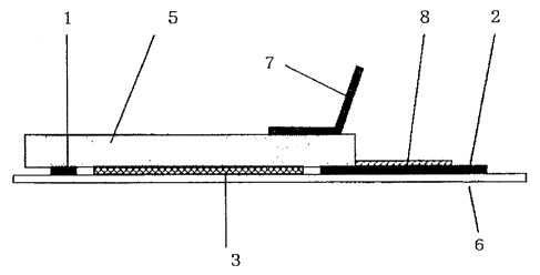
Fig. 1 shows the arrangement of the main-electrode
layer, the sub-electrode layer, and the medication
reservoir layer of the electrode device of Example 1.
Fig. 2 shows a sectional view taken along the line 2-
2' in Fig. 1.
Fig. 3 shows a sectional view taken along the line 3-
3' in Fig. 1.
Fig. 4 shows the arrangement of the main-electrode
layer, the sub-electrode layer, and the medication
reservoir layer of the electrode device of Example 2.
Fig. 5 shows the operation for attaching and bonding
the sub-electrode layer to the medication reservoir layer
by applying an electric current, in the electrode device
shown in Fig. 1 (Example 1).
Fig. 6 shows the schematic diagram of the exemplary
test 3.
Fig. 7 shows a graph illustrating a relationship
between an electric quantity applied to the sub-electrode
layer and the bonding strength.
CA 02784178 2015-11-27
. .
MODES FOR CARRYING OUT THE INVENTION
[0020]
Hereinafter, the present invention will be described
in more detail by way of Examples and exemplary tests,
which however should not be construed as limiting the scope
of the present invention in any way.
In the following
description, the unit % means percentage by weight, unless
otherwise specified.
EXAMPLE 1
[0021]
The medication reservoir layer 5 to be used in
iontophoresis treatment was prepared by the following
procedure, and was then gelled.
Completely hydrolyzed polyvinyl alcohol: 15%
Sodium chloride: 7.65%
Water: 77.35%
Total: 100%
These components were heated, stirred and dissolved,
and then spread to have a thickness of about 1 mm. This
spread layer was frozen at -30 and was then thawed at room
temperature.
The resultant gel was crosslinked to be
shaped.
After that, the shaped gel was punched out to
obtain a disc with a diameter of 30 mm. This disc was used
as the medication reservoir layer 5.
[0022]
Fig. 1 shows the arrangement of the respective members
of the electrode device of Example 1.
Fig. 2 shows a
11
CA 02784178 2015-11-27
sectional view taken along the line 2-2' in Fig. 1; and
Fig. 3 shows a sectional view taken along the line 3-3' in
Fig. 1. In Fig. 1, the auxiliary electrode 7 shown in Figs.
2 and 3 is omitted.
[0023]
The substrate sheet 6 was a polyethylene terephthalate
film with a thickness of 100 pm, a width of 100 mm and a
length of 70 mm, on which the electrode device was
assembled. Firstly,
the main-electrode layer 3 and the
sub-electrode layer 1 were formed on the substrate sheet 6.
As described later, an electric current is applied to the
sub-electrode layer 1 in manufacturing the electrode device,
so that the shaped gel-like medication reservoir layer 5 is
firmly secured to the substrate sheet 6. On the other hand,
an electric current is applied to the main-electrode layer
3 in practical use of the electrode device, by which ions
migrate into the skin. The main-
electrode layer 3 was
electrically insulated from the sub-electrode layer 1.
[0024]
The main-electrode layer 3 was formed from paste
containing silver particles and in the shape of a circle
with a diameter of 27 mm on the substrate sheet 6, by
screen printing. The main-
electrode layer 3 is to be
connected to an external power supply (not shown) via the
lead 4, when the medication was introduced. The lead 4 was
formed to have a width of 1 mm and a length of 15 mm,
extending from a part of the main-electrode layer 3, by
screen printing.
The intersecting area of the lead 4 and the sub-
electrode layer 1 was coated with an insulating ink 8,
12
CA 02784178 2015-11-27
. .
thereby insulating the main-electrode layer 3 from the sub-
electrode layer 1.
[0025]
The sub-electrode layer I was formed from paste
containing silver particles and in the shape of a ring with
a width of 1 mm on the substrate sheet 6, by screen
printing.
The ring-shaped sub-electrode layer 1 was
located around the main-electrode layer 3, with a space of
1 mm therebetween.
The sub-electrode layer 1 is to be
connected to the power supply 9 (see Fig. 5) via the lead 2.
The lead 2 was formed to have a width of 1 mm and a length
of 30 mm, extending from a part of the sub-electrode layer
1, by screen printing.
The lead 2 was coated with the insulating layer 8,
except the connecting portion to the power supply 9.
[0026]
Next, the disc-shaped medication reservoir layer 5
formed as above was placed on the main-electrode layer 3,
so as to be evenly supported by the ring-shaped sub-
electrode layer 1. Then, on the medication reservoir layer
5, the auxiliary electrode 7 formed from previously
containing silver chloride particles by screen printing was
provided.
After that, as shown in Fig. 5, the negative terminal
of the power supply was connected to the auxiliary
electrode 7, while the positive terminal of the power
supply was connected to the lead 2, which is in connection
with the sub-electrode layer 1. Then, an electric current
of 1.0 mA was applied for one minute, to thereby adhere and
secure the sub-electrode layer 1 to the medication
reservoir layer 5.
13
CA 02784178 2015-11-27
EXAMPLE 2
[0027]
The medication reservoir layer 5 to be used in
iontophoresis treatment was prepared by the following
procedure and was then gelled.
Agarose: 3%
Glycerin: 10%
Sodium chloride: 0.8%
Water: 86.2%
Total: 100%
These components were heated, stirred and dissolved,
and then spread to have a thickness of about 1 mm while it
was still hot. This
spread layer was cooled to room
temperature, and the resultant gel was crosslinked to be
shaped. After
that, the shaped gel was punched out to
obtain a disc with a diameter of 30 mm. This disc was used
as the medication reservoir layer 5
[0028]
Fig. 4 shows the arrangement of the respective members
of the electrode device of Example 2. Like in Example 1,
the substrate sheet 6 was a polyethylene terephthalate film
with a thickness of 100 ,Um, a width of 100 mm and a length
of 70 mm. The electrode device was assembled on this film.
[0029]
The main-electrode layer 3 and the lead 4 were formed
as one integrated member, which was cut out a silver foil
with a thickness of 0.05 mm. This cut-
out integrated
member was adhered on the substrate sheet 6 with a double-
14
CA 02784178 2015-11-27
sided tape. The portion for the main-electrode layer 3 was
in the shape of a circle with a diameter of 27 mm. The
portion for the lead 4 was in the shape of a rectangle with
a width of 1 mm and a length of 15 mm, extending from a
part of the main-electrode layer 3.
[0030]
Likewise, the sub-electrode layer 1 was also prepared
by cutting out a silver foil having a thickness of 0.05 mm,
and then adhered on the substrate sheet 6 with a double-
sided tape.
Four rectangular portions each having a width of 1 mm
and a length of 15 mm were cut out of a silver foil. These
were adhered to extend radially from four points
substantially equally spaced on a circle, which was 1 mm
away from the outer periphery of the circular main-
electrode layer 3. Then,
the outer ends of the radially
arranged four rectangular portions were connected to one
another through a silver foil strip having a width of 1 mm,
from where the connection lead 2 was branched to the power
supply 9 (see Fig. 5).
[0031]
Except for the areas of 2 mm length of the sub-
electrode layer 1 on the side of the main-electrode layer 3,
and for the connecting portion of the lead 2 to the power
supply 9, the entire circuit connecting the sub-electrode
layer 1 to the power supply 9 was covered by cellotapee to
provide the insulating layer 8.
[0032]
Next, the disc-shaped medication reservoir layer 5
formed as above was placed on the main-electrode layer 3,
to be evenly supported by the four non-insulated end
CA 02784178 2015-11-27
. .
portions of the sub-electrode layer 1.
Then, on the
medication reservoir layer 5, the auxiliary electrode 7
formed from previously containing silver chloride particles
by screen printing was provided.
After that, as shown in Fig. 5, the negative terminal
of the power supply 9 was connected to the auxiliary
electrode 7, while the positive terminal of the power
supply 9 was connected to the lead 2 of the sub-electrode
layer 1. Then, an electric current of 1.0 mA was applied
for one minute, to thereby adhere and bond the sub-
electrode layer 1 to the medication reservoir layer 5.
COMPARATIVE EXAMPLE 1
[0033]
Except for the "sub-electrode layer" which was not
bonded to the "medication reservoir layer" by application
of an electric current, a "medication reservoir layer" was
prepared in the same manner as in Example 1, and a similar
electrode device was obtained.
COMPARATIVE EXAMPLE 2
[0034]
Except for the "sub-electrode layer" which was not
bonded to the "medication reservoir layer" by application
of an electric current, a "medication reservoir layer" was
prepared in the same manner as in Example 2, and a similar
electrode device was obtained.
16
h
CA 02784178 2016-12-09
EXEMPLARY TEST 1
[0035]
<Bonding Force Test 1>
Each of the electrode devices of Examples 1 and 2 and
Comparative Examples 1 and 2 was gently turned upside down.
As a result, in Comparative Example 2, the medication
reservoir layer 5 was separated from the electrode layers,
peeling and falling off the sheet substrate 6.
[0036]
On the other hand, in each of Examples 1 and 2 and
Comparative Example 1, the medication reservoir layer 5 was
still bonded to the sheet substrate 6. An end of
each
bonded medication reservoir layer 5 was pinched up with
tweezers to evaluate the bonding degree. As a result, the
medication reservoir layer 5 of Comparative Example 1 was
easily peeled off the sheet substrate 6, while the
medication reservoir layers 5 of Examples 1 and 2 showed a
stronger bonding force, enough to bring up the sheet
substrates together.
EXEMPLARY TEST 2
[0037]
<Bonding Force Test 2>
For each of Examples 1 and 2 and Comparative Example 1,
two electrode devices were prepared. One of the
two
electrode devices was used as a donor patch for
iontophoresis, and the other was used as a reference patch,
both of the electrode devices being adhered to the back of
a rat. Then, an
electric current of 0.7 mA was applied
through the main-electrode layers of both patches, and the
fluctuation in voltage was observed.
17
CA 02784178 2015-11-27
[0038]
As a result, no fluctuation in voltage was observed in
Examples 1 and 2, and stable electric current was observed.
On the other hand, in Comparative Example 1, the medication
reservoir layers got dislocated from the main-electrode
layers when the patches were adhered to the back of the rat,
so that an electric current flowed with a voltage slightly
higher than those in Examples 1 and 2. Further,
in
Comparative Example 1, the gel remained on the skin of the
rat when the patches were peeled off after completion of
the electric current application.
Further, it was found that, in the region where the
medication reservoir layer got dislocated from the main-
electrode layer, an electrode portion which lost contact
with the gel due to such dislocation did not work. This
result suggested that, in Comparative Example 1, the
electrode layer and the medication reservoir layer were not
sufficiently contacted, and thus possibly a stable electric
current could not be obtained when the medication was
introduced. In the
case that the medication reservoir
layer got dislocated from the main-electrode layer, the
main-electrode layer could not make its performance
sufficiently.
[0039]
Generally, when dermal administration of a
biologically active substance is conducted by iontophoresis,
it is designed that a predetermined amount of electric
current may flow into a predetermined area. The reason for
such design may be that uneven electric current would
possibly induce damages to the skin. Further, in the case
that the medication reservoir layer peeled off the
18
CA 02784178 2015-11-27
electrode layer, or the medication reservoir layer got
dislocated from the electrode layer, the main-electrode
layer would be likely naked. Then,
the electric current
would flow directly into the skin without passing through
the medication reservoir layer. As a
result, undesired
effect may be brought, for example, a degradation of
efficiency for introducing medication.
Therefore, the
electrode devices of Comparative Examples 1 and 2 are
considered unsuitable for use in iontophoresis.
On the other hand, it was found that the electrode
devices of Examples 1 and 2 can provide stable electric
currents, because the medication reservoir layer and the
electrode layer were contacted firmly.
EXEMPLARY TEST 3
[0040]
<Bonding Force Test 3>
To find a relationship between the electric quantity
and the bonding strength, a peel test using a rheometer was
conducted. Fig. 6
schematically shows the peel test.
Firstly, a medication reservoir layer 5 was prepared,
having the same components as those used in Example 1.
Besides, two rectangular sheets of silver foils 10 (0.05 mm
thickness X 10 mm width X 20 mm height) were separately
prepared. Each of the rectangular silver foils was folded
at a right angle at its point of 10 mm height. Each one
side of the two foils was made contact with the medication
reservoir layer 5, so that the medication reservoir layer 5
was sandwiched between the two silver foils 10.
19
CA 02784178 2015-11-27
[0041]
Further, a silver foil 7 coated with a silver chloride
film was made in contact with the medication reservoir
layer 5, so that the silver foil 7 did not contact with the
two silver foils 10 (if the silver foil 7 contacts with the
silver foils 10, a possible short circuit would prevent
bonding of the gel 5 and the silver foils 10). The
positive terminal of the power supply was connected to one
of the silver foils 10, and the negative terminal was
connected to the silver foil 7 coated with the silver
chloride film, and then an electric current was applied for
one minute. Next, the positive terminal was switched to be
connected to the other silver foil 10, and then the same
amount of electric current was applied for one minute.
Thus, the two silver foils 10 were adhered and secured to
both surfaces of the gel 5. The Peel test was conducted on
the two silver foils 10, using a rheometer (Model CR-500DX
manufactured by SUN SCIENTIFIC CO., LTD.) to measure the
peel strength of the silver foils.
For some samples wherein silver foils 10 are bonded to
a medication reservoir layer 5 with various electric
quantity, the peel strengths were measured in the same way.
The results are shown in Fig. 7.
[0042]
According to this graph, it is found that the more the
electric current flows, the higher the bonding force
becomes between the medication reservoir layer 5 (the gel)
and the electrode layer, and that a peel strength from 100
to 200 mN is obtained at 1 mA=min/cm2 or more.
Regarding a gel equivalent to that in Example 1 used
in this test, the gel was torn when the applied tensile
CA 02784178 2015-11-27
strength exceeded about 100 mN, and thus a bonding strength
could not be measured above that strength (this fact
indicates that the bonded portion had a bonding strength of
at least 100 mN or more).
[0043]
Any of the bonding force tests 1 to 3 shows the
effectiveness of the bonding strength between "the
medication reservoir layer" and "the sub-electrode layer",
which was obtained according to the present invention,
utilizing the electric current.
DESCRIPTION OF REFERENCE NUMERALS
[0044]
1: sub-electrode layer
2: lead for the sub-electrode layer
3: main-electrode layer
4: lead for the main-electrode layer
5: medication reservoir layer
6: substrate sheet
7: auxiliary electrode for bonding use
8: insulating layer
9: external power supply
10: silver foil
21