Note: Descriptions are shown in the official language in which they were submitted.
NEUREGULIN ANTAGONISTS AND USE THEREOF IN TREATING CANCER
FIELD OF THE INVENTION
The present invention relates to the treatment of cancer with neuregulin
antagonists.
BACKGROUND
The identity and properties of cancer stem cells (CSCs) has been a field of
intense study in
recent years. Evidence is accumulating that tumors are a heterogeneous mixture
of cells with different
biological properties. The isolation of distinct cell populations with the
unique ability to initiate tumor
growth has been reported for numerous hematologic malignancies and solid
tumors. However,
inconsistencies have emerged in the use of specific cell surface markers to
prospectively identify
CSCs. For example, disparate findings on stem cell phenotype have been
reported for lculcemias,
pancreatic, colorectal, brain and breast cancers (Reviewed in Brennan and
Matsui 2009). Futhennore,
estimates of CSC frequency vary dramatically between tumor types and patients.
The role of CSCs in
maintaining the growth of an established tumor or in re-initiating a tumor
after chemotherapy either at
a primary or distant site, remains to be determined.
For most cancer patients, disease relapse after chemotherapy is a major cause
of mortality.
Accordingly, a better understanding of the tumor re-initiating cells (TRICs)
responsible for relapse is
needed in order to better treat patients who have experienced a recurrence of
cancer after initially
responding to chemotherapeutic treatment. This is particularly relevant for
non-small cell lung cancer
(NSCLC) because more than two thirds of NSCLC patients are not candidates for
surgical resection.
Most patients present with advanced disease and are treated with chemotherapy,
radiation or a
combination of the two (lung cancer principles and practice). However, the 5
year survival rate for
locally advanced disease remains at 23.7% and at 3.5% for advanced disease
despite good initial
responses to therapy (Homer et al. SEER).
Deregulation of EGFR signaling via overexpression or activating mutations has
been shown to
be a frequent event in NSCLC (reviewed in Dahabreh et al., 2010). EGFR is the
prototypical member
of the HER family of tyrosine kinases, which includes EGFR (Hen), Her2, Her3
and Hcr4. Hcr2 lacks
1.
CA 2784211 2017-10-05
CA 02784211 2012-06-12
WO 2011/103242 PCT/US2011/025163
a functional ligand binding domain (Graus-Porta 1997) and Her3 lacks tyrosine
kinase activity (Guy
1994), so these receptors must act as heterodimers. Recent evidence shows that
other Her family
members may also play a role in NSCLC. However their contributions to the
disease are less well
characterized and studies have often focused on their interactions with EGFR
activation (Kuyama et al.
2008, Hirsch 2009, Zhou 2006, Johnson 2006, Ding 2008).
Neuregulin is a ligand for the Her3 and Her4 receptor tyrosine kinases. There
are four known
members of the neuregulin family, NRG1, NRG2, NRG3, and NRG4 (Falls 2003). The
NRG1
transcript undergoes extensive alternative splicing resulting in at least 15
different isoforms. All active
isoforms share an EGF-like domain that is necessary and sufficient for
activity (Holmes 1992, Yarden
1991). NRG1 autocrine signaling has been shown to regulate lung epithelial
cell proliferation (Jinbo
2002) and to play a role in human lung development (Patel 2000) and has been
implicated in
insensitivity of NSCLC to EGFR inhibitors (Zhou 2006).
The need exists to provide therapeutics effective in treating resistant
cancers and patients who
have experienced a recurrence of cancer.
SUMMARY
One aspect of the invention provides for a method of increasing time to tumor
recurrence in a
cancer patient comprising administering to the patient an effective amount of
a neuregulin antagonist.
In one embodiment, the method further comprises administering a therapeutic
agent to the patient. In
one embodiment, the therapeutic agent is a chemotherapeutic agent or an
antibody. In certain
embodiments the chemotherapeutic agent is paclitaxal or cisplatin or a
combination of paclitaxal and
cisplatin.
In certain embodiments the antibody is an EGFR, HER2, HER3, or HER4 antibody.
In certain
embodiments, the cancer the patient is suffering from is non-small cell lung
cancer, breast cancer,
ovarian cancer, head and neck cancer, cervical cancer, bladder cancer,
oesophageal cancer, prostate
cancer, or colorectal cancer.
In one embodiment, the increase in time to tumor recurrence is at least 1.25
fold greater than
the time to recurrence in the absence of the neuregulin antagonist. In one
embodiment, the increase in
time to tumor recurrence is at least 1.50 fold greater than the time to
recurrence in the absence of the
neuregulin antagonist.
In certain embodiments, the neuregulin antagonist is an antibody, a small
molecule, an
immunoadhesin, or an RNA. In one embodiment, the neuregulin antagonist is a
NRG1
antagonist. In one embodiment, the NRG antagonist is an anti-NRG1 antibody.
2
CA 02784211 2012-06-12
WO 2011/103242 PCT/US2011/025163
BRIEF DESCRIPTION OF THE DRAWINGS
Figure 1. Schematic representation of an in vivo model for studying tumor re-
initiating cells
(TRICs).
Figure 2A. Calu3 human NSCLC xenograft model in athymic nude mice wherein
.. chemotherapy consisted of paclitaxel and cisplatin. Data presented as mean
tumor volume SEM,
n=12 mice/group.
Figure 2B. H441 human NSCLC xenograft model in athymic nude mice wherein
chemotherapy consisted of paclitaxel and cisplatin. Data presented as mean
tumor volume SEM,
11=12 mice/group.
Figure 2C. KPL4 human breast model with orthotropic transplantation of tumor
cells to
mammary fat pad of SCID/beiz mice wherein chemotherapy consisted of
paclitaxel. Data presented as
mean tumor volume SEM, n=12 mice/group.
Figure 2D. Treatment of K-rasLSLG12D CAG-LSL-GFP genetically engineered mouse
NSCLC
model with cisplatin. Data presented as the average number of GFP positive
cells per lung SEM, n=6
.. mice/group.
Figure 3A. Enrichment of NRG1 mRNA in TRICs in the Calu3 xenograft model was
demonstrated by two independent probes in a microarray analysis. Enrichment
was validated by
quantitative real time PCR (qPCR) for NRGla and NRGlb using RNA isolated from
independent
tumor samples.
Figure 3B. Enrichment of NRG1 mRNA in TRICs in the H441 xenograft model shown
by
two different microarray probes. This was validated by qPCR for NRGla and
NRGlb using RNA from
the same tumor samples used for microarray analysis.
Figure 3C. Enrichment of NRG1 mRNA in TRICs in the KPL4 breast cancer
xenograft model
shown by two different microarray probes.
Figure 3D. Enrichment of NRG1 mRNA in TRICs in the KrasTST2D mouse NSCLC model
shown by one micorarray probe and validated by qPCR.
Figure 4. NRG1 enrichment is specific to residual cells as evidenced by qPCR
analysis of
tumor cell NRG1 mRNA levels in tumors of various sizes and times after
chemotherapy.
Figure 5A. Graph showing tumor growth curves for mice with established Calu3-
shNRG1
xenograft tumors administered vehicle (sucrose) or dox (2gm/L) in their
drinking water ad libitum.
Tumor volume was measured twice a week for the duration of the study. Data
presented as Linear
Mixed Effect (LME) model generated fit of tumor volume graphed as cubic
splines with auto-
determined knots.
3
CA 02784211 2012-06-12
WO 2011/103242 PCT/US2011/025163
Figure 5B. Graph showing tumor growth curves for mice with established Calu3-
shNRG1
xenograft tumors treated with chemo+sucrose or chemo+dox. Data presented as
LME model generated
fit of tumor volume graphed as cubic splines with auto-determined knots.
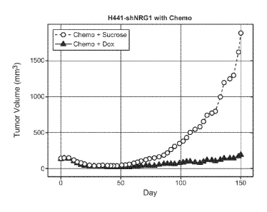
Figure 6A. Graph showing tumor growth curves for mice with established H441-
shNRG1
xenograft tumors treated with sucrose or dox (n=12 /group). Data is presented
as LME fit analysis of
tumor volume graphed as cubic splines with auto-determined knots.
Figure 6B. Graph showing tumor growth curves for mice with established H441-
shNRG1
xenograft tumors treated with chemo + sucrose or chemo + dox (n=12/group).
Data presented as LME
fit analysis of tumor volume graphed as cubic splines with auto-determined
knots.
Figure 7A. Graph showing tumor growth curves for mice with established H1299-
shNRG1
xenograft tumors treated with sucrose or dox (n=12 mice/group). Data is
presented as LME fit analysis
of tumor volume graphed as cubic splines with auto-determined knots.
Figure 7B. Graph showing tumor growth curves for mice with established H1299-
shNRG1
xenograft tumors treated with chemo+sucrose or chemo+dox (n=12/group). Data is
presented as LME
fit analysis of tumor volume graphed as cubic splines with auto-determined
knots.
Figure 8A. Graph showing average tumor volume +/- SEM for LSL-K-rasG120;
53F1/+ mice
treated with vehicle + control IgG (n=6), cisplatin + control IgG (n=6), or
cisplatin + HER4ECD-Fe
(n=8). Ragweed, control murine IgG2a antibody.
Figure 8B. Graph showing the daily fold change in tumor burden by treatment
regimen with
95% confidence intervals.
Figure 8C. Graph showing average percent change in tumor burden from baseline
+ SEM for
LSL-K-rasG120; p53FliF1
mice treated with vehicle + control IgG (n=10), cisplatin + control IgG
(n=11),
cisplatin + HER4-ECD (n=8) or Vehicle + HER4-ECD (n=7).
DETAILED DESCRIPTION OF EMBODIMENTS OF THE INVENTION
I. DEFINITIONS
An "acceptor human framework" for the putposes herein is a framework
comprising the amino
acid sequence of a light chain variable domain (VL) framework or a heavy chain
variable domain (VH)
framework derived from a human immunoglobulin framework or a human consensus
framework, as
defined below. An acceptor human framework "derived from" a human
immunoglobulin framework or
a human consensus framework may comprise the same amino acid sequence thereof,
or it may contain
amino acid sequence changes. In some embodiments, the number of amino acid
changes are 10 or less,
9 or less, 8 or less, 7 or less, 6 or less, 5 or less, 4 or less, 3 or less,
or 2 or less. In some embodiments,
4
CA 02784211 2012-06-12
WO 2011/103242 PCT/US2011/025163
the VL acceptor human framework is identical in sequence to the VL human
immunoglobulin
framework sequence or human consensus framework sequence.
"Affinity" refers to the strength of the sum total of noncovalent interactions
between a single
binding site of a molecule (e.g., an antibody) and its binding partner (e.g.,
an antigen). Unless
indicated otherwise, as used herein, "binding affinity" refers to intrinsic
binding affinity which reflects
a 1:1 interaction between members of a binding pair (e.g., antibody and
antigen). The affinity of a
molecule X for its partner Y can generally be represented by the dissociation
constant (Kd). Affinity
can be measured by common methods known in the art, including those described
herein. Specific
illustrative and exemplary embodiments for measuring binding affinity are
described in the following.
An "affinity matured" antibody refers to an antibody with one or more
alterations in one or
more hypervariable regions (HVRs), compared to a parent antibody which does
not possess such
alterations, such alterations resulting in an improvement in the affinity of
the antibody for antigen.
The terms "anti-NRG antibody- and "an antibody that binds to NRG- refer to an
antibody that
is capable of binding NRG with sufficient affinity such that the antibody is
useful as a diagnostic
and/or therapeutic agent in targeting NRG. In one embodiment, the extent of
binding of an anti-NRG
antibody to an unrelated, non- NRG protein is less than about 10% of the
binding of the antibody to
NRG as measured, e.g., by a radioimmunoassay (RIA). In certain embodiments, an
antibody that binds
to NRG has a dissociation constant (Kd) of < 11.LM, < 100 nM, < 10 nM, < 1 nM,
< 0.1 nM, < 0.01 nM,
or < 0.001 nM (e.g. 10-g M or less, e.g. from 10-8M to 10-13M, e.g., from 10-
9M to 10-13 M). In certain
embodiments, an anti-NRG antibody binds to an epitope of NRG that is conserved
among NRG from
different species.
The term "antibody" herein is used in the broadest sense and encompasses
various antibody
structures, including but not limited to monoclonal antibodies, polyclonal
antibodies, multispecific
antibodies (e.g., bispecific antibodies), and antibody fragments so long as
they exhibit the desired
antigen-binding activity.
An "antibody fragment" refers to a molecule other than an intact antibody that
comprises a
portion of an intact antibody that binds the antigen to which the intact
antibody binds. Examples of
antibody fragments include but are not limited to Fv, Fab, Fab', Fab'-SH,
F(a02; diabodies; linear
antibodies; single-chain antibody molecules (e.g. scFv); and rnultispecific
antibodies formed from
antibody fragments.
An "antibody that binds to the same epitope" as a reference antibody refers to
an antibody that
blocks binding of the reference antibody to its antigen in a competition assay
by 50% or more, and
conversely, the reference antibody blocks binding of the antibody to its
antigen in a competition assay
by 50% or more. An exemplary competition assay is provided herein.
5
CA 02784211 2012-06-12
WO 2011/103242 PCT/US2011/025163
The term "chimeric" antibody refers to an antibody in which a portion of the
heavy and/or light
chain is derived from a particular source or species, while the remainder of
the heavy and/or light chain
is derived from a different source or species.
The terms "cancer" and "cancerous" refer to or describe the physiological
condition in
.. mammals that is typically characterized by unregulated cell
growth/proliferation. Examples of cancer
include, but are not limited to, carcinoma, lymphoma (e.g., Hodgkin's and non-
Hodgkin's lymphoma),
blastoma, sarcoma, and leukemia. More particular examples of such cancers
include squamous cell
cancer, small-cell lung cancer, non-small cell lung cancer, adenocarcinoma of
the lung, squamous
carcinoma of the lung, cancer of the peritoneum, hepatocellular cancer,
gastrointestinal cancer,
pancreatic cancer, glioma, cervical cancer, ovarian cancer, liver cancer,
bladder cancer, hepatoma,
breast cancer, colon cancer, colorectal cancer, endometrial or uterine
carcinoma, salivary gland
carcinoma, kidney cancer, liver cancer, prostate cancer, vulval cancer,
thyroid cancer, hepatic
carcinoma, leukemia and other lymphoproliferative disorders, and various types
of head and neck
cancer.
The "class" of an antibody refers to the type of constant domain or constant
region possessed
by its heavy chain. There are five major classes of antibodies: IgA, IgD, IgE,
IgG, and IgM, and
several of these may be further divided into subclasses (isotypes), e.g.,
IgGi, IgG2, IgG3, IgG4, IgAi,
and IgiV. The heavy chain constant domains that correspond to the different
classes of
immunoglobulins are called a, 6, a, y, and u, respectively.
The term "cytotoxic agent" as used herein refers to a substance that inhibits
or prevents a
cellular function and/or causes cell death or destruction. Cytotoxic agents
include, but are not limited
to, radioactive isotopes (e.g., At2'1, 1131, 1125, y90
, Re186, Re188, sm153, Bi212, P32, Pb 212
and radioactive
isotopes of Lu); chemotherapeutic agents or drugs (e.g., methotrexate,
adriamicin, vinca alkaloids
(vincristine, vinblastine, etoposide), doxorubicin, melphalan, mitomycin C,
chlorambucil, daunorubicin
or other intercalating agents); growth inhibitory agents; enzymes and
fragments thereof such as
nucleolytic enzymes; antibiotics; toxins such as small molecule toxins or
enzymatically active toxins of
bacterial, fungal, plant or animal origin, including fragments and/or variants
thereof; and the various
antitumor or anticancer agents disclosed below.
"Effector functions- refer to those biological activities attributable to the
Fe region of an
antibody, which vary with the antibody isotype. Examples of antibody effector
functions include: Clq
binding and complement dependent cytotoxicity (CDC); Fe receptor binding;
antibody-dependent cell-
mediated cytotoxicity (ADCC); phagocytosis; down regulation of cell surface
receptors (e.g. B cell
receptor); and B cell activation.
An "effective amount" of an agent, e.g., a pharmaceutical formulation, refers
to an amount
effective, at dosages and for periods of time necessary, to achieve the
desired therapeutic or
prophylactic result.
6
CA 02784211 2012-06-12
WO 2011/103242 PCT/US2011/025163
The term "Fe region" herein is used to define a C-terminal region of an
immunoglobulin heavy
chain that contains at least a portion of the constant region. The term
includes native sequence Fc
regions and variant Fe regions. In one embodiment, a human IgG heavy chain Fe
region extends from
Cys226, or from Pro230, to the carboxyl-terminus of the heavy chain. However,
the C-terminal lysine
.. (Lys447) of the Fe region may or may not be present. Unless otherwise
specified herein, numbering of
amino acid residues in the Fe region or constant region is according to the EU
numbering system, also
called the EU index, as described in Kabat et al., Sequences of Proteins of
Immunological Interest, 5th
Ed. Public Health Service, National Institutes of Health, Bethesda, MD, 1991.
"Framework" or "FR" refers to variable domain residues other than
hypervariable region
(HVR) residues. The FR of a variable domain generally consists of four FR
domains: FR1, FR2, FR3,
and FR4. Accordingly, the HVR and FR sequences generally appear in the
following sequence in VH
(or VL): FR1-H1(L1)-FR2-H2(L2)-FR3-H3(L3)-FR4.
The terms "frill length antibody," "intact antibody,- and "whole antibody" are
used herein
interchangeably to refer to an antibody having a structure substantially
similar to a native antibody
structure or having heavy chains that contain an Fe region as defined herein.
The terms "host cell," "host cell line," and "host cell culture" are used
interchangeably and
refer to cells into which exogenous nucleic acid has been introduced,
including the progeny of such
cells. Host cells include "transformants" and "transformed cells," which
include the primary
transformed cell and progeny derived therefrom without regard to the number of
passages. Progeny
may not be completely identical in nucleic acid content to a parent cell, but
may contain mutations.
Mutant progeny that have the same function or biological activity as screened
or selected for in the
originally transformed cell are included herein.
A "human antibody" is one which possesses an amino acid sequence which
corresponds to that
of an antibody produced by a human or a human cell or derived from a non-human
source that utilizes
human antibody repertoires or other human antibody-encoding sequences. This
definition of a human
antibody specifically excludes a humanized antibody comprising non-human
antigen-binding residues.
A "human consensus framework" is a framework which represents the most
commonly
occurring amino acid residues in a selection of human immunoglobulin VL or VH
framework
sequences. Generally, the selection of human immunoglobulin VL or VH sequences
is from a
subgroup of variable domain sequences. Generally, the subgroup of sequences is
a subgroup as in
Kabat et al., Sequences of Proteins of Immunological Interest, Fifth Edition,
NIH Publication 91-3242,
Bethesda MD (1991), vols. 1-3. In one embodiment, for the VL, the subgroup is
subgroup kappa I as
in Kabat et al., supra. In one embodiment, for the VH, the subgroup is
subgroup III as in Kabat et al.,
supra.
A "humanized.' antibody refers to a chimeric antibody comprising amino acid
residues from
non-human HVRs and amino acid residues from human FRs. In certain embodiments,
a humanized
7
CA 02784211 2012-06-12
WO 2011/103242 PCT/US2011/025163
antibody will comprise substantially all of at least one, and typically two,
variable domains, in which
all or substantially all of the HVRs (e.g., CDRs) correspond to those of a non-
human antibody, and all
or substantially all of the FRs correspond to those of a human antibody. A
humanized antibody
optionally may comprise at least a portion of an antibody constant region
derived from a human
antibody. A "humanized form" of an antibody, e.g., a non-human antibody,
refers to an antibody that
has undergone humanization.
The term "hypervariable region" or "HVR," as used herein, refers to each of
the regions of an
antibody variable domain which are hypervariable in sequence and/or form
structurally defined loops
("hypervariable loops"). Generally, native four-chain antibodies comprise six
HVRs; three in the VH
(H1, H2, H3), and three in the VL (L1, L2, L3). HVRs generally comprise amino
acid residues from
the hypervariable loops and/or from the "complementarity determining regions"
(CDRs), the latter
being of highest sequence variability and/or involved in antigen recognition.
Exemplary hypervariable
loops occur at amino acid residues 26-32 (L1), 50-52 (L2), 91-96 (L3), 26-32
(H1), 53-55 (H2), and
96-101 (H3). (Chothia and Lesk, Ifol. Biol. 196:901-917 (1987)) Exemplary CDRs
(CDR-L1,
CDR-L2, CDR-L3, CDR-H1, CDR-H2, and CDR-H3) occur at amino acid residues 24-34
of Ll, 50-56
of L2, 89-97 of L3, 31-35B of H1, 50-65 of H2, and 95-102 of H3. (Kabat et
al., Sequences of Proteins
of Immunological Interest, 5th Ed. Public Health Service, National Institutes
of Health, Bethesda, MD
(1991).) With the exception of CDR1 in VH, CDRs generally comprise the amino
acid residues that
form the hypervariable loops. CDRs also comprise "specificity determining
residues," or "SDRs,"
which are residues that contact antigen. SDRs are contained within regions of
the CDRs called
abbreviated-CDRs, or a-CDRs. Exemplary a-CDRs (a-CDR-L1, a-CDR-L2, a-CDR-L3, a-
CDR-H1, a-
CDR-H2, and a-CDR-H3) occur at amino acid residues 31-34 of Li, 50-55 of L2,
89-96 of L3, 31-35B
of HI, 50-58 of H2, and 95-102 of H3. (See Almagro and Fransson, Front.
Biosci. 13:1619-1633
(2008).) Unless otherwise indicated, HVR residues and other residues in the
variable domain (e.g., FR
residues) are numbered herein according to Kabat et al., supra.
An "immunoconjugate" is an antibody conjugated to one or more heterologous
molecule(s),
including but not limited to a cytotoxic agent.
An "individual" or "subject" or "patient" is a mammal. Mammals include, but
are not limited
to, domesticated animals (e.g., cows, sheep, cats, dogs, and horses), primates
(e.g., humans and non-
human primates such as monkeys), rabbits, and rodents (e.g., mice and rats).
In certain embodiments,
the individual, subject, or patient is a human.
An "isolated" antibody is one which has been separated from a component of its
natural
environment. In some embodiments, an antibody is purified to greater than 95%
or 99% purity as
determined by, for example, electrophoretic (e.g., SDS-PAGE, isoelectric
focusing (1EF), capillary
electrophoresis) or chromatographic (e.g., ion exchange or reverse phase
HPLC). For review of
8
CA 02784211 2012-06-12
WO 2011/103242 PCT/US2011/025163
methods for assessment of antibody purity, see, e.g., Flatman et al., J.
Chromatogr. B 848:79-87
(2007).
An "isolated" nucleic acid refers to a nucleic acid molecule that has been
separated from a
component of its natural environment. An isolated nucleic acid includes a
nucleic acid molecule
contained in cells that ordinarily contain the nucleic acid molecule, but the
nucleic acid molecule is
present extrachromosomally or at a chromosomal location that is different from
its natural
chromosomal location.
"Isolated nucleic acid encoding an anti-NRG antibody" refers to one or more
nucleic acid
molecules encoding antibody heavy and light chains (or fragments thereof),
including such nucleic acid
molecule(s) in a single vector or separate vectors, and such nucleic acid
molecule(s) present at one or
more locations in a host cell.
The term "monoclonal antibody" as used herein refers to an antibody obtained
from a
population of substantially homogeneous antibodies, i.e., the individual
antibodies comprising the
population are identical and/or bind the same epitope, except for possible
variant antibodies, e.g.,
containing naturally occurring mutations or arising during production of a
monoclonal antibody
preparation, such variants generally being present in minor amounts. In
contrast to polyclonal antibody
preparations, which typically include different antibodies directed against
different determinants
(epitopes), each monoclonal antibody of a monoclonal antibody preparation is
directed against a single
determinant on an antigen. Thus, the modifier "monoclonal" indicates the
character of the antibody as
being obtained from a substantially homogeneous population of antibodies, and
is not to be construed
as requiring production of the antibody by any particular method. For example,
the monoclonal
antibodies to be used in accordance with the present invention may be made by
a variety of techniques,
including but not limited to the hybridoma method, recombinant DNA methods,
phage-display
methods, and methods utilizing transgenic animals containing all or part of
the human immunoglobulin
loci, such methods and other exemplary methods for making monoclonal
antibodies being described
herein.
A "naked antibody" refers to an antibody that is not conjugated to a
heterologous moiety (e.g.,
a cytotoxic moiety) or radiolabel. The naked antibody may be present in a
pharmaceutical formulation.
"Native antibodies" refer to naturally occurring immunoglobulin molecules with
varying
structures. For example, native IgG antibodies arc heterotetrameric
glycoproteins of about 150,000
daltons, composed of two identical light chains and two identical heavy chains
that are disulfide-
bonded. From N- to C-terminus, each heavy chain has a variable region (VH),
also called a variable
heavy domain or a heavy chain variable domain, followed by three constant
domains (CH1, CH2, and
CH3). Similarly, from N- to C-terminus, each light chain has a variable region
(VL), also called a
variable light domain or a light chain variable domain, followed by a constant
light (CL) domain. The
9
CA 02784211 2012-06-12
WO 2011/103242 PCT/US2011/025163
light chain of an antibody may be assigned to one of two types, called kappa
(I() and lambda (X), based
on the amino acid sequence of its constant domain.
The term "package insert" is used to refer to instructions customarily
included in commercial
packages of therapeutic products, that contain information about the
indications, usage, dosage,
.. administration, combination therapy, contraindications and/or warnings
concerning the use of such
therapeutic products.
"Percent (%) amino acid sequence identity" with respect to a reference
polypeptide sequence is
defined as the percentage of amino acid residues in a candidate sequence that
are identical with the
amino acid residues in the reference polypeptide sequence, after aligning the
sequences and
introducing gaps, if necessary, to achieve the maximum percent sequence
identity, and not considering
any conservative substitutions as part of the sequence identity. Alignment for
purposes of determining
percent amino acid sequence identity can be achieved in various ways that are
within the skill in the
art, for instance, using publicly available computer software such as BLAST,
BLAST-2, ALIGN or
Megalign (DNASTAR) software. Those skilled in the art can determine
appropriate parameters for
aligning sequences, including any algorithms needed to achieve maximal
alignment over the full length
of the sequences being compared. For purposes herein, however, % amino acid
sequence identity
values are generated using the sequence comparison computer program ALIGN-2.
The ALIGN-2
sequence comparison computer program was authored by Genentech, Inc., and the
source code has
been filed with user documentation in the U.S. Copyright Office, Washington
D.C., 20559, where it is
registered under U.S. Copyright Registration No. TXU510087. The ALIGN-2
program is publicly
available from Genentech, Inc., South San Francisco, California, or may be
compiled from the source
code. The ALIGN-2 program should be compiled for use on a UNIX operating
system, including
digital UNIX V4.0D. All sequence comparison parameters are set by the ALIGN-2
program and do
not vary.
In situations where ALIGN-2 is employed for amino acid sequence comparisons,
the % amino
acid sequence identity of a given amino acid sequence A to, with, or against a
given amino acid
sequence B (which can alternatively be phrased as a given amino acid sequence
A that has or
comprises a certain % amino acid sequence identity to, with, or against a
given amino acid sequence B)
is calculated as follows:
100 times the fraction X/Y
where X is the number of amino acid residues scored as identical matches by
the sequence alignment
program ALIGN-2 in that program's alignment of A and B, and where Y is the
total number of amino
.. acid residues in B. It will be appreciated that where the length of amino
acid sequence A is not equal
to the length of amino acid sequence B, the % amino acid sequence identity of
A to B will not equal
the % amino acid sequence identity of B to A. Unless specifically stated
otherwise, all % amino acid
CA 02784211 2012-06-12
WO 2011/103242
PCT/US2011/025163
sequence identity values used herein are obtained as described in the
immediately preceding paragraph
using the ALIGN-2 computer program.
The term "pharmaceutical formulation" refers to a preparation which is in such
form as to
permit the biological activity of an active ingredient contained therein to be
effective, and which
contains no additional components which are unacceptably toxic to a subject to
which the formulation
would be administered.
A "pharmaceutically acceptable carrier" refers to an ingredient in a
pharmaceutical
formulation, other than an active ingredient, which is nontoxic to a subject.,
A pharmaceutically
acceptable carrier includes, but is not limited to, a buffer, excipient,
stabilizer, or preservative.
The term "NRG" as used herein, refers to any native neuregulin (also known as
lieregulin)
from any vertebrate source, including mammals such as primates (e.g. humans)
and rodents (e.g., mice
and rats), unless otherwise indicated. The term encompasses "full-length,"
unprocessed NRG as well
as any form of NRG that results from processing in the cell. The term also
encompasses naturally
occurring variants of NRG, e.g., splice variants or allelic variants. There
are four known forms of
NRG: NRG1 (Holmes, W.E. et al., Science 256:1205-1210 (1992)); NRG2 (Caraway,
K.L. et al.,
Nature 387:512-516 (1997)); NRG3 (Zhang, E. et al., Proc Natl Acad Sci USA
94:9562-9567)); and
NRG4 (Harari, D. et al., Oncogene 18:2681-2689)). Due to alternative splicing
there are two active
isoforms of the NRG1 EGF-like domain that is required for receptor binding,
referred to as NRGlalpha
(NRG1a) and NRGlbeta (NRGf3). Sequences of exemplary human NRGls are shown in
Genbank
Accession No. BK000383 (Falls, D. L., Ex Cell Res, 284:14-30 (2003) and in US
Patent No.
5,367,060.
As used herein, "treatment" (and grammatical variations thereof such as
"treat" or "treating")
refers to clinical intervention in an attempt to alter the natural course of
the individual being treated,
and can be performed either for prophylaxis or during the course of clinical
pathology. Desirable
effects of treatment include, but are not limited to, preventing occurrence or
recurrence of disease,
alleviation of symptoms, diminishment of any direct or indirect pathological
consequences of the
disease, preventing metastasis, decreasing the rate of disease progression,
amelioration or palliation of
the disease state, and remission or improved prognosis. In some embodiments,
the NRG antagonists of
the invention are used to delay development of a disease, slow the progression
of a disease, prevent
relapse, or to increase time to tumor recurrence. In certain embodiments,
treatment results in a
reduction in the number of or complete absence of tumor reinitating cells; a
decrease in number of
tumor reinitating cells in a solid tumor relative to cells in the tumor that
are not tumor reinitating cells;
and/or inhibition of the proliferation of tumor reinitating cells. In certain
embodiments, treatment with
a NRG antagonist results in an increase in time to tumor recurrence of at
least 1.25, 1.50, 1.75, 2.0 fold
greater than the time to tumor recurrence in the absence of treatment with an
NRG antagonist.
11
CA 02784211 2012-06-12
WO 2011/103242 PCT/US2011/025163
The term "variable region" or "variable domain" refers to the domain of an
antibody heavy or
light chain that is involved in binding the antibody to antigen. The variable
domains of the heavy chain
and light chain (VH and VL, respectively) of a native antibody generally have
similar structures, with
each domain comprising four conserved framework regions (FRs) and three
hypervariable regions
(HVRs). (See, e.g., Kindt et al. Kuby Immunology, 6th ed., W.H. Freeman and
Co., page 91 (2007).) A
single VH or VL domain may be sufficient to confer antigen-binding
specificity. Furthermore,
antibodies that bind a particular antigen may be isolated using a VH or VL
domain from an antibody
that binds the antigen to screen a library of complementary VL or VH domains,
respectively. See, e.g.,
Portolano et al., J. Immunol. 150:880-887 (1993); Clarkson et al., Nature
352:624-628 (1991).
The term "vector," as used herein, refers to a nucleic acid molecule capable
of propagating
another nucleic acid to which it is linked. The term includes the vector as a
self-replicating nucleic
acid structure as well as the vector incorporated into the genome of a host
cell into which it has been
introduced. Certain vectors are capable of directing the expression of nucleic
acids to which they are
operatively linked. Such vectors are referred to herein as "expression
vectors."
II. COMPOSITIONS AND METHODS
The present invention is based on the finding that NRG autocrine signaling
plays an important
role in the survival and proliferation of a small population of tumor cells
after chemotherapy in an
otherwise chemosensitive tumor. These surviving tumor cells, referred to
herein as "tumor reinitating
cells", or "TRICs", arc responsible for the relapse and recurrence of cancer
in patients whose cancer
previously was treated with a therapeutic agent. In one embodiment, the
therapeutic agent used to treat
the patient is a chemotherapeutic agent. In another embodiment, the
therapeutic agent used to treat the
patient is an antigen binding agent, such as an antibody, or fragment thereof.
Inhibition of NRG signaling results in the delay or prevention of tumor
relapse or recurrence
after treatment with a therapeutic agent. Accordingly, one aspect of the
invention provides for NRG
antagonists that inhibit NRG induced signaling. In one embodiment, the NRG
antagonist is an NRG1
antagonist. NRG antagonists find use in treating cancer and in preventing
resistance and/or recurrence
of cancer after treatment with a therapeutic agent. Another aspect of the
invention provides for a
method of preventing resistance to treatment with a therapeutic agent in a
patient by administering to
the patient a NRG antagonist. Another aspect of the invention provides for
preventing recurrence of
cancer after treatment with a therapeutic agent by administering to the
patient a NRG antagonist. Yet
another aspect of the invention provides for a model characterizing TRICs. As
described in the
Examples and accompanying Figures, this model comprises cells that show a
robust response to
treatment resulting in significant tumor regression followed by disease
relapse after the cessation of the
treatment. The model finds use in screening for compounds that can be used to
target the TRICs and
for determining the molecular basis for the TRICs.
12
CA 02784211 2012-06-12
WO 2011/103242 PCT/US2011/025163
Specific aspects include a method of preventing tumor recurrence or increasing
time to tumor
recurrence comprising administering to the patient an effective amount of a
NRG antagonist. In one
embodiment, the patient has been treated with a therapeutic agent, such as a
chemotherapeutic agent or
an antigen binding agent, such as an antibody. In one embodiment, the cancer
comprises tumor re-
intiating cells. In one embodiment, the cancer is non-small cell lung cancer.
In one embodiment, the
cancer is breast cancer. In one embodiment, the patient was treated with a
chemotherapeutic agent. In
one embodiment, the chemotherapeutic agent is an agent used as a standard of
care treatment for
cancer. In one embodiment, the chemotherapeutic agent is paclitaxal or
cisplatin or a combination of
paclitaxal and cisplatin. In one embodiment, the chemotherapeutic agent is not
a tyrosine kinase
inhibitor. In another embodiment, the chemotherapeutic agent is a tyrosine
kinase inhibitor. in one
embodiment, the chemotherapeutic agent is an inhibitor EGFR, HER2, HER3 and/or
HER4. Another
embodiment comprises additionally administering a chemotherapeutic agent to
the patient in
combination with a NRG antagonist.
In another embodiment, the patient was treated with an antibody. In one
embodiment, the
antibody is an anti-tyrosine kinase antibody. In one embodiment, the antibody
is an EGFR, HER2,
HER3 and/or HER4 antibody. Another embodiment comprises additionally
administering an antibody
to the patient in combination with a NRG antagonist.
In certain embodiments, the time to tumor recurrence is at least 1.25, 1.50,
1.75, 2.0, 2.5, 5.0,
10, 20, or 50 times greater than the time to tumor recurrence in the absence
of the neuregulin
antagonist.
Another aspect provides for a method of treating a patient with a resistant
cancer comprising
administering to a patient an effective amount of a NRG antagonist. In one
embodiment, the cancer
comprises tumor re-intiating cells. In one embodiment, the cancer is non-small
cell lung cancer. In
one embodiment, the cancer is breast cancer. In one embodiment, the cancer is
resistant to treatment
with chemotherapeutic agents. In one embodiment, the cancer is resistant to
treatment with paclitaxal
or cisplatin or a combination of paclitaxal and cisplatin. In one embodiment,
the cancer is resistant to
treatment with a tyrosine kinase inhibitor. In one embodiment, the cancer is
resistant to treatment with
an EGFR, HER2, HER3 and/or HER4 inhibitor. Another embodiment comprises
additionally
administering a chemotherapeutic agent to the patient. In one embodiment, the
chemotherapeutic agent
is paclitaxal or cisplatin or a combination of paclitaxal and cisplatin. In
one embodiment, the
chemotherapeutic agent is an EGFR, HER2, HER3 and/or HER4 inhibitor.
In one embodiment, the cancer is resistant to treatment with a therapeutic
antibody. In one
embodiment, the cancer is resistant to treatment with an EGFR, HER2, HER3, or
HER4 antibody.
Another embodiment comprises additionally administering an antibody to the
patient. In one
embodiment, the antibody is trastuzumab or pertuzumab.
13
CA 02784211 2012-06-12
WO 2011/103242 PCT/US2011/025163
Another aspect provides for a method of preventing resistance in a cancer
comprising
administering to a patient who has cancer an effective amount of a NRG
antagonist and a therapeutic
agent. In one embodiment, the cancer comprises tumor re-intiating cells. In
one embodiment, the
cancer is non-small cell lung cancer. In one embodiment, the cancer is breast
cancer. In one
embodiment, the cancer is resistant to treatment with chemotherapeutic agents.
In one embodiment,
the cancer resistant to treatment with paclitaxal or cisplatin or a
combination of paclitaxal and
cisplatin. In one embodiment, the chemotherapeutic agent is not a tyrosine
kinasc inhibitor. In another
embodiment, the chemotherapeutic agent is a tyrosine kinase inhibitor. In one
embodiment, the
chemotherapeutic agent is an inhibitor EGFR, HER2, HER3 and/or HER4. Another
embodiment
comprises additionally administering a chemotherapeutic agent to the patient.
In one embodiment, the
chemotherapeutic agent is paclitaxal or cisplatin or a combination of
paclitaxal and cisplatin.
In one embodiment, the cancer is resistant to treatment with a therapeutic
antbody. In one
embodiment, the cancer is resistant to treatment with an EGFR, HER2, HER3, or
HER4 antibody.
Another embodiment comprises additionally administering an antibody to the
patient. In one
.. embodiment, the antibody is trastuzumab or pertuzumab.
In these methods of therapeutic use, the NRG antagonist is an antibody, a
small molecule, an
immunoadhesin, or an RNA. In one embodiment, the NRG antagonist is a NRG1
antagonist. In one
embodiment, the NRG antagonist is an anti-NRG1 antibody.
In a further aspect, the invention provides for the use of a neuregulin
antagonist in the
.. manufacture or preparation of a medicament. In one embodiment, the
neuregulin antagonist, or
medicament manufactured with the neuregulin antagonist, is used to increase
the time to tumor
recurrence in a patient. In another embodiment, the neuregulin antagonist, or
medicament
manufactured with the neuregulin antagonist, is used to treat a patient with a
cancer that is
resistant to a therapeutic agent.
A. NRG Antagonists
NRG antagonists useful in the methods of the invention include polypeptides
that specifically
bind to NRG, NRG antibodies (anti-NRG antibodies), RNA, such as RNAi, si RNA,
shRNA, etc.,
small molecules, receptor molecules and derivatives, such as immunoadhesins,
which bind specifically
to NRG. (see, for example, US Patents 6,696,290 and 7,659,368, US publications
2010055093 and
20100278801) and fusions proteins. NRG antagonists also include antagonistic
variants of NRG
polypeptides, RNA aptamers and peptibodies against NRG. Examples of each of
these are described
below. In one embodiment, the NRG antagonists are NRG1 antagonists. In other
embodiments, the
NRG antagonists are NRG2, NRG3, or NRG4 antagonists.
14
CA 02784211 2012-06-12
WO 2011/103242 PCT/US2011/025163
1. Antibodies
Anti-NRG antibodies that are useful in the methods of the invention include
any antibody that
binds with sufficient affinity and specificity to NRG and can reduce or
inhibit NRG signaling. NRG
antibodies are described in W01992020798, US Patent No. 6,953,842, and US
Patent No. 6,252,051.
a) Antibody Affinity
In certain embodiments, an antibody provided herein has a dissociation
constant (Kd) of
< 104, < 100 nM, < 10 nM, < 1 nM, < 0.1 nM, < 0.01 nM, or < 0.001 nM (e.g. 10-
8M or less, e.g.
from 10-8M to 1013M, e.g., from 10-9M to 10-13 M).
In one embodiment, Kd is measured by a radiolabeled antigen binding assay
(R1A) performed
with the Fab version of an antibody of interest and its antigen as described
by the following assay.
Solution binding affinity of Fabs for antigen is measured by equilibrating Fab
with a minimal
concentration of (121)-labeled antigen in the presence of a titration series
of unlabeled antigen, then
capturing bound antigen with an anti-Fab antibody-coated plate (see, e.g.,
Chen et al., J. Mol. Biol.
293:865-881(1999)). To establish conditions for the assay, MICROTITER multi-
well plates (Thermo
Scientific) are coated overnight with 5 g/m1 of a capturing anti-Fab antibody
(Cappel Labs) in 50 mM
sodium carbonate (pH 9.6), and subsequently blocked with 2% (w/v) bovine serum
albumin in PBS for
two to five hours at room temperature (approximately 23 C). In a non-adsorbent
plate (Nunc
#269620), 100 pM or 26 pM t251]
antigen are mixed with serial dilutions of a Fab of interest (e.g.,
consistent with assessment of the anti-VEGF antibody, Fab-12, in Presta et
al., Cancer Res. 57:4593-
4599 (1997)). The Fab of interest is then incubated overnight; however, the
incubation may continue
for a longer period (e.g., about 65 hours) to ensure that equilibrium is
reached. Thereafter, the
mixtures are transferred to the capture plate for incubation at room
temperature (e.g., for one hour).
The solution is then removed and the plate washed eight times with 0.1%
polysorbate 20 (TWEEN-
20 ) in PBS. When the plates have dried, 150 pl/well of scintillant
(MICROSCINT-201m; Packard) is
added, and the plates are counted on a TOPCOUNTIm gamma counter (Packard) for
ten minutes.
Concentrations of each Fab that give less than or equal to 20% of maximal
binding are chosen for use
in competitive binding assays.
According to another embodiment, Kd is measured using surface plasmon
resonance assays
using a BIACORE -2000 or a BIACORE -3000 (BIAcore, Inc., Piscataway, NJ) at
25 C with
immobilized antigen CMS chips at ¨10 response units (RU). Briefly,
carboxymethylated dextran
biosensor chips (CMS, BIACORE, Inc.) are activated with N-ethyl-N'- (3-
dimethylaminopropy1)-
carbodiimide hydrochloride (EDC) and N-hydroxysuccinimide (NHS) according to
the supplier's
instructions. Antigen is diluted with 10 mM sodium acetate, pH 4.8, to 5 Kg/m1
(-0.2 M) before
injection at a flow rate of 5 1/minute to achieve approximately 10 response
units (RU) of coupled
protein. Following the injection of antigen, 1 M ethanolamine is injected to
block unreacted groups.
CA 02784211 2012-06-12
WO 2011/103242 PCT/US2011/025163
For kinetics measurements, two-fold serial dilutions of Fab (0.78 nM to 500
nM) are injected in PBS
with 0.05% polysorbate 20 (TWEEN-20'M) surfactant (PBST) at 25 C at a flow
rate of approximately
25 ul/min. Association rates (kon) and dissociation rates (koff) are
calculated using a simple one-to-
one Langmuir binding model (BIACORE Evaluation Software version 3.2) by
simultaneously fitting
the association and dissociation sensorgrams. The equilibrium dissociation
constant (Kd) is calculated
as the ratio koff/kon. See, e.g., Chen et al., J. Mol. Biol. 293:865-881
(1999). If the on-rate exceeds
106 M-1 s-1 by the surface plasmon resonance assay above, then the on-rate can
be determined by
using a fluorescent quenching technique that measures the increase or decrease
in fluorescence
emission intensity (excitation = 295 nm; emission = 340 nm, 16 nm band-pass)
at 250C of a 20 nM
.. anti-antigen antibody (Fab form) in PBS, pH 7.2, in the presence of
increasing concentrations of
antigen as measured in a spectrometer, such as a stop-flow equipped
spectrophometer (Aviv
Instruments) or a 8000-series SLM-AMINCO Tm spectrophotometer
(ThermoSpectronic) with a stirred
cuvette.
b) Antibody Fragments
In certain embodiments, an antibody provided herein is an antibody fragment.
Antibody
fragments include, but are not limited to, Fab, Fab', Fab'-SH, F(ab')2, Fv,
and scFv fragments, and
other fragments described below. For a review of certain antibody fragments,
see Hudson et al. Nat.
Med. 9:129-134 (2003). For a review of scFv fragments, see, e.g., Pluckthiln,
in The Pharmacology of
Monoclonal Antibodies, vol. 113, Rosenburg and Moore eds., (Springer-Verlag,
New York), pp. 269-
315 (1994); see also WO 93/16185; and U.S. Patent Nos. 5,571,894 and
5,587,458. For discussion of
Fab and F(ab')2 fragments comprising salvage receptor binding epitope residues
and having increased
in vivo half-life, see U.S. Patent No. 5,869,046.
Diabodies are antibody fragments with two antigen-binding sites that may be
bivalent or
bispecific. See, for example, EP 404,097; WO 1993/01161; Hudson et al., Nat.
Med. 9:129-134
(2003); and Hollinger et al., Proc. Natl. Acad. Sci. USA 90: 6444-6448 (1993).
Triabodies and
tetrabodies are also described in Hudson et al., Nat. Med. 9:129-134 (2003).
Single-domain antibodies are antibody fragments comprising all or a portion of
the heavy chain
variable domain or all or a portion of the light chain variable domain of an
antibody. In certain
embodiments, a single-domain antibody is a human single-domain antibody
(Domantis, Inc., Waltham,
MA; see, e.g., U.S. Patent No. 6,248,516 B1).
Antibody fragments can be made by various techniques, including but not
limited to
proteolytic digestion of an intact antibody as well as production by
recombinant host cells (e.g. E. coli
or phagc), as described herein.
16
CA 02784211 2012-06-12
WO 2011/103242
PCT/US2011/025163
c) Chimeric and Humanized Antibodies
In certain embodiments, an antibody provided herein is a chimeric antibody.
Certain chimeric
antibodies are described, e.g., in U.S. Patent No. 4,816,567; and Morrison et
al., Proc. Natl. Acad. Sci.
USA, 81:6851-6855 (1984)). In one example, a chimeric antibody comprises a non-
human variable
region (e.g., a variable region derived from a mouse, rat, hamster, rabbit, or
non-human primate, such
as a monkey) and a human constant region. In a further example, a chimeric
antibody is a "class
switched" antibody in which the class or subclass has been changed from that
of the parent antibody.
Chimeric antibodies include antigen-binding fragments thereof.
In certain embodiments, a chimeric antibody is a humanized antibody.
Typically, a non-human
antibody is humanized to reduce immunogenicity to humans, while retaining the
specificity and affinity
of the parental non-human antibody. Generally, a humanized antibody comprises
one or more variable
domains in which HVRs, e.g., CDRs, (or portions thereof) are derived from a
non-human antibody, and
FRs (or portions thereof) are derived from human antibody sequences. A
humanized antibody
optionally will also comprise at least a portion of a human constant region.
In some embodiments,
some FR residues in a humanized antibody are substituted with corresponding
residues from a non-
human antibody (e.g., the antibody from which the HVR residues are derived),
e.g., to restore or
improve antibody specificity or affinity.
Humanized antibodies and methods of making them are reviewed, e.g., in Almagro
and
Fransson, Front. Biosci. 13:1619-1633 (2008), and are further described, e.g.,
in Riechmann et al.,
Nature 332:323-329 (1988); Queen et al., Proc. Nat'l Acad. Sci. USA 86:10029-
10033 (1989); US
Patent Nos. 5, 821,337, 7,527,791, 6,982,321, and 7,087,409; Kashmiri etal.,
Methods 36:25-34
(2005) (describing SDR (a-CDR) grafting); Padlan,
Immunol. 28:489-498 (1991) (describing
"resurfacing"); Dall'Acqua et al., Methods 36:43-60 (2005) (describing "FR
shuffling"); and Osbourn
et al., Methods 36:61-68 (2005) and Klimka et al., Br. J. Cancer, 83:252-260
(2000) (describing the
"guided selection" approach to FR shuffling).
Human framework regions that may be used for humanization include but are not
limited to:
framework regions selected using the "best-fit" method (see, e.g., Sims et al.
J. Immunol. 151:2296
(1993)); framework regions derived from the consensus sequence of human
antibodies of a particular
subgroup of light or heavy chain variable regions (see, e.g., Carter et al.
Proc. Natl. Acad. Sci. USA,
89:4285 (1992); and Presta et al. J. Immunol., 151:2623 (1993)); human mature
(somatically mutated)
framework regions or human germline framework regions (see, e.g., Almagro and
Fransson, Front.
Biosci. 13:1619-1633 (2008)); and framework regions derived from screening FR
libraries (see, e.g.,
Baca et al., J. Biol. Chern. 272:10678-10684 (1997) and Rosok et al., J. Biol.
Chem. 271:22611-22618
(1996)).
17
CA 02784211 2012-06-12
WO 2011/103242
PCT/US2011/025163
d) Human Antibodies
In certain embodiments, an antibody provided herein is a human antibody. Human
antibodies
can be produced using various techniques known in the art. Human antibodies
are described generally
in van Dijk and van de Winkel, Curr. Opin. Phartnacol. 5: 368-74 (2001) and
Lonberg, Curr. Opin.
Immunol. 20:450-459 (2008).
Human antibodies may be prepared by administering an immunogen to a transgenic
animal that
has been modified to produce intact human antibodies or intact antibodies with
human variable regions
in response to antigenic challenge. Such animals typically contain all or a
portion of the human
immunoglobulin loci, which replace the endogenous immunoglobulin loci, or
which are present
extraehromosomally or integrated randomly into the animal's chromosomes. In
such transgenic mice,
the endogenous immunoglobulin loci have generally been inactivated. For review
of methods for
obtaining human antibodies from transgenic animals, see Lonberg, Nat. Biotech.
23:1117-1125 (2005).
See also, e.g., U.S. Patent Nos. 6,075,181 and 6,150,584 describing
XENOMOUSETm technology; U.S.
Patent No. 5,770,429 describing HuMAst technology; U.S. Patent No. 7,041,870
describing K-M
MOUSE technology, and U.S. Patent Application Publication No. US
2007/0061900, describing
VELOCIMOUSE technology). Human variable regions from intact antibodies
generated by such
animals may be further modified, e.g., by combining with a different human
constant region.
Human antibodies can also be made by hybridoma-based methods. Human myeloma
and
mouse-human heteromyeloma cell lines for the production of human monoclonal
antibodies have been
described. (See, e.g., Kozbor J. Immunol., 133: 3001 (1984); Brodeur et al.,
Monoclonal Antibody
Production Techniques and Applications, pp. 51-63 (Marcel Dekker, Inc., New
York, 1987); and
Boerner et al., J. Immunol., 147: 86 (1991).) Human antibodies generated via
human B-cell hybridoma
technology are also described in Li at al.. Proc. Aral. Acad, Sci, USA,
103:3557-3562 (2006).
Additional methods include those described, for example, in U.S. Patent No.
7,189,826 (describing
production of monoclonal human IgM antibodies from hybridoma cell lines) and
Ni, Xiandai
Mianyixue, 26(4):265-268 (2006) (describing human-human hybridomas). Human
hybridoma
technology (Trioma technology) is also described in Vollmers and Brandlein,
Histology and
Histopathology, 20(3):927-937 (2005) and Vollmers and Brandlein, Methods and
Findings in
Experimental and Clinical Pharmacology, 27(3):185-91 (2005).
Human antibodies may also be generated by isolating Fv clone variable domain
sequences
selected from human-derived phage display libraries. Such variable domain
sequences may then be
combined with a desired human constant domain. Techniques for selecting human
antibodies from
antibody libraries are described below.
18
CA 02784211 2012-06-12
WO 2011/103242 PCT/US2011/025163
e) Library-Derived Antibodies
Antibodies of the invention may be isolated by screening combinatorial
libraries for antibodies
with the desired activity or activities. For example, a variety of methods are
known in the art for
generating phage display libraries and screening such libraries for antibodies
possessing the desired
binding characteristics. Such methods are reviewed, e.g., in Hoogenboom et al.
in Methods in
Molecular Biology 178:1-37 (O'Brien et al., ed., Human Press, Totowa, NJ,
2001) and further
described, e.g., in the McCafferty et al., Nature 348:552-554; Clackson et
al., Nature 352: 624-628
(1991); Marks et al., J. MoL Biol. 222: 581-597 (1992); Marks and Bradbury, in
Methods in Molecular
Biology 248:161-175 (Lo, ed., Human Press, Totowa, NJ, 2003); Sidhu et al., J.
MoL Biol. 338(2): 299-
310 (2004); Lee et al., J. 'VIOL Biol. 340(5): 1073-1093 (2004); Fellouse,
Proc. Natl. Acad. Sci. USA
101(34): 12467-12472 (2004); and Lee et al., J. ImmunoL Methods 284(1-2): 119-
132(2004).
In certain phage display methods, repertoires of VH and VL genes are
separately cloned by
polymerase chain reaction (PCR) and recombined randomly in phage libraries,
which can then be
screened for antigen-binding phage as described in Winter et al., Ann. Rev.
ImmunoL, 12: 433-455
(1994). Phage typically display antibody fragments, either as single-chain Fv
(scFv) fragments or as
Fab fragments. Libraries from immunized sources provide high-affinity
antibodies to the immunogen
without the requirement of constructing hybridomas. Alternatively, the naive
repertoire can be cloned
(e.g., from human) to provide a single source of antibodies to a wide range of
non-self and also self
antigens without any immunization as described by Griffiths et al., EMBO J,
12: 725-734 (1993).
Finally, naive libraries can also be made synthetically by cloning
unrearranged V-gene segments from
stem cells, and using PCR primers containing random sequence to encode the
highly variable CDR3
regions and to accomplish rearrangement in vitro, as described by Hoogenboom
and Winter, J. MoL
BioL , 227: 381-388 (1992). Patent publications describing human antibody
phage libraries include, for
example: US Patent No. 5,750,373, and US Patent Publication Nos. 2005/0079574,
2005/0119455,
2005/0266000, 2007/0117126, 2007/0160598, 2007/0237764, 2007/0292936, and
2009/0002360.
Antibodies or antibody fragments isolated from human antibody libraries are
considered
human antibodies or human antibody fragments herein.
Multispecific Antibodies
In certain embodiments, an antibody provided herein is a multispecific
antibody, e.g. a
bispecific antibody. Multispecific antibodies are monoclonal antibodies that
have binding specificities
for at least two different sites. In certain embodiments, one of the binding
specificities is for NRG and
the other is for any other antigen. In certain embodiments, bispecific
antibodies may bind to two
different epitopes of NRG. Bispecific antibodies may also be used to localize
cytotoxic agents to cells
which express NRG. Bispecific antibodies can be prepared as full length
antibodies or antibody
fragments.
19
CA 02784211 2012-06-12
WO 2011/103242 PCT/US2011/025163
Techniques for making multispecific antibodies include, but are not limited
to, recombinant co-
expression of two immunoglobulin heavy chain-light chain pairs having
different specificities (see
Milstein and Cuello, Nature 305: 537 (1983)), WO 93/08829, and Traunecker et
al., EiVIBO J. 10: 3655
(1991)), and "knob-in-hole" engineering (see, e.g., U.S. Patent No.
5,731,168). Multi-specific
.. antibodies may also be made by engineering electrostatic steering effects
for making antibody Fc-
heterodimeric molecules (WO 2009/089004A1); cross-linking two or more
antibodies or fragments
(see, e.g., US Patent No. 4,676,980, and Brennan et al., Science, 229:
81(1985)); using leucine zippers
to produce bi-specific antibodies (see, e.g., Kostelny et al., J. Immunol.,
148(5):1547-1553 (1992));
using ''diabody" technology for making bispecific antibody fragments (see,
e.g., Hollinger et al., Proc.
Natl. Acad. Sci. USA, 90:6444-6448 (1993)); and using single-chain Fv (sFv)
dimers (see,e.g. Gruber et
al., J. litununol., 152:5368 (1994)); and preparing trispccific antibodies as
described, e.g., in Tutt et al.
J. Inirnunol. 147: 60 (1991).
Engineered antibodies with three or more functional antigen binding sites,
including "Octopus
antibodies," are also included herein (see, e.g. US 2006/0025576A1).
The antibody or fragment herein also includes a -Dual Acting FAb" or -DAF"
comprising an
antigen binding site that binds to NRG as well as another, different antigen
(see, US 2008/0069820, for
example).
g) Antibody Variants
In certain embodiments, amino acid sequence variants of the antibodies
provided herein are
contemplated. For example, it may be desirable to improve the binding affinity
and/or other biological
properties of the antibody. Amino acid sequence variants of an antibody may be
prepared by
introducing appropriate modifications into the nucleotide sequence encoding
the antibody, or by
peptide synthesis. Such modifications include, for example, deletions from,
and/or insertions into
and/or substitutions of residues within the amino acid sequences of the
antibody. Any combination of
deletion, insertion, and substitution can be made to arrive at the final
construct, provided that the final
construct possesses the desired characteristics, e.g., antigen-binding.
11) Substitution, Insertion, and Deletion Variants
In certain embodiments, antibody variants having one or more amino acid
substitutions are
provided. Sites of interest for substitutional mutagenesis include the HVRs
and FRs. Conservative
substitutions are shown in Table 1 under the heading of ''conservative
substitutions." More substantial
changes are provided in Table 1 under the heading of "exemplary
substitutions," and as further
described below in reference to amino acid side chain classes. Amino acid
substitutions may be
introduced into an antibody of interest and the products screened for a
desired activity, e.g.,
retained/improved antigen binding, decreased immunogenicity, or improved ADCC
or CDC.
TABLE 1
CA 02784211 2012-06-12
WO 2011/103242 PCT/US2011/025163
Original Exemplary Preferred
Residue Substitutions
Substitutions
Ala (A) Val; Leu; Ile Val
Arg (R) Lys; Gin; Asn Lys
Asn (N) Gln; His; Asp, Lys; Arg Gin
Asp (D) Glu; Asn Glu
Cys (C) Ser; Ala Ser
Gln (Q) Asn; Glu Asn
Glu (E) Asp; Gin Asp
Gly (G) Ala Ala
His (H) Asn; Gin; Lys; Arg Arg
Ile (I) Leu; Val; Met; Ala; Phe; Norleucine Leu
Leu (L) Norleucine; Ile; Val; Met; Ala; Phe Ile
Lys (K) Arg; Gin; Asn Arg
Met (M) Lew Phe; Ile Leu
Phe (F) Trp; Leu; Val; Ile; Ala; Tyr Tyr
Pro (P) Ala Ala
Ser (S) Thr Thr
Thr (T) Val; Ser Ser
Trp (W) Tyr; Phe Tyr
Tyr (Y) Trp; Phe; Thr; Ser Phe
Val (V) Ile; Leu; Met; Phe; Ala; Norleucine Leu
Amino acids may be grouped according to common side-chain properties:
(1) hydrophobic: Norleucine, Met, Ala, Val, Leu, Ile;
(2) neutral hydrophilic: Cys, Ser, Thr, Asn, Gin;
(3) acidic: Asp, Glu;
(4) basic: His, Lys, Arg;
(5) residues that influence chain orientation: Gly, Pro;
(6) aromatic: Trp, Tyr, Phe.
Non-conservative substitutions will entail exchanging a member of one of these
classes for
another class.
One type of substitutional variant involves substituting one or more
hypervariable region
residues of a parent antibody (e.g. a humanized or human antibody). Generally,
the resulting variant(s)
selected for further study will have modifications (e.g., improvements) in
certain biological properties
21
CA 02784211 2012-06-12
WO 2011/103242 PCT/US2011/025163
(e.g., increased affinity, reduced immunogenicity) relative to the parent
antibody and/or will have
substantially retained certain biological properties of the parent antibody.
An exemplary substitutional
variant is an affinity matured antibody, which may be conveniently generated,
e.g., using phage
display-based affinity maturation techniques such as those described herein.
Briefly, one or more HVR
residues are mutated and the variant antibodies displayed on phage and
screened for a particular
biological activity (e.g. binding affinity).
Alterations (e.g., substitutions) may be made in HVRs, e.g., to improve
antibody affinity. Such
alterations may be made in HVR "hotspots," i.e., residues encoded by codons
that undergo mutation at
high frequency during the somatic maturation process (see, e.g., Chowdhury,
Methods Hol.
207:179-196 (2008)), and/or SDRs (a-CDRs), with the resulting variant VH or VL
being tested for
binding affinity. Affinity maturation by constructing and reselecting from
secondary libraries has been
described, e.g., in Hoogenboom et al. in Methods in Molecular Biology 178:1-37
(O'Brien et al., ed.,
Human Press, Totowa, NJ, (2001).) In some embodiments of affinity maturation,
diversity is
introduced into the variable genes chosen for maturation by any of a variety
of methods (e.g., error-
prone PCR, chain shuffling, or oligonucleotide-directed mutagenesis). A
secondary library is then
created. The library is then screened to identify any antibody variants with
the desired affinity.
Another method to introduce diversity involves HVR-directed approaches, in
which several HVR
residues (e.g., 4-6 residues at a time) are randomized. HVR residues involved
in antigen binding may
be specifically identified, e.g., using alanine scanning mutagenesis or
modeling. CDR-H3 and CDR-L3
in particular are often targeted.
In certain embodiments, substitutions, insertions, or deletions may occur
within one or more
HVRs so long as such alterations do not substantially reduce the ability of
the antibody to bind antigen.
For example, conservative alterations (e.g., conservative substitutions as
provided herein) that do not
substantially reduce binding affinity may be made in HVRs. Such alterations
may be outside of HVR
"hotspots" or SDRs. In certain embodiments of the variant VH and VL sequences
provided above,
each HVR either is unaltered, or contains no more than one, two or three amino
acid substitutions.
A useful method for identification of residues or regions of an antibody that
may be targeted
for mutagenesis is called ''alanine scanning mutagenesis" as described by
Cunningham and Wells
(1989) Science, 244:1081-1085. In this method, a residue or group of target
residues (e.g., charged
residues such as arg, asp, his, lys, and glu) are identified and replaced by a
neutral or negatively
charged amino acid (e.g., alanine or polyalanine) to determine whether the
interaction of the antibody
with antigen is affected. Further substitutions may be introduced at the amino
acid locations
demonstrating functional sensitivity to the initial substitutions.
Alternatively, or additionally, a crystal
structure of an antigen-antibody complex to identify contact points between
the antibody and antigen.
Such contact residues and neighboring residues may be targeted or eliminated
as candidates for
substitution. Variants may be screened to determine whether they contain the
desired properties.
22
CA 02784211 2012-06-12
WO 2011/103242 PCT/US2011/025163
Amino acid sequence insertions include amino- and/or carboxyl-terminal fusions
ranging in
length from one residue to polypeptides containing a hundred or more residues,
as well as
intrasequence insertions of single or multiple amino acid residues. Examples
of terminal insertions
include an antibody with an N-terminal methionyl residue. Other insertional
variants of the antibody
molecule include the fusion to the N- or C-terminus of the antibody to an
enzyme (e.g. for ADEPT) or
a polypeptide which increases the serum half-life of the antibody.
Glycosylation variants
In certain embodiments, an antibody provided herein is altered to increase or
decrease the
extent to which the antibody is glycosylated. Addition or deletion of
glycosylation sites to an
antibody may be conveniently accomplished by altering the amino acid sequence
such that one or more
glycosylation sites is created or removed.
Where the antibody comprises an Fc region, the carbohydrate attached thereto
may be altered.
Native antibodies produced by mammalian cells typically comprise a branched,
biantennary
oligosaccharide that is generally attached by an N-linkage to Asn297 of the
CH2 domain of the Fe
region. See, e.g., Wright et al. TIBTECH 15:26-32 (1997). The oligosaccharide
may include various
carbohydrates, e.g., mannose, N-acetyl glucosamine (G1cNAc), galactose, and
sialic acid, as well as a
fucose attached to a GlcNAc in the "stem" of the biantennaly oligosaccharide
structure. In some
embodiments, modifications of the oligosaccharide in an antibody of the
invention may be made in
order to create antibody variants with certain improved properties.
In one embodiment, antibody variants are provided having a carbohydrate
structure that lacks
fucose attached (directly or indirectly) to an Fe region. For example, the
amount of fucose in such
antibody may be from 1% to 80%, from 1% to 65%, from 5% to 65% or from 20% to
40%. The
amount of fucose is determined by calculating the average amount of fucose
within the sugar chain at
Asn297, relative to the sum of all glycostructures attached to Asn 297 (e. g.
complex, hybrid and high
mannose structures) as measured by MALDI-TOF mass spectrometry, as described
in
WO 2008/077546, for example. Asn297 refers to the asparagine residue located
at about position 297
in the Fe region (Eu numbering of Fe region residues); however, Asn297 may
also be located about 3
amino acids upstream or downstream of position 297, i.e., between positions
294 and 300, due to minor
sequence variations in antibodies. Such fucosylation variants may have
improved ADCC function.
See, e.g., US Patent Publication Nos. US 2003/0157108 (Presta, L.); US
2004/0093621 (Kyowa Hakko
Kogyo Co., Ltd). Examples of publications related to "defucosylated" or
"fucose-deficient" antibody
variants include: US 2003/0157108; WO 2000/61739; WO 2001/29246; US
2003/0115614; US
2002/0164328; US 2004/0093621; US 2004/0132140; US 2004/0110704; US
2004/0110282; US
2004/0109865; WO 2003/085119; WO 2003/084570; WO 2005/035586; WO 2005/035778;
W02005/053742; W02002/031140; Okazaki et al. J. illoL Biol. 336:1239-1249
(2004); Yamane-
Ohnuki et al. Biotech. Bioeng. 87: 614 (2004). Examples of cell lines capable
of producing
23
CA 02784211 2012-06-12
WO 2011/103242 PCT/US2011/025163
defucosylated antibodies include Lec13 CHO cells deficient in protein
fucosylation (Ripka et al. Arch.
Biochem. Biophys. 249:533-545 (1986); US Pat Appl No US 2003/0157108 Al,
Presta, L; and
WO 2004/056312 Al, Adams et al., especially at Example 11), and knockout cell
lines, such as alpha-
1,6-fucosyltransferase gene, FUT8, knockout CHO cells (see, e.g., Yamane-
Ohnuki et al. Biotech.
Bioeng. 87: 614 (2004); Kanda, Y. et al., Biotechnol. Bioeng., 94(4):680-688
(2006); and
W02003/085107).
Antibodies variants arc further provided with bisected oligosaccharides, e.g.,
in which a
biantennary oligosaccharide attached to the Fe region of the antibody is
bisected by GlcNAc. Such
antibody variants may have reduced fucosylation and/or improved ADCC function.
Examples of such
antibody variants are described, e.g., in WO 2003/011878 (Jean-Mairet et al.);
US Patent No.
6,602,684 (Umana et al.); and US 2005/0123546 (Umana etal.). Antibody variants
with at least one
galactose residue in the oligosaccharide attached to the Fe region are also
provided. Such antibody
variants may have improved CDC function. Such antibody variants are described,
e.g., in WO
1997/30087 (Patel et al.); WO 1998/58964 (Raju, S.); and WO 1999/22764 (Raju,
S.).
Fc rmion variants
In certain embodiments, one or more amino acid modifications may be introduced
into the Fe
region of an antibody provided herein, thereby generating an Fe region
variant. The Fe region variant
may comprise a human Fe region sequence (e.g., a human IgGl, IgG2, IgG3 or
IgG4 Fe region)
comprising an amino acid modification (e.g. a substitution) at one or more
amino acid positions.
In certain embodiments, the invention contemplates an antibody variant that
possesses some
but not all effector functions, which make it a desirable candidate for
applications in which the half life
of the antibody in vivo is important yet certain effector functions (such as
complement and ADCC) are
unnecessary or deleterious. In vitro and/or in vivo cytotoxicity assays can be
conducted to confirm the
reduction/depletion of CDC and/or ADCC activities. For example, Fe receptor
(FcR) binding assays
.. can be conducted to ensure that the antibody lacks FcyR binding (hence
likely lacking ADCC activity),
but retains FcRn binding ability. The primary cells for mediating ADCC, NK
cells, express FcyRIII
only, whereas monocytes express FcyRI, FcyRII and FcyRIII. FcR expression on
hematopoietic cells
is summarized in Table 3 on page 464 of Ravetch and Kinet, Annu. Rev. Immunol.
9:457-492 (1991).
Non-limiting examples of in vitro assays to assess ADCC activity of a molecule
of interest is described
.. in U.S. Patent No. 5,500,362 (see, e.g. Hellstrom, I. et al. Proc. Nat'l
Acad. Sci. USA 83:7059-7063
(1986)) and Hellstrom, Jet al., Proc. Nat'l Acad. Sci. USA 82:1499-1502
(1985); 5,821,337 (see
Bruggemann, M. et al., J. Exp. Med. 166:1351-1361 (1987)). Alternatively, non-
radioactive assays
methods may be employed (see, for example, ACTITm non-radioactive cytotoxicity
assay for flow
cytometry (CellTechnology, Inc. Mountain View, CA; and CytoTox 96 non-
radioactive cytotoxicity
assay (Promcga, Madison, WI). Useful effector cells for such assays include
peripheral blood
24
CA 02784211 2012-06-12
WO 2011/103242 PCT/US2011/025163
mononuclear cells (PBMC) and Natural Killer (NK) cells. Alternatively, or
additionally, ADCC
activity of the molecule of interest may be assessed in vivo, e.g., in a
animal model such as that
disclosed in Clynes et al. Proc. Nat'l Acad. Sci. USA 95:652-656 (1998). Clq
binding assays may also
be carried out to confirm that the antibody is unable to bind Clq and hence
lacks CDC activity. See,
e.g., Clq and C3c binding ELISA in WO 2006/029879 and WO 2005/100402. To
assess complement
activation, a CDC assay may be performed (see, for example, Gazzano-Santoro et
al., J. IinmunoL
Methods 202:163 (1996); Cragg, M.S. et al., Blood 101:1045-1052 (2003); and
Cragg, M.S. and M.J.
Glennie, Blood 103:2738-2743 (2004)). FcRn binding and in vivo clearance/half
life determinations
can also be performed using methods known in the art (see, e.g., Petkova, S.B.
et al., Int 'L ImmunoL
18(12):1759-1769 (2006)).
Antibodies with reduced effector function include those with substitution of
one or more of Fe
region residues 238, 265, 269, 270, 297, 327 and 329 (U.S. Patent No.
6,737,056). Such Fe mutants
include Fe mutants with substitutions at two or more of amino acid positions
265, 269, 270, 297 and
327, including the so-called "DANA" Fe mutant with substitution of residues
265 and 297 to alanine
(US Patent No. 7,332,581).
Certain antibody variants with improved or diminished binding to FcRs are
described. (See,
e.g., U.S. Patent No. 6,737,056; WO 2004/056312, and Shields et al., J. Biol.
Chem. 9(2): 6591-6604
(2001).)
In certain embodiments, an antibody variant comprises an Fe region with one or
more amino
acid substitutions which improve ADCC, e.g., substitutions at positions 298,
333, and/or 334 of the Fe
region (EU numbering of residues).
In some embodiments, alterations are made in the Fe region that result in
altered (i.e., either
improved or diminished) Clq binding and/or Complement Dependent Cytotoxicity
(CDC), e.g., as
described in US Patent No. 6,194,551, WO 99/51642, and Idusogie et al. J.
IminunoL 164: 4178-4184
(2000).
Antibodies with increased half lives and improved binding to the neonatal Fe
receptor (FcRn),
which is responsible for the transfer of maternal IgGs to the fetus (Guyer et
al., J. ImmunoL 117:587
(1976) and Kim et al., J. ImmunoL 24:249 (1994)), are described in
U52005/0014934A1 (Hinton et
al.). Those antibodies comprise an Fe region with one or more substitutions
therein which improve
binding of the Fe region to FcRn. Such Fe variants include those with
substitutions at one or more of
Fe region residues: 238, 256, 265, 272, 286, 303, 305, 307, 311, 312, 317,
340, 356, 360, 362, 376,
378, 380, 382, 413, 424 or 434, e.g., substitution of Fe region residue 434
(US Patent No. 7,371,826).
See also Duncan & Winter, Nature 322:738-40 (1988); U.S. Patent No. 5,648,260;
U.S.
Patent No. 5,624,821; and WO 94/29351 concerning other examples of Fe region
variants.
CA 02784211 2012-06-12
WO 2011/103242 PCT/US2011/025163
Cysteine engineered antibody variants
In certain embodiments, it may be desirable to create cysteine engineered
antibodies, e.g.,
"thioMAbs," in which one or more residues of an antibody are substituted with
cysteine residues. In
particular embodiments, the substituted residues occur at accessible sites of
the antibody. By
substituting those residues with cysteine, reactive thiol groups are thereby
positioned at accessible sites
of the antibody and may be used to conjugate the antibody to other moieties,
such as drug moieties or
linker-drug moieties, to create an immunoconjugate, as described further
herein. In certain
embodiments, any one or more of the following residues may be substituted with
cysteine: V205
(Kabat numbering) of the light chain; A118 (EU numbering) of the heavy chain;
and S400 (EU
numbering) of the heavy chain Fe region. Cysteine engineered antibodies may be
generated as
described, e.g., in U.S. Patent No. 7,521,541.
1) Antibody Derivatives
In certain embodiments, an antibody provided herein may be further modified to
contain
additional nonproteinaceous moieties that are known in the art and readily
available. The moieties
suitable for derivatization of the antibody include but are not limited to
water soluble polymers. Non-
limiting examples of water soluble polymers include, but are not limited to,
polyethylene glycol (PEG),
copolymers of ethylene glycol/propylene glycol, carboxymethylcellulose,
dextran, polyvinyl alcohol,
polyvinyl pyrrolidone, poly-1, 3-dioxolane, poly-1,3,6-trioxane,
ethylene/maleic anhydride copolymer,
polyaminoacids (either homopolymers or random copolymers), and dcxtran or
poly(n-vinyl
pyrrolidone)polyethylene glycol, propropylene glycol homopolymers,
prolypropylene oxide/ethylene
oxide co-polymers, polyoxyethylated polyols (e.g., glycerol), polyvinyl
alcohol, and mixtures thereof.
Polyethylene glycol propionaldehyde may have advantages in manufacturing due
to its stability in
water. The polymer may be of any molecular weight, and may bc branched or
unbranched. The
number of polymers attached to the antibody may vary, and if more than one
polymer are attached, they
can be the same or different molecules. In general, the number and/or type of
polymers used for
derivatization can be determined based on considerations including, but not
limited to, the particular
properties or functions of the antibody to be improved, whether the antibody
derivative will be used in
a therapy under defined conditions, etc.
In another embodiment, conjugates of an antibody and nonproteinaceous moiety
that may be
selectively heated by exposure to radiation are provided. In one embodiment,
the nonproteinaceous
moiety is a carbon nanotube (Kam et al., Proc. Natl. Acad. Sci. USA 102: 11600-
11605 (2005)). The
radiation may be of any wavelength, and includes, but is not limited to,
wavelengths that do not harm
ordinary cells, but which heat the nonproteinaceous moiety to a temperature at
which cells proximal to
the antibody-nonproteinaceous moiety are killed.
26
CA 02784211 2012-06-12
WO 2011/103242 PCT/US2011/025163
m) Recombinant Methods and Compositions
Antibodies may be produced using recombinant methods and compositions, e.g.,
as described
in U.S. Patent No. 4,816,567. In one embodiment, isolated nucleic acid
encoding an anti-NRG
antibody described herein is provided. Such nucleic acid may encode an amino
acid sequence
.. comprising the VL and/or an amino acid sequence comprising the VH of the
antibody (e.g., the light
and/or heavy chains of the antibody). In a further embodiment, one or more
vectors (e.g., expression
vectors) comprising such nucleic acid are provided. In a further embodiment, a
host cell comprising
such nucleic acid is provided. In one such embodiment, a host cell comprises
(e.g., has been
transformed with): (1) a vector comprising a nucleic acid that encodes an
amino acid sequence
comprising the VL of the antibody and an amino acid sequence comprising the VH
of the antibody, or
(2) a first vector comprising a nucleic acid that encodes an amino acid
sequence comprising the VL of
the antibody and a second vector comprising a nucleic acid that encodes an
amino acid sequence
comprising the VH of the antibody. In one embodiment, the host cell is
eukaryotic, e.g. a Chinese
Hamster Ovary (CHO) cell or lymphoid cell (e.g., YO, NSO, Sp20 cell). In one
embodiment, a method
.. of making an anti-NRG antibody is provided, wherein the method comprises
culturing a host cell
comprising a nucleic acid encoding the antibody, as provided above, under
conditions suitable for
expression of the antibody, and optionally recovering the antibody from the
host cell (or host cell
culture medium).
For recombinant production of an anti-NRG antibody, nucleic acid encoding an
antibody, e.g.,
.. as described above, is isolated and inserted into one or more vectors for
further cloning and/or
expression in a host cell. Such nucleic acid may be readily isolated and
sequenced using conventional
procedures (e.g., by using oligonucleotide probes that are capable of binding
specifically to genes
encoding the heavy and light chains of the antibody).
Suitable host cells for cloning or expression of antibody-encoding vectors
include prokaryotic
or eukaryotic cells described herein. For example, antibodies may be produced
in bacteria, in
particular when glycosylation and Fc effector function are not needed. For
expression of antibody
fragments and polypeptides in bacteria, see, e.g., U.S. Patent Nos. 5,648,237,
5,789,199, and
5,840,523. (See also Charlton, Methods in Molecular Biology, Vol. 248 (B.K.C.
Lo, ed., Humana
Press, Totowa, NJ, 2003), pp. 245-254, describing expression of antibody
fragments in E. co/i.) After
.. expression, the antibody may be isolated from the bacterial cell paste in a
soluble fraction and can be
further purified.
In addition to prokaryotes, eukaryotic microbes such as filamentous fungi or
yeast are suitable
cloning or expression hosts for antibody-encoding vectors, including fungi and
yeast strains whose
glycosylation pathways have been "humanized," resulting in the production of
an antibody with a
partially or fully human glycosylation pattern. See Gerngross, Nat. Biotech.
22:1409-1414 (2004), and
Li et al., Nat. Biotech. 24:210-215 (2006).
27
CA 02784211 2012-06-12
WO 2011/103242 PCT/US2011/025163
Suitable host cells for the expression of glycosylated antibody are also
derived from
multicellular organisms (invertebrates and vertebrates). Examples of
invertebrate cells include plant
and insect cells. Numerous baculoviral strains have been identified which may
be used in conjunction
with insect cells, particularly for transfection of Spodoptera frugiperda
cells.
Plant cell cultures can also be utilized as hosts. See, e.g., US Patent Nos.
5,959,177,
6,040,498, 6,420,548, 7,125,978, and 6,417,429 (describing PLANTIBODIESThl
technology for
producing antibodies in transgcnic plants).
Vertebrate cells may also be used as hosts. For example, mammalian cell lines
that are adapted
to grow in suspension may be useful. Other examples of useful mammalian host
cell lines are monkey
kidney CV1 line transformed by SV40 (COS-7); human embryonic kidney line (293
or 293 cells as
described, e.g., in Graham et al., J. Gen Vim'. 36:59 (1977)); baby hamster
kidney cells (BHK); mouse
sertoli cells (TM4 cells as described, e.g., in Mather, Biol. Reprod. 23:243-
251 (1980)); monkey kidney
cells (CV1); African green monkey kidney cells (VERO-76); human cervical
carcinoma cells (HELA);
canine kidney cells (MDCK; buffalo rat liver cells (BRL 3A); human lung cells
(W138); human liver
cells (Hep G2); mouse mammary tumor (MMT 060562); TR1 cells, as described,
e.g., in Mather et al.,
Annals N.Y. Acad. Sci. 383:44-68 (1982); MRC 5 cells; and FS4 cells. Other
useful mammalian host
cell lines include Chinese hamster ovary (CHO) cells, including DHFR- CHO
cells (Urlaub et al., Proc.
Natl. Acad. Sci. USA 77:4216 (1980)); and myeloma cell lines such as YO, NSO
and Sp2/0. For a
review of certain mammalian host cell lines suitable for antibody production,
see, e.g., Yazaki and Wu,
Methods in Molecular Biology, Vol. 248 (B.K.C. Lo, ed., Humana Press, Totowa,
NJ), pp. 255-268
(2003).
n) Assays
NRG antagonists provided herein may be identified, screened for, or
characterized for their
physical/chemical properties and/or biological activities by various assays
known in the art.
In one aspect, an antibody of the invention is tested for its antigen binding
activity, e.g., by
known methods such as ELISA, Western blot, etc.
In another aspect, assays are provided for identifying NRG antagonists thereof
having
biological activity. Biological activity may include, e.g., inhibition of NRG
induced receptor tyrosine
kinase signaling, inhibition of tumor growth, inhibition of cellular
proliferation. etc. Antagonists
having such biological activity in vivo and/or in vitro are also provided.
In certain embodiments, an antagonist of the invention is tested for such
biological activity. In
one embodiment, the ability of an antagonist to inhibit NRG induced receptor
tyrosine kinase signaling
can be measured by determining the level of NRG induced phosphorylation of the
tyrosine residues of
receptor tyrosine kinases in the presence and absence of a potential NRG
antagonist. Holmes, et al.
1992. The following is an exemplary assay to determine the phosphorylation
state of receptor tyrosine
kinases. Cells expressing Her2 and Her3 (such as Caov3 cells, or cells
engineered to express Her2 and
28
CA 02784211 2012-06-12
WO 2011/103242 PCT/US2011/025163
Her3) are stimulated with 10 nM NRG following a 60 minute pre-incubation with
either the potential
NRG antagonist or buffer (control). Whole cell lysates are analyzed on a
Western blot probed with an
anti-phosphotyrosine antibody to determine level of tyrosine phosphorylation.
The blots may be
scanned to quantitate the anti-phosphotyrosine signal. NRG antagonists would
reduce the level of
tyrosine phosphorylation as compared to the buffer control. In one embodiment,
the NRG antagonist
inhibits NRG induced tyrosine kinase signaling by at least 30%, 40%, 50%, 60%
70%, 80%, 85%,
90%, 95%, 96%, 97%, 98%, or 99% as compared to an untreated control.
In certain embodiments, an antibody of the invention is tested for its ability
to inhibit cell
growth or proliferation in vitro. Assays for inhibition of cell growth or
proliferation are well known in
the art. Certain assays for cell proliferation, exemplified by the "cell
killing" assays described herein,
measure cell viability. One such assay is the CellTiter-Glorq Luminescent Cell
Viability Assay, which
is commercially available from Promega (Madison, WI). That assay determines
the number of viable
cells in culture based on quantitation of ATP present, which is an indication
of metabolically active
cells. See Crouch et al (1993) J. Immunol. Meth. 160:81-88, US Pat. No.
6602677. The assay may be
conducted in 96- or 384-well format, making it amenable to automated high-
throughput screening
(HTS). See Cree et al (1995) AntiCancer Drugs 6:398-404. The assay procedure
involves adding a
single reagent (CellTiter-Glo Reagent) directly to cultured cells. This
results in cell lysis and
generation of a luminescent signal produced by a luciferase reaction. The
luminescent signal is
proportional to the amount of ATP present, which is directly proportional to
the number of viable cells
present in culture. Data can be recorded by luminometer or CCD camera imaging
device. The
luminescence output is expressed as relative light units (RLU).
Another assay for cell proliferation is the "MTT" assay, a colorimetric assay
that measures the
oxidation of 3-(4,5-dimethylthiazol-2-y1)-2,5-diphenyltetrazolium bromide to
formazan by
mitochondrial reductase. Like the CellTiter-Glom4 assay, this assay indicates
the number of
metabolically active cells present in a cell culture. See, e.g., Mosmann
(1983) J. Itnniunol. Meth.
65:55-63, and Zhang et al. (2005) Cancer Res. 65:3877-3882.
In one aspect, a NRG antagonist is tested for its ability to induce cell death
in vitro. Assays for
induction of cell death are well known in the art. In some embodiments, such
assays measure, e.g., loss
of membrane integrity as indicated by uptake of propidium iodide (PI), trypan
blue (see Moore et al.
(1995) Cytotechnology, 17:1-11), or 7AAD. In an exemplary PI uptake assay,
cells arc cultured in
Dulbecco's Modified Eagle Medium (D-MEM):Ham's F-12 (50:50) supplemented with
10% heat-
inactivated FBS (Hyclone) and 2 mM L-glutamine. Thus, the assay is performed
in the absence of
complement and immune effector cells. Cells are seeded at a density of 3 x 10⁶
per dish in 100 x 20
mm dishes and allowed to attach overnight. The medium is removed and replaced
with fresh medium
alone or medium containing various concentrations of the antibody or
immunoconjugate. The cells are
incubated for a 3-day time period. Following treatment, monolayers are washed
with PBS and
29
CA 02784211 2012-06-12
WO 2011/103242 PCT/US2011/025163
detached by trypsinization. Cells are then centrifuged at 1200 rpm for 5
minutes at 4 C, the pellet
resuspended in 3 ml cold Ca2 binding buffer (10 mM Hepes, pH 7.4, 140 mM NaCl,
2.5 mM CaCl2)
and aliquoted into 35 mm strainer-capped 12 x 75 mm tubes (1 ml per tube, 3
tubes per treatment
group) for removal of cell clumps. Tubes then receive P1(10 pg/m1). Samples
are analyzed using a
.. FACSCANTM flow cytometer and FACSCONVERT" CellQuest software (Becton
Dickinson).
Antibodies which induce statistically significant levels of cell death as
determined by PI uptake are
thus identified.
In one aspect, a NRG antagonist is tested for its ability to induce apoptosis
(programmed cell
death) in vitro. An exemplary assay for antibodies or immunconjugates that
induce apoptosis is an
.. annexin binding assay. In an exemplary annexin binding assay, cells are
cultured and seeded in dishes
as discussed in the preceding paragraph. The medium is removed and replaced
with fresh medium
alone or medium containing 0.001 to 10 pg/ml of the antibody or
immunoconjugate. Following a three-
day incubation period, monolayers are washed with PBS and detached by
trypsinization. Cells are then
centrifuged, resuspended in Ca2- binding buffer, and aliquoted into tubes as
discussed in the preceding
paragraph. Tubes then receive labeled annexin (e.g. annexin V-FITC) (1 1g/ml).
Samples are analyzed
using a FACSCANTM flow cytometer and FACSCONVERT" CellQuest software (BD
Biosciences).
Antibodies that induce statistically significant levels of annexin binding
relative to control are thus
identified. Another exemplary assay for antibodies or immunconjugates that
induce apoptosis is a
histone DNA ELISA colorimetric assay for detecting intemucleosomal degradation
of genomic DNA.
.. Such an assay can be performed using, e.g., the Cell Death Detection ELISA
kit (Roche, Palo Alto,
CA).
Cells for use in any of the above in vitro assays include cells or cell lines
that naturally express
NRG or that have been engineered to express NRG. Such cells include tumor
cells that overexpress
NRG relative to normal cells of the same tissue origin. Such cells also
include cell lines (including
.. tumor cell lines) that express NRG and cell lines that do not normally
express NRG but have been
transfected with nucleic acid encoding NRG.
In one aspect, a NRG antagonist is tested for its ability to inhibit cell
growth or proliferation in
vivo. In certain embodiments, a NRG antagonist is tested for its ability to
inhibit tumor growth in vivo.
In vivo model systems, such as xenograft models, can be used for such testing.
In an exemplary
.. xenograft system, human tumor cells are introduced into a suitably
immunocompromised non-human
animal, e.g., an athymic "nude" mouse. An antibody of the invention is
administered to the animal.
The ability of the antibody to inhibit or decrease tumor growth is measured.
In certain embodiments of
the above xenograft system, the human tumor cells are tumor cells from a human
patient. Such
xenograft models are commercially available from Oncotest GmbH (Frieberg,
Germany). In certain
embodiments, the human tumor cells are cells from a human tumor cell line. In
certain embodiments,
CA 02784211 2012-06-12
WO 2011/103242 PCT/US2011/025163
the human tumor cells are introduced into a suitably immunocompromised non-
human animal by
subcutaneous injection or by transplantation into a suitable site, such as a
mammary fat pad.
In certain embodiments, the NRG antagonist inhibits cellular proliferation by
at least 30%,
40%, 50%, 60% 70%, 80%, 85%, 90%, 95%, 96%, 97%, 98%, or 99% as compared to an
untreated
control. In other embodiments, the NRG antagonist inhibits tumor growth by at
least 30%, 40%, 50%,
60% 70%, 80%, 85%, 90%, 95%, 96%, 97%, 98%, or 99% as compared to an untreated
control.
2. NRG binding polypeptides
NRG binding polypeptides or fragments thereof that specifically bind to NGR
can be used in
the methods of the invention, e.g., to bind to and sequester the NGR protein,
thereby preventing it from
signaling. Preferably, the NRG polypeptides or fragment thereof, is a soluble
form. In some
embodiments, a soluble form of the polypeptide exerts an inhibitory effect on
the biological activity of
the NGR by binding to NGR, thereby preventing it from associating with its
natural binding partners.
3. Aptamers
Aptamers are nucleic acid molecules that form tertiary structures that
specifically bind to a
.. target molecule, such as a NRG polypeptide. The generation and therapeutic
use of aptamers are well
established in the art. See, e.g., U.S. Pat. No. 5,475,096. Additional
information on aptamers can be
found in U.S. Patent Application Publication No. 20060148748.
4. Peptibody
A peptibody is a peptide sequence linked to an amino acid sequence encoding a
fragment or
portion of an immunoglobulin molecule. Polypeptides may be derived from
randomized sequences
selected by any method for specific binding, including but not limited to,
phage display technology. In
a preferred embodiment, the selected polypeptide may be linked to an amino
acid sequence encoding
the Fc portion of an immunoglobulin. Peptibodies that specifically bind to and
antagonize NRG are
also useful in the methods of the invention.
5. Antagonistic Nucleic Acids
Other NRG antagonists are antisense RNA or DNA constructs prepared using
antisense
technology, where, e.g., an antisense RNA or DNA molecule acts to block
directly the translation of
mRNA by hybridizing to targeted mRNA and preventing protein translation.
Antisense technology can
be used to control gene expression through triple-helix formation or antisense
DNA or RNA, both of
which methods are based on binding of a polynucleotide to DNA or RNA. For
example, the 5' coding
portion of the polynucleotide sequence, which encodes the mature NRG
polypeptide herein, can be
used to design an antisense RNA oligonucleotide of from about 10 to 40 base
pairs in length. A DNA
oligonucleotide is designed to be complementary to a region of the gene
involved in transcription
31
CA 02784211 2012-06-12
WO 2011/103242 PCT/US2011/025163
(triple helix - see Lee et al., Nucl. Acids Res., 6:3073 (1979); Cooney et
al., Science, 241: 456 (1988);
Dervan et al., Science, 251:1360 (1991)), thereby preventing transcription and
the production of the
NRG polypeptide. The antisense RNA oligonucleotide hybridizes to the mRNA in
vivo and blocks
translation of the mRNA molecule into the NRG polypeptide (antisense - Okano,
Neurochem., 56:560
(1991); Oligodeoxynucleotides as Antisense Inhibitors of Gene Expression (CRC
Press: Boca Raton,
FL, 1988). The oligonucleotides described above can also be delivered to cells
such that the antisense
RNA or DNA may be expressed in vivo to inhibit production of the NRG
polypeptide. When antisense
DNA is used, oligodeoxyribonucleotides derived from the translation-initiation
site, e.g., between
about -10 and +10 positions of the target gene nucleotide sequence, are
preferred.
Small interfering RNAs (siRNAs) are double stranded RNA molecules generally
less than 30
nucleotides in length that reduce expression of a target gene. siRNAs have
proven useful as a tool in
studies of modulating gene expression where traditional antagonists such as
small molecules or
antibodies have failed. (Shi Y., Trends in Genetics 19(1):9-12 (2003)). In
vitro synthesized, double
stranded RNAs that are 21 to 23 nucleotides in length can act as interfering
RNAs (iRNAs) and can
specifically inhibit gene expression (Fire A., Trends in Genetics 391; 806-810
(1999)). These iRNAs
act by mediating degradation of their target RNAs. Since they are under 30
nuclotides in length,
however they do not trigger a cell antiviral defense mechanism. In some
embodiments of the
invention, the siRNA has at least about 90%, 91%, 92%, 93%, 94%, 95%, 96%,
97%, 98%, 99%, or
100% nucleic acid sequence identity with a portion of the coding sequence of
the NRG encoding
polynucleotide or its complement.
6. Oligopeptides
NRG binding oligopeptides of the invention are oligopeptides that bind,
preferably
specifically, to NRG as described herein. NRG binding oligopeptides may be
chemically synthesized
using known oligopeptide synthesis methodologies or may be prepared and
purified using recombinant
technology. NRG binding oligopeptides are usually at least about 5 amino acids
in length,
alternatively at least about 6, 7, 8, 9, 10, 11, 12, 13, 14, 15, 16, 17, 18,
19, 20, 21, 22, 23, 24, 25, 26,
27, 28, 29, 30, 31, 32, 33, 34, 35, 36, 37, 38, 39, 40, 41, 42, 43, 44, 45,
46, 47, 48, 49, 50, 51, 52, 53,
54, 55, 56, 57, 58, 59, 60, 61, 62, 63, 64, 65, 66, 67, 68, 69, 70, 71, 72,
73, 74, 75, 76, 77, 78, 79, 80,
81, 82, 83, 84, 85, 86, 87, 88, 89, 90, 91, 92, 93, 94, 95, 96, 97, 98, 99, or
100 amino acids in length or
more, wherein such oligopeptides that are capable of binding, preferably
specifically, to a NRG as
described herein. NRG binding oligopeptides may be identified without undue
experimentation using
well known techniques. In this regard, it is noted that techniques for
screening oligopeptide libraries
for oligopeptides that are capable of specifically binding to a polypeptide
target are well known in the
art (see, e.g., U.S. Patent Nos. 5,556,762, 5,750,373, 4,708,871, 4,833,092,
5,223,409, 5,403,484,
5,571,689, 5,663,143; PCT Publication Nos. WO 84/03506 and W084/03564; Geysen
et al., Proc.
32
CA 02784211 2012-06-12
WO 2011/103242 PCT/US2011/025163
Natl. Acad. Sci. U.S.A., 81:3998-4002 (1984); Geysen etal., Proc. Natl. Acad.
Sci. U.S.A., 82:178-182
(1985); Geysen et al., in Synthetic Peptides as Antigens, 130-149 (1986);
Geysen et al., J. Immunol.
Meth., 102:259-274 (1987); Schoofs etal., J. 1mmunol., 140:611-616 (1988),
Cwirla, S. E. et al. (1990)
Proc. Natl. Acad. Sci. USA, 87:6378; Lowman, H.B. et al. (1991) Biochemistry,
30:10832; Clackson,
T. etal. (1991) Nature, 352: 624; Marks, J. D. et al. (1991), J. Mol. Biol.,
222:581; Kang, A.S. et al.
(1991) Proc. Natl. Acad. Sci. USA, 88:8363, and Smith, G. P. (1991) Current
Opin. Biotechnol.,
2:668).
In this regard, bacteriophage (phage) display is one well known technique
which allows one to
screen large oligopeptide libraries to identify member(s) of those libraries
which are capable of
specifically binding to a polypeptide target. Phage display is a technique by
which variant
polypeptides are displayed as fusion proteins to the coat protein on the
surface of bactcriophage
particles (Scott, J.K. and Smith, G. P. (1990) Science 249: 386). The utility
of phage display lies in
the fact that large libraries of selectively randomized protein variants (or
randomly cloned cDNAs) can
be rapidly and efficiently sorted for those sequences that bind to a target
molecule with high affinity.
Display of peptide (Cwirla, S. E. etal. (1990) Proc. Natl. Acad. Sci. USA,
87:6378) or protein
(Lowman, H.B. etal. (1991) Biochemistry, 30:10832; Clackson, T. etal. (1991)
Nature, 352: 624;
Marks, J. D. et al. (1991), J. Mol. Biol., 222:581; Kang, A.S. et al. (1991)
Proc. Natl. Acad. Sci. USA,
88:8363) libraries on phage have been used for screening millions of
polypeptides or oligopeptides for
ones with specific binding properties (Smith, G. P. (1991) Current Opin.
Biotechnol., 2:668). Sorting
phage libraries of random mutants requires a strategy for constructing and
propagating a large number
of variants, a procedure for affinity purification using the target receptor,
and a means of evaluating the
results of binding enrichments. U.S. Patent Nos. 5,223,409, 5,403,484,
5,571,689, and 5,663,143.
Methods of generating peptide libraries and screening these libraries are also
disclosed in U.S.
Patent Nos. 5,723,286, 5,432,018, 5,580,717, 5,427,908, 5,498,530, 5,770,434,
5,734,018, 5,698,426,
5,763,192, and 5,723,323.
7. Small Molecules
NRG binding small molecules are, in some embodiments, organic molecules other
than
oligopeptides or antibodies as defined herein that bind, preferably
specifically, to NRG as described
herein. NRG binding organic small molecules may be identified and chemically
synthesized using
known methodology (see, e.g., PCT Publication Nos. W000/00823 and W000/39585).
NRG binding
organic small molecules are usually less than about 2000 daltons in size,
alternatively less than about
1500, 750, 500, 250 or 200 daltons in size, wherein such organic small
molecules that are capable of
binding, preferably specifically, to NRG as described herein may be identified
without undue
experimentation using well known techniques. In this regard, it is noted that
techniques for screening
organic small molecule libraries for molecules that are capable of binding to
a polypeptide target are
well known in the art (see, e.g., PCT Publication Nos. W000/00823 and
W000/39585). NRG binding
33
CA 02784211 2012-06-12
WO 2011/103242 PCT/US2011/025163
organic small molecules may be, for example, aldehydes, ketones, oximes,
hydrazones,
semicarbazones, carbazides, primary amines, secondary amines, tertiary amines,
N-substituted
hydrazines, hydrazides, alcohols, ethers, thiols, thioethers, disulfides,
carboxylic acids, esters, amides,
ureas, carbamates, carbonates, ketals, thioketals, acetals, thioacetals, aryl
halides, aryl sulfonates, alkyl
halides, alkyl sulfonates, aromatic compounds, heterocyclic compounds,
anilines, alkenes, alkynes,
diols, amino alcohols, oxazolidines, oxazolines, thiazolidines, thiazolines,
enamines, sulfonamides,
epoxides, aziridines, isocyanates, sulfonyl chlorides, diazo compounds, acid
chlorides, or the like.
8. Immunoconjugates
The invention also provides immunoconjugates comprising an NRG antagonist
herein
conjugated to one or more cytotoxic agents, such as chemotherapeutic agents or
drugs, growth
inhibitory agents, toxins (e.g., protein toxins, enzymatically active toxins
of bacterial, fungal, plant, or
animal origin, or fragments thereof), or radioactive isotopes.
In one embodiment, an immunoconjugate is an antibody-drug conjugate (ADC) in
which an
antibody is conjugated to one or more drugs, including but not limited to a
maytansinoid (see U.S.
Patent Nos. 5,208,020, 5,416,064 and European Patent EP 0 425 235 B1); an
auristatin such as
monomethylauristatin drug moieties DE and DF (MMAE and MMAF) (see U.S. Patent
Nos. 5,635,483
and 5,780,588, and 7,498,298); a dolastatin; a calicheamicin or derivative
thereof (see U.S. Patent Nos.
5,712,374, 5,714,586, 5,739,116, 5,767,285, 5,770,701, 5,770,710, 5,773,001,
and 5,877,296; Hinman
et al., Cancer Res. 53:3336-3342 (1993); and Lode et al., Cancer Res. 58:2925-
2928 (1998)); an
anthracycline such as daunomycin or doxorubicin (see Kratz et al., Current
Med. Chem. 13:477-523
(2006); Jeffrey et al., Bioorganic &Med. Chem. Letters 16:358-362 (2006);
Torgov et al., Bioconj.
Chem. 16:717-721 (2005); Nagy et al., Proc. Natl. Acad. Sci. USA 97:829-834
(2000); Dubowchik et
al., Bioorg. & Med. Chem. Letters 12:1529-1532 (2002); King et al., J. Med.
Chem. 45:4336-4343
(2002); and U.S. Patent No. 6,630,579); methotrexate; vindesine; a taxane such
as docetaxel,
paclitaxel, larotaxel, tesetaxel, and ortataxel; a trichothecene; and CC1065.
In another embodiment, an immunoconjugate comprises an antibody as described
herein
conjugated to an enzymatically active toxin or fragment thereof, including but
not limited to diphtheria
A chain, nonbinding active fragments of diphtheria toxin, exotoxin A chain
(from Pseudomonas
aeruginosa), ricin A chain, abrin A chain, modeccin A chain, alpha-sarcin,
Aleurites fordii proteins,
dianthin proteins, Phytolaca americana proteins (PAPI, PAPII, and PAP-S),
momordica charantia
inhibitor, curcin, crotin, sapaonaria officinalis inhibitor, gelonin,
mitogellin, restrictocin, phenomycin,
enomycin, and the tricothecenes.
In another embodiment, an immunoconjugate comprises an antibody as described
herein
conjugated to a radioactive atom to form a radioconjugate. A variety of
radioactive isotopes are
available for the production of radioconjugates. Examples include At2II,
1125, y90, Re'
R6, RelgS,
sm153, Bi212, F.32, Pb 212
and radioactive isotopes of Lu. When the radioconjugate is used for detection,
it
34
CA 02784211 2012-06-12
WO 2011/103242 PCT/US2011/025163
may comprise a radioactive atom for scintigraphic studies, for example tc99m
or 1123, or a spin label
for nuclear magnetic resonance (NMR) imaging (also known as magnetic resonance
imaging, mri),
such as iodine-123 again, iodine-131, indium-111, fluorine-19, carbon-13,
nitrogen-15, oxygen-17,
gadolinium, manganese or iron.
Conjugates of an antibody and cytotoxic agent may be made using a variety of
bifunctional
protein coupling agents such as N-succinimidy1-3-(2-pyridyldithio) propionate
(SPDP), succinimidy1-4-
(N-malcimidomethyl) cyclohexane-l-carboxylate (SMCC), iminothiolane (IT),
bifunctional derivatives
of imidoesters (such as dimethyl adipimidate HC1), active esters (such as
disuccinimidyl suberate),
aldehydes (such as glutaraldehyde), bis-azido compounds (such as bis (p-
azidobenzoyl)
hexanediamine), bis-diazonium derivatives (such as bis-(p-diazoniumbenzoy1)-
ethylenediamine),
diisocyanates (such as toluene 2,6-diisocyanate), and bis-active fluorine
compounds (such as 1,5-
difluoro-2,4-dinitrobenzene). For example, a ricin immunotoxin can be prepared
as described in
Vitetta et al., Science 238:1098 (1987). Carbon-14-labeled 1-
isothiocyanatobenzy1-3-methyldiethylene
friaminepentaacetic acid (MX-DTPA) is an exemplary chelating agent for
conjugation of
.. radionucleotide to the antibody. See W094/11026. The linker may be a -
cleavable linker" facilitating
release of a cytotoxic drug in the cell. For example, an acid-labile linker,
peptidase-sensitive linker,
photolabile linker, dimethyl linker or disulfide-containing linker (Chan et
al., Cancer Res. 52:127-131
(1992); U.S. Patent No. 5,208,020) may be used.
The immunuoconjugates or ADCs herein expressly contemplate, but are not
limited to such
conjugates prepared with cross-linker reagents including, but not limited to,
BMPS, EMCS, GMBS,
HBVS, LC-SMCC, MBS, MPBH, SBAP, SIA, SIAB, SMCC, SMPB, SMPH, sulfo-EMCS, sulfo-
GMBS, sulfo-KMUS, sulfo-MBS, sulfo-SIAB, sulfo-SMCC, and sulfo-SMPB, and SVSB
(succinimidy1-(4-vinylsulfone)benzoate) which are commercially available
(e.g., from Pierce
Biotechnology, Inc., Rockford, IL., U.S.A).
B. Methods and Compositions for Diagnostics and Detection
In certain embodiments, NRG antagonists provided herein are useful for
detecting the presence
of NRG in a biological sample. The term "detecting" as used herein encompasses
quantitative or
qualitative detection. In certain embodiments, a biological sample comprises a
cell or tissue, such as
lung tissue or breast tissue.
In one embodiment, an anti-NRG antibody for use in a method of diagnosis or
detection is
provided. In a further aspect, a method of detecting the presence of NRG in a
biological sample is
provided. In certain embodiments, the method comprises contacting the
biological sample with an anti
-NRG antibody as described herein under conditions permissive for binding of
the anti -NRG antibody
to NRG, and detecting whether a complex is formed between the anti-NRG
antibody and NRG. Such
method may be an in vitro or in vivo method. In one embodiment, an anti-NRG
antibody is used to
CA 02784211 2012-06-12
WO 2011/103242 PCT/US2011/025163
select subjects eligible for therapy with an anti NRG antibody, e.g. where NRG
is a biomarker for
selection of patients.
In one embodiment, a patient is selected for treatment with an NRG antagonist
if the patient
has a cancer which is or is likely to become resistant to therapy. One aspect
of the invention provides
for an assay which determines if a patient has a cancer which is or is likely
to become resistant to
therapy. In one embodiment, the assay comprises assaying tumor cells taken
from the patient for NRG
expression, wherein expression of NRG is indicative that the patient has a
cancer which is or is likely
to become resistant to therapy. In one embodiment, the patient is selected as
one who has a cancer
which or is likely to become resistant to therapy if the level of NRG
expression in the tumor is less
than the level of NRG expression in the TR1Cs of the tumor.
In one embodiment, a patient is selected for treatment with an NRG antagonist
if the patient
has a cancer which is likely to relapse after treatment with a therapeutic
agent. One aspect of the
invention provides for an assay which determines if a patient has a cancer
which is likely to relapse
after treatment with a therapeutic agent. In one embodiment, the assay
comprises assaying tumor cells
.. taken from the patient for NRG expression, wherein expression of NRG is
indicative that the patient
has a cancer which is likely to relapse after treatment with a therapeutic
agent. In one embodiment, the
patient is selected as one who has a cancer which is likely to relapse after
treatment with a therapeutic
agent if the level of NRG expression in the tumor is less than the level of
NRG expression in the
TRICs of the tumor.
In certain embodiments, a diagnostic assay comprises determining the
expression of neuregulin
in a tumor cell, using, for example, immunohistochemistry, in situ
hybridization, or RT-PCR. In other
embodiments, a diagnostic assay comprises determining expression levels of
neuregulin in a tumor cell
using, for example, quantitative RT-PCR. In some embodiments, a diagnostic
assay further comprises
determining expression levels of neuregulin compared to a control tissue such
as, for example, non-
cancerous adjacent tissue.
In certain embodiments, labeled anti-NRG antibodies are provided. Labels
include, but are not
limited to, labels or moieties that are detected directly (such as
fluorescent, chromophoric, electron-
dense, chemiluminescent, and radioactive labels), as well as moieties, such as
enzymes or ligands, that
are detected indirectly, e.g., through an enzymatic reaction or molecular
interaction. Exemplary labels
include, but arc not limited to, the radioisotopes 32P, 14C, 1251, 3H, and
1311, fluorophores such as rare
earth chelates or fluorescein and its derivatives, rhodamine and its
derivatives, dansyl, umbelliferone,
luceriferases, e.g., firefly luciferase and bacterial luciferase (U.S. Patent
No. 4,737,456), luciferin, 2,3-
dihydrophthalazinediones, horseradish peroxidase (HRP), alkaline phosphatase,
P-galactosidase,
glucoamylase, lysozyme, saccharide oxidases, e.g., glucose oxidase, galactose
oxidase, and glucose-6-
phosphate dehydrogenase, heterocyclic oxidases such as unease and xanthine
oxidase, coupled with an
36
CA 02784211 2012-06-12
WO 2011/103242 PCT/US2011/025163
enzyme that employs hydrogen peroxide to oxidize a dye precursor such as HRP,
lactoperoxidase, or
microperoxidase, biotin/avidin, spin labels, bacteriophage labels, stable free
radicals, and the like.
C. Therapeutic Methods and Compositions
The NRG antagonists provided herein may be used in therapeutic methods.
In one aspect, a NRG antagonist for use as a medicament is provided. In
further aspects, a
NRG antagonist for use in treating cancer is provided. In certain embodiments,
a NRG antagonist for
use in a method of treatment is provided. In certain embodiments, the
invention provides a NRG
antagonist for use in a method of treating an individual having cancer
comprising administering to the
individual an effective amount of a NRG antagonist. In one such embodiment,
the method further
comprises administering to the individual an effective amount of at least one
additional therapeutic
agent, e.g., as described below. In further embodiments, the invention
provides a NRG antagonist for
use in treating a patient who has experienced a recurrence of cancer. In
certain embodiments, the
invention provides a NRG antagonist for use in a method of preventing
resistance to treatment with a
therapeutic agent in an individual comprising administering to the individual
an effective of the a NRG
antagonist to prevent resistance to the therapeutic agent.
In a further aspect, the invention provides for the use of a NRG antagonist in
the manufacture
or preparation of a medicament. In one embodiment, the medicament is for
treatment of cancer. In a
further embodiment, the medicament is for use in a method of treating cancer
comprising administering
to an individual having cancer an effective amount of the medicament. In one
such embodiment, the
method further comprises administering to the individual an effective amount
of at least one additional
therapeutic agent, e.g., as described below. In a further embodiment, the
medicament is for preventing
resistance to treatment with a therapeutic agent in patient. In a further
embodiment, the medicament is
for preventing recurrence of cancer in a patient.
In a further aspect, the invention provides pharmaceutical formulations
comprising any of the
NRG antagonists provided herein, e.g., for use in any of the above therapeutic
methods. In one
embodiment, a pharmaceutical formulation comprises any of the NRG antagonists
provided herein and
a pharmaceutically acceptable carrier. In another embodiment, a pharmaceutical
formulation
comprises any of the NRG antagonists provided herein and at least one
additional therapeutic agent,
e.g., as described below.
One embodiment provides pharmaceutical compositions or medicaments containing
a NRG
antagonist and a therapeutically inert carrier, diluent or excipient, as well
as methods of using the
compounds of the invention to prepare such compositions and medicaments. In
one example,
compounds may be formulated by mixing at ambient temperature at the
appropriate pH, and at the
desired degree of purity, with physiologically acceptable carriers, i.e.,
carriers that are non-toxic to
recipients at the dosages and concentrations employed into a galenical
administration form. The pH of
37
CA 02784211 2012-06-12
WO 2011/103242 PCT/US2011/025163
the formulation depends mainly on the particular use and the concentration of
compound, but
preferably ranges anywhere from about 3 to about 8. In one example, a compound
is formulated in an
acetate buffer, at pH 5. In another embodiment, the compounds are sterile. The
compound may be
stored, for example, as a solid or amorphous composition, as a lyophilized
formulation or as an
aqueous solution.
Pharmaceutical formulations of a NRG antagonist as described herein are
prepared by mixing
such antagonist having the desired degree of purity with one or more optional
pharmaceutically
acceptable carriers (Remington s Pharmaceutical Sciences 16th edition, Osol,
A. Ed. (1980)), in the
form of lyophilized formulations or aqueous solutions. Pharmaceutically
acceptable carriers are
generally nontoxic to recipients at the dosages and concentrations employed,
and include, but are not
limited to: buffers such as phosphate, citrate, and other organic acids;
antioxidants including ascorbic
acid and methionine; preservatives (such as octadecyldimethylbenzyl ammonium
chloride;
hexamethonium chloride; benzalkonium chloride; benzethonium chloride; phenol,
butyl or benzyl
alcohol; alkyl parabens such as methyl or propyl paraben; catechol;
resorcinol; cyclohexanol; 3-
pentanol; and m-cresol); low molecular weight (less than about 10 residues)
polypeptides; proteins,
such as serum albumin, gelatin, or immunoglobulins; hydrophilic polymers such
as
polyvinylpyrrolidone; amino acids such as glycine, glutamine, asparagine,
histidine, arginine, or lysine;
monosaccharides, disaccharides, and other carbohydrates including glucose,
mannose, or dextrins;
chelating agents such as EDTA; sugars such as sucrose, mannitol, trehalose or
sorbitol; salt-forming
counter-ions such as sodium; metal complexes (e.g. Zn-protein complexes);
and/or non-ionic
surfactants such as polyethylene glycol (PEG). Exemplary pharmaceutically
acceptable carriers herein
further include insterstitial drug dispersion agents such as soluble neutral-
active hyaluronidase
glycoproteins (sHASEGP), for example, human soluble PH-20 hyaluronidase
glycoproteins, such as
rHuPH20 (HYLENEX , Baxter International, Inc.). Certain exemplary sHASEGPs and
methods of
use, including rHuPH20, are described in US Patent Publication Nos.
2005/0260186 and
2006/0104968. In one aspect, a sHASEGP is combined with one or more additional
glycosaminoglycanases such as chondroitinases.
Exemplary lyophilized antibody formulations arc described in US Patent No.
6,267,958.
Aqueous antibody formulations include those described in US Patent No.
6,171,586 and
W02006/044908, the latter formulations including a histidine-acetate buffer.
Compositions are formulated, dosed, and administered in a fashion consistent
with good medical
practice. Factors for consideration in this context include the particular
disorder being treated, the
particular patient being treated, the clinical condition of the individual
patient, the cause of the
disorder, the site of delivery of the agent, the method of administration, the
scheduling of
administration, and other factors known to medical practitioners.
38
CA 02784211 2012-06-12
WO 2011/103242 PCT/US2011/025163
The pharmaceutical composition (or formulation) for application may be
packaged in a variety of
ways depending upon the method used for administering the drug. Generally, an
article for distribution
includes a container having deposited therein the pharmaceutical formulation
in an appropriate form.
Suitable containers are well-known to those skilled in the art and include
materials such as bottles
(plastic and glass), sachets, ampoules, plastic bags, metal cylinders, and the
like. The container may
also include a tamper-proof assemblage to prevent indiscreet access to the
contents of the package. In
addition, the container has deposited thereon a label that describes the
contents of the container. The
label may also include appropriate warnings.
Sustained-release preparations may be prepared. Suitable examples of sustained-
release
preparations include semipermeable matrices of solid hydrophobic polymers
containing a compound,
which matrices are in the form of shaped articles, e.g. films, or
microcapsules. Examples of sustained-
release matrices include polyesters, hydrogels (for example, poly(2-
hydroxyethyl-methacrylate), or
poly(vinylalcohol)), polylactides, copolymers of L-glutamic acid and gamma-
ethyl-L-glutamate, non-
degradable ethylene-vinyl acetate, degradable lactic acid-glycolic acid
copolymers such as the
.. LUPRON DEPOTTm (injectable microspheres composed of lactic acid-glycolic
acid copolymer arid
leuprolide acetate), and poly-D-(-)-3-hydroxybutyric acid.
In one example, the pharmaceutically effective amount of the NRG antagonist
administered
parenterally per dose will be in the range of about 0.01-100 mg/kg,
alternatively about 0.1 to 20 mg/kg
of patient body weight per day, with the typical initial range of compound
used being 0.3 to 15
mg/kg/day. In another embodiment, oral unit dosage forms, such as tablets and
capsules, preferably
contain from about 5-100 mg of the compound of the invention.
The NRG antagonists may be administered by any suitable means, including oral,
topical
(including buccal and sublingual), rectal, vaginal, transdennal, parenteral,
subcutaneous,
intraperitoneal, intrapulmonary, intradermal, intrathecal and epidural and
intranasal, and, if desired for
.. local treatment, intralesional administration. Parenteral infusions include
intramuscular, intravenous,
intraarterial, intraperitoneal, or subcutaneous administration.
Dosing can be by any suitable route, e.g. by injections, such as intravenous
or subcutaneous
injections, depending in part on whether the administration is brief or
chronic. Various dosing
schedules including but not limited to single or multiple administrations over
various time-points,
bolus administration, and pulse infusion are contemplated herein.
The NRG antagonist may be administered in any convenient administrative form,
e.g., tablets,
powders, capsules, solutions, dispersions, suspensions, syrups, sprays,
suppositories, gels, emulsions,
patches, etc. Such compositions may contain components conventional in
pharmaceutical preparations,
e.g., diluents, carriers, pH modifiers, sweeteners, bulking agents, and
further active agents.
39
CA 02784211 2012-06-12
WO 2011/103242 PCT/US2011/025163
A typical formulation is prepared by mixing a compound of the present
invention and a carrier or
excipient. Suitable carriers and excipients are well known to those skilled in
the art and are described
in detail in, e.g., Ansel, Howard C., et al., Ansel's Pharmaceutical Dosage
Forms and Drug Delivery
Systems. Philadelphia: Lippincott, Williams & Wilkins, 2004; Gennaro, Alfonso
R., et al. Remington:
.. The Science and Practice of Pharmacy. Philadelphia: Lippincott, Williams &
Wilkins, 2000; and
Rowe, Raymond C. Handbook of Pharmaceutical Excipients. Chicago,
Pharmaceutical Press, 2005.
The formulations may also include one or more buffers, stabilizing agents,
surfactants, wetting agents,
lubricating agents, emulsifiers, suspending agents, preservatives,
antioxidants, opaquing agents,
glidants, processing aids, colorants, sweeteners, perfuming agents, flavoring
agents, diluents and other
.. known additives to provide an elegant presentation of the drug (i.e., a
compound of the present
invention or pharmaceutical composition thereof) or aid in the manufacturing
of the pharmaceutical
product (i.e., medicament).
An example of a suitable oral dosage form is a tablet containing about 25 mg,
50 mg, 100 mg, 250
mg or 500 mg of the compound of the invention compounded with about 90-30 mg
anhydrous lactose,
about 5-40 mg sodium croscarmellose, about 5-30 mg polyvinylpyrrolidone (PVP)
K30, and about 1-10
mg magnesium stearate. The powdered ingredients are first mixed together and
then mixed with a
solution of the PVP. The resulting composition can be dried, granulated, mixed
with the magnesium
stearate and compressed to tablet form using conventional equipment. An
example of an aerosol
formulation can be prepared by dissolving the compound, for example 5-400 mg,
of the invention in a
.. suitable buffer solution, e.g. a phosphate buffer, adding a tonicifier,
e.g. a salt such sodium chloride, if
desired. The solution may be filtered, e.g., using a 0.2 micron filter, to
remove impurities and
contaminants.
An embodiment, therefore, includes a pharmaceutical composition comprising a
compound, or a
stereoisomer or pharmaceutically acceptable salt thereof. In a further
embodiment includes a
.. pharmaceutical composition comprising a compound, or a stereoisomer or
pharmaceutically acceptable
salt thereof, together with a pharmaceutically acceptable carrier or
excipient.
In the case of an antibody, the antibody is suitably administered to the
patient at one time or
over a series of treatments. Depending on the type and severity of the
disease, about 1 g/kg to 15
mg/kg (e.g. 0.1mg/kg-10mg/kg) of antibody can be an initial candidate dosage
for administration to the
patient, whether, for example, by one or more separate administrations, or by
continuous infusion. One
typical daily dosage might range from about 1 jig/kg to 100 mg/kg or more,
depending on the factors
mentioned above. For repeated administrations over several days or longer,
depending on the
condition, the treatment would generally be sustained until a desired
suppression of disease symptoms
occurs. One exemplary dosage of the antibody would be in the range from about
0.05 mg/kg to about
10 mg/kg. Thus, one or more doses of about 0.5 mg/kg, 2.0 mg/kg, 4.0 mg/kg or
10 mg/kg (or any
CA 02784211 2012-06-12
WO 2011/103242
PCT/US2011/025163
combination thereof) may be administered to the patient. Such doses may be
administered
intermittently, e.g. every week or every three weeks (e.g. such that the
patient receives from about two
to about twenty, or e.g. about six doses of the antibody). An initial higher
loading dose, followed by
one or more lower doses may be administered. However, other dosage regimens
may be useful. The
progress of this therapy is easily monitored by conventional techniques and
assays.
It is understood that any of the above formulations or therapeutic methods may
be carried out
using an immunoconjugate of the invention in place of or in addition to a NRG
antagonist.
NRG antagonists of the invention can be used either alone or in combination
with other agents
in a therapy. For instance, a NRG antagonist of the invention may be co-
administered with at least one
additional therapeutic agent.
In certain embodiments, an additional therapeutic agent is an agent that
inhibits a tyrosine
kinase receptor pathway. In one embodiment, the additional therapeutic agent
inhibits a HER pathway.
In one embodiment the additional therapeutic agent is an inhibitor of EGFR,
HER2, HER3, and/or
HER4.
As used herein, the term "EGFR inhibitor" refers to compounds that bind to or
otherwise
interact directly with EGFR and prevent or reduce its signaling activity, and
is alternatively referred to
as an "EGFR antagonist." Examples of such agents include antibodies and small
molecules that bind
to EGFR. Examples of antibodies which bind to EGFR include MAb 579 (ATCC CRL
HB 8506),
MAb 455 (ATCC CRL HB8507), MAb 225 (ATCC CRL 8508), MAb 528 (ATCC CRL 8509)
(see,
.. US Patent No. 4,943, 533, Mendelsohn et al.) and variants thereof, such as
chimerized 225 (C225 or
Cctuximab; ERBUTIX ) and reshaped human 225 (H225) (see, WO 96/40210, Imclonc
Systems Inc.);
IMC-11F8, a fully human, EGFR-targeted antibody (Imclone); antibodies that
bind type II mutant
EGFR (US Patent No. 5,212,290); humanized and chimeric antibodies that bind
EGFR as described in
US Patent No. 5,891,996; and human antibodies that bind EGFR, such as ABX-EGF
or Panitumumab
(see W098/50433, Abgcnix/Amgen); EMD 55900 (Stragliotto et al. Eur. J. Cancer
32A:636-640
(1996)); EMD7200 (matuzumab) a humanized EGFR antibody directed against EGFR
that competes
with both EGF and TGF-alpha for EGFR binding (EMD/Merck); human EGFR antibody,
HuMax-
EGFR (GenMab); fully human antibodies known as E1.1, E2.4, E2.5, E6.2, E6.4,
E2.11, E6. 3 and
E7.6. 3 and described in US 6,235,883; MDX-447 (Medarex Inc); and mAb 806 or
humanized mAb
806 (Johns etal., J. Biol. Chem. 279(29):30375-30384 (2004)). The anti-EGFR
antibody may be
conjugated with a cytotoxic agent, thus generating an immunoconjugate (see,
e.g., EP659,439A2,
Merck Patent GmbH). EGFR antagonists include small molecules such as compounds
described in US
Patent Nos: 5,616,582, 5,457,105, 5,475,001, 5,654,307, 5,679,683, 6,084,095,
6,265,410, 6,455,534,
6,521,620, 6,596,726, 6,713,484, 5,770,599, 6,140,332, 5,866,572, 6,399,602,
6,344,459, 6,602,863,
.. 6,391,874, 6,344,455, 5,760,041, 6,002,008, and 5,747,498, as well as the
following PCT publications:
W098/14451, W098/50038, W099/09016, and W099/24037. Particular small molecule
EGFR
41
CA 02784211 2012-06-12
WO 2011/103242 PCT/US2011/025163
antagonists include OSI-774 (CP-358774, erlotinib, TARCEVA Genentech/OSI
Pharmaceuticals); PD
183805 (CI 1033, 2-propenamide, N-[4-[(3-chloro-4-fluorophenyl)amino]-7-[3-(4-
morpholinyl)propoxy]-6-quinazoliny1]-, dihydrochloride, Pfizer Inc.); ZD1839,
gefitinib (IRESSATM)
4-(3'-Chloro-4'-fluoroanilino)-7-methoxy-6-(3-morpholinopropoxy)quinazoline,
AstraZeneca); ZM
105180 ((6-amino-4-(3-methylphenyl-amino)-quinazoline, Zeneca); BIBX-1382 (N8-
(3-chloro-4-
fluoro-pheny1)-N2-(1-methyl-piperidin-4-y1)-pyrimido[5,4-d]pyrimidine-2,8-
diamine, Boehringer
Ingelheim); PKT-166 ((R)-4-[4-[(1-phenylethyparnino]-1H-pyrrolo[2,3-
d]pyrimidin-6-y1]-phenol); (R)-
6-(4-hydroxypheny1)-4-[(1-phenylethyl)amino]-7H-pyrrolo[2,3-d]pyrimidine); CL-
387785 (N44-[(3-
bromophenyl)amino]-6-quinazoliny11-2-butynamide); EKB-569 (N-[4-[(3-chloro-4-
fluorophenyl)amino]-3-cyano-7-ethoxy-6-quinoliny1]-4-(dimethylamino)-2-
butenamide) (Wyeth);
AG1478 (Pfizer); AG1571 (SU 5271; Pfizer); dual EGFR/HER2 tyrosine kinase
inhibitors such as
lapatinib (TYKERBg, GSK572016 or N-[3-chloro-4-[(3 fluorophenyl)
methoxy]phenyl]
6[5[[[2methy15u1f0ny1) ethyl] amino]methy1]-2-furany1]-4-quinazolinamine;
Glaxo-SmithKline).
As used herein, the term "HER2 inhibitor" refers to compounds that bind to or
otherwise
interact directly with HER2 and prevent or reduce its signaling activity, and
is alternatively referred to
as an "HER2 antagonist." Examples of such agents include antibodies and small
molecules that bind to
HER2. Particular HER2 antibodies include pertuzumab and trastuzumab. As used
herein, the term
"HER3 inhibitor" refers to compounds that bind to or otherwise interact
directly with HER3 and
prevent or reduce its signaling activity, and is alternatively referred to as
an "HER3 antagonist."
Examples of such agents include antibodies and small molecules that bind to
HER3. As used herein,
the term "HER4 inhibitor" refers to compounds that bind to or otherwise
interact directly with HER4
and prevent or reduce its signaling activity, and is alternatively referred to
as an "HER4 antagonist."
Examples of such agents include antibodies and small molecules that bind to
HER4.
Patent publications related to HER antibodies include: U.S. Pat. No.
5,677,171, U.S. Pat. No.
5,720,937, U.S. Pat. No. 5,720,954, U.S. Pat. No. 5,725,856, U.S. Pat. No.
5,770,195, U.S. Pat. No.
5,772,997, U.S. Pat. No. 6,165,464, U.S. Pat. No. 6,387,371, U.S. Pat. No.
6,399,063,
U52002/0192211A1, U.S. Pat. No. 6,015,567, U.S. Pat. No. 6,333,169, U.S. Pat.
No. 4,968,603, U.S.
Pat. No. 5,821,337, U.S. Pat. No. 6,054,297, U.S. Pat. No. 6,407,213, U.S.
Pat. No. 6,719,971, U.S.
Pat. No. 6,800,738, US2004/0236078A1, U.S. Pat. No. 5,648,237, U.S. Pat. No.
6,267,958, U.S. Pat.
No. 6,685,940, U.S. Pat. NO. 6,821,515, W098/17797, U.S. Pat. No. 6,333,398,
U.S. Pat. No.
6,797,814, U.S. Pat. No. 6,339,142, U.S. Pat. No. 6,417,335, U.S. Pat. No.
6,489,447, W099/31140,
US2003/0147884A1, US2003/0170234A1, U52005/0002928A1, U.S. Pat. No. 6,573,043,
US2003/0152987A1, W099/48527, US2002/0141993A1, W001/00245, U52003/0086924,
U52004/0013667A1, W000/69460, W001/00238, W001/15730, U.S. Pat. No.
6,627,19681, U.S. Pat.
No. 6,632,979B1, W001/00244, U52002/0090662A1, W001/89566, U52002/0064785,
US2003/0134344, WO 04/24866, U52004/0082047, US2003/0175845A1, W003/087131,
42
CA 02784211 2012-06-12
WO 2011/103242 PCT/US2011/025163
US2003/0228663, W02004/008099A2, US2004/0106161, W02004/048525,
US2004/0258685A1,
U.S. Pat. No. 5,985,553, U.S. Pat. No. 5,747,261, U.S. Pat. No. 4,935,341,
U.S. Pat. No. 5,401,638,
U.S. Pat. No. 5,604,107, WO 87/07646, WO 89/10412, WO 91/05264, EP 412,116 Bl,
EP 494,135 Bl,
U.S. Pat. No. 5,824,311, EP 444,181 B1, EP 1,006,194 A2, US 2002/0155527A1, WO
91/02062, U.S.
Pat. No. 5,571,894, U.S. Pat. No. 5,939,531, EP 502,812 Bl, WO 93/03741, EP
554,441 Bl, EP
656,367 Al, U.S. Pat. No. 5,288,477, U.S. Pat. No. 5,514,554, U.S. Pat. No.
5,587,458, WO 93/12220,
WO 93/16185, U.S. Pat. No. 5,877,305, WO 93/21319, WO 93/21232, U.S. Pat. No.
5,856,089, WO
94/22478, U.S. Pat. No. 5,910,486, U.S. Pat. No. 6,028,059, WO 96/07321, U.S.
Pat. No. 5,804,396,
U.S. Pat. No. 5,846,749, EP 711,565, WO 96/16673, U.S. Pat. No. 5,783,404,
U.S. Pat. No. 5,977,322,
U.S. Pat. No. 6,512,097, WO 97/00271, U.S. Pat. No. 6,270,765, U.S. Pat. No.
6,395,272, U.S. Pat. No.
5,837,243, WO 96/40789, U.S. Pat. No. 5,783,186, U.S. Pat. No. 6,458,356, WO
97/20858, WO
97/38731, U.S. Pat. No. 6,214,388, U.S. Pat. No. 5,925,519, WO 98/02463, U.S.
Pat. No. 5,922,845,
WO 98/18489, WO 98/33914, U.S. Pat. No. 5,994,071, WO 98/45479, U.S. Pat. No.
6,358,682 Bl, US
2003/0059790, WO 99/55367, WO 01/20033, US 2002/0076695 Al, WO 00/78347, WO
01/09187,
WO 01/21192, WO 01/32155, WO 01/53354, WO 01/56604, WO 01/76630, W002/05791,
WO
02/11677, U.S. Pat. No. 6,582,919, U52002/0192652A1, US 2003/0211530A1, WO
02/44413, US
2002/0142328, U.S. Pat. No. 6,602,670 B2, WO 02/45653, WO 02/055106, US
2003/0152572, US
2003/0165840, WO 02/087619, WO 03/006509, W003/012072, WO 03/028638, US
2003/0068318,
WO 03/041736, EP 1,357,132, US 2003/0202973, US 2004/0138160, U.S. Pat. No.
5,705,157, U.S.
Pat. No. 6,123,939, EP 616,812 Bl, US 2003/0103973, US 2003/0108545, U.S. Pat.
No. 6,403,630 Bl,
WO 00/61145, WO 00/61185, U.S. Pat. No. 6,333,348 Bl, WO 01/05425, WO
01/64246, US
2003/0022918, US 2002/0051785 Al, U.S. Pat. No. 6,767,541, WO 01/76586, US
2003/0144252, WO
01/87336, US 2002/0031515 Al, WO 01/87334, WO 02/05791, WO 02/09754, US
2003/0157097, US
2002/0076408, WO 02/055106, WO 02/070008, WO 02/089842, WO 03/86467, and US
2010/0255010.
In certain embodiments, an additional therapeutic agent is a chemotherapeutic
agent. A
"chemotherapeutic agent" refers to a chemical compound useful in the treatment
of cancer. Examples
of chemotherapeutic agents include alkylating agents such as thiotcpa and
cyclosphosphamidc
(CYTOXANg); alkyl sulfonates such as busulfan, improsulfan and piposulfan;
aziridines such as
benzodopa, carboquone, meturedopa, and uredopa; ethylenimines and
methylamelamines including
altretamine, friethylenemelamine, triethylenephosphoramide,
triethylenethiophosphoramide and
trimethylomelamine; acetogenins (especially bullatacin and bullatacinone);
delta-9-
tetrahydrocannabinol (dronabinol, MARINOLg); beta-lapachone; lapachol;
colchicines; betulinic
acid; a camptothecin (including the synthetic analogue top otecan
(HYCAMTINA)), CPT-11
(irinotecan, CAMPTOSARg), acetylcamptothecin, scopolectin, and 9-
aminocamptothecin); bryostatin;
callystatin; CC-1065 (including its adozelesin, carzelesin and bizelesin
synthetic analogues);
43
CA 02784211 2012-06-12
WO 2011/103242 PCT/US2011/025163
podophyllotoxin; podophyllinic acid; teniposide; cryptophycins (particularly
cryptophycin 1 and
cryptophycin 8); dolastatin; duocarmycin (including the synthetic analogues,
KW-2189 and CB1-
TM1); eleutherobin; pancratistatin; a sarcodictyin; spongistatin; nitrogen
mustards such as
chlorambucil, chlornaphazine, chlorophosphamide, estramustine, ifosfamide,
mechlorethamine,
mechlorethamine oxide hydrochloride, melphalan, novembichin, phenesterine,
prednimustine,
trofosfamide, uracil mustard; nitrosoureas such as carmustine, chlorozotocin,
fotemustine, lomustine,
nimustinc, and ranimnustinc; antibiotics such as thc cnediyne antibiotics (c.
g., calichcamicin,
especially calicheamicin gammalI and calicheamicin omegall (see, e.g.,
Nicolaou etal., Angew. Chem
Intl. Ed. Engl., 33: 183-186 (1994)); CDP323, an oral alpha-4 integrin
inhibitor; dynemicin, including
dynemicin A; an esperamicin; as well as neocarzinostatin chromophore and
related chromoprotein
enediync antibiotic chromophorcs), aclacinomysins, actinomycin, authramycin,
azaserine, blcomycins,
cactinomycin, carabicin, carminomycin, carzinophilin, chromomycins,
dactinomycin, daunorubicin,
detorubicin, 6-diazo-5-oxo-L-norleucine, doxorubicin (including ADRIAMYCIN ,
morpholino-
doxorubicin, cyanomorpholino-doxorubicin, 2-pyrrolino-doxorubicin, doxorubicin
HC1 liposome
injection (DOXILg), liposomal doxorubicin TLC D-99 (MYOCET49, peglylated
liposomal
doxorubicin (CAELYX ), and deoxydoxorubicin), epirubicin, esorubicin,
idarubicin, marcellomycin,
mitomycins such as mitomycin C, mycophenolic acid, nogalamycin, olivomycins,
peplomycin,
porfiromycin, puromycin, quelamycin, rodorubicin, streptonigrin, streptozocin,
tubercidin, ubenimex,
zinostatin, zorubicin; anti-metabolites such as methotrexate, gemcitabine
(GEMZARt), tegafur
(UFTORAL01), capecitabine (XELODAO), an epothilone, and 5-fluorouracil (5-FU);
folic acid
analogues such as denopterin, methotrexate, pteropterin, trimetrexate; purine
analogs such as
fludarabine, 6-mercaptopurine, thiamiprine, thioguanine; pyrimidine analogs
such as ancitabine,
azacitidine, 6-azauridine, carmofur, cytarabine, dideoxyuridine,
doxifluridine, enocitabine, floxuridine;
androgens such as calusterone, dromostanolone propionate, epitiostanol,
mepitiostane, testolactone;
anti-adrenals such as aminoglutethimide, mitotane, trilostane; folic acid
replenisher such as frolinic
acid; aceglatone; aldophosphamide glycoside; aminolevulinic acid; eniluracil;
amsacrine; bestrabucil;
bisantrene; edatraxate; defofamine; demecolcine; diaziquone; elfornithine;
elliptinium acetate; an
epothilone; etoglucid; gallium nitrate; hydroxyurea; lentinan; lonidainine;
maytansinoids such as
maytansine and ansamitocins; mitoguazone; mitoxantrone; mopidanmol;
nitraerine; pentostatin;
phenamet; pirarubicin; losoxantronc; 2-ethylhydrazidc; procarbazinc; PSK
polysaccharide complex
(JHS Natural Products, Eugene, OR); razoxane; rhizoxin; sizofiran;
spirogermanium; tenuazonic acid;
triaziquone; 2,2',2'-trichlorotriethylamine; trichothecenes (especially T-2
toxin, verracurin A, roridin
A and anguidine); urethan; vindesine (ELDISINE)t, FILDESIN*); dacarbazine;
mannomustine;
mitobronitol; mitolactol; pipobroman; gacytosine; arabinoside ("Ara-C");
thiotepa; taxoid, e.g.,
paclitaxel (TAXOLg), albumin-engineered nanoparticle formulation of paclitaxel
(ABRAXANETm),
and docetaxel (TAXOTERE*); chloranbucil; 6-thioguanine; mercaptopurine;
methotrexate; platinum
44
CA 02784211 2012-06-12
WO 2011/103242
PCT/US2011/025163
agents such as cisplatin, oxaliplatin (e.g., ELOXATINt), and carboplatin;
vincas, which prevent
tubulin polymerization from forming microtubules, including vinblastine
(VELBAN4)), vincristine
(ONCOVINg), vindesine (ELDISINEO, FILDESINk), and vinorelbine (NAVELBINEk);
etoposide
(VP-16); ifosfamide; mitoxantrone; leucovorin; novantrone; edatrexate;
daunomycin; aminopterin;
ibandronate; topoisomerase inhibitor RFS 2000; difluoromethylomithine (DMF0);
retinoids such as
retinoic acid, including bexarotene (TARGRETINO); bisphosphonates such as
clodronate (for
example, BONEFOSg or OSTAC4)), etidronate (DIDROCALg), NE-58095, zolcdronic
acid/zoledronate (ZOMETAt), alendronate (FOSAMAXg), pamidronate (AREDIAt),
tiludronate
(SKELIDCO, or risedronate (ACTONEL*); troxacitabine (a 1,3-dioxolane
nucleoside cytosine
analog); antisense oligonucleotides, particularly those that inhibit
expression of genes in signaling
pathways implicated in aberrant cell proliferation, such as, for example, PKC-
alpha, Raf, H-Ras, and
epidermal growth factor receptor (EGF-R); vaccines such as THERATOPE vaccine
and gene therapy
vaccines, for example, ALLOVECTINIt vaccine, LEUVECTIN vaccine, and VAXID
vaccine;
topoisomerase 1 inhibitor (e.g., LURTOTECANO); rinRH (e.g., ABARELIXt);
BAY439006
(sorafenib; Bayer); SU-11248 (sunitinib, SUTENTg, Pfizer); perifosine, COX-2
inhibitor (e.g.
celecoxib or etoricoxib), proteosome inhibitor (e.g. PS341); bortezomib
(VELCADEk); CCI-779;
tipifarnib (R11577); orafenib, ABT510; Bc1-2 inhibitor such as oblimersen
sodium (GENASENSEt);
pixantrone; EGFR inhibitors (see definition below); tyrosine kinase inhibitors
(see definition below);
serine-threonine kinase inhibitors such as rapamycin (sirolimus, RAPAMUNEt);
farnesyltransferase
inhibitors such as lonafarnib (SCH 6636, SARASARTh4); and pharmaceutically
acceptable salts, acids
or derivatives of any of the above; as well as combinations of two or more of
the above such as CHOP,
an abbreviation for a combined therapy of cyclophosphamide, doxorubicin,
vincristine, and
prednisolone; and FOLFOX, an abbreviation for a treatment regimen with
oxaliplatin (ELOXATINTm)
combined with 5-FU and leucovorin.
Chemotherapeutic agents as defined herein include "anti-hormonal agents" or
"endocrine
therapeutics- which act to regulate, reduce, block, or inhibit the effects of
hormones that can promote
the growth of cancer. They may be hormones themselves, including, but not
limited to: anti-estrogens
with mixed agonist/antagonist profile, including, tamoxifen (NOLVADEXg), 4-
hydroxytamoxifen,
toremifene (FARESTONg), idoxifene, droloxifene, raloxifene (EVISTAg),
trioxifene, keoxifene, and
selective estrogen receptor modulators (SERMs) such as SERM3; pure anti-
estrogens without agonist
properties, such as fulvestrant (FASLODEX*), and EM800 (such agents may block
estrogen receptor
(ER) dimerization, inhibit DNA binding, increase ER turnover, and/or suppress
ER levels); aromatase
inhibitors, including steroidal aromatase inhibitors such as formestane and
exemestane
(AROMASINO), and nonsteroidal aromatase inhibitors such as anastrazole
(ARIMIDEX0), letrozole
(FEMARAg) and aminoglutethimide, and other aromatase inhibitors include
vorozole (RIVISORk),
megestrol acetate (MEGASEt), fadrozole, and 4(5)-imidazoles; lutenizing
hormone-releaseing
CA 02784211 2012-06-12
WO 2011/103242 PCT/US2011/025163
hormone agonists, including leuprolide (LUPRON and ELIGARD ), goserelin,
buserelin, and
tripterelin; sex steroids, including progestines such as megestrol acetate and
medroxyprogesterone
acetate, estrogens such as diethylstilbestrol and premarin, and
androgensiretinoids such as
fluoxymesterone, all transretionic acid and fenretinide; onapristone; anti-
progesterones; estrogen
receptor down-regulators (ERDs); anti-androgens such as flutamide, nilutamide
and bicalutamide; and
pharmaceutically acceptable salts, acids or derivatives of any of the above;
as well as combinations of
two or more of the above.
Such combination therapy also includes: (i) lipid kinase inhibitors; (ii)
antisense
oligonucleotides, particularly those which inhibit expression of genes in
signaling pathways implicated
in abherant cell proliferation, such as, for example, PKC-alpha, Ralf and H-
Ras; (iii) ribozymes such as
a VEGF expression inhibitor (e.g., ANGIOZYMEO ribozyme) and a HER2 expression
inhibitor; (iv)
vaccines such as gene therapy vaccines, for example, ALLOVECTIN*, vaccine,
LEUVECTIN
vaccine, and VAX1Dg vaccine; PROLEUKIN*) r1L-2; LURTOTECANg topoisomerase 1
inhibitor;
ABAREL1X rmRH; (v) anti-angiogenic agents such as bevacizumab (AVASTINg,
Genentech); and
pharmaceutically acceptable salts, acids or derivatives of any of the above.
Such combination therapies noted above encompass combined administration
(where two or
more therapeutic agents are included in the same or separate formulations),
and separate
administration, in which case, administration of the NRG antagonist of the
invention can occur prior to,
simultaneously, and/or following, administration of the additional therapeutic
agent and/or adjuvant.
NRG antagonists of the invention can also be used in combination with
radiation therapy.
D. Articles of Manufacture
In another aspect of the invention, an article of manufacture containing
materials useful for the
treatment, prevention and/or diagnosis of the disorders described above is
provided. The article of
manufacture comprises a container and a label or package insert on or
associated with the container.
Suitable containers include, for example, bottles, vials, syringes, IV
solution bags, etc. The containers
may be formed from a variety of materials such as glass or plastic. The
container holds a composition
which is by itself or combined with another composition effective for
treating, preventing and/or
diagnosing the condition and may have a sterile access port (for example the
container may be an
intravenous solution bag or a vial having a stopper pierceable by a hypodermic
injection needle). At
least one active agent in the composition is an antibody of the invention. The
label or package insert
indicates that the composition is used for treating the condition of choice.
Moreover, the article of
manufacture may comprise (a) a first container with a composition contained
therein, wherein the
composition comprises an antibody of the invention; and (b) a second container
with a composition
contained therein, wherein the composition comprises a further cytotoxic or
otherwise therapeutic
agent. The article of manufacture in this embodiment of the invention may
further comprise a package
46
CA 02784211 2012-06-12
WO 2011/103242 PCT/US2011/025163
insert indicating that the compositions can be used to treat a particular
condition. Alternatively, or
additionally, the article of manufacture may further comprise a second (or
third) container comprising a
pharmaceutically-acceptable buffer, such as bacteriostatic water for injection
(BWFI), phosphate-
buffered saline, Ringer's solution and dextrose solution. It may further
include other materials
desirable from a commercial and user standpoint, including other buffers,
diluents, filters, needles, and
syringes.
III. EXAMPLES
The following are examples of methods and compositions of the invention. It is
understood
that various other embodiments may be practiced, given the general description
provided above.
Example 1: Methods
Cell Lines
NSCLC cell lines Calu3, H441, H1299, H1993, A549 and H596, and KPL4 breast
cancer cell
line were obtained from American Type Culture Collection (ATCC), Manassas, VA.
These cell lines
were maintained in RPMI containing 10% FBS, Pen/Strep and L-Glutamine. Calu3
was cultured in
.. ATCC media instead of RPMI. Calu3, H441 and KPL4 cell lines were transduced
with TZV-b-actin-
eGFP lentivirus. After multiple passages, high GFP expressing cells were
sorted and amplified to get
¨95% GFP positive cells, and these sub-lines were described as Calu3-GFP and
H441-GFP and KPL4-
GFP. Mouse NSCLC cell lines LKPH1 and LKPH2 were derived from two independent
tumors from a
KrasLSL-G12D/-; p53F1J-F;
Z/EG lung tumor-bearing mouse. Cell lines were initially established in
DMEM/F12 media containing 5% FBS, Bovine Pituitary Extract, N2 supplement,
EGF, FGF,
Pen/Strep and L-Glutamine. LKPHI and LKPH2 were cultured in DMEM high-glucose
media
containing 10% FBS, Pen/Strep and L-Glutamine.
Inducible shRNA lentivirus: Hairpin oligonucleotides used in this study are as
follows:
shNRG1: 5'- GAT C CCC CATGGTGAACATAGC GAATTTCAAGAGAA
TTCGCTATGTTCACCATGTTTTTTGGAAA-3' (sense) (SEQ ID NO: 1)
and
5'- AGCTTTTCC AAAAAACATGGTGAACAT AGC GAATTCTCTTGAAATTCGCTATGTTCAC
CATGGGG-3' (antisense) (SEQ ID NO: 2).
shNRG1.2: 5' GATC CC CGAGTATAT GTGCAAAGTGAT T CAAGAGAT CAC TTTG
CACATATACTCTTTTTTGGAAA-3' (sense) (SEQ ID NO: 3) and
47
CA 02784211 2012-06-12
WO 2011/103242 PCT/US2011/025163
5'-AGCTTTTCCAAAAAAGAGTATATGTGCAAAGTGATCTCTTGAATCACTTTGCA
CATATACTCGGG-3' (antisense) (SEQ ID NO: 4).
shErbB4: 5'- GATCCCCGATCACAACTGCTGCTTAATTCAAGAGATTAAGCAGCAGTTGT
GATCTTTTTTGGAAA-3" (sense) (SEQ ID NO: 5) and
5' AGCTTTTCCAAAAAAGATCACAACTGCTGCTTAATCTCTTGA ATTAAGCAGCAGTT
GTGATCGGG-3' (antisense) (SEQ ID NO: 6).
shErbB3: 5' -
GATCCCCAAGAGGATGTCAACGGTTATTCAAGAGATAACCGTTGACATCCTCTTTTTTTTG
GAAA-3' (sense) (SEQ ID NO: 7) and
5'-AGCTTTTCCAAAAAAAAGAGGATGTCAACGGTTATCTCTTGAATAACCGTT
GACATCCTCTTGGG-3' (antisense) (SEQ ID NO: 8).
Mouse shNRG1: 5"- GATCCCCCATGGTGAACATAGCGAATTTCA AGAGAA
TTCGCTATGTTCACCATGTTTTTTGGAAA-3' (sense) (SEQ ID NO: 9) and
5'- AGCTTTTCC AAAAAACATGGTGAACATAGC GAATTCTCTTGAAATTCG CTATGTTCAC
CATGGGG-3" (antisense) (SEQ ID NO: 10).
The complementary double-stranded shRNA oligonucleotides were inserted into a
Tet-
inducible viral gene transfer vector as described (Hoeflich et al. Cancer Res.
2006). The vector system
is composed of a shuttle vector and a dsRed expressing viral vector backbone
that contains a codon-
optimized Tet repressor-internal ribosomal entry site¨dsRed cassette to enable
Tet-regulated shRNA
expression. The luciferase shRNA construct was previously described (Hoeflich
et al.).
Viral packaging and canine generation: inducible-shRNA hearing lentivirus
constructs were made
based on previously described methods by co-transfecting pflUSIT-Lenti-dsRed
constructs containing a
desired shRNA with plasmids expressing the vesicular stomatitis virus (VSV-G)
envelope glycoprotein
and HIV-1 packaging proteins (GAG-P01,) in HF,K293T cells using Lipofectarnine
(Invitrogen,
Carlsbad, CA). Target cells were transduced with these viruses. After >3
passages, FACS sorting was
used to select the top --20% dsRed expressing tumor cells which were
collected, pooled and expanded.
In vitro studies: To induce shRNA expression, stable cell lines harboring
doxcycline-inducible
shNRG1 or shLuciferase were grown in lug/m1doxcycline for a total of 6 days.
The first day of
induction cells were grown in 10% FBS, followed by a titration of FBS over the
course of 4 more days.
The cells were then completely serum starved during the last 6 hours of
growth. Cells were then
processed for RNA extraction or western blotting. For HER4ECD studies in mouse
lung tumor cell
lines, LKPH cells were grown in serum starved conditions for 24 hours prior to
addition of HER4ECD
at a concentration of 2mg/ml. LKPH cells were then incubated for another 48
hours prior to processing
48
CA 02784211 2012-06-12
WO 2011/103242 PCT/US2011/025163
for Western blotting. Addition of exogenous NRG1 on H441 cells were performed
as follows: H441
cells were serum starved for 18 hours prior to addition of luM recombinant
human NRG1 beta-1
extracellular domain (R&D systems) or luM anti-ragweed IgG2A as the control.
Ten minutes after
addition of NRG1 or ragweed, cells were processed for Western blotting.
.. RNA isolation, eDNA preparation and qPCR: RNA was isolated using the Qiagen
RNeasy Micro
Kit. Complementary DNA was prepared from total RNA using ABI high fidelity kit
according to
manufacturer's instructions. NRG1 alpha, NRGlbeta, HER3, HER4 expression was
determined using
ABI gene specific primers/probe by quantitative real time PCR (ABI 7500). Gene
expression was
normalized using GAPDH or RAB14 house keeping genes.
.. In vivo xenograft tumor studies: Tumor cells (10-20 million) were
transplanted into right flank of
athymic nude mice. When tumor size reached ¨200 mm/, the mice were divided
into different
treatment groups. Mice were then treated with either vehicle or chemotherapy
(paclitaxel, i.v. +
cisplatin, i.p.) for the initial studies. The chemotherapy dosing regimen was
paclitaxel 20 mg/kg i.v.
every other day for 5 doses and cisplatin 5 mg/kg i.p. on days 1 and 7 for the
Calu3 model and days 1
and 14 for the H441 model. Regressed tumors and time matched vehicle controls
were collected at
least 1 week after the last dose of chemo. Tumors were dissociated using
dispase/collagenase and
samples were FACS sorted to collect the GFP positive tumor cells. For the NRG1
knockdown studies,
the treatment groups were: sucrose, doxycycline (dox), chemotherapy + sucrose,
and chemotherapy+
doxycycline. Treatment with sucrose or doxycycline was started at the same
time as the first dose of
chemotherapy and continued for the duration of the study. 5% sucrose water was
provided ad libitum
for the vehicle groups and 1 mg/ml doxycyline in 5% sucrose was provided for
the doxycycline groups.
Xenograft Tumor Growth Analysis: To appropriately analyze the repeated
measurement of tumor
volumes from the same animals over time, a mixed-modeling approach was used
(Pinheiro et al. 2009).
This approach can address both repeated measurements and modest drop-out rate
due to
nontreatment-related termination of animals prior to study end. Cubic
regression splines were used to
fit a nonlinear profile to the time courses of 1og2 tumor volume for each
treatment group.
In vivo LSL-K-rasGl2D gi53-Fv and LSL-K-rasG12D;p53F1/F1Her4ECD study: LSL-K-
rasal2D ;1353Fy+
were infected with Adeno-Cre virus and allowed to age for 16 weeks post-tumor
induction. Baseline
CT scans were performed at 16 weeks post-tumor induction (day 0 of study) and
mice were grouped
such that average starting tumor volume per group were equal. Mice were dosed
once a week for three
weeks with cisplatin (7mg/kg) or phosphate-buffered saline, and bi-weekly with
HER4ECD-Fc
(25mg/kg) or anti-ragweed IgG2A (25mg/kg) for the duration of the study.
Serial CT scans were
performed at days 14, 45, and 66.
X-ray micro-computed tomography (micro-CT): Two micro-CT systems (vivaCT 40
and vivaCT
.. 75, Scanco Medical, Switzerland) were utilized for longitudinal lung
imaging. Animals were
49
CA 02784211 2012-06-12
WO 2011/103242 PCT/US2011/025163
randomized between micro-CT systems and rescanned on the same system used for
baseline imaging.
Data was acquired at 38 lam (vivaCT 40) or 50 mm (vivaCT 75) isotropic voxel
size, 1000 projections,
250 ms (vivaCT 40) or 200 ms (vivaCT 75) integration time, 45 keV photon
energy, and 177 mA
current. For the duration of the in-vivo imaging, the animals were
anesthetized with 2% isoflurane in
medical air and maintained at constant 37 C temperature by regulated warm
airflow. The imaging time
for each session was approximately 15 minutes (vivaCT 75) or 25 minutes
(vivaCT 40) per animal and
the estimated radiation dose was approximately 0.2 Gy (vivaCT 75) or 0.1 Gy
(vivaCT 40). The
imaging data were evaluated in the corona] plane using the image analysis
software package Analyze
(AnalyzeDirect, Inc., Lenexa, KS, USA). Once the largest cross-sectional plane
of each tumor was
identified, estimates of maximal tumor diameter (d1) and the largest
perpendicular diameter (d7) were
determined. The total tumor burden was calculated as the sum of the cross-
product of the directional
estimates (d1 x d2) of all the tumors. In-vivo micro-CT tumor analysis was
previously validated and was
found to be well correlated with the total tumor volume as determined by ex-
vivo microCT analysis
(Singh et al., 2010).
.. Microarray Analysis: The quantity of total RNA used in two round T7
amplification protocol ranged
from 10 ng to 50 ng per sample. First round of amplification and second round
cDNA synthesis were
done using Message Amp II aRNA Amplification kit (Applied Biosystems, Foster
City, CA). Cye-5
dye was then incorporated through an IVT reaction using Agilent's Quick Amp
Labeling kit (Agilent
Technologies, Palo Alto, CA). Each Cy-5 labeled test sample was pooled with Cy-
3 labeled Universal
Human Reference RNA (Agilent Technologies, Palo Alto, CA) and hybridized onto
Agilent's Whole
Human Genome 4x44K arrays as described in manufacturer's protocol. The arrays
were washed, dried
and scanned on Agilent's DNA microarray scanner. Agilent's Feature Extraction
software 9.5 was used
to analyze acquired array images and Individual 1og2 ratios of background
subtracted signal intensities
were obtained. A modified Cybert-T test (Baldi and Long, 2001) was performed
to compare the
.. expression profiles between the vehicle treated and chemotherapy groups. A
false discovery rate
(qvalue) was applied for multiple testing corrections (Storey and Tibshirani,
2003)
siRNA: Small interfering RNA oligo (siRNA) pools for HER3 (M-003127-03), HER1
(M-003114-01),
HER2, HER4 and non targeting control (D-001206-14-20) were purchased from
Dhannacon Lafayette,
CO. siRNAs were introduced into H522 cells by reverse transfection. cells/well
were seeded in 96
well microtiter plates containing a pre-incubated mix of pooled RNAi oligos at
50 mmol/L and
DharmaFECT# (T-2001-02, Dharmacon) transfection reagent diluted in OPTI-MEM
(Invitrogen) as
per manufacturer's recommendation. 96h post transfection the effect on cell
proliferation was
measured by AlamarBlue staining.
Western Blotting: For Western blots of in vitro cell culture, adherent cells
were washed three times
with ice cold lx phosphate-buffered saline (PBS) and lysed in R1PA buffer
(Pierce Biotechnology),
CA 02784211 2012-06-12
WO 2011/103242 PCT/US2011/025163
Halt protease inhibitor, and Halt phosphatase inhibitor cocktail (Thermo
Scientific). The lysate was
collected, homogenized, and clarified by centrifuging for 10 minutes. Primary
mouse tumor lysates
were prepared as stated above, without the PBS washes. Supernatant proteins
were fractionated in a 4-
12% NuPAGE Novex bis-tris gel (Invitrogen). Blotting was carried out using the
iBlot dry blotting
system (Invitrogen) according to manufacturer's specifications. Nitrocellulose
membrane blocking and
antibody staining was performed using the Odyssey Western blot anaylsis and
infrared imaging system
(Li-Cor Biosciences) according to manufacturer's instructions. Blots were
visualized on the Odyssey
scanner (Li-Cor Biosciences).
Antibodies: The following primary antibodies were used in Western blotting
experiments: anti-actin
(612656, BD Biosciences), anti-GAPDH (sc-25778, Santa Cruz Biotechnology),
anti-EGF receptor
(2232, Cell Signaling Technology), anti-Neu (sc-284, Santa Cruz
Biotechnology), anti-ErbB3 (sc-285,
Santa Cruz Biotechnology), anti-phospho-HER3 (4791, Cell Signaling
Technology), anti-ErbB4 (sc-
283, Santa Cruz Biotechnology), anti-phospho-HER4 (4757, Cell Signaling
Technology), anti-Akt
(4691, Cell Signaling Technology), anti-phospho-Akt (4058, Cell Signaling
Technology),
Stat/phospho-Stat antibody sampler kit (9939/9914, Cell Signaling Technology),
anti-MEK 1/ 2 (9126,
Cell Signaling Technology), anti-phospho-MEK 1/ 2 (2338, Cell Signaling
Technology). The
following secondary antibodies from Li-Cor Biosciences were used: 1RDye 680
conjugated goat anti-
mouse IgG, IRDye 800 CW conjugated goat anti-rabbit IgG.
Example 2: Optimization of in vivo models for the study of residual disease
and relapse
Several cancer models that show significant regression in response to
chemotherapy followed
by tumor relapse after the cessation of therapy (Fig. 1) were generated and
used to study the cells that
are responsible for tumor re-initiation. These cells are tumor re-intiating
cells (TRICs). In order to
generate the models, GFP labeled human tumor cells were transplanted
subcutaneously, and when
tumor size reached ¨200mm3' mice were treated with either vehicle or
chemotherapy as shown in the
respective models. GFP+ tumor cells were isolated from regressed or vehicle
treated tumors by FACS-
sorting after enzymatic digestion and dissociation. Tumors were collected a
minimum of one week
after the last dose of chemotherapy and before the resumption of tumor growth.
GFP-expressing sublines of the human non-small cell lung cancer (NSCLC) cell
lines Calu3
and H441 were transplanted into athymic nude mice to generate the xenograft
models. For the Calu3
model, chemotherapy consisted of paclitaxel (20mg/kg, i.v. every other day for
5 doses) and cisplatin
(5mg/kg, i.p. every 7 days for 2 doses). Fig. 2A (data presented as mean tumor
volume SEM,
n=15/group). For the H441 model, chemotherapy consisted of paclitaxel
(20mg/kg, i.v. every other
day for 5 doses) and cisplatin (5mg/kg, i.p. every 14 days for 2 doses). Fig.
2B (data presented as mean
tumor volume SEM, n=9/group for vehicle treated and n=14/group for
chemotherapy treated).
51
CA 02784211 2012-06-12
WO 2011/103242 PCT/US2011/025163
GFP-expressing sublines of the human breast cancer cell line KPL4 were
transplanted
orthotropically to the mammary fat pad of SCID/beiz mice. For the KPL4 model,
chemotherapy
consisted of Paclitaxel (20mg/kg, i.v. every other day for 5 doses). Fig. 2C
(data presented as mean
tumor volume SEM, n=12 mice/group).
Following completion of the chemotherapy regimen, the tumors of chemo-treated
mice were
significantly smaller than vehicle-treated mice. Regression persisted for
several weeks after the last
dose of chemotherapy but the tumors subsequently recurred. Fig. 2A-C.
In addition, the LSL-K-rasGI2D genetically engineered mouse model of NSCLC
(Jackson et al.,
2001) was crossed to the Z/EG Cre-reporter strain (Novak et al., 2000) and
used in this study.
.. Cisplatin (7 mg/kg i.p. every 7 days for 3 doses) was started 12 weeks
after AdenoCre infection of the
lungs (ie. tumor initiation). Lungs were collected 1 week after the last dose
of chemotherapy and
tumor cells were isolated by FACS after enzymatic digestion and dissociation.
FACS analysis of the of
GFP-positive tumor cells present in the lung one week after the final dose of
cisplatin revealed a
significant decrease in tumor cell number in the cisplatin-treated mice
compared to vehicle controls.
.. Fig. 2D (data presented as the average number of GFP positive cells per
lung SEM, n=6/group).
Thus, cisplatin treatment of the LSL-K-ralii2D mice resulted in a significant
reduction in tumor burden,
but it does not result in prolonged survival, indicating that tumors recur
after therapy (Oliver et al.,
2010).
Although each of the models described above responded to chemotherapy, the
tumors relapsed
at varying times after therapy, despite the nearly complete cytoreduction. The
GFP-labeled cells that
survived chemotherapy prior to the onset of tumor re-growth contain the TRICs
and were isolated for
further study.
Example 3 Enrichment of NRG1 in TRICs
Based on predetermined growth curves for each model, regressed or vehicle
treated tumors
were collected between 1-3 weeks after the last treatment, but before the
onset of regrowth of the
chemotherapy treated tumors. Tumor tissue was enzymatically digested and
dissociated and GFP
positive tumor cells were isolated by fluorescence-activated cell sorting
(FACS).
In order to characterize differences in the gene expression profiles of
vehicle treated and
residual tumor cells, RNA was isolated from the tumor cells & GFP'), and
expression profiling
was performed. Microarray analysis of both the Calu3 and H441 models revealed
that NRG1
expression was significantly higher in residual chemo-treated tumor cells
compared to vehicle-treated
tumor cells (Fig. 3A-B). Enrichment in residual chemo-treated cells was
determined using two
independent microarray probes. For the Calu3 xenograft model, an 8.6-fold
enrichment, (p=0.003,
q=0.003) was measured using the first probe (Fig. 3A, left microarray panel)
and a 5.3-fold enrichment
52
CA 02784211 2012-06-12
WO 2011/103242 PCT/US2011/025163
(p=0.001, q=0.002 (n=8/group)) was measured using the second probe (Fig. 3A,
right microarray
panel). For the H441 xenograft model, a 4.9-fold enrichment (p<0.001, q=0.009)
was determined
using the first probe ((Fig. 3B, left microarray panel)) and a 2.8-fold
enrichment (p=0.001, q=0.013
(n=8/group)) was determined using the second probe ((Fig. 3B, right microarray
panel)).
Due to alternative splicing there are two active isoforms of the NRG1 EGF-like
domain that is
required for receptor binding, referred to as NRGlalpha (NRG1a) and NRGlbeta
(NRGI3). We
confirmed the enrichment of NRGla and NRG113 by quantitative real time PCR
(qPCR) (Fig. 3A-B).
For the Calu3 xenograft model, NRGla expression was enriched 4.7-fold (p=0.02)
(Fig. 3A, left
qPCR panel) and NRG113 was enriched 3.4-fold (p=0.04) (Fig. 3A, right qPCR
panel) (n=6/group)
using independent tumor samples. For the H441 xenograft model, NRGla was
enriched 11.4-fold
(Fig. 3B, left qPCR panel) and NRG1I3 was enriched 12.1-fold (Fig. 3B, right
qPCR panel) using the
same tumor samples as the microarray analysis.
NRG1 mRNA is enriched in TRICs from the KPL4 breast cancer xenograft model.
Enrichment was determined using two independent microarray probes (Fig. 3C).
For the LSL-K-rasG12D model, NRG1 expression was significantly higher in
residual chemo-
treated tumor cells versus bulk vehicle-treated tumor cells based on
microarray. A 13.7-fold enrichment
(p<0.001 , q=1) (n=6/group) was determined using microarray analysis (Fig. 3D,
microarray panel).
Enrichment was validated by qPCR on independent samples showing a 9-fold
enrichment (p=0.04)
(Fig. 3D, qPCR panel).
NRG1 was one of a few genes significantly enriched in the residual treated
cells in all 3
models, Calu3, H441 and LSL-K-rasG12D. Interestingly, neither HER3 nor HER4
receptor expression
was consistently enriched in all models.
The activation of the NRG1 receptor, HER3, was accessed by immunostaining
tumors for
phospho-HER3. The majority of tumor cells in the residual tumors were p-HER3
positive whereas the
vehicle treated tumors showed only scattered clusters of p-HER3 positive
cells. Expression of the other
HER3 ligand, NRG2, was not found in residual tumor cells. Thus, residual tumor
cells express NRG1
and show enhanced receptor activation, demonstrating increased NRG1 autocrine
activity.
Example 4 - Regulation of NRG1 Expression
The increased expression of NRG1 observed in the residual tumor cells could
result from an
enrichment of a NRG1 -expressing subpopulation of cells present in the primary
tumor. Alternatively,
chemotherapy might induce NRG1 expression in cells that are then resistant to
its cytotoxic effects, or
expression levels could be influenced by the tumor size or growth kinetics. To
distinguish between
these possibilities, NRG1 expression levels was assessed in tumors of
different volumes and at various
times after chemotherapy by qPCR (Fig. 4). NRG1 mRNA levels did not increase
after a single dose of
53
CA 02784211 2012-06-12
WO 2011/103242 PCT/US2011/025163
chemotherapy (cisplatin + paclitaxel). In fact, with the exception of the
residual tumors, NRG1 levels
were equivalent at all of the times and volumes tested. These results
demonstrate that NRG1
expression is not induced by chemotherapy or influenced by tumor size, and are
consistent with the
enrichment of a pre-existing subpopulation of NRG1-expressing cells.
Example 5 - Autocrine NRG1-HER Signaling in NSCLC
In order to identify models that co-express both the ligand and its receptors,
the expression of
NRG1 and its receptors in the parental Calu3 and H441 cells as well as a panel
of additional human
NSCLC cell lines was examined. Although the expression levels of NRGla and
NRG1i3 transcripts
were heterogeneous among cell lines, they were much higher in the majority of
cell lines when
compared to normal lung. Surprisingly, in the H441 cells, NRG1 transcript was
present only when cells
were grown as tumors in vivo. Cultured H441 cells did not express detectable
NRGlot or NRG1I3
transcripts, highlighting the differences in the properties of cells grown in
vitro and in vivo. Western
analysis of the four HER receptors revealed heterogeneous expression among the
6 human NSCLC
lines. Calu3 had the highest level of in vitro expression for all four
receptors relative to other cell lines.
To evaluate the possible downstream mediators of NRG1 autocrine signaling,
stable sub-lines
of Calu3, H441 and H1299 parental cell lines carrying a doxycycline-inducible
shRNA (Gray et al.,
2007) to NRG1 (sliNRG1) and a constitutively expressed dsRED reporter gene
were generated. The
NRG-1 gene contains multiple promoters and undergoes extensive alternative
splicing resulting in at
least 15 different isoforms. All active isoforms contain an EGF-like domain
that is necessary and
sufficient for RTK activation (Holmes et al., 1992; Yarden and Peles, 1991).
The shNRG1 was
targeted to the common EGF-like domain to enable knockdown of all possible
isotypes. Matched
stable cell lines with doxycycline-inducible shRNA to Luciferase (shLuc) were
generated as controls.
There was effective and specific reduction of NRG1 a and NRG113 transcripts (-
90%) only in the
Calu3-shNRG1 cells when cultured in the presence of doxycycline (dox). An
associated decrease in p-
HER3, p-AKT levels and a slight decrease in p-HER4 in serum-starved Calu3-
shNRG1 cells cultured
in the presence of dox was measured. There were no detectable changes in the
levels of p-Stat3 or p-
Mek1/2 in these lysates.
Since H441 cells did not express any NRG1 transcript in vitro, the mediators
of NRG1
signaling in these cells were assessed by stimulation with exogenous NRG1
ligand. Upon NRG1
stimulation of serum starved cells, there was an increase in p-HER3 and p-AKT.
There were no
detectable changes in the levels of p-Stat3 or p-Mek1/2.
NRG1 effector pathways were also evaluated in murine lung cancer cells. Two
independent
cell lines were derived from LSL-K-rasG12D,p53F1/' lung tumors, LKPH1 and
LKPH2 and collectively
referred to as LKPH lines (see Example 1). Stable sublines carrying dox-
inducible shNrgl or shLuc
54
CA 02784211 2012-06-12
WO 2011/103242 PCT/U S2011/025163
were generated. Decreased Nrgl transcript was seen only in the LKPH shNrgl
cell lines in the
presence of dox. Furthermore, there was a decrease in the levels of p-HER3 and
p-AKT in serum
starved LKPH-shNRG1 cells cultured in the presence of dox. Changes in the
levels of p-Mek1/2 and p-
Stat3 in cultures lacking Nrgl mRNA were not detectible by western blot,
suggesting that these
effector pathways were not engaged by Nrgl.
Together, the data from NRG1 stimulation of H441 cells and NRG1 knockdown in
Calu3 and
LKPH1/2 cells suggest that the PI3K pathway is the major downstream effector
of NRG1 signaling in
NSCLC cells. Increased expression of NRG1 transcripts and HER receptors in
both human and mouse
NSCLC models and decreased activity of HER3 signaling in cultured tumor cells
upon NRG1
knockdown suggest that there is an autocrinc NRG1-HER3 signaling loop in
NSCLC. In addition, its
increased expression in residual tumor cells (Fig. 3) suggests that NRG1
autocrine signaling may play a
role in chemoresistance and/or disease relapse.
Example 6 - NRG1 Knockdown Delays Tumor Relapse after Chemotherapy
The effects of NRG1 knockdown on primary tumor growth and relapse after
chemotherapy was
determined by evaluating effects of NRG1 knockdown alone or in combination
with chemotherapy.
Three human NSCLC models that exhibit varying expression patterns of the HER
family receptors
were used in this study. The Calu3 model has high protein levels of all the
receptors, H441 shows
strong expression of HER2 and HER3 and moderate HER1, and H1299 shows moderate
levels HER1,
2 and 3.
To determine the efficacy of NRG1-targeting in the Calu3 model, Calu3-shNRG1
tumor
bearing mice were assigned to four groups; 1) vehicle + sucrose, 2) vehicle +
dox, 3) chemotherapy +
sucrose, and 4) chemotherapy + dox. The same chemotherapeutic regimens as
described above in
Example 2 were used and 5% sucrose or dox (2g/L) was administered orally in
the drinking water ad
libitum. Tumor volume was measured twice a week for the duration of the study.
Tumor Growth
curves were generated for the individual mice used in the study (n= 12 mice
for vehicle+sucrose and
n=13 mice for vehicle+dox) and are presented as Linear Mixed Effect (LME)
model generated fit of
tumor volume graphed as cubic splines with auto-determined knots in Fig. 5A
and 5B. There was a
significant delay in tumor volume doubling time in the vehicle + dox group
(time to doubling (TDT) =
44.5 days) vs. vehicle + sucrose (TDT = 17 days), suggesting that NRG1
knockdown partially inhibits
tumor growth (Fig. 5A).
The effect of NRG1 on tumor relapse was assessed by comparing the growth of
tumors in the
chemotherapy + sucrose with those in the chemotherapy + dox group. There was
significant delay in
tumor relapse in the chemotherapy + dox group (TDT >181 days, not reached by
end of study) vs.
chemotherapy + sucrose (TDT = 124 days) (Fig. 5B). Furthermore, many of the
relapsed tumors from
the dox-treated mice, both with and without chemotherapy, were composed
primarily of a
CA 02784211 2012-06-12
WO 2011/103242 PCT/US2011/025163
brownish/black mucus-like liquid with only a small region of viable tumor
tissue. Therefore, the
measured volumes were considerably larger than the actual tumor volume in the
dox-treated groups.
No differences were observed between any of the groups in the Calu3-shLuc
control study. In addition,
immunohistochemistry (IHC) was performed for the proliferation marker Ki67 on
the Calu3 tumors 3
days after the last dose of chemotherapy. There was a markedly lower
proportion of Ki67 positive cells
in the chemotherapy + dox treated tumors compared to the chemotherapy +
sucrose tumors, suggesting
that NRG1 signaling stimulates proliferation in residual tumor cells after
chemotherapy.
The mRNA levels of NRG1 et and NRG1(3 isoforms in tumor cells collected at
early and late
time points was determined. NRG1 transcripts increased at the late timepoint
indicating that
knockdown is not maintained in vivo.
The effect of NRG1 knockdown in the H441 xenograft model was also examined.
Although
there was a minimal effect on primary tumor growth (Fig. 6A (n=12 /group)
tumor growth curves
presented as LME fit analysis of tumor volume graphed as cubic splines with
auto-determined knots),
there was a significant delay in tumor relapse in the chemotherapy + dox group
(TDT >150 days, not
reached by end of study) vs. chemotherapy + sucrose (TDT = 94 days) (Fig. 6B
((n=12/group)). There
was no such difference in tumor volume in the H441-shLuc in vivo study.
Similar to the Calu3
xenograft model, the H441 model also exhibited increased levels of NRG1
transcripts at the late time
point.
To investigate the mechanism behind the restoration of NRG1 levels the tumor
cells were
analyzed for expression of the lentivirally transduced genes. Because the
lentivirus used to transduce
the cells with the shRNA also includes a dsRed marker gene, the proportion of
dsRED positive tumor
cells at early and late time points were compared by flow cytometry. In vivo
loss of lentiviral gene
expression was assessed for tumors at early (5 days) and late time points
(>100 days) by FACS
analysis examining the proportion of tumor cells (human specific-ESA positive)
that express the
lentiviral dsRed transgene. Mice in the early time point received sucrose or
dox and mice in the late
timepoint received chemo+sucrose or chemo+dox. A significant reduction in the
proportion of dsRed
positive cells at the late time point for both sucrose and dox treated tumors
was observed, with the
reduction being significantly greater for the dox treated tumors (1.8-fold vs.
4.1-fold, p=0.007). This
suggests that loss of viral transgene expression correlates with a restoration
of NRG1 levels.
Both Calu3 and H441 cells show increased levels of HER3 protein, raising the
question of
whether the role of NRG1 in tumor relapse is specific to tumors with receptor
overexpression. To
address this question the H1299 xenograft model that has much lower levels of
HER3 was used.
Knockdown of NRG1 alone had only a modest effect on primary tumor growth,
similar to the H441
model. In contrast and despite the very aggressive growth of the H1299 tumors,
NRG1 knockdown led
to an enhanced response to chemotherapy resulting and a significant delay in
tumor relapse in the
56
CA 02784211 2012-06-12
WO 2011/103242 PCT/US2011/025163
chemotherapy + dox group (TDT = 30.45 days) vs. chemotherapy + sucrose (TDT =
11.5 days)
(n=12/group) (Figs. 7A, B).
Furthermore, a stable subline of H1299 expressing a different shRNA to NRG1
(shNRG1.2)
was generated that resulted in a more modest reduction in NRG1 mRNA levels. In
vivo studies with
H1299-shNRG1.2 also demonstrated an enhanced response to chemotherapy upon
NRG1 knockdown.
However, the magnitude of the growth inhibition was smaller in this model,
consistent with the lesser
degree of NRG1 knockdown. There was no difference in tumor volumes between
sucrose and dox
treated groups with or without chemotherapy in H1299-shLuc in vivo studies.
Inhibition of NRG1 autocrine signaling by shRNA mediated knockdown had only
modest to
moderate effects on primary tumor growth but dramatically delayed tumor
relapse after chemotherapy.
Despite the inability to maintain long-term knockdown of NRG1 in the xenograft
models, we observed
a significant delay in tumor relapse upon NRG1 knockdown. These findings
suggest there are
differences in the key pathways regulating primary tumor growth, and
chemoresistance and relapse.
Example 7 - Inhibition of NRG1 Signaling Using a Ligand Trap Delays Tumor
Relapse
To test the role of NRG1 signaling in promoting relapse after chemotherapy in
the LSL-K-
rasGi2D,p53/+ mouse model, a ligand-trap approach to sequester NRG1 and
prevent its binding to
receptors in vivo was employed. A fusion of the human HER4 extracellular
domain (HER4-ECD)
fused to murinc IgG2A Fe was generated. HER4 shows high affinity binding for
NRG1 (Tzahar et al.,
1994). When HER4-ECD was added to serum starved LKPH1 and LKPH2 cells in
vitro, inhibition of
NRG1/HER3 signaling was observed as demonstrated by diminished p-HER3 levels.
Thus, in vitro the
molecule behaved as expected in interfering with autocrine-mediated NRG1
signaling.
Lung tumor bearing LSL-K-ras612- D,p53F17' mice were imaged by X-ray micro-
computed
tomography (micro-CT) at the start of the study (day 0), segregated into three
groups of equal starting
tumor burdens and treated as follows: 1) PBS + control IgG2A; 2) cisplatin +
control IgG2A; and 3)
cisplatin + HER4-ECD. Mice underwent longitudinal micro-CT scans to measure
changes in tumor
burden. Analysis of average tumor burden (Fig. 8A (graph represents average
tumor volume +/- SEM,
ragweed, control murine IgG2a antibody)) and tumor growth rate ((Fig. 8B
(graph showing daily fold
change in tumor burden by treatment regimen with 95% confidence intervals))
revealed that only the
combination of cisplatin + HER4-ECD but not cisplatin alone resulted in a
significant inhibition of
tumor growth. Although the cisplatin treated mice showed stasis of their tumor
growth at the first
micro-CT scan after chemotherapy, the average tumor burden at the conclusion
of the study and overall
tumor growth rate were not significantly different between the vehicle and
cisplatin treated groups
(Fig. 8A-B).
57
CA 02784211 2012-06-12
WO 2011/103242 PCT/US2011/025163
A second study was carried out in LSL-K-rasGI2D ;1253FI/F)
mice as described above. However,
this study included a HER4-ECD single agent arm in addition to the groups
described above. Analysis
of tumor burden by micro-CT on day 28 revealed a significant reduction in
tumor burden in the
cisplatin + HER4-ECD treated mice compared to cisplatin + vehicle treated mice
and all other groups
(Fig. 8C). In contrast, there was no effect of HER4-ECD treatment alone on
tumor growth, further
supporting a unique role for NRG1 autocrine signaling in chemoresistance
and/or tumor regrowth. In
this study, LSL_K_rasGv2D; p53rvn
mice were treated with vehicle + control IgG (n=10), cisplatin +
control IgG (n=11), cisplatin + HER4-ECD (n=8) or Vehicle + HER4-ECD (n=7).
The graph in Fig. 8C
represents average percent change in tumor burden from baseline + SEM.
Dunnett's Multiple
Comparison Test was utilized to compare all of the treatment groups against
the vehicle control ** p =
0.0016. Combination activity was assessed using an unpaired t-test against its
monotherapy * p <0.05,
** p < 0.01.
Example 8- NRG1 Receptor Usage in NSCLC
To understand which HER receptors are employed in NRG1 autocrine signaling in
NSCLC, we
evaluated the effects of HER3 and HER4 knockdown on tumor cell proliferation.
The Calu3 NSCLC
model expressed high levels of all the HER family receptors compared to other
cell lines. Stable dox-
inducible shHER3 (Calu3-shHER3) and shHER4 (Calu3-shHER4) Calu3 cell sub-lines
were generated,
as well as a control cell line carrying a dox-inducible shRNA to Luciferase.
HER3 and HER4 transcript
levels were decreased in Calu3-shHER3 and Calu3-shHER4 respectively in the
presence of dox (2
ug/ml) as measured by qPCR, resulting in decreased protein levels, as measured
by Western blot.
Interestingly, the extent of p-AKT down-regulation observed in Calu3-shHER3 in
the presence of dox
was much greater that seen in Calu3-shHER4, suggesting that HER3 is the
predominant receptor
mediating NRG1 autocrine signaling in the Calu3 model.
To confirm this role in vivo, studies using Calu3-shHER3 and Calu3-shHER4
xenograft
models treated with either sucrose or dox were performed. Mice with
established Calu3-shHER3 or
Calu3-shHer4 xenograft tumors were administered vehicle (sucrose) or dox
(2gm/L) in their drinking
water ad libitum (n=14/group). There was substantial inhibition of Calu3-
shHER3 tumor growth in the
mice receiving dox treatment (TDT = 19 days) compared to those receiving
sucrose treatment (TDT =
11 days). However, there was not a notable inhibition of tumor growth in the
Calu3-shHER4 in vivo
study.
The in vitro receptor analysis and in vivo studies indicate that despite high
HER4 levels, NRG1
autocrine signaling occurs mainly through HER3 in this model.
NRG1 autocrine signaling was assessed in the H522 human NSCLC cell line, which
expresses
high levels of HER4 but no detectable HER3. A H522-sliNRG1 subline was
generated. Administration
of dox to scrum starved H522-shNRG1 cells results in decreased levels of
phospho-HER4 and
58
phospho-S6. No differences were observed in H522-shLue control cells siRNA-
mediated knockdown
was used to test the requirement for each HER family incinber in cell
proliferation. Only knockdown
of HER4 and not the other HER family receptors resulted in decreased cell
proliferation. These data
suggest that NRG1 autocrine signaling occurs through HER4 in H522 cells. Thus,
NRCil autocrine
signaling in NSCLC can be mediated by both HER3 and HER4.
Although the foregoing invention has been described in some detail by way of
illustration and
example for purposes of clarity of understanding, the descriptions and
examples should not be
construed as limiting the scope of the invention.
References
1) Lung Cancer Principles and Practice, Third edn (2005) (Philadelphia:
Lippincott Williams &
Wilkins).
2) Agus, D. B., et al. (2002). Targeting ligand-activated ErbB2 signaling
inhibits breast and
prostate tumor growth. Cancer cell 2, 127-137.
3) al Moustafa, A. E., et al (1999). Expression of P185erbB-2, P160erbB-
3, P180erbB-4, and
heregulin alpha in human normal bronchial epithelial and lung cancer cell
lines. Anticancer
Res 19,481-486.
4) Baldi, P., and Long, A. D. (2001). A Bayesian framework for the analysis of
microarray
expression data: regularized t -test and statistical inferences of gene
changes. Bioinformatics
17, 509-519.
5) Bao, S., et al. (2006). Stem cell-like glioma cells promote tumor
angiogenesis through vascular
endothelial growth factor. Cancer research 66, 7843-7848.
6) Brennan, S. K., and Matsui, W. (2009). Cancer stem cells: controversies in
multiple myeloma.
Journal of molecular medicine (Berlin, Germany) 87, 1079-1085,
7) Carey, K. D., et al. (2006). Kinetic analysis of epidermal growth
factor receptor somatic
mutant proteins shows increased sensitivity to the epidermal growth factor
receptor tyrosine
kinase inhibitor, erlotinib. Cancer research 66, 8163-8171.
8) Costello, R. T., et al. (2000). Human acute myeloid leukemia CD34+/CD38-
progenitor cells
have decreased sensitivity to chemotherapy and Fas-induced apoptosis, reduced
immunogenicity, and impaired dendritic cell transformation capacities. Cancer
research 60,
4403-4411.
59
CA 2784211 2017-10-05
CA 02784211 2012-06-12
WO 2011/103242 PCT/US2011/025163
9) Dahabreh, I. J., et al. (2010). Somatic EGFR mutation and gene copy gain as
predictive
biomarkers for response to tyrosine kinase inhibitors in non-small cell lung
cancer. Clin Cancer
Res 16, 291-303.
10) Dean, M., Fojo, T., and Bates, S. (2005). Tumour stem cells and drug
resistance. Nature
reviews 5, 275-284.
11) Ding, L., et al. (2008). Somatic mutations affect key pathways in lung
adenocarcinoma. Nature
455, 1069-1075.
12) Doebele, R. C., et al. (2010). New strategies to overcome limitations of
reversible EGFR
tyrosine kinase inhibitor therapy in non-small cell lung cancer. Lung Cancer
69, 1-12.
13) Dylla, S. J., et al. (2008). Colorectal cancer stem cells are enriched in
xenogeneic tumors
following chemotherapy. PloS one 3, e2428.
14) Goffin, J., et al. (2010). First-line systemic chemotherapy in the
treatment of advanced non-
small cell lung cancer: a systematic review. J Thorac Oncol 5, 260-274.
15) Gollamudi, M., et al. (2004). Autocrine activation of ErbB2/ErbB3 receptor
complex by NRG-
1 in non-small cell lung cancer cell lines. Lung Cancer 43, 135-143.
16) Gray, D. C., etal. (2007). pHUSH: a single vector system for conditional
gene expression.
BMC Biotechnol 7, 61.
17) Hirsch, F. R., etal. (2007). Combination of EGFR gene copy number and
protein expression
predicts outcome for advanced non-small-cell lung cancer patients treated with
gefitinib. Ann
Oncol 18, 752-760.
18) Holmes, W. E., et al. (1992). Identification of heregulin, a specific
activator of p185erbB2.
Science (New York, NY 256, 1205-1210.
19) Horner MJ, et al (eds) SEER Cancer Statistics Review, 1975-2006. hi,
(National Cancer
Institute. Bethesda, MD).
20) Jackson, E. L., etal. (2001). Analysis of lung tumor initiation and
progression using
conditional expression of oncogenic K-ras. Genes Dev 15, 3243-3248.
21) Johnson, B. E., and Janne, P. A. (2006). Rationale for a phase II trial of
pertuzumab, a HER-2
dimelization inhibitor, in patients with non-small cell lung cancer. Clin
Cancer Res 12, 4436s-
4440s.
22) Junttila, T. T., et al. (2009). Ligand-independent HER2/HER3/PI3K complex
is disrupted by
trastuzumab and is effectively inhibited by the PI3K inhibitor GDC-0941.
Cancer cell 15, 429-
440.
CA 02784211 2012-06-12
WO 2011/103242 PCT/US2011/025163
23) Kosaka, T., et al. (2004). Mutations of the epidermal growth factor
receptor gene in lung
cancer: biological and clinical implications. Cancer research 64, 8919-8923.
24) Kuyama, S., et al. (2008). Impact of HER2 gene and protein status on the
treatment outcome of
cisplatin-based chemoradiotherapy for locally advanced non-small cell lung
cancer. J Thorac
Oncol 3, 477-482.
25) Li, Q., et al. (2004). Development of an autocrine neuregulin signaling
loop with malignant
transformation of human breast epithelial cells. Cancer research 64, 7078-
7085.
26) Liu, L. Z., etal. (2007). AKT1 amplification regulates cisplatin
resistance in human lung
cancer cells through the mammalian target of rapamycin/p70S6K1 pathway. Cancer
research
67, 6325-6332.
27) Lynch, T. J., et al. (2004). Activating mutations in the epidermal growth
factor receptor
underlying responsiveness of non-small-cell lung cancer to gefitinib. N Engl J
Med 350, 2129-
2139.
28) Marchetti, A., etal. (2005). EGFR mutations in non-small-cell lung cancer:
analysis of a large
series of cases and development of a rapid and sensitive method for diagnostic
screening with
potential implications on pharmacologic treatment. J Clin Oncol 23, 857-865.
29) Matsui, W., etal. (2004). Characterization of clonogenic multiple myeloma
cells. Blood 103,
2332-2336.
30) Novak, A., et al. Z/EG, a double reporter mouse line that expresses
enhanced green fluorescent
protein upon Cre-mediated excision. Genesis 28, 147-155.
31) Oliver, T. G., etal. (2010). Chronic cisplatin treatment promotes enhanced
damage repair and
tumor progression in a mouse model of lung cancer. Genes Dev 24, 837-852.
32) Paez, J. G., et al. (2004). EGFR mutations in lung cancer: correlation
with clinical response to
gefitinib therapy. Science (New York, NY 304, 1497-1500.
33) Pao, W., et al (2005). Acquired resistance of lung adenocarcinomas to
gefitinib or erlotinib is
associated with a second mutation in the EGFR kinase domain. PLoS Med 2, e73.
34) Patel, N. V., et al. (2000). Neuregulin-1 and human epidermal growth
factor receptors 2 and 3
play a role in human lung development in vitro. American journal of
respiratory cell and
molecular biology 22, 432-440.
35) Phillips, T. M., et al. (2006). The response of CD24(-/low)/CD44+ breast
cancer-initiating
cells to radiation. Journal of the National Cancer Institute 98, 1777-1785.
61
CA 02784211 2012-06-12
WO 2011/103242 PCT/US2011/025163
36) Reissmann, P. T., etal. (1999). Amplification and overexpression of the
cyclin D1 and
epidermal growth factor receptor genes in non-small-cell lung cancer. Lung
Cancer Study
Group. J Cancer Res Clin Oncol 125, 61-70.
37) Schaefer, G., etal. (1997). Gamma-heregulin: a novel heregulin isoform
that is an autocrine
growth factor for the human breast cancer cell line, MDA-MB-175. Oncogenc 15,
1385-1394.
38) Sheng, Q., etal. (2010). An Activated ErbB3/NRG1 Autocrine Loop Supports
In Vivo
Proliferation in Ovarian Cancer Cells. Cancer cell 17, 298-310.
39) Shi, D., etal. (1992). Overexpression of the c-erbB-2/neu-encoded p185
protein in primary
lung cancer. Mol Carcinog 5, 213-218.
40) Shigematsu, H., Lin, L., etal. (2005). Clinical and biological features
associated with
epidermal growth factor receptor gene mutations in lung cancers. Journal of
the National
Cancer Institute 97, 339-346.
41) Singh, M., Lima, etal. (2010). Assessing therapeutic responses in Kras
mutant cancers using
genetically engineered mouse models. Nat Biotechnol 28, 585-593.
42) Sinnberg, T., et al. (2009). Inhibition of PI3K-AKT-mTOR signaling
sensitizes melanoma cells
to cisplatin and temozolomide. J Invest Dermatol 129, 1500-1515.
43) Storey, J. D., and Tibshirani, R. (2003). Statistical significance for
genomewide studies.
Proceedings of the National Academy of Sciences of the United States of
America 100, 9440-
9445.
44) Tzahar, E., etal. (1994). ErbB-3 and ErbB-4 function as the respective low
and high affinity
receptors of all Neu differentiation factorlheregulin isoforms. J Biol Chem
269, 25226-25233.
45) Weiner, D. B., etal. (1990). Expression of the neu gene-encoded protein
(P185neu) in human
non-small cell carcinomas of the lung. Cancer research 50, 421-425.
46) Yarden, Y., and Peles, E. (1991). Biochemical analysis of the ligand for
the neu oncogenic
receptor. Biochemistry 30, 3543-3550.
47) Yuste, L., et al. (2005). Activation of ErbB2 by overexpression or by
transmembrane
neuregulin results in differential signaling and sensitivity to herceptin.
Cancer research 65,
6801-6810.
48) Zhou, B. B., et al. (2006). Targeting ADAM-mediated ligand cleavage to
inhibit HER3 and
EGFR pathways in non-small cell lung cancer. Cancer cell /0, 39-50.
62
CA 02784211 2012-06-12
SEQUENCE LISTING IN ELECTRONIC FORM
This description contains a sequence listing in electronic form in ASCII text
format. A copy of
the sequence listing in electronic form is available from the Canadian
Intellectual Property
Office. The sequences in the sequence listing in electronic form are
reproduced in the
following Table.
SEQUENCE TABLE
<110> Genentech, Inc.; The Board of Trustees of the Leland
Stanford Junior University
<120> Neuregulin Antagonists and Use Thereof in Treating
Cancer
<130> 81014-435
<140> PCT/US2011/025163
<141> 2011-02-17
<150> US 61/305,878
<151> 2010-02-18
<160> 10
<170> PatentIn version 3.5
<210> 1
<211> 65
<212> DNA
<213> Artificial Secluence
<220>
<223> synthetic oligonucleotide
<400> 1
gatcccccat ggtgaacata gcgaatttca agagaattcg ctatgttcac
catgtttttt 60
ggaaa
<210> 2
<211> 65
62a
CA 02784211 2012-06-12
<212> DNA
<213> Artificial sequence
<220>
<223> synthetic oligonucleotide
<400> 2
agcttttcca aaaaacatgg tgaacatagc gaattctctt gaaattcgct
atgttcacca 60
tgggg
<210> 3
<211> 65
<212> DNA
<213> Artificial sequence
<220>
<223> synthetic oligonucleotide
<400> 3
gatccccgag tatatgtgca aagtgattca agagatcact ttgcacatat
actctttttt 60
ggaaa
<210> 4
<211> 65
<212> DNA
<213> Artificial sequence
<220>
<223> synthetic oligonucleotide
<400> 4
agcttttcca aaaaagagta tatgtgcaaa gtgatctctt gaatcacttt
gcacatatac 60
tcggg
<210> 5
62b
CA 02784211 2012-06-12
<211> 65
<212> DNA
<213> Artificial sequence
<220>
<223> synthetic oligonucleotide
<400> 5
gatccccgat cacaactgct gcttaattca agagattaag cagcagttgt
gatctttttt 60
ggaaa
<210> 6
<211> 65
<212> DNA
<213> Artificial sequence
<220>
<223> synthetic oligonucleotide
<400> 6
agcttttcca aaaaagatca caactgctgc ttaatctctt gaattaagca
gcagttgtga 60
tcggg
<210> 7
<211> 65
<212> DNA
<213> Artificial sequence
<220>
<223> synthetic oligonucleotide
<400> 7
gatccccaag aggatgtcaa cggttattca agagataacc gttgacatcc
tctttttttt 60
ggaaa
62c
= CA 02784211 2012-06-12
<210> 8
<211> 65
<212> DNA
<213> Artificial sequence
<220>
<223> synthetic oligonucleotide
<400> 8
agcttttcca aaaaaaagag gatgtcaacg gttatctctt gaataaccgt
tgacatcctc 60
ttggg
<210> 9
<211> 65
<212> DNA
<213> Artificial Sequence
<220>
<223> synthetic oligonucleotide
<400> 9
gatcccccat ggtgaacata gcgaatttca agagaattcg ctatgttcac
catgtttttt 60
ggaaa
<210> 10
<211> 65
<212> DNA
<213> Artificial Sequence
<220>
<223> synthetic oligonucleotide
<400> 10
agcttttcca aaaaacatgg tgaacatagc gaattctctt gaaattcgct
atgttcacca 60
tgggg
62d