Note: Descriptions are shown in the official language in which they were submitted.
CA 02784267 2012-06-13
WO 2011/084394
PCT/US2010/060170
METHOD AND DEVICE FOR POINT-OF-CARE NEURO-ASSESSMENT AND
TREATMENT GUIDANCE
Technical Field
[001] The present disclosure relates to the field of neurological assessment,
and specifically, to a portable apparatus and method for performing
neurological
assessment on a patient at the point-of-care.
Background
[002] The brain performs the most complex and essential processes in the
human body. Surprisingly, contemporary health care lacks sophisticated tools
to
objectively assess brain function at the point-of-care. A patient's mental and
neurological status is typically assessed by an interview and a subjective
physical
exam. Clinical laboratories currently have no capacity to assess brain
function or
pathology, contributing little more than identification of poisons, toxins, or
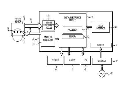
drugs that
may have externally impacted the central nervous system (CNS).
[003] Brain imaging studies, such as computed tomography (CT) and
magnetic resonance imaging (MRI), are widely used to visualize the structure
of the
brain. However, CT scan and MRI are anatomical tests and reveal very little
information about brain function. For example, intoxication, concussion,
active
seizure, metabolic encephalopathy, infections, and numerous other conditions
(e.g.
diabetic coma) show no abnormality on CT scan. A classical stroke, or a
traumatic
brain injury (TBI), may not be immediately visualized by an imaging test even
if there
is a clear and noticeably abnormal brain function. Similarly, diffuse axonal
injury
(DAD, related to shearing of nerve fibers which is present in majority of
concussive
brain injury cases, can remain invisible on most routine structural images. If
1
CA 02784267 2012-06-13
WO 2011/084394
PCT/US2010/060170
undetected at an early stage, swelling or edema from DAI can subsequently lead
to
coma and death.
[004] Functional MRI (fMRI) is a recent improvement over MRI, which
provides relative images of the concentration of oxygenated hemoglobin in
various
parts of the brain. While the concentration of oxygenated hemoglobin is a
useful
indication of the metabolic function of specific brain regions, it provides
very limited
information about the underlying electrochemical processes within the brain.
[005] Further, CT and MRI/fMRI testing devices are not field-deployable due
to their size, power requirements and cost. These assessment tools play an
important role in selected cases, but they are not universally available,
require
experienced personnel to operate, and they do not provide critical information
at the
early stages of acute neurological conditions. Current technologies are unable
to
provide the immediate information critical to timely intervention, appropriate
triage, or
the formulation of an appropriate plan of care for acute brain trauma.
Unfortunately,
the brain has very limited capacity for repair, and thus time-sensitive triage
and
intervention is very important in treating brain injuries.
[006] Currently, emergency room patients with altered mental status, acute
neuropathy, or head trauma must undergo costly and time-consuming tests to
determine an appropriate course of treatment. Unfortunately, in many cases,
the
clinical condition of patients continue to deteriorate as they wait for
equipment to
become available or for specialists to interpret tests. The problem that faces
ER
physicians is that their resources are limited to a subjective physical exam
using a
flashlight and a reflex hammer, and all of the physician's decisions
concerning the
administration of emergency treatment, additional consultation by a
neurologist, or
patient discharge, are based on the results of this simplistic exam. Often, ER
2
CA 02784267 2012-06-13
WO 2011/084394
PCT/US2010/060170
patients are sent for imaging studies, yet many functional brain
abnormalities, as
discussed earlier, are not visible on a CT scan or MRI. Some abnormalities
which
eventually have anatomical and structural consequences often take time to
become
visible on an imaging test. This is true for many important conditions, such
as
ischemic stroke, concussion/traumatic brain injury (TBI), raised intracranial
pressure,
and others. This indicates the need for real-time, functional brain state
assessment
technology, which can be performed in the ER, or in an ambulatory setting, and
can
detect emergency neurological conditions hours ahead of the standard clinical
assessment tools available today. Similarly, there is a need for a point-of-
care
assessment tool for detection of TBI in soldiers out in the battlefield, and
also for
detection of sports-related brain injury in athletes. Rapid, on-the-field
assessments
may help prevent repeat injuries and "second impact syndrome" in soldiers and
athletes already suffering from a first traumatic brain impact.
[007] All of the brain's activities, whether sensory, cognitive, emotional,
autonomic, or motor function, is electrical in nature. Through a series of
electro-
chemical reactions, mediated by molecules called neurotransmitters, electrical
potentials are generated and transmitted throughout the brain, traveling
continuously
between and among the myriad of neurons. This activity establishes the basic
electrical signatures of the electroencephalogram (EEG) and creates
identifiable
frequencies which have a basis in anatomic structure and function.
Understanding
these basic rhythms and their significance makes it possible to characterize
the brain
electrical signals as being within or beyond normal limits. At this basic
level, the
electrical signals serve as a signature for both normal and abnormal brain
function.
Just as an abnormal electrocardiogram (ECG) pattern is a strong indication of
a
3
CA 02784267 2012-06-13
WO 2011/084394
PCT/US2010/060170
particular heart pathology, an irregular brain wave pattern is a strong
indication of a
particular brain pathology.
[008] Even though EEG-based neurometric technology is accepted today in
neurodiagnostics, application in the clinical environment is notably limited.
Some of
the barriers limiting its adoption include: the cost of EEG equipment, the
need for a
skilled technician to administer the test, the time it takes to conduct the
test, and the
need for expert interpretation of the raw data. The instrument produces
essentially
raw waveforms which must be carefully interpreted by an expert. Data is
collected
and analyzed by an EEG technician, and is then presented to a neurologist for
interpretation and clinical assessment. This makes the currently available EEG
equipment unfeasible for neuro-triage applications in emergency rooms or at
other
point-of-care settings. More importantly, the current technology is not field-
portable
which makes it unfeasible for various field applications, e.g., at a battle
field, or a
sports event. Thus, there is an immediate need for a brain state assessment
technology for providing rapid, point-of-care neurological triage and
treatment
guidance for patients with acute brain injury or disease, so as to prevent
further brain
damage and disability.
Summary of the Invention
[009] The present disclosure addresses the need for point-of-care neuro-
triage by providing a portable device for rapid, real-time evaluation of the
brain
electrical signals of a patient. A first aspect of the present disclosure
includes a
method for performing neurological triage on a patient. The method comprises
the
steps of providing a patient sensor comprising at most eight disposable
neurological
electrodes and at least one ear phone, acquiring spontaneous brain electrical
signals
4
CA 02784267 2012-06-13
WO 2011/084394
PCT/US2010/060170
using the neurological electrodes, providing a handheld base unit comprising a
signal processor, the base unit being operatively coupled to the patient
sensor for
processing the acquired spontaneous brain electrical signals, and further
calculating
an index indicating a statistical probability of the patient's brain
electrical signals
being normal or abnormal using discriminant classification analysis, and
displaying
the index on the handheld base unit.
[010] Another aspect of the present invention comprises an apparatus for
performing neurological triage on a patient. The apparatus comprises a patient
sensor comprising at most eight disposable neurological electrodes and at
least one
ear phone, and a handheld base unit operatively connected to the patient
sensor.
The base unit further comprises a digital signal processor configured to
perform
automatic identification and removal of artifacts from acquired spontaneous
brain
electrical signals, discriminant-based classification using pre-selected
subsets of
quantitative signal features, and calculating an index capable of indicating a
statistical probability of the patient's brain electrical signals being normal
or
abnormal. Additionally, the base unit comprises a display panel to display the
index.
[011] It is to be understood that both the foregoing general description and
the following detailed description are exemplary and explanatory only and are
not
restrictive of the invention, as claimed.
[012] The accompanying drawings, which are incorporated in and constitute
a part of this specification, illustrate embodiments of the invention and
together with
the description, serve to explain the principles of the various aspects of the
invention.
CA 02784267 2012-06-13
WO 2011/084394
PCT/US2010/060170
Brief Description of Drawings
[013] FIG. 1A shows an exemplary embodiment of a neuro-assessment
apparatus for acquiring and processing brain electrical signals;
[014] FIG. 1B shows the placement of electrodes on a subject's forehead, in
accordance with the International 10/20 electrode placement system;
[015] FIG. 2A illustrates a graphic data summary displayed on the screen of
a neuro-assessment apparatus in accordance with an exemplary embodiment of the
present disclosure;
[016] FIG. 2B illustrates the display of detailed date regarding quantitative
signal features on the screen of a neuro-assessment apparatus in accordance
with
an exemplary embodiment of the present disclosure;
[017] FIG. 2C illustrates Z-score Normalized Maps displayed on the screen
of a neuro-assessment apparatus in accordance with an exemplary embodiment of
the present disclosure; and
[018] FIG. 3 shows a flowchart diagramming the steps of performing
neurological triage on a subject using a handheld device in accordance with an
exemplary embodiment of the present disclosure.
Detailed Description
[019] Reference will now be made in detail to embodiments consistent with
the present invention, examples of which are illustrated in the accompanying
drawings. Wherever possible, the same reference numbers will be used
throughout
the drawings to refer to the same or like parts.
[020] In an exemplary embodiment, data corresponding to brain electrical
activity is used to detect acute neurological injury or disease in patients.
The brain
6
CA 02784267 2012-06-13
WO 2011/084394
PCT/US2010/060170
electrical signals are measured and analyzed at the point-of-care using a
portable
neuro-triage device developed using BxTM technology, so as to provide an
immediate
evaluation of the subject's neurological condition. In accordance with an
exemplary
embodiment of the BxTM technology, a subject's brain electrical activity is
recorded
using a varying number of electrodes located at standardized positions on the
scalp
and forehead, and the subject's brain electrical signals are assessed with
reference
to one or more databases. For example, collected normative data, indicative of
normal brain electrical activity, is used to establish quantitative features
of brain
electrical activity, which clearly distinguish brain signals produced in the
presence
and absence of acute neurological disorder. This normative dataset includes
brain
activity data of a control group of population. A normative population in the
database
comprises of individuals similar to a subject in one or more aspects, such as
age,
gender, etc. In one exemplary embodiment, a subject is compared to individuals
in
the database using a regression equation as a function of age. The collected
normative database employed by the inventor has been shown to be independent
of
racial background and to have extremely high test-retest reliability,
specificity (low
false positive rate) and sensitivity (low false negative rate).
[021] In accordance with embodiments consistent with the present
disclosure, FIG. 1A shows a neuro-assessment apparatus for acquiring and
processing brain electrical signals using BxTM technology, and providing an
evaluation of the subjects neurological condition. In an exemplary embodiment,
the
neuro-assessment apparatus is implemented as a portable device for point-of-
care
applications. This apparatus consists of a patient sensor 40 which may be
coupled to
a base unit 42, which can be handheld, as illustrated in FIG. 1A. Patient
sensor 40
may include an electrode array 35 comprising at least one disposable
neurological
7
CA 02784267 2012-06-13
WO 2011/084394
PCT/US2010/060170
electrode to be attached to a patient's head to acquire brain electrical
signals. The
electrodes are configured for sensing both spontaneous brain activity as well
as
evoked potentials generated in response to applied audio stimuli. In one
exemplary
embodiment, the apparatus comprises of five (active) channels and three
reference
channels. The electrode array 35 consists of anterior (frontal) electrodes:
Fl, F2, F7,
F8, AFz (also referred to as Fz') and Fpz (reference electrode) to be attached
to a
subject's forehead, and electrodes Al and A2 to be placed on the front or back
side
of the ear lobes, or on the mastoids, in accordance with the International
10/20
electrode placement system (with the exception of AFz). The electrode
placement is
illustrated in FIG. 1B. The use of a limited number of electrodes enable rapid
and
repeatable placement of the electrodes on a subject, which in turn facilitates
efficient,
and more accurate, patient monitoring. Further, in one embodiment, the
electrodes
may be positioned on a low-cost, disposable platform, which can serve as a
"one-
size-fits-all" sensor. For example, electrodes 35 may be positioned on a head
gear
that is configured for easy and/or rapid placement on a patient. Other
electrode
configurations may be utilized as and when required, as would be understood by
those of ordinary skill in the art.
[022] In an exemplary embodiment, the neuro-assessment device utilizes
the advantages of auditory evoked potential (AEP) signals to map specific
auditory,
neurological and psychiatric dysfunctions. In such an embodiment, the patient
sensor 40 includes reusable earphone 31 to provide auditory stimuli clicks in
either
ear. In some embodiments, the auditory evoked potential signal used is
auditory
brainstem response (ABR). In such embodiments, the auditory stimuli may be
delivered at 100 dB Peak-to-Peak Equivalent Sound Pressure Level and at a
frequency (rate) of 27 Hz (27 clicks per second) to evoke electrical signals
from the
8
CA 02784267 2012-06-13
WO 2011/084394
PCT/US2010/060170
brainstem in response to the applied auditory stimuli. In another embodiment,
patient
sensor 40 may include an additional ear phone to deliver white noise in the
other
ear.
[023] The patient sensor 40 also includes two reusable patient interface
cables which are designed to plug into the base unit 42 and provide direct
communication between the patient sensor 40 and the base unit 42. The first
cable is
an electrical signal cable 41a, which is equipped with standard snap
connectors to
attach to the disposable electrodes placed on the patient's scalp. The second
cable
is the AEPstimulus cable 41b which provides connection to the earphone 31 for
auditory stimulus delivery. Other auditory stimuli may also be used, to evoke
mid-
latency (20-80 milliseconds) or late auditory responses (>80 milliseconds),
including
the P300.
[024] The base unit 42 primarily includes an analog electronics module 30, a
digital electronics module 50, user interface 46, stimulus generator 54 and
battery 44
as illustrated in FIG. 1A. The analog electronics module receives signals from
one or
more of the neurological electrodes operatively connected through the
electrical
cable 41a. The analog module is configured to amplify, filter, and preprocess
the
analog waveforms acquired from the electrode channels. The analog module may
comprise signal amplifier channels including at least one preamplifier, at
least one
differential amplifier, at least one common mode detector, and at least one
gain
stage with filter. The analog amplifier channels correspond to the number of
electrode channels. In an embodiment consistent with the present disclosure,
there
are 8 analog amplifier channels corresponding to 8 electrode channels (5
active, 3
reference channels). The analog module 30 may further include a multiplexer
(MUX),
which combines many analog input signals and outputs that into a single
channel,
9
CA 02784267 2012-06-13
WO 2011/084394
PCT/US2010/060170
and an analog-to-digital converter (ADC) to digitized the received analog
signal.
Digital electronics module 50 can then process the digitized data acquired
through
analog module 30 and can perform analysis of data to aid in interpretation of
data
pertaining to brain electrical activity. Further, as shown in FIG. 1A, the
digital
electronics module 50 may be operatively connected with a number of additional
device components.
[025] In an exemplary embodiment, the analog electronics module 30 is
further configured to check an impedance by feeding a signal back into each
electrode. Checking an impedance may function to characterize the
effectiveness of
connection of a surface electrode to a subject. This would enable an user to
test the
applied electrodes at a patient site before signal acquisition is started, and
also
monitor the electrode impedance continuously in real-time throughout the test.
In an
exemplary embodiment, the impedance of the applied electrodes are measured
periodically and the impedance values are displayed on the user interface 46
using a
color-coded electrode visual display, which allows the user to monitor the
electrode
impedances before and during a test session. If an impedance value is found to
be
unacceptable at the time of the measurement, a warning indication may be
displayed
on the screen instructing the user to check the electrode connection.
[026] The digital electronics module 50 comprises a digital signal processor
(DSP) 51 for processing the data corresponding to the acquired brain
electrical
signals, and a memory 52 which stores the instructions for processing the
data, such
as a DSP algorithm. The processor 51 is configured to perform the following
tasks-
[027] a) Automatic identification and removal of several types of signal
artifacts from the acquired spontaneous brain electrical signal data;
[028] b) Extraction of linear and non-linear signal features; and
CA 02784267 2012-06-13
WO 2011/084394
PCT/US2010/060170
[029] c) Linear and non-linear discriminant analysis-based classification
using pre-selected subsets of age-normalized features (z-scores).
[030] In some embodiments, the processor 51 is further configured to
process the acquired auditory evoked potential signals. For example, in some
embodiments, processor 51 is configured for reconstruction of acquired ABR
waveforms, removal of epochs containing artifacts, filtering, synchronized
averaging
and computation of Fsp, which is a measure of reconstructed signal quality.
Similarly, in some embodiments, processor 51 is configured to process other
auditory evoked potentials.
[031] The processor 51 is configured to implement the DSP algorithm to
identify data that is contaminated by non brain-generated artifacts, such as
eye
movements, electromyographic activity (EMG) produced by muscle tension, spike
(impulse), external noise, etc., as well as unusual electrical activity of the
brain not
part of the estimation of stationary background state. Artifact identification
is
performed using as input the signals from the five active leads Fp1, Fp2, F7,
F8, AFz
referenced to linked ears (A1+A2)/2, and sampled at 100 Hz. In one embodiment,
incoming data epochs of 2.56 seconds (256 samples per epoch) are split into 8
basic
data units (sub-epochs) of length 320 ms (32 data points per sub-epoch).
Artifact
identification is done on a per-sub-epoch basis and guard bands are
implemented
around identified artifact segments of each type. Artifact-free epochs are
then
constructed from at most two continuous data segments, with each data segment
being no shorter than 960 ms (which corresponds to the time span of 3
contiguous
sub-epochs). The resulting artifact-free data is then processed to extract
signal
features and classify the extracted features to provide a triage result.
11
CA 02784267 2016-12-16
[032] In another embodiment, signal denoising is performed using a signal
processing method described in commonly-assigned U.S. Patent Application
Publication No. 2009/0263034. In one embodiment consistent with the present
disclosure, the artifact identification and rejection algorithm follows the
following steps:
a. Transforming the signal into a plurality of signal components;
b. Computing fractal dimension of the components;
c. Identifying noise components based on their fractal dimension;
d. Automatically attenuating the identified noise components;
e. Reconstructing a denoised signal using inverse transform.
[033] The input analog brain electrical signal is at first digitized and then
deconstructed into its constitutive coefficients using a linear or non-linear
signal
transformation method, such as Fast Fourier Transform, Independent Component
Analysis (ICA)-based transform, wavelet transform, wavelet packet transform
etc.
The fractal dimensions of the coefficients are then calculated in the
transform
domain, and the coefficients that have a fractal dimension higher than a
preset
threshold value are attenuated. The intact and re-scaled coefficients are then
remixed using an inverse signal transform to generate a denoised signal, which
is
further processed to extract signal features and classify the extracted
features.
[034] Processor 51 is configured to execute instructions contained in
memory 52 to perform an algorithm for quantitative feature extraction from
processed signals. In one embodiment, the algorithm extracts various
quantitative
features from the brain wave frequency bands: Delta (1.5-3.5 Hz), Theta (3.5-
7.5
Hz), Alpha (7.5-12.5 Hz), Alpha1 (7.5-10 Hz), Alpha2 (10-12.5 Hz), Beta (12.5-
25
Hz), Beta2 (25-35 Hz), Gamma (35-50 Hz), and high frequency EEG (>50 Hz). In
12
CA 02784267 2012-06-13
WO 2011/084394
PCT/US2010/060170
some embodiments, the features computed are: absolute power, relative power,
mean frequency, coherence, symmetry, fractal dimension, wavelet features, and
several statistical harmonics variables. The feature extraction algorithm
takes as
input a number of "artifact-free" or "denoised" epochs having a temporal
length of
2.56 seconds, which corresponds to 256 samples for data sampled at 100 Hz. In
an
exemplary embodiment, processor 51 is configured to perform a linear feature
extraction algorithm based on Fast Fourier Transform (FFT). In another
embodiment,
processor 51 is configured to perform a non-linear feature extraction
algorithm based
on wavelet transforms, such as Discrete Wavelet Transform (DINT), Complex
Wavelet Transforms (Cl/VT), Biorthogonal Discrete Wavelet Transform (BDVVT),
Wavelet Packet Decomposition, etc. A full set of monopolar and bipolar
features are
calculated and then transformed for Gaussianity. Once a Gaussian distribution
has
been demonstrated and age regression applied, statistical Z transformation is
performed to produce Z-scores. The Z-transform is used to describe the
deviations
from age expected normal values:
[035] Z= Probability that subject value lies within the normal range
Z= Subiect Value - Norm for Age
Standard Deviation for Age
[036] The Z-scores are calculated for each feature and for each electrode
using a database of response signals from a large population of subjects
believed to
be normal, or to have other pre-diagnosed conditions. In particular, each
extracted
feature is converted to a Z-transform score, which characterizes the
probability that
the extracted feature observed in the subject will conform to a normal value.
[037] Processor 51 is further configured to perform a discriminant-based
classification algorithm wherein the extracted features, or the Z-scores, are
classified. In one embodiment, the classification is performed using Linear
13
CA 02784267 2012-06-13
WO 2011/084394
PCT/US2010/060170
Discriminant Analysis (LDA), which optimally combines the features (Z-scores)
into a
discriminant score that possesses the maximum discriminating power. Linear
Discriminant Analysis is a two category classifier, such as a classification
between
normal and abnormal, which assigns for each given subject a discriminant score
between 1 and 100. For example, the discriminant scores, SN and SAB
corresponding
to classes "normal" and "abnormal", are computed for any subject with the
following
Fisher LDA formulas:
[038] SN= 100.G(1)/(G(1) + G(2)), SAS= 100.G(2)/(G(1)+G(2))
G(1)= exp(Z .WN + CN), G(2)= exp(Z .WAB + CAB)
[039] where Z denote the set of age-regressed Z-transformed features
(discriminants) computed for any subject. WN and WAB denote two weight vectors
that are derived from a stored reference database, and CN and CAB are two
constants which are commonly called bias or threshold weights. The weights for
the
different monopolar and/or bipolar univariate and multivariate features are
pre-
selected using a training routine such that they result in the 'best'
separation
between the classes. The weights may be estimated from a stored population
reference database, such as a database comprising of population normative data
indicative of brain electrical activity of a first plurality of individuals
having normal
brain state, or population reference data indicative of brain electrical
activity of a
second plurality of individuals having an abnormal brain state. Similarly, the
weights
may be selected from a database of the subjects own brain electrical activity
data
generated in the absence or presence of an abnormal brain state. In some
embodiments, the classification task may be performed using one or more
14
CA 02784267 2016-12-16
discriminant functions, and in such a case, the discriminant outputs may be
combined using a majority voting rule.
[040] In an exemplary embodiment, processor 51 is configured to calculate
an index indicating a statistical probability of the patients brain electrical
signals
being normal or abnormal, known as the "Probability of Normal" index. In
certain
embodiments, the "Probability of Normal" index is calculated from the
discriminant
score using Receiver Operating Characteristics (ROC) curves and Classification
Accuracy Curves (CAC), as described in U.S, Application Publication No.
2010/0191139. Using this method, the "Probability of
Normar for any normal/abnormal classification can be derived, which is an
integer in
the range 0-100. Additionally, in some embodiments, processor 51 is configured
to
identify one or more features making the largest contribution to the
"Probability of
Normal" classification statement (whenever the index is smaller than 10% or
larger
than 90%).
[041] In addition to the acquisition and processing of spontaneous brain
electrical signals, the device collects auditory evoked potential response
data. For
example, in some embodiments, the device collects auditory brainstem response
(ABR) data and displays the averaged ABR waveforms. For each of the two
modalities ("Left ABR," and "Right ABR"), raw data is collected for
approximately 2.5
minutes (corresponding to 4096 raw ABR epochs). The ABR waveform is
constructed and displayed for lead AFz only, using contra-lateral referencing,
which
means that for the "Left ABR" modality where the acoustic stimulus is in the
left ear,
the device computes and displays the ABR waveform for the signal AFz A2 (by
synchronized averaging of artifact-free epochs). Similarly, for the Right ABR
modality, the waveform for AFz ¨ Al is computed and displayed. At the end of
the
CA 02784267 2012-06-13
WO 2011/084394
PCT/US2010/060170
ABR data acquisition process, the device computes and displays the Fsp next to
the
waveform. For the computation of the ABR waveform the following processing
steps
are performed: bandpass-filtering of raw ABR epochs, rejection of artifacted
("over-
range") epochs, followed by Bayesian averaging of the remaining artifact-free
epochs. Optionally, adaptive filtering may be performed for ABR waveform
reconstruction.
[042] The memory 52 may further contain interactive instructions for using
and operating the device to be displayed on a screen of the user interface 46.
The
instructions may comprise an interactive feature-rich presentation including a
multimedia recording providing audio/video instructions for operating the
device, or
alternatively simple text, displayed on the screen, illustrating step-by-step
instructions for operating and using the device. The inclusion of interactive
instructions with the device eliminates the need for extensive training for
use,
allowing for deployment and use by persons other than medical professionals.
The
memory 52 may also contain a database, which includes the collected normative
data and reference data used for feature classification. In an exemplary
embodiment,
the database may be accessed from a remote storage device via a wireless or a
wired connection. Similarly, data collected from the subject by the neuro-
triage
apparatus may be recorded in the database for future reference.
[043] The neuro-triage device can be a standalone system or can operate in
conjunction with a mobile or stationary device to facilitate display or
storage of data,
and to signal healthcare personnel when therapeutic action is needed, thereby
facilitating early recognition of emergency conditions. Mobile devices can
include,
but are not limited to, handheld devices and wireless devices distant from,
and in
communication with, the neuro-triage device. Stationary devices can include,
but are
16
CA 02784267 2012-06-13
WO 2011/084394
PCT/US2010/060170
not limited to, desktop computers, printers and other peripherals that display
or store
the results of the neurological evaluation. In an exemplary embodiment, the
neuro-
triage device stores each patient file, which includes a summary of the
session and
test results, on a removable memory card 47, such as compact flash (CF) card.
The
user can then use the memory card 47 to transfer patient information and
procedural
data to a computer, or to produce a printout of the data and session summary.
In
another embodiment, results from the processor 51 are transferred directly to
an
external mobile or stationary device to facilitate display or storage of data.
For
example, the results from the processor 51 may be displayed or stored on a PC
48
connected to the base unit 42 using a PC interface, such as an USB port, IRDA
port,
BLUETOOTHO or other wireless link. In yet another embodiment, the results can
be
transmitted wirelessly or via a cable to a printer 49 that prints the results
to be used
by attending medical personnel. n an embodiment consistent with the present
disclosure, user interface 46 may be configured to communicate patient
information
and/or procedural data to an attending medical personnel, such as an ER
physician,
a triage nurse, or an emergency response technician. Information that is
conveyed
through user interface 46 can include a variety of different data types,
including, but
not limited to, diagnostic results, intermediate analysis results, usage
settings, etc. In
an exemplary embodiment, the user interface 46 displays the brain electrical
signal
graphs drawn in real-time for the five active and the three reference
channels, along
with the Right ABR waveform and Left ABR waveform graphs, Fsp value and the
actual number of clean epochs used for the computation of the ABR waveforms.
Additionally, as shown in FIG. 2A, the screen provides a graphic data summary,
which includes a Discriminant Classification screen showing a horizontal index
bar
with numerical indications of "Probability of Normal" (PoN). The display of
the index
17
CA 02784267 2012-06-13
WO 2011/084394
PCT/US2010/060170
is accompanied by a message which states the statistical interpretation of the
index.
In some exemplary embodiments, the index is an integer in the range 0-100 and
is
graphically represented by a black bar in a 0-100 scale. For patients whose
frontal
discriminant score is defined by a PoN greater than 10% and lesser that 90%,
the
device displays a Data Classification message notifying the user that the
discriminant score does not allow a confident determination of the presence of
abnormality. The user interface 46 further displays a Detailed Data screen, as
illustrated in FIG. 2B, which provides access to detailed data about the
features that
made the largest contribution to the abnormal classification. From this
screen, the
user would be able to access tabular screens showing values of the
quantitative
features and the Z-scores for each feature extracted from the artifact-free
data
epochs. The Detailed Data screen allows the user to select tables for Absolute
Power, Relative Power, Mean Frequency, Coherence, Symmetry, Fractal Dimension
and Harmonics Statistics. The frequencies included in this tables are Delta
(1.5-3.5
Hz), Theta (3.5-7.5 Hz), Alpha (7.5-12.5 Hz), Beta (12.5-35 Hz), Gamma (35-50
Hz),
and S (1.5-25 Hz). From the Detailed Data screen, the user will also be able
to
navigate to the Z-scores Normalized Maps screen, which is shown in FIG. 2C.
This
screen gives the user an option to view a graphical representation of a pre-
selected
subset of Z-scores on a "frontal head map" where Z-scores are color-coded to
show
deviation from normal. Additionally, in some embodiments, the user interface
46
gives the user the option to view and statistically compare multiple sessions
of an
individual for the purpose of treatment evaluation or progression of disorder.
[044] In another exemplary embodiment, user interface 46 may receive and
display usage setting information, such as the name, age and/or other
statistics
pertaining to the patient. The user interface 46 comprises a touchscreen
interface for
18
CA 02784267 2016-12-16
entering the user input A virtual keypad may be provided on the touchscreen
interface for input of patient record fields. Additionally, as shown in FIG.
2A, the
battery charge status may be indicated continuously on the display, along with
the
available memory status of the CF card, and the electrode impedance values.
Further, the neuro-assessment device can transmit data to another mobile or
stationary device to facilitate more complex data processing or analysis. For
example, the neuro-assessment device, operating in conjunction with PC 48, can
send data to be further processed by the computer. In another embodiment
consistent with the Bem technology, the processor 50 transmits a raw,
unprocessed
signal acquired from a subject to PC 48 for analyzing the recorded data and
outputting the results. The unprocessed brain electrical signals recorded from
a
subject may also be stored in a remote database for future reference and/or
additional signal processing.
[045] In an embodiment consistent with the present disclosure, the base unit
42 includes a stimulus generator 54, which is operatively coupled to the
processor
51, for applying auditory stimuli to the subject to elicit ABR waveforms, The
stimulus
generator 54 interfaces with earphone 31 positioned in proximity to the
patient's ear
to deliver auditory stimuli that can generate evoked potentials. The processor
51
then removes artifacts from the received evoked potential signala and displays
the
artifact-free waveforms, as described earlier in this paper. Additionally,
base unit 42
contains an internal rechargeable battery 44 that can be charged during or in
between uses by battery charger 39 connected to an AC outlet 37.
[046] The neuro-assessment apparatus, developed in accordance with the
Bxn" technology, is designed for near-patient testing in emergency rooms,
ambulatory setting, and other field applications. The neuro-assessment device
is
19
CA 02784267 2012-06-13
WO 2011/084394
PCT/US2010/060170
intended to be used in conjunction with CT scan, MRI or other imaging studies
to
provide complementary or corroborative information about a patient's
neurological
condition. The key objective of point-of-care neuro-assessment is to provide
fast
results indicating the severity of a patient's neurological condition, so that
appropriate treatment can be quickly provided, leading to an improved overall
clinical
outcome. For example, the neuro-assessment device may be used by an EMT, ER
nurse, or any other medical professional during an initial patient processing
in the ER
or ambulatory setting, which will assist in identifying the patients with
emergency
neurological conditions. It will also help ER physicians in corroborating an
immediate
course of action, prioritizing patients for imaging, or determining if
immediate referral
to a neurologist or neurosurgeon is required. This in turn will also enable ER
personnel to optimize the utilization of resources (e.g., physicians' time,
use of
imaging tests, neuro consults, etc.) in order to provide safe and immediate
care to all
patients.
[047] In addition, the neuro-assessment device is designed to be field-
portable, that is, it can be used in locations far removed from a full-service
clinic¨for
example, in remote battlefield situations distant from military healthcare
systems,
during sporting events for indentifying if an injured athlete should be
transported for
emergency treatment, at a scene of mass casualty in order to identify patients
who
need critical attention and immediate transport to the hospital, or at any
other remote
location where there is limited access to well-trained medical technicians.
[048] FIG. 3 shows a flowchart diagramming the steps of performing
neurological triage on a subject using a handheld device, in accordance with
an
embodiment of the present invention, and will be described in conjunction with
FIG. 1
to illustrate the method. The electrodes 35 are first placed on the head of
the patient
CA 02784267 2012-06-13
WO 2011/084394
PCT/US2010/060170
(step 301). The handheld base 42 unit is then powered on using power supplied
by
the battery 44. The processor 51 executes instructions stored in the memory 52
to
display instructions for operating the device. An user can use the user
interface 46 to
enter a command to start signal acquisition. If the user determines that
auditory
evoked potentials (for example, ABR signals) may also have to be recorded
(step
302), he may initiate stimulus generator 54 and apply auditory stimuli to
elicit evoked
potential responses (step 303). Brain electrical signals, which may include
the
spontaneous brain electrical signals and the evoked potential waveforms, are
acquired using electrodes 35 (step 304 and 305), and the signals are then
amplified
and digitized in the handheld base unit 42 (step 306). The processor 51 is
configured
for processing the signal (i.e. feature extraction and discriminant-based
classification) (step 308) using instructions stored in memory 52. The user
interface
46 then displays a horizontal index bar with numerical indications of
"Probability of
Normal" (PoN) (step 310), which specifies the statistical value of probability
of a
patient's brain electrical signals being normal or abnormal. If an user wants
to view
the detailed data showing quantitative features and Z-scores (step 312), he
may
navigate to the Detailed Data screen (step 314) which shows the quantitative
features that made the most contribution towards the determination of PoN.
From
this screen the user will also be able to access table sets showing values of
quantitative features and Z-scores. The user will also be given an option to
see a Z-
score Normalized Maps (step 316), which shows a graphical representation of a
pre-
selected subset of Z-scores on a "frontal head map". Following the display of
the
result indicating the probability of brain activity being normal or abnormal,
and
optionally the detailed data showing the values of quantitative features and Z-
scores
Normalized Maps, the user may terminate the test and incorporate the result
with
21
CA 02784267 2012-06-13
WO 2011/084394
PCT/US2010/060170
data from other clinical tests, or he may repeat the test session to perform
additional
evaluation.
[049] Embodiments consistent with the present disclosure, using advanced
signal processing algorithms and stored data of the brain activity of
thousands of
subjects having different neurological indications, may provide a rapid and
accurate
assessment of the brain state of a subject. The advanced signal processing
algorithms may be executed by a processor capable of integration in a portable
handheld device. The portable handheld device used with a reduced electrode
set
allows for a rapid, on-site neurological triage, and determining an
appropriate course
of treatment at the early stage of an injury or other acute neurological
disorder
requiring immediate medical attention.
[050] Other embodiments of the invention will be apparent to those skilled in
the art from consideration of the specification and practice of the invention
disclosed
herein. It is intended that the specification and examples be considered as
exemplary only, with a true scope and spirit of the invention being indicated
by the
following claims.
22