Note: Descriptions are shown in the official language in which they were submitted.
CA 2789782 2017-04-13
W02011/075651 PCT/US2010/061042
PATENT
Docket No. 27793W002
LIMITED ECHELETTE LENS, SYSTEMS AND METHODS
10
BACKGROUND OF THE INVENTION
[0002] Embodiments of the present invention relate to vision treatment
techniques and in
particular, to ophthalmic lenses such as, for example, contact lenses, corneal
inlays or onlays, or
intraocular lenses (IOLs) including, for example, phakic IOLs and piggyback
IOLs (i.e. 10Ls
implanted in an eye already having an 10L).
[0003] Presbyopia is a condition that affects the accommodation properties of
the eye. As
objects move closer to a young, properly functioning eye, the effects of
ciliary muscle
contraction and zonular relaxation allow the lens of the eye to change shape,
and thus increase its
optical power and ability to focus at near distances. This accommodation can
allow the eye to
focus and refocus between near and far objects.
[0004] Presbyopia normally develops as a person ages, and is associated with a
natural
progressive loss of accommodation. The presbyopic eye often loses the ability
to rapidly and
CA 02784782 2012-06-15
WO 2011/075651 PCT/US2010/061042
Docket No. 27793W002
easily refocus on objects at varying distances. The effects of presbyopia
usually become
noticeable after the age of 45 years. By the age of 65 years, the crystalline
lens has often lost
almost all elastic properties and has only limited ability to change shape.
[0005] Along with reductions in accommodation of the eye, age may also induce
clouding of
the lens due to the formation of a cataract. A cataract may form in the hard
central nucleus of the
lens, in the softer peripheral cortical portion of the lens, or at the back of
the lens. Cataracts can
be treated by the replacement of the cloudy natural lens with an artificial
lens. An artificial lens
replaces the natural lens in the eye, with the artificial lens often being
referred to as an
intraocular lens or "IOL".
[0006] Monofocal IOLs are intended to provide vision correction at one
distance only, usually
the far focus. Predicting the most appropriate IOL power for implantation has
limited accuracy,
and an inappropriate IOL power can leave patients with residual refraction
following surgery.
Accordingly, it may be necessary for a patient who has received an IOL implant
to also wear
spectacles to achieve good far vision. At the very least, since a monofocal
IOL provides vision
treatment at only one distance and since the typical correction is for far
distance, spectacles are
usually needed for good near vision and sometimes intermediate vision. The
term "near vision"
generally corresponds to vision provided when objects are at a distance from
the subject eye of
between about 1 to 2 feet are substantially in focus on the retina of the eye.
The term "distant
vision" generally corresponds to vision provided when objects at a distance of
at least about 6
feet or greater are substantially in focus on the retina of the eye. The term
"intermediate vision"
corresponds to vision provided when objects at a distance of about 2 feet to
about 5 feet from the
subject eye are substantially in focus on the retina of the eye.
[0007] There have been various attempts to address limitations associated with
monofocal
IOLs. For example, multifocal IOLs have been proposed that deliver, in
principle, two foci, one
near and one far, optionally with some degree of intermediate focus. Such
multifocal or bifocal
IOLs are intended to provide good vision at two distances, and include both
refractive and
diffractive multifocal IOLs. In some instances, a multifocal IOL intended to
correct vision at
two distances may provide a near add power of about 3.5 or 4.0 diopters.
[0008] Multifocal IOLs may, for example, rely on a diffractive optical surface
to direct
portions of the light energy toward differing focal distances, thereby
allowing the patient to
clearly see both near and far objects. Multifocal ophthalmic lenses (including
contact lenses or
2
CA 02784782 2012-06-15
WO 2011/075651 PCT/US2010/061042
Docket No. 27793W002
the like) have also been proposed for treatment of presbyopia without removal
of the natural
crystalline lens. Diffractive optical surfaces, either monofocal or
multifocal, may also be
configured to provide reduced chromatic aberration.
[0009] Diffractive monofocal and multifocal lenses can make use of a material
having a given
refractive index and a surface curvature which provide a refractive power.
Diffractive lenses
have a diffractive profile which confers the lens with a diffractive power
that contributes to the
overall optical power of the lens. The diffractive profile is typically
characterized by a number
of diffractive zones. When used for ophthalmic lenses these zones are
typically annular lens
zones, or echelettes, spaced about the optical axis of the lens. Each
echelette may be defined by
an optical zone, a transition zone between the optical zone and an optical
zone of an adjacent
echelette, and an echelette geometry. The echelette geometry includes an inner
and outer
diameter and a shape or slope of the optical zone, a height or step height,
and a shape of the
transition zone. The surface area or diameter of the echelettes largely
determines the diffractive
power(s) of the lens and the step height of the transition between echelettes
largely determines
the light distribution between the different add powers. Together, these
echelettes form a
diffractive profile.
[0010] A multifocal diffractive profile of the lens may be used to mitigate
presbyopia by
providing two or more optical powers; for example, one for near vision and one
for far vision.
The lenses may also take the form of an intraocular lens placed within the
capsular bag of the
eye, replacing the original lens, or placed in front of the natural
crystalline lens. The lenses may
be in the form of a contact lens, most commonly a bifocal contact lens, or in
any other form
mentioned herein.
[0011] Although multifocal ophthalmic lenses lead to improved quality of
vision for many
patients, additional improvements would be beneficial. For example, some
pseudophakic
patients experience undesirable visual effects (dysphotopsia), e.g. glare or
halos. Halos may
arise when light from the unused focal image creates an out-of-focus image
that is superimposed
on the used focal image. For example, if light from a distant point source is
imaged onto the
retina by the distant focus of a bifocal IOL, the near focus of the IOL will
simultaneously
superimpose a defocused image on top of the image formed by the distant focus.
This defocused
image may manifest itself in the form of a ring of light surrounding the in-
focus image, and is
referred to as a halo. Another area of improvement revolves around the typical
bifocality of
3
CA 02784782 2012-06-15
WO 2011/075651 PCT/US2010/061042
Docket No. 27793W002
multifocal lenses. Since multifocal ophthalmic lenses typically provide for
near and far vision,
intermediate vision may be compromised.
[0012] A lens with an extended depth of focus may provide certain patients the
benefits of
good vision at a range of distances, while having reduced or no dysphotopsia.
Various
techniques for extending the depth of focus of an IOL have been proposed. For
example, some
approaches are based on a bulls-eye refractive principle, and involve a
central zone with a
slightly increased power. Other techniques include an asphere or include
refractive zones with
different refractive zonal powers.
[0013] Although certain proposed treatments may provide some benefit to
patients in need
thereof, still further advances would be desirable. For example, it would be
desirable to provide
improved JUL systems and methods that confer enhanced image quality across a
wide and
extended range of foci without dysphotopsia. Embodiments of the present
invention provide
solutions that address the problems described above, and hence provide answers
to at least some
of these outstanding needs.
BRIEF SUMMARY OF THE INVENTION
[0014] Embodiments of the present invention generally provide improved lenses
and imaging
techniques. Exemplary embodiments provide improved ophthalmic lenses (such as,
for example,
contact lenses, corneal inlays or onlays, or intraocular lenses (IOLs)
including, for example,
phakic IOLs and piggyback IOLs) and associated methods for their design and
use.
[0015] Embodiments of the present invention encompass IOL optics having a
circular surface
structure with one to four echelettes surrounding the surface structure. The
profile is designed
such that it increases the depth of focus of the pseudophakic eye, where the
natural crystalline
lens of the eye is substituted with a synthetic lens. Such limited ring IOL
techniques suppress
the distinct bifocality associated with traditional multifocal IOLs which have
many diffractive
rings. Consequently, dysphotopsia (e.g., halo effects) associated with
traditional multifocal IOLs
can be alleviated by lenses according to embodiments of the present invention.
[0016] An exemplary limited ring JUL includes an anterior face and a posterior
face. A profile
can be imposed on the anterior or posterior surface or face. The profile can
have an inner portion
and an outer portion. The inner portion typically presents a parabolic curved
shape. The inner
4
CA 02784782 2012-06-15
WO 2011/075651 PCT/US2010/061042
Docket No. 27793W002
portion may also be referred to as a microstructure, or a central or inner
echelette. Between the
inner portion and the outer portion, there may be a transition zone that
connects the inner and
outer portions. The outer portion may be comprised of four or fewer
echelettes.
[0017] In addition to parabolic shapes, the central/ inner echelette can have
any of a variety of
shapes including hyperbolic, spherical, aspheric, and sinusoidal. The
transition between the
inner and outer portions of the central/ inner echelette can be a sharp
transition, or it can be a
smooth transition.
[0018] The surface of the outer portion at the outside of the microstructure
can have any
spherical or aspherical shape and is comprised of a limited number of
echelettes, preferably less
than four. The shape of the outer portion can be optimized for having the
desired optical
performance for a range of pupil sizes. The desired optical performance can be
based on
elements such as the depth of focus, the optical quality in the far focus, and
the change in best
focus (or far focus) position as a function of the pupil size. Optimization
rules may be applied as
if the shape were a refractive monofocal IOL, or a refractive IOL having an
extended depth of
focus, or a refractive design that corrects or modifies the ocular spherical
aberration. Specific
designs can be made in which the interplay between the central echelette and
the outer zone is
incorporated in the design or optimization. The techniques described herein
are well suited for
implementation with any of a variety of ophthalmic lenses. including IOLs,
corneal inlays or
onlays, and/or contact lenses.
[0019] In one aspect, embodiments of the present invention encompass
ophthalmic lens
systems and methods for treating an eye of a patient. An exemplary lens may
include an anterior
face with an anterior refractive profile and a posterior face with a posterior
refractive profile.
The faces may be disposed about an optical axis. The lens may also include a
diffractive profile
imposed on the anterior refractive profile or the posterior refractive
profile. In some cases, the
diffractive profile may include no more than 5 echelettes. Optionally, the
central echelette can
be disposed within a central zone of the lens. Relatedly, the central
echelette may be disposed
within an annular ring surrounding a central refractive zone of the lens. In
some cases, the lens
includes a peripheral zone with a limited number of echelettes that surround
the central echelette
or annular ring. The limited number of echelettes may be characterized by a
constant phase shift.
[0020] According to some embodiments, an ophthalmic lens can include a limited
number of
echelettes that are characterized by parabolic curves. The central echelette
can have a diameter
5
CA 02784782 2012-06-15
WO 2011/075651 PCT/US2010/061042
Docket No. 27793W002
within a range from about 1 mm to about 4 mm. For example, the central
echelette may have a
diameter of about 1.5 mm. In some cases, the central echelette can have a
diameter within a
range from about 1.0 mm to about 5.0 mm. Lens embodiments may include a
peripheral portion
comprised of a limited number of echelettes and a refractive portion. Central
and peripheral
echelettes can have a surface area that is between 1 and 7 mm2. For example,
the echelettes may
have a surface area that is 2.3 mm2. In some cases, a lens may include a
peripheral portion
which surrounds the echelettes. A lens may include a peripheral portion having
an outer
diameter within a range from about 4 mm to about 6 mm. In some cases, the
peripheral portion
will have an outer diameter within a range of about lmm to about 7mm. For
example, a lens
may include a peripheral portion having an outer diameter of about 5 mm.
[0021] The echelettes may be characterized by a step height having a value
within a range
from about 0.5 pm and about 4 m. According to some embodiments, a transition
can be
characterized by a step height having a value within a range of about 1.5 in
and 2.5 ium.
According to some embodiments, a transition can be characterized by a step
height having a
value of about 1.7 1J m. In other embodiments, the step height may have a
value of about 2.0 ium.
[0022] Optionally, a diffractive profile can be characterized by a design
wavelength, and a lens
can include a transition characterized by a step height producing a phase
shift between about
0.25 and about 1 times the design wavelength. In some cases, a diffractive
profile can be
characterized by a design wavelength, and the lens can include a transition
characterized by a
step height producing a phase shift between about 0.15 and about 2 times the
design wavelength.
[0023] In some aspects, embodiments of the present invention encompass systems
and
methods involving an ophthalmic lens that include an anterior face with an
anterior refractive
profile and a posterior face with a posterior refractive profile, such that
the faces are disposed
about an optical axis, and a diffractive profile imposed on the anterior
refractive profile or the
posterior refractive profile, such that the diffractive profile includes an
inner echelette and four
or fewer outer echelettes. According to some embodiments, an inner echelette
can be disposed
within a central zone of the lens. In some cases, an inner echelette can be
disposed within an
annular ring surrounding a central zone of the lens. Optionally, an inner
echelette and outer
echelettes can be characterized by a parabolic curve. In some cases, an inner
echelette and outer
echelettes can be characterized by a constant phase shift. According to some
embodiments, an
ophthalmic lens may include an accommodating lens and/or a multifocal lens.
6
CA 02784782 2012-06-15
WO 2011/075651 PCT/US2010/061042
Docket No. 27793W002
[0024] For a fuller understanding of the nature and advantages of the present
invention,
reference should be had to the ensuing detailed description taken in
conjunction with the
accompanying drawings.
BRIEF DESCRIPTION OF THE DRAWINGS
[0025] FIG. lA is a cross-sectional view of an eye with a multifocal
refractive intraocular lens.
[0026] FIG. 1B is a cross-sectional view of an eye having an implanted
multifocal diffractive
intraocular lens.
[0027] FIG. 2A is a front view of a diffractive multifocal ophthalmic lens.
[0028] FIG. 2B is a cross-sectional view of the lens of FIG. 2A.
[0029] FIGS. 3A-3B are a graphical representations of a portion of the
diffractive profile of a
conventional diffractive multifocal lens.
[0030] FIG. 4 shows aspects of the central echelette of a lens according to
embodiments of the
present invention.
[0031] FIG. 4A-4E illustrates aspects of a lens profile according to
embodiments of the present
invention.
[0032] FIG.5 shows aspects of calculated defocus curves according to a central
echelette
embodiment.
[0033] FIG.6 shows aspects of calculated defocus curves according to a
embodiments of the
present invention.
[0034] For illustration purposes, the profile geometries shown in certain
aforementioned
figures were not drawn exactly to scale. The heights of the profiles shown in
the figures are
generally on the order of about 0.1 lam to about 8.0 'um although the heights
may vary depending
on factors such as the amount of correction needed by the patient, the
refractive index of the lens
material and surrounding medium, and the desired distribution of light between
wanted
diffraction orders.
7
CA 02784782 2012-06-15
WO 2011/075651 PCT/US2010/061042
Docket No. 27793W002
DETAILED DESCRIPTION OF THE INVENTION
[0035] It is to be understood that the figures and descriptions of the present
invention have
been simplified to illustrate elements that are relevant for a clear
understanding of the present
invention, while eliminating, for the purpose of clarity and brevity, many
other elements found in
typical ophthalmic lenses, implantable optic apparatuses, systems and methods.
Those of
ordinary skill in the art may thus recognize that other elements and/or steps
are desirable and/or
required in implementing the present invention. However, because such elements
and steps are
well known in the art, and because they do not facilitate a better
understanding of the present
invention, a discussion of such elements and steps is not provided herein. The
disclosure herein
is directed to all such variations and modifications to the disclosed elements
and methods known
to those skilled in the art.
[0036] Embodiments of the present invention encompass systems and methods that
provide
improved image quality over an extended range of focal points or foci. Systems
and methods
disclosed herein can encompass various ophthalmic lenses such as, for example,
contact lenses,
intraocular lenses, spectacle lenses, and corneal inlays or onlays. Exemplary
embodiments
include ophthalmic lenses having an extended depth of focus, as compared to
conventional
monofocal lenses, and reduced dysphtopsia as compared to conventional
multifocal ophthalmic
lenses. In some cases, such techniques involve an IOL approach that includes a
limited number
of rings or echelettes, and typically involves an expanded depth of focus.
Advantageously, such
approaches can provide a patient with good distance vision, as well as good
vision at
intermediate distances without dysphotopsia.
[0037] Embodiments of the present invention generally provide improved lenses
and imaging
systems and may be incorporated into any system in which a lens with an
extended depth of
focus may be advantageous, such as camera/ video lenses, including those used
for surveillance
or for surgical procedures, as well as for cameras in mobile phones or other
related devices.
Embodiments of the invention may find their most immediate use in the form of
improved
ophthalmic devices, systems, and methods. Exemplary embodiments of the present
invention
provide improved ophthalmic lenses (including, for example contact lenses,
intraocular lenses
(IOLs), corneal implants and the like) and associated methods for their design
and use.
Embodiments of the present invention may be used with monofocal diffractive or
refractive
lenses, bifocal diffractive or refractive lenses, and multifocal diffractive
or refractive lenses, e.g.
embodiments of the present invention could be added to the opposite surface of
multifocal IOLs,
8
CA 02784782 2012-06-15
WO 2011/075651 PCT/US2010/061042
Docket No. 27793W002
e.g. TECNIS Multifocal or REZOOM or RESTOR IOLs. In other words, an extended
depth of
focus feature may be added to, for example the opposite surface of a
diffractive or refractive
multifocal embodiment.
[0038] In addition, an extended depth of focus feature may be added to, for
example, a toric
IOL, an IOL that modifies ocular spherical and/or chromatic aberration, and/or
an
accommodating IOL. In general, an extended depth of focus feature may be added
to an IOL
that modifies ocular aberrations.
[0039] Reading is often done in bright light conditions in which the pupil is
small. In contrast,
night-time driving is done in low light conditions in which the pupil is
large. Embodiments of
the present invention encompass lenses that relatively emphasize intermediate
or near vision for
small pupil sizes, while also relatively emphasizing far vision for large
pupil sizes. In some such
ophthalmic lenses, a greater proportion of light energy may be transmitted to
the far focus from a
peripheral portion of the lens to accommodate for low light, far viewing
conditions such as night
time driving, with the near or intermediate viewing receiving relatively more
light energy from a
central portion of the diffractive profile ¨ for reading or computer work for
example and/or to
provide depth of focus and intermediate or near viewing under low light
reading conditions as in
for example reading restaurant menus.
[0040] FIG. lA is a cross-sectional view of an eye E fit with a multifocal IOL
11. As shown,
multifocal IOL 11 may, for example, comprise a bifocal IOL. Multifocal IOL 11
receives light
from at least a portion of cornea 12 at the front of eye E and is generally
centered about the
optical axis of eye E. For ease of reference and clarity, FIGS. lA and 1B do
not disclose the
refractive properties of other parts of the eye, such as the corneal surfaces.
Only the refractive
and/or diffractive properties of the multifocal IOL 11 are illustrated.
[0041] Each major face of lens 11, including the anterior (front) surface and
posterior (back)
surface, generally has a refractive profile, e.g. biconvex, piano-convex,
piano-concave, meniscus,
etc.. The two surfaces together, in relation to the properties of the
surrounding aqueous humor,
cornea, and other optical components of the overall optical system, define the
effects of the lens
11 on the imaging performance by eye E. Conventional, monofocal IOLs have a
refractive
power based on the refractive index of the material from which the lens is
made, and also on the
curvature or shape of the front and rear surfaces or faces of the lens.
9
CA 02784782 2012-06-15
WO 2011/075651 PCT/US2010/061042
Docket No. 27793W002
[0042] In a young, healthy eye contraction and relaxation of ciliary muscles
17 surrounding the
capsular bag 14 contribute to accommodation of the eye, the process by which
the eye increases
optical power to maintain focus on objects as they move closer. As a person
ages, the degree of
accommodation decreases and presbyopia, the diminished ability to focus on
near objects, often
results. A patient may therefore conventionally use corrective optics having
two optical powers,
one for near vision and one for far vision, as provided by multifocal JUL 11.
[0043] Multifocal lenses may optionally also make special use of the
refractive properties of
the lens. Such lenses generally include different powers in different regions
of the lens so as to
mitigate the effects of presbyopia. For example, as shown in FIG. 1A, a
perimeter region of
refractive multifocal lens 11 may have a power which is suitable for viewing
at far viewing
distances. The same refractive multifocal lens 11 may also include an inner
region having a
higher surface curvature and a generally higher overall power (sometimes
referred to as a
positive add power) suitable for viewing at near distances.
[0044] Rather than relying entirely on the refractive properties of the lens,
multifocal
diffractive IOLs or contact lenses can also have a diffractive power, as
illustrated by the JUL 18
shown in FIG. 1B. The diffractive power can, for example, comprise positive or
negative add
power, and that add power may be a significant (or even the primary)
contributor to the overall
optical power of the lens. The diffractive power is conferred by a plurality
of concentric
diffractive zones which form a diffractive profile. The diffractive profile
may either be imposed
on the anterior face or posterior face or both.
[0045] The diffractive profile of a diffractive multifocal lens directs
incoming light into a
number of diffraction orders. As light 13 enters from the front of the eye,
the multifocal lens 18
directs light 13 to form a far field focus 15a on retina 16 for viewing
distant objects and a near
field focus 15b for viewing objects close to the eye. Depending on the
distance from the source
of light 13, the focus on retina 16 may be the near field focus 15b instead.
Typically, far field
focus 15a is associated with Oth diffractive order and near field focus 15b is
associated with the
1St diffractive order, although other orders may be used as well.
[0046] Multifocal ophthalmic lens 18 typically distributes the majority of
light energy into the
two viewing orders, often with the goal of splitting imaging light energy
about evenly
(50%:50%), one viewing order corresponding to far vision and one viewing order
corresponding
to near vision, although typically, some fraction goes to non-viewing orders.
CA 02784782 2012-06-15
WO 2011/075651 PCT/US2010/061042
Docket No. 27793W002
[0047] In some embodiments, corrective optics may be provided by phakic IOLs,
which can be
used to treat patients while leaving the natural lens in place. Phakic IOLs
may be angle
supported, iris supported, or sulcus supported. The phakic IOL can be placed
over the natural
crystalline lens or piggy-backed over another IOL. It is also envisioned that
the present
invention may be applied to inlays, onlays, accommodating IOLs, spectacles,
and even laser
vision correction.
[0048] FIGS. 2A and 2B show aspects of a standard diffractive multifocal lens
20. Multifocal
lens 20 may have certain optical properties that are generally similar to
those of multifocal IOLs
11, 18 described above. Multifocal lens 20 has an anterior lens face 21 and a
posterior lens face
22 disposed about optical axis 24. The faces 21, 22 of lens 20 typically
define a clear aperture
25. As used herein, the term "clear aperture" means the opening of a lens or
optic that restricts
the extent of a bundle of light rays from a distant source that can be imaged
or focused by the
lens or optic. The clear aperture is usually circular and is specified by its
diameter, and is
sometimes equal to the full diameter of the optic.
[0049] When fitted onto the eye of a subject or patient, the optical axis of
lens 20 is generally
aligned with the optical axis of eye E. The curvature of lens 20 gives lens 20
an anterior
refractive profile and a posterior refractive profile. Although a diffractive
profile may also be
imposed on either anterior face 21 and posterior face 22 or both, FIG. 2B
shows posterior face
22 with a diffractive profile. The diffractive profile is characterized by a
plurality of annular
optical zones or echelettes 23 spaced about optical axis 24. While analytical
optics theory
generally assumes an infinite number of echelettes, a standard multifocal
diffractive IOL
typically has at least 9 echelettes, and may have over 30 echelettes. For the
sake of clarity, FIG.
2B shows only 4 echelettes. Typically, an IOL is biconvex, or possibly plano-
convex, or
convex-concave, although an IOL could be plano-plano, or other refractive
surface
combinations.
[0050] FIGS. 3A and 3B are graphical representations of a portion of a typical
diffractive
profile of a multifocal lens. While the graph shows only 3 full echelettes,
typical diffractive
lenses extend to at least 9 echelettes to over 32 echelettes. In FIG 3A, the
height of the surface
relief profile (from a plane perpendicular to the light rays) of each point on
the echelette surface
is plotted against the square of the radial distance (r2 or p) from the
optical axis of the lens. In
multifocal lenses, each echelette 23 may have a diameter or distance from the
optical axis which
11
CA 02784782 2012-06-15
WO 2011/075651 PCT/US2010/061042
Docket No. 27793W002
is often proportional to Ain, n being the number of the echelette 23 as
counted from optical axis
24. Each echelette has a characteristic optical zone 30 and transition zone
31. Optical zone 30
has a shape or downward slope that may be linear when plotted against p as
shown in FIG. 3A.
When plotted against radius r, optical zone 30 has a shape or downward slope
that is parabolic as
shown in FIG. 3B. As for the typical diffractive multifocal lens, as shown
here, all echelettes
have the same surface area. The area of echelettes 23 determines the add power
of lens 20, and,
as area and radii are correlated, the add power is also related to the radii
of the echelettes.
[0051] As shown in FIGS. 3A and 3B, transition zone 31 between adjacent
echelettes is sharp
and discontinuous. The height of the lens face sharply transitions from
sloping steadily
downwards to stepping vertically upwards, and the transitions abruptly back to
sloping steadily
downwards again. In doing so, echelettes 23 also have a characteristic step
height 32 defined by
the distance between the lowest point and height point of the echelette.
Hence, the slope (or first
derivative) and/or the curvature (second derivative) of the diffractive
surface are discontinuous
adjacent the transitions.
[0052] Structure of Central Echelette
[0053] FIG. 4 provides a graphical representation of a cross section of a
portion of an
exemplary lens illustrating the central echelette structure. The lens profile
200 has a ring
diameter of 1.21mm and a stepheight at 220 of 2.05 p m. corresponding with a
phase delay of 0.5
lambda (see table 2). In this example, the ring diameter was reduced from
1.5mm (which is the
inner ring diameter for a 2.0 Diopter conventional IOL diffractive lens) to
1.21mm by a scaling
factor Ai2, as described in patent US 5,121,980 (Cohen). Only the inner
portion and part of the
outer portion of half of the lens is shown, although since the lens is
rotationally symmetric, the
other half is a mirror image.
[0054] The adjacent echelette(s) in the outer portion (not shown) are detailed
below. Profile
200 includes an inner portion 210 or single ring, a step or transition 220,
and an outer portion
230. The outer portion 230 extends beyond that disclosed in FIG. 4F to 2.5mm
and may be
comprised of limited additional echelettes. Inner portion 210 extends between
a central location
210 of profile 200 and transition 220. Outer portion 230 extends between
transition 220 and a
peripheral location (not shown). In some cases, transition 220 can be disposed
at a distance from
the optical axis that is within a range from about 0.5 mm to about 2.0 mm, and
peripheral
location can be disposed at a distance from the optical axis that is within a
range from about 2.0
12
CA 2789782 2017-04-13
WO 2011/075651 PCT/US2010/061042
Docket No. 27793W002
to about 3.5 mm, or bigger (for example, for contact lenses, the ranges would
be approximately
15% larger due to the optically more powerful position of contact lens
compared to an IOL;
those skilled in the art would appropriately scale certain dimensions
depending on the
application).
[0055] The inner portion or echelette 210 includes a center 210a and a
peripheral edge 210b.
At center or central section 210a of inner portion 210 where radial distance
is zero, the sag (d) of
inner portion is between the sag (d) of the diffractive base curve 240 and the
sag (d) of the
peripheral curve 260 at 1.03 um from the peripheral curve 260, corresponding
with a phase delay
of 0.25 lambda (see table 2), At peripheral edge 210b, the sag (d) of inner
portion 210 is
substantially equivalent to the sag (d) of diffractive base curve 240 at 13.8
pm. The value of sag
(d) between radial distance zero and radial distance at the peripheral edge
210b at 0.61 mm,
gradually and smoothly changes from 1.03 um (at r=0) to the value of the base
curve 240 (at
1=0.61 mm) which is 13.8 gm. This change occurs in a parabolic fashion. As
shown here, inner
portion can present a parabolic shape, for example as described in Equation 4a
of Cohen,
IS Applied Optics, 31:19, pp. 3750-3754 (1992).
[0056] At the peripheral edge 210b where the radial distance (r) is 0.61 mm,
the value of sag
(d) steps or changes from the value of diffractive base curve 240 to the value
of peripheral curve
260. Where radial distance (r) corresponds to transition 220, sag (d) of inner
portion is
equivalent to the value of the diffractive base curve 240. Relatedly, the
displacement of the
profile approaches that of the diffractive base curve as the radial distance
increases from a value
of zero to a value of about 0.61 mm. The stepheight is 2.05 i.tm resulting in
a phase delay of 0.5.
[0057] The outer portion 230 includes an inner or central edge 230a and a
peripheral edge (not
shown). At inner edge 230a, the sag (d) of outer portion is substantially
equivalent to the sag (d)
of peripheral curve 260. At peripheral edge, the sag (d) of outer portion
remains substantially
equivalent to the sag (d) of peripheral curve 260. As detailed below, a
limited number of
echelettes may be located between inner edge 230a and peripheral edge.
[0058] FIG. 4A provides a graphical representation of a portion of a lens
diffractive profile
with a central echelette and one peripheral adjacent echelette according to
embodiments of the
present invention. In FIG. 4A, the height of the surface relief profile (from
a plane
perpendicular to the light rays) of each point on the echelettes surface is
plotted against the
distance from the optical axis of the lens. The echelettes can have a
characteristic optical zone
13
CA 02784782 2012-06-15
WO 2011/075651 PCT/US2010/061042
Docket No. 27793W002
930 and transition zone 931. Optical zone 930 can have a shape or downward
slope that may be
linear when plotted against p as shown in FIG. 4A. When plotted against radius
r, optical zone
930 can have a shape or downward slope that is parabolic. Central and
peripheral echelettes can
have a surface area that is between 1 and 7 mm2. For example, the echelettes
may have a surface
area that is 2.3 mm2. An outer (refractive) zone can follow the base radius
with a fixed offset.
[0059] As shown in FIG. 4A, transition zones 931 between the optical zones 930
and the
adjacent optical zones can be sharp and discontinuous. Similarly, a vertical
transition between
adjacent echelettes and also the peripheral portion or refractive zone can be
sharp and
discontinuous. The height of the lens face sharply transitions from sloping
steadily downwards
(e.g. across optical zones 930) to stepping vertically upwards (e.g. at
transition zone 931), and
the transitions abruptly back to sloping steadily downward or substantially
horizontal at outer
refractive zone. In doing so, echelette 930 also has a characteristic step
height 932 defined by
the distance between the lowest point and highest point of the echelette.
Hence, the slope (or
first derivative) and/or the curvature (second derivative) of the diffractive
surface are
discontinuous adjacent the transition. The first derivative can be indicated
by the direction of the
lines, and the second derivative can be indicated by the curve of the line.
[0060] According to some embodiments, light comes from below, in the direction
indicated by
arrow A, and only hits the echelettes 930 of the profile. According to some
embodiments, in
theoretical terms light does not hit the vertical connection of the optical
zones, and hence the
profile can be said to have no transition zone. According to some embodiments,
in practice
when one attempts to produce such a profile, for instance by lathe cutting, it
may be difficult to
reproduce the sharp corner (e.g. at where the optical zone connects with the
adjacent optical
zone) and hence the corner may be rounded to some extent due to the finite
chisel radius. Such
rounding may have a negligible effect on the optical performance. According to
related
embodiments, transition zone 931, which can be referred to as the transition
from the echelette to
the adjacent zone or zones, can be shaped in a specific way, so as to optimize
the optical
performance, for example to minimize scatter from a sharp transition.
[0061] PROFILE PARAMETERS
[0062] The profile design can be characterized in terms of a set of
parameters. For example.
the limited echelette profile can be described as having a central echelette
with a diameter and
surface area, an adjacent echelette(s) with the same surface area, and an
associated stepheight at
14
CA 02784782 2012-06-15
WO 2011/075651 PCT/US2010/061042
Docket No. 27793W002
each transition resulting in a phase delay. The central echelette may have a
diameter within a
range from about 1 mm to about 5 mm. For example, the central echelette may
have a diameter
of about 1.5 mm. Central echelette may have a surface area that is between 1
and 7 mm2. For
example, the central echelette may have a surface area that is 2.3 mm2. The
peripheral
echelette(s) may have a surface area equal to the central echelette. In
particular, Table 1
discloses the dimensions of the radius and diameter of the central echelette,
along with the
surface area of the central and peripheral echelettes.
R (mm) De (mm) Area (mm2)
1.48 3 6.9
1.05 2.1 3.5
0.86 1.7 2.3
0.74 1.5 1.7
0.66 1.3 1.4
0.61 1.2 1.2
[0063] The step height or profile height can determine the phase delay or
phase shifting
profile. A greater step height can correspond to a greater phase shift.
According to some
embodiments, a lens can include a transition characterized by a step height
producing a phase
shift between about 0.25 and about 1 times the design wavelength. In some
cases, a diffractive
profile can be characterized by a design wavelength, and the lens can include
a transition
characterized by a step height producing a phase shift between about 0.15 and
about 2 times the
design wavelength. According to some embodiments the lens may include a
transition
characterized by a step height producing a phase shift of about 0.5. In other
embodiments, the
lens may include a transition characterized by a step height of about 0.4.
[0064] Table 2 provides dimensions of various samples disclosing the
relationship between
phase delay (in wavelengths) and step height (in um), as valid for an example
IOL material.
Table 2
Phase Delay Stepheight
0.896 3.68
0.700 2.87
0.590 2.42
0.509 2.09
0.500 2.05
CA 02784782 2012-06-15
WO 2011/075651 PCT/US2010/061042
Docket No. 27793W002
0.423 1.74
0.366 1.50
0.350 1.44
0.250 1.03
0.150 0.62
[0065] FIG. 4B provides a graphical representation of a portion of a lens
diffractive profile
with a central echelette and two peripheral echelettes according to
embodiments of the present
invention. The height of the surface relief profile (from a plane
perpendicular to the light rays)
of each point on the echelettes surface is plotted against the distance from
the optical axis of the
lens. According to some embodiments, a lens with a central and peripheral
adjacent echelette, as
disclosed in FIG. 4A may also be comprised of an additional peripheral
echelette with a
refractive region between the outermost echelette and the interior echelettes.
[0066] FIG. 4C also details a portion of a lens diffractive profile with a
central echelette and
two peripheral echelettes. In this embodiment, however, the refractive zone is
immediately
adjacent to the central echelette and separates the central echelette from
three peripheral and
adjacent echelettes.
[0067] Although the above preferred embodiments disclose lenses with
echelettes that have
equal stepheights, lenses with echelettes with varying stepheights are also
covered herein as
detailed in FIG. 411. FIG. 4D. discloses a four echelette embodiment wherein a
refractive
region separates the central and adjacent echelette from three peripheral
adjacent echelettes. As
seen in FIG. 411, the stepheight (defined by the distance between the lowest
point and highest
point of the echelette) of the three outer echelettes is less than the
stepheight of the inner
echelettes. Of course, in addition to covering embodiments where the
stepheight of the outer
echelette(s) is less than the inner echelette(s), the stepheight of the inner
echelette(s) may be less
than the outer echelette(s). It is also foreseeable, that the inner and outer
echelettes may all have
varying stepheights whether the stepheights be increasing, decreasing, or
alternating.
[0068] FIG. 4E provides a graphical representation of a portion of a lens
diffractive profile
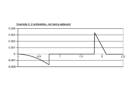
with a central echelette and a peripheral echelette which is not adjacent to
the central echelette.
The central echelette may have a shape or downward slope that is parabolic. A
refractive region
may then separate the central echelette from the peripheral echelette. The
peripheral echelette
may then be characterized by a sharp and discontinuous stepheight followed by
a downward
16
CA 02784782 2012-06-15
WO 2011/075651 PCT/US2010/061042
Docket No. 27793W002
slope. As in the embodiments above, a peripheral refractive region may
surround the outermost
echelette. Additionally, other exemplary embodiments include non-adjacent
echelette variations
analogous to FIG. 4A-4D. By way of non-limiting example, two echelettes that
are not
separated by a refractive region may also be non-adjacent.
[0069] Pupil Dependence
[0070] The size of the human pupil varies with illumination. In bright light
the pupil is small,
and in dim or low light conditions the pupil is large. In addition, the size
of the human pupil
varies with accommodative effort. Without accommodative effort, the pupil is
larger than with
accommodative effort. Hence, for a smaller pupil, it may be desirable to
provide a design that
places a relative emphasis on intermediate or near vision. For a larger pupil,
it may be desirable
to provide a design that places a relative emphasis on far vision.
[0071] In typical reading or near vision conditions where the light is bright,
the size of the
pupil is small, e.g. between about 1 mm and 2 mm in diameter, and the eye has
a large depth of
focus (for example from a pinhole effect), almost irrespective of the optics
of the IOL. When the
size of the pupil is large, e.g. larger than about 4-5 mm in diameter, the
situation generally
applies to low light conditions, and is often associated with distance vision
for which the power
of the IOL is typically established. Therefore, many patients would benefit
most from an IOL
that enhances the depth of focus in order to view at intermediate distances.
An IOL having a
central echelette with limited adjacent echelettes may effectively increase
the depth of focus for
intermediate pupil sizes, while maintaining the general increased depth of
focus of small pupil
sizes, and also maintaining an emphasis on far vision for large pupil sizes.
[0072] At the same time, since the limited echelettes and the remaining
surface area of the
optic or remaining lens portion ("non-echelette") have unequal surface areas
for almost all pupil
sizes, there is an incomplete split between the foci. The condition of
dysphotopsia (e.g. halos)
that is present for multifocal lenses is observed to be dominated by
separation of two foci and
pupil size effects. Accordingly, pursuant to exemplary embodiments of the
present invention,
the lens may include only a limited number of echelettes, so that light
separation between distinct
foci is not complete, as compared to standard diffractive multifocal IOLs.
Since the split of light
is incomplete, the separation of foci is incomplete. The incomplete separation
of foci contributes
to the extended depth of focus and the attenuation of dysphotopsia (e.g.
halos).
17
CA 02784782 2012-06-15
WO 2011/075651 PCT/US2010/061042
Docket No. 27793W002
[0073] In an exemplary embodiment, the limited echelette design has an optical
performance
that depends on the pupil size. For very small pupils, where the pupil is
smaller than the size of
the central and adjacent echelette(s), the echelette will act as a refractive
lens, having a very large
depth of focus due to the pinhole effect. For medium and higher pupil sizes,
where the pupil
covers the central echelette and the adjacent echelette, the lens will act as
a diffractive/refractive
lens, directing the light to several foci. For higher pupil sizes, more light
is being directed to the
lower order foci. The size of the central and adjacent echelette(s) influences
the pupil
dependence of the lens. As such, the size of the central and adjacent
echelette(s) can be chosen,
depending on the pupil sizes of a specific patient. For example, the pupil
sizes of a patient may
be measured in bright light, in dim light, during far vision and during near
vision, and in the
different combinations of light level and accommodative effort. These
different pupil sizes,
which may be defined as pupil dynamics, can be used as input parameters for an
optimal design
of the limited echelette design.
[0074] For example, if a patient has a pupil diameter during near vision (e.g.
viewing target at
close distance, with high accommodative effort) smaller than 2 mm, having this
pupil dimension
with both bright and dim light, then the size of the central and adjacent
echelette(s) may be
selected to be smaller than 2 mm (e.g. outer diameter of the adjacent
echelette of FIG. 4A), as to
provide adequate near and intermediate vision. Relatedly, if a patient has a
pupil diameter
during near vision larger than 2 mm, having this pupil dimension with both
bright and dim light,
then the size of the central and adjacent echelette(s) may be 2 mm or larger,
as to provide
adequate near and intermediate vision. In general, the diameter of the central
and adjacent
echelette(s) can be smaller than the smallest pupil size the patient has under
any condition (e.g.
bright/dim light; near/far vision). For any type of pupil dynamics, the size,
the profile, and the
offsets may be chosen to maximize the lens performance for that specific
patient, or group of
patients. Generally, this is a trade off between the different vision
circumstances (combinations
of light level and accommodative effort) at which the pupil of the patient is
measured.
Accordingly, exemplary embodiments include a method of designing an ophthalmic
lens
comprised of utilizing pupil size measurements and based on the measurements
determining the
size of an isolated echelette to impose on the surface of a lens. The pupil
size measurements
may be based on a group of patients.
[0075] EVALUATION OF VARIATIONS OF A SPECIFIC EXAMPLE
18
CA 02784782 2012-06-15
WO 2011/075651 PCT/US2010/061042
Docket No. 27793W002
[0076] FIGS. 5 and 6 show calculated defocus curves in the ACE eye model of an
embodiment with a central ring diameter of 1.48 mm, an echelette surface area
of 1.7 mm2, and a
phase delay of 0.4 wavelength. The horizontal axis denotes the defocus value
in the image
plane, in millimeters. Negative defocus values represent the myopic eye, and
therefore, simulate
vision at intermediate and near distances. The vertical axis denotes the
modulus (MTF) at 50
cycles per millimeter. Data for 5mm pupil diameters is included. FIG. 5 shows
the defocus
curve for an embodiment having only a single central echelette. FIG. 6 shows
an exemplary
embodiment as disclosed in section 4E, having, in addition to the central
echelette, a peripheral
echelette. The peripheral echelette and has a surface area of 3.5 mm2, and a
phase delay of 0.82
wavelength. The MTF at intermediate vision distances, with defocus values of
about -0.2mm to -
0.3mm, as shown in FIG. 6 is higher than the MTF at corresponding defocus
values in FIG 5.
As illustrated in the figures, a central plus peripheral echelette increases
the depth of focus as
compared to a central echelette only.
[0077] Embodiments of the present invention may be combined with a multifocal
lens design,
and with that extend the depth of focus of each focus of the multifocal lens.
Similarly,
embodiments of the present invention may be combined with an accommodating
lens design, by
which the range of accommodation of the accommodating lens can be extended. In
addition,
embodiments of the present invention may be combined with lenses correcting
ocular
aberrations, like toric lenses, aspherical lenses, lenses correcting chromatic
aberrations, and the
like.
[0078] Embodiments of the present invention may be combined with a lens design
correcting
chromatic aberrations. In one embodiment, the phase delay of the echelettes in
the preceding
examples is increased by a discrete multiple of wavelengths, in order to
correct for chromatic
aberration. For example, if a phase delay of 0.5 was used, corresponding to a
stepheight of 2.05
um, an alternative embodiment would have a phase delay of 1.5, corresponding
to a stepheight
of 6.15 lam. This embodiment directs the first order diffraction to the far
focus, and the second
order diffraction establishes the depth of focus at the intermediate and near
range.
[0079] While the exemplary embodiments have been described in some detail, by
way of
example and for clarity of understanding, those of skill in the art will
recognize that a variety of
modification, adaptations, and changes may be employed. Hence, the scope of
the claims should
not be limited to the description of the preferred versions contained herein.
19