Note: Descriptions are shown in the official language in which they were submitted.
CA 02785009 2012-06-19
1
DESCRIPTION
MEASURING APPARATUS AND SENSOR PLACEMENT METHOD
Technical Field
[0001] The present invention relates to a measuring apparatus and a sensor
placement method that are for measuring numerical information relating to a
substance contained in interstitial fluid or blood, and particularly for
measuring
glucose concentration.
Background Art
[0002] With conventional blood sugar level measurement, it is necessary to
puncture
the patient's body with an instrument called a lancet and take a blood sample
whenever measurement is carried out, and thus there is a problem in that a
large
burden is placed on the patient, and, furthermore, continuous measurement
cannot be
carried out. In order to solve such problems, a method of continuously
measuring
glucose concentration in subcutaneous tissue called CGM (Continuous Glucose
Monitoring) has been proposed in recent years.
[0003] With CGM, a sensor is disposed so as to be partially embedded under the
patient's skin, and the signal of a current value or the like that depends on
the
concentration of glucose in subcutaneous interstitial fluid is continuously
output by
this sensor. The signal is then converted to a blood sugar level by a
measurement
apparatus or the like. CGM enables blood sugar levels to be measured
continuously
(e.g., see Patent Document 1). Although interstitial fluid differs from blood,
it is
thought that the concentration of glucose in interstitial fluid reflects the
concentration
of glucose (blood sugar level) in blood. Therefore, blood sugar levels can be
known by
CA 02785009 2012-06-19
2
measuring the concentration of glucose in subcutaneous interstitial fluid.
[0004] Also, generally, the sensor, in order to be able to flexibly deal with
the body
movement of muscles and the like under the skin, is constituted by a flexible
strip-like
substrate or linear wire. In the case of the former, a sensor electrode that
outputs a
signal, a terminal for external connection, and wiring that connects the
sensor
electrode and the external connection terminal are formed on the substrate
(e.g., see
Patent Document 1).
[0005] Furthermore, since CGM requires that the sensor be partially implanted
under the patient's skin, Patent Document 1 discloses a device (implanting
device) that
is able to drive out the sensor toward the skin together with a puncture
needle, and
implant the sensor under the skin. The implanting device is provided with a
mechanism that drives out the sensor together with the puncture needle using a
spring or the like, and thereafter pulls back only the puncture needle. Here,
the
procedure for inserting the sensor disclosed in Patent Document 1 is
described.
[0006] First, a mount unit for mounting the sensor is disposed on the
patient's skin.
The implanting device in which the sensor and the puncture needle are set is
then
disposed on a prescribed position of the mount unit, and the sensor and the
puncture
needle are both driven under the skin by the implanting device. Thereafter,
the
puncture needle returns to its original position, and the sensor is disposed
with the
portion on which a terminal for connection is provided projecting above the
skin and
the remaining portion placed under the skin.
[0007] When the implanting device has been removed from the mount unit, a
control
unit for controlling the sensor is disposed on the mount unit. At this time,
the portion
of the sensor on which in the terminal is provided (terminal portion) is
sandwiched
between the mount unit and the control unit, and, at the same time, the
terminal of
CA 02785009 2012-06-19
3
the control unit and the terminal of the sensor that projects above the skin
are
connected.
[00081 If sensing by the sensor is performed in this state, the signal
obtained by the
sensor is converted to a digital signal by the control unit, and is
furthermore sent to an
external measurement apparatus by wireless or cable. The measurement apparatus
calculates the specific concentration of glucose from the received signal, and
displays
the calculated value on a display screen.
Citation List
Patent Documents
[00091 Patent Document 1: JP 2008-62072A (FIG. 11, FIG. 14, FIGS. 26.28D, FIG.
33)
Disclosure of the Invention
Problem to be Solved by the Invention
Incidentally, while the terminal portion of the sensor is, as mentioned above,
sandwiched between the mount unit and the control unit in order to connect the
terminal of the sensor and the terminal of the control unit, the sensor needs
to be
elastically deformed at this time. If the portion of the sensor embedded under
the
skin moves when the sensor is elastically deformed, the wound formed in the
skin by
the implanting device becomes bigger.
[00111 In such a case, since the body covers the sensor in protein in order to
heal the
wound, the sensor may not be able to output a signal, or a signal may be
output but
include noise, thus preventing accurate measurement. Furthermore, since the
sensor
thus covered in protein cannot be used, it needs to be removed and a new
sensor
CA 02785009 2012-06-19
4
reinserted, placing a not insignificant physically and financial burden on the
patient.
Since it is only when the control unit and the measurement apparatus are
operated
that it first becomes evident whether or not the sensor is outputting a
signal, the case
may also arise where the patient has to visit a medical facility again.
[00121 Also, while the sensor disclosed in Patent Document 1 is, as mentioned
above,
connected to the control unit by the terminal portion exposed outside the body
after
being placed under the skin, this connection process is performed by the user
himself
or herself (see FIG. 14 of Patent Document 1). For this reason, situations may
occur
where human operational error at the time of connection results in a load
being placed
on the portion of the sensor inserted under the skin or the wound formed in
the
insertion site being made bigger.
[00131 Since the body also covers the sensor in protein in these cases in
order to heal
the wound, the sensor may not be able to output a signal or a signal maybe
output but
include noise, thus preventing accurate measurement. Furthermore, since a new
sensor needs to be reinserted, the physically and financial burden placed on
the
patient is not insignificant. The case may also arise where the patient has to
visit a
medical facility again.
[00141 Also, given that the sensor has, for example, a full length of several
centimeters and a width of several millimeters, or is smaller in size than
this, the
external connection terminal of the sensor and the terminal of the control
unit are
minute. For this reason, a poor connection may occur between the sensor and
the
control unit during the above-mentioned connection process by the user.
Furthermore, the substrate on which the external connection terminal of the
sensor is
formed may also move due to movement of the body such as intense physical
activity,
also resulting in a poor connection between the sensor and the control unit.
In the
CA 02785009 2012-06-19
case where a poor connection such as this arises, the signal from the sensor
is not
transmitted to the control unit or, moreover, to the measurement apparatus,
rendering
measurement impossible.
[00151 An exemplary object of the present invention is to solve the above-
mentioned
5 problems, and to provide a measuring apparatus and a sensor placement method
that
enable situations where the function of an embedded sensor is impaired when
embedding the sensor under the skin and performing measurement to be
suppressed.
Means for Solving the Problem
[00161 In order to attain the above-mentioned object, the first measuring
apparatus
of the present invention is a measuring apparatus for measuring numerical
information relating to a substance contained in a body fluid within a body
that
includes a sensor unit and a control unit, the sensor unit including a sensor
that
generates a signal according to a state of the substance, a base that holds
the sensor,
and a variable mechanism that is attached to the base and enables at least one
of a
position and an orientation of the sensor to be changed, and the control unit
being
formed so as to be attachable to the base and executing processing after
receiving the
signal generated by the sensor.
[0017] With the first measuring apparatus in the present invention, the base
and the
sensor are thus attached via the variable mechanism. Thus, even if the base
moves
when attaching the control unit, external force generated thereby is absorbed
by the
variable mechanism and the occurrence of a situation where the sensor itself
moves is
suppressed. Furthermore, even if stress such as jarring or twisting occurs due
to
physical activity when the patient is wearing the sensor, the influence
exerted on the
embedded sensor is reduced. Thus, according to the first measuring apparatus
of the
CA 02785009 2012-06-19
6
present invention, the occurrence of a situation where the function of an
embedded
sensor is impaired when embedding the sensor under the skin and performing
measurement is suppressed.
[0018] Also, the first measuring apparatus of the present invention may adopt
a
mode in which the variable mechanism includes a ball joint, and a shaft at one
end of
the ball joint is attached to the sensor and a shaft at the other end of the
ball joint is
attached to the base. Furthermore, the first measuring apparatus of the
present
invention may adopt a mode in which the variable mechanism includes a rotating
member that is held in a rotatable state, and the rotating member is attached
to the
sensor. These modes enable external force to be efficiently absorbed with a
simple
configuration.
[0019] Also, in order to attain the above-mentioned object, a second measuring
apparatus of the present invention is a measuring apparatus for measuring
numerical
information relating to a substance contained in a body fluid within a body
that
includes a sensor unit and a control unit, the sensor unit including a sensor
that
generates a signal according to a state of the substance, a base that holds
the sensor,
and an external terminal that is provided in the base and directs the signal
generated
by the sensor to the outside, and the control unit being formed so as to be
attachable to
the base and executing processing after receiving the signal generated by the
sensor
via the external terminal.
[0020] According to the second measuring apparatus in the present invention,
the
sensor is thus connected to the control unit via the external terminal
provided in the
base. For this reason, the load placed on the portion of the sensor inserted
within the
body (e.g., under the skin) when connecting the sensor and the control unit is
reduced.
Also, a poor connection between the sensor and the control unit is less likely
to occur.
CA 02785009 2012-06-19
7
As a result, using the measuring apparatus, the sensor unit, and sensor
placement
apparatus of the present invention enables the occurrence of a loss of sensor
function
or a situation where measurement cannot be performed when embedding a sensor
within the body and performing measurement to be suppressed.
[0021] Also, the second measuring apparatus of the present invention
preferably
adopts a mode in which the sensor unit further includes a variable mechanism
that is
attached to the base and enables at least one of a position and an orientation
of the
sensor to be changed. With this mode, because external force exerted on the
sensor
and the control unit when they are being connected is absorbed by the variable
mechanism, the occurrence of a loss of sensor function is further suppressed.
[0022] Also, in order to attain the above-mentioned object, a first sensor
placement
method of the present invention is a method for placing a sensor within a
body, the
sensor generating a signal according to a state of a substance contained in a
body fluid
within the body, the method including the steps of (a) disposing a base on
skin, the
base being provided with an external terminal that directs the signal
generated by the
sensor to the outside, (b) partially implanting the sensor within the body,
and causing
the sensor to be held by the base, and (c) attaching a control unit to the
base, the
control unit executing processing after receiving the signal generated by the
sensor via
the external terminal.
[0023] With the above first sensor placement method, a variable mechanism that
enables at least one of a position and an orientation of the sensor to be
changed is
attached to the base.
[0024] The first sensor placement method may adopt a mode in which the
variable
mechanism includes a ball joint, and a shaft at one end of the ball joint is
attached to
the sensor and a shaft at the other end of the ball joint is attached to the
base.
CA 02785009 2012-06-19
8
[00251 Also, the first sensor placement method of the above may adopt a mode
in
which the variable mechanism includes a rotating member that is held in a
rotatable
state, and the rotating member is attached to the sensor.
[00261 Furthermore, the first sensor placement method of the above may adopt a
mode in which the step (b) comprises partially implanting the sensor within
the body,
at the same time as which the base and the sensor become electrically
connected.
[00271 Also, in order to attain the above-mentioned object, a second sensor
placement
method of the present invention is a method for placing a sensor within a
body, the
sensor generating a signal according to a state of a substance contained in a
body fluid
within the body, the method including the steps of (a) disposing a base on
skin in a
state where the sensor is held by the base, and partially implanting the
sensor within
the body, and (b) attaching a control unit to the base, the control unit
executing
processing after receiving the signal generated by the sensor.
[00281 With the above second sensor placement method, a variable mechanism
that
enables at least one of a position and an orientation of the sensor to be
changed maybe
attached to the base. In this case, a mode may be adopted in which the
variable
mechanism includes a ball joint, and a shaft at one end of the ball joint is
attached to
the sensor and a shaft at the other end of the ball joint is attached to the
base. Also, a
mode may be adopted in which the variable mechanism includes a rotating member
that is held in a rotatable state, and the rotating member is attached to the
sensor.
[00291 Furthermore, in the above second sensor placement method, an external
terminal that directs the signal generated by the sensor to the outside may be
provided
in the base, and the control unit may include a terminal that contacts with
the
external terminal included in the base. In this case, the step (b) comprises
connecting
the external terminal provided in the base and the terminal included in the
control
CA 02785009 2012-06-19
9
unit.
Effects of the Invention
[0030] As described above, a measuring apparatus and a sensor placement method
of
the present invention enable the occurrence of situations where the
performance of an
embedded sensor deteriorates when embedding the sensor under the skin and
performing measurement to be suppressed.
Brief Description of Drawings
[0031] FIG. 1 is a perspective view showing configurations of a measuring
apparatus
and a sensor unit in Embodiment 1 of the present invention.
FIG. 2 is a perspective view showing a tip portion of the sensor shown in FIG.
1.
FIG. 3A and FIG. 3B are diagrams showing a series of steps of a sensor
placement method in Embodiment 1 of the present invention.
FIG. 4A and FIG. 4B are diagrams showing a series of steps of the sensor
placement method in Embodiment 1 of the present invention, these steps being
executed after execution of the step shown in FIG. 3B.
FIG. 5 is a cross-sectional view showing an exemplary schematic configuration
of an implanting device used in implementation of the sensor placement method
in
Embodiment 1 of the present invention.
FIG. 6 is a perspective view showing a first exemplary configuration of a
sensor unit in Embodiment 2 of the present invention.
FIG. 7 is a perspective view showing a second exemplary configuration of the
sensor unit in Embodiment 2 of the present invention.
CA 02785009 2012-06-19
54712-2
FIG. 8 includes perspective views showing a configuration of a sensor unit in
Embodiment 3 of the present invention, FIG. 8A showing a state where the
sensor is
removed and FIG. 8B showing a state where the sensor is attached.
FIG. 9A and FIG. 9B are diagrams showing a series of steps of a sensor
5 placement method in Embodiment 3 of the present invention.
FIG. 10A and FIG. iOB are diagrams showing a series of steps of the sensor
placement method in Embodiment 3 of the present invention, these steps being
executed after execution of the step shown in FIG. 9B.
FIG. 11 is a perspective view showing a configuration of a measuring
10 apparatus in Embodiment 4 of the present invention.
FIG. 12A and FIG. 12B are diagrams showing a series of steps of a sensor
placement method in Embodiment 4 of the present invention.
FIG. 13A and FIG. 13B are diagrams showing a series of steps of the sensor
placement method in Embodiment 4 of the present invention, these steps being
executed after execution of the step shown in FIG. 12B.
FIG. 14 includes perspective views showing a configuration of a sensor unit in
Embodiment 5 of the present invention, FIG. 14A showing a state where the
sensor is
removed and FIG. 14B showing a state where the sensor is attached.
FIG. 15A and FIG. 15B are diagrams showing a series of steps of a sensor
placement method in Embodiment 5 of the present invention.
FIG. 16A and FIG. 16B are diagrams showing a series of steps of the sensor
placement method in Embodiment 5 of the present invention, these steps being
executed after execution of the step shown in FIG. 15B.
Best Mode for Carrying Out the Invention
CA 02785009 2012-06-19
11
[0032]Embodiment 1
Hereinafter, a measuring apparatus and a sensor placement method in
Embodiment 1 of the present invention are described, with reference to FIG. 1
to FIG.
3. Initially, configurations of a measuring apparatus 1 and a sensor unit 2 in
the
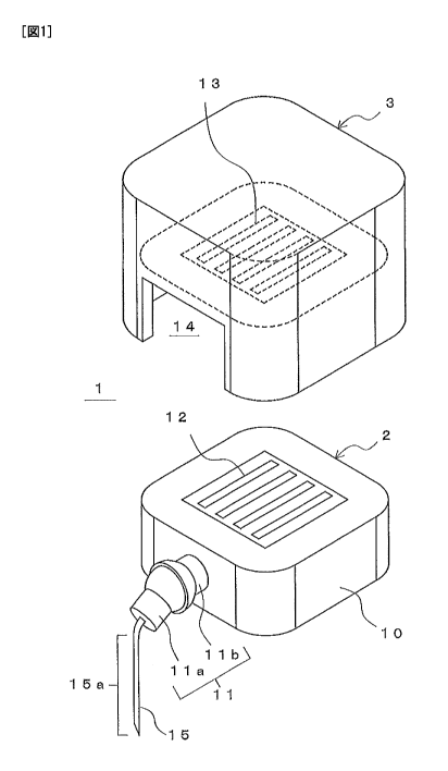
present Embodiment 1 are described using FIG. 1. FIG. 1 is a perspective view
showing the configurations of the measuring apparatus and the sensor unit in
Embodiment 1 of the present invention.
[00331 The measuring apparatus 1 shown in FIG. 1 is an apparatus that measures
numerical information relating to a substance contained in a body fluid within
the
body. As shown in FIG. 1, the measuring apparatus 1 is provided with the
sensor
unit 2 and a control unit 3. Note that examples of body fluid within the body
include
interstitial fluid, blood and plasma. Furthermore, in the present
specification, "within
the body" includes "under the skin" indicating below the skin surface.
[00341 The sensor unit 2 is provided with a base 10, a variable mechanism 11,
and a
sensor 15. Of these, the sensor 15 is placed partially under the skin, in
order to
execute CGM (see FIG. 4A and FIG. 4B discussed below). The sensor unit 2 will
also
function as a sensor placement apparatus for placing the sensor 15. Also, the
sensor
15 generates a signal according to the state of a substance in interstitial
fluid or blood.
[00351 The base 10 is disposed on the skin of the patient who is being
measured and
holds the sensor 15. The variable mechanism 11 is attached to the base 10, and
interposes between the base 10 and the sensor 15. Also, the variable mechanism
11
enables at least one of a position and an orientation of the sensor 15 to be
changed
based on the base 10. Note that the position and the orientation of the sensor
15
based on the base 10 denotes the relative position and relative orientation of
the sensor
to the base.
CA 02785009 2012-06-19
12
[0036] The control unit 3 receives the signal generated by the sensor 15 via
an
external terminal 12, and executes processing based on the received signal.
Also, the
control unit 3 is formed so as to be attachable to the base 10.
[0037] In the present Embodiment 1, the sensor 15 is thus held by the base 10
via the
variable mechanism 11. Therefore, even if the control unit 3 is attached in a
state
where the sensor 15 is partially embedded and the base 10 moves at that time,
the
external force generated thereby is absorbed by the variable mechanism 11,
preventing
the sensor 15 itself from moving.
[0038] Here, configurations of the measuring apparatus 1 in the present
Embodiment 1 and the sensor unit 2 and the control unit 3 constituting the
measuring
apparatus 1 are more specifically described using FIG. 2. FIG. 2 is a
perspective view
showing a tip portion of the sensor shown in FIG. 1.
[0039] In the present Embodiment 1, the sensor unit 2 includes the external
terminal
12. The external terminal 12 is provided in the base 10. Also, the external
terminal
12 is electrically connected to the sensor 15 as discussed later, enabling the
signal
generated by the sensor 15 to be directed to the outside. Furthermore, the
control
unit 3 receives the signal generated by the sensor 15 via the external
terminal 12. In
the present Embodiment 1, the signal generated by the sensor 15 is sent to the
control
unit 3 via the external terminal 12 provided in the base 10.
[0040] Also, in the present Embodiment 1, the substance that is measured is
glucose
in interstitial fluid, and the numerical information relating to the substance
is the
concentration of glucose. The sensor 15 generates a signal according to the
state
(concentration) of glucose in interstitial fluid. In the following, an example
is
described in which the numerical information relating to the substance is the
concentration of glucose, and the sensor 15 is a glucose sensor. Note that in
the
CA 02785009 2012-06-19
13
present Embodiment 1, the substance that is measured maybe a substance other
than
glucose, and maybe a substance in blood. Also, the numerical information may
be
information other than concentration.
[0041] Also, in the present Embodiment 1, the sensor 15 is able to
continuously
output a signal that depends on the state of glucose in interstitial fluid,
and allow the
measuring apparatus 1 to function as a monitoring apparatus capable of
continuously
monitoring the concentration of glucose. In this case, the measuring apparatus
1 is
able to perform the above-mentioned CGM.
[0042] As shown in FIG. 1 and FIG. 2, the sensor 15 is formed in a long, thin
belt-like
shape. Also, the sensor 15 is disposed on the patient's skin, in a state where
a portion
15a at the tip end is placed under the skin, using an implanting device
discussed later
(see FIG. 3A and B). The sensor 15 in such a state can also be said to be
implanted
under the skin.
[0043] Also, as shown in FIG. 2, the sensor 15 includes a substrate 18 having
insulating properties and flexibility. The formation material of the substrate
18 is not
particularly limited. In terms of having little effect on the body, however,
exemplary
formation materials of the substrate 18 include thermoplastic resins such as
polyethylene terephthalate (PET), polypropylene (PP) and polyethylene (PE) and
thermosetting resins such as polyimide resin and epoxy resin.
[0044] Furthermore, as shown in FIG. 2, the tip of the sensor 15 can be formed
into a
sharp point, in order to facilitate piercing the skin. The tip is, however,
not
particularly limited in shape, and maybe formed into a shape other than a
sharp point.
Also, in the present Embodiment 1, the sensor 15, being a glucose sensor,
includes an
electrode 16a and an electrode 16b forming a pair and a portion (enzyme
reagent layer)
17 on which glucose oxidoreductase is disposed, in addition to the substrate
18.
CA 02785009 2012-06-19
14
[0045] The electrode 16a and the electrode 16b are used in order to apply
voltage to
the enzyme reagent layer 17. The electrode 16a and the electrode 16b are
formed on
the surface of the substrate 18 in the longitudinal direction of the sensor
15, and also
function as wiring. The electrodes 16a and 16b can be formed, for example, by
performing vapor deposition or screen printing using a non-corrosiveness metal
or a
conductive material such as carbon ink.
[0046] The enzyme reagent layer 17, in the example of FIG. 2, is formed by
immobilizing glucose oxidoreductase on the electrode 16a. In this case, the
electrode
16a functions as the working electrode. A product produced by the reaction of
the
glucose oxidoreductase with the glucose (substrate) in interstitial fluid or
blood is
detected on the electrode, and electrons generated by the reaction are passed
directly
or via a mediator such as a metal complex to the electrode. Accordingly, when
voltage
is applied between the electrodes 16a and 16b, the electrons produced by the
enzyme
catalyst reaction can be detected with the electrode 16a, according to the
amount of
reaction of the glucose in the reaction.
[0047] In the present Embodiment 1, examples of applicable glucose
oxidoreductase
include glucose oxidase (GOD) and glucose dehydrogenase (GDH). Furthermore,
methods of immobilizing glucose oxidoreductase include various well-known
methods,
such as cross-linking using glutaraldehyde, for example.
[0048] Since the current value of current flowing through the electrode 16a
and the
electrode 16b changes according to the glucose concentration, such a
configuration
enables the glucose concentration to be specified by measuring this current.
In the
present embodiment, the current flowing through the electrode 16a and the
electrode
16b is equivalent to "the signal that depends on the state of the substance."
[0049] Also, the electrode 16a and the electrode 16b provided in the sensor 15
are
CA 02785009 2012-06-19
electrically connected to the external terminal 12 of the base 10, via wiring
provided
inside the variable mechanism 11 and the base 10 (not shown in FIG. 1 or FIG.
2).
The external terminal 12 is thereby able to direct the signal generated by the
sensor 15
to the outside.
5 [0050] In the present Embodiment 1, a ball joint is used as the variable
mechanism
11. A shaft 11a atone end of the balljoint is attached to the portion of the
sensor 15
that is not placed under the skin (portion other than the tip-end portion
15a), and a
shaft 11b at the other end of ball joint is attached to the base 10. In the
present
Embodiment 1, the variable mechanism 11 thus enables the orientation of the
sensor
10 15, or in other words, the orientation of the portion 15a at the tip end of
the sensor 15
to be changed.
[0051] Also, in the present Embodiment 1, the control unit 3 is provided with
a
recessed portion 14 into which the base 10 can be fitted. The control unit 3
is
attached to the base 10 by placing the control unit 3 over the base 10
disposed on the
15 skin, and housing the base 10 within the recessed portion 14. Also, a
terminal 13 for
connecting to the external terminal 12 is provided in the bottom surface
within the
recessed portion 14, and the external terminal 12 and the terminal 13 are
electrically
connected when the control unit 3 is attached to the base 10.
[0052] The control unit 3 receives the signal generated by the sensor 15 via
the
external terminal 12 and the terminal 13 contacting therewith. Specifically,
in the
present Embodiment 1, the control unit 3 applies voltage to the electrode 16a
and the
electrode 16b of the sensor 15, and monitors the current value of current
flowing
through the electrode 16a and the electrode 16b. Also, the control unit 3, as
arithmetic processing, generates an analog signal specifying the current value
and
converts the analog signal to a digital signal.
CA 02785009 2012-06-19
16
[0053] Thereafter, the control unit 3 transmits the generated digital signal
to an
external measurement apparatus by cable or wireless. The measurement
apparatus,
which is similar to a conventional apparatus, calculates the specific
concentration of
glucose from the received signal, and displays the calculated value on a
display screen.
[0054] Next, the sensor placement method in Embodiment 1 of the present
invention
is described using FIG. 3 to FIG. 5. FIG. 3A and FIG. 3B are diagrams showing
a
series of steps of the sensor placement method in Embodiment 1 of the present
invention. FIG. 4A and FIG. 4B are diagrams showing a series of steps of the
sensor
placement method in Embodiment 1 of the present invention, these steps being
executed after execution of the step shown in FIG. 3B. FIG. 5 is a cross-
sectional view
showing an exemplary schematic configuration of the implanting device used in
implementation of the sensor placement method in Embodiment 1 of the present
invention.
[0055] First, as shown in FIG. 3A, the sensor unit 2 to which the sensor 15 is
attached is set in an implanting device 41. The implanting device 41 is
disposed on
the patient's skin 40. The implanting device 41 is provided with the function
of
driving out the sensor unit 2 and the sensor 15 attached thereto toward the
skin 40
together with a puncture needle (not shown), using an elastic body such as a
spring.
[0056] Next, as shown in FIG. 3B, the sensor 15 attached to the base 10 is
driven out
toward the skin 40 by the implanting device 41 together with the puncture
needle (not
shown). At this time, the base 10 is also simultaneously sent toward the skin
40.
The portion 15a at the tip end of the sensor 15 is thereby embedded in the
skin 40
together with the puncture needle, and, at the same time, the base 10 is
disposed on
the skin 40.
[0057] The implanting device 41 also includes a mechanism for pulling back
only the
CA 02785009 2012-06-19
54712-2
17
puncture needle after driving out the sensor 15 and the puncture needle.
Therefore,
the puncture needle returns to its original position after piercing the skin
40, and only
the sensor 15 is placed under the skin. Note that, in the present Embodiment
1,
implantation of the portion 15a of the sensor 15 in the skin 40 and
disposition of the
base 10 on the skin 40 favorably are performed at the same time. It is
permissible,
however, for there to be a time lag between the implantation and the
disposition.
[0058] In the present Embodiment 1, the implanting device 41 need only be
provided
with the function of driving out the base 2, the sensor 15 and the puncture
needle
together, and the configuration thereof is not particularly limited.
Specifically,
examples of the implanting device 41 include an apparatus provided with a
similar
configuration to an apparatus shown in FIG. 7 to FIG. 12 of JP 2005-503243A.
[0059] Here, a specific example of the implanting device 41 is described using
FIG. 5.
As shown in FIG. 5, the implanting device 41 is provided with a body 43, an
extrusion
spring 44, a pair of guide rails 45, an extrusion member 46, a return spring
47, a
puncture needle 48, and a restriction member 49.
[0060] The body 43 is formed in a cylindrical shape open at one end. The guide
rails
45 are disposed in the longitudinal direction of the body. The extrusion
member 46 is
passed through by the guide rails 45 at two locations, and moves along the
guide rails
45. Also, the projecting restriction member 49 is provided near the opening
within
the body 43, and the movement of the extrusion member 46 is restricted.
[0061] Also, the extrusion spring 44 is installed between the extrusion member
46
and the wall surface of the body 43 on the blocked side, and the extrusion
member 46
is pushed toward the open side by the elastic force thereof. On the other
hand, the
return spring 47 is installed between the extrusion member 46 and the
restriction
member 49, and the extrusion member 46, having been pushed toward the open
side,
CA 02785009 2012-06-19
18
is pushed back toward its original position by the elastic force thereof.
[0062] The sensor unit 2 is disposed on the surface of the extrusion member 46
on the
open side. Also, although not illustrated in FIG. 5, a holding mechanism for
holding
the base 10 of the sensor unit 2 is provided in the extrusion member 46. The
holding
mechanism is configured such that holding of the base 10 is released when the
extrusion member 46 approaches furthest on the open side. Furthermore, the
downwardly projecting puncture needle 48 is provided on the surface of the
extrusion
member 46 on the open side. The sensor 15 is in a state of being appended to
the
puncture needle 48.
[0063] Accordingly, if the extrusion spring 44 is contracted and released with
the
sensor unit 2 disposed on the extrusion member 46, the base 10 and the sensor
15 will
both be pushed out forcefully toward the open side. The sensor 15 then pierces
the
skin 40 together with the puncture needle 48, and the base 10 contacts with
the skin.
Thereafter, the puncture needle 48 is pushed upward by the return spring 47
together
with the extrusion member 46 and drawn out from the skin 40. If the implanting
device 41 shown in FIG. 5 is used, implantation of the portion 15a of the
sensor 15 in
the skin 40 and disposition of the base 10 on the skin 40 are executed at the
same
time.
[0064] Next, the implanting device 41 is removed, as shown in FIG. 4A. The
control
unit 3 is then attached onto the sensor unit 2 disposed on the skin 40, as
shown in FIG.
4B. The external terminal 12 provided in the base 10 and the terminal 13 of
the
control unit 3 (see FIG. 1) are thereby electrically connected, enabling
measurement by
the sensor 15. At this time, even if external force is exerted on the base 10,
the
external force is absorbed by the variable mechanism 11, making it extremely
unlikely
that the sensor 15 will move inadvertently.
CA 02785009 2012-06-19
19
[0065] As described above, in the present Embodiment 1, because movement of
the
sensor 15 due to external force when embedding the sensor 15 under the skin
and
performing measurement is suppressed, the occurrence of a situation where the
function of the sensor is impaired due to expansion of the wound formed in the
skin 40
is avoided. Note that situations where the function of the sensor is impaired
include a
situation where output of a signal from the embedded sensor 15 stops and a
situation
where a signal is output but measurement is difficult due a large amount of
noise.
[0066]
Embodiment 2
Next, a measuring apparatus and a sensor placement method in Embodiment
2 of the present invention are described, with reference to FIG. 6 and FIG. 7.
Initially,
a first example in the present Embodiment 2 is described. FIG. 6 is a
perspective
view showing a first exemplary configuration of a sensor unit in Embodiment 2
of the
present invention.
[0067] As shown in FIG. 6, the sensor unit 20 in the first example of the
present
Embodiment 2 differs from the sensor unit 2 shown in FIG. 1 in Embodiment 1 in
terms of the configuration of the variable mechanism 21. The variable
mechanism 21
includes a shaft-like rotating member (rotating shaft) 22 and a holding member
23
that rotatably holds the rotating member.
[0068] The holding member 23 is provided with a plate-like portion 23c and a
pair of
portions 23a and 23b that project perpendicularly from the portion 23c. The
holding
member 23 holds both ends of the rotating member 22 with the portion 23a and
the
portion 23b, in a state where the rotating member 22 is rotatable. Also, while
the
holding member 23 is attached to the base 10 at the plate-like portion 23c,
the
attachment of the portion 23c to the base 10 is carried out such that the
holding
CA 02785009 2012-06-19
member 23 will be rotatable around the normal of the lateral surface of the
base 10 to
which the portion 23c is attached. The normal is perpendicular to the rotating
member 22.
[0069] Also, the sensor 15 is attached to the rotating member 22 by the
portion that
5 is not placed under the skin (portion other than tip-end portion 15a).
Accordingly,
with the sensor unit 20, the orientation of the sensor 15 is changeable in two
directions
by the variable mechanism 21. In other words, the orientation of the sensor 15
can
also be changed in the first example of the present Embodiment 2, similarly to
Embodiment 1. Note that although not illustrated in FIG. 6, in the first
example of
10 the present Embodiment 2, the electrodes formed on the sensor 15 are
electrically
connected to the external terminal 12.
[0070] Next, a second example in the present Embodiment 2 is described. FIG. 7
is
a perspective view showing a second exemplary configuration of the sensor unit
in
Embodiment 2 of the present invention. As shown in FIG. 7, a sensor unit 24 in
the
15 second example of the present Embodiment 2 also differs from the sensor
unit 2 shown
in FIG. 1 in Embodiment 1 in terms of the configuration of a variable
mechanism 25.
[0071] The variable mechanism 25 includes a rotating member 22, a first
holding
member 26 that rotatably holds the rotating member 22, and a second holding
member 28 that rotatably holds the first holding member 26. The first holding
20 member 26 includes a plate-like portion 26c and a pair of portions 26a and
26b
projecting perpendicularly from the portion 26c.
[0072] The first holding member 26, similarly to the holding member 23 of the
first
example shown in FIG. 6, holds both ends of the rotating member 22 with the
portion
26a and the portion 26b, such that the rotating member 22 is rotatable. The
sensor
15, similarly to the first example, is also attached to the rotating member 22
by the
CA 02785009 2012-06-19
54712-2
21
portion that is not placed under the skin (portion other than the tip-end
portion 15a) in
the second example. Although not illustrated in FIG. 7, electrodes formed on
the
sensor 15 are also electrically connected to the external terminal 12 in the
second
example.
[0073] Incidentally, in the second example, the first holding member 26 is
also
provided with a pair of portions 26d and 26e that project perpendicularly from
the
plate-like portion 26. The portions 26d and 26e project in opposite directions
to the
portions 26a and 26b, and, furthermore, hold a pair of protrusions 27 that are
formed in
two opposing locations of the second holding member 28. Also, the protrusions
27 are
10, held by the portions 26d and 26e such that the first holding member 26 is
rotatable
around an axis passing through the pair of protrusions 27 (lower protrusion is
not
shown). Furthermore, the portions 26d and 26e are formed such that the axis
direction of the axis passing through this pair of these protrusions 27 is
perpendicular
to the axis direction of the rotating member 22.
[0074] The second holding member 28 is attached to the base 10, similarly to
the
holding member 23 of the first example. The second holding member 28 is also
attached to the base 10 such that the second holding member 28 will be
rotatable
around the normal of the lateral surface of the base 10 to which the second
holding
member 28 is attached. The normal is perpendicular to both the axis direction
of the
rotating member 22 and the axis direction of the axis passing through the pair
of
protrusions 27.
[0075] With the sensor unit 24, the variable mechanism 25 thus includes three
axes
of rotation, and the orientation of the sensor 15 is changeable in three
directions.
According to the second example, the orientation of the sensor 15 can be
changed with
more degrees of freedom, compared to the first example.
CA 02785009 2012-06-19
22
[0076] Also, the control unit 3 shown in FIG. 1 in Embodiment 1 can be
attached to
either of the sensor units 20 and 24 in the present Embodiment 2. The
measuring
apparatus in the present Embodiment 2 can be constituted by attaching the
control
unit 3 to the sensor unit 20 or 24. Furthermore, the sensor placement method
in the
present Embodiment 2 is implemented according to the steps shown in FIG. 3A to
FIG.
4B in Embodiment 1.
[0077] As described above, movement of the sensor 15 due to external force
when
embedding the sensor 15 under the skin and performing measurement is also
suppressed in the present Embodiment 2, similarly to Embodiment 1. The
occurrence of a situation where the function of the sensor 15 is impaired due
to
expansion of the wound formed in the skin 40 is also avoided in the case where
the
present Embodiment 2 is used.
[0078]
Embodiment 3
Next, a measuring apparatus, a sensor unit and a sensor placement method
that uses the measuring apparatus and the sensor unit in Embodiment 3 of the
present invention are described, with reference to FIG. 8 to FIG. 10.
Initially, the
configuration of a sensor unit 30 in the present Embodiment 3 is described
using FIG.
8. FIG. 8 includes perspective views showing the configuration of the sensor
unit in
Embodiment 3 of the present invention, FIG. 8A showing a state where the
sensor is
removed, and FIG. 8B showing a state where the sensor is attached.
[0079] As shown in FIG. 8A and FIG. 8B, the sensor unit 30 is provided with a
variable mechanism 31. The variable mechanism 31 includes a rotating member 32
and a holding member 33 that rotatably holds the rotating member 32, similarly
to the
variable mechanism 21 shown in the first example of Embodiment 2 (see FIG. 6).
CA 02785009 2012-06-19
54712-2
23
[0080] The holding member 33 is provided with a plate-like portion 33c and a
pair of
portions 33a and 33b that project perpendicularly from the plate-like portion
33c,
similarly to the holding member 23 (see FIG. 6). The holding member 33 hold
both
ends of the rotating member 32 with the portion 33a and the portion 33b, in a
state
where the rotating member 32 is rotatable. Furthermore, the holding member 33,
similarly to the holding member 23 (see FIG. 6), is attached to the base 10 at
the
plate-like portion 33c, so as to be rotatable around the normal of the lateral
surface of
the base 10.
[0081] In the present Embodiment 3, the variable mechanism 31, although thus
10. provided with a similar configuration to the variable mechanism 21 shown
in the first
example of Embodiment 2 (see FIG. 6), differs from the first example of
Embodiment 2
in terms of the holding of the sensor 36 by the variable mechanism 31. This is
described hereinafter.
[0082] In the present Embodiment 3, as shown to FIG. 8A, the sensor 36 can be
removed from the variable mechanism 31. The sensor 36 includes a portion (tip-
end
portion) 36a that is embedded under the skin, and a portion (base-end portion)
36b
that is held by the variable mechanism 31. Also, the sensor 36, similarly to
the sensor
15 shown in FIG. 2, includes a substrate, an enzyme reagent layer formed
thereon,
and a pair of electrodes likewise formed thereon. Furthermore, a connection
terminal
37 electrically connected to the electrodes (see FIG. 2) formed on the sensor
36 is
provided at the base-end portion 36b.
[0083] Also, in the variable mechanism 31, a terminal 34 connectible to the
connection terminal 37 is provided on the portion 33c side of the rotating
member 32.
Furthermore, although not illustrated in FIG. 8A or B, the terminal 34 and the
external terminal 12 provided in the base 10 are electrically connected.
CA 02785009 2012-06-19
54712-2
24
[00841 As shown in FIG. 8B, the sensor 36, at the time of usage, is inserted
into a slit
35 formed with the rotating member 32 and the portion 33c, and is thereby held
by the
variable mechanism 31. At this time, the connection terminal 37 of the sensor
36 and
the terminal 34 provided in the rotating member 32 are electrically connected,
resulting in the electrodes formed on the sensor 36 (see FIG. 2) being
electrically
connected to the external terminal 12.
[00851 According to the present Embodiment 3, the sensor 36 can thus be easily
removed from the variable mechanism 31. In the present Embodiment 3, the
orientation of the sensor 36 is also changeable in two directions by the
variable
mechanism 31, similarly to the first example of Embodiment 2.. Furthermore,
the
control unit 3 shown in FIG.1 in Embodiment 1 can also be attached to the
sensor unit
30 in the present Embodiment 3. The measuring apparatus in the present
Embodiment 3 can be constituted by attaching the control unit 3 to the sensor
unit 30.
[00861 Next, the sensor placement method in Embodiment 3 of the present
invention
is described using FIG. 9 and FIG. 10. FIG. 9A and FIG. 9B are diagrams
showing a
series of steps of the sensor placement method in Embodiment 3 of the present
invention. FIG. l0A and FIG. 10B are diagrams showing a series of steps of the
sensor placement method in Embodiment 3 of the present invention, these steps
being
executed after execution of the step shown in FIG. 9B.
[00871 First, the sensor unit 30 to which the sensor 36 is not attached is
disposed on
the patient's skin 40, as shown in FIG. 9A. Next, an implanting device 42 in
which
the sensor 36 has been,set is disposed over the sensor unit 30, as shown in
FIG. 9B.
[00881 The implanting device 42 is provided with the function of driving out
the
sensor 36 toward the skin 40 together with a puncture needle (not shown),
using an
elastic body such as a spring. Also, the implanting device 42 is disposed such
that the
CA 02785009 2012-06-19
sensor 36 is inserted in the slit 35 (see FIG. 8A) formed between the rotating
member
32 and the portion 33c, after being driven in.
[0089] In the present Embodiment 3, the implanting device 42, unlike the
implanting
device 41, need only be provided with the function of driving out only the
sensor 36 and
5 the puncture needle toward the skin 40, and the configuration thereof is not
particularly limited. Examples of the implanting device 42 include an
apparatus
provided with a similar configuration to an apparatus shown in FIG. 6 to FIG.
8 of US
Patent No. 7310544.
[0090] Next, as shown in FIG. 10A, the sensor 36 is driven out toward the skin
40 by
10 the implanting device 42 together with the puncture needle (not shown), and
the
portion 36a at the tip end of the sensor 36 is implanted in the skin 40
together with the
puncture needle. Also, the implanting device 42 is provided with a mechanism
for
pulling back only the puncture needle after driving out the sensor 36 and the
puncture
needle. Therefore, the puncture needle returns to its original position after
having
15 pierced the skin 40, and only the sensor 36 is placed under the skin.
[0091] Also, the connection terminal 37 of the sensor 36 and the terminal 34
provided
in the rotating member 32 are electrically connected at the same time as the
implantation of the sensor 36 shown in FIG. 10A. The electrodes formed on the
sensor 36 (see FIG. 2) and the external terminal 12 are thereby electrically
connected.
20 The implanting device 42 is removed once the sensor 36 has been implanted.
Note
that, in the present Embodiment 3, the connection terminal 37 and the terminal
34
favorably are electrically connected at the same time as the implantation of
the sensor
36 in the skin 40. It is permissible, however, for there to be a time lag
between the
implantation and the electrical connection.
25 [0092] The control unit 3 is then attached onto the sensor unit 30 disposed
on the
CA 02785009 2012-06-19
26
skin 40, as shown in FIG. 10B. The external terminal 12 provided in the base
10 and
the terminal 13 of the control unit 3 (see FIG. 1) are thereby electrically
connected,
enabling measurement by the sensor 36. Also, at this time, even if external
force is
exerted on the base 10, the external force is absorbed by the variable
mechanism 31,
making it extremely unlikely that the sensor 36 will move inadvertently.
[0093] As described above, because movement of the sensor 36 due to external
force
when embedding the sensor 36 under the skin and performing measurement is also
suppressed in the case where the present Embodiment 3 is used, the occurrence
of a
situation where the function of the sensor 36 is impaired due to expansion of
the
wound formed in the skin 40 is avoided.
[0094] Also, although not illustrated in the above-mentioned Embodiments 1 to
3, in
the present invention the variable mechanism preferably is provided with a
function of
locking the position and orientation of the sensor. The possibility of the
sensor
moving inadvertently due to external force exerted on the base after the
sensor has
been embedded and the control unit has been attached decreases, and locking
the
position and orientation of the sensor in fact increases the possibility of
being able to
avoid a situation where the sensor moves due an external impact.
[0095] Furthermore, although the sensor 15 (or 36) is connected to the
terminal 13 of
the control unit 3 (see FIG. 1) via the external terminal 12 provided in the
sensor unit
2 (20 or 30) in the above-mentioned Embodiments 1 to 3, the present invention
is not
limited to this mode. The present invention may, for example, adopt a mode in
which
the connection terminal 37 of the sensor 36 (FIG. 8A) is electrically
connected directly
to the terminal 13 of the control unit 3. In this case, the control unit is
able to directly
receive the signal from the sensor 36.
[0096]
CA 02785009 2012-06-19
27
Embodiment 4
Next, a measuring apparatus and a sensor placement method in Embodiment
4 of the present invention are described, with reference to FIG. 11 to FIG.
13. Initially,
the configuration of the measuring apparatus in the present Embodiment 4 is
described using FIG. 11. FIG. 11 is a perspective view showing the
configuration of
the measuring apparatus in Embodiment 4 of the present invention.
[0097] A measuring apparatus 100 in the present Embodiment 4 shown in FIG. 11
is
an apparatus that measures numerical information relating to a substance
contained
in a body fluid within the body, similarly to the measuring apparatuses shown
in
Embodiments 1 to 3. As shown in FIG. 11, the measuring apparatus 100 is
provided
with a sensor unit 50 and a control unit 54. Note that examples of body fluid
within
the body include interstitial fluid, blood and plasma. Furthermore, in the
present
specification, "within the body" includes "under the skin" indicating below
the skin
surface.
[0098] The sensor unit 50 is provided with a base 53, an external terminal 52,
a
sensor 15, and a sensor holding member 51. Of these, the sensor 15 is similar
to the
sensor 15 shown in FIG. 2 in Embodiment 1, and is partially placed within the
patient's body, or specifically, under the patient's skin, in order to execute
CGM (see
FIG. 2). The sensor unit 50 also functions as a sensor placement apparatus for
placing the sensor 15. Also, the sensor 15 generates a signal according to the
state of
the substance contained in the body fluid within the body.
[0099] The base 53, similarly to the base 10 shown in FIG. 1, is disposed on
the skin
of the patient who is being measured and holds the sensor 15. The sensor
holding
member 51 is attached to the base 53, and interposes between the base 53 and
the
sensor 15. Also, the external terminal 52, similarly to the external terminal
12 shown
CA 02785009 2012-06-19
28
in FIG. 1, is provided in the base 53 and electrically connected to the sensor
15. The
external terminal 52 is able to direct the signal generated by the sensor 15
to the
outside.
[0100] The control unit 54, similarly to the control unit 3 shown in FIG. 1,
receives
the signal generated by the sensor 15 via the external terminal 52, and
executes
processing based on the received signal. The signal generated by the sensor 15
is also
sent to the control unit 54 via the external terminal 52 provided in the base
53 in the
present Embodiment 4. Also, the control unit 54 is formed so as to be
attachable to
the base 53.
[0101] In the present Embodiment 4, the sensor 15 is thus connected to the
control
unit 54 via the external terminal 52 provided in the base 53. For this reason,
the load
placed on the portion of the sensor 15 inserted under the skin when connecting
the
sensor 15 and the control unit 54 is reduced. A poor connection between the
sensor 15
and the control unit 54 is also unlikely to occur.
[0102] Here, the configurations of the measuring apparatus 100 in the present
Embodiment 4 and the sensor unit 50 and the control unit 54 constituting the
measuring apparatus 100 are more specifically described.
[0103] In the present Embodiment 4, the substance that is measured is glucose
in
interstitial fluid, similarly to Embodiment 1, and the numerical information
relating to
the substance is the concentration of glucose. The sensor 15 generates a
signal
according to the state (concentration) of glucose in interstitial fluid. In
the following,
an example is described in which the numerical information relating to the
substance
is the concentration of glucose, and the sensor 15 is a glucose sensor. Note
that
similarly in the present Embodiment 4, the substance that is measured may be a
substance other than glucose, and may be a substance in blood. Also, the
numerical
CA 02785009 2012-06-19
29
information may be information other than concentration.
[0104] In the present Embodiment 4, the sensor 15 is similarly provided with
the
configuration shown in FIG. 2 in Embodiment 1. In the present Embodiment 4,
the
sensor 15 is, however, partially inserted inside the sensor holding member 51,
and held.
by the sensor holding member 51. The electrodes 16a and 16b of the sensor 15
are
electrically connected to the external terminal 52 via the inside of the
sensor holding
member 51.
[0105] Such a configuration enables the sensor 15 to continuously output a
signal
that depends on the state of glucose in interstitial fluid, and allow the
measuring
apparatus 100 to function as a monitoring apparatus capable of continuously
monitoring the concentration of glucose. In this case, the measuring apparatus
100 is
able to execute the above-mentioned CGM.
[0106] Furthermore, in the present Embodiment 4, the control unit 54 is also
provided with a recessed portion 56 into which the base 10 can be fitted,
similarly to
the control unit 3 shown in FIG. 1. Attachment of the control unit 54 to the
base 53 is
also carried out by placing the control unit 54 over the base 53 disposed on
the skin,
and housing the base 53 within the recessed portion 56. Also, a terminal 55
for
connecting to the external terminal 52 is provided in the bottom surface
within the
recessed portion 56, and the external terminal 52 and the terminal 55 are
electrically
connected when the control unit 54 is attached to the base 53.
[0107] Also, the control unit 54, similarly to the control unit 3, receives
the signal
generated by the sensor 15, via the external terminal 52 and the terminal 55
contacting therewith. Specifically, in the present Embodiment 4, the control
unit 54
similarly applies voltage to the electrode 16a and the electrode 16b of the
sensor 15
(see FIG. 2) and monitors the current value of current flowing through the
electrode
CA 02785009 2012-06-19
16a and the electrode 16b. Also, the control unit 54, as arithmetic
processing,
generates an analog signal specifying the current value and converts the
analog signal
to a digital signal.
[0108] Thereafter, the control unit 3 transmits the generated digital signal
to an
5 external measurement apparatus by cable or wireless. The measurement
apparatus,
which is similar to a conventional apparatus, calculates the specific
concentration of
glucose from the received signal, and displays the calculated value on a
display screen.
[0109] Next, the sensor placement method in Embodiment 4 of the present
invention
is described using FIG. 12 and FIG. 13. FIG. 12A and FIG. 12B are diagrams
10 showing a series of steps of the sensor placement method in Embodiment 4 of
the
present invention. FIG. 13A and FIG. 13B are diagrams showing a series of
steps of
the sensor placement method in Embodiment 4 of the present invention, these
steps
being executed after execution of the step shown in FIG. 12B.
[0110] First, as shown in FIG. 12A, the sensor unit 50 to which the sensor 15
is
15 attached is set in an implanting device 41. The implanting device 41, which
is similar
to the implanting device 41 shown in FIG. 5 in Embodiment 1, is disposed on
the
patient's skin 40.
[0111] Next, as shown in FIG. 12B, the sensor 15 attached to the base 50 is
driven
out toward the skin 40 by the implanting device 41 together with a puncture
needle
20 (see FIG. 5). At this time, the base 50 is also simultaneously sent toward
the skin 40.
The portion 15a at the tip end of the sensor 15 is thereby implanted in the
skin 40
together with the puncture needle, and, at the same time, the base 50 is
disposed on
the skin 40.
[0112] Next, the implanting device 41 is removed, as shown in FIG. 13A. The
25 control unit 54 is then attached onto the sensor unit 50 disposed on the
skin 40, as
CA 02785009 2012-06-19
54712-2
31
shown in FIG.13B. The external terminal 52 provided in the base 53 and the
terminal 55 of the control unit 54 (see FIG. 1) are thereby electrically
connected,
enabling measurement by the sensor 15.
[0113] As described above, in the present Embodiment 4, the external terminal
52 is
provided in the base 53, allowing the load placed on the portion of the sensor
15
inserted under the skin when connecting the sensor 15 and the control unit 54
to be
reduced. As a result, the occurrence of a situation where the function of the
sensor 15
is impaired due to expansion of the wound formed in the skin 40 is avoided.
Apoor
connection between the sensor 15 and the control unit 54 is also unlikely to
occur.
[0114]
Embodiment 5
Next, a measuring apparatus and a sensor placement method in Embodiment
5 of the present invention are described, with reference to FIG. 14 to FIG.
16. Initially,
the configuration of a sensor unit 60 in the present Embodiment 5 is described
using
FIG. 14. FIG. 14 includes perspective views showing the configuration of the
sensor
unit in Embodiment 5 of the present invention, FIG. 14A showing a state where
the
sensor is removed, and FIG. 14B showing a state where the sensor is attached.
[0115] As shown in FIG. 14A and FIG. 14B, the sensor unit 60 is provided with
a
base 64, an external terminal 65, a sensor 36, and a sensor holding member 61.
Of
these, the base 64 and the external terminal 65 are constituted similarly to
the base 53
and the external terminal 52 shown in FIG. 11 in Embodiment 3.
[0116] Also, the sensor 36 is similar to the sensor 36 shown in FIG. 8A and
FIG. SB
in Embodiment 3. The sensor 36 is provided with a portion (tip-end portion)
36a that
is embedded under the skin, and a portion (base-end portion) 36b that is held
by the
sensor holding member 61. Also, a connection terminal 37 electrically
connected to
CA 02785009 2012-06-19
54712-2
32
electrodes formed on the sensor 36 (see FIG. 2) is provided at the base-end
portion 36b
of the sensor 36.
[0117] The sensor holding member 61 is attached to the base 64, and interposes
between the base 64 and the sensor 36. The sensor holding portion 61 is
provided
with a slit 62. The slit 62 is formed such that the sensor 36 can be inserted
therein,
and a terminal 63 connectible to the connection terminal 37 of the sensor 36
is
provided on an inner wall surface thereof Also, although not illustrated in
FIG. 14A
or FIG. 14B, the terminal 63 and the external terminal 65 provided in the base
64 are
electrically connected.
[0118] Accordingly, when the sensor 36 has been inserted in the slit 62 at the
time of
usage, as shown in FIG. 14B, the connection terminal 37 of the sensor 36 and
the
terminal 63 provided in the sensor holding member 61 are electrically
connected,
resulting in the electrodes formed on the sensor 36 (see FIG. 2) and the
external
terminal 65 being electrically connected.
[0119] In this way, in the present Embodiment 5, unlike Embodiment 4, the
sensor
36 can be easily removed from the sensor holding member 61. A control unit 66
(see
FIG. 16B) similar to the control unit 3 shown in FIG.1 in Embodiment 1 can
also be
attached to the sensor unit 60 in the present Embodiment 5. The measuring
apparatus in the present Embodiment 5 is constituted by attaching the control
unit 66
to the sensor unit 60.
[0120] Next, the sensor placement method in Embodiment 5 of the present
invention
is described using FIG. 15 and FIG. 16. FIG. 15A and FIG. 15B are diagrams
showing a series of steps of the sensor placement method in Embodiment 5 of
the
present invention. FIG. 16A and FIG. 16B are diagrams showing a series of
steps of
the sensor placement method in Embodiment 5 of the present invention, these
steps
CA 02785009 2012-06-19
33
being executed after execution of the step shown in FIG. 15B.
[0121] First, as shown in FIG. 15A, the sensor unit 60 to which the sensor 36
is not
attached is disposed on the patient's skin 40. Next, as shown in FIG. 15B, an
implanting device 42 in which the sensor 36 has been set is disposed over the
sensor
unit 60.
[0122] The implanting device 42, which is similar to the implanting device
shown in
FIG. 9A in Embodiment 3, is provided with the function of driving out the
sensor 36
toward the skin 40 together with a puncture needle (not shown), using an
elastic body
such as a spring. Also, the implanting device 42 is disposed such that the
sensor 36 is
inserted into the slit 62 of the sensor holding member 61 (see FIG. 14A) after
being
driven in.
[0123] Note that the configuration of the implanting device 42 is also not
particularly
limited in the present Embodiment 5, and examples of the implanting device 42
include an apparatus provided with a similar configuration to an apparatus
shown in
FIG. 6 to FIG. 8 of US Patent No. 7310544.
[0124] Next, as shown in FIG. 16A, the sensor 36 is driven out toward the skin
40 by
the implanting device 42 together with the puncture needle (not shown), and
the
portion 36a at the tip end of the sensor 36 is implanted in the skin 40
together with the
puncture needle.
[0125] Also, the connection terminal 37 of the sensor 36 and the terminal 63
provided
in the sensor holding member 61 are electrically connected at the same time as
the
implantation of the sensor 36 shown in FIG. 16A. The electrodes formed on the
sensor 36 (see FIG. 2) and the external terminal 65 are thereby electrically
connected.
The implanting device 42 is removed once the sensor 36 has been implanted.
Note
that, in the present Embodiment 5, the connection terminal 37 and the terminal
63
CA 02785009 2012-06-19
34
favorably are electrically connected at the same time as the implantation of
the sensor
36 in the skin 40. It is permissible, however, for there to be a time lag
between the
implantation and the electrical connection.
[0126] The control unit 66 is then attached onto the sensor unit 60 disposed
on the
skin 40, as shown in FIG. 16B. The external terminal 65 provided in the base
64 and
the terminal of the control unit 66 (see FIG. 1) are thereby electrically
connected,
enabling measurement by the sensor 36.
[0127] As described above, in the present Embodiment 5, the external terminal
65 is
similarly provided in the base 64, allowing the load placed on the portion of
the sensor
36 inserted under the skin when connecting the sensor 36 and the control unit
66 to be
reduced. As a result, the occurrence of a situation where the function of the
sensor 36
is impaired due to expansion of the wound formed on the skin 40 is avoided. A
poor
connection between the sensor 36 and the control unit 66 is also unlikely to
occur.
[0128] While some or all of the above-mentioned embodiments can be represented
by
the following supplementary notes 1 to 40, implementation of the present
invention is
not limited to the following description.
[0129] (Supplementary note 1)
A measuring apparatus for measuring numerical information relating to a
substance contained in a body fluid within a body, comprising a sensor unit
and a
control unit,
wherein the sensor unit includes:
a sensor that generates a signal according to a state of the substance;
a base that holds the sensor; and
a variable mechanism that is attached to the base and enables at least one of
a
position and an orientation of the sensor to be changed, and
CA 02785009 2012-06-19
the control unit is formed so as to be attachable to the base, and executes
processing after receiving the signal generated by the sensor.
[01301 (Supplementary note 2)
The measuring apparatus according to supplementary note 1, wherein the
5 variable mechanism includes a ball joint, and
a shaft at one end of the ball joint is attached to the sensor and a shaft at
the
other end of the ball joint is attached to the base.
[01311 (Supplementary note 3)
The measuring apparatus according to supplementary note 1,
10 wherein the variable mechanism includes a rotating member that is held in a
rotatable state, and
the rotating member is attached to the sensor.
[01321 (Supplementary note 4)
The measuring apparatus according to any of supplementary notes 1 to 3,
15 wherein the sensor unit further includes an external terminal that is
provided
in the base and directs the signal generated by the sensor to the outside, and
the control unit includes a terminal that contacts with the external terminal
included in the base, when the control unit is attached to the base, and
receives the
signal generated by the sensor via the external terminal and the terminal
contacting
20 therewith.
[01331 (Supplementary note 5)
The measuring apparatus according to any of supplementary notes 1 to 3,
wherein the sensor includes a connection terminal for connecting to the
outside, and
25 the control unit includes a terminal that contacts with the connection
terminal
CA 02785009 2012-06-19
36
included in the sensor, when the control unit is attached to the base, and
receives the
signal generated by the sensor via the terminal contacting with the connection
terminal.
[0134] (Supplementary note 6)
A sensor unit comprising:
a sensor that generates a signal according to a state of a substance contained
in a body fluid within a body;
a base that holds the sensor; and
a variable mechanism that is attached to the base and enables at least one of
a
position and an orientation of the sensor to be changed.
[0135] (Supplementary note 7)
The sensor unit according to supplementary note 6,
wherein the variable mechanism includes a ball joint, and
a shaft at one end of the ball joint is attached to the sensor and a shaft at
the
other end of the ball joint is attached to the base.
[0136] (Supplementary note 8)
The sensor unit according to supplementary note 6,
wherein the variable mechanism includes a rotating member that is held in a
rotatable state, and
the rotating member is attached to the sensor.
[0137] (Supplementary note 9)
A sensor placement apparatus comprising:
a sensor that generates a signal according to a state of a substance contained
in a body fluid in a body;
a base that holds the sensor; and
CA 02785009 2012-06-19
37
a variable mechanism that is attached to the base and enables at least one of
a
position and an orientation of the sensor to be changed.
[0138] (Supplementary note 10)
The sensor placement apparatus according to supplementary note 9,
wherein the variable mechanism includes a ball joint, and
a shaft at one end of the ball joint is attached to the sensor and a shaft at
the
other end of the ball joint is attached to the base.
[0139] (Supplementary note 11)
The sensor placement apparatus according to supplementary note 9,
wherein the variable mechanism includes a rotating member that is held in a
rotatable state, and
the rotating member is attached to the sensor.
[0140] (Supplementary note 12)
A sensor placement method for placing a sensor within a body, the sensor
generating a signal according to a state of a substance contained in a body
fluid within
the body, comprising the steps of
(a) disposing a base on skin in a state where the sensor is held by the base
via
a variable mechanism that is attached to the base and enables at least one of
a
position and an orientation of the sensor to be changed, and partially
implanting the
sensor within the body;
(b) attaching a control unit to the base, the control unit executing
processing
after receiving the signal generated by the sensor.
[0141] (Supplementary note 13)
The sensor placement method according to supplementary note 12,
wherein the variable mechanism includes a ball joint, and
CA 02785009 2012-06-19
38
a shaft at one end of the ball joint is attached to the sensor and a shaft at
the
other end of the ball joint is attached to the base.
[01421 (Supplementary note 14)
The sensor placement method according to supplementary note 12, wherein
the variable mechanism includes a rotating member that is held in a rotatable
state,
and
the rotating member is attached to the sensor.
[01431 (Supplementary note 15)
The sensor placement method `according to any of supplementary notes 12 to
14,
wherein an external terminal that directs the signal generated by the sensor
to the outside is provided in the base,
the control unit includes a terminal that contacts with the external terminal
included in the base, and
the step (b) comprises connecting the external terminal provided in the base
and the terminal included in the control unit.
[01441 (Supplementary note 16)
The sensor placement method according to any of supplementary notes 12 to
14,
wherein the sensor includes a connection terminal for connecting to the
outside,
the control unit includes a terminal that contacts with the connection
terminal
included in the sensor, and
the step (b) comprises connecting the connection terminal included in the
sensor and the terminal included in the control unit.
CA 02785009 2012-06-19
39
[0145] (Supplementary note 17)
A sensor placement method for placing a sensor within a body, the sensor
generating a signal according to a state of a substance contained in a body
fluid within
the body, comprising the steps of
(a) disposing a base on skin, the base having attached thereto a variable
mechanism that enables at least one of a position and an orientation of the
sensor to be
changed;
(b) partially implanting the sensor within the body, and causing the sensor to
be held by the base via the variable mechanism; and
(c) attaching a control unit to the base, the control unit executing
processing
after receiving the signal generated by the sensor.
[0146] (Supplementary note 18)
The sensor placement method according to supplementary note 17,
wherein the variable mechanism includes a ball joint, and
a shaft at one end of the ball joint is attached to the sensor and a shaft at
the
other end of the ball joint is attached to the base.
[0147] (Supplementary note 19)
The sensor placement method according to supplementary note 17,
wherein the variable mechanism includes a rotating member that is held in a
rotatable state, and
the rotating member is attached to the sensor.
[0148] (Supplementary note 20)
The sensor placement method according to any of supplementary notes 17 to
19,
wherein an external terminal that directs the signal generated by the sensor
CA 02785009 2012-06-19
to the outside is provided in the base,
the control unit includes a terminal that contacts with the external terminal
included in the base, and
the step (c) comprises connecting the external terminal provided in the base
5 and the terminal included in the control unit.
[0149] (Supplementary note 21)
The sensor placement method according to any of supplementary notes 17 to
19,
wherein the sensor includes a connection terminal for connecting to the
10 outside,
the control unit includes a terminal that contacts with the connection
terminal
included in the sensor, and
the step (c) comprises connecting the connection terminal included in the
sensor and the terminal included in the control unit.
15 [0150] (Supplementary note 22)
A sensor placement method for placing a sensor that generates a signal
according to a state of a substance contained in a body fluid within a body,
comprising
the steps of.
(a) disposing a base on skin in a state where the sensor is held by the base,
20 and partially implanting the sensor under the skin; and
(b) attaching a control unit to the base, the control unit executing
processing
after receiving the signal generated by the sensor.
[0151] (Supplementary note 23)
The sensor placement method according to supplementary note 22, wherein a
25 variable mechanism that enables at least one of a position and an
orientation of the
CA 02785009 2012-06-19
41
sensor to be changed is attached to the base.
[0152] (Supplementary note 24)
The sensor placement method according to supplementary note 23,
wherein the variable mechanism includes a ball joint, and
a shaft at one end of the ball joint is attached to the sensor and a shaft at
the
other end of the ball joint is attached to the base.
[0153] (Supplementary note 25)
The sensor placement method according to supplementary note 23,
wherein the variable mechanism includes a rotating member that is held in a
rotatable state, and
the rotating member is attached to the sensor.
[0154] (Supplementary note 26)
The sensor placement method according to any of supplementary notes 23 to
25,
wherein an external terminal that directs the signal generated by the sensor
to the outside is provided in the base,
the control unit includes a terminal that contacts with the external terminal
included in the base, and
the step (b) comprises connecting the external terminal provided in the base
and the terminal included in the control unit.
[0155] (Supplementary note 27)
The sensor placement method according to any of supplementary notes 23 to
25,
wherein the sensor includes a connection terminal for connecting to the
outside,
CA 02785009 2012-06-19
42
the control unit includes a terminal that contacts with the connection
terminal
included in the sensor, and
the step (b) comprises connecting the connection terminal included in the
sensor and the terminal included in the control unit.
[0156] (Supplementary note 28)
A measuring apparatus for measuring numerical information relating to a
substance contained in a body fluid within a body, comprising a sensor unit
and a
control unit,
wherein the sensor unit includes:
a sensor that generates a signal according to a state of the substance;
a base that holds the sensor; and
an external terminal that is provided in the base and directs the signal
generated by the sensor to the outside, and
the control unit is formed so as to attachable to the base, and executes
processing after receiving the signal generated by the sensor via the external
terminal.
[0157] (Supplementary note 29)
The measuring apparatus according to supplementary note 28, wherein the
sensor unit further includes a variable mechanism that is attached to the base
and
enables at least one of a position and an orientation of the sensor to be
changed.
[0158] (Supplementary note 30)
The measuring apparatus according to supplementary note 28 or 29, wherein
the control unit includes a terminal that contacts with the external terminal
included
in the base, when attached to the base, and receives the signal generated by
the sensor
via the external terminal and the terminal contacting therewith.
[0159] (Supplementary note 31)
CA 02785009 2012-06-19
43
A sensor unit comprising:
a sensor that generates a signal according to a state of a substance contained
in a body fluid within a body;
a base that holds the sensor; and
an external terminal that is provided in the base and directs the signal
generated by the sensor to the outside.
[0160] (Supplementary note 32)
The sensor unit according to supplementary note 31 further comprising a
variable mechanism that is attached to the base and enables at least one of a
position
and an orientation of the sensor to be changed.
[0161] (Supplementary note 33)
A sensor placement apparatus comprising:
a sensor that generates a signal according to a state of a substance contained
in a body fluid within a body;
a base that holds the sensor; and
an external terminal that is provided in the base and directs the signal
generated by the sensor to the outside.
[0162] (Supplementary note 34)
The sensor placement apparatus according to supplementary note 33 further
comprising a variable mechanism that is attached to the base and enables at
least one
of a position and an orientation of the sensor to be changed.
[0163] (Supplementary note 35)
A sensor placement method for placing a sensor that generates a signal
according to a state of a substance contained in a body fluid within a body,
comprising
the steps of
CA 02785009 2012-06-19
44
(a) disposing a base on skin in a state where the sensor is held by the base
which is provided with an external terminal that directs the signal generated
by the
sensor to the outside, and partially implanting the sensor within the body;
and
(b) attaching a control unit to the base, the control unit executing
processing
after receiving the signal generated by the sensor via the external terminal.
[0164] (Supplementary note 36)
The sensor placement method according to supplementary note 35, wherein a
variable mechanism that enables at least one of a position and an orientation
of the
sensor to be changed is attached to the base.
[0165] (Supplementary note 37)
The sensor placement method according to supplementary note 35 or 36,
wherein the step (b) comprises partially implanting the sensor within the body
at the
same time as disposing the base on the slin.
[0166] (Supplementary note 38)
A sensor placement method for placing a sensor that generates a signal
according to a state of a substance contained in a body fluid within a body,
comprising
the steps of
(a) disposing a base on skin, the base being provided with an external
terminal that directs the signal generated by the sensor to the outside;
(b) partially implanting the sensor within the body, and causing the sensor to
be held by the base; and
(c) attaching a control unit to the base, the control unit executing
processing
after receiving the signal generated by the sensor via the external terminal.
[0167] (Supplementary note 39)
The sensor placement method according to supplementary note 38, wherein a
CA 02785009 2012-06-19
variable mechanism that enables at least one of a position and an orientation
of the
sensor to be changed is attached to the base.
[0168] (Supplementary note 40)
The sensor placement method according to supplementary note 38 or 39,
5 wherein the step (b) comprises partially implanting the sensor within the
body, and, at
the same time, causing the sensor to be held by the base.
[0169] Although the invention is described above with reference to
embodiments, the
invention is not limited to the embodiments. Those skilled in the art will
appreciate
that various modifications can be made to the configurations and details of
the
10 invention without departing from the scope of the invention.
[0170] This application is based upon and claims the benefit of priority of
prior
Japanese Patent Application No. 2009-291706, filed on December 24, 2009, and
Japanese Patent Application No. 2010-017723, filed on January 29, 2010, the
entire
contents of which are incorporated herein by reference.
Industrial Applicability
[0171] As described above, the present invention enables the occurrence of a
situation
where the function of an embedded sensor is impaired when embedding the sensor
under the skin and performing measurement to be suppressed. The present
invention is useful in the case of measuring numerical information of a living
body
continuously such as CGM.
List of Reference Numerals
[0172]1 measuring apparatus
2 sensor unit (Embodiment 1)
CA 02785009 2012-06-19
46
3 control unit (Embodiments 1-3)
base
11 variable mechanism (Embodiment 1 ball joint)
11a, 11b shafts of ball joint
5 12 external terminal
13 terminal
14 recessed portion
sensor
15a tip-end portion of sensor
10 16a, 16b electrodes of sensor
17 enzyme reagent layer
18 substrate
sensor unit (Embodiment 2)
21 variable mechanism (Embodiment 2)
15 22 rotating member
23 holding member
23a-23c portions of holding member
24 sensor unit (Embodiment 2)
variable mechanism (Embodiment 2)
20 26 first holding member
26a-26e portions of first holding member
27 protrusions
28 second holding member
sensor unit (Embodiment 3)
25 31 variable mechanism (Embodiment 3)
CA 02785009 2012-06-19
47
32 rotating member
33 holding member
33a-33c portions of holding member
34 terminal
35 slit
36 sensor (Embodiment 3)
36a tip-end portion of sensor
36b base-end portion of sensor
37 connection terminal
40 skin
41 implanting device (Embodiments 1, 2)
42 implanting device (Embodiment 3)
50 sensor unit (Embodiment 4)
51 sensor holding member
52 external terminal
53 base
54 control unit
56 recessed portion
60 sensor unit (Embodiment 5)
61 sensor holding member
62 slit
63 terminal
64 base
65 external terminal
66 control unit
CA 02785009 2012-06-19
48
100 measuring apparatus (Embodiment 4)
101 measuring apparatus (Embodiment 5)