Note: Descriptions are shown in the official language in which they were submitted.
CA 02785509 2012-06-22
WO 2011/087834 PCT/US2010/061752
-1-
BIMODAL TRICUSPID ANNULOPLASTY RING
Related Applications
[0001] The present application claims priority under 35 U.S.C. 119 to U.S.
Provisional Application No. 61/289,238, filed on December 22, 2009, which is
incorporated herein by reference in its entirety.
Field of the Invention
[0002] The present invention relates generally to medical devices and
particularly to a tricuspid annuloplasty ring.
Background of the Invention
[0003] In vertebrate animals, the heart is a hollow muscular organ having four
pumping chambers: the left and right atria and the left and right ventricles,
each
provided with its own one-way valve. The native heart valves are identified as
the
aortic, mitral (or bicuspid), tricuspid, and pulmonary, and each is mounted in
an annulus
comprising dense fibrous rings attached either directly or indirectly to the
atrial and
ventricular muscle fibers. Each annulus defines a flow orifice.
[0004] Heart valve disease is a widespread condition in which one or more of
the valves of the heart fails to function properly. Diseased heart valves may
be
categorized as either stenotic, wherein the valve does not open sufficiently
to allow
adequate forward flow of blood through the valve, and/or incompetent, wherein
the
valve does not close completely, causing excessive backward flow of blood
through the
valve when the valve is closed (regurgitation). Valve disease can be severely
debilitating and even fatal if left untreated.
[0005] A healthy tricuspid valve annulus is substantially ovoid in the X-Y
plane,
having a bimodal saddle shape in the Z direction. A diseased tricuspid valve
annulus is
often substantially flat in the Z direction, and can experience severe
distension in the X-
Y plane. During the cardiac cycle, a healthy valve annulus typically expands
in the X-Y
direction, as well as slightly accentuates the saddle in the Z direction. In
diseased
CA 02785509 2012-06-22
WO 2011/087834 PCT/US2010/061752
-2-
valves, there is often suppressed orifice expansion, as well as substantially
no saddle
accentuation during the cardiac cycle.
[0006] Various surgical techniques may be used to repair a diseased or damaged
valve. In a valve replacement operation, the damaged leaflets are excised and
the
annulus sculpted to receive a replacement valve. Another less drastic method
for
treating defective valves is through repair or reconstruction, which is
typically used on
minimally calcified valves. One repair technique is remodeling annuloplasty,
in which
the deformed valve annulus is reshaped by attaching a prosthetic annuloplasty
repair
segment or ring to the valve annulus. The annuloplasty ring is designed to
support the
functional changes that occur during the cardiac cycle: maintaining coaptation
and valve
integrity to prevent reverse flow while permitting good hemodynamics during
forward
flow.
[0007] An annuloplasty ring typically comprises an inner substrate of a metal
such as rods or bands of stainless steel or titanium, or a flexible material
such as silicone
rubber or Dacron cordage, covered with a biocompatible fabric or cloth to
allow the ring
to be sutured to the fibrous annulus tissue. Annuloplasty rings may be stiff
or flexible,
split or continuous, and may have a variety of shapes, including circular, D-
shaped, C-
shaped, or kidney-shaped. Examples are seen in U.S. Patent Nos. 5,041,130,
5,104,407,
5,201,880, 5,258,021, 5,607,471, 6,187,040, and 6,908,482.
[0008] FIG. 1 shows a schematic representation of the anatomic orientation of
the heart, illustrating the atrioventricular (AV) junctions within the heart
and the body in
the left anterior oblique projection. The body is viewed in the upright
position and has
three orthogonal axes: superior-inferior, posterior-anterior, and right-left.
[0009] FIG. 2 is a cutaway view of the heart from the front, or anterior,
perspective, with most of the primary structures marked. As is well known, the
pathway
of blood in the heart is from the right atrium to the right ventricle through
the tricuspid
valve, to and from the lungs, and from the left atrium to the left ventricle
through the
mitral valve. The present application has particular relevance to the repair
of the
tricuspid valve, which regulates blood flow between the right atrium and right
ventricle,
CA 02785509 2012-06-22
WO 2011/087834 PCT/US2010/061752
-3-
although certain aspects may apply to repair of other of the heart valves. The
tricuspid
and mitral valves together define the AV junctions.
[0010] As seen in FIG. 2, four structures embedded in the wall of the heart
conduct impulses through the cardiac muscle to cause first the atria then the
ventricles to
contract. These structures are the sinoatrial node (SA node), the
atrioventricular node
(AV node), the bundle of His, and the Purkinje fibers. On the rear wall of the
right
atrium is a barely visible knot of tissue known as the sinoatrial, or SA node.
This tiny
area is the control of the heart's pacemaker mechanism. Impulse conduction
normally
starts in the SA node. It generates a brief electrical impulse of low
intensity
approximately 72 times every minute in a resting adult. From this point, the
impulse
spreads out over the sheets of tissue that make up the two atria, exciting the
muscle
fibers as it does so. This causes contraction of the two atria and thereby
thrusts the
blood into the empty ventricles. The impulse quickly reaches another small,
specialized
knot of tissue known as the AV node, located between the atria and the
ventricles. This
node delays the impulse for about 0.07 seconds, which is exactly enough time
to allow
the atria to complete their contractions. When the impulses reach the AV node,
they are
relayed by way of the several bundles of His and Purkinje fibers to the
ventricles,
causing them to contract. As those of skill in the art are aware, the
integrity and proper
functioning of the conductive system of the heart is critical for good health.
[0011] FIG. 3 is a schematic view of the tricuspid valve orifice seen from its
inflow side (from the right atrium), with the peripheral landmarks labeled as:
antero-
septal commissure, anterior leaflet, posterior commissure, posterior leaflet,
postero-
septal commissure, and septal leaflet. Contrary to traditional orientation
nomenclature,
the tricuspid valve is nearly vertical, as reflected by these sector markings.
From the
same viewpoint, the tricuspid valve is shown surgically exposed in FIG. 4 with
an
annulus 22 and three leaflets 24a, 24b, 24c extending inward into the flow
orifice.
Chordae tendineae 26 connect the leaflets to papillary muscles located in the
right
ventricle to control the movement of the leaflets. The tricuspid annulus 22 is
an ovoid-
shaped fibrous ring at the base of the valve that is less prominent than the
mitral
annulus, but larger in circumference.
CA 02785509 2012-06-22
WO 2011/087834 PCT/US2010/061752
-4-
[0012] Reflecting their true anatomic location, the three leaflets in FIG. 4
are
identified as septal 24a, anterior 24b, and posterior (or "mural") 24c. The
leaflets join
together over three prominent zones of apposition, and the peripheral
intersections of
these zones are usually described as commissures 28. The leaflets 24 are
tethered at the
commissures 28 by the fan-shaped chordae tendineae 26 arising from prominent
papillary muscles originating in the right ventricle. The septal leaflet 24a
is the site of
attachment to the fibrous trigone, the fibrous "skeletal" structure within the
heart. The
anterior leaflet 24b, the largest of the 3 leaflets, often has notches. The
posterior leaflet
24c, the smallest of the 3 leaflets, usually is scalloped.
[0013] The ostium 30 of the right coronary sinus opens into the right atrium,
and
the tendon of Todaro 32 extends adjacent thereto. The AV node 34 and the
beginning of
the bundle of His 36 are located in the supero-septal region of the tricuspid
valve
circumference. The AV node 34 is situated directly on the right atrial side of
the central
fibrous body in the muscular portion of the AV septum, just superior and
anterior to the
ostium 30 of the coronary sinus 30. Measuring approximately 1.0 mm x 3.0 mm x
6.0
mm, the node is flat and generally oval shaped. The AV node is located at the
apex of
the triangle of Koch 38, which is formed by the tricuspid annulus 22, the
ostium 30 of
the coronary sinus, and the tendon of Todaro 32. The AV node 34 continues on
to the
bundle of His 36, typically via a course inferior to the commissure 28 between
the septal
24a and anterior 24b leaflets of the tricuspid valve; however, the precise
course of the
bundle of His 36 in the vicinity of the tricuspid valve may vary. Moreover,
the location
of the bundle of His 36 may not be readily apparent from a resected view of
the right
atrium because it lies beneath the annulus tissue.
[0014] The triangle of Koch 30 and tendon of Todaro 32 provide anatomic
landmarks during tricuspid valve repair procedures. A major factor to consider
during
surgery is the proximity of the conduction system (AV node 34 and bundle of
His 36) to
the septal leaflet 24a. Of course, surgeons must avoid placing sutures too
close to or
within the AV node 34. C-shaped rings are good choices for tricuspid valve
repairs
because they allow surgeons to position the break in the ring adjacent the AV
node 34,
thus avoiding the need for suturing at that location.
CA 02785509 2012-06-22
WO 2011/087834 PCT/US2010/061752
-5-
[0015] One prior art rigid C-shaped ring is the Carpentier-Edwards Classic
Tricuspid Annuloplasty Ring sold by Edwards Lifesciences Corporation of
Irvine, CA,
which is seen in FIGS. 5A and 5B. Although not shown, the planar ring 40 has
an inner
titanium core covered by a layer of silicone and fabric. Rings for sizes 26 mm
through
36 mm in 2 mm increments have outside diameters (OD) between 31.2-41.2 mm, and
inside diameters (ID) between 24.3-34.3 mm. These diameters are taken along
the
"diametric" line spanning the greatest length across the ring because that is
the
conventional sizing parameter. A gap G between free ends 42a, 42b in each
provides
the discontinuity to avoid attachment over the AV node 34. The gap G for the
various
sizes ranges between about 5-8 mm, or between about 19%-22% of the labeled
ring size.
The "ring size" is the size labeled on the annuloplasty ring packaging. As
seen in the
implanted view of FIG. 6, the gap G is sized just larger than the AV node 34.
Despite
this clearance, some surgeons are uncomfortable passing sutures so close to
the
conductive AV node 34, particularly considering the additional concern of the
bundle of
His 36.
[0016] A flexible C-shaped tricuspid ring is sold under the name SoveringTm by
Sorin Biomedica Cardio S.p.A. of Via Crescentino, Italy. The SoveringTm is
made with
a radiopaque silicone core covered with a knitted polyester (PET) fabric so as
to be
totally flexible. Rings for sizes 28 mm through 36 mm in 2 mm increments have
outside diameters (OD) between 33.8-41.8 mm, and inside diameters (ID) between
27.8-
35.8 mm. As with other tricuspid rings, a gap between the free ends provides a
discontinuity to avoid attachment over the AV node. The gap for the various
sizes
ranges of the SoveringTm ranges between about 18-24 mm, or between about 60%-
70%
of the labeled size. Although this gap helps avoid passing sutures close to
the
conductive AV node 34 and bundle of His 36, the ring is designed to be
attached at the
commissures on either side of the septal leaflet and thus no support is
provided on the
septal side.
[0017] Whether totally flexible, rigid, or semi-rigid, annuloplasty rings have
sometimes been associated with a certain degree of arrhythmia. Prior art
annuloplasty
rings have also been associated with a 10% to 15% incidence of ring dehiscence
and/or
CA 02785509 2012-06-22
WO 2011/087834 PCT/US2010/061752
-6-
conduction tissue disturbance at 10 years post implantation. Additionally,
prior art
annuloplasty rings have been associated with residual tricuspid regurgitation
after
implantation. Thus, despite numerous designs presently available or proposed
in the
past, there is a need for an improved prosthetic tricuspid ring that addresses
these and
other issues with prior art tricuspid rings.
Summary of the Invention
[0018] Disclosed embodiments of a tricuspid ring can at least partially
restore
the correct anatomy of the tricuspid valve annulus and the right ventricle.
Tricuspid
annuloplasty rings according to the present disclosure can be configured to
restore the
anatomically correct shape of the valve annulus and right ventricle in all
three
dimensions and/or to restore the anatomically correct movement of the
tricuspid valve.
Disclosed tricuspid rings can be combined with a subvalvular apparatus in some
embodiments. While the term "tricuspid ring" is used throughout this
disclosure,
embodiments include both continuous, complete rings and discontinuous rings,
with two
free ends separated by a gap. Disclosed tricuspid rings are sometimes referred
to as
having one or more different segments, such as a septal-anterior segment, a
lateral-
posterior segment, a posterior-septal segment, and an anterior-lateral
segment. These
segments can correspond to portions of native valve anatomy when the ring is
implanted
in the valve, as will be described further.
[0019] The term "Z axis" in reference to the illustrated rings, and other non-
circular or non-planar rings, refers to a line generally perpendicular to the
ring that
passes through the approximate area centroid of the ring when viewed in plan
view.
"Axial" or the direction of the "Z axis" can also be viewed as being parallel
to the
direction of blood flow through the valve orifice, and thus within the ring
when
implanted therein. Stated another way, the implanted tricuspid ring orients
about a
central flow axis aligned along an average direction of blood flow through the
tricuspid
annulus. A "plane" or "X-Y plane" of the ring is perpendicular to the Z axis.
However,
rings of the present invention are 3-dimensional, meaning that in addition to
familiar
contours in the X-Y "plane" that can be seen in plan view as looking along the
blood
CA 02785509 2012-06-22
WO 2011/087834 PCT/US2010/061752
-7-
flow axis, they also curve up or down from that plane along the flow or Z-
axis, as will
be seen.
[0020] For example, one embodiment of a tricuspid annuloplasty ring for use in
a tricuspid valve repair , the tricuspid annulus having peripheral landmarks
as viewed
from above in a clockwise direction of an antero-septal commissure, anterior
leaflet,
posterior commissure, posterior leaflet, postero-septal commissure, and septal
leaflet,
comprising a core made of a relatively rigid material, defined by a septal-
anterior
segment located around portions of the septal and anterior leaflets when
implanted
having a free first end and a second end, an anterior-lateral segment located
around
portions of the anterior and posterior leaflets when implanted having a second
end and a
first end adjacent the second end of the septal-anterior segment, a lateral-
posterior
segment located around the posterior leaflet when implanted having a second
end and a
first end adjacent the second end of the anterior-lateral segment, and a
posterior-septal
segment located around the septal leaflet when implanted having a free second
end and
a first end adjacent the second end of the lateral-posterior segment. The
tricuspid ring
can be configured such that a gap exists between the free first end of the
septal-anterior
segment and the free second end of the posterior-septal segment. The tricuspid
ring can
have a bimodal saddle shape having a first and second high point and a first
and second
low point, the first high point being located within the septal-anterior
segment, the
second high point being located within the lateral-posterior segment, the
first low point
being located within the anterior-lateral segment, and the second low point
being located
within the posterior-septal segment.
[0021] In some embodiments, the ratio of the greatest length between any two
points on an interior surface of the tricuspid ring to the greatest width
between any two
points on the interior of the tricuspid ring is at least 1.56. The tricuspid
annuloplasty
ring can further comprise a subvalvular apparatus. Preferably, the ring is
configured to
substantially restore the anatomically correct shape in all three dimensions
of a native
tricuspid valve in which the ring is designed to be implanted. Further, when
the ring is
positioned within a native tricuspid valve, the first high point of the ring
is
approximately positioned adjacent the septal-anterior commissure of the native
tricuspid
CA 02785509 2012-06-22
WO 2011/087834 PCT/US2010/061752
-8-
valve and the second high point of the ring is approximately positioned
adjacent the
center of the posterior leaflet of the native tricuspid valve. The elevation
of the first
high point can be from about 0.5 mm to about 4 mm, and the elevation of the
second
high point can be from about 2 mm to about 4 mm. The first low point of the
ring is
approximately positioned adjacent the center of the anterior leaflet of the
native
tricuspid valve and the second low point of the ring is approximately
positioned adjacent
the center of the septal leaflet of the native tricuspid valve. The elevation
of the first
low point is from about -2 mm to about -4 mm. The elevation of the second low
point is
from about -I mm to about -4mm.
[0022] The tricuspid annuloplasty ring is configured to move during the normal
cardiac cycle once implanted in a native tricuspid valve, such that a first
elevation of
one or more of the high points and a second elevation of one or more of the
low points
change during each cardiac cycle. Further, the diameter of the ring can change
during
each cardiac cycle. The area of the orifice defined by the ring can also
change during
each cardiac cycle.
[0023] In another embodiment of a tricuspid annuloplasty ring for use in a
tricuspid valve repair procedure, the tricuspid annulus having peripheral
landmarks as
viewed from above in a clockwise direction of an antero-septal commissure,
anterior
leaflet, posterior commissure, posterior leaflet, postero-septal commissure,
and septal
leaflet, comprising a core made of a relatively rigid material, defined by a
septal-anterior
segment located around portions of the septal and anterior leaflets when
implanted
having a free first end and a second end, an anterior-lateral segment located
around
portions of the anterior and posterior leaflets when implanted having a second
end and a
first end adjacent the second end of the septal-anterior segment, a lateral-
posterior
segment located around the posterior leaflet when implanted having a second
end and a
first end adjacent the second end of the anterior-lateral segment, and a
posterior-septal
segment located around the septal leaflet when implanted having a free second
end and
a first end adjacent the second end of the lateral-posterior segment. The ring
can be
configured such that a gap exists between the free first end of the septal-
anterior
segment and the free second end of the posterior-septal segment. The ring can
have an
CA 02785509 2012-06-22
WO 2011/087834 PCT/US2010/061752
-9-
undulating contour with a local high point located within the septal-anterior
segment at
the antero-septal commissure when implanted, and a local low point located
within the
posterior-septal segment. The elevation of the local high point can be from
about 0.5
mm to about 4 mm. The tricuspid annuloplasty ring can include a second local
high
point located within the lateral-posterior segment and having an elevation of
from about
2 mm to about 4 mm. The elevation of the local low point is from about -2 mm
to about
-4 mm. The tricuspid annuloplasty ring can include a second local low point
located
within the posterior-septal segment and having an elevation of from about -1
mm to
about -4 mm.
[0024] The ratio of the greatest length between any two points on an interior
surface of the tricuspid ring to the greatest width between any two points on
the interior
of the tricuspid ring can be used to characterize the tricuspid annuloplasty
rings
disclosed herein. The ratio of the major to minor axis dimensions can be
greater than
the ratios of conventional tricuspid rings. For example, the ratio can be at
least 1.56.
Further, the ratio can be altered from one size of tricuspid ring to another.
For example,
the ratio can decrease as the tricuspid ring size increases. Further, the
change in ratio
from one size to another size can also change, such that there is a greater
change in ratio
between larger sizes of tricuspid rings than the change between the ratios of
the small
sizes of tricuspid rings.
[0025] Disclosed embodiments of a tricuspid ring can be three dimensional in
shape (e.g., not flat in the Z direction). In some embodiments, a tricuspid
ring can be
shaped to have a sinusoidal bimodal saddle shape in the Z direction. The
amplitude of
the sinusoid can be adjustable and can increase with increasing orifice size
(e.g., from
one size of tricuspid ring to the next). A tricuspid ring can have two high
points, and
two low points along the Z axis. The high points and low points can be located
along
different segments of a tricuspid ring. For example, the septal-anterior
segment and the
lateral-posterior segment can be shaped to form high points of the tricuspid
ring, while
the posterior-septal segment and the anterior-lateral segment can be shaped to
form low
points of the tricuspid ring. In some embodiments, the high point of the
lateral-posterior
segment is higher than the high point of the septal-anterior segment (e.g. has
a greater
CA 02785509 2012-06-22
WO 2011/087834 PCT/US2010/061752
-10-
positive displacement along the Z axis). In some embodiments, the low point of
the
posterior-septal segment is lower than the low point of the anterior-lateral
segment (e.g.,
has a greater negative displacement along the Z axis). In some embodiments,
the high
point of the septal-anterior segment can be from about 0.5 to about 6 mm in
the Z
direction (e.g., 0.5 to 6 mm above the X-Y plane at the zero point along the Z
axis, or
having an elevation of 0.5 to 6 mm), the high point of the lateral-posterior
segment can
be from about 2 mm to about 6 mm in the Z direction, the low point of the
posterior-
septal segment can be from about 1 mm to about 6 mm in the negative Z
direction (e.g.,
1 to 6 mm below the X-Y plane at the zero point along the Z axis), and the low
point of
the anterior-lateral segment can be from about 2 mm to about 6 mm in the
negative Z
direction (e.g., the elevation can be from about -2 mm to about -6 mm).
[0026] In some embodiments, when the tricuspid ring is implanted in a native
tricuspid valve, the first high point of the tricuspid ring can be
approximately positioned
adjacent the antero-septal commissure of the native tricuspid valve and the
second high
point of the tricuspid ring can be approximately positioned adjacent the
center of the
posterior leaflet of the native tricuspid valve. In some embodiments, when the
tricuspid
ring is positioned within a native tricuspid valve, the first low point of the
tricuspid ring
can be approximately positioned adjacent the center of the anterior leaflet of
the native
tricuspid valve and the second low point of the tricuspid ring can be
approximately
positioned adjacent the center of the septal leaflet of the native tricuspid
valve.
[0027] Tricuspid rings according to the present disclosure can also be
configured
to exhibit movement during the normal cardiac cycle after implantation in a
native
valve. Embodiments of a tricuspid ring can exhibit movement in the X-Y plane
and/or
in the Z direction during each cardiac cycle. For example, the area of the
orifice can
expand and contract during the cardiac cycle, such as by expanding by between
about
20% and about 40% of its original area. In one embodiment, the area of the
orifice can
expand by about 29% during each cardiac cycle. In some embodiments, the
diameter of
the tricuspid ring can expand and contract during the cardiac cycle. For
example, the
diameter can expand by between about 14.7% and about 17.2% of its static
diameter in
CA 02785509 2012-06-22
WO 2011/087834 PCT/US2010/061752
-11-
some embodiments. In one embodiment, the diameter of the tricuspid ring can
expand
by about 16% during each cardiac cycle.
[0028] Disclosed tricuspid rings can also exhibit movement in the Z direction
during cardiac cycles after implantation in a valve annulus. For example, a
tricuspid
ring can undergo sinusoidal bimodal movement in the Z axis, such as by
increasing the
displacement from the zero point of the Z axis of the high points and low
points of the
tricuspid ring. In some embodiments, this change in amplitude can increase
with
increasing ring size (e.g., increasing orifice size). For example, during
contraction of
the right side of the heart, the amplitude of the bimodal saddle shape can
increase in the
Z axis, while the area of the orifice and/or the diameter of the tricuspid
ring contract. In
some embodiments, the changes in displacement from the zero point of the Z
axis
during contraction can vary by segment. For example, the high point of the
septal-
anterior segment can move in either direction by about 1 mm, the high point of
the
lateral-posterior segment can move in either direction by about 1 mm, the low
point of
the posterior-septal segment can move in either direction by about 1 mm, and
the low
point of the anterior-lateral segment may not move significantly in some
embodiments.
In some embodiments, the change in amplitude of the lateral-posterior segment
is
greater than the change in amplitude of the septal-anterior segment.
[0029] Also disclosed is a set of a plurality tricuspid annuloplasty rings.
Each
tricuspid ring is adapted for use in a tricuspid valve repair procedure,
wherein the
tricuspid annulus has peripheral landmarks as viewed from above in a clockwise
direction of an antero-septal commissure, anterior leaflet, posterior
commissure,
posterior leaflet, postero-septal commissure, and septal leaflet. Each ring
comprises a
core made of a relatively rigid material, and is defined by a septal-anterior
segment
located around portions of the septal and anterior leaflets when implanted
having a free
first end and a second end, an anterior-lateral segment located around
portions of the
anterior and posterior leaflets when implanted having a second end and a first
end
adjacent the second end of the septal-anterior segment, a lateral-posterior
segment
located around the posterior leaflet when implanted having a second end and a
first end
adjacent the second end of the anterior-lateral segment, and a posterior-
septal segment
CA 02785509 2012-06-22
WO 2011/087834 PCT/US2010/061752
-12-
located around the septal leaflet when implanted having a free second end and
a first end
adjacent the second end of the lateral-posterior segment. The tricuspid ring
can be
configured such that a gap exists between the free first end of the septal-
anterior
segment and the free second end of the posterior-septal segment. The tricuspid
ring can
have a bimodal saddle shape having a first and second high point and a first
and second
low point, the first high point being located within the septal-anterior
segment, the
second high point being located within the lateral-posterior segment, the
first low point
being located within the anterior-lateral segment, and the second low point
being located
within the posterior-septal segment. Each tricuspid annuloplasty ring in the
set can be
partially defined by a ring ratio of the greatest length between any two
points on an
interior surface of the ring to the greatest width between any two points on
the interior
of the ring, and the ratio can be different for each tricuspid ring in the
set.
[0030] The set of tricuspid annuloplasty rings can be ordered from the
smallest
ring to the largest ring, and the change in the ring ratio from one ring to
the next largest
ring can be non-constant. In some embodiments, the static elevation of the
first and
second high points (e.g., the distance of each high point from the X-Y plane
bisecting
the ring while the ring is static, or at rest) varies with each different
sized ring in the set.
Further, each tricuspid annuloplasty ring in the set can be configured to move
during the
normal cardiac cycle when implanted in a native valve such that the elevation
of the first
and second high points changes during each cardiac cycle. Each tricuspid ring
can be
configured to undergo a larger change in the elevation of the first and second
high points
than the next smaller ring in the set.
[0031] The elevation of the first and second low points can vary with each
different sized ring in the set. Each ring in the set can be configured to
move during the
normal cardiac cycle when implanted in a native tricuspid valve such that the
elevation
of the first and second low points changes during each cardiac cycle. Each
ring in the
set can be configured to undergo a larger change in the elevation of the first
and second
low points than the next smaller tricuspid ring in the set.
CA 02785509 2012-06-22
WO 2011/087834 PCT/US2010/061752
-13-
[0032] The foregoing and other objects, features, and advantages of the
invention will become more apparent from the following detailed description,
which
proceeds with reference to the accompanying figures.
Brief Description of the Drawings
[0033] FIG. 1 is a schematic representation of the AV junctions within the
heart
and the body in the left anterior oblique projection.
[0034] FIG. 2 is a cutaway view of the heart from the front, or anterior,
perspective.
[0035] FIG. 3 is a schematic plan view of the tricuspid annulus with typical
orientation directions noted as seen from the inflow side.
[0036] FIG. 4 is a plan view of the native tricuspid valve and surrounding
anatomy from the inflow side.
[0037] FIGS. 5A and 5B are plan and septal elevational views, respectively, of
a
planar tricuspid annuloplasty ring of the prior art.
[0038] FIG. 6 is a plan view of the native tricuspid valve and surrounding
anatomy from the inflow side with the annuloplasty ring of FIGS. 5A-5B
implanted.
[0039] FIG. 7 is a plan view of one embodiment of a tricuspid ring according
to
the present disclosure.
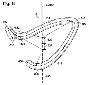
[0040] FIG. 8 is a perspective view of one embodiment of a tricuspid ring
according to the present disclosure.
[0041] FIG. 9 is a plan view of a tricuspid valve, with orientation reference
points indicated.
[0042] FIG. 10 is a plan view of the tricuspid ring according to the present
disclosure as in FIG. 7, with segments and saddle points corresponding to FIG.
8.
Detailed Description of the Preferred Embodiments
[0043] Embodiments of a tricuspid ring according to the present disclosure can
mimic the shape of the native tricuspid valve and right ventricle in order to
substantially
CA 02785509 2012-06-22
WO 2011/087834 PCT/US2010/061752
-14-
restore a diseased or damaged annulus to its correct anatomical shape.
Tricuspid
annuloplasty rings that better conform to the native annulus can be shaped to
protect
certain features of the surrounding anatomy. The rings of the present
disclosure can be
designed to support a majority of the tricuspid annulus without risking injury
to the
leaflet tissue and/or the heart's conductive system, such as the AV node 34
and bundle
of His 36 (see FIG. 4). Additionally, disclosed embodiments of a tricuspid
ring can be
contoured to better approximate the three-dimensional shape of the tricuspid
annulus,
and can thereby reduce residual tricuspid regurgitation post-operatively.
Disclosed
embodiments of a tricuspid ring can provide remodeling of diseased tricuspid
valve
annuluses in a bimodal, anatomically correct shape (e.g., in all three
dimensions). Thus,
some embodiments can improve durability of the repair by imparting less stress
on the
native valve leaflets and annulus.
[0044] The term "axis" in reference to the illustrated ring, and other non-
circular
or non-planar rings, refers to a line that passes through the area centroid of
the ring
when viewed in plan view. "Axial" or the direction of the "axis" can also be
viewed as
being parallel to the direction of blood flow within the valve orifice and
thus within the
ring when implanted therein. Stated another way, the implanted tricuspid ring
orients
about a central flow axis aligned along an average direction of blood flow
through the
tricuspid annulus.
[0045] One embodiment of a tricuspid ring according to the present disclosure
is
shown in plan view in FIG. 7. Tricuspid ring 70 can comprise a ring 72 and
subvalvular
device (not shown) that mimics the shape of the native valve and right
ventricle. The
tricuspid ring 70 can thus at least partially restore the correct anatomy of a
tricuspid
valve annulus and right ventricle into which the ring 70 is implanted.
Suitable
subvalvular devices are described in U.S. Patent Publication No. 2010/0063586
to
Hasenkam, which is incorporated herein by reference, in its entirety.
[0046] For instance, a ring and subvalvular system according to one embodiment
of the present application includes a tricuspid annuloplasty ring 70 and a
tension and
anchoring subsystem adapted to align the papillary muscles with the tricuspid
annulus,
and to align the wall of the right ventricle with respect to the tricuspid
valve in order to
CA 02785509 2012-06-22
WO 2011/087834 PCT/US2010/061752
- 15-
eliminate regurgitation. The tension and anchoring subsystem comprises a set
of tension
members, e.g. in the form of strings or sutures. Each of the tension members
comprises
a first end routed through the tricuspid ring 70 to a position at the exterior
of the heart
for adjustment of a set of anatomical lengths/distances defining the geometry
of the right
ventricle of the heart. Second ends fix to a position on or through the
papillary muscles.
The tricuspid ring 70 in this embodiment is either hollow to allow passage of
the tension
members, or otherwise includes channels that route the tension members. The
tricuspid
ring 70 attaches to the annulus, and its rigidity will support the geometry of
the annulus
via the tension members once they are fixed to the ring. Preferably, one or
more tension
members extend from one side of the tricuspid ring 70 and one or more tension
members extend from the opposite side.
[0047] Tricuspid annuloplasty rings 70 disclosed herein can at least partially
restore the anatomically correct shape in all three dimensions. As seen in
FIG. 7, the
shape of a tricuspid ring 70 is asymmetric and generally ovoid surrounding an
axis in
the direction of blood flow through the ring, and can be partially defined or
characterized by a major axis 80 along its length and a minor axis 82 along
its width,
and more specifically, by the ratio of the major axis 80 to the minor axis 82.
In terms of
anatomical references, the length dimension of the tricuspid ring 70 when
implanted
extends generally from the middle of the posterior leaflet to the antero-
septal
commissure, as seen in Fig. 3, while the width dimension extends generally
from the
anterior leaflet adjacent the antero-posterior commissure to the septal
leaflet. The major
axis 80 is defined by the length A between a first point 84 and a second point
86 located
on the interior 88 of the tricuspid ring 70. The length A represents the
length of the line
spanning the greatest length between two points on the interior 88 of the ring
70. The
minor axis 82 is defined by the vertical displacement B between a third point
90 and a
fourth point 92 on the interior 88 of the tricuspid ring 70. The length B
represents the
length of the line spanning the greatest width between two points on the
interior 88 of
the ring 70. Prior art tricuspid rings disclose designs having a major to
minor axis ratio
of 1.55. Tricuspid rings according to the present disclosure can be designed
to have a
major to minor axis ratio greater than that of prior art tricuspid rings. For
example, the
CA 02785509 2012-06-22
WO 2011/087834 PCT/US2010/061752
-16-
ratio can be around 1.56 or greater, such as between about 1.56 and about 2.
Increasing
the major to minor axis ratio can reduce residual tricuspid regurgitation post-
operatively
in some embodiments, such as by increasing septal-posterior coaptation.
[0048] The tricuspid rings of the present disclosure can be designed and
manufactured in several different sizes, to form a set of tricuspid rings of
various sizes.
For example, a set of tricuspid rings can include ring sizes ranging from 24
mm to 40
mm, at intervals of 2 mm. Once again, the "ring size" is the size labeled on
the
particular annuloplasty ring packaging. A "set of rings" means a collection of
annuloplasty rings of different sizes marketed together as one type of ring or
for the
same pathological condition, typically under one tradename. Although a set of
rings is
made available by the manufacturer, customers such as hospitals regularly
order one or
two sizes as needed, though orders of multiple sizes and even whole sets occur
to
maintain a supply of different sized rings on site. Smaller and larger sizes
of rings can
also be included in sets of tricuspid rings. In some embodiments of a set of
tricuspid
rings, the major to minor axis ratios can be the same for each size ring in
the set. In
other embodiments of a set of tricuspid rings, the major to minor axis ratios
can vary for
each different size of tricuspid ring. For example, in some embodiments, the
major to
minor axis ratio can increase with decreasing ring size. Thus, within a set of
tricuspid
rings, the major to minor axis ratio of one size of ring can be greater than
the major to
minor axis ratio of the next smaller sized ring. In some embodiments, the
major to
minor axis ratio can decrease with increasing ring size. Thus, within a set of
tricuspid
rings, the major to minor axis ratio of one size of ring can be less than the
major to
minor axis ratio of the next larger sized ring. As a result of the varying
major to minor
axis ratios, the minor axis 82 can more aggressively decrease in length in
smaller sizes
of tricuspid rings.
[0049] Incidence of tricuspid regurgitation can be further reduced by
selecting a
tricuspid ring size smaller than would conventionally be selected for a
particular subject.
[0050] Furthermore, as seen in FIG. 8, embodiments of a tricuspid ring can be
designed to substantially restore the anatomically correct shape to the valve
annulus
and/or right ventricle along the Z axis 820. The anatomically correct valve
annulus
CA 02785509 2012-06-22
WO 2011/087834 PCT/US2010/061752
-17-
includes two local high points (indicated by HIGH in FIG. 9), and two local
low points
(indicated by LOW in FIG. 9), along the Z axis, thus forming a bimodal saddle
shape, as
seen in FIG. 8. A tricuspid ring can be designed to account for the elevation
of the
native annulus' high and low points, and thus help correct the shape of a
diseased
annulus along the Z axis.
[0051] Embodiments of a tricuspid ring according to the present disclosure can
include one or more points or portions of elevation in the Z direction, such
as a primary
saddle and a secondary saddle. As used herein, the elevation of a point refers
to the
distance of that point from the X-Y plane bisecting the tricuspid ring (i.e.,
the distance
along the Z axis from a plane perpendicular to the blood flow through the ring
that
passes through the center of the overall elevation span of the ring). The
static elevation
of a point refers to the elevation of that point while the tricuspid ring is
static and not
implanted. When the tricuspid ring is implanted in a native valve, the
elevation of some
points can change with each cardiac cycle. The elevation of a portion or
segment of a
tricuspid ring refers to the elevation of the highest and lowest points of
that portion or
segment. The amplitude of the tricuspid ring is defined as the distance along
the Z axis
between a high point (e.g., the highest high point or a local maximum point)
and a low
point (e.g., the lowest low point or a local minimum point) of the ring. Thus,
the
amplitude can be determined by summing the absolute value of the elevations of
the
high and low points of the ring. An amplitude of a portion or segment of the
tricuspid
ring is defined by the distance along the Z axis between the highest point of
that
segment above the X-Y plane and the lowest point of that segment below the X-Y
plane.
[0052] Portions of the elevated segments of the ring can correspond to native
valve anatomy. For example, a tricuspid ring can include a primary saddle
located at the
posterior leaflet of the native valve when implanted in the valve annulus,
with the lowest
point of the primary saddle, for example, within the anterior leaflet. The
elevation of
the primary saddle can be about 2 mm in the Z direction. A high point of a
secondary
saddle can be located at the antero-septal commissure of the native valve when
implanted in the valve annulus, and can have an elevation of about 0.5 mm.
CA 02785509 2012-06-22
WO 2011/087834 PCT/US2010/061752
-18-
[0053] In one embodiment of a tricuspid ring seen in FIGS. 8 and 10, the ring
8
can have high points 800, 802 at approximately the center of the posterior
leaflet and at
approximately the antero-septal commissure (the aortic bulge), respectively,
when
implanted. The elevation of the antero-septal commissure can be from about 0.5
mm to
about 4 mm, and the elevation of the center of the posterior leaflet can be
from about 2
mm to about 4 mm. For example, the local high point 800 can be a vertical
distance 822
along the Z axis 820 above an X-Y plane cutting through the center of the ring
8.
Embodiments of a tricuspid ring 8 can have low points 804, 806 at
approximately the
lateral center of the anterior leaflet and at approximately the center of the
septal leaflet,
when implanted. The elevation of the center of the anterior leaflet can be
from about -2
mm to about -4 mm, and the elevation of the center of the posterior leaflet
can be from
about -1 mm to about -4 mm. For example, the local low point 804 can be a
vertical
distance 824 along the Z axis 820 below an X-Y plane cutting through the
center of the
ring 8.
[0054] FIG. 10 shows the tricuspid annuloplasty ring 8 in plan view, with
segments (812, 814, 816, 818) and saddle points (800, 802, 804, 806)
corresponding to
FIG. 8. For reference to the native anatomy, the approximate location of the
three
commissures 28 as depicted in FIGS. 3 and 9 are indicated.
[0055] FIG. 9 illustrates reference anatomy that corresponds to high points
and
low points of a tricuspid ring when implanted. FIG. 9 shows the approximate
locations
of the local maxima, or high points, (indicated by HIGH) in the native valve,
at about
the center of the posterior leaflet 24c and at approximately the antero-septal
commissure
28. FIG. 9 also shows the approximate locations of the local minima, or low
points,
(indicated by LOW) in the native valve, at about the center of the anterior
leaflet 24b
and at about the center of the septal leaflet 24a.
[0056] Further, some areas of a tricuspid ring can have a greater positive
elevation than others. For example, as seen in FIG. 8, a lateral-posterior
segment 816
can have a greater elevation than a septal-anterior segment 812. For example,
in some
embodiments, the elevation at the septal-anterior segment 812 can be between
about 0.5
mm and about 10 mm, or between about 0.5 mm and about 6 mm. In some
CA 02785509 2012-06-22
WO 2011/087834 PCT/US2010/061752
-19-
embodiments, the elevation at the lateral-posterior segment 816 can be between
about 2
mm and 10 mm, or between about 2 mm and 6mm.
[0057] In some embodiments, an anterior-lateral segment 814 can have a greater
(e.g., more pronounced) negative elevation than a posterior-septal 818
segment. For
example, in some embodiments, the elevation at the anterior-lateral segment
814 can be
between about 2 mm and about 10 mm, or between about 2 mm and about 6 mm. In
some embodiments, the elevation at the posterior-septal segment 818 can be
between
about 1 mm and 10 mm, or between about 1 mm and 6mm.
[0058] In some embodiments, the total height, or the maximum distance
between the highest point of the tricuspid ring 8 along the Z axis 820 and the
lowest
point of the tricuspid ring 8 along the Z axis 820 is about 20 mm or less
(e.g., a total
amplitude of about 10 or 15 mm), as measured from the center of the ring 8 at
the
highest point to the center of the ring 8 at the lower point, along the Z
axis. In some
embodiments, the height along the Z axis 820 of the tricuspid ring 8 is about
15% of the
width of the tricuspid ring (e.g., the major axis length A, as seen in FIG.
7). For
example, the height of a tricuspid ring can be about 5 mm for a 36 mm ring.
[0059] Sizing a tricuspid ring as described can yield advantages in some
embodiments, such as producing a tricuspid ring that more accurately mimics
the shape
of the native tricuspid valve, imparting less stress on the valve tissues and
annulus, and
improving short and long term outcomes for treating tricuspid regurgitation
and other
abnormalities in the tricuspid valve.
[0060] In some embodiments of a set of tricuspid rings, the proportional
elevation in the Z direction can remain substantially constant as the size of
the ring
increases. For example, each tricuspid ring in a set of rings can have a ratio
of elevation
in the Z direction to the width A within the range of from about 15% to about
25%. In
some embodiments of a set of tricuspid rings, the proportional elevation in
the Z
direction can increase or decrease as the size of the ring increases. For
example, the
elevation can increase in proportion to the increasing major axis dimension A,
such as
increasing from about 15% to about 25%, or decrease in proportion to the
increasing
CA 02785509 2012-06-22
WO 2011/087834 PCT/US2010/061752
-20-
major axis dimension A, such as decreasing from about 25% to about 15%, as the
size
of the ring increases.
[0061] There are several reasons for varying the proportional elevation to
width
for different ring sizes. For example, for subjects with severe cases of
tricuspid
regurgitation and/or severe damage to the right ventricle, it can be
advantageous to
provide a progressively decreasing height to width ratio, such as a height to
width ratio
that decreases progressively from about 25% to about 5% over a size range of
24 mm to
40 mm rings. This could mean, for instance, that the absolute elevations
around the ring
remain the same as the ring size increases, or that the elevations increase
but at a slower
rate than the major and minor axes. The tissue of the tricuspid annulus is
somewhat
more fragile than other valve annuli such as the mitral valve, and
proportionally raising
or lowering segments of the ring may place excessive stress on the tissue
during the
cycling motion of the annulus. Thus, a set of similarly contoured rings whose
major and
minor axes increase but whose elevations remain substantially constant, or
increase at a
lower rate than the ring size, help reduce the chance of damaging the fragile
annulus
tissue.
[0062] Embodiments of a tricuspid ring can be configured to mimic the motion
of a native tricuspid valve during the cardiac cycle, and can thereby
substantially or at
least partially restore the anatomically correct motion of the tricuspid valve
annulus in
the X-Y plane and/or the Z direction.
[0063] The orifice of disclosed tricuspid rings can expand during diastole and
contract during systole, such that the area of the orifice expands from about
20% to
about 40% during diastole. In one specific embodiment, the area of the orifice
can
expand an average of about 29% during a series of cardiac cycles. The orifice
of
disclosed tricuspid rings can expand an amount sufficient to allow efficient
filling of the
ventricle during diastole. At a later point in each cardiac cycle, the orifice
of disclosed
tricuspid rings can contract an amount sufficient to provide an efficient
sphincter-like
motion to substantially effectively seal the repaired valve shut during the
increased
ventricular pressure of systole.
CA 02785509 2012-06-22
WO 2011/087834 PCT/US2010/061752
-21-
[0064] Expansion and contraction of the orifice area and circumference of
disclosed tricuspid rings can be accomplished in any suitable fashion. In some
exemplary embodiments, such expansion and contraction can be provided by
mechanisms such as one or more springs, polymeric materials, and/or an
accordion-like
core construction.
[0065] Similarly, the diameter (e.g., the major axis A and/or the minor axis
B) of
the tricuspid ring can expand and contract during the cardiac cycle. In some
embodiments, the diameter of the tricuspid ring can increase by a percentage
of from
about 14.7% to about 17.2%. In one specific embodiment, the diameter of the
tricuspid
ring expands by about 16% during diastole. In some embodiments, the orifice
expansion and the diameter increase is not evenly distributed around the
circumference
of the ring. For example, some embodiments of a tricuspid ring according to
the present
disclosure avoid expansion at the commissures. Such an arrangement can
substantially
prevent or reduce leakage through commissural clefts after implantation. On
the other
hand, segments of disclosed tricuspid rings corresponding to the center of
each of the
three native valve leaflets can be configured to expand.
[0066] Expansion and contraction of the diameter of disclosed embodiments of a
tricuspid ring can be provided by any suitable fashion. For example, tricuspid
rings
according to the present disclosure can be provided with mechanisms such as
springs,
polymeric materials, an accordion-like core construction, selectively
segmented core
sections, selectively flexible core materials, one or more hinge points
creating a jaw-like
expansion, and/or a cable-based core design. For example, U.S. Patent
Publication No.
2009/0287303 to Carpentier, which is incorporated by reference, describes
various
constructions of a tricuspid ring that can be incorporated in the embodiments
disclosed
in the present disclosure.
[0067] In some embodiments of sets of tricuspid rings, different sizes of
tricuspid rings can be configured to expand to a greater or lesser extent
during the
cardiac cycle. For example, in some embodiments of a set of tricuspid rings,
the larger
size rings can be configured to undergo a larger orifice area expansion and/or
a greater
diameter increase than the small size rings.
CA 02785509 2012-06-22
WO 2011/087834 PCT/US2010/061752
-22-
[0068] Similarly, embodiments of a tricuspid ring can be configured for
desirable movement in the Z direction, in order to at least partially restore
anatomically
correct movement of the native valve. For example, the elevation of
embodiments of a
tricuspid ring can increase during the systolic heart contraction and decrease
during
diastolic filling. Such movement can decrease leaflet stress during systole
and/or
decrease stress on the annuloplasty sutures holding the ring in place, which
can reduce
incidence of dehiscence.
[0069] The change in the elevation of the tricuspid ring can coincide with a
change in circumference of the ring. For example, an increase in the elevation
of the
ring in the Z direction can coincide with a decrease in the circumference of
the ring.
Such movement can increase efficiency in opening and closing of the tricuspid
valve.
[0070] Further, in embodiments of a set of tricuspid rings, the movement, or
change in amplitude, in the Z direction can vary according to the size of
tricuspid ring.
For example, larger sizes of rings can be configured to undergo a relatively
larger
change in amplitude (e.g., a larger increase in elevation). Thus, the movement
of the
tricuspid ring in the Z direction can increase with increasing ring size.
[0071] In some embodiments of a tricuspid ring, the ring can comprise a
plurality of segments. The term "segments" can refer different areas or
portions along a
continuous ring body. In such embodiments, different segments of the ring can
be
configured to different amplitude changes in the Z direction during the
cardiac cycle.
For example, still with reference to FIG. 8, the elevation of the septal-
anterior segment
812 can decrease by approximately 1 mm. In some embodiments, the elevation can
change by between about 0 mm and about -2 mm (e.g., move about 0 to 2 mm down
in
the Z direction, below the X-Y plane). The elevation of the anterior-lateral
segment 814
can substantially remain unchanged during the cardiac cycle in some
embodiments. The
elevation of the lateral-posterior segment 816 can increase by approximately 1
mm, or
between about 1 mm and about 2 mm. The elevation of the posterior-septal
segment
818 can decrease by approximately 1 mm, or between about 0 mm and about -2 mm.
In
some embodiments, the elevation increase of the lateral-posterior segment 816
is the
largest movement seen in the ring circumference. The lateral-posterior segment
816 of
CA 02785509 2012-06-22
WO 2011/087834 PCT/US2010/061752
-23-
the tricuspid ring 8 can be associated with the lateral free wall of the right
ventricle
when implanted.
[0072] The incomplete, C-shaped tricuspid ring therefore experiences an out-of-
plane motion of the free ends 808, 810 of the ring 8 with the septal-anterior
free end 810
decreasing in the vertical axis and the posterior-septal free end 808
increasing in the
vertical axis. The result is that the free ends 808, 810 of the ring move
separately from
each other with the distance between the two increasing by at least about 1 mm
and by
as much as about 4 mm. In some embodiments, the static vertical distance
(along the Z
axis) between the two free ends 808, 810 is between about 0 mm and about 6 mm.
Thus, the total vertical distance between the two free ends 808, 810 in a
dynamic heart
with a dynamic ring (e.g., a ring that undergoes movement in the Z direction
during the
cardiac cycle) is between about 0 mm and about 10 mm.
[0073] Embodiments of tricuspid rings can provide for movement in the Z
direction by any suitable design features. For example, some embodiments
comprise
specifically designed ring cores that include polymeric materials with varying
flexibilities, stacked Elgiloy core members, a ring core that is thinner in
height (along
the Z axis) than in thickness (along the X-Y plane), and/or a composite core
design,
such as a metallic and polymer composite core design.
[0074] Some embodiments of a tricuspid ring can have a flexibility that varies
along the length of the ring, such as having a relatively stiff first segment
and getting
progressively more flexible to a relatively flexible fourth segment. This
varying
flexibility can allow the ring to adapt (harmonize) its motion and three-
dimensional
shape to that of the annulus, rather than impose its own motion and 3-D
geometry
thereto which tends to increase the risk of ring dehiscence. In particular,
the motion of
the tricuspid annulus during systole-diastole is believed to exert some
torsional forces
on the implanted ring, and the variable flexibility accommodates such torques.
Localized points of flexibility or "hinges" around the ring can conform and
harmonize
the physical properties of the ring to the annulus motion, while at the same
time
providing the needed corrective support.
CA 02785509 2012-06-22
WO 2011/087834 PCT/US2010/061752
-24-
[0075] Embodiments of a tricuspid ring can comprise an inner core
encompassed by an elastomeric interface and an outer fabric covering. The
inner core
can extend substantially around the entire periphery of the ring body and can
be a
material such as stainless steel, titanium, Elgiloy (an alloy primarily
including Ni, Co,
and Cr), and/or polymers. Any material suitable to support the annulus while
allowing
for the movement described above can be used.
[0076] More specifically, the inner core is formed from a relatively rigid
material such as stainless steel, titanium, and Cobalt Chromium (CoCr family
of alloys:
CoCr, L605, MP, MP25, MP35N, Elgiloy, FW-1058). The term "relatively rigid"
refers
to the ability of the core to support the annulus without substantial
deformation, and
implies a minimum elastic strength that enables the ring to maintain its
original shape
after implant even though it may flex somewhat. Indeed, as will be apparent,
the ring
desirably possesses some flexibility around its periphery. To further
elaborate, the core
would not be made of silicone, which easily deforms to the shape of the
annulus and
therefore will not necessarily maintain its original shape upon implant.
Instead, the ring
core is preferably formed from one of the relatively rigid metals or alloys
listed above,
or even a polymer that exhibits similar material and mechanical properties.
For
instance, certain blends of Polyether ether ketone (PEEK) with carbon and an
alloy
might be used, in which case the core could be injection molded.
[0077] In some embodiments, the elastomeric interface can be silicone rubber
molded around the core, or a similar expedient. The elastomeric interface can
provide
bulk to the ring for ease of handling and implant, and can permit passage of
sutures.
The fabric covering can be any biocompatible material such as, for example,
Dacron
(polyethylene terepthalate).
[0078] Disclosed tricuspid rings can possess a varying flexibility around its
periphery. For example, the ring can be stiffer adjacent the first free end
than adjacent
the second free end, and can have a gradually changing degree of flexibility
for at least a
portion in between. For instance, the first segment can be relatively stiff
while the
remainder of the ring body gradually becomes more flexible through the second
segment, third segment, and fourth segment.
CA 02785509 2012-06-22
WO 2011/087834 PCT/US2010/061752
-25-
[0079] It should also be understood that features of the present tricuspid
ring can
also be applicable and beneficial to rings for other of the heart's annuluses,
such as the
mitral valve annulus.
[0080] In view of the many possible embodiments to which the principles of the
disclosed invention may be applied, it should be recognized that the
illustrated
embodiments are only preferred examples of the invention and should not be
taken as
limiting the scope of the invention. Rather, the scope of the invention is
defined by the
following claims. We therefore claim as our invention all that comes within
the scope
and spirit of these claims.