Note: Descriptions are shown in the official language in which they were submitted.
CA 02785758 2012-06-26
WO 2011/080188 PCT/EP2010/070550
1
METHOD AND DEVICE FOR DETECTING A CONFIGURATION OF
WITHDRAWAL AND RETURN DEVICES
Technical Field
The present invention generally relates to detection of configuration of
access
devices, and in particular to detection based on a pressure measurement. The
present
invention is e.g. applicable in arrangements for extracorporeal blood
treatment.
Background art
In extracorporeal blood treatment, blood is taken out of a subject, treated
and then
reintroduced into the subject by means of an extracorporeal blood flow
circuit. Generally,
the blood is circulated through the circuit by one or more pumping devices.
The circuit is
connected to a blood vessel access of the patient, typically via one or more
access devices,
such as needles, which are inserted into the blood vessel access. Such
extracorporeal blood
treatments include hemodialysis, hemodiafiltration, hemofiltration,
plasmapheresis,
bloodbanking, blood fraction separation (e.g. cells) of donor blood, etc.
In extracorporeal blood treatment, it is vital to minimize the risk for
malfunctions in
the extracorporeal blood flow circuit, since these may lead to inefficient
treatment due to
impaired delivery of hemodialysis prescription having potentially severe
effects on the
condition of the subject.
Malfunctions may be caused by i) accidental misplacement of the access devices
for
blood extraction (withdrawal and return, e.g. arterial and venous
needles/catheters) or ii)
faulty connection of the access devices to the blood lines. For instance, the
connection of
the access devices to the blood vessels may be reversed, causing recirculation
of the treated
blood during extra-corporeal circulation such that the dialyzed blood
returning through the
venous line is drawn back into the arterial line without having passed through
the heart and
thereby reducing the treatment dose given to the patient, which may have
negative
consequences to the patient's health both in the short and long term
perspective. Another
example of malfunction includes a reversed connection of the blood lines to
the access
devices.
These malfunctions all originate in a "connection system" between the patient
and
the extracorporeal blood flow circuit. The connection system includes one or
more access
devices and possibly one or more releasable connectors for attaching the
access devices to
tubing in the extracorporeal blood flow circuit.
Recirculation in extracorporeal blood treatment arises when the whole or a
fraction
of treated extracorporeal blood flow returns directly to the inlet of the
extracorporeal blood
line instead of flowing back to the heart.
There are several reasons for recirculation, for instance a) low access blood
flow Qa
compared to the blood flow Qb of extra-corporeal circulation, specific
problems related to
CA 02785758 2012-06-26
WO 2011/080188 PCT/EP2010/070550
2
the b) blood access physiology or return of treated blood directly to inlet
due c) to too close
positioning of needles. However, another common cause for needle problems is
accidental
misplacement of the arterial and venous access devices or tubes to reversing
the
configuration and causing a substantial recirculation and significant
reduction of treatment
dose.
To avoid recirculation of the treated blood during extra-corporeal treatment
the
arterial access must be placed in an upstream position compared to the venous
access.
Studies have shown that reversed needles fault may occur in approximately one
of ten
treatments.
Situations with access recirculation require intervention. To this end, an
apparatus for
extracorporeal blood treatment may include one or more surveillance devices
that detect
either recirculation in general or specifically the reversal of the needles.
Methods of access surveillance include clinical examination, urea or tracer
recirculation measurement, continuous wave Doppler methods, duplex
ultrasonography,
and radiograph angiography.
A method involving continuous wave Doppler measurements to detect access
function related recirculation by ultrasound dilution is disclosed in EDTNA
ERCA J 1998
Apr-Jun; 24(2):3-6 "Theoretical and practical issues in recirculation;
assessment of
vascular access". This method involves continuous wave Doppler measurements
where a
reversed position of the needles is detected by the Doppler frequencies being
higher with
the pump on than with the pump off.
Urea recirculation measurements involves comparing the blood urea taken from
the
access lines with that from a peripheral vein. When there is no recirculation,
the urea in the
arterial line and peripheral vein should be the same. However, this method
will measure
not only any access recirculation, but also the so called cardio-pulmonary
recirculation.
Cardio-pulmonary recirculation occurs because the treated blood traveling back
towards
the heart will mix with blood returning from the body, be pumped by the heart
through the
lungs and back to the heart where it will be pumped back into the body, and a
fraction of
this arterial blood is directed towards the access. This means that a fraction
of the blood
going out to the access will come from the newly treated blood. Just as with
access
recirculation this cardio-pulmonary recirculation therefore causes some newly
treated
blood to enter the extracorporeal circulation directly, without having passed
the main parts
of the body in between. This urea method will therefore measure the sum of
access and
cardio-pulmonary recirculation.
Hematocrit dilution has also been promoted as an indicator. However, the known
methods require special training and/or additional laboratory tests. Hence,
there is a need
for a method for detecting the configuration of withdrawal and return lines in
a
cardiovascular access that may be applied each time a subject undergoes
extracorporeal
blood treatment.
CA 02785758 2012-06-26
WO 2011/080188 PCT/EP2010/070550
3
No integrated means in dialysis machines exist today which allow automatic
detection of reversed needles. Since recirculation does not disturb the
treatment in any
other way than decreasing the treatment efficiency, it may go undetected
throughout the
whole treatment, and there is a large need for an automatic detection device.
Various
devices and methods have been disclosed for recirculation measurement,
however, none of
these are fully integrated and automatic. Furthermore, even if recirculation
is detected,
there is still a need to distinguish between various causes for recirculation,
in particular
reversed needles.
One example of another kind of malfunction in the extracorporeal blood flow
circuit,
which however is not attempted to be remedied by the present invention, is
disclosed in
JP2006198141. This documents deals with a connection mistake between arterial
and
venous detection lines between a dialyzer and pressure sensors, resulting in
reversed
pressure measurements, a potentially directly harmful situation if not
discovered in time.
Hence, this document relates to a problem and solution which is remote and
significantly
different from the problem of the present invention.
Summary
It is an object of the invention to at least partly overcome one or more of
the above-
identified limitations of the prior art. Specifically, it is an object to
provide an alternative
or complementary technique for detecting a reverse configuration of withdrawal
and return
devices coupling an extracorporeal blood flow circuit to a cardiovascular
system using
pressure measurements. Hence, it is an object to provide a technique for
detecting a
reversed connection between an extracorporeal blood circuit and a
cardiovascular system.
This and other objects, which will appear from the description below, are at
least
partly achieved by means of a method, a device, and a computer program product
according to the independent claims, embodiments thereof being defined by the
dependent
claims.
A first aspect of a first inventive concept of the invention is a device for
detecting a
configuration of withdrawal and return devices coupling an extracorporeal
blood flow
circuit to a cardiovascular system of a subject, the device comprising a
signal processor
configured to receive a primary measurement signal obtained by a primary
pressure sensor
in the extracorporeal blood flow circuit, process the primary measurement
signal for
extraction of primary pressure data originating from a subject pulse generator
in the
cardiovascular system or extracorporeal blood flow circuit, the primary
pressure data
comprising at least a part of a subject pulse from the subject pulse
generator; calculate a
parameter value from the primary pressure data; and determine the
configuration based at
least partly on the parameter value.
In one embodiment, the configuration is determined by comparing the parameter
value with a reference parameter value.
CA 02785758 2012-06-26
WO 2011/080188 PCT/EP2010/070550
4
In one embodiment, the device is further configured to receive a secondary
measurement signal obtained from a secondary sensor.
In one embodiment, the secondary sensor is a pressure sensor in the
extracorporeal
blood flow circuit, and wherein said secondary measurement signal is processed
for
extraction of secondary pressure data originating from said subject pulse
generator, and
wherein said parameter value is calculated from the primary pressure data and
secondary
pressure data.
In one embodiment, the primary pressure sensor is located on a venous side of
the
extracorporeal blood flow circuit and the secondary pressure sensor is located
on an
arterial side of the extracorporeal system.
In one embodiment, the parameter value is represented by a pressure amplitude
measure of the primary pressure data.
In one embodiment, the pressure amplitude measure comprises an arterial
pressure
amplitude or a venous pressure amplitude.
In one embodiment, the parameter value is represented by a pressure amplitude
ratio
of said primary pressure data and said secondary pressure data.
In one embodiment, the pressure amplitude ratio in a reversed configuration is
greater than the pressure amplitude ratio in a normal configuration.
In one embodiment, the parameter value represents a time delay of a pressure
pulse
detected by said primary pressure sensor at a first instance in time and said
pressure pulse
subsequently detected by said secondary pressure sensor at a second instance
in time.
Alternatively, the parameter value represents a time delay between a first
pressure pulse
obtained by said primary pressure sensor and a second pressure pulse obtained
by a
secondary pressure sensor.
In one embodiment, further to calculating, the signal processor is further
configured
to extract shape indicative data from the primary pressure data and matching
the shape
indicative data with shape reference data.
In one embodiment, the parameter value represents a deviation between the
shape
indicative data and the shape reference data. Alternatively, the parameter
value is a
correlation measure between shape indicative data from the primary pressure
data and
reference shape indicative pressure data.
In one embodiment, the shape reference data represents a temporal pulse
profile of
the subject pulse generator.
In one embodiment, the shape reference data represents a frequency spectrum of
the
subject pulse generator.
In one embodiment, the withdrawal and return devices comprise single or double
lumen needles or catheters.
CA 02785758 2012-06-26
WO 2011/080188 PCT/EP2010/070550
In other embodiments, the parameter value has been derived from one or more of
a
plurality of monitoring sessions of one subject, a plurality of monitoring
sessions of two or
more subjects, and a mathematical model.
In one embodiment, the configuration comprises a normal configuration and a
5 reverse configuration and wherein in said normal configuration a withdrawal
device is in
an upstream position of said cardiovascular system for withdrawal of fluid and
a return
device is in a downstream position of said cardiovascular system for return of
fluid and
wherein in said reverse configuration the positioning of the access devices is
reversed.
Hence, in a normal configuration the return device is in a downstream position
with respect
to the withdrawal device and in a reverse configuration the return device is
in an upstream
position with respect to the withdrawal device.
In one embodiment, the signal processor is further configured to aggregate a
plurality of
pulses within an aggregation time window in the measurement signal.
In one embodiment, the device is further configured to issue an alarm
subsequent to
determining a reverse configuration of access devices.
In one embodiment, the parameter represents a deviation in transit time of the
subject
pulse from the subject pulse generator to the primary pressure sensor and the
secondary
pressure sensor.
A second aspect of the invention is a method for detecting a configuration of
withdrawal and return devices coupling an extracorporeal blood flow circuit to
a
cardiovascular system of a subject, the method comprising: receiving a primary
measurement signal obtained by a primary pressure sensor in the extracorporeal
blood flow
circuit, processing the primary measurement signal for extraction of primary
pressure data
originating from a subject pulse generator in the cardiovascular system or
extracorporeal
blood flow circuit, the primary pressure data comprising at least a part of a
subject pulse
from the subject pulse generator, calculating a parameter value from the
primary pressure
data, and determining the configuration based at least partly on the parameter
value.
In one embodiment, the configuration is determined by comparing the parameter
value with a reference parameter value.
In one embodiment, the method further comprises receiving a secondary
measurement signal obtained from a secondary sensor.
In one embodiment, the method further comprises extracting shape indicative
data
from the primary pressure data and matching the shape indicative data with
shape
reference data.
In one embodiment, the method further comprises extracting the shape reference
data
from a secondary measurement signal received from a secondary sensor.
In one embodiment, the method further comprises aggregating a plurality of
pulses
within an aggregation time window in the measurement signal.
CA 02785758 2012-06-26
WO 2011/080188 PCT/EP2010/070550
6
In one embodiment, the method further comprises issuing an alarm subsequent to
determining a reverse configuration of access devices.
A third aspect of the invention is a computer-readable medium comprising
computer
instructions which, when executed by a processor, cause the processor to
perform the
method of the second aspect.
A fourth aspect of the invention is a device for detecting a configuration of
withdrawal and return devices coupling an extracorporeal blood flow circuit to
a
cardiovascular system of a subject, said device comprising: means for
receiving a primary
measurement signal obtained by a primary pressure sensor in the extracorporeal
blood flow
circuit; means for processing the primary measurement signal for extraction of
primary
pressure data originating from a subject pulse generator in the cardiovascular
system or
extracorporeal blood flow circuit, the primary pressure data comprising at
least a part of a
subject pulse from the subject pulse generator; means for calculating a
parameter value
from the primary pressure data; and means for determining the configuration
based at least
partly on the parameter value.
According to one embodiment, a plurality of pulses are aggregated to enhance
noise
reduction. Preferably, an aggregate comprise at least twenty pulses to allow
for sufficient
extraction of parameter values associated with any of amplitude, phase, shape
of a pulse
profile or any combinations thereof.
Timing information from the measurement may be used to allow for accurate
alignment in the aggregation process. The timing information may be obtained
from the
measurement signal or otherwise. The timing information is indicative of the
timing of the
pressure pulses in the measurement signal. Subsequently, the measurement
signal is
processed based on the timing information, to calculate a value of an
evaluation parameter
which is indicative of the relative configuration of access devices in a blood
access. Based
on the resulting value of the evaluation parameter, it is decided whether the
access device
configuration is normal or reversed, typically by comparing the resulting
value to a
threshold value.
Thus, the provision of timing information allows for signal enhancement by
identifying and averaging pulse segments in one or more measurement signals.
Although the present invention preferably applies to on-line processing of
measurement signals, i.e. during, e.g. concurrently, a treatment, it may also
apply to off-
line processing, for instance subsequent to or separate from a treatment such
as upon
studying the efficiency of a treatment. The processing may for instance
involve pre-
processing including general signal filtration, removal of particular signal
noise and
artefacts, such as from a running pump, and signal analysis. The
cardiovascular system, for
instance a blood circuit of a human or an animal, may also be referred to as a
fluid system
or fluid circuit.
CA 02785758 2012-06-26
WO 2011/080188 PCT/EP2010/070550
7
Embodiments of the second to fourth aspects of the first inventive concept may
correspond to the above-identified embodiments of the first aspect of the
first inventive
concept.
A fifth aspect of the invention is a device for detecting a reversed
configuration of
withdrawal and return devices coupling an extracorporeal blood flow circuit to
a
cardiovascular system of a subject, the device comprising a signal processor
configured to:
receive a primary measurement signal obtained by a primary pressure sensor in
the
extracorporeal blood flow circuit; process the primary measurement signal for
extraction of
primary pressure data originating from a pump pulse generator in the
extracorporeal blood
flow circuit, the primary pressure data comprising at least a part of a pump
pulse from the
pump pulse generator; calculate a parameter value from the primary pressure
data, the
parameter value being indicative of a cross-talk pressure pattern generated
from a
combination of pressure pulses from the pump pulse generator obtained by the
primary
pressure sensor from two directions, one passing through the cardiovascular
system of the
subject and the other from the pump pulse generator within the extracorporeal
circuit; and
determine the reversed configuration based at least partly on the parameter
value. This may
particularly be used for detection of the reversed configuration by comparing
effects of
pressure pulse cross-talk between venous and an arterial branch contributions
in the
extracorporeal circuit
A sixth aspect of the invention is a method for detecting a configuration of
withdrawal and return devices coupling an extracorporeal blood flow circuit to
a
cardiovascular system of a subject, the method comprising: receiving a primary
measurement signal obtained by a primary pressure sensor in the extracorporeal
blood flow
circuit; processing the primary measurement signal for extraction of primary
pressure data
originating from a pump pulse generator in the extracorporeal blood flow
circuit, the
primary pressure data comprising at least a part of a subject pulse from the
subject pulse
generator; calculating a parameter value from the primary pressure data, the
parameter
value being indicative of a cross-talk pressure pattern generated from a
combination of
pressure pulses from the pump pulse generator obtained by the primary pressure
sensor
from two directions, one passing through the cardiovascular system of the
subject and the
other from the pump pulse generator within the extracorporeal circuit; and
determining the
configuration based at least partly on the parameter value.
The attributes primary and secondary have been used to distinguish
equivalents, for
instance primary and secondary pressure data and primary and secondary
pressure sensors,
and do not indicate certain order or importance.
The signal processor of the device of the first aspect of the invention may
further be
configured to carry out any of the steps of the methods according to the
second and sixth
aspects of the invention.
CA 02785758 2012-06-26
WO 2011/080188 PCT/EP2010/070550
8
Still other objectives, features, aspects and advantages of the present
invention will
appear from the following detailed description, from the attached claims as
well as from
the drawings and the appendixes.
Brief Description of the Drawings
Embodiments of the inventive concepts will now be described in more detail
with
reference to the accompanying schematic drawings.
Fig. 1 is a schematic view of a general fluid arrangement in which the
inventive
concepts may be used for monitoring the configuration of a fluid connection.
Fig. 2 is a partially schematic view of a forearm of a subject provided with
an
arterial/venous (AV) fistula.
Fig. 3 is a schematic view of a system for hemodialysis treatment including an
extracorporeal blood flow circuit.
Fig. 4 (a) is a plot in the time domain of a venous pressure signal containing
both
pump frequency components and heart frequency components, and Fig. 4(b) is a
plot of the
corresponding signal in the frequency domain.
Fig. 5 is a flow chart of a monitoring process according to an embodiment of
the
invention.
Fig. 6(a) is a plot of a pressure signal as a function of time, and Fig. 6(b)
is a plot of
the pressure signal after filtering.
Fig. 7(a) is a schematically view of withdrawal and return devices in a normal
configuration at an access site, and Fig. 7(b) is a plot of the corresponding
access site with
the withdrawal and return devices in a reversed configuration.
Fig. 8(a) is a schematically view of withdrawal and return lines of a double
lumen
needle or catheter in a normal configuration at an access site, and Fig. 8(b)
is a plot of the
corresponding access site with the withdrawal and return lines in a reversed
configuration.
Fig. 9 is a flow chart of a monitoring process according to an embodiment of
the
invention.
Fig. 10(a) is a plot in the time domain of venous and arterial pressure signal
segments with the withdrawal and return devices in a normal configuration, and
Fig. 10(b)
is a plot of the corresponding venous and arterial pressure signal segments in
a reverse
configuration.
Fig. 11 is a block diagram of a hydraulic model to simulate the
characteristics in
normal and reversed configurations according to the present invention.
Fig. 12 is a plot of a venous/arterial heart signal amplitude ratio of a
normal fistula
with the needles in reversed and normal positions compared to the blood flow.
Fig. 13 is a plot of a venous/arterial heart ratio of a normal fistula with
the needles in
reversed and normal positions compared to the dialysis monitor blood flow Qb
in a normal
fistula.
CA 02785758 2012-06-26
WO 2011/080188 PCT/EP2010/070550
9
Fig. 14 is a plot of a venous/arterial heart ratio compared to the dialysis
monitor
blood flow Qb in a fistula having intra-fistula stenosis
Detailed Description of Example Embodiments
In the following, different embodiments for detecting a normal and/or reversed
configuration of access devices will be described with reference to an
exemplifying circuit
for extracorporeal blood treatment. In particular, the present invention
discloses a solution
involving venous and/or arterial line pressure measurements during for
instance dialysis
for monitoring the configuration of withdrawal and return lines at a
cardiovascular access.
Throughout the following description, like elements are designated by the same
reference signs.
I. GENERAL
Fig. 1 illustrates a general fluid arrangement in which a fluid connection C
is
established between a first fluid containing system Si and a second fluid
containing system
S2. The fluid connection C may or may not transfer fluid from one system to
the other. A
first pulse generator 3 is arranged to generate a series of pressure waves in
the fluid within
the first system S 1, and a second pulse generator 3' is arranged to generate
a series of
pressure waves in the fluid within the second system S2. Pressure sensors 4a
to 4c are
arranged to measure the fluid pressure in the first system S 1. As long as the
fluid
connection C is intact, pressure waves generated by the second pulse generator
3' will
travel from the second system S2 to the first system S 1, and thus second
pulses originating
from the second pulse generator 3' will be detected by the pressure sensors 4a
to 4c in
addition to first pulses originating from the first pulse generator 3. It is
to be noted that
either one of the first and second pulse generators 3, 3' may include more
than one pulse-
generating device. Further, any such pulse-generating device may or may not be
part of the
respective fluid containing system S 1, S2.
As used herein, a "pressure wave" denotes a mechanical wave in the form of a
disturbance that travels or propagates through a material or substance. The
pressure waves
typically propagate in the fluid at a velocity of about 3-20 m/s. The pressure
sensor
generates measurement data that forms a pressure pulse for each pressure wave.
A
"pressure pulse" or "pulse" is thus a set of data samples that define a local
increase or
decrease (depending on implementation) in signal magnitude within a time-
dependent
measurement signal ("pressure signal"). The pressure pulses appear at a rate
proportional
to the generation rate of the pressure waves at the pulse generator. The
pressure sensor may
be of any type, e.g. operating by resistive, capacitive, inductive, magnetic
or optical
sensing, and using one or more diaphragms, bellows, Bourdon tubes, piezo-
electrical
components, semiconductor components, strain gauges, resonant wires, photo-
plethysmography (PPG), accelerometers, bioimpedance, etc.
CA 02785758 2012-06-26
WO 2011/080188 PCT/EP2010/070550
The fluid arrangement of Fig. 1 further includes a surveillance device 25
which is
connected to the pressure sensor 4c, and possibly to one or more further
pressure sensors
4a, 4b, as indicated in Fig. 1. Thereby, the surveillance device 25 acquires
one or more
measurement signals that are time-dependent to provide a real time
representation of the
5 fluid pressure in the first system S 1. The surveillance device 25 monitors
the configuration
of the fluid connection C, based on the principle that characteristics, such
as magnitude,
shape and/or phase, of the first and/or second pulses vary depending on the
configuration
of the connection. A malfunction in the connection alter the characteristics
of the pulse and
upon detection of such an irregularity the surveillance device 25 may issue an
alarm or
10 warning signal, and/or alert a control system of the first or second fluid
containing systems
S 1, S2 to take appropriate action.
The surveillance device 25 is thus configured to continuously process the time-
dependent measurement signal(s) to determine whether pressure characteristics
associated
with a normal or irregular configuration are detected. Typically, the
determination involves
analyzing the measurement signal(s), or a pre-processed version thereof, in
the time
domain to calculate a value of an evaluation parameter which is indicative of
the
characteristics of the first and/or second pulses in the measurement
signal(s). Depending
on implementation, the surveillance device 25 may use digital components or
analogue
components, or a combination thereof, for receiving and processing the
measurement
signal(s).
In the following, references to a subject pulse generator or second pulse
generator
relates to a physiological pulse generator of the subject, such as the heart,
breathing system
or autonomous system or a pulse generator coupled to a subject, such as a
blood pressure
cuff or other external pulse generator. Subject pulses or second pulses, are
generated from
the subject pulse generator or second pulse generator. An interference pulse
generator or
first pulse generator may be present in the extracorporeal system, and may
include a pump,
such as a peristaltic pump. Interference pulses or first pulses are generated
by the
interference pulse generator or first pulse generator. The interference pulses
or first pulses
may also be used for detection of configuration of withdrawal and return
devices due to a
cross-talk effect in the pressure measurement signals differing in normal and
reversed
configurations.
II. EXAMPLE OF A VASCULAR ACCESS AND EXTRACORPOREAL CIRCUIT
Fig. 2. discloses a forearm 200 of a subject. The forearm 200 comprises an
artery
201, in this case the radial artery, and a vein 202, in this case the cephalic
vein. The blood
flow in the artery (201) and vein (202) is indicated with arrows. Openings are
surgically
created in the artery 201 and the vein 202 and the openings are connected to
form an
anastomosis 203, in which the arterial blood flow is cross-circuited to the
vein. Such a
configuration with the anastomosis and nearby sections of the artery 201 and
vein 202 are
CA 02785758 2012-06-26
WO 2011/080188 PCT/EP2010/070550
11
commonly referred to as a fistula 208. Due to the fistula, the blood flow
through the artery
and vein is increased and the vein forms a thickened area downstream of the
connecting
openings. When the fistula has matured a few months after surgery, the vein is
thicker and
may be punctured repeatedly. Normally, the thickened vein area is called a
fistula.
An arterial or withdrawal device 211 in the form of a needle 204, to which is
connected a piece of arterial or withdrawal tube 205, is placed in an upstream
position 209
in the fistula, in the enlarged vein close to the connected anastomosis
openings and a
venous or return device 212 also in the form of a needle 206, to which is
connected a piece
of venous or return tube 207, is placed in a position downstream 210 of the
arterial or
withdrawal needle 204, normally at least five centimetres downstream thereof.
The
withdrawal 205 and return 207 tubes are connected to an extracorporeal circuit
(not shown)
such as described in Fig. 3. In use, the withdrawal tube 205 may transport
blood from the
artery 201 via the arterial or withdrawal needle 204 to an inlet of the
extracorporeal circuit,
and the return tube 207 then returns the treated blood from an outlet of the
extracorporeal
circuit to the vein 202 via the venous or return needle 206. Arrows at the
ends of the blood
lines (205, 207) indicate the direction of blood flow in a normal
configuration. In a
reversed configuration of the needles/catheters (204, 206), connection of
blood lines (205,
207) to the needles or connection of the blood lines to the extracorporeal
circuit, the arrows
would be reversed.
The vascular access may also be an arterio-venous graft, Scribner-shunt, one
or more
catheters, a double lumen catheter or other similar arrangements. For the
purpose of the
following discussion, the blood vessel access is assumed to be a fistula. The
withdrawal
and return needles may also be catheters. The withdrawal and return devices
generally
comprises a needle or catheter, a tubing and a connector (not shown)
connecting the tubing
to the needle or catheter.
The needles 204 and 206 of Fig. 2 are connected to a tube system, shown in
Fig. 3,
forming an extracorporeal blood flow circuit 20 of the type which is used for
dialysis.
Withdrawal or artery needle 1 and return or venous needle 14 are shown
connected to a
vessel 30 of the subject, which vessel is a part of the cardiovascular system
of the subject.
The extracorporeal blood flow circuit comprises a blood pump 3, such as a
peristaltic
pump. At the inlet of the pump 3 there is a pressure sensor 4a, hereafter
referred to as
arterial sensor, which measures the pressure before the pump in the withdrawal
tube
segment 2. The blood pump 3 propels the blood from the fistula, through the
withdrawal
needle 1, via a pre-dialyser tube segment 5, to the blood-side of a dialyser
6. Many dialysis
machines are additionally provided with a pressure sensor 4b that measures the
pressure
between the blood pump 3 and the dialyser 6. The blood is lead via a post-
dialyser tube
segment 10 from the blood-side of the dialyser 6 to a venous drip chamber or
deaeration
chamber 11 and from there back to the subject via the return tube segment 12
and return
needle 14. A pressure sensor 4c, hereafter referred to as venous sensor, is
provided to
CA 02785758 2012-06-26
WO 2011/080188 PCT/EP2010/070550
12
measure the pressure on the venous side of the dialyser 6. In the illustrated
example, the
pressure sensor 4c measures the pressure in the venous drip chamber. Both the
withdrawal
needle 1 and the return needle 14 are connected to the subject by means of the
vascular
access.
As discussed by way of introduction, it may be vital to monitor the fluid
connection
to the blood vessel access with respect to anomalies. In many dialysis
machines, one or
more of said pressure detectors 4a-4c are not present. However, there will be
at least one
venous pressure sensor. The following description is focused on detection of
the
configuration of access devices in the fluid connection based on a measurement
signal
from one or more of the pressure sensors.
Further in Fig. 3, a control unit 23 is provided, i.e., to control the blood
flow in the
circuit 20 by controlling the revolution speed of the blood pump 3. The
extracorporeal
blood flow circuit 20 and the control unit 23 may form part of an apparatus
for
extracorporeal blood treatment, such as a dialysis machine. Although not shown
or
discussed further it is to be understood that such an apparatus performs many
other
functions, e.g. controlling the flow of dialysis fluid, controlling the
temperature and
composition of the dialysis fluid, etc.
Also in Fig. 3, a surveillance device 25 is configured to detect the
configuration of
the access devices in the fluid connection between blood accesses of the
subject and the
extracorporeal blood flow circuit 20, specifically by detecting the presence
of a
predetermined pressure response determined by magnitude, shape and phase, or
timing,
and indicative of a normal configuration of the access devices, the pressure
response for
instance originating from the patient's heart in a blood pressure signal.
Absence of such a
predetermined pressure response is taken as an indication of a reversed
positioning of the
access devices, and brings the device 25 to activate an alarm or notification
for the staff to
check the configuration of withdrawal and return devices and adjust the
configuration if
necessary. The surveillance device 25 is at least connected to receive a
measurement signal
of the pressure sensor 4c. The device 25 may also be connected to further
pressure sensors
such as 4a, 4b, as well as any additional pressure sensors included in the
extracorporeal
blood flow circuit 20. As indicated in Fig. 3, the device 25 may also be
connected to the
control unit 23. Alternatively or additionally, the device 25 may be connected
to a
measurement device 26, such as a rotary encoder (e.g. conductive, optical or
magnetic) or
the like, for indicating the frequency and phase of the blood pump 3. The
device 25 is
tethered or wirelessly connected to a local or remote device 27 for generating
an
audible/visual/tactile alarm or warning signal. The surveillance device 25
and/or the alarm
device 27 may alternatively be incorporated as part of apparatus such as a
dialysis monitor.
Additionally, in Fig. 3, the surveillance device 25 comprises a data
acquisition part
28 for pre-processing the incoming signal(s), e.g. including an A/D converter
with a
required minimum sampling rate and resolution, one or more signal amplifiers,
one or
CA 02785758 2012-06-26
WO 2011/080188 PCT/EP2010/070550
13
more filters to remove undesired components of the incoming signal(s), such as
offset,
high frequency noise and supply voltage disturbances.
In the examples given herein, the data acquisition part 28 comprises a DAQ
card
USB-6210 from National Instruments with a sampling rate of 1 kHz and
resolution of 16
bits, an operation amplifying circuit AD620 from Analogue Devices, a high-pass
filter with
a cut-off frequency of 0.03 Hz (i.a., for removal of signal offset) together
with a low-pass
filter with a cut-off frequency of 402 Hz (i.a., for removal of high frequency
noise). To
obtain a short convergence time, a low-order filter is used for the high-pass
filter. Further-
more, the data acquisition part 28 may include an additional fixed band-pass
filter with
upper and lower cut-off frequencies of 0.5 Hz and 2.7 Hz, respectively, which
corresponds
to heart pulse rates between 30 and 160 beats per minute. This filter may be
used to sup-
press disturbances outside the frequency interval of interest. Corresponding
filters may be
applied to extract pressure pulses originating from breathing or other
physiological signals,
which may be used separately or in combination with the heart pulse rates to
determine the
configuration of access devices.
After the pre-processing in the data acquisition part 28, the pre-processed
pressure
signal is provided as input to a main data processing part 29, which executes
the inventive
data processing. The data processing part 29 may also be referred to as a
signal processor
29. Fig. 4(a) shows an example of such a pre-processed pressure signal 401 in
the time
domain, and Fig. 4(b) shows the corresponding power spectrum, i.e. the pre-
processed
pressure signal in the frequency domain. The power spectrum reveals that the
detected
pressure signal contains a number of different frequency components emanating
from the
blood pump 3. In the illustrated example, there is a frequency component at
the base
frequency (fo) of the blood pump (at 1.5 Hz in this example), as well as its
harmonics 2fo,
3f0 and 4f0. The base frequency, also denoted pump frequency in the following,
is the
frequency of the pump strokes that generate pressure waves in the
extracorporeal circuit
20. For example, in a peristaltic pump of the type shown in Fig. 3, two pump
strokes are
generated for each full revolution of the rotor 3a. Fig. 4(b) also indicates
the presence of a
frequency component at half the pump frequency (0.5fo) and harmonics thereof,
in this
example at least fo, 1.5f0, 2f0 and 2.5f0. Fig. 4(b) also shows a heart signal
(at 1.1 Hz)
which in this example is approximately 40 times weaker than the blood pump
signal at the
base frequency fo.
Typically, the surveillance device 25 is configured to continuously process
the time-
dependent pressure signal(s) to isolate any second pulses originating from a
physiological
pulse generator, such as the heart or breathing system. This processing is
schematically
depicted in the flow chart of Fig. 5. The illustrated processing involves a
step 501 of
obtaining a first pulse profile u(n) which is a predicted temporal signal
profile of the
second pulse(s), and a step 502 of filtering the pressure signal d(n), or a
pre-processed
version thereof, in the time-domain, using the first pulse profile u(n), to
essentially
CA 02785758 2012-06-26
WO 2011/080188 PCT/EP2010/070550
14
eliminate or cancel the first pulse(s) while retaining the second pulse(s)
contained in d(n).
In the context of the present disclosure, n indicates a sample number and is
thus equivalent
to a (relative) time point in a time-dependent signal. In step 503, the
resulting filtered
signal e(n) is then analysed for the purpose of monitoring the aforesaid
predetermined
pressure response or parameter for a heart signal corresponding to a normal or
reversed
configuration.
The first pulse profile is a shape template or standard signal profile,
typically given
as a time-sequence of data values, which reflects the shape of the first pulse
in the time
domain. The first pulse profile is also denoted "predicted signal profile" in
the following
description.
By "essentially eliminating" is meant that the first pulse(s) is(are) removed
from the
pressure signal to such an extent that the second pulse(s) can be detected and
analysed for
the purpose of monitoring the aforesaid functional state or parameter.
By filtering the pressure signal in the time-domain, using the first pulse
profile, it is
possible to essentially eliminate the first pulses and still retain the second
pulses, even if
the first and second pulses overlap or nearly overlap in the frequency domain.
Such a
frequency overlap is not unlikely, e.g. if one or both of the first and second
pulses is made
up of a combination of frequencies or frequency ranges.
The effectiveness of the inventive filtering is exemplified in Fig. 6, in
which Fig. 6(a)
shows an example of a time-dependent pressure signal d(n) containing first and
second
pulses with a relative magnitude of 10:1. The first and second pulses have a
frequency of 1
Hz and 1.33 Hz, respectively. Due to the difference in magnitude, the pressure
signal is
dominated by the first pulses, i.e. pump pulses. Fig. 6(b) shows the time-
dependent filtered
signal e(n) that is obtained after applying the inventive filtering technique
to the pressure
signal d(n). The filtered signal e(n) is made up of second pulses and noise.
The main data processing part 29 executes the aforesaid steps 501-503 of Fig.
5. In
step 502, the main data processing part 29 operates to filter the pre-
processed pressure
signal in the time domain, and outputs a filtered signal or monitoring signal
(e(n) in Fig. 5)
in which the signal components of the blood pump 3 have been removed. The
monitoring
signal still contains any signal components that originate from the subject
(cf. Fig. 6(b)),
such as pressure pulses caused by the beating of the patient's heart,
breathing or other
physiological signals. There are a number of sources to cyclic physiological
phenomena
that may generate pressure pulses in the blood stream of the patient,
including the heart,
the breathing system, or the vasomotor, which is controlled by the autonomic
nervous
system. Thus, the monitoring signal may contain pressure pulses resulting from
a
combination of cyclic phenomena in the patient. Generally speaking, the signal
components in the monitoring signal may originate from any type of
physiological
phenomenon in the patient, or combinations thereof, be it cyclic or non-
cyclic, repetitive or
non-repetitive, autonomous or non-autonomous. The signal components may
additionally
CA 02785758 2012-06-26
WO 2011/080188 PCT/EP2010/070550
involve artificial origin, for instance by a separate, external pressure
inducing component,
such as integrated in a blood pressure cuff, or the blood pressure cuff itself
with pressure
waves induced by puffing air into the cuff.
Depending on implementation, the surveillance device 25 may be configured to
5 apply further filtering to the monitoring signal to isolate signal
components originating
from a single cyclic phenomenon in the patient. Alternatively, such signal
component
filtering is done during the pre-processing of the pressure signal (by the
data acquisition
part 28). The signal component filtering may be done in the frequency domain,
e.g. by
applying a cut-off or band pass filter, since the signal components of the
different cyclic
10 phenomena in the patient are typically separated in the frequency domain.
Generally, the
heart frequency is about 0.5-4 Hz, the breathing frequency is about 0.15-0.4
Hz, the
frequency of the autonomous system for regulation of blood pressure is about
0.04-0.14
Hz, the frequency of the autonomous system for regulation of body temperature
is about
0.04 Hz.
15 Alternatively or additionally, vibrations, and thus pressure waves,
resulting from
coughing, sneezing, vomiting, seizures may also be used to detect the
positioning of
needles.
The surveillance device 25 may be configured to monitor the heart rate of the
patient,
by identifying heart pulses in the monitoring signal.
The surveillance device 25 may be configured to collect and store data on the
evolution of the amplitude, phase, shape, etc, e.g. for subsequent analysis in
connection
with treatment efficiency and positioning of access devices since the
reference signal may
be corrected for the actual position of needles, e.g. distance.
The surveillance device 25 may be configured to monitor the configuration of
the
access devices coupling the patient with the extracorporeal circuit 20, in
particular for
detecting positioning according to a reverse configuration. This may be done
by
monitoring characteristics of a signal component originating from, e.g., the
patient's heart
or breathing system in the monitoring signal or the monitoring signal itself
where the
composite signal is analysed. It may further be done by monitoring the
characteristics of a
signal component originating from a pulse generator in the extracorporeal
circuit, e.g. a
pump, as a result of a cross-talk effect arising from influence of components
arriving from
two directions, i.e. the venous and arterials branches of the extracorporeal
circuit.
The extracorporeal circuit 20 may have the option to operate in a
hemodiafiltration
mode (HDF mode), in which the control unit 23 activates a second pumping
device (HDF
pump, not shown) to supply an infusion solution into the blood line upstream
and/or
downstream of the dialyser 6, e.g. into one or more of tube segments 2, 5, 10
or 12.
The obtaining of the predicted signal profile of pulses originating from a
pump will
be described below in the section "Obtaining the predicted signal profile of
first pulses".
CA 02785758 2012-06-26
WO 2011/080188 PCT/EP2010/070550
16
In addition, one of the pressure sensors 4a, 4b, 4c or even an external signal
source
indicated by 4' in Fig. 1, such as a photoplethysmograph (PPG), an
electrocardiograph
(ECG) signal or a blood pressure cuff may be used as a timing reference to the
pressure
based signal originating from the actuation of the heart.
III. NORMAL AND REVERSED CONFIGURATIONS
Fig. 7 illustrates an access site 700 with a blood vessel access 701 and
access devices
702 and 703 in a normal a) and a reversed b) configuration. The blood flow in
the blood
vessel access and access devices are indicated by arrows. In normal
configuration a), the
arterial access device 703 is positioned upstream for extracting blood and the
venous
access device 702 is positioned downstream for returning blood to the blood
vessel access.
In reversed configuration b), the arterial access device 703 is positioned
downstream and
the venous access device upstream, with the consequence of treated blood being
returned
upstream and being extracted downstream by the arterial access device. In the
reversed
configuration, some of the blood is withdrawn and redialyzed without being
passed
through the blood circulating through the body, with significantly reduced
treatment
efficiency as a consequence.
In extracorporeal blood treatments, two needles are commonly used to puncture
the
skin to gain access to the patient's blood supply. The arterial needle removes
the blood,
and the venous needle is used to return the treated blood to the patient.
Alternatively, a
double lumen catheter may be used as shown in Fig. 8. A double lumen catheter
comprises
two parallel channels which terminate at a distance from each other. One lumen
removes
the blood, and the other lumen is used to return the treated blood to the
patient. Fig. 8
shows an access site 800 with a venous blood vessel access 801 and a double
lumen needle
802 inserted and having an arterial lumen 803 and a venous lumen 804. Section
a) of Fig. 8
illustrates a normal situation with the withdrawal and return blood lines (not
shown)
connected to the right respective lumen, hence the arterial lumen 803 is
withdrawing blood
and the venous lumen 804 is returning blood. Flow directions are indicated
with arrows.
Section b) of Fig. 8 illustrates a situation where the blood lines to the
respective lumen
have been reversed, such that the venous lumen 804 withdraws blood and the
arterial
lumen returns blood, resulting in recirculation since the arterial lumen 803
is upstream in
relation to the venous 804 lumen. Another type of malfunction may occur if the
double
lumen catheter is inserted in a reversed direction into a blood vessel, then
the inlet and
outlet of the double lumen catheter will be reversed with respect to the flow
in the blood
vessel, i.e. with a configuration according to section a) of Fig. 8, but with
blood flow of
venous access directed towards the openings of the catheter.
IV. ANALYSIS
CA 02785758 2012-06-26
WO 2011/080188 PCT/EP2010/070550
17
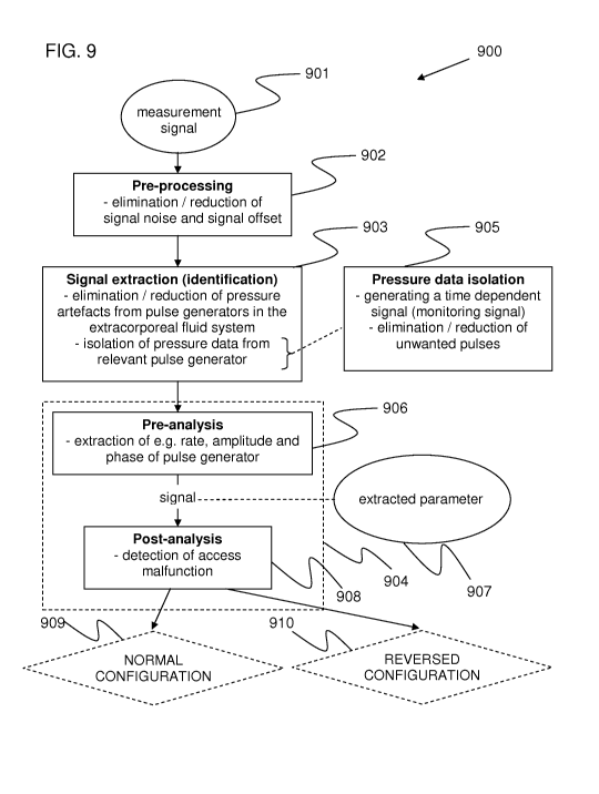
Fig. 9 is a flow chart that illustrates steps of a signal analysis process 900
according
to an embodiment of the present invention. It is initiated by receiving a
measurement
signal 901, e.g. from the venous, arterial and/or system pressure sensors,
e.g. 4b of Fig. 3,
comprising a number of pressure induced signal components.
The measurement signal comprises signals originating from one or more sources
and
thus constitutes a composite signal of the signals from said sources. The
measurement
signal may be used without further processing, although preferably, the
measurement
signal may be processed for extraction of pressure data originating from a
pulse generator
in the cardiovascular system. The extraction may be performed by filtering to
remove
unwanted pressure data.
In the cardiovascular system, the pulse generator may be a physiological
phenomena,
such as the pulse from the heart or breathing from the lungs. Other
physiological
phenomena pulse generators may be an autonomous system for blood pressure
regulation
and an autonomous system for body temperature regulation.
In the extracorporeal system, the pulse generator may be a fluid pump, such as
a
blood pump. The pump may be on the blood side or the fluid side of the
extracorporeal
blood flow circuit in a dialysis system. The pump may be of any type that
generates
pressure waves, for instance a peristaltic type of pump.
The pulse generators may be repetitive, such as the heart, breathing or pump
or non-
repetitive, such as pulses generated from coughing, sneezing, vomiting or
seizures.
Additionally, pulses may also be generated from separate, independent pulse
generators,
such as by rapid inflation of a blood pressure cuff to induce a pressure wave
which
propagates from the body part it is coupled to a blood vessel of the
cardiovascular system.
The signal analysis process may be divided into a pre-processing part 902, a
signal
extraction part 903 and an analysis part 904. The pre-processing part 902
includes
elimination or reduction of signal noise, e.g. measurement noise, and signal
offset, as
detailed in the section above relating to the data acquisition part 28. The
signal extraction
part 903 involves elimination or reduction of pressure artefacts originating
from pulse
generators in the extracorporeal blood flow circuit and isolation of pressure
data
originating from a relevant physiological phenomenon. In the context of the
present
disclosure, "pressure data isolation" 905 denotes a process of generating a
time-dependent
signal (also denoted monitoring signal herein) which is free or substantially
free from
pressure modulations caused by any unwanted physiological phenomena. Such
unwanted
physiological phenomena may vary between different applications, but generally
include
breathing, coughing, etc. In a case of cross-talk pressure modulations, all
such unwanted
physiological phenomena may be eliminated. The elimination of signal noise and
signal
offset, as well as the elimination of pressure artefacts, may be included in
algorithms for
pressure data isolation. For instance, the measurement signal may be band pass
filtered or
low pass filtered to isolate a heart signal, in a way such that signal noise
and/or signal
CA 02785758 2012-06-26
WO 2011/080188 PCT/EP2010/070550
18
offset and/or pressure artefacts are eliminated from the measurement signal.
The
elimination of pressure artefacts may thus be performed before, after or
during the pressure
data isolation.
In pre-analysis step 906 of the analysis part 904, one or more specific signal
analysis
algorithm(s) are applied for extraction of e.g. rate, amplitude and phase or
timing of the
one or more physiological phenomena. In post-analysis step 908, based on
predetermined
criteria, the output 907 of the signal analysis algorithm(s) is analysed, e.g.
by pattern
recognition, for signs a withdrawal and return line configuration, for
instance indicated by
detection of a normal configuration 909 or detection of a reversed
configuration 910. The
pressure data is then analysed and a parameter value is calculated. The access
configuration is then determined based at least partly on the parameter value.
The
parameter value is compared to a threshold value, interval or range to
determine a specific
condition. Alternatively or additionally, a signal feature may be extracted
from pressure
data and compared to a reference value, from which comparison a resulting
parameter
value may be compared to a threshold or interval. The reference data may
comprise
predicted data, predetermined data, secondary measurement data, or any
combinations
thereof.
The calculation may be designed such that the parameter value represents time,
amplitude or shape of the pulse. However, the detection may also be performed
in the
frequency domain by analysis of the amplitude and/or phase spectrum.
In the general case, one or more pumps are running or other sources of cyclic
or non-
cyclic repetitive and non-repetitive artefacts are present during the data
acquisition.
Information on the cyclic disturbances may be known from external sources,
e.g. other
sensors, or may be estimated or reconstructed from system parameters.
Cyclic pressure artefacts may originate from operating a peristaltic pump,
repetitive
actuation of valves, movements of membranes in balancing chambers. According
to the
findings in connection with the present invention, artefacts may also
originate from
mechanical resonance of system components such as swinging movements of blood
line
energized by e.g. a pump. Frequencies of blood line movements are given by the
tube
lengths and harmonics thereof and by the beating between any frequencies
involved, i.e.
between different self-oscillations and pump frequencies. These frequencies
may differ
between the venous and arterial lines. Mechanical fixation of the blood lines
and other free
components may remedy the problem of mechanical resonance. Alternatively, an
operator
may be instructed to touch or jolt the blood lines to identify natural
frequencies associated
with the blood lines, which information may be used in the analysis for
improved removal
of components not belonging to the pressure data of interest.
Examples of non-cyclic artefacts are subject movement, valve actuation,
movements
of tubings etc.
Various techniques for signal extraction will be discussed in a section
further below.
CA 02785758 2012-06-26
WO 2011/080188 PCT/EP2010/070550
19
The invention has mainly been described above with reference to a few
embodiments. However, as is readily appreciated by a person skilled in the
art, other
embodiments than the ones disclosed above are equally possible within the
scope and spirit
of the invention, which is defined and limited only be the appended patent
claims.
For example, the illustrated embodiments are applicable for surveillance of
all types
of extracorporeal blood flow circuits in which blood is taken from a patient's
circulation to
have a process applied to it before it is returned to the circulation. Such
blood flow circuits
include hemodialysis, hemofiltration, hemodiafiltration, plasmapheresis,
aphaeresis, extra-
corporeal membrane oxygenation, assisted blood circulation, and extracorporeal
liver
support/dialysis, bloodbanking, blood fraction separation (e.g. cells) of
donor blood, etc.
Further, the inventive monitoring techniques are applicable to any type of
pumping
device that generates pressure pulses in the first fluid containing system,
not only rotary
peristaltic pumps as disclosed above, but also other types of positive
displacement pumps,
such as linear peristaltic pumps, diaphragm pumps, as well as centrifugal
pumps.
Tests have shown that different evaluation parameters may be preferable in
different
situations. For example, the use of variance or averaged sum of multiple
pulses may
increase the detectability in various situations. Pattern recognition may be
resorted when
other detection methods fail.
Furthermore, although it may generally be enough for evaluation purposes to
involve
one parameter value, it may be advantageous to base an evaluation on
combinations of two
or more parameter values, such as to improve the reliability of the detection
of
configuration. The reliability may also be enhanced by increasing the
evaluation time
period. It may further be advantageous to increase the resolution in the
measurements to
further improve the reliability.
In the following, a few examples of situations involving extraction of a fluid
from a
flow, where recirculation reduces the efficiency of the process. In these
examples, any non-
physiological pulse generator may be utilized, such as a pressure generator.
In one example, the fluid connection is provided between a blood processing
circuit
and a container/machine, wherein blood is pumped from one container/machine
through a
blood processing device in the blood processing circuit and back to the
container/machine,
or to another container/machine downstream of the blood processing device. The
blood
processing device could be any known device configured to modify and/or
analyse the
blood.
In another example, the fluid connection is provided in a heart lung machine,
which
mechanically circulates and oxygenates blood for the body while bypassing the
heart and
lungs.
In a further example, the fluid connection is provided in an arrangement for
cleaning
and disinfecting the dialysis solution flow path of a dialysis machine, which
pumps a
CA 02785758 2012-06-26
WO 2011/080188 PCT/EP2010/070550
cleaning fluid via a flow path to a dialyser tubing. The cleaning fluid may
e.g. be hot
water, etc.
In all of these examples, and in other applications related to medical
treatment of
human or animal patients, it may be vital to monitor the positioning of access
devices.
5 Such monitoring can be accomplished according to the inventive concepts
disclosed
herein.
V. DETECTING REVERSED CONFIGURATION OF ACCESS DEVICES
This section relates to detection of an access configuration. It is based on
analysis of
10 one or more physiological or other signals that are extracted out of one or
more pressure
signals acquired from an extra-corporeal fluid system. Although mainly
referring to a
single signal, such as the heart amplitude in a venous (Hv) or arterial (Ha)
pressure signal,
the examples may be equally valid for combinations of signals, such as a ratio
(R=Hv/Ha)
between the heart amplitude in the venous and arterial pressure signals.
15 Although the strength of pulse pressure wave in the arterial signal in
general is
stronger than the pulse pressure wave in the venous signal, techniques
described in the
section Signal extraction and in greater detail in the Appendices A and B
enables sufficient
extraction of pressure data representing the pulse pressure wave in the venous
signal.
On a general level, the detection, may involve calculating an evaluation
parameter
20 value based on the isolated pressure data resulting from the aforesaid
signal extraction. The
evaluation parameter value is then analysed as part of a process for detecting
an access
configuration.
In the context of the present disclosure, irregularities in the
characteristics of a pulse
may imply that the pulse has deviating magnitude, shape, phase, timing or
other
measurable attributes. The assessment of normal or irregularity may involve
calculating an
evaluation parameter value based on the measurement signal(s) and comparing
the
parameter value to a threshold value. The assessment may further involve a
comparing the
parameter value to a predetermined reference value such as a previous
measurement of
pressure pulses from the same pulse generator, but with a deliberate normal or
irregular
configuration.
Different techniques for calculating such an evaluation parameter value are
further
disclosed and exemplified in Appendix B, in which the isolated pressure data
is a time-
dependent monitoring signal including pressure pulses originating from the
relevant
physiological phenomenon, and in which the monitoring signal is subjected to a
time
domain analysis. Thus, all techniques disclosed in Appendix B with respect to
the
evaluation of heart pulses, including the use of timing information, are
equally applicable
to other physiological phenomena, such as breathing, autonomic regulation of
body
temperature, and autonomic regulation of blood pressure, or combinations
thereof. In
addition to Appendix B, reference is also made to Applicant's International
patent
CA 02785758 2012-06-26
WO 2011/080188 PCT/EP2010/070550
21
publication W02009/156174, entitled "Methods and Devices for Monitoring the
Integrity
of a Fluid Connection", which is incorporated herein in its entirety by this
reference.
For instance, the timing information from Appendix B may be used to compare
arrival of pulses with withdrawal and return devices in normal and reversed
configurations
respectively, particularly for detection of a reversed configuration.
There are of course other techniques for calculating the evaluation parameter
value,
including other types of time domain analyses, as well as different types of
frequency
domain analyses, e.g. as indicated in the following.
Other factors, such as the medical history of the patient, e.g. heart status,
blood
pressure and heart rate may also be utilized for improving the performance of
the detection
and monitoring of the configuration of the withdrawal and return devices.
There are many different markers or measurements of a pulse which may indicate
the
relative location of access devices, such as pulse amplitude, local maximum,
local average,
shape/contour.
Pressure data extracted from the measurement signal may be represented as a
temporal pulse profile in the time domain. The temporal pulse profile may be
transformed
into a frequency spectrum and a phase spectrum, or only a frequency spectrum
and thus
loosing the timing information. From the pressure data, a parameter value may
be
calculated. The parameter value may be related to the amplitude, shape or
timing of the
pressure pulse.
Fig. 10 a) and b) show signal segments of the heart pulse in the arterial and
venous
lines. 101 and 121 represents the arterial signal in a a) normal and b)
reversed
configuration respectively. Other pulse sources such as the blood pump have
been
removed. The duration of each signal segment is two seconds. Also shown is an
average of
the signals 103 and 123 respectively. Fig. 10(a) shows the signals with the
needles in a
normal configuration (also denoted "correct positions"), and Fig. 10(b) shows
the signals
with the needles in a reverse configuration (also denoted "incorrect
positions" or
"backward positions"). Various measures may be extracted and compared to
thresholds in
determining the configuration.
Detection by amplitude
The needle that is positioned closest to the upstream anastomosis is expected
to pick
up the strongest heart pressure signal. Normally, the amplitude of the heart
pulse is larger
in the arterial pressure than in venous pressure when the needles are inserted
in correct
positions with the arterial needle closest to the upstream anastomosis. This
may be
explained by the pressure drop between the needles due to the access blood
flow and the
flow resistance of the fistula/graft. In addition, the compliance of the blood
line
components e.g. the venous drip chamber cause the venous signal to be
attenuated to a
larger degree than the arterial signal. Therefore, a reverse needle
configuration may be
CA 02785758 2012-06-26
WO 2011/080188 PCT/EP2010/070550
22
detected if e.g. the amplitude or integral of amplitudes of the heart pulse in
the venous
pressure is larger than in the arterial pressure, or if the relationship
between them is outside
a specific range, which could be generic or personalized.
This is for instance illustrated in section b) of Fig. 10 with needles in
reverse
configuration. For instance, a local maximum 130 of the heart signal of the
venous needle
pressure 122 is greater than a local maximum 132 of the heart signal of the
arterial needle
pressure 121 may indicate a reverse configuration. The quotient between two
local maxima
130 and 131 of the venous component exceeding a threshold may also be an
indicator of
reverse configuration, e.g. the quotient of the reversed configuration
indicated with
reference numbers 131/130 is less than the quotient of the normal
configuration 141/140.
Alternatively, the detection may be performed in the frequency domain by
analysis of the
amplitude and/or phase spectrum.
To illustrate the principles of detection by amplitude, examples with the
R=Hv/Ha
ratio measure from simulations performed based on a model explained in a
separate section
will be shown and discussed briefly in the following. Although the examples
show a ratio
measure, other amplitude measures may be used, such as Hv or Ha alone.
Fig. 12 shows a venous/arterial heart ratio compared to blood flow with a
normal
fistula at cnostant mean arterial pressure (MAP). It is seen that the ratio RR
in the reversed
configuration is greater than the ratio RN in the normal configuration.
Fig. 13 shows a venous/arterial heart ratio compared to the mean arterial
pressure
(MAP) with a normal fistula. It is again seen that the ratio RR in the
reversed configuration
is greater than the ratio RN in the normal configuration also in varying blood
flow Qb.
Fig. 14 shows a venous/arterial heart ratio compared to the dialysis monitor
blood
flow Qb in a fistula having intra-fistula stenosis, i.e. stenosis between the
access sites. It is
seen an extreme difference between the ratio RR in the reversed configuration
is greater
than the ratio RN in the normal configuration.
Detection by phase
Generally, a normal configuration of the needles is recognized in that the
heart pulse
arrives to the arterial needle before it arrives to the venous needle.
Analogous to the
cardiovascular system, it is assumed that the pressure pulse velocity is
higher in a
pressurized tube than in an evacuated tube. This may cause a delay of the
heart pulse in the
arterial line compared to the venous line. Since the average arterial pressure
is lower than
the venous pressure, the difference in arrival time of the sensor will show up
as a phase
difference. The phase difference will change in the reverse configuration
since the arterial
pressure in this case is even lower and the venous pressure is higher in this
configuration.
So, if the phase difference is larger than a threshold, the reverse
configuration may be
detected. In Fig. 10(a) for instance, the arterial signal 101 is greater in
amplitude but is
lagging a bit compared to the venous signal 102, which is seen in that the
venous signal
CA 02785758 2012-06-26
WO 2011/080188 PCT/EP2010/070550
23
102 has its lowest point 106 at around 0.3 seconds and where the arterial
signal has its
lowest point 107 at around 0.4 seconds. In Fig. 10(b) on the other hand, the
arterial signal
121 is smaller and is lagging much compared to the venous signal 122 which has
its lowest
point 116 at around 0.3 seconds and the arterial signal 121 has its lowest
point 117 at
around 0.5 seconds.
The detection may also be performed in the frequency domain by analysis of the
amplitude and/or phase spectrum.
Detection by shape
Damping and delay of the frequency components of the heart pulses affecting
their
shape depend the on the compliance of the fluid system. The compliance is a
function of
the actual pressure, flows and the physical properties of the system
comprising fistula and
extra-corporeal circuit. In a blood vessel with normal elasticity and/or
exposed to moderate
pressures, the heart pulse may present a more complex shape such as with a
greater number
of troughs and/or peaks, whereas the heart pulse in situations with a stiffer
vessel and/or
exposed to higher pressures may present a less complex shape with fewer
troughs/peaks.
The venous and arterial pressures and the blood flow between the needles in
the fistula
change with the arrangement of the needles. Therefore the shape of the heart
pulses may be
different in the reverse configuration compared to the normal configuration of
the needles
and may be used for detection of the reverse configuration. In the following,
a few
examples denoted i)-iv) will be described. A first example of an indicator may
be i) the
accentuation of bi- and tri-phases in the pulse. For instance, as seen in Fig.
10a)
representing a normal configuration, the venous signal 102 exhibits an
anacrotic or "bi-
phasic" shape determined by the troughs 106 and 108. However, in Fig. 10b),
the venous
signal 122 exhibits an catacrotic or "tri-phasic" shape determined by the
troughs 116, 118
and 119. A second indicator example may be ii) comparison of the shape of a
temporal
pulse profile with a reference pulse, for instance by cross-correlating the
measured pulse
with a reference pulse for identifying a best match with a reference pulse
representing
normal or reverse configuration. In particular, the reference pulse may have
been derived
from a subject's individual pulse characteristics for a normal and a reverse
configuration
respectively. A third indicator example is a iii) comparison with another
measurement, for
instance the venous pulse compared to the arterial pulse. Alternatively, in a
fourth indicator
example, iv) the venous or arterial pulse may be compared to a measurement of
the venous
or arterial pulse by an independent sensor. According to fifth indicator
example, v) the
width 133 of a component at a predetermined distance from a local maximum
exceeding a
threshold may be a further indicator of reverse configuration. Other indicator
examples
may involve vi) the slope, or rate of change, 134 and 135 of the pressure
pulse may also be
an indicator of the configuration. The slope may be measured on the leading
edge 136, 137
CA 02785758 2012-06-26
WO 2011/080188 PCT/EP2010/070550
24
or the trailing edge 138, 139. In the frequency domain, the corresponding
frequency and
phase spectra, which represent shape, may be compared with reference data.
The parameter value may further comprise a quotient between amplitudes of a
first
and a second local maxima in the pulse.
The parameter value may comprise a rate of change of the amplitude on a first
side,
second side or both sides of a local maximum in the pulse, for instance the
raise and/or fall
time or slope of the pulse. Alternatively, the rate of decrease in amplitude
from an
amplitude maximum may be cross-correlated with an exponential function e-kt by
varying
a factor k of exponential change.
The parameter value may further comprise the width 133 of the pulse at a
predetermined distance 150 from a local maximum in the pulse.
The temporal pulse profile may also be compared to a reference pulse profile
on a
more general basis, such as matching profiles by cross-correlation of the
signal profiles. A
match between a measured temporal pulse profile with a reference temporal
pulse profile
representative of a normal or reversed configuration may thus be indicated by
a cross-
correlation value exceeding a predetermined threshold. Comparison with
different
reference temporal pulse profiles may be advantageous where the representation
of normal
or reversed configurations may vary. Additionally, references pulse profiles
may be
compensated for distances between needles, mean arterial pressure, access flow
Qb etc.
The parameter value may further comprise a quotient between a relative time
difference between the artery and vein signal components in a normal versus a
reversed
position.
Artery and vein arrival times may also be visible as a phase shift in the
phase
spectrum of the frequency domain.
All measures or relation/ratio of these, i.e. magnitude, shape, phase etc, of
the heart,
breathing or other pulse signals in the venous and/or arterial pressures may
also be
compared to previous corresponding data and utilized for detection of reversed
positioning
of detection devices. For instance, if for a particular subject, the breathing
signal based on
arterial pressure normally is larger than breathing based on the venous
signal, but the
opposite relation appears during a treatment, it may be an indication of
accidental access
misplacement or faulty connection such as misplaced needles or reversed
connections of
needles or catheters to the blood lines.
Detection by pump signal analysis.
The pump pressure profiles in the venous line and in the arterial line may
differ if the
needles are reversed than in normal position. Detection may be carried out
analogous to the
previous cases.
The detection process may operate on the pressure signal(s) obtained from one
or
more of the pressure sensors in the extracorporeal circuit, optionally after
pre-processing.
CA 02785758 2012-06-26
WO 2011/080188 PCT/EP2010/070550
However, it may be advantageous to operate the detection process on isolated
pressure data
which is extracted from the pressure signal(s). For example, the isolated
pressure data may
be obtained by proper filtering, which suppresses/removes the physiological
pulses from
the pressure signal(s) while retaining at least part of pressure pulses
originating from a
5 pulse generator in the extracorporeal system, or in the dialysis machine
(i.e. the pressure
pulses that have been denoted "pressure artefacts" in the foregoing). The
filtering may
involve one or more of the filtering techniques described in Section VI below,
and in
particular the techniques disclosed under "Isolating pressure data from a
physiological
phenomenon", albeit adapted to isolate the pressure artefacts instead of the
pulses from a
10 physiological phenomenon). The skilled person should have no difficulty
adapting the
techniques in that chapter to achieve the desired filtering. With respect to
time domain
filtering, it should be noted that the techniques of Appendix A are
applicable. Thus,
although Appendix A is concerned with eliminating pressure pulses originating
from a
pulse generator in an extracorporeal circuit, such as a pumping device, it is
equally
15 applicable for eliminating pressure pulses originating from unwanted
physiological
phenomena. For example, an adaptive filter may be fed with a reconstructed
pressure
profile originating from all of the physiological phenomena to be removed, and
the signal
of interest may be extracted as an error signal of the adaptive filter.
Alternatively, the
adaptive filter may be fed with a reconstructed pressure profile of the
pressure artefacts,
20 and the signal of interest may be extracted as an estimated measurement
signal of the
adaptive filter. In addition to Appendix A, reference is also made to
Applicant's PCT
publication W02009/156175, entitled "Method and device for processing a time-
dependent measurement signal", which is incorporated herein in its entirety by
this
reference.
25 A particular embodiment relates to a cross-talk effect that may be detected
in one of
the pressure sensors in the extracorporeal circuit. When for instance a blood
pump is
running, its generated pressure pulses are transported in two directions from
the pump
along the extracorporeal circuit. A pressure sensor may then obtain the
pressure pulses
from two directions, i.e. a first set of pressure pulses which have been
transported through
the extracorporeal circuit only and a second set of pressure pulses having
been transported
through the cardiovascular system on its way.
Detection by timing information
According to an embodiment of the present invention, detection of a reversed
configuration may be detected by analysing the transit time of heart pulse
waves to the
pressure sensors, which differs since the pressure sensors are located at
different distances
from the heart and the length of blood lines interconnecting the heart and
pressure sensors
are different. Hence, a deviation in transit time from expected values
associated with a
normal configuration would represent a reversed configuration. For instance,
in a reversed
CA 02785758 2012-06-26
WO 2011/080188 PCT/EP2010/070550
26
configuration, the heart pulse wave would arrive earlier to the venous
pressure transducer
and later to the arterial pressure transducer. The difference of the arrival
times, i.e. between
the arterial pressure transducer and the venous pressure transducer would be
greater, it
would be approximately twice the distance between needle access sites divided
by the
heart wave velocity greater in a reversed position. According to an example,
assuming a
distance of between the access devices and a pulse propagation speed of 10m/s,
we have
0.1m*2/(lOm/s), which is approximately 20ms.
VI. SIGNAL EXTRACTION OF PHYSIOLOGICAL PULSE
In the following, embodiments for eliminating or reducing various pressure
artefacts
( also denoted "pump pulses" or "interference pulses") originating from one or
more pulse
generators in or associated with extracorporeal circuit will be described.
Then,
embodiments for isolating pressure data originating from a relevant
physiological
phenomenon among pressure pulses or pressure modulations originating from
other
physiological phenomena are described.
The pressure data to be extracted is not limited to a single physiological
phenomenon
and may originate from one or more physiological phenomena, including the
heart. As
used herein, the pressure data to be isolated is also denoted "subject pulses"
or "patient
pulses".
Elimination of artefacts
Elimination of artefacts may be provided by:
- Controlling a pulse generator in the extracorporeal blood flow circuit, such
as a
pump
o By temporarily shutting down the pulse generator;
o Shifting the frequency of the pulse generator;
- Low pass, band pass or high pass filtering;
- Spectral analysis and filtering in the frequency domain;
- Time domain filtering.
Controlling a pulse generator
Artefacts from a pulse generator, such as a pump, in the extracorporeal fluid
circuit
may be avoided by temporarily shutting down (disabling) the pulse generator,
or by
shifting the frequency of the pulse generator away from frequencies of one or
more
relevant physiological phenomena.
A feedback control with respect to the heart rate, e.g. obtained from a
dedicated
pulse sensor attached to the patient or obtained via analysis of previous
parts of the
monitoring signal, may be used to set the pump frequency optimally for
detection of heart
pulses. Similar feedback control may be used to eliminate artefacts with
respect to pressure
CA 02785758 2012-06-26
WO 2011/080188 PCT/EP2010/070550
27
pulses originating from breathing, e.g. based on a breathing signal from an
independent
source, such as a capnograph instrument. Hence, the control unit 23 of Fig. 1
may be
operated to control the pump frequency in order to facilitate the detection of
the subject
pulses, e.g. the pump frequency is controlled to minimize any overlap in
frequency
between the pump pulses and the subject pulses. For example, the pump
frequency may be
periodically increased and decreased around the overlap frequency, so as to
maintain the
overall blood flow rate. In a variant, the pump frequency is instead
controlled so as to
synchronize the rate of pump pulses with the rate of subject pulses while
applying a phase
difference between the pump pulses and the subject pulses. Thereby, the pump
pulses and
the subject pulses will be separated in time, and the subject pulses may be
detected in the
time domain, even without removal of the pump pulses. The phase difference may
be
approximately 180 , since this may maximize the separation of the pump pulses
and the
subject pulses in the time domain. This so-called phase-locking technique may
be activated
when it is detected that the rate of subject pulses approaches the rate of
pump pulses, or
vice versa.
Applying low pass, band pass or high pass filters
The input signal to step 903 (Fig. 9) may be fed into a filter, e.g. digital
or analogue,
with frequency characteristics, such as frequency range and/or centre of
frequency range,
matched to the frequencies generated by a pulse generator, such as a pump, in
the
extracorporeal circuit. For instance, in a case where the pulse generator,
such as a pump,
operates within the frequency range of 1Hz, a suitable low pass filter may be
applied in
order to remove pressure artefacts above 1 Hz while retaining frequency
components of the
physiological phenomenon below 1 Hz. Correspondingly, a high pass filter may
be applied
to retain frequency components above the frequency of the pulse generator.
Alternatively,
one or more notch filters or the like may be utilised to remove/attenuate
frequencies in one
or more confined ranges.
Spectral analysis and filtering in the frequency domain;
The input signal to part 133 may be subjected to a spectral analysis, e.g. by
applying
a Fourier transformation technique, such as FFT (Fast Fourier Transform) to
convert the
input signal into the frequency domain. The resulting energy spectrum
(amplitude
spectrum) may then be multiplied by an appropriate filter function and then re-
transformed
into the time domain. There are many alternative and equivalent filtering
techniques
available to the skilled person. The Fourier transformation technique may also
be used for
tailoring/adjusting filters used for eliminating artefacts.
Time domain filtering
CA 02785758 2012-06-26
WO 2011/080188 PCT/EP2010/070550
28
Artefact elimination by filtering in the time domain is further disclosed and
exemplified in Appendix A. In the context of Appendix A, the input signal to
step 903
(Fig. 9) is denoted "measurement signal", and the resulting "filtered signal
e(n)"
corresponds to, or may be processed for extraction of, the monitoring signal.
In addition to
Appendix A, reference is also made to Applicant's International patent
publication
W02009/156175, entitled "Method and device for processing a time-dependent
measurement signal", which is incorporated herein in its entirety by this
reference.
Isolating pressure data from a physiological phenomenon
Isolating pressure data originating from a relevant physiological phenomenon
may be
provided by any or a combination of-
- Low pass, band pass or high pass filtering;
- Spectral analysis and filtering in the frequency domain; or
- Time domain filtering.
Applying low pass, band pass or high pass filters
The input signal to step 905 may be fed into a filter, e.g. digital or
analogue, with
frequency characteristics, such as frequency range and/or centre of frequency
range,
matched to the frequencies of pressure pulses from a relevant physiological
phenomenon
where e.g. in case the isolation concerns:
- Heart pulses, a frequency range substantially of 0.5 - 4 Hz will be allowed
to
pass the filter;
- Breathing, a frequency range substantially of 0.15 - 0.4 Hz will be allowed
to
pass the filter;
- Blood pressure regulation due to the autonomous system, a frequency range
substantially of 0.04 - 0.15 Hz will be allowed to pass the filter; and
- Temperature regulation due to the autonomous system, a frequency range
substantially of 0.001 - 0.1 Hz will be allowed to pass the filter.
Spectral analysis and filtering in the frequency domain
The input signal to step 905 may be subjected to a spectral analysis, e.g. by
applying
a Fourier transformation technique, such as FFT (Fast Fourier Transform) to
convert the
input signal into the frequency domain. The resulting energy spectrum
(amplitude
spectrum) may then be multiplied by an appropriate filter function and then re-
transformed
into the time domain. There are many alternative and equivalent filtering
techniques
available to the skilled person.
Time domain filtering
CA 02785758 2012-06-26
WO 2011/080188 PCT/EP2010/070550
29
The signal of interest may be extracted from the input signal to step 905 as
an error
signal of an adaptive filter. The adaptive filter is fed with both the
measured pressure
signal and a predicted signal profile of a cyclic disturbance. The cyclic
disturbance may be
any of the other physiological signals (e.g. heart pulsation or breathing).
Particularly, a
reconstructed pressure profile originating from the unwanted physiological
phenomenon
may be input to the adaptive filter. This and other time domain filtering
techniques for
removing unwanted signal components from a measurement signal is further
disclosed and
exemplified in Appendix A. Although Appendix A is concerned with eliminating
first
pulses originating from a pulse generator in an extracorporeal circuit, such
as a pumping
device, it is equally applicable for eliminating first pulses originating from
unwanted
physiological phenomena, as long as a predicted signal profile of the first
pulses may be
obtained. The skilled person realizes that such a predicted signal profile may
be obtained in
any of the ways described in Appendix A. In addition to Appendix A, reference
is also
made to aforesaid W02009/156175.
Some of the filtering techniques described above may automatically be achieved
by
down-sampling, since it may be taken care of by the anti-aliasing filter
included in a down-
sampling signal processing algorithm. Additionally, some of the above
described filtering
techniques may also be achieved directly in hardware, e.g., in the Analogue-to-
Digital
conversion by choosing an appropriate sample frequency, i.e. due to the anti-
aliasing filter
which is applied before sampling.
VII. MODELLING OF A FISTULA
For a better understanding of the inventive concept, modelling of an exemplary
fistula of Fig. 2 will be described in the following. Other fistula
configurations may be
modelled according to the same principles, although requiring adjustments to
the actual
configuration.
The fistula configuration may be translated into a hydraulic model as is shown
in fig.
11, where the blood flow in the access devices (111, 112) are in a reversed
configuration,
i.e. with blood withdrawal 112 at venous access site and blood return 111 at
arterial access
site. U represents a systemic blood pressure such as the mean arterial
pressure MAP or a
pressure pulse close to the heart. Table 1 below lists the parameter
definitions that are used
in fig. 11.
Since the model is dynamic, it may be applied to pressure levels as well as
pressure
variations.
Table 1. Parameter definitions
UFR: ultrafiltration rate (i.e. fluid removal rate)
Qb: blood flow of dialysis machine
CA 02785758 2012-06-26
WO 2011/080188 PCT/EP2010/070550
ci: flow admittance between anastomosis and first access point, `pre-fistula
admittance'
c2: flow admittance between first and second access points, `intra-fistula
admittance'
c3: flow admittance between second access point and common venous return
vessel, `post-
fistula admittance
c4: flow admittance in vein between common venous return vessel and venous
return to
heart
c5: flow admittance in artery entering the fistula
c6: flow admittance in vessels to hand
no: arterial blood pressure, e.g. mean arterial pressure (MAP)
Reversed position of access needles:
ul: blood pressure at access point 1
u2: blood pressure at access point 2
u3: blood pressure at common venous return vessel
Normal position of access needles:
u' 1: blood pressure at access point 1
u'2: blood pressure at access point 2
u'3: blood pressure at common venous return vessel
io:blood flow pumped out of the fistula, Qb
ii: blood flow pumped back to the fistula: Qb-UFR
i2: arterial blood flow required by the tissues downstream of the fistula,
ia: blood access flow entering the fistula
Ha: heart pressure amplitude at "A" in fig 11, i.e. at arterial pressure
transducer
Han: Ha when normal needle position
Har: Ha when reversed needle position
Hv: heart pressure amplitude at "V" in fig 11, i.e. at venous pressure
transducer
Hvn: Hv when normal needle position
Hvr: Hv when reversed needle position
R: Ven/Art heart amplitude ratio, i.e. R= Hv/Ha,
Rn: Hvn/Han
Rr: Hvr/Har
Rrhist: "average of Rr over a number of previous treatments"
Rrmod: "detection limit for R in reversed position that is based on modelling"
In the model, it has been assumed that the blood pressure of the artery e.g.
mean
arterial pressure MAP is controlled to a constant value (uo), the demand of
nutrition and
oxygen of tissue, the fistula arm is constant (i2) and that all flow
admittances (ci - c4) do
not vary. Moreover, it is assumed that in a normal fistula configuration the
pre-fistula
5 admittance (ci) is less than the post-fistula admittance (c3) which, in
turn, is less than the
intra-fistula admittance (ca). Both c5 and c6 are omitted in the modelling,
since they are
assumed to be very large. Moreover, the magnitude of total fistula flow
admittance is
chosen as to result in a physiological range of fistula flow.
CA 02785758 2012-06-26
WO 2011/080188 PCT/EP2010/070550
31
The sum of the flows entering each of connections 1, 2 and 3 are zero -
therefore the
following equations can be defined:
ci(uo - ui) + ii + c2(u2 - ui) = 0 (1)
C2(UI - U2) - to + C03 -U2) = 0 (2)
C02 - U3) + i2 - C4U3 = 0 (3)
u1, u2 and u3 may then be calculated if no, ii to i2 and ci to c4 are known.
With the blood flowing in a direction according to a normal configuration,
blood
pressures u1' and u2' at the first and second access points respectively, may
be determined
analogously after replacing ii with -io and io with -il in the equations
above.
Several simulations have been performed with the needles in reversed and
normal
position to illustrate R, Ha and Hv at different settings of blood pressure,
blood flow,
absolute and relative flow resistances (inverse of conductances). Results from
the
simulations are shown in figures 12 to 14, explained above under section V.
VIII. SIGNAL PROCESSING
According to one embodiment of the present invention, it relates to a method
for
automatic detection of needle reversal based on analysis of readings given by
one or more
pressure sensors integrated in a dialysis monitor. The pressure readings of
each pressure
sensor form a measurement signal. The measurement signal may comprise pressure
data
from different pulse generators in the extracorporeal circuit and in the
subject. Such pulse
generators include the blood pump, the heart and other physiological phenomena
in the
subject, such as breathing. Before analysis, the measurement signal may be
processed to
isolate a pressure data from a particular origin, for instance the heart,
breathing or a pump
in the extracorporeal circuit. Alternatively, the amplitude and/or phase
spectra of the
composite signal may be analysed. The aforesaid measurement signal comprises
continuously detected measurements from the pressure sensor(s), thus
representing an
inherently time-dependent signal, and the aforesaid pressure data comprises at
least a part
of a pressure pulse or one or more pressure pulses originating from one or
more pulse
generators, such as the heart, the breathing system of the subject, one or
more pumps or
valves in the extracorporeal circuit, etc. Unless otherwise stated, it will in
the following be
assumed that the heart pulse is used, although the text is also applicable to
the use of the
breathing pulse or the pump pulse.
The isolation of pressure data from a particular origin may involve filtering.
However, in situations where no efficient filtration can be accomplished, for
instance
CA 02785758 2012-06-26
WO 2011/080188 PCT/EP2010/070550
32
where the noise-to-signal ratio is too large, it may be advantageous to
control the pump
behaviour. Methods of filtering are detailed above in a separate section.
The signal extraction part 903 involves elimination or reduction of pressure
artefacts
originating from pulse generators in the extracorporeal blood flow circuit and
isolation of
pressure data originating from a relevant pulse generator. In the context of
the present
disclosure, "pressure data isolation" 905 denotes a process of generating a
time-dependent
signal (also denoted monitoring signal herein) which is free or substantially
free from
pressure modulations caused by any unwanted pressure generator. Such unwanted
pressure
generators may vary between different applications, but generally include a
blood pump
and/or breathing signal components. The elimination of signal noise and signal
offset, as
well as the elimination of pressure artefacts, may be included in algorithms
for pressure
data isolation. For instance, the measurement signal may be band pass filtered
or low pass
filtered to isolate a breathing signal, in a way such that signal noise and/or
signal offset
and/or pressure artefacts are eliminated from the measurement signal. The
elimination of
pressure artefacts may thus be performed before, after or during the pressure
data isolation.
For instance, in situations where harmonics of the pump (fo/2, fo, 2fo, 3fo ,
etc)
overlap or are near the frequency of the physiological signal, such as the
heart or breathing,
the rotation speed of the pump may be adjusted so that the frequencies are
separated. For
instance, where the fundamental frequency of the pump to is 1Hz, the pump may
be
adjusted to a new frequency of 2/3Hz, such that the heart frequency operates
in the
frequency range between the fundamental frequency of the pump and its first
harmonic 2fo.
Alternatively, the rotation speed of the pump may be adjusted to a relative
level, such as 25
percent, from the frequency of the heart. Removal of the pump pulses, or vice
versa, may
then be accomplished easier.
Alternatively, the blood pump may be temporarily stopped to accomplish
complete
removal of influence caused by the pump. In order to obtain the necessary
measurement
data, while avoiding blood coagulation, it may be advantageous to stop the
pump for at
least 30 seconds and maximum five minutes. Longer duration of the stop
increases the
precision of the determination, which may also be achieved by repeatedly
stopping the
pump for shorter time periods.
The test for detection of configuration may be activated manually by a nurse
or
medical technician, although preferably the test is initiated automatically by
an algorithm
implemented in the dialysis machine, such as during a treatment start-up
initiating routine.
The term "comprises/comprising" when used in this specification is taken to
specify
the presence of stated features, integers, steps or components. However, the
term does not
preclude the presence or addition of one or more additional features,
integers, steps or
components or groups thereof.
The invention is not restricted to the described embodiments in the figures,
but may
be varied freely within the scope of the claims.
CA 02785758 2012-06-26
WO 2011/080188 PCT/EP2010/070550
33
It is to be understood that Appendix A and Appendix B are to be treated as
integral
parts of the present application. However, reference numerals are defined
within the
context of each Appendix separately. In the event of conflicting use of
terminology
between the Appendix A, Appendix B and the main specification, the terminology
should
be interpreted within the context of Appendix A, Appendix B and the main
specification,
respectively.
CA 02785758 2012-06-26
WO 2011/080188 PCT/EP2010/070550
34
APPENDIX A
Brief Description of the Drawings
Exemplifying embodiments of the invention will now be described in more detail
with reference to the accompanying schematic drawings.
Fig. Al is a schematic view of a general fluid containing system in which the
inventive data processing may be used for filtering a pressure signal.
Fig. A2 is a flow chart of a monitoring process according to an embodiment of
the
invention.
Fig. A3(a) is a plot of a pressure signal as a function of time, and Fig.
A3(b) is a plot
of the pressure signal after filtering.
Fig. A4 is a schematic view of a system for hemodialysis treatment including
an
extracorporeal blood flow circuit.
Fig. A5(a) is a plot in the time domain of a venous pressure signal containing
both
pump frequency components and a heart signal, and Fig. A5(b) is a plot of the
corresponding signal in the frequency domain.
Fig. A6 is a plot of a predicted signal profile originating from a peristaltic
pump in
the system of Fig. A4.
Fig. A7 is a flow chart of a process for obtaining the predicted signal
profile.
Fig. A8 is a plot to illustrate an extrapolation process for generating the
predicted
signal profile.
Fig. A9(a) is a plot to illustrate an interpolation process for generating the
predicted
signal profile, and Fig. A9(b) is an enlarged view of Fig. A9(a).
Fig. A10(a) represents a frequency spectrum of a pressure pulse originating
from a
pumping device at one flow rate, Fig. A10(b) represents corresponding
frequency spectra
for three different flow rates, wherein each frequency spectrum is given in
logarithmic
scale and mapped to harmonic numbers, Fig. A10(c) is a plot of the data in
Fig. A10(b) in
linear scale, and Fig. A10(d) is a phase angle spectrum corresponding to the
frequency
spectrum in Fig. A10(a).
Fig. All is schematic view of an adaptive filter structure operable to filter
a
measurement signal based on a predicted signal profile.
Fig. A12(a) illustrates a filtered pressure signal (top) and a corresponding
heart
signal (bottom), obtained from a venous pressure sensor, and Fig. A12(b)
illustrates a
filtered pressure signal (top) and a corresponding heart signal (bottom),
obtained from an
arterial pressure sensor.
Detailed Description of Exemplifying Embodiments
In the following, exemplifying embodiments of the invention will be described
with
reference to fluid containing systems in general. Thereafter, the embodiments
and
CA 02785758 2012-06-26
WO 2011/080188 PCT/EP2010/070550
implementations of the invention will be further exemplified in the context of
systems for
extracorporeal blood treatment.
Throughout the following description, like elements are designated by the same
reference signs.
5
GENERAL
Fig. Al illustrates a fluid containing system in which a fluid connection C is
established between a first fluid containing sub-system S 1 and a second fluid
containing
sub-system S2. The fluid connection C may or may not transfer fluid from one
sub-system
10 to the other. A first pulse generator 3 is arranged to generate a series of
pressure waves in
the fluid within the first sub-system S 1, and a second pulse generator 3' is
arranged to
generate a series of pressure waves in the fluid within the second sub-system
S2. A
pressure sensor 4a is arranged to measure the fluid pressure in the first sub-
system S 1.
Pressure waves generated by the second pulse generator 3' will travel from the
second sub-
15 system S2 to the first sub-system S1, via the connection C, and thus second
pulses
originating from the second pulse generator 3' will be detected by the
pressure sensor 4a in
addition to first pulses originating from the first pulse generator 3. It is
to be noted that
either one of the first and second pulse generators 3, 3' may include more
than one pulse-
generating device. Further, any such pulse-generating device may or may not be
part of the
20 respective sub-system S1, S2.
The system of Fig. Al further includes a surveillance device 25 which is
connected
to the pressure sensor 4a, and possibly to one or more additional pressure
sensors 4b, 4c, as
indicated in Fig. Al. Thereby, the surveillance device 25 acquires one or more
pressure
signals that are time-dependent to provide a real time representation of the
fluid pressure in
25 the first sub-system S1.
Generally, the surveillance device 25 is configured to monitor a functional
state or
functional parameter of the fluid containing system, by isolating and
analysing one or more
second pulses in one of the pressure signals. As will be further exemplified
in the
following, the functional state or parameter may be monitored to identify a
fault condition,
30 e.g. in the first or second sub-systems S 1, S2, the second pulse generator
3' or the fluid
connection C. Upon identification of a fault condition, the surveillance
device 25 may
issue an alarm or warning signal and/or alert a control system of the first or
second sub-
systems S 1, S2 to take appropriate action. Alternatively or additionally, the
surveillance
device 25 may be configured to record or output a time sequence of values of
the
35 functional state or parameter.
Depending on implementation, the surveillance device 25 may use digital
components or analog components, or a combination thereof, for receiving and
processing
the pressure signal. The device 25 may thus be a computer, or a similar data
processing
device, with adequate hardware for acquiring and processing the pressure
signal in
CA 02785758 2012-06-26
WO 2011/080188 PCT/EP2010/070550
36
accordance with different embodiments of the invention. Embodiments of the
invention
may e.g. be implemented by software instructions that are supplied on a
computer-readable
medium for execution by a processor 25a in conjunction with a memory unit 25b
in the
computer.
Typically, the surveillance device 25 is configured to continuously process
the time-
dependent pressure signal(s) to isolate any second pulses. This processing is
schematically
depicted in the flow chart of Fig. A2. The illustrated processing involves a
step 201 of
obtaining a first pulse profile u(n) which is a predicted temporal signal
profile of the first
pulse(s), and a step 202 of filtering the pressure signal d(n), or a pre-
processed version
thereof, in the time-domain, using the first pulse profile u(n), to
essentially eliminate or
cancel the first pulse(s) while retaining the second pulse(s) contained in
d(n). In the context
of the present disclosure, n indicates a sample number and is thus equivalent
to a (relative)
time point in a time-dependent signal. In step 203, the resulting filtered
signal e(n) is then
analysed for the purpose of monitoring the aforesaid functional state or
parameter.
The first pulse profile is a shape template or standard signal profile,
typically given
as a time-sequence of data values, which reflects the shape of the first pulse
in the time
domain. The first pulse profile is also denoted "predicted signal profile" in
the following
description.
By "essentially eliminating" is meant that the first pulse(s) is(are) removed
from the
pressure signal to such an extent that the second pulse(s) can be detected and
analysed for
the purpose of monitoring the aforesaid functional state or parameter.
By filtering the pressure signal in the time-domain, using the first pulse
profile, it is
possible to essentially eliminate the first pulses and still retain the second
pulses, even if
the first and second pulses overlap or nearly overlap in the frequency domain.
Such a
frequency overlap is not unlikely, e.g. if one or both of the first and second
pulses is made
up of a combination of frequencies or frequency ranges.
Furthermore, the frequency, amplitude and phase content of the first pulse or
the
second pulse may vary over time. Such variations may be the result of an
active control of
the first and/or second pulse generator 3, 3', or be caused by drifts in the
first and/or
second pulse generator 3, 3' or by changes in the hydrodynamic properties of
the sub-
systems S1, S2 or the fluid connection C. Frequency variations may occur,
e.g., when the
second pulse generator 3' is a human heart, and the second sub-system S2 thus
is the blood
system of a human. In healthy subjects under calm conditions, variations in
heart rhythm
(heart rate variability, HRV) may be as large as 15%. Unhealthy subjects may
suffer from
severe heart conditions such as atrial fibrillation and supraventricular
ectopic beating,
which may lead to an HRV in excess of 20%, and ventricular ectopic beating,
for which
HRV may be in excess of 60%. These heart conditions are not uncommon among,
e.g.,
dialysis patients.
CA 02785758 2012-06-26
WO 2011/080188 PCT/EP2010/070550
37
Any frequency overlap may make it impossible or at least difficult to isolate
the
second pulses in the pressure signal by conventional filtering in the
frequency domain, e.g.
by operating a comb filter and/or a combination of band-stop or notch filters,
typically
cascade coupled, on the pressure signal to block out all frequency components
originating
from the first pulse generator 3. Furthermore, frequency variations make it
even harder to
successfully isolate second pulses in the pressure signal, since the frequency
overlap may
vary over time. Even in the absence of any frequency overlap, frequency
variations make it
difficult to define filters in the frequency domain.
Depending on how well the first pulse profile represents the first pulse(s) in
the
pressure signal, it may be possible to isolate the second pulses by means of
the inventive
filtering in the time-domain even if the first and second pulses overlap in
frequency, and
even if the second pulses are much smaller in amplitude than the first pulses.
Still further, the inventive filtering in the time domain may allow for a
faster
isolation of second pulses in the pressure signal than a filtering process in
the frequency
domain. The former may have the ability to isolate a single second pulse in
the pressure
signal whereas the latter may need to operate on a sequence of first and
second pulses in
the pressure signal. Thus, the inventive filtering may enable faster
determination of the
functional state or functional parameter of the fluid containing system.
The effectiveness of the inventive filtering is exemplified in Fig. A3, in
which Fig.
A3(a) shows an example of a time-dependent pressure signal d(n) containing
first and
second pulses with a relative magnitude of 10:1. The first and second pulses
have a
frequency of 1 Hz and 1.33 Hz, respectively. Due to the difference in
magnitude, the
pressure signal is dominated by the first pulses. Fig. A3(b) shows the time-
dependent
filtered signal e(n) that is obtained after applying the inventive filtering
technique to the
pressure signal d(n). The filtered signal e(n) is made up of second pulses and
noise. It
should be noted that there is an absence of second pulses after about 4
seconds, which may
be observed by the surveillance device (25 in Fig. Al) and identified as a
fault condition of
the fluid containing system.
Reverting to Fig. A2, the inventive data processing comprises two main steps:
a
determination of the first pulse profile u(n) (step 201) and a removal of one
or more first
pulses from a measurement signal d(n) using the first pulse profile u(n) (step
202).
There are many ways to implement these main steps. For example, the first
pulse
profile (standard signal profile) may be obtained in a reference measurement,
based on a
measurement signal from one or more of the pressure sensors 4a-4c in the first
sub-system
S1, suitably by identifying and possibly averaging a set of first pulse
segments in the
measurement signal(s). The first pulse profile may or may not be updated
intermittently
during the actual monitoring of the aforesaid functional state or parameter.
Alternatively, a
predetermined (i.e. predefined) standard signal profile may be used, which
optionally may
be modified according to a mathematical model accounting for wear in the first
pulse
CA 02785758 2012-06-26
WO 2011/080188 PCT/EP2010/070550
38
generator, fluid flow rates, tubing dimensions, speed of sound in the fluid,
etc. Further, the
removal may involve subtracting the first pulse profile from the measurement
signal at
suitable amplitude and phase. The phase may be indicated by phase information
which
may be obtained from a signal generated by a phase sensor coupled to the first
pulse
generator 3, or from a control signal for the first pulse generator 3.
The inventive filtering may also be combined with other filtering techniques
to
further improve the quality of the filtered signal e(n). In one embodiment,
the filtered
signal e(n) could be passed through a bandpass filter with a passband in the
relevant
frequency range for the second pulses. If the second pulses originate from a
human heart,
the passband may be located within the approximate range of 0.5-4 Hz,
corresponding to
heart pulse rates of 30-240 beats per minute. In another embodiment, if the
current
frequency range (or ranges) of the second pulses is known, the passband of the
bandpass
filter could be actively controlled to a narrow range around the current
frequency range.
For example, such an active control may be applied whenever the rates of first
and second
pulses are found to differ by more than a certain limit, e.g. about 10%. The
current
frequency range may be obtained from the pressure signal, either by
intermittently shutting
off the first pulse generator 3, or intermittently preventing the first pulses
from reaching the
relevant pressure sensor 4a-4c. Alternatively, the current frequency range may
be obtained
from a dedicated sensor in either the first or the second sub-systems S 1, S2,
or based on a
control unit (not shown) for the second pulse generator 3'. According to yet
another
alternative, the location and/or width of the passband could be set, at least
in part, based on
patient-specific information, i.e. existing data records for the patient, e.g.
obtained in
earlier treatments of the same patient. The patient-specific information may
be stored in an
internal memory of the surveillance device (25 in Fig. Al), on an external
memory which
is made accessible to the surveillance device, or on a patient card where the
information is
e.g. transmitted wirelessly to the surveillance device, e.g. by RFID (Radio
Frequency
IDentification).
These and other embodiments will be explained in further detail below, within
the
context of a system for extracorporeal blood treatment. To facilitate the
following
discussion, details of an exemplifying extracorporeal blood flow circuit will
be first
described.
MONITORING IN AN EXTRACORPOREAL BLOOD FLOW CIRCUIT
Fig. A4 shows an example of an extracorporeal blood flow circuit 20 of the
type
which is used for dialysis. The extracorporeal blood flow circuit 20 (also
denoted
"extracorporeal circuit") comprises components 1-14 to be described in the
following.
Thus, the extracorporeal circuit 20 comprises an access device for blood
extraction in the
form of an arterial needle 1, and an arterial tube segment 2 which connects
the arterial
needle 1 to a blood pump 3 which may be of peristaltic type, as indicated in
Fig. A4. At the
CA 02785758 2012-06-26
WO 2011/080188 PCT/EP2010/070550
39
inlet of the pump there is a pressure sensor 4b (hereafter referred to as
"arterial sensor")
which measures the pressure before the pump in the arterial tube segment 2.
The blood
pump 3 forces the blood, via a tube segment 5, to the blood-side of a dialyser
6. Many
dialysis machines are additionally provided with a pressure sensor 4c
(hereafter referred to
as "system sensor") that measures the pressure between the blood pump 3 and
the dialyser
6. The blood is lead via a tube segment 10 from the blood-side of the dialyser
6 to a venous
drip chamber or deaeration chamber 11 and from there back to the patient via a
venous
tube segment 12 and an access device for blood reintroduction in the form of a
venous
needle 14. A pressure sensor 4a (hereafter referred to as "venous sensor") is
provided to
measure the pressure on the venous side of the dialyser 6. In the illustrated
example, the
pressure sensor 4a measures the pressure in the venous drip chamber. Both the
arterial
needle 1 and the venous needle 14 are connected to the patient by means of a
blood vessel
access. The blood vessel access may be of any suitable type, e.g. a fistula, a
Scribner-
shunt, a graft, etc. Depending on the type of blood vessel access, other types
of access
devices may be used instead of needles, e.g. catheters. The access devices 1,
14 may
alternatively be combined into a single unit.
In relation to the fluid containing system in Fig. Al, the extracorporeal
circuit 20
corresponds to the first sub-system S 1, the blood pump 3 (as well as any
further pulse
source(s) within or associated with the extracorporeal circuit 20, such as a
dialysis solution
pump, valves, etc) corresponds to the first pulse generator 3, the blood
system of the
patient corresponds to the second sub-system S2, and the fluid connection C
corresponds to
at least one of the venous-side and arterial-side fluid connections between
the patient and
the extracorporeal circuit 20.
In Fig. A4, a control unit 23 is provided, i.a., to control the blood flow in
the
extracorporeal circuit 20 by controlling the revolution speed of the blood
pump 3. The
extracorporeal circuit 20 and the control unit 23 may form part of an
apparatus for
extracorporeal blood treatment, such as a dialysis machine. Although not shown
or
discussed further it is to be understood that such an apparatus performs many
other
functions, e.g. controlling the flow of dialysis fluid, controlling the
temperature and
composition of the dialysis fluid, etc.
The system in Fig. A4 also includes a surveillance/monitoring device 25, which
is
connected to receive a pressure signal from at least one of the pressure
sensors 4a-4c and
which executes the inventive data processing. In the example of Fig. A4, the
surveillance
device 25 is also connected to the control unit 23. Alternatively or
additionally, the device
25 may be connected to a pump sensor 26 for indicating the revolution speed
and/or phase
of the blood pump 3. It is to be understood that the surveillance device 25
may include
inputs for further data, e.g. any other system parameters that represent the
overall system
state (see e.g. discussion with reference to Fig. A7 below). The device 25 is
tethered or
wirelessly connected to a local or remote device 27 for generating an
audible/visual/tactile
CA 02785758 2012-06-26
WO 2011/080188 PCT/EP2010/070550
alarm or warning signal. Alternatively or additionally, either device 25, 27
may include a
display or monitor for displaying the functional state or parameter resulting
from the
analysis step (203 in Fig. A2), and/or the filtered signal e(n) resulting from
the filtering
step (202 in Fig. A2), e.g. for visual inspection.
5 In Fig. A4, the surveillance device 25 comprises a data acquisition part 28
for pre-
processing the incoming signal(s), e.g. including an A/D converter with a
required
minimum sampling rate and resolution, one or more signal amplifiers, and one
or more
filters to remove undesired components of the incoming signal(s), such as
offset, high
frequency noise and supply voltage disturbances.
10 After the pre-processing in the data acquisition part 28, the pre-processed
pressure
signal is provided as input to a main data processing part 29, which executes
the inventive
data processing. Fig. A5(a) shows an example of such a pre-processed pressure
signal in
the time domain, and Fig. A5(b) shows the corresponding power spectrum, i.e.
the pre-
processed pressure signal in the frequency domain. The power spectrum reveals
that the
15 detected pressure signal contains a number of different frequency
components emanating
from the blood pump 3. In the illustrated example, there is a frequency
component at the
base frequency (fo) of the blood pump (at 1.5 Hz in this example), as well as
its harmonics
2fo, 3f0 and 4f0. The base frequency, also denoted pump frequency in the
following, is the
frequency of the pump strokes that generate pressure waves in the
extracorporeal circuit
20 20. For example, in a peristaltic pump of the type shown in Fig. A4, two
pump strokes are
generated for each full revolution of the rotor 3a. Fig. A5(b) also indicates
the presence of
a frequency component at half the pump frequency (0.5fo) and harmonics
thereof, in this
example at least fo, 1.5f0, 2f0 and 2.5f0. Fig. A5(b) also shows a heart
signal (at 1.1 Hz)
which in this example is approximately 40 times weaker than the blood pump
signal at the
25 base frequency fo.
The main data processing part 29 executes the aforesaid steps 201-203. In step
202,
the main data processing part 29 operates to filter the pre-processed pressure
signal in the
time domain, and outputs a filtered signal or monitoring signal (e(n) in Fig.
A2) in which
the signal components of the blood pump 3 have been removed. The monitoring
signal still
30 contains any signal components that originate from the patient (cf. Fig.
A3(b)), such as
pressure pulses caused by the beating of the patient's heart. There are a
number of sources
to cyclic physiological phenomena that may generate pressure pulses in the
blood stream
of the patient, including the heart, the breathing system, or the vasomotor,
which is
controlled by the autonomic nervous system. Thus, the monitoring signal may
contain
35 pressure pulses resulting from a combination of cyclic phenomena in the
patient. Generally
speaking, the signal components in the monitoring signal may originate from
any type of
physiological phenomenon in the patient, or combinations thereof, be it cyclic
or non-
cyclic, repetitive or non-repetitive, autonomous or non-autonomous.
CA 02785758 2012-06-26
WO 2011/080188 PCT/EP2010/070550
41
Depending on implementation, the surveillance device 25 may be configured
apply
further filtering to the monitoring signal to isolate signal components
originating from a
single cyclic phenomenon in the patient. Alternatively, such signal component
filtering is
done during the pre-processing of the pressure signal (by the data acquisition
part 28). The
signal component filtering may be done in the frequency domain, e.g. by
applying a cut-off
or bandpass filter, since the signal components of the different cyclic
phenomena in the
patient are typically separated in the frequency domain. Generally, the heart
frequency is
about 0.5-4 Hz, the breathing frequency is about 0.15-0.4 Hz, the frequency of
the
autonomous system for regulation of blood pressure is about 0.04-0.14 Hz, the
frequency
of the autonomous system for regulation of body temperature is about 0.04 Hz.
The surveillance device 25 could be configured to monitor the breathing
pattern of
the patient, by identifying breathing pulses in the monitoring signal. The
resulting
information could be used for on-line surveillance for apnoea,
hyperventilation,
hypoventilation, asthmatic attacks or other irregular breathing behaviours of
the patient.
The resulting information could also be used to identify coughing, sneezing,
vomiting or
seizures. The vibrations resulting from coughing/sneezing/vomiting/seizures
might disturb
other measurement or surveillance equipment that is connected to the patient
or the
extracorporeal circuit 20. The surveillance device 25 may be arranged to
output
information about the timing of any coughing/sneezing/vomiting/seizures, such
that other
measurement or surveillance equipment can take adequate measures to reduce the
likelihood that the coughing/sneezing/vomiting/seizures results in erroneous
measurements
or false alarms. Of course, the ability of identifying
coughing/sneezing/vomiting/seizures
may also have a medical interest of its own.
The surveillance device 25 could be configured to monitor the heart rate of
the
patient, by identifying heart pulses in the monitoring signal.
The surveillance device 25 could be configured to collect and store data on
the time
evolution of the heart rate, the breathing pattern, etc, e.g. for subsequent
trending or
statistical analysis.
The surveillance device 25 may be configured to monitor the integrity of the
fluid
connection between the patient and the extracorporeal circuit 20, in
particular the venous-
side fluid connection (via access device 14). This could be done by monitoring
the
presence of a signal component originating from, e.g., the patient's heart or
breathing
system in the monitoring signal. Absence of such a signal component may be
taken as an
indication of a failure in the integrity of the fluid connection C, and could
bring the device
25 to activate an alarm and/or stop the blood flow, e.g. by stopping the blood
pump 3 and
activating a clamping device 13 on the tube segment 12. For monitoring the
integrity of the
venous-side fluid connection, also known as VNM (Venous Needle Monitoring),
the
surveillance device 25 may be configured to generate the monitoring signal
based on a
pressure signal from the venous sensor 4a. The device 25 may also be connected
to
CA 02785758 2012-06-26
WO 2011/080188 PCT/EP2010/070550
42
pressure sensors 4b, 4c, as well as any additional pressure sensors included
in the
extracorporeal circuit 20.
The extracorporeal circuit 20 may have the option to operate in a
hemodiafiltration
mode (HDF mode), in which the control unit 23 activates a second pumping
device (HDF
pump, not shown) to supply an infusion solution into the blood line upstream
and/or
downstream of the dialyser 6, e.g. into one or more of tube segments 2, 5, 10
or 12.
OBTAINING THE PREDICTED SIGNAL PROFILE OF FIRST PULSES
This section describes different embodiments for predicting or estimating the
signal
profile of first pulses in the system shown in Fig. A4. The predicted signal
profile is typically
given as a series of pressure values over a period of time normally
corresponding to at least
one complete pump cycle of the blood pump 3.
Fig. A6 illustrates an example of a predicted signal profile for the system in
Fig. A4.
Since the blood pump 3 is a peristaltic pump, in which two rollers 3b engage a
tube segment
during a full revolution of the rotor 3a, the pressure profile consists of two
pump strokes. The
pump strokes may result in different pressure values (pressure profiles), e.g.
due to slight
differences in the engagement between the rollers 3b and the tube segment, and
thus it may be
desirable for the predicted signal profile to represent both pump strokes. If
a lower accuracy
of the predicted signal profile can be tolerated, i.e. if the output of the
subsequent removal
process is acceptable, the predicted signal profile might represent one pump
stroke only.
On a general level, the predicted signal profile may be obtained in a
reference
measurement, through mathematical simulation of the fluid system, or
combinations thereof.
Reference measurement
A first main group of methods for obtaining the predicted signal profile is
based on
deriving a time-dependent reference pressure signal ("reference signal") from
a pressure
sensor in the system, typically (but not necessarily) from the same pressure
sensor that
provides the measurement signal (pressure signal) that is to be processed for
removal of first
pulses. During this reference measurement, the second pulses are prevented
from reaching the
relevant pressure sensor, either by shutting down/deactivating the second
pulse generator 3' or
by isolating the pressure sensor from the second pulses. In the system of Fig.
A4, the
reference measurement could be carried out during a priming phase, in which
the
extracorporeal circuit 20 is detached from the patient and a priming fluid is
pumped through
the blood lines. Alternatively, the reference measurement could be carried in
a simulated
treatment with blood or any other fluid. Optionally, the reference measurement
could involve
averaging a plurality of pressure profiles to reduce noise. For example, a
plurality of relevant
signal segments may be identified in the reference signal, whereupon these
segments are
aligned to achieve a proper overlap of the pressure profiles in the different
segments and then
added together. The identifying of relevant signal segments may be at least
partially based on
CA 02785758 2012-06-26
WO 2011/080188 PCT/EP2010/070550
43
timing information which indicates the expected position of each first pulse
in the reference
signal. The timing information may be obtained from a trigger point in the
output signal of the
pump sensor 26, in a control signal of the control unit 23, or in the pressure
signal from
another one of the pressure sensors 4a-4c. For example, a predicted time point
of a first
pulse in the reference signal can be calculated based on a known difference in
arrival time
between the trigger point and the pressure sensor that generates the reference
signal. In
variant, if the reference signal is periodic, relevant signal segments may be
identified by
identifying crossing points of the reference signal with a given signal level,
wherein the
relevant signal segments are identified to extend between any respective pairs
of crossing
points.
In a first embodiment, the predicted signal profile is directly obtained in a
reference
measurement before the extracorporeal circuit 20 is connected to the patient,
and is then used
as input to the subsequent removal process, which is executed when the
extracorporeal circuit
is connected to the patient. In this embodiment, it is thus assumed that the
predicted signal
15 profile is representative of the first pulses when the system is connected
to the patient.
Suitably, the same pump frequency/speed is used during the reference
measurement and
during the removal process. It is also desirable that other relevant system
parameters are
maintained essentially constant.
Fig. A7 is a flow chart of a second embodiment. In the second embodiment, a
reference
20 library or database is first created based on the reference measurement
(step 701). The
resulting reference library is typically stored in a memory unit, e.g. RAM,
ROM, EPROM,
HDD, Flash, etc (cf. 25b in Fig. Al) of the surveillance device (cf. 25 in
Fig. Al). During the
reference measurement, reference pressure signals are acquired for a number of
different
operational states of the extracorporeal circuit. Each operational state is
represented by a
unique combination of system parameter values. For each operational state, a
reference profile
is generated to represent the signal profile of the first pulses. The
reference profiles together
with associated system parameter values are then stored in the reference
library, which is
implemented as a searchable data structure, such as a list, look-up table,
search tree, etc.
During the actual monitoring process, i.e. when first pulses are to be
eliminated from
the measurement signal, current state information indicating the current
operational state of
the fluid containing system is obtained from the system, e.g. from a sensor, a
control unit or
otherwise (step 702). The current state information may include a current
value of one or
more system parameters. The current value is then matched against the system
parameter
values in the reference library. Based on the matching, one or more reference
profiles are
selected (step 703) and used for preparing the predicted signal profile (step
704).
Generally, the aforesaid system parameters represent the overall system state,
including
but not limited to the structure, settings, status and variables of the fluid
containing system or
its components. In the system of Fig. A4, exemplary system parameters may
include:
CA 02785758 2012-06-26
WO 2011/080188 PCT/EP2010/070550
44
Pump-related parameters: number of active pumps connected directly or
indirectly (e.g. in a fluid preparation system for the dialyser) to the
extracorporeal circuit, type of pumps used (roller pump, membrane pump, etc),
flow rate, revolution speed of pumps, shaft position of pump actuator (e.g.
angular or linear position), etc
Dialysis machine settings: temperature, ultrafiltration rate, mode changes,
valve
position/changes, etc
Disposable dialysis equipment/material: information on pump chamber/pump
segment (material, geometry and wear status), type of blood line (material and
geometry), type of dialyser, type and geometry of access devices, etc
Dialysis system variables: actual absolute pressures of the system upstream
and
downstream of the blood pump, e.g. venous pressure (from sensor 4a), arterial
pressure (from sensor 4b) and system pressure (from sensor 4c), gas volumes
trapped in the flow path, blood line suspension, fluid type (e.g. blood or
dialysis
fluid), etc
Patient status: blood access properties, blood properties such as e.g.
hematocrit,
plasma protein concentration, etc
It is to be understood that any number or combination of system parameters may
be
stored in the reference library and/or used as search variables in the
reference library during
the monitoring process.
In the following, the second embodiment will be further explained in relation
to a
number of examples. In all of these examples, the pump revolution frequency
("pump
frequency"), or a related parameter (e.g. blood flow rate) is used to indicate
the current
operational state of the fluid containing system during the monitoring
process. In other words,
the pump frequency is used as search variable in the reference library. The
pump frequency
may e.g. be given by a set value for the blood flow rate output from the
control unit, or by an
output signal of a sensor that indicates the frequency of the pump (cf. pump
sensor 26 in Fig.
A4). Alternatively, the pump frequency could be obtained by frequency analysis
of the
pressure signal from any of the sensors 4a-4c during operation of the fluid
system. Such
frequency analysis could be achieved by applying any form of harmonics
analysis to the
pressure signal, such as Fourier or wavelet analysis. As indicated in Fig.
A5(b), the base
frequency to of the pump can be identified in a resulting power spectrum.
In a first example, the reference library is searched for retrieval of the
reference profile
that is associated with the pump frequency that lies closest to the current
pump frequency. If
no exact match is found to the current pump frequency, an extrapolation
process is executed
to generate the predicted signal profile. In the extrapolation process, the
retrieved reference
profile is scaled in time to the current pump cycle, based on the known
difference ("pump
frequency difference") between the current pump frequency and the pump
frequency
associated with the retrieved reference profile. The amplitude scale may also
be adjusted to
CA 02785758 2012-06-26
WO 2011/080188 PCT/EP2010/070550
compensate for amplitude changes due to pump frequency, e.g. based on a known
function of
amplitude as a function of pump frequency. Fig. A8 illustrates a reference
profile ri(n)
obtained at a flow rate of 470 ml/min, and predicted signal profile u(n) which
is obtained by
scaling the reference profile to a flow rate of 480 ml/min. For comparison
only, a reference
5 profile rac (n) obtained at 480 ml/min is also shown, to illustrate that
extrapolation process
indeed may yield a properly predicted signal profile.
In a second example, the reference library is again searched based on current
pump
frequency. If no exact match is found to the current pump frequency, a
combination process is
executed to generate the predicted signal profile. Here, the reference
profiles associated with
10 the two closest matching pump frequencies are retrieved and combined. The
combination
may be done by re-scaling the pump cycle time of the retrieved reference
profiles to the
current pump frequency and by calculating the predicted signal profile via
interpolation of the
re-scaled reference profiles. For example, the predicted signal profile u(n)
at the current pump
frequency v may be given by:
u(n) = g(v - vd - ra(n) + (1- g(v - vd) - rj(n),
wherein ra(n) and rj(n) denotes the two retrieved reference profiles, obtained
at a pump
frequency vi and vj, respectively, after re-scaling to the current pump
frequency v, and g is a
relaxation parameter which is given as a function of the frequency difference
(v - vi), wherein
v, < v < vj and 0 < g < 1. The skilled person realizes that the predicted
signal profile u(n) may
be generated by combining more than two reference profiles.
Fig. A9(a) illustrates a predicted signal profile u(n) at a current flow rate
of 320 ml/min
for a measurement signal obtained from the venous sensor 4a in the system of
Fig. A4. The
predicted signal profile u(n) has been calculated as an average of a reference
profile rl (n)
obtained at a flow rate of 300 ml/min from the venous sensor and a reference
profile r2(n)
obtained at a flow rate of 340 ml/min from the venous sensor. For comparison
only, a
reference profile ra (n) obtained at 320 ml/min is also shown, to illustrate
that the
combination process indeed may yield a properly predicted signal profile. In
fact, the
differences are so small that they are only barely visible in the enlarged
view of Fig. A9(b).
The first and second examples may be combined, e.g. by executing the
extrapolation
process of the first example if the pump frequency difference is less than a
certain limit, and
otherwise executing the combination process of the second example.
In a third embodiment, like in the second embodiment shown in Fig. A7, a
number of
reference signals are acquired in the reference measurement, wherein each
reference signal is
obtained for a specific combination of system parameter values. The reference
signals are
then processed for generation of reference spectra, which are indicative of
the energy and
phase angle as function of frequency. These reference spectra may e.g. be
obtained by Fourier
analysis, or equivalent, of the reference signals. Corresponding energy and
phase data are then
CA 02785758 2012-06-26
WO 2011/080188 PCT/EP2010/070550
46
stored in a reference library together with the associated system parameter
values (cf. step 701
in Fig. A7). The implementation of the reference library may be the same as in
the second
embodiment.
During the actual monitoring process, i.e. when first pulses are to be
eliminated from
the measurement signal, a current value of one or more system parameters is
obtained from
the fluid containing system (cf. step 702 in Fig. A7). The current value is
then matched
against the system parameter values in the reference library. Based on the
matching, a specific
set of energy and phase data may be retrieved from the reference library to be
used for
generating the predicted signal profile (cf. step 703 in Fig. A7). Generally,
the predicted
signal profile is generated by adding sinusoids of appropriate frequency,
amplitude and phase,
according to the retrieved energy and phase data (cf. step 704 in Fig. A7).
Generally speaking, without limiting the present disclosure, it may be
advantageous to
generate the predicted signal profile from energy and phase data when the
first pulses (to be
removed) contain only one or a few base frequencies (and harmonics thereof),
since the
predicted signal profile can be represented by a small data set (containing
energy and phase
data for the base frequencies and the harmonics). One the other hand, when the
power
spectrum of the first pulses is more complex, e.g. a mixture of many base
frequencies, it may
instead be preferable to generate the predicted signal profile from one or
more reference
profiles.
Fig. A10(a) represents an energy spectrum of a reference signal acquired at a
flow rate
of 300 ml/min in the system of Fig. A4. In this example, the reference signal
essentially
consists of a basic pump frequency at 1.2 Hz (fo, first harmonic) and a set of
overtones of this
frequency (second and further harmonics). Compared to the power spectrum of
Fig. A5(b),
the pressure signals used for generating the graphs in Fig. A10(a)-10(d) do
not contain any
significant frequency component at 0.5fo and its harmonics. The graph in Fig.
A10(a) displays
the relative energy distribution, wherein the energy values have been
normalized to the total
energy for frequencies in the range of 0-10 Hz. Fig. A10(b) represents energy
spectra of
reference signals acquired at three different flow rates in the system of Fig.
A4. The energy
spectra are given in logarithmic scale versus harmonic number (first, second,
etc). As shown,
an approximate linear relationship can be identified between the logarithmic
energy and
harmonic number for the first four to five harmonic numbers. This indicates
that each energy
spectrum may be represented by a respective exponential function. Fig. A10(c)
illustrates the
data of Fig. A10(b) in linear scale, wherein a respective polynomial function
has been fitted to
the data. As indicated in Figs A10(a)-A10(c), the energy spectra may be
represented in
different formats in the reference library, e.g. as a set of energy values
associated with discrete
frequency values or harmonic numbers, or as an energy function representing
energy versus
frequency/harmonic number.
Fig. A10(d) illustrates a phase angle spectrum acquired together with the
energy
spectrum in Fig. A10(a), i.e. for a flow rate of 300 ml/min. The graph in Fig.
I0(d) illustrates
CA 02785758 2012-06-26
WO 2011/080188 PCT/EP2010/070550
47
phase angle as a function of frequency, and a linear function has been fitted
to the data. In an
alternative representation (not shown), the phase spectrum may be given as a
function of
harmonic number. Like the energy spectra, the phase spectra may be represented
in different
formats in the reference library, e.g. as a set of phase angle values
associated with discrete
frequency values or harmonic numbers, or as a phase function representing
phase angle
versus frequency/harmonic number.
From the above, it should be understood that the energy and phase data that
are stored
the reference library can be used to generate the predicted signal profile.
Each energy value in
the energy data corresponds to an amplitude of a sinusoid with a given
frequency (the
frequency associated with the energy value), wherein the phase value for the
given frequency
indicates the proper phase angle of the sinousoid. This method of preparing
the predicted
signal profile by combining (typically adding) sinusoids of appropriate
frequency, amplitude
and phase angle allows the predicted signal profile to include all harmonics
of the pump
frequency within a desired frequency range.
When a predicted signal profile is to be generated, the reference library is
first searched
based on a current value of one or more system parameters, such as the current
pump
frequency. If no exact match is found in the reference library, a combination
process may be
executed to generate the predicted signal profile. For example, the two
closest matching pump
frequencies may be identified in the reference library and the associated
energy and phase
data may be retrieved and combined to form the predicted signal profile. The
combination
may be done by interpolating the energy data and the phase data. In the
example of Figs
A10(a)-A10(d), an interpolated energy value may be calculated for each
harmonic number,
and similarly an interpolated phase value could be calculated for each
harmonic number. Any
type of interpolation function could be used, be it linear or non-linear.
In the first, second and third embodiments, the reference signals and the
measurement
signals are suitably obtained from the same pressure sensor unit in the fluid
containing
system. Alternatively, different pressure sensor units could be used, provided
that the pressure
sensor units yield identical signal responses with respect to the first pulses
or that the signal
responses can be matched using a known mathematical relationship.
To further improve the first, second and third embodiments, the process of
generating
the predicted signal profile may also involve compensating for other
potentially relevant
factors that differ between the reference measurement and the current
operational state. These
so-called confounding factors may comprise one or more of the system
parameters listed
above, such as absolute average venous and arterial pressures, temperature,
blood
hematocrit/viscosity, gas volumes, etc. This compensation may be done with the
use of
predefined compensation formulas or look-up tables.
In further variations, the second and third embodiments may be combined, e.g.
in that
the reference library stores not only energy and phase data, but also
reference profiles, in
association with system parameter value(s). When an exact match is found in
the library, the
CA 02785758 2012-06-26
WO 2011/080188 PCT/EP2010/070550
48
reference profile is retrieved from the library and used as the predicted
signal profile,
otherwise the predicted signal profile is obtained by retrieving and combining
(e.g.
interpolating) the energy and phase data, as in the third embodiment. In a
variant, the
predicted signal profile u(n) at the current pump frequency v is obtained by:
u(n) = ra(n) - rfi(n) + rf(n),
wherein ra(n) denotes a reference profile that is associated with the closest
matching
pump frequency vi in the reference library, rfi(n) denotes a reference profile
that is
reconstructed from the energy and phase data associated with the closest
matching pump
frequency vi in the reference library, and rf(n) denotes an estimated
reference profile at the
current pump frequency v. The estimated reference profile rf(n) may be
obtained by applying
predetermined functions to estimate the energy and phase data, respectively,
at the current
pump frequency v based on the energy and phase data associated with the
closest matching
pump frequency vi. With reference to Figs A10(b)-Al0(c), such a predetermined
function
may thus represent the change in energy data between different flow rates.
Alternatively, the
estimated reference profile rf(n) may be obtained by retrieving and combining
(e.g.
interpolating) energy and phase data for the two closest matching pump
frequencies vi and vj
as in the third embodiment.
In a further variant, the reference measurement is made during regular
operation of the
fluid containing system, instead of or in addition to any reference
measurements made before
regular operation (e.g. during priming or simulated treatments with blood).
Such a variant
presumes that it is possible to intermittently shut off the second pulse
generator, or to
intermittently prevent the second pulses from reaching the relevant pressure
sensor. This
approach is more difficult in the extracorporeal circuit 20 of Fig. A4 if the
reference signals
and the measurement signals are obtained from the one and the same pressure
sensor.
However, this approach can e.g. be applied if the fluid system includes one
pressure sensor
that is substantially isolated from the second pulses. In such a situation,
the reference profile
(or reference spectra) may be obtained from the isolated sensor, and used for
generating the
predicted signal profile (optionally after adjustment/modification for
differences in
confounding factors), which is then used for removing first pulses from a
measurement signal
that contains both first and second pulses. For example, the pressure signal
from the system
sensor 4c in the circuit 20 of Fig. A4 may be essentially isolated from the
second pulses that
originate from the patient, and this pressure signal may thus be used in a
reference
measurement.
As explained above, the extracorporeal circuit 20 in Fig. A4 may be switched
into a
HDF mode, in which an additional HDF pump is activated to supply an infusion
liquid into
the blood line of the extracorporeal circuit 20. Such a change of operating
mode may cause a
change in the signal characteristics of the first pulses in the measurement
signal. Thus, it may
CA 02785758 2012-06-26
WO 2011/080188 PCT/EP2010/070550
49
necessary to account for this change, by ensuring that the reference library
includes
appropriate reference data (reference profiles and/or energy and phase angle
data) associated
with this operational state.
Alternatively, it may be desirable to isolate the pressure pulses originating
from the
HDF pump. This could be achieved by obtaining a reference profile from the
pressure signal
of the arterial sensor 4b (Fig. A4). The arterial pressure signal includes
pressure pulses
originating from the patient and from the blood pump 3, whereas pressure
pulses originating
from the HDF pump are significantly damped by the patient and the blood pump
3,
respectively, and thus barely reach the arterial sensor 4b. On the other hand,
the pressure
signals of the venous sensor 4a and the system sensor 4c contain pressure
pulses originating
from both the patient, the blood pump 3 and the HDF pump. Thus, the arterial
pressure signal
may be used for obtaining the predicted signal profile of the combined
pressure pulses
originating from the blood pump 3 and the patient as they should look in the
pressure signal
from the venous sensor 4a or the system sensor 4c. The predicted signal
profile may then be
used for isolating the pressure pulses originating from the HDF pump in the
pressure signal
from the venous sensor 4a or the system sensor 4c. In this example, the
patient and the
extracorporeal circuit 20 could be regarded as a first sub-system (Si in Fig.
Al) and the HDF
pump and the associated infusion tubing could be regarded as a second sub-
system (S2 in Fig.
Al), which are connected via a fluid connection. Thus, in this example, the
inventive data
processing is not applied to isolate pulses originating from a cyclic
physiological phenomenon
in the patient, but pulses originating from another pump in the fluid system.
It should be
realized that in other arrangements, the reference profile may be obtained
from the pressure
signal of the venous sensor 4a (Fig. A4), and used for processing the pressure
signal of the
arterial sensor 4b or system sensor 4c.
Simulations
As an alternative to the use of reference measurements, the predicted signal
profile may
be obtained directly through simulations, i.e. calculations using a
mathematical model of the
fluid containing system, based on current state information indicating the
current operational
state of the system. Such current state information may include a current
value of one or more
of the above-mentioned system parameters. The model may be based on known
physical
relationships of the system components (or via an equivalent representation,
e.g. by
representing the system as an electrical circuit with fluid flow and pressure
being given by
electrical current and voltage, respectively). The model may be expressed,
implicitly or
explicitly, in analytical terms. Alternatively, a numerical model may be used.
The model
could be anything from a complete physical description of the system to a
simple function. In
one example, such a simple function could convert data on the instantaneous
angular velocity
of the pump rotor 3a to a predicted signal profile, using empirical or
theoretical data. Such
CA 02785758 2012-06-26
WO 2011/080188 PCT/EP2010/070550
data on the instantaneous angular velocity might be obtained from the pump
sensor 26 in Fig.
A4.
In another embodiment, simulations are used to generate reference profiles for
different
operational states of the system. These reference profiles may then be stored
in a reference
5 library, which may be accessed and used in the same way as described above
for the second
and third embodiments. It is also to be understood that reference profiles
(and/or
corresponding energy and phase angle data) obtained by simulations may be
stored together
with reference profiles (and/or corresponding energy and phase angle data)
obtained by
reference measurement.
REMOVAL OF FIRST PULSES
There are several different ways of removing one or more first pulses from the
measurement signal, using the predicted signal profile. Here, two different
removal
processes will be described: Single Subtraction and Adaptive Filtering. Of
course, the
description of removal processes and their implementations is not
comprehensive (neither
of the different alternatives nor of the implementations), which is obvious to
a person
skilled in the art.
Depending on implementation, the predicted signal profile may be input to the
removal
process as is, or the predicted signal profile may be duplicated to construct
an input signal of
suitable length for the removal process.
Single Subtraction
In this removal process, a single predicted signal profile is subtracted from
the
measurement signal. The predicted signal profile may be shifted and scaled in
time and
scaled in amplitude in any way, e.g. to minimize the error of the removal.
Different
minimization criterions may be used for such an auto-scaling, e.g., minimizing
the sum of
the squared errors, or the sum of the absolute errors. Alternatively or
additionally, the
predicted signal profile is shifted in time based on timing information that
indicates the
expected timing of the first pulse(s) in the measurement signal. The timing
information
may be obtained in the same way as described above in relation to the
averaging of
pressure segments in the reference signal.
One potential limitation of this removal process is that the relationship
between
different frequencies in the predicted signal profile is always the same,
since the process
only shifts and scales the predicted signal profile. Thus, it is not possible
to change the
relationship between different harmonic frequencies, neither is it possible to
use only some
of the frequency content in the predicted signal profile and to suppress other
frequencies.
To overcome this limitation, adaptive filtering may be used since it uses a
linear filter
before subtraction, e.g. as described in the following.
CA 02785758 2012-06-26
WO 2011/080188 PCT/EP2010/070550
51
Adaptive Filtering
Fig. All is a schematic overview of an adaptive filter 30 and an adaptive
filter
structure which is designed to receive the predicted signal profile u(n) and a
measurement
signal d(n), and to output an error signal e(n) which forms the aforesaid
monitoring signal
in which the first pulses are removed.
Adaptive filters are well-known electronic filters (digital or analog) that
self-adjust
their transfer function according to an optimizing algorithm. Specifically,
the adaptive
filter 30 includes a variable filter 32, typically a finite impulse response
(FIR) filter of
length M with filter coefficients w(n).
Even if adaptive filters are known in the art, they are not readily applicable
to cancel
the first pulses in the measurement signal d(n). In the illustrated
embodiment, this has been
achieved by inputting the predicted signal profile u(n) to the variable filter
32, which
processes the predicted signal profile u(n) to generate an estimated
measurement signal
d(n) , and to an adaptive update algorithm 34, which calculates the filter
coefficients of the
variable filter 32 based on the predicted signal profile u(n) and the error
signal e(n). The
error signal e(n) is given by the difference between the measurement signal
d(n) and the
estimated measurement signal d(n).
Basically, the adaptive filtering also involves a subtraction of the predicted
signal
profile u(n) from the measurement signal d(n), since each of the filter
coefficients operates
to shift and possibly re-scale the amplitude of the predicted signal profile
u(n). The
estimated measurement signal d(n) , which is subtracted from the measurement
signal d(n)
to generate the error signal e(n), is thus formed as a linear combination of M
shifted
predicted signal profiles u(n), i.e. a linear filtering of u(n).
The adaptive update algorithm 34 may be implemented in many different ways,
some
of which will be described below. The disclosure is in no way limited to these
examples,
and the skilled person should have no difficulty of finding further
alternatives based on the
following description.
There are two main approaches to adaptive filtering: stochastic and
deterministic.
The difference lies in the minimization of the error signal e(n) by the update
algorithm 34,
where different minimization criteria are obtained whether e(n) is assumed to
be stochastic
or deterministic. A stochastic approach typically uses a cost function J with
an expectation
in the minimization criterion, while a deterministic approach typically uses a
mean. The
squared error signal e2(n) is typically used in a cost function when
minimizing e(n), since
this results in one global minimum. In some situations, the absolute error
le(n)I may be
used in the minimization, as well as different forms of constrained
minimizations. Of
course, any form of the error signal may be used, however convergence towards
a global
minimum is not always guaranteed and the minimization may not always be
solvable.
In a stochastic description of the signal, the cost function may typically be
according
to,
CA 02785758 2012-06-26
WO 2011/080188 PCT/EP2010/070550
52
J(n) = E { e(n) 2 },
and in a deterministic description of the signal the cost function may
typically be
according to,
J(n) _ Y e2 (n).
The first pulses will be removed from the measurement signal d(n) when the
error
signal e(n) (cost function J(n)) is minimized. Thus, the error signal e(n)
will be cleaned
from first pulses while retaining the second pulses, once the adaptive filter
30 has
converged and reached the minimum error.
In order to obtain the optimal filter coefficients w(n) for the variable
filter 32, the
cost function J needs to be minimized with respect to the filter coefficients
w(n). This may
be achieved with the cost function gradient vector VJ , which is the
derivative of J with
respect to the different filter coefficients wo, wl, ..., Wm_,. Steepest
Descent is a recursive
method (not an adaptive filter) for obtaining the optimal filter coefficients
that minimize
the cost function J. The recursive method is started by giving the filter
coefficients an
initial value, which is often set to zero, i.e., w(O) = 0. The filter
coefficients is then updated
according to,
w(n+1) = w(n) + ~,u[-VJ(n)],
where w is given by,
W = Wo W1 ... WM-1T M X 1 .
Furthermore, the gradient vector V/J points in the direction in which the cost
is
growing the fastest. Thus, the filter coefficients are corrected in the
direction opposite to
the gradient, where the length of the correction is influenced through the
step size
parameter y. There is always a risk for the Steepest Descent algorithm to
diverge, since the
algorithm contains a feedback. This sets boundaries on the step size parameter
in order to
ensure convergence. It may be shown that the stability criterion for the
Steepest Descent
algorithm is given by,
0<< 2
A.
CA 02785758 2012-06-26
WO 2011/080188 PCT/EP2010/070550
53
where Xmax is the largest eigenvalue of R, the correlation matrix of the
predicted
signal profile u(n), given by
r(0) r(1) = = = r(M - 1)
R E T (n) r(1) r(0) r(M - 2)
= [U-(n)
r(M - 1) r(M - 2) = = = r(0)
where u(n) is given by,
u(n)=[u(n) u(n-1) ... u(n-M+1)]T Mxl.
If the mean squared error (MSE) cost function (defined by J = E { e(n) 2 }) is
used,
it may be shown that the filter coefficients are updated according to,
w(n+1) = w(n)+,uE[ u(n) e(n)
where e(n) is given by,
e(n) = d (n) - u T (n) w(n).
The Steepest Descent algorithm is a recursive algorithm for calculation of the
optimal filter coefficients when the statistics of the signals are known.
However, this
information is often unknown. The Least Mean Squares (LMS) algorithm is a
method that
is based on the same principles as the Steepest Descent algorithm, but where
the statistics
is estimated continuously. Thus, the LMS algorithm is an adaptive filter,
since the
algorithm can adapt to changes in the signal statistics (due to continuous
statistic
estimations), although the gradient may become noisy. Because of the noise in
the
gradient, the LMS algorithm is unlikely to reach the minimum error Jmtn, which
the
Steepest Descent algorithm does. Instantaneous estimates of the expectation
are used in the
LMS algorithm, i.e., the expectation is removed. Thus, for the LMS algorithm,
the update
equation of the filter coefficients becomes
w(n + 1) = w(n) +,u u(n) e(n) .
The convergence criterion of the LMS algorithm is the same as for the Steepest
Descent algorithm. In the LMS algorithm, the step size is proportional to the
predicted
signal profile u(n), i.e., the gradient noise is amplified when the predicted
signal profile is
CA 02785758 2012-06-26
WO 2011/080188 PCT/EP2010/070550
54
strong. One solution to this problem is to normalize the update of the filter
coefficients
with
I I u (n) 112= u T (n) u (n) .
The new update equation of the filter coefficients is called the Normalized
LMS, and
is given by
w(n + 1) = w(n) + 'u 2 u(n) e(n) ,
a + 11 u(n) 11
where 0 < ,u < 2, and a is a positive protection constant.
There are many more different alternatives to the LMS algorithm, where the
step size
is modified. One of them is to use a variable adaptation step,
w(n + 1) = w(n) + a(n) u(n) e(n),
where a(n) for example may be,
a(n) = 1
n+c
where c is a positive constant. It is also possible to choose independent
adaptation
steps for each filter coefficient in the LMS algorithm, e.g., according to,
w(n + 1) = w(n) + A u(n) e(n),
where A is given by,
al 0 0 === 0
0 a2 0 === 0
A= 0 0 a3 === 0
0 0 0 === am
If instead the following cost function
J(n) = E 11 e(n) }
CA 02785758 2012-06-26
WO 2011/080188 PCT/EP2010/070550
is used, then the update equation becomes
w(n + 1) = w(n) + a sign[e(n)] u (n) .
5
This adaptive filter is called the Sign LMS, which is used in applications
with
extremely high requirements on low computational complexity.
Another adaptive filter is the Leaky LMS, which uses a constrained
minimization
with the following cost function
112.
J(n) = E { e(n) 2 }+ a w(n)
This constraint has the same effect as if white noise with variance a was
added to
the predicted signal profile u(n). As a result, the uncertainty in the input
signal u(n) is
increased, which tends to hold the filter coefficients back. The Leaky LMS is
preferably
used when R, the correlation matrix of u(n), has one or more eigenvalues equal
to zero.
However, in systems without noise, the Leaky LMS makes performance poorer. The
update equation of the filter coefficients for the Leaky LMS is given by,
w(n + 1) _ (1- ,ua) w(n) +,u U -(n) e(n).
Instead of minimizing the MSE cost function as above, the Recursive Least
Squares
(RLS) adaptive filter algorithm minimizes the following cost function
n
J(n)_LV-` e(i) 2
where k is called forgetting factor, 0 < X < 1, and the method is called
Exponentially
Weighted Least Squares. It may be shown that the update equations of the
filter
coefficients for the RLS algorithm are, after the following initialization
w(0) = OMX1
P(0) _ 8-1 IMxM
where IMxM is the identity matrix MxM, given according to
k(n) _ 2 iP(n-1)u(n)
1+2 1 UT (n)P(n-1)u(n)
CA 02785758 2012-06-26
WO 2011/080188 PCT/EP2010/070550
56
J(n) = d (n) - wT (n -1) u (n)
w(n) = w(n -1) + k(n) ~(n)
P(n) _ 2-i P(n -1) -2-' k(n) u T (n) P(n -1) ,
where 6 is a small positive constant for high signal-to-noise ratio (SNR), and
a large
positive constant for low SNR, 8 0.016õ, and fi(n) corresponds to e(n) in the
preceding
algorithms. During the initialization phase the following cost function
tZ
J(n) = j.V-t e(i) 2 +82 11 w(n) 112,
is minimized instead, due to the use of the initialization P(O) = 6-1 I. The
RLS
algorithm converges in approximately 2M iterations, which is considerably
faster than for
the LMS algorithm. Another advantage is that the convergence of the RLS
algorithm is
independent of the eigenvalues of R, which is not the case for the LMS
algorithm.
Several RLS algorithms running in parallel may be used with different k and 6,
which may be combined in order to improve performance, i.e., k = 1 may also be
used in
the algorithm (steady state solution) with many different 8:s.
It should be noted that both the LMS algorithm and the RLS algorithm can be
implemented in fixed-point arithmetic, such that they can be run on a
processor that has no
floating point unit, such as a low-cost embedded microprocessor or
microcontroller.
To illustrate the effectiveness of the removal process using an adaptive
filter, the top
graph in Fig. A12(a) illustrates the error signal e(n) output by the adaptive
filter structure
in Fig. All, using an RLS algorithm as adaptive update algorithm 32, operating
on a
measurement signal from the venous sensor 4a in Fig. A4, at a flow rate of 430
ml/min.
The adaptive filter structure is provided with a predicted signal profile
obtained in a
reference measurement at the same flow rate. The RLS algorithm, designed with
M=15,
converges after about 2M, which equals 3 seconds with the current sampling
frequency of
10 Hz. The top graph thus shows the measurement signal after elimination of
the first
pulses. The bottom graph in Fig. A12(a) is included for reference, and shows
the
measurement signal from the venous sensor 4a while the blood pump 3 is
stopped. Clearly,
the adaptive filtering is operable to provide, after a convergence period, a
monitoring
signal that properly represents the second pulses.
Fig. A12(b) corresponds to Fig. A12(a), but is obtained for a measurement
signal
from the arterial sensor 4b in Fig. A4.
Irrespective of implementation, the performance of the adaptive filter 30
(Fig. All)
may be further improved by switching the adaptive filter 30 to a static mode,
in which the
CA 02785758 2012-06-26
WO 2011/080188 PCT/EP2010/070550
57
update algorithm 34 is disabled and thus the filter coefficients of the filter
32 (Fig. All)
are locked to a current set of values. The switching of the adaptive filter 30
may be
controlled by an external process that analyses the second pulses in the error
signal e(n),
typically in relation to first pulse data. The first pulse data may be
obtained from the
measurement signal, a reference signal (see above), a dedicated pulse sensor,
a control unit
for the first pulse generator, etc. The adaptive filter 30 may be switched
into the static
mode if the external process reveals that the rate of second pulses starts to
approach the
rate of the first pulses and/or that the amplitude of the second pulses is
very weak (in
relation to an absolute limit, or in relation to a limit given by the
amplitude of the first
pulses). The adaptive filter may remain in static mode for a predetermined
time period, or
until released by the process.
The invention has mainly been described above with reference to a few
embodiments. However, as readily appreciated by a person skilled in the art,
other
embodiments than the ones disclosed above are equally possible with the scope
and spirit
of the invention, which is defined and limited only by the appended patent
"items".
For example, the measurement and reference signals may originate from any
conceivable type of pressure sensor, e.g. operating by resistive, capacitive,
inductive,
magnetic or optical sensing, and using one or more diaphragms, bellows,
Bourdon tubes,
piezo-electrical components, semiconductor components, strain gauges, resonant
wires,
accelerometers, etc.
Although Fig. Al indicates that the pressure sensor 4a-4c is connected to the
first
sub- system S 1, it may instead be connected to measure the fluid pressure in
the second
sub-system S2. Further, the fluid containing system need not be partitioned
into first and
second sub-systems S1, S2 connected via a fluid connection C, but could
instead be a
unitary fluid containing system associated with a first pulse generator and a
second pulse
generator, wherein the each pressure sensor is arranged in the fluid
containing system to
detect a first pulse originating from the first pulse generator and a second
pulse originating
from the second pulse generator.
Further, the inventive technique is applicable for monitoring in all types of
extracorporeal blood flow circuits in which blood is taken from the systemic
blood circuit
of the patient to have a process applied to it before it is returned to the
patient. Such blood
flow circuits include circuits for hemodialysis, hemofiltration,
hemodiafiltration,
plasmapheresis, apheresis, extracorporeal membrane oxygenation, assisted blood
circulation, and extracorporeal liver support/dialysis. The inventive
technique is likewise
applicable for monitoring in other types of extracorporeal blood flow
circuits, such as
circuits for blood transfusion, infusion, as well as heart-lung-machines.
The inventive technique is also applicable to fluid systems containing other
liquids
than blood.
CA 02785758 2012-06-26
WO 2011/080188 PCT/EP2010/070550
58
Further, the inventive technique is applicable to remove pressure pulses
originating
from any type of pumping device, not only rotary peristaltic pumps as
disclosed above, but
also other types of positive displacement pumps, such as linear peristaltic
pumps,
diaphragm pumps, as well as centrifugal pumps. In fact, the inventive
technique is
applicable for removing pressure pulses that originate from any type of pulse
generator, be
it mechanic or human.
Likewise, the inventive technique is applicable to isolate pressure pulses
originating
from any type of pulse generator, be it human or mechanic.
The inventive technique need not operate on real-time data, but could be used
for
processing off-line data, such as a previously recorded measurement signal.
CA 02785758 2012-06-26
WO 2011/080188 PCT/EP2010/070550
59
APPENDIX A
ITEMS
1. A method for processing a time-dependent measurement signal (d(n)) obtained
from a pressure sensor (4a-4c) in a fluid containing system associated with a
first pulse
generator (3) and a second pulse generator (3'), wherein the pressure sensor
(4a-4c) is
arranged in the fluid containing system to detect a first pulse originating
from the first
pulse generator (3) and a second pulse originating from the second pulse
generator (3'),
said method comprising:
receiving the measurement signal (d(n)),
obtaining a first pulse profile (u(n)) which is a predicted temporal signal
profile of
the first pulse, and
filtering the measurement signal (d(n)) in the time-domain, using the first
pulse
profile (u(n)), to essentially eliminate the first pulse while retaining the
second pulse.
2. The method of item 1, wherein the step of filtering comprises subtracting
the first
pulse profile (u(n)) from the measurement signal (d(n)).
3. The method of item 2, wherein step of subtracting comprises adjusting a
phase of
the first pulse profile (u(n)) in relation to the measurement signal (d(n)),
wherein said
phase is indicated by phase information obtained from a phase sensor (26)
coupled to the
first pulse generator (3), or from a control unit (23) for the first pulse
generator (3).
4. The method of any preceding item, wherein the first pulse profile (u(n)) is
obtained in a reference measurement in said fluid containing system, wherein
the reference
measurement comprises the steps of. operating the first pulse generator (3) to
generate at
least one first pulse, and obtaining the first pulse profile (u(n)) from a
reference signal
generated by a reference pressure sensor (4a-4c) in the fluid containing
system.
5. The method of item 4, wherein the first pulse generator (3) is operated to
generate
a sequence of first pulses during the reference measurement, and wherein the
first pulse
profile (u(n)) is obtained by identifying and averaging a set of first pulse
segments in the
reference signal.
6. The method of item 4 or 5, wherein the reference measurement is effected
intermittently during operation of the fluid containing system to provide an
updated first
pulse profile (u(n)).
7. The method of any one of items 4-6, wherein the pressure sensor (4a-4c) is
used as
said reference pressure sensor.
8. The method of any one of items 1-3, wherein the step of obtaining comprises
obtaining a predetermined signal profile.
9. The method of item 8, wherein the step of obtaining further comprises
modifying
the predetermined signal profile according to a mathematical model based on a
current
value of one or more system parameters of the fluid containing system.
CA 02785758 2012-06-26
WO 2011/080188 PCT/EP2010/070550
10. The method of any one of items 4-7, wherein the fluid containing system is
operated, during the reference measurement, such that the reference signal
contains a first
pulse and no second pulse.
11. The method of any one of items 4-7, wherein the reference measurement
5 comprises: obtaining a combined pulse profile based on a first reference
signal containing
a first pulse and a second pulse; obtaining a second pulse profile based on a
second
reference signal containing a second pulse and no first pulse, and obtaining
the predicted
signal profile by subtracting the second pulse profile from the combined pulse
profile.
12. The method of item 1, further comprising the step of obtaining a current
value of
10 one or more system parameters of the fluid containing system, wherein the
first pulse
profile (u(n)) is obtained as a function of the current value.
13. The method of item 12, wherein said step of obtaining the first pulse
profile
(u(n)) comprises: identifying, based on the current value, one or more
reference profiles
(rl(n), r2(n)) in a reference database; and obtaining the first pulse profile
(u(n)) based on
15 said one or more reference profiles (rl(n), r2(n)).
14. The method of item 13, wherein said one or more system parameters is
indicative
of the rate of first pulses in the fluid containing system.
15. The method of item 14, wherein the first pulse generator (3) comprises a
pumping device and the system parameter is indicative of a pump frequency of
the
20 pumping device.
16. The method of any one of items 13-15, wherein each reference profile
(rl(n),
r2(n)) in the reference database is obtained by a reference measurement in the
fluid
containing system for a respective value of said one or more system
parameters.
17. The method of item 12, wherein said step of obtaining the first pulse
profile
25 (u(n)) comprises: identifying, based on the current value, one or more
combinations of
energy and phase angle data in a reference database; and obtaining the first
pulse profile
(u(n)) based on said one or more combinations of energy and phase angle data.
18. The method of item 17, wherein the first pulse profile (u(n)) is obtained
by
combining a set of sinusoids of different frequencies, wherein the amplitude
and phase
30 angle of each sinousoid is given by said one or more combinations of energy
and phase
angle data.
19. The method of item 12, wherein said step of obtaining the first pulse
profile
(u(n)) comprises: inputting the current value into an algorithm which
calculates the
response of the pressure sensor (4a-4c) based on a mathematical model of the
fluid
35 containing system.
20. The method of any preceding item, wherein the step of filtering comprises
subtracting the first pulse profile (u(n)) from the measurement signal (d(n)),
and wherein
the step of subtracting is preceded by an adjustment step, in which at least
one of the
CA 02785758 2012-06-26
WO 2011/080188 PCT/EP2010/070550
61
amplitude, the time scale and the phase of the first pulse profile (u(n)) is
adjusted with
respect to the measurement signal (d(n)).
21. The method of item 20, wherein the adjustment step comprises minimizing a
difference between the first pulse profile (u(n)) and the measurement signal
(d(n)).
22. The method of any one of items 1-19, wherein the step of filtering
comprises:
supplying the first pulse profile (u(n)) as input to an adaptive filter (30);
calculating an
error signal (e(n)) between the measurement signal (d(n)) and an output signal
(d(n)) of
the adaptive filter (30); and providing the error signal (e(n)) as input to
the adaptive filter
(30), whereby the adaptive filter (30) is arranged to essentially eliminate
the first pulse in
the error signal (e(n)).
23. The method of item 22, wherein the adaptive filter (30) comprises a finite
impulse response filter (32) with filter coefficients that operate on the
first pulse profile
(u(n)) to generate the output signal (d (n) ), and an adaptive algorithm (34)
which
optimizes the filter coefficients as a function of the error signal (e(n)) and
the first pulse
profile (u(n)).
24. The method of item 22 or 23, further comprising the step of controlling
the
adaptive filter (30) to lock the filter coefficients, based on a comparison of
the rate and/or
amplitude of the second pulses to a limit value.
25. The method of any preceding item, wherein the fluid containing system
comprises an extracorporeal blood flow circuit (20) for connection to a blood
system in a
human body, and wherein the first pulse generator comprises a pumping device
(3) in the
extracorporeal blood flow circuit (20), and wherein the second pulse generator
(3')
comprises a physiological pulse generator in the human body.
26. The method of item 25, wherein the second pulse generator (3') is at least
one of
a heart, a breathing system, and a vasomotor affected by an autonomic nervous
system.
27. The method of item 25 or 26, wherein the extracorporeal blood flow circuit
(20)
comprises an arterial access device (1), a blood processing device (6), and a
venous access
device (14), wherein the human blood system comprises a blood vessel access,
wherein the
arterial access device (1) is configured to be connected to the human blood
system,
wherein the venous access device (14) is configured to be connected to the
blood vessel
access to form a fluid connection (C), and wherein the first pulse generator
comprises a
pumping device (3) arranged in the extracorporeal blood flow circuit (20) to
pump blood
from the arterial access device (1) through the blood processing device (6) to
the venous
access device (14), said method comprising the step of receiving the
measurement signal
(d(n)) either from a venous pressure sensor (4a) located downstream of the
pumping device
(3), or from an arterial pressure sensor (4b) located upstream of the pumping
device (3).
28. A computer program product comprising instructions for causing a computer
to
perform the method of any one of items 1-27.
CA 02785758 2012-06-26
WO 2011/080188 PCT/EP2010/070550
62
29. A device for processing a time-dependent measurement signal (d(n))
obtained
from a pressure sensor (4a-4c) in a fluid containing system associated with a
first pulse
generator (3) and a second pulse generator (3'), wherein the pressure sensor
(4a-4c) is
arranged in the fluid containing system to detect a first pulse originating
from the first
pulse generator (3) and a second pulse originating from the second pulse
generator (3'),
said device comprising:
an input (28) for the measurement signal (d(n)),
a signal processor (25a) connected to said input (28) and comprising a
processing
module (29) configured to obtain a first pulse profile (u(n)) which is a
predicted temporal
signal profile of the first pulse, and to filter the measurement signal (d(n))
in the time-
domain, using the first pulse profile (u(n)), to essentially eliminate the
first pulse while
retaining the second pulse.
30. A device for processing a time-dependent measurement signal (d(n))
obtained
from a pressure sensor (4a-4c) in a fluid containing system associated with a
first pulse
generator (3) and a second pulse generator (3'), wherein the pressure sensor
(4a-4c) is
arranged in the fluid containing system to detect a first pulse originating
from the first
pulse generator (3) and a second pulse originating from the second pulse
generator (3'),
said device comprising:
means (28) for receiving the measurement signal (d(n)),
means (29) for obtaining a first pulse profile (u(n)) which is a predicted
temporal
signal profile of the first pulse, and
means (29) for filtering the measurement signal (d(n)) in the time-domain,
using the
first pulse profile (u(n)), to essentially eliminate the first pulse while
retaining the second
pulse.
31. A method for processing a time-dependent measurement signal (d(n))
obtained
from a pressure sensor (4a-4c) in a fluid containing system associated with a
first pulse
generator (3) and a second pulse generator (3'), wherein the pressure sensor
(4a-4c) is
arranged in the fluid containing system to detect a first pulse originating
from the first
pulse generator (3) and a second pulse originating from the second pulse
generator (3'),
said method comprising:
receiving the measurement signal (d(n)),
obtaining a standard signal profile (u(n)) of the first pulse, and
subtracting the standard signal profile (u(n)) from the measurement signal
(d(n)) in
the time-domain, wherein the standard signal profile (u(n)) has such an
amplitude and
phase that the first pulse is essentially eliminated and the second pulse is
retained.
32. A device for processing a time-dependent measurement signal (d(n))
obtained
from a pressure sensor (4a-4c) in a fluid containing system associated with a
first pulse
generator (3) and a second pulse generator (3'), wherein the pressure sensor
(4a-4c) is
arranged in the fluid containing system to detect a first pulse originating
from the first
CA 02785758 2012-06-26
WO 2011/080188 PCT/EP2010/070550
63
pulse generator (3) and a second pulse originating from the second pulse
generator (3'),
said device comprising:
an input (28) for the measurement signal (d(n)),
a signal processor (25a) connected to said input (28) and comprising a
processing
module (29) configured to obtain a standard signal profile (u(n)) of the first
pulse, and to
subtract the standard signal profile (u(n)) from the measurement signal (d(n))
in the time-
domain, wherein the standard signal profile (u(n)) has such an amplitude and
phase that the
first pulse is essentially eliminated and the second pulse is retained.
END APPENDIX A
CA 02785758 2012-06-26
WO 2011/080188 PCT/EP2010/070550
64
APPENDIX B
Brief Description of the Drawings
Embodiments of the inventive concepts will now be described in more detail
with
reference to the accompanying schematic drawings.
Fig. B 1 is a schematic view of a general fluid arrangement in which the
inventive
concepts may be used for monitoring the integrity of a fluid connection.
Fig. B2 is a flow chart of a monitoring process according to a first inventive
concept.
Fig. B3(a) is a plot of the measurement signal as a function of time, Fig.
B3(b) is a
plot of the measurement signal in Fig. B3(a) after filtering, and Fig. B3(c)
illustrates a
statistical dispersion measure calculated for a sequence of time windows in
the signal in
Fig. B3(b).
Fig. B4(a) illustrates a matching procedure between a measurement signal and a
predicted signal profile, Fig. B4(b) illustrates the position of best match,
and Fig. B4(c) is a
correlation curve resulting from the matching procedure in Fig. B4(a).
Fig. B5(a) is a plot of a signal segment containing a second pulse, and Fig.
B5(b) is
plot of an evaluation segment generated by averaging ten signal segments.
Fig. B6 is a flow chart of a monitoring process according to a second
inventive
concept.
Fig. B7(a)-7(d) illustrate processing of candidate pulses identified in a
measurement
signal.
Fig. B8 is a flow chart of part of a monitoring process according to the
second
inventive concept.
Fig. B9 is a flow chart of a monitoring process that combines the first and
second
inventive concepts.
Detailed Description of Inventive Concepts and Embodiments
In the following, inventive concepts and associated embodiments will be
described
with reference to fluid containing systems in general. Thereafter, the
inventive concepts
will be further exemplified in the context of systems for extracorporeal blood
treatment.
Throughout the following description, like elements are designated by the same
reference signs.
GENERAL
Fig. B 1 illustrates a general fluid arrangement in which a fluid connection C
is
established between a first fluid containing system Si and a second fluid
containing system
S2. The fluid connection C may or may not transfer fluid from one system to
the other. A
first pulse generator 3 is arranged to generate a series of pressure waves in
the fluid within
CA 02785758 2012-06-26
WO 2011/080188 PCT/EP2010/070550
the first system S 1, and a second pulse generator 3' is arranged to generate
a series of
pressure waves in the fluid within the second system S2. A pressure sensor 4c
is arranged
to measure the fluid pressure in the first system S 1. As long as the fluid
connection C is
intact, pressure waves generated by the second pulse generator 3' will travel
from the
5 second system S2 to the first system S 1, and thus second pulses originating
from the
second pulse generator 3' will be detected by the pressure sensor 4c in
addition to first
pulses originating from the first pulse generator 3. It is to be noted that
either one of the
first and second pulse generators 3, 3' may include more than one pulse-
generating device.
Further, any such pulse-generating device may or may not be part of the
respective fluid
10 containing system Si, S2.
The fluid arrangement of Fig. B 1 further includes a surveillance device 25
which is
connected to the pressure sensor 4c, and possibly to one or more further
pressure sensors
4a, 4b, as indicated in Fig. B1. Thereby, the surveillance device 25 acquires
one or more
measurement signals that are time-dependent to provide a real time
representation of the
15 fluid pressure in the first system S 1. The surveillance device 25 monitors
the integrity of
the fluid connection C, based on the principle that the presence of second
pulses indicates
that the fluid connection C is intact, whereas absence of second pulses
indicates that the
fluid connection C is compromised. The absence of second pulses may bring the
surveillance device 25 to issue an alarm or warning signal, and/or alert a
control system of
20 the first or second fluid containing systems Si, S2 to take appropriate
action.
The surveillance device 25 is thus configured to continuously process the time-
dependent measurement signal(s) to determine whether second pulses are present
or not.
Typically, the determination involves analyzing the measurement signal(s), or
a pre-
processed version thereof, in the time domain to calculate a value of an
evaluation
25 parameter which is indicative of the presence or absence of second pulses
in the
measurement signal(s). Depending on implementation, the surveillance device 25
may use
digital components or analog components, or a combination thereof, for
receiving and
processing the measurement signal(s).
In the context of the present disclosure, "absence" of a pulse may imply that
the
30 pulse has disappeared, or at least that it has decreased sufficiently in
magnitude compared
to the pulse deemed to be "present". The assessment of presence or absence may
involve
calculating an evaluation parameter value based on the measurement signal(s)
and
comparing the parameter value to a threshold value.
35 FIRST INVENTIVE CONCEPT
Fig. B2 is a flow chart that illustrates steps of a monitoring process
according to a
first inventive concept. A measurement signal is received (step 201) and
subjected to a
filtering process (step 202) that essentially removes the first pulses from
the measurement
signal, while leaving at least part of the second pulses intact. The filtered
measurement
CA 02785758 2012-06-26
WO 2011/080188 PCT/EP2010/070550
66
signal is then subjected to a time domain analysis (step 203), in which a
value of an
evaluation parameter is calculated based on signal values within a time window
in the
filtered measurement signal, which is denoted "evaluation segment" in the
following. The
calculation is typically designed such that the evaluation parameter
represents the
distribution of signal values within the evaluation segment. Based on the
resulting value of
the evaluation parameter, it is decided (step 204) whether the fluid
connection is intact or
not, typically by comparing the resulting value to a threshold value.
For continuous surveillance, a time sequence of evaluation parameter values is
calculated based on a time sequence of evaluation segments obtained from the
measurement signal. These evaluation segments may be overlapping or non-
overlapping in
time. In one embodiment, individual sections of the measurement signal are
acquired,
filtered and analyzed, one after the other. Each evaluation segment may
correspond to one
such section of the measurement signal; the time window is thus applied
already when the
measurement signal is acquired. In another embodiment, the measurement signal
is
continuously acquired and filtered, whereupon evaluation segments are
extracted from the
filtered signal and analyzed.
Fig. B3(a) shows an example of a time-dependent measurement signal containing
first and second pulses with a relative magnitude of 10:1. The first and
second pulses have
a frequency of 1 Hz and 1.33 Hz, respectively. Fig. B3(b) shows the time-
dependent
measurement signal after removal of the first pulses, leaving only second
pulses and noise.
It should be noted that there is an absence of second pulses after about 4
seconds. Fig.
B3(c) illustrates a variance measure calculated for a sequence of non-
overlapping time
windows in the filtered measurement signal in Fig. B3(b), each time window
being about
0.75 seconds. Clearly, by using the variance measure as an evaluation
parameter, it is
possible to detect the absence of the second pulse at the time point of about
4 seconds. An
exemplifying threshold value is indicated by a dotted line.
The first inventive concept has the potential of providing a comparatively
robust
measure of the integrity of the fluid connection C. By analyzing the temporal
distribution
of signal values within the evaluation segment, an improved tolerance to noise
and
disturbing signals may be obtained.
Furthermore, compared to techniques that rely on frequency domain analysis of
the
measurement signal for detecting the presence of second pulses, the first
inventive concept
may provide an improved tolerance to variations in the pulse repetition
interval of the
second pulse generator 3', since the first inventive concept relies on a time
domain
analysis. Such variations may occur, e.g., when the second pulse generator 3'
is a human
heart, and the second system S2 thus is the blood system of a human.
Variations in heart
rhythm (heart rate variability, HRV) will cause the peak from the heart in the
frequency
domain to be smeared out, making it harder to detect. In healthy subjects
under calm
conditions, HRV may be as large as 15%. Unhealthy subjects may suffer from
severe heart
CA 02785758 2012-06-26
WO 2011/080188 PCT/EP2010/070550
67
conditions such as atrial fibrillation and supraventricular ectopic beating,
which may lead
to an HRV in excess of 20%, and ventricular ectopic beating, for which HRV may
be in
excess of 60%. These heart conditions are not uncommon among, e.g., dialysis
patients.
As long as the time window is selected such that each evaluation segment
contains at
least one second pulse, the presence/absence of second pulses will affect the
evaluation
parameter, if properly chosen. A fixed-length time window may be used, with
the length of
the time window being chosen with respect to a maximum pulse repetition rate
of the
second pulse generator 3'. The length of the time window may be set by
constraints in the
second pulse generator 3' or by a selected performance limit of the
surveillance method.
Alternatively, the length of the time window and/or the location of the time
window in the
filtered measurement signal may be selected based on a predicted timing of the
second
pulse(s) to be detected. The acquisition and use of such a predicted timing
("timing
information") will be further exemplified below with reference to the second
inventive
concept.
Still further, the time domain analysis according to the first inventive
concept may
allow for faster detection than a frequency domain analysis, since the former
may have the
ability to detect a single second pulse in the evaluation segment whereas the
generation of
a frequency spectrum requires a greater number of second pulses in the
evaluation
segment. Thus, frequency domain analysis may be associated with a greater time
lag than
time domain analysis.
The evaluation parameter may be calculated as a statistical dispersion measure
of the
signal values within the evaluation segment. Non-limiting examples of
potentially useful
statistical dispersion measures include standard deviation (6), variance (02),
coefficient of
variation (6/ ) and variance-to-mean (02/ ). Other examples include a sum of
differences,
e.g. given by
n n n
lxi-xi-1, orIyxi -xj ,
i=2 i=1 j=1
or an energy measure, such as
n
2
xi
i=1
with n being the number of signal values x in the evaluation segment. Yet
other
examples include a measure based on a sum of absolute differences from an
average value
m, with the average value m being calculated for the signal values in the
evaluation
segment using any suitable function, such as arithmetic mean, geometric mean,
median,
etc. It is to be noted that all of the above suggested dispersion measures
also include
normalized and/or weighted variants thereof.
As an alternative or supplement to calculating a statistical dispersion
measure, the
evaluation parameter may result from a matching procedure, in which the
evaluation
CA 02785758 2012-06-26
WO 2011/080188 PCT/EP2010/070550
68
segment is matched to one or more predicted signal profiles of a second pulse.
Preferably,
but not necessarily, each predicted signal profile represents a single second
pulse.
Typically, the matching procedure involves convolving or cross-correlating the
evaluation
segment and the predicted signal profile, and the evaluation parameter value
is a resulting
correlation value, typically the maximum correlation value.
A matching procedure based on cross-correlation is further exemplified in Figs
B4(a)-B4(c). The matching procedure is used to distinguish between the
hypotheses
Ho: x(n) = w(n)
H1: x(n) = s(n) + w(n)
with x(n) being the evaluation segment, w(n) being an error signal
representing
disturbances introduced by noise/signal interference/measurement errors, etc,
and s(n)
being the predicted signal profile of the second pulse. If H1 is deemed more
likely than Ho,
then a second pulse has been identified and the fluid connection C is deemed
intact. If Ho is
deemed more likely than H1, then a second pulse cannot be identified and the
fluid
connection C may be compromised.
Fig. B4(a) is a graph showing an example of a predicted signal profile s(n)
and an
evaluation segment x(n). In this particular example, the evaluation segment
has a signal-to-
noise ratio (SNR) of 4.8 dB, i.e. the energy of the signal profile s(n) is 3
times the energy
of the error signal w(n). During the cross-correlation, the signal profile
s(n) is slid in a
number of time steps along the time axis, as indicated by arrow in Fig.
134(a), and the
integral of the product s(n)=x(n) is calculated for each time step. The cross-
correlation thus
results in a time sequence of correlation values, with the maximum correlation
value
indicating the time point of best match between x(n) and s(n). Fig. B4(b)
illustrates the
relative position between x(n) and s(n) at the time point for best match, and
Fig. B4(c)
illustrates the resulting correlation values as a function of said time steps.
The magnitude
of the maximum correlation value, optionally calculated as a weighted average
within a
range around the maximum correlation value (cmax), may thus be used to
distinguish
between the above hypotheses.
As indicated in Fig. B4(c), the matching procedure not only identifies the
presence of
a second pulse, it also provides an indication of the location of the second
pulse in the
evaluation segment, given by the time point (tp) for the maximum correlation
value (cmax)=
This time point may be used to assess the reliability of the determined
maximum
correlation value, by comparing this time point to a predicted time point.
Such a predicted
time point may be obtained from aforesaid timing information, as will be
further explained
below in relation to the second inventive concept.
CA 02785758 2012-06-26
WO 2011/080188 PCT/EP2010/070550
69
The predicted signal profile may be generated as an average of a number of
recordings of second pulses. For example, it may be generated by averaging a
number of
evaluation segments, before and/or during the monitoring process.
To improve the signal quality of the predicted profile, with or without
averaging, the
measurement signal may be acquired while the first pulse generator is stopped,
whereby
the measurement signal is free of first pulses. Thus, the first pulse
generator may be
intermittently stopped during the monitoring process for calculation of an
updated signal
profile of the second pulses.
In another variant, the predicted signal profile is obtained from one or more
reference
signals originating from a reference pressure sensor (e.g. any one of pressure
sensors 4a-4c
in Fig. B1) in the first system. Such a reference pressure sensor is suitably
arranged to
detect second pulses even if the fluid connection is compromised, e.g. via a
second fluid
connection between the first and second fluid containing systems. The
reference pressure
sensor may be installed to be isolated from the first pulses, such that the
reference signal is
essentially free of first pulses. Alternatively, if the reference signal
includes both first and
second pulses, the reference signal may be subjected to a filtering process
(e.g. according
to step 202 in Fig. B2) to remove the first pulses while leaving the second
pulses intact in
the reference signal. An example of such a reference pressure sensor is an
arterial pressure
sensor in an extracorporeal blood flow circuit. In such an extracorporeal
blood flow circuit,
the measurement signal(s) may originate from one or more venous pressure
sensors, e.g. if
the monitoring process aims at monitoring the integrity of the venous-side
fluid connection
between the extracorporeal blood flow circuit and a patient.
In one specific implementation, the reference signal is obtained continuously
or
intermittently during the monitoring process, and the predicted signal profile
is
continuously or intermittently calculated based on the reference signal. Thus,
in the context
of the above-mentioned extracorporeal blood flow circuit, the integrity of the
venous-side
fluid connection may be monitored by continuously matching evaluation segments
from
the venous pressure sensor against a predicted signal profile obtained from
the arterial
pressure sensor. It is even conceivable that the predicted signal profile is
updated for each
evaluation segment (denoted "synchronous monitoring" in the following). The
matching
procedure may benefit from the use of timing information, as will be further
explained
below in relation to the second inventive concept. Alternatively, the
predicted signal
profile may be pre-generated, e.g. by averaging recordings of second pulses
from a number
of fluid arrangements, similar to the one that is being monitored (cf. Fig.
B1). Optionally,
such a pre-generated signal profile may be adapted to specifics of the fluid
arrangement to
be monitored, by applying a mathematical model taking into account arrangement-
specific
parameters, such a type of fluid connection, flow rate, fluid characteristics,
etc.
Alternatively, the predicted signal profile may be obtained entirely by
mathematical
modelling based on arrangement-specific parameters. According to yet another
alternative,
CA 02785758 2012-06-26
WO 2011/080188 PCT/EP2010/070550
a standard profile is used as predicted signal profile, e.g. a bell-shaped
function such as a
Gaussian distribution function.
In order to improve the detection of second pulses, it is conceivable to
subject the
filtered measurement signal/evaluation segment to a signal enhancement
process, which
5 removes high-frequency components (cf. error signal w(n)), before
calculation of the
evaluation parameter value. Such a signal enhancement process may involve
subjecting the
filtered measurement signal/evaluation segment to a low-pass filtering.
However, a more
significant improvement in SNR of the evaluation segment may be achieved by
averaging
several consecutive second pulses in the filtered measurement signal, again
based on the
10 above-mentioned predicted timing of the second pulse(s) (i.e. timing
information). Such a
signal enhancement process would thus involve using the predicted timing to
identify a set
of second pulse segments in the filtered measurement signal, aligning the
second pulse
segments in the time domain based on the predicted timing, and generating an
average
representation by summing the aligned signal values for each time value in the
time
15 domain. Optionally, the average representation is normalized by the number
of second
pulse segments to generate a true average. The average representation may then
be used as
the above-mentioned evaluation segment, or the evaluation segment may be
extracted from
a time window within the average representation.
The signal enhancement process is further exemplified in Figs B5(a)-B5(b).
Fig.
20 B5(a) is a time domain representation of a filtered measurement signal x(n)
= s(n) + w(n)
with a SNR of -9 dB, i.e. the energy of the error signal w(n) is 8 times the
energy of the
signal profile s(n), making time domain analysis for detection of the second
pulse difficult,
if not impossible. Fig. B5(b) is a time domain representation after averaging
of 10 different
second pulse segments similar to the one in Fig. B5(a). Clearly, the SNR has
been
25 improved significantly, allowing a second pulse to be detected using time
domain analysis.
It is to be understood that the monitoring process of Fig. B2 may operate on
more
than one measurement signal, if the fluid arrangement to be monitored includes
more than
one pressure sensor (cf. 4a, 4b in Fig. B1). In such a configuration, the
above-described
signal enhancement process may involve using aforesaid timing information to
identify
30 and average second pulse segments from at least two filtered measurement
signals
originating from different pressure sensors. Thus, the second pulse segments
may be
extracted from plural time windows in each measurement signal, and/or from one
or more
time windows in different measurement signals.
The filtering process according to step 202 in Fig. B2 aims at removing the
first
35 pulses from the measurement signal to such an extent that the second pulses
can be
detected by the subsequent time domain analysis (step 203). For example, a
comb filter
and/or a combination of band-stop or notch filters, typically cascade coupled,
may be
operated on the measurement signal to block out all frequency components
originating
from the first pulse generator 3. Alternatively, such blocking may be achieved
by the use
CA 02785758 2012-06-26
WO 2011/080188 PCT/EP2010/070550
71
of one or more adaptive filters and notch-equivalent filters, e.g. as
disclosed in aforesaid
WO 97/10013. In yet another alternative embodiment, the measurement signal is
processed
in the time domain to cancel the first pulses. In such an embodiment, a
standard signal
profile of the first pulses may be obtained, which is then subtracted from the
measurement
signal at suitable amplitude and phase. The phase is indicated by phase
information which
may be obtained from a signal generated by a phase sensor coupled to the first
pulse
generator 3, or from a control signal for the first pulse generator 3. The
standard signal
profile may be obtained from one or more of the pressure sensors 4a-4c in the
first fluid
containing circuit S 1, suitably by identifying and averaging a set of first
pulse segments in
the measurement signal(s) similarly to the above-mentioned signal enhancement
process.
The standard signal profile may or may not be updated intermittently during
the
monitoring process. Alternatively, a predetermined standard signal profile is
used, which
optionally may be modified according to a mathematical model accounting for
wear in the
first pulse generator, fluid flow rates, tubing dimensions, speed of sound in
the fluid, etc. It
should be noted that by filtering the measurement signal in the time domain,
instead of the
frequency domain, it is possible to eliminate the first pulses and still
retain the second
pulses, even if the first and second pulses overlap in the frequency domain.
SECOND INVENTIVE CONCEPT
Fig. B6 is a flow chart that illustrates steps of a monitoring process
according to a
second inventive concept. In this process, a measurement signal is received
(step 601) and
timing information is obtained, from the measurement signal or otherwise (step
602). The
timing information is indicative of the timing of second pulses in the
measurement signal.
Subsequently, the measurement signal is processed (step 603) based on the
timing
information, to calculate a value of an evaluation parameter which is
indicative of the
presence or absence of a second pulse in the measurement signal. Based on the
resulting
value of the evaluation parameter, it is decided (step 604) whether the fluid
connection is
intact or not, typically by comparing the resulting value to a threshold
value.
Thus, in the second inventive concept, timing information indicates the
expected
position of a second pulse in the measurement signal. This additional
information may
allow the second pulse to be identified from other types of signal features,
e.g.
different/simpler evaluation parameters, and/or it may allow for an increased
reliability in
detecting presence/absence of second pulses.
Furthermore, as explained above, the provision of timing information allows
for
signal enhancement by identifying and averaging second pulse segments in one
or more
measurement signals. The signal enhancement may increase the SNR of the
measurement
signal, allowing for the use of a rudimentary measure as evaluation parameter,
such as
signal amplitude, local maximum, local average, etc. This may serve to improve
the
processing speed and/or allow for less sophisticated detection equipment.
CA 02785758 2012-06-26
WO 2011/080188 PCT/EP2010/070550
72
It is to be understood that the second inventive concept can be combined with
any of
the features of the first inventive concept. For example, the measurement
signal may be
filtered to remove first pulses, and the evaluation parameter may be
calculated for an
evaluation segment given by signal values within a time window in the filtered
measurement signal. Also, any one of the evaluation parameters suggested in
relation to
the first inventive concept is equally applicable to the second inventive
concept. It is to be
noted, however, that the filtering of the measurement signal is not an
essential feature of
the second inventive concept, since the use of timing information may allow
second pulses
to be detected in the measurement signal even in the presence of first pulses.
The second inventive concept may also improve the detection speed, since the
timing
information may provide a predicted time point for the second pulse in the
measurement
signal/filtered measurement signal/evaluation segment. Thereby, the number of
signal
values that need to be processed for calculation of the evaluation parameter
value may be
reduced. For example, the aforesaid matching procedure may be simplified,
since the
correlation between the predicted signal profile and the evaluation segment
need only be
calculated for the predicted time point, or a confined time range around this
predicted time
point. Correspondingly, the calculation of a statistical dispersion measure or
the above-
mentioned rudimentary measure may be simplified, since the provision of timing
information makes it possible to reduce the size of the time window for
extracting the
evaluation segment, while still ensuring that each evaluation segment includes
at least one
second pulse. For example, the size of the time window may be reduced if the
timing
information indicates a shortened pulse interval between the second pulses,
and/or the time
window may be centred on the predicted time point of each second pulse.
Still further, the second inventive concept allows for assessing the
reliability of a
calculated evaluation parameter value, by comparing a time point associated
with the
evaluation parameter value with a predicted time point given by the timing
information.
For example, the time point for a maximum correlation value obtained in the
aforesaid
matching procedure may be compared with a predicted time point for a second
pulse. If
these time points deviate too much, the monitoring process may determine that
a second
pulse is absent, even though the magnitude of the correlation value might
indicate presence
of a second pulse.
The timing information may be obtained in any one of a plurality of different
ways.
For example, the timing information may be extracted from the output signal of
a pulse
sensor coupled to the second fluid containing system. The output signal may
indicate
individual second pulses or an average time between second pulses. In either
case, a
predicted time point for a second pulse in the measurement signal can be
calculated based
on the output signal of the pulse sensor and a known difference in arrival
time between the
pulse sensor and the pressure sensor(s) that generates the measurement
signal(s). The pulse
sensor may sense the pressure waves that are generated in the fluid by second
pulse
CA 02785758 2012-06-26
WO 2011/080188 PCT/EP2010/070550
73
generator, or it may directly reflect the pulse generation process in the
second pulse
generator, e.g. via a control signal for the second pulse generator or a pulse
rate meter
mechanically coupled to the second pulse generator. In one application, to be
further
exemplified below, the second fluid containing system is a blood system of a
human, and
the pulse generator is a human heart. In such an application, the timing
information may be
provided by any conventional pulse sensor such as a pulse watch, a pulse
oximeter, an
electrocardiograph, etc.
Alternatively, the timing information may be obtained based on the relative
timing of
previously detected second pulses in the measurement signal, e.g. given by the
time points
associated with previously calculated evaluation parameter values. For
example, the time
difference between the two most recently detected second pulses may be used to
predict
the time point for subsequent second pulse(s).
Alternatively, the timing information may be obtained from one or more
reference
signals originating from a reference pressure sensor in the first system. Such
a reference
pressure sensor is suitably arranged to detect second pulses even if the fluid
connection is
compromised, e.g. via a second fluid connection between the first and second
fluid
containing systems.
An example of such a reference pressure sensor is an arterial pressure sensor
in an
extracorporeal blood flow circuit. In such an extracorporeal blood flow
circuit, the
measurement signal(s) may originate from one or more venous pressure sensors,
e.g. if the
monitoring process aims at monitoring the integrity of the venous-side fluid
connection
between the extracorporeal blood flow circuit and a patient. The reference
signal may be
processed for detection of at least one second pulse, using any suitable
technique,
including the time domain techniques disclosed herein. The time point of the
detected
second pulse in the reference signal can then be converted to a predicted time
point in the
measurement signal/filtered measurement signal/evaluation segment using a
known/measured difference in pulse arrival/transit time between the reference
sensor and
the pressure sensor(s) used for monitoring. Thus, in one embodiment, the
difference in
transit time is given by a fixed and predefined value.
In another embodiment, the difference in transit time between a blood line on
the
arterial side and a blood line on the venous side in the extracorporeal blood
flow circuit is
determined based on the actual arterial and venous pressures (absolute,
relative, or
average), which may be derived from any suitable sensor in the extracorporeal
blood flow
circuit (including the venous and arterial pressure sensors). The transit time
decreases if the
pressure increases, i.e., high pressure equals short transit time. During
operation of the
extracorporeal blood flow circuit, the venous pressure should be higher than
the arterial
pressure, and thus the transit time should be shorter in the venous blood line
compared to
the transit time in the arterial blood line. The difference in transit time
may be determined
based on, e.g., a physical model or a look-up table. The model/table may not
only include
CA 02785758 2012-06-26
WO 2011/080188 PCT/EP2010/070550
74
information about pressure (absolute, relative, or average), but also
information about
material (elasticity, plasticity, etc), geometry (length, diameter, wall
thickness, etc),
temperature (both fluids and ambient temperature), mechanical factors (clamp,
tension,
actuators, kinking/occlusion, etc), fluid properties (viscosity, wateriblood,
chemical
composition, etc), etc. The thus-determined difference in transit time may
then be used to
relate a time point of a detected second pulse in the reference signal from
the arterial
pressure sensor to a predicted time point in the measurement signal/filtered
measurement
signal/evaluation segment originating from the venous pressure sensor.
In a variant, an improved estimation of the timing information may be obtained
by
aligning and adding the filtered measurement signal/evaluation segment
(derived from the
venous pressure signal) with a correspondingly filtered reference signal
(derived from the
arterial pressure signal), to thereby calculate an average time-dependent
signal with
improved SNR. The aligning may be based on the aforesaid difference in transit
time,
given by the actual arterial and venous pressures (absolute, relative, or
average). By
identifying one or more second pulse(s) in the average time-dependent signal,
an improved
estimation of the timing information is obtained.
Alternatively or additionally, to potentially improve the precision of the
timing
information, the timing information may be obtained by intermittently stopping
the first
pulse generator, while identifying at least one second pulse in the reference
signal or the
measurement signal.
Optionally, the process of obtaining timing information based on an identified
second pulse, be it in the reference signal or the measurement signal, may
involve
validating the identified second pulse (a candidate pulse) against a temporal
criterion. Such
a temporal criterion may, e.g., indicate an upper limit and/or a lower limit
for the time
difference between the time point for the candidate pulse and one or more
previously
identified (and suitably validated) second pulses. These limits may be fixed,
or they may
be set dynamically in relation to a preceding time difference. Any candidate
pulse that
violates the temporal criterion may be removed/discarded from use in obtaining
the timing
information.
In yet another alternative, the timing information is obtained from a
measurement
signal using an iterative approach. In this iterative approach, the
measurement signal is
processed to calculate a time-sequence of evaluation parameter values, e.g.
based on the
first inventive concept. These evaluation parameter values identify a sequence
of candidate
pulses and associated candidate time points, which is validated against a
temporal criterion.
Such a temporal criterion may, e.g., indicate an upper limit and/or a lower
limit for the
time difference between the candidate time points. The temporal criterion may
be given by
constraints in the second pulse generator 3'. Any candidate time points that
violate the
temporal criterion may be removed/discarded, and the timing information may be
obtained
from the remaining time points.
CA 02785758 2012-06-26
WO 2011/080188 PCT/EP2010/070550
Different validation methods may be used depending on the availability of
previous
timing information, i.e. information about time points of preceding second
pulses. Such
previous timing information may be given by any one of the methods described
in the
foregoing, or resulting from a previous iteration of the iterative approach.
5 Fig. B7(a) illustrates a sequence of candidate pulses (denoted by X), as
well as a
sequence of preceding second pulses (denoted by Y), laid out on a time axis.
In a first
validation step, predicted time points (arrows 1 in Fig. B7(b)) are calculated
based on the
previous timing information (e.g. second pulses Y). In a second validation
step, a first
temporal criterion is applied to remove/discard any candidate pulses that lie
too far from
10 the predicted time points, as also shown in Fig. B7(b). In a third
validation step, a second
temporal criterion is applied to retain only the candidate pulse with the
largest evaluation
parameter value among any candidate pulses that lie too close to each other,
as shown in
Fig. B7(c).
A different validation method may be used if previous timing information is
not
15 available. Fig. B8 is a flow chart for such a validation method. The
initial step 801 of
identifying candidate pulses is followed by a first validation step 802, in
which a first
temporal criterion is applied to retain only the candidate pulse with the
largest evaluation
parameter value among any candidate pulses that lie too close to each other.
Fig. B7(d)
shows an exemplifying result of applying the first validation step 802 to the
sequence of
20 candidate pulses in Fig. B7(a). Then, in step 803, different combinations
of the remaining
candidate pulses are formed. In step 804, an average representation is
calculated for each
such combination, by aligning and summing corresponding signal segments of the
measurement signal/filtered measurement signal. The combinations may be formed
based
on a second temporal criterion that defines an upper limit and/or a lower
limit for the time
25 difference between the candidate pulses. In a second validation step 805,
an evaluation
parameter value is calculated for each such average representation, and the
maximum
evaluation parameter value is extracted. Finally, in step 806, it is decided
whether the fluid
connection is intact or not, by comparing the maximum evaluation parameter
value to a
threshold value. If the maximum evaluation parameter value exceeds the
threshold value, it
30 may be concluded that a second pulse is present and that the fluid
connection is intact. It
may be noted that there is no need to explicitly extract the timing
information in the
validation method in Fig. B8, since the use of the timing information is
embedded in the
final step 806 of determining the integrity of the fluid connection.
It should also be noted that different evaluation parameters and/or threshold
values
35 may be used in steps 801 and 806. It is also conceivable to use a
combination of two or
more of the above alternative methods for obtaining the timing information.
Fig. B9 is a flow chart of an embodiment that combines features of the first
and
second inventive concepts. Specifically, a measurement signal is obtained and
filtered
according to steps 201 and 202 of the first inventive concept. Then, in step
202', the
CA 02785758 2012-06-26
WO 2011/080188 PCT/EP2010/070550
76
filtered measurement signal is processed for signal enhancement, based on
timing
information. As discussed above in relation to Fig. B5, step 202' typically
involves
identifying, aligning and summing a set of second pulse segments in the
filtered
measurement signal, to create an average signal representation. An evaluation
parameter
value is then calculated based on the enhanced signal representation according
to step
203/603 of the first/second inventive concept, and it is decided whether the
fluid
connection is intact or not (steps 204/604). The method also involves
receiving a
measurement signal (which may be the same measurement signal as in step 201,
or the
aforesaid reference signal) according to step 601 of the second inventive
concept. Then,
the measurement/reference signal is filtered to remove the first pulse, if
required, according
to step 202 of the first inventive concept. Finally, the timing information is
obtained
according to step 602 of the second inventive concept.
COMBINATIONS OF MONITORING TECHNIQUES
As explained in the foregoing, the technique for monitoring the integrity of
the fluid
connection can be based on either of the first and second inventive concepts,
or a
combination thereof. It is also possible to combine such an inventive
monitoring technique
with one or more conventional monitoring techniques, which e.g. involve the
use of an air
detector, or a comparison of average pressure levels with threshold values as
described by
way of introduction. Other conventional monitoring techniques are disclosed in
aforesaid
WO 97/10013 and US2005/0010118.
It might also be desirable to combine the inventive monitoring techniques with
other
techniques that are specially designed to handle adverse operating conditions.
One such
operating condition may arise when the first and second pulses overlap in the
frequency
domain. As discussed above in relation to step 202 of Fig. B2, such an
operating condition
could be handled by filtering the measurement signal in the time domain.
However, the
monitoring precision may be increased further by combining the inventive
monitoring
technique with a phase-locking technique or a beating detection method, to be
described in
the following.
The phase-locking technique involves controlling the first/second pulse
generator 3,
3' so as to synchronize the pulse rate of the first and second pulse
generators 3, 3' while
applying a phase difference between the first and second pulses. Thereby, the
first and
second pulses will be separated in time, and can be detected using the time
domain analysis
according to the first and/or second inventive concepts. The phase difference
may be
approximately 180 , since this may maximize the separation of the first and
second pulses
in the time domain. The phase-locking technique may be activated when it is
detected that
the frequency of the second pulse generator approaches a frequency of the
first pulse
generator, or vice versa.
CA 02785758 2012-06-26
WO 2011/080188 PCT/EP2010/070550
77
The beating detection method is an alternative or complementary monitoring
technique which involves evaluating the presence or absence of a beating
signal in the
measurement signal to determine the integrity of the fluid connection. The
beating signal
manifests itself as an amplitude modulation of the measurement signal and is
formed by
interference between pressure waves generated by the first pulse generator and
pressure
waves generated by the second pulse generator. Instead of trying to identify
second pulses
in the measurement signal, the presence of second pulses is identified via the
secondary
effect of beating. Generally, beating is a phenomenon which is especially
noticeable when
two signals with closely spaced frequencies are added together. Thus, the
beating signal
detection is inherently well-suited to be used when the first and second
pulses are closely
spaced in the frequency domain. The beating signal may or may not be detected
by
analysing the measurement signal in the time domain. Suitably, the beating
detection
involves obtaining one or more specific frequencies related to the first pulse
generator, and
creating at least one filtered measurement signal in which all but one of said
specific
frequencies are removed. The beating signal may then be detected by
determining an
envelope of the filtered measurement signal. The beating detection method is
the subject of
Applicant's PCT publication W02009/127683, which is incorporated herein in its
entirety
by reference.
It is to be understood that in any one of the above combinations, the
different
monitoring techniques may be carried out in series, in any order, or in
parallel.
PERFORMANCE IMPROVEMENTS
The performance of the different methods for monitoring the integrity of a
fluid
connection as described herein may be improved by applying any of the
following
variations.
Hypothesis Test
The determination of the integrity of the fluid connection between the first
and
second fluid containing systems could be represented by a hypothesis test. In
this
hypothesis test, the above-mentioned evaluation parameter value,Q is compared
to a
threshold. The output of the hypothesis is a decision, which may be "intact
fluid
connection" (Hi) if,8>yi, "compromised fluid connection" (Ho) if,Q<yo, or
"uncertain
decision" if yo,<yl, wherein yo and yl are different thresholds.
Magnitude Dependent Monitoring Technique
The monitoring technique may be dynamically adjusted based on the magnitude of
the first and/or second pulses in the measurement signal and/or in the
reference signal. The
dynamic adjustment may affect the process for obtaining timing information
and/or the
process for obtaining the parameter value based on the measurement signal.
CA 02785758 2012-06-26
WO 2011/080188 PCT/EP2010/070550
78
For example, if the magnitude (e.g. amplitude) of second pulses in the
reference
signal are found to be smaller than the magnitude (e.g. amplitude) of second
pulses in the
measurement signal, or smaller than a predetermined absolute limit, the timing
information
may be obtained based on the measurement signal, whereas the timing
information
otherwise is obtained based on the reference signal (or vice versa). Thus,
with reference to
Fig. B9, step 601 is adjusted based on the magnitude of second pulses.
In another example, if the magnitude (amplitude) of the second pulses in the
reference signal again are found to be too small, the monitoring method may
switch to
another method for detecting presence or absence of second pulses in the
measurement
signal, e.g. a method that operates without timing information (e.g. by
omitting steps 601,
602, 202 and 202' in Fig. B9).
In the above examples, if the magnitude of first and second pulses are
covariant
entities, the dynamic adjustment may alternatively be based on the magnitude
of first
pulses, or the magnitude of a combination of first and second pulses.
Monitoring Technique Based on Patient Data Records
When the second fluid containing system (S2 in Fig. B1) is a blood system of a
patient, the monitoring method may be configured to access and use patient-
specific
information, i.e. existing data records for the patient, e.g. obtained in
earlier treatments of
the same patient. The patient-specific information may be stored in an
internal memory of
the surveillance device (25 in Fig. B1), on an external memory which is made
accessible to
the surveillance device, or on a patient card where the information is e.g.
transmitted
wirelessly to the surveillance device, e.g. by RFID (Radio Frequency
IDentification). For
example, the surveillance device may compare the filtered measurement signal,
or a
parameter derived therefrom, to the patient-specific information. If large
differences are
identified, a warning may be issued and/or the monitoring technique may be
modified (or
chosen according to a predetermined table). Furthermore, the patient- specific
information
may be used by the surveillance device to optimize the monitoring technique by
e.g.
determining personal threshold values for use in the foregoing
algorithms/processes. The
patient-specific information may also be used by the surveillance device to
determine if an
alternative monitoring technique or combinations of monitoring techniques
should be used.
Use of Information from Regular Stops of First Pulse Generator
In one embodiment, the first pulse generator is regularly (intermittently or
periodically) stopped, and the measurement signal and/or reference signal is
analysed for
determination of amplitude, frequency and phase of second pulses. This
resulting
information may then be used to achieve detection by the above-mentioned phase-
locking
technique.
CA 02785758 2012-06-26
WO 2011/080188 PCT/EP2010/070550
79
Alternatively or additionally, if the magnitude (e.g. amplitude) of the second
pulse(s)
detected during such a stop is smaller than a certain limit (chosen with a
margin for safe
detection), an alert on "uncertain detection" may be issued. Alternatively, if
the magnitude
is smaller than another limit, the first pulse generator may be actively
controlled to be
stopped at specific time intervals, where the information obtained during each
stop may be
used to modify the monitoring technique. For example, the thus-obtained
information may
be used to change (or add) threshold values in the foregoing
algorithms/processes, or to
determine if an alternative monitoring technique or combinations of monitoring
techniques
should be used. In another example, if the thus-obtained information indicates
the pulse
rate of second pulses, a dedicated bandpass filter (e.g. centred on the thus-
obtained pulse
rate) may be operated on the measurement signal/filtered measurement
signal/evaluation
segment to further improve the input to the process for obtaining timing
information (cf.
step 602 in Fig. B6) and/or the process for obtaining the parameter value
based on the
measurement signal (cf. step 203/603 in Figs B2 and B9). In one embodiment,
such a
bandpass filter is applied if the rates of first and second pulses are found
to differ by more
than a certain limit, e.g. about 10%.
In another embodiment, the first pulse generator is selectively controlled so
as to
reduce the flow rate through the fluid arrangement. By reducing the flow rate,
it is possible
to accept a longer response time of the monitoring process to a fault
condition, while such
a longer response time may serve to improve the precision of the monitoring
process in
detecting fault conditions.
The invention has mainly been described above with reference to a few
embodiments. However, as is readily appreciated by a person skilled in the
art, other
embodiments than the ones disclosed above are equally possible within the
scope and spirit
of the invention, which is defined and limited only by the appended patent
"items".
The inventive monitoring techniques are applicable when the measurement signal
originates from a pressure sensor arranged to sense the pressure in an
extracorporeal blood
flow circuit. In such an embodiment, the first fluid containing system (Si) is
the
extracorporeal blood flow circuit, the second fluid containing system (S2) is
human blood
system, and the fluid connection (C) may be formed by a connection between an
access
device and a blood vessel access. The first pulses may originate from the
pumping device
in the extracorporeal blood flow circuit (and/or any other pulse generator
within or
associated with the extracorporeal blood flow circuit), and the second pulses
may originate
from the human heart, and the integrity of the fluid connection is determined
by applying
the first and/or second inventive concepts to detect the presence/absence of
the second
pulses in the measurement signal.
The pressure (measurement) signal may originate from any conceivable type of
pressure sensor, e.g. operating by resistive, capacitive, inductive, magnetic
or optical
CA 02785758 2012-06-26
WO 2011/080188 PCT/EP2010/070550
sensing, and using one or more diaphragms, bellows, Bourdon tubes, piezo-
electrical
components, semiconductor components, strain gauges, resonant wires, etc.
Further, the disclosed embodiments are applicable for surveillance of all
types of
extracorporeal blood flow circuits in which blood is taken from a patient's
circulation to
5 have a process applied to it before it is returned to the circulation. Such
blood flow circuits
include hemodialysis, hemofiltration, hemodiafiltration, plasmapheresis,
apheresis,
extracorporeal membrane oxygenation, assisted blood circulation, and
extracorporeal liver
support/dialysis.
Further, the inventive monitoring techniques are applicable to any type of
pumping
10 device that generates pressure pulses in the first fluid containing system,
not only rotary
peristaltic pumps as disclosed above, but also other types of positive
displacement pumps,
such as linear peristaltic pumps, diaphragm pumps, as well as centrifugal
pumps.
Still further, the inventive monitoring techniques are applicable also for
monitoring
the integrity of the fluid connection between the blood vessel access and the
arterial needle
15 based on a measurement signal from one or more arterial pressure sensors.
Such a
monitoring technique may provide a faster detection of malfunction than the
conventional
air detector, and more reliable detection of malfunction than conventional
comparison of
average pressure levels to threshold values. In such an application, the
aforesaid reference
signal may be derived from one or more venous pressure sensors in the
extracorporeal
20 blood flow circuit.
Also, it is to be understood that the monitoring technique is equally
applicable to
single-needle dialysis.
The inventive monitoring techniques are also applicable when the measurement
signal originates from a pressure sensor arranged to sense the pressure in the
human blood
25 system. In such an embodiment, the first fluid containing system (S1) is
the human blood
system, the second fluid containing system (S2) is the extracorporeal blood
flow circuit,
and the fluid connection (C) may be formed by a connection between an access
device and
a blood vessel access. The first pulses thus originate from the human heart,
and the second
pulses originate from the pumping device in the extracorporeal blood flow
circuit (and/or
30 any other pulse generator within or associated with the extracorporeal
blood flow circuit),
and the integrity of the fluid connection is determined by applying the first
and/or second
inventive concepts to detect the presence/absence of the second pulses in the
measurement
signal.
The above-described inventive concepts may also be applicable to monitoring
the
35 integrity of fluid connections for transferring other liquids than blood.
Likewise, the fluid
connections need not be provided in relation to a human, but could be provided
in relation
to any other type of fluid containing system.
In one example, the fluid connection is provided between a blood processing
circuit
and a container/machine, wherein blood is pumped from one container/machine
through a
CA 02785758 2012-06-26
WO 2011/080188 PCT/EP2010/070550
81
blood processing device in the blood processing circuit and back to the
container/machine,
or to another container/machine downstream of the blood processing device. The
blood
processing device could be any known device configured to modify and/or
analyse the
blood.
In a further example, the fluid connection is provided between a dialyser and
a
reprocessing system, which reprocesses the dialyser by pumping water,
optionally together
with suitable chemicals through the dialyser. An example of a dialyser
reprocessing system
is known from US2005/0051472.
In another example, the fluid connection is provided between a dialysate
supply and
a dialysate regeneration system, which circulates dialysate from the dialysate
supply
through a dialysate regeneration device and back to the supply. An example of
a dialysate
regeneration device is known from WO 05/062973.
In yet another example, the fluid connection is provided in an arrangement for
priming an extracorporeal blood flow circuit by pumping a priming fluid from a
supply via
the blood flow circuit to a dialyser. The priming fluid may e.g. be dialysis
solution, saline,
purified water, etc.
In a still further example, the fluid connection is provided in an arrangement
for
cleaning and disinfecting the dialysis solution flow path of a dialysis
machine, which
pumps a cleaning fluid via a flow path to a dialyser/dialyser tubing. The
cleaning fluid may
e.g. be hot water, a chemical solution, etc.
In a further example, the fluid connection is provided in an arrangement for
purifying
water, which pumps water from a supply through a purifying device. The
purifying device
may use any known water purification technique, e.g. reverse osmosis,
deionization or
carbon absorption.
In another example, the fluid connection is provided in an arrangement for
providing
purified water to a dialysis machine, e.g. to be used in the preparation of
dialysis solution
therein.
In all of these examples, and in other applications related to medical
treatment of
human or animal patients, it may be vital to monitor the integrity of the
fluid connection.
Such monitoring can be accomplished according to the inventive concepts
disclosed
herein.
CA 02785758 2012-06-26
WO 2011/080188 PCT/EP2010/070550
82
APPENDIX B
ITEMS
1. A method for monitoring the integrity of a fluid connection (C) between
first and
second fluid containing systems (Si, S2) based on at least one time-dependent
measurement signal from at least one pressure sensor (4a-4c) in the first
fluid containing
system (Si), wherein the first fluid containing system (Si) comprises a first
pulse
generator (3), and the second fluid containing system (S2) comprises a second
pulse
generator (3'), and wherein said at least one pressure sensor (4a-4c) is
arranged to detect
first pulses originating from the first pulse generator (3) and second pulses
originating
from the second pulse generator (3'), said method comprising:
receiving said at least one measurement signal;
generating, based on said at least one measurement signal, a time-dependent
monitoring signal in which the first pulses are essentially eliminated;
calculating a parameter value based on signal values within a time window in
the
monitoring signal, the parameter value representing a distribution of the
signal values; and
determining the integrity of the fluid connection based at least partly on the
parameter value.
2. The method of item 1, wherein said calculating comprises: calculating the
parameter value as a statistical dispersion measure of the signal values
within the time
window.
3. The method of item 2, wherein the statistical dispersion measure includes
at least
one of. a standard deviation, a variance, a coefficient of variation, a sum of
differences, an
energy, a power, a sum of absolute deviations from an average value, and an
average of
absolute differences from an average value.
4. The method of item 1, wherein said calculating comprises: matching the
signal
values within the time window to a predicted temporal signal profile of a
second pulse.
5. The method of item 4, wherein the parameter value is a correlation value
resulting
from said matching.
6. The method of items 4 or 5, wherein said calculating comprises: calculating
a
cross-correlation between the signal values within the time window and the
predicted
temporal signal profile; and identifying a maximum correlation value in the
cross-
correlation; wherein said determining comprises: comparing the maximum
correlation
value to a threshold value.
7. The method of item 6, wherein said calculating comprises: obtaining a time
point
of the maximum correlation value, and validating the maximum correlation value
by
comparing the time point to a predicted time point.
CA 02785758 2012-06-26
WO 2011/080188 PCT/EP2010/070550
83
8. The method of any one of items 4-7, further comprising the step of
obtaining a
reference pressure signal from a reference sensor (4a-4c) in the first fluid
containing
system (Si), wherein the reference sensor (4a-4c) is arranged to detect said
second pulses
even if the fluid connection (C) is compromised, and calculating the predicted
temporal
signal profile based on the reference pressure signal.
9. The method of item 8, further comprising the steps of calculating a
magnitude
value indicative of the magnitude of the second pulses in the reference
pressure signal, and
comparing the magnitude value to a limit, wherein the step of calculating the
predicted
temporal signal profile based on the reference pressure signal is conditioned
upon said step
of comparing.
10. The method of item 8 or 9, wherein the step of calculating the predicted
temporal
signal profile comprises adjusting for a difference in transit time between
the reference
sensor and said at least one pressure sensor.
11. The method of item 10, wherein said difference in transit time is given by
a
predefined value.
12. The method of item 10, wherein said difference in transit time is
calculated based
on a difference in fluid pressure between the location of the reference sensor
and said at
least one pressure sensor.
13. The method of any preceding item, wherein the time window is selected so
as to
contain at least one second pulse.
14. The method of item 13, wherein the length of the time window is chosen to
exceed a maximum pulse repetition interval of the second pulse generator (3').
15. The method of item 13 or 14, wherein the time window is chosen based on
timing information indicative of the timing of the second pulses in said at
least one
measurement signal.
16. The method of any preceding item, wherein said monitoring signal is
generated
by: filtering said at least one measurement signal to remove the first pulses;
deriving, based
on timing information indicative of the timing of the second pulses in said at
least one
measurement signal, a set of signal segments in the thus-filtered measurement
signal(s);
and aligning and adding the signal segments, based on the timing information,
to generate
said monitoring signal.
17. The method of any preceding item, wherein said calculating comprises:
identifying a candidate second pulse in the monitoring signal and a
corresponding
candidate time point; and validating the candidate second pulse based on the
candidate
time point in relation to timing information indicative of the timing of the
second pulses in
said at least one measurement signal.
18. The method of any one of items 15-17, wherein the timing information is
obtained from a pulse sensor coupled to the second fluid containing system
(3').
CA 02785758 2012-06-26
WO 2011/080188 PCT/EP2010/070550
84
19. The method of any one of items 15-17, wherein the timing information is
obtained as a function of the relative timing of second pulses identified
based on preceding
parameter values.
20. The method of any one of items 15-17, wherein the first fluid containing
system
(Si) is an extracorporeal blood flow circuit (20) comprising an arterial
access device (1), a
blood processing device (6), and a venous access device (14), wherein the
second fluid
containing system (S2) is a human blood system comprising a blood vessel
access, wherein
the arterial access device (1) is connected to the human blood system, wherein
the venous
access device (14) is connected to the blood vessel access to form the fluid
connection (C),
wherein the first pulse generator (3) is a pumping device arranged in the
extracorporeal
blood flow circuit (20) to pump blood from the arterial access device (1)
through the blood
processing device (6) to the venous access device (14), wherein said at least
one
measurement signal comprises at least one venous measurement signal derived
from at
least one venous pressure sensor (4c) located downstream of the pumping device
(3), and
at least one arterial measurement signal derived from at least one arterial
pressure sensor
(4a) located upstream of the pumping device (3), and wherein the monitoring
signal is
generated based on said at least one venous measurement signal, said method
comprising:
identifying at least one second pulse in said at least one arterial
measurement signal; and
calculating the timing information from the thus-identified second pulse(s).
21. The method of any one of items 15-17, further comprising: intermittently
turning
off the first pulse generator (3); identifying at least one second pulse in
said at least one
measurement signal; and calculating the timing information from the thus-
identified
second pulse.
22. The method of any one of items 15-17, further comprising: identifying a
set of
candidate second pulses based on said at least one measurement signal;
deriving a
sequence of candidate time points based on the set of candidate second pulses;
validating
the sequence of candidate time points against a temporal criterion; and
calculating the
timing information as a function of the thus-validated sequence of candidate
time points.
23. The method of any preceding item, wherein the first fluid containing
system (Si)
is an extracorporeal blood processing system (20) comprising an access device
(1, 14),
wherein the second fluid containing system (S2) is a human blood system
comprising a
blood vessel access, and wherein a connection between the access device (1,
14) and the
blood vessel access forms the fluid connection (C).
24. A computer program product comprising instructions for causing a computer
to
perform the method of any one of item 1-23.
25. A device for monitoring the integrity of a fluid connection (C) between
first and
second fluid containing systems (S1, S2) based on at least one time-dependent
measurement signal from at least one pressure sensor (4a-4c) in the first
fluid containing
system (Si), wherein the first fluid containing system (Si) comprises a first
pulse
CA 02785758 2012-06-26
WO 2011/080188 PCT/EP2010/070550
generator (3), and the second fluid containing system (S2) comprises a second
pulse
generator (3'), and wherein said at least one pressure sensor (4a-4c) is
arranged to detect
first pulses originating from the first pulse generator (3) and second pulses
originating
from the second pulse generator (3'), said device comprising:
5 an input (28) for said at least one measurement signal; and
a signal processor (29) connected to said input (28) and comprising a
processing
module (52) configured to generate, based on said at least one measurement
signal, a time-
dependent monitoring signal in which the first pulses are essentially
eliminated, and to
calculate a parameter value based on signal values within a time window in the
monitoring
10 signal, the parameter value representing a distribution of the signal
values, said signal
processor (29) being configured to determine the integrity of the fluid
connection (C)
based at least partly on the parameter value.
26. A device for monitoring the integrity of a fluid connection (C) between
first and
second fluid containing systems (Si, S2) based on at least one time-dependent
15 measurement signal from at least one pressure sensor (4a-4c) in the first
fluid containing
system (Si), wherein the first fluid containing system (Si) comprises a first
pulse
generator (3), and the second fluid containing system (S2) comprises a second
pulse
generator (3'), and wherein said at least one pressure sensor (4a-4c) is
arranged to detect
first pulses originating from the first pulse generator (3) and second pulses
originating
20 from the second pulse generator (3'), said device comprising:
means (28) for receiving said at least one measurement signal;
means (52) for generating, based on said at least one measurement signal, a
time-
dependent monitoring signal in which the first pulses are essentially
eliminated;
means (52) for calculating a parameter value based on signal values within a
time
25 window in the monitoring signal, the parameter value representing a
distribution of the
signal values; and
means (52) for determining the integrity of the fluid connection (C) based at
least
partly on the parameter value.
27. A method for monitoring the integrity of a fluid connection (C) between
first and
30 second fluid containing systems (Si, S2) based on at least one time-
dependent
measurement signal from at least one pressure sensor (4a-4c) in the first
fluid containing
system (Si), wherein the first fluid containing system (Si) comprises a first
pulse
generator (3), and the second fluid containing system (S2) comprises a second
pulse
generator (3'), and wherein said at least one pressure sensor (4a-4c) is
arranged to detect
35 first pulses originating from the first pulse generator (3) and second
pulses originating
from the second pulse generator (3'), said method comprising:
receiving said at least one measurement signal;
obtaining timing information indicative of the timing of the second pulses in
said at
least one measurement signal;
CA 02785758 2012-06-26
WO 2011/080188 PCT/EP2010/070550
86
processing said at least one measurement signal based on the timing
information, to
calculate a parameter value indicative of presence or absence of the second
pulses; and
determining the integrity of the fluid connection (C) based at least partly on
the
parameter value.
28. The method of item 27, wherein said processing comprises: locating a time
window in the measurement signal, or a monitoring signal obtained therefrom,
based on
the timing information; and calculating the parameter value based on the
signal values
within said time window.
29. The method of item 28, wherein said processing further comprises:
selecting the
length of the time window based on the timing information.
30. The method of any one of items 27-29, wherein said processing comprises:
generating a time-dependent monitoring signal by filtering said at least one
measurement
signal to remove the first pulses; wherein the parameter value is calculated
based on the
monitoring signal.
31. The method of item 30, wherein said generating further comprises:
selecting a set
of signal segments in the thus-filtered measurement signal(s); and aligning
and adding the
signal segments, based on the timing information, to generate the monitoring
signal.
32. The method of item 30 or 31, wherein said calculating comprises:
identifying a
candidate second pulse in the monitoring signal and a corresponding candidate
time point;
and validating the candidate second pulse based on the candidate time point in
relation to
the timing information.
33. The method of any one of items 27-32, wherein the timing information is
obtained from a pulse sensor coupled to the second fluid containing system
(S2).
34. The method of any one of items 27-32, wherein the timing information is
obtained as a function of the relative timing of second pulses identified
based on preceding
parameter values.
35. The method of any one of items 27-32, further comprising the step of
obtaining a
reference pressure signal from a reference sensor (4a-4c) in the first fluid
containing
system (Si), wherein the reference sensor (4a-4c) is arranged to detect said
second pulses
even if the fluid connection (C) is compromised, and wherein said step of
obtaining the
timing information comprises: identifying at least one second pulse in the
reference
pressure signal and obtaining an estimated difference in arrival time between
the reference
sensor and said at least one pressure sensor.
36. The method of item 35, wherein the estimated difference in arrival time is
given
by a predefined value.
37. The method of item 35, wherein the estimated difference in arrival time is
calculated based on a difference in fluid pressure between the location of the
reference
sensor and said at least one pressure sensor.
CA 02785758 2012-06-26
WO 2011/080188 PCT/EP2010/070550
87
38. The method of any one of items 35-37, further comprising the steps of
calculating a magnitude value indicative of the magnitude of said at least one
second pulse
in the reference pressure signal, and comparing the magnitude value to a
limit, wherein the
step of obtaining an estimated difference in arrival time is conditioned upon
said step of
comparing.
39. The method of any one of items 27-32, wherein the first fluid containing
system
(Si) is an extracorporeal blood flow circuit comprising an arterial access
device (1), a
blood processing device (6), and a venous access device (14), wherein the
second fluid
containing system (S2) is a human blood system comprising a blood vessel
access, wherein
the arterial access device (1) is connected to the human blood system, wherein
the venous
access device (14) is connected to the blood vessel access to form the fluid
connection (C),
wherein the first pulse generator (3) is a pumping device arranged in the
extracorporeal
blood flow circuit (20) to pump blood from the arterial access device (1)
through the blood
processing device (6) to the venous access device (14), wherein said at least
one
measurement signal comprises at least one venous measurement signal derived
from at
least one venous pressure sensor (4c) located downstream of the pumping device
(3), and
at least one arterial measurement signal derived from at least one arterial
pressure sensor
(4a) located upstream of the pumping device (3), and wherein the monitoring
signal is
generated based on said at least one venous measurement signal, said method
comprising:
identifying at least one second pulse in said at least one arterial
measurement signal; and
calculating the timing information from the thus-identified second pulse(s).
40. The method of any one of items 27-32, further comprising: intermittently
turning
off the first pulse generator (3); identifying at least one second pulse in
said at least one
measurement signal; and calculating the timing information from the thus-
identified
second pulse.
41. The method of any one of items 27-32, further comprising: identifying a
set of
candidate second pulses based on said at least one measurement signal;
deriving a
sequence of candidate time points based on the set of candidate second pulses;
validating
the sequence of candidate time points against a temporal criterion; and
calculating the
timing information as a function of the thus-validated sequence of candidate
time points.
42. The method of item 27, wherein said obtaining further comprises:
identifying a
set of candidate second pulses based on said at least one measurement signal;
deriving a
sequence of candidate time points based on the set of candidate second pulses;
generating a
set of validated candidate second pulses by validating the sequence of
candidate time
points against a temporal criterion; wherein said processing comprises:
calculating a set of
average representations, each average representation being formed by aligning
and adding
signal segments of said at least one measurement signal that correspond to a
unique
combination of validated candidate second pulses; and calculating the
parameter value for
CA 02785758 2012-06-26
WO 2011/080188 PCT/EP2010/070550
88
each of said average representations; and wherein said determining comprises
comparing a
maximum parameter value to a threshold value.
43. The method of any one of items 27-42, wherein the parameter value
represents a
distribution of signal values.
44. A computer program product comprising instructions for causing a computer
to
perform the method of any one of item 27-43.
45. A device for monitoring the integrity of a fluid connection (C) between
first and
second fluid containing systems (Si, S2) based on at least one time-dependent
measurement signal from at least one pressure sensor (4a-4c) in the first
fluid containing
system (Si), wherein the first fluid containing system (Si) comprises a first
pulse
generator (3), and the second fluid containing system (S2) comprises a second
pulse
generator (3'), and wherein said at least one pressure sensor (4a-4c) is
arranged to detect
first pulses originating from the first pulse generator (3), and second pulses
originating
from the second pulse generator (3'), said device comprising:
an input (28) for said at least one measurement signal; and
a signal processor (29) connected to said input (28) and comprising a
processing
module (52) configured to obtain timing information indicative of the timing
of the second
pulses in said at least one measurement signal, and to process said at least
one
measurement signal based on the timing information so as to generate a
parameter value
indicative of presence or absence of the second pulses, said signal processor
(29) being
configured to determine the integrity of the fluid connection (C) based at
least partly on the
parameter value.
46. A device for monitoring the integrity of a fluid connection between first
and
second fluid containing systems (Si, S2) based on at least one time-dependent
measurement signal from at least one pressure sensor (4a-4c) in the first
fluid containing
system (Si), wherein the first fluid containing system (Si) comprises a first
pulse
generator (3), and the second fluid containing system (S2) comprises a second
pulse
generator (3'), and wherein said at least one pressure sensor (4a-4c) is
arranged to detect
first pulses originating from the first pulse generator (3), and second pulses
originating
from the second pulse generator (3'), said device comprising:
means (28) for receiving said at least one measurement signal;
means (52) for obtaining timing information indicative of the timing of the
second
pulses in said at least one measurement signal;
means (52) for processing said at least one measurement signal based on the
timing
information, to generate a parameter value indicative of presence or absence
of the second
pulses, and
means (52) for determining the integrity of the fluid connection (C) based at
least
partly on the parameter value.
CA 02785758 2012-06-26
WO 2011/080188 PCT/EP2010/070550
89
END APPENDIX B