Note: Descriptions are shown in the official language in which they were submitted.
CA 2785835 2017-03-10
CA2785835
MODE OF ACTION SCREENING METHOD
CROSS-REFERENCING
This patent application claims the benefit of U.S. provisional patent
application serial
number 61/335,897, filed on January 12, 2010.
BACKGROUND
Drug discovery, as currently practiced in the art, is a long, multiple step
process
involving identification of specific disease targets, development of an assay
based on a specific
target, validation of the assay, optimization and automation of the assay to
produce a screen,
high throughput screening of compound libraries using the assay to identify
"hits", hit validation,
and hit compound optimization. The output of this process is a lead compound
that goes into
pre-clinical and, if validated, eventually into clinical trials. In this
process, the screening phase
is distinct from the assay development phases, and involves testing compound
efficacy in living
biological systems. Drug discovery efforts often lead to identification of
bioactive agents that
have unknown or only partially understood systemic effects. Determining how
these agents act
is usually a labor-intensive process with an uncertain conclusion.
Certain aspects of this disclosure relate to a high-throughput cell-based
screening assay
that may be employed in drug discovery.
SUMMARY
Certain aspects of this disclosure relate to a screening method. In general
terms, the
screening method comprises contacting test cells with a test compound to
provide contacted test
cells, obtaining values for a plurality of cytological attributes of the
contacted test cells, and
scoring the cells using the values to provide a likelihood score for at least
one of a plurality of
classifiers, where the plurality of classifiers are defined using values for
cytological attributes of
cells that have been contacted with compounds of known mode of action. In
certain
embodiments, the method may involve comparing values obtained from the
individual cells in
the population to a classifier, determining whether the individual cells are
classified or are not
classified by the classifier, and calculating the likelihood score using the
number of individual
cells that are classified by the classifier and the number of cells that that
are not classified by the
classifier.
1
CA 2785835
Also provided is a microscopy system comprising a device for capturing an
image of a
population of cells; and a computer, operably linked to the device, comprising
programming
for: i. analyzing the image to provide values for a plurality of cytological
attributes of the cells;
and ii. scoring the cells using the values to provide a likelihood score for
at least one of a
plurality of classifiers, where the plurality of classifiers are defined using
values for cytological
attributes obtained from cells that have been contacted with compounds of
known mode of
action.
An image standardization method is also provided. In general terms, this
method
includes: a) subtracting the median background pixel value of a first image of
cells that are
present in a first well of a multi-well plate and contacted with a test agent,
from the pixel values
of the image to provide a second image, and b) dividing the pixel values of
the second image
by the median foreground pixel values of untreated cells in a second well of
the multi-well
plate, thereby providing a third image. In this method, the pixel values of
the third image may
be resealed. A computer readable medium comprising executable instructions for
performing
this method is also provided.
A method for providing a phenotypic classifier is also provided. In general
terms, this
method comprises: a) contacting a first population of cells with a first
compound having a first
known mode of action to provide a first population of contacted cells; and b)
contacting a
second population of cells with a second compound having a second known mode
of action to
provide a second population of contacted cells; c) obtaining values for a
plurality of cytological
attributes of the first and second populations of contacted cells as well as
an untreated
population of cells; and d) identifying ranges of values for each of the
cytological attributes that,
together, distinguish the first population of contacted cells from the second
population of
contacted cells and the untreated population of cells.
Various embodiments of the claimed invention relate to a screening method
comprising:
a) contacting a population of test cells with a test compound to provide
contacted test cells; b)
obtaining values for a plurality of cytological attributes of said contacted
test cells, wherein
obtaining the values comprises capturing an image of said contacted test cells
and analyzing
said image to provide said values; c) determining whether the phenotype of the
test cells
matches the phenotype of cells that have been treated with a compound having
known mode of
action, wherein the determining is done by: (i) obtaining a set of classifiers
for a plurality of
2
CA 2785835 2020-03-06
CA 2785835
compounds of known mode of action, wherein the classifiers are defined using
values for said
cytological attributes obtained from cells that have been contacted with
compounds of known
mode of action; and (ii) calculating a likelihood score indicating the
likelihood that the values
obtained for the contacted cells match a classifier of the set of classifiers,
wherein said
likelihood score is calculated using P values or Bayesian analysis, wherein an
increased
likelihood score increases the confidence that the phenotype of the test cells
matches the
phenotype of cells that have been treated with a compound having known mode of
action; and
d) identifying the test compound as having a desired mode of action, wherein
said test
compound has a profile of likelihood scores that is similar to that of a
compound of known
mode of action.
Various embodiments of the claimed invention relate to a microscopy system
comprising: a) a device for capturing an image of a population of cells; and
b) a computer,
operably linked to said device, comprising computer readable memory storing
computer
readable instructions that, when executed by the computer, perform the steps
of: i. analyzing
said image to provide values for a plurality of cytological attributes of said
cells; and ii. scoring
said cells using said values to provide a likelihood score for at least one of
a set of classifiers,
wherein said likelihood score is calculated using P values or Bayesian
analysis, wherein said set
of classifiers are defined using values for said cytological attributes
obtained from cells that
have been contacted with compounds of known mode of action and wherein the
likelihood
score indicates the likelihood that the values of (b)(i) match a classifier of
the set of classifiers.
Various embodiments of the claimed invention relate to a method for providing
a
phenotypic classifier, comprising: a) contacting a first population of cells
with a first compound
having a first known mode of action to provide a first population of contacted
cells; and b)
contacting a second population of cells with a second compound having a second
known mode
of action to provide a second population of contacted cells; c) obtaining
values for a plurality of
cytological attributes of: i. said first population of contacted cells, ii.
said second population of
contacted cells, and iii. an untreated population of cells, wherein obtaining
the values comprises
capturing an image of each of said first population of contacted cells, said
second population of
contacted cells, and said untreated population of cells and analyzing said
images to provide said
values; and d) identifying ranges of values for each of said cytological
attributes that, together,
distinguish said first population of contacted cells from said second
population of contacted
2a
CA 2785835 2020-03-06
CA 2785835
cells and said untreated population of cells.
BRIEF DESCRIPTION OF THE DRAWINGS
FIG. 1 schematically illustrates one embodiment of a method.
FIG. 2 shows that staining intensity is a major source of assay variation.
FIG. 3 provides a method by which an image is standardized.
FIG. 4 illustrates that an offset of 0.01 and a multiple of 3 provides
acceptable results.
FIG. 5 shows that the image standardization method decreases false positive
predictions.
FIG. 6 shows that classifiers can be made more robust by increasing the number
of
.. control training sets.
2b
CA 2785835 2020-03-06
CA 02785835 2012-06-27
WO 2011/087945 PCT/US2011/020262
FIG. 7 schematically illustrates a typical dose calculation.
FIG. 8 schematically illustrates one embodiment of a method.
FIG. 9 illustrates a well classification using a Bayesian approach.
FIG. 10 shows that classifier performance can be assessed by recall and
precision
metrics.
FIG. 11 illustrates an exemplary assay.
FIG. 12 is a graph of exemplary results.
FIG. 13 is a table of exemplary results.
FIG. 14 shows graphs illustrating the average recall performance of some
classifiers.
FIG. 15 shows graphs illustrating the average performance of some classifiers.
FIG. 16 is a table showing exemplary compounds and their mechanism of action.
FIG. 17 shows heatmaps that reveal phenotypic patterns.
FIG. 18 shows a close-up of a portion of the heatmap shown in FIG. 17.
DEFINITIONS
The terms "determining", "measuring", "evaluating", "assessing" and "assaying"
are
used interchangeably herein to refer to any form of measurement, and include
determining if
an element is present or not. These terms include both quantitative and/or
qualitative
determinations. Assessing may be relative or absolute. "Determining the
presence of"
includes determining the amount of something present, as well as determining
whether it is
present or absent.
The term "contacting" means to bring or put together. As such, a first item is
contacted with a second item when the two items are brought or put together,
e.g., by
touching them to each other or combining them in the same solution. Unless
otherwise
indicated, a cell that is contacted with an agent is a cell in vitro, i.e., a
cultured cell.
"Introducing into a cell", e.g., introducing a nucleic acid into a cell, is
encompassed by the
term "contacting".
The terms "candidate agent" and "test compounds" are used to refer to an
oligonucleotide, polynucleotide, inhibitory RNA (which may be administered as
a shRNA),
gene product, polypeptide, small molecule, e.g., up to 2500 Daltons (Da) in
size, and any
pharmacological compound that is combined with cells in an assay to determine
if the agent
has a biological activity. In certain cases, a candidate agent may be
delivered as a nucleic
acid that is transcribed and/or translated to provide the candidate agent, for
example, an
inhibitory RNA molecule or a polypeptide.
3
CA 2785835 2017-03-10
CA2785835
The term "cytological attribute" refers to a phenotypic attribute of a cell or
a subcellular
structure thereof, e.g., a cell's nucleus or an actin staining pattern. Size,
staining intensity, shape,
elipticity and texture are examples of cytological attributes. The term
"cytological attribute"
may be referred to as a "phenotypic attribute", "parameter", or "feature" in
certain other
publications (e.g., Young et al, Nature Chemical Biology 2007 4: 59-68; Feng
et al, Nature
Reviews 2009 8: 567-578). Cytological attributes may be identified by
staining. Many examples
of cytological attributes are described in reference cited below.
With reference to a "cytological attribute", the term "value" (e.g., as in the
phrase
"obtaining values for a plurality of cytological attributes") refers to a
numerical evaluation of
(e.g., a measurement) of a cytological attribute or a statistical derivative
(e.g., the average,
median or variation in) of a plurality of numerical evaluations. Exemplary
values for cytological
attributes include size measurements for either the cell or nucleus of a cell,
which can include
measurements of area, length, width, diameter, etc., total, median or the
variation in intensity of
staining of the cell or nucleus thereof, irregularity in shape, degree of
elipticity and texture, etc.
In general terms the number of values obtained for a single cell may be in the
range of 20 to 500
or more, depending on the desired level of complexity.
The term "classifier" refers to a collection of ranges of values of
cytological attributes
that, together, define a phenotype produced by contacting a cell with a
bioactive agent. If the
bioactive agent has a defined mode of action, the phenotype of the contacted
cell, and therefore
the classifier, defines the mode of action of the bioactive agent. For
example, a particular
phenotype that defines a mode of action of a bioactive agent may be defined
using ranges of
over 100 different values, which ranges distinguish the phenotype of a
contacted cell from the
phenotype of control cells or other cells that are contacted with other
bioactive agents that have
a different mode of action.
The term "likelihood score" refers to an estimate of the certainty of a
prediction. A
likelihood score is not binomial. Rather it is a continuously variable number,
which may be a
ratio, an odds or a scaled number, e.g., a percentage.
The term "Bayesian theorem" is a theorem in which one conditional probability
(such as
the probability of a hypothesis given observed evidence) depends on its
inverse (in this case, the
probability of that evidence given the hypothesis). Bayesian theory is
described in Howson
(Scientific Reasoning: The Bayesian Approach 1993 Open Court) and Jaynes
(Probability
theory: the logic of science 2003. Cambridge University Press).
4
CA 02785835 2012-06-27
WO 2011/087945
PCT/US2011/020262
The term "profile of likelihood scores" refers to a set of likelihood scores
for
different classifiers, where each likelihood score provides an estimate of a
certainty of the
prediction.
The term "mode of action" refers to a specific biochemical interaction through
which
a bioactive agent produces a pharmacological effect.
The term "pixel value" refers to the intensity of pixel. For example, for an
image
captured by a 16-bit imaging system, a pixel value may be a natural number in
the range of 0
to 65,536. A pixel value may here-scaled to fall in the range of 0-1, e.g., by
dividing the
pixel value by 65,536 in the case of a 16-bit image.
The term "plurality" refers to two or more, e.g., at least 2, at least 5, at
least 10, at
least 50, at least 100, at least 1,000, up to 10,000 or 100,000 or more.
With reference to an image of cells, the term "background" refers to those
parts of
the image that correspond to areas between cells.
With reference to an image of cells, the term "foreground" refers to those
parts of the
image that are within the outer perimeter of each cell in the image.
DESCRIPTION OF EXEMPLARY EMBODIMENTS
Before the present subject invention is described further, it is to be
understood that
this invention is not limited to particular embodiments described, as such
may, of course,
vary. It is also to be understood that the terminology used herein is for the
purpose of
describing particular embodiments only, and is not intended to be limiting,
since the scope of
the present invention will be limited only by the appended claims.
Where a range of values is provided, it is understood that each intervening
value, to
the tenth of the unit of the lower limit unless the context clearly dictates
otherwise, between
the upper and lower limit of that range and any other stated or intervening
value in that
stated range is encompassed within the invention.
Unless defined otherwise, all technical and scientific terms used herein have
the same
meaning as commonly understood by one of ordinary skill in the art to which
this invention
belongs. Although any methods and materials similar or equivalent to those
described
herein can be used in the practice or testing of the present invention, the
preferred methods
and materials are now described. All publications mentioned herein are
incorporated herein
by reference to disclose and describe the methods and/or materials in
connection with which
the publications are cited.
5
CA 2785835 2017-03-10
CA2785835
It must be noted that as used herein and in the appended claims, the singular
forms "a",
"and", and "the" include plural referents unless the context clearly dictates
otherwise. Thus, for
example, reference to "a cell" includes a plurality of cells, reference to "a
candidate agent"
includes reference to one or more candidate agents and equivalents thereof
known to those
skilled in the art, and reference to "a value" includes reference to values
that are averaged across
two or more samples, and so forth. It is further noted that the claims may be
drafted to exclude
any optional element. As such, this statement is intended to serve as
antecedent basis for use of
such exclusive terminology as "solely", "only" and the like in connection with
the recitation of
claim elements, or use of a "negative" limitation.
The publications discussed herein are provided solely for their disclosure
prior to the
filing date of the present application. Nothing herein is to be construed as
an admission that the
present invention is not entitled to antedate such publication by virtue of
prior invention.
Further, the dates of publication provided may be different from the actual
publication dates
which may need to be independently confirmed.
The citation of any publication is for its disclosure prior to the filing date
and should not
be construed as an admission that the present invention is not entitled to
antedate such
publication by virtue of prior invention. Further, the dates of publication
provided may be
different from the actual publication dates which may need to be independently
confirmed.
As will be apparent to those of skill in the art upon reading this disclosure,
each of the
.. individual embodiments described and illustrated herein has discrete
components and features
which may be readily separated from or combined with the features of any of
the other several
embodiments without departing from the scope or spirit of the present
invention. Any recited
method can be carried out in the order of events recited or in any other order
which is logically
possible.
With reference to Fig. 1, the method generally includes contacting cells grown
in culture
with a test agent, and then imaging the cells to provide an image. The
cytological attributes of
the cells in the image are analyzed to provide a plurality of values for each
of the cells, and the
values for each cell are compared to a classifier that either does or does not
classify the cell.
This step, for each population of cells, produces a binomial output that
indicates whether the
individual cells in the population are classified or not classified by the
classifier. The classifier
is defined using values for the same cytological attributes as analyzed for
the test cells, except
that the values for the cytological attributes are obtained from cells that
have been contacted
6
CA 2785835
with compound having a known mode of action. The classifier distinguishes the
phenotype of
cells exposed to a biological agent with a known mode of action from other
cells. Using the
binomial output, as well as an estimate of the performance of the classifier
using positive
controls, a likelihood score that the test agent has the same mode of action
as the agent having a
known mode of action is calculated. The method may be employed in a "high-
throughput"
manner in which multiple populations of cells are grown in the wells of a
multi-well plate,
there is a control on every multi-well plate (e.g., a population of cells that
are not contacted
with any agent), and the imaging and analysis is done using an automated
microscope system.
The following publications describe exemplary assay steps and hardware that
could be
employed in the subject method: Catalano (Discovery and Development of an
Aurora Kinase
Inhibitor Clinical Candidate Using an Image-Based Assay for Measuring
Proliferation,
Apoptosis, and DNA Content Assay Drug Development Technologies 2009 7: 105-
109),
McLaughlin (Preclinical characterization of Aurora kinase inhibitor
R763/AS703569 identified
through an image-based phenotypic screen J. Cancer Res. Clin. Oncol. 2009 136:
99-113;
Boland (Automated Recognition of Patterns Characteristic of Subcellular
Structures in
Fluorescence Microscopy Images Michael Cytometry 1998 33: 366-375); Perlman
(Multidimensional Drug Profiling By Automated Microscopy Science 2004 306:
1194-8), Loo
(Image-based multivariate profiling of drug responses from single cells Nat.
Methods 2007 4:
445-53); Young (Integrating high-content screening and ligand-target
prediction to identifi,
mechanism of action Nat. Chem. Biol. 2008 4: 59-68; Feng et al (Multi-
parameter phenotypic
profiling: using cellular effects to characterize small-molecule compounds
Nat. Chem. Biol.
2008 4:59-68) and Kauvar (Affinity Fingerprinting A novel approach to
quantitative chemical
classification proves useful in drug discovery Bio/Technology 1995 13, 965 -
966).
Certain aspects of the method are described in greater detail below.
7
CA 2785835 2020-03-06
CA 02785835 2012-06-27
WO 2011/087945 PCT/US2011/020262
Standardization method
This disclosure provides an image standardization method. In this method, an
image
of cells in a well of a multi-well plate is used to standardize an image of
cells in a different
well of the same multi-well plate. In this method, a first population of cells
is cultured in a
test well of a multi-well culture plate and contacted with a test agent. An
image of those cells
is then standardized against an image of a population cells grown in a control
well that is
present on the same plate as the test well. In this method the values of the
pixels that make
up the image of the first population of cells are adjusted. First, the image
of the test cells (i.e.,
the "first" image) is analyzed to identify foreground and background pixels,
and the median
intensity of the background pixels is calculated. The median intensity of the
background
pixels of the first image is subtracted from the pixel values of the first
image to provide a
second "background-subtracted" image. The pixel values for the second image
are then
divided by the median foreground pixel values of an image of untreated cells
in a second
well of the same multi-well plate, thereby providing a third image. In certain
embodiments,
the cells in the test well are contacted with a test agent that is present in
an inert excipient,
e.g., water, ethanol or a dipolar aprotic solvent such as DMSO, and the cells
in the control
well are contacted with only the inert excipient. In one embodiment, the cells
in the test well
are contacted with an agent dissolved in DMSO, and the cells in the control
well are
contacted with DMSO alone. As would be readily apparent, results from multiple
control
wells may be employed in this method, e.g., by averaging their results.
In particular embodiments, the pixel values that make up the third image may
be
further adjusted so that they are above zero, and so that they are
approximately in the same
scale as for other images. In these embodiments, the pixel values that make up
the third
image may be resealed to produce a scaled image in which the pixel values
equal an offset +
pixel values for the third image value/multiple*(1-offset), where the offset
raises the values
for all of the pixels in the third image above zero and the multiple is at
least 1. In one
embodiment, the offset is below 0.1, and the multiple is at least 1 (e.g., in
the range of 1-10).
Such a method is illustrated in Fig. 3.
The method may be repeated for images of cells in other wells of the multi-
well plate.
In particular embodiments, at least some of the cells in the other wells have
been contacted
with further test agents. The method may be used to standardize every test
well of a multi-
well plate prior to further processing of the images of the test wells.
8
CA 02785835 2012-06-27
WO 2011/087945 PCT/US2011/020262
A computer readable medium comprising executable instructions for performing
this
method is also provided. Such a computer readable medium is described in
greater detail
below.
Method for defining a classifier
Also provided herein is a method for defining a phenotypic classifier, which,
as noted
above, is a collection of ranges of values of cytological attributes that,
together, define a
phenotype produced by a bioactive compound. Classifiers, when used in a
screening method
such as that described below, not only allow cells to be distinguished from
one another based
on their phenotype, but also identify a cell's phenotype as being similar or
identical to that of
the cells that were used to define the classifier. Once such a "phenotypic
fingerprint" of a
bioactive agent having a known mode of action has been defined, agents can be
screened for
those that produce a similar fingerprint. Thus, a compound that has a mode of
action that is
similar to that of a compound with a known mode of action can be identified.
This method involves identifying ranges of values for a plurality of
cytological
attributes for cells that have been exposed to a first bioactive agent, e.g.,
an agent that has a
defined mode of action, where the ranges of values distinguish those cells
from other cells
that have been exposed to excipient alone and/or cells exposed to other
bioactive agents that
produce a different phenotype to the first bioactive agent, e.g., agents that
have a different
mode of action to the first bioactive agent.
In certain embodiments, the first step of the method involves contacting a
first
population of cells with a first compound having a first known mode of action
to provide a
first population of contacted cells, and contacting a second population of
cells with a second
compound having a second known mode of action to provide a second population
of
contacted cells. Values for a plurality of cytological attributes for the
first population of
contacted cells, the second population of contacted cells and for control
cells that have been
exposed only to excipent are obtained from images of the cells, and ranges of
values for each
of the cytological attributes that, together, distinguish the population of
contacted cells from
the second population of contacted cells and the untreated population of cells
are identified.
In this method, the populations of cells may be on the same or different multi-
well plates,
and in certain embodiments, the first population of cells and the untreated
population of cells
are present in a first multi-well plate. The method may further include the
step of obtaining
values for another untreated population of cells grown on a second multi-well
plate, and
identifying ranges of values for each of the cytological attributes that,
together, distinguish
the first population of contacted cells from the second population of
contacted cells, the
9
CA 02785835 2012-06-27
WO 2011/087945 PCT/US2011/020262
untreated population of cells grown on the first multi-well plate, and the
second untreated
population of cells. Further untreated populations of cells, grown on
different multi-well
plates or grown at different times (e.g., within at least a week, month or
year earlier or later
than the time at which the first population of cells was grown) may also be
employed.
Likewise, the method may also involve contacting a third population of cells
with a
third compound having a third known mode of action to provide a third
population of
contacted cells, obtaining values for the plurality of cytological attributes
of the third
population of contacted cells, and identifying ranges of values for each of
the cytological
attributes that, together, distinguish the first population of contacted cells
from the second
and third populations of contacted cells and the untreated population of
cells. Further images
of populations cells that have been exposed to other bioactive agents with
different modes of
action may also be employed. As with the untreated cells, these cells may be
grown on
different multi-well plates or grown at different times (e.g., within at least
a week, month or
year earlier or later than the time at which the first population of cells was
grown). As
illustrated in Fig. 5, the classifier becomes more robust as more untreated
populations of
cells and more populations of cells that have been exposed to bioactive agents
having
different modes of action are used to build the classifier. The classifier for
each bioactive
agent may be recalculated periodically using new data.
In general terms, the bioactive agents that are used to define a classifier
are used at
concentrations at which they produce a phenotype. For example, the bioactive
agents may be
employed at a concentration that is at or above their EC50.
Exemplary bioactive agents that can be employed in this method and their modes
of
action are shown in Fig. 16. Of particular interest are chemotherapeutic
agents for the
treatment of cancer, and anti-inflammatory agents. The agent may target a cell
surface
receptor (e.g., a GPCR or cell surface tyrosine kinase receptor), or a
cytoplasmic protein, for
example. In some embodiments, the bioactive agent may be an antisense RNA, or
an
inhibitory RNA molecule (which may be administered directly to the cell or
indirectly to the
cell using a vector encoding the RNA, for example).
Exemplary agents that can be employed in this method include:
(i) antiproliferative/antineoplastic drugs such as alkylating agents (for
example cis-
platin, oxaliplatin, carboplatin, cyclophosphamide, nitrogen mustard,
melphalan,
chlorambucil, busulphan, temozolamide and nitrosoureas); antimetabolites (for
example
gemcitabine and antifolates such as fluoropyrimidines like 5-fluorouracil and
tegafur,
raltitrexed, methotrexate, cytosine arabinoside, and hydroxyurea); antitumour
antibiotics (for
CA 02785835 2012-06-27
WO 2011/087945 PCT/US2011/020262
example anthracyclines like adriamycin, bleomycin, doxorubicin, daunomycin,
epirubicin,
idarubicin, mitomycin-C, dactinomycin and mithramycin); antimitotic agents
(for example
vinca alkaloids like vincristine, vinblastine, vindesine and vinorelbine and
taxoids like taxol
and taxotere and polokinase inhibitors); and topoisomerase inhibitors (for
example
epipodophyllotoxins like etoposide and teniposide, amsacrine, topotecan and
camptothecin);
(ii) cytostatic agents such as antioestrogens (for example tamoxifen,
fulvestrant,
toremifene, raloxifene, droloxifene and iodoxyfene), antiandrogens (for
example
bicalutamide, flutamide, nilutamide and cyproterone acetate). LHRH antagonists
or LHRH
agonists (for example goserelin, leuprorelin and buserelin), progestogens (for
example
megestrol acetate), aromatase inhibitors (for example as anastrozole,
letrozole, vorazole and
exemestane) and inhibitors of 5a-reductase such as finasteride;
(iii) anti-invasion agents (for example c-Src kinase family inhibitors like 4-
(6-chloro-
2,3-methylenedioxyanilino)-7-[2-(4-methylpiperazin-1-yl)ethox- y]-5-
tetrahydropyran-4-
yloxyquinazoline (AZD0530; International Patent Application WO 01/94341), N-(2-
chloro-
6-methylpheny1)-2-{6-[4-(2-hydroxyethyl)piperazin-1-y11-2-met- hylpyrimidin-4-
ylaminolthiazole-5-carboxamide (dasatinib, BMS-354825; J. Med. Chem., 2004,
47, 6658-
6661), and bosutinib (SK1-606), and metalloproteinase inhibitors like
marimastat, inhibitors
of urokinase plasminogen activator receptor function or antibodies to
Heparanase);
(iv) inhibitors of growth factor function: for example, such inhibitors
include growth
factor antibodies and growth factor receptor antibodies (for example the anti-
erbB2 antibody
trastuzumab [HerceptinTm], the anti-EGFR antibody panitumumab, the anti-erbB1
antibody
cetuximab [Erbitux, C225] and any growth factor or growth factor receptor
antibodies
disclosed by Stem et al. Critical reviews in oncology/haematology, 2005, Vol.
54, pp 11-29);
such inhibitors also include tyrosine kinase inhibitors, for example
inhibitors of the
epidermal growth factor family (for example EGFR family tyrosine kinase
inhibitors such as
N-(3-chloro-4-fluoropheny1)-7-methoxy-6-(3-morpholinopropoxy)quinazolin 4
amine
(gefitinib, ZD1839), N-(3-ethynylpheny1)-6,7-bis(2-methoxyethoxy)quinazolin-4-
amine
(erlotinib, OSI-774), and 6-acrylamido-N-(3-chloro-4-fluoropheny1)-7-(3-
morpholinopropoxy)-quinazol- in-4-amine (CI 1033), and erbB2 tyrosine kinase
inhibitors
such as lapatinib); inhibitors of the hepatocyte growth factor family;
inhibitors of the insulin
growth factor family; inhibitors of the platelet-derived growth factor family
such as imatinib
and/or nilotinib (AMN107); inhibitors of serine/threonine kinases (for example
Ras/Raf
signalling inhibitors such as farnesyl transferase inhibitors, for example
sorafenib (BAY 43-
9006), tipifarnib (R115777) and lonafarnib (5CH66336)), inhibitors of cell
signalling
11
CA 02785835 2012-06-27
WO 2011/087945 PCT/US2011/020262
through MEK and/or AKT kinases, c-kit inhibitors, abl kinase inhibitors, PI3
kinase
inhibitors, Plt3 kinase inhibitors, CSF-1R kinase inhibitors, IGF receptor
(insulin-like
growth factor) kinase inhibitors; aurora kinase inhibitors (for example
AZD1152, PH739358,
VX-680, MLN8054, R763, MP235, MP529, VX-528 AND AX39459) and cyclin dependent
.. kinase inhibitors such as CDK2 and/or CDK4 inhibitors;
(v) antiangiogenic agents such as those which inhibit the effects of vascular
endothelial growth factor, for example the anti-vascular endothelial cell
growth factor
antibody bevacizumab (Avastin) and for example a VEGF receptor tyrosine kinase
inhibitor
such as vandetanib (ZD6474), vatalanib (PTK787), sunitinib (SU11248), axitinib
(AG-
013736), pazopanib (GW 786034) and 4-(4-fluoro-2-methylindo1-5-yloxy)-6-
methoxy-7-(3-
pyrrolidin-1-ylpropoxy)- quinazoline (AZD2171; Example 240 within WO
00/47212),
compounds such as those disclosed in International Patent Applications
W097/22596, WO
97/30035, WO 97/32856 and WO 98/13354 and compounds that work by other
mechanisms
(for example linomide, inhibitors of integrin av133 function and angiostatin);
(vi) vascular damaging agents such as Combretastatin A4 and compounds
disclosed
in International Patent Applications WO 99/02166, WO 00/40529, WO 00/41669, WO
01/92224, WO 02/04434 and WO 02/08213;
(vii) an endothelin receptor antagonist, for example zibotentan (ZD4054) or
atrasentan;
(viii) antisense therapies, for example those which are directed to the
targets listed
above, such as ISIS 2503, an anti-ras antisense;
(ix) gene therapy approaches, including for example approaches to replace
aberrant
genes such as aberrant p53 or aberrant BRCA1 or BRCA2, GDEPT (gene-directed
enzyme
pro-drug therapy) approaches such as those using cytosine deaminase, thymidine
kinase or a
.. bacterial nitroreductase enzyme and approaches to increase patient
tolerance to
chemotherapy or radiotherapy such as multi-drug resistance gene therapy.
The bioactive agent used in the method may be an antitumor alkylating agent,
antitumor antimetabolite, antitumor antibiotic, plant-derived antitumor agent,
antitumor
platinum complex, antitumor campthotecin derivative, antitumor tyrosine kinase
inhibitor,
monoclonal antibody, interferon, biological response modifier, hormonal anti-
tumor agent,
anti-tumor viral agent, angiogenesis inhibitor, differentiating agent,
PI3K/mTOR/AKT
inhibitor, cell cycle inhibitor, apoptosis inhibitor, hsp 90 inhibitor,
tubulin inhibitor, DNA
repair inhibitor, anti-angiogenic agent, receptor tyrosine kinase inhibitor,
topoisomerase
inhibitor, taxane, agent targeting Her-2, hormone antagonist, agent targeting
a growth factor
12
CA 02785835 2012-06-27
WO 2011/087945 PCT/US2011/020262
receptor, or a pharmaceutically acceptable salt thereof. In some embodiments,
the anti-tumor
agent is citabine, capecitabine, valopicitabine or gemcitabine. In some
embodiments, the
agent is selected from the group consisting of Avastin, Sutent, Nexavar,
Recentin, ABT-869,
Axitinib, Irinotecan, topotecan, paclitaxel, docetaxel, lapatinib, Herceptin,
lapatinib,
tamoxifen, a steroidal aromatase inhibitor, a non-steroidal aromatase
inhibitor, Fulvestrant,
an inhibitor of epidermal growth factor receptor (EGFR), Cetuximab,
Panitumimab, an
inhibitor of insulin-like growth factor 1 receptor (IGF1R), and CP-751871.
In one embodiment, the performance of a classifier may be evaluated by
contacting
further populations of cells with the test compound (i.e., the same compound
as contacted
with the first population of cells), obtaining values for the cytological
attributes of individual
cells in those populations of cells, and determining if the values correctly
classify the
individual cells. The results from these assays can be summed to provide a
performance
characteristic for that new classifier that indicates the true positive/true
negative rate of the
classifier. As would be expected, some cells in a population of cells exposed
to an agent
having a known mode of action retain a "wild-type" appearance and may resemble
controls
that are not contacted with the agent. This performance characteristic, among
other things,
accommodates for variation in the phenotype in individual cells in a
population. The method
provides a metric of classifier performance, which, as described below, may be
employed to
calculate a likelihood score using, for example, Bayesian theory.
Screening method
As noted above, a screening method is provided in which a population of cells
is
contacted with a test agent, values for cytological attributes are obtained,
and the values are
compared to a classifier in order to determine if the cells can be classified
by the classifier.
The comparison provides a score of the likelihood that the agent produces the
same
phenotype as that used to produce the classifier. The method may further
comprise
identifying a test compound having a desired mode of action.
In certain embodiments, the method involves: contacting a population of test
cells
with a test compound to provide contacted test cells; obtaining values for a
plurality of
cytological attributes of the contacted test cells; and scoring the contacted
test cells using the
values to provide a likelihood score for at least one of a plurality of
classifiers, where the
plurality of classifiers are defined using values for the cytological
attributes obtained from
cells that have been contacted with compounds of known mode of action. A
plurality of
assays may be run in parallel with different agent concentrations to obtain a
differential
response to the various concentrations. The concentrations may be chosen to
encompass an
13
CA 02785835 2012-06-27
WO 2011/087945 PCT/US2011/020262
agent's predicted EC50. In particular embodiments, the concentration used in
the assay that
is immediately above an agent's EC50 (i.e., the agent's "EC50+1") may be used
in the
method.
In particular embodiments, the values may be obtained by capturing an image of
the
contacted test cells and analyzing the image to provide the values. The image
may be
captured using an automated microscope and the analysis may be done by a
computer
operably linked to the automated microscope.
The scoring may be done by comparing values obtained from the individual cells
in
the population to a classifier; determining whether the individual cells are
classified or are
not classified by the classifier, thereby providing a binomial output
indicating whether the
cell is or is not classified; and calculating the likelihood score using the
number of individual
cells in the population that are classified by the classifier and the number
of individual cells
in the population that that are not classified by the classifier. In certain
embodiments and as
illustrated in Fig. 9, the scoring employs a Bayesian theory that uses a
metric of the
performance of the classifier as an input. As explained above, this metric can
be
experimentally determined by contacting test cells with the same compound as
that used to
contact the cells to produce the classifier, and then determining whether the
test cells are
classified by the classifier. More robust classifiers correctly classify test
agents more than
less robust classifiers. In certain cases, the likelihood score is calculated
by inputting the
binomial output of the comparison (which indicates the number of individual
cells in the
population that are classified by the classifier and the number of individual
cells in the
population that that are not classified by the classifier), and the
performance score of the
classifier.
In some embodiments, a population of cells is contacted with a test agent, and
the
values for the population of cells are compared to at least one classifier
(e.g., one classifier,
two or more classifiers, or all classifiers) of a plurality of different
classifiers, where each
classifier is determined using an agent having a known mode of action. For
example, the
values may be compared to at least two, at least 5, at least 10, at least 20,
up to 50 or 100 or
more classifiers, where each of the classifiers is determined using a
different agent having a
known mode of action.
In particular embodiments and as illustrated in Figs. 17 and 18, the method
may be
employed to produce, for each test compound, a likelihood score for each of a
plurality of
different classifiers. The profile of likelihood scores may be cross-compared
with one
another to identify agents that have similar likelihood score profiles. Such a
hierarchical
14
CA 02785835 2012-06-27
WO 2011/087945 PCT/US2011/020262
cluster analysis method may be adapted from the methods generally described in
Eisen
(Cluster analysis and display of genorne-wide expression patterns Proc. Natl.
Acad. Sci.
1998 95: 14863-14868) and Ling (A computer generated aid for cluster analysis.
Communications of the ACM 1973 16: 355-361), for example. In one embodiment,
the
cluster analysis may be used to generate a so called "heat map", i.e., a two
dimensional
graphical representation of data where the likelihood scores are represented
by different
colors and/or different intensities, where the compounds are listed in one
dimension and
classifiers in the other. A tree map may also be generated. Using this method,
test agents
may be clustered by their mode of action, and test agents having a similar
likelihood score
profile to an agent with a known mode of action may be identified.
In particular embodiments, such an analysis may be employed to identify
compounds
with other modes of action, i.e., modes of action that are different to those
represented by the
classifiers. In these embodiments, certain test compounds may provide a new
pattern of
scores for a plurality of classifiers, thereby indicating that the test
compounds have a third
mode of action. For instance, a test compound may be strongly or
intermediately positive for
a combination of two or more classifiers, in which case the test compound may
have a mode
of action that is different to those used to define the classifiers. Thus,
compounds having a
mode of action that is different to those used to define the classifiers may
still be identified.
In particular embodiments, the new mode of action may be identified only after
a number of
different test compounds have been assayed and a pattern that is consistently
different to the
patterns produced by the compounds of known mode of action has been
identified. In these
embodiments, a test compound with a different pattern may be tested to further
define the
mode of action of that compound.
Classifier performance may also be evaluated using other statistical means,
e.g.,
using precision (which is a measure of exactness, i.e., how frequently the
method produces
false positives and false negatives) and recall (which is a measure of
completeness, i.e., how
well the method identifies desired compounds) metrics, as illustrated in Fig.
10. As
illustrated in Fig. 10, in this method, precision may be defined as the number
of items
conectly labeled as belonging to the positive class divided by the total
number of elements
belonging to the positive class, whereas recall may be defined as the number
of true
positives divided by the total number of elements that actually belong to the
positive class.
Methods for calculating precision and recall are described in Makhoul et al
(Performance
measures for information extraction. In: Proceedings of DARPA Broadcast News
Workshop,
Herndon, VA, February 1999).
CA 02785835 2012-06-27
WO 2011/087945 PCT/US2011/020262
In certain embodiments, the test agent has an unknown mode of action. In
particular
embodiments, the test agent may be a bioactive agent or a derivative thereof,
identified using
a different screen, where the term "agent" as used herein describes any
molecule, e.g. protein
or non-protein organic or inorganic compound. Test agents encompass numerous
chemical
classes, e.g., synthetic, semi-synthetic, or naturally-occurring inorganic or
organic molecules.
Candidate agents include those found in large libraries of synthetic or
natural compounds.
For example, synthetic compound libraries are commercially available from
Maybridge
Chemical Co. (Trevillet, Cornwall, UK), ComGenex (South San Francisco, CA),
and
MicroSource (New Milford, CT). Alternatively, libraries of natural compounds
in the form
of bacterial, fungal, plant and animal extracts are available from Pan Labs
(Bothell, WA) or
are readily producible.
Candidate agents may be small organic or inorganic compounds having a
molecular
weight of more than 50 and less than about 2,500 Da. Candidate agents may
comprise
functional groups necessary for structural interaction with proteins,
particularly hydrogen
bonding, and may include at least an amine, carbonyl, hydroxyl or carboxyl
group, and may
contain at least two of the functional chemical groups. The candidate agents
may comprise
cyclical carbon or heterocyclic structures and/or aromatic or polyaromatic
structures
substituted with one or more of the above functional groups. Candidate agents
are also
found among biomolecules including peptides, saccharides, fatty acids,
steroids, purines,
pyrimidines, derivatives, structural analogs or combinations thereof.
Candidate agents are obtained from a wide variety of sources including
libraries of
synthetic or natural compounds. For example, numerous means are available for
random
and directed synthesis of a wide variety of organic compounds and
biomolecules, including
expression of randomized oligopeptides. Alternatively, libraries of natural
compounds in the
form of bacterial, fungal, plant and animal extracts are available or readily
produced.
Additionally, natural or synthetically produced libraries and compounds are
readily modified
through conventional chemical, physical and biochemical means, and may be used
to
produce combinatorial libraries. Known pharmacological agents may be subjected
to
directed or random chemical modifications, such as acylation, alkylation,
esterification,
amidification, etc. to produce structural analogs. New potential therapeutic
agents may also
be created using methods such as rational drug design or computer modeling.
Screening may be directed to known pharmacologically active compounds and
chemical analogs thereof, or to new agents with unknown properties such as
those created
through rational drug design.
16
CA 02785835 2012-06-27
WO 2011/087945 PCT/US2011/020262
Agents that modulate a phenotype may decrease the phenotype by at least 10%,
at
least 20%, at least 30%, at least 40%, at least 50%, at least 60%, at least
70%, at least 80%,
or at least 90%, or more, relative to a control that has not been exposed to
the agent.
Agents of interest may be subjected to directed or random and/or directed
chemical
modifications, such as acylation, alkylation, esterification, amidification,
etc. to produce
structural analogs. Such structural analogs include those that increase
bioavailability, and/or
reduced cytotoxicity. Those skilled in the art can readily envision and
generate a wide
variety of structural analogs, and test them for desired properties such as
increased
bioavailability and/or reduced cytotoxicity, etc.
The cultured cell employed in the assay may be any cell, including
immortalized
cells and inflammatory system cells which can be screened to identify anti-
cancer and anti-
inflammatory agents, respectively. Cultured cells from any animal, e.g.,
cultured mammalian
cells, may be employed, including but not limited to: monkey kidney cells (COS
cells),
monkey kidney CVI cells transformed by SV40 (COS-7, ATCC CRL 165 1); human
embryonic kidney cells (HEK-293, Graham et al. J. Gen Virol. 36:59 (1977));
baby hamster
kidney cells (BHK, ATCC CCL 10); chinese hamster ovary-cells (CHO, Urlaub and
ChasM,
Proc. Natl. Acad. Sci. (USA) 77:4216, (1980); mouse sertoli cells (TM4,
Mather, Biol.
Reprod. 23:243-251 (1980)); monkey kidney cells (CVI ATCC CCL 70); african
green
monkey kidney cells (VERO-76, ATCC CRL-1587); human cervical carcinoma cells
(HELA, ATCC CCL 2); canine kidney cells (MDCK, ATCC CCL 34); buffalo rat liver
cells
(BRL 3A, ATCC CRL 1442); human lung cells (W138, ATCC CCL 75); human liver
cells
(hep 02, HB 8065); mouse mammary tumor (MMT 060562, ATCC CCL 51); TRI cells
(Mather et al., Annals N. Y. Acad. Sci 383:44-68 (1982)); NIH/3T3 cells (ATCC
CRL-
1658); and mouse L cells (ATCC CCL-1). Additional cell lines will become
apparent to
those of ordinary skill in the art. A wide variety of cell lines are available
from the American
Type Culture Collection, 10801 University Boulevard, Manassas, Va. 20110-2209.
In
particular embodiments, the cultured cell may be a cultured myocyte, e.g., a
cultured cell of
skeletal muscle, smooth muscle, or cardiac muscle origin. Methods for
culturing such cells
are known.
In particular embodiments, the method may be used to identify an agent that
does not
produce "side-effects" e.g., undesirable phenotypic changes to a cell. In
certain cases, a test
agent having a desired mode of action has a profile of likelihood scores that
is similar to
those of an agent of known mode of action. Any agent identified by the above-
described
17
CA 02785835 2012-06-27
WO 2011/087945 PCT/US2011/020262
method may be tested in a further in vitro assay or using an animal model
prior to a clinical
evaluation.
Microscopy system
Consistent with the above, a microscopy system is also provided. This system
comprises: a device for capturing an image of a population of cells (which may
contain a
digital camera (e.g., a CMOS camera), an appropriate light source (e.g., a
lasers, etc.) and a
optical system that may include a beam splitter, a polarizer, a prism, a
filter and lenses for
transporting light from the light source to the population of cells and for
transporting light
from the cells to a detector); and a computer, operably linked to the device
via, e.g., a cable
or wireless connection, that contains programming for: i. analyzing an image
of cells to
provide values for a plurality of cytological attributes of the cells in the
image; and ii.
scoring the cells using the values to provide a likelihood score for at least
one of a plurality
of classifiers, where, as noted above, the plurality of classifiers are
defined using values for
the cytological attributes obtained from cells that have been contacted with
compounds of
known mode of action. The device of the microscopy system may be an automated
microscope.
In one embodiment, a physical memory of the computer contain a physical
computer-
readable medium containing instructions (i.e. "programming") for performing
the method
described above. The programming can be provided in a physical storage or
transmission
medium. A computer receiving the instructions can then execute the algorithm
and/or
process data obtained from the subject method. Examples of storage media that
are
computer-readable include floppy disks, magnetic tape, DVD, CD-ROM, a hard
disk drive, a
ROM or integrated circuit, a magneto-optical disk, or a computer readable card
such as a
PCMCIA card and the like, whether or not such devices are internal or external
to the
computer. A file containing information can be "stored" on computer readable
medium,
where "storing" means recording information such that it is accessible and
retrievable at a
later date by a computer on a local or remote network.
In one embodiment, data from the microscope is collected, and programming
containing the classifier is executed. The method described above can be
executed
.. (automatically or manually) each time a sample is run.
18
CA 02785835 2012-06-27
WO 2011/087945
PCT/US2011/020262
EXAMPLES
The following examples are provided in order to demonstrate and further
illustrate
certain embodiments and aspects of the present invention and are not to be
construed as
limiting the scope thereof.
Materials
Active compounds are suspended in DMSO to a concentration of 10mM from
powder.
Controls: DMSO (Sigma-Aldrich (St. Louis, MO), D2650), Taxol (T7402) and
Etoposide (E1383) (Sigma-Aldrich (St. Louis, MO)
Cell Lines: Tumor cell lines were obtained from ATCC and cultured using the
recommended media. Cell splitting was done with calcium and magnesium-free
Phosphate
Buffered Saline (PBS) and Trypsin-EDTA (25-052-CI) obtained from Mediatech.
Cell Lines Tissue Media PAD Provider Location
Origin of Raw
Data
(Pages)
A549 Lung F12K+FCS(10%)+PS 1.9 ATCC 1
(CCL-185)
H1299 Lung RPMI1640+FCS(10%)+PS 1.9 ATCC 2
(CRL-5803)
Equipment: Cells were imaged on a MDS IX5000 fluorescent microscope equipped
with a 10X S Fluor objective, a Xenon light source, Chroma Filters for Dapi
and Texas Red
and a CCD camera. Hardware components were connected to a PC using Win2000
operating system and controlled with MetaX software (MDS Molecular Devices,
Sunnyvale,
CA USA). Images were captured and analyzed in 16-bit format using segmentation
and
morphological routines contained in the CellProfiler image analysis software
(Broad
Institute Boston, MA USA). Identified nuclei were counted and pixel data for
each cell
along with experimental conditions were stored in a MySQL 5.0 database.
Subsequent
analysis of experimental results and graph creation including EC50 curve
fitting was
performed with MatLab R2007b (MathWorks Inc. Natick, MA USA).
Methods
NCI: National Cancer Institute aa: Amino Acids
ATCC: American Tissue Culture
Glc: Glucose
Collection
PAD: Plating density in 96-well plates Gin: Glutamine
19
CA 02785835 2012-06-27
WO 2011/087945 PCT/US2011/020262
(x1000 cells/well)
PS: Penicillin/Streptomycin BSA: Bovine Serum Albumin
FBS: Fetal Bovine Serum
Experiments were performed in RPMI 1640 modified media with L-Glutamine
(Mediatech 10-040-CM) and 5% FBS and Pen/Strep. Cells were plated using a
Labsystems
Multidrop 384 at an empirically determined density in ViewPlate96 96-well
plates from
Packard and allowed to grow for 24 hours prior to the addition of compounds in
duplicate
replicates. The compound dilutions for the 6-point were performed on a Beckman
FX.
Following incubation with the compound for 48 hours, cells were fixed with 2.0
%
paraformaldehyde (Alf Aesar 16% solution) in PBS (Ca++/Mg++-free) for 1 hour,
washed
with PBS 2X, stained overnight with 1:1000 phalloidin-Alexa 568 from
Invitrogen (A12380)
then washed 1X and stained for 60 minutes with a 6 ng/mL solution of 4'6-
diamidino-2¨
pheylindole, dihydrochloride (DAPI) in PBS from Invitrogen (D-1306), and
washed with
PBS. Fixing, washing and staining were performed using a Bio-Tek Elx405 plate
washer
integrated with a Beckman FX.
Nine images per well were taken in an adjacent grid pattern in each well of
the 96
well plates of treated tumor cells. Normally all conditions were done in
duplicate on each
plate. Dose responses were done at six concentrations per curve (each
concentration in
duplicate) in 3-fold serial.
Results for the nine images per well were summed for each well and then
averaged
across duplicates. EC50s were generated by fitting the cell counts to a
variable slope four
parameter sigmoidal dose response curve using non-linear least squares method
with the
Trust-Region algorithm. Error bars on dose response points reflect standard
deviations.
Data was fit using five different sets of parameters forcing the top or the
bottom to negative
or positive (or zero) controls (Taxol 20nM, Etoposide 5uM) included on each
plate. The
different parameter sets were bottom to zero, bottom to the most potent of
either of the
positive controls, bottom to the positive control and the top to the negative
control, bottom to
zero and the top to the negative control and one curve was fit by letting the
top and bottom
float. All fits allowed the slope to float. The best fit for each compound was
assessed by
manual inspection after taking into account the quality of the fit and the
biological relevance
of the fit result. Inactive compounds were designated 9999 for EC50.
Cell cycle results were determined by manual inspection of the DNA content 1D
frequency histograms output by the PAD analysis platform. DNA content plots
were
CA 02785835 2012-06-27
WO 2011/087945 PCT/US2011/020262
smoothed using the Lowess method. Generally results were coded as G1, G2,
G1/G2 arrest,
absent or as 'Cannot determine'. Apoptosis was also assessed by manual
inspection of
sample images for fragmented nuclei. The concentration at which significant
fragmented
nuclei were first observed is the value recorded for Apoptosis. Observations
were noted in a
comments section for each compound.
Z' results were calculated per plate using the DMSO negative control and a
high dose
of the positive controls Taxol and Etoposide.
Assay Biology, Microscopy and Image Analysis: The tumor cell line H1299 from
ATCC was cultured in media with 10% Fetal Bovine Serum and without Pen-Strep.
Cells
.. were plated using a Multidrop on 96-well Coming plates at a density of 18K
per ml and
allowed to grow for 24 hours prior to the addition of compounds. Cell plating
and further
experiments were performed in RPMI with 5% FBS and 1% P/S. Compound dilutions
were
performed in DMSO on a Beckman FX equipped with a Span-8 pod. Following 48
hours
incubation, cells were fixed and stained for 1 hour with 2% PAF, then washed
with a Elx405
plate washer, incubated for 18 hrs with Alexa-568 Phalloidin at 1:1000 and
washed again
and stained for 1 hr with a 7 ng/mL solution of DAPI. The assay was performed
weekly.
Five compounds per plate were dosed in 6 point at 3-fold serial dilution in
duplicate. Each
plate contained DMSO negative controls and the positive controls Taxol and
Etoposide at a
single concentration. Five fields per well were taken of both DAPI and Actin
at 35 ms and
150 ms, respectively, with a MDS IX5000A using a 20X Plan Apo objective.
Images were
exported as tiffs and analyzed using CellProfiler (CP). Nuclear regions were
found in the
DAPI using Otsu's method and cytoplasmic regions in Actin with the CP
Propagation
algorithm. All intensity, position, area, shape and texture (at 3 pixel
distance) feature
measurements available in CP were collected into a MySQL database. Correlation
information was not included. 70 features each for color gave 140 features per
cell. As
compounds were tested, dose response curves were inspected to assure proper
EC50
determination. Images were inspected and a morphological category or QC
comments were
noted if appropriate.
Example 1
Training Set Generation Strategy
Training sets were drawn from treatment wells at concentrations in relation to
the
EC50 for that compound. Fig. 7 shows images from three wells of a six-point
dose response
with one well below the estimated EC50 and two above. One treatment training
set is drawn
21
CA 02785835 2012-06-27
WO 2011/087945
PCT/US2011/020262
from the first concentration above the EC50 and a different one from the
second step.
Classifiers generated to these training sets were applied to their respective
concentrations.
This strategy allowed for comparison of compounds with different EC50 response
ranges to
be compared. Control (DMSO) treated cells were drawn randomly from negative
control
wells contained in each plate. Training sets were generally 1000 cells, if
available. As
illustrated by Fig. 6, classifiers were made more robust by adding training
sets of DMSO
controls from other plates and compounds having a different mode of action.
Example 2
Classifier Generation Strategy
Fig. 8 shows how classifiers are trained and how new data is classified and
analyzed
to provide a prediction value. After standardizing feature values for all
cells in a training set
and mapping to feature space classifiers were trained to differentiate between
DMSO treated
wells and wells exposed to one of the MOA control compounds using the 140
features
measured for each cell. These new classifiers were then used to classify
results from a 100
compound test set of newly assayed compounds. Due to the binary classification
nature of
svm algorithms all the cells in every well were classified as either like
control (DMSO) or
trial (compound) yielding a binomial distribution. As classifiers were trained
performance
characteristics were obtained. The process most likely responsible for a
well's labeled
results could be found by taking the ratio of the probability that the process
is trial given the
observed results to the probability the process is control. This is the
likelihood ratio that can
be written in the form of Bayes Theorem. Given a classifer's performance
characteristics
and number of cells classified as either trial or control, the binomial
coefficient was used to
find the likelihood ratio. This value is the prediction value reported in the
figures which
typically ranged from -2000 to +500, across many classifiers, cell lines and a
few thousand
compounds, with negative values predicting similarity to control and positive
values
meaning similarity to trial.
Classifiers were generated against the 140 features for each cell. Ranges for
features
collected were standardized using the mean and standard deviation of each
feature.
Parameters were selected by examining the 'grid' of possible parameter values
for a SVM
using a radial kernel. C and gamma were varied between 0.01 and 10, and 0.001
and 1,
respectively. Five steps were chosen in each direction to give 25 possible
parameter pairs.
For each parameter pair 3-fold cross-validation accuracy was calculated. SVM
were trained
using the full 'control' and 'experimental' training sets and then tested on
the standardized
22
CA 02785835 2012-06-27
WO 2011/087945 PCT/US2011/020262
'control' and 'experimental' test sets, and basic quality control measures
including the per-
well false positive and negative rate were calculated. The composition of
control training
sets was varied to increase accuracy as described below.
Training sets were created from treated wells spanning ¨1000 96-well plates
screened over a year and a half. Compounds with known mechanisms of action
(MOA)
were identified and used to investigate the ability of classifiers to
generalize beyond the
specific treatment for a given training. These groups included inhibitors to
AuroraB, the 26S
proteosome, Tubulin, Actin, Topoisomerase I, and antibiotic DNA synthesis.
Classifiers
created for these groups used training sets containing 40% each DMSO from the
same plate
as the trial and 40% from any other plate, in addition to 20% from wells
treated with
compounds that were not of similar MOA. For example, R769 classifier control
training sets
contained cells treated with MG132, Taxol, Latrunculin A, and Camptothecin
(and other
similar compounds), but not any "AS" compounds, which had known AuroraB
activity.
After creating these classifiers, 100 compounds including the above mentioned,
along with
known inactive compounds and other controls, were retested in the PAD_48hr
assay.
Classifier results shown are for the retested 100 compounds.
Example 3
Well Classification Strategy
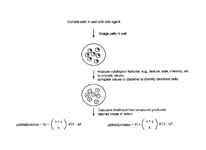
One goal is to look at the cells from a given well and infer which process
generated
the cells. A binary classifier is the tool used at the cell level to say
whether the cell was
more likely to have been generated via trial or control. As classifiers were
trained
performance characteristics were obtained. Given these characteristics and the
fraction of a
given well labeled as trial or control by the classifier the process most
likely responsible for
a well's labeled results could be found by taking the ratio of:
P(process = X or 'trial' I data) ¨ the probability that the process is trial
given the
observed results to:
P (process =Y or 'control' I data). This is the likelihood ratio.
The second term p (process = X)/p (process = Y) is the prior odds and is
ignored
under the assumption that either possibility is equally likely. Given a
classifer's
performance characteristics and number of cells classified as either trial or
control, the
binomial coefficient was used to find the likelihood ratio.
23
CA 02785835 2012-06-27
WO 2011/087945
PCT/US2011/020262
a and b are the probability of given classifier labeling a cell trial or
control,
respectively, and x and y are the number of cells classified trial Or control.
To avoid issues
related to dividing very small numbers logarithm were used to calculate the
likelihood for
each model, and the difference between the two logarithms is reported. This
value is the
prediction value reported in the figures. Prediction values typically ranged
from -2000 to
+500, across many classifiers, cell lines and a few thousand compounds, with
negative
values predicting similarity to control and positive values meaning similarity
to trial. This
strategy is illustrated in Fig. 9.
svm classifiers are made to the moa control set of compounds. The number of
experiments (essentially plates) the training set examples were drawn from is
N. The
classifier validation statistics are listed (True Pos, etc.) and the precision
and recall of each
svm against a test set of 100 compounds is listed as well. This is done for
both the +1 set of
svm's and the +2 set.
Example 3
Data sets
The assay was performed weekly with all plates plated with cell, dosed, fixed
and
stained as a group. Compounds were dosed 5 to a 96 well plate in 6-point dose
response in
duplicate for each 96 well plate. On each plate a negative control of DMSO was
dosed at the
same percentage of concentration as compound dosing and positive controls
Taxol and
Etoposide were dosed at a single concentration. These controls were used to
calculate Z
prime factors for each plate and to assess the staining intensity from the
cell cycle plots.
Digital images of DAPI stained nuclei were captured and segmented to locate
nuclei and measure features such as intensity, area, shape and texture for
each nuclei. We
used CellProfiler to segment images and quantify features. As compounds were
tested
sample images from each concentration of the dose responses were visually
inspected and a
morphological category was assigned if appropriate. Dose response curves were
inspected
to assure proper EC50 determination and quality control assessments such as
incorrect
concentration range of dosing were annotated. Individual cell data,
experimental properties
and manual inspection results were stored in a custom built software system
that allowed us
to retrieve individual cell data by experimental properties such as compound
name,
concentration, and cell line and to filter out data from experiments of
insufficient quality.
Figs. 11 illustrates exemplary results from a proliferation-apoptosis-DNA
content
(PAD) assay, and Fig. 12 provides a graph of results obtained for inactive
compounds
24
CA 02785835 2012-06-27
WO 2011/087945 PCT/US2011/020262
screened by the subject method. Likelihood scores for true negatives and false
positives are
indicated.
FIG. 13 is a table showing recall and precision results for selected
classifiers. A
threshold may be chosen for both the recall and precision in order to increase
or decrease
classifier robustness. FIG. 14 shows graphs illustrating the average recall
performance of
some classifiers, whereas FIG. 15 shows graphs illustrating the average
precision
performance of some classifiers.
Example 4
Image Standardization
By keeping the assay parameters as consistent as possible it was determined
that the
primary sources of variation in the assay was the cell density and the
cellular staining
intensity (see. Fig. 2). This assay was performed in bulk each week thus assay
intensity and
cell density was normally very similar within each week's assay, but
potentially different
between weeks.
Fluctuations in the rate of cell growth over multiple passages and the
individual
plating the cells contributed to variations in cell density. Cell passage
values were
eventually confined to greater than 5 but less than 25. Other sources of
variation in cell
density were found to be difficult to control for but it has been determined
that as long as the
cell density is a above a fairly low minimum value the quality of the EC50
determination
and ability to apply pattern recognition techniques is not effected by cell
density differences.
A number of factors including instrument lamp intensity and length of staining
contributed to changes in staining intensity. To compensate for this
fluctuation images were
standardized within each plate to control wells contained in each plate. This
technique
yielded significant improvements in classifier discrimination.
Images were standardized within each plate by first finding the median
foreground
intensity of the DMSO negative control. Then, for each treatment image, the
median
background of that image was subtracted and the result was divided by the
control median
foreground. Then each image was modified according to this formula: image +
Offset /
(Multiple * (1 ¨ Offset) = 'standardized image', where 'Offset' raises the
image above zero
and 'Multiple' is the number of times a treatment could reasonably be expected
to be above
the DMSO control. This method is illustrated in Fig. 3, and Fig. 4, with
exemplary results
shown in Fig. 2 and Fig. 5.
25