Note: Descriptions are shown in the official language in which they were submitted.
CA 2786569 2017-05-04
SAMPLE-TO-ANSWER MICROFLUIDIC CARTRIDGE
BACKGROUND
Field
This disclosure is directed to microfluidic devices and methods for
diagnostic, molecular, and biochemical assays and, more particularly, to
microfluidic
technologies for dispensing and distributing fluid from on-cartridge reagent
reservoirs,
for pumping, heating and mixing, and for rehydrating dried reagents without
bubble
entrainment and without reagent washout.
Description of Related Art
Microfluidic devices have found increasing use as tools for diagnostic
assays. The devices described by Wilding in US Patent No. 5,304,487 consisted
of
"mesoscale" channels and chambers formed on reusable silicon substrates which
were
infused with fluid reagents from off-cartridge syringe pumps. No consideration
was
given to on-cartridge fluid and reagent storage and delivery. However,
practical
commercial applications have lead in the direction of "consumable" cartridges
disposable, single use "sample-to-answer" cartridges that are self-contained
for all
reagents needed for a particular assay or panel of assays. This is
particularly true in the
case of molecular biological assay applications, where contamination
associated with
sample carryover or handling absolutely must be avoided.
On board reagents may include both liquid and dry reagent forms. Both
such reagent classes have been subject to certain problems in realization of
successful
products. Here we address liquid handling issues associated with initial
wetout of the
channels and chambers of the cartridge and with rehydration of dried reagents.
During
filling and operation of a cartridge containing microfluidic channels and
chambers,
particularly those cartridges having a plastic body, liquid wetout is often
uneven, such
CA 02786569 2012-07-05
WO 2011/094577 PCT/US2011/022973
that air pockets are not infrequently entrained in the fluid column by the
advancing
meniscus against surfaces and in comers. During pumping and mixing of
biological
samples, foam and bubbles may form that negatively impact the assay
performance of
the device. Bubbles may arise due to uneven filling of channels or chambers
containing
dried reagents. Reagent rehydration, wetout and venting are interlinked with
the
problem of bubble formation. The problem is exacerbated in more complex fluid
networks such as described in US Patent Nos. 6,068,752 to Dubrow and 6,086,740
to
Kennedy, for example, and in capillary flow-driven devices such as described
by
Buechler in US Patent Application No. 2005/0136552 or Wyzgol in US Patent
Application No. 2004/024051, which have proved notoriously difficult in
plastic body
devices.
Bubbles may also arise during heating of a sample liquid due to
degassing. It is well known that gas solubility is inversely related to
temperature and
that solutions which are heated readily become supersaturated. Also a source
of
bubbles by degassing is cavitation, where a fluid is sheared, such as during
mechanical
or ultrasonic mixing in microfluidic cavities.
Bubbles interfere with optical interrogation of liquids in microfluidic
"cuvettes". The path of light may be altered due to a lensing effect created
by the
curvature of the gas bubble surface and/or due to the gas bubble refracting
the light.
Bubbles may also interfere with biochemical reactions by altering solute
concentrations
at bubble interfaces, by denaturing protein structure, and by impacting bulk
heating rate
and the homogeneity of temperature in a liquid. For example, in the PCR
reaction, in
which a thermostable polymerase is used to amplify copies of a target nucleic
acid,
heating and cooling is uneven in the presence of bubbles in the fluid,
reducing the
efficiency of the process and limiting sensitivity. The presence of bubbles
also reduces
the volume of fluid in the reaction chambers, and in assays which rely on
detecting
analyte in volumes of 10 - 50 uL or less, the presence of a large trapped
bubble in a
reaction chamber can effectively kill the assay.
In reactions that rely on rate determination, bubbles can drastically
interfere with optical determination of slopes and with homogeneous rapid
rehydration
of dried reagents as is needed to start the reaction with proper availability
of substrates.
A variety of dried reagents, such as a fluorescent probe, enzyme, buffer or
control
analyte, may be placed within chambers of a microfluidic device and are needed
for
2
CA 02786569 2012-07-05
WO 2011/094577 PCT/US2011/022973
proper conduct of the assay. During wetout, entrapment of one or more bubbles
may
result in incomplete dissolution and mixing of the dry reagent and the sample,
thereby
impairing the reaction efficiency and reducing the sensitivity of the test.
Lei, in US Patent No. 6,637,463 proposes varying flow impedance in
parallel channels through use of surface tension features and/or cross-
sectional area so
as to equalize pressure drops, and hence flow, through the multiple flow
paths. In one
instance, a plurality of exit channels is used to drain fluid from a well so
as to avoid
formation of recirculating currents or fluid stagnation that would otherwise
tend to
inefficient washing of fluid and trapping of air bubbles. However, each such
feature
must be designed by trial and error, and the designs are thus not robust or
readily
adapted for different assays. Because microscopic variations in dimensions and
surface
chemistry are difficult to control in microfluidic circuit manufacture, the
methods have
not been proven a practical solution to the problem of equally dividing flow
between
parallel subcircuits within a microfluidic card. No description of the use of
diaphragms
with features for improving wetout was offered.
Ulmanella (US Patent Application No. 2007/0280856) reported efforts to
control the meniscus of a fluid filling a microfluidic chamber by physically
modifying
the bottom surface of the chamber, for example by installing an energy barrier
to slow
down or stop the leading edge of the meniscus as it crosses the floor of the
chamber, or
by use of a plurality of grooves or posts on the bottom surface, or by
sculpting the depth
of the chamber so as to modulate capillary action, or by using a syringe pump,
by
centrifugation, or by application of a vacuum on the outlet side of the
chamber. None
of these methods has proved a practical solution to the problem. Capillary
action is ,
highly unpredictable and tends to promote formation of air pockets and use of
a syringe
pump or application of vacuum, as commonly practiced in the prior art, tends
to shear
the fluid and drive fluid down the path of least resistance, further
exacerbating the
problem. For example, when two or more microfluidic channels branching from a
single inlet are presented to a fluid, such as is useful for splitting a
sample or reagent
between multiple diagnostic assays pathways in parallel, the fluid may fill
the path most
readily wetted and leave empty the path having higher fluid resistance. Very
tiny
differences in resistance between channels lead to preferential wetting of a
single
channel and no wetting of branching parallel channels, a problem well known to
those
skilled in the art.
3
CA 02786569 2012-07-05
WO 2011/094577 PCT/US2011/022973
Ulmanella further addresses the effect of dried reagents in wetout of
microfluidic chambers and concludes that filling efficiency of chambers
containing
center-spotted dried reagent was less than 50%, chambers having inlet side
spotted
reagent were wetted at 65% efficiency, but for chambers having outlet side
spotted
reagent, the filling efficiency without bubbles increased to 95%. However,
positioning
of reagent spots with millimeter accuracy during manufacturing is neither a
necessary
nor a satisfactory means of achieving wetout in the presence of dried reagent
spots
because it is preferential that the chamber be fully wetted before the reagent
is
rehydrated so that the concentration of the reagent is not diluted by washout
into a
downstream channel, as is highly likely if the dry reagent is positioned at
the
downstream outlet from the chamber!
It is further known that reduction in interfacial and surface tensions in
the microfluidic channels or chambers can be achieved, for example, by plasma
treatment of the substrate(s) or incorporation of surfactants to decrease
hydrophobicity,
and by applying a radius to channel intersections. These treatments are also
known to
improve wettability, but are not effective in eliminating mechanically
entrained bubbles
and bubbles resulting from thermal degassing, cavitation or stagnation zones.
In fact,
surfactants can increase the propensity of the gaseous phase to form stable
bubbles and
foams which can defeat performance of the assay by their persistence.
Moreover, the
.. modification of surfaces by processes such as plasma treatment are
anticipated to be
difficult to control in manufacturing and may be impermanent, degrading
progressively
during device storage. Therefore it is desirable and is an object of this
invention to
develop mechanical means and methods for reducing the formation and
entrainment of
bubbles during initial wetout of assay channels, during rehydration of dry
reagents, and
for preventing or reducing accumulation and interference of bubbles during
operation of
the device.
BRIEF SUMMARY
Microfluidic cartridges of the invention, herein termed more generally
"devices", are generally formed of a flexible plastic body which houses
fluidic channels
and chambers patterned and fluidly intercommunicating according to the needs
of a
diagnostic or biochemical assay to be performed therein. The assay is
conducted by
reacting a sample with one or more reagents in one or more steps, typically in
one or
4
CA 02786569 2012-07-05
WO 2011/094577 PCT/US2011/022973
more channels or chambers of the device, for times and at temperatures
effective in
forming a detectable product that indicates the presence or absence of an
analyte in the
sample. The cartridges are typically consumables; i.e., they are used once and
then
discarded; and contain all reagents needed for one or more assays.
To perform an assay, a device of the invention is inserted into a host
instrument which relies on optical detection (or other detection means), such
as a
spectrophotometer or fluorometer for the detection of a chromogen or
fluorophore
indicative of the presence, absence, and/or amount of any target analytes of
interest. In
a preferred embodiment, optical windows in the device are interfaced with
detection
.. means in the host instrument. However, the presence of one or more gas
bubbles in an
optical window may impair the detection of the analyte. Bubbles may also
interfere
with the reactions required to form a detectable product, such as for example
an
amplicon or other product of a biochemical or molecular reaction, where a
bubble may
be responsible for uneven heating of a reaction mixture, inadequate mixing, or
incomplete or untimely reconstitution of a dry reagent.
In use, a sample fluid is introduced into the inventive device, and the
fluidly intercommunicating channels and chambers of the device are then wetted
with
either a biological liquid sample alone, with liquid reagents, or with a
mixture of a
sample and one or more liquid reagents. The wettable, fluidly
intercommunicating
aspects of the device are termed the "hydraulic works" of the device and
comprise one
or more microfluidic subcircuits having channels and chambers. Control of the
hydraulics is effected through pneumatically actuated valves, pumps and
diaphragms
superimposed as a separate, secondary network or manifold of chambers and
channels
in the device and supplied by external sources of pressurized air and vacuum.
This
secondary network is termed the "pneumatic works" of the device. Thus the
device is
composed of a primary "hydraulic network" for conveying a liquid or liquids
and a
secondary "pneumatic network" for conveying a gas. The pneumatic network
provides
a) process control and b) positive and negative pressure for driving the
liquid or liquids
through the hydraulic network, according to valve and pump logic of a host
instrument
with which the cartridge is interfaced for performing an assay.
Sample handling and mixing of liquid reagents, including rehydration of
any dry reagents disposed within the hydraulic channels and chambers of the
device,
has been problematic in that bubbles readily become entrained in the fluid
during
5
CA 02786569 2012-07-05
WO 2011/094577 PCT/US2011/022973
wetting of the hydraulics. This particularly occurs during initial wetout,
where bubbles
are engulfed by a meniscus advancing rapidly through the device, and
subsequently
such as by cavitation or degassing associated with mixing and heating. The
present
invention addresses this problem through one or more fluid handling mechanisms
and
methods.
Inventive mechanisms, features and methods include pneumohydraulic
diaphragms characterized as:
1) an elastic, energy-storing pneumohydraulic diaphragm
configured for passively storing a liquid volume under a hydraulic pressure
and
releasing the liquid volume during wetout of a downstream channel or chamber
of the
wettable microfluidic subcircuit;
2) a duplexedly layered pneumohydraulic diaphragm having a
liquid center for storing and releasing a liquid reagent;
3) a pneumohydraulic diaphragm configured for eliminating
headspace from a hydraulic chamber during wetout; or
4) a pair of pneumohydraulic diaphragms comprising a first
pneumohydraulic diaphragm interfacing a first hydraulic chamber with valved
inlet and
a second pneumohydraulic diaphragm interfacing a second hydraulic chamber with
valved outlet, and an elevated directly intercommunicating channel between the
first
and second hydraulic chambers, wherein the pair is configured for reciprocally
exchanging fluid through the intercommunicating channel by applying opposing
pressure differentials across the first and second pneumohydraulic diaphragms;
and
where the hydraulic chambers and diaphragms are configured for
preventing or reducing bubble entrainment or reagent washout during wetout,
fill,
pumping or rehydration steps of an assay.
In accordance with various exemplary embodiments, one or more liquid
reagents are disposed in sealed reservoirs on the device as manufactured. Dry
reagents
are printed or "spotted" in channels or chambers and are rehydrated at the
time of use.
The liquid reagents function as buffers, diluents, solvents, eluants, wash
reagents, and
as rehydrating reagents. In these capacities, the liquids are dispensed as
required from
their sealed reservoirs into the hydraulics of the device by pneumatic
actuation.
In a preferred liquid reagent embodiment, a sealed liquid storage
reservoir of the invention is structured as a two-layered diaphragm with a
liquid center,
6
CA 02786569 2012-07-05
WO 2011/094577 PCT/US2011/022973
the duplex diaphragm sealedly separating the pneumatics works and the
hydraulic
works of the device. The duplex diaphragm is composed of two impermeable film
layers separated by a liquid center and crimped or fused around the edges and
sealed in
the device so that the diaphragm separates a hydraulic chamber and a pneumatic
chamber. The upper layer, which faces the pneumatics works of the device, is
formed
of a film having a composition for resisting puncture and the lower layer,
which faces
the hydraulic works of the device, is composed of a film having a composition
that is
more susceptible to puncture. Pressurizing the pneumatic side of the diaphragm
forces
the liquid-filled reservoir against a sharp or "barb" disposed in a fluid
receiving basin
and punctures the lower layer, but not the upper layer. Following rupture,
liquid then
flows into the hydraulic chamber and from there into the microfluidic wettable
channels
of the device. By applying pressure on the pneumatic side of the diaphragm,
one or
more volumes of reagent can be forced under pressure into the hydraulic works,
and by
reversing pressure, the fluid can be cause to reflux.
In this aspect, an inventive assay cartridge is characterized as having
therein:
a) a duplexedly layered diaphragm sealedly separating a
pneumatic chamber of a pneumatic works and a hydraulic chamber of a hydraulic
works, the duplexedly layered diaphragm having a first side facing the
pneumatic works
and a second side facing the hydraulic works, a first layer forming the first
side thereof,
and a second layer forming the second side thereof, the first and second
layers
enclosing therebetween a liquid volume as a liquid center;
b) a fluid outlet for receiving and conveying the liquid
volume to the downstream microfluidic subcircuit; and
c) a sharp or "barb" disposed in
the hydraulic chamber, the
sharp for selectively rupturing the second layer and for releasing the liquid
volume into
the hydraulic works when the duplexedly layered diaphragm is piercingly urged
into
contact with sharp by application of a pressure differential across the
diaphragm.
Surprisingly, the liquid may be released from the on-board reagent
reservoir in a series of smaller liquid volumes by the action of serial pulses
of
pneumatic pressure applied to the first layer of the diaphragm, which remains
intact.
7
CA 02786569 2012-07-05
WO 2011/094577 PCT/US2011/022973
Optionally the first layer of the duplexedly layered diaphragm is a
rupture-resistant layer and the second layer is a rupture-sensitive layer. The
liquid
center may contain a liquid reactant, a buffer, a rehydrating fluid, a
solvent, or a diluent.
On-board storage of liquid is useful for, for example, rehydrating a dry
reagent disposed
in a downstream chamber or channel, for rinsing a solid phase, for eluting a
target
analyte or analytes from a solid phase substrate, for making a dilution, for
making a
chromatographic separation, for actuating or stopping a reaction, or for
detecting the
target analyte or analytes, and minimizes the possibility of carry-over
contamination.
Optionally the liquid volume is degassed and the duplexedly layered diaphragm
is gas
impervious. Advantageously, any entrained bubbles are likely to be resorbed in
degassed liquids, and degassed liquids are not susceptible to degassing on
heating, such
as is useful for thermocycling in PCR.
While the devices are generally planar, they may be mounted in the host
instrument in a canted position (i.e. angularly with respect to a ground
plane), typically
at about 15 degrees from flat and are vented at a downstream aspect of each
microfluidic subcircuit. As a liquid sample or reagent is introduced upstream
into the
hydraulic subcircuitry, air is displaced downstream and is vented. The liquid
sample
and reagents progressively fill and move through the device. By canting the
card at an
angle of 10 to 35 degrees, air in the device during priming (termed here
"wetout") is
found to be more readily displaced from the hydraulic works. By careful
management
of the advancing meniscus during initial fill of the canted card, the problem
of bubble
entrainment, particularly during fill, is substantially reduced or prevented.
Thus optionally, the hydraulic works may be configured for operation
when mounted at an angle of 10 ¨ 35 degrees relative to the ground plane on a
tilted
stage of a host instrument and at least one hydraulic chamber is configured
with an
outlet and intercommunicating channel positioned superiorly relative to that
chamber
for venting a gas or discharging a bubble from the chamber.
In another aspect of the invention, entrainment of bubbles during wetout
is limited by a filling mechanism that involves passive relaxation of an
elastically
stretched or distended pneumohydraulic diaphragm. This passive mechanism was
found to be superior to fill by capillarity and to fill by positive
displacement pump
action or vacuum. A liquid is first forced under pressure into a specially
designed
manifold having a "pneumatic chamber" stacked on top of a "hydraulic chamber",
8
CA 02786569 2012-07-05
WO 2011/094577 PCT/US2011/022973
where the two chambers are separated by an elastic diaphragm stretched over
the roof
of the hydraulic chamber. Optionally, liquid may instead be aspirated into the
lower
chamber, but advantageously, the upper pneumatic chamber is vented and open to
atmospheric pressure. The position of the two chambers, while termed "upper"
and
"lower" or "top" and "bottom" chambers for purposes of explanation, is
relative, and is
not limiting on the operation of the device. As a liquid volume enters the
liquid-
receiving chamber, the diaphragm is stretched to hold the volume and
resiliently stores
the energy of deformation, a form of potential energy having a returning force
and a
spring constant. Diaphragm material and deformation conditions are chosen so
that the
"elastic limit" of the material is not exceeded. Then by opening a valve to a
downstream
channel or channels, the distendedly stretched diaphragm returns to its
relaxed state and
fluid gently fills the downstream fluid structures without entrainment of
bubbles in the
advancing meniscus.
This mechanism and method has proved startlingly advantageous where
flow is split into multiple channels. By providing an upstream staging
manifold with
multiple liquid-receiving chambers having elastic diaphragms, each with
separately
valved outlets that are opened in synchrony, the hydraulic pressure for
initiating and
sustaining liquid flow into multiple downstream fluidic subcircuits in
parallel is
segregated or "quantized" so that the flow into all channels is essentially
equal and
sufficient. Total pressure and volume per downstream channel can be precisely
calibrated by selection of the spring constant and the deformation of the
elastic
diaphragm member so that the restoring flow of liquid into the downstream
channel is
the volume required to fill the downstream channel to a desired mark; the
displaced
volume delivered by each diaphragm of the staging manifold is neither
insufficient nor
in excess for the fluidic operation of splitting flow equally among multiple
parallel
channels or subcircuits, a necessary fluidic operation in devices intended for
multiple
assays in parallel. This is a technological advance in the art. Any air
downstream is
readily displaced by the advancing meniscus and is conveyed to a downstream
vent by
this means.
In this aspect, an inventive assay cartridge includes:
a) a
staging manifold having a plurality of chambers, wherein each
chamber of the plurality of chambers is separated into a hydraulic chamber and
a
pneumatic chamber by an elastic, energy-storing pneumohydraulic diaphragm
sealedly
9
CA 02786569 2012-07-05
WO 2011/094577 PCT/US2011/022973
mounted therebetween, such that a liquid volume admitted through an inlet into
each
hydraulic chamber in series or in parallel distends each energy-storing
pneumohydraulic
diaphragm according to an isobaric pressure proportionate throughout said
staging
manifold to the displacement volume thereof;
b) the inlet is valvedly
closeable for equilibrating the hydraulic
pressure throughout the staging manifold after filling is complete; and,
c) a
plurality of vented downstream channels in parallel, wherein
one the channel of the plurality of channels is in fluidic communication with
each
hydraulic chamber of the staging manifold, each vented downstream channel
having a
valve for closing during filling and pressurization and for opening during
draining and
depressurization, whereby the hydraulic pressure of the elastic,
pneumohydraulic
diaphragm in a distended state is passively converted to the work of advancing
a
meniscus during initial wetout of the plurality of vented downstream channels
equally
in parallel.
More generally, wetout or 'priming' is improved by harnessing the
mechanical properties an elastic, pneumohydraulic diaphragm in a fluidly
distended
state to do the work of advancing a meniscus through a wettable downstream
microfluidic circuit fluidly connected thereto and thereby displacing any gas
therein to a
downstream vent without bubble entrainment. This
principle is particularly
advantageous in equally splitting a fluid into a plurality of downstream
microfluidic
subcircuits in parallel. In this way, multiple assays may be conducted in
parallel and a
single sample may be split equally for parallel assays having separate
downstream
detection means. Surprisingly, the mechanical properties of the elastic
diaphragm can
be calibrated to fill one or more downstream microfluidic subcircuits to a
mark, as is
useful in reconstituting a defined mass of a dried reagent in a defined
volume, for
example.
Microfluidic devices may typically also include at least one dried reagent
disposed within the downstream hydraulic network. These reagents are typically
spotted or printed during manufacture. During an assay, the dried reagents are
rehydrated by sample or by contact with a liquid reagent dispensed as
described above.
Serendipitously, we have found that the passive liquid wetting mechanism and
method
described here is advantageously suited to the rehydration of dry reagents
without
entrainment of bubbles, another technological advance in the art.
CA 02786569 2012-07-05
WO 2011/094577 PCT/US2011/022973
In a related embodiment, we have found that by providing pneumatically
actuated diaphragms in downstream chambers where dried reagents are spotted,
the
diaphragms overlying those reagent spots can be pressurized so as to a)
temporarily seal
the reagent zone (typically central to and on the floor of the chamber) from
contact with
bulk fluid during the chamber wetting process and b) remove or expel
essentially all of
the headspace above the dried reagent. When deformed so as to fill the
hydraulic
chamber, the diaphragm is not fully sealed around the periphery of the
chamber. Liquid
entering the chamber around the diaphragm is shunted around the lower edges of
the
chamber and readily displaces any residual air, which is vented from the
hydraulics
during filling. By relaxing or by reversing the pressure differential
across the
diaphragm, additional fluid is readily aspirated into the chamber without the
formation
or entrapment of gas bubbles. Reagents are rehydrated only after the
downstream outlet
of the chamber is valvedly closed, thereby reducing reagent losses to washout.
The
reduced dead volume of the dry reagent chambers is thus turned to advantage.
Happily,
in this way, dry reagent spots can be precisely reconstituted with a desired
volume of
rehydrating reagent or sample, ensuring that the biological activity of the
reagent is
quantitatively correct for the assay conditions, a useful refinement in art.
Thus the invention also may feature at least one microfluidic subcircuit
having a downstream reaction chamber with upstream inlet and downstream vent,
the
downstream reaction chamber containing a dried reagent spot or spots, further
characterized in that the pneumohydraulic diaphragm is configured to operate
with a
first position wherein the diaphragm is distended against the floor of the
chamber so as
to displace headspace air and form a protective temporary tent around and over
the
reagent spot or spots during wetout, and a second position wherein the
diaphragm is
relaxedly positioned or aspirated against the roof of the chamber so as to
fill the
chamber with the liquid volume and uncover and dissolve the reagent spot at
full
strength without bubble entrainment or reagent washout. The dried reagent spot
may be
a buffer, an enzyme, a co-enzyme, a co-factor, a polymerase, a primer, a
molecular
beacon, a probe, a fluorophore, a dehydrogenase, an oxidase, a reactant, a
chromogen, a
substrate, an antibody, an antigen, or a control.
Also claimed is a method for wetting a microfluidic cartridge while
limiting bubble entrainment therein, which comprises:
11
CA 02786569 2012-07-05
WO 2011/094577 PCT/US2011/022973
a) pumping a liquid volume through an inlet and into a plurality of
hydraulic chambers forming a staging manifold of a microfluidic card so that
an elastic
pneumohydraulic diaphragm overlying the liquid volume in each said hydraulic
chamber is stretchedly distended, thereby isobarically pressurizing the liquid
volume in
the plurality of hydraulic chambers;
b) valvedly opening an outlet from each of the hydraulic chambers
of the staging manifold, each outlet with fluidic connection to a vented
downstream
microfluidic subcircuit; and
c) splitting the liquid volume substantially in equal measure into
each said wettable downstream microfluidic subcircuit by passive relaxing the
distended elastic diaphragm __ without bubble entrainment.
Wetting a microfluidic device by passive relaxation of an elastic
diaphragm is readily distinguished from wetting by capillary action or by
active
pumping, and has proven surprisingly advantageous in overcoming difficulties
with
bubble entrainment as are known in the art.
Also claimed is a method for wetting a microfluidic cartridge which
contains dried reagent spots, while limiting bubble entrainment therein, which
comprises:
a) pumping a liquid volume through an inlet and into a plurality of
hydraulic chambers of a microfluidic card so that an elastic pneumohydraulic
diaphragm overlying the liquid volume in each the hydraulic chamber is
distended,
thereby isobarically pressurizing the liquid volume;
b) pressurizing a second diaphragm in a plurality of downstream
reaction chambers, each downstream reaction chamber containing a dried reagent
spot,
the second diaphragm forming a protective temporary tent for sealing around
and over
the reagent spot and for displacing headspace air from the downstream reaction
chamber;
c) valvedly opening an outlet from each the hydraulic chamber,
each the outlet with fluidic connection to one of the plurality of downstream
reaction
chambers;
d) wetting the downstream reaction chamber around the temporary
tent and displacing any residual air from the reaction chamber by allowing the
12
CA 02786569 2012-07-05
WO 2011/094577 PCT/US2011/022973
distended elastic pneumohydraulic diaphragm to relax, the liquid volume
forming an
advancing meniscus;
e)
optionally closing a valve downstream from the downstream
reaction chamber;
lifting the temporary tent and conveying a remaining part of the
liquid volume into each reaction chamber, thereby dissolving the reagent spot
at full
strength without bubble entrainment or reagent washout. The temporary tent is
lifted by
relaxing or by reversing the pressure differential across the second diaphragm
member.
In another method, pairs of chambers with pneumohydraulic diaphragms
may be used to aid wetout and reagent dissolution for PCR, and for
reciprocally
pumping fluid when interconnected in series by a channel. By application of
alternating
positive and negative pneumatic pulses to a first diaphragm in a first
chamber, a second
diaphragm in a second chamber is driven in synchrony. The second diaphragm may
be
an elastic diaphragm that functions in accommodating and elastically storing
the pulsed
energy of the first diaphragm. Mixing is readily achieved by conveying a
liquid volume
back and forth between the two chambers. By providing each hydraulic chamber
with
a thin heat exchange film and suitable contact heating elements, "two-zone"
PCR is
readily achieved. In an improved device, the intercommunicating channel
between the
chambers is contoured and elevatedly positioned so that bubbles are
gravitationally
urged to clear the chambers during initial wetout and pumping, and will trap
any
additional bubbles that form during heating. The intercommunicating channel is
preferably configured and contoured to be operated at a tilt of 10 ¨ 35
degrees and is
positioned on the high side of the paired chambers so as to reduce
interference from
bubbles. Fluid is cycled between a first chamber at a denaturing temperature
of a target
nucleic acid and a second at an annealing temperature. The plastic body of the
device
limits parasitic heat capacitance of the device during PCR. Nucleic acid
amplification
at rates of 8 seconds or less per thermal cycle is readily achieved.
For PCR, amplification reagents are provided with the device. Typically
the first chamber contains a first reagent or reagents and the second chamber
contains a
second reagent or reagents. Typically the reagents are spotted in a centric or
pericentric
zone in each chamber. During initial wetout, the diaphragms in the chambers
are
inflatedly distended to press down on and cover the reagents so as to limit
rehydration
and any washout that would otherwise occur as the meniscus of the rehydrating
fluid or
13
CA 02786569 2012-07-05
WO 2011/094577 PCT/US2011/022973
sample dissolves the spotted reagents and carries them downstream with the
solvent
front. After initial wetout, a suction pressure may be applied to the
diaphragm so as to
aspirate a fluid into the chamber and dissolve the reagents therein.
Alternatively, an
upstream chamber may be pressurized so as to hydraulically inflate the
downstream
chamber and dissolve the reagents. Fluid direction of flow may be reversed one
or
more times so at to improve mixing and rehydration.
Thus the invention may also include a cartridge for use with a host
instrument having thermal interfaces for "two-zone thermocycling" and a
pneumatic
interface with pneumatic means for driving and controlling a PCR
amplification. The
device works by reciprocating pneumohydraulic action of paired diaphragms in
two
interconnected hydraulic chambers so as to cyclically denature and anneal a
target
nucleic acid, the cartridge advantageously having one or more wettability
features of
the invention for improving wetout of the chambers with liquid without
entrainment of
bubbles. The device is also advantageous for dissolving reagents in a fixed
volume
without washout losses during wetout, ensuring that primers, buffers and other
reagents
are at a fixed strength when reconstituted.
Thus the various aspects of the invention offer novel utility in operation
of microfluidic cartridges for diagnostic and biochemical assays and are found
to be
advantageous as mechanisms and methods for limitation of the bubble
interferences that
have been a longstanding source of problems with these devices.
In the following description, certain aspects and embodiments of the
invention will become evident. It should be understood that these aspects and
embodiments are merely exemplary and explanatory and are not restrictive of
the
invention. Other features and advantages will become apparent from the
detailed
description which follows.
BRIEF DESCRIPTION OF THE DRAWINGS
FIGS. 1A and 1B show two perspective views of a disposable, single-
use, sample-to-answer microfluidic cartridge of the invention, the cartridge
containing
all reagents for an assay and requiring only introduction of a biological
sample.
FIG. 2 demonstrates insertion of the assay cartridge in a host instrument
for performance of an assay thereon.
14
CA 02786569 2012-07-05
WO 2011/094577 PCT/US2011/022973
FIG. 3A is a detailed view of the cartridge as inserted in a mechanism of
a host instrument. The mechanism includes a heating manifold shown in FIG. 3B
and a
pneumatic control interface.
FIG. 4 is an exploded view of disposable microfluidic cartridge with
liquid center foil diaphragm packs carrying liquid reagents.
FIG. 5A is a perspective view of a microfluidic circuit for extraction of a
nucleic acid target from a biosample; FIG. 5B is a schematic of the extraction
process.
FIGS. 6A-G provide views of a reagent reservoir formed of a bilayered
duplex diaphragm with liquid reagent center and a sharp or "barb" for
puncturing and
releasing the liquid into the hydraulic works of the microfluidic device.
FIGS. 7A and 7B show a microfluidic cartridge canted with a tilt as
mounted in a host instrument.
FIGS. 8A and 8B show a worms-eye view of a network of channels and
chambers for performing PCR on a microfluidic cartridge; the positions of dry
reagents
are also marked.
FIGS. 8C and 8D illustrate an alternative cartridge configuration in
worm's-eye view.
FIGS. 9A-9L schematically depict a passive initial wetout mechanism
with staging manifold.
FIGS. 10A-10C depict the operation of a staging manifold whereby
reagents are rehydrated in preparation for PCR. The operational sequence is
continued
in FIGS. 10D-G.
FIGS. 10D-10G illustrate a PCR amplification using dual chambers with
reciprocating diaphragm action.
FIG. 11 describes the steps of a method for extracting nucleic acids from
a sample, where a bilayered duplex diaphragm with liquid center is used to
dispense the
reagents.
FIG. 12 describes the steps of a method for priming the microfluidic
channels of a hydraulic works with liquids dispensed from a bilayered duplex
diaphragm with liquid center.
FIG. 13 describes the steps of a method for rehydrating dry reagents
without bubble entrainment.
CA 02786569 2012-07-05
WO 2011/094577 PCT/US2011/022973
FIGS. 14A and 14C are worm's eye views of a network of microfluidic
channels and chambers for reverse-transcriptase-mediate PCR. FIG. 14B is a
detail
view of an in-line chamber for production of cDNA.
FIGS. 14D and 14E illustrate an alternative configuration of a cartridge
with modified features.
FIG. 15 illustrates use of optical windows of a detection chamber of a
device of the invention for monitoring a fluorescent endpoint.
FIG. 16 depicts more detail of a two piece microfluidic card assembly
for performing PCR and a pneumatic interface with gasket for interfacing the
cards with
a compatible host instrument.
FIG. 17 is a plot showing a positive and negative fluorescence assay in
the detection chambers of the cartridge, including multiple scans of the
sample while
increasing the temperature of the reaction mix.
FIGS. 18A and 18B analyze the pooled data of FIG. 17. Scans of a
molecular beacon-amplicon duplex demonstrate a FRET melting curve capability
of the
cartridge when interfaced with a compatible host instrument.
FIG. 19 depicts FRET data for amplicons obtained with a device of the
invention when used in a host instrument compatible therewith.
DETAILED DESCRIPTION
Although the following detailed description contains specific details for
the purposes of illustration, one of skill in the art will appreciate that
many variations
and alterations to the following details are within the scope of the claimed
invention.
The following definitions are set forth as an aid in explaining the invention
as claimed.
Definitions
A "cartridge" is an analytical device designed for operation by insertion
into a host instrument. The host instrument supplies the pneumatic pressure,
pulses,
and detection means for performance of the assay. The cartridge contains
hydraulic
works and pneumatic works, and may include embedded microfluidic "cards" with
embedded microfluidic channels and chambers. Sample and reagent liquids are
conveyed in a hydraulic network of the cartridge or card; fluid flow is
controlled and
driven by a pneumatic network that interfaces with the hydraulics at selected
junctions,
16
CA 02786569 2012-07-05
WO 2011/094577 PCT/US2011/022973
channels and chambers. Typically, the body of the cartridge or card is made of
a
flexible plastic and may be formed by lamination, molding or a combination
thereof.
Plastics may include, but are not limited to, polycarbonate, polyethylene
terephthalate,
cyclic polyolefins, acrylates, methacrylates, polystyrene, graft and block
copolymers,
and composites thereof. A preferred cartridge is made from rollstock and
includes dry
reagents printed thereon.
"Hydraulic works" of a device: includes the network or networks of
intercommunicating channels and chambers that are intended to be wetted by
sample or
liquid reagents in the course of an assay. The hydraulic networks are
configured with
microfluidic subcircuits for performing the steps of an assay.
"Pneumatic works" of a device: includes the network or networks of
pneumatically actuated valves, pumps and diaphragms and interconnecting
circuitry and
manifolds that are useful for powering and controlling the hydraulics of the
device. The
pneumatic works of the cartridge device interface with positive and negative
pressure
sources on the host instrument and with valves, diaphragms, pumps and other
pneumatically actuated elements that control and drive liquids in the
hydraulic network.
"Microfluidic works" of a device: include the hydraulic works formed of
a network or networks of internal channels and chambers wetted in the course
of the
assay and the pneumatic works formed of valve control and pump driving
circuits
powered by positive and negative pressure sources on the host instrument.
The microfluidic works may be divided into microfluidic subcircuits,
where each subcircuit comprises channels and chambers for performing a
particular
function on a liquid sample or reagent. The microfluidic subcircuits may be
organized
into serial subcircuits (such as for extraction, amplification and detection
of a nucleic
acid target or targets) and parallel subcircuits and networks such as for
simultaneous
assay for multiple targets on a single sample by splitting the sample.
"Top", "bottom", "up", "down", "above", "below", "upward",
"downward", "superior to", "floor", "roof', and so forth are indications of
relative
position and not absolute position, unless reference is made to a specific
frame of
reference, such as the "ground plane", which is taken as orthogonal to an
intersecting
plumb line.
"Wetout" ("wet out") refers to the initial hydration of a plastic surface
interior to the hydraulic works of a cartridge. Because of interfacial tension
effects,
17
CA 02786569 2012-07-05
WO 2011/094577 PCT/US2011/022973
initial wetout can involve overcoming a substantial energy barrier and is a
major factor
in resistance to capillary flow in these devices.
"Target analyte": or "analyte of interest", or "target molecule", may
include a nucleic acid, a protein, an antigen, an antibody, a carbohydrate, a
cell
.. component, a lipid, a receptor ligand, a small molecule such as a drug, and
so forth.
Target nucleic acids include genes, portions of genes, regulatory sequences of
genes,
mRNAs, rRNAs, tRNAs, siRNAs, cDNA and may be single stranded, double stranded
or triple stranded. Some nucleic acid targets have polymorphisms, single
nucleotide
polymorphisms, deletions and alternate splice sequences, such as allelic
variants.
Multiple target domains may exist in a single molecule, for example an
immunogen
may include multiple antigenic determinants. An antibody includes variable
regions,
constant regions, and the Pc region, which is of value in immobilizing
antibodies.
Target analytes are not generally provided with the cartridge as manufactured,
but are
contained in the liquid sample to be assayed; in contrast, "control analytes"
are typically
provided with the cartridge or are routinely present in a sample of a
particular type and
are assayed in order to ensure proper performance of the assay. Spiked samples
may be
used in certain quality control testing and for calibration, as is well known
in the art.
"Means for Amplifying:" of which the grandfather technique is the
polymerase chain reaction (referred to as PCR) which is described in detail in
U.S.
Patent Nos. 4,683,195, 4,683,202 and 4,800,159, Ausubel et at. (Current
Protocols in
Molecular Biology, John Wiley and Sons, Baltimore, Md. 1989), and in Innis et
al.,
("PCR Protocols", Academic Press, Inc., San Diego Calif., 1990). Polymerase
chain
reaction methodologies require thermocycling and are well known in the art.
Briefly, in
PCR, two primer sequences are prepared that are complementary to regions on
opposite
complementary strands of a target sequence. An excess of deoxynucleoside
triphosphates are added to a reaction mixture along with a DNA polymerase,
e.g., Taq
polymerase. If the target sequence is present in a sample, the primers will
bind to the
target and the polymerase will cause the primers to be extended along the
marker
sequence by adding on nucleotides. By raising and lowering the temperature of
the
reaction mixture, the extended primers will dissociate from the template to
form
reaction products, excess primers will bind to the template and to the
reaction products
and the process is repeated. By adding fluorescent intercalating agents, PCR
products
can be detected in real time.
18
CA 02786569 2012-07-05
WO 2011/094577 PCT/US2011/022973
Other amplification protocols include LAMP (loop-mediated isothermal
amplification of DNA) reverse transcription polymerase chain reaction (RT-
PCR),
ligase chain reaction ("LCR"), transcription-based amplification systems
(TAS),
including nucleic acid sequence based amplification (NASBA), "Rolling Circle",
"RACE" and "one-sided PCR", also termed "asymmetrical PCR" may also be used,
having the advantage that the strand complementary to a detectable probe is
synthesized
in excess.
These various non-PCR amplification protocols have various advantages
in diagnostic assays, but PCR remains the workhorse in the molecular biology
laboratory and in clinical diagnostics. Embodiments disclosed here for
microfluidic
PCR should be considered representative and exemplary of a general class of
microfluidic devices capable of executing one or various amplification
protocols.
Typically, nucleic acid amplification or extension involves mixing one
or more target nucleic acids which can have different sequences with a "master
mix"
containing the reaction components for performing the amplification reaction
and
subjecting this reaction mixture to temperature conditions that allow for the
amplification of the target nucleic acid. The reaction components in the
master mix can
include a buffer which regulates the pH of the reaction mixture, one or more
of the
natural nucleotides (corresponding to A, C, G, and T or U¨often present in
equal
concentrations), that provide the energy and nucleosides necessary for the
synthesis of
nucleic acids, primers or primer pairs that bind to the template in order to
facilitate the
initiation of nucleic acid synthesis and a polymerase that adds the
nucleotides to the
complementary nucleic acid strand being synthesized. However, means for
amplication
also include the use of modified or "non-standard" or "non-natural" bases such
as
described in US Patent No. 7,514,212 to Prudent and US Patent Nos. 7,517,651
and
7,541,147 to Marshall as an aid to detecting a nucleic acid target.
"Means for detection": as used herein, refers to an apparatus for
displaying an endpoint, ie. the result of an assay, which may be qualitative
or
quantitative, and may include a machine equipped with a spectrophotometer,
fluorometer, luminometer, photomultiplier tube, photodiocle, nephlometer,
photon
counter, voltmeter, ammeter, pH meter, capacitative sensor, radio-frequency
transmitter, magnetoresistometer, or Hall-effect device. Magnifying lenses in
the cover
plate, optical filters, colored fluids and labelled probes may be used to
improve
19
CA 02786569 2012-07-05
WO 2011/094577 PCT/US2011/022973
detection and interpretation of assay results. "Labels" or "tags" include, but
not limited
to, dyes such as chromophores and fluorophores; and chemoluminescence as is
known
in the prior art. QDots, such as CdSe coated with ZnS, decorated on magnetic
beads, or
amalgamations of QDots and paramagnetic Fe304 microparticles, are a convenient
method of improving the sensitivity of an assay of the present invention.
Fluorescence
quenching detection endpoints are also anticipated. A variety of substrate and
product
chromophores associated with enzyme-linked immunoassays are also well known in
the
art and provide a means for amplifying a detection signal so as to improve the
sensitivity of the assay, for example "up-converting" fluorophores.
"Molecular beacon": is a single stranded hairpin-shaped oligonucleotide
probe designed to report the presence of specific nucleic acids in a solution.
A
molecular beacon consists of four components; a stem, hairpin loop, end
labelled
fluorophore and opposite end-labelled quencher. When the hairpin-like beacon
is not
bound to a target, the fluorophore and quencher lie close together and
fluorescence is
suppressed. In the presence of a complementary target nucleotide sequence, the
stem of
the beacon opens to hybridize to the target. This separates the fluorophore
and
quencher, allowing the fluorophore to fluoresce. Alternatively, molecular
beacons also
include fluorophores that emit in the proximity of an end-labelled donor.
'Wavelength-
shifting Molecular Beacons' incorporate an additional harvester fluorophore
enabling
the fluorophore to emit more strongly. Current reviews of molecular beacons
include
Wang K et al, 2009, Molecular engineering of DNA:molecular beacons. Angew Chem
Int Ed Engl, 48(5):856-870; Cissell KA et al, 2009, Resonance energy transfer
methods
of RNA detection, Anal Bioanal Chem 393(1):125-35 and Li Y, et al, 2008,
Molecular
Beacons: an optimal multifunctional biological probe, Biochem Biophys Res Comm
373(4):457-61. Recent advances include Cady NC, 2009, Quantum dot molecular
beacons for DNA detection. Methods Mol Biol 554:367-79.
Fluorescence nucleic acid assays include amplification with tagged
primers and probe-based detection chemistries. Fluorescent products can be
assayed at
the end of the assay, or by measuring the amount of amplified product in real
time.
While not limiting, TaqMan Probe (Applied Biosystems) which relies on
displacement
and polymerase-mediated hydrolysis of a 5' reporter dye with 3' quencher
construct,
FRET hybridization probes, dual oligo FRET-based probes (Roche), minor groove
binder-conjugated hybridization probes (MGB probes, Applied Biosystems),
Eclipse
CA 02786569 2012-07-05
WO 2011/094577 PCT/US2011/022973
probes, Locked NA Probes (Exiqon/Roche), Amplifluor primer chemistries,
Scorpions
primer chemistries, LUX primers, Qzyme primers, RT-PCR, among others, are all
suitable in the present invention. Fluorescent probes include intercalating
probes, such
as Syber Green (Molecular Probes), ethidium bromide, or thiazole orange, FRET
probes, TaqMan probes (Roche Molecular Systems), molecular beacon probes,
Black
Hole QuencherTM (Biosearch Technologies), MGB-Eclipse probes (Nanogen),
ScorpionsTM (DxS Ltd) probes, LUXTM primer-probes (Invitrogen), SunriseTM
probes
(Oncor), MGB-Pleiades (Nanogen), and so forth. Recent advances in probe
technologies are reviewed by Lukhtanov EA et al, 2007, Novel DNA probes with
low
background and high hybridization-triggered fluorescence, Nucl Acids Res
35:e30, for
example. Reverse transcriptase is used to analyze RNA targets and requires a
separate
step to form cDNA. Recent advances include Krasnoperov LN et al. [2010.
Luminescent probes for ultrasensitive detection of nucleic acids. Bioconjug
Chem 2010
Jan 19 epub].
In addition to chemical dyes, probes include green fluorescent proteins,
quantum dots, and nanodots, all of which are fluorescent. Molecules such as
nucleic
acids and antibodies, and other molecules having affinity for an assay target,
may be
tagged with a fluorophore to form a probe useful in fluorescent assays of the
invention.
"FRET" (Fluorescence Resonance Energy Transfer) ¨ is a fluorescence
technique that enables investigation of molecular interactions. It depends on
the transfer
of energy from one fluorophore to another fluorophore (ie. a donor and a
quencher)
when the two molecules are in close proximity such a when hybridized. Recent
advances include Carmona AK et al, 2009, The use of fluorescence resonance
energy
transfer (FRET) peptides for measurement of clinically important proteolytic
enzymes,
Ann Acad Bras Cienc 81(3):381-92.
Unless the context requires otherwise, throughout the specification and
claims which follow, the word "comprise" and variations thereof, such as,
"comprises"
and "comprising" are to be construed in an open, inclusive sense, that is as
"including,
but not limited to". Reference throughout this specification to "one
embodiment", "an
embodiment", "one aspect", or "an aspect" means that a particular feature,
structure or
characteristic described in connection with the embodiment or aspect may be
included
one embodiment but not necessarily all embodiments of the invention.
Furthermore,
the features, structures, or characteristics of the invention disclosed here
may be
21
CA 02786569 2012-07-05
WO 2011/094577 PCT/US2011/022973
combined in any suitable manner in one or more embodiments. "Conventional" is
a
term designating that which is known in the prior art to which this invention
relates.
"About" and "generally" are broadening expressions of inexactitude, describing
a
condition of being "more or less", "approximately", or "almost" in the sense
of "just
about", where variation would be insignificant, obvious, or of equivalent
utility or
function, and further indicating the existence of obvious minor exceptions to
a norm,
rule or limit.
Description of the Drawings
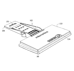
Turning to the figures, FIGS. lA and 1B show two perspective views of
a disposable, single-use, sample-to-answer microfluidic cartridge of the
invention, the
cartridge containing all reagents for an assay and requiring only
introduction= of a
biological sample. In this representative embodiment, the cartridge 100
includes a
protective chassis or body 102 with coverplate 103 for convenience in
handling. The
coverplate includes and contains an inlet port 104 for addition of sample. The
projecting nose 105 of the cartridge is inserted into a docking bay of a host
instrument
(FIG. 2). The projecting nose of the cartridge body includes optical window
cutout 101
that aligns with a backside mirror of the docking bay for reflective
transillumination
and fluorescence detection, while not limited thereto, of a target analyte
when inserted
into the host instrument. Also on the underside of the cartridge is a thermal
interface
110 for heating zones of the microfluidic cartridge and a disposable gasket
111 for
sealedly seating the cartridge to a pneumatic control interface of the host
instrument in
the docking bay. The cartridge body may include microfluidic cards as shown in
FIG.
16; however microfluidic works may optionally be integral to the cartridge
body.
FIG. 2 demonstrates reversible insertion (double arrow) of the assay
cartridge 100 in a docking bay 201 of a host instrument 200. Performance of an
assay
is controlled with an operator interface generally as shown. Optical window
101 aligns
with a detection apparatus inside the chassis 202 of the host instrument.
FIG. 3A is a detailed view of the cartridge as inserted into a mechanism
of a host instrument. An inclined mounting plate 300 is used to angle the
mechanism
(and the cartridge) at a fixed angle theta (cf. FIG. 7B), which aids in
venting air and
entrainment of bubbles during initial wetout. The host instrument includes an
optics
assembly with track-mounted scanning detector head 303 and motorized clamping
22
mechanism 304 for interfacing with optical window 101 of the cartridge. The
optics
assembly and docking bay arc mounted as part of a floating stage that is
bolted to the
inclined mounting plate but is suspension-mounted so that the cartridge may be
clamped against the thermal control module 310 and pneumatics interface ports
130
shown in FIG. 3B. Further description of a host instrument, docking bay, and
optics
package is provided in copending World Patent Appl. Publ. No, WO 2010/088514,
titled "PORTABLE HIGH GAIN FLUORESENCE DETECTION SYSTEM."
A thermal control module 310 and pneumatic control interface 330 with
ten pneumatic ports are shown in more detail in FIG. 3B, which includes a
partial view
of inclined mounting plate 300. The underside of a cartridge (which is sealed
with a
thin layer of a heat-conductive polymer as a thermal interface) contacts the
upper
surfaces of first, second, third and fourth "zone" heating elements (311, 312,
313, 314).
A fan 315 is provided for cooling. The top face of the first heating element
is provided
with a mirror face 320 and operates in conjunction with the optics of host
instrument for
transillumination and capturing reflected light and/or fluorescent emissions
through the
optical window 101 of the cartridge when aligned in the docking bay.
FIG. 4 is an exploded view of a disposable microfluidic cartridge 100
with on-board liquid reagents in frangible liquid reservoirs. Each reagent
reservoir is a
bilayered duplex diaphragm pack carrying a liquid reagent. The cartridge
chassis
supports reagent reservoirs (421, 422, 423, 424) in separate wells 426 within
the
housing. The cartridge as illustrated here is a cartridge designed for PCR and
includes
four liquid reagents. Optical window cutout 101 on the anterior nose 105 of
the
cartridge chassis 102 is again shown. Also inside the chassis under the
coverplate 103
is an adsorbent pad 430 for sequestering liquid wastes generated in the assay.
The
cartridge 100 is disposable and is sealed to prevent loss of biohazardous
waste. The
sample inlet 104 on the coverlid 103 of the device is the sole externally
accessible fluid
port in the device. All reagents (including any dry reagents and any liquids
reagents or
rehydrating fluids) are provided within the structure of the device.
On the underside of the cartridge chassis, two "cards" containing
microfluidic works are provided, an "outboard card" 410 and an "inboard card"
400.
These cards are built up of laminated and/or molded layers' and contain
hydraulic and
pneumatic networks designed for a PCR assay. They are generally flexible and
made of
23
CA 2786569 2018-02-16
CA 02786569 2012-07-05
WO 2011/094577 PCT/US2011/022973
plastics such as polyethylene terephthalate and polycarbonate, although not
limited
thereto. Disk 409 is a glass solid phase adsorbent used in the extraction of
nucleic acids
from the sample. A seal patch 425 is needed to seal the hydraulic works of the
outboard
card after installation of the solid phase disk 409.
The outboard card 410 contains a fluidic circuit that works in
conjunction with liquid reagent reservoirs 421, 422, 423, 424 and solid phase
adsorbent
disk 409 to extract nucleic acids by the protocol outlined in FIG. 5B. The
inboard card
400 receives purified nucleic acids via the fluidic interface (overlapping
tongues
411a/411b for forming a card junction) between the two cards 400 and 410 and
conducts amplification and detection within the hydraulic network of
microfluidic
channels embedded in the card. The inboard card includes thin surface films
that form
a detection window 101a sealing the top and bottom of detection chambers
enclosed
within the card body. These chambers contain less than 50 uL of fluid and are
heated
by contact with the heating blocks of FIG. 3B. Gasket 111 is provided for
sealing the
.. pneumatic control interface to the undersurface of the inboard card at card
tongue 411b,
which connects to and extends the pneumatic distribution manifold of the host
instrument within the microfluidic device.
FIG. 5A is a perspective view of the outboard card 411, which interfaces
with the cartridge chassis and liquid reagent reservoirs for extraction of a
nucleic acid
target from a biosample; FIG. 5B is a schematic of the extraction process. In
the
extraction process, which is based on the Boom method (US Patent No.
5,234,809), the
sample is first mixed with a lysis buffer, consisting of a mixture of a
chaotropic agent
and a detergent, and contacted with solid phase adsorbent 409. Following
washing with
multiple aliquots of wash buffer, which are conveyed to waste, the adsorbed
nucleic
acids 501 are eluted with a dilute buffer solution and transferred (open
arrow, to FIG.
7A) through a fluidically communicating port system under tongue 411a to a
staging
manifold on the inboard card 400. The liquid contents of this staging manifold
are used
for nucleic acid amplification as described below. In each step of the
extraction, a
liquid reagent is required. Each liquid reagent is stored in a bilayer foil
diaphragm with
a liquid center and the liquid is released under control of a pneumatic
actuator that
impels the two-layer diaphragm against a sharp, which ruptures (only) the
lower layer
of the diaphragm and forces the liquid into the hydraulic works of the cards.
This
process is illustrated in FIGS. 6A-6G.
24
CA 02786569 2012-07-05
WO 2011/094577 PCT/US2011/022973
FIGS. 6A-6G provide various views of a reagent reservoir pouch 600
formed of a bilayered (i.e. two-layered) diaphragm (layers 602, 603) with
liquid reagent
center 601 and a "sharp" 610 or "barb" disposed below the reservoir, the sharp
tip
pointing upwards against the lower of the two diaphragm layers 603a/603b, in a
sealed
internal chamber 615 formed with well 426. The sharp member 610 is shaped for
puncturing and releasing the liquid contents into the hydraulic works of the
microfluidic
cartridge or card.
FIG. 6A illustrates a fluid-filled pouch or reservoir consisting of two
diaphragm layers surrounding a liquid center. The two layers are illustrated
in a cross-
section through the pouch in FIGS. 6B and 6C. Layers 602 and 603 enclose
liquid
center 601. The two layers are sealed at the edges 604. Foil coated layers of
polyester
and other plastics were used in forming the diaphragm layers 602, 603. Top
layer 602
is generally tough, flexible and resists puncture. Contrastingly, bottom layer
603 is
designed to be punctured by sharp 610 and to release its contents into the
microfluidic
works of the cartridge via reagent outlet channel 611 (FIG. 6D), shown here
not to
scale. FIG. 6B describes a biconvex reservoir with diaphragm layers 602a and
603a
surrounding liquid center 601a with sealed edge 604a, FIG. 6C describes a
planoconvex
reservoir with diaphragm layers 602b and 603b surrounding liquid center 601b
with
sealed edge 604b, each having particular advantages in assembly and use.
In FIG. 6D the reagent reservoir is shown mounted as a duplex
diaphragm enclosing a liquid center 601 in a reagent chamber 615 of the
cartridge
housing. Lip seals 605 isolate the pneumatic works 606 from the hydraulic
works 612.
While not limited thereto, lip seals 605 may be formed by gluing with a UV-
actuated
adhesive or other sealing method known in the art. When pressurized by air
through
pneumatic control port 607, the lower surface of the duplex diaphragm assembly
(600)
is pressed against sharp 610 so that the bottom film layer 603 is ruptured,
but not top
film layer 602 (FIGS. 6B-6C). In this way, the mechanism becomes a micro-
dimensioned pneumatic diaphragm-actuated liquid dispenser. Surprisingly, once
the
liquid center is pierced, serial pneumatic pulses may be used to force
successive
microliter volumes of liquid through outlet channel 611 and into the hydraulic
works.
The reagent outlet channel 611 is in fluidic communication with channels and
chambers
of the hydraulic network involved in assay reactions dependent on wetting,
mixing,
eluting and so forth. Plastic cover layers 616 and 617 seal the chamber 615.
CA 02786569 2012-07-05
WO 2011/094577 PCT/US2011/022973
FIGS. 6E ¨ 6G provide detailed views of the sharp member 610, which
is designed so that puncture of lower film layer 602 is not self-sealing
around the
contour of the sharp. FIG. 6E is a face elevation view; FIG. 6F is a side
elevation
view, and FIG. 6G is a CAD-generated isometric view. While not limited to the
precise
form and detailed dimension shown, the sharp is formed as a bisected cone 620
or
frustrum of a cone with a barb tip 621, a planar first face 622 that is
modified by the
molded addition of a protruding convex 2nd facet 623 and a recessed concave 3d
facet
624, which forms the mouth of outlet channel 611. The delicately molded
concavity
(concave 3d facet 624) in the projecting tip of the sharp, particularly in
combination
with the male convexity of the 2nd facet 623, confounds the tendency of the
film layer to
close the rupture in diaphragm 603, thus ensuring operation as what is
essentially a
pneumatically actuated "spigot" formed for piercing and draining the liquid
centered
diaphragm. The spigot remains open and fluid flows freely in response to
controlled
pneumatic pressure applied via port 607. Pan 625 aids in draining the fluid of
the
reservoir into outlet channel 611.
After extensive experimentation, the piercing action of the sharp was
found to be most advantageously effective when the barb tip 621 of the
frustrated cone
was brought to a radius of from 0.004 to 0.0045 inches, and a preferred radius
for this
feature as determined to be 0.004 inches (four thousandths of an inch). Sharps
outside
the range where not found to be as effective by comparison. A microfluidic
cartridge of
the invention optionally may be characterized as having a sharp for piercing a
reagent
reservoir where the sharp is a frustrum section of a cone, the cone formed
with a tip for
selectively piercing a puncture sensitive layer of a duplex diaphragm, the tip
having a
cutting point with radius of 0.0040 to 0.0045 inches.
The frustrum section of the cone is provided with a planar first facet, a
convex second facet formed on the planar first facet, and a concave third
facet formed
on the concave second facet, the concave third facet forming a mouth of a
fluid outlet
descending therefrom for draining the released liquid into the hydraulic
works.
In a preferred embodiment of the reagent reservoir with liquid center, the
first layer of the duplexedly layered diaphragm is rupture resistant and the
second layer,
proximate to the sharp, is rupture sensitive. The first layer may be a
laminated polymer
with outer nylon film configured to be puncture resistant and the second layer
may be a
laminated polymer with outer polyethylene terephthalate film configured to be
puncture
26
CA 02786569 2012-07-05
WO 2011/094577 PCT/US2011/022973
susceptible. Suitable polymer layers may also contain a sandwiched metallized
layer,
and are available for example from Technipaq Inc (Crystal Lake, IL), with a
laminated
polyethylene/metal/polymer backing sandwich trilayer structure. An
opposable
polyethylene film between the two diaphragm members of the fluid pouch is
useful to
permit heat sealing. UV-activated glues may be used to form a seal or gasket
for
assembling the diaphragm in a cartridge housing.
FIGS. 7A and 7B show the inboard microfluidic card 400 canted with a
tilt as mounted in a host instrument. The card is inclined at about 15 degrees
(0) on its
side as detailed in FIG. 7B, which is a sectional view through three detection
chambers
enclosed in the card. The tilt of the card is configured so air in the card is
buoyantly
directed to one or more venting ports during wetout and fill, and any bubbles
that do
arise are trapped in upstream channels and chambers of the card and are
limited in entry
into the heated zones and detection chambers of the card. Fluid 501 from the
nucleic
acid elution operation of FIG. 5B enters the inboard card as shown and is
routed into a
network of microfluidic channels and chambers described in the following
figure. A tilt
of 10 to 35 degrees has been found to be useful in reducing interference by
bubble
entrainment and may be implemented for automated assay systems by configuring
the
host instrument to accommodate a canted stage whereupon a microfluidic card or
cartridge is supported during the assay. A vibration assist may also be
provided to
further isolate bubbles from critical paths. These features also aid in
removing air
during initial wetout, thus reducing the overall air available for bubble
formation.
FIGS. 8A and 8B show a "worms-eye" view of a network 800 of
channels and chambers for performing PCR as within a microfluidic card 400.
The
illustration depicts the appearance of the internal wettable surfaces forming
a
microfluidic subcircuit, but depth of the channels and chambers is exaggerated
for
clarity. As shown in FIG. 8A, where three channels a, b and c are depicted,
eluate 501
(containing any nucleic acids of a sample) is ported into the card through via
801 and
enters a three-chambered staging manifold 802', the purpose of which is to
split the
fluid into three downstream channels equally and to gently and evenly urge the
fluid
into downstream chambers 804 and 805 while avoiding entrainment of bubbles
during
initial wetout of internal plastic surfaces. Valves 811 are initially closed.
The
mechanism of FIG. 8B depicts a single channel.
27
CA 02786569 2012-07-05
WO 2011/094577 PCT/US2011/022973
The splitting of a liquid volume 501 between multiple channels initially
was found to be problematic because of uneven wetting, but is desirable so
that multiple
amplifications or assays can be performed in parallel. As reduced to practice,
during
the first stage of the filling process, liquid 501 enters three chambered
manifold 802'
under pressure. Each of chambers 802a, 802b, 802c is bisected horizontally by
an
elastic diaphragm (see FIGS. 91-9L, 900) that segregates the fluid contents
from an
interfacing pneumatic chamber (i.e., the vented upper cavity in a stack of two
cavities
separated by a diaphragm) and passively stretches during fill. During this
step, pressure
is equalized between the multiple channels. During the fill, air beneath the
diaphragms
exits through vent 803, which contains as a sanitary feature a gas permeable,
liquid
impermeable filter membrane that seals when wetted. Continued pressurization
inflates
the diaphragms in chambers 802, so that when released by opening valves 811
(and all
downstream valves thereto), the pressurized liquid flows evenly into the three
(or more)
parallel channels as urged by a restorative spring force or pressure exerted
by the elastic
diaphragm 900, which is distended during filling of chambers 802. Because the
restorative pressure can be precisely controlled and limited, and is a
function of the
spring constant of the diaphragm, and because the displacement volume of the
elastic
diaphragms can be precisely controlled, the extent of wetout or "priming" of
the
downstream channels can be precisely calibrated in the manner of
volumetrically filling
a pipet, a clear advance in the art. Elastomeric diaphragms were achieved with
polyurethane, polyvinylidene chloride, and/or polyester as diaphragm material.
One
such material is SaranexTM (Dow Chemical), which is a polyvinylidene chloride
extruded sheet sandwiched between polyolefin layers as a composite thin film.
Other
materials may be used.
Advantageously, the passively stretching diaphragms 900 (FIGS. 9A-9L)
of each chamber 802 thus become an energy storing device for distributing
fluid into
one or more parallel downstream channels without entrainment of bubbles. By
knowing the downstream volume, the energy in the stretched diaphragms may be
adjusted so that each parallel channel is filled to a mark, as in a volumetric
pipet, the fill
volume generally falling short of the detection chambers 806 and final valve
structure
812 in each branch, but fully wetting chambers 804 and 805. During wetout, all
downstream structures are cleared of air ahead of a steadily advancing
meniscus via
terminal vent 807, which may be operated under sanitary conditions by capping
with a
28
CA 02786569 2012-07-05
WO 2011/094577 PCT/US2011/022973
hydrophobic liquid impermeable gas-permeable membrane in the manner
illustrated for
vent 803, if desired. Because the flow of liquid during relaxation of the
diaphragms is
not forced by pneumatic overpressure, does not depend on capillary flow, and
is finite,
the advance of the meniscus is progressive and orderly, limiting entrainment
of air
pockets in its wake. This is a technological advance in the art, permitting
precise filling
of parallel downstream networks without entrainment of bubbles. The method is
facilitated by the tilt of the card and by removing corner radii (as are
sometimes
associated with localized increases in surface tension that may impede
wetting) from
junctions of channels and chambers.
In a further refinement of this method, chambers 804 and 805 are also
fitted with internal diaphragms. Unlike the passively flexing diaphragm of
chamber
802, the pneumatic face of the diaphragms of chambers 804 and 805 are not
vented to
atmosphere and can be driven by positive pneumatic pressure or negative
pneumatic
pressure supplied from a pressure manifold, thus serving as pumps. During the
fill
cycle, the diaphragms are "tented" or "inflated" downward to occupy volume of
the
lower hydraulic chamber so to as to reduce or eliminate any dead volume of the
chambers. Liquid seeping past these diaphragms on the outside bottom edges of
the
chambers fully wets the chambers and displaces any residual air. Then upon
releasing
the diaphragms after closing valve 812, liquid is aspirated from upstream to
fill and
make up the volume of the chamber.
In a further refinement of this method, dry reagents are placed in
chambers 804 and 805, the nature of the dry reagents relating to the nature of
the assay
to be performed. The reagents are generally spotted near the center of the
chamber.
During initial wetout, the diaphragm is fully tented downward to occupy the
volume of
the lower hydraulic chamber so as to reduce the dead volume therein and
protectively
covers and protects the dry reagent spots from dissolution and washout as the
chamber
residual dead volume is wetted. After valve 812 is closed and the chamber is
flooded
with liquid by reversing pressure differential across the diaphragm, the
reagent
dissolves rapidly and at full strength.
The positions of dry reagents are marked in FIG. 8B. As can be seen,
dry reagents having specific functions are placed in designated chambers. Dry
reagent
spot 821 contains for example master mix and primers that are advantageously
mixed
with and denatured in the presence of target template. This chamber 804 is
29
CA 02786569 2012-07-05
WO 2011/094577 PCT/US2011/022973
preferentially heated at a temperature sufficient for the denaturing of
template nucleic
acid. Chamber 805 contains for example dried polymerase 822 and is at a
temperature
suitable for annealing of primers and target and for initiation of
polymerization. In the
detection chamber 806, dry reagent spot 820 contains probes such as, for
example,
"molecular beacons" or intercalation dyes which are used to detect amplicon
produced
in the amplification. Detection chamber 806 is bounded at a "top" and a
"bottom" by
thin film optical windows and is reflectively transilluminated for
fluorometric detection
of amplified target. Synergically, the bottom thin film layer is also
effective in heat
transfer from the mirror faced heating element shown in FIG. 3B, with which
the card
interfaces during the assay, and can thus be termed a "thermo-optical window",
such as
is useful in assaying by thermal melting curve as will be described below.
FIGS. 8C and 8D describe an alternative cartridge 830 for PCR. In this
cartridge, sample 501 entering the cartridge under pressure at sample inlet
831 is split at
trifurcation 833 and fills each of three chambers 832a, 832b, 832c, which are
independently vented at hydrophobic vents 834. Each chamber 832 contains an
elastic
pneumohydraulic diaphragm, which when stretched during fill exerts a pressure
on the
liquid volume contained in the chamber. The chambers may be filled by
injecting a
series of pressurized volumes from an upstream pump. Fluid flow into the three
branches of the distribution manifold is not necessarily split equally, but
volume and
pressure in each chamber (832a, 832b, 832c) become isobaric and equalized as
the
staging manifold equilibrates. During the fill process, downstream valves 835
are
closed.
After pressurization of the staging manifold 836 is completed and
equilibrated, valves 835 are opened so that the elastic diaphragm of chambers
832 can
relax while passively urging the liquid contents into amplification chambers
837 and
838. During this process, the diaphragm elements of chambers 837 and 838 are
inflated
to occupy the lower hydraulic chamber so that headspace is removed and any
dried
reagents in the chambers are protected from being washed away by the advancing
meniscus. PCR amplification is performed as described for FIGS. 8A and 8B.
Downstream valve 839 is opened to convey any amplification products through an
antechamber 840 to a detection chamber 846 by pressurizing diaphragms in both
chambers 837 and 838 while valve 835 is closed. Any air is flushed out of the
system
through terminal vent 847. Advantageously, dried probe 841 printed or spotted
in the
CA 02786569 2012-07-05
WO 2011/094577 PCT/US2011/022973
antechamber is dissolved and mixed with amplicon prior to injection into the
detection
chamber, which improves the transparency of the thermo-optical window bounding
the
detection chamber and reduces or prevents autofluorescence of certain dyes
useful as
molecular beacons or FRET probes. By operating the card at a tilt angle 0 as
described
in FIG. 7, air is advantageously purged to a vented port superiorly disposed
on the
detection chamber.
As can be seen in FIG. 8D, the inlet, outlet, and venting ports of
detection chamber 846 and the amplification chambers 837 and 838 are
asymmetrically
placed. When the cartridge or card is canted on a tilted stage of the host
instrument
(referencing FIGS. 3A and 7B), communicating ports (842, 843) between the
amplification chambers and at the terminal venting port (848) associated with
the
detection chamber are elevated relative to the chambers themselves and are
contoured
to overcome any surface tension effects of the geometry. Air in the system is
thus
preferentially flushed from the system by the advancing liquid during wetout,
which
fills the lower aspects of the chambers first, and any bubbles generated by
heating-
associated degassing of the liquid during PCR are preferentially trapped
between the
amplification chambers so as to not interfere with heat transfer, and do not
enter the
detection chamber.
In more methodological detail, FIGS. 9A-9L present a simplified
chronology and schematic of the steps or stages of passive initial wetout with
staging
manifold. Cross-sectional and plan views are shown so that the progress of the
advancing meniscus may be seen. In the first view, FIG. 9A, the diaphragm 900
in the
staging manifold chamber 802 is shown to be upwardly distended, turgid with a
liquid
reagent entering from the left through open valve 910, and the pneumatic face
of the
diaphragm is vented at 905 to atmosphere. Downstream chamber 903 is dry, valve
911
is closed. The initial dry state of reagent spot 905 is monitored in plan view
in FIG. 9B
on the right.
In FIG. 9C, both valves 910 and valve 911 are closed and valve 912 is
open for venting. Diaphragm 900 is pressurized and is tented down over reagent
spot
905; the footprint of the diaphragm in contact with the base of chamber 903 is
illustrated by a dotted line 902a in FIG. 9D.
In FIG. 9E, valve 910 remains closed and valves 911 and 912 are open.
As shown in plan view in FIG. 9F, an advancing meniscus begins to enter
chamber 903.
31
CA 02786569 2012-07-05
WO 2011/094577 PCT/US2011/022973
In FIG. 9G, liquid continues to wet chamber 903 and fill any dead
volume on the periphery of the chamber around the collapsed roof formed by the
diaphragm. This process continues as shown by snapshot in FIG. 91. The
progressive
deflation of the passively stretched diaphragm 900 in chamber 802 is shown on
the left
in timelapse snapshots in FIGS. 9G, and 91. It can be seen in FIG. 9K that
pressure
applied across diaphragm 902 can be reversed when valves 910 and 912 are
closed so
that liquid is aspirated from the staging manifold chamber 901 and fills
chamber 903,
dissolving dried reagent 905. Complete dissolution is shown figuratively in
FIG. 9L.
During this process, air has been effectively displaced from the wetted areas,
first by
elimination of deadspace in chamber 903, then by the progressive elimination
of
residual air by the relaxation of diaphragm 900, and finally by sealing the
purged
system and aspirating the contents of chamber 802 into chamber 903 to
solubilize and
to be mixed quantitatively as a reagent solution. The volume of liquid filling
downstream chambers can be precisely controlled by configuring a displacement
volume of elastic diaphragm 900 and chamber 802; the rate of passive
downstream
fluid wetout is controlled by selecting a spring constant for the elastic
diaphragm 900.
FIGS. 10A ¨ 10C demonstrate a further advantageous use of the above
inventive mechanism for priming a PCR reaction, where a system having two
zones for
thermal cycling of the nucleic acid substrate and polymerase is demonstrated.
All
fluidic systems are contained in a card or cartridge body 1000. Staging
manifold
chamber 1001 is vented to atmosphere and contains a passive elastic diaphragm
1002
capable of storing a pressurized liquid, which enters from the left through
valve 1003.
As shown in FIG. 10B, this diaphragm 1002 becomes distended during fluid entry
and
valve 1003 is then closed. Diaphragms 1011 and 1021 in chambers 1010 and 1020,
respectively, are pressurized to form a protective tent over dried reagent
spots 1012 and
1022, and to displace deadspace air from the chambers as shown in FIG. 10B.
Downstream valves 1023 and 1033 are open at this stage so that displaced air
is vented
from the system via terminal vent 1034. In FIG. 10C, valve 1004 is opened and
liquid
enters the two chambers where PCR will occur, first in amounts sufficient for
priming
the chambers. This sequence is continued in FIGS. 10D through G. Fluid is
introduced
in an amount sufficient to fill the denaturation hot chamber 1010 but no more;
applying
a vacuum to diaphragm 1011 aids this process. FIG. 10D shows that the "hot" or
"denaturing" chamber 1010 is under vacuum and liquid has been aspirated to
fill the
32
CA 02786569 2012-07-05
WO 2011/094577 PCT/US2011/022973
chamber. After any dry reagent 1012 is quantitatively dissolved and nucleic
acid
denaturation is sufficient, the liquid contents of chamber 1010 are
transferred to the
second chamber 1020 at a temperature suitable for annealing of primers so that
polymerase-mediated extension may begin upon dissolution of reagent spot 1022.
FIG.
10E shows liquid pumped from the hot chamber to the "annealing" chamber by
reversing the pressure differential across the two diaphragms. This process is
again
reversed in FIG. 10F, demonstrating the reciprocating pumping action of the
two
diaphragms in forcing the liquid back and forth between the hot zone at 1010
(which is
contacted with heating block 313, FIG 3B) and the annealing zone at 1020
(which is
contacted with heating block 312). This reciprocating pneumohydraulic action
is the
basis of nucleic acid amplification by thermocycling in the apparatus.
Finally, the fluid
with any amplicons is ejected into the detection chamber as shown in FIG. 10G.
The
detection chamber as shown here is fitted with a pair of optical windows 1035.
In a more complex configuration, an additional temperature station and
' 15 associated thermal interface with thermal block 314 (FIG. 3B) is
used, for example, for
reverse transcriptase mediated synthesis of cDNA prior to a PCR-type
amplification
process. Thus additional chambers may be useful and the geometry and
configuration
may be varied, mutatis mutandi, by logical extensions of the teachings of the
invention.
During wetout, diaphragms in each chamber are used to reduce initial deadspace
volume. Subsequently, application of pressure differentials across the
diaphragms are
used to harness serial diaphragm assemblies and valve elements as pumps and
mixing
elements for hydraulic movement of fluid volumes through the hydraulic works
of the
devices. A first embodiment of a reverse-transcriptase device is shown in
FIGS. 14A-
14C, and will be described in more detail below.
FIGS. 11 ¨ 13 summarize the steps of the methods described above.
FIG. 11 describes the steps of a method for extracting nucleic acids from a
sample,
where a bilayered duplex diaphragm with liquid center is used to dispense the
reagents.
After starting the host instrument, a liquid sample is placed in a
microfluidic cartridge
and the cartridge is inserted in the docking bay of the instrument. The host
instrument
reads a bar code on the microfluidic cartridge indicating the type of assay to
be run.
The liquid sample is aspirated into a mixing chamber and cell lysis buffer is
dispensed
and mixed with the liquid sample. To dispense the lysis buffer a "liquid-
centered
diaphragm" is urged by pneumatic actuation against a sharp, rupturing the
lower layer
33
CA 02786569 2012-07-05
WO 2011/094577 PCT/US2011/022973
of the diaphragm and pumping the liquid into the card. The sample lysate is
then
contacted with a solid phase nucleic acid adsorbent positioned in a chamber in
the card
and the depleted sample lysate is directed to on-board waste. Ethanolic
solution is
dispensed from a second liquid-centered diaphragm member and used to wash
contaminants from the solid phase adsorbent. The wash step may be repeated.
The
washes are sequestered to on-board waste. The solid phase matrix is briefly
dried under
a stream of air to remove residual solvent. Elution buffer is then dispensed
from a final
liquid-centered diaphragm reservoir and contacted with the solid phase matrix.
The
eluate with eluted nucleic acids 501 is then transferred to a staging manifold
for entry
into a detection subcircuit. In the example provided here, a nucleic acid
assay with
PCR amplification is conducted on the eluate. Other nucleic acid amplification
methods are known in the art and, as would be understood from the teachings
and
drawings herein, may be practiced by reconfiguration of the various components
of the
devices of the invention
FIG. 12 describes steps of a method for "priming" (i.e., wettingly
loading) channels and chambers of a hydraulic works with liquids dispensed
from a
bilayered duplex diaphragm with liquid center 601 as pictured in FIG. 6D.
After
eluting a nucleic acid extract 501 with an elution buffer released by
rupturing a reagent
reservoir containing the buffer, the fluid can be oscillated when contacting
solid phase
absorbent 409 (FIG. 4) so as to efficiently take up adsorbed nucleic acids.
The eluate is
then pumped under pressure into a staging chamber of a microfluidic card so
that an
elastic diaphragm which covers the chamber becomes distended and stores the
potential
energy. The staging chamber inlet is sealed and pressure throughout the
staging
manifold equalizes rapidly. A downstream valve to each channel is then opened.
All
downstream fluid channels and chambers are vented during this operation, which
is
useful to wet out or prime the downstream wettable surfaces. Advantageously,
as the
elastic diaphragm relaxes, releasing its stored energy, the elasticity of the
diaphragm
gently but firmly forces a liquid volume into the downstream channels and
chambers
equally in parallel, the advancing meniscus displacing any residual air
without bubble
entrainment, an advance in the art. The liquid volume is split into branching
parallel
fluid pathways in this way.
FIG. 13 describes steps of a method for rehydrating dry reagents without
bubble entrainment or reagent washout. During manufacture of a cartridge of
the
34
CA 02786569 2012-07-05
WO 2011/094577 PCT/US2011/022973
invention, a dried reagent spot is printed in the center of a reagent chamber.
The
reagent chamber is configured with an overlying pressurizable diaphragm. A
liquid
sample is added and the cartridge is inserted into the docking bay of a host
instrument,
which supplies pressure and valve commands for operation of the cartridge. The
sample is first pumped under pressure into an unvented staging chamber, which
distends an elastic energy-storing diaphragm covering the liquid in the
staging chamber.
The aforementioned downstream reagent chamber is vented and the pressurizable
diaphragm therein is pressurized so as to form a protective temporary seal
around and
over the dried reagent spot. The downstream valve of the staging chamber is
then
opened; the elastic diaphragm relaxes and elastic energy of the diaphragm's
recovery
gently forces sample fluid into the downstream reagent chamber, displacing any
residual air around the protective temporary seal. Finally, the pressure
differential
across the pressurizable diaphragm is reversed or relaxed, uncovering the
reagent spot
and aspirating a full volume of liquid into the reagent chamber so that the
reagent spot
advantageously dissolves at full strength in the sample fluid without bubble
entrainment, an advance in the art.
FIGS. 14A and 14C are worm's eye views of a network of microfluidic
channels and chambers for reverse-transcriptase-mediated PCR. A device having
three
parallel channels a, b, and c is shown. Sample 501 is split between the
channels so that
three (or more) separate multiplex assays may be performed in parallel, for
example.
Unlike previously depicted embodiments, here a reagent 1220 is printed in a
channel
1205 rather than in a diaphragm-actuated chamber. The passive wetting
principle
articulated in FIG. 9, however, is retained: liquid is expelled into the
channel by the
passive relaxation of an energy storing diaphragm that had been primed by an
upstream
pump. This principle is effective in limiting entrained air and in balancing
fluid flow in
parallel channels branching from a common staging manifold, where each channel
provided with a discrete passive diaphragm. Devices utilizing this passively
driven
wetting principle, as realized herein, are an advance in the art, overcoming
deficiencies
associated with both capillary-wetted and actively-wetted devices of the prior
art.
In one embodiment, FIGS. 14A and 14C show a network 1200 of
channels and chambers for performing rtPCR within another microfluidic card of
the
invention. Depths of the channels and chambers in this "worms-eye" view are
exaggerated for clarity. Eluate 501 (containing any nucleic acids of a sample)
is ported
CA 02786569 2012-07-05
WO 2011/094577 PCT/US2011/022973
into the card through via 1201 and enters a fluidly interconnected three-
chambered
staging manifold 1202, the purpose of which is to split the fluid into three
downstream
fluid pathways equally and to gently and evenly urge the fluid through
downstream
valves 1204, reagent channel 1205, valve 1206 and into chamber 1207 while
avoiding
entrainment of bubbles during initial wetout of internal plastic surfaces.
Valves 1204
are initially closed.
During the first stage of the filling process, liquid 501 enters the poly-
chambered manifold 1202 under pressure. Each chamber 1202 is bisected
horizontally
by an elastic diaphragm (see FIG. 9, 900) that segregates the fluid contents
from a
vented upper pneumatic cavity in the chamber and passively stretches during
fill.
During the fill, air beneath the diaphragms exits through vent 1203, which
contains as a
sanitary feature a gas permeable, liquid impermeable filter membrane that
seals when
wetted and allows an increase in pressure, distending the diaphragms.
Continued
pressurization inflates the diaphragms in chambers 1202 with liquid, so that
when
released by opening valves 1204 (and all downstream valves thereto), the
pressurized
liquid flows evenly into the three (or more) parallel downstream channels as
urged by a
restorative force exerted by the elastic diaphragms. The restorative pressure
can be
controlled and limited, and is a function of the spring constant of the
diaphragm. The
volumetric displacement of the elastic diaphragms can be controlled, so that
the extent
of wetout (or "priming") of the downstream channels is calibrated in the
manner of
volumetrically filling a pipet q.s. to a mark. The capacity to equally split a
sample is
advantageous in performing assays in parallel in a microfluidic device and has
been
problematic when attempted by capillary flow and by suction or positive
displacement
methods (such as a syringe pump) because there is no assurance that flow in
each of the
channels will progress at an equal rate. Surprisingly, using the principle of
wetout
driven by passive relaxation of mated diaphragms in a staging manifold, this
problem is
advantageously solved for multiple parallel channels.
Chambers 1207 and 1208 are fitted with internal diaphragms that
interface between a hydraulic chamber and a pneumatic chamber. However, unlike
the
passively flexing diaphragm of chamber 1202, the pneumatic faces of the
diaphragms
of chambers 1207 and 1208 are not vented to atmosphere and can be actively
driven by
positive pneumatic pressure or negative pneumatic pressure supplied from an
external
source, thus serving as pumps. During the fill cycle, the diaphragms are fully
distended
36
CA 02786569 2012-07-05
WO 2011/094577 PCT/US2011/022973
down into the hydraulic cavity to as to reduce or eliminate any dead volume of
the
chambers. Liquid seeping past these diaphragms on the outside bottom edges of
the
chambers fully wets the chambers and displaces any residual air. Then upon
releasing
the diaphragms after closing valves 1209, liquid is aspirated under suction
pressure
from upstream and fills the entire volume of hydraulic chamber 1207, air
having been
entirely flushed from the system.
In a variant, one of the pneumatic chambers is vented to atmosphere, and
is slaved to the action of the unvented diaphragm. The two chambers are
isolated from
the remaining circuit elements by valves. When the active diaphragm is pulsed
with
positive pressure, liquid is forced to the adjoining chamber; when the active
diaphragm
is pulsed with negative pressure, liquid is aspirated from the adjoining
chamber.
Optionally, the passive diaphragm may be an elastic diaphragm.
In a further refinement of this method, dry reagents are placed in
chambers 1207 and 1208 and in channel 1205. The reagents are generally spotted
on
the floor of a hydraulic chamber or channel where the breadth of the
passageway
permits access by a printing head. The reagent 1220 spotted in channel 1205
comprises
a reverse transcriptase and nucleotide substrates in a suitable buffer.
Typically a PCR
master mix and suitable primers are provided as reagent spot 1221 in chamber
1207.
Spot 1222 is a dehydrated Taq reagent spot. Spot 1223 includes optional
detection
reagents, such as a fluorescent probe. Multiple separate spots may be printed
using a
roll-type or sheet-type process in each chamber or channel.
RNA target in the eluate 501 is converted to cDNA by the action of
reverse transcriptase, generally at a temperature of 20 to 45 C. This action
is effected
within channel 1205 in the elution buffer, and is depicted in more detail in
FIG. 14B,
where valves 1204 and 1206 are separated by a modified channel segment 1205
containing a dried reagent spot 1220. The reagent, for example a reverse
transcriptase,
is dissolved in sample transiting the specially modified channel segment.
Substrates
and any cofactors for full enzyme activity are also provided.
During initial wetout, diaphragms in chamber 1207 and 1208 are fully
distended down into the hydraulic chamber so as to reduce the dead volume
therein and
the covering provided by the diaphragm protectively seals the underlying dry
reagent
spot or spots from premature dissolution and washout during wetting. Vent 1211
is
open to exhaust air that is displaced by entry of the fluid, generally as a
smoothly
37
CA 02786569 2012-07-05
WO 2011/094577 PCT/US2011/022973
advancing meniscus. After valve 1209 is closed and the chamber 1207 is filled
with
liquid by reversing pressure across the diaphragm (i.e. aspirating the liquid
into the
chamber), valve 1206 is also closed. Any spotted reagent dissolves rapidly and
at full
strength, without dilution, essentially as described with respect to FIG. 8
for a direct
PCR process, where there was no need to form a cDNA from an RNA target.
Reconfiguration of the device is thus flexible and may be adapted to a variety
of
molecular assay processes.
Chamber 1207 is preferentially heated at a temperature sufficient for the
denaturing of template nucleic acid. Chamber 1208 contains for example dried
polymerase 1222 and is at a temperature suitable for annealing of primers and
target
and for initiation of polymerization. Hot start of PCR is initiated for
example by
dissolution of a Taq polymerase reagent spot 1222 in chamber 1208. Then, by
alternating pressure applied to the diaphragms of the two chambers 1207 and
1208,
fluid may be moved back and forth from denaturing to annealing conditions by a
reciprocating pneumohydraulic action of the diaphragms, and chain elongation
and
amplification has been found to be successful in generating amplicons during
this
process. In the detection chamber 1210, dry reagent spot 1223 contains probes
such as,
for example, "molecular beacons" which are used to detect any amplicon
produced in
the amplification. As before, detection chamber 1210 is bounded on top and
bottom by
thin film optical windows and is reflectively transilluminated for
fluorometric detection
of amplified target. Synergically, the bottom thin film layer is also
effective in heat
transfer from the mirror faced heating element shown in FIG. 3B, with which
the card
interfaces during the assay, and can thus be termed a "thermo-optical window",
such as
is useful in assaying or confirming amplicon identification by thermal melting
curves as
will be described below (FIGS. 17, 18A and 18B).
FIGS. 14D and 14E describe an alternative cartridge 1230 for PCR. In
this cartridge, eluate 501 entering the cartridge under pressure at sample
inlet 1231 is
split at trifurcation 1233 and fills each of three chambers 1232a, 1232b, and
1232c,
which are independently vented at hydrophobic vents 1234. Each chamber 1232
contains an elastic pneumohydraulic diaphragm, which when stretched or
distended
exerts a pressure on the liquid volume contained in the chamber. If needed,
the
chambers may be filled by injecting a series of pressurized volumes from an
upstream
pump; fluid flow into the three branches of the distribution manifold is not
necessarily
38
CA 02786569 2012-07-05
WO 2011/094577 PCT/US2011/022973
split equally, but volume and pressure in each chamber (1232a, 1232b, 1232c)
become
equalized as the staging manifold equilibrates. During the fill process,
downstream
valves 1235 are closed.
After pressurization of the staging manifold 1236 is completed, valves
1235 and 1237 are opened so that the elastic diaphragm of chambers 1232 can
relax
while passively urging the' liquid contents into reverse transcription channel
1238.
Reverse transcription is conducted under buffer, substrate and temperature
conditions
adapted for reverse transcriptase; buffer and any enhancers are generally
supplied as a
dried reagent spot 1257 in chambers 1238. The sample is then urged into
amplification
chambers 1247 and 1248. Each amplification chamber is fitted with a
pneumohydraulic
diaphragm. During this process, the diaphragm elements of chambers 1247 and
1248
are inflated under pneumatic pressure so that headspace is removed and any
dried
reagents in the chambers are protected from being washed away by the advancing
meniscus by the inflated diaphragms, which are tented into the hydraulic
chambers to
cover the reagent spots. PCR amplification is performed on cDNAs made by
reverse
transcription as described for FIGS. 14A and 14C. Downstream valve 1249 is
opened
to convey any amplification products through an antechamber 1250 to a
detection
chamber 1251 by pressurizing diaphragms in both chambers 1247 and 1248 while
valve
1237 is closed. At each stage, any air in the system is flushed out through
terminal vent
1252. Advantageously, dried probe 1253 printed or spotted in the antechamber
1250 is
dissolved and mixed with amplicon prior to injection into the detection
chamber, which
improves the transparency of the thermo-optical window bounding the detection
chamber and reduces or prevents autofluorescence of certain dyes useful as
molecular
beacons or FRET probes.
As can be seen in FIG. 14E, the inlet, outlet, and venting ports of
detection chamber 1251 and the amplification chambers 1247 and 1248 are
asymmetrically placed. When the device is canted on a tilted stage of the host
instrument (referencing FIGS. 3A and 7B), communicating ports (1254, 1255)
between
the amplification chambers and at the terminal venting port (1256) associated
with the
detection chamber are elevated relative to the chambers themselves and are
contoured
to overcome any surface tension effects of the geometry. Air in the system is
thus
preferentially flushed from the system by the advancing liquid during wetout,
which
fills the lower aspects of the chambers first, and any bubbles generated by
heating-
39
CA 02786569 2012-07-05
WO 2011/094577 PCT/US2011/022973
associated degassing of the liquid during reciprocal pumping for PCR are
preferentially
trapped between the amplification chambers so as to not interfere with heat
transfer, and
do not enter the detection chamber.
Alternatively, reverse transcriptase cDNA and amplification may be
performed in one of the cartridges of FIG. 8. This is achieved by spotting
reverse
transcriptase (MMLV-RT, AMV-RT) and substrates in first amplification chamber
804
and by first incubating at 40 to 50 C. Nucleic acids are extracted in the
presence of an
RNAase inhibitor. Suitable buffers for one-pot rtPCR are described in the
literature and
result in what is essentially a pre-amplification of RNA targets, thus
improving
.. sensitivity and the range of detectable molecular targets. Buffer systems
for one-pot
rtPCR are described for example by Young [Young et al. 1993. Detection of
Hepatitis C
virus RNA by a combined reverse transcription-polymerase chain reaction assay.
J Clin
Microbiol 31:882-86] and by others. Generally an RNAase inhibitor is added.
FIG. 15 illustrates use of optical windows of a detection chamber of a
device of the invention for monitoring a fluorescent endpoint. An objective
lens 1520
is used to transilluminate a detection chamber 1500 holding a liquid sample
1501. The
detection chamber is bounded by an upper optical window 1502 and a lower
optical
window 1503. The chamber rests on a mirror face 320 of a heating block 311,
the
heating block thus fulfilling dual functions of reflecting back the optical
path for
.. reflected light rays absorbed or emitted by chromogens or fluorophores in
the chamber
and for modulating the temperature of detection chemistry in the fluid. This
configuration has value for example in FRET detection and for confirmation of
detection of nucleic acid targets.
Photons emitted by a target molecule 1510 may be emitted in a cone that
is capture by the objective lens or may be reflected from mirror face 320,
thus forming
a virtual image 1511 of the target molecule, and again are captured by the
objective
lens, increasing sensitivity. The detection chamber is thus mirrored by a
"virtual
detection chamber" (dotted lines) in the body of the heating block 311.
Advantageously, bubbles 1505 forming in the detection chamber are
gravitationally
urged away from the center of the chamber by the inclination angle theta at
which the
device is disposed in the docking bay within the host instrument (see FIG.
7B).
Synergically, the mirror-smooth surface 320 also improves heat transfer, and
lower
optical window 1503 also serves as a heat transfer film. The heat transfer
film is
CA 02786569 2012-07-05
WO 2011/094577 PCT/US2011/022973
advantageously very thin and is forced into thermal contact with heating block
311. A
preferred heat transfer film is described in US Patent Nos. 7,544,506 and
7,648,835,
which are coassigned, but may also include cyclic polyolefin films of similar
dimensions, for example. The assembly thus functions as a thermo-optical
window,
achieving improved heating and optical interrogation of any amplicons or other
detectable species present.
FIG. 16 depicts a representative level of complexity of the microfluidic
works for performing PCR and a pneumatic interface with gasket for interfacing
the
microfluidic works with a host instrument. Shown for purposes of illustration
are
fluidic channels and chambers comprising a hydraulic works with microfluidic
subcircuits and a pneumatic works for operating a molecular detection assay,
exemplary
details of which have been described here. An outboard card 410 and an inboard
card
400 are joined at a common pneumatic junction which is sealed using a
disposable
gasket 405 during operation to the pneumatic control interface in order to
pneumatically
control and drive the hydraulic workings of the cards. This subassembly 1600
is
generally mounted in a cartridge chassis containing reagent reservoirs as
described with
reference to FIG. 4. The microfluidic works of the cards include the hydraulic
works
formed of a network or networks of internal channels and chambers that are
wetted in
the course of the assay and the pneumatic works formed of valve control and
pump
driving circuits powered by positive and negative gas pressure sources on the
host
instrument. Diaphragm valves are pneumatically opened and closed to control
steps of
the assays. Larger diaphragms disposed at the interface between the hydraulic
works
and the pneumatic works also serve as pneumohydraulic devices for moving
fluids and
also for converting kinetic motion of fluids into potential energy in the form
of
elastically distended diaphragm elements of a staging manifold and/or
passively driven
pumps in the amplification chambers, for example. In this figure, the outboard
card 410
is responsible for nucleic acid extraction from a biological sample, and the
inboard card
400 is used for amplification and detection. Other combinations are readily
conceived
within the scope and spirit of the invention, which is not limited by the
illustrative
examples provided.
FIGS. 17, 18A and 18B are representative of the types of assay results
obtained with the microfluidic cartridges of the present invention, while not
limiting
thereto. FIG. 17 shows scanning data collected for a molecular beacon
hybridized to an
41
CA 02786569 2012-07-05
WO 2011/094577 PCT/US2011/022973
amplicon. The scanning axis (x-axis of plot) transects detection wells
representing
positive and negative test conditions respectively, and it can be seen that
signal is
limited to the detection wells. In the figure, the sample is scanned
repetitively as the
temperature in the detection chamber is systematically varied. The scans are
overlaid in
the plot to illustrate the spatial fidelity of the optical scanning apparatus.
Fluorescence
scans for 35 C, 65 C, 70 C, 75 C and 80 C test conditions are marked.
Test scans at
40, 45, 50, 55, and 60 C, and the 85 and 90 C plots were not well
differentiated, as
would be expected, and are not individually marked. It can be seen that
fluorescent
signal is a function of temperature.
Fluorescence quenching in this example is
observed to increase as the double stranded probe-target is melted, ie. signal
is greatest
at 35 C and is essentially not present at 80 C. In FIG. 18A, the data is
replotted for
signal versus temperature for the positive (2301, solid line) and negative (no
target,
2302, dotted line) test conditions. In FIG. 18B, a first derivative is
plotted, indicating a
FRET melt temperature of about 70 C.
Example I
By example, the apparatus of the invention is shown to be useful in
diagnosis of infectious disease by detection of the nucleic acids of a
pathogen in a
human sample such as feces. Of
interest by way of illustration were the rib gene
useful in genetically detecting Enterobacteriaceae-like 0-antigen serotype and
the six/
and stx2 genes (for shigatoxins). These genes are found for example in
Shigella,
Salmonella, Campylobacter, and Escherichia coil serotypes of interest in
diarrheal
disease.
Negative fecal swabs were diluted in 2.5 mL of PBS and spiked with
0157:H7 bacterial culture. Diluted samples 250 uL were loaded for analysis
onto a
cartridge of the invention. These cartridges contained all required dried and
liquid
reagents for PCR and molecular beacon amplicon detection. After DNA
extraction,
target and primers were denatured at 94C for 2 minutes and then cycled for PCR
amplification at about 12 sec per thermal cycle. After loading, an instrument
having
thermal, pneumatic and optical interfaces designed to be compatible with the
cartridge
was used to run a multiplex nucleic acid assay on the sample. Bacteroides DNA
was
used as an internal positive control on the amplification; negative controls
were also run
and produce no false positives.
42
A FAM-labelled probe for bacteroides is detected by a first fluorescence
(excitation 485 nm, emission 535 nm). A CAL fluor Red 610-labelled probe
(excitation
590 nm, emission 610 nm) is used to detect the target analyte in this assay.
Biplex
amplification products were detected at or near a minimum of 80 target copies
per
extinct against an internal control background estimated at 400,000 copies,
indicating a
high level of sensitivity and specificity. Details of the optics are described
in World
Patent Application Publication No. PCT/US10/22581, titled PORTABLE HIGH GAIN
FLUORESCENCE DETECTION SYSTEM.
FRET curves for amplicons detected for stx2 (2411), six/ (2412), and rfh
(2413) genes in fecal samples in a device of the invention are illustrated in
FIG. 19.
Control data is not shown.
Example IT
Using on-board dry and liquid reagents, a blood sample may be
processed and RNA associated with Measles virus detected in 30 minutes or
less. In a
first step, cDNA is formed from the sample at an incubation temperature of
about 50 C
in one of the devices shown in FIG. 14, and the reverse transcriptase product
is then
subjected to PCR using two microfluidic chambers (1221, 1222) with dual
temperature
zone control generally as described in US Patent Nos. 7,344,506, 7,648,835,
7,763,453,
and 7,763,453 which are co-assigned, and in pending applications titled,
"Integrated
Nucleic Acid Assays". Amplicon is then detected using a FAM fluorescence-
tagged
molecular beacon directed at the amplified target. Optionally, a control
consisting of a
California Red-tagged RNAase P leukocyte exon sequence (which is generally
characteristic of any genuine human blood sample) with multiplex amplification
in a
one-pot reaction mixture, is used to validate the assay.
Other examples illustrating various combinations of inventive elements
and features are readily demonstrated. Devices configured per the teachings of
the
invention may be used in molecular assays for a target nucleic acid (either
DNA or
RNA) associated with, for example, an infectious agent selected from a
bacterium
(including Acinetobacter bautnannii, Actinobacillus equuli, Bacillus
anthracis, Brucella
melnensis, Brucella abortus, Bordatella pert ussis, Bordatella
bronchioseptica,
43
CA 2786569 2018-02-16
CA 02786569 2012-07-05
WO 2011/094577 PCT/US2011/022973
Burkholderia pseudomallei, Corynebacterium diptheriae, Coxiella burnetii,
Eikenella
corrodens, Escherichia coli, Francisella tularensis,
Francisella novicida,
Fusobacterium necrophorum, Haemophilus influenzae, Klebsiella oxytoca,
Klebsiella
pneumoniae, Kingella denitrificans, Legionella pneumophila, Leishmania ssp,
Listeria
monocytogenes, Moraxella catarrhalis, Mycobacterium tuberculosis, Mycoplasma
pneumoniae, Neisseria gonorrhoeae, Neisseria meningitides, Pasteurella
multocida,
Proteus vulgaris, Proteus mirabilis, Pseudomonas aeruginosa, Pseudomonas
putrefaciens, Pseudomonas cepacia, Salmonella typhi, Shigella dysenteriae,
Staphylococcus aureus, Streptococcus pyogenes, Streptococcus pneumoniae,
Treponema pallidum, Yersinia pestis, or Vibrio cholera), a Rickettsial agent
(including
Chlamydia pneumoniae, Chlamydia trachomatis, Rickettsia prowazekii, or
Rickettsia
typhi), a viral agent (including Measles virus, HIV virus, Hepatitis C virus,
Hepatitis B
virus, Dengue Virus, Western Equine Encephalitis virus, Eastern Equine
Encephalitis
virus, Venezuelan Equine Encephalitis virus, Enteroviruses, Influenza virus,
bird flu,
Coronavirus, SARS Coronavirus, Polio virus, Adenovirus, Parainfluenza virus,
Hanta
virus, Rabies virus, Argentine Hemorrhagic Fever virus, Machupo virus, Sabia
virus,
Guanarito virus, Congo-Crimean Hemorrhagic Fever virus, Lassa Hemorrhagic
Fever
virus, Marburg virus, Ebola virus, Rift Valley Fever virus, Kyasanur Forest
Disease
virus, Omsk Hemorrhagic Fever, Yellow Fever virus, Smallpox virus, a
retrovirus,
Monkeypox virus, and foot and mouth disease virus), a fungal agent (including
Coccidiodes immitis, Candida albicans, Cryptococcus neoformans, Histoplasma
capsulatum, Blastomyces dermatitidis, Sporotrhix schenki, or Aspergillus
fumigates), a
parasitic agent (including Plasmodium falciparum, Plasmodium vivax, Plasmodium
ovale, Plasmodium malariae, Toxoplasma gondii, Plasmodium bergeri, Schistosoma
mansoni, Schistosoma hematobium, Schistosoma japonicum, Entamoeba histolytica,
Babesia, Toxoplasma gondii, Trypanosoma cruzi, Leishmania ssp, Trypanosoma
brucei, Trichinella spiralis, Toxocara canis, Necator americanus, Trichuris
trichura,
Enterobius vermicularis, Dipylidium caninum, Entamoeba histolytica,
Dracunculus
medinensis, Wuchereria bancrofti, Brugia malai, Brugia timori, Strongyloides
stercoralis, Ascaris lumbricoides, Onchocerca volvulus, Naegleria fowleri,
Clonorchis
sinensis, Cryptosporidium parvum, Leishmania spp), or also a gene or a
sequence
including an antibiotic resistance gene, a gene associated with virulence or
toxigenicity,
a molecular marker, a single-nucleotide polymorphism, an insect gene, a bee
disease
44
CA 2786569 2017-05-04
agent gene, a plant gene, a plant disease agent, a molecular marker associated
with a
cell having a pathogenic or carcinogenic condition, a mitochondria) nucleotide
sequence, a plasmid sequence, a messenger RNA, a ribosomal RNA, or a panel of
target
nucleic acids, and the like, as may be interesting or useful. And may be used
in
5 molecular diagnosis of infectious agents in a mammal or vertebrate,
including livestock,
= veterinary and aquaculture applications broadly. And also diagnostic
applications in
plants, animals or insects suffering more generally from a pathogenic
condition, for
example, infectious or otherwise. Immunological and biochemical assays
employing
cartridge devices having the features of the invention are also conceived and
claimed
10 for diagnostic use.
While the above is a description of the preferred embodiments of the
present invention, it is possible to use various alternatives, modifications,
combinations,
and equivalents. Therefore, the scope of the present invention should be
determined not
15 with reference to the above description but should, instead, be
determined with
reference to the appended claims, along with their full scope of equivalents.
The
appended claims are not to be interpreted as including means-plus-function
limitations,
unless such a limitation is explicitly recited in a given claim using the
phrase "means
for."
20 Aspects of the embodiments may be modified, if necessary to
employ
concepts of the various patents, applications and publications to provide yet
further
embodiments. These and other changes may be made to the embodiments in light
of
the above-detailed description. In general, in the following claims, the terms
used
should not be construed to limit the claims to the specific embodiments
disclosed in
25 the specification and the claims, but should be construed to include all
possible
embodiments along with the full scope of equivalents to which such claims are
entitled. Accordingly, the claims are not limited by the specifics of the
disclosure.