Note: Descriptions are shown in the official language in which they were submitted.
CA 02786851 2012-07-09
WO 2011/090591 PCT/US2010/059924
1
INTRAOCULAR MENISCUS LENS PROVIDING PSEUDO-
ACCOMMODATION
RELATED APPLICATIONS
This application claims priority to U.S. provisional application Serial
No. 61/298,096 , filed on January 25, 2010 , the contents which are
incorporated herein by reference.
TECHNICAL FIELD OF THE INVENTION
The present invention relates to intraocular lenses. More particularly, the
present invention relates to intraocular meniscus lenses providing pseudo-
accommodation.
BACKGROUND OF THE INVENTION
The human eye is a generally spherical body defined by an outer wall called
the sclera, having a transparent bulbous front portion called the cornea. The
lens of
the human eye is located within the generally spherical body, behind the
cornea,
enclosed in a capsular bag. The iris is located between the lens and the
cornea,
dividing the eye into an anterior chamber in front of the iris and a posterior
chamber
in back of the iris. A central opening in the iris, called the pupil, controls
the amount
of light that reaches the lens. Light is refracted by the cornea and by the
lens onto the
retina at the rear of the eye. The lens is a bi-convex, highly transparent
structure
surrounded by a thin lens capsule. The lens capsule is supported at its
periphery by
suspensory ligaments called zonules, which are continuous with the ciliary
muscle.
The focal length of the lens is changed by the ciliary muscle pulling and
releasing the
zonules to allow the shape of the capsular bag and the lens within to change,
a process
known as "accommodation." Just in front of the zonules, between the ciliary
muscle
and iris, is a region referred to as the ciliary sulcus.
CA 02786851 2012-07-09
WO 2011/090591 PCT/US2010/059924
2
A cataract condition results when the material of the lens becomes clouded,
thereby obstructing the passage of light. To correct this condition, three
alternative
forms of surgery are generally used, known as intracapsular extraction,
extracapsular
extraction, and phacoemulsification. In intracapsular cataract extraction, the
zonules
around the entire periphery of the lens capsule are severed, and the entire
lens
structure, including the lens capsule, is then removed. In extracapsular
cataract
extraction and phacoemulsification, only the clouded material within the lens
capsule
is removed, while the transparent posterior lens capsule wall with its
peripheral
portion, as well as the zonules, are left in place in the eye.
Intracapsular extraction, extracapsular extraction, and phacoemulsification
eliminate the light blockage due to the cataract condition. The light entering
the eye,
however, is thereafter defocused due to the lack of a lens. A contact lens can
be
placed on the exterior surface of the eye, but this approach has the
disadvantage that
the patient has virtually no useful sight when the contact lens is removed. A
preferred
alternative is to implant an artificial lens, known as an intraocular lens
(IOL), directly
within the eye. An intraocular lens generally comprises a disk-shaped,
transparent
lens optic and two curved attachment arms referred to as haptics. The lens is
implanted through an incision made near the periphery of the cornea, which may
be
the same incision as is used to remove the cataract. An intraocular lens may
be
implanted in either the anterior chamber of the eye, in front of the iris, or
in the
posterior chamber, behind the iris.
One drawback of using intraocular lenses is that the size and shape is
typically
so different from the natural crystalline lens that the accommodation process
no
longer works to change the focal length of the lens. This results in the lens
being
incapable of achieving a clear image of nearby objects, a condition known as
presbyopia. Various structures have been proposed to provide some degree of
pseudo-accommodation by, for example, moving the intraocular lens forward or
increasing the spacing between a positive-power optic and a negative-power
optic in
response to contraction and relaxation of the ciliary muscles. But these
devices have
questionable effectiveness, particularly as the capsular bag collapses around
the
intraocular lens to effectively "shrink-wrap" the lens. Therefore, there
remains a need
CA 02786851 2012-07-09
WO 2011/090591 PCT/US2010/059924
3
for new lenses providing pseudo-accommodation, also known as "accommodating
intraocular lenses."
SUMMARY OF THE INVENTION
An intraocular lens providing pseudo-accommodation includes a a haptic
assembly configured to position the accommodating intraocular lens; and a
meniscus-
shaped optic having a convex face and a concave face. The meniscus-shaped
optic
has an uncompressed state within an eye when the ciliary muscles are relaxed
and a
compressed state within the eye when the ciliary muscles are contracted. A
principal
plane of the meniscus-shaped optic in the uncompressed state is anterior to
the
principal plane of the meniscus-shaped optic in the compressed state. A
spherical
aberration of the meniscus-shaped optic is substantially different in the
compressed
state than in the uncompressed state.
BRIEF DESCRIPTION OF THE FIGURES
A more complete understanding of the present invention and the advantages
thereof may be acquired by referring to the following description, taken in
conjunction with the accompanying drawings in which like reference numbers
indicate like features.
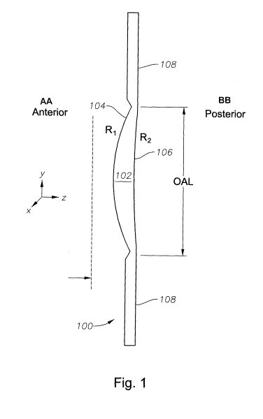
FIGURE 1 depicts a meniscus-shaped intraocular lens (IOL) according to a
particular embodiment of the present invention;
FIGUREs 2A and 2B illustrate a shape change in the optic of FIGURE 1
according to a particular embodiment of the present invention; and
FIGUREs 3A and 3B illustrate an example wavefront showing a change in
spherical aberration according to a particular embodiment of the present
invention.
CA 02786851 2012-07-09
WO 2011/090591 PCT/US2010/059924
4
DETAILED DESCRIPTION OF PREFERRED EXEMPLARY
EMBODIMENTS
Various embodiments of the disclosure are illustrated in the FIGURES, like
numerals being generally used to refer to like and corresponding parts of the
various
drawings. As used herein, the terms "comprises," "comprising," "includes,"
"including," "has," "having" or any other variation thereof, are intended to
cover a
non-exclusive inclusion. For example, a process, article, or apparatus that
comprises
a list of elements is not necessarily limited to only those elements but may
include
other elements not expressly listed or inherent to such process, article, or
apparatus.
Further, unless expressly stated to the contrary, "or" refers to an inclusive
or and not
to an exclusive or.
Additionally, any examples or illustrations given herein are not to be
regarded
in any way as restrictions on, limits to, or express definitions of, any term
or terms
with which they are utilized. Instead, these examples or illustrations are to
be
regarded as being described with respect to one particular embodiment and as
illustrative only. Those of ordinary skill in the art will appreciate that any
term or
terms with which these examples or illustrations are utilized will encompass
other
embodiments which may or may not be given therewith or elsewhere in the
specification and all such embodiments are intended to be included within the
scope
of that term or terms. Language designating such nonlimiting examples and
illustrations includes, but is not limited to: "for example", "for instance",
"e.g.", "in
one embodiment".
FIGURE 1 depicts a meniscus-shaped intraocular lens (IOL) 100 according to
a particular embodiment of the present invention. The meniscus-shaped IOL 100
has
an optical portion ("optic") 102 with an anterior convex face 104 having a
radius of
curvature R1 and a posterior concave face 106 having a radius of curvature R2.
For
purposes of this specification, "anterior" and "posterior" refer to the
directions of the
IOL 100 facing, respectively, away from and toward the retina. The "optical
axis"
refers to an axis extending transversely to a center of the anterior face 104
("vertex")
in the anterior-posterior direction.
CA 02786851 2012-07-09
WO 2011/090591 PCT/US2010/059924
The optic 102 is formed of a generally transparent material capable of
transmitting light to the retina of the eye. Any suitable material, including
a wide
variety of biocompatible polymeric materials, may be used. Examples of
suitable
materials include silicone, acrylics, hydroxyl ethyl methacrylate (HEMA),
polymethyl
5 methacrylate (PMMA) and numerous other materials known in the art. The optic
102
may also include materials for absorbing ultraviolet light, blue light, or
other
wavelengths to protect ocular tissue from light toxicity and/or to improve
visual
performance of the IOL 100.
The IOL 100 also includes a haptic assembly 108. The haptic assembly 108
fixes the position of the IOL 100 when the IOL 100 is disposed within the eye.
In
various embodiments of the present invention 108, the haptic assembly 108 may
be
configured for placement in the capsular bag or ciliary sulcus of the
posterior chamber
of the eye. In certain embodiments, the haptic assembly 108 could include
multiple
haptic arms having a proximal portion extending from the optic 102 connected
by a
joint to a distal portion contacting the capsular bag or ciliary sulcus. In
alternative
embodiments, the haptic assembly 108 could include a shaped periphery of the
IOL
100 directly contacting the capsular bag or ciliary sulcus.
When positioned within the eye, the IOL 100 provides pseudo-accommodation
by changing shape in response to contraction of the ciliary muscles.
Specifically, the
peripheral edge of the IOL 100 is compressed toward the optical axis so that
the
vertex of the anterior face 104 and the peripheral edge move relative to one
another in
a direction parallel to the optical axis. This compression changes the shape
factor of
the IOL 100 so that the principal plane is shifted posteriorly and the
spherical
aberration imparted by the IOL 100 substantially changes. The shape change in
the
optic 102 is illustrated in FIGUREs 2A and 2B.
The effective change in vision can be illustrated by the wavefront at the
image
plane illustrated in FIGUREs 3A-3B. In FIGURE 3A, an example wavefront image
for an IOL 100 according to a particular embodiment of the present invention
is
illustrated. In this example, the pupil size is set within 1 mm to 4 mm, and
the
wavefront is emitted from a source at infinite distance with a wavelength of
550 rim.
The central peak at the image plane illustrated a sharply focused image, and
the peak-
to-valley spherical aberration is within 0.5 waves (about 0.135 waves RMS).
CA 02786851 2012-07-09
WO 2011/090591 PCT/US2010/059924
6
FIGURE 3B shows the same lens when compressed. The curvature of the wavefront
illustrates the introduction of spherical aberration, with a peak-to-valley
now over 3
waves (about .905 waves RMS). A change in object distance from an infinite
distance
to 140 cm corresponds to an effective power change of 0.71 D at the corneal
plane or
0.92 D at the IOL plane. In general, a change of spherical aberration in the
wavefront
of at least I wave peak-to-valley for a 550-nm wavefront emitted by an object
at
infinity will be considered sufficient to be a substantial difference for
purposes of this
specification.
The mechanism to produce the shape change in the meniscus-shaped optic 102
of the IOL 100 can vary. In certain embodiments, the haptic assembly 108 can
be
placed in the ciliary sulcus and can transfer force from contraction of the
ciliary
muscles to the optic 102. In other embodiments, the haptic assembly 108 can be
placed in the capsular bag so as to respond to the flattening or rounding of
the
capsular bag as the zonules of the eye tighten or loosen in response to
relaxation and
contraction of the ciliary muscles, respectively. In such embodiments, the
haptic
assembly 108 may be formed to exhibit a mechanical bias so that, for example,
the
shape change results from a spring-like response of the haptic assembly 108 to
reduced tension on the capsular bag. The optic 102 can likewise exhibit a
spring-like
response to reduced force from the haptic assembly 108. The haptic assembly
108
may also be adapted to vault the optic in order to provide greater mechanical
stability
and/or more efficient mechanical response to the ciliary muscle contraction.
In
general, any mechanical arrangement for producing a change in shape in the
optic 102
that would be contemplated by one skilled in the art may be employed in
conjunction
with various embodiments of the present invention.
While single-optic embodiments of the present invention have been described,
it should be understood that the techniques of the present invention can be
applied to
multi-optic and/or multi-lens systems. Thus, for example, the meniscus-shaped
IOL
100 could be placed in the ciliary sulcus anterior to a biconvex IOL in the
capsular
bag. In another example, a phakic IOL could be placed in the anterior chamber,
and
the meniscus-shaped IOL 100 could be placed in the posterior chamber. The
meniscus-shaped IOL 100 may also be adapted so that the convex face 104 faces
anteriorly in such combinations and, in such embodiments, the change in shape
in
CA 02786851 2012-07-09
WO 2011/090591 PCT/US2010/059924
7
response to contraction of the ciliary muscles can also be reversed so that
the optical
effect is identical. Alternatively, the meniscus-shaped IOL 100 can be adapted
to
provide so-called "reverse accommodation," wherein the brain of the patient
can be
trained to concentrate on a distant image when the ciliary muscles are
contracted and
on a near image when the ciliary muscles are relaxed, thus reversing the
effect of the
accommodation reflex.
Although embodiments have been described in detail herein, it should be
understood that the description is by way of example only and is not to be
construed
in a limiting sense. It is to be further understood, therefore, that numerous
changes in
the details of the embodiments and additional embodiments will be apparent to,
and
may be made by, persons of ordinary skill in the art having reference to this
description. It is contemplated that all such changes and additional
embodiments are
within scope of the claims below and their legal equivalents.
