Note: Descriptions are shown in the official language in which they were submitted.
CA 02787194 2017-02-08
SET OF PROBES FOR THE DETECTION AND TYPING OF 46 HUMAN PAPILLOMAVIRUS
MUCOSAL TYPES
FIELD OF THE INVENTION
The present invention relates to reagents and methods for genotyping human
papillomaviruses (HPV). In particular, the present invention relates to
testing clinical samples
for the type of HPV infection using a multiplex assay based on PCR
amplification and
detection using microspheres.
BACKGROUND OF THE INVENTION
Human papillomaviruses cause ubiquitous infectious of the keratinised
epithelia of the
skin and of the mucosae. About 120 HPV types have been characterized so far,
which differ
in prevalence, epidemiology and clinical manifestations (de Villiers et al.,
2004). In particular,
mucosal types infect the keratinised epithelia of the genital, anal and oro-
pharyngeal
mucosae (Munoz et al., 2003; Munoz and Bosch, 1997; Van Ranst et al, 1992;
Chan et al.,
1995, D'Souza et al., 2007, Bosh et al., 2008). Mucosal HPVs are most commonly
transmitted by sexual contact, and infect sexually active people with a very
high prevalence. It
is estimated that the lifetime incidence of HPV infection in women is 80%
(Bekkers et al.,
2004), and the overall prevalence of active infection worldwide varies form
1.4% to 25%
(Clifford et al, 2005).
Although the vast majority of infections are benign and self-limiting, a
subset of "high
risk" HPV types have the potential to cause persistent infection that may
progress to
malignant transformation and invasive cancer (Munoz et al., 2003). Cervical
cancer is the
most common HPV-associated malignancy and it is now clear that HPV is a
necessary cause
of virtually all cervical cancers (Bosch and Munoz, 2002; zur Hausen, 2002,
Bosch et al.,
2002; Munoz et al., 2003; Walboomers et al.,
CA 02787194 2012-07-16
WO 2011/088573 PCT/CA2011/050026
2
1999, Smith et al, 2007). HPV associated malignancies are also found in the
anal
canal (Melbye and Sprogel, 1991; Palefsky et al., 1991), vulva (Buscema et
al., 1988),
the penis (Gregoire et al., 1995; lwasawa et al., 1993), oro-pharyngeal
mucosae and
other head and neck tissues (D'Souza, et al., 2007; Mork et al., 2001;
Gillison et al.,
2000; Syrjanen, 2005).
Since HPV infection is necessary for the development of virtually all cervical
cancers, detection of high risk HPV types is being considered as a screening
method
for cervical cancer, alongside, or even in substitution of, traditional
cytological
screening using the Papanicolau methods (pap test), with the promise of
improving
the sensitivity and cost effectiveness of cervical cancer screening programs
(Cuzik et
al., 2008; Cuzick et al., 2003; Ronco et al., 2006; Schiffman et al., 2005;
Kim et al.,
2005;Davies et al., 2006; Mayrand et al., 2006; Cuzik et al., 2006).
Two type-specific HPV vaccines (Gardasil, from Merck-Frosst for types 16, 18,
6 and 11; Cervarix form Glaxo-Smith-Kline for types 16 and 18) have recently
been
developed and clinical trials have shown that they are extremely effective in
preventing both persistent infection with HPV and the dysplastic changes in
the
cervical epithelium that lead to malignant transformation (Koutsky et al.,
2002;Villa et
al., 2005; Harper et al., 2004; Harper et al., 2006). However, since vaccines
are type-
specific it is important to know the distribution of the various HPV types in
a
population, as well as to have a surveillance system in place to monitor
vaccine
efficacy and unexpected shifts in the frequency of HPV types not covered by
the
vaccines.
It is therefore expected that the routine use of type-specific tests for HPV
will
become more widespread, outside their current use in epidemiological studies
for
research purposes.
Currently, typing of HPV requires amplification by various PCR methods,
followed by detection of specific sequences using either direct sequencing of
the PCR
products, RFLP methods (many methods have been described in the literature,
for
example Lungo et al., 1992; Menzo et al., 2008, Nobre et al., 2008; Santiago
et al.,
2006), Southern blot or dot blot using specific probes(for example Gregoire et
al.,
CA 02787194 2012-07-16
WO 2011/088573 PCT/CA2011/050026
3
1989; Josefsson et al., 1999), reverse line hybridization (Gravitt el al.,
1998; Kleter et
al., 1999; van der Brule et al., 2002; Melchers et al, 1999), DNA microarray
methods
(Min et al., 2006; Albrecht et al, 2006; Choi et al., 2003; Huang et al.,
2004; Hwang
eta la., 2003; Oh et al., 2004; Nuovo et al., 2008), and others (for example
Nishiwaki
et al., 2008; Dell'At-ti, 2007; Gao et al., 2003; Gharizadeh et al., 2007; Han
et al.,
2006; Lee et al., 2005; Liu et al, 2003; Zhang et al, 2003). In particular,
reverse line
blot methods have been validated and have been used extensively for
epidemiological studies. Two leading commercial genotyping methods, Inn LiPA
(van
Hamont, 2006) and Roche linear array (Coutlee et al., 2006), are based on the
reverse hybridization technology. The Roche Linear Array genotyping kit as
been
approved by FDA and it is the leading commercial genotyping method. However,
these methods are not suitable for high throughput testing and they rely on a
subjective visual assessment of band intensity for determining the results.
The xMAP technology developed by Luminex (Austin, TX, USA) is based on
microspheres that can be produced in 100 different "colours" depending on they
ratio
of two spectrally distinct fluorophores coupled to the microspheres. The
different
colours can be recognized by flow cytorneters and the different type of
microspheres
can be enumerated and analyzed for the presence of specific bound ligands.
This
technology has been the basis for a variety of multiplex assays for serology,
genotyping and other analytical applications. A description of the Luminex
technology
and a list of publications can be found at the Luminex web site.
Each type of microsphere can be coupled with a specific ligand, e.g. with DNA
probes specific for each type of HPV in this work, and mixed together to form
a
multiplex assay. The PCR products derived from HPV samples are labelled with
biotin
and mixed with the beads carrying the probes, so that HPV DNA will hybridize
with
the cognate probe. The flow cytometer will then sort the different "coloured"
microspheres and determine which type carries the fluorescence due to the HPV
DNA. The computer software driving the flow cytometer will indicate which
beads are
fluorescent, thereby identifying the HPV type(s) present in the sample. The
advantages of this method is the low cost per assay, the possibility of
automation for a
CA 02787194 2012-07-16
WO 2011/088573 PCT/CA2011/050026
4
high throughput assay, and the flexibility derived from the possibility of
adding or
removing types of microspheres depending on the need of the assay or on the
discovery of new types. Several microsphere-based multiplex assay for HPV
genotyping have been published. The method by Wallace et al. (2005) is a
multiplex
microsphere assay with probes for 45 mucosal HPV. However, formal validation
was
performed for only a few types and only 20 types were detected from clinical
samples,
without independent validation of the genotyping result. The method published
by Oh
et al. (2007) detects 15 HPV types and it has been validated against a 132
clinical
samples. A 56 sample comparison with a DNA microarray genotyping method is
also
shown. The method, by Schmitt et al. (2006), has been carefully validated with
HPV
plasmids and clinical samples and covers the 22 most common mucosal HPV types.
The method by Jiang et al. (2006) describes specific probes for 26 HPV mucosal
types. Validation was performed with synthetic oligonucleotides complementary
to
the probes and with a limited number of clinical samples. A commercial method
developed by Qiagen (Hi!den, Germany) is able to type 18 HPV high-risk using a
proprietary set of primers, followed by detection using a Luminex system. At
least one
study comparing this Luminex Qiagen test to a reverse line blot hybridization
has
been published (Seme et al., 2009).
Herein, we report the design of novel HPV type-specific probes and the
development of a rnicrosphere multiplex assay that can detect 46 different
mucosal
types in a single reaction. In addition the unique probe set, compared to the
previous
method we introduce 2 innovations: i) the use of longer probes (30 mers) to
provide
for a greater specificity for variants and closely related types; ii) the
production of
single stranded DNA products by selective digestion of the PCR products with
exonuclease, which produces a greater signal to noise ratio, making a washing
step
unnecessary.
SUMMARY OF THE INVENTION
We have described a set of 46 DNA probes and a PCR amplification method
for the detection of 46 mucosal HPV types using the Luminex xMAP technology.
This
CA 02787194 2012-07-16
WO 2011/088573 PCT/CA2011/050026
technology uses a mixture of sortable microsphere coupled to the specific HPV
probes, so that all the 46 types can be detected simultaneously in one
reaction tube.
Our data shows that all the probes are sensitive and specific for the
detection
of the 46 HPV types, without cross-hybridization. This conclusion is supported
by the
5
use of reference DNA from the 46 types and an extensive validation using
direct
sequencing as a gold standard for the identification of the HPV types.
Amplified DNAs from at least 32 HPV types can be detected simultaneously
and precisely by this Luminex method.
Comparison with a leading commercial HPV typing method, the Roche Linear
Array, confirms that the NML Luminex method is suitable for the identification
of HPV
types in clinical samples containing 3 or less HPV types. However, the PCR
amplification method is less efficient in amplifing DNA from samples with
multiple
infections containing 4 or more HPV types. This is a problem caused by the PCR
amplification method and not by the set of probes or the Luminex detection
system.
The less efficient amplification in multiple infections is a significant
problems for HPV
types 52, 53, 61, 73 84 and 89 but not for the major oncogenic HPV types,
which are
most important in epidemiology and clinical practice.
When samples with 4 or more HPV types are excluded, detection by NML
Luminex and Roche Linear array are equivalent. Therefore, use of the NML
Luminex
method on populations with high frequency of multiple infections (such as HIV
patients, men who have sex with men or sex workers) will lead to an
underestimation
of the prevalence with certain types. On the contrary, use of the NML Luminex
method on a general population of women, where the prevalence of infections
with 4
or more types is negligible, will produce accurate prevalence results for most
types.
The NML Luminex HPV genotyping method has the advantage of detecting
almost all genital HPV types and of being very sensitive thanks to the nested
PCR
method. The Luminex xMAP technology allows for a very quick, hands-off reading
of
the samples and an objective computational interpretation of the results.
Because our
method has no washing steps or visual reading steps, it is easily amenable to
automation.
CA 02787194 2012-07-16
WO 2011/088573 PCT/CA2011/050026
6
According to a first aspect of the invention, there is provided a method of
detecting and typing a human papillomavirus (HPV) type infection in a sample
comprising:
a) providing a sample suspected of comprising at least one HPV
type;
b) adding to the sample primers suitable for amplifying the Ll region of
HPV;
c) incubating the sample under conditions suitable for DNA amplification;
d) adding at least one probe having a nucleotide sequence as set forth in
any one of SEQ ID Nos. 1-46, said probe binding to only one HPV type under
hybridization conditions, each said at least one probe further comprising a
unique tag;
e) incubating said probe and said sample under conditions suitable for
hybridization; and
f) detecting hybridization of at least one said tagged probe.
According to a second aspect of the invention, there is provided a set of
probes
for detection and typing human papilloma virus (HPV) types, each said probe of
said
set hybridizing to only one HPV type under hybridizing conditions, each said
probe of
said set consisting of a unique tag and a nucleotide sequence as set forth in
one of
SEQ ID Nos. 1-46.
According to a third aspect of the invention, there is provided a set of
probes
for detection and typing human papilloma virus (HPV) types, each said probe of
said
set hybridizing to only one HPV type under hybridizing conditions, each said
probe of
said set consisting of a unique tag and a nucleotide sequence as set forth in
one of
SEQ ID No. 1, 2, 4 or 5.
According to a fifth aspect of the invention, there is provided a set of
probes for
detection and typing human papilloma virus (HPV) types, each said probe of
said set
hybridizing to only one HPV type under hybridizing conditions, each said probe
of said
set consisting of a unique tag and a nucleotide sequence as set forth in one
of SEQ
ID. No. 4, 5 and 17.
According to a sixth aspect of the invention, there is provided a set of
probes
for detection and typing human papilloma virus (HPV) types, each said probe of
said
CA 02787194 2017-02-08
7
set hybridizing to only one HPV type under hybridizing conditions, each said
probe of said set
consisting of a unique tag and a nucleotide sequence as set forth in one of
SEQ ID No. 4, 5,
8, 10, 11, 12, 17, 18, 19, 22, 23, 24, 27 and 29.
According to a seventh aspect of the invention, there is provided a set of
probes for
detection and typing human papilloma virus (HPV) types, each said probe of
said set
hybridizing to only one HPV type under hybridizing conditions, each said probe
of said set
consisting of a unique tag and a nucleotide sequence as set forth in one of
SEQ ID Nos 6, 4,
5, 7, 8, 10, 11, 12, 17, 18, 19, 20, 22, 23, 24, 27, 28, 29, 30, 31, 34, 37,40
and 46.
According to an eighth aspect of the invention, there is provided a set of
probes for
detection and typing human papilloma virus (HPV) types, each said probe of
said set
hybridizing to only one HPV type under hybridizing conditions, each said probe
of said set
consisting of a unique tag and a nucleotide sequence as set forth in one of
SEQ ID Nos. 1, 2,
9, 13, 14, 15, 16, 21, 25, 26, 27, 28, 29, 30, 31, 33, 35, 36, 38, 39, 41, 42,
43, 44 and 45.
According to a further aspect of the invention, there is provided a method of
detecting
and typing a human papillomavirus (HPV) type infection in a sample comprising:
a) providing a sample suspected of comprising at least one HPV type;
b) adding to the sample primers suitable for amplifying the L1 region of
HPV;
c) incubating the sample under conditions suitable for DNA amplification;
d) adding a probe consisting of the nucleotide sequence of SEQ ID NO:46,
said
probe binding to only HPV type 97 under hybridization conditions, said probe
further
comprising a unique tag, said unique tag comprising a combination of two
fluorescent dyes;
e) incubating said probe and said sample under conditions suitable for
hybridization; and
f) detecting hybridization of said probe.
CA 02787194 2017-02-08
7a
BRIEF DESCRIPTION OF THE DRAWINGS
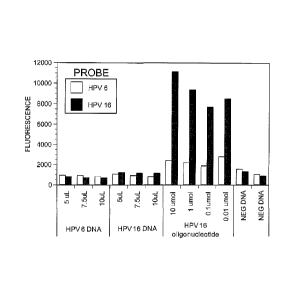
Fig. 1 ¨ Preliminary hybridization tests using 20 mer probes for HPV 6 and HPV
16 ¨
A mixture of two types of microspheres coupled with 20 mer probes for HPV 6
and 16 (as
described in Table 1) were hybridized with the indicated volume of PCR
reaction (panel A and
B), with a 20 mer oligonucleotide exactly complementary to the HPV 16 probe
(panel C), or
with an unrelated PCR product (3-globin DNA).
Fig.2 ¨Position of the probes for the 46 HPV types considered in this
submission. The
alignment of the L1 region comprised between the GP5+/GP6+ primers is shown.
The
positions of the primers in indicated by the boxes while the probe sequences
are in bold and
underlined.
Fig. 3 ¨ Effect of T7 exonuclease digestion of nested PCR products on
hybridization
to Luminex beads. HPV 16 DNA was amplified by MY09/My11 and GP5+/GP6+ nested
FOR,
as described above, and the products were digested with T7 exonuclease for the
indicated
times. After digestion, the FOR products were
CA 02787194 2017-02-08
8
hybridized to Luminex beads carrying the HPV 16 probe and detected as
described above.
The GP6+ primer contained a 5' biotin moiety, for detection by the Luminex
technology, and
phosporothioate bonds in the first 4 nucleotides on the 5', to protect this
strand from the
action of the T7 exonuclease. The black bar and the white bar represent the
fluorescence
signals of a negative sample and of a sample containing a biotylinated
oligonucleotide
complementary to the HPV 16 probe.
Figure 4 ¨ Graphic representation of the data of Table 4- The probes are on
the right
axis and the HPV DNAs on the left axis. The vertical axis represents the
fluorescence read
for each microsphere carrying a specific HPV probe. The bars on the diagonal
represent the
hybridization of HPV DNA type with the intended cognate probe.
Fig. 5 ¨ Simultaneous detection of multiple HPV types ¨ Samples containing DNA
from increasing numbers of HPV types were prepared as described in the text
and then
detected by the NML Luminex method as described. The number of types in each
sample is
indicated in the leftmost column. The second column from the left indicates
what additional
HPV type was added to the mixture. A "+" sign indicates a positive result
(over 50 FU). The
asterisks indicate false positive or false negative results.
DESCRIPTION OF THE PREFERRED EMBODIMENTS
Unless defined otherwise, all technical and scientific terms used herein have
the same
meaning as commonly understood by one of ordinary skill in the art to which
the invention
belongs. Although any methods and materials similar or equivalent to those
described herein
can be used in the practice or testing of the present invention, the preferred
methods and
materials are now described.
DNA probes comprising SEQ ID Nos 1 through 46 were generated according to the
specific sequences of 46 types of genital HPV, namely type 6 (SEQ ID No. 1),
11 (SEQ ID
No. 2), 13 (SEQ ID NO. 3), 16 (SEQ ID No. 4), 18 (SEQ ID No. 5), 26 (SEQ ID
No. 6), 30
(SEQ ID No. 7), 31 (SEQ ID No. 8), 32 (SEQ ID No. 9), 33 (SEQ
CA 02787194 2012-07-16
WO 2011/088573 PCT/CA2011/050026
9
ID No. 10), 35 (SEQ ID No. 11), 39 (SEQ ID No. 12), 40 (SEQ ID No. 13), 42
(SEQ ID
No. 14), 43 (SEQ ID No. 15), 44 (SEQ ID No. 16), 45 (SEQ ID No. 17), 51 (SEQ
ID
No. 18), 52 (SEQ ID No. 19), 53 (SEQ ID No. 20), 54 (SEQ ID No. 21), 56 (SEQ
ID
No. 22), 58 (SEQ ID No. 23), 59 (SEQ ID No. 24), 61 (SEQ ID No.25), 62 (SEQ ID
No. 26), 66 (SEQ ID No. 27), 67 (SEQ ID No. 28), 68 (SEQ ID No. 29), 69 (SEQ
ID
No. 30), 70 (SEQ ID No. 31), 71 (SEQ ID No: 32), 72 (SEQ ID No. 33), 73 (SEQ
ID
No. 34), 74 (SEQ ID No.35), 81 (SEQ ID No. 36), 82 (SEQ ID No.37), 83 (SEQ ID
No.
38), 84 (SEQ ID No. 39), 85 (SEQ ID No. 40), 86 (SEQ ID No. 41), 87 (SEQ ID
No.
42), 89 (SEQ ID No. 43), 90 (SEQ ID No. 44), 91 (SEQ ID No. 45) and 97 (SEQ ID
No. 46). In order to make each probe sensitive and specific, the probes were
tested in
a multipex assay as described below. Probes that in these tests did not
hybridize to
the intended HPV type or that cross-hybridized to other types were re-
designed,
sometimes repeatedly, until all probes hybridized to unique HPV type DNA.
Accordingly, each respective probe binds specifically to only one specific HPV
genome or HPV type. The history of the probe design is shown in Table 2.
For the multiplex assay, each probe was conjugated to one of 46 types of
fluorescent microspheres, each with different ratios of red and infrared
fluorophores,
according to the manufacturers instructions. The micropsheres produced by
Luminex
Corp. are colour coded with a combination of two fluorescence dyes into 100
different
sets that can be recognized and counted by a flow cytometer using a red laser.
The
flow cytometer can also detect a reporter dye bound to any set of beads using
a
separate green laser. For this embodiment, 46 sets of beads were selected and
each
set was coupled to a unique 30mer oligonucleotide probe designed to hybridize
sensitively and specifically to one of 46 types of genital HPV DNA, amplified
as
described below. The 46 sets of beads were mixed to constitute a multiplex
reaction
that could detect any combination of the 46 types of HPV DNA present in
clinical
specimens.
The probes were designed to amplify the region comprised between the PCR
amplification primers GP5+/GP6+. This region is 141bp long for HPV 16
(nucleotides
6624 to 6764, GenBank accession no. AF125673), but varies in length slightly
CA 02787194 2012-07-16
WO 2011/088573 PCT/CA2011/050026
depending on the HPV type. GP54-/GP6+ are general primers that amplify the DNA
from most HPV types. Published primers sets MY09/MY11 and primer set PGMY are
also general primers which amplify most genital HPV types. They are situated
outside
the GP4-/GP6-1- region and therefore they can be used for a nested PCR
reaction with
5 the GP5+/GP6+ primers, in order to improve the sensitivity and the
spectrum of HPV
types that can be amplified, especially when multiple types are present in the
same
sample.
Using these primers, HPV DNA from clinical samples was amplified and then
treated with T7 exonuclease to produce a single stranded, biotin labelled DNA
10 complementary to the probes coupled to the microspheres. The single
stranded HPV
DNA and the tagged microspheres were then co-incubated, so that the HPV DNA
could bind to its cognate probe on the microspheres. Streptavidin conjugated
to the
fluorophore phycoerythrine was then added. Streptavidin binds tightly to
biotin
conferring phycoerythrine fluorescence to those microsphere that are bound to
HPV
DNA. The samples were then analyzed by flow cytometry which provided an
analysis
of the numbers of each type of bound microspheres and their level of
phycoerythrine
fluorescence. High phycoerythrine fluoresce on specific beads indicates the
presence
of HPV DNA of specific types.
In addition to the multiplex assay for 46 HPV types, the microspheres can be
mixed in different combinations to test separately only for HPV types
contained in
vaccines (HPV type 6 (SEQ ID No. 1), 11 (SEQ ID No. 2), 16 (SEQ ID No. 4) and
18
(SEQ ID No. 5)), or to test for the most malignant HPV types (HPV types 16
(SEQ ID
No. 4), 18 (SEQ ID No. 5) and 45 (SEQ ID No. 17)), or for the most common HPV
types (HPV type 16 (SEQ ID No. 4), 18 (SEQ ID No. 5), 31 (SEQ ID No. 8), 33
(SEQ
ID No. 10), 35 (SEQ ID No. 11), 39 (SEQ ID No. 12), 45 (SEQ ID No. 17), 51
(SEQ ID
No. 18), 52 (SEQ ID No. 19), 56 (SEQ ID No. 22), 58 (SEQ ID No. 23), 59 (SEQ
ID
No. 24), 66 (SEQ ID No. 27) and 68 (SEQ ID No. 29)), or to test for all
oncogenic HPV
types (HPV type 26 (SEQ ID No. 6), 16 (SEQ ID No. 4), 18 (SEQ ID No. 5), 30
(SEQ
ID No. 7), 31 (SEQ ID No.8), 33 (SEQ ID No. 10), 35 (SEQ ID No. 11), 39 (SEQ
ID
No. 12), 45 (SEQ ID No. 17), 51 (SEQ ID No. 18), 52 (SEQ ID No. 19), 53 (SEQ
ID
CA 02787194 2012-07-16
WO 2011/088573 PCT/CA2011/050026
11
No. 20), 56 (SEQ ID No. 22), 58 (SEQ ID No. 23), 59 (SEQ ID No. 24), 66 (SEQ
ID
No. 27), 67 (SEQ ID No. 28), 68 (SEQ ID No. 29), 69 (SEQ ID No. 30), 70 (SEQ
ID
No. 31), 73 (SEQ ID No, 34), 82 (SEQ ID No. 37), 85 (SEQ ID No. 40), and 97
(SEQ
ID No. 46)), or to test only for non-oncogenic (low risk) types (6 (SEQ ID No.
1), 11
(SEQ ID No. 2), 32 (SEQ ID No. 9), 40 (SEQ ID No. 13), 42 (SEQ ID No. 14), 43
(SEQ ID No. 15), 44 (SEQ ID No. 16), 54 (SEQ ID No. 21), 61 (SEQ ID No. 25),
62
(SEQ ID No. 26), 66 (SEQ ID No. 27), 67 (SEQ ID No. 28), 68 (SEQ ID No. 29),
69
(SEQ ID No. 30), 70 (SEQ ID No. 31), 72 (SEQ ID No. 33), 74 (SEQ ID No. 35),
81
(SEQ ID No. 36), 83 (SEQ ID No. 38), 84 (SEQ ID No. 39), 86 (SEQ ID No.41), 87
(SEQ ID No. 42), 89 (SEQ ID No. 43), 90 (SEQ ID No. 44) and 91 (SEQ ID No.
45)).
In accordance with a first embodiment of the present invention, there is
provided a series of DNA probes that can be used in conjunction with DNA
amplification techniques to genotype various strains of HPV.
In a second embodiment of the invention, the series of DNA probes that can be
used in a multiplexed format assay to simultaneously detect multiple strains
of HPV
In a third embodiment of the invention, the DNA probes can be used with other
detection systems including Southern or Northern blots, reverse line blot
hybridization,
DNA microarray or ELISA, or other such systems as will be obvious to those
skilled in
the art.
According to an aspect of the invention, there is provided a method of
detecting
and typing a human papillomavirus (HPV) type infection in a sample comprising:
a) providing a sample suspected of comprising at least one HPV type;
b) adding to the sample primers suitable for amplifying the L1 region of
HPV;
c) incubating the sample under conditions suitable for DNA amplification;
d) adding at least one probe having a nucleotide sequence as set forth in
any one Of SEQ ID Nos. 1-46, said probe binding to only one HPV type under
hybridization conditions, each said at least one probe further comprising a
unique tag;
e) incubating said probe and said sample under conditions suitable for
hybridization; and
CA 02787194 2012-07-16
WO 2011/088573 PCT/CA2011/050026
12
f) detecting hybridization of at least one said tagged probe.
As discussed herein, the sample may contain more than one HPV type and the
'at least one probe' may be a set of probes comprising or consisting of
respective
probes having nucleotide sequences as set forth in any one of SEQ ID Nos 1-46
and
a unique tag or identification tag which uniquely identifies the respective
probe. For
example, all probes having a nucleotide sequence as set forth according to SEQ
ID
No.1 will have the same tag as will all probes having a nucleotide sequence as
set
forth in SEQ ID No. 2.
As discussed herein, the hybridization conditions are sufficiently stringent
that
the probe will bind only to the target DNA. For example, the hybridization
conditions
may be sufficiently stringent for hybridization of two strands to occur only
if there is
15, 16, 17, 18, 19, 20 or more consecutive nucleotides having an exact match.
As will be appreciated by one of skill in the art, the probes consisting of
nucleotide sequences as set forth in any one of SEQ ID Nos, 1-46 and a unique
tag
can be used together or in any sub-combination thereof in a multiplex assay to
specifically type HPV types in a given sample. Specifically, because each
probe has a
unique tag associated therewith, hybridization of a respective probe to a DNA
molecule within the sample indicates the presence of the corresponding HPV
type in
that sample. The probe set is unique in that the probes do not cross-
hybridize, as
discussed below.
In some embodiments, at least one probe may refer to a mixture of probes,
each representative probe of said mixture having a nucleotide sequence as set
forth
in SEQ ID No. 1, 2, 4 or 5 or as set forth in SEQ ID No. 4, 5 or 17 or as set
forth in
SEQ ID No. 4, 5, 8, 10, 11, 12, 17, 18, 19, 22, 23, 24, 27 or 29 or as set
forth in SEQ
ID No. 6, 4, 5, 7, 8, 10, 11, 12, 17, 18, 19, 20, 22, 23, 24, 27, 28, 29, 30,
31, 34, 37,
40 or 46 or as set forth in SEQ ID No. 1, 2, 9, 13, 14, 15, 16, 21, 25, 26,
27, 28, 29,
30, 31, 33, 35, 36, 38, 39, 41, 42, 43, 44 or 45.
The unique tag is a combination of two fluorescent dyes.
The unique tag is a combination of different ratios of red and infra-red
fluorophores, as discussed herein.
CA 02787194 2012-07-16
WO 2011/088573 PCT/CA2011/050026
13
According to another aspect of the invention, there is provided a set of
probes
for detection and typing human papilloma virus (HPV) types, each said probe of
said
set hybridizing to only one HPV type under hybridizing conditions, each said
probe of
said set consisting of a unique tag and a nucleotide sequence as set forth in
one of
SEQ ID Nos. 1-46.
According to another aspect of the invention, there is provided a set of
probes
for detection and typing human papilloma virus (HPV) types, each said probe of
said
set hybridizing to only one HPV type under hybridizing conditions, each said
probe of
said set consisting of a unique tag and a nucleotide sequence as set forth in
one of
SEQ ID No. 1, 2, 4 or 5. As will be appreciated by one of skill in the art,
additional
probes having sequences as set forth in any one of SEQ ID Nos 3 and 6-46and
any
combination thereof may be added to the probe set.
According to another aspect of the invention, there is provided a set of
probes
for detection and typing human papilloma virus (HPV) types, each said probe of
said
set hybridizing to only one HPV type under hybridizing conditions, each said
probe of
said set consisting of a unique tag and a nucleotide sequence as set forth in
one of
SEQ ID. No. 4, 5 and 17.
According to another aspect of the invention, there is provided a set of
probes
for detection and typing human papilloma virus (HPV) types, each said probe of
said
set hybridizing to only one HPV type under hybridizing conditions, each said
probe of
said set consisting of a unique tag and a nucleotide sequence as set forth in
one of
SEQ ID No. 4, 5, 8, 10, 11, 12, 17, 18, 19, 22, 23, 24, 27 and 29.
According to another aspect of the invention, there is provided a set of
probes
for detection and typing human papilloma virus (HPV) types, each said probe of
said
set hybridizing to only one HPV type under hybridizing conditions, each said
probe of
said set consisting of a unique tag and a nucleotide sequence as set forth in
one of
SEQ ID Nos 6, 4, 5, 7, 8, 10, 11, 12, 17, 18, 19, 20, 22, 23, 24, 27, 28, 29,
30, 31, 34,
37, 40 and 46.
According to another aspect of the invention, there is provided a set of
probes
for detection and typing human papilloma virus (HPV) types, each said probe of
said
CA 02787194 2012-07-16
WO 2011/088573 PCT/CA2011/050026
14
set hybridizing to only one HPV type under hybridizing conditions, each said
probe of
said set consisting of a unique tag and a nucleotide sequence as set forth in
one of
SEQ ID Nos. 1, 2, 9, 13, 14, 15, 16, 21, 25, 26, 27, 28, 29, 30, 31, 33, 35,
36, 38, 39,
41, 42, 43, 44 and 45.
EXAMPLES:
Oligonucleotides
Oligonucleotides were synthesized at the DNA Core Section of the National
Microbiology Laboratory. The probes carried a 5' 012 amino linker modification
for
coupling to the carboxyl group of the Luminex microspheres. The MY09, MY11,
GP5+
and the modified GP6+ primer for the PCR amplification of HPV DNA, were
purchased from Invitrogen (Burlington ON, Canada).
PCR amplification
HPV DNA from plasmid or clinical specimens was amplified by a nested PCR
method using the MY09/MY11 primers for the first step (Manos et al., 1989) and
GP5+/GP6+ primers for the second step (Roda Husman et al., 1995). For optimal
amplification of clinical samples with multiple HPV types, PGMY primers were
used
for the first step (Gravitt et al, 2000). The GP6+ primer carried the
following
modification: i) a 5' biotin label to be used as a ligand for the streptavidin-
PE for
detection of PCR products (See below); ii) the first 4 nucleotides on the 5'
end were
linked by phosphorothioate bonds to confer resistance to the action of the
bacteriophage T7 gene 6 exonuclease (See below and in the result section). PCR
amplification was performed in 1X PCR Buffer (Invitrogen, Cat # 10342-020) in
the
presence of 4mM MgC12, 200 pM of dNTP (Invitrogen, Cat# 10297-018), 0.2mM of
each primer and 1.25U of Amplitaq Gold polymerase (Applied Biosystem, Cat #
4311816). The first round of nested PCR amplification with the MY09/MY11
primers
started with a 5 min initial denaturation step at 94 C, followed by 30 cycles
of
denaturation at 94 C for 30 seconds, annealing at 55 C for 30 seconds and
elongation at 72 C for 60 seconds, followed by a 7 min final extension at 72
C.
CA 02787194 2012-07-16
WO 2011/088573
PCT/CA2011/050026
Amplification with PGMY primers was carried out for 40 cycles (denaturation at
94 C
for 30 sec, annealing 55 C for 30 sec, elongation 72 C for 30 sec) in the
presence of
6 mM MgC12, 200 pM dNTPs and 0.6 pM each of the 18 primers comprising the
PGMY mixture (Gravitt et al., 2000). One to 5 (typically 2)
of this reaction were
5
used for the second round of amplification with GP5+/GP6+ primers under the
following conditions: 5 min initial denaturation at 94 C, followed by 30
cycles of 94 C
for 30 seconds, 40 C for 20 seconds and 72 C for 30 seconds, followed by a 7
min
final extension at 72 C. One-step PCR with GP5+/GP6+ primers was conducted
under the following conditions: a 5 min initial denaturation at 94 C followed
by 30
10
cycles of 94 C for 30 seconds, 40 C for 20 seconds and 72 C for 30 seconds
followed by a 7 min final extension at 72 C.
Digestion of PCR products with T7 exonuclease
After PCR, the GP5+ strand complementary to the biotinylated strand, was
15
removed by digestion with T7 genes exonuclease, a 5' 4 3' processive
exonuclease.
The other strand was protected from the action of T7 exonuclease by the 4
phosphorothioate bonds on the 5' (Nikiforov et al, 1994). This digestion
produced a
single stranded, biotin labelled DNA complementary to the probes coupled to
the
Luminex beads and it was performed by adding T7 exonuclease (New New England
Biolabs, Cat# M0263L) to PCR products at a final concentration of 0.4U/ill.
The
reaction was stopped by adding 0.5M EDTA at a final concentration of 12.5111
of 0.5M
EDTA.
Preparation of microspheres
Microspheres labelled with different ratios of red and infrared fiuorophores
were purchased from Luminex (Austin, TX, USA, Cat #L100-CXXX-01) and coupled
to HPV type-specific probes carrying a 5' amino modification that reacts with
the
carboxyl groups on the microspheres following the instruction of the
manufacturer with
minor modifications. Briefly, the microsphere stock (Luminex) was vortexed
vigorously
then an aliquot containing 5.0 x 10⁶ microspheres from each set was placed in
a
CA 02787194 2012-07-16
WO 2011/088573 PCT/CA2011/050026
16
separate 1.5m1 microfuge tube, resuspended in a in a sonicating water bath
(Branson) and centrifuged at 14000 x g for 2min. The supernatant was removed
and
the microshperes were resuspended in 5041 of 0.1 M 2-N-
rnorpholinoethansulfonic
acid (MES) (Sigma Cat #M-2933) at a pH of 4.5. Then 11.11 of a 1mM solution of
the
appropriate type of amino substituted oligonucleotide was added to a different
set of
microspheres and 2.54 of a 10mg/nriL solution of 1-Ethy1-3-3-
dimethylaminopropyl
carbodiimide HC1 (EDC) (Fisher Cat # 22980) were added to each tube. The tubes
were vortexed and, after an incubation of 30 min at RT in the dark, 2.54 of
10rrig/m1
EDC were added to each tube and incubated in the dark for 30 minutes. After
the
second incubation period 1m1 of 0.02% Tween 20 (Sigma Cat # P-9416) was added
and the tubes were centrifuged for 2 minutes at 14,000 X g. The supernatant
was
removed and 1m1 of 0.1% SDS (sodium dodecyl sulfate) was added to the
microsphere pellet, the tubes were vortexed and then microcentrifuged for 2
minute at
14,000 X g. The supernatant was removed and the pellet was resuspended in
100111
of TE. The microspheres coupled to the probes were stored in the dark at 4 C
for a
maximum of 6 months.
Luminex assay
For the Luminex assay typically 15 microspheres/ul of each set were mixed in
a reaction mixture. Exonuclease-digested PCR products were placed in a 96 well
PCR microplate (Fisher, Cat # CS006509) in a total volume of 17u1 and sealed
with a
96 well sealing cover (Fisher, Cat # CS006555). The microplate was incubated
at
95 C for 10 minutes to denature the DNA and 331.1L of the microsphere mix was
added. The samples were incubated at the hybridization temperature of for 10
min
and, after addition of 25111 of a 0.04mg/u1 solution of streptavidine-
phycoerythrin
(lnvitrogen Cat # S-866) in 1X tetramethyl ammonium chloride (TMAC) (Sigma,
Cat #
T-3411) was added to the samples and incubated for 5 more minutes at 60 C.
Samples were analyzed on a Luminex Liquid Chip 200 flow cytorneter using the
Luminex IS software. The analysis was carried out at 60 C with a maximum
volume
CA 02787194 2012-07-16
WO 2011/088573
PCT/CA2011/050026
17
of 50p.L of sample and a minimum count of 100 microspheres per type, with a
setting
of 8,300 and 16,500 for the lower and upper gate, respectively.
Example 1: Design and Selection of Probes
The probes were targeted at the region of the Ll gene comprised between the
GP5+/GP6+ primers (Roda Husman et al., 1995). This is a relatively poorly
conserved
region bracketed by two conserved regions were the GP5-F/GP6+ primers bind.
The
length of this segment varies slightly among different types and, for example,
it is
141bp long in HPV16, corresponding to nt 6624 to 6764 of the sequence
published by
Flores et al., 1999 (GenBank accession no. AF125673).
Previous literature on the use of Lurninex Xmap technology for detecting DNA
sequence typically reported the use of 20 nt long probes. We therefore
designed first
nt long probes, using the ArrayDesigner computer software (Premier BioSoft
International) (Table 1), but preliminary experiments with probes and DNA from
HPV
15 type 6 and 16 showed that these probes were not sensitive for the
detection of HPV
DNA under our conditions. As shown in Fig. 1, DNA amplified from HPV 6 and HPV
16 clones failed to hybridize to the microsphere carrying the cognate 20 mer
probe
(Pane A and By A biotylinated oliginucleotide exactly complementary to the HPV
16
probe did produce a considerable fluorescence of the HPV 16 microsphere but it
also
20 non-specifically increased the fluorescence of the HPV 6 microsphere
(panel C).
Therefore, the probes were then re-designed as 30mers by adding 10 nt to the
left or the right of the original probe. Longer probes also provide greater
specificity
and a better chance of discriminating among closely related HPV types or
variants, for
example HPV16 and HPV 31. This initial set or 30mers contained numerous
unsuitable probes, either because they were cross-reactive (poor specificity)
or
because they were not binding efficiently to the intended target (poor
sensitivity), or
both. Unsuitable probes were redesigned typically by shifting their position
10
nucleotides to the right or to the left along the variable region of the
GP5+/GP6+
fragment. This process was repeated until all probes were both specific and
sensitive
for the intended target. Attempts to predict the efficiency and specificity of
the probes
CA 02787194 2012-07-16
WO 2011/088573 PCT/CA2011/050026
18
or to weed out probes with hairpins or other cross-reactive sequences proved
ineffectual, because often probes behaved in an unexpected manner.
The history of the development of the probes is shown of Table 2, while the
final
sets of probes used for this method is shown on Table 3. Fig. 2 shows the
location of
the probes on the aligned sequences of the 46 HPV types covered by this
method.
Example 2: Effect of Exonuclease
Simple denaturation of the double-stranded PCR products followed by
hybridization to the probes on the microspheres produced a fluorescence signal
that
was much lower compared to the signal produced by hybridizing the microspheres
to
biotin-labelled single-stranded oligonucleotides (Fig. 3). We suspected that
rehybridization of the long strands of the PCR products might have been
thermodynamically more favourable than the hybridization of the GP6+ strand to
the
short (30nt) probe physically constrained on the microsphere. We therefore
decided
to remove the non-labelled strand of the PCR product using bacteriophage T7
gene 6
exonuclease, according to the method described earlier (Nikiforov et al.,
1994). T7
exonulease is a 5'.-> 3' processive enzyme that rapidly degrades one of the
strand on
a duplex DNA molecule (Kerr and Sadowski, 1972). In order to protect the GP6+
strand, carrying the biotin label, and selectively digest only GP5+ strand,
the first 4
nucleotides at the 5' end of the molecule were modified to include
phosphorothioate
bonds between the deoxyribose moieties, instead of the usual phosphodiester
bonds.
This chemical modification is known to inhibit the action of T7 exonuclease,
that can
no longer digest the DNA molecule starting from such modified end (Nikiforov
et
al.,1994).
Optimal digestion conditions were determined by incubating 40 units of T7
exonuclease with 100u1 of PCR product for various times, and then measuring
the
fluorescence on the Luminex system. These experiments, like the one showed in
Fig.
3 determined that an incubation of 40 minutes is optimum for the sensitivity
of the test
and increased the fluorescence signal by about 2 fold.
Example 3: Typing of HPV
CA 02787194 2012-07-16
WO 2011/088573 PCT/CA2011/050026
19
Specificity and sensitivity for each type was determined by adding PCR product
from a known source of HPV, clones carrying the whole HPV genome, when
available, or clones of the MY region of the genome amplified by PCR form
clinical
samples or synthesized using published genomic sequences (see Material and
methods for a complete list). All clones were confirmed by direct sequencing
and
comparison with published HPV sequences.
Using the PCR amplification method, exonuclease digestion and microsphere
hybridization described above, amplified HPV DNA from each type was hybridized
to
a mixture of the 46 types of microspheres carrying the 46 specific HPV probes.
After
hybridization, the microsphere mixture was analyzed by the Luminex LiquidChip
200
flow cytonneter. Four negative controls, containing only host cell DNA, were
run
alongside the samples. The average background fluorescence of each bead in the
controls was subtracted from the fluorescence of each bead of the samples.
This type
of background correction is necessary because different bead types may have
different background fluorescence. This corrections avoids the need for a bead
washing step, used in other Lunninex procedures (Wallace et al., 2005; Oh et
al.,
2007; Schmitt et al., 2006; Jiang et al., 2006; Senne et al., 2009). A
fluorescence
signal greater than 100 FU after correction was chosen as threshold for
positivity.
The complete results of are shown in Table 4, where each column represents
the fluorescence associated with the microsphere carrying the probe for the
indicated
HPV type in the presence of the HPV DNA of the types indicated on the leftmost
column. Figure 4 shows the same results in graphic format. It can be seen that
all the
46 probes strongly hybridize with the corresponding HPV DNA, but not with HPV
DNA
of different types. It should be noted that in the particular experiment shown
in Table
4 and Fig. 4, the microsphere for HPV 89 also shows fluorescence above the 100
FU
threshold level in the presence of HPV 44 DNA (513 FU), the microsphere for
HPV 72
in the presence HPV 81 DNA (118 FU) and the microsphere for HPV 44 in the
presence of HPV 86 DNA (391 FU). This should be interpreted as random
fluctuations, rather than systematic cross-reactivity, because the abnormal
fluorescence reading was not reproducible in other experiments. This
corresponds to
CA 02787194 2012-07-16
WO 2011/088573 PCT/CA2011/050026
a false positive rate of 3/1980 measurements or 0.15%. To avoid false
positives,
clinical samples are tested in duplicate and the measurement is repeated if
the
duplicates give discordant results.
We then tested the ability of the Luminex method to detect infections with
5 multiple types in the same sample, by amplifying DNAs from different HPV
types
separately and then mixing them together in a single Luminex detection
reaction. The
amount of DNA per type was kept constant, to simulate the situation of
clinical
samples, in which a mixture of different DNA is amplified to the maximum
capacity of
the PCR reaction, regardless of the number of types present. The results are
10 presented in Figure 5, that shows that at least 30 different types can
be detected
simultaneously with minimal cross hybridization. Some false negatives and
false
positives are however present. The false negatives are probably due to the
fact that
the fluorescence for each HPV type is low when many types are present and
therefore some microsphere may fall under the 50 FU that was established as
15 positivity threshold. False positive for HPV 72 are
due to fluctuation in the
background fluorescence of this rnicrosphere.
Example 4: Validation using clinical samples ¨ direct sequencing
Validation against clinical samples was performed by comparing the results of
20 the NML Luminex genotyping method with direct sequencing of the amplified
products. Because direct sequencing identifies any HPV type without
misclassification, this is a further test of the specificity of the probes of
the NML
Luminex assay.
Seven hundred seventy five samples were amplified by nested PCR as
described above and the products were typed with the NML Luminex method. The
same samples were amplified separately by nested PCR and run on an agarose gel
to determine the presence of HPV DNA. Positive samples were sent for
sequencing
at the NML DNA Core facility, using GP5+ and GP6+ primers to sequence both
strands of the amplified products. The assembled sequenced was compared
against
GenBank sequences using BLAST (Altschul et al., 1990). Type identification
required
CA 02787194 2012-07-16
WO 2011/088573 PCT/CA2011/050026
21
a nucleotide identity greater that 90% on a fragment of at least 60 nucleotide
in
length.
The results presented in Table 5, show that the two methods were 97.7%
concordant for the detection of HPV, regardless of type. The sensitivity and
specificity
of the NML Luminex method vs direct sequencing, taken as a gold standard, were
98.8 % (97.1 ¨ 99.6, 95% Cl) and 96.4 % (96.4 ¨ 93.8, 95% CI), respectively.
When positive identification of HPV type is taken into consideration, the
direct
sequencing method could not determine the sequence of 34 positive samples, 32
of
which were typed by the NML Luminex method. There was no agreement on the
HPV type detected for 13 out of 429 samples positive with both methods (3.3%).
The
NML Luminex method detected a total of 793 HPV types, vs 577 for direct
sequencing. This discrepancy is due to the fact that direct sequencing cannot
detect
multiple HPV types present in the same sample.
A breakdown of HPV types detected by the two methods is presented in Table
6.
From the validation against the direct sequencing method, it is impossible to
establish if the extra types detected by the NML Luminex assay are due to
better
sensitivity for multiple infections or to poor specificity.
Example 5: Validation using clinical samples ¨ comparison to Roche Linear
Assay
Therefore we compared the performance of the NML Luminex assay using the
Roche LinearArray HPV genotyping method as the gold standard. The Linear Array
kit
can detect 37 different genotypes and its amplification system, based on the
PGMY
primers, is particularly efficient in amplifying multiple types. Linear Array
is FDA
approved and it is one of the standard methods used in the literature on HPV
epidemiology.
For this comparison we used a set of 880 samples that were tested for HPV at
the National Microbiology Laboratory in parallel by the Roche Linear Array
kit,
according to the instruction of the manufacturer, and by the NML Luminex
genotyping
method.
CA 02787194 2017-02-08
22
the linear array. This is due to the greater sensitivity of the nested PCR
used for the NML
Luminex method and to the detection of HPV types not present on the linear
array set of
probes.
Table 8 shows the comparison of the NML Luminex method with the Roche Linear
array for the detection of all HPV types and multiple infections. The Roche
Linear array
detected considerably more types of HPV (1111 vs. 917), due to the better
performance in
samples with high numbers of multiple infections. This reduced performance for
multiple
infections is not due is not a problem with the Luminex detection system,
which can detect at
least 32 different types simultaneously, as shown above (Fig. 5), but it is a
shortcoming of the
PCR amplification step, which is less efficient when a mixture of different
types is present.
Table 9 shows the comparison results for the individual types. Apart from the
types not
detected by the Roche Linear Array (HPV 13, 32, 74, 85, 86, 87, 90 and 91) the
detection of
HPV types 52, 53, 61, 73 84 and 89 was statistically significantly more
sensitive (x2 test) in
the Roche linear Array, while the detection of HPV type 67 was more sensitive
in the NML
Luminex,
Table 10 shows the results after exclusion of samples with multiple infections
with 4 or
more types, as determined by the Roche linear array. This Table shows a much
better
concordance between NML Luminex and Roche Linear Array with respect of total
number of
types detected (535 vs 534, respectively) and type breakdown. In addition to
the types not
detected by the Roche Linear Array, only type 52 (better detection for Linear
Array) and type
67 (better detection for NM Luminex) are now significantly different.
The scope of the claims should not be limited by the preferred embodiments set
forth
in the examples, but should be given the broadest interpretation consistent
with the
description as a whole.
CA 02787194 2012-07-16
WO 2011/088573 PCT/CA2011/050026
23
respect of total number of types detected (535 vs 534, respectively) and type
breakdown. In addition to the types not detected by the Roche Linear Array,
only type
52 (better detection for Linear Array) and type 67 (better detection for NM
Luminex)
are now significantly different.
While the preferred embodiments of the invention have been described above,
it will be recognized and understood that various modifications may be made
therein,
and the appended claims are intended to cover all such modifications which may
fall
within the spirit and scope of the invention.
CA 02787194 2012-07-16
WO 2011/088573
PCT/CA2011/050026
24
REFERENCES
Albrecht, V., A. Chevallier, V. Magnone, P. Barbry, F. Vandenbos, A. Bongain,
J. C. Lefebvre,
and V. Giorcianengo. 2006. Easy and fast detection and genotyping of high-risk
human papillomavirus
by dedicated DNA microarrays. J. Viral. Methods. 137:236-244.
Altschul, S. F., W. Gish, W. Miller, E. W. Myers, and D. J. Lipman. 1990.
Basic local alignment
search tool. J. Mal. Biol. 215:403-410.
Bekkers, R. L., L. F. Massuger, J. Bulten, and W. J. !Welchers. 2004.
Epidemiological and clinical
aspects of human papillomavirus detection in the prevention of cervical
cancer. Rev. Med. Viral. 14:95-
105.
Bosch, F. X. and N. Munoz. 2002. The viral etiology of cervical cancer. Virus
Res. 89:183-190.
Bosch, F. X., A. Lorincz, N. Munoz, C. J. Meijer, and K. V. Shah. 2002. The
causal relation between
human papillomavirus and cervical cancer. J. Clin. Pathol. 55:244-265.
Bosch, F. X., A. N. Burchell, M. Schiffman, A. R. Giuliano, S. de Sanjose, L.
Bruni, G. Tortolero-
Luna, S. K. Kjaer, and N. Munoz. 2008. Epidemiology and natural history of
human papillomavirus
infections and type-specific implications in cervical neoplasia. Vaccine 26
Suppl 10:K1-16.
Buscema, J., Z. Naghashfar, E. Sawada, R. Daniel, J. D. Woodruff, and K. Shah.
1988. The
predominance of human papillomavirus type 16 in vulvar neoplasia. Obstet.
Gynecol. 71:601-606.
Chan, S. Y., H. Delius, A. L. Halpern, and H. U. Bernard. 1995. Analysis of
genomic sequences of 95
papillomavirus types: uniting typing, phylogeny, and taxonomy. J. Viral. 69
:3074-3083.
Choi, B. S., 0. Kim, M. S. Park, K. S. Kim, J. K. Jeong, and J. S. Lee. 2003.
Genital human
papillomavirus genotyping by HPV oligonucleotide microarray in Korean
commercial sex workers. J.
Med. Virol. 71:440-445.
Clifford, G. M., S. Gallus, R. Herrero, N. Munoz, P. J. Snijders, S.
Vaccarella, P. T. Anh, C.
Ferreccio, N. T. Hieu, E. Matos, M. Molano, R. Rajkumar, G. Ronco, S. de
Sanjose, H. R. Shin, S.
Sukvirach, J. 0. Thomas, S. Tunsakul, C. J. Meijer, and S. Franceschi. 2005.
Worldwide
distribution of human papillomavirus types in cytologically normal women in
the International Agency
for Research on Cancer HPV prevalence surveys: a pooled analysis. Lancet
366:991-998.
Coutlee, F., D. Rouleau, G. Ghattas, C. Hankins, S. Vezina, P. Cote, J.
Macleod, A. de
Pokomandy, D. Money, S. Walmsley, H. Voyer, P. Brassard, and E. Franco. 2007.
Confirmatory
real-time PCR assay for human papillomavirus (HPV) type 52 infection in
anogenital specimens
screened for HPV infection with the linear array HPV genotyping test. J. air'.
Microbial. 45:3821-3823.
Goutlee, F., D. Rouleau, P. Petignat, G. Ghattas, J. R. Kornegay, P. Schlag,
S. Boyle, C. Hankins,
S. Vezina, P. Cote, J. Macleod, H. Voyer, P. Forest, S. Waimsley, and E.
Franco. 2006. Enhanced
detection and typing of human papillomavirus (HPV) DNA in anogenital samples
with PGMY primers
and the Linear array HPV genotyping test. J. Clin. Microbial. 44:1998-2006.
Cuzick, J., A. Szarewski, H. Cubie, G. Hu!man, H. Kitchener, D. Luesley, E.
McGoogan, U.
Menon, G. Terry, R. Edwards, C. Brooks, M. Desai, C. Gie, L. Ho, I. Jacobs, C.
Pickles, and P.
Sasieni. 2003. Management of women who test positive for high-risk types of
human papillomavirus:
the HART study. Lancet 362:1871-1876.
Cuzick, J., M. Arbyn, R. Sankaranarayanan, V. Tsu, G. Ronco, M. H. Mayrand, J.
Diliner, and C. J.
Meijer. 2008. Overview of human papillomavirus-based and other novel options
for cervical cancer
screening in developed and developing countries. Vaccine 26 Suppl 10:K29-K41.
Cuzick, J., M. H. Mayrand, G. Ronco, P. Snijders, and J. Wardle. 2006. Chapter
10: New
dimensions in cervical cancer screening. Vaccine 24 Suppl 3:S3-90-S3/97.
Davies, P., M. Arbyn, J. Diliner, H. C. Kitchener, C. J. Meijer, G. Ronco, and
M. Hakama. 2006. A
report on the current status of European research an the use of human
papillomavirus testing for
primary cervical cancer screening. Int. J. Cancer 118:791-796.
de Villiers, E. M., C. Fauquet, T. R. Broker, H. U. Bernard, and H. zur
Hausen. 2004. Classification
of papillomaviruses. Virology 324:17-27.
CA 02787194 2012-07-16
WO 2011/088573
PCT/CA2011/050026
Dell'Atti, D., M. Zavaglia, S. Tombelli, G. Bertacca, A. 0. Cavazzana, G.
Bevilacqua, M. Minunni,
and M. Mascini. 2007. Development of combined DNA-based piezoelectric
biosensors for the
simultaneous detection and genotyping of high risk Human Papilloma Virus
strains. Clin. Chim. Acta.
383:140-146.
5 D'Souza, G., A. R. Kreimer, R. Viscidi, M. Pawlita, C. Fakhry, W. M.
Koch, W. H. Westra, and M. L.
Gillison. 2007. Case-control study of human papillomavirus and oropharyngeal
cancer. N. Engl. J.
Med. 356:1944-1956.
Flores, E. R., B. L. Allen-Hoffmann, D. Lee, C. A. Sattler, and P. F. Lambert.
1999. Establishment of
the human papillomavirus type 16 (HPV-16) life cycle in an immortalized human
foreskin keratinocyte
10 cell line. Virology 262:344-354.
Gao, Y. E., J. Zhang, J. Wu, Z. C. Chen, and X. J. Yan. 2003. Detection and
genotyping of human
papillomavirus DNA in cervical cancer tissues with fluorescence polarization.
Shang Wu Hua Xue. Yu
Shang Wu Wu Li Xue. Bao. (Shanghai). 35:1029-1034.
Gharizadeh, B., M. Oggionni, B. Zheng, E. Akom, N. Pourmand, A. Ahmadian, K.
L. Wallin, and P.
15 Nyren. 2005. Type-specific multiple sequencing primers: a novel strategy
for reliable and rapid
genotyping of human papillomaviruses by pyrosequencing technology. J. Mol.
Diagn. 7:198-205.
Giliison, M. L., W. M. Koch, R. B. Capone, M. Spafford, W. H. Westra, L. Wu,
M. L. Zahurak, R. W.
Daniel, M. Viglione, D. E. Symer, K. V. Shah, and D. Sidransky. 2000. Evidence
for a causal
association between human papillomavirus and a subset of head and neck
cancers. J. Natl. Cancer
20 Inst. 92:709-720.
Gravitt, P. E., C. L. Peyton, R. J. Apple, and C. M. Wheeler. 1998. Genotyping
of 27 human
papillomavirus types by using Ll consensus PCR products by a single-
hybridization, reverse line blot
detection method. J. Clin. Microbiol. 36:3020-3027.
Gravitt, P. E., C. L. Peyton, T. Q. Alessi, C. M. Wheeler, F. Coutlee, A.
Hildesheim, M. H.
25 Schiffman, D. R. Scott, and R. J. Apple. 2000. Improved amplification of
genital human
papillomaviruses. J. Clin. Microbiol. 38:357-361.
Gregoire, L., A. L. Cubilla, V. E. Reuter, G. P. Haas, and W. D. Lancaster.
1995. Preferential
association of human papillomavirus with high-grade histologic variants of
penile-invasive squamous
cell carcinoma. J. Natl. Cancer Inst. 87:1705-1709.
Gregoire, L., M. AreIla, J. Campione-Piccardo, and W. D. Lancaster. 1989.
Amplification of human
papillomavirus DNA sequences by using conserved primers. J. Clin. Microbiol.
27:2660-2665.
Han, J., D. C. Swan, S. J. Smith, S. H. Lum, S. E. Sefers, E. R. Unger, and Y.
W. Tang. 2006.
Simultaneous amplification and identification of 25 human papillomavirus types
with Templex
technology. J. Clin. Microbiol. 44:4157-4162.
Harper, D. M., E. L. Franco, C. M. Wheeler, A. B. Moscicki, B. Romanowski, C.
M. RoteII-Martins,
D. Jenkins, A. Schuind, S. A. Costa Clemens, and G. Dubin. 2006. Sustained
efficacy up to 4.5
years of a bivalent L1 virus-like particle vaccine against human
papillomavirus types 16 and 18: follow-
up from a randomised control trial. Lancet 367:1247-1255.
Harper, D. M., E. L. Franco, C. Wheeler, D. G. Ferris, D. Jenkins, A. Schuind,
T. Zahaf, B. Innis, P.
Naud, N. S. De Carvalho, C. M. Roteli-Martins, J. Teixeira, M. M. Blatter, A.
P. Korn, W. Quint, and
G. Dubin. 2004. Efficacy of a bivalent L1 virus-like particle vaccine in
prevention of infection with
human papillomavirus types 16 and 18 in young women: a randomised controlled
trial. Lancet
364:1757-1765.
Huang, H. J., S. L. Huang, C. Y. Lin, R. W. Lin, F. Y. Chao, M. Y. Chen, T. C.
Chang, S. Hsueh, K.
H. Hsu, and C. H. Lai. 2004. Human papillomavirus genotyping by a polymerase
chain reaction-based
genechip method in cervical carcinoma treated with neoadjuvant chemotherapy
plus radical surgery.
Int. J. Gynecol. Cancer. 14:639-649.
Hwang, T. S., J. K. Jeong, M. Park, H. S. Han, H. K. Choi, and T. S. Park.
2003. Detection and
typing of HPV genotypes in various cervical lesions by HPV oligonucleotide
microarray. Gynecol.
Oncol. 90:51-56.
CA 02787194 2012-07-16
WO 2011/088573 PCT/CA2011/050026
26
lwasawa, A., Y. Kumamoto, and K. Fujinaga. 1993. Detection of human
papillomavirus
deoxyribonucleic acid in penile carcinoma by polymerase chain reaction and in
situ hybridization. J.
Urol. 149:59-63.
Jiang, H. L., H. H. Zhu, L. F. Zhou, F. Chen, and Z. Chen. 2006. Genotyping of
human papillomavirus
in cervical lesions by L1 consensus PCR and the Luminex xMAP system. J. Med.
Microbiol. 55:715-
720.
Josefsson A, Magnusson P, and Gyllensten U. 1999. Human papillomavirus
detection by PCR and
typing by dot-blot., p. 171-193. In Peeling RW and Sparling PF (eds.),
Sexually Transmitted
Diseases.methods and Protocols. Humana Press, Totowa, NJ.
Kerr, C. and P. D. Sadowski. 1972. Gene 6 exonuclease of bacteriophage T7. II.
Mechanism of the
reaction. J. Biol. Chem. 247:311-318.
Kim, J. J., T. C. Wright, and S. J. Go!die. 2005. Cost-effectiveness of human
papillomavirus DNA
testing in the United Kingdom, The Netherlands, France, and Italy. J. Natl.
Cancer Inst. 97:888-895.
Kleter, B., L. J. van Doom, J. ter Schegget, L. Schrauwen, K. van Krimpen, M.
Burger, B. ter
Harmsel, and W. Quint. 1998. Novel short-fragment PCR assay for highly
sensitive broad-spectrum
detection of anogenital human papillomaviruses. Am. J. Pathol. 153:1731-1739,
Koutsky, L. A., K. A. Ault, C. M. Wheeler, D. R. Brown, E. Barr, F. B.
Alvarez, L. M. Chiacchierini,
and K. U. Jansen. 2002. A controlled trial of a human papillomavirus type 16
vaccine. N. Engl. J. Med.
347:1645-1651.
Lee, G. Y., S. M. Kim, S. Y. Rim, H. S. Choi, C. S. Park, and J. H. Nam. 2005.
Human papillomavirus
(HPV) genotyping by HPV DNA chip in cervical cancer and precancerous lesions.
In J. Gynecol.
Cancer. 15:81-87.
Liu, C. H., W. L. Ma, R. Shi, Y. F. Peng, Q. Ouyang, and W. L. Zheng. 2003.
Application of Agilent
2100 Bioanalyzer in detection of human papilloma virus. Di Yi. Jun. Yi. Da.
Xue. Xue. Bao. 23:213-215.
Lungu, 0., T. C. Wright, Jr., and S. Silverstein. 1992. Typing of human
papillomaviruses by
polymerase chain reaction amplification with Ll consensus primers and RFLP
analysis. Mol. Cell
Probes 6:145-162.
Manos MM, Ting Y, Wright DK, Lewis AJ, Broker TR, and Wolinsky SM. 1989. USe
of PCR
amplification for the detection of genital HPV. Cancer Cells 7:209-214.
Mayrand, M. H., E. Duarte-Franco, I. Rodrigues, S. D. Walter, J. Hanley, A.
Ferenczy, S. Ratnam,
F. Coutlee, and E. L. Franco. 2007. Human papillomavirus DNA versus
Papanicolaou screening tests
for cervical cancer. N. Engl. J. Med. 357:1579-1588.
Melbye, M., C. Rabkin, M. Frisch, and R. J. Biggar. 1994. Changing patterns of
anal cancer
incidence in the United States, 1940-1989. Am. J. Epidemiol. 139:772-780.
Melchers, W. J., J. M. Bakkers, J. Wang, P. C. de Wilde, H. Boonstra, W. G.
Quint, and A. G.
Hanselaar. 1999. Short fragment polymerase chain reaction reverse
hybridization line probe assay to
detect and genotype a broad spectrum of human papillomavirus types. Clinical
evaluation and follow-
up. Am. J. Pathol. 155:1473-1478.
Menzo, S., A. Ciavattini, P. Bagnarelli, K. Marinelli, S. Sisti, and M.
Clement!. 2008. Molecular
epidemiology and pathogenic potential of underdiagnosed human papillomavirus
types. BMC.
Microbiol. 8:112.
Min, W., M. Wen-Li, Z. Bao, L. Ling, S. Zhao-Hui, and Z. Wen-Ling. 2006.
Oligonucleotide
microarray with RD-PCR labeling technique for detection and typing of human
papillomavirus. Curr.
Microbic!. 52:204-209.
Mork, J., A. K. Lie, E. Glattre, G. HalInnans, E. Jellum, P. Koskela, B.
Moller, E. Pukkala, J. T.
Schiller, L. Youngman, M. Lehtinen, and J. Diliner. 2001. Human papillomavirus
infection as a risk
factor for squamous-cell carcinoma of the head and neck. N. Engl. J. Med.
344:1125-1131.
Munoz, N. and F. X. Bosch. 1997. Cervical cancer and human papillomavirus:
epidemiological
evidence and perspectives for prevention. Salud PubLica Mex. 39:274-282.
CA 02787194 2012-07-16
WO 2011/088573 PCT/CA2011/050026
27
Munoz, N., F. X. Bosch, S. de Sanjose, R. Herrero, X. Castelisague, K. V.
Shah, P. J. Snijders,
and C. J. Meijer. 2003. Epidemiologic classification of human papillomavirus
types associated with
cervical cancer. N. Engl. J. Med. 348:518-627.
Nikiforov, T. T., R. B. Rendle, M. L. Kotewicz, and Y. H. Rogers. 1994. The
use of phosphorothioate
primers and exonuclease hydrolysis for the preparation of single-stranded PCR
products and their
detection by solid-phase hybridization. PCR Methods Appl. 3:286-291.
Nishiwaki, M., T. Yamamoto, S. Tone, T. Mural, T. Ohkawara, T. Matsunami, M.
Koizumi, Y.
Takagi, J. Yamaguchi, N. Kondo, J. Nishihira, T. Horikawa, and T. Yoshiki.
2008. Genotyping of
human papillomaviruses by a novel one-step typing method with multiplex PCR
and clinical
applications. J. Clin. Microbial. 46:1161-1168.
Nobre, R. J., L. P. de Almeida, and T. C. Martins. 2008. Complete genotyping
of mucosal human
papillomavirus using a restriction fragment length polymorphism analysis and
an original typing
algorithm. J. Clin. Virol. 42:13-21.
Nuovo, G. J., D. Bartholomew, W. W. Jung, I. K. Han, T. Um, D. F. Grabarz, D.
J. Lee, and R. T.
McCabe. 2008. Correlation of Pap smear, cervical biopsy, and clinical follow-
up with an HPV typing
microarray system. Diagn. Mol. Pathol. 17:107-111.
Oh, T. J., C. J. Kim, S. K. Woo, T. S. Kim, D. J. Jeong, M. S. Kim, S. Lee, H.
S. Cho, and S. An.
2004. Development and clinical evaluation of a highly sensitive DNA microarray
for detection and
genotyping of human papillomaviruses. J. Clin. Microbiol. 42 :3272-3280.
Palefsky, J. M. 1991. Human papillomavirus-associated anogenital neoplasia and
other solid tumors in
human immunodeficiency virus-infected individuals. Curr. Opin. Oncol. 3:881-
885.
Roda Husman, A. M., J. M. Walboomers, A. J. van den Brule, C. J. Meijer, and
P. J. Snijders.
1995. The use of general primers GP5 and GP6 elongated at their 3' ends with
adjacent highly
conserved sequences improves human papillomavirus detection by PCR. J. Gen.
Virol. 76:1057-1062
Ronco, G., P. Giorgi-Rossi, F. Carozzi, P. P. Dalla, A. Del Mistro, L. De
Marco, M. De Lillo, C.
Naldoni, P. Pierotti, R. Rizzolo, N. Segnan, P. Schincaglia, M. Zorzi, M.
Confortini, and J. Cuzick.
2006. Human papillomavirus testing and liquid-based cytology in primary
screening of women younger
than 35 years: results at recruitment for a randomised controlled trial.
Lancet Oncol. 7:547-555.
Santiago, E., L. Camacho, M. L. Junquera, and F. Vazquez. 2006. Full HPV
typing by a single
restriction enzyme. J. Clin. Virol. 37:38-46.
Seme, K., S. Z. Lepej, M. M. Lunar, J. lscic-Bes, A. Planinic, B. J. Kocjan,
A. Vince, and M. Poljak.
2009. Digene HPV Genotyping RH Test RUO: comparative evaluation with INNO-LiPA
HPV
Genotyping Extra Test for detection of 18 high-risk and probable high-risk
human papillomavirus
genotypes. J. Clin. Viral. 46:176-179.
Smith, J. S., L. Lindsay, B. Hoots, J. Keys, S. Franceschl, R. Winer, and G.
M. Clifford. 2007.
Human papillomavirus type distribution in invasive cervical cancer and high-
grade cervical lesions: a
meta-analysis update. Int. J. Cancer 121:621-632.
Syrjanen, S. 2005. Human papillomavirus (HPV) in head and neck cancer. J.
Clin. Viral. 32 Suppl
1:S59-S66.
van den BruIe, A. J., R. Pol, N. Fransen-Daalmeijer, L. M. Schouls, C. J.
Meijer, and P. J.
Snijders. 2002. GP5 /6+ PCR followed by reverse line blot analysis enables
rapid and high-throughput
identification of human papillomavirus genotypes. J. Clin. Microbial. 40:779-
787.
van Hannont, D., M. A. van Ham, J. M. Bakkers, L. F. Massuger, and W. J.
Melchers. 2006.
Evaluation of the SPF10-INNO LiPA human papillomavirus (HPV) genotyping test
and the roche linear
array HPV genotyping test. J. Clin. Microbial. 44:3122-3129.
Van Ranst, M., J. B. Kaplan, and R. D. Burk. 1992. Phylogenetic classification
of human
papillomaviruses: correlation with clinical manifestations. J. Gen. Virol. 73
:2653-2660.
Villa, L. L., R. L. Costa, C. A. Petta, R. P. Andrade, K. A. Ault, A. R.
Giuliano, C. M. Wheeler, L. A.
Koutsky, C. Maim, M. Lehtinen, F. E. Skjeldestad, S. E. Olsson, M. Steinwall,
D. R. Brown, R. J.
Kurman, B. M. Ronnett, M. H. Stoler, A. Ferenczy, D. M. Harper, G. M. Tamms,
J. Yu, L.
CA 02787194 2012-07-16
WO 2011/088573 PCT/CA2011/050026
28
Lupinacci, R. Railkar, F. J. Taddeo, K. U. Jansen, M. T. Esser, H. L. Sings,
A. J. Saah, and E.
Barr. 2005. Prophylactic quadrivalent human papillomavirus (types 6, 11, 16,
and 18) L1 virus-like
particle vaccine in young women: a randomised double-blind placebo-controlled
multicentre phase II
efficacy trial. Lancet Oncol. 6:271-278.
Walboomers, J. M., M. V. Jacobs, M. M. Nianos, F. X. Bosch, J. A. Kummer, K.
V. Shah, P. J.
Snijders, J. Peto, C. J. Meijer, and N. Munoz. 1999. Human papillomavirus is a
necessary cause of
invasive cervical cancer worldwide. J. Pathol. 189:12-19.
Zhang, J., X. Yan, J. Sun, Z. Chen, Y. Gao, Y. Bai, and Z. Liu. 2003. A high
throughout assay for
human papillomavirus genotypes with fluorescence polarization. Chin Med. J.
(Engl.). 116:1137-1140.
zur Hausen, H. 2002. Papillomaviruses and cancer: from basic studies to
clinical application. Nat. Rev.
Cancer 2:342-350.
CA 02787194 2012-07-16
VIM) 20111088573
PCT/CA2011/050026
29
Table 1 Sequences
Sequence Definition Probe Sequence Sequence ID Number
HPV 6 ACCACACGCAGTACCAACAT 47
HPV 6 CATGCGTCATGTGGAAGAGT 48
HPV11 ATGCGCCATGTGGAGGAGTT 49
HPV11 TGGTAGATACCACACGCAGT 50
HPV13 TGACTGTGTGTGCAGCCACT 51
HPV13 GTTGAMACTACACGCAGTAC 52
HPV16 ACCTACGACATGGGGAGGAA 53
HPV16 ATGTCATTATGTGCTGCCAT 54
HPV18 CAGTCTCCTGTACCTGGGCA 55
HPV18 AGATACCACTCCCAGTACCA 56
HPV26 CCTGTGTTGATACCACCCGC 57
HPV26 CAGCATCTGCATCCACTCCA 58
HPV30 TGGACACCACTAGGAACACA. 59
HPV30 ATCTGCAACCACACAAACGT 60
HPV31 TGTCTGTTTGTGCTGCAATT 61
HPV31 AGATACCACACGTAGTACCA 62
HPV32 ATCTACGCCATGCAGAGGAA 63
HPV32 ACTGTTGTGGATACTACCCG 64
HPV33 TGGTAGATACCACTCGCAGT 65
HPV33 GCACACAAGTAACTAGTGAC 66
HPV34 CCACAAGTACAACTGCACCA 67
HPV34 ACCTCAGACATGCAGAAGAG 68
HPV35 TGTCTGTGTGTTCTGCTGTG 69
HPV35 AGGCATGGTGAAGAATATGA 70
HPV39 ACTGTTGTGGACACTACCCG 71
HPV39 TACCAGGCACGTGGAGGAGT 72
HPV40 ATGTGCTGCCACACAGTCCC 73
HPV40 TTTGCGTCATGGGGAGGAGT 74
HPV42 GCCACTGCAACATCTGGTGA 75
HPV42 ACTGTGGTTGATACTACCCG 76
HPV44 GTGCTGCCACTACACAGTCC 77
HPV44 CATGCGACATGTTGAGGAGT 78
HPV45 GTGGACACTACCCGCAGTAC 79
HPV45 GTGCCAAGTACATATGACCC 80
HPV47 TTACTCTCAGGCAGGGGACA 81
HPV47 GTCACAGTTGTAGACAACAC 82
HPV51 GCACTGCCACTGCTGCGGTT 83
HPV51 AGGCATGGGGAAGAGTATGA 84
HPV52 ACCTTCGTCATGGCGAGGAA 85
HPV52 TGGATACCACTCGTAGCACT 86
HPV53 ACTCTTTCCGCAACCACACA 87
HPV53 TGTTGTGGATACCACCAGGA 88
HPV54 GCTACAGCATCCACGCAGGA 89
HPV54 CAGTTGTAGATACCACCCGT 90
CA 02787194 2012-07-16
WO 2011/088573
PCT/CA2011/050026
HPV56 AC CTTAGACATGTGGAGGAA 91
HPV56 CTGCTACAGAACAGTTAAGT 92
HPV58 GGTTGATACCACTCGTAGCA 93
HPV58 TGCACTGAAGTAACTAAGGA 94
HPV59 ACTACTCGCAGCACCAATCT 95
....
HPV59 ATGCCAGACATGTGGAGGAA 96
HPV61 CCGTTGTGGATACCACCCGC 97
HPV61 TTGCGCCATACAGAGGAGTT 98
HPV62 TGTACCGCCTCCACTGCTGC 99
HPV62 TTTGCGACACACGGAGGAAT 100
HPV66 ACCTTCGCCATGTGGAGGAA 101
HPV66 ACCAGAAGCACCAACATGAC 102
HPVG7 ACACGTAGTACCAACATGAC 103
HPV67 ACCTTAGACATGTGGAAGAA 104
HPV68 TTGTGGATACAACGCGCAGT 105
HPV68 CAGACTCTACTGTACCAGCT 106
HPV69 AC CCGCAGTACCAAC CTCAC 107
HPVG9 GCACAATCTGCATCTGC CAC 108
HPV70 , TCTGCCTGCACCGAAACGGC 109
HPV70 ACTGTGGTGGACACTACACG 110
H PV71 ATGTCCATCTGTGCTACCAA. 111
. HPV71 ACAGTTGTGACACATCACGT 112
HPV72 ACTGCCACAGCGTCCTCTGT 113
HPV72 ATCTTCGCCACACTGAGGAA 114
HPV73 GGTACACAGGCTAGTAGCTC 115
HPV73 CTACAACGTATGCCAACTCT 116
HPV74 ACCTCACAATCGCCTTCTGC 117
HPV74 , TGGATACCACACGCAGTACT 118
HPV82 GCACTGCTGTTACTCCATCT 119
HPV82 AGCAGTACATTAGGCATGGG 120
HPV82 , GCACTGCTGCTACTCCATCA 121
HPV82 GCACAGACATTCACTCCAAC 122
HPV83 GCTGCTGCTACACAGGCTAA 123
HPV83 AC CTC CGC CACACAGAGGAA 124
HPV84 AGATACCACCCGCAGCACCA 125
HPV84 AGTGCTGCTACCAACACCGA 126
HPV85 ACACACGCCATGTAGAGGAA 127
HPV85 ACTGTGGTAGACACAACACG 128
HPV85 , AGTGCCGCTACCCAGAAGGC 129
HPV86 TCGACACCACCCGCAGTACT 130
HPV87 TGCTGCCACTCAAACAACCA _ 131
HPV87 CGGTTGTTGATACTACTCGC 132
HPV89 GTGCTGCTTCCCA.GTCTGGC 133
HPV89 ACCACC CGTAGTAC CAAC CT 134
HPV91 TGTGGATACAACTCGCAGCA 135
HPV91 GCATCCACTGAGTCTGTGCT 136
CA 02787194 2012-07-16
WO 2011/088573
PCT/CA2011/050026
31
Table 2: History of Probe Development
HPV TYPE OLIGO SEQUENCE 5' TO 3 ' DNA SOURCE BEAD # COMMENTS
SEQ ID
6 AC TACACGCAGTACCAACATGACATTATGT PLASMID 50 low hybridization
137
CGTAACTACATCTTCCACATACACCAATTC PLASMID 50 no
hybridization 138
CAACATGACATTATGTGCATCCGTAACTAC PLASMID 50 no
hybridization 139
CATGACATTATGTGCATCCGTAACTACAT CTTC PLASMID 50 no
hybridization 140
CATCTTCCACATACACCAATTCTGATTATA PLASMID 50 ok
1
TCCGTAACTACATCTTCCACATACACCAAT PLASMID 50 no
hybridization 141
11 ACTATGTGCATCTGTGTCTAAATCTGCTAC PLASMID 13 good
2
13 AGCCACTACATCATCTCTTTCAGACACATA PLASMID 34 no hybridization
142
TAACATGACTGTGTGTGCAGCCACTACATC PLASMID 34 no
hybridization 143
GTGTGTGCAGCCACTACATCATCTCTTTCA PLASMID 34 no
hybridization 3
GTGTGTGCAGCCACTACAT CATCTCTTTCA PLASMID 34 good
3
16 GCCATATCTACTTCAGAAACTACATATAAA PLASMID 98 no hybridization
144
AAATATGTCATTATGTGCTGCCATATCTAC PLASMID 98 no
hybridization 4
cross hybridization with
145
AT GTCATTATGTGCTGCCATATCTACTTCA PLASMID 98 62
GTCATTATGTGCTGCCATATCTACTTCAGA PLASMID 98 cross
hybridization 146
GCCATATCTACTTCAGAAACTACATATAAA PLASMID 98 no
hybridization 147
18 ATATGTGCTTCTACACAGTCTCCTGTACCT PLASMID 15 good
AACAATATGTGCTTCTACACAGT CTCCTGT PLASM 15 cross
hybridization 148
TCCTGTACCTGGGCAATATGATGCTACCAA PLASMID 15 cross
hybridization 149
150
TATGTGCTTCTACACAGTCTCCTGTACCTG PLASMID 15 no
hybridization
150
TATGTGCTTCTACACAGTCTCCTGTACCTG PLASMID 15 no
hybridization
26 CCTTACCATTAGTACATTATCT GCAGCATC PLASMID 5 good
6
AACCTTACCATTAGTACAT TATCTGCAGCA PLASMID 5 no
hybridization 151
ACATTATCTGCAGCATCTGCATCCACTCCA PLASMID 5 good
152
30 ATCTGCAACCACACAAACGTTATCCACATA PLASMID 53 good
7
CCACACAAACGTTATCCACATATAATT CAA PLASMID 40 no
hybridization 153
GACTATATCTGCAACCACACAAAC GTTATC PLASMID 40 no
hybridization 154
ATCTGCAACCACACAAACGTTATCCACATA PLASMID 40 no
hybridization 155
31 AAGTAGTAATTTTAAAGAGTATTTAAGACA PLASMID 55 no hybridization
156
ATGTCTGTTTGTGCT GCAATTGCAAACAGT PLASMID 55 no
hybridization 157
CAATATGTCTGTTTGTGCTGCAATTGCAAA PLASMID 55 low
hybridization 8
CAATATGTCTGTTTGTGCTGCAATTGCAAA PLASMID 25 no
hybridization 8
CAATATGTCTGTTTGTGCTGCAATTGCAAA PLASM 25 no
hybridization 8
AACAGTGATACTACATTTAAAAGTAGTAAT PLASMID 25 no
hybridization 158
TCTGTTTGTGCTGCAATTGCAAACAGTGAT PLASMID 25 no
hybridization 159
GCAATTGCAAACAGTGATACTACATTTAAA PLASMID 55 no
hybridization 160
32 ACTGTAACAACTGAAGACACATACAAGT CT Test Sample 4330
36 good 9
7 CACCAACACCATATGACAATAGTAAGTTTA Test Sample 3228 33
take out not mucosa! 161
33 TAGTGACAGTACATATAAAAAT GAAAATTT PLASMID 58 cross
hybridization 162
TAATATGACTTTATGCACACAAGTAACTAG PLASMID 30 ok
10
CA 02787194 2012-07-16
WO 2011/088573 PCT/CA2011/050026
32
GCACACAAGTAACTAGTGACAGTACATATA PLASMID 58 no
hybridization 163
GTGACAGTACATATAAAAATGAAAATTTTA PLASMID 30 ok
164
34 TAGGTACACAATCCACAAGTACAACTGCAC PLASMID 37 no dna sample
165
35 TGTCTGTGTGTTCTGCTGTGTCTTCTAGTG Test Sample 4498
30 no hybridization 166
GTGTCTTCTAGTGACAGTACATATAAAAAT Test Sample 4498
30 no hybridization 167
AACCCGTAGTACAAATATGTCTGTGTGTTC Test Sample 4498
77 no hybridization 168
AAATATGTCTGTGTGTTCTGCTGTGTCTTC Test Sample 4498 77
good 11
TCTGCTGTGTCTTCTAGTGACAGTACAATA Test Sample 4498
77 no hybridization 169
39 CTTTACATTATCTACCTCTATAGAGTCTTC Test Sample 4317
25 low hybridization 170
AGAGTCTTCCATACCTTCTACATATGATCC Test Sample 4317
25 low hybridization 171
ccGTAGTAccAAcTrrAcATTATcmccTc PLASMID 76 cross
hybridization 172
CAACTTTACATTATCTACCTCTATAGAGTC PLASMID 76 no
hybridization 173
ATCTACCTCTATAGAGTCTTCCATACCTTC PLASMID 76 good
12
CTACCTCTATAGAGTCTTCCATACCTTTCT Test Sample 4317
76 dna sample no good 174
40 GTCCCCCACACCAACCCCATATAATAACAG TEST SAMPLE 3343 38
cross hybridization 175
CTTATGTGCTGCCACACAGTCCCCCACACC TEST SAMPLE 3343 38 ok
13
ACCCCATATAATAACAGTAATTTCAAGGAA TEST SAMPLE 3343 38 cross
hybridization 176
ACAGTCCCCCAcAccAAccccATATAATAA TEST SAMPLE 3343 38 cross
hybridization 177
42 TCTGGTGATACATATACAGCTGCTAATTTT test sample 42 multiple
infection 14
ACATCTGGTGATACATATACAGCTGCTAAT none 42 cross
hybridization 178
CACTGCAACATCTGGTGATACATATACAGC Test Sample 3398
42 dna sample not good 179
43 AAACTTAACGTTATGTGCCTCTACTGACCC none 64 waiting
15
TGACCCTACTGTGCCCAGTACATATGACAA none 64 cross
hybridization 180
TGCAAAGTTTAAGGAATACCTGCGGCA none 64 cross
hybridization 181
44 GCCACTACACAGTCCCCTCCGTCTACATAT PLASMID 3 no
hybridization 182
GACAATATGTGCTGCCACTACACAGTCCCC PLASMID 10 ok
183
AikAcATGAcAATATGTGcmccAcTAcAcA PLASMID 10 good
16
AATATGTGCTGCCACTACACAGTCCCCTCC PLASMID 10 cross
hybridization 184
45 TAATTTAACATTATGTGCCTCTACACAAAA PLASMID 18 good
17
TAACATTATGTGCCTCTACACAAAATCCTG Plasmid 18 no
hybridization 185
186
CTCTACACAAAATCCTGTGCCAAGTACATA PLASMID 18 low
hybridization
51 TTAACTATTAGCACTGCCACTGCTGCGGTT Test Sample 98066
28 no hybridization 187
GCCACTGCTGCGGTTTCCCCACATTTACTC Test Sample 98066
28 no hybridization 18
TTTAACTATTAGCACTGCCACTGCTGCGGT Test Sample 98066
28 no hybridization 188
TAGCACTGCCACTGCTGCGGTTTCCCCAAC Test Sample 98066
28 no hybridization 189
52 CACAGTTGTGGATACCACTCGTAGCACTAA PLASMID 23 no
hybridization 190
AAAAGGAAAGCACATATAAAAATGAAAATT PLASMID 23 no
hybridization 191
CACTGCTAGCACTAACATGACTTTATGTGC PLASMID 8 no
hybridization 192
CATGACTTTATGTGCTGAGGTTAAAAAGGA PLASMID 8 no
hybridization 193
AGCACATATAAAAATGAAAATTTTAAGGAA PLASMID 8 no
hybridization 194
GACTTTATGTGCTGAGGTTAAAAAGGAAAG PLASMID 8 no
hybridization 19
AAAAGGAAAGCACATATAAAAATGAAAATT PLASMID 8 no
hybridization 195
GAGGTTAAAAAGGAAAGCACATATAAAAAT PLASMID 8 cross
hybridization 196
53 GATCTCTTTCCGCAACCACACAGTCTATGT PLASMID 44 no
hybridization 197
CA 027871 94 2012-07-16
WO 2011/088573 PCT/CA2011/050026
33
53 CTCTTTCCGCAACCACACAGTCTATGTCTA PLASMID 44 no
hybridization 198
53 CTACATATAATTCAAAGCAAATTAAACAGT PLASMID 44 no
hybridization 199
53 CGCAACCACACAGTCTATGTCTACATATAA PLASMID 44 good
20
54 ACAGCATCCACGCAGGATAGCTTTAATAAT Plasmid 65 good
21
56 AGTTAAGTAAATATGATGCACGAAAAATTA PLASMID 35 cross
hybridization 200
56 GTACTGCTACAGAACAGTTAAGTAAATATG PLASMID 35 no
hybridization 201
56 TAGAAGTACTAACATGACTATTAGTACTGC PLASMID 35 no
hybridization 202
56 CATGACTATTAGTACTGCTACCAGAACAGT PLASMID 35 good
22
58 TGCACTGAAGTAACTAAGGAAGGTACATAT Test Sample 98028 43
no hybridization 203
58 CACTAATATGACATTATGCACTGAAGTAAC Test Sample 98028 43
some cross hybridization 204
58 ATGACATTATGcAcTGAAGTAAcTAAGGAA Test Sample 98028 43
good 23
58 AcTAAGGAAGGTAcATATAAAAATGATAAT Test Sample 98028 43
no hybridization 205
59 ctttctgtgtgtgcttctactacttcttct PLASMID 60 good
24
significant amounts of
206
59 ACTACTCGCAGCACCAATCTTTCTGTGTGT PLASM 60 cross
hybridization
61 CATTTGTACTGCTACATCCCCCCCTGTATC PLASMID good
25
61 TAATTTAACCATTTGTACTGCTACATCCCC Plasmid 46 no
hybridization 207
61 AACCATTTGTACTGCTACATCCCCCCCTGT PLASMID 46 low
hybridization 208
62 ACCGCCTCCACTGCTGCAGCAGAATACACG Test sample N34 66
good 26
66 TGAAATCAATCAATACCTTCGCCATGTGGA Test Sample 99081 40
no hybridization 209
66 GACTATTAATGCAGCTAAAAGCACATTAAC Test sample 4491 75
good 27
66 AGCTAAAAGCACATTAACTAAATATGATGC Test sample 4491 75
no hybridization 210
66 TAATGCAGCTAAAAGCACATTAACTAATAT Test sample 4491 75
cross hybridization 211
66 TTAACTAAATATGATGCCCGTGAAATCAAT SAMPLE 4491 75 cross
hybridization 212
66 TAATGCAGCTAAAAGCACATTAACTACATA Test sample 4491 20
cross hybridization 213
67 AAAAATCAGAGGCTACATACAAAAATGAAA PLASMID 200 waiting
214
67 TCTGAGGAAAAATCAGAGGCTACATACAAA PLASMID 200 good
28
68 ATTGTCCACTACTACAGACTCTACTGTACC none 45 no dna sample
29
69 AcTGTATcmcAcAATcTGcATcTGccAcT none 72 no dna sample
30
70 GTCTGCCTGCACCGAAACGGCCATACCTGC Test Sample 4190 47
good 31
71 ACCAAAACTGTTGAGTCTACATATAAAGCC none 73 no dna sample
32
72 CAGCTTCTAATTTTCGTGAGTATCTTCGCC PLASMID 51 . good
33
72 CACAGCGTCCTCTGTATCAGAATATTACAG PLASMID 51 good
215
73 TAGGTACACAGGCTAGTAGCTCTACTACAA PLASMID 52 good
34
74 TAACATGACTGTGTGTGCTCCTACCTCACA Plasmid 54 good
35
74 CTCACAATCGCCTTCTGCTACATATAATAG PLASMID 54 no
hybridization 216
81 CACAGCTACATCTGCTGCTGCAGAATACAA PLASMID 56 cross
hybridization 217
81 TAcTATTTGcAcAGcmcATcTGcTGcrGc Plasmid 56 good
36
81 ATCTGCTGCTGCAGAATACAAGGCCTCTAA PLASMID 56 low
hybridization 218
82 GCTGTTACTCCATCTGTTGCACAAACATTT PLASMID 61 good
37
83 CAGCTGCTGCTACACAGGCTAATGAATACA test sample 22038 74
no dna sample 38
84 GCTACCAACACCGAATCAGAATATAAACCT test sample 21A 17
no dna sample 39
85 TGCAACTACTAATCCAGTTCCATCTATATA none 19 no dna sample
40
86 CCCCTCTAAGTTTAATGAATATCTAAG none 20 cross
hybridization 219
86 TAATTTTACTATTAGTGCCGCTACCCAGAA none 20 no dna sample
41
86 TCTGAATATGACCCCCTCTAAGTTTAATGA none 20 cross
hybridization 220
86 CGCTACCCAGAAGGCCTCTGAATATGACCC none 20 cross
hybridization 221
87 TGCCACTCAAACAACCACTGAATATGACCC none 62 cross
hybridization 222
87 CAATTTTACTATTAGTGCTGCCACTCAAAC none 62 no dna sample
42
87 CACAAAGTTTAAGGAATATTTAAGGCA none 62 cross
hybridization 223
87 AACAACCACTGAATATGACCCCACAAAGTT none 62 cross
hybridization 224
89 GCTTCCCAGTCTGGCACAGAATAC none 23 good
43
89 CCGTAGTACCAACCTTACCATTTGTGCTGC none 23 cross
hybridization 225
89 CATTTGTGCTGCTTCCCAGTCTGGCACAGA none 23 cross
hybridization 226
90 CACCAATATGACTATTTGTGCCACACAAAC test sample 4015 83
no dna sample 44
90 CACACAAACACCCTCTGACACATACAAGGC test sample 4015 83
cross hybridization 227
CA 02787194 2012-07-16
WO 2011/088573 PCT/CA2011/050026
34
91 TAACTTAACCTTGTGTGCATCCACTGAGTC test sample 50211 63
hybridizes with 89 45
91 CTACCTACTACATATGACAACACAAAGTTC find new dna 63 no
hybridization 228
91 ATCCACTGAGTCTGTGCTACCTACTACATA find new dna 63 no
hybridization 229
97 TTTAACACTGTGTGCTTCTACACAAAATGG PLASMID Fair
230
97 TCTACACAAAATGGCGTAGCTACCACATAT PLASMID good
46
CA 02787194 2012-07-16
WO 2011/088573 PCT/CA2011/050026
Table 3 Probe Sequences
Final list of probes for the detection of 46 HPV types with the Luminex
Microsphere
technology.
SE Q ID Hpv OLIGO SEQUENCE 5' TO 3'
TYPE
1 6 CATCTTCCACATACACCAATTCTGATTATA
2 11 ACTATGTGCATCTGTGTCTAAATCTGCTAC
3 13 GTGTGTGCAGCCACTACATCATCTCTTTCA
4 16 AAATATGTCATTATGTGCTGCCATATCTAC
5 18 ATATGTGCTTCTACACAGTCTCCTGTACCT
6 26 CCTTACCATTAGTACATTATCTGCAGCATC
7 30 ATCTGCAACCACACAAACGTTATCCACATA
8 31 CAATATGTCTGTTTGTGCTGCAATTGCAAA
9 32 ACTGTAACAACTGAAGACACATACAAGTCT
10 33 TAATATGACTTTATGCACACAAGTAACTAG
11 35 AAATATGTCTGTGTGTTCTGCTGTGTCTTC
12 39 ATCTACCTCTATAGAGTCTTCCATACCTTC
13 40 CTTATGTGCTGCCACACAGTCCCCCACACC
14 42 TCTGGTGATACATATACAGCTGCTAATTTT
15 43 AAACTTAACGTTATGTGCCTCTACTGACCC
16 44 AAACATGACAATATGTGCTGCCACTACACA
17 45 TAATTTAACATTATGTGCCTCTACACAAAA
18 51 GCCACTGCTGCGGTTTCCCCACATTTACTC
19 52 GACTTTATGTGCTGAGGTTAAAAAGGAAAG
20 53 CGCAACCACACAGTCTATGTCTACATATAA
21 54 ACAGCATCCACGCAGGATAGCTTTAATAAT
22 56 CATGACTATTAGTACTGCTACCAGAACAGT
23 58 ATGACATTATGCACTGAAGTAACTAAGGAA
24 59 CTTTCTGTGTGTGCTTCTACTACTTCTTCT
25 61 CATTTGTACTGCTACATCCCCCCCTGTATC
26 62 ACCGCCTCCACTGCTGCAGCAGAATACACG
27 66 GACTATTAATGCAGCTAAAAGCACATTAAC
28 67 TCTGAGGAAAAATCAGAGGCTACATACAAA.
29 68 ATTGTCCACTACTACAGACTCTACTGTACC
30 69 ACTGTATCTGCACAATCTGCATCTGCCACT
31 70 GTCTGCCTGCACCGAAACGGCCATACCTGC
32 71 ACCAAAACTGTTGAGTCTACATATAAAGCC
33 72 CAGCTTCTAATTTTCGTGAGTATCTTCGCC
34 73 TAGGTACACAGGCTAGTAGCTCTACTACAA
35 74 TAACATGACTGTGTGTGCTCCTACCTCACA
36 81 TACTATTTGCACAGCTACATCTGCTGCTGC
37 82 GCTGTTACTCCATCTGTTGCACAAACATTT
38 83 CAGCTGCTGCTACACAGGCTAATGAATACA
39 84 GCTACCAACAC CGAATCAGAATATAAAC CT
85 TGCAACTACTAATCCAGTTCCATCTATATA
41 86 TAATTTTACTATTAGTGCCGCTACCCAGAA
42 87 CAATTTTACTATTAGTGCTGCCACTCAAAC
43 89 GCTTCCCAGTCTGGCACAGAATAC
44 90 CACCAATATGACTATTTGTGCCACACAAAC
91 TAACTTAACCTTGTGTGCATCCACTGAGTC
46 97 TCTACACAAAATGGCGTAGCTACCACATAT
CA 02787194 2012-07-16
WO 2011/088573
PCT/CA2011/050026
36
irs Aaa Co
a o 0 0 a a a a 0 N C. 0
N
ES Ad H _ cq
a 1; co a 0000000000000Z"..00
>
a-
2 z S /OH a)
o Tao a aa aaa aaa 00000q.00
0 N
I.
CZ
C ES AdH Ln 0 to =t- ch
a 0
C;) a a a co a a a a a t? 0 co 0 Q
0
(3
CD S I, AdH co cr N
-C 0 r r- 0 II/ 0 .4. 0 0 0 0 0 0 0 0 0? SI 0 "
= 1 0, 0 0
-F-, r
.0
MEI cv co
a 91 e- In CO 0 CO 0 0 0 r-) a a to a a a a? a a
N
=
0 =d=
AdH us
o , ti,000000Tr0000e, 1c:7,00000
tZ
N
.....
Z V AdH C71
.i."7.. 0 CP r- 0 0 0 0 0
0N060280 tl- 00 00
-0 M
>s
-C
1... 0 t, Adli
it)0 r, o %--
. T" o a a. 0 0 o .0 0 a, 0 a, a- 0 0o o
a-
6i- =1;
...---.
CO
C
6E AdH Tr
a a cif, a a csi a µ-, a a .,- 2 a a 0 c? 0 0 N 0 0
E 0
m
.....
O seAdli a 0 co
==rv.)
0 a ,_ 7 h a a C2 0 CZ. C:P 0 0 0 0 0 0 N
c.466
...-... N
U)
'D CO
-0in 0 0 11) 0 0 0 0. 5,2 0 0 x
0 0
o 1 a7 a-
s-
0-
C0 ,-.-- Z E AdH ,--
..,- a a
. ¨ cli 01 0 0, c. c , Fg c.4 c, c. 0 c, o 1 0 c
¨
N
(1) 0
T E AH cT 0,
-1--, ....... S',7 ID a
o 0.4 I a =/- a a , a a a a a a a ce a a nt a o
>I >s
OE AdH co
7 "-> a tr; Co
000g060160000N00 V.60
7" .-."--
'-'o._ .
co
C = 9 Z AdH N r
0 la, T-, 0 0 1) 00 0 0 0 0 0 0 0 111 C. 0 1- 0 0
(D= o .
-0 CZ
C a) 8I AdH1..... h N0 in CO
CO 1- 0 to- v. = 0 0 a a 0000000
t...00,00
.'
>1
0 N
st}-
,Z4''
- Cl)
0 ¨ 9I Adli co a
.r...22 a .%,- ,- a a a a (0 0 0 . .
0 ,_ (0 0 Lc? c, o
(1) c
a. 0 ET AM r-
u) 0 NT s,000000 0
000.601-=000000
'4""" -0 ,=-=
O a)
CC TT Acill Lc)
O 0 a 4,,:,
aoLn0000000c)a..taartoo
-0
CU CD
=E 9 AdH
a f=-= =
a- a a o 0 o o CO0 0 0 a a CI, 0 0 141 o o
E2 0, a
N
1¨ 4.====
C
.4., 0
a) a) 0 r4in CoCO k.00 r(
N m LO ON ON C,1 V 1O 1-1 N M =rd=
47 0
CI ,-L 1-1 H r( N m
f=1 in r7 in C.1 d= .1. ci. =cl= .44 111 in irl LI
1
..1.E c.0
wmmm=zwmmmmmz=mmzmmmm
1-
a,
a)
03 Z CO
1-0 I--
LC)
CA 02787194 2012-07-16
WO 2011/088573
PCT/CA2011/050026
37
4 ,.... loy .co .0) õy. IGO IN . to- .co 1.1. 3.14..N leo .., .t..,. COI
cm. COl co I, It..
L6 AcTli n- N N N . r r r N N N r N t- N N T. . N r
16 14H 00 r.
0000000C.C.00 0 C000 C1.100
06 Adll 0 r.N0ra
00 ,72CD 0 Cl) o o o o 19 o a r- o a
68 Adli to N CI
Or t- 0=0 a 0 0000 0 a 0 r00 TOO
CO
1. 8 Adli '- 02 r
000000'100 a 00100r:000
98 Ac1H a 0.)
c.,2 a a a 00000a a CD0'400C,Z,300
S B AcIli o cr; +7
.441. o o o o I o 0 a o a c:4 CO 0 CD a 0 0
t8 hall a ..cr
c? co cc a a a a r_ a a a a a I a a a; a a
E S AcTH WI 0
om?õ-ao aoao 1-- 00004o.,00,Too.
Z8 Actli03N
N r r 0 ra r 0 0 0 0 0 0 0 0
Fc2 0 0 1-7 0 0
18 Ad 1-11.0
0 911100000 0000000 r00 rt.00
'L AdH o (9,
1..,g4 cr o o a 7 c 1 . o V. o o o o 42 o o c...) o o
EL ALM a rsi
.cs o o o o a a in o o o o a t; o a CO a a
L lidH c't 11) N r CI 1.." VD
0 r . CO C. 1O a .o/ C)
0 0 0 r 0 0 . 'Cr ,,_. 0
. (N r r N r t.-
V. naHco ,_ co ca
N . .1- ., a 03 a co a a a a a a 0 a , a co õ... a a.
¨
01. AcTHa
...- .a CM a
a a CM a CM a a 0 0 , .l. C. C. 0 2 o o
69 AcTH0 N `11"
0 r r 0 0 0 .rõ, 0 0 1.0 0 0 0 a 0 C? 0 0 Cl) 0 0
r
89 hillc' co a a a
a a a . c a a c? a a
7 ' .4-9 a a a a a `, C4
L9 AcTHa)
o a ,-. o o a v a ,re , tl a a a a c? a a tr? a a
99 Acili o 04
a 14 t.. ao a o o a a a a a a a c-a a t? a a.
Z9 AcIll in
a a .,- a a o a o a o a a
a a ,- a a a a a
.1.'.
T9 AcTHm
in , 4? a o a o o a o a in o o a 0? o a 0? a a
65 Araliu,
ON,- 0.0 0 CZ a 000000014100 470 a
8 5 AcTH a . 1,4" cii, a 0 a a a o o a o o o a It a a . o 0
95Adli m
o c? ',2 a a a cr. a o o r=-= o o a a , a a I o 0
us tr, .-I Cl W OD (C a L-L 04 r'l Li) CPL 0 N Cl V ICS rl 01 Cl 1.
C-it-I r-t I-1 .-1 N Cl Cl r.1 cn Cl ("I .4, .4L .:11 .4, 1, tn Ln in Ln
t
as m
ta z m M M
x x x x x x m m x = m m Z X X x
CA 02787194 2012-07-16
WO 2011/088573
PCT/CA2011/050026
38
t'fiAdH
EG AdH co. o o co a ca o o o 9 !L.e '44
Z=
9AdHoo0o,00 r=;- o 9 9 o c" " op tz
AdH
000 0 U.,) 0 0 t- 7 070 Riel N00.4, I=;. r,
AdH OO N
0 0 0 0 C? 0 C. C.? CO " ..- 0 C.) 0 C'
CNI ==== µ2"
=e". eer
e e a
trP Ad H
0000000 7c.4 o ,S' N 0 0 9 co
cn
Et7 AdH o co a o LO
Z, Ad H a 0 0 0 0 0 lel .c71 n o c9, c Z1; 0 C4 CO
C11 CS)
OP AdH o o CNI N CO 1- 0 C7 N. 0 =L- N
Cs.) PZ
6 AdH 000a co a o o o r- o
GE AdH o r- o o u? o co 9 e as. r- o 'V)
e? =cr
EE AdH 01
2
ZE Ad H co a o o
Ci 0 0 0) v4 CO
%I.
AdH LO 0 03
0 0 0 o 7 co o .cr o co
. ,
OE Ad1-1
00007oo .7"7 o o 9 ce o r-
C14 r-
9Z AdH En
aaoa o r=-= o co a 9 o C14 o 7 c../
fr4,
9.1.AdH co
coorm o to> o n.1 co o = o ^
u^ ? 7
91.AdH o o o .cr o o =e-.C 9 o r^ - 0 0 C.4 M
N
..=======
1:3 El. AdH o oo ono o co I'=;- 7`..) o g
F14) V 0 A' 0? =. "4- CI g.)
LE- AdH
0
o 9 Ad H 000l'ao co co .0 N 00 CO = o r-
cu to co al NW IT 0 ri rl t.1 Ui
D N 01 0
LO LO tO
to N N N N N CO CO 03 co 03 CO CO CO Cr) 0) ON
.r)
CO mmz z z z z
CA 02787194 2012-07-16
WO 2011/088573
PCT/CA2011/050026
39
0 N r" 0 ..,._ ,,,, ,e... CO 0 01 N
CO =Cl= N 0 r==== 0 r,
.04 t- 01 Cs1 01 = i' 7 c- cq ,- ,
L6 AaN . , . I I 1 t I 1 I I I
1 I I c- 'T c.., '
r-
.6-
i 6 AdH 000 U)0 It? 0
(1":671 Z_n 9 0 co r. 0 01 .,¨ ,¨
01
N
co
1.
0 o o 0 LC, ), 0 0
N Q? 0 ,- 0 :4:4 0 r- o fl u? o c.,2 r-
06 AdH so.
co co
o o cc o 01 o o c4 `- ''''' .ci= o et cNi co 01 'k- L'I 0 cl " cn ''') 't '
GB AdH = r r n- 6 . 7 =,-, ,..-
CT) N N
C'')
cr, co _ c'?
a o a a I o a L9 7 a ,- o o c,1 in a OD .1i2 c3 2 cc'g ...7_- !,12 7,
Le nan ,-
0 0
01
0 0 o 0 ==- o o I9c+Ic+ g I, Vt 0 0 0
98 11,3H , 1-. ..-
l¨
U) N
S 8 AdH = c- = = 7 =;- up ¨ . v-
v.
.zr to
oc.c,c,Nc.o=loDac'co v r- 2 ,0,. ol ol co .r., co
co
N
V 8 AI r c- = = ; ==== T.
0 , . 0)
U) co
cl
E El AdH N
0 i=-6 ,._. 0 0
0 C. O 0 6t-. 0 0 0 .6-. 0 V? 0 X 0 ...6.4 IQ 10 Oil 0 00 =It 0 T V.L.: 7
Z 8 AdH = (.4
li
0 0 0 0 0 0 P C., F2.4 0 '5? 0 0 gr. v; c, u2 ,a, . CO zi: .9 0 i=-.
T B AdH ' 01
4 v= ,
0, 0 0
t L ild H , =
0
CP N
0 en Nr, CO
O 0 0,....000T0 7 a 7,-,. r? ,.- o to ..- 0 un ,-
N
. =,- ta r=
E L Ad H r
0 0 r 0 cO l=-= 14 N N
0')
CO 0 0 r.. 'L- 0 0 el' Q1 0 N I."' CI' eg =t-' `6- "V Cie 0 .µ- V, 4:6 04 in
r*."
Z LAd F3 N- 7 *t.. 1.". rii CCilt "
. .
to 0000 a a
t i, Ad El r = =
0 0000,00CD0 0 co co c I
¨ .6-CN C.1 0? ,- m
0 L Aau
v) N 't- l ... o o co
ar-.
a a 0 a Z,r, a a in ca), a CI o c9, ,¨, NI. 0 N C? 0 V., C,) :S. ,9 41
6 9 AdH r = = ''
0 ==== ,_ o. co 1-- r-
el-
U) CO
N r- N op '' l.=-= 0 1,-. CO t
V N .
0 0 0 CP
L 9 Ad 7
li =r
co 1'
0 0 0 0 rs r, 0 co co 0 g 0 ..4. 0 :20 '4:: 9 c , .111 `,2 V2 4- + co
9 9 Ad H
CO ei=
,---. 0 0 0 0 F.,1 00 '
1:3 Z9 Ad H 01
a)
=
L ..- co
a T9 lid H co
0
0 0 0
Oa N r-
I 6 9 Adli co
`Zr
_ro 8S Adli CO
HU)roc
9 5 AdH F., = r .
LO
4) lt3C001-
INI6DI,03006-1N0-1=61,6-1NM .1. LI) to r=-= cr) 0 ri r-
.-i Lel III
LSI 0 43 43 0 40 10 l", t.= t`= C"- t... OD 03 CO CO CO CO 03 CO Cll CM 0
oPc
R
U) xZ X xmm
x=m ZXxx xx X x xxwm.40..4.4
CA 02787194 2012-07-16
WO 2011/088573 PCT/CA2011/050026
-40 -
Table 5¨ Comparison of NML Luminex with direct sequencing for detection of any
HPV type
Direct sequencing
NML Luminex Negative Positive Totals
Positive 14 429 442
Negative 348 5 353
Totals 361 434 795
CA 02787194 2012-07-16
WO 2011/088573 PCT/CA2011/050026
41
Table 6- Distribution of HPV types as detected by NML Luminex and direct
sequencing
NML LUMINEX Direct Sequencing
% of % of
HPV % of positive % of positive
type n types samples n types samples
6 43 7.5% 5.4% 39 4.9% 4.9%
11 12 2.1% 1.5% 11 1.4% 1.4%
= 13 0 0.0% 0.0% 0 0.0% 0.0%
16 87 15.1% 10.9% 68 8.6% 8.6%
18 26 4.5% 3.3% 15 1.9% 1.9%
26 0 0.0% 0.0% 0 0.0% 0.0%
30 2 0.3% 0.3% 0 0.0% 0.0%
31 29 5.0% 3.6% 29 3.7% 3.6%
32 3 0.5% 0.4% 2 0.3% 0.3%
33 11 1.9% 1.4% 14 1.8% 1.8%
35 8 1.4% 1.0% 2 0.3% 0.3%
39 22 3.8% 2.8% 16 2.0% 2.0%
40 9 1.6% 1.1% 5 0.6% 0.6%
42 13 2.3% 1.6% 5 0.6% 0.6%
43 0 0.0% 0.0% 1 0.1% 0.1%
44 3 0.5% 0.4% 1 0.1% 0.1%
45 12 2.1% 1.5% 10 1.3% 1.3%
51 16 2.8% 2.0% 9 1.1% 1.1%
52 33 5.7% 4.2% 17 2.1% 2.1%
53 25 4.3% 3.1% 12 1.5% 1.5%
54 11 1.9% 1.4% 8 1.0% 1.0%
56 10 1.7% 1.3% 4 0.5% 0.5%
58 28 4.9% 3.5% 25 3.2% 3.1%
59 11 1.9% 1.4% 5 0.6% 0.6%
61 7 1.2% 0.9% 1 0.1% 0.1%
62 26 4.5% 3.3% 17 2.1% 2.1%
66 39 6.8% 4.9% 30 3.8% 3.8%
67 7 1.2% 0.9% 8 1.0% 1.0%
68 2 0.3% 0.3% 4 0.5% 0.5%
69 3 0.5% 0.4% 2 0.3% 0.3%
70 11 1.9% 1.4% 10 1.3% 1.3%
71 3 0.5% 0.4% 0 0.0% 0.0%
72 5 0.9% 0.6% 4 0.5% 0.5%
73 5 0.9% 0.6% 3 0.4% 0.4%
74 1 0.2% 0.1% 0 0.0% 0.0%
81 8 1.4% 1.0% 6 0.8% 0.8%
82 7 1.2% 0.9% 5 0.6% 0.6%
83 11 1.9% 1.4% 5 0.6% 0.6%
84 12 2.1% 1.5% 1 0.1% 0.1%
85 2 0.3% 0.3% 2 0.3% 0.3%
86 1 0.2% 0.1% 1 0.1% 0.1%
CA 02787194 2012-07-16
WO 2011/088573 PCT/CA2011/050026
42
87 2 0.3% 0.3% 3 0.4% 0.4%
89 8 1.4% 1.0% 4 0.5% 0.5%
90 3 0.5% 0.4% 1 0.1% 0.1%
91 1 0.2% 0.1% 0 0.0% 0.0%
102 N/A N/A N/A 2 0.3% 0.3%
CA 02787194 2012-07-16
WO 2011/088573 PCT/CA2011/050026
43
Table 7 ¨ Comparison of NML Luminex vs Roche linear array for detection of
samples
positive for any HPV type.
Roche Linear Array
NML Lurninex Negative Positive Totals
Positive 46 394 440
Negative 424 16 440
Totals 470 410 880
Table 8 ¨ Comparison between NML Luminex and Roche Linear array in the ability
to
detect multiple HPV infections.
Roche Linear
NML Luminex Array
Positive for any type 435 405
Total HPV types detected 917 1111
Single infections 200 156
Multiple infections 235 249
2 types 122 87
3 types 49 59
4+ types 64 103
CA 02787194 2012-07-16
WO 2011/088573 PCT/CA2011/050026
44
Table 9 - Comparison of HPV type distribution as detected by NML Luminex and
the
Roche Linear Array method.
NML Luminex Roche Linear Array
n % n %
HPV06 48 5.2% 49 4.4%
HPV11 39 4.3% 45 4.1%
HPV13* 0 0.0%
HPV16 136 14.8% 137 12.3%
HPV18 47 5.1% 41 3.7%
HPV26 0 0.0% 3 0.3%
HPV30* 2 0.2%
HPV31 31 3.4% 48 4.3%
HPV32* 11 1.2%
HPV33 15 1.6% 16 1.4%
HPV35 30 3.3% 23 2.1%
HPV39 27 2.9% 39 3.5%
HPV40 10 1.1% 8 0.7%
HPV42 30 3.3% 38 3A%
HPV43* 3 0.3%
HPV44 11 12% 16 1.4%
HPV45 34 3.7% 30 2.7%
HPV51 31 3.4% 57 5.1%
HPV52** 26 2.8% 74 6.7%
HPV53** 25 2.7% 53 4.8%
HPV54 14 1.5% 21 1.9%
HPV56 34 3.7% 29 2.6%
HPV58 27 2.9% 30 2.7%
HPV59 31 3.4% 44 4.0%
HPV61** 10 1.1% 27 2.4%
HPV62 14 1.5% 30 2.7%
HPV66 38 4.1% 38 3.4%
HPV67** 19 2.1% 6 0.5%
HPV68 13 1.4% 21 1.9%
HPV69 9 1.0% 9 0.8%
HPV70 30 3.3% 29 2.6%
HPV71 1 0.1% 1 0.1%
HPV72 7 0.8% 12 1.1%
HPV73** 6 0.7% 18 1.6%
HPV74* 12 1.3%
HPV81 12 1.3% 12 1.1%
HPV82 4 0.4% 11 1.0%
HPV83 12 1.3% 18 1.6%
HPV84** 18 2.0% 39 3.5%
HPV85* 5 0.5%
HPV86* 11 1.2%
HPV87 4 0.4%
HPV89** 18 2.0% 38 3.4%
CA 02787194 2012-07-16
WO 2011/088573
PCT/CA2011/050026
HPV90* 12 1.3%
HPV91* 0 0.0%
* Type not detected by the Roche Linear Array
** Statistically significant difference (p<0.05)
CA 02787194 2012-07-16
WO 2011/088573 PCT/CA2011/050026
46
Table 10 - Comparison of HPV type distribution as detected by NML Luminex and
the
Roche Linear Array method when multiple infections with 4 or more types are
excluded.
Strain NML Luminex Roche Linear Array
n % n %
HPV06 26 4.9% 23 4.3%
HPV11 26 4.9% 30 5.6%
HPV13* 0 0
HPV16 86 16.1% 90 16.9%
HPV18 24 4.5% 17 3.2%
HPV26 0 0% 0 0%
HPV30* 1 0.2%
HPV31 24 4.5% 25 4.7%
HPV32* 5 0.9% 0 0.0%
HPV33 7 1.3% 5 0.9%
HPV35 14 2.6% 8 1.5%
HPV39 15 2.8% 16 3.0%
HPV40 8 1.5% 4 0.7%
HPV42 21 3.9% 21 3.9%
HPV43* 2 0.4%
HPV44 9 1.7% 5 0.9%
HPV45 12 2.2% 8 1.5%
HPV51 23 4.3% 29 5.4%
HPV52** 15 2.8% 34 6.4%
HPV53 19 3.6% 25 4.7%
HPV54 10 1.9% 12 2.2%
HPV56 20 3.7% 11 2.1%
HPV58 14 2.6% 11 2.1%
HPV59 11 2.1% 16 3.0%
HPV61 6 1.1% 14 2.6%
HPV62 10 1.9% 16 3.0%
HPV66 21 3.9% 19 3.6%
HPV67** 10 1.9% 2 0.4%
HPV68 9 1.7% 8 1.5%
HPV69 5 0.9% 5 0.9%
HPV70 15 2.8% 12 2.2%
HPV71 0 0%
HPV72 4 0.7% 6 1.1%
HPV73 4 0.7% 9 1.7%
HPV74* 7 1.3% 0 0.0%
HPV81 4 0.7% 3 0.6%
HPV82 2 0.4% 4 0.7%
HPV83 8 1.5% 10 1.9%
HPV84 10 1.9% 17 3.2%
HPV85* 2 0.4%
CA 02787194 2012-07-16
WO 2011/088573 PCT/CA2011/050026
47
HPV86* 5 0.9%
H PV87 1 0.2%
H PV89 11 2.1% 18 3.4%
HPV90* 9 1.7%
HPV91* 0 0%