Note: Descriptions are shown in the official language in which they were submitted.
CA 02787376 2012-07-18
WO 2011/090954 PCT/US2011/021603
ENGINEERED OPSONIN FOR PATHOGEN DETECTION AND TREATMENT
CROSS REFERENCE TO RELATED APPLICATIONS
[0001] This application claims the benefit of U.S. Provisional Application No.
61/296,222, filed January 19, 2010, the contents of which are incorporated
fully herein by
reference in its entirety.
FIELD OF THE INVENTION
[0002] The present invention relates to molecular immunology, microbial
pathogens, and
systems for detecting and/or removing pathogens in fluids, including bodily
fluids such as blood.
More specifically, for example, the present invention provides for an
engineered molecular
opsonin that may be used to bind biological pathogens or identify subclasses
or specific
pathogen species for use in devices and systems for treatment and diagnosis of
patients with
infectious diseases, blood-borne infections, or sepsis.
BACKGROUND
[0003] In the U.S., sepsis is the second-leading cause of death in non-
coronary ICU
patients, and the tenth-most-common cause of death overall. Sepsis is a
serious medical
condition that is characterized by a whole-body inflammatory state (called a
systemic
inflammatory response syndrome) and the presence of a known or suspected
infection. Sepsis
typically occurs during bacteremia, viremia or fungemia, and may result from
infections that are
caused by pathogens, such as Staphylococcus aureus, that are not typical
bloodborne pathogens.
Bloodborne pathogens are microorganisms that cause disease when transferred
from an infected
person to another person through blood or other potentially infected body
fluids. The most
common diseases include Hepatitis B, Human Immunodeficiency Virus, malaria,
Hepatitis C,
and syphilis.
[0004] Unfortunately, systemic inflammatory response syndrome may become life
threatening before an infective agent has been identified by blood culture.
This immunological
response causes widespread activation of acute-phase proteins, affecting the
complement system
and the coagulation pathways, which then cause damage to both vasculature and
organs. Various
neuroendocrine counter-regulatory systems are also activated, often
compounding the problem.
Even with immediate and aggressive treatment, this can progress to multiple
organ dysfunction
syndrome and eventually death. Hence, there remains a need for improved
techniques for
diagnosis and treatment of patients with infectious diseases, blood-borne
infections, sepsis, or
systemic inflammatory response syndrome.
1
CA 02787376 2012-07-18
WO 2011/090954 PCT/US2011/021603
SUMMARY
[0005] The present invention provides for an engineered molecular opsonin that
may be
used to bind biological pathogens or identify subclasses or specific pathogen
species for use in
devices and systems for treatment and diagnosis of patients with infectious
diseases, blood-
borne infections or sepsis; or in the identification of water- or food-borne
pathogens. An aspect
of the invention provides for mannose-binding lectin (MBL), which is an
abundant natural
serum protein that is part of the innate immune system. The ability of this
protein lectin to bind
to surface molecules on virtually all classes of biopathogens (viruses,
bacteria, fungi,
protozoans) make engineered forms of MBL extremely useful in diagnosing and
treating
infectious diseases and sepsis.
[0006] An embodiment of the present invention provides for a recombinant
opsonin
comprising a carbohydrate recognition domain of an opsonin, a substrate
binding domain, and a
flexible peptide domain that links the recognition domain to the solid surface
binding domain. In
aspects of the invention, the carbohydrate recognition domain is a lectin or
fragment of a lectin.
Alternatively, the carbohydrate recognition domain is a collectin or ficollin,
or a portion or
fragment of these. In a particular aspect, the carbohydrate recognition domain
(CRD) comprises
the portion of MBL starting at the residue proline 81 at the N-terminal end of
the lectin portion
of the engineered opsonin. In another particular aspect, the carbohydrate
recognition domain
comprises the portion of MBL starting at the residue glycine 111 at the N-
terminal end of the
lectin portion for the engineered opsonin.
[0007] In a particular aspect of the invention, the substrate binding domain
of the
recombinant opsonin comprises one or more cysteine residues that allow
chemical cross-linking
to a solid substrate. The solid substrate may comprise a magnetic microbead
(which may be
coated with protein A), a microporous membrane, a hollow-fiber reactor, or any
other blood
filtration membrane or flow device. In other aspects, the substrate can be the
surface of cells,
such as immune cells (e.g., macrophages), the surfaces of cells that line the
tissues or organs of
the immune system (e.g., lymph nodes or spleen), or the surface of the
extracellular matrix of
tissues or organs of the immune system.
[0008] In another aspect of the invention, the flexible peptide domain may
comprise at
least one Glycine+Serine segment and/or at least one Proline+Alanine+Serine
segment. In
another aspect of the present invention, the flexible linker is a Fc portion
of immunoglobulin,
such as Fcy. Fusion of human IgG1 Fc to the neck and CRD regions of MBL
improves the
expression and purification and coupling to a substrate in an active form.
2
CA 02787376 2012-07-18
WO 2011/090954 PCT/US2011/021603
[0009] An embodiment of the invention provides for a method of collecting an
opsonin-
binding microorganism from a fluid comprising contacting the fluid with a
recombinant opsonin
conjugated to a solid surface; wherein the recombinant opsonin consists of a
carbohydrate
recognition domain of an opsonin, a solid substrate binding domain, and a
flexible peptide
domain that links the recognition domain to the solid surface binding domain;
allowing the
opsonin-binding microorganism to bind to said recombinant opsonin-solid
surface conjugate;
and separating said fluid from said microorganism-bound recombinant opsonin-
solid
surface conjugate. The fluid may be a biological fluid, such as blood,
obtained from a subject.
The fluid may then be returned to the subject.
[0010] Another embodiment of the invention provide a method of treating a
blood
infection in a subject comprising administering a recombinant opsonin to the
blood of the
subject, wherein the recombinant opsonin consists of a carbohydrate
recognition domain of an
opsonin, a substrate binding domain, and a flexible peptide domain that links
the recognition
domain to the substrate binding domain, wherein the carbohydrate recognition
domain binds an
opsonin-binding microorganism, and wherein the substrate binding domain binds
with a cell,
tissue or organ of the immune system; allowing the recombinant opsonin to bind
to the opsonin-
binding microorganism; andallowing the microorganism-bound recombinant opsonin
to bind
with a cell, tissue or organ of the immune system wherein the microorganism is
killed. The
subject may be an animal or a human.
BRIEF DESCRIPTION OF THE DRAWINGS
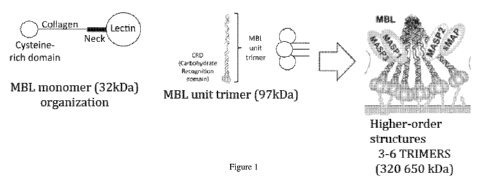
[0011] Figure 1 shows a diagram of mannose-binding lectin (MBL) engineered
into sets
of trimers (polymers) in an embodiment of the present invention.
[0012] Figures 2A and 2B are diagrams of an embodiment of the present
invention in
which an artificial protein (Figure 2A) comprising a sterically unhindered N-
terminus
(optionally with a cysteine at or near the N-terminus), followed by a long,
flexible peptide
segment, then an MBL lectin domain at the C-terminus, is crosslinked to a
solid substrate in the
example device in Figure 2B.
[0013] Figure 3 shows a diagram of an embodiment of the invention, Fc-MBL.81,
both
in cartoon and in model form based on the X ray crystallography models of Fc
and of the neck
and carbohydrate recognition domains (CRD) of MBL.
[0014] Figure 4 is a scheme of a vector encoding Fc in an aspect of the
invention.
[0015] Figure 5 shows the calcium-dependent binding of dynabead-MBL to C.
albicans
in which calcium maintains binding and EDTA destabilizes binding.
3
CA 02787376 2012-07-18
WO 2011/090954 PCT/US2011/021603
[0016] Figure 6 shows the binding of MBL-magnetic beads to different
pathogens.
Pathogens were bound by MBL-coated magnetic beads (control: beads without
MBL), washed,
and eluted onto culture plates.
[0017] Figure 7 shows data from MBL-magnetic beads binding to microorganisms
and
overnight culture assay. The pathogens were bound by MBL-coated magnetic beads
(control:
beads without MBL), washed, and eluted onto culture plates and incubated
overnight.
[0018] Figure 8 demonstrates high levels of FcMBL expression from transient
transfection. Figure 8A is a western blot of a reduced gel loaded with
unpurified supernatant
of 293 cells transfected with pFUSEFc MBL.81 (and pFUSE Fc) probed with anti-
hFc.
Figure 8B shows Protein A-purified FcMBL.81.
[0019] Figure 9 shows results of a depletion assay in which the FcMBL.81
construct was
as active as full-length MBL in binding C. albicans.
DETAILED DESCRIPTION
[0020] It should be understood that this invention is not limited to the
particular
methodology, protocols, and reagents, etc., described herein and as such may
vary. The
terminology used herein is for the purpose of describing particular
embodiments only, and is not
intended to limit the scope of the present invention, which is defined solely
by the claims.
[0021] As used herein and in the claims, the singular forms include the plural
reference
and vice versa unless the context clearly indicates otherwise. Other than in
the operating
examples, or where otherwise indicated, all numbers expressing quantities of
ingredients or
reaction conditions used herein should be understood as modified in all
instances by the
term "about."
[0022] All patents and other publications identified are expressly
incorporated herein by
reference for the purpose of describing and disclosing, for example, the
methodologies described
in such publications that might be used in connection with the present
invention. These
publications are provided solely for their disclosure prior to the filing date
of the present
application. Nothing in this regard should be construed as an admission that
the inventors are not
entitled to antedate such disclosure by virtue of prior invention or for any
other reason. All
statements as to the date or representation as to the contents of these
documents is based on the
information available to the applicants and does not constitute any admission
as to the
correctness of the dates or contents of these documents.
[0023] Unless defined otherwise, all technical and scientific terms used
herein have the
same meaning as those commonly understood to one of ordinary skill in the art
to which this
invention pertains. Although any known methods, devices, and materials may be
used in the
4
CA 02787376 2012-07-18
WO 2011/090954 PCT/US2011/021603
practice or testing of the invention, the methods, devices, and materials in
this regard are
described herein.
[0024] In the broadest sense, opsonins are proteins that bind to the surface
of a particle.
In nature, opsonins act as binding enhancers for the process of phagocytosis,
for example, by
coating the negatively-charged molecules on a target pathogen's membrane. The
present
invention provides for an engineered molecular opsonin, such as mannose-
binding lectin (MBL),
that may be used to bind biological pathogens or identify subclasses or
specific pathogen species
for use in devices and systems for treatment and diagnosis of patients with
infectious diseases,
blood-borne infections or sepsis. Treatment may be carried out in vivo or ex
vivo.
[0025] MBL is a serum lectin opsonin that binds to mannose, N-
acetylglucosamine
(NAG) -containing carbohydrates, and various other carbohydrates that are
present on the surface
of many microbial pathogens. MBL (also called mannose- or mannan-binding
protein, MBP) is
a polymeric protein assembled from three or more 32 kDa monomers. Each monomer
has an
N-terminal cysteine rich region, a collagen-like gly-X-Y region, a neck region
and a
carbohydrate recognition domain. The assembly of the higher molecular weight
(MW) polymers
begins with formation of trimers of the 32 kDa monomer; these trimers then
self-assembly into
higher MW polymers of three to six sets of trimers. See Figure 1.
[0026] MBL is a key component in opsonization of microbial pathogens and in
the
activation of complement (via the lectin pathway) and coagulation.
Opsonization is the binding
of proteins to target cells and the targeting these cells for uptake and
destruction by phagocytic
cells, such as macrophages and neutrophils. This opsonization appears to be
mediated by the
small, cysteine-rich N-terminal domain of MBL as well as C3b deposited on the
target cell
surface by MBL-mediated lectin complement pathway activation.
[0027] In the activation of complement via the lectin pathway, the microbe and
specialized proteins, i.e., MASP-1 (Mannan-binding lectin Associated Serine
Protease)
(Matsushita & Fujita, 176 J. Exp. Med. 1497 (1992)), and MASP-2 (Thiel et al.,
386 Nat. 506
(1997)), interact with bound MBL and activate complement in the absence of
antibody. The
higher molecular weight MBL complexes (5 to 6 repeats of the functional MBL
trimer) are
potent activators of complement via this lectin pathway, in which MASP 2
appears to activate
complement, and MASP 1 activates coagulation. The smaller complexes (three to
four repeats of
the MBL trimer unit) are the most potent activators of coagulation. Krarup et
al., 2 PLoS
One e623 (2007).
[0028] In certain human populations, there is a high allele frequency of
mutations in
MBL in the collagen helix, at codons 52, 54, and 57. Garred et al., 7 Genes
Immun. 85 (2006).
These mutations prevent the formation of the higher molecular weight MBL forms
and suppress
CA 02787376 2012-07-18
WO 2011/090954 PCT/US2011/021603
complement activation. In these cases, MBL still functions as an opsonin and
stimulates
coagulation, but without activating complement. There is also some evidence
for heterozygote
advantage with respect to sepsis, in that heterozygotes have the best
survival, homozygous
"wild-type" second best, and homozygous "mutant" have the worst survival. See
Sprong et
al., 49 Clin. Infect Dis. 1380 (2009). In addition, homozygous mutant neonates
are particularly
susceptible to infection before the acquired immune system begins to function.
[0029] There has been much debate on the usefulness of MBL as a recombinant
therapeutic protein for treatment of infectious diseases. Intact MBL has been
used in Phase 1
and Phase 2 clinical trials, both as a recombinant protein and when purified
from human blood
donations. In fact, plasma-derived MBL has been used as a therapeutic in Phase
1 and Phase II
trials of MBL deficient, pediatric patients with chemotherapy induced
neutropenia. Frakking et
al., 45 Eur. J. Cancer 50 (2009). Commercial efforts to develop MBL have
foundered because of
difficulties in both producing the recombinant protein and establishing
efficacy. As used herein,
treatment or treating a subject can refer to medical care provided to manage,
improve, or relieve
disease, illness, or symptoms thereof.
[0030] The present invention provides for engineered opsonins, e.g.,
engineered MBL or
MBL polymers, for use in devices and systems for pathogen detection and
clearance. Figure 5
shows the calcium-dependent binding of MBL-conjugated magnetic microbeads to
the yeast
C. albicans. Figures 6 and 7 compare the MBL-magnetic bead binding between
several
difference pathogens, including the gram positive bacterium, S. aureus; gram
negative bacteria,
Klebsiella and E. coli; and yeast, C. albicans. Recent work has demonstrated
the feasibility of
using combined micromagnetic and microfluidic techniques to clear living
pathogens from
flowing fluids, such as biological fluids, such as blood. Xia et al., 8
Biomed. Dev. Biomed.
Microdev. 299 (2006); Yung et al., Lab on a Chip DOI: 10.1039/b816986a (2009).
In these
microdevices (magnetic microbeads that are coated with molecules that bind
specifically to
surface markers on pathogen cells), are allowed to bind to these cells in
whole human blood,
and then are pulled free from blood flowing through microfluidic channels
using an applied
magnetic field gradient. See WO/2008/130618; WO/2007/044642.
[0031] Among other uses, these devices have great promise to rapidly clear
blood of
septic patients of toxin-producing pathogens, and hence greatly increase
response to
conventional antibiotic therapies. The ability to rapidly (within minutes)
bind, detect and isolate
living pathogens circulating in blood, or present within other biological
fluids, using a
potentially inexpensive and easy-to-use microdevice also circumvents the major
limitations of
current pathogen detection and sensitivity testing assays that require
multiple days of microbial
culture in hospital or commercial laboratories.
6
CA 02787376 2012-07-18
WO 2011/090954 PCT/US2011/021603
[0032] Biological fluids that may by used in the present invention include,
for example,
blood, cerebrospinal fluid, joint fluid, urine, semen, saliva, tears, and
fluids collected by
insertion of a needle. Additionally, fluids may be collected from food or
water samples for rapid,
general contamination assays according to the present invention: such fluid
can be collected and
analyzed for natural microbial contamination or for possible "bio-terrorism"
contamination.
[0033] Further, the current effectiveness of these methods harnesses prior
knowledge of
the specific pathogen that one desires to clear from the blood, because a
specific ligand for that
pathogen (e.g., specific antibody) is placed on the magnetic microbeads prior
to using the blood
cleansing device. Thus, the present invention bolsters the current approaches
by providing
engineered generic binding molecules that function like biological opsonins
and bind to specific,
many or all, types of microbial pathogens as the application requires. In this
regard, the present
invention has therapeutic applications.
[0034] Another need addressed herein is the development of specialized
pathogen class-
specific opsonins that bind, for example, all types of fungi or all gram
negative bacteria or all or
specific gram positive bacteria or all viruses or all protozoans, as this
knowledge could quickly
advise physicians in their choice of anti-microbial therapies before complete
characterization of
species type of antibiotic sensitivity is identified with conventional methods
that often take
many days to complete.
[0035] In addition, with the use of genetic engineering, and directed
evolution and
selection strategies, modified versions of natural opsonins can be engineered,
such as MBL, that
bind to pathogens in a species-specific manner. Finally, binding that is
specific for pathogen
sensitivity to different antibiotics or antimicrobial therapeutics can be
accomplished using
appropriate selection strategies. Hence, this invention provides for
development of engineered
opsonins that provide these high value properties.
[0036] MBL is an excellent choice for use as a generic opsonin for the
purposes
described herein; however, the intact molecule is not typically used in the
presence of whole
blood because it has multiple functional domains that promote blood
coagulation that may
interfere with diagnostic and therapeutic microdevice function. This
characteristic of MBL can
be separated from its pathogen binding function as provided herein. More
specifically, MBL
contains four parts, from N- to C-terminus: a small N-terminal domain of
essentially unknown
function that may be involved in macrophage binding and/or MASP binding; a
collagen segment
that may also be involved in MASP binding and higher-order oligomerization; an
alpha-helical
"neck" segment that is sufficient for trimerization; and the CRD lectin domain
at the C-terminus
that mediates direct pathogen binding. The lectin domain is useful for the
application at hand,
and the other domains may be present or deleted depending on the needs of the
user, and can be
7
CA 02787376 2012-07-18
WO 2011/090954 PCT/US2011/021603
determined by routine testing. Additionally, the lectin activity is calcium-
dependent, so bound
microbes could be released by a chelating agent for diagnostic purposes.
[0037] One embodiment of an engineered configuration of MBL, useful as a
generic
opsonin for diagnostic and therapeutic applications, comprises the lectin
domain of MBL. For
example, Glycine 111 (as defined in the Research Collaboratory for Structural
Bioinformatics
(RCSB), Protein Data Bank structure file 1HUP) is a convenient N-terminal
point at which to
begin the lectin portion of the engineered opsonin. Because the binding of MBL
to a given
monomeric sugar is weak, the MBL may be attached to the solid matrix in a
flexible manner so
that the proteins on the surface can move and adjust to the shape of the
microbe. For example, a
flexible peptide, such as one or more Glycine+Serine segment or one or more
Proline+Alanine+Serine segment, or other peptide linker(s) known in the art,
may be placed
at the MBL N-terminus, as in Figure 2A, because these segments tend to not
form
folded structures.
[0038] Another embodiment of an engineered configuration of MBL, useful as a
generic
opsonin for diagnosis and therapeutic applications, comprises the neck and
lectin domains of
MBL. Proline 81 (as defined, for example, in the Research Collaboratory for
Structural
Bioinformatics, Protein Data Bank (RCSB PDB) structural file 1HUP) is a
convenient
N-terminal point at which to begin the lectin sequence for this engineered
opsonin construct.
This portion of MBL is fused downstream (C-terminal) to Fc portion of human
IgG (Fcy). The
Fc portion may include the CH2-CH3 interface of the IgG Fc domain, which
contains the
binding sites for a number of Fc receptors including Staphylococcal protein A.
In use, the Fc
portion dimerizes and strengthens the avidity affinity of the binding by MBL
lectins to
monomeric sugars. Additionally, when used as a diagnostic reagent, the n-
linked glycosylation
of the recombinant opsonin can be removed. For example, in Fc MBL.81 the
glycosylation can
be removed by changing the amino acid at residue 297 from asparagine to
aspartic acid (N297D)
in the Kabat system of numbering amino acids in antibodies, this corresponds
to amino acid 82
in this particular Fc construct. Glycosylated Fc maintains the correct
orientation for Fc mediated
antibody-dependent cell-mediated cytotoxicity (ADCC) and complement-mediated
cytotoxicity (CDC).
[0039] The engineered Fc MBL opsonin could be used in the activation of Fc
receptor-
mediated uptake of Fc MBL opsonized Mycobacterium tuberculosis, bypassing
mannose
receptor mediated uptake of M. tuberculosis. Recent publications (Kang et al.,
202 J. Exp.
Med. 987 (2005)), suggest that lipoarabinomannan (ManLaM) on the cells surface
of
M. tuberculosis engage macrophage mannose receptor (MMR) during the phagocytic
process.
This directs M. tuberculosis to its initial phagosomal niche and inhibits
phagosome-lysosome
8
CA 02787376 2012-07-18
WO 2011/090954 PCT/US2011/021603
(P-L) fusion, thereby enhancing survival in human macrophages. Interestingly,
inhibition of P-L
fusion did not occur with entry via Fcy receptors. In one embodiment, uptake
by Fc recetor
endocytosis routes the bacterium, e.g., M. tuberculosis, to different
intracellular vesicles.
[0040] The configuration of the engineered opsonin of the present invention
also aids
attachment of the fusion protein to a substrate, such as a solid surface of a
magnetic microbead
or a microporous membrane, using a chemical cross-linker that is specific for
the amino group at
the N-terminus, or to a free cysteine residue that has been engineered to be
near the N-terminus
of the protein, as in Figure 2B. (Lysine is an alternative to cysteine,
optionally following
removal of the rest of the lysine residues in the protein).
[0041] In some embodiments, the substrate to which the opsonin binds is a
living cell or
extracellular matrix of a tissue or organ. For example, the substrate may be
the surface of a cell,
tissue or organ associated with the immune response. For example, the cell may
be a phagocyte
(macrophage, neutrophil, and dendritic cell), mast cell, eosinophil, basophil,
and/or natural killer
cell. The cell may be the cell of tissues or organs of the immune system, such
as spleen, lymph
nodes, lymphatic vessels, tonsils, thymus, bone marrow, Peyer's patches,
connective tissues,
mucous membranes, the reticuloendothelial system, etc. The surface to which
the opsonin binds
may also be the extracellular matrix of one or more of these tissues or
organs.
[0042] In some embodiments, the solid substrate may comprise magnetic beads or
other
structured materials, which then pull microbes out from fluids, including
biological fluids such
as blood, and concentrate and collect the microbes, including living microbes.
This approach is
advantageous because the beads can then be examined for the presence of the
microbe, or be
used to transfer the collected microbes to conventional pathogen culture and
sensitivity testing
assays. In other words, the engineered opsonin may be used in diagnostics as a
means of
collecting potential pathogens for identification; not only in the diagnosis
of disease, but in the
identification of water- or food-borne pathogens, particulates or other
contaminants.
Alternatively, the solid substrate may comprise a hollow-fiber reactor or any
other blood
filtration membrane or flow device (e.g., a simple dialysis tube) or other
resins, fibers, or sheets
to selective bind and sequester the biological pathogens.
[0043] The magnetic beads can be of any shape, including but not limited to
spherical,
rod, elliptical, cylindrical, disc, and the like. In some embodiments,
magnetic beads having a
true spherical shape and defined surface chemistry are used to minimize
chemical agglutination
and non-specific binding. As used herein, the term "magnetic beads" refers to
a nano- or micro-
scale particle that is attracted or repelled by a magnetic field gradient or
has a non-zero magnetic
susceptibility. The magnetic bead can be paramagnetic or super-paramagnetic.
In some
embodiments, magnetic beads are super-paramagnetic. Magnetic beads are also
referred to as
9
CA 02787376 2012-07-18
WO 2011/090954 PCT/US2011/021603
magnetic particles herein. In some embodiments, magnetic beads having a
polymer shell are
used to protect the pathogen from exposure to iron. For example, polymer-
coated magnetic
beads can be used to protect pathogens from exposure to iron.
[0044] The magnetic beads can range in size from 1 nm to 1 mm. For example,
magnetic beads are about 250 nm to about 250 m in size. In some embodiments,
magnetic bead
is 0.1 m to 100 m in size. In some embodiments, magnetic bead is 0.1 m to
50 m in size.
In some embodiments, magnetic bead is 0.1 m to 10 m in size. In some
embodiments, the
magnetic bead is a magnetic nano-particle or magnetic micro-particle. Magnetic
nanoparticles
are a class of nanoparticle which can be manipulated using magnetic field or
magnetic field
gradient. Such particles commonly consist of magnetic elements such as iron,
nickel and cobalt
and their chemical compounds. Magnetic nano-particles are well-known and
methods for their
preparation have been described in the art. See, e.g., U.S. Patents No.
6,878,445; No. 5,543,158;
No. 5,578,325; No. 6,676,729; No. 6,045,925; and No. 7,462,446; and U.S.
Patent Publications
No. 2005/0025971; No. 2005/0200438; No. 2005/0201941; No. 2005/0271745;
No. 2006/0228551; No. 2006/0233712; No. 2007/01666232; and No. 2007/0264199.
[0045] Magnetic beads are easily and widely available commercially, with or
without
functional groups capable of binding to affinity molecules. Suitable magnetic
beads are
commercially available such as from Dynal Inc. (Lake Success, NY); PerSeptive
Diagnostics,
Inc. (Cambridge, MA); Invitrogen Corp. (Carlsbad, CA); Cortex Biochem Inc.
(San Leandro,
CA); and Bangs Laboratories (Fishers, IN). In particular embodiments, magnetic
particles are
MyOneTM Dynabeads magnetic beads (Dynal Inc.).
[0046] The solid substrate can be fabricated from or coated with a
biocompatible
material. As used herein, the term "biocompatible material" refers to any
material that does not
deteriorate appreciably and does not induce a significant immune response or
deleterious tissue
reaction, e.g., toxic reaction or significant irritation, over time when
implanted into or placed
adjacent to the biological tissue of a subject, or induce blood clotting or
coagulation when it
comes in contact with blood. Suitable biocompatible materials include, for
example, derivatives
and copolymers of a polyimides, poly(ethylene glycol), polyvinyl alcohol,
polyethyleneimine,
and polyvinylamine, polyacrylates, polyamides, polyesters, polycarbonates, and
polystyrenes.
[0047] In some embodiments, the solid substrate is fabricated or coated with a
material
selected from the group consisting of polydimethylsiloxane, polyimide,
polyethylene
terephthalate, polymethylmethacrylate, polyurethane, polyvinylchloride,
polystyrene
polysulfone, polycarbonate, polymethylpentene, polypropylene, a polyvinylidine
fluoride,
polysilicon, polytetrafluoroethylene, polysulfone, acrylonitrile butadiene
styrene,
polyacrylonitrile, polybutadiene, poly(butylene terephthalate), poly(ether
sulfone), poly(ether
CA 02787376 2012-07-18
WO 2011/090954 PCT/US2011/021603
ether ketones), poly(ethylene glycol), styrene-acrylonitrile resin,
poly(trimethylene
terephthalate), polyvinyl butyral, polyvinylidenedifluoride, poly(vinyl
pyrrolidone), and any
combination thereof.
[0048] In an aspect of the invention, the recombinant opsonins described
herein can be
conjugated with the solid substrate by methods well known in the art for
conjugating peptides
with other molecules. For example, Hermanson, BIOCONJUGATE TECHNIQUES (2nd
Ed.,
Academic Press (2008)) and Niemeyr, Bioconjugation Protocols: Strategies &
Methods, in
METHODS IN MOLECULAR BIOLOGY (Humana Press, 2004), provide a number of methods
and
techniques for conjugating peptides to other molecules. de Graaf, et al., 20
Biocojugate
Chem. 1281 (2009), provides a review of site-specific introduction of non-
natural amino acids
into peptides for conjugation.
[0049] Alternatively, the surface of the solid substrate can be functionalized
to include
binding molecules that bind selectively with the recombinant opsonin. These
binding molecules
are also referred to as affinity molecules herein. The binding molecule can be
bound covalently
or non-covalently on the surface of the solid substrate. As used herein, the
term "binding
molecule" or "affinity molecule" refers to any molecule that is capable of
specifically binding a
recombinant opsonin described herein. Representative examples of affinity
molecules include,
but are not limited to, antibodies, antigens, lectins, proteins, peptides,
nucleic acids (DNA, RNA,
PNA and nucleic acids that are mixtures thereof or that include nucleotide
derivatives or
analogs); receptor molecules, such as the insulin receptor; ligands for
receptors (e.g., insulin for
the insulin receptor); and biological, chemical or other molecules that have
affinity for another
molecule, such as biotin and avidin. The binding molecules need not comprise
an entire
naturally occurring molecule but may consist of only a portion, fragment or
subunit of a
naturally or non-naturally occurring molecule, as for example the Fab fragment
of an antibody.
The binding molecule may further comprise a marker that can be detected.
[0050] The binding molecule can be conjugated to surface of the solid
substrate using
any of a variety of methods known to those of skill in the art. The binding
molecule can be
coupled or conjugated to surface of the solid substrate covalently or non-
covalently. Covalent
immobilization may be accomplished through, for example, silane coupling. See,
e.g.,
Weetall, 15 Adv. Mol. Cell Bio. 161 (2008); Weetall, 44 Meths. Enzymol. 134
(1976). The
covalent linkage between the binding molecule and the surface can also be
mediated by a linker.
The non-covalent linkage between the affinity molecule and the surface can be
based on ionic
interactions, van der Waals interactions, dipole-dipole interactions, hydrogen
bonds, electrostatic
interactions, and/or shape recognition interactions.
11
CA 02787376 2012-07-18
WO 2011/090954 PCT/US2011/021603
[0051] As used herein, the term "linker" means a molecular moiety that
connects two
parts of a composition. Peptide linkers may affect folding of a given fusion
protein, and may
also react/bind with other proteins, and these properties can be screened for
by known
techniques. Example linkers, in addition to those described herein, include is
a string of histidine
residues, e.g., His6; sequences made up of Ala and Pro, varying the number of
Ala-Pro pairs to
modulate the flexibility of the linker; and sequences made up of charged amino
acid residues
e.g., mixing Glu and Lys. Flexibility can be controlled by the types and
numbers of residues in
the linker. See, e.g., Perham, 30 Biochem. 8501 (1991); Wriggers et al., 80
Biopolymers 736
(2005). Chemical linkers may comprise a direct bond or an atom such as oxygen
or sulfur, a unit
such as NH, C(O), C(O)NH, SO, SO2, SO2NH, or a chain of atoms, such as
substituted or
unsubstituted CI-C6 alkyl, substituted or unsubstituted C2-C6 alkenyl,
substituted or
unsubstituted C2-C6 alkynyl, substituted or unsubstituted C6-C12 aryl,
substituted or unsubstituted
C5-C12heteroaryl, substituted or unsubstituted C5-C12 heterocyclyl,
substituted or unsubstituted
C3-C12 cycloalkyl, where one or more methylenes can be interrupted or
terminated by 0, S,
S(O), SO2, NH, or C(O).
[0052] Nucleic acid based binding molecules include aptamers. As used herein,
the term
"aptamer" means a single-stranded, partially single-stranded, partially double-
stranded or
double-stranded nucleotide sequence capable of specifically recognizing a
selected non-
oligonucleotide molecule or group of molecules by a mechanism other than
Watson-Crick base
pairing or triplex formation. Aptamers can include, without limitation,
defined sequence
segments and sequences comprising nucleotides, ribonucleotides,
deoxyribonucleotides,
nucleotide analogs, modified nucleotides and nucleotides comprising backbone
modifications,
branchpoints and nonnucleotide residues, groups or bridges. Methods for
selecting aptamers for
binding to a molecule are widely known in the art and easily accessible to one
of ordinary skill
in the art.
[0053] The recombinant opsonin can be conjugated with surface of the solid
substrate by
an affinity binding pair. The term "affinity binding pair" or "binding pair"
refers to first and
second molecules that specifically bind to each other. One member of the
binding pair is
conjugated with the solid substrate while the second member is conjugated with
the recombinant
opsonin. As used herein, the term "specific binding" refers to binding of the
first member of the
binding pair to the second member of the binding pair with greater affinity
and specificity than
to other molecules.
[0054] Exemplary binding pairs include any haptenic or antigenic compound in
combination with a corresponding antibody or binding portion or fragment
thereof (e.g.,
digoxigenin and anti-digoxigenin; mouse immunoglobulin and goat antimouse
immunoglobulin)
12
CA 02787376 2012-07-18
WO 2011/090954 PCT/US2011/021603
and nonimmunological binding pairs (e.g., biotin-avidin, biotin-streptavidin),
hormone (e.g.,
thyroxine and cortisol-hormone binding protein), receptor-receptor agonist,
receptor-receptor
antagonist (e.g., acetylcholine receptor-acetylcholine or an analog thereof),
IgG-protein A,
lectin-carbohydrate, enzyme-enzyme cofactor, enzyme-enzyme inhibitor, and
complementary
oligonucleoitde pairs capable of forming nucleic acid duplexes), and the like.
The binding pair
can also include a first molecule that is negatively charged and a second
molecule that is
positively charged.
[0055] One example of using binding pair conjugation is the biotin-sandwich
method.
See, e.g., Davis et al., 103 PNAS 8155 (2006). The two molecules to be
conjugated together are
biotinylated and then conjugated together using tetravalent streptavidin as a
linker. A peptide
can be coupled to the 15-amino acid sequence of an acceptor peptide for
biotinylation (referred
to as AP; Chen et al., 2 Nat. Methods 99 (2005)). The acceptor peptide
sequence allows site-
specific biotinylation by the E. coli enzyme biotin ligase (BirA; Id.). A
recombinant opsonin can
be similarly biotinylated for conjugation with a solid substrate. Many
commercial kits are also
available for biotinylating proteins. Another example for conjugation to a
solid surface would be
to use PLP -mediated bioconjugation. See, e.g., Witus et al., 132 JACS 16812
(2010). In this
example, an AKT sequence on the Fc N terminal allows conjugation to the solid
surface and
orientation of the lectin binding domain in the optimal orientation pointing
away from the
solid surface.
[0056] It should be noted that the affinity of a single lectin domain for a
sugar is low,
and binding is normally driven by avidity and multivalency. In the case of the
present devices,
the multimerization domains are deleted from the protein, and multivalency of
the protein is
effectively produced by attachment to a solid substrate (e.g., a bead) at high
density, which
density can be varied to provide optimal functionality.
[0057] Further regarding the MBL, its binding characteristics can be
manipulated by
directed evolution for altered binding specificity. MBL may be modified so
that it binds to a
more limited set of sugars or other molecular features, with the result that
the modified MBL
will bind to a more limited set of microbes to provide a capability for
pathogen class
identification (e.g., one of virus, bacteria, fungi, or protozoan), subclass
typing (e.g., gram
negative or gram positive bacteria) or specific species determination.
Numerous strategies
are available in the art.
[0058] For example, a straightforward directed evolution strategy visually
examines an
atomic structure of MBL complexed with a sugar, and then mutates appropriate
amino acids that
make contact in a sugar-specific manner, so that distinctive contacts are lost
or particular types
of steric hindrance are created. The three dimensional structure of rat MBL
has been solved in a
13
CA 02787376 2012-07-18
WO 2011/090954 PCT/US2011/021603
complex with a high-mannose oligosaccharide and with N-acetylglucosamine, a
methylated
fucose, and so on. His189Val and Ile207Val are examples of substitutions that
modifications
alter specificity.
[0059] In another strategy of directed evolution, the protein is subjected to
random
mutagenesis and the resulting proteins are screened for desired qualities.
This is a particularly
useful technology for affinity maturation of phage display antibodies, where
the antibody
complementary determining regions (CDRs) are mutated by saturation mutagenesis
and
successful variants of the six CDRs are shuffled together to form the highest
affinity antibodies.
[0060] The directed evolution paradigm can be applied to MBL in order to
select MBL
variants with specific binding to yeast, gram-positive bacteria, gram-
negative, coagulase
negative, aerobic bacteria, etc. For this to work, however, the pattern and
nature of the target
sugars or related surface features on these target organisms may have to
differ between the
classes or species.
[0061] MBL is known to bind strongly to mannose and N-acetylglucosamine sugars
on
fungi, gram-positive, and gram-negative bacteria. For example, MBL binds
strongly to Candida
spp., Aspergillusfumigatus, Staphylococcus aureus, and 0 hemolytic group A
streptococci.
MBL has intermediate affinity to Escherichia coli, Klebsiella spp., and
Haemophilus influenzae
type b. MBL binds weakly to 0 hemolytic group B streptococci, Streptococcus
pneumoniae, and
Staphylococcus epidermidis. Neth et al., 68 Infect. & Immun. 688 (2000). The
capsular
polysaccharide of Neisseria meningitides serogroup B, H.influenzae type b and
Cryptococcus
neoformans are thought to decrease MBL binding, as does bacterial endotoxin.
Id.; Van
Emmerik et al., 97 Clin. Exp. Immunol. 411 (1994); Schelenz et al., 63 Infect.
Immun. 3360 (1995).
[0062] Others have reported that MBL facilitates opsonophagocytosis of yeasts
but not
of bacteria, despite MBL binding: MBL (Lectin) pathway of complement was
critical for the
opsonophagocytosis of yeast, but the classical complement pathway was critical
for
opsonophagocytosis of bacteria. Brouwer et al., 180 J. Immunol. 4124 (2008).
It was not
reported that MBL bound to the bacterial species tested, however, only that
MBL binding did
not promote significant complement activation and opsonophagocytosis.
[0063] Derivatives of MBL with a particular specificity can be isolated by the
following
approach, which is a standard phage display strategy: First, express a set of
MBL variants from a
phagemid vector; then bind this library to a target of interest (e.g., E.
coli) and perform one or
two rounds of selection; and then perform a round of negative selection
against a related target
(e.g., Candida), taking those phagemids that fail to bind. These cycles of
positive and negative
selection are then repeated until a population of phages that generally bind
to the target and do
14
CA 02787376 2012-07-18
WO 2011/090954 PCT/US2011/021603
not bind to the non-target is generated. This method may be applied to any
pair of microbial
strains against which differential binding is desired, such as bacteria that
are resistant and
sensitive to a given antibiotic. This positive/negative enrichment strategy
may also be used with
an antibody-phage display library, which is an even more standard way to
isolate such
specific binders.
[0064] MBL belongs to the class of collectins in the C-type (calcium-
dependent) lectin
superfamily, other members of which, such as surfactant protein A, surfactant
protein D, CL-L1
and CL-P1, may be useful in the present invention. Other possible opsonins
include ficollins
(Thiel et al., 1997), which also activate the lectin pathway of complement and
bind MASP
proteins. These proteins are related to MBL but have a different, more limited
specificity. In the
context of the diagnostic device described herein, one option is to simply use
the lectin domain
of a ficollin that corresponds to the lectin domain of MBL described above.
Another approach is
to use `shuffling' of segments or individual amino acids between MBL and one
or more
Ficollins to create hybrid molecules that may have hybrid specificities. The
directed evolution
and selection approach described above also could potentially be used to
generate human
antibody fragments or peptides that provide the class, subclass and species
specificity
described above.
[0065] The present invention may be defined in any of the following
numbered paragraphs:
1. A recombinant opsonin comprising: a carbohydrate recognition domain of an
opsonin; a
substrate binding domain; and a peptide domain that links the recognition
domain to the
substrate binding domain.
2. The recombinant opsonin of paragraph 1, wherein said carbohydrate
recognition domain
is a collectin or ficollin or derived from a collectin or ficollin.
3. The recombinant opsonin of paragraph 1, wherein said carbohydrate
recognition domain
is a lectin or a portion or a fragment of a lectin.
4. The recombinant opsonin of paragraph 3, wherein said lectin is mannose-
binding
lectin (MBL).
5. The recombinant opsonin of paragraph 4, wherein the lectin consists of
amino acid
residues 81 (proline) to 228 (isoleucine) of MBL (SEQ ID NO:2).
6. The recombinant opsonin of any of the foregoing paragraphs, wherein said
substrate
binding domain comprises at least one cysteine residue that allows chemical
cross-linking to
a solid substrate.
7. The recombinant opsonin of any of the foregoing paragraphs, wherein the
flexible peptide
comprises a Glycine+Serine segment or a Proline+Alanine+Serine segment.
CA 02787376 2012-07-18
WO 2011/090954 PCT/US2011/021603
8. The recombinant opsonin of the foregoing paragraphs, where the flexible
peptide
comprises a portion of immunoglobulin Fc.
9. The recombinant opsonin of paragraph 8, wherein the Fc portion includes the
CH2-CH3
interface of the IgG Fc domain.
10. The recombinant opsonin of any of the foregoing paragraph, wherein the
substrate is a
magnetic microbead, a paramagnetic microbead, a microporous membrane, a hollow-
fiber
reactor, or any other fluid filtration membrane or flow device.
11. The recombinant opsonin of any of the foregoing paragraphs, wherein the
substrate is a
living cell or extracellular matrix of a biological tissue or organ.
12. The recombinant opsonin of paragraph 11, wherein the substrate is a
phagocyte.
13. A method of collecting an opsonin-binding microorganism from a fluid
comprising
contacting the fluid with a recombinant opsonin conjugated to a solid surface;
wherein the
recombinant opsonin consists of a carbohydrate recognition domain of an
opsonin, a solid
substrate binding domain, and a flexible peptide domain that links the
recognition domain to the
solid surface binding domain; allowing the opsonin-binding microorganism to
bind to said
recombinant opsonin-solid surface conjugate; and separating said fluid from
said
microorganism-bound recombinant opsonin-solid surface conjugate.
14. The method of paragraph 13, wherein the solid surface is a magnetic
particle, and the
separating is achieved by applying magnetic force to the fluid after the
opsonin-binding
microorganism has bound to the recombinant opsonin-solid surface conjugate.
15. The method of paragraph 13, further comprising the step of identifying
the microorganism.
16. The method of paragraph 13, wherein the fluid is a biological fluid.
17. The method of paragraph 16, wherein the biological fluid is selected from
the group
consisting of blood, cerebrospinal fluid, joint fluid, urine, semen, saliva,
tears, and fluids
collected by needle, biopsy, or aspiration procedures.
18. The method of paragraph 17, wherein the biological fluid is blood.
19. The method of paragraph 18, further comprising the step of returning the
blood to
its source.
20. The method of paragraph 19, wherein the source is a subject.
21. The method of paragraph 20, wherein the subject is suffering from
infection or sepsis.
22. The method of paragraph 13, wherein the fluid is derived from a water or a
food sample.
23. The use of the recombinant opsonin of any of paragraphs 1 to 10 in the
identification of
a pathogen.
16
CA 02787376 2012-07-18
WO 2011/090954 PCT/US2011/021603
24. The use of the recombinant opsonin of any of paragraphs 1 to 10 in the
diagnosis
of disease.
25. The use of the recombinant opsonin of any of paragraphs 1 to 10 in the
identification of
water or food contamination.
26. The use of the recombinant opsonin of any of paragraphs 1 to 12 in the
treatment
of disease.
27. The use of the recombinant opsonin as in paragraph 26, further combined
with additional
treatment or therapy.
28. A method of treating a blood infection in a subject comprising
administering a
recombinant opsonin to the blood of the subject, wherein the recombinant
opsonin consists of a
carbohydrate recognition domain of an opsonin, a substrate binding domain, and
a flexible
peptide domain that links the recognition domain to the substrate binding
domain, wherein the
carbohydrate recognition domain binds an opsonin-binding microorganism, and
wherein the
substrate binding domain binds with a cell, tissue or organ of the immune
system; allowing the
recombinant opsonin to bind to the opsonin-binding microorganism; and allowing
the
microorganism-bound recombinant opsonin to bind with a cell, tissue or organ
of the immune
system wherein the microorganism is killed.
29. The method of paragraph 28, wherein the subject is an animal.
30. The method of paragraph 28, wherein the subject is a human.
EXAMPLES
Example 1. Construction and expression of FcMBL.81
[0066] An embodiment of an engineered configuration of MBL, useful as a
generic
opsonin for diagnosis and therapeutic applications, was constructed using the
"neck" and
"lectin" domains of MBL. Proline 81 (as defined in the Research Collaboratory
for Structural
Bioinformatics, Protein Data Bank structural file 1HUP) was selected as the N-
terminal point at
which to begin the lectin sequence. This portion of the lectin molecule was
fused downstream
(C-terminal) to Fc portion of human gamma 1 (Fcy). A diagram of the engineered
opsonin
construct is shown in Figure 3. A schematic of the Fc portion of a clone is
shown in Figure 4.
The amino acids for this construct include the following residues:
Fc protein sequence:
001 epkssdktht cppcpapell ggpsvflfpp kpkdtlmisr tpevtcvvvd vshedpevkf
061 nwyvdgvevh naktkpreeq ynstyrvvsv ltvlhqdwln gkeykckvsn kalpapiekt
121 iskakgqpre pqvytlppsr deltknqvsl tclvkgfyps diavewesng qpennykttp
181 pvldsdgsff lyskltvdks rwqqgnvfsc svmhealhnh ytqkslslsp ga (SEQ ID NO:1)
17
CA 02787376 2012-07-18
WO 2011/090954 PCT/US2011/021603
MBL.81 Protein Sequence (this includes the coiled-coil neck region and the
carbohydrate
recognition domains(CRD) of human MBL):
81 pdgdsslaas erkalqtema rikkwltfsl gkqvgnkffl tngeimtfek vkalcvkfqa
141 svatprnaae ngaignlike eaflgitdek tegqfvdltg nrltytnwne gepnnagsde
201 dcvlllkngq wndvpcstsh lavcefpi (SEQ ID NO:2)
Fc-MBL.81 sequence:
001 epkssdktht cppcpapell ggpsvflfpp kpkdtlmisr tpevtcvvvd vshedpevkf
061 nwyvdgvevh naktkpreeq ynstyrvvsv ltvlhqdwln gkeykckvsn kalpapiekt
121 iskakgqpre pqvytlppsr deltknqvsl tclvkgfyps diavewesng qpennykttp
181 pvldsdgsff lyskltvdks rwqqgnvfsc svmhealhnh ytqkslslsp gapdgdssla
241 aserkalqte marikkwltf slgkqvgnkf fltngeimtf ekvkalcvkf qasvatprna
301 aengaignli keeaflgitd ektegqfvdl tgnrltytnw negepnnags dedcvlllkn
361 gqwndvpcst shlavcefpi (SEQ ID NO:3)
[0067] Thus, the FcMBL.81 construct consists of a lectin having amino acid
residues 81
(proline) to 228 (isoleucine) of MBL, fused a portion of Fcy. In use, the Fc
portion dimerizes
and adds avidity to the weak affinity of the binding by MBL lectins to
monomeric sugars. When
Fc MBL.81 is designed for use as a diagnostic reagent, the n-linked
glycosylation can be
removed by changing the amino acid at 297 from asparagine to aspartic acid
(N297D), or amino
acid 82 in the Fc construct. Glycosylated Fc is maintains the correct
orientation for Fc mediated
ADCC and CDC. Additionally, a cysteine residue can be cloned onto the
engineered opsonin to
allow binding to a solid substrate via chemical conjugation. The construction
and expression of
an engineered opsonin, such as FcMBL, may be achieved by various techniques
known in the
art, see, e.g., U.S. Patent No. 5,541,087.
[0068] Expression of the construct in transiently transfected cells is
demonstrated in
Figures 8A and 8B. The FcMBL.81 expressed about 35mg/L.
Example 2. Comparison of Fc MBL.81 construct with full-length MBL in binding
yeast.
[0069] Approximately 5.5 million Candida albicans yeast cells were inoculated
with
varying numbers of MBL beads coated with either wild-type, full-length MBL
(hexamers of
trimers) or Fc MBL.81. As depicted graphically in Figure 9, 18 million wild-
type, full-length
MBL or Fc MBL.81 beads bound all 5.5 million fungal cells. This example
demonstrates that
Fc MBL.81 beads are as active as wild-type, full-length MBL beads in binding
to C albicans.
18