Note: Descriptions are shown in the official language in which they were submitted.
. ,
INTRACAMERAL SUSTAINED RELEASE THERAPEUTIC AGENT IMPLANTS
INVENTORS: MICHAEL R. ROBINSON, JAMES BURKE, RHETT SCHIFFMAN
[0001]
FIELD OF THE INVENTION
[0002] The present invention relates to intracameral sustained release
implants and
methods of making and using the same.
SUMMARY
[0003] Described herein are intraocular systems and methods for
treating ocular
conditions. In particular, local administration of a sustained release
therapeutic agent
delivery system to the anterior chamber and/or to anterior vitreous chamber of
the eye
to treat aqueous chamber elevated intraocular pressure is described.
[0004] Further, described herein are methods for treating an ocular
condition
comprising the steps of: providing at least two biodegradable sustained
release
implants containing at least one therapeutic agent; implanting the at least
two
biodegradable sustained release implants into the anterior chamber of an eye;
and
treating the ocular condition, wherein the at least two biodegradable
sustained release
implants release about 100 ng per day of the at least one bioactive agent for
a period
greater than about 1 month.
[0005] Further still, described herein are methods for treating
glaucoma in an eye
comprising the steps of: providing at least two biodegradable sustained
release
implants containing at least one therapeutic agent; implanting the at least
two
biodegradable sustained release implants into the anterior chamber of the eye;
allowing
a sufficient time for the at least two biodegradable sustained release
implants to settled
out in the inferior angle; allowing a sufficient time for the at least two
biodegradable
sustained release implants to release the at least one therapeutic agent; and
treating
glaucoma, wherein the at least two biodegradable sustained release implants
release
about 100 ng per day of the at least one bioactive agent for a period greater
than about
1 month.
1
CA 2787514 2018-05-24
CA 02787514 2012-07-18
WO 2011/091205 PCT/US2011/021971
[0006] In one embodiment, the ocular condition is glaucoma and/or elevated
intraocular pressure. The sustained release implants can release about 70% of
the at
least one therapeutic agent over the first month. In some embodiments, the at
least
one therapeutic agent can comprise about 30% of the at least two biodegradable
sustained release implants and is selected from the group consisting of
latanoprost,
bimatoprost and travoprost and their salts, esters and prodrugs.
[0007] In another embodiment, the at least two biodegradable sustained
release
implants comprise about 5% to about 70% poly(D,L-lactide). In other
embodiments, the
at least two biodegradable sustained release implants comprise about 5% to
about 40%
poly(DL-lactide-co-glycolide). In yet other embodiments, the at least two
biodegradable
sustained release implants comprise about 5% to about 40% polyethylene glycol.
[0008] In still other example embodiments, the at least two biodegradable
sustained
release implants comprise about 30% therapeutic agent, 65% poly(D,L-lactide),
and 5%
polyethylene glycol or about 20% therapeutic agent, 55% poly(D,L-lactide), 10%
poly(DL-lactide-co-glycolide), and 5% polyethylene glycol.
[0009] The implants themselves can be inserted into the ocular tissue using
an
appropriate applicator. Once implanted, the at least two biodegradable
sustained
release implants can settle out in the inferior angle within 24 hours of
implanting within
the anterior chamber.
[0010] In one embodiment, the the sufficient time for the at least two
biodegradable
sustained release implants to release the at least one therapeutic agent is
greater than
about 42 days.
BRIEF DESCRIPTION OF THE DRAWINGS
[0011] Figure 1 illustrates the two different pathways for aqueous humor
outflow
from the anterior chamber both located in the iridocorneal angle.
[0012] Figure 2 illustrates the placement of an implant as described herein
at the
location of aqueous humor outflow from the anterior chamber.
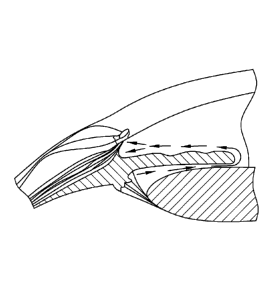
[0013] Figure 3 illustrates the currents located within the anterior
chamber of an eye
as well as a possible location of an implant or implants as described herein.
[0014] Figure 4 graphically illustrates a release profile of implants of
the present
description.
2
CA 02787514 2012-07-18
WO 2011/091205 PCT/US2011/021971
[0015] Figure 5 graphically illustrates a release profile of implants of
the present
description.
[0016] Figure 6 illustrates the placement of an implant according the
present
description.
DEFINITION OF TERMS
[0017] "About" means plus or minus ten percent of the number, parameter or
characteristic so qualified.
[0018] "Biodegradable polymer" means a polymer or polymers which degrade in
vivo, and wherein erosion of the polymer or polymers over time occurs
concurrent with
or subsequent to release of the therapeutic agent. The terms "biodegradable"
and
"bioerodible" are used interchangeably herein. A biodegradable polymer may be
a
homopolymer, a copolymer, or a polymer comprising more than two different
polymeric
units. The polymer can be a gel or hydrogel type polymer, polylactic acid or
poly(lactic-
co-glycolic) acid or polyethylene glycol polymer or mixtures or derivatives
thereof.
[0019] "Ocular condition" means a disease, ailment or condition which
affects or
involves the ocular region. Broadly speaking, the eye includes the eyeball and
the
tissues and fluids which constitute the eyeball, the periocular muscles (such
as the
oblique and rectus muscles) and the portion of the optic nerve which is within
or
adjacent to the eyeball.
[0020] An anterior ocular condition is a disease, ailment or condition
which affects
or which involves an anterior (i.e. front of the eye) ocular region or site,
such as a
periocular muscle, an eye lid or an eye ball tissue or fluid which is located
anterior to
the posterior wall of the lens capsule or ciliary muscles. Thus, an anterior
ocular
condition primarily affects or involves the conjunctiva, the cornea, the
anterior chamber,
the iris, the posterior chamber (behind the retina but in front of the
posterior wall of the
lens capsule), the lens or the lens capsule and blood vessels and nerve which
vascularize or innervate an anterior ocular region or site.
[0021] Thus, an anterior ocular condition can include a disease, ailment or
condition, such as for example, aphakia; pseudophakia; astigmatism;
blepharospasm;
cataract; conjunctival diseases; conjunctivitis; corneal diseases; corneal
ulcer; dry eye
syndromes; eyelid diseases; lacrimal apparatus diseases; lacrimal duct
obstruction;
myopia; presbyopia; pupil disorders; refractive disorders and strabismus.
Glaucoma
3
CA 02787514 2012-07-18
WO 2011/091205 PCT/US2011/021971
can also be considered to be an anterior ocular condition because a clinical
goal of
glaucoma treatment can be to reduce a hypertension of aqueous fluid in the
anterior
chamber of the eye (i.e. reduce intraocular pressure).
[0022] A posterior ocular condition is a disease, ailment or condition
which primarily
affects or involves a posterior ocular region or site such as choroid or
sclera (in a
position posterior to a plane through the posterior wall of the lens capsule),
vitreous,
vitreous chamber, retina, optic nerve (i.e. the optic disc), and blood vessels
and nerves
which vascularize or innervate a posterior ocular region or site.
[0023] Thus, a posterior ocular condition can include a disease, ailment or
condition, such as for example, acute macular neuroretinopathy; Behcet's
disease;
choroidal neovascularization; diabetic uveitis; histoplasmosis; infections,
such as fungal
or viral-caused infections; macular degeneration, such as acute macular
degeneration,
non-exudative age related macular degeneration and exudative age related
macular
degeneration; edema, such as macular edema, cystoid macular edema and diabetic
macular edema; nnultifocal choroiditis; ocular trauma which affects a
posterior ocular
site or location; ocular tumors; retinal disorders, such as central retinal
vein occlusion,
diabetic retinopathy (including proliferative diabetic retinopathy),
proliferative
vitreoretinopathy (PVR), retinal arterial occlusive disease, retinal
detachment, uveitic
retinal disease; sympathetic opthalmia; Vogt Koyanagi-Harada (VKH) syndrome;
uveal
diffusion; a posterior ocular condition caused by or influenced by an ocular
laser
treatment; posterior ocular conditions caused by or influenced by a
photodynannic
therapy, photocoagulation, radiation retinopathy, epiretinal membrane
disorders, branch
retinal vein occlusion, anterior ischemic optic neuropathy, non-retinopathy
diabetic
retinal dysfunction, retinitis pigmentosa, and glaucoma. Glaucoma can be
considered a
posterior ocular condition because the therapeutic goal is to prevent the loss
of or
reduce the occurrence of loss of vision due to damage to or loss of retinal
cells or optic
nerve cells (i.e. neuroprotection).
[0024] "Ocular region" or "ocular site" means any area of the eyeball,
including the
anterior and posterior segment of the eye, and which generally includes, but
is not
limited to, any functional (e.g., for vision) or structural tissues found in
the eyeball, or
tissues or cellular layers that partly or completely line the interior or
exterior of the
eyeball. Specific examples of areas of the eyeball in an ocular region include
the
anterior (aqueous) chamber, the posterior chamber, the vitreous cavity, the
choroid, the
4
CA 02787514 2012-07-18
WO 2011/091205 PCT/US2011/021971
suprachoroidal space, the conjunctiva, the subconjunctival space, the
episcleral space,
the intracorneal space, the epicorneal space, the sclera, the pars plana,
surgically-
induced avascular regions, the macula, and the retina.
[0025] "Sustained release" or "controlled release" refers to the release of
at least
one therapeutic bioactive agent, or drug, from an implant at a predetermined
rate.
Sustained release implies that the therapeutic bioactive agent is not released
from the
implant sporadically in an unpredictable fashion and does not "burst" from the
implant
upon contact with a biological environment (also referred to herein as first
order
kinetics) unless specifically intended to do so. However, the term "sustained
release"
as used herein does not preclude a "burst phenomenon" associated with
deployment.
In some example embodiments according to the present description an initial
burst of at
least one therapeutic agent may be desirable followed by a more gradual
release
thereafter. The release rate may be steady state (commonly referred to as
"timed
release" or zero order kinetics), that is the at least one therapeutic agent
is released in
even amounts over a predetermined time (with or without an initial burst
phase) or may
be a gradient release. For example, sustained release can have substantially
no
fluctuations in therapeutic agent delivery as compared to topical
administration.
[0026] "Therapeutically effective amount" means level or amount of agent
needed
to treat an ocular condition, or reduce or prevent ocular injury or damage
without
causing significant negative or adverse side effects to the eye or a region of
the eye. In
view of the above, a therapeutically effective amount of a therapeutic agent,
such as a
latanoprost, is an amount that is effective in reducing at least one symptom
of an ocular
condition.
DETAILED DESCRIPTION
[0027] Described herein are intracameral implants including at least one
therapeutic
agent. The implants described herein are placed in the anterior chamber of an
eye, but
are not anchored to the ocular tissue. Rather, the implants are held in place
by currents
and gravity present in the anterior chamber of the eye. The implants are
preferably
polymeric, biodegradable and provide sustained release of at least one
therapeutic
agent to both the trabecular meshwork (TM) and associated ocular tissues, and
the
fluids within the anterior chamber of the implanted eye.
[0028] Direct intracameral or anterior intravitreal administration of
sustained release
implants or therapeutic agent delivery systems, as set forth herein, are
effective in
CA 02787514 2012-07-18
WO 2011/091205 PCT/US2011/021971
treating an array of ocular conditions outlined herein. On such condition is
glaucoma
characterized by elevated intraocular pressure which can be treated as
described
herein by bypassing the robust scleral drug clearance mechanisms (e.g. topical
drops).
[0029]
lntraocular pressure (10P) variation appears to be an independent risk factor
for glaucomatous damage. Conventional therapy for treating ocular hypertension
or
glaucoma is the use of anti-hypertensive topical ophthalmic drops to lower the
10P.
Unfortunately, bolus dosing with topical ophthalmic drops results in anterior
chamber therapeutic agent levels with peak and trough levels that results in
variability of 10P control over time. This
fluctuation in 10P can result in
glaucomatous field progression, especially in patients with advanced glaucoma.
Addressing this unmet need in patients with ocular hypertension or glaucoma
that
require medical therapy, are the sustained-release intracameral implants
described
herein. The implants can establish low fluctuations of the 10P throughout the
day
and the night when topical drops are inconvenient. A nocturnal 10P spike
occurs
between 11 pm and 6 am in patients with open angle glaucoma, and this may
contribute to progressive visual field loss in some patients. The additional
limitation
of topical therapy is the lack of steady state drug concentrations in the
anterior
chamber with bolus dosing not controlling nocturnal 10P elevations in a number
of
patients. The implants described herein establish low fluctuations of the 10P
throughout the night as well, thereby alleviating the complications of topical
administration in the nighttime hours.
[0030] Non-
compliance with a medical regimen containing one or more topical eye
drops to treat ocular hypertension or glaucoma occurs in over 50% of patients
and this
may contribute to 10P fluctuation during the day when drops are not used on a
regular
schedule. The implants described herein do not require such compliance, and
are
therefore more patient friendly.
[0031]
Described herein are intracanneral sustained release therapeutic agent
implants that provide continuous release of the therapeutic agent thereby
avoiding the
peak and trough therapeutic agent levels that occur in the aqueous humor with
topical
dosing. The steady state drug concentrations achieved in the aqueous humor
with the
implants described herein can significantly lower the 10P fluctuation during
the day
and night unlike conventional topical administration of drugs.
6
CA 02787514 2012-07-18
WO 2011/091205 PCT/US2011/021971
[0032] The anterior and posterior chambers of the eye are filled with
aqueous
humor, a fluid predominantly secreted by the ciliary body with an ionic
composition
similar to the blood. The function of the aqueous humor is two-fold: 1) to
supply
nutrients to the avascular structures of the eye, such as the lens and cornea,
2)
maintain 10P within its physiological range. Maintenance of 10P and supply of
nutrients to the anterior segment are factors that are critical for
maintaining normal
visual acuity.
[0033] Aqueous humor is predominantly secreted to the posterior chamber of
the
eye by the ciliary processes of the ciliary body and a minor mechanism of
aqueous
humor production is through ultrafiltration from arterial blood (Figure 1).
Aqueous
humor then reaches the anterior chamber by crossing the pupil and there are
convection currents where the flow of aqueous adjacent to the iris is upwards,
and
the flow of aqueous adjacent to the cornea flows downwards (Figure 2).
[0034] There are two different pathways of aqueous humor outflow, both
located in
the iridocorneal angle of the eye (Figure 1). The uveoscleral or
nonconventional
pathway refers to the aqueous humor leaving the anterior chamber by diffusion
through intercellular spaces among ciliary muscle fibers. Although this seems
to be
a minority outflow pathway in humans, the uveoscleral or nonconventional
pathway
is the target of specific anti-hypertensive drugs such as the hypotensive
lipids that
increase the functionality of this route through remodeling of the
extracellular matrix.
[0035] The aqueous humor drains 360 degrees into the trabecular meshwork
that
initially has pore size diameters ranging from 10 to under 30 microns in
humans.
Aqueous humor drains through Schlemm's canal and exits the eye through 25 to
30
collector channels into the aqueous veins, and eventually into the episcleral
vasculature
and veins of the orbit (see Figure 3). Figure 3 is a schematic drawing in
which the
arrows indicate aqueous humor convection currents in the anterior chamber of
an eye.
An implant as described herein releasing at least one therapeutic agent is
shown
placed inferiorly. Free therapeutic agents eluting from the implant enters the
aqueous
humor convection currents (arrows). The therapeutic agents are then dispersed
throughout the anterior chamber and enter the target tissues such as the
trabecular
meshwork and the ciliary body region through the iris root region.
[0036] An advantage of intracameral injection and placement of the
biodegradable
implant described herein is that the anterior chamber is an immune privileged
site in the
7
CA 02787514 2012-07-18
WO 2011/091205 PCT/US2011/021971
body and less likely to react to foreign material, such as polymeric
therapeutic agent
delivery systems. This is not the case in the sub-Tenon's space where
inflammatory
reactions to foreign materials are common. In addition to the anterior chamber
containing immunoregulatory factors that confer immune privilege, particles
with
diameters greater than 30 microns are less immunogenic and have a lower
propensity
toward causing ocular inflammation. Resident macrophages in the eye are the
first line
of defense with foreign bodies or infectious agents; however, particles larger
than 30
microns are difficult to phagocytose. Therefore, particles larger than 30
microns are
less prone to macrophage activation and the inflammatory cascade that follows.
This
reduction in inflammation response is beneficial to a patient.
[0037] The
efficiency of delivering therapeutic agents or drugs to the aqueous
humor with a polymeric release system is much greater with an intracameral
location
when compared to a sub-Tenon application. Thus, less than 1% of therapeutic
agent
delivered in the sub-Tenon's space will enter the aqueous humor whereas 100%
of the
drug released intracamerally will enter the aqueous humor.
Therefore, lower
therapeutic agent loads are required for the intracameral drug delivery
systems
described herein compared to sub-Tenon's applications.
[0038] As
such, there will be less exposure of the conjunctiva to therapeutic agents,
and as a result, less propensity toward developing conjunctival hyperemia when
delivering topical therapeutic agents, such as prostaglandins and prostamines.
Lastly,
the therapeutic agent(s) will enter the conjunctival/episcleral blood vessel
via the
aqueous veins directly following intracameral implantation. This minimizes
conjunctival
hyperemia with, for example, prostaglandin analogues compared with a sub-
Tenon's
injection where numerous vessels are at risk of dilation with a high
concentration of
therapeutic agent present diffusely in the extravascular space of the
conjunctiva. Direct
intracameral implantation also obviates the need for preservatives, which when
used in
topical drops, can irritate the ocular surface.
[0039] The
implants described herein are made of polymeric materials to provide
maximal approximation of the implant to the iridocorneal angle. In addition,
the size of
the implant, which ranges from a diameter, width or cross-section of about 0.1
mm
to about 1 mm, and lengths from about 0.1 mm to about 6 mm, enables the
implant
to be inserted into the anterior chamber using an applicator with a small
gauge
needle ranging from about 22G to about 30G.
8
CA 02787514 2012-07-18
WO 2011/091205 PCT/US2011/021971
[0040] The polymer materials used to form the implants described herein can
be
any combination of polylactic acid, glycolic acid, and/or polyethylene glycol
that
provides sustained-release of the therapeutic agent into the outflow system of
the eye
over time. Other polymer-based sustained release therapeutic agent delivery
systems for hypotensive lipids can also be used intracamerally to reduce 10P.
[0041] The intracameral implants described herein can release therapeutic
agent
loads over various time periods. The implants, when inserted intracamerally or
into the
anterior vitreous, provide therapeutic levels of at least one therapeutic
agent for
extended periods of time. Extended periods of time can be about 1 week, about
6
weeks, about 6 months, about 1 year or longer.
[0042] Suitable polymeric materials or compositions for use in the implants
include
those materials which are compatible, that is biocompatible, with the eye so
as to cause
no substantial interference with the functioning or physiology of the eye.
Such materials
preferably are at least partially, and more preferably, substantially
biodegradable or
bioerodible.
[0043] In one embodiment, examples of useful polymeric materials include,
without
limitation, such materials derived from and/or including organic esters and
organic
ethers, which when degraded result in physiologically acceptable degradation
products,
including the monomers. Also, polymeric materials derived from and/or
including,
anhydrides, amides, orthoesters and the like, by themselves or in combination
with
other monomers, may also find use. The polymeric materials may be addition or
condensation polymers, advantageously condensation polymers. The polymeric
materials may be cross-linked or non-cross-linked, for example not more than
lightly
cross-linked, such as less than about 5%, or less than about 1% of the
polymeric
material being cross-linked. For the most part, besides carbon and hydrogen,
the
polymers will include at least one of oxygen and nitrogen, advantageously
oxygen. The
oxygen may be present as oxy, e.g. hydroxy or ether, carbonyl, e.g. non-oxo-
carbonyl,
such as carboxylic acid ester, and the like. The nitrogen may be present as
amide,
cyano and amino.
[0044] In one embodiment, polymers of hydroxyaliphatic carboxylic acids,
either
homopolymers or copolymers, and polysaccharides are useful in the implants.
Polyesters can include polymers of D-lactic acid, L-lactic acid, racemic
lactic acid,
glycolic acid, polycaprolactone, and combinations thereof. Generally, by
employing the
9
CA 02787514 2012-07-18
WO 2011/091205 PCT/US2011/021971
L-lactate or D-lactate, a slowly eroding polymer or polymeric material is
achieved, while
erosion is substantially enhanced with the lactate racemate. Useful
polysaccharides
and polyethers can include, without limitation, polyethylene glycol (PEG),
calcium
alginate, and functionalized celluloses, particularly carboxymethylcellulose
esters
characterized by being water insoluble and having a molecular weight of about
5 kD to
about 500 kD, for example.
[0045] Other polymers of interest include, without limitation, polyvinyl
alcohol,
polyesters, and combinations thereof which are bioconnpatible and may be
biodegradable and/or bioerodible. Some preferred characteristics of the
polymers or
polymeric materials for use in the present implants may include
biocompatibility,
compatibility with the selected therapeutic agent, ease of use of the polymer
in making
the therapeutic agent delivery systems described herein, a desired half-life
in the
physiological environment, and water insolubility.
[0046] In one embodiment, an intracameral implant according to the present
description has a formulation of 30% therapeutic agent, 45% R203S poly(D,L-
lactide),
20% R202H poly(D,L-lactide), and 5% PEG 3350. In another embodiment, the
formulation is 20% therapeutic agent, 45% R2035 poly(D,L-lactide), 10% R202H
poly(D,L-lactide), 20% RG752S poly(DL-lactide-co-glycolide), and 5% PEG 3350.
The range of concentrations of the constituents that can be used are about 5%
to
about 40% therapeutic agent, about 10% to about 60% R203S, about 5% to about
20% R202H, about 5% to about 40% RG752S, and 0 to about 15% PEG 3350.
Specific polymers may be omitted, and other types added, to adjust the
therapeutic
agent release rates. The polymers used are commercially available.
[0047] The polymers used to form the implant have independent properties
associated with them that when combined provide the properties needed for
sustained release of at least one therapeutic agent once implanted. For
example,
R2035 poly(D,L-lactide) has an inherent viscosity, or mean viscosity, of about
0.25 to
about 0.35 dl/g whereas R202H poly(D,L-lactide) has a lower inherent viscosity
of about
0.16 to about 0.24 dl/g. As such, the polymer compositions described herein
can have
a mixture of higher and lower molecular weight poly(D,L-lactide). Likewise,
RG752S
poly(DL-lactide-co-glycolide) has a molar ratio of D,L-lactide:glycolide of
about
73:27 to about 77:23 and an inherent viscosity of about 0.16 to about 0.24
dl/g. The
polyethylene glycol used herein can have a molecular weight for example of
about
CA 02787514 2012-07-18
WO 2011/091205 PCT/US2011/021971
3,000 to about 3,500 g/mol, preferably about 3,350 g/rnol. Polymers having
different
inherent viscosities and/or molecular weights can be combined to arrive at a
polymeric composition appropriate for sustained release of a particular
therapeutic
agent or agents.
[0048] The biodegradable polymeric materials which are included to form the
implant's polymeric matrix are preferably subject to enzymatic or hydrolytic
instability.
Water soluble polymers may be cross-linked with hydrolytic or biodegradable
unstable
cross-links to provide useful water insoluble polymers. The degree of
stability can be
varied widely, depending upon the choice of monomer, whether a homopolymer or
copolymer is employed, employing mixtures of polymers, and whether the polymer
includes terminal acid groups.
[0049] Equally important to controlling the biodegradation of the polymer
and hence
the extended release profile of the implant is the relative average molecular
weight of
the polymeric composition employed in the implants. Different molecular
weights of the
same or different polymeric compositions may be included to modulate the
release
profile of the at least one therapeutic agent.
[0050] The implants described herein can be monolithic, i.e. having the at
least one
therapeutic agent homogenously distributed throughout the polymeric matrix, or
encapsulated, where a reservoir of therapeutic agent is encapsulated by the
polymeric
matrix. In addition, the therapeutic agent may be distributed in a non-
homogenous
pattern in the matrix. For example, the implants may include a portion that
has a
greater concentration of the therapeutic agent relative to a second portion of
the implant
which may have less.
[0051] The total weight of an implant is dependent on the volume of the
anterior
chamber and the activity or solubility of the therapeutic agent. Often, the
dose of
therapeutic agent is generally about 0.1 mg to about 200 mg of implant per
dose. For
example, an implant may weigh about 1 mg, about 3 mg, about 5 mg, about 8 mg,
about 10 mg, about 100 mg about 150 mg, about 175 mg, or about 200 mg,
including
the incorporated therapeutic agent.
[0052] A load of therapeutic agent associated with an implant will have a
sustained
release property or profile associated with it. For example, over the first 30
days after
implantation, the implants described herein can release about 1 pg/day to
about 20
pg/day. Over the lifetime of an implant, about 100 ng/day to about 900ng/day
can be
11
CA 02787514 2012-07-18
WO 2011/091205 PCT/US2011/021971
released. In other embodiments, about 300 ng/day, about 675 ng/day or about
700
ng/day of therapeutic agent is released.
[0053] The proportions of the therapeutic agent, polymer and any other
modifiers
may be empirically determined by formulating several implant batches with
varying
average proportions. Release rates can be estimate, for example, using the
infinite sink
method, a weighed sample of the implants is added to a measured volume of a
solution
containing 0.9% NaCI in water, where the solution volume will be such that the
therapeutic agent concentration after release is less than 5% of saturation.
The mixture
is maintained at 37 C and stirred slowly. The appearance of the dissolved
therapeutic
agent as a function of time may be followed by various methods known in the
art, such
as spectrophotonnetrically, HPLC, mass spectroscopy, and the like until the
absorbance
becomes constant or until greater than 90% of the therapeutic agent has been
released.
[0054] The therapeutic agents that can be used with the implants described
herein
are prostaglandins, prostaglandin analogues, and prostamides. Examples include
prostaglandin receptor agonists including prostaglandin E1 (alprostadil),
prostaglandin E2 (dinoprostone), latanoprost and travoprost. Latanoprost and
travoprost are prostaglandin prodrugs (i.e. 1-isopropyl esters of a
prostaglandin);
however, they are referred to as prostaglandins because they act on the
prostaglandin F receptor, after being hydrolyzed to the 1-carboxylic acid. A
prostamide (also called a prostaglandin-ethanolamide) is a prostaglandin
analogue,
which is pharmacologically unique from a prostaglandin (i.e. because
prostamides
act on a different cell receptor [the prostamide receptor] than do
prostaglandins),
and is a neutral lipid formed a as product of cyclo-oxygenase-2 ("COX-2")
enzyme
oxygenation of an endocannabinoid (such as anandamide). Additionally,
prostamides do not hydrolyze in situ to the 1-carboxylic acid. Examples of
prostamides are bimatoprost (the synthetically made ethyl amide of 17-phenyl
prostaglandin F20) and prostamide F20. Other prostaglandin analogues that can
be
used as therapeutic agents include, but are not limited to, unoprostone, and
EP2/EP4 receptor agonists.
[0055] Prostaglandins as used herein also include one or more types of
prostaglandin derivatives, prostaglandin analogues including prostamides and
12
CA 02787514 2012-07-18
WO 2011/091205 PCT/US2011/021971
prostamide derivatives, prodrugs, salts thereof, and mixtures thereof. In
certain
implants, the prostaglandin comprises a compound having the structure
R1
X
A _____________________________ B
R2
wherein the dashed bonds represent a single or double bond which can be in the
cis or
trans configuration; A is an alkylene or al kenylene radical having from two
to six carbon
atoms, which radical may be interrupted by one or more oxide radicals and
substituted
with one or more hydroxy, oxo, alkyloxy or akylcarboxy groups wherein the
alkyl radical
comprises from one to six carbon atoms; B is a cycloalkyl radical having from
three to
seven carbon atoms, or an aryl radical, selected from hydrocarbyl aryl and
heteroaryl
radicals having from four to ten carbon atoms wherein the heteroatom is
selected from
nitrogen, oxygen and sulfur atoms; X is¨OR4 or ¨N(R4)2 wherein R4 is selected
from
hydrogen, a lower alkyl radical having from one to six carbon atoms,
0 0
R5_c_ or R5-0¨c_
wherein R5 is a lower alkyl radical having from one to six carbon atoms; Z is
=0 or
represents two hydrogen radicals; one of R1 and R2 is =0, -OH or a -0(CO)R6
group,
and the other one is -OH or -0(CO)R6, or R1 is =0 and R2 is hydrogen, wherein
R6 is a
saturated or unsaturated acyclic hydrocarbon group having from 1 to about 20
carbon
atoms, or -(CH2)mR7 wherein m is 0 or an integer of from 1 to 10, and R7 is
cycloalkyl
radical, having from three to seven carbon atoms, or a hydrocarbyl aryl or
heteroaryl
radical, as defined above, or a pharmaceutically-acceptable salt thereof.
[0056]
Pharmaceutically acceptable acid addition salts of the compounds described
are those formed from acids which form non-toxic addition salts containing
pharmaceutically acceptable anions, such as the hydrochloride, hydrobronnide,
hydroiodide, sulfate, or bisulfate, phosphate or acid phosphate, acetate,
maleate,
fumarate, oxalate, lactate, tartrate, citrate, gluconate, saccharate and p-
toluene
sulphonate salts.
13
CA 02787514 2012-07-18
WO 2011/091205 PCT/US2011/021971
[0057] In one example embodiment, the implants include a prostaglandin
having the
structure
(rn
(CH2)y(0)x¨K
R2
R3
wherein y is 0 or 1, x is 0 or 1 and x and y are not both 1, Y is selected the
group
consisting of alkyl, halo, nitro, amino, thiol, hydroxy, alkyloxy,
alkylcarboxy and halo
substituted alkyl, wherein said alkyl radical comprises from one to six carbon
atoms, n
is 0 or an integer of from 1 to 3 and R3 is =0, -OH or 0(CO)R6.
[0058] In additional example embodiments, the prostaglandin has the formula
ssµµµµµ X
=
= ¨
=
=
(CH2) (Y)ny(0)x __
R2
R3
wherein hatched lines indicate the alpha configuration and solid triangles
indicate the
beta configuration.
[0059] In some implants described herein, the prostaglandin has the formula
x
(cH2)y(o)x __________________________________
R'
wherein Y1 is Cl or trifluoromethyl.
[0060] Other prostaglandins can have the following formula
14
CA 02787514 2012-07-18
WO 2011/091205 PCT/US2011/021971
HC2, X
y 1
HO-
and 9-, 11- and/or 15 esters thereof.
[0061] In one example embodiment, the prostaglandin component comprises a
compound having the formula
Hg.
0
31-1
[0062] This compound is also known as binnatoprost and is publicly
available in a
topical ophthalmic solution under the tradename, LUMIGAN (Allergan, Inc.,
Irvine,
CA).
[0063] In another example embodiment of an intraocular implant, the
prostaglandin
comprises a compound having the structure
0
HO
Ha
5H
[0064] This prostaglandin is known as latanoprost and is publicly available
in a
topical ophthalmic solution under the tradename, XALATANO. Thus, the implants
may
comprise at least one therapeutic bioactive agent which comprises, consists
essentially
of, or consists of latanoprost, a salt thereof, isomer, prodrug or mixtures
thereof.
[0065] The prostaglandin component may be in a particulate or powder form
and it
may be entrapped by the biodegradable polymer matrix. Usually, prostaglandin
CA 02787514 2012-07-18
WO 2011/091205 PCT/US2011/021971
particles will have an effective average size less than about 3000 nanometers.
In
certain implants, the particles may have an effective average particle size
about an
order of magnitude smaller than 3000 nanometers. For example, the particles
may
have an effective average particle size of less than about 500 nanometers. In
additional
implants, the particles may have an effective average particle size of less
than about
400 nanometers, and in still further embodiments, a size less than about 200
nanometers.
[0066] Other therapeutic agents useful with the intracameral implants
described
herein, include, but are not limited to beta-adrenergic receptor antagonists
(such as
timolol, betaxolol, levobetaxolol, carteolol, levobunolol, and propranolol,
which decrease
aqueous humor production by the ciliary body); alpha adrenergic receptor
agonists such
as brimonidine and apraclonidine (iopidine) (which act by a dual mechanism,
decreasing aqueous production and increasing uveoscleral oufflow); less-
selective
sympathomimetics such as epinephrine and dipivefrin (act to increase oufflow
of
aqueous humor through trabecular meshwork and possibly through uveoscleral
oufflow
pathway, probably by a beta 2-agonist action); carbonic anhydrase inhibitors
such as
dorzolamide, brinzolamide, acetazolamide (lower secretion of aqueous humor by
inhibiting carbonic anhydrase in the ciliary body); rho-kinase inhibitors
(lower 10P by
disrupting the actin cytoskeleton of the trabecular meshwork; vaptans
(vasopressin-
receptor antagonists); anecortave acetate and analogues; ethacrynic acid;
cannabinoids; cholinergic agonists including direct acting cholinergic
agonists (miotic
agents, parasympathomimetics) such as carbachol, pilocarpine hydrochloride;
pilocarbine nitrate, and pilocarpine (acts by contraction of the ciliary
muscle, tightening
the trabecular meshwork and allowing increased outflow of the aqueous humor);
chlolinesterase inhibitors such as demecarium, echothiophate and
physostignnine;
glutamate antagonists; calcium channel blockers including memantine,
amantadine,
rimantadine, nitroglycerin, dextrophan, detromethorphan, dihydropyridines,
verapamil,
emopamil, benzothiazepines, bepridil, diphenylbutylpiperidines,
diphenylpiperazines,
fluspirilene, eliprodil, ifenprodil, tibalosine, flunarizine, nicardipine,
nifedimpine,
nimodipine, bamidipine, verapamil, lidoflazine, prenylamine lactate and
amiloride;
prostannides such as binnatoprost, or pharmaceutically acceptable salts or
prodrugs
thereof; and prostaglandins including travoprost, chloprostenol, fluprostenol,
13,14-
dihydro-chloprostenol, isopropyl unoprostone, and latanoprost; AR-I 02 (a
prostaglandin FP agonist available from Aerie Pharmaceuticals, Inc.); AL-3789
16
CA 02787514 2012-07-18
WO 2011/091205 PCT/US2011/021971
(anecortave acetate, an angiostatic steroid available from Alcon); AL-6221
(travaprost rravatan] a prostaglandin FP agonist; PF-03187207 (a nitric oxide
donating prostaglandin available from by Pfizer) PF-04217329 (also available
from
Pfizer); INS1 15644 (a lantrunculin B compound available from Inspire
Pharmaceuticals), and; INS1 17548 (Rho-kinase inhibitor also available from
inspire
Pharmaceuticals).
[0067] Combinations of ocular anti-hypertensives, such as a beta blocker
and a
prostaglandin/prostamide analogue, can also be used in the delivery systems
described
herein. These include bimatoprostltimolol, travoprostltimolol,
latanoprostltimolol,
brimonidine/timolol, and dorzolamide/timolol. In combination with an 10P
lowering
therapeutic agent, an agent that confers neuroprotection can also be placed in
the
delivery system and includes memantine and serotonergics [e.g., 5-HT2
agonists, such as but no limited to, S-(+)-I -(2-aminopropy1)-indazole-6-01)].
[0068] Other therapeutic agents outside of the class of ocular hypotensive
agents
can be used with the intracameral implants to treat a variety of ocular
conditions. For
example, anti-VEGF and other anti-angiogenesis compounds can be used to treat
neovascular glaucoma. Another example is the use of corticosteroids or
calcineurin
inhibitors that can be used to treat diseases such as uveitis and corneal
transplant
rejection. These implants can also be placed in the subconjunctival space and
in
the vitreous.
[0069] Additionally, described herein are novel methods for making
implants. The
therapeutic agent of the present implants is preferably from about 1% to about
90% by
weight of the implant. More preferably, the therapeutic agent is from about 5%
to about
30% by weight of the implant. In a preferred embodiment, the therapeutic agent
is an
anti-hypertensive agent and comprises about 15% by weight of the implant
(e.g., 5%-30
weight %). In another embodiment, the anti-hypertensive agent comprises about
20%
or about 30% by weight of the implant.
[0070] In addition to the therapeutic agent, the implants described herein
can
include or may be provided in compositions that include effective amounts of
buffering
agents, preservatives and the like. Suitable water soluble buffering agents
include,
without limitation, alkali and alkaline earth carbonates, phosphates,
bicarbonates,
citrates, borates, acetates, succinates and the like, such as sodium
phosphate, citrate,
17
CA 02787514 2012-07-18
WO 2011/091205 PCT/US2011/021971
borate, acetate, bicarbonate, carbonate and the like. These agents can be
present in
amounts sufficient to maintain a pH of the system of between about 2 to about
9 and
more preferably about 4 to about 8. As such the buffering agent may be as much
as
about 5% by weight of the total implant. Suitable water soluble preservatives
include
sodium bisulfite, sodium bisulfate, sodium thiosulfate, ascorbate,
benzalkonium
chloride, chlorobutanol, thimerosal, phenylmercuric acetate, phenylmercuric
borate,
phenylmercuric nitrate, parabens, methylparaben, polyvinyl alcohol, benzyl
alcohol,
phenylethanol and the like and mixtures thereof. These agents may be present
in
amounts of from about 0.001% to about 5% by weight and preferably about 0.01
/0 to
about 2% by weight of the implant.
[0071] In
one embodiment, a preservative such as benzylalkonium chloride is
provided in the implant. In
another embodiment, the implant can include both
benzylalkonium chloride and bimatoprost. In yet another embodiment, the
bimatoprost
is replaced with latanoprost.
[0072]
Various techniques may be employed to produce the implants described
herein. Useful techniques include, but are not necessarily limited to, self-
emulsification
methods, super critical fluid methods, solvent evaporation methods, phase
separation
methods, spray drying methods, grinding methods, interfacial methods, molding
methods, injection molding methods, combinations thereof and the like.
[0073] In
one embodiment, the methods for making the implants involve dissolving
the appropriate polymers and therapeutic agents in a solvent. Solvent
selection will
depend on the polymers and therapeutic agents chosen. For the implants
described
herein, including a therapeutic agent such as latanoprost, dichloronnethane
(DCM) is an
appropriate solvent. Once the polymers and therapeutic agent(s) have been
dissolved,
the resulting mixture is cast into a die of an appropriate shape.
[0074] Then,
once cast, the solvent used to dissolve the polymers and therapeutic
agent(s) is evaporated at a temperature between about 20 C and about 30 C,
preferably about 25 C. The polymer can be dried at room temperature or even in
a
vacuum. For example, the cast polymers including therapeutic agents can be
dried by
evaporation in a vacuum.
[0075] The
dissolving and casting steps form the implants because dissolving the
polymers and therapeutic agents allows the system to naturally partition and
form into
its most natural configuration based on properties such as polymer viscosity
and hence
18
CA 02787514 2012-07-18
WO 2011/091205 PCT/US2011/021971
molecular weight, polymer hydrophobicity/hydophilicty, therapeutic agent
molecular
weight, therapeutic agent hydrophobicity/hydophilicty and the like.
[0076] Once
the cast polymers are dried, they can be processed into an implant
using any method known in the art to do so. In an example embodiment, the
dried
casted polymer can be cut into small pieces and extruded into rounded or
squared rod
shaped structures at a temperature between about 50 C and about 120 C,
preferably
about 90 C. In other example embodiments, the films can simply be cast without
extrusion.
[0077] Other
methods involve extrusion of dry polymer powders and dry or liquid
therapeutic agents. The implants are extruded and formed into a random
orientation
depending on the dry powder mix itself and not based on physical properties of
the
components. Prostaglandins such as latanoprost are very difficult to
incorporate into
hot-melt extruded implants because they generally exude the prostaglandin when
heated. Therefore, the extrusion temperature is kept as low as possible to
avoid loss
and degradation of the prostaglandin. This can be accomplished by using a
select
combination of appropriate molecular weight polymers and a plasticizer like
(polyethyleneglycol) PEG that are compatible with the prostaglandin. The
prostaglandin
and PEG plasticize the polymers to a degree that allows the mixture to be
extruded at a
temperature where the prostaglandin is not degraded or lost.
[0078] The
therapeutic agent containing implants disclosed herein can be used to
treat other ocular conditions in addition to glaucoma and/or increased 10P,
such as the
following: maculopathies/retinal degeneration: macular degeneration, including
age
related macular degeneration (ARMD), such as non-exudative age related macular
degeneration and exudative age related macular degeneration, choroidal
neovascularization, retinopathy, including diabetic retinopathy, acute and
chronic
macular neuroretinopathy, central serous chorioretinopathy, and macular edema,
including cystoid macular edema, and diabetic macular edema.
Uveitis/retinitis/choroiditis: acute multifocal placoid pigment
epitheliopathy, Behcet's
disease, birdshot retinochoroidopathy, infectious (syphilis, lyme,
tuberculosis,
toxoplasmosis), uveitis, including intermediate uveitis (pars planitis) and
anterior uveitis,
multifocal choroiditis, multiple evanescent white dot syndrome (MEWDS), ocular
sarcoidosis, posterior scleritis, serpignous choroiditis, subretinal fibrosis,
uveitis
syndrome, and Vogt-Koyanagi-Harada syndrome.
Vascular diseases/exudative
19
CA 02787514 2012-07-18
WO 2011/091205 PCT/US2011/021971
diseases: retinal arterial occlusive disease, central retinal vein occlusion,
disseminated
intravascular coagulopathy, branch retinal vein occlusion, hypertensive fundus
changes, ocular ischemic syndrome, retinal arterial nnicroaneurysms, Coat's
disease,
parafoveal telangiectasis, hemi-retinal vein occlusion, papillophlebitis,
central retinal
artery occlusion, branch retinal artery occlusion, carotid artery disease
(CAD), frosted
branch angitis, sickle cell retinopathy and other hemoglobinopathies, angioid
streaks,
familial exudative vitreoretinopathy, Eales disease. Traumatic/surgical:
sympathetic
ophthalmia, uveitic retinal disease, retinal detachment, trauma, laser, PDT,
photocoagulation, hypoperfusion during surgery, radiation retinopathy, bone
marrow
transplant retinopathy. Proliferative disorders: proliferative vitreal
retinopathy and
epiretinal membranes, proliferative diabetic retinopathy. Infectious
disorders: ocular
histoplasmosis, ocular toxocariasis, presumed ocular histoplasmosis syndrome
(POHS), endophthalmitis, toxoplasmosis, retinal diseases associated with HIV
infection,
choroidal disease associated with HIV infection, uveitic disease associated
with HIV
Infection, viral retinitis, acute retinal necrosis, progressive outer retinal
necrosis, fungal
retinal diseases, ocular syphilis, ocular tuberculosis, diffuse unilateral
subacute
neuroretinitis, and myiasis. Genetic disorders: retinitis pigmentosa, systemic
disorders
with associated retinal dystrophies, congenital stationary night blindness,
cone
dystrophies, Stargardt's disease and fundus flavimaculatus, Bests disease,
pattern
dystrophy of the retinal pigmented epithelium, X-linked retinoschisis,
Sorsby's fundus
dystrophy, benign concentric maculopathy, Bietti's crystalline dystrophy,
pseudoxanthoma elasticum. Retinal tears/holes: retinal detachment, macular
hole,
giant retinal tear. Tumors:
retinal disease associated with tumors, congenital
hypertrophy of the RPE, posterior uveal melanoma, choroidal hemangioma,
choroidal
osteoma, choroidal metastasis, combined hamartoma of the retina and retinal
pigmented epithelium, retinoblastoma, vasoproliferative tumors of the ocular
fundus,
retinal astrocytoma, intraocular lymphoid tumors. Miscellaneous: punctate
inner
choroidopathy, acute posterior multifocal placoid pigment epitheliopathy,
myopic retinal
degeneration, acute retinal pigment epithelitis and the like.
[0079] In
one example embodiment, an implant comprising both PLA, PEG and
PLGA and including an anti-hypertensive agent is used because implants of such
a
composition result in significantly less inflammatory (e.g. less corneal
hyperemia) upon
intracameral or anterior vitreal administration. Another embodiment can
comprise a
therapeutic agent delivery system with a plurality of anti-hypertensive agents
contained
CA 02787514 2012-07-18
WO 2011/091205 PCT/US2011/021971
in different segments of the same implant. For example, one segment of an
implant
can contain a nnuscarinic anti-hypertensive agent, a second segment of the
implant can
contain a anti-hypertensive prostaglandin and third segment of the implant can
contain
an anti-hypertensive beta blocker. Such an implant can be injected to enhance
aqueous outflow through the trabecular meshwork, to enhance uveoscleral flow
and to
reduce aqueous humor production.
Multiple hypotensive agents with different
mechanisms of action can be more effective at lowering 10P than monotherapy,
that is
use of a single type of an anti-hypertensive agent. A multiple segmented
implant has
the advantage of permitting lower doses of each separate therapeutic agent
used than
the dose necessary with monotherapy, thereby reducing the side effects of each
therpaeutic agent used.
[0080] In
one embodiment, when using a multiple segmented implant, each
segment is preferably has a length no greater than about 2 mm. Preferably, the
total
umber of segments administered through a 22G to 25G diameter needle bore is
about
four. With a 27G diameter needle total segments length within the needle bore
or
lumen can be up to about 12 mm.
[0081] The
fluid uptake action of the TM can be exploited to keep implants that
have an appropriate geometry from floating around the anterior chamber causing
visual
obscuration. Gravity brings these implants down to about the 6 o'clock
position, for
example from about 20 degrees plus or minus, and the implants are stable
(immobile)
in this position. Implants that can be intraocularly administered by a 22G to
30G
diameter needle with lengths totaling no more than about 6 to 8 mm are most
preferred
to take advantage of the TM fluid uptake mechanism with resulting intraocular
implant
immobility and no visual obscuration. Thus, despite being firmly in the 6
o'clock
position in the anterior chamber due to TM fluid uptake effect, the implants
can have
release rates that exceed the TM clearance rate and this allows therapeutic
agent(s)
released by the implants to rapidly fill the anterior chamber and distribute
well into the
target tissues along a 360 degrees distribution pattern. Examination of
implants in the
angle of the anterior chamber with gonioscopy have shown that the there is no
encapsulation of nor inflammatory tissue in the vicinity of the implants.
[0082]
Delivery of therapeutic agents to the front of the eye (anterior chamber) can
both lower intraocular pressure (10P) and evade aggressive clearance of the
transscleral barriers.
Intracameral injections (i.e. direct injection into the anterior
21
CA 02787514 2012-07-18
WO 2011/091205 PCT/US2011/021971
chamber) of implants as described herein and anterior vitreous injections of
the same
through the pars plana effectively avoid the transscleral barriers and improve
the
efficacy of the ocular anti-hypertensive compounds. Importantly, the present
implants
required development of new sustained released therapeutic agent delivery
systems
with particular physical features and required therapeutic efficacy because of
the unique
anatomy and physiology of the anterior chamber.
[0083] In
one example embodiment, bimatoprost can be used in the implants
described herein. Bimatoprost may improve aqueous outflow through the
trabecular
meshwork (TM) mediated through a prostamide receptor. In the human eye, the
main
outflow route is the trabecular or conventional outflow pathway. This tissue
contains
three differentiated layers. From the inner to the outermost part, the layer
of tissue
closest to the anterior chamber is the uveal meshwork, formed by prolongations
of
connective tissue arising from the iris and ciliary body stromas and covered
by
endothelial cells. This layer does not offer much resistance to aqueous humor
outflow because intercellular spaces are large. The next layer, known as the
corneoscleral meshwork, is characterized by the presence of lamellae covered
by
endothelium-like cells on a basal membrane. The lamellae are formed by
glycoproteins, collagen, hyaluronic acid, and elastic fibers. The higher
organization
of the comeoscleral meshwork, in relation to the uveal meshwork, as well as
their
narrower intercellular spaces, are responsible for the increase in flow
resistance.
The third layer, which is in direct contact with the inner wall of endothelial
cells from
Schlemm's canal, is the juxtacanalicular meshwork. It is formed by cells
embedded
in a dense extracellular matrix, and the majority of the tissue resistance to
aqueous
flow is postulated to be in this layer, due to its narrow intercellular
spaces. The layer
of endothelial cells from Schlemm's canal has expandable pores that transfer
the
aqueous into the canal and accounts for approximately 10% of the total
resistance.
It is thought that aqueous humor crosses the inner wall endothelium of
Schlemm's
canal by two different mechanisms: a paracellular route through the junctions
formed between the endothelial cells and a transcellular pathway through
intracellular expandable pores of the same cells. Once
there is entry into
Schlemm's canal (Figure 2), the aqueous drains directly into the collector
ducts and
aqueous veins that anastomose with the episcleral and conjunctival plexi of
vessels.
Aqueous humor outflow via the trabecular pathway is 10P dependent, usually
22
CA 02787514 2012-07-18
WO 2011/091205 PCT/US2011/021971
measured as outflow facility, and expressed in microliters per minute per
millimeter
of mercury.
[0084] The episcleral venous pressure controls outflow through the
collector
channels and is one factor that contributes to the intraocular pressure.
Increases in the
episcleral venous pressure such as seen with carotid-cavernous sinus fistulas,
orbital varices, and Sturge-Weber Syndrome, can lead to difficult to manage
glaucoma. Reducing episcleral venous pressure in disease states, such as
treating
carotid-cavernous sinus fistulas, can normalize the episcleral venous pressure
and
reduce the intraocular pressure. Targeting the outflow channels and vessels to
reduce the episcleral venous pressure with pharmacotherapy may reduce the 10P.
Example 1
[0085] A series of three experiments were performed comparing the
fluctuations of
10P over time in groups of animals treated with either bimatoprost eye drops
or an
intracameral sustained release bimatoprost implant as described herein. 10Ps
were
recorded over time and the mean of the 10Ps for each animal was calculated
after
dosing. The standard deviation (SD) of the mean was used to compare the
variability of 10P control for each animal, and the average of all the SD
means was
calculated. A lower number for example, would correspond to less 10P
fluctuation.
This final SD value was calculated for all animals in the topical dosed group
and
also calculated for all animals receiving an intracameral implant, and the
values
were compared to determine if the intracameral implants were more effective at
reducing 10P fluctuation.
[0086] Experiment 1: Six normal beagle dogs had one drop bimatoprost 0.03%
ophthalmic solution (LUMIGAN ) instilled in the left eye daily. Recordings of
10P were
made with a pneumatonometer at about 10 am. Table 1 displays 10P recordings in
mmHG at weekly intervals for 1 month in 6 dogs taking daily bimatoprost eye
drops.
The average of the mean of the SD for each animal is 1.38 mm Hg.
Table 1: Bimatoprost 0.03% Ophthalmic Drops: 10P Results
Dog A Dog B Dog C Dog D Dog E Dog F
Baseline 15.7 20.2 16.5 20.7 12.7 20.7
10P
(mmHG)
23
CA 02787514 2012-07-18
WO 2011/091205
PCT/US2011/021971
Dog A Dog B Dog C Dog D Dog E Dog F
Day 8 8.3 8.0 9.7 10.0 10.0 7.5
Day 15 7.2 6.2 8.8 9.0 6.8 10.3
Day 22 8.5 7.8 12.8 9.2 7.5 14.5
Day 29 9.0 7.7 11.7 9.3 9.5 11.3
Mean 8.3 7.4 10.8 9.4 8.5 10.9
SD 0.76 0.83 1.83 0.43 1.54 2.89
[0087] Experiment 2: A bimatoprost implant with a formulation containing
30%
therapeutic agent, 45% R203S, 20% R202H and 5% PEG 3350 was manufactured with
a total implant weight of 900 ug (drug load 270 ug). The in vitro release
rates of this
implant are graphically illustrated in Figure 4. This implant released about
70% over
first 30 days. An implant with a 270 ug drug load would release 189 ug over
first 30
days or 6.3 ug per day. The remainder of the implant (81 ug) is released over
the next
four months (e.g. 675 ng per day).
[0088] Normal beagle dogs were given general anesthesia and a 3 mm wide
keratome blade was used to enter the anterior chamber of the right eyes. A
bimatoprost implant was placed in the anterior chamber and it settled out in
the
inferior angle within 24 hours. The 10P results are shown in Table 2. The
average
of the mean of the SD for each animal is 0.57 mm Hg with Dog #4 having a first
month
mean SD of 0.
Table 2: Intracameral Bimatoprost Implant: 10P Results
Dog #1 Dog #2 Dog #3 Dog #4
120 ug 120 ug 120 ug 270 ug
Baseline 10P 17.0 16.5 22.5 25.0
(mmHG)
Day 7 11.5 9.0 14.0 9.0
Day 14 10.5 9.0 14.5 n/a
Day 21 11.5 11.0 13.5 n/a
Day 28 11.0 11.0 13.0 9.0
Mean 11.1 10.0 13.8 9.0
24
CA 02787514 2012-07-18
WO 2011/091205
PCT/US2011/021971
Dog #1 Dog #2 Dog #3 Dog #4
120 ug 120 ug 120 ug 270 ug
SD 0.48 1.15 0.65 0
[0089] Experiment 3: An additional bimatoprost implant formulation with 20%
therapeutic agent, 45% R203S, 10% R202H, 20% RG752S and 5% PEG 3350
formulation was manufactured with a total implant weight of about 300ug (drug
load of
about 60 ug). Implant weights are shown in Table 3, each animal received two
implants. The in vitro release rates of this implant are shown in Figure 5.
Table 3
shows implant weights and therapeutic agent loads used in the dogs for
Experiment 3.
Each animal received 2 intracameral implants to 1 eye. The implants release
about
15% of the drug load over the first month. An implant with a 60 ug drug load
would
release 9 ug over the first 30 days or 300 ng per day, thereafter. In other
words, the
implant releases about 50 ug over 60 days or about 700 ng/day.
Table 3 Implant weights
Dog ID Implant Weight Total Therapeutic Agent
(mg) Dose (20% load, ug)
Dog #1 0.302 126.6
0.331
Dog #2 0.298 125.4
0.329
Dog #3 0.0306 126.6
0.327
[0090] Implants were loaded in customized applicators with a 25G UTW
needles.
Under general anesthesia, normal beagle dogs had the implant inserted in the
right
anterior chamber through clear cornea and the wound was self-sealing. Each
animal (n=3) received two implants in the right eye. The implant demonstrated
no
inflammation clinically and a representative photograph of an implant in the
anterior
chamber is seen in Figure 6. The 10P results and the SD of the mean over the
first
month are shown in Table 2. The average of the mean of the SD's in Table 2 of
the
CA 02787514 2012-07-18
WO 2011/091205 PCT/US2011/021971
four dogs (total) from experiments 2 and 3 treated with bimatoprost implants
was
0.57 mmHg.
[0091] The variability in the 10P of the dogs in Experiment 1 dosed with
bimatoprost
eye drops as measured by the final SD value was 1.38 mmHg. In contrast, the
final SD
value with sustained-release bimatoprost implants was 0.57 mmHg. There was
approximately a three-fold reduction in the final SD value demonstrating that
sustained-release bimatoprost implant described herein is superior to bolus
dosing
with topical bimatoprost to reduce 10P fluctuations over time.
[0092] Unless otherwise indicated, all numbers expressing quantities of
ingredients,
properties such as molecular weight, reaction conditions, and so forth used in
the
specification and claims are to be understood as being modified in all
instances by the
term "about." Accordingly, unless indicated to the contrary, the numerical
parameters
set forth in the specification and attached claims are approximations that may
vary
depending upon the desired properties sought to be obtained by the present
invention.
At the very least, and not as an attempt to limit the application of the
doctrine of
equivalents to the scope of the claims, each numerical parameter should at
least be
construed in light of the number of reported significant digits and by
applying ordinary
rounding techniques. Notwithstanding that the numerical ranges and parameters
setting forth the broad scope of the invention are approximations, the
numerical values
set forth in the specific examples are reported as precisely as possible. Any
numerical
value, however, inherently contains certain errors necessarily resulting from
the
standard deviation found in their respective testing measurements.
[0093] The terms "a," "an," "the" and similar referents used in the context
of
describing the invention (especially in the context of the following claims)
are to be
construed to cover both the singular and the plural, unless otherwise
indicated herein or
clearly contradicted by context. Recitation of ranges of values herein is
merely
intended to serve as a shorthand method of referring individually to each
separate value
falling within the range. Unless otherwise indicated herein, each individual
value is
incorporated into the specification as if it were individually recited herein.
All methods
described herein can be performed in any suitable order unless otherwise
indicated
herein or otherwise clearly contradicted by context. The use of any and all
examples,
or exemplary language (e.g., "such as") provided herein is intended merely to
better
illuminate the invention and does not pose a limitation on the scope of the
invention
26
otherwise claimed. No language in the specification should be construed as
indicating
any non-claimed element essential to the practice of the invention.
[0094] Groupings of alternative elements or embodiments of the invention
disclosed
herein are not to be construed as limitations. Each group member may be
referred to
and claimed individually or in any combination with other members of the group
or other
elements found herein. It is anticipated that one or more members of a group
may be
included in, or deleted from, a group for reasons of convenience and/or
patentability.
When any such inclusion or deletion occurs, the specification is deemed to
contain the
group as modified thus fulfilling the written description of all Markush
groups used in the
appended claims.
[0095] Certain embodiments of this invention are described herein,
including the
best mode known to the inventors for carrying out the invention. Of course,
variations
on these described embodiments will become apparent to those of ordinary skill
in the
art upon reading the foregoing description. The inventor expects skilled
artisans to
employ such variations as appropriate, and the inventors intend for the
invention to be
practiced otherwise than specifically described herein. Accordingly, this
invention
includes all modifications and equivalents of the subject matter recited in
the claims
appended hereto as permitted by applicable law. Moreover, any combination of
the
above-described elements in all possible variations thereof is encompassed by
the
invention unless otherwise indicated herein or otherwise clearly contradicted
by context.
[0096] Furthermore, numerous references have been made to patents and
printed
publications throughout this specification.
[0097] In closing, it is to be understood that the embodiments of the
invention
disclosed herein are illustrative of the principles of the present invention.
Other
modifications that may be employed are within the scope of the invention.
Thus, by
way of example, but not of limitation, alternative configurations of the
present invention
may be utilized in accordance with the teachings herein. Accordingly, the
present
invention is not limited to that precisely as shown and described.
27
CA 2787514 2018-05-24