Note: Descriptions are shown in the official language in which they were submitted.
CA 02787820 2012-08-22
DEMANDES OU BREVETS VOLUMINEUX
LA PRESENTE PARTIE DE CETTE DEMANDE OU CE BREVETS
COMPREND PLUS D'UN TOME.
CECI EST LE TOME-1- DE -2
NOTE: Pour les tomes additionels, veillez contacter le Bureau Canadien des
Brevets.
JUMBO APPLICATIONS / PATENTS
THIS SECTION OF THE APPLICATION / PATENT CONTAINS MORE
THAN ONE VOLUME.,
THIS IS VOLUME 1 OF 2
NOTE: For additional volumes please contact the Canadian Patent Office.
CA 02787820 2012-08-22
21159-548D
GLYCOPROTEIN ANTIGEN SIMA135 EXPRESSED
IN METASTATIC HUMAN TUMOR CELLS
This is a division of Canadian Patent Application Serial No. 2,516,366 filed
on
February 18, 2004.
It is to be understood that the expression "the present invention" or the like
used in this specification encompasses not only the subject-matter of this
divisional
application but that of the parent also.
Field of the Invention
The invention relates generally to methods to diagnose cancer and to decrease
metastasis by cancer cells in a mammal, such as a human. More specifically,
the invention
relates to the use of a tumor marker protein to create antibodies that can be
used to detect
increased production of the protein in a cell, and use of the antibodies to
decrease
metastasis by cancer cells that produce the tumor marker protein.
Background of the Invention
A malignant tumor sheds cells which migrate to new tissues and create
secondary
tumors while a benign tumor does not generate secondary tumors. The process of
generating secondary tumors is called metastasis and is a complex process in
which tumor
cells colonize sites distant from the primary tumor. Tumor metastasis remains
the major
cause of deaths in cancer patients, yet the molecular mechanisms underlying
tumor cell
dissemination are not clearly understood.
Metastasis is a multi-step process in which tumor cells must detach from the
primary tumor, invade the cellular matrix, penetrate through blood vessels,
thus enter the
circulatory system (intravasate), arrest at a distant site, exit the blood
stream (extravasate),
and grow. See, e.g., G. L. Nicolson (1982) Biochim. Biophis. Acta. 695: 113-
176; G. L.
Nicolson and G. Poste (1983) In. Rev. Exp. Pathol. 25: 77-18 1; G. Poste and
I. J. Fidler
(1980) Nature 283: 139-145; and E. Roos (1984) Biochim. Biophis. Acta. 738:
263-284.
Given the complexity of the process, it is thought that numerous genes mediate
tumor cell
metastasis. Indeed, the metastatic phenotype has been correlated with
expression of a
variety of proteins, including proteases, adhesion molecules, and the like.
However,
evidence that a given protein is directly involved in dissemination is often
lacking, or
difficult to prove. L. A. Liotta and W. Stetler-Stevenson (1989) J. Natl.
Cancer Inst. 81:
556-557.
1
CA 02787820 2012-08-22
WO 2004/074481 PCT/EP2004/001556
The human epidermoid carcinoma, HEp-3, provides a unique system that can be
used to detect and characterize genes which effect metastatic dissemination.
HEp-3 cells,
propagated by serial passage on the chick chorioallantoic membrane (CAM), are
both
tumorigenic and spontaneously metastatic (T+M+). L. Ossowski and E. Reich (1
980a)
Cancer Res. 40: 2300-2309. However, when such cells are grown continuously in
vitro,
they readily form primary tumors, but progressively become non-metastatic (T+M-
) with
time. L. Ossowski and E. Reich (1980b) Cancer Res. 40: 2310-2315. With
prolonged
cultivation in vitro, they eventually become non-tumorigenic also (T-M-). The
loss of
metastatic ability is reversible. T+M- cells carried on the chorioallantoic
membrane for
two to three passages regain the ability to form spontaneous metastases. Thus,
by altering
growth conditions, the metastatic potential of these cells can be manipulated
by the
investigator.
Human urokinase-type plasminogen activator (uPA) was shown to be directly
involved in dissemination of HEp-3, as spontaneous metastasis of HEp-3 cells
in the chick
embryo was inhibited by antibodies that were specific for human uPA. L.
Ossowski and E.
Reich (1983b) Cell 35: 611-619. Subsequently, it was observed that inhibition
of uPA
activity blocked infiltration of the CAM mesenchyme by individual HEp-3 cells.
L.
Ossowski (1988a) Cell 52: 321-328. However, active uPA appeared to be required
for
tumor cell intravasation but not entravasation, L. Ossowski (1988a). Thus,
some other
factor(s) must be also involved in HEp-3 dissemination and in dissemination of
cancer cells
in general. J. P. Quigley et al. (1988) Ciba Foundation Symposium 141: 22-47,
Brooks et
al. (1993) J. Cell Biol. 122 (6): 1351-1359 and Testa, et al. in U.S. Patent
No's 6,245,898
and 6,498,014 describe the generation of monoclonal antibodies (mAbs), using
"subtractive
immunization", which recognize cell surface antigens expressed on HEp-3 cells
and inhibit
tumor metastasis in the chorioallantoic membrane model. Nevertheless, these
antigens
have not been correlated to in vivo metastasis nor shown to be directly
involved in the
process of in vivo metastasis.
Consequently, there is a need to identify biological molecules that are
functionally
involved in cancer cell dissemination in order to develop therapies that can
be used to
inhibit the migration of tumor cells to new tissues. Also, methods to inhibit
tumor cell
metastasis and to diagnose cancer are needed to help in the battle to control
cancer by
reducing or eliminating the spread of cancer cells throughout the body of a
mammal
afflicted with cancer.
2
CA 02787820 2012-08-22
WO 2004/074481 PCT/EP2004/001556
Summary of the Invention
The present invention concerns the identification and characterization of a
protein
(SIMA135)(SEQ ID NO:1) that is produced in metastatic cells. Accordingly, the
invention
provides SIMA135 in glycosylated and non-glycosylated form. The invention also
provides antibodies that specifically and selectively bind to SIvIA135, and
fragments of
SIMA135. These antibodies can bind to SIMA135, or fragments of SIMA135, that
are
glycosylated or non-glycosylated. Accordingly, the invention provides methods
to prevent,
reduce, or eliminate metastasis of cancer cells through use of antibodies that
specifically
bind to SIMA135. The invention also provides methods to diagnose cancer, and
to
determine the presence of cancer cells in a test sample. These methods may be
used in
association with a mammal, such as a human. The invention also provides
pharmaceutical
compositions and kits that contain antibodies that specifically and
selectively bind to
SIMA135 and fragments of SIMA135. The invention also provides a method to
screen for
agents that modulate production of SIMA135 by a cell.
The invention provides SIMA135, and fragments of SIMA135. Preferably S1MA135
or a fragment of SIMA135 is not glycosylated. More preferably the SIMA135 or
the
fragment of SIMA135 is glycosylated. Preferably fragments of SIMA135 are
antigenic and
are able to elicit an immune response when administered to an organism, such
as a mammal
or an avian. Preferably the antigenic fragments of SIMA135 are glycosylated.
The invention provides antibodies that bind to SIMA135 or fragments of SIMA135
such as monoclonal antibody 41-2. Preferably, the antibody is not monoclonal
antibody 41-
2 as the monoclonal antibody preferably selectively and specifically binds
with SIMA135.
Preferably the antibodies are recombinant antibodies. More preferably the
antibodies are
polyclonal antibodies. Even more preferably the antibodies are humanized
antibodies.
Most preferably the antibodies are monoclonal antibodies. Preferably the
antibodies bind
to non-glycosylated SIMA135 or to a non-glycosylated fragment of SIMA135. More
preferably the antibodies bind to glycosylated SIMA135, or a glycosylated
fragment of
SIMA135.
The invention provides a method to prevent, reduce, or eliminate metastasis of
a
cancer cell. The method involved administering an antibody that binds to
SIMA135 to an
organism in need thereof. Monoclonal antibody 41-2 can bind SIMA 135 and also
with
other antigens involved in metastasis but it is preferred that the antibody of
the method
3
CA 02787820 2012-08-22
WO 2004/074481 PCT/EP2004/001556
should be other than monoclonal antibody 41-2. Such other antibody will
selectively and
specifically bind with SIMA 135. Preferably the organism is a mammal. More
preferably
the organism is a human. Preferably the antibody binds to SIMA135, or to a
fragment
thereof, non-specifically. More preferably the antibody binds to SIMA135, or a
fragment
thereof, specifically. Preferably the antibody is a polyclonal antibody. More
preferably the
antibody is a recombinant antibody. Even more preferably the antibody is a
monoclonal
antibody. Most preferably the antibody is a humanized antibody. Preferably the
antibodies
bind to non-glycosylated SIMA135 or to a non-glycosylated fragment of SIMA135.
More
preferably the antibodies bind to glycosylated SIMA135, or a glycosylated
fragment of
SIMA135. Preferably the antibody is administered to the organism is need
thereof in as a
pharmaceutical composition.
The invention also provides methods to diagnose cancer in an organism. In one
embodiment, antibodies that bind to SIMA135 can be contacted with a test
sample obtained
from the organism, and then the relative amount of antibodies that bind to the
test sample
are compared to the relative amount of antibodies that bind to a non-cancerous
control
sample. Increased antibody binding to the test sample relative to the control
sample
indicates that the organism has cancer. In another embodiment, the invention
provides an
immunohistochemical method to diagnose cancer in an organism wherein
antibodies are
contacted with a test sample obtained from the organism, and the antibody
binding pattern
exhibited by the test sample is compared to an antibody binding pattern
produced through
use of a control sample. If the antibody binding pattern produced using the
test sample
matches an antibody pattern produced through use of a cancerous control
sample, the
organism is diagnosed as having cancer. Alternatively, if the antibody binding
pattern to
the test sample is different than an antibody binding pattern produced through
use of a non-
cancerous control sample, then the organism is diagnosed as having cancer.
Preferably the
antibody binds non-specifically to SIMA135, or a fragment thereof. More
preferably the
antibody binds specifically to S1MA135, or a fragment thereof.
The invention also provides pharmaceutical compositions that contain an
antibody
that binds to SIMA135, or a fragment thereof, and a pharmaceutical carrier
provided that
the antibody is not monoclonal antibody 41-2.
The invention also provides kits that contain an antibody that binds to
S1MA135, or a
fragment of SIMA135, and packaging material.
4
CA 02787820 2012-08-22
21159-548D
The invention provides a method to identify agents that modulate production of
SIMA135 by a cell. The invention as well includes the cell line operable in
the method as
well as the assay method itself. Preferably an agent identified according to
the method
increases production of SIMA135 by a cell. Such an identification will
demonstrate
carcinogenicity of such an agent and thus can be used as a rapid test for
cancer-causing
agents. More preferably an agent identified according to the method decreases
production of
SIMA135 by a cell. Such an identification will demonstrate the anti-
carcinogenicity of such
an agent. In one embodiment, a candidate agent is contacted with a test cell
and production of
SIMA135 by the test cell is compared to a control cell that was not contacted
with the
candidate agent. An increase or decrease in SIMA135 production by the test
cell as compared
to the control cell indicates that the candidate agent modulates production of
SIMA135 by a
cell. Preferably the cell is a mammalian cell. More preferably the cell is a
human cell. Even
more preferably the cell is a non-metastatic HEp3 cell. Most preferably the
cell is a metastatic
HEp3 cell.
In a particular embodiment, the invention relates to a soluble immunoreactive
fragment of SEQ ID NO: 1 with a molecular weight of approximately 110 kDa by
reducing
SDS-PAGE.
In another particular embodiment, the invention relates to a monoclonal
antibody that binds specifically to the fragment as described herein, wherein
said antibody is
not monoclonal antibody 41-2.
In another particular embodiment, the invention relates to the use of the
antibody as described above for the preparation of a medicament for use to
inhibit metastasis
by a cancer cell in a mammal.
In another particular embodiment, the invention relates to a pharmaceutical
composition comprising the antibody as described above and a pharmaceutically
acceptable
carrier.
In another particular embodiment, the invention relates to a kit comprising
the
antibody as described above and packaging material.
5
CA 02787820 2012-08-22
21159-548D
In another particular embodiment, the invention relates to the antibody as
described above for use in a diagnostic method of cancer which comprises: (a)
contacting the
antibody as described above with a test sample obtained from the mammal, and;
(b) determining if the antibody binds to the test sample to a greater extent
than the antibody
binds to a control sample of non-cancerous tissue.
In another particular embodiment, the invention relates to the antibody as
described above for use in a diagnostic method of cancer which comprises (a)
visualizing
binding of an antibody as described above with a tissue section and (b)
determining if the
antibody causes heterologous staining of the tissue sample, indicates
extensive expression of
SIMA-135 of SEQ ID NO: 1 throughout the tissue sample, or stains malignant
glands in the
colonic serosa.
In another particular embodiment, the invention relates to the antibody as
described above for use to determine if a test sample contains metastatic
cells comprising:
(a) contacting the test sample with the antibody as described above, and; (b)
comparing binding
of the antibody to the test sample to binding of the antibody to a control
sample containing
non-metastatic human epidermoid carcinoma HEp3 cells; wherein increased
binding of the
antibody to the test sample as compared to binding of the antibody to the
control sample
indicates that the test sample contains metastatic human epidermoid carcinoma
HEp3 cells.
In another particular embodiment, the invention relates to the antibody as
described above for use to determine the metastasis modulating ability of an
agent,
comprising: combining the agent with a cell that expresses SIMA-135 of SEQ ID
NO: 1 to
produce a tested cell, determining whether the expression of SIMA-135 of SEQ
ID NO: 1
from the tested cell is greater or lesser than the amount expressed by the
cell before its
combination with the agent.
In another particular embodiment, the invention relates to the antibody as
described above for use to determine if a candidate agent modulates SIMA-135
production by
a cell comprising, (a) contacting a test cell with the candidate agent, and
(b) determining if
SIMA-135 of SEQ ID NO: 1 production by the test cell is increased or decreased
relative to
SIMA-135 of SEQ ID NO: 1 production by a control.
5a
CA 02787820 2012-08-22
21159-548D
Brief Description of the Drawing
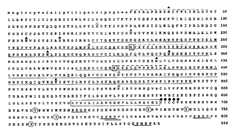
Figure 1 illustrates the amino acid sequence of SIMA135. The signal sequence
is in lower
case lettering and the putative transmembrane domain is boxed. Twelve
consensus N-
glycosylation motifs are indicated with filled triangles. Cytoplasmic tyrosine
residues are
circled. CUB domains that are thought to span residues 221 to 348 and 417 to
544 are
underlined. The three peptides identified from trypsin digestion and
sequencing are overlined.
The Arg residue preceding peptide 2 and the Lys preceding peptide 3 are boxed
to highlight
the consistency with trypsin specificity for Arg/Lys containing substrates.
Cytoplasmic
domain PXXP sequences are underlined. A consensus palmitylation motif,
following the
putative transmembrane domain, is indicated by filled circles. A consensus
palmitylation
motif, following the putative transmembrane domain, is indicated by filled
circles.
Detailed Description of the Invention
The invention relates to the discovery of a glycosylated protein that was
purified
from metastatic HEp3 cells through subtractive immunization using a monoclonal
antibody
5b
CA 02787820 2012-08-22
WO 2004/074481 PCT/EP2004/001556
designated 41-2. The protein is designated SIMA135 (subtractive immunization
M+ HEp3
associated 135 kDa protein).
SIMA135 refers to a protein that can be physically isolated from cells, as
opposed to
being a punitive protein predicted from translation of a nucleic acid
sequence. Physical
isolation of the SIMA135 protein is significant for a number of reasons. One
reason is that
isolation of the protein indicates that the mRNA is actually translated into a
polypeptide.
Secondly, isolation and characterization of the protein as reported herein
indicates that the
protein is glycosylated. Physical isolation of the glycosylated protein
confirms that
glycosylation sites within the polypeptide are available for glycosylation and
are not buried
within the folded protein to become inaccessible to glycosyltransferases. Such
conformation is important due to the known role that glycosylation plays in
protein folding
and immunogenicity. Therefore, isolation of the S1MA135 protein is a
significant advance
when compared to a theoretical polypeptide sequence predicted from a nucleic
acid
sequence.
The SIMA135 cDNA is shown herein to encode a 135 kDa type I transmembrane
cell surface protein that specifically immunoreacts with mAb 41-2.
Immunopurification and
amino acid sequencing confirms that the mature protein commences at Phe30
following
removal of a 29 amino acid signal peptide. Immunocytochemical analysis
confirms
localization of the protein to the cell surface and the type I orientation of
this protein. In
addition, consistent with the presence of 12 potential extracellular
glycosylation sites,
Western blot analysis of deglycosylated cell lysates indicates that up to 40
kDa of the
difference between the apparent (-135 kDa) and theoretical (-90 kDa) molecular
weight of
mature SIMA135 is due to N-linked glycans. Western blot analysis demonstrates
that
SIMA135 is a phosphotyrosine protein, consistent with the presence of 5
intracellular
tyrosine residues. In addition, the inhibitor PP2 has been used to demonstrate
that a Src
kinase family member acts to phosphorylate tyrosines of SIIVIA135 in HEp3
cells.
The domain structure of SIMA135 indicates that it may interact with
extracellular
proteins such as soluble ligands, other cell surface proteins and/or matrix
components;
potentially via putative CUB domains present within its amino terminal region.
These
structures are thought to mediate binding to a variety of protein ligands. For
example,
homodimerization of the MASP serine proteases acting within the lectin branch
of the
complement cascade is stabilized through interactions involving CUB domains
(Chen and
Wallis, 2001). Also a number of the type II transmembrane serine proteases
contain CUB
6
CA 02787820 2012-08-22
WO 2004/074481 PCT/EP2004/001556
domains thought to mediate enzyme-substrate interactions (Hooper et al.,
2001). In
addition, CUB domains of cubilin mediate binding to both the intrinsic factor-
cobalamin as
well as albumin (Yammani et al., 2001). As SIMA135 is heavily glycosylated
within its
extracellular domain, it is thought that ligand binding will be, at least
partially, dependent
on carbohydrate moieties as has been demonstrated for various isoforms of the
cell surface
glycoprotein CD44 (Bajorath, 2000). Glycosylation is also thought to
contribute to
SIvIA135 protein folding, and trafficking to and maintenance at the cell
surface (Gorelik et
al., 2001; Grogan et al., 2002).
SIMA135 displayed differences in amino acid sequence from other proteins
associated with signet ring carcinoma (GenBank entry AK026622) and the non
small lung
cell carcinoma cell line Calu 6 (GenBank entry AY026461) (Scherl-Mostageer et
al.,
2001). These differences are thought to affect the ability of SIMA135 to
interact with other
molecules, as compared to previously known proteins. The first amino acid
change,
525Arg-Gln, occurs within an extracellular potential ligand binding domain;
the second
of the potential CUB domains of S1MA135. The second amino acid change, 709G1y-
>Asp,
is located 2 residues after a tyrosine residue. This change from a non-polar
amino acid to a
charged residue could be expected to have a significant impact on the ability
of the
proximal tyrosine to be phosphorylated, and therefore is thought to have an
impact on the
capacity of SIMA135 to bind to, for example, SH2 domains. The last change,
827Ser>Asn, is located 4 residues from a PXXP motif. Accordingly, this change
may also
impact on the ability of SIMA135 to interact with other proteins; in this case
SH3 domain
containing proteins.
In normal colon tissue, SIMA135 protein is observed on basal and apical
surfaces of
epithelial cells lining the colon lumen and on the apical surface of crypt
epithelial cells. In
contrast to its distinct localization in normal colon, SIMA135 distribution in
colon tumor
tissue is disarrayed and heterogeneous, appearing dysregulated with both
plasma membrane
and cytoplasmic staining. It appears that expression of SIMA135 is more
intense in
invading glands deeper in the colonic serosa and within draining blood
vessels. These
results indicate that increased SIMA135 protein expression is associated more
with later
stages of carcinogenesis, such as local invasion and metastasis. This proposal
is partly
supported by Western blot analysis of pairs of human tumor cell lines
originating from the
same tissue. For example, SIMA-135 levels were much higher in highly-
metastatic M+
HEp3 cells compared to the congenic and low metastatic variant, M- HEp3. In
addition, the
7
CA 02787820 2012-08-22
WO 2004/074481 PCT/EP2004/001556
noncongenic prostate cancer cell lines PC-3 and LNCaP showed a similar trend;
the former,
a metastatic cell line, showing much higher levels of SEAM 35 compared to the
latter, a
low metastatic cell type (Soos et al., 1997).
The observation of apparently free SIMA135 in glandular mucus of both normal
and
malignant glands is consistent with the observation that a 110 kDa soluble
form of this
protein is released in vitro by HEp3 cells. The distinct loss of glandular
tissue ultrastructure
that is apparent during tumorigenesis may permit the release of the soluble
form of
SIMA135/CDCP1 into the fluid and vascular system of the colon cancer patient.
Accordingly, SIMA135 is thought to have utility as a serum or tissue fluid
marker as has
been proposed for the transmembrane proteins MUC1 (Rye and McGuckin, 2001),
CD44
(Adham et al., 1990) and ICAM-1 (Maruo et al., 2002).
1. SIMA135, fragments, and variants thereof that are glycosylated or non-
glycosylated.
The invention provides the SIMA135 protein (SEQ ID No:1), fragments of
SIMA135, and variants of SIMA135 that can be glycosylated or non-glycosylated.
These
proteins, fragments, and variants of SJMA135 can be used as antigens to induce
production
of antibodies that bind to SWA135, e.g. antibodies that bind specifically
and/or selectively
to SIMA135. Such selectively binding antibodies include those that bind to
SIMA135, or a
portion of SIvIA135, but that do not bind to proteins and fragments of
proteins that are not
SIMA135, or a fragment of SIMA135. These proteins, fragments, and variants can
also be
used to select for antibodies that specifically and selectively bind to
SIMA135. Such
specifically and selectively binding antibodies include those that bind to
SIMA135, or a
portion of SIMA135, but that do not bind to proteins and fragments of proteins
that are not
SIMA135, or a fragment of SIMA135. In particular, the selectivity of such
antibodies
means that they bind to SIMA135 or a portion of SIMA135 but do not also bind
to the
180kD protein produced from metastatic Hep-3 cell lysate as described in US
Pat No.
6,498,014 with a dissociation constant of the same order of magnitude as that
resulting
from binding to SIMA135 or a fragment thereof, although binding of such
antibodies with
the 18OkD protein may occur at a dissociation constant at least two orders of
magnitude
greater than that for binding with SIMA135 or a fragment thereof. The
specificity of such
antibodies means that the immunogenic binding is the result of epitopal -
hypervariable
region interaction and not the result of non-specific protein - protein
interaction. Non-
8
CA 02787820 2012-08-22
21159-548
specific protein - protein interaction typically will have a dissociation
constant at least 3
orders of magnitude greater than the dissociation constant for the specific
binding of an
epitopal - hypervariable region interaction. The dissociation constant for an
antibody -
antigen immunobinding pair can be measured according to the techniques
described in
Harlow et al., Antibodies: A Laboratory Manual, (Cold Spring Harbor Pub.
1988)).
A fragment of a SIMA135 protein as used herein, refers to a peptide fragment
of a
sufficient length to be antigenic. Generally speaking, the fragment includes
at least 5 amino
acids. The invention pertains to a fragment of SIMA135, e.g. a fragment of SEQ
ID No:1,
glycosylated or non-glycosylated wherein said fragment contains amino acid 525
and or
amino acid 827. The present invention pertains to a fragment of SIMA135, e.g.
a fragment
of SEQ ID No: 1, wherein in said fragment the amino acid 525 is not a
Glutamine and for
amino acid 827 is not an Asparagine. The present invention pertains to a
fragment of
SIMA135, e.g. a fragment of SEQ ID No: 1,.wherein in said fragment the amino
acid 525 is
Arginine and /or.amino acid 827 is Serine.
Variant proteins include proteins having amino acid substitutions that are
biologically active, or that elicit antibody production when used as an
antigen. A variant of
SIMA135 is intended to include a protein derived from native S]MA135 by
deletion (so-
called truncation) or addition of one or more amino acids to the N-terminal
and/or C-
terminal end of the native protein; deletion or addition of one or more amino
acids at one or
more sites in the native protein; or substitution of one or more amino acids
at one or more
sites in the native protein. Preferably, the variant at position 525 is not
Glutamine, the
variant at position 827 is not Asparagine and preferably the amino acid at
position 709
Glycine. Such variants may result from, for example, genetic polymorphism or
from human
manipulation. Methods for such manipulations are generally known in the art.
Thus, the SIMA135 proteins of the invention may be altered in various ways
including amino acid substitutions, deletions, truncations, and insertions.
Methods for such
manipulations are-generally known in the art. For example, amino acid sequence
variants
of the polypeptides can be prepared by mutations in DNA encoding SIMA135.
Methods
for mutagenesis and nucleotide sequence alterations are well known in the art.
See, for
example, Kunkel, Proc. Natl. Acad. Sci. USA, 82 488 (1985);.Kunkel et al.,
Methods in
Enzyrnol., 154:367 (1987); U. S. Patent No. 4,873,192; Walker and Gaastra,
eds.,
Techniques in Molecular biology, MacMillan Publishing Company, New York (1983)
and
9
CA 02787820 2012-08-22
.1159-548
the references cited therein.. Guidance as to appropriate amino acid
substitutions that do
not affect biological. activity of the. protein of interest may be found in
the model of
Dayhoff et al., Atlas of Protein Sequence and Structure, Natl. Biomed. Res.
Found.,
Washington, C.D. (1978). Conservative substitutions, such as exchanging
one amino acid with another having similar properties, are preferred.
Conservative amino acid substitutions are preferred and include, for example;
aspartic-
glutamic as acidic amino acids; lysine/arginine/bistidine as basic, amino
acids;
leucine/isoleucine, methionine/valine, alanine/valine as hydrophobic amino
acids;
serine/glycine/alaninelthreonine as hydrophilic amino acids. Conservative
amino acid
substitution also includes groupings based on side chains. Members in each
group can be
substituted for one another. For example, a group of amino acids having
aliphatic side
chains is glycine, alanine, valine, leucine, and isoleucine. These may be
substituted for one
another. A group of amino acids having aliphatic-hydroxyl side chains is
serine and
threonine; A group of amino acids having amide-containing side chains is
asparagine and
glutamine. A group of amino acids having aromatic side chains is'
phenylalanine, tyrosine,
and tryptophan. A group of amino acids having basic side chains is lysine,
arginine, and
histidine. A group of amino. acids-having sulfur-containing side chains is
cysteine and
methionine. For example, replacement of a leucine with an isoleucine or
valine, an
aspartate with a glutamate, a threonine with a serine, or a similar
replacement of an amino
acid with a structurally related amino acid may be accomplished to produce a
variant
polypeptide of the invention.
The proteins of the invention may be glycosylated or not glycosylated. The
proteins
may be glycosylated in vivo by expressing the proteins in a cell that is able
to glycosylate
the recombinant protein. Alternatively, the proteins of the invention can be
glycosylated in
vitro through use of sugar transferases. The proteins may be treated to cleave
any linked
glycans through use of commercially available enzymes, for example PNGase F
(New
England-Biolabs, Beverly, MA). Accordingly, the proteins of the invention can
be used to
produce antibodies that bind to native SIMA135, denatured SIMA135, specific
portions of
SIMA135, glycosylated SIMA135, and to non-glycosylated SIMA135.
II. An antibody that selectively.binds,to SIMA135, or. to a fragment of
SIMA135-.
The invention provides antibodies that bind to SIMA135. Preferred antibodies
to be
used in pharmaceutical compositions include those antibodies that inhibit
tumor metastasis.
CA 02787820 2012-08-22
WO 2004/074481 PCT/EP2004/001556
Inhibition of tumor metastasis can be determined by a number of assays, such
as the
migration assay, the invasion assay or the chick chorioallantoic membrane
assay.
Antibodies can be prepared that recognize natively folded SIMA135, or
denatured
SIMA135 by immunizing an animal with native SIMA135 or denatured SIM.A135
respectively. In addition, antibodies can be prepared that recognize SIMA135
that is
glycosylated, or SIMA135 that is not glycosylated by immunizing an animal with
SIMA135
that is glycosylated or non-glycosylated respectively. Antibodies that
recognize various
forms of SIMA135 (for example, native vs. denatured, and glycosylated vs. non-
glycosylated) are useful for determining if a cell is able to properly fold
and glycosylate
SIMA135. Such antibodies are useful for determining if a candidate agent is
able to
interfere with cellular actions that process SIMA135 during metastasis.
Accordingly, such
antibodies may be used to identify the action of agents that can be used to
inhibit metastasis
by cancer cells.
Antibodies that bind to SIMA135, fragments of SIMA135, and variants of
SIMA135, can be prepared using an intact protein or fragment containing small
peptides of
interest as the immunizing antigen. Fragments of SIMA135 that can be used as
antigens
include those that produce an immune response in an animal. These fragments
will
generally be five amino acids or greater in length. The protein or a peptide
used to
immunize an animal can be derived from translated cDNA or chemical synthesis
which can
be conjugated to a carrier protein, if desired. Such commonly used carriers
which are
chemically coupled to the peptide include keyhole limpet hemocyanin (KLH),
thyroglobulin, bovine serum albumin (BSA), and tetanus toxoid. The coupled
protein or
peptide is then used to immunize the animal (e.g., a mouse, a rat, or a
rabbit). The
monoclonal antibody 41-2 is an antibody that was initially employed to isolate
SJMA135.
It recognizes SIMA135 and in addition several other metastasis proteins
including the 180
1cD protein described in U.S Patent No's. 6,245,898 and 6,498,014. For this
reason, a
preferred antibody according to the invention is a monoclonal antibody that
binds, e.g.
selectively and/or specifically, with SIMA135, e.g. said antibody does not
bind with other
metastasis proteins, e.g. said antibody recognizes an epitope of SIMA135.
If desired, polyclonal or monoclonal antibodies can be purified, for example,
by
binding to and elution from a matrix to which the polypeptide or a peptide to
which the
antibodies were raised is bound. Those of skill in the art will know of
various techniques
common in the immunology arts for purification and/or concentration of
polyclonal
11
CA 02787820 2012-08-22
-1159-548
antibodies, as well as monoclonal antibodies (Coligan, et al., Unit 9, Current
Protocols in
Immunology, Wiley Interscience, 1991).
An antibody suitable for binding to a protein of the invention is specific for
at least
one portion of a region of the protein. For example, one of skill in the art
can use a protein
or peptide to generate appropriate antibodies of the invention. Antibodies of
the invention
include polyclonal antibodies, monoclonal antibodies, and fragments of
polyclonal and
monoclonal antibodies.
The present invention pertains to an antibody according to the invention for
use as a
therapeutic treatment of the human and animal body, the invention also relates
to an
antibody according to the invention for the preparation of a medicament for
use to inhibit
metastasis by a cancer cell in a mammal.
The preparation of polyclonal antibodies- is well-known to those skilled in
the art
(Green et al., Production of Polyclonal Antisera, in Immunoehen]ical Protocols
(Manson,
ed.), pages 1-5 (Humana Press 1992); Coligan et al., Production of Polyclonal
Antisera in
Rabbits, Rats, Mice and Hamsters, in Current Protocols in Immunology, section
2.4.1
(1992)).
The preparation of monoclonal antibodies likewise is conventional (Kohler &
Milstein, Nature, 256:495 (1975); Coligan et al., sections 2.5.1-2.6.7; and
Harlow et al.,
Antibodies: A Laboratory Manual, page 726 (Cold Spring Harbor Pub.
1988)). Briefly, monoclonal antibodies can be obtained by
injecting mice with a composition comprising. an antigen, verifying the
presence of
antibody production by removing a serum sample, removing the spleen to obtain
B
lymphocytes, fusing the B lymphocytes with myeloma cells to produce
hybridomas, cloning
the hybridomas, selecting positive clones that produce antibodies to the
antigen, and
isolating the antibodies from the hybridoma cultures. Monoclonal antibodies
can be
isolated and purified from hybridoma cultures by a variety of well-established
techniques.
Such isolation techniques include affinity chromatography with ProteinA
Sepharose, size-
exclusion chromatography, and ion-exchange chromatography (Coligan et al.,
sections
2.7.1-2.7.12 and sections 2.9.1-2.9.3; Barnes et at, Purification
ofImmunoglobulin G
(IgG), in Methods in Molecular Biology, Vol. 10, pages 79-104 (Humana Press
1992)).
Methods of in vitro and in vivo multiplication of monoclonal antibodies is
well-known to
those skilled in the art.. Multiplication in vitro may be carried out in
suitable culture media
such as Dulbecco's Modified Eagle Medium or'RPMI 1640 medium, optionally
replenished
12
CA 02787820 2012-08-22
1159-548
by a mammalian serum such as fetal calf serum or trace elements and growth-
sustaining
supplements such as normal mouse peritoneal exudate cells, spleen cells, bone
marrow
macrophages. Production in vitro provides relatively pure antibody
preparations and allows
scale-up to yield large amounts of the desired antibodies. Large scale
hybridoma cultivation
can be carried out by homogenous suspension culture in an air reactor, in a
continuous
stirrer reactor, or immobilized or entrapped cell culture. Multiplication in
vivo may be
carried out by injecting cell clones into mammals histocompatible with the
parent cells,
e.g., osyngeneic mice, to cause growth of antibody-producing tumors.
Optionally, the
animals are primed with a hydrocarbon, especially oils such as pristine
tetramethylpentadecane prior to injection. After one to three weeks, the
desired
monoclonal antibody is recovered from the body fluid of the animal.
An antibody of the invention may be derived from -a "humanized" monoclonal
antibody. Humanized monoclonal antibodies are produced by transferring mouse
complementarity determining regions from heavy and light variable chains of
the mouse
immunoglobulin into a human variable domain, and then substituting human
residues in the
framework regions of the murine counterparts. The use of antibody components
derived
from humanized monoclonal antibodies obviates potential problems associated
with the
immunogenicity of murine constant regions. General techniques for cloning
murine
immunoglobulin variable domains are described (Orlandi et al., Proc. Natl.
Acad. Sci.
USA, 06:3833 (1989) Which is hereby incorporated in its entirety by
reference).
Techniques for producing humanized monoclonal antibodies are described (Jones
et al.,
Nature, 321:522 (1986); Riechmann et al., Nature, 332:323 (1988); Verhoeyen et
al,
Science, 239:1534 (1988); Carter et al., Proc. Natl. Acad. Sci. USA. 59:4285
(1992);
Sandhu, Crit. Rev. Biotech., 12:437 (1992); and Singer et al., J. Immunol.,
150:2844
(1993)).
In addition, antibodies of the present invention may be derived from a human
monoclonal antibody. Such antibodies are obtained from transgenic mice that
have been
"engineered" to produce specific human antibodies in response to antigenic
challenge. In
this technique, elements of the human heavy and light chain loci are
introduced into strains
of mice derived from embryonic stem cell lines that contain targeted
disruptions of the
endogenous heavy and light chain loci. The transgenic mice can synthesize
human
antibodies specific for human antigens, and the mice can be used to produce -
human
antibody-secreting hybridomas. Methods for obtaining human antibodies from
transgenic
13
CA 02787820 2012-08-22
.21159-548
mice are described (Green et al., Nature Genet., 7:13 (1994); Lonberg et al.,
Nature,
368:856 (1994); and Taylor et al., Int. Immunol., 6:579 (1994)).
Antibody fragments of the invention can be prepared by proteolytic hydrolysis
of
the antibody or by expression in E. coli of-DNA encoding the fragment.
Antibody
fragments can be obtained by pepsin or papain digestion of whole antibodies by
conventional methods. For example, antibody fragments can be produced by
enzymatic
cleavage of antibodies with pepsin to provide a 5S fragment denoted F(ab')2.
This fragment
can be further cleaved using a thiol reducing agent, and optionally a blocking
group for the
sulfhydryl groups resulting from cleavage of disulfide linkages, to produce
3.5S Fab'
monovalent fragments. Alternatively, an enzymatic cleavage using pepsin
produces two
monovalent Fab' fragiiients and an Fc fragment directly. These methods are
described
(Goldenberg, U.S. patents No. 4,036,945 and 4,331,647; and references
contained therein;
Porter, Biochem. J., 73:119 (1959); Edelman et al., Methods in Enzymology,
Vol. 1, page
422 (Academic Press 1967); and Coligan et al. at sections 2.8.1-2.8.10 and
2.10.1-2.10.4).
Other methods of cleaving antibodies, such as separation of heavy chains to
form
monovalent light-heavy chain fragments, further cleavage of fragments, or
other enzymatic,
chemical, or genetic techniques may also be used, so long as the fragments
bind to the
antigen that is recognized by the intact antibody.
For example, Fv fragments include, an association of VH and VL chains. This
association may be noncovalent (mbar et al., Proc. Natl. Acad. Sci. USA,
69:2659 (1972)).
Alternatively, the variable chains can be linked by an intermolecular
disulfide bond or.
cross-linked by chemicals such as glutaraldehyde (Sandhu, supra). Preferably,
the Fv
fragments comprise VH and VL chains connected by a peptide linker. These
single-chain
antigen binding proteins (sFv) are prepared by constructing a structural gene
comprising
DNA sequences encoding the VH and VL domains connected by an oligonucleotide.
The
structural gene is inserted into an expression vector, which is subsequently
introduced into
a host cell such as E. coli. The recombinant host cells synthesize a single
polypeptide
chain with a linker peptide bridging the two V domain's. Methods for producing
sFvs are
described (Whitlow et al., Methods: A Companion to Methods in Enzymology, Vol.
2, page
97 (1991); Bird et al., Science, 242:423 (1988), Ladner et al., U.S. patent
No. 4,946,778;
Pack et al., Bio(Teclinology; 11:1271 (1993); and Sandhu, supra).
14
CA 02787820 2012-08-22
WO 2004/074481 PCT/EP2004/001556
Another form of an antibody fragment is a peptide coding for a single
complementarity-determining region (CDR). CDR peptides ("minimal recognition
units")
can be obtained by constructing genes encoding the CDR of an antibody of
interest. Such
genes are prepared, for example, by using the polymerase chain reaction to
synthesize the
variable region from RNA of antibody-producing cells (Larrick et al., Methods:
A
Companion to Methods in Enzymology, Vol. 2, page 106 (1991)).
III. A method to treat a metastatic tumor and to inhibit metastasis by a tumor
cell.
The invention also includes methods of treating metastatic tumors. "Treating a
metastatic tumor" means that the metastasis of the tumor is prevented,
delayed, or inhibited.
Metastatic tumors include both tumors at the primary site capable of
metastasizing and
metastasized tumors at a secondary site. Such metastatic tumors can be of a
tissue origin of
the lung, liver, kidney, mammary gland, epithelial, thyroid, leukemic,
pancreatic,
endometrial, ovarian, cervical, skin, colon and lymphoid tissue. A subject
which can be
treated can be any mammalian subject, including humans, dogs, monkeys, cows
and the
like, with the exception of mice.
One embodiment of the present invention provides methods of treating a
metastatic
tumor in a subject by administering to the subject a therapeutically effective
amount of a
tumor metastasis-inhibiting antibody of the present invention. Tumor
metastasis-inhibiting
antibodies of the present invention have been described hereinabove. Preferred
tumor
metastasis-inhibiting antibodies include those antibodies that selectively
bind to SIMA135,
or a fragment thereof.
A tumor metastasis-inhibiting antibody can be administered alone or together
with
a pharmaceutically acceptable carrier. A pharmaceutically acceptable carrier
includes all
solvents, such as fats, oils, water, saline solutions, lipids, liposomes,
resins, binders, fillers,
dispersion media, cell culture media, and the like, or combinations thereof,
that are non-
toxic to the recipient subject.
In accordance with the present invention, the active ingredients can be
combined
with the carrier in any convenient and practical manner, e.g., by solution,
suspension,
emulsification, admixture, encapsulation, absorption and the like, and if
necessary, by
shaping the combined compositions into pellets or tablets. Such procedures are
routine for
those skilled in the art.
CA 02787820 2012-08-22
WO 2004/074481 PCT/EP2004/001556
Dosages of an antibody to be therapeutically effective depend on the disease
state
and other clinical factors, such as weight and condition of the subject, the
subject's
response to the therapy, the type of formulations and the route of
administration. The
precise dosage of a compound to be therapeutically effective can be determined
by those
skilled in the art. As a general rule, the therapeutically effective dosage of
an antibody can
be in the range of about 0.5 g to about 2 grams per unit dosage form. A unit
dosage form
refers to physically discrete units suited as unitary dosages for mammalian
treatment: each
unit containing a predetermined quantity of the active material calculated to
produce the
desired therapeutic effect in association with any required pharmaceutical
carrier. The
methods of the present invention contemplate single as well as multiple
administrations,
given either simultaneously or over an extended period of time.
The administration of a tumor metastasis-inhibiting antibody may be carried
out in
any convenient manner, including by aerosol inhalation, injection, ingestion,
transfusion,
implantation or transplantation. Preferably, the antibodies of the present
invention are
administered to a patient by subcutaneous (s.c.), intraperitoneal (i.p.),
intra-arterial (i.a.), or
intravenous (i.v.) injection.
IV. A method to diagnose cancer in a mammal.
The invention also includes methods of diagnosing metastatic tumors in a
subject by
detecting the expression of SIA/IA135.
Metastatic tumors include both tumors at the primary site capable of
metastasizing
and metastasized tumors at a secondary site. Such metastatic tumors can be of
a tissue
origin of the lung, liver, kidney, mammary gland, epithelial, thyroid,
leukemic, pancreatic,
endometrial, ovarian, cervical, skin, colon and lymphoid tissues.
The expression of SIMA135 can be detected by using an antibody that binds to
SIMA135, or a fragment of S1MA135. Both polyclonal antibodies and monoclonal
antibodies can be employed.
In one embodiment, a sample is taken from the subject, e.g., a biopsy specimen
taken
from tissue suspected of having a metastatic tumor. Generally, the sample is
treated before
an assay is performed. Assays which can be employed include ELISA, RIA, EIA,
Western
Blot analysis, immunohistological staining and the like. Depending upon the
assay used,
the antigens or the antibodies can be labeled by an enzyme, a fluorophore or a
radioisotope.
16
CA 02787820 2012-08-22
WO 2004/074481 PCT/EP2004/001556
See, e.g., Coligan et al. Current Protocols in Immunology, John Wiley & Sons
Inc., New
York, N.Y. (1994); and Frye et al., Oncogen 4: 1153-1157, 1987.
The treatment of the sample may vary depending on the assay that is used to
detect
SIMA135. For example, cells of tissue biopsy can be lysed and the cell lysates
are used in
e.g., Western Blot analysis. For assays such as the Whole Cell ELISA assay,
cells can be
washed with, e.g., PBS, and then fixed with 0.25% glutaraldehyde in PBS before
the assay.
The expression of SIMA135, or a fragment of SIMA135, detected by using any of
the above-described methods, is compared with the expression of the same
antigen in the
normal part of the tissue. A substantial increase in the level of expression
of the antigen
when compared with the expression in the normal tissue, is indicative of a
metastatic
tumor. A substantial increase means an increase of at least about 20%,
preferably, at least
about 25%, more preferably, at least about 35%.
In another embodiment, immunohistochemistry can be used to diagnose a
metastatic
tumor in an organism. In this embodiment, a sample is taken from an organism,
e.g., a
biopsy specimen taken from tissue suspected of having a metastatic tumor. The
sample can
be affixed to a slide and contacted with antibodies, as disclosed herein, that
bind to
SIMA135. The antibodies can be labeled by an enzyme, a fluorophore or a
radioisotope.
See, e.g., Coligan et al. Current Protocols in Immunology, John Wiley & Sons
Inc., New.
Following binding of the antibodies to S1MA135, the position of the antibodies
is
determined through use of known techniques, Heterologous staining, extensive
expression
of SIMAI35 throughout the tissue sample, and staining within malignant glands
in the
colonic serosa indicate metastatic cancer.
V. A kit containing an antibody that selectively binds to SIMA135, or
fragments
thereof, and packaging material.
The invention provides a kit containing an antibody that binds to SIMA135 and
packaging material. Such kits are useful for shipping and storage of
antibodies that can be
used for treating and detecting cancer. Specifically, such kits may be used by
medical
personal in a laboratory for detecting metastatic cancer in a tissue sample
obtained from an
organism. Furthermore, such kits may be useful for medical personal for the
formulation of
pharmaceutical compositions that contain an antibody of the invention.
17
CA 02787820 2012-08-22
WO 2004/074481 PCT/EP2004/00156
The packaging material will provide a protected environment for the antibody.
For
example, the packaging material may keep the antibody from being contaminated.
In
addition, the packaging material may keep an antibody in solution from
becoming dry.
Examples of suitable materials that can be used for packaging materials
include
glass, plastic, metal, and the like. Such materials may be silanized to avoid
adhesion of an
antibody to the packaging material.
VI. A pharmaceutical composition containing an antibody that selectively binds
to
SIMA135 or to a fragment of SIMA135 and a pharmaceutically acceptable
carrier.
A pharmaceutical composition of the invention includes an antibody that binds
to
SEVA135 that is formulated as a pharmaceutical composition and administered to
an
animal host, such as a human patient in a variety of forms adapted to the
chosen route of
administration, i.e., orally or parenterally, by intravenous, intramuscular,
topical or
subcutaneous routes.
Thus, an antibody may be systemically administered, e.g., orally, in
combination
with a pharmaceutically acceptable vehicle such as an inert diluent or an
assimilable edible
carrier. They may be enclosed in hard or soft shell gelatin capsules, may be
compressed
into tablets, or may be incorporated directly with the food of the patient's
diet. For oral
therapeutic administration, the antibody may be combined with one or more
excipients and
used in the form of ingestible tablets, buccal tablets, troches, capsules,
elixirs, suspensions,
syrups, wafers, and the like. Such compositions and preparations should
contain at least
0.1 % of the antibody. The percentage of the compositions and preparations
may, of course,
be varied and may conveniently be between about 2 to about 60% of the weight
of a given
unit dosage form. The amount of the one or more antibodies in such
therapeutically useful
compositions is such that an effective dosage level will be obtained. When
administered
orally, the compositions of the invention can preferably be administered in a
gelatin
capsule.
The tablets, troches, pills, capsules, and the like may also contain the
following:
binders such as gum tragacanth, acacia, corn starch or gelatin; excipients
such as dicalcium
phosphate; a disintegrating agent such as corn starch, potato starch, alginic
acid and the
like; a lubricant such as magnesium stearate; and a sweetening agent such as
sucrose,
fructose, lactose or aspartame or a flavoring agent such as peppermint, oil of
wintergreen,
18
CA 02787820 2012-08-22
WO 2004/074481 PCT/EP2004/001556
or cherry flavoring may be added. When the unit dosage form is a capsule, it
may contain,
in addition to materials of the above type, a liquid carrier, such as a
vegetable oil or a
polyethylene glycol. Various other materials may be present as coatings or to
otherwise
modify the physical form of the solid unit dosage form. For instance, tablets,
pills, or
capsules may be coated with gelatin, wax, shellac or sugar and the like. A
syrup or elixir
may contain the antibody or antibodies, sucrose or fructose as a sweetening
agent, methyl
and propylparabens as preservatives, a dye and flavoring such as cherry or
orange flavor.
Of course, any material used in preparing any unit dosage form should be
pharmaceutically
acceptable and substantially non-toxic in the amounts employed. In addition,
the antibody
or antibodies may be incorporated into sustained-release preparations and
devices.
The antibody or antibodies of the invention may also be administered
intravenously
or intraperitoneally by infusion or injection. Solutions of the antibody or
antibodies can be
prepared in water, optionally mixed with a nontoxic surfactant. Dispersions
can also be
prepared in. glycerol, liquid polyethylene glycols, triacetin, and mixtures
thereof and in oils.
Under ordinary conditions of storage and use, these preparations contain a
preservative to
prevent the growth of microorganisms.
The pharmaceutical dosage forms suitable for injection or infusion can include
sterile aqueous solutions or dispersions or sterile powders comprising the
antibody or
antibodies which are adapted for the extemporaneous preparation of sterile
injectable or
infusible solutions or dispersions, optionally encapsulated in liposomes. In
all cases, the
ultimate dosage form should be sterile, fluid and stable under the conditions
of manufacture
and storage. The liquid carrier or vehicle can be a solvent or liquid
dispersion medium
comprising, for example, water, ethanol, a polyol (for example, glycerol,
propylene glycol,
liquid polyethylene glycols, and the like), vegetable oils, nontoxic glyceryl
esters, and
suitable mixtures thereof. The proper fluidity can be maintained, for example,
by the
formation of liposomes, by the maintenance of the required particle size in
the case of
dispersions or by the use of surfactants. The prevention of the action of
microorganisms
can be brought about by various antibacterial and antifungal agents, for
example, parabens,
chlorobutanol, phenol, sorbic acid, thimerosal, and the like. In many cases,
it will be
preferable to include isotonic agents, for example, sugars, buffers or sodium
chloride.
Prolonged absorption of the injectable compositions can be brought about by
the use in the
compositions of agents delaying absorption, for example, aluminum monostearate
and
gelatin.
19
CA 02787820 2012-08-22
WO 2004/074481 PCT/EP2004/001 56
Sterile injectable solutions are prepared by incorporating an antibody or
antibodies
in the required amount in the appropriate solvent with various of the other
ingredients
enumerated above, as required, followed by filter sterilization.
VII. A method to identify an agent that modulates production of SIMA135 by a
cell
The invention provides a method to identify an agent that increases or
decreases
production of S]MA135 by a cell. Generally, the method involves contacting a
test cell
with a candidate agent and determining if production of SIMA135 by the test
cell is
increased or decreased relative to a control cell that was not contacted with
the candidate
agent.
SIMA135 production by the test cell and the control cell can be detected
through use
of many art recognized methods. Such methods are exemplified by immunological
methods that include radioimmunoassay (RIA), enzyme-linked immunosorbant assay
(ELISA), use of fluorescently labeled antibodies, and the like.
Many examples of candidate agents that can be screened according to the method
are
described in the official United States Pharmacopeia, official National
Formulary, or any
supplement to them. Briefly, examples of candidate agents include,
hydrocarbons, cyclic
organic molecules, bicyclic organic molecules, aryl organic molecules, alkyl
organic
molecules, and the like. Merck Manual, Merck Research Laboratories, Whitehouse
Station, N.J. 17"' edition, eds, Beers and Berk-ow 1999; Merck Index, Merck
Research
Laboratories, Whitehouse Station, N.J., 13'h ed., 2001.
The metastatic HEp3 cells such as those exemplified in the following section
may be
used as test cells and control cells in the method of the invention. However,
the method
may also be practiced with cells that produce SIMA135 normally or through
recombinant
methods. For example, an expression construct that provides for the production
of
SIMA135 may be introduced into a cell that does not produce SIMA135 prior to
the
introduction of the expression cassette. The transformed cell may then be used
within the
method of the invention to identify agents that modulate SIMA135 production.
The
diagnostic methods described above for detection of the presence and quantity
of SIMA
135 can be used with such cell systems to assay the SIMA 135 production by
test and
control cells.
The method of the invention may also be practiced in vivo. As exemplified in
the
following section, a candidate agent may be administered to a test animal. A
tissue sample
CA 02787820 2012-08-22
WO 2004/074481 PCT/EP2004/001556
may be obtained from the test animal and SIMA135 production by cells in the
tissue
sample maybe compared to SIMA135 production by cells in a tissue sample
obtained from
a control animal. An increase in SIMA135 production by the test animal
relative to the
control animal indicates that the candidate agent increases SIMA135
production. A
decrease in SIMA135 production by the test animal relative to the control
animal indicates
that the candidate agent decreases SIMA135 production. The assay of the SIMA
135 may
be determined by any of the analytic methods given in the diagnosis section
above.
Numerous animals may be used within the method of the invention. Examples of
such
animals include rabbits, rats, mice, monkeys, and the like.
The method of the invention may be practiced in vitro. For example, test cells
and
control cells may be grown in tissue culture. This allows the candidate agent
to be
contacted with a test cell in vitro. SIMA135 production by the test cells can
then be
compared to SIMA135 production by control cells as described above to
determine if the
candidate agent increases or decreases SIMA135 production by a cell.
The in vitro and in vivo methods will determine the ability of a test agent to
promote
and to minimize or prevent metastasis. Determination of promotion will
identify the agent
as a cancer causing or enhancing agent. This determination has practical
application for the
rapid identification of cancer causing agents. Determination of minimization
or prevention
will identify the agent as a cancer inhibiting agent. This determination has
practical
application for the identification of anti-cancer agents.
Example I
Cell Lines and Hybridomas
Human cervical adenocarcinoma HeLa, fibrosarcoma HT1080, colon adenocarcinoma
DLD-l and SW480, breast adenocarcinoma MCF7, prostate adenocarcinoma PC-3,
prostate
carcinoma lymph node metastasis LNCaP, lung carcinoma A549 and kidney rhabdoid
tumor G401 cells were obtained from the American Type Culture Collection
(Rockville,
MD). Human liver cancer HuH7 and HLE, and gastric cancer MKN45 and STKM-1
cells
were provided by Dr. Peter Vogt (The Scripps Research Institute, La Jolla, CA)
and breast
adenocarcinoma MDA-MB-231 cells by Dr. Liliana Ossowski (Mount Sinai School of
Medicine, NY). Human epidermoid carcinoma HEp3 cells, were obtained from solid
tumors serially passaged on the chorioallantoic membrane (CAM) of chicken
embryos
(Testa, 1992; Brooks et al., 1993). The metastatic variant of HEp3 cells, M+
HEp3, was
21
CA 02787820 2012-08-22
-,1159-548
cultured for less than 20 days before use. The low metastatic variant, M-
HEp3, was
maintained in culture for at least 80 days before use. Human microvascular
endothelial
cells (HEC) and dermal fibroblasts (I DF) were obtained from Clonetics (San
Diego, CA)
and maintained in EGM-2 MV and FGM-2 media (Clonetics) respectively. Cancer
cell
lines were maintained as monolayer cultures in DMEM (Invitrogen, Carlsbad, CA)
supplemented with 10% FBS (HyClone, Logan, UT), sodium pyruvate,
penicillin/streptomycin and non-essential amino acids (Invitrogen) and grown
in a
humidified 5% CO, atmospheres at 37 C. Hybridomas producing MoAb 41-2 were
generated by a previously described subtractive immunization approach (Brooks
et al.,
1993). Hybridoma culturing and purification of mAbs were performed by the
Protein and
Nucleic Acids Core Facility of The Scripps Research Institute using standard
procedures.
Example II
Reagents
Protease inhibitors, normal mouse IgG, anti-FLAG M2 mAb, DAB reagent and Gill
hematoxylin were purchased from Sigma (St. Louis, MO). Reverse transcription
and PCR
reagents and the pCR-II Topo vector were from Invitrogen. PP2 was obtained
from
Calbiochem (La Jolla, CA).
Example III
Protein Purification, Peptide Sequencingand Protein Analysis
Immunoprecipitations were performed on lysates from either unlabelled or 31S-
labelled
HEp3 cells (5x10). Metabolic labeling was performed overnight in
methionine/cysteine
free DMEM containing Tran 35S-label (100 pCi/ml; ICN, Costa Mesa, CA). Cells
were
washed thoroughly with PBS then lysed in a buffer containing 0.1 M Tris (pH
8.0), 0.1%
Triton X.=100, 150 mM NaCl, 5 mM EDTA, 10.pM trans-epoxysuccinyl-L-leucylamido
(4-
guanidino) butane, 20 g/ml soybean trypsin inhibitor and 25 pg/mI aprotinin:
Lysates were
pre-cleared against protein G-Sepharose'(Pharmacia Biotech, Piscataway, NJ) at
4 C for 30
minutes then incubated overnight at 4 C with 20 Etg of either mAb 41-2 or. as
control,
nmIgG. Immunocomplexes was precipitated using protein G-Sepharose and
complexes
were denatured by boiling in reducing SDS loading buffer before analysis by
polyacrylamide gel electrophoresis. For 35S-labelled proteins, the gel was
dried and
exposed to film at -80 C. Otherwise proteins were transferred to
polyvinylidine difluoride
*Trade-mark
22
CA 02787820 2012-08-22
21159-548
(PVDF) membranes (Millipore, Bedford, MA). The predominant coomassie stained
band,
at 135 kDa, was excised then digested with trypsin. The resulting peptides
were separated
by high pressure liquid chromatography and sequenced on a Procise 494 protein
sequencer
(Applied Biosystems, Inc., Foster City, CA). Trypsin digestion and peptide
sequencing
were performed by the Protein and Nucleic Acids Core Facility of The Scripps
Research
Institute. Peptide sequences were used to search the GenBank database using
algorithms
available at the National Center for Biotechnology Information (NCBI) website.
The
complete SIMA135 protein sequence was analyzed for structural domains,
cellular
processing signals and consensus post translational modification motifs using
the Prosite
database (Falquet et al., 2002), the SMART algorithm (Schultz et al., 1998),
the PSORT
algorithm (Nakai and Kanehisa, 1992) and the NetPhos 2.0 algorithm (Blom et
al., 1999).
Example IV
Expression Constructs and Transient Transfections
SlI\4A135 cDNA in the eukaryotic expression vector pl\4El8S-FL3 (GenBank
accession
number AK026622) was generated as part of the Japanese NEDO human cDNA
sequencing project and kindly provided by Dr. Hiroko Hata (Dept of Virology,
Institute of
Medical Science, University of Tokyo). The SIMA135FLAGin construct was
generated by
PCR placing sequences encoding the FLAG epitope (DYKDDDDK) immediately before
the stop codon of the parent construct. Both constructs were sequenced. HeLa
cells
(4x10⁵) were transiently transfected with either the SIMA135 or SJMA135FLAGin
expression constructs using Superfect reagent (Qiagen, Valencia, CA) as
described by the
manufacturer. Cells were lysed in ice cold buffer containing 10 mM Tris (pH
8.0), 150
mM NaCI, 1% Triton X-I00, 5 mM EDTA and lx Complete mini EDTA-free protease
inhibitor cocktail (Roche, Indianapolis, IN). Insoluble material was removed
by
centrifugation at 14000 rpm for 10 min.
Example V
Cloning of the SIMA135 cDNA from HEp3 Cells
Total RNA was isolated using an RNeasy kit (Qiagen) and 2 g served as
template in a
reverse transcription reaction using Superscript II reverse transcriptase. PCR
was
performed on 1 gl of the resulting cDNA using primers TCCCCACCGTCGTTTTCC (SEQ
ID NO:2) and GGTTAGGAACACGGACGGGTG (SEQ ID NO:3)(designed based upon
*Trade-mark
23
CA 02787820 2012-08-22
:1159-548
GenBank accession AK026622) and the proof reading enzyme Platinum Pfx DNA
polymerase. PCR cycling conditions were 94 C for 3 min, 30 cycles of 94 C for
30 see,
55 C for 30 sec and 72 C for 150 sec followed by a final 72 C extension for 10
min. PCR
products were gel purified (Qiagen) adenosine tailed using Platinum Taq DNA
polymerase
then cloned in the pCR-ll Topo vector and sequenced.
Example VI
Immunofluorescence
HeLa cells transiently transfected with the SAZAI35FLAGin expression construct
and
10. HEp3 cells were plated on coverslips. After incubation for 48 hr at 37 C
cells were washed
with PBS then fixed in 2% formaldehyde. HeLa cells to be incubated with anti-
FLAG mAb
were either not permeabilized or permeabilized by incubating in 0.5% Triton X-
100 in PBS
for 5 min at room temperature. Both cell types were blocked in 5% BSA in PBS.
Following overnight incubation at 4 C with either mAb 41-2 (5 pg/ml) or anti-
FLAG M2
mAb (4 Rg/ml) in blocking buffer, cells were washed with PBS then incubated
with Alexa
Fluor 546 conjugated goat anti-mouse IgG (2 gg/ml) (Molecular Probes). Labeled
cells
were visualized and photographed using a BioRad 1024 MRC2 scanning confocal
imaging
system.
Example VII
Northern Blot Analysis
A human 12 lane multiple tissue Northern blot (Clontech) was hybridized with
[a-32P]dCTP
labeled (Ambion) EcoRUHinci DNA insert fragments of the S]MA135 cDNA overnight
in
Ultralyb solution (Ambion) at 68 C. The blot was washed to a final stringency
of 0.1 x
SSC, 0.1 % SDS at 68 C then exposed to film at -80 C. Blots were reprobed with
B-actin
cDNA to determine consistency of RNA loading in each lane.
Example VIII
Western Blot Analysis
Cell lysates, serum free conditioned media and immunoprecipitated proteins
were separated
by electrophoresis through 8% SDS-polyacrylamide gels then transferred to
nitrocellulose
membranes (Millipore). Membranes were blocked in 5% non-fat skim milk powder
in PBS
then incubated overnight at 4 C with either mAb 41-2 (2 p/ml), anti-FLAG M2
mAb (0.8
*Trade-mark
24
CA 02787820 2012-08-22
WO 2004/074481 PCT/EP2004/001556
pg/ml) or anti-phosphotyrosine mAb (1 g/ml; Upstate Biotechnology, Lake
Placid, NY).
Following extensive washing membranes were incubated for 2 hr at room
temperature with
goat anti-mouse IgG (0.16 pg/ml, Pierce, Rockford, 11) and immunoreactive
bands detected
by enhanced chemiluminescence (Pierce).
Example IX
Biochemical Characterization Procedures
For removal of N-linked glycans, lysates (50 .d) from M+ HEp3 cells and HeLa
cells
transiently transfected with the SIMA135FLAGin expression construct were
denatured and
reduced in 0.5% SDS, 1% (3-mercaptoethanol for 10 minutes at 100 C then
incubated with
PNGase F (New England Biolabs, Beverly, MA) at 37 C for 45 minutes. For
analysis of
the basal level of tyrosine phosphorylation of SIMA135, subconfluent cultures
of HEp3
and HeLa (as negative control) cells were incubated at 37 C for 30 min with
serum free
DIvIEM containing 50 mM NaF and 1 mM Na3V04 then washed with ice cold PBS. For
inhibition of Src kinase family phosphorylation, HEp3 cells were cultured in
serum free
DMEM without NaF and Na3V04 for 30 minutes at 37 C with PP2 (50 M). Cells
were
then lysed in ice cold buffer containing 50 mM Tris (pH 7.4), 150 mM NaCl, 1%
Triton X-
100, 1 mM phenylmethylsulfonyl fluoride, 1 mM benzamidine, 25 gg/ml aprotinin,
25
g/ml leupeptin, 50 mM NaF and 1 mM Na3V04. Insoluble material was removed by
centrifugation at 14000 rpm for 10 min. Immunoprecipitation was performed as
described
above on 300 pg of cell lysates using 1 pg of either mAb 41-2 or nmIgG (as
negative
control). For assays for the presence of soluble SIMA135, HEp3 cells
approaching
confluence, were washed three times with PBS then incubated in serum free
conditioned
media for 24 hr. The media was collected and centrifuged at 4 C and 1 0,000g
then
concentrated 10 fold using micron centrifugal filters with a molecular weight
cut off of
30,000 kDa (Millipore). Cells lysates were collected as described above.
Example X
Immunohistochemistry
Cryostat sections (6 m) from archival human adenocarcinoma colon tissue
samples from
three patients were fixed in zinc-formalin for 15 min, rinsed briefly with PBS
then non-
specific binding sites blocked by incubating in PBS containing 3% BSA. mAb 41-
2 (5
g/ml) was applied at 4 C overnight. Specific antibody binding was detected by
the
CA 02787820 2012-08-22
WO 2004/074481 PCT/EP2004/001556
addition of biotin conjugated anti-mouse antibodies (Pierce) followed by
peroxidase
conjugated neutravidin (Pierce) which was visualized using DAB reagent.
Sections were
counterstained using Gill hematoxylin.
Example XI
mAb 41-2 Recognizes a 135 kDa Antigen Expressed at Elevated Levels in Highly
Metastatic Human Tumor HEp3 Cells
The antigen recognized by the antibody mAb 41-2 was identified and
characterized. As an
initial step in determining the significance of the antigen recognized by mAb
41-2, Western
blot analysis was performed using mAb 41-2 on lysates (20 g) prepared from
high
metastatic (M+) and low metastatic (M) HEp3 cells electrophoresed under non-
reducing
conditions. Monoclonal antibody 41-2 (mAb), generated by subtractive
immunization,
detected a single band of approximately 135 kDa in both cell types. Consistent
with the
subtractive immunization approach taken in generating mAb 41-2, the
immunoreactive
protein was expressed at higher levels in M+ than in M" HEp3 cells. Parallel
Coomassie
stained gels of the lysates prepared from high metastatic (M+) and low
metastatic (N f)
HEp3 cells demonstrate that the overall protein pattern and content was
indistinguishable
for M+ and M HEp3 cell extracts. The difference in mAb 41-2 innnunoreactivity
shows a
significant difference in the level of expression of the cognate antigen
between the two cell
lines.
Example XII
Identification of the Antigen Recognized by mAb 41-2 from Metastatic HEp3
Cells
Purified mAb 41-2 was used to immunoprecipitate the antigen of Example XI from
M'
HEp3 cells. 35S labeled proteins inmunoprecipitated from M+ HEp3 cells with
mAb 41-2
or normal mouse IgG were analyzed on SDS-PAGE under reducing conditions. The
gel was
dried and exposed to film at -80 C overnight. The major protein
immunoprecipitated from
radiolabeled HEp3 cells had a molecular weight of approximately 135 kDa. This
was
consistent with the molecular weight of the antigen detected in Example XI. In
a parallel
experiment using unlabeled HEp3 cells, in which immunoprecipitated proteins
were
transferred to a PVDF membrane, the 135 kDa protein band was excised,
subjected to
26
CA 02787820 2012-08-22
WO 2004/074481 PCT/EP2004/001556
trypsin digestion and the separated fragments sequenced from the N-terminus.
Three major
peptide sequences were obtained. Searches of the GenBank non-redundant protein
database indicated that each of the peptide sequences had exact or near exact
matches with
the theoretical sequence of an unpublished entry with accession number BAB
15511,
translated from unpublished cDNA entry AK026622.
Peptide 1 FEIALPRESQITVLG(I)KXGT SEQ ID No: 4
BAB15511 FEIALPRESNITVLIKXGT SEQ ID No:5
Peptide 2 XXXXIPGSTTNPE SEQ ID No:6
BAB15511 VEYYIPGSTTNPE SEQ ID No:7
Peptide 3 XYXLQVPSDILH SEQ ID No:8
BAB15511 SYSLQVPSDILH SEQ ID No:9
The complete sequence of the identified protein, which was designated
subtractive
immunization M' HEp3 associated 135 kDa protein (SIMA135), is shown in Figure
1. To
confirm that SIMA135 was the same protein specifically recognized by mAb 41-2,
Western
blot was performed on lysates from HEp3 cells, untransfected HeLa cells and
HeLa cells
transiently transfected with the SEV A.135 cDNA. Western blot analysis probing
with mAb
41-2 of total cell lysates (25 g) electrophoresed under non-reducing
conditions from HEp3
cells, mock transfected HeLa and HeLa cells transiently transfected with the
SIMA135
cDNA. mAb 41-2 reacted with the same 135 kDa protein band that is present in
HEp3 cells
and in HeLa cells transiently transfected with the SIMA135 cDNA, but is absent
in the
untransfected HeLa cells. To provide additional confirmation, the protein
encoding region
of the SIMA135 mRNA was cloned from HEp3 cells by reverse transcription-PCR.
DNA
sequence analysis of two clones generated by this approach confirmed that
SIMA135
mRNA is expressed by these cells. Four nucleotide differences were identified
between
GenBank entry AK026622 and SIMA135 sequence obtained from HEp3 cells:
nucleotide
1684-A, 1847T-.c, 2236c-.A and 25906-"A. The second transition is silent and
the others
result in amino acid changes 525^s- i", 70901''-. and 827ser-.Asn
respectively.
Example XIII
27
CA 02787820 2012-08-22
WO 2004/074481 PCT/EP2004/001556
SIMA135 Structural Features
The SIMA135 protein sequence includes 836 amino acids (SEQ ID No.I - Figure 1)
and
has a deduced molecular weight of 92.9 kDa. Sequence analysis identified the
following
structural features. A putative amino terminal signal peptide with cleavage
predicted to
occur following Ala29, This feature is consistent with the sequence of peptide
1 indicating
that mature SIMAI35, with a predicted molecular weight of 90.1 kDa, starts at
Phe30. A
potential transmembrane domain, spanning residues 666 to 686 (boxed in Figure
1), is
predicted (Hartmann et al., 1989) to orient SIMA135 with its carboxy terminus
located
intracellularly. Twelve consensus motifs for N-glycosylation are indicted in
Figure 1. A
consensus type 1 palmitylation motif (IICCV) (Hansen et al., 1999) is located
at residues
687 to 691. Five PXXP motifs are present (Figure 1) which in other proteins
have been
shown to mediate binding to Src homology (SH) 3 domains (Pawson 1995; Mayer,
2001).
Five tyrosine residues (circled in Figure 1) may be phosphorylation sites. Two
closely
spaced tyrosine residues (Tyr734 and Tyr743) are present in consensus motifs
(YXXL/1) for
SH2 domain binding (Songyang et al., 1993). SIMA135 lacked homology with other
confirmed proteins in the GenBank non-redundant database. However, SIMA135 did
have
high homology to the theoretical protein CDCP 1 as determined by translation
of the
reported nucleic acid sequence (Scherl-Mostageer et al., 2001). In addition,
two regions of
the SIMA135 protein, spanning residues 221 to 348 and 417 to 544, were
identified that
had low homology to CUB (complement protein subcomponents Clr/C1s, urchin
embryonic growth factor and bone morphogenetic protein 1) domains (Bork and
Beckmann, 1993). These domains have been reported to act in mediating protein-
protein
interactions (Chen and Wallis, 2001; Sieron at al., 2000). A third putative
CUB domain
described by Scherl-Mostageer and co-workers spanning residues 545 to 660
(Scherl-
Mostageer et al., 2001) was below the homology detection threshold of the
search
algorithms used by us to scan the SEv1AI35 amino acid sequence and the
theoretical amino
acid sequence for CDCP1.
Example XIV
Expression Pattern of SIMA135 in Normal and Malignant Cells and Tissues
Northern blot analysis of poly AA RNA (1 g per lane) from 12 normal human
tissues
probed with a 32P labeled SIMA135 cDNA probe was performed.. Levels of (3-
actin mRNA
28
CA 02787820 2012-08-22
WO 2004/074481 PCT/EP2004/001556
are shown as a measure of loading. The expression pattern of SIMA135 mRNA in
12
normal human tissues was examined by Northern blot analysis by hybridizing
with a 32P-
labeled 2.8 kb SIMA135 cDNA probe. A band of approximately 6.0 kb was detected
at
highest levels in skeletal muscle and colon with lower levels of expression in
kidney, small
intestine, placenta and lung. A barely detectable signal at -6.0 kb also was
present in
peripheral blood leukocytes. In addition, a much weaker signal at
approximately 3.3 kb
was present in skeletal muscle, colon, placenta and lung. SIMA135 mRNA was not
detected in brain, heart, thymus, spleen or liver. Based upon alignment of
SIMA135 with
CDCP1 cDNA7 and genomic sequences (data not shown), it appears most likely
that the
two SIMA135 transcripts detected by Northern blot analysis result from use of
alternate
polyadenylation signals within the SIMA-135 3' UTR. The longer, more highly
expressed
transcript is thought to have resulted from use of a more 3' consensus
poladenylation signal
(at nucleotide 5950'), whereas the shorter, more lowly expressed transcript
likely results
from use of a variant, less efficient polyadenylation signal located at
nucleotide 3186'. It is
also possible that these variant transcripts result from alternate splicing of
the SIMA135
pre-mRNA.
SIMA135 protein expression in 16 human cell lines, 14 human tumor cell lines
and two
normal human cell lines probing with mAb 41-2, was analyzed by Western blot
analysis
under non-reducing conditions in which equal amounts of cell lysate protein
(20pg) were
electrophoresed for each cell line. SIMA135 was most highly expressed in
metastatic HEp3
cells, with the prostate cancer cell line PC3, and the colon cancer cell line
DLD-1 also
manifesting high levels of expression. Moderate levels of the antigen were
detected in the
fibrosarcoma cell line HT1080, the gastric cancer cell lines MKN45 and STKM-1,
the
colon cancer cell line SW480 and the non-metastatic prostate cancer cell line
LNCaP. Low
levels of SIMA135 were detected in 2 liver cancer cell lines, 2 breast cancer
cell lines, the
lung cancer cell line A549 and the kidney rhabdoid tumor cell line G401.
SIMA135 was
not detectable in normal human microvascular endothelial cells and dermal
fibroblast cells.
Varying levels of SIMA135 protein was expressed in a number of human tumor
cell lines,
while two normal human cell types did not express the protein.
Example XV
SIMA135 is a Cell Surface, Phosphorylated Glycoprotein.
29
CA 02787820 2012-08-22
1159-548
Immunocytochemistry was used to determine the cellular location of S]MA135 in
HEp3
cells, and also in HeLa cells that were transiently transfected with a STMA135
expression
construct. The expression construct contained a FLAG epitope fused after the
carboxy
terminus.of the SIMA135 protein. HEp3 cells incubated with mAb 41-2 showed
strong
staining on the plasma membrane. HeLa cells transiently transfected with FLAG
tagged
SIMA135 also showed similar strong membrane staining when incubated with mAb
41-2.
In addition, the SIMA135 carboxy terminus was determined to be intracellular
as
*
transiently transfected HeLa cells permeabilized using 0.5% Triton X-100 to
permit access
of the antibody to the intracellular located FLAG tag and incubated with anti-
FLAG mAb
showed strong membrane staining when incubated with an anti-FLAG epitope mAb,
while
non-permeabilized cells exhibited low or near background staining with anti-
FLAG mAb.
Untransfected HeLa cells were essentially free of staining when incubated with
either mAb
41-2 or an anti-FLAG epitope mAb. These data confirmed the predicted cell
surface
location as well as the type I orientation of SIMA135. Also, the coincidence
of staining
observed with mAb 41-2 and anti-FLAG mAb in HeLa cells transiently transfected
with the
SIIIJA135-FLAG tag expression construct, additionally confirmed that SIMA135
is the
target antigen for mAb 41-2.
The theoretical molecular weight of mature SIMA-135 is 90.1 kDa. However, the
apparent
molecular weight of the protein detected by mAb 41-2 was 135 kDa.. Cell
lysates from
HeLa cells transiently transfected with a SIMA135 FLAG tag expression
construct were
treated with N-glycosidase F under conditions optimal for enzyme activity to
determine if
the difference. in molecular weight was due to N-glycosylation. Proteins were
examined by
Western blot analysis under reducing conditions using an anti-FLAG epitope mAb
of
untreated (-) and N-glycosidase F (PNGase F) treated (+) lysates from HeLa
cells
transiently transfected with the SIMAI35FLAGin expression construct. Bands due
to
SIMAI35 are indicated. A non-specific cross reacting band is apparent at 80
kDa. N-
glycosidase F treatment resulted in the disappearance of the SIMA135 protein
band at 135
kDa and replacement with a broad lower molecular weight band of approximately
95 to 105
kDa (data not shown). Lysates of 1v[' HEp3 cells were also immunoprecipitated
with mAb
41-2 and treated with N-glycosidase F. The proteins detected according to this
method also
manifested a similar diminished molecular weight. Therefore, up to 30-40 kDa
of the
apparent molecular weight of SIMAI35 is due to N-glycosylation, consistent
with the large
*Trade-mark
CA 02787820 2012-08-22
WO 2004/074481 PCT/EP2004/001556
number of consensus glycosylation sites in the extracellular region of this
protein (Figure 1,
SEQ ID No.1).
The intracellular region of SIMA135 contained 5 tyrosine residues (Figure 1).
Western
blot analysis under reducing conditions with an anti-phosphotyrosine antibody
was
performed on proteins immunoprecipitated from HEp3 cell lysates with mAb 41-2
to
determine whether any of these residues are phosphorylated.
immunoprecipitations that
were performed with either normal mouse IgG (nmIgG) or mAb 41-2 from HEp3 and
HeLa
(negative control) cell lysates. Immunoprecipitated proteins were probed with
an anti-
phosphotyrosine antibody (Anti-P-Tyr) and with mAb 41-2 to indicate the
presence of
phosphorylated and total SIMA135, respectively. Cell lysate proteins were
immunoprecipitated with mAb 41-2. Lysates were prepared from HEp3 cells either
untreated (-) or (+) incubated for 30 minutes at 37 with PP2 (50 M). The
anti-
phosphotyrosine antibody detected a protein of 135 kDa that immunoprecipitated
from
HEp3 cells with mAb 41-2. The same protein band was detected when the
immunoprecipitated proteins were probed with mAb 41-2. Western blot analysis
was also
performed on proteins inmunoprecipitated from HeLa cell lysates with mAb 41-2,
and
proteins immunoprecipitated from HEp3 cell lysates with normal mouse IgG as
controls.
Both immunoprecipitations were free of immunoreactivity when probed with
either the
anti-phosphotyrosine antibody or mAb 41-2, demonstrating the specificity of
the
irmnunoreactions observed with HEp3 cells. The involvement of a Src kinase
family
member in SIMA135 tyrosine phosphorylation was examined using PP2, a Src
family-
selective tyrosine kinase inhibitor (Hanke et al., 1996). Western blot
analysis with an anti-
phosphotyrosine antibody of proteins immunoprecipitated from HEp3 cell lysates
with
mAb 41-2, showed that HEp3 cells treated with PP2 for 30 minutes had a
significant
reduction (-75%) in the level of SIMA135 tyrosine phosphorylation compared to
protein
from untreated HEp3 cells. Western blot analysis, using mAb 41-2, of the same
immunoprecipitated proteins, indicated that approximately equal amounts of
SIMA135
protein were present in both lanes on the membrane. These data suggested that
a Src
kinase family member acts during tyrosine phosphorylation of SIMA135 in HEp3
cells.
A number of integral cell surface proteins, such as c-met (Wajih et al., 2002)
and CD44
(Goebeler et al., 1996), are also produced as soluble molecules. Western blot
analysis,
probing with mAb 41-2, was employed to examine whether HEp3 cells produce a
soluble
31
CA 02787820 2012-08-22
WO 2004/074481 PCT/EP2004/001556
form of SIMA135. HEp3 cell cultures were washed extensively with PBS then
incubated
for 20 hr with serum-free (SF) medium. The conditioned medium (CM) was
harvested and
cellular material was removed by centrifugation and the media then
concentrated 10 fold.
The antibody mAb 41-2 detected an immunoreactive band of approximately 110 kDa
in
HEp3 SFCM. The cell-associated S1MA135 from HEp3 lysates was detected at 135
kDa.
In contrast, untransfected HeLa cells, which do not produce SIMA135, yielded
no
immunoreactive bands in either the lysate or concentrated SFCM. These data
indicate that
HEp3 cells release a soluble form of SIMA135 and the soluble form presents as
a lower
molecular weight immunoreactive protein.
Exam lp e XVI
Expression of SIMA135 in Normal and Cancerous Colon
Immunohistochemical analysis was performed to determine the in vivo
localization of
SHV A135 in normal and cancerous colon. Sections (6 pm) were stained with mAb
41-2 as
primary antibody. Representative normal colonic mucosa shows epithelial
expression of
SIMA135 (red-brown in color photograph, not shown). Colon carcinomas display
heterogeneous and extensive expression of SDAA135. SIMA135 is expressed by
cells
within invading malignant glands in the colonic serosa. SIMA135 is expressed
by
malignant epithelial cells within the lumen of a draining blood vessel in the
serosa of the
colon. In normal colonic mucosa, SEVIA135 was expressed exclusively by
epithelial cells
where it was present uniformly on the luminal and basal surfaces of cells
lining the colonic
lumen, and on the apical surfaces of cells lining the glandular crypts (data
not shown). The
presence of intense staining in the contents of goblet cells of the crypts and
in the mucus in
the lumen of glands supports the thought that SIMA135 is produced in a soluble
form by
colonic epithelial cells. In colon carcinoma specimens SIMA135 was extensively
and
heterogeneously expressed with some focal accentuation in the mucus within
malignant
glands. Some groups of invading cancer cells, were heavily stained showing the
presence
of SIMAI35 on the basal, apical and lateral membranes as well as within the
glandular
mucus. There was a definite trend towards an association of intense staining
with more
malignant, invading glands as carcinoma cells deeper in the colonic serosa and
within
draining blood vessels were often strongly positive for the SIMA135 antigen.
Control
sections that were incubated with the secondary but not the primary antibody
were free of
staining.
32
CA 02787820 2012-08-22
21159-548
Example XVII
Following the transfection procedures described above for HeLa cells, 7
different cell lines
were used to study and monitor expression of SIMA-135 mRNA. Of these cell
lines, clone
no. 3 produced a significant, appropriate base pair signal.
The ability of these cell lines to transport in an in vitro study, to
metastasize and to form
tumors in SCID mice and to colonize secondary organs in SCIP mice was also
studied. It
was determined that clone 3 was able to detach and migrate through porous
membranes and
in vivo and to colonize secondary organs. These results demonstrate that SIMA
135 is a
key, central factor managing metastasis. The transformed clone no. 3 cells are
also useful
as the cellular system for identification of agents that promote or
inhibit/minimize
metastasis of malignant cells.
Pursuant to the transfection procedures outlined above, 7 different cell lines
were
employed to study the effects of SIMA-135 expression. Following the analytic
procedures for SIMA-135 RMA detection given above, these 7 cell lines were
analyzed.
HeLa untransfected yields no signal, HeLa no. 4 also yields no signal and Eff
(2) no. 3,
which is now called clone no. 3, generates a substantial 600 bp signal. These
data
indicate that clone no. 3 cells have elevated levels of SIMA-135 mRNA.
Samples of these 7 cell lines were lysed and the cellular contents analyzed
according to a
standard Western blot procedure to determine the presence of SIMA 135 through
binding
with MoAb 41-2. Western blot on'.lysates prepared from the 7 different cell
lines probed
with MoAb 41-2. The results demonstrate that clone no.3 (Eff (2) no.3)
produces
substantial levels of the SIMA-135 i.mmuno-reactive, protein. This cloned cell
line produces
more SIMA-135 protein then even our highly metastatic IMP-3 cell line, M.
Clone #4 and
parental HeLa cells produce no detectable SIMA-135 protein.
Once it was confirmed that SHVIA-135 negative cells and SIMA-135
overexpressors had
been obtained, these cells were tested for their malignant potential i.e.
their ability to grow
and form tumors in SCID mice and their ability to colonize secondary organs in
SCID mice.
Table I is a summary of two separate types of assays: Panel A shows the
results of an
33
CA 02787820 2012-08-22
1159-548
experimental metastasis assay where the cells of interest are inoculated
(i.v.) directly into
the tail vein of the mice. A few weeks later, selected organs of the
inoculated mice were
analyzed for the presence of human cells (human DNA) in the background of
total organ
mouse DNA. The analysis was performed by real time PCR using human specific
primers
based on alu repeat sequences as described in the following reference (A.
Zijlstra, et al.,
Cancer Research, 2002). The results shown in panel A indicate that clone no.3
cells
colonize and/or grow in mouse lung and bone marrow at levels substantially
over that of
similarly inoculated clone no.4 cells. It is also apparent that clone no.3
does not just spread
all over the inoculated mice since another organ shown here, the pancreas,
only contains
near background levels of both clone no.3 and clone no.4 cells.
Panel B of Table I contains the results of a standard spontaneous metastasis
assay where
cells are inoculated subcutaneously in the flanks of SCID mice, tumors are
allowed to
develop to over 100 mg and then selected organs, usually the lungs, are
analyzed for
secondary metastatic deposits. The secondary metastases were measured by the
same real
time alu PCR procedures that are specific for human DNA. The results indicate
that clone
no.3 and clone no.4 form primary tumors of approximately equal size (weight in
milligrams-mg). However, clone. no.3 appears to have metastasized to the lungs
at a level
that is at least 10 times greater than clone no. 4. It may be more than 10
times greater since
the level of cells in the clone no.4 lung is close to background (50-100-
cells) at barely
detectable levels.
The results demonstrate that the introduction or expression of the SIMA-135
protein into
cells that normally do not produce it, conveys malignant properties to those
cells. In order
to characterize these two clones for properties that niiglit indicate why they
have gained
malignant potential we carried. out a few cell biological assays on the
clones.
A cell growth. or proliferation assay was carried out in cell culture
according to the
procedures given above. The growth in vitro of these two clones is similar and
also similar
to the. parental HeLa cells. Thus, simple proliferation rate is not the reason
for their
differential malignant potential.
A trans-well migration assay whereby cells of interest are forced to migrate
across a porous
filter inserted between two chambers was also carried out. The assay was
conducted with
culture medium. The upper chamber contains the cells in medium while the
lower chamber contains enriched medium with fetal calf serum (FCS)
-34
CA 02787820 2012-08-22
WO 2004/074481 PCT/EP2004/001556
to attract the migrating cells from the upper chamber. The results show that
the clone no.3
cells are much more migratory than clone #4 cells. This could be one of the
acquired
properties of SIMA-135 overexpressors that aid them in their malignant
potential.
Preliminary data also indicate that clone no.3 cells appear to be more
resistant than clone
no.4 cells to apoptosis induced chemically by the compound ara C.
Table I
In vivo malignancy of two HeLa clones; Clone #4 (SIMA-135 negative) and clone
#3
(SIMA-135 overexpressors) inoculated into SCID mice.
A. Experimental Metastasis (i.v. inoculation into tail vein).
Calculated number of human cellslorgan*
#4 #3
Lung 150 2300
Bone Marrow <100 9,500
Pancreas 150 200
B. Spontaneous Metastasis (subcutaneous inoculation)
#4 inoculated mice #3 inoculated mice
Primary Tumor Weight (mg) 227.5 +/- 68 (n=8) 184.1 +/- 26 (n=7)
Human Cells in Lung (# cells)" 250* 2800;'-'
*based on real time alu PCR performed on total DNA extracted from organ
excised 2
weeks after i.v. inoculation and 4 weeks after s.c. inoculation.
Documents
Adham IM, Klemm U, Maier WM and Engel W. (1990). Hum. Genet., 84, 125-8.
Bajorath J. (2000). Proteins, 39, 103-111.
Blom N, Gammeltoft S and Brunak S. (1999). J. Mol. Biol., 294, 1351-1362.
Bork P and Beckmann G. (1993). J. Mol. Biol., 231, 539-545.
Briner TJ, Kuo MC, Keating KM, Rogers BL and Greenstein JL. (1993). Proc.
Natl. Acad.
Sci. USA, 90, 7608-7612.
Brooks PC, Lin JM, French DL and Quigley JP. (1993). J. Cell Biol., 122, 1351-
1359.
Chen CB and Wallis R. (2001). J. Biol. Chem., 276, 25894-25902.
CA 02787820 2012-08-22
WO 2004/074481 PCT/EP2004/001556
Falquet L, Pagni M, Bucher P, Hulo N, S igrist CJ, Hoffmann K and Bairoch A.
(2002).
Nucleic Acids Res., 30, 235-238.
Goebeler M, Kaufmann D, Brocker EB and Klein CE. (1996). J. Cell Sci., 109 (Pt
7),
1957-1964.
Gorelik E, Galili U and Raz A. (2001). Cancer Metastasis Rev., 20, 245-277.
Grogan MJ, Pratt MR, Marcaurelle LA and Bertozzi CR. (2002). Annu. Rev.
Biochein., 71,
593-634.
Hanke JH, Gardner JP, Dow RL, Changelian PS, Brissette WH, Weringer EJ, Pollok
BA
and Connelly PA. (1996). J. Biol. Chem., 271, 695-701.
Hansen SG, Grosenbach DW and Hruby DE. (1999). Virology, 254, 124-137.
Hartmann E, Rapoport TA and Lodish BE (1989) Proc. Natl. Acad. Sci. USA, 86,
5786-90.
Hooper JD, Clements JA, Quigley JP and Antalis TM. (2001). J. Biol. Chem.,
276, 857-60.
King SW and Morrow KJ, Jr. (1988). Biotechniques, 6, 856-861.
Martin GS. (2001). Nat. Rev. Mol. Cell Biol., 2, 467-475.
Maruo Y, Gochi A, Kaihara A, Shimamura H, Yamada T, Tanaka N and Orita K.
(2002).
Int. J. Cancer, 100, 486-490.
Matthew WD and Patterson PH. (1983). Cold Spring Harb. Symp. Quant. Biol., 48
Pt 2,
625-631.
Mayer BJ. (2001). J. Cell Sci., 114, 1253-1263.
Nakai K and Kanehisa M. (1992). Genonzics, 14, 897-911.
Nielsen-Preiss, SM and Quigley JP. (1993). J Cell Biochein 51:219-235
Ossowski L and Reich E. (1983). Cell, 33, 323-333.
Pawson T. (1995). Nature, 373, 573-580.
Resh MD. (1994) Cell, 76,411-4-13.
Riggott MJ and Matthew WD. (1996). J. Neurosci. Methods, 68,235-245,
Rye PD and McGuckin MA. (2001). Tumour. Biol., 22, 269-272.
Scherl-Mostageer M, Sommergruber W, Abseher R, Hauptmann R, Ambros P and
Schweifer N. (2001). Oncogene, 20,4402-4408.
Schultz J, Milpetz F, Bork P and P onting C P. (1998). P roc. Natl. A cad.
Sci. USA, 9 5,
5857-5864.
Sieron AL, Tretiakova A, Jameson BA, Segall ML, Lund-Katz S, Khan MT, Li S and
Stocker W. (2000). Biochemistry, 39, 3231-3239.
Sleister HM and Rao AG. (2002). J. Immunol. Methods, 261, 213-220.
36
CA 02787820 2012-08-22
21159-548
Songyang Z, Shoelson SE, Chaudhuri M, Gish G, Pawson T, Haser WG, King F,
Roberts
T, Ratnofsky S, Lechleider RJ, Neel BG, Birge RB, Fajardo JE, Chou MM,
Hanafusa H, Schaffhausen B and Cantley LC. (1993). Cell, 72, 767-778.
Soos G, Jones RF, Haas GP, Wang CY. (1997). Anticancer Res., 17, 4253-4258.
Stocker JW and Nossal GJ. (1976). Contemp. Top. Inununobiol., 5, 191-210.
Testa JE.(1992). Cancer Res., 52, 5597-5603.
Testa JE, Brooks PC, Lin JM and Quigley JP. (1999). Cancer Res., 59, 3812-
3820.
Wajih N, Walter J and Sane DC. (2002). Circ. Res., 90, 46-52.
Williams CV, Stechmann CL and McLoon SC. (1992). Biotechniques, 12, 842-847.
Yanunani RR, Seetbaram S and SeetharamB. (2001). J. Biol. Chenn., 276, 44777-
44784.
Zhang W, Trible RP and Samelson LE. (1998). Immunity, 9,,239-246.
U.S. Patent No: 6,245,898.
A. Zij Istra, et al., Cancer Research, 2002.
While in the foregoing specification this invention has been described in
relation
to certain preferred embodiments thereof, and many details have been set forth
for
purposes of illustration, it will be apparent to those skilled in the art that
the invention is
susceptible to additional embodiments and that certain of the. details
described herein. may
be varied considerably without departing from the basic principles of the
invention.
37
CA 02787820 2012-08-22
DEMANDES OU BREVETS VOLUMINEUX
LA PRESENTE PARTIE DE CETTE DEMANDE OU CE BREVETS
COMPREND PLUS D'UN TOME.
CECI EST LE TOME 1 DE 2
NOTE: Pour les tomes additionels, veillez contacter le Bureau Canadien des
Brevets.
JUMBO APPLICATIONS / PATENTS
THIS SECTION OF THE APPLICATION / PATENT CONTAINS MORE
THAN ONE VOLUME..
THIS IS VOLUME 1 OF 2
NOTE: For additional volumes please contact the Canadian Patent Office.