Note: Descriptions are shown in the official language in which they were submitted.
CA 02788579 2012-07-31
WO 2011/097242 PCT/US2011/023369
1
Methods of Isolating and Culturing Mesenchymal Stem Cells
This application claims the benefit of U.S. Provisional Application No.
61/300,625, filed February 2, 2010, which is hereby incorporated in its
entirety by this
reference.
The invention was made with government support under grant numbers
AR057022-01 and AR059733-01 awarded by the National Institutes of Health. The
government has certain rights in the invention.
BACKGROUND
MSCs can be isolated from various human tissues and compartments,
including bone marrow, blood, adipose tissue, synovium, and fetal tissues.
Human
MSCs tend to grow slowly in culture, undergo cell senescence, and lose their
"stem-
like" properties during growth and cell passaging. Human MSC (hMSC)
populations
commonly express a number of cell surface markers including CD105, CD166,
CD44,
Stro-1 and lack expression of hematopoietic and endothelial lineage markers
including CD34, CD45, and CD31. Many of these markers have been successfully
used to enrich the clonogenic progenitor cell populations from bone marrow.
Only a
subset of bone marrow stromal cells are clonogenic and multipotent, and can
therefore
be identified as true MSCs. Clonogenic and multipotent MSCs have been
classically
identified using colony forming unit-fibroblast (CFU-F) assays. When sorted or
when total bone marrow stromal cells are plated in low density, single cell-
expanded
colonies form. The frequency of colony forming units (CFU-Fs) is directly
correlated
with the incidence of clonogenic and multipotent MSCs isolated from bone
marrow
stromal cell populations.
SUMMARY
Provided herein is a method of isolating from a subject a population of
mesenchymal stem cells (MSCs). The method includes the steps of obtaining a
biological sample comprising MSCs from the subject and selecting for MSCs
expressing a Notch 2 receptor from the biological sample to obtain a
population of
Notch 2+ MSCs. Also provided is a relatively pure population of MSCs
expressing
the Notch 2 receptor (Notch 2+ MSCs).
CA 02788579 2012-07-31
WO 2011/097242 PCT/US2011/023369
2
Provided is a method of culturing a population of Notch 2+ MSCs including
the step of culturing the Notch 2+ MSCs in the presence of an activator of the
Notch
signaling pathway. Also provided is a method of treating a subject with a
disorder
associated with a deficiency or defect in cells of mesenchymal lineage. The
treatment
method comprises administering a population of Notch 2+ MSCs to the subject.
The details of one or more embodiments are set forth in the accompanying
drawings and the description below. Other features, objects, and advantages
will be
apparent from the description and drawings, and from the claims.
DESCRIPTION OF DRAWINGS
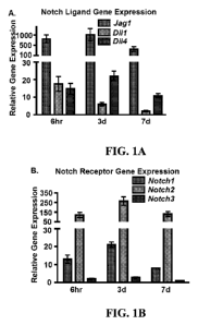
Figure IA is a graph showing real-time (RT)-PCR gene expression levels
expressed as relative gene expression of the Notch ligands, Jagl, D111, and
D114 in
limb-bud MSCs isolated from El 1.5 mouse embryos and cultured for 6 hours, 3
days
or 7 days. Figure lB is a graph showing RT-PCR gene expression levels
expressed as
relative gene expression of the Notch receptors, Notchl-3, in limb-bud MSCs
isolated
from E 11.5 mouse embryos and cultured for 6 hours, 3 days or 7 days. Figure 1
C is a
graph showing RT-PCR gene expression levels expressed as relative gene
expression
of the RBPjK-dependent Notch target genes, Hesl, Heyl, and HeyL, in limb-bud
MSCs isolated from E11.5 mouse embryos and cultured for 6 hours, 3 days or 7
days.
Y-axis of the graphs of Figures lA-1C show relative gene expression normalized
to
(3-actin and represented in arbitrary units. hr, hours; d, days. Figures 1D1-
1D8 are
photomicrographs showing in situ hybridization gene expression analyses in
limb-bud
MSCs from E11.5 mouse embryos for Jagl (Fig. 1D1), Dlll (Fig. 1D2), D114 (Fig.
1D3), Notchl (Fig. 1D4), Notch2 (Fig. 1D5), Notch3 (Fig. 1D6), Hesl (Fig.
1D7),
and Heyl (Fig. 1D8). Figures 1D9 and 1D10 are photomicrographs showing in situ
hybridization gene expression analyses in limb-bud MSCs from E12.0 mouse
embryos for Notch2 (Fig. 1D9) and Hesl (Fig. 1D10). Black boxes outline region
of
vascular canals shown in inset. Insets show high magnification of vascular
canal
containing blood cells and gene expression in surrounding endothelial cells
for N1
and D114. Figure lE is an image of Western blot analyses for active, cleaved
Notch2
protein (NICD2) isolated from limb bud-derived MSCs (LB-MSCs) cultured in the
presence and absence of DAPT or from whole limb-bud (WLB) tissue.
CA 02788579 2012-07-31
WO 2011/097242 PCT/US2011/023369
3
Figures 2A-2C are images and graphs showing DAPT-mediated Notch
inhibition enhances limb-bud MSC differentiation without biasing lineage
determination. Specifically, Figures 2A-2C show staining and molecular
analyses of
limb-bud MSC cultures following continuous treatment with the Notch inhibitor,
DAPT (1 M), or vehicle. Figure 2A shows micrographs of Alcian blue staining of
limb-bud MSC micromass cartilage nodules and graphs of RT-PCR gene expression
levels of the early chondrogenic markers, Sox9, Col2al, and Agcl. Figure 2B
shows
micrographs of alkaline phosphatase staining of limb-bud MSC osteogenic
monolayer
cultures and graphs of RT-PCR gene expression levels of the osteoblast
markers,
Collal, AP, and Oc. Figure 2C shows micrographs of oil Red-O staining of limb-
bud
MSC adipogenic monolayer cultures and a graph of RT-PCR gene expression levels
of the adipocyte marker, Ppary. Y-axis of graphs show relative gene expression
normalized to (3-actin and to the control. (* p<0.05 vs. control). hr, hours;
d, days.
Figures 3A1-3A8 show images and Figure 3B shows graphs indicating a loss
of RBPjK-dependent Notch signaling in vivo accelerates chondrogenesis during
limb
development. Figures 3A1 and 3A2 show Alcian blue staining of wild-type (WT)
and
Prx1Cre/Rbpjxfif(RBPjx) E12.5 hindlimbs. Figures 3A3-3A8 show in situ
hybridization gene expression analyses of the chondrogenic marker genes Sox9
(Figs.
3A3 and 3A4), Col2al (Figs. 3A5 and 3A6), and Agcl (Figs. 3A7 and 3A8).
Figure 3B shows graphs of RT-PCR gene expression levels from whole limb-buds
of
WT and RBPjK mutant E12.5 hindlimbs. Y-axis of graphs show relative gene
expression normalized to (3-actin and to the WT control. (* p<0.05 vs.
control).
Figures 4A 1-4A6 and 4B 1-4B 10 show images and Figure 4C shows graphs
indicating sustained activation of Notch signaling suppresses MSC
differentiation
during skeletal development. Figures 4A1-4A6 show Alcian blue/Alizarin red
staining of wild-type (WT) and Prx I Cre/Rosa-NICD f/+ (NICD) mutant E18.5
whole
skeletons (Figs. 4A1 and 4A2), forelimbs (Figs. 4A3 and 4A4), and hindlimbs
(Figs. 4A5 and 4A6). Black arrows indicate NICD mutant forelimb and hindlimb.
Figures 4B1 and 4B2 show Alcian blue staining of WT and NICD hindlimbs at
E12.5.
Figures 4B3-4B8 show in situ hybridization gene expression levels of the
chondrogenic marker genes Sox9 (Figs. 4B3 and 4B4), Col2al (Figs. 4B5 and
4B6),
CA 02788579 2012-07-31
WO 2011/097242 PCT/US2011/023369
4
and Agcl (Figs. 4B7 and 4B8). Figures 4B9 and 4B10 show Gfp expression
monitored to assess NICD expression and activity in WT (Fig. 4B9) and NICD
mutant (Fig. 4B10) hindlimbs. Figure 4C shows graphs of RT-PCR gene expression
levels from whole limb-buds for the chondrogenic markers, Sox9, Col2a1, Agcl,
and
Runx2 and the RBPJK-dependent Notch target genes, Hesl, Heyl, and HeyL. Y-axis
of graphs show relative gene expression normalized to (3-actin and to the WT
control.
(* p<0.05 vs. control). d, digits; r, radius; u, ulna; h, humerus; s, scapula;
t, tibia; fi,
fibula; fe, femur; il, illium; pu, pubic.
Figures 5A1-5A6 and 5C1-5C2 show images and Figure 5B and 5C3-5C4
show graphs showing sustained activation of Notch signaling in the limb
mesenchyme
does not significantly affect limb patterning or apoptosis, but increases MSC
proliferation during limb development. Figures 5A1-5A6 show in situ
hybridization
analyses of wild-type (WT) (Fig. SAl, 5A3 and 5A5) and PrxlCre/Rosa-NICDV+
mutant (NICD) (Figs. 5A2, 5A4, and 5A6) limb-bud sections at El 1Ø Gene
expression patterns were analyzed for the limb-bud outgrowth and patterning
markers:
Fgf8 (Figs. 5Al and 5A2), Fgfl 0 (Figs. 5A3 and 5A4), and Ptcl (Figs. 5A5 and
5A6).
Figure 5B shows fluorescent TUNEL staining and statistical analyses of MSC
apoptosis performed on WT and NICD mutant sections at El 1Ø BrdU
immunohistochemistry (Figs. 5C1 and 5C2) and statistical analyses of MSC
proliferation (Fig. 5C3) were performed on WT (Fig. 5C1) and NICD mutant
(Fig. 5C2) sections at El 1.5. (* p<0.05 vs. control). AZ, apical zone. Dashed
boxes
denote regions analyzed for MSC proliferation. Figure 5C4 shows RT-PCR levles
of
cyclinDl using RNA derived from NICD mutant and control limb-buds at E11. 5.
Figures 6A 1-6A4 and 6B 1-6B 15 show images indicating Notch signaling
suppresses MSC differentiation in an RBPJK-dependent manner. Figures 6A1-6A4
show Alcian blue/Alizarin red staining of wild-type (WT); PrxlCre/Rosa-NICDV+
(NICD); PrxlCre/Rbpjxfif(RBPjx); andPrx1Cref/Rosa-NICDf/+/Rbpjxf/f (NICD;
RBPjK) mutant E18.5 whole skeletons. Black arrows indicate NICD mutant
forelimb
and hindlimb. Gray arrows mark the length of WT, RBPjK, and NICD; RBPjK
tibiae.
Asterisks identify location of the parietal bones. Figures 6B1-6B3 show Alcian
blue
staining of WT, NICD, and NICD; RBPjK littermate hindlimb sections at E12.5
(Bl-
CA 02788579 2012-07-31
WO 2011/097242 PCT/US2011/023369
B3). Figures 6B4-6B12 show in situ hybridization gene expression analyses of
the
chondrogenic marker genes Sox9 (Figs. 6B4-6B6), Col2al (Figs. 6B7-6B9), and
Agcl (Figs. 6B10-6B12). Figures 6B13-6B15 show Gfp expression monitored to
assess NICD expression and activity in WT (Fig. 6B13), NICD mutant (Fig.
6B14),
5 and NICD; RBPjK rescue (Fig. 6B15) hindlimb sections.
Figures 7A1-7A6 show images and Figure 7B shows graphs indicating Hesl is
a critical RBPjK-dependent Notch target gene regulating MSC differentiation
and
chondrogenesis. Figures 7A1-7A6 show Alcian blue staining of control infected
(Figs.
7A1, 7A3, and 7A5,) and Hesl shRNA infected (shHesl) (Figs. 7A2, 7A4, and 7A6)
limb-bud MSC cells cultured in micromass for 3, 5, or 7-days. Figure 7B shows
RT-
PCR gene expression levels for the chondrogenic markers Sox9, Col2al, Agcl
during
in vitro chondrogenesis following knock-down of Hest. Y-axis of graphs show
relative gene expression normalized to (3-actin and to the control at day 3.
(* p<0.05
vs. control). d, days.
Figure 8 is a graph showing apoptotic cell counts in E 11.5 sections from WT
and NICD mutant limb mesenchyme. Using activated caspace-3
immunohistochemistry, the data show sustained activation of Notch signaling in
the
limb mesenchyme does not affect MSC apoptosis.
Figures 9A1-9A6 and 9B1-9B6 show images and Figures 9C and 9D show
graphs indicating Hesl is a critical regulator of MSC differentiation in a
C3H10T1/2
model of chondrogenesis. Figures 9A1-9A6 and 9B1-9B6 show Alcian blue staining
of control infected (Figs. 9A1, 9A3, and 9A5,), Hesl shRNA infected (shHesl)
(Figs. 9A2, 9A4, and 9A6), control transfected (Figs. 9B1, 9B3, and 9B5), and
Hesl
transfected (CMV-Hesl) (Figs. 9B2, 9B4, and 9B6) C3H10T1/2 cells cultured in
micromass for 5, 10, or 14-days. Figures 9C and 9D show RT-PCR gene expression
levels for the chondrogenic markers Sox9, Col22al, Agcl and the Notch target
gene,
Hesl during in vitro chondrogenesis following knock-down of Hesl (Fig. 9C) or
over-expression of Hesl (Fig. 9D). Y-axis of graphs show relative gene
expression
normalized to (3-actin and to the control at day 5. (* p<0.05 vs. control). d,
days.
CA 02788579 2012-07-31
WO 2011/097242 PCT/US2011/023369
6
Figures 1 OA and I OB are graphs showing Notch molecules expressed in
hMSCs. Gene expression is normalized to beta-actin and represented in
arbitrary
units
Figures 11A-11C are graphs showing recombinant JaggedI induction of
multipotent stem cell markers and hMSC proliferation. Figure 1 IA shows gene
expression levels for Notch components and regulators of stem cell
multipotency in
hMSCs at passage 1 (P1) and passage 15 (P 15). Figure 1lB shows gene
expression
levels for Notch target genes and regulators of stem cell multipotency in
hMSCs
cultured on control IgG or Jagl coated plates. All gene expression is
normalized to
beta-actin and then normalized to P1 controls (Fig. 1 IA) or IgG controls
(Fig. 11B).
Figure 11 C shows BrdU ELISA assay measuring proliferation of hMSCs cultured
on
IgG control or Jagl coated plates.
Figure 12A and B show flow cytometry data for hMSC cell surface marker,
CD 105 (A), and the Notch receptor, Notch2 (B), following passages 2 and 10 in
standard hMSC culture conditions.
Figure 13A-C show that Jag 1-mediated Notch activation in Notch2-selected
hMSCs induces stem cell regulators, cell proliferation, and stem cell
expansion.
Figure 13A shows real-time RT-PCR gene expression analyses for Notch signaling
molecules (Notch2 and Hesl ), important stem cell regulatory molecules (Oct4,
Sox2,
and Nanog), and a marker of cell proliferation (CycDl) in total hMSCs and
Notch2-
selected hMSCs cultured on Jagl coated plates. Figure 13B shows a BrdU ELISA
assay performed on total, Notch2-negative, and Notch2-positive hMSCs cultured
on
Jagl coated plates. Figure 13C shows a CFU-F assay performed on total, Notch2-
negative, and Notch2-positive hMSCs following culture on Jagl coated plates.
Figures 14A-D show Notch2-selected hMSCs display enhanced chondrogenic
and osteogenic properties following Jag 1-mediated maintenance and expansion.
Figures 14A and C show real-time RT-PCR gene expression analyses for
chondrogenic (Sox9, Col2al, and Agcl) (A) and osteogenic (Collal, Ap, and Oc)
(C)
marker genes from total, Notch2-negative, and Notch2-positive hMSCs after
being
cultured in chondrogenic or osteogenic conditions for two to three weeks.
Figure 14B
shows Alcian Blue staining of total, Notch2-negative and positive hMSCs
(Passage 2)
following chondrogenic differentiation. Figure 14D shows AP staining of Notch2-
CA 02788579 2012-07-31
WO 2011/097242 PCT/US2011/023369
7
negative and positive hMSCs (Passage 2 and 5) following osteogenic
differentiation.
hMSCs were initially cultured on Jagl coated plates for two passages (3-4
days/passage).
DETAILED DESCRIPTION
To determine the exact role and mode of action for the Notch pathway in
mesenchymal stem cells (MSCs), tissue specific loss-of-function (PrxlCre;
Rbpjxfr),
gain-of-function (PrxlCre; Rosa-NICDV+) and genetic rescue mice (PrxlCre; Rosa-
NICDV+; Rbpjxf/) were generated and analyzed for defects in MSC proliferation
and
differentiation during early limb development. The results are presented in
Example 1
below. These data show that Hes 1 is the primary RBPjK-dependent Notch target
gene
of the Hes/Hey family expressed in MSCs and required for the Notch mediated
suppression of MSC differentiation during chondrogenesis. Further, these data
demonstrate that the RBPjK-dependent Notch signaling pathway is critical for
the
maintenance and expansion of MSCs during skeletal development. Thus,
manipulation of the Notch pathway provides a means to maintain, expand, and
regulate the differentiation of MSCs for the purpose skeletal repair and
tissue
engineering applications that utilize MSC populations. Controlled Notch
activation of
hMSCs promotes the maintenance and expansion of hMSCs, while preserving their
chondrogenic, osteogenic, and adipogenic differentiation potential.
Accordingly,
disclosed herein are relatively pure populations of MSCs and methods of
isolating and
culturing MSCs.
Provided is a relatively pure population of MSCs expressing the Notch 2
receptor (Notch 2+ MSCs). As used herein, the term relatively pure means that
at
least 90, 91, 92, 93, 94, 95, 96, 97, 98, 99 or 100% of the MSCs in the
population
express Notch 2. Optionally, the Notch 2+ MSCs maintain the capacity to expand
through multiple passages. Optionally, the Notch 2+ MSCs express one or more
additional markers associated with mesenchymal stem cells selected from the
group
consisting of CD105, CD106, CD156, CD44, CD29, CD166, Stro-1, FGF10, Prxl,
Oct4, Sox2, and Nanog. Optionally, the Notch 2+ MSCs express CD105 and CD156.
Optionally, the Notch 2+ MSCs do not express one or more markers associated
with
CA 02788579 2012-07-31
WO 2011/097242 PCT/US2011/023369
8
hematopoietic or endothelial cell lineage selected from the group consisting
of CD34,
CD45, CD14, and CD31.
The relatively pure population of Notch 2+ MSCs is stable in non-
differentiating culture conditions. As used herein, non-differentiating
culture
conditions include, but are not limited to, culture conditions that promote
proliferation
without promoting differentiation. For example, the cells can be maintained in
medium, e.g. DMEM, RPMI, and the like, in the presence of fetal bovine serum
or
serum-free replacement without differentiation.
Specifically, provided is a method of isolating from a subject MSCs. The
method includes the steps of obtaining a biological sample comprising MSCs
from the
subject and selecting for MSCs expressing a Notch 2 receptor from the
biological
sample to obtain a population of Notch 2+ MSCs. Also provided is a relatively
pure
population of Notch 2+ MSCs made by the provided methods. The MSCs maintain
the capacity to expand through multiple passages. The MCSs can be passaged at
least
about 5, 10, 15 or 20 times or any number of times between 5 to 20.
Optionally, the
MSCs can be passaged 10 or more times. For example, the MSCs can be passaged
10, 11, 12, 13, 14, 15, 16, 17, 18, 19 or 20 times.
As used herein, the terms passaged or passaging refers to the process of sub-
culturing cells. The methods and materials for culturing and passaging cells
are
known. For example, cells are grown on a substrate, e.g., in a dish or plate,
with
media in an incubator. During passaging, the growth media is removed, and the
cells
may be washed, followed by the addition of an agent to detach the cells from
the
substrate. The detached cells are suspended and an appropriate number of cells
in
suspension is then transferred to new substrates, fresh medium is added, the
new
substrates are put in the incubator, and the cycle begins again. Cells are
often kept
less than 100% (log phase of growth) but more than 10% confluent. Cells may
die if
they are too few or much too crowded.
The selection step is carried out using any one of a variety of methods
including, but not limited to, flow cytometry, magnetic bead separation,
panning,
fluorescence activated cell sorting (FACS) or affinity chromatography. For
example,
flow cytometry, or FACS, can be used to separate cell populations based on the
CA 02788579 2012-07-31
WO 2011/097242 PCT/US2011/023369
9
intensity of fluorescence, as well as other parameters such as cell size and
light
scatter.
The selection step is, optionally, carried out using a Notch 2 receptor
antibody
or other Notch 2 receptor ligand. Optionally, the antibody or ligand is bound
to a
substrate, which can be, for example, a mobile or immobile solid support.
Optionally,
the mobile solid support is a fluorescent bead. Optionally, the immobile solid
support
is a column or a plate. The sample is contacted with the substrate and, either
the
substrate with the Notch 2+ cells is sorted from substrate lacking the Notch
2+ cells,
or the bound MSCs in the sample are isolated from the substrate, e.g. with a
competitive binding step. Fluorescent labels or other labeling means can be
used to
sort the MSCs. With sorting techniques like FACS, the various populations of
MSCs
can be sorted to have the specifically desired expression profiles.
The sample from the subject is selected from an MSC-containing sample, e.g.,
from the group consisting of bone marrow, adipose tissue, synovium,
periosteum,
perichondrium, cartilage, dental tissue, placental tissue, liver tissue,
muscle tissue,
lung tissue, heart tissue, connective tissue, and spleen tissue.
The isolated Notch 2+ MSCs are collected, for example, in any appropriate
medium that maintains the viability of the cells. Optionally, the medium is
located in
a collection vessel, such as a tube. Various media are commercially available
and
may be used, including, but not limited to, Dulbecco's Modified Eagle Medium
(DMEM), Hanks' Buffered Salt Solution (HBSS), Dulbecco's Phosphate Buffered
Saline (dPBS), Roswell Park Memorial Institute (RPMI) medium, Iscove's medium,
and the like, optionally, supplemented with fetal calf serum.
Also provided is a method of culturing the population of Notch 2+ MSCs
including the step of culturing the MSCs in the presence of an activator of
the Notch
signaling pathway. Optionally, the culture conditions are such that the
population of
Notch 2+ MSCs is expanded. Various media are commercially available and may be
used to culture MSCs, including, but not limited to, DMEM, HBSS, dPBS, RPMI
medium, Iscove's medium, and the like, optionally, supplemented with fetal
calf
serum.
Optionally, the activator of the Notch signaling pathway is selected from the
group consisting of delta-like 1, delta-like 3, delta-like 4, Jaggedl, Jagged
2,
CA 02788579 2012-07-31
WO 2011/097242 PCT/US2011/023369
Dlkl/Prefl, DNER, Contactinl (F3), Contactin6 (NB3), CCN3/NOV, MAGP1, and
MAGP2. Optionally, the activator of the Notch signaling pathway is an
intracellular
domain of a Notch receptor. Optionally, the Notch receptor is Notch 1, Notch
2,
Notch 3, or Notch 4. The activator of the Notch signaling pathway can be
partially or
5 completely immobilized on a culture dish. Alternatively, the activator can
be soluble
in the culture medium.
Notch activation can be induced by a ligand, which causes cleavage and
release of the Notch intracellular domain (ICD). The NICD translocates to the
nucleus, interacts with RBPjk, and activates target genes. Notch signaling in
MSCs
10 can also be activated by directly expressing a Notch ICD. Notch ICD
expression can
be provided using any means for expressing a peptide in a cell, for example,
using an
expression vector (e.g., a viral vector). Expression of the Notch ICD can be
transient
or stable.
The culturing method can also include the step of culturing the population of
Notch 2+ MSCs in the presence of one or more differentiating agents. Notch
activation is "turned off' to allow the cell to differentiate. Optionally, the
one or more
differentiating agents selectively induce differentiation into chondrogenic,
osteogenic
or adipogenic lineages. Culturing the Notch 2+ MSCs under differentiating
culture
conditions is carried out by culturing or differentiating MSC in a growth
environment
that enriches for selected cells with the desired phenotype, e.g. osteoblasts,
adipocytes, chondrocytes, or the like. Thus, the culture medium may include
agents
that enhance differentiation to a specific lineage. For example, osteogenic
differentiation may be enhanced by culturing MSCs in medium comprising 9-
glycerol
phosphate, ascorbic acid and retinoic acid (Cowan et al. (2005) Tissue
Engineering
11:645-658). Adipogenic differentiation may be enhanced, for example, by
culturing
the MSCs in a medium comprising dexamethasone, indomethacin, 3-isobutyl-l-
methylxanthine (IBMX), and insulin, then maintaining in growth media with
insulin.
Myocyte differentiation may be enhanced, for example, by culturing in a medium
comprising 5-azacytidine (Fukuda et al. (2001) Artificial Organs 25:187), or
in a
medium comprising horse serum, dexamethasone, and hydrocortisone (Eun et al.
(2004) Stem Cells 22:617-624). Chondrocyte differentiation may be enhanced,
for
CA 02788579 2012-07-31
WO 2011/097242 PCT/US2011/023369
11
example, by culturing in a medium comprising dexamethasone, ascorbic acid 2-
phosphate, insulin, transferrin, and selenous acid, with or without TGF-,91
(Williams
et al. (2003) Tissue Engineering 9(4):679). Following differentiation in
culture, the
cells obtained may be used directly, or may be further isolated, e.g. in a
negative
selection to remove MSCs and other undifferentiated cells. Further, enrichment
for
the desired cell type may be obtained by selection for markers characteristic
of the
cells, e.g. by flow cytometry, magnetic bead separation, panning, and the
like, as is
known.
Provided is a method of treating a subject with a disorder associated with a
deficiency or defect in cells of mesenchymal lineage comprising administering
a
population of Notch 2+ MSCs to the subject. The population of Notch2+ MSCs are
derived from the same or a different subject.
The Notch 2+ MSCs are administered to the subject as appropriate. For
example, the Notch 2+ MSCs are injected into the subject at or near the site
of the
bone or cartilage defect or administered to the subject systemically. The
Notch 2+
MSCs are administered in a manner that permits them to graft or migrate to the
intended tissue site and reconstitute or regenerate the functionally deficient
area.
Optionally, targeting molecules on the surface of the MSCs are used to promote
proper migration to the desired site. MSCs are used, for example, for
engineering
cartilage, growth plate, bone and tendon/ligament as well as autologous
chondrocyte
implantation. Thus, administration of MSCs can be performed by administering
the
cells via a relatively pure population or in a construct generated using
tissue
engineering.
Administration of the Notch 2+ MSCs can promote bone formation following
bone surgery, wherein the bone surgery is selected from the group consisting
of facial
reconstruction, maxillary or mandibular reconstruction, fracture repair, bone
graft,
prosthesis implant, joint replacement (e.g., hip and knee replacement).
Optionally, the Notch 2+ MSCs are differentiated (as described above) and
delivered to an affected area of a subject. For example, osteogenic lineages
can be
delivered to a subject with a bone disease or defect.
CA 02788579 2012-07-31
WO 2011/097242 PCT/US2011/023369
12
Bone disorder or defect, as used herein, refers to any bone defect, disease or
state which results in or is characterized by loss of health or integrity to
bone and
includes, but is not limited to, osteoporosis, osteopenia, faulty bone
formation or
resorption, Paget's disease, fractures and broken bones, bone metastasis,
osteopetrosis,
osteosclerosis and osteochondrosis. Bone defects and disorders include
fractures and
inherited or acquired disease states like osteogenesis imperfecta or
osteoporosis.
Bone diseases or defects that can be treated and/or prevented in accordance
with
methods described herein, include bone diseases characterized by a decreased
bone
mass relative to that of corresponding non-diseased bone (e.g., osteoporosis,
osteopenia and Paget's disease). Cartilage defects include an articular
cartilage defect
or vertebral disc defect, which can be caused by trauma or diseases such as
osteoarthritis or rheumatoid arthritis.
Treatment of a bone or cartilage defect or disorder or a symptom related to a
bone or cartilage defect or disorder encompasses actively intervening after
onset to
slow down, ameliorate symptoms of, or reverse the disease or symptoms.
Treating, as
used herein, refers to a method that modulates bone or cartilage mass or
integrity to
more closely resemble that of corresponding non-affected bone (that is a
corresponding bone of the same type, e.g., long and vertebral) or cartilage in
a non-
diseased or non-affected state. By way of example, following treatment post
surgery,
the bone or cartilage would resemble healthy, non-surgically affected bone.
The Notch 2+ MSCs can be administered in the form of a pharmaceutical
composition. Such a composition comprises a therapeutically effective amount
of the
MSCs and a pharmaceutically acceptable carrier or excipient. Such a carrier
includes
but is not limited to, saline, buffered saline, dextrose, water, and
combinations thereof.
The formulation should suit the mode of administration. Optionally, the MSC
composition is formulated for intravenous, intra-articular, or intervertebral
administration. Compositions for intravenous administration are, for example,
solutions in sterile isotonic aqueous buffer.
A composition including the Notch 2+ MSCs for use in the methods described
herein can also be formulated as a sustained and/or timed release formulation.
Such
sustained and/or timed release formulations may be made by sustained release
means,
delivery devices or tissue-engineered constructs. The compositions can be used
to
CA 02788579 2012-07-31
WO 2011/097242 PCT/US2011/023369
13
provide slow or sustained release of one or more of the active ingredients
using, for
example, hydropropylmethyl cellulose, other polymer matrices, gels, permeable
membranes, osmotic systems, multilayer coatings, microparticles, liposomes,
microspheres or a combination thereof to provide the desired release profile
in
varying proportions. Various suitable sustained release formulations may be
readily
selected for use with the compositions described herein. Optionally, the
compositions
can be delivered by a controlled-release system. For example, the composition
can be
administered using intravenous infusion, an implantable osmotic pump,
liposomes, or
other modes of administration. A controlled release system can be placed in
proximity of the target. For example, a micropump can deliver controlled doses
directly into a joint or directly into bone or cartilage, thereby requiring
only a fraction
of the systemic dose (see e.g., Goodson, 1984, in Medical Applications of
Controlled
Release, vol. 2, pp. 115-138, which is incorporated by reference in its
entirety at least
for the material related to micropumps). In another example, the composition
can be
formulated with a hydrogel (see, e.g., U.S. Pat. Nos. 5,702,717; 6,117,949;
6,201,072,
which are incorporated by reference in their entireties at least for the
material related
to hydrogels).
It may be desirable to administer the composition locally, i.e., to the area
in
need of treatment. Local administration can be achieved, for example, by local
infusion during surgery, topical application, injection, or implant. An
implant can be
of a porous, non-porous, or gelatinous material, including membranes, such as
sialastic membranes, or fibers and include tissue engineered constructs
designed to
replace tissues like bone or cartilage.
The Notch 2+ MSCs are used in an effective amount. In general, such amount
ranges from at least 1X10⁴ MSC per kg of body weight to 3X10⁶ MSCs/kg of body
weight. Optionally, the MSCs are administered at 1X10⁶ MSCs/kg of body weight.
The MSCs are administered, for example, one to three times per day, and may be
adjusted to meet optimal efficacy and pharmacological dosing. One of skill in
the art
can determine dosage amounts and frequency based on the route of
administration;
age, sex, health and weight of the recipient; nature and extent of symptoms;
kind of
concurrent treatment, frequency of treatment and the effect desired.
CA 02788579 2012-07-31
WO 2011/097242 PCT/US2011/023369
14
Also provided herein is a pack or kit comprising one or more containers filled
with one or more of the ingredients (e.g., an activator of the Notch signaling
pathway
or Notch 2+ MSCs) described herein. Thus, for example, a kit described herein
comprises a population of Notch 2+ MSCs. Also described is a kit with
compositions
for isolating Notch 2+ MSCs. Optionally, the kit further includes agents for
culturing
the Notch 2+ MSCs. Such kits optionally comprise solutions and buffers as
needed or
desired. Optionally associated with such pack(s) or kit(s) are instructions
for use.
As used throughout, by a subject is meant an individual. Thus, the subject can
include, for example, domesticated animals, such as cats and dogs, livestock
(e.g.,
cattle, horses, pigs, sheep, and goats), laboratory animals (e.g., mice,
rabbits, rats, and
guinea pigs) mammals, non-human mammals, primates, non-human primates,
rodents, birds, reptiles, amphibians, fish, and any other animal. The subject
can be a
mammal such as a primate or a human.
Disclosed are materials, compositions, and components that can be used for,
can be used in conjunction with, can be used in preparation for, or are
products of the
disclosed methods and compositions. These and other materials are disclosed
herein,
and it is understood that when combinations, subsets, interactions, groups,
etc. of
these materials are disclosed that while specific reference of each various
individual
and collective combinations and permutation may not be explicitly disclosed,
each is
specifically contemplated and described herein. For example, if a method is
disclosed
and discussed and a number of modifications that can be made to the method are
discussed, each and every combination and permutation of the method, and the
modifications that are possible, are specifically contemplated unless
specifically
indicated to the contrary. Likewise, any subset or combination of these is
also
specifically contemplated and disclosed. This concept applies to all aspects
of this
disclosure including, but not limited to, the MSCs themselves and steps in the
methods of isolating, culturing and using the disclosed MSCs. Thus, if there
are a
variety of additional steps that can be performed it is understood that each
of these
additional steps can be performed with any specific method steps or
combination of
method steps of the disclosed methods, and that each such combination or
subset of
combinations is specifically contemplated and should be considered disclosed.
CA 02788579 2012-07-31
WO 2011/097242 PCT/US2011/023369
Throughout this application, various publications are referenced. The
disclosures of these publications in their entireties are hereby incorporated
by
reference into this application.
A number of aspects have been described. Nevertheless, it will be understood
5 that various modifications may be made. Furthermore, when one characteristic
or step
is described it can be combined with any other characteristic or step herein
even if the
combination is not explicitly stated. Accordingly, other aspects are within
the scope
of the claims.
Examples
10 Example 1. RBPjk-dependent Notch signaling maintains and expands
mesenchymal stem cells (MSCs) during skeletal development.
Materials and Methods
Mouse strains. All mouse strains including Rosa-NICD, RbpjK, and PrxlCre
are as previously described (Han et al., Int. Immunol. 14:637-45 (2002); Logan
et al.,
15 Genesis 33:77-80 (2002); and Murtaugh et al., PNAS 100:14290-5 (2003)).
PrxlCre
mice were obtained from the Jackson Laboratory (Bar Harbor, ME).
Analyses of mouse embryos. Embryonic tissues were harvested at El 1.0-
E12.5 in PBS, fixed in 10% neutral buffered formalin overnight at room
temperature,
then processed and embedded in paraffin prior to sectioning at 4 m. Standard
Alcian
blue/orange g staining was performed in order to analyze tissue architecture
and
cartilage composition of the limb-buds. In situ hybridization was performed as
described previously (Hilton et al., Development 132:4339-51 (2005); Hilton et
al.,
Dev. Biol. 308:93-105 (2007); and Hilton et al., Nat. Med. 14:306-14 (2008)),
using
35S-labeled riboprobes. Unpublished riboprobes were generated from the
following
cDNA clones: Sox9 (4165469), Agcl (5345931), Hesl (10469606), Heyl (9792713),
Jagl (10699187), Dlll (10698888), and D114 (7492828). The cDNA clones are
available from Open Biosystems (Huntsville, AL) or ATCC (Manassas, VA). The
Gfp probe was generated by cloning the enhanced Gfp coding sequence into the
pGEM-T Easy vector. Notchl, Notch2, Notch3, Fgf8, and FgflO cDNAs and
riboprobes are as described (Bellusci et al., Development 124:4867-78 (1997);
CA 02788579 2012-07-31
WO 2011/097242 PCT/US2011/023369
16
Crossley and Martin, Development 121:439-51 (1995); and Mitsiadis et al., J.
Cell
Biol. 130:407-18 (1995)). For BrdU immunostaining analyses, pregnant females
were
injected with BrdU at 0.1 mg/g body weight 2 hours prior to harvest. BrdU
detection
was performed on paraffin sections using a kit from Zymed Laboratories (San
Francisco, CA) as per manufacturer's instructions. Proliferation studies were
confirmed using anti-Ki67 immunostaining (DAKO; Denmark) of mouse limb-bud
paraffin sections according to manufacture's instructions. Analyses of
apoptotic
MSCs were performed using both anti-Cleaved Caspase-3 immunostaining (Cell
Signaling; Danvers, MA) and TUNEL staining (Roche Cell Death In situ Kit;
Roche;
Basel, Switzerland) on limb-bud sections according to the manufacturers'
instructions. Whole-mount skeletal staining of embryos was performed as
previously
described (Hilton et al., Development 132:4339-51 (2005); McLeod, Teratology
22:299-301 (1980)).
Limb-bud MSC and C3HJ0TJ/2 cell culture. Limb-bud derived MSCs were
isolated from E1 1.5 CD1 mouse embryos as previously described (Zhang et al.,
Bone
34:809-17 (2004)). For chondrogenic differentiation, MSCs were seeded in
micromass (1 x 10⁵ cells in 10 Tl) in 12-well plates for 1.5 hours before
adding
standard media, media containing DAPT (1 M), or media containing Hes 1 shRNA
lentivirus. Cells were cultured for a time-course of 6 hours, 3, 5, and 7 days
prior to
harvest for cartilage staining (1% Alcian blue/3% glacial acetic acid) or
total RNA
isolations. Limb-bud derived MSCs were also cultured in monolayer for 21 days
and
treated with either osteogenic (10 nM dexamethasone; 50 M ascorbic acid; 10
mM
(3-glycerolphosphate) or adipogenic medium (Millipore; Billerica, MA) in the
presence and absence of DAPT. Fixed MSCs were stained for osteoblastic
differentiation using an alkaline phosphatase stain (nitro blue tetrazolium
chloride/5-
bromo-4-chloro-3-indolyhosphate P-toluidine salt) or adipogenic
differentiation using
an Oil Red-O staining solution (0.36%). Total RNA was isolated from monolayer
cultures at day 21 for use in real-time RT-PCR analyses.
C3H10T1/2 cells were expanded and plated in monolayer for experiments as
previously described (Denker et al., Differentiation 64:67-76 (1999); Haas and
Tuan,
Differentiation 64:77-89 (1999)). Monolayers were either transfected with
500ng of
CA 02788579 2012-07-31
WO 2011/097242 PCT/US2011/023369
17
CMV-Hesl or CMV- control plasmid using the Lipofectamine 2000 reagent
(Invitrogen; Carlsbad, CA) as suggested by the manufacturer's protocol, or
infected
with control virus or shRNA lentivirus against Hesl, Heyl, and HeyL (Sigma;
St.
Louis, MO). After 1 day of transfection/infection, cells were trypsonized and
replated
in micromass at a density of 1x10⁵ cells/l0 Tl medium in each well of 12 well
plates.
Cells were harvested at days 5, 10, and 14 for Alcian blue staining and total
RNA
isolation.
Real-time RT-PCR. Embryonic limb-bud tissues or micromass cultures were
frozen in liquid nitrogen and then homogenized in Trizol Reagent (Invitrogen;
Carlsbad, CA) via rendering through a 25-gauge needle and syringe. Total
cellular
RNA was extracted following the manufacture's protocol. RNA was quantified
using
a NanoDrop spectrophotometer (NanoDrop; Wilmington, DE) and equal
concentrations of total RNA were pooled for synthesis of cDNA. Total RNA (1
g)
was reverse transcribed using the iScriptTM cDNA synthesis kit (Bio-Rad;
Hercules,
CA) according to the manufacture's instructions. Reverse transcribed cDNA was
analyzed by real-time RT-PCR with mouse-specific primers for: Sox9, Runx2,
Col2al, Agcl, Collal, Ap, Oc, Ppary, Jaggedl, Jagged2, Delta-likel, Delta-
like3,
Delta-like4, Notchl, Notch2, Notch3, Notch4, Hesl, Hes3, HesS, Hes7, Heyl,
Hey2,
HeyL, and CyclinD 1. Primers were designed using Applied Biosystems software
(Applied Biosystems; Foster City, CA). Sequences are available upon request.
DNA
amplification was achieved using the SYBR Green PCR Master Mix (Applied
Biosystems; Foster City, CA) and the RotorGene real-time DNA amplification
system
(Corbett Research; Sydney, Australia). Gene expression was normalized to (3-
actin
expression levels and then normalized to control samples.
Western blot analyses. Total protein was isolated from either whole mouse
limb-bud tissue or cultured limb-bud derived MSCs using Golden lysis buffer.
The
cultured limb-bud derived MSCs were plated at the density of 6X10⁶ cells in 10
cm
dishes and cultured overnight in 10% FBS DMEM media both in the presence and
absence of DAPT (lum). Protein samples (-100 g) from each isolation were
subsequently separated on 10% SDS-polyacrylamide and transferred to a PVDF
membrane. NICD 1 and NICD2 cleaved proteins were detected using the bTAN 20
CA 02788579 2012-07-31
WO 2011/097242 PCT/US2011/023369
18
(Notchl) and C651.6DdHN (Notch2) primary antibodies (0.4ug/ml) and then
further
probed with appropriate secondary antibody (1:3000). Anti-(3-actin antibody
(Sigma;
St. Louis, MO) was used as a control for equal protein loading. Immunoblots
were
detected using Supersignal west femto maximum sensitivity substrate (Pierce;
Rockford, IL).
Results
Expression of Notch pathway components during MSC differentiation in vitro
and in vivo. Real-time (RT) PCR was performed to identify the exact temporal
expression of the five (5) murine Notch ligands (Jagged 1 (Jag 1), Jagged 2
(Jag2),
Delta-like 1 (Dlll), Delta-like 3 (D113), and Delta-like 4 (D114)), the four
(4) Notch
receptors (Notch 1 (Ni), Notch 2 (N2), Notch 3 (N3), Notch 4 (N4)), and the
six (6)
canonical Notch target genes (Hest, HesS, Hes7, Heyl, Hey2, and HeyL) during
limb-bud MSC differentiation and in vitro chondrogenesis. Limb-bud MSCs were
isolated from E11.5 mouse embryos and cultured for 6 hours, 3 days, and 7 days
in
micromass. Of the five (5) possible Notch ligands, only Jagl, Dlll, and D114
were
detected at significant levels, with Jag 1 showing the highest level of
expression at all
time-points (Fig. IA). Only three (3) of the four (4) Notch receptors (Ni, N2,
and
N3) were detected during limb-bud MSC differentiation, with Notch2 displaying
dramatically higher levels of expression at each time-point as compared to the
other
Notch receptors (Fig. 1B). To determine the downstream components of the Notch
signaling pathway important during limb-bud MSC differentiation and
chondrogenesis, the expression of RBPjK-dependent Notch target genes was
examined. Of the six (6) possible targets, only Hesl, Heyl, and HeyL were
identified. Heyl and HeyL were the most abundant Notch target genes showing
similar levels of expression at each time-point that increased during MSC
differentiation in vitro (Fig. 1C). While Hesl displayed a lower level of
expression as
compared to Heyl and/or HeyL, Hesl expression was most pronounced in early
limb-
bud MSCs with declining expression levels during MSC differentiation,
indicating a
potential role in regulating the earliest stages of MSC commitment to the
chondrocyte
lineage (Fig. 1C).
CA 02788579 2012-07-31
WO 2011/097242 PCT/US2011/023369
19
In situ hybridization analyses was performed on E11.5 and E12.0 limb-bud
sections to identify the exact in vivo spatial expression pattern for the
Notch signaling
molecules identified in the RT-PCR analyses. These data demonstrated that
Notch
ligands Jagl, Dll1, and D114 all had very different expression profiles. At El
1.5, Jagl
was expressed moderately throughout much of the limb-bud mesenchyme but was
highly expressed in a concentrated region of the distal, medial mesenchyme
adjacent
to the apical zone (Fig. 1D1). Of the other two Notch ligands, Dlll was
sporadically
expressed throughout the limb-bud mesenchyme (Fig 1D2), while D114
demonstrated
a more concentrated expression pattern around vascular structures (Fig. 1D3,
high
magnification insert) at E11.5. D114 is a regulator of angiogenesis, which,
along with
Notchl, is a critical regulator of the vascular endothelium (Hellstrom et al.,
Nature
445:776-80 (2007); Shutter et al., Genes Dev. 14:1313-8 (2000)). The Notch
receptor, Notchl, was also primarily expressed in regions of vascular tissues
(Fig.
1D4, high magnification insert) and the early ectoderm at E11.5, with lower
levels of
expression observed throughout some of the limb-bud mesenchyme. Notch2 was
expressed more ubiquitously throughout most of the limb-bud MSCs at the same
stage
(Fig. 1D5). Notch3 was expressed sporadically in the limb-bud mesenchyme, with
higher concentrations in the proximal and peripheral MSCs. The Notch target
genes,
Hest and Hey 1, each had expression patterns similar to that of Notch2 at E11.
5 (Figs.
1D5, 1D7, and 1D8), although a slight elevation of Hesl expression could be
observed in the distal, medial MSCs overlapping regions where Jagl expression
is
concentrated (Figs. 1D1 and 1D7). By E12.0-E12.5, most of the Notch pathway
components are difficult to detect via in situ hybridization. Only Notch2 and
Hes 1
expression were maintained in limb-bud MSCs surrounding chondrogenic
condensations, but showed significant down-regulation within the condensations
themselves (Figs. 1 D9 and 1 D 10, black and white contours), while components
like
Heyl maintained a more ubiquitous expression pattern.
To determine which Notch receptor is active in the limb-bud mesenchyme,
total protein was isolated from cultured MSCs in the presence and absence of
the
Notch inhibitor, N-(3,5-difluorophenylacetyl-L-alanyl)]-S-phenylglycine t-
ButylEster
(DAPT, Calbiochem; San Diego, CA), or directly from wild-type E11.5 whole limb-
bud tissue, and performed western blot analyses using Notchl and Notch2
antibodies
CA 02788579 2012-07-31
WO 2011/097242 PCT/US2011/023369
that can detect the cleaved or active (NICD) form of the receptor. Western
blot
analyses revealed that Notch2 was the prominent receptor activated in E11. 5
limb-bud
MSCs, and that DAPT treatment of cultured MSCs can reduce the abundance of the
cleaved Notch2 (NICD2) (Fig. IE). Notchl (NICD1) was nearly undetectable at
total
5 protein concentrations up to 100 g. Therefore, taken together these data
suggest that
Notch2 is the primary Notch receptor activated in MSCs, while other components
of
the Notch pathway (Jag1, Dlll, N3, Hes1, Heyl, and HeyL) may also be important
mediators of MSC proliferation and differentiation during limb development.
Notch signaling is a general regulator of MSC differentiation. To determine
10 the role of Notch signaling in MSCs, Notch loss-of-function assays were
performed
on El 1.5 limb-bud derived MSC cultures using the Notch inhibitor, DAPT.
Chondrogenesis was first examined in limb-bud micromass cultures by measuring
cartilage nodule formation in the presence and absence of 1 M DAPT. DAPT
treatment significantly enhanced cartilage nodule formation (Fig. 2A), showing
that
15 Notch inhibition accelerates commitment of MSCs to the chondrocyte lineage,
a
finding that is consistent with a prior study (Fujimaki et al., J. Bone Miner.
Metab.
24:191-8 (2006)). The effect of DAPT was also assessed on the expression of
the
chondrogenic markers Sox9, Col2al, and Agcl via real-time RT-PCR. Compared to
untreated cultures, DAPT enhanced Sox9, Col2al, and Agcl expression (Fig. 2A)
20 within the first 3-5 days of culture, although Agcl expression was
significantly
reduced by day 7 indicating that Notch plays a later role in chondrocyte
maturation or
maintenance of the committed chondrocyte phenotype.
To determine whether Notch specifically regulates chondrogenesis or
generally controls MSC differentiation, limb-bud MSC differentiation assays
were
performed in both osteogenic and adipogenic conditions. Limb-bud MSCs were
plated in monolayer and cultured the cells for 21 days in osteogenic media in
the
absence and presence of DAPT (1 M) (Fig. 2B). DAPT treatment enhanced normal
osteoblastic differentiation of MSCs. Cultures displayed elevated alkaline
phosphatase staining and real-time RT-PCR analyses demonstrated a significant
increase in the expression of osteoblast marker genes: Collal, AP, and Oc
(Fig. 2B).
Finally, limb-bud MSCs were plated in monolayers and cultured the cells for 21
days
CA 02788579 2012-07-31
WO 2011/097242 PCT/US2011/023369
21
in adipogenic media in the absence and presence of DAPT (1 M) (Fig. 2C). DAPT
treatment similarly enhanced normal adipogenic differentiation of MSCs.
Cultures
displayed elevated Oil Red-O staining and real-time RT-PCR analyses
demonstrated
an increase in the expression of the adipocyte marker gene, Ppary (Fig. 2C).
These
data demonstrate that inhibition of Notch signaling in vitro enhances limb-bud
MSC
differentiation toward the chondrocyte, osteoblast, and apipocyte lineages,
showing a
general role for Notch signaling in the maintenance of MSCs.
RBPjx-dependent Notch signaling suppresses MSC differentiation during
chondrogenesis. As a first step in assessing the requirement for Notch
signaling
during limb-bud MSC differentiation and chondrogenesis in vivo, embryonic
mouse
limb-buds were analyzed in which the canonical Notch effector, RbpjK, was
selectively deleted in the early limb mesenchyme using the PrxlCre transgene
(PrxlCre; Rbpjxf/f where "f' represents the floxed allele) (Fig. 3). The
PrxlCre
mouse line was used in this study because it specifically targets MSCs of the
lateral
plate mesoderm that give rise to chondrocytes, osteoblasts, and connective
tissue
cells, but not myoblasts, blood lineage cells, or vascular endothelial cells
within the
developing limb. To assay for changes in the commitment of limb-bud MSCs to
cells
of the chondrocyte lineage, Alcian blue staining, in situ hybridization, and
real-time
RT-PCR were performed for Sox9, Col2a 1, and Age 1. PrxlCre; RbpjxFf mutant
(RBPjK) limb-buds at E12.5 exhibited an increase in Alcian blue staining of
chondrogenic rudiments, as compared to controls that demonstrated nearly
undetectable levels of Alcian blue staining (Fig. 3A1 and 3A2). In situ
hybridization
analyses revealed an increase in both Col2al and Agcl expression in RBPjK
mutant
sections. All of the mutant Col2al positive cells also expressed Agcl
indicating that
these cells are now fully committed chondrocytes (Fig. 3A6 and 3A8). Wild-type
sections at this stage demonstrated that only a central core of Col2al
positive cells
expressed Age I, highlighting the normal progression of chondrocyte
differentiation
(Fig. 3A5 and 3A7). Additionally, RBPjK mutant sections displayed reduced
levels of
Sox9 expression suggesting that the mutant cells have progressed beyond the
earliest
stages of chondrogenesis. Real-time RT-PCR analyses performed on mRNA isolated
from E12.5 whole limb-buds are consistent with the in situ hybridization
results for
CA 02788579 2012-07-31
WO 2011/097242 PCT/US2011/023369
22
each of the chondrogenic marker genes: Sox9, Col2al, and Agcl (Fig. 3B). Real-
time RT-PCR performed on earlier limb-buds (E11.5) demonstrated elevated
expression of all chondrogenic markers from RBPjK mutant samples. These data
suggest that RBPjK-dependent Notch signaling normally maintains limb-bud MSCs,
and that loss of RBPjK results in accelerated chondrogenic differentiation for
those
cells determined to undergo the process of chondrogenesis.
Sustained Notch activation maintains and expands MSCs in an RBPjx-
dependent manner. Notch gain-of-function experiments were performed to
determine
whether Notch activation in vivo could suppress or delay MSC differentiation
and
chondrogenesis in the developing limb. Gain-of-function experiments were
performed using a mouse model system in which the intracellular domain of
mouse
Notchl and GFP (NICD-IRES-GFP) were targeted to the Rosa26 Reporter locus
containing upstream transcriptional stop sequences flanked by loxP sites (Rosa-
NICD-IRES-GFP). It has been established that following Cre activation, the
NICD
and GFP expression is sustained specifically within Cre expressing cell
populations
(Murtaugh et al., PNAS 100:14920-5 (2003)). The PrxlCre transgene was used to
induce NICD expression and sustained Notch activity within the early limb-bud
MSCs prior to chondrogenesis (PrxlCre; Rosa-NICDV+), hereafter referred to as
NICD mutants. Analyses of NICD mutant E 18.5 skeletal preparations
demonstrated a
clear suppression of normal limb (black arrows), skull (asterisk), and sternum
formation (gray arrow), all specific areas of PrxlCre expression (Fig. 4A1 and
4A2).
Closer examination of the limbs revealed that only a few of the most proximal
and
distal cartilaginous rudiments developed in NICD mutants, although even these
elements were hypoplastic with evidence of delayed cartilage development (Fig
4A3-
4A6). To determine if the limb phenotypes arose from the inhibition of MSC
differentiation during chondrogenesis, E12.5 limb-buds were analyzed from NICD
and WT control littermates. Sections from the NICD mutant limb-buds exhibited
fewer condensations and thereby showed reduced Alcian blue staining as
compared to
controls (Fig. 4B1 and 4B2). Mutants always displayed 3 digit condensations
(apparent loss of 1st and 5th digits) and often did not develop more proximal
condensations. When proximal condensations formed, they were always
hypoplastic
CA 02788579 2012-07-31
WO 2011/097242 PCT/US2011/023369
23
and were delayed in the chondrogenic differentiation process. To assess for
disruptions in chondrogenesis and MSC differentiation, in situ hybridization
was
performed for Sox9, Col2a 1, and Age 1. NICD mutant sections showed a near
complete suppression of these marker genes, although the rudimentary digit
condensations that did form seemed to express significant levels of each
marker gene
(Fig. 4B3-4B8). To investigate why these rudimentary condensations formed at
all in
the NICD mutants, in situ hybridization was performed for Gfp, which marks
MSCs
that actively express the NICD-IRES-GFP transcript and therefore have Notch
activation. Each of the rudimentary condensations did not display evident Gfp
expression while most other MSCs within the limb-bud showed robust Gfp
expression, suggesting that the Prxl Cre transgene did not target this
population of
cells efficiently (Fig 4B9 and 4B 10). RT-PCR analyses were performed on mRNA
isolated from E12.5 whole limb-buds. These data are consistent with the in
situ
hybridization results for each of the chondrogenic marker genes, showing
significant
decreases in Sox9, Col2al, and Agcl expression (Fig. 4C). The expression of
the
early osteoblast differentiation regulator, Runx2, which like Sox9 showed
significantly reduced levels of expression in the NICD mutants, was also
performed
(Fig. 4C). Analyses of the RBPjK-dependent Notch target genes, Hest, Heyl, and
HeyL demonstrated increased levels of expression in NICD mutants as compared
to
WT littermate controls (Fig. 4C). These data suggest that Notch signaling
suppresses
MSC differentiation in a localized and possibly cell autonomous manner acting
upstream of Sox9 and Runx2, potentially via RBPJK-dependent signaling
mechanisms.
To exclude the possibility that sustained Notch activation impaired skeletal
patterning and growth or massively induced MSC apoptosis, the expression of
limb
patterning regulators was analyzed and assessed alterations in proliferation
and
apoptosis. In situ hybridization studies were performed on E11.0 hindlimb
sections
for the FGF and Shh signaling molecules, Fgf8, Fgfl 0, and Ptc1, to determine
whether critical regulators of limb development and patterning were
significantly
affected by NICD over-expression. While a slight thickening of the AER and an
apparent increase in Fgf8 and Fgfl 0 expression was observed (Fig. 5A1-5A4),
it was
CA 02788579 2012-07-31
WO 2011/097242 PCT/US2011/023369
24
not thought that this can account for the cell autonomous suppression of MSC
differentiation previously observed in these animals. Additionally, Patchedl
(Ptcl)
expression was unchanged between NICD mutant and WT sections (Fig. 5A5 and
5A6) indicating uninterrupted Shh activity, which is critical for normal digit
patterning and identity. TUNEL labeling and cleaved Caspase-3 IHC experiments
were then performed to detect apoptotic MSCs on E1 1.0 hindlimb sections. NICD
mutant sections showed no significant change in MSC apoptosis as compared to
WT
littermate controls (Fig. 5B and Fig. 8). No significant change in apoptosis
at later
time-points of MSC differentiation was detected. Finally, BrdU labeling
experiments
were performed on E11. 5 hindlimb sections to determine whether sustained
Notch
activation has an adverse effect on MSC proliferation and limb growth. The
data
showed that NICD mutant sections displayed a significant increase in the
percentage
of BrdU labeled nuclei throughout the limb-bud, but was very evident in
regions
(dashed boxes) proximal to the highly proliferative apical zone (AZ) or
progress zone
(Fig. 5C1-5C3). To verify the BrdU data, RT-PCR was performed for the
proliferation and cell cycle regulator, CyclinD 1, using RNA derived from NICD
mutant and control limb-buds at El 1.5. NICD mutants exhibited a greater than
30%
increase in CyclinDI expression as compared to controls (Fig. 5C4). These data
indicated that the limb phenotype in NICD mutants is likely caused by the cell
autonomous suppression of MSC differentiation, and not due to perturbations in
limb
patterning, MSC apoptosis, or MSC proliferation. Furthermore, these data
indicate
that sustained Notch activation in limb-bud MSCs both maintains and expands
this
population of cells.
To determine whether Notch suppression of MSC differentiation and
chondrogenesis was mediated solely via RBPjK-dependent signaling mechanisms,
Notch gain-of-function experiments were performed in the absence of the RBPjK
transcriptional effector. Mice carrying a PrxlCre transgene, an activatable
Rosa-
NICD allele, and homozygous Rbpjx floxed alleles (PrxlCre; Rosa-NICDV+;
Rbpjxf/)
were generated (NICD; RBPjK). Analyses of alizarin red and Alcian blue stained
skeletons at E18.5 demonstrated that in contrast to the NICD mutants which
lacked
normal limbs, specific skull bones, and sternum, the NICD; RBPjK mutant
animals
CA 02788579 2012-07-31
WO 2011/097242 PCT/US2011/023369
failed to show a similar arrest in the development of these elements (Fig.
6A1, 6A2,
and 6A4). Upon closer examination, the NICD; RBPjK mutant animals closely
resembled the RBPjK mutant skeletons, such that they had shorter skeletal
elements
(arrows highlight tibiae lengths) as compared to WT littermates (Fig. 6 Al,
6A3, and
5 6A4). Detailed histological and molecular analyses of E12.5 hindlimb
sections from
WT, NICD, and NICD; RBPjK mutant littermates further demonstrated that
suppression of MSC differentiation via Notch activation requires RBPjK. NICD
mutants, which for this experiment had the genotype PrxlCre; Rosa-NICDV+;
Rbpjxf/+, displayed an identical phenotype to the previously described
PrxlCre;
10 Rosa-NICDV+ mutant mice (Fig. 6 NICD mutant compared to Fig. 4 NICD
mutant).
NICD mutants lacking a single Rbpjx allele again demonstrated a near complete
suppression of MSC differentiation resulting in limbs with only three distal
digit
condensations. E12.5 NICD limb-bud sections exhibited reduced Alcian blue
staining
and complete loss of chondrogenic marker gene expression (Sox9, Col2al, and
15 Agcl), except for within cells confined to the three distal digits (Fig. 6
B2, 6B5, 6B8,
and 6B11). When Gfp expression was assessed, once again the three digit
condensations showed the near absence of Gfp expression and therefore a lack
of
sustained NICD activation (Fig. 6 B 14). NICD mutants lacking both Rbpjx
alleles
(NICD; RBPjK) demonstrated a complete rescue of MSC differentiation and
20 chondrogenesis. E12.5 NICD; RBPjK mutant limb-bud sections showed the re-
appearance of all chondrogenic elements with slightly expanded and more robust
Alcian blue staining when compared to WT littermate controls (Fig. 6B1, 6B3).
Additionally, in situ hybridization analyses of NICD, RBPjK mutant sections
demonstrated that the double mutants displayed accelerated and expanded Sox9,
Co12,
25 and Agc1 expression as compared to WT littermate controls, phenotypes
strikingly
similar to RBPjK mutant littermates (Fig. 6B4, 6B6, 6B7, 6B9, 6B10, and 6B12).
To
determine that the genetic rescue of MSC differentiation in NICD, RBPjK
mutants
was not due to inefficient recombination and loss of NICD expression, in situ
hybridization analyses were performed for Gfp expression on adjacent sections.
NICD; RBPjK mutant sections displayed robust levels of Gfp expression, and
therefore NICD activation, throughout the limb-bud mesenchyme except for those
CA 02788579 2012-07-31
WO 2011/097242 PCT/US2011/023369
26
regions previously identified in NICD mutant sections (Fig. 6B14, 6B15).
Therefore,
these data demonstrate for the first time that Notch suppression of MSC
differentiation and chondrogenesis is solely mediated via RBPjK-dependent
signaling
mechanisms.
The RBPjx-dependent Notch target gene, Hesl, is a critical regulator of MSC
differentiation during chondrogenesis. The data indicate that Notch regulation
of
chondrogenesis is mediated via RBPjK-dependent Notch signaling mechanisms.
Several RBPjK-dependent Notch target genes of the Hes and Hey family mediate
Notch control of stem/progenitor cell differentiation in several organ
systems. Hes 1,
Heyl, and HeyL were the only classical Notch target genes significantly
expressed in
limb-bud MSCs and C3H10T1/2 mesenchymal cells cultured in high-density
micromass (Fig. 1B). Therefore, loss-of-function experiments were performed by
infecting the easily transducible C3H10T1/2 mesenchymal cells with Hesl, Heyl,
and
HeyL shRNA viruses while culturing in high-density micromass. Similar to limb-
bud
MSCs, the multi-potent mesenchymal cell line, C3H10T1/2, undergoes
chondrogenesis when cultured in high-density micromass over a two-week culture
period (Denker et al., Differentiation 64:67-76 (1999); and Haas and Tuan,
Differentiation 64:77-89 (1999)). C3H10T1/2 cells transduced with Hesl shRNA
virus, and not Heyl or HeyL shRNA virus, resulted in an acceleration or
enhancement
of chondrogenesis as assayed by Alcian blue staining and real-time RT-PCR for
Sox9,
Col2a1, and Agcl (Fig. 9A1-9A6 and 9C) similar to the other Notch loss-of-
function
studies. Heyl and/or HeyL shRNA transduced cultures exhibited no significant
change in Alcian blue staining, with inconsistent and relatively unchanged
chondrogenic marker gene expression. Additionally, transient CMV-Hesl over-
expression gain-of-function experiments were performed in C3H10T1/2 micromass
cultures, which demonstrated a significant suppression of chondrogenesis as
assessed
by Alcian blue staining (Fig. 9B1-9B6) and RT-PCR analyses were performed for
each of the chondrogenic markers: Sox9, Col2a1, and Agcl (Fig. 9D) similar to
the
other Notch gain-of-function studies. Since Hesl appeared to be an important
regulator of mesenchymal cell differentiation and chondrogenesis using the
C3H10T1/2 cell model, analogous Hesl shRNA loss-of-function studies were
CA 02788579 2012-07-31
WO 2011/097242 PCT/US2011/023369
27
performed using limb-bud derived MSCs cultured in high-density micromass for
3, 5,
and 7 days. Significant reductions in Hesl expression resulted in accelerated
chondrogenesis as observed by enhanced Alcian blue staning (Fig. 7A1-7A6) and
elevated gene expression of the chondrogenic markers: Sox9, Col2al, and Agcl
at
nearly all time points in Hesl shRNA cultures (Fig. 7B). At the later time-
points,
days 5 and 7, Agcl expression was unchanged or mildly suppressed showing a
role
for Hest in promoting chondrocyte maturation or maintaining the committed
chondrocyte phenotype. This was consistent with the experiments in which limb-
bud
derived MSCs cultured in high-density micromass were treated with the Notch
inhibitor, DAPT (Fig. 2A). Collectively, these data showed that Hesl is the
primary
RBPjK-dependent Notch target gene of the Hes/Hey family expressed in MSCs and
required for the Notch mediated suppression of MSC differentiation during
chondrogenesis. Further, these data demonstrated that the RBPjK-dependent
Notch
signaling pathway is critical for the maintenance and expansion of MSCs during
skeletal development. Thus, manipulation of the Notch pathway provides a means
to
maintain, expand, and regulate the differentiation of MSCs ex vivo for the
purpose
skeletal repair and tissue engineering applications that utilize MSC
populations.
Example 2. Notch regulation of human MSCs (hMSCs)
To explore how Notch signaling regulates hMSC maintenance and expansion,
the expression profile for each Notch receptor and all known RBPjK-dependent
Notch
target genes (Hesl, HesS, Hes7, Heyl, Hey2, HeyL) from first passage, bone
marrow
derived hMSCs purchased from Lonza Inc. (Basel, Switzerland) (Fig. 10). All
Notch
receptors and most of the Hes/Hey target genes were expressed at variable
levels.
Notch2 (Fig. 1OA) and Hesl (Fig. lOB) were identified as the most highly
expressed
Notch components in hMSCs. This was consistent with the data from Example 1
analyzing Notch component expression and function in MSCs of the early
developing
mouse limb skeleton.
To demonstrate the ability to infect hMSCs with lentiviral constructs and
induce Notch signaling in hMSCs via Jagl coated plates, several control
experiments
were performed. hMSCs were first infected with the EF.v.CMV.GFP control
lentivirus construct obtained from ATCC. This lentivirus expresses GFP
allowing
CA 02788579 2012-07-31
WO 2011/097242 PCT/US2011/023369
28
determination of infection efficiency after 24 hours and during multiple
passages of
the cells. The results demonstrated a greater than 85% infection efficiency
within 24
hours, which is maintained during long-term cultures and continuous passages
with no
apparent change in hMSC growth or cell survival. A protocol for coating
culture
dishes with the recombinant Jagl protein using 5 g/ml, 10 g/ml, and 15 g/ml
concentrations of Jagl and 10 gg /ml concentration of IgG as controls was
established. Immunostaining for the Jag 1 protein on coated plates using an
anti-Jag 1
antibody and color reaction demonstrated that maximal and even coating of the
plates
was achieved at a concentration of 10 g/ml recombinant Jag I. Higher
concentrations
did not appear to increase the yield of Jagl bound to the culture dish.
Alternatively,
the 5 g/ml concentration exhibited a Jag1 coating that appeared to be of
significantly
lower concentration, as well as, an uneven distribution of the protein around
the
periphery of the dish. IgG control plates also showed no color reaction as
expected
for a plate that did not contain the Jagl recombinant protein. Next, to
confirm that this
Jagl coating technology induced Notch signaling in hMSCs, hMSCs transfected
with
the RBPjP-dependent Notch luciferase reporter were cultured on 5 g/ml, 10
g/ml,
and 15 g/ml Jagl and IgG coated plates. The data demonstrated that 10 g/ml
Jagl
protein induces maximal luciferase activity. It is also of note that the hMSCs
appeared to grow normally on both the IgG and Jagl coated plates with no
obvious
changes in cell size, shape, or cell survival.
Since Notch signaling is a potent regulator of hMSC "sternness," Notch
molecules highly expressed in early passage hMSCs (Notch2 and Hesl) would
change in their levels of expression as cells are passaged several
generations, slowly
losing their "stem-like" properties. The same rational would also apply to
important
regulators of "sternness" including Oct4, Sox2, and Nanog. Therefore, RT-PCR
experiments were performed analyzing the gene expression of Notch2, Hest,
Oct4,
Sox2, and Nanog from hMSCs that were passaged on normal culture plates in
Mesenchymal Stem Cell Growth Medium (MSCGMTM) (Lonza, Inc; Basel,
Switzerland). The expression of these genes following passage 1 (P1) and
passage 15
(P 15) were compared. These data demonstrate that the Notch molecules (Notch2,
Hest) and the multipotent stem cell markers (Oct4, Sox2, Nanog) were
significantly
CA 02788579 2012-07-31
WO 2011/097242 PCT/US2011/023369
29
reduced in P15 hMSCs as compared to P1 (Fig. 11A). These data indicate a role
for
each of these factors in maintaining hMSC "sternness" during the ex vivo
passaging of
these cells. Flow cytometry data for hMSC cell surface marker CD105 and the
Notch
receptor, Notch 2 following passages 2 and 10 in standard hMSC culture
conditions
was also performed (Figs. 12A and 12B).
To determine if Notch signaling can induce important regulators of stem cell
maintenance, passage 3 hMSCs were cultured on Jagl and IgG coated plates for
24
hours and isolated RNA for real-time RT-PCR analyses (Fig. 11B). The study
showed
that Jagl coated plates (10 g/ml) effectively induced RBPjP-dependent Notch
signaling and enhanced Hesl expression approximately 7-fold over controls.
Additionally, Jagl induced the expression of Oct4, Sox2, and Nanog, although
Oct4
expression was only mildly enhanced compared to Sox2 and Nanog. Therefore,
Jagl/Notch signaling regulated hMSC maintenance and expansion via this network
of
stem cell factors. Finally, the same culture system and passage 3 hMSCs were
used to
determine if Jagl regulated the proliferation of hMSCs over a relatively short
time
interval. BrdU ELISA assays were performed for hMSCs cultured on Jagl and IgG
coated plates for 24 hours. The data demonstrated that Jagl induced Notch
signaling
increases BrdU incorporation by more than 50% as compared to controls (Fig. 11
C),
showing that Notch signaling regulated both the maintenance and expansion of
hMSCs ex vivo.
Example 3. Jagged l-mediated Notch activation in Notch-2 selected
hMSCs
As shown in Figure 13A, Jagged l-mediated Notch activation in Notch-2
selected hMSCs induced stem cell regulators, cell proliferation and stem cell
expansion. More specifically, RT-PCR gene expression analysis for Notch
signaling
molecules (Notch 2 and Hesl), important stem cell regulatory molecules (Oct 4,
Sox2
and Nanog) and a marker of cell proliferation (CycD 1) in total hMSCs and
Notch2-
selected hMSCs culture on Jagl coated plates showed increased gene expression
in
Notch2-selected hMSCs. Figure 13B shows the results of a BrdU ELISA assay
performed on total, Notch2-negative and Notch2-positive hMSCs cultured on Jagl
coated plates. Notch2-selected hMSCs showed increased proliferation as
compared to
CA 02788579 2012-07-31
WO 2011/097242 PCT/US2011/023369
total or Notch2-negative hMSCS. Figure 13C shows the results of the CFU-F
assay
performed on total, Notch2-negative and Notch2-positive hMSCs cultured on Jagl
coated plates. Notch2-selected hMSCs showed increased stem cell expansion as
compared to total or Notch2-negative hMSCS.
5 Example 4. Notch2-selected hMSCs display enhanced chondrogenic and
osteogenic properties
As shown in Figures 14A and 14C respectively, Notch2-selected hMSCs
displayed enhanced chondrogenic and osteogenic properties. Real-time RT-PCR
gene
expression analyses showed increases in chondrogenic (Sox9, Col2al, and Agcl)
(A)
10 and osteogenic (Coll al, Ap, , and Oc) (C) marker genes in Notch2-positive
hMSCS
as compared to total and Notch2-negative hMSCs after being cultured in
chondrogenic or osteogenic conditions for two to three weeks. Alcian Blue
staining
of total, Notch2-negative and positive hMSCs (Passage 2) following
chondrogenic
differentiation are shown in Figure 14B. AP staining of Notch2-negative and
positive
15 hMSCs (Passage 2 and 5) following osteogenic differentiation are shown in
Figure
14D. hMSCs were initially cultured on Jagl coated plates for two passages (3-4
days/passage).
These examples show that Notch2 and Hesl are Notch signaling molecules
expressed in human bone marrow derived MSCs (hMSCs). The expression of these
20 Notch genes and important stem cell regulators decreased as hMSCs are
passaged.
Also shown is that Notch activation of hMSCs significantly induced not only
the
expression of Notch target genes, but also important stem cell regulatory
molecules.
Further, Notch2-selected hMSCs showed a superior induction of Notch pathway
gene
and stem cell regulatory molecule expression, proliferation, and stem cell
expansion
25 as compared to total or Notch2-negative hMSCs following Notch activation.
Notch2-
selected hMSCs also showed a superior ability to undergo chondrogenic and
osteogenic differentiation as compared to total or Notch2-negative hMSCs after
being
removed from, for example, Jaggedl-mediated hMSC maintenance and expansion.
CA 02788579 2012-07-31
WO 2011/097242 PCT/US2011/023369
31
Example 5. Effects of ex vivo expanded Notch2 positive populations on
bone defect healing in a femoral allograft mouse model
In order to assess the effects of Notch2 positive populations in vivo, an
adequate number of Notch2 positive mouse MSCs are generated using the novel
MSC
selection methods and Jaggedl induced MSC maintenance and expansion procedures
described herein. Both Notch2-selected MSCs and total (traditionally selected)
mouse
MSCs are isolated from Rosa26LacZ mice so that the cells can be traced in
vivo.
Following maintenance and expansion, the MSCs are removed from the Jaggedl
coated plates and the cells are seeded on devitalized allografts for
transplantation into
a femoral allograft mouse model of a critical segmented bone defect.
Devitalized
allograft without MSCs serve as a negative control group. On days 3, 7, 10,
14, 21,
and 28 following transplantation, femurs are harvested from sets of mice (n=5-
8) for
use in X-ray, micro-CT, histology, immunohistochemistry (IHC), in situ
hybridization
(ISH), and lineage tracing (LacZ staining) analyses to assess the MSC
incorporation,
bone regeneration, and allograft osteointegration process. Biomechanical
torsion
testing is also performed at specific end-points to assess strength and
integrity of the
healing bones from each experimental and control group.
Methods
Devitalization of bone allografts: Ten week-old female mice of the 129 strain
are obtained from Jackson Labs for donation of devitalized allografts.
Briefly, mice
are euthanized and a 4mm mid-diaphyseal segment (about 20% of the femur
length) is
removed from each femur by osteotomy using a rotary Dremel and 2 parallel
custom-
fitted circular diamond blades with 4 mm spacing in between them. Allograft
segments are flushed of the bone marrow using 25-guage needles, the periosteum
is
manually stripped, and they are washed repeatedly in 70% ethanol for at least
4 hours.
Allograft segments are inspected and the final removal of any remaining cells
is
performed if necessary. The allografts are stored in 100% ethanol at -80 C for
at least
days to complete the devitalization process.
Seeding ofMSCs on devitalized allografts: Following Jaggedl-mediated MSC
30 maintenance and expansion (as described above), Notch2-selected and total
MSCs are
seeded onto devitalized allografts. Briefly, the devitalized allografts are
removed from
the -80 C freezer and allowed to equilibrate to room temperature. The grafts
are
CA 02788579 2012-07-31
WO 2011/097242 PCT/US2011/023369
32
placed in 96-well culture plates containing standard media for 30 min prior to
the
initial seeding of 5X10⁵ MSCs. MSCs are allowed to incubate for an additional
30
minutes at 37 C in 5% CO2 on the devitalized grafts. The grafts are rotated
180 and
another 5X10⁵ MSCs are seeded onto the other side of the graft allowing for
complete
and even distribution of MSCs. The MSC seeded "revitalized" allografts are
incubated at 37 C in 5% CO2 for about 1 hour to allow the cells to fully
attach and
integrate into the graft. The devitalized allografts that do not receive MSCs
are placed
in the same culture conditions prior to implantation. All devitalized and MSC
revitalized bone allografts are then implanted into a 4mm segmental defect
created in
the C57BL/6J recipient mice.
Surgical reconstruction of the mouse femoral defects: Ten week-old female
C57BL/6J mice are used in all experiments as allograft recipients. The mice
are
anesthetized via intraperitoneal injection with Ketamine (60 mg/kg body
weight) and
xylazine (4 mg/kg body weight). A 7-8 mm long lateral skin incision is made,
and the
mid-shaft of the femur is exposed by blunt dissection of muscles. A 4 mm mid-
diaphyseal segment is removed from the femur by osteotomy as described above.
The
medullary canal is opened proximally and distally using a 22-gauge needle. The
prepared devitalized allografts and MSC revitalized allografts are then
inserted into
the 4 mm defect and stabilized by a sterile Titanium pin which is placed
through
intramedullary marrow cavity. The intramedullary pin is bent both at the knee
and at
the hip to stabilize the pin. The incision is closed with interrupted silk
sutures to allow
for any initial imaging studies, following which the skin is closed with
surgical
staples. To control any acute pain induced by the bone grafting, buprenorphine
(0.5mg/kg) can be given post-operatively. Grafted samples are harvested at
days 3, 7,
10, 14, 21 and 28 for evaluation of graft healing as well as MSC contribution
to bone
formation.
Micro-CT bone imaging analyses: Some of the reconstructed femurs from
days 14, 21, and 28 (n=5) are imaged after careful dissection and removal of
the
intramedullary pin using a micro CT system (VivaCT 40, Scanco Medical).
Briefly,
the femurs are scanned using a protocol that utilizes high resolution (10.5
microns) x-
ray energy settings of 55 kVp and 145 IA, an integration time of 200
milliseconds and
a cone beam reconstruction algorithm. A region of about 8.00 mm (-800 slices)
of the
CA 02788579 2012-07-31
WO 2011/097242 PCT/US2011/023369
33
middiaphysis centered on the implanted allograft is scanned. Quantification of
bone
and graft volume and bone mineral density (BMD) is performed using the Scanco
analysis software.
Biomechanical testing: After the micro CT imaging, specimens are moistened
with saline and frozen at -20 C until thawed for biomechanical testing. The
ends of
the femurs are cemented into 6.35 mm square aluminum tube holders using PMMA
in
a custom jig to ensure axial alignment and to maintain a gage length of 7-8
mm,
allowing a length of at least 3mm to be potted at each end. Specimens are
bathed in
PBS at room temperature for at least 2 hours after potting to allow for
rehydration of
the tissue and hardening of the PMMA. Specimens are mounted on an EnduraTec
TestBenchTM system (200 N.mm torque cell; Bose Corporation) and tested in
torsion
at a rate of 1 /sec until failure. The torque data is plotted against the
rotational
deformation (normalized by the gage length and expressed as rad/mm) to
determine
the Ultimate Torque (TU1t), yield torque, torsional rigidity (TR; which is
computed
from the slope of the linear region of the torque normalized rotational
deformation
curve), and torsional fracture energy (area under the torque-deformation
curve). After
testing to failure, all samples will be X-rayed to examine the mode of
failure.
Histologic and molecular evaluation of grafted femurs: The femoral samples
that are to be used for histology and molecular analyses are fixed in neutral
buffered
formalin for 3 days, decalcified in 14% EDTA, pH 7.2, and processed in
paraffin.
Paraffin embedded samples (n=5) from days 3, 7, 10, 14, 21, 28 are sectioned
at 5 m.
Several sections per block at defined depths within the healing femurs will be
stained
with OrangeG/alcian blue (H&E) to determine the contributions of cartilage,
bone,
and fibrotic tissue. Intervening unstained sections are used to perform in
situ
hybridization for specific markers of chondrocyte (Sox9, Col2a 1, Age 1,
Col1Oa1, and
Mmpl3) and osteoblast (Collal, Ap, Bsp, and Oc) differentiation using S-35
labeled
riboprobes as previously described. Remodeling of the bone tissue will also be
monitored using TRAP staining procedures. Histomorphometric analyses and
quantification of areas of cellular staining and gene expression are performed
using
the OsteoMetrics system and OsteoMeasure software (see Tiyapatanaputi et al. A
novel murine segmental femoral graft model. J Orthop Res 2004;22-6:1254-60.)
CA 02788579 2012-07-31
WO 2011/097242 PCT/US2011/023369
34
Beta-galactosidase staining and MSC lineage tracing: The femoral samples to
be used for MSC lineage tracing analyses are fixed in 4% paraformaldehyde for
2
hours, decalcified in 14% EDTA, pH 7.2, processed through a 15% and 30%
sucrose
gradient, and frozen in O.C.T. embedding media. Frozen samples (n=3) from days
3,
14, and 28 will be sectioned at 8 m. Sections from various depths of the
healing
femurs will be collected and stained for beta-galactosidase activity. Sections
are
analyzed as described below. Some of the frozen sections will be utilized for
double
labeling procedures to define the lineage of the LacZ stained cells. These
sections will
be first subjected to beta-galactosidase staining and then immediately used
for in situ
hybridization and/or immunohistochemistry with probes and antibodies specific
for
the chondrocyte (Col2al, CollOal) and osteoblast (Bsp, Oc) lineage. Staining,
imaging, and image analysis of the dual labeled tissue sections is performed
as
previously described (see Hilton et al., Notch signaling maintains bone marrow
mesenchymal progenitors by suppressing osteoblast differentiation. Nat Med
2008; 14-
3:306-14).
Notch2-selected, maintained, and expanded mouse MSCs will exhibit
a more robust effect on revitalized allograft incorporation and bone
regeneration than
revitalized allografts using traditionally selected MSCs or devitalized
allografts alone
as measured by X-ray, micro-CT, histology, IHC, ISH, LacZ staining, and
biomechanical testing procedures. Furthermore, histological and molecular
analyses
will demonstrate that revitalized allografts with Notch2-selected MSCs exhibit
an
early enhancement in chondrogenic differentiation followed by an increase in
osteoblast differentiation and accumulation of bone. The bone remodeling
process
will be similar in both of the revitalized allografts using Notch2-selected
and total
MSC populations as assessed by TRAP staining. Finally, beta-galactosidase
staining
and lineage tracing data from the revitalized allografts using Notch2-selected
MSCs
will show more chondrogenic and osteogenic differentiated cell lineages
leading to
enhanced bone formation directly from the donor cells as compared to
revitalized
allografts using total MSCs.
A number of embodiments have been described. Nevertheless, it will be
understood that various modifications may be made. Accordingly, other
embodiments
are within the scope of the following claims.