Note: Descriptions are shown in the official language in which they were submitted.
METHODS FOR THE DETECTION OF RIOLOCICALLV RELEVANT MOLECULES
AND THEIR .INTERACTION CHARACTERISTICS
Inventor: Christopher Gordon ATWOOD
BACKGROUND OF THE INVENTION
[00031 1, Field of the Invention
100041 This- .invention relates to methods for the detection of analyte
particles, and
determining :their interaction characteristics within complex media. More
particularly, this
invention relates to difInsion immunoassays and 'their functional equivalents.
10051 2. Description of the Related An
10006] relevant molecules., such as proteins and nucleic acids, arc
commonly
associated with hioloeical systems, Where they fonn a complex network of
interactions ft.-3r the,
Performance of raslcs such as cell replication, meta.boliSill, self-
regulation, intercellular signaling,
and immune response. Di.Wat3es distort this network, and understanding this.
distortion is
fundamental to early detection of disease and chemical repair of the
distortion through drug
therapy. There are a numher-of eXisting techniques fOr identifying and
characterizing these large
molecules to gain an understanding of the interaction network, bet each
suffers from particular
limitations, Techniques used Tor nucleic acids, such-as DNA, have been largely
successful chic -to
their analytically favorable properties, but proteins art chemically and
physically diverse. This
diversity results in analytical techniques that are by necessity narrowly
I:berried, when a broad
technic.tue would he much more helpful in characterizing a complex protein
awl:work.
Purthermorc,õ proteins may be functionally significant at. even undetectable
concentrations_ yet
cannot be amplified with the ease nf nucleic acids, =neeesitating techniques
that have a high
intrinsic sensitivity..
100071 Genezie methods
[00081 Protein interactions can he investigated by using classical
genetics. Different
CA 2788813 2018-10-19
CA 02788813 2012-08-01
WO 2011/106198 PCT/US2011/024882
mutations are combined into in the same cell or organism, and then the
resulting phenotype is
observed. This ensures, that protein interactions occur in their near
perfectly native environments,
Unfortunately, these methods are applicable only to a small .group of
proteins, and can not be
used for exploring the whole proteome. Furthermore, phenotypic changes can be
caused for a
multitude of reasons related to the gene mutations, and thus protein
interactions suggested by
experimental results would require confirmation at the biochemical level.
100091 litoinfbrmatic methods
100101 Protein interactions can be investigated by using comparative
gmomics for the
functional annotation of proteins, currently, there are three major
techniques. The first
technique is called Domain Fusion (or Rosetta Stone), which assumes that
protein domains are
structurally and functionally independent units that can operate as discrete
polypeptides. The
second technique is based on the operon organization of bacterial genes, where
such genes are
often functionally related even if their actual sequences are disparate. The
third technique uses
phylogenic profiling, exploiting the evolutionary conservation of genes
involved together in a
particular function. Unfortunately, these bioinformatic methods require a
complete genome
sequence, and are generally limited to bacteria or other organisms with well-
defined operons.
Furthermore, the results are not conclusive evidence of' specific protein
interactions, and require
confirmation at the biochemical level.
fOtHli Atfinity-hased methods
100121 Protein interactions can be investigated at the biochemical level by
directly
determining affinity between a protein and candidate interaction partners,
such as in.
immunoassays. Proteins are immobilized onto a stationary phase or flat glass
surface, and a
mixture of' potential complementary ligands is flooded Over the immobilized
protein. Binding is
indicated by fluorescent or radioactive probes chemically attached to the
ligands, which are then
imaged. Unfortunately, protein functionality can be severely restricted by the
immobilization
process. A related technique chemically labels the proteins themselves and
then floods them
over a surfitce coated with immobilized ligands. However, this process suffers
from the fact that
proteins do not label uniformly with the same efficiency, and the Chemical
attachment of the
labels can interfere with the range of the protein's interactions.
Furthermore, attachment of labels
can adversely affect protein solubility, and fluorescent probes may be
quenched by the
attachment. Detection may also be performed by electrochemical amperometty
(e.g. U.S. patent
2
CA 02788813 2012-08-01
WO 2011/106198 PCT/US2011/024882
US 7,297,312), but the drawbacks remain.
[00131 Diffusion Immunoassays may use a pair of adjacent fluid flows in a
microcapillary
channel (e.g. U.S. patents US 6,541,213, US 7,271,007, US 7,306,672, US
7,060,446,
US 7,704,322, and U.S. patent applications US 2010/0263732, US 2008/0182273,
US 2003/0096310, US 2009/0053732, and US 2003/0234356), or functional,
equivalents, where
interactions between components in the two fluids at the flow interface causes
a change in
diffiision characteristics that affects the concentration profile near the
fluid interthce. Detection.
of the concentration profile provides information on the interactions. This
avoids complications
associated with a stationary phase, but still prefers the use of labeling, and
only one measurement
per sample is practical (i.e., it is non-cyclable). The use of multivalent
reactants (e.g.
US 7,550,267) allows the use of components with a greater disparity of
diffusion coefficients,
but the measurement drawbacks of labeling and non-cyclability remain. Related
devices using
porous membranes (e.g. US 5,212,065) suffer from the same disadvantages. The
use of a thin
polymer layer over an array of electrochemical sensors (e.g. US 7,144,553) is
capable of
determining diffusion characteristics, via time delays involved in permeating
the polymer, but
does not concentrate the analyte in a narrow hole (thereby enhancing
sensitivity), and is not
amenable to cycling the analyte towards and away from the sensor via
hydrodynamic flow.
Diffusion may be measured by optically tracking an analyte system (e.g. US
2008/0145856), but
this has the drawback of preferring the use of labeling technology. Diffusion
may be measured
by detection of penetration depth into a hydrogel (e.g. US 2006/0115905), but
this has the
drawback of preferring the use of labeling technology.
E00141 Microchannel Conductometry measures changes to transverse
conductance as protein
molecules pass through a microchannel, and this has been described as possibly
useful for label.
free protein interaction detection (e.g. US 2005/0109621). However, that
method only indirectly
determines diffitsion properties, and is not, amenable cycling the
measurements. Conductometry
has also been used for label-free cell culture monitoring (e.g. US 7,732,127
and US 7,192,752),
but these are not direct measurements of proteins and their interactions. The
use of nanogaps
(e.g. US 2005/0136419) avoids certain double-layer complications of
electrochemical
measurements, but has the drawback of preferring the. use of tethering
technology.
100151 .Physical methodv
P0161 Protein interactions can be investigated at the physical level. The
techniques of X-ray
3
CA 02788813 2012-08-01
WO 2011/106198 PCT/US2011/024882
crystallography and. nuclear magnetic resonance (NMR) determine the locations
of protein atoms
within the molecule, and the resulting 3-dimensional map can be used to
suggest which other
molecules are likely to fit into, its topology and charge distribution.
Unfortunately, X-ray
crystallography requires the growth of protein crystals for each protein to be
investigated, which
is a difficult and time consuming process, and the crystal environment is
drastically different
than the aqueous environment in which the protein functions. NMR rewires a
large quantity of
purified protein, and analysis of the resulting complex data can be
inconclusive.
100171 The technique of surface plasmon resonance uses protein adherence to
metal films,
but this can adversely affect protein ftmetionality.
100181 The technique of Fluorescence Resonance Energy Transfer (FRET) takes
advantage
of energy transfer that can occur between nearby fluorophores when the
emission spectrum of
one fluorophore overlaps the excitation of the other fluorophore. By labeling
one candidate
interaction partner with one fluorophore, and the other candidate interaction
partner with another
ffuorophore, then interactions will be indicated, by an increase in the
fluorescence of one
fluorophore at the expense of the other. This works well with even transient
interactions.
Unfortunately, this requires chemical attachment of a fluorophore to every
protein, which may
adversely affect protein functionality.
100191 The technique of atomic force microscopy of dendron-isolated
analytes (e.g.
US 6,645,558, US 2008/0113353, US 2009/0048120 and US 2010/0261615) can detect
individual analyte molecules, but requires tethering bonds and extensive
sample preparation.
100201 Slandard expression libraries
100211 Protein interactions can be investigated through the use of
libraries of cDNA that
produce bait proteins that can be labeled and used as a probe. Typically, the
bait proteins are
produced through the use of phage particles. The technique allows for the
association of a bait
protein with its corresponding cDNA, but suffers from the major drawback of a
low throughput;
screenings for each bait protein are required. Furthermore, the production of
the bait proteins is
not under native conditions, leading to possibly erroneous folding and false
negatives,
100221 Phage interaction display
100231 Protein interactions can be investigated through the use of an
expression cloning
strategy. A CDNA sequence is inserted into a phage protein coat gene, and
cultured in bacterial
cells. The phage than expresses a new protein on its coat, which then can be
used for protein
4
CA 2788813 2017-04-07
WO 2011/106198 PCl/US2011/024882
interaction analyses, -if a mixture of such phages interacts with an
immi.ihilized labeled protein in
a well, the well can be rinsed to leave behind only the interacting phages,
along with the cDNA
segitenceg that formed them. The cDNA sequences in turn can then .be massively
amplified by
bacterial infection. This technique is highly amenable :to automated parallel
Screenings.
1.irtfOrtunately, as with standard expression libraries, the proteins are net
formed under native
conditions. Also, the: technic-pc is limited to short peptides that can be
formed on the phage
surface.
f00241 Yeast itt.0-1143thl sv..itern
[09251 Protein interactions can be investigated through the: use of
transcription factors within
yeast cells, which is a more. native environment for protein expression than
in vitro. A protein
under investigation is expressod in a haploid yeast cell as a fusion with the
DNA-binding domain
from a transcription factor. Another protein is expressed. in another haploid
yeast cell as a. fusion
with the Lransaetivation domain attic same transeription-factor. Mating the
two yeast strains into
a diploid strain allows the two proteins to interact. if they -do interact,
the transcription factor
will be assembled, causing a -test gene to be activated, the technique is
amenable to large-scale
screenings, but there arc several drawbacks. INperimental repeatability is
quite low, suggesting
inordinate sensitivity to environmental conditions, or that the screens were
not comprehensive.
There are a significant number of failures to detect interactions well-
established from other more
-
specific techniques, indicating a high level, of false negatives. .Lastly, a
significant number ol
detected interactions are determined to not he valid by further analysis.
indicating a bight level of
raise positives.
[00261.
SUMMARY OF THE INVENTION-
10027[ The methods described herein use a combination of existing
technologies comprising
spheroids. magnetic fields, .fluorescence, optics, filter technology, gel
technology, chemistry,
:electrochemistry, chromatography, and Matrix-Assisted laser 'Desorption
ionization (MALD1),
to detect and characterize analyte .particles (e.g. biologically relevant
molecules),. to characterize
any structural changes to analyte particles, and to ident4 their interactions.
10928.1 In one method in accordance with the invention, analyte particles
are trapped within
CA 02788813 2012-08-01
WO 2011/106198 PCT/US2011/024882
microscopic reservoirs in an ionic environment (Such as aqueous saline), by
means such as
solvent flow or an electric field. Once trapped, the means of trapping is
terminated, and the
electrical conductivity of the reservoir is measured as the analyte particles
dame out of the
reservoirs. The electrical conductivity is a measure of the ease of ion flow.
Since the ion flow is
restricted by the analyte particles, the conductivity will drop as the analyte
particles diffuse out
of the reservoirs. The time frame over which the conductivity drops provides a
measure of the
analyte particle difibsion coefficient. The diffusion coefficient in turn
provides a measure of the
analyte particle characteristics, such as its size and shape. The use of an
electric field to
supplement or suppress the diffusion can also be used to study the charge
characteristics of the
enable particle. Furthermore, two analyte particles that exhibit a binding
interaction will display
a reduction in the diffusion coefficient relative to the individual analyte
particles; The reduction
provides a measure of the binding interaction. After the. analyte particles
have diffused out of the
reservoirs, they may be trapped again within the reservoirs, so that the
measurement can be
repeated in a continual cycle.
100291 In another method that is similar to the first method, fluorescence
is used instead of
electrical conductivity to provide a measure of the analyte particle diffusion
coefficient. The
fluorescence can be used either by causing the restriction of ion flow in an
electric field to delay
the onset of fluorescence, or by causing restriction of the flow of
fluorescent particles
themselves.
pool Further methods can involve physically trapping the analyte particles
within the
reservoirs, so that the reservoirs can be moved en mane into a vacuum chamber
of a mass
spectrometer while remaining in an aqueous environment Laser ablation can then
be used to
vaporize the aqueous matrix of the analyte particles to provide a mass
spectrometry sample with
improved control over the molecular fragmentation.
100311 The methods of the invention can be used for finding binary,
ternary, or greater
interactions, for analyte particles having a large size difference, and for
situations where one or
more of the participating analyte particles are in a lipid environment. Such
methods may find
widespread applicability for "biomarker discovery, drug discovery, and drug
evaluation. A strong
advantage of these methods over existing methods is that it is label-free and
tether-free, ensuring
that analyte particles interact in their native state without chemically
attached labels or tethers;
labels and tethers may still be used, but they are not required. The methods
are also largely
6
CA 02788813 2012-08-01
WO 2011/106198 PCT/US2011/024882
independent of pH or other solution characteristics, can be used with opaque
complex aqueous
mixtures, can use extremely small sample volumes, and allow the measurements
to be cycled
(i.e. repeated) for enhanced sensitivity.
100321 In accordance with one aspect of the invention a method for the
detection of analyte
particle presence, characteristics, and interactions, comprises: providing a
sheet of material
having a plurality of through holes that are of substantially similar
diameter; restricting the hole
openings of' at least. one face of the material; inserting analyte particles
into a sub-population of
said holes; applying an electric field through said through holes containing
the analyte particles;
measuring a change in electric current flow with time indicative of the
diffusion rates of said
enable particles; and wherein the diffusion rates of said analyte particles
provide a measure of
analyte particle presence, characteristics, and interaction.
100331 The sheet of material can comprise a polycarbonate; a track-etched
polycarbonate; a
polymer drilled with a plurality of holes; a polymer chemically etched with a
plurality of holes; a
glass drilled with a plurality of holes; a glass chemically etched with a
plurality of holes; a
perforated polymer film; a perforated monolayer film or a perforated
multilayer film. Typically
the material is electrically insulating and is substantially chemically inert.
The sheet of material
can be of a thickness in a range 500 urn to 1000000 um. Alternatively it is of
a thickness in a
range I urn to 10 cm. The through holes can be of diameter 10mu to 5000nm.
Alternatively the
through holes can be of diameter I nm to 1 cm. In one arrangement an inner
surface of the
through holes is chemically delivatized. Alternatively an outer surface of the
through holes can
be chemically derivatized. The through holes can be filled with a gel
100341 In one embodiment the hole openings are restricted by applying a
layer of gel in
contact with a surface of said sheet of material, The gel can comprise a
gelatin; an agartise; a
polyacrylamide; a polyacrylate; a permeable polymer; a permeable copolymer; a
starch; an
aerogel; a collodion; a dialysis membrane; a fluid immiscible with the analyte
particle matrix;
any of the above-listed materials in a chemically modified form; any of the
above-listed
materials embedded with particles and combinations thereof:
100351 in another embodiment the hole openings are restricted using
spheroids having a
diameter configured to cause substantial restriction to fluid flow through the
holes. The spheroids
can be .held in position by gravity; centripetal force; centrifugal force;
.hydrodynamic pressure;
hydrostatic pressure; chemical bonds or using a gel matrix. Depending on the
composition of the
7
CA 02788813 2012-08-01
WO 2011/106198 PCT/US2011/024882
spheroids the spheroids can be held in position by applying an electric field
or by applying a
magnetic field gradient.
100361 Where the spheroids are held in position using a gel matrix the
method can further
comprise removing the spheroids from the gel matrix.
100371 Advantageously the electric current flow is measured using an
amperometer that is
configured to measure the electric current through a selected area of the
sheet, at a rate sufficient
for the diffusion rates being measured.
ROA The said selected area can be selected using an insulating tube with
one end in
physical contact with the sheet of material; an insulating sheet with a hole
that is applied to the
surface of said sheet of material or an insulating water-immiscible fluid that
is applied to the
surface of the sheet of material.
100391 According to another aspect of the invention a method for the
detection. of moire
particle presence, characteristics, and interactions, comprises: providing a
sheet of material
having a plurality of through holes that are of substantially similar
diameter; restricting the hole
openings of at least one face of the material; inserting analyte particles
into a sub-population of
said holes; passing a migration force axially through said through holes
containing the analyte
particles; measuring a change in fluorescence with time indicative of the
diffusion rates of said
analyte particles; and wherein the diffusion rates of said enable particles
provide a measure of
analyte particle presence, characteristics, and interaction.
100401 Preferably fluorescence is measured by a photometric system capable
of measuring
the fluorescence of a selected area of the sheet, at a rate sufficient for the
diffusion rates being
measured.
100411 According to a further aspect of the invention a method for
identification, of analyte
particles, comprises: providing a sheet of material having a plurality of
through holes that are of
substantially similar diameter; restricting the hole openings of at least one
face of the material;
inserting analyte particles into a sub-population of said holes; closing
substantially said hole
openings; inserting said sheet of material into a mass spectrometer; ablating
a selected area of
said sheet of material; ionizing the resulting products of ablation; measuring
the mass/charge
ratios of the resulting ions; and wherein the mass/charge ratios provide a
means for identification
of the analyte particles.
8
CA 02788813 2012-08-01
WO 2011/106198 PCT/US2011/024882
100421 The method can further comprise restricting the hole openings by
compressing
spheroids into the entrances of said hole openings such that said openings are
substantially
closed.
MIFF DESCRIFFION OF THE DRAWINGS
100431 In order that the present invention is better understood, methods in
accordance with
the invention will now be described, by way of example only, with reference to
the
accompanying drawings in which;
100441 FIG, 1A is schematic of a perforated material used in the method of
the invention;
100451 Fla 111 is a sectional side view of the perforated material of FIG.
IA through a line
100461 Fla 2 is a magnified view of a single hole in the perforated
material of Fla .1;
100471 HO, 3 is an illustration of the manufacture of a bilayer material of
controlled
thickness;
100481 FIG. 4 is an illustration of the known chemistry for forming
polyacrylamide eel;
100491 FIG. 5 is a magnified view of the bilayer material from Fla 3;
100501 MG. 6 is an illustration of the known chemistry of peptide bond
synthesis;
100511 FIG. 7 is a schematic of both the perforated material of FIG. 1 and
the bilayer
material of FIG. 3, in close proximity;
100521 FIG. 8 is a schematic of a structure resulting from the perforated
material of FIG. 1
and the bilayer material of FIG. 3 being bound together by peptide bonds;
100531 FIG. 9 is a schematic of the structure of Fla 8, with the outer
layer of the bilayer
removed, leaving a layer of gel adhered to the perforated material;
100541 FIG. 10 is a schematic of a variation in FIG. 9, having fluorophores
or surfactants
chemically bound to the outer surface of the gel;
100551 Fla 11 is a schematic of a variation of Fla 9, having a spheroid
clogging one end of
a hole in the perforated material before assembly of the structure, with a
small gap;
100561 Fla .12 is a schematic of the unclogging effect of a force, such as
from a magnetic
field gradient, by moving the position of the spheroid of FIG. 11;
100571 Fla .13 is a schematic of a variation. of FIG. 9, having a spheroid
clogging one end of
a hole in the perforated material before assembly of the structure, with no
gap;
9
CA 02788813 2012-08-01
WO 2011/106198 PCT/US2011/024882
100581 FIG. 14 is a schematic of a variation of FIG. 9, having spheroids-
clogging both ends
of a hole in the perforated material;
[0059] FIG. .15 is a schematic of a tear in the gel resulting from removal
of one of the
spheroids of FIG. 14;
100601 FIG. 16 is a schematic of a variation of FIG. 9, having a spheroid
clogging one end of
a hole in the perforated material, with no gel, held in place by a force, such
as from a magnetic
field gradient;
100611 FIG. 17 is a schematic of fluid flow through the hole of the
structure of FIG. 9;
[00621 FICi. 18 is a schematic of the accumulation of analyte particles in
the hole of the
structure of FIG. 9;
[0063] FIG. 19 is a schematic of fluorophore migration induced by an
electric field E;
100641 FIG. 20 is a. schematic of fluorophore migration induced by a
magnetic field gradient
nabla 8;
100651 FIG. 21 is a schematic of fluorophore migration induced by a
gravitational field G;
100661 FIG. 22 is a schematic of electrolyte migration induced by an
electric field E;
100671 FIG. 23 is a schematic of capacitor formation resulting from
electrolyte migration
induced by an electric field E;
[00681 FIG. 24 is an illustration of the electric field associated with the
capacitor of FIG. 23;
(00691 FIG. 25 is an illustration of the transfer of electric charge to the
fluorophore of
FIG. 23;
100701 FIG. 26 is a schematic of fluorophore migration around molecular
obstacles for a) no
obstacles 13) small obstacles and c) substantial obstacles;
100711 FIG. 27 is a schematic of electrolyte migration around molecular
obstacles tbr a) no
obstacles b) small obstacles and c) substantial Obstacles;
[0072] FIG. 28 is a schematic of electrolyte migration around charged
molecular obstacles
for a.) no obstacles b) small obstacles and c) substantial obstacles;
100731 Fla 29 is a schematic of excitation 017 migrated fluorophoms with
ultraviolet light,
and their emission of visible light;
100741 FIG. 30 is a schematic of excitation of charged floorophores with
ultraviolet light,
and their emission of visible light;
100751 FIG. 31 is a schematic of electric current flow to an electrode;
CA 02788813 2012-08-01
WO 2011/106198 PCT/US2011/024882
100761 FIG. 32 is a schematic of how analyte particles are loaded into the
holes of the
structure of FIG. 9;
100771 FIG. 33A is a schematic of how electric current is detected in an
analytically useful
way;
100781 FIG. 33B is an expanded view of the tip of the tube that delivers
analyte particles in
FIG. 33A;
100791 FIG. 33C is a view of the conductance map resulting from FIG. 33A;
100801 FIG. 34A is a schematic of how fluorescence is detected in an
analytically useful.
w a y
100811 FIG. 3413 is a view of the photographic image resulting from FIG.
34A;
100821 FIG. 35 is an illustration of the conductance maps that evolve over
time when the
electric field is applied;
100831 HO. 36 is an illustration of the photographic images that evolve
over time when the
migration force is applied;
100841 FIG. 37 is an illustration of a vertical (increasing) shift in the
electric current for a
small grouping of holes, protein molecules absent versus protein molecules
present;
100851 FIG. 38 is an illustration of a horizontal (temporal) shift in the
fluorescence inflection
point for a small grouping of holes, protein molecules absent versus protein
molecules present;
100861 FIG. 39 is an illustration of repeated delta current measurement of
the presence of
protein molecules in a small grouping of holes, and. the absence of protein
molecules in another
small grouping of holes;
100871 FIG. 40 is an illustration of repeated delta time measurement. of
the presence of
protein molecules in a small grouping of holes, and the absence of protein
molecules in another
small grouping of holes;
100881 FIG. 41 is an illustration of repeated delta current measurement of
protein binary
interaction in a small grouping of holes, and of protein binary non-
interaction in another small
grouping of holes:
100891 FIG. 42 is an illustration of repeated delta time measurement of
protein binary
interaction in a small grouping of holes, and of protein binary non-
interaction in another small
grouping of holes;
100901 FIG. 43 is a schematic of a large population of holes of the
perforated material of
CA 02788813 2012-08-01
WO 2011/106198 PCT/US2011/024882
FIG. 9;
100911 FIG. 44 is a schematic of a first stage of permanent. entrapment of
molecular specks
in the perforated material;
100921 FIG. 45 is a schematic of a second stage of permanent entrapment of
molecular
species in the perforated material;
100931 FM. 46 is a schematic of permanently trapped molecular species in
the perforated
material;
100941 FIG. 47 is a schematic of illumination by a laser of trapped
molecular species in a
hole of the perforated material; and
100951 FIG. 48 is a schematic of shows vaporization of the trapped
molecular species of
FIG. 43.
DETAILED DESCRIPTION OF THE INVENTION
100961 Embodiments of the invention use a combination of existing
technologies in a unique
manner. These existing technologies comprise spheroids, magnetic fields,
fluorescence, optics,
filter technology, gel technology, chemistry, electrochemistry,
chromatography, and Matrix-
Assisted Laser Desorption Ionization (MALL)!). These will be overviewed
individually, and
then methods in accordance with various embodiments of the invention will be
described.
'Throughout this patent specification like reference numerals are used to
denote like parts.
100971 Spheroids
100981 Spheroids are commercially available from a wide array of
manufacturers, such as
from: http://www.microsphercs-nanospheres.com/
100991 They have a high-precision diameters ranging from 50 mu up to 5000
nm, and can be
made out various materials such as polystyrene or polystyrene/melamine
copolymer.
Additionally, they can be impregnated with magnetically active materials; and
impregnated with
fluorescent materials. Furthermore, these particles can be manufactured with a
derivatized
surface, such as amine (N112) or carboxyl (COOH), to which many ligands can be
attached with
well-established chemistry. They are commonly used for protein purification
procedures, where
a particular protein binds to the surface ligands, and. then can be
magnetically extracted from
bulk solution.
1001001 Magnetic Reidy
12
CA 02788813 2012-08-01
WO 2011/106198 PCT/US2011/024882
1001011 in a magnetic field gradient, a particle having a magnetic moment,
such as a particle
of ferrous oxide, will experience a force directed, towards the convergence of
the field, with a
magnitude that is simply the product of the magnetic moment M and the
intensity of the gradient
nabla B.
1001021 A magnetic field gradient can be generated by a simple solenoid
carrying an electric
current. Typical solenoids have the point of maximum magnetic field gradient
just outside the
ends of the solenoid, always directed towards the center of the solenoid. A
pair of opposing
solenoids, each carrying an offset sine wave of electric current, can be used
to generate a
magnetic field gradient of arbitrary strength and polarity.
1001031 Fluorescence
1001041 Fluorescent materials absorb light of a short wavelength, and then
release the energy
as light of a longer wavelength. There is a very large array of commercially
available fluorescent
dyes that find widespread use in the biotechnology field. One example is
sodium fluoreseeinate,
which absorbs ultraviolet light and emits a green-yellow light. The neutral
form, fluorescein, is
non-fluorescent. in addition to fluorescent molecules, a newer type of
material called Quantum
Dots are also fluorescent. They are composed of particles of a semiconductor,
such as cadmium
sulphide (CdS) or cadmium telluride (CdTe). They are very small, of the order
of a few
nanometers in diameter, and are extremely fluorescent. The surface of the
particles can be
derivatized with various materials, such as amine (NH2) or carboxyl (WOW, to
which many
ligands can be attached with well-established chemistry. They are commonly
used for
microscopy stains.
1001051 Optics
1001061 Fluorescence can be detected by exciting the fluorescence with. short
wavelength light
(such as ultraviolet light), and collecting the emitted longer wavelength
light (such as visible
light) with a photosensor. Examples of ultraviolet light sources are mercury-
vapor bulbs and
LED lasers. For near-ultraviolet, the 405 mu LED lasers commonly used for Elu-
Ray disks are
particularly convenient. The emitted fluorescence may be collected with
optical lenses or light
pipes and directed into a photosensor. Examples of photosensors are
photomultiplier tubes and
digital cameras. Photomultiplier tubes have high sensitivity and speed, and
digital cameras can
measure broad areas.
1001071 Filter Technology
13
CA 02788813 2012-08-01
WO 2011/106198 PCT/US2011/024882
1001081 A unique type of perforated material 10 comprising track-etched
polycarbonate
(TUC) is commercially available for use in filtering., such as from Millipore
Corp. or Whatman
Corp.
1001091 TEPC comprises a sheet of polycarbonate material 12 having a smooth,
glass-like
surface, randomly punctured by very uniformly sized through holes 14. FIGS. IA
and 1B are
schematic representations of such a perforated material 10. These holes 14 are
available with
sizes from 10 run to 5000 ntn diameter, and material thicknesses of 6000 to
11000 rim. They are
produced by irradiating a sheet of proprietary polycarbonate material. The
holes 14 are fairly
parallel. The material exhibits a small degree of fluorescence, but is
available dyed black. The
material strongly absorbs ultraviolet light. The surface is specially treated
with polyvinyl
pyrrolidone to render it. hydrophilic, removable by soaking in alcohol.
Depending on the density
of the holes, occasionally there is some overlap of the holes.
I001101 Gel Technology
1001111 Technically, gels are composed of an open cross-linked structure
filled with liquid.
However, the term "gel" as used herein is not restricted to its strict
technical definition, but. rather
refers to generally any material that is relevant to restricting diffusion of
analytes. Common gels
are made from gelatin, agarose, or acrylamide. The density of their open cross-
linked structure is
easily controlled by adjusting the concentrations of the materials used to
form the gel. Some gels,
such as made from gelatin or agarose, have a melting point, below which the
gel structure forms.
Other gels, such as made from acrylamide, can be formed by chemically induced
polymerization
or ultraviolet induced polymerization. These gels are heavily used in the
biotechnology .field for
separating complex mixtures of proteins, in a popular technique called gel
electrophoresis. hi
this technique, a concentrated aliquot of protein mixture is injected into the
middle of a sheet of
gel, and then an electric field is applied to the gel at each end of the
sheet. The open cross-linked
structure of the gel is essentially a collection of pores through which the
protein molecules can.
pass. Since protein molecules typically have a characteristic charge and
diameter, they will tend
to migrate in the electric field at particular rates through the pores of the
gel. Since each protein
type has a unique charge and diameter, the protein mixture will physically
separate into its
components.
1001121 If the cross-linked structure of the gel is sufficiently dense, only
small molecules will
be able to pass through the pores, and large molecules will not be able to
migrate at all from the
14
CA 02788813 2012-08-01
WO 2011/106198 PCT/US2011/024882
injection point. The molecular size above which migration does not occur is
commonly referred.
to as the Exclusion Limit of the gel. For polyacrylamide, the Exclusion Limit
can be made
smaller than most typical proteins.
100113) Chemisig
100114] Acryamide copolymerization is a well-known reaction, commonly used to
generate
gels used for gel electrophoresis. It is prepared by mixing acrylamide monomer
with a cross-
linking agent such as NõAP-methylenebisa- crylamide (bis), and catalyzing with
free radicals such
as from persulfate anion and an initiator such as the tertiary aliphatic amine
tetramethylethylenediamine (TEMED). It can also be polymerized with riboflavin
and long-
wavelength ultraviolet light. Additional reagents such as urea may be used to
reduce the porosity
of the gel. Additionally, other polymers may be included, such as
polyacrylate, to aid in
lamination chemistry.
100115.1 Carboxylate derivatization and amine derivatization are processes by
which a
substrate, such as a molecule or surface, is caused to chemically react with
aqueous reagents,
resulting in attachment of carboxylate or amine moieties to the substrate. For
example, a
polycarbonate surface may be derivatized by treatment with appropriate organic
azides to yield
primary amine moieties, or its polyvinyl pyrrolidone surface opened with
strong base to form
carboxylate moieties. Generally, these moieties are substantially exposed to
the bulk solution,
where they can then be used for a variety of purposes. The chemistry used fOr
the attachment is
highly dependent on the chemistry of the substrate.
100.1161 Peptide bond synthesis is a well-known reaction,. commonly used for
peptide
synthesis. A carboxylate moiety is treated with 1-ethy1-343-
dimethylaminopropypcarbodiimide
(EDC) to generate an unstable reactive o-acylisourea ester. This ester can
react with a primary
amine moiety to form a stable amide bond. However, since this ester is so
reactive, it will also
react with the solvent (water) to regenerate the carboxyl moiety. This reduces
the efficiency of
the reaction. Improvement can be made by reacting the ester with N-
hydroxystilfosuceinimide
(Sulfo-NHS), which forms a semi-stable amine-reactive NHS ester. This latter
ester will not
react with water, but only with a primary amine moiety to form a stable amide
bond.
100117) Electrochemistry
1001.181 If two electrodes are placed in an aqueous solution, and an electric
potential is applied
between the electrodes, then an electric field will be generated in the
aqueous solution. Water
CA 02788813 2012-08-01
WO 2011/106198 PCT/US2011/024882
naturally dissociates slightly into 1130+ and OH- ions. Since these
dissociation products carry a
charge, they will migrate to the electrode surfaces, where the metal will
provide or extract an
electron, completing a circuit of electric current. Ionic salts added to the
water, such as sodium
chloride, introduce a much higher concentration of ions, allowing a
significantly greater flow of
electric current. If a variety of ionic salts are present in the aqueous
solution (each having
chemistry that defines the ease of the electron transfer process), then
scanning the electrode
potential while monitoring the electric current provides a characterization of
the ionic salts in
aqueous solution. Often, it is useful in the field of .electrochemistry to add
an ionic salt with a
very difficult electron transfer process; such ionic salts are commonly
referred to as "supporting
electrolyte". The structure of the electric field at the surface of the metal
electrodes is very
complex, as the various ions tbrm layers near the metal surface.
1001191 ChagtiataWAY
1001201 Chromatography is the physical separation of components in fluid
media, followed by
an appropriate detection method. Typically, a concentrated plug of a complex
mixture is swept
by a carrier stream (the "mobile phase') through a narrow tube (the "column")
that has been
packed with particles (the "stationary phase") coated with a material that
weakly binds to the
components of the mixture. As the components flow past the stationary phase,
some components
bind more strongly than others, and will elute out of the column slower.
Hence, the daunt from
the column will first consist of non-binding components, and then a series of
components of
ever-increasing binding strength. A detector that monitors some universal
characteristic of the
components, such as ultraviolet absorption, indicates the presence of material
as it elutes out of
the column. The eluant can be diverted with valves to collect each component.
1001211 in the biotechnology field, gel electrophoresis has been used widely
for the separation
and detection of complex mixtures of proteins. However, the technique is
somewhat unwieldy
and requires fairly large volumes of protein. Recently, this technique is
starting to be replaced by
"Multidimensional Protein Identification Technology", otherwise known as
MudPIT. This
technique uses a combination of columns having different properties, along
with a set of fluidic
valves, to attain protein separation that is superior to gel electrophoresis.
The use of Capillary
Zone Electrophoresis can also reduce the volume of protein used.
1001221 AfatrirAssisted Laser Desorption lonizationiKiLDI).
1001231 In conventional use, MALDI involves spotting a microscopic quantity of
biological
16
CA 02788813 2012-08-01
WO 2011/106198 PCT/US2011/024882
sample onto a glass or silicon wafer, and mixing it with a vaporizable
compound. The wafer is
dried, and then place in a mass spectrometer chamber. A. -focused laser pulse
vaporizes each.
sample., and the resulting vapor ionized and accelerated through the main
chamber of a mass
spectrometer. During vaporization, -the biological samples are severely
fragmented, and the
fragmentation pattern extracted from the mass spectrometry data. This
procedure is widely used
in industry, with equipment manufactured by Sequenom and other manufacturers.
101241 First Embodiment Summaiy
(001251 A. specially-constructed, layered material forms a set of reservoirs
that are loaded
with a variety of spatially-separated analyte particles, and said layered
material scanned with an
electrochemical probe. This yields a map of the characteristics of the various
analyte particles,
which can provide useful information about biological samples.
(00126) Method in accordance with afirst embodiment elite invention
[00127.1 In a method in accordance with a first embodiment of the invention, a
perforated
material, such as a TUC filter, has holes with restricted openings.
Homogeneous or
heterogeneous populations of analyte particles within a controlled matrix are
loaded into the
TEPC filter holes, and electrical current is used to measure the diffusion
outwards, which is a
measure of presence, structural changes, and any binding interactions
involving the analyte
particles.
(001281 The method of the invention will now be described by way of reference
to FIGS. 1 to
43.
1001291 Referring to FIGS. IA, 18, and 2, the TITC filter 1 has a random
distribution of
uniformly-sized holes. FIG. 1A shows the top view, and FIG. 1B is a cross
sectional view. The
UPC filter 1 may have an outer surface chemically derivatized with carboxylate
moieties 3.
Other materials having similar characteristics may be substituted.
1001301 Referring to FIGS. 3, 44 and 5, a bilayer material may be formed by
the following
process. Firstly, a dilute suspension of spheroids 4 of uniform size is formed
in a matrix of a gel
precursor (such as acrylamide or melted agarose). The suspension is then
applied to a thin plastic
Sheet 5 and wrapped around a smooth cylinder 6 that has a hydrophobic surface.
The gel 7 is
formed by chemical, thermal, or light polymerization. An example of chemical
polymerization is
illustrated in FIG. 4. The spacer particles 4 enforce a uniform, known
thickness to the gel 7.
After polymerization, the thin plastic sheet 5 is peeled off of the smooth
cylinder 6, creating a
17
CA 02788813 2012-08-01
WO 2011/106198 PCT/US2011/024882
bilayer material 8 consisting of a gel 7 layer and a plastic 5 layer. A
magnified view of this
bilayer material is shown in FIG. 5; the spheroids 4 are not shown in this
magnified view
because they are sufficiently dilute. The chemistry of the bilayer material 8
interface 9 is chosen
such that the thin plastic sheet 5 layer may be easily removed in the future
by physical or
chemical means. The outer surface of the gel 7 may be chemically derivatized
with amino
moieties .10, but these are indigenous to polyacrylamide.
1001311 Alternatively to said bilayer material, a gel. precursor may be
sandwiched between.
two hydrophobic smooth plates, and allowed to polymerize and dry. This forms a
robust dried
gel film that can be easily handled, and then re-hydrated when needed.
1001321 Alternatively to said bilayer material, a dialysis membrane may be
used, which may
be purchased commercially from many vendors, such as Millipore or Whatman.
1001331 Referring to FIGS. 6, 7, and 8, the bilayer material 8 may be
chemically bonded to the
TEPC filter 1. An example of a chemical bonding mechanism is shown itt FIG. 6,
where
carboxylate moieties and amino moieties chemically bind to form a peptide bond
11. Other
chemistries may be. substituted. FIG. 7 shows the TEPC filter 1 and the
bilayer material 8 in
close proximity, with the carboxylate derivatized surface 3 and the amino
derivatized surface 10
facing each other. FIG. 8 shows the TEPC filter 1 and the bilayer material 8
chemically bonded
together with the peptide bond 11.
1001341 Referring to FIGS. 8 and 9, the thin plastic sheet 5 may be removed by
physical or
chemical means, leaving only the gel 7 layer adhered to the TEPC filter 1.
100.1351 Alternatively to said chemical bonding, the gel 7 layer and the TEPC
filter 1 may
simply be compressed together by physical force without chemical bonds, such
as by wrapping
around a cylinder and tensioning the outer TEPC filter 1 layer.
1001361 Alternatively to said. chemical bonding, the TEPC filter 1 may be
placed on a surface
of a conductive fluid that is immiscible with the fluid comprising the analyte
particle matrix.
1001371 There are a number of variations that are possible to the schematic
shown in FIG. 9,
and the examples of these are shown in FIGS. 10, 11, 12, 13, 14, 15, and 16.
Although the
remainder of this Method will. focus on the simple case of FIG. 9, it will be
appreciated that these
variations are also applicable.
1001381 Referring to FIG. 10, the outer surface of the gel 7 may be chemically
derivatized
with fluorophores 12, surfactants 13, other materials, or any combination of
materials. Likewise,
18
CA 02788813 2012-08-01
WO 2011/106198 PCT/US2011/024882
the inner surface of the gel 7 (facing the hole 2) or the inner surface of the
hole 2 may also be
chemically derivatized.
1001391 Referring to FIGS. 1.1,12, and 1.3, a spheroid 14 may be lodged in the
opening of the
hole 2, prior to formation of the peptide bond IL This spheroid 14 would have
a diameter
slightly larger than the diameter of the hole 2. The- gel 7 layer would be
dimpled by the presence
of the -spheroid 14, creating an empty volume 15 around the spheroid 14. A
force 16, such as
from a magnetic field eradient, electric field, gravitational field, or
hydrodynamic flow, may be
used to lift the spheroid 14 from its seating in the opening of hole 2,
creating a small gap 17
between the surface of the spheroid 14 and the opening of the hole 2. The
empty volume 15 may
be removed by brief heating of the spheroid 14 to melt the surrounding gel 7,
or by saturation
with gel precursors followed by chemical polymerization. The resulting
structure is shown in
FIG. 13.
1001401 Referring to FIGS. 14 and 15, both ends of the hole 2 may be capped by
spheroids 14.
If the gel 7 is sufficiently pliable, then one of the spheroids 14 may be
removed by a force, such
as from a magnetic field gradient. This would leave a small tear 18 in the gel
7. This particular
configuration would be especially usefid for retention of materials within the
hole 2 without an
active retention mechanism.
1001411 Referring to FIG. 16, the gel may be not used, and the spheroid 14
held in place with
a force, such as from a magnetic field gradient 16.
1001421 Focusing on the simple example of FIG. 9, this structure may be used
to collect and
concentrate analyte particles within the hole 2. FIG. 17 is a schematic of
fluid fiow 19 passing
from the open end of the hole 2, through the hole 2, and then through the gel
7. Analyte particles
that are suspended within the fluid flow 19 become filtered by the gel 7, if
the gel 7 has a
sufficiently tight cross-linked structure to prevent passage. Examples of such
enable particles
are proteins 20, nucleic acids, viruses, protoplasmic structures, Quantum
Dots, large
fluorophores-21, large electrolyte cations, large electrolyte anions, and
large redox reagents. The
permeability of the gel .7 may be reduced by inclusion of particles within the
gel 7 during
formation. Examples of particles that are able to pass through the gel 7 are
water molecules,
small electrolyte cations 22, small electrolyte anions 23, and small redox
reagents. FIG. 18 is a
schematic of the net result of accumulated analyte particles within the hole
2.
1001431 Once there are accumulated analyte particles in the hole 2, the fluid
'low 19 can be
19
CA 02788813 2012-08-01
WO 2011/106198 PCT/US2011/024882
stopped. At this point, the accumulated analyte particles will begin to
diffuse outward, eventually
emptying the hole 2. The fluid flow 19 may then be reinstated, and the
accumulation. / diffusion
cycle repeated. A gel that weakly limits diffusion may be placed at the hole 2
outlet, to improve
eyelability. A gel that weakly limits diffusion may be placed within the hole,
to extend the
diffusion times.
1001441 During diffusion, the. analyte particles in the hole 2 would have
movement pathways
that can intersect with ionic particles. Since the particles can not pass
through each other, they go
around each other, Slowing down the movement pathways of the ionic particles.
This slowing
down is dependent on the presence of analyte particles and their binding
processes, and thereby
forms the basis of this Method.
1001451 During diffusion, the analyte particles in the hole 2 may be driven
axially with a
migration force (e.g. force resulting from an electric field) in addition to
diffusion.
1001461 This process is also applicable to the accumulation part of the cycle,
but analysis is
complicated by the addition of fluid flow 19 forces.
1001471 During the diffusion or accumulation parts of the cycle, the fluid
flow 19 may be
given a high-frequency axial oscillation, for the purpose of modifying
particle movement. For
example, the spheroid 14 of FIG. 15 may have a magnetic moment and be
magnetically
oscillated to pump analyte particles through the tear 18. As another example,
the outer (or inner)
surface of the gel 7 in FIG. 9 may be subjected to pressure pulsations,
causing the analyte
particles within the bole 2 to likewise oscillate.
1001481 There are numerous mechanisms by which particles in the hole 2 may by
driven
axially with a force in addition to diffusion. An example of one of these
mechanisms is
illustrated in FIG. 22.
1001491 Referring to FIG. 22, electrolytes or redox reagents that are able to
traverse the gel 7
are moved through the gel 7 by an electric field generated by Working (4V),
Counter (C+), and
Reference (R) electrodes.
1001501 In each of these examples for mechanisms by which particles in the
hole 2 may by
driven axially with a force in addition to diffusion., the movement pathways
themselves may be
slowed down by several different mechanisms. Examples of some of these
mechanisms are
illustrated in FIGS. 27 and 28.
1001511 Referring to FIG. 27, a small electrolyte cation 22 and small
electrolyte anion 23 may
CA 02788813 2012-08-01
WO 2011/106198 PCT/US2011/024882
be moved in a straight line by an electric field if there are no obstacles in
its path. However, if
there are obstacles, such as an analyte particle 20 and an analyte particle
27, then the cations 22
and anions 23 will need to go around the analyte particles, slowing down the
ionic axial
movement. If the analyte particles have a binding interaction, they will form
a large complex 28,
causing the ionic axial movement to be slowed down even more. For example, a
typical protein,
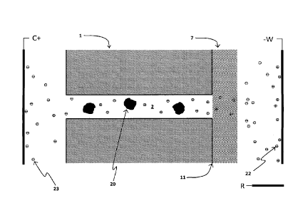
such as Bovine Serum Albumin (BSA), has a size of about 4 urn x 4 nm x 14 inn.
With a TEPC
filter I hole 2 diameter of 50 urn, a single BSA protein molecule would be
blocking a significant.
fraction (0.8% to 2.9%) of the cross-sectional area of the hole 2, and hence
significantly reduce
the ionic particle flow.
1001521 Referring to FIG. 28, obstacles 29, 30, and 31 themselves may have an
electric
charge. Upon application of an electric field, they may interact in complex
ways with the
movement of the electrolytes or redox reagents.
1001531 The transmission of electrolyte molecules or redox reagents through
the gel 7,
dependent upon obstacle characteristics within the hole 2, provides a
sensitive way to perform
measurements of the obstacle characteristics. FIG. 31 illustrates the
application of an electric
field with Working, Counter, and Reference electrodes. The ionic particle flow
will create an
electric current in the electrodes that is resisted by both the obstacles and
by the gel 7, but since
the resistance of the gel 7 is relatively constant, this resistance can be
subtracted out by cycling
the measurements.
1001541 The overall apparatus in the sample loading state is shown in FIG. 32.
A fluid flow
of analyte particles, such as a complex sequence of proteins eluting from a
chromatography
column 32, is directed to flow into a particular region of the TEPC filter 1.
A lower pressure on
the opposite side causes the solvent and other small molecules to pass through
the TUC filter I.
and gel 7, accumulating analyte particles within the TEPC filter I holes 2. As
analyte particles
are eluted from the chromatography column 32, the TEPC filter 1 surface is
moved in a scanning
motion. The different analyte particles that elute are trapped in spatially
distinct locations within
the TEPC filter 1. Following this operation, a second scan may be done with
different analyte
particles eluting from the chromatography column 32, producing a large number
of unique
binary mixtures (of controllable proportions) across the area of the TEPC
filter 1 . Further scans
could be used to add even more complexity to the populations within the holes
2. This would be
functionally equivalent to "Sandwich Array" technology, for reducing the
problem of protein
21
CA 02788813 2012-08-01
WO 2011/106198 PCT/US2011/024882
cross-reactivity. It is even possible to include lipid micelles, colloids,
immobilized materials, or
Phage Antibody Display technology within the holes 2, allowing retained
analyte particles to
interact with that environment. Furthermore, it is possible to extract analyte
particles from one
set of holes 2 and add it to another set of holes 2.
1001551 The overall apparatus in the measurement state is shown in FM. 33. An
electrode 33,
or plurality of electrodes, is scanned across the surface such as the TEPC
filter 1 surface, and the
electric current measured. information about the position of the electrode and
the electric current
is compiled into a conductance map 34. Examples of the structure of the
electrode include an
exposed metal surface surrounded by an insulator, and a tube filled with
conductive .fluid.
1001561 Referring to FIG. 35, the result of measurement is a set of largely
uniform
conductance maps 34. Most of the area is uniformly conductive with the
exception of a few low-
conductance areas having significant migration obstacles (e.g. analyte
particles). The
characteristics of these low-conductance areas, relative to the uniformly
conductive areas, is the
basis of the analytical signal.
1001571 Referring to FIG. 33, restriction of the area to be measured can be
achieved by an
insulating tube, filled with an electrically conductive fluid, with one end in
physical contact with
said sheet of material, an insulating sheet with a hole that is applied to the
surface of said sheet of
material, or an insulating water-immiscible fluid that is applied to the
surface of said shoo of
material. In the latter case, upon pressurization of the gel side of the TEPC
filter 1, surface
tension of water within the holes forms isolated aqueous protuberances Meng
the other surface of
the TUC filter 1, that may be scanned with an insulating tube, filled with an
electrically
conductive fluid,
[001581 Referring to FIG. 37, the electrical, conductance is graphed as a
fimction of time for
an area that has protein present, and another area that has no protein
present.. After the electric
field E is actuated, the presence of protein causes a vertical (increased)
shift in the current i level,
which can be quantified by a delta current i measurement,
1001591 Referring to FIG. 39, the theoretical results for protein present
versus no protein
present are compared. The electric field E is repeatedly actuated in a cycle,
yielding repeated
delta current measurements. On a longer time scale, the hydrodynamic pressure
P is also
repeatedly actuated in a cycle. When increased pressure causes protein to
accumulate, the delta
current increases. When the pressure is released, the protein diffbses
outward, and the delta
22
CA 02788813 2012-08-01
WO 2011/106198 PCT/US2011/024882
current decreases. Cycling the hydrodynamic pressure thus provides a continual
series of delta
current peaks that can be averaged for sensitive detection of the presence of
the protein.
1001601 Referring to FIG. 41, the theoretical results for protein binding
present versus protein
non-binding are compared. Protein that binds to another protein will have
greater steric effects,
becoming a large obstacle and having a slower diffusion rate; this results in
a series of continual
delta current i peaks that are of large amplitude. Protein that. does not bind
to another protein
will have lesser steric effects, not becoming a large obstacle and not having
a slower diffusion
rate; this results in a series of continual delta current peaks that are of
small amplitude. Note that
since there are multiple proteins present, this smaller amplitude may have
multiple peaks.
1001611 A summary of the measurement results is shown in FIG. 43. The
measurements
provide a map of the analyte particle characteristics across the area of the
IF,PC filter 1. Certain
areas 38 will have measurement characteristics of interest to a scientist
performing the method.
The identity of the contents of these areas can then be done by a variety of
techniques, such as by
mass spectrometry, or by knowledge of the chromatography system that
originally delivered the
contents.
1001621 Substantially the same functionality may be achieved by use of similar
structures,
such as a perforated monolayer film instead of a TEPC/gel structure, where
diffusion is radial
instead of axial.
1001631 Second Embodiment Summary
(001641 A specially-constructed, layered material forms a set of reservoirs
that are loaded
with a variety of spatially-separated analyte particles, and said layered
material imaged for
fluorescence emission. This yields a map of the characteristics of the various
analyte particles,
which can provide usefill information about biological samples.
101651 Method in accordance with a second embodiment of the invention
1001661 In a method in accordance with a second embodiment of the invention, a
perforated
material, such as a TEpc filter, has holes with restricted openings.
Homogeneous or
heterogeneous populations of analyte particles within a controlled matrix are
loaded into the
TEPC filter holes, and fluorescence is used to measure the diffusion outwards,
which is a
measure of presence, structural changes, and any binding interactions
involving the analyte
particles.
1001671 The method of the invention will now be described by way of reference
to FIGS. 1 to
23
CA 02788813 2012-08-01
WO 2011/106198 PCT/US2011/024882
43.
1001681 Referring to. FIGS. 1A, .1B, and 2, the TEPC filter 1 has a random
distribution of
uniformly-sized holes. FIG. lA shows the top view, and FIG. 1B is a cross
sectional view. The
TEPC filter 1 may have an outer surface chemically derivatized with
carboxylate moieties 3.
Other materials having similar characteristics may be substituted.
1001691 Referring to FIGS. 3, 4, and 5, a bilayer material may be formed by
the following
process. Firstly, a dilute suspension of spheroids 4 of uniform size is filmed
in a matrix of a gel
precursor (such as acrylamide or melted agarose). The suspension is then
applied to a thin plastic
sheet 5 and wrapped around a smooth cylinder 6 that has a hydrophobic
surfitce. The gel 7 is
formed by chemical, thermal, or light polymerization. An example of chemical
polymerization is
illustrated in FIG. 4. The spacer particles 4 enforce a uniform, known
thickness to the gel 7.
After polymerization, the thin plastic sheet 5 is peeled off of the smooth
cylinder 6, creating a
bilayer material 8 consisting of a gel 7 layer and a plastic 5 layer. A
magnified view of this
bilayer material is shown in Ha 5; the spheroids 4 are not shown in this
magnified view
because they are sufficiently dilute. The chemistry of the bilayer material 8
interface 9 is chosen
such that. the thin plastic sheet 5 layer may be easily removed in the future
by physical or
chemical means. The outer surface of the gel 7 may be chemically derivatized
with amino
moieties 10, but these are indigenous to polyaerylamide.
1001701 Ahematively to said bilayer material, a gel precursor may be
sandwiched between
Iwo hydrophobic smooth plates, and allowed to polymerize and dry. This forms a
robust dried
gel .fihn that can be easily handled, and then re;hydrated when needed.
1001711 Alternatively to said bilayer material, a dialysis membrane may be
used, which may
be purchased commercially from many vendors, such as Millipore or Whatman.
1001721 Referring to FIGS. 6, 7, and 8, the bilayer material 8 may be
chemically bonded to the
TEPC filter I. An example of a chemical bonding mechanism is shown in FIG. 6,
where
carboxylate moieties and amino moieties chemically bind, to form a peptide
bond 11. Other
chemistries may be substituted. FIG. 7 shows the =TEPC filter 1 and the
bilayer material 8 in
close proximity, with the carboxylate derivatized surface 3 and the amino
derivatized surface 1.0
facing each other. FIG. 8 shows the TEPC filter 1 and the bilayer material 8
chemically bonded
together with the peptide bond 11.
1001731 Referring to FIGS. 8 and 9, the thin plastic sheet 5 may be removed by
physical or
24
CA 02788813 2012-08-01
WO 2011/106198 PCT/US2011/024882
chemical means, leaving only the gel 7 layer adhered to the TEPC filter 1.
1001741 Alternatively to said chemical bonding, the gel 7 layer and. the TEPC
filter .1 may
simply be compressed together by physical force without chemical bonds, such
as by wrapping
around a cylinder and tensioning the outer TEPC filter 1 layer.
1001751 Alternatively to said chemical bonding, the TEPC filter .1 may be
placed on a surface
of a conductive fluid that is immiscible with the fluid comprising the analyte
matrix.
1001761 There are a number of variations that are possible to the schematic
shown on FIG. 9,
and the examples of these are shown in FIGS. 10, 11., .12, 13, 14, 15, and 16.
Although the
remainder of this Method will focus on the simple ease of FIG. 9, it will be
appreciated that. these
variations are also applicable.
1001771 Referring to FIG. 10, the outer surface of the gel 7 may be chemically
derivatized
with fluorophores .12, surfactants 13, other materials, or any combination of
materials. Likewise,
the inner surface of the gel 7 (facing the hole 2) or the inner surface of the
hole 2 may also be
chemically derivatized.
1001781 Referring to FIGS. 11, 12, and 13, a spheroid 14 may be lodged in the
opening of the
hole 2, prior to formation of the peptide bond 11. This spheroid 14 would have
a diameter
slightly larger than the diameter of the hole 2. The gel 7 layer would be
dimpled by the presence
of the spheroid 14, creating an empty volume 1$ around the spheroid 14. A
force 16, such as
from a magnetic field gradient, electric field, gravitational field, or
hydrodynamic flow, may be
used to lift the spheroid 14 from its seating in the opening of hole 2,
creating a small gap 17
between the surface of the spheroid 14 and the opening of the hole 2. The
empty volume 1.5 may
be removed by brief heating of the spheroid 14 to melt the surrounding gel 7,
or by saturation
with gel precursors followed by chemical polymerization. The resulting
structure is shown in
FIG. 13.
1001791 Referring to FIGS, 14 and15, both ends of the hole 2 may be capped by
spheroids 14.
If the gel 7 is sufficiently pliable, then one of the spheroids 14 may be
removed by a force, .such
as from a magnetic field gradient. This would leave a small tear 18 in the gel
7. This particular
configuration would be especially useful for retention of materials within the
hole 2 without an
active retention mechanism.
1001801 Referring to FIG. 1.6, the gel may be not used, and the spheroid 14
held in place with
a force, such as from a magnetic field gradient 16.
CA 02788813 2012-08-01
WO 2011/106198 PCT/US2011/024882
1001811 Focusing on the simple example of FIG. 9, this structure may be used
to collect and.
concentrate analyte particles within the hole 2. FIG. 17 is a schematic of
fluid flow 19 passing
from the open end of the hole 2, through the hole 2, and then through the gel
7. Analyte particles
that are suspended within the fluid flow 19 become filtered by the gel 7, if
the gel 7 has a
sufficiently tight cross-linked structure to prevent passage. Examples of such
analyte particles
are proteins 20, nucleic acids, viruses, protoplasmic structures, Quantum
Dots, large
fluorophores 21, large electrolyte cations, large electrolyte anions, and
large redox reagents. The
permeability of the gel 7 may be reduced by inclusion of particles within the
gel 7 during
formation. Examples of particles that are able to pass through the gel 7 are
water molecules,
small electrolyte cations 22, small electrolyte anions 23, and small redox
reagents. FIG. 18 is a
schematic of the net result of accumulated analyte particles within the hole
2.
1001821 Once there are accumulated analyte particles in the hole 2, the fluid
flow 19 can. be
stopped. At this point, the accumulated analyte particles will begin to
diffuse outward, eventually
emptying the hole 2. The fluid flow .19 may then be reinstated, and the
accumulation diftlision
cycle repeated. A gel that weakly limits difftision may be placed at the hole
2 outlet, to improve
cyclability. A gel that weakly limits diffusion may be placed within the hole,
to extend the
diffusion. times.
1001831 During diffusion, the analyte particles in the hole 2 would have
movement pathways
that can intersect with ionic or fluorescent particles. Since the particles
can not pass through each
other, they go around each other, slowing down the movement pathways of the
ionic or
fluorescent particles. This slowing down is dependent on the presence of
analyte particles and
their binding processes, and thereby forms the basis of this Method.
[001841 During diffusion, the analyte particles in the hole 2 may be driven
axially with a
migration force (e.g. force resulting from an electric field) in addition to
diffusion.
[00185f This process is also applicable to the accumulation part of the cycle,
but analysis is
complicated by the addition of fluid flow 19 forces.
[00186] During the diffusion or accumulation parts of' the cycle, the fluid
flow 1.9 may be
given a high-frequency axial oscillation, for the purpose of Modifying
particle movement. For
example, the spheroid 14 of FIG. 15 may have a magnetic moment and be
magnetically
oscillated to pump large analyte particles through. the tear 18. As another
example, the outer (or
inner) surface of the gel 7 in FIG. 9 may be subjected to pressure pulsations,
causing the analyte
26
CA 02788813 2012-08-01
WO 2011/106198 PCT/US2011/024882
particles within the hole 2 to likewise oscillate.
[001871 There are numerous mechanisms by which particles in the hole 2 may by
driven
axially with a three in addition to diffusion. Examples of' some of these
mechanisms are
illustrated in FIGS. 19, 20, 21, and 23.
1001881 Referring to FIG. .19, fluotophores that are unable to traverse the
gel 7 are moved
towards (or away from) the inner gel 7 surface by an electric field generated
by Working (-W),
Counter (C+), and Reference (R) electrodes.
(001891 Referring to FIG. 20, fluorophores that are unable to traverse the gel
7 are moved
towards (or away from) the inner gel 7 surface by a magnetic field gradient.
1001901 Referring to FIG. 21, fluorophores that are unable to traverse the gel
7 are moved
towards (or away from) the inner gel 7 surface by a gravitational field.
(00.1911 Referring to FIG. 23õ large electrolyte cations 24, large electrolyte
anions 25, or redox.
reagents that are unable to traverse the gel 7 are moved towards (or away
from) the inner gel 7
surface and the outer gel 7 surface, by an electric field, creating a
capacitor.
1001921 The capacitor has behavior that warrants additional description in
FIGS. 24, 25, and
26.
(00193J Referring to FIG. 24, the electric field that is axial to the hole 2
has a complex
structure. This complex structure is analogous to the electric field that
exists near metallic
electrodes in ordinary electrochemical studies. At a distance far away from
the outer surface of
the gel 7, in the bulk solution, the electric field is small and does not
change significantly with.
distance. Approaching the outer surface of the gel 7, the electrolyte exhibits
an increased
concentration, causing the electric field to rise exponentially; this is
commonly called the Ciouy-
Chapman Layer. Extremely close to the outer surface of the gel 7, the
electrolyte forms a double-
layer of alternating charge; this is commonly called the Helmholtz Layer. The
'presence of
surfactant molecules 13 may assist in the shaping of the field within the
Helmholtz Layer.
Continuing onward into the gel 7, the electric field subsides. Upon exiting
the gel 7 on the inner
surface, the electric field has a structure that mirrors the structure for the
outer surface.
1001941 Referring to FIG. 25, the strong electric field in the Helmholtz Layer
of the outer gel
7 surface causes an electric charge, such as an electron, to be transferred
from an electrolyte (or a
suitable redox reagent) anion 25 (or cation) to a fiuorophore 12 bound to the
surface. After the
molecule 25 transfers its charge, it becomes another molecule 26. The
.fluorophore 12 will have
27
CA 02788813 2012-08-01
WO 2011/106198 PCT/US2011/024882
its fluorescence characteristics Changed by the charge transfer. For example,
fluorescein in its
uncharged state is non-fluorescent, but when negatively charged becomes
intensely fluorescent.
1001951 In each of these examples for mechanisms by which particles in the
hole 2 may by
driven axially with a force in addition to diffusion, the movement pathways
themselves may be
slowed down by several different mechanisms. :Examples of some of these
mechanisms are
illustrated in FIGS. 26, 27, and 28.
1001961 Referring to FIG. 26, a fluorophore 21 may be moved in a straight line
by a migration.
force (such as from an electric field, magnetic field gradient, or
gravitational field) if there are no
obstacles in its path. However, if there are obstacles, such as an analyte
particle 20 and an
enable particle 27, then the fluorophore 21 will need to go around the analyte
particles, slowing
down the fluorophore axial movement. If the analyte particles have a binding
interaction, they
will from a large complex 28, causing the fluorophore axial movement to be
slowed down even.
more. For example, a typical protein, such as Bovine Serum Albumin (BSA), has
a size of 4 nm
x 4 nm x 14 nm. With a TEPC filter 1 hole 2-diameter of SO nm, a single BSA
protein. molecule
would be blocking a significant fraction (0.8% to 2.9%) of the cross-sectional
area of the hole 2,
and hence significantly reduce the fluorophore flow.
1001971 Referring to FIG. 27, a large electrolyte cation 24 and large
electrolyte anion 25 may
be moved in a straight line by an electric field if there are no obstacles in
its path. However, if
there are obstacles, such as an analyte particle 20 and an analyte particle
27, then the cation 24
and anion 25 will need to go around the analyte particles, slowing down the
ionic axial
movement. If the analyte particles have a binding interaction, they will form
a large complex 28,
causing the ionic axial movement to be slowed down even more.
1001981 Referring to FIG. 28, obstacles 29, 30, and 31 themselves may have an
electric
charge. Upon application of an electric field, they may interact in complex
ways with the
movement of the electrolytes or redox reagents.
1001991 The accumulation or activation of fluorophores in the vicinity of the
gel 7, depending
upon obstacle characteristics within the hole 2, provides a sensitive way to
perform
measurements of the obstacle characteristics. FIGS. 29 and 30 illustrate the
application of
ultraviolet light to the fluorophores, and the resulting emitted visible
light. The intensity of the
emitted visible light is dependent upon the concentration of active
fluorophores in the vicinity of
the gel 7. Fluorophores that are located at a distance down the hole 2 will
not fluoresce, because
28
CA 02788813 2012-08-01
WO 2011/106198 PCT/US2011/024882
the polycarbonate material of the *IsEPC'. filter 1 is strongly absorbing for
ultraviolet light. Note
that. although a primary advantage of this method is to avoid the need to
label an analyte particle
with a fluoro.phore, it is within the scope of this method to include analyte
particles labeled with
fluomphores.
1002001 'Me overall apparatus in the sample loading state is shown in FIG. 32.
A fluid flow
of analyte, such as a complex sequence of proteins eluting from a
chromatography column 32, is
directed to flow into a particular region of the TEPC filter 1. A low pressure
on the opposite side
causes the solvent and other small molecules to pass through the TEPC filter
.1 and gel 7,
accumulating analyte particles within the TEPC filter 1 holes 2. As analyte
particles are doted
from the chromatography column 32, the TEPC filter 1 surface is moved in a
scanning motion.
The different analyte particles that elute are trapped in spatially distinct
locations within the
'aim filter 1. Wowing this operation, a second scan may be done with different
analyte
particles eluting from the chromatography column 32, producing a large number
of unique
binary mixtures (of controllable proportions) across the area of the TEPC
filter 1. Further scans
could be used to add even more complexity to the populations within the holes
2. This would be
functionally equivalent to "Sandwich Array" technology, for reducing the
problem of protein
cross-reactivity. It is even possible to include lipid micelles, colloids,
immobilized materials, or
Phage Antibody Display technology within the holes 2, allowing retained.
analyte particles to
interact with that environment. Furthermore, it is possible to extract analyte
particles from one
set of holes 2 and add it to another set of holes 2.
1002011 The overall apparatus in the measurement state is shown in FIG. 34. A
beam of
fluorescence excitation light, such as ultraviolet light, is directed towards
a surface such as the
gel 7 surface. The fluorescence emission light, such as visible light, is
collected by a lens 35 or
light pipe, and. a photosensor 36, such as a digital camera. Information from
the camera is
compiled into a photographic image 37.
[002021 Referring to FIG. 36, the result of measurement is a series of
changing photographic
images 37 when the migration force (such as from an electric field, magnetic
flekl gradient, or
gravitational field) is applied.. Initially, the photographic image is
relatively dark.. After a brief
amount of time, most of the area is fluorescent with the exception of a few
delayed areas having
significant migration obstacles (e.g. analyte particles). Soon, however, as
migration completes in.
these delayed areas, the whole area of the image becomes uniformly
fluorescent. The temporal
29
CA 02788813 2012-08-01
WO 2011/106198 PCT/US2011/024882
characteristics of these delayed areas, relative to the un-delayed areas, is
the basis of the
analytical signal.
[002031 Referring to FM. 38. the fluorescence is graphed as a function of time
for an area that
has protein present, and another area that has no protein present. After the
electric field E is
actuated, the presence of protein causes a horizontal (temporal) shift in the
sigmoidal curve,
which can be quantified by a delta time t measurement.
1002041 Referring to FIG. 40, the theoretical results for protein present
versus no protein
present are compared. The migration three is repeatedly actuated in a cycle,
yielding repeated
delta time t measurements. On a longer time scale, the hydrodynamic pressure P
is also
repeatedly actuated in a cycle. When increased pressure causes protein to
accumulate, the delta
time increases. When the pressure is released, the protein di ffitses outward,
and the delta time
decreases. Cycling the hydrodynamic pressure. thus provides a continual series
of delta time
peaks that can be averaged for sensitive detection of the presence of the
protein.
1002051 Referring to FIG. 42, the theoretical results for protein binding
present versus protein
non-binding are compared. Protein that binds to another protein will have
greater steric effects,
becoming a large obstacle and having a slower diffusion, rate; this results in
a series of continual
delta time t peaks that are of large amplitude. Protein that does not bind to
another protein will.
have lesser steric effects, not becoming a large obstacle and not having a
slower diffitsion rate;
this results in a series of continual delta time peaks that are of small
amplitude. Note that since
there are multiple proteins present, this mailer amplitude may have multiple
peaks.
1002061 A. summary of the measurement results is shown in FIG. 43. The
measurements
provide a map of the amble particle characteristics across the area of the
TEPC filter 1. Certain
areas 38 will have measurement characteristics of interest to a scientist
performing the method.
The identity of the contents of these areas can then be done by a variety of
techniques, such as by
mass spectrometry, or by knowledge of the chromatography system that
originally delivered the
contents.
[002071 Substantially the same functionality may be achieved by use of similar
structures,
such as a perforated monolayer film instead of a TEPC/gel structure, where
diffusion is radial
instead of axial.
1002081 Third Embodiment Summary
[002091 A specially-constructed, layered material forms a set of reservoirs
that are loaded
CA 02788813 2012-08-01
WO 2011/106198 PCT/US2011/024882
with a. variety of -spatially-separated analyte particles, and said layered
material sampled by mass
spectrometry. This yields a map of the compositions of the various analyte
particles, which can
provide useful information about biological samples.
1002101 Method in accordance with a third embodinnnit 4),,I the invention
1002111 In a method in accordance with a third embodiment of the invention,
the holes of a
TEPC filter (or functionally equivalent structure) are restricted, at the
openings. Homogeneous or
heterogeneous populations of analyte particles within a controlled matrix are
loaded into the
TEPC filter holes, and the hole ends are tightly capped by spheroids
forcefully corked into the
hole ends. The assembly is loaded into a vacuum chamber, targeted by a laser
beam, and the
resulting vapor directed into a mass speetrometry Chamber. Ionization and
mass/charge detection
provides a means of identification of the analyte particles.
1002121 The method of the invention will now be described by way of reference
to FIGS. 44
to 48.
1002131 Referring to FIG. 44, a composition of analyte particles .20, 22, and
23 is enclosed
within the hole 2 of TEPC filter 1, by having a spheroid 14 at each end of the
hole 2.
1002141 Referring to FIGS. 45 and 46, tightly squeezing the TEPC filter .1
between two
smooth rollers to exert a compressive force 39 would cause each spheroid 14 to
stopper both
ends of the hole 2. The "MK filter I could then be removed from the
preparatory apparatus,
yielding a stable material shown in FIG. 46 that contains the analyte
particles.
[002151 :Referring to FIG. 47, the stable material is put into a vacuum
chamber. The aqueous
matrix containing the analyte particles within the hole 2 is protected from
the vacuum by the
spheroids 14 stoppering the hole 2 and sealing in the aqueous matrix.
Additional sealing may be
provided with a thin polymer film overcoat. An intense, focused laser beam 40
is then directed
at the hole 2.
1002161 Referring to FIG. 48, the laser beam would vaporize the upper stopper
of the TEPC
filter I hole 2, causing the aqueous matrix to explode outwards into the
vacuum, dispersing it as
a gas. This is analogous to matrix-assisted laser desorption ionization
(MALI)1) analysis.
However, the biological sample receives much less thermal stress, leading to a
less complex
fragmentation pattern. This reduced complexity may be analytically useful
1002171 SUMMIT
1002181 The scope of these Methods covers the tasks of detection,
Characterization, and
31
CA 02788813 2012-08-01
WO 2011/106198 PCT/US2011/024882
identifying of analyte particles, and the characterization of any interactions
involving them, using
the properties of nanoscale reservoirs.
1002.191 Current methods tbr pertbrming these taSks suffer finm a variety of
deficiencies, such
as the interference from labeling techniques and cross-reactivity. These
deficiencies are
eliminated. Furthermore, the methods are useful for analysis of extremely low
concentrations of
analyte particles.
1002201 A specific field where these Methods would be useful is in
biotechnology, fir the
measurement of protein interactions. There are an extremely large number of
proteins used in
every biological system, which interact in a complex network that is dependent
on many factors.
Diseases distort this network, adding or removing components and interaction
pathways. An
understanding of these systems allows early diagnosis of disease, and a way to
chemically repair
the system through drug therapy. The. most populous and stable proteins within
these systems
have been partially studied, but much further study is warranted. The addition
of new and more
powerful tools, such as the Methods described herein, to the repertoire of
medical researchers
would deepen the understanding of the protein networks and allow the
development of new drug
therapies.
100221.1 The term "particles" as used herein includes molecules, cells,
multicellular structures,
subcellular components, viruses, prions, proteins, polymers, ions, colloids,
and fluorophores. The
particles may be suspended or dissolved. The particles do not necessarily need
to be biologically
relevant.
1002221 The term "analyte" as used herein describes particles that are to be
measured.
1002231 The term "get" as used herein is not restricted to its strict
technical definition, but
rather includes generally any material that is relevant to restricting
diffusion of analyte particles.
The term "gel" is intended to include, but is not limited to, gelatin,
agarose, polyacrylamide,
polyacrylate, permeable polymers, permeable copolymers, starch, aerogel,
collodion, dialysis
membrane, immiscible fluid, any of the above-listed materials in a chemically
modified form,
any of the above-listed materials embedded with particles, and. any
combination of the above-
listed materials.
1002241 The term "immiscible fluid" as used herein includes fluids that are
immiscible with
the analyte particle matrix.
[002251 The term "large" as used herein describes those particles that can not
pass through the
32
CA 02788813 2012-08-01
WO 2011/106198 PCT/US2011/024882
restriction at the end of the TEPC filter hole, such as by the gel. The term
"small" as used herein
describes those particles that can pass through the restriction at the end of
the TEPC filter hole,
such as by the gel.
1002261 The term "force" as used herein includes a force resulting from an
electric field, a
force resulting from a magnetic field gradient, a force resulting from a
gravitational field,
centripetal force, centrifugal force, force resulting from hydrodynamic
pressure, a force resulting
from hydrostatic pressure, or a combination of such forces.
(002271 Electric fields may be generated capacitively, without direct
electrode contact with
ionic or redox species. Multiple sets of electrodes may be used as needed to
achieve the
necessary electric fields. For example, one set of electrodes may be used to
exert strong
migration forces, while another set of electrodes is used for analyte particle
measurement.
1002281 Measurement of electric current may be structured to constitute
resistance,
conductance, impedance, capacitance, and inductance measurements, and
combinations thereof.
For example, characterization of lipid micelles, whole cells, or other
materials with impedance
boundaries may be assisted by capacitance measurements, and characterization
of chiral analytes
may be assisted by inductance measurements.
(00229j Analyte particles may be delivered to the apparatus by local rupture
of intact cells, or
by otht.T techniqum, in addition to standard chromatographic techniques.
1002301 The holes or perforations in said TEPC material, or its functional
equivalent, may be
sized for close fitting of individual cells, so that electrical impedance and
capacitance are
determined largely through the bulk of the cell.
(002311 The methods described herein may be combined with conventional
microchannel
array technology, commonly referred to as "lab-on-a-chip" technology.
1002321 While the present invention has been described with reference to
certain preferred
embodiments, one of ordinary skill in the art will recognize that other
additions, deletions,
substitutions, modifications, and improvements can be made while remaining
within the spirit
and scope of the present invention as defined by the claims.
33