Note: Descriptions are shown in the official language in which they were submitted.
CA 02789384 2012-08-09
WO 2011/109907 PCT/CA2011/000273
1
METHOD OF GENERATING LOW-ENERGY SECONDARY ELECTRONS
FOR APPLICATIONS IN BIOLOGICAL SCIENCES, RADIOCHEMISTRY,
AND CHEMISTRY OF POLYMERS AND PHYSICS OF RADIOTHERAPY
TECHNICAL FIELD
[0001] The present disclosure relates to generation of low-energy
secondary electrons. More specifically, the present disclosure relates to a
method and a system for generating low-energy electrons in a biological
material.
BACKGROUND
[0002] Secondary electrons are electrons generated as ionization
products. They are called "secondary" because they are generated by other
radiation, called primary radiation. This primary radiation may be in the form
of
ions, electrons, or photons with sufficiently high energy to exceed an
ionization
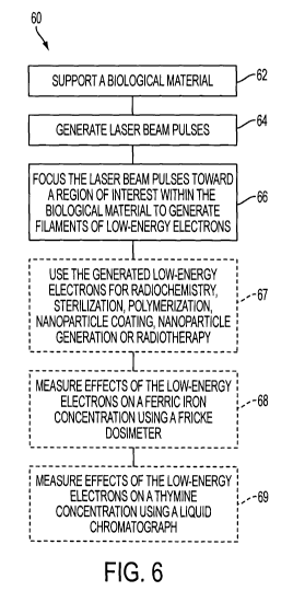
potential. Photoelectrons are an example of secondary electrons where the
primary radiation consists of photons. Low-energy secondary electrons play a
crucial role in the degradation of high-energy ionizing radiation such as X-
rays,
y-photons or charged particles. Low-energy secondary electrons are a means
to define the geometry of the radiation track.
SUMMARY
[0003] The present disclosure broadly relates to generation and
applications of low-energy secondary electrons.
[0004] Therefore, according to the present disclosure, there is
provided a method for generating low-energy electrons in a biological
material.
The method comprises a step of supporting the biological material. Laser
beam pulses are generated. The laser beam pulses are focused pulses toward
a region of interest within the biological material to generate filaments of
low-
energy electrons.
[0005] According to the present disclosure, there is also provided a
CA 02789384 2012-08-09
WO 2011/109907 PCT/CA2011/000273
2
system for generating low-energy electrons in a biological material. The
system comprises a support for the biological material, a pulsed laser and a
focusing mechanism. The focusing mechanism directs laser beam pulses
toward a region of interest within the biological material to generate
filaments
of low-energy electrons.
[0006] The foregoing and other features will become more apparent
upon reading of the following non-restrictive description of illustrative
embodiments thereof, given by way of example only with reference to the
accompanying drawings.
BRIEF DESCRIPTION OF THE DRAWINGS
[0007] Embodiments of the disclosure will be described by way of
example only with reference to the accompanying drawings, in which:
[0008] Figure 1 is schematic view of a laboratory system for
generating femtosecond laser filamentation in accordance with an illustrative
embodiment;
[0009] Figure 2 is a graph of a relative dose distribution using X-rays,
proton Bragg peak and an effective spread-out proton peak for radiotherapy
treatment;
[0010] Figure 3 is a graph of an irradiation dose deposition
equivalent of femtosecond laser filamentation and Gamma irradiation as a
function of time;
[0011] Figure 4 is a graph of a comparative concentration of thymine
production as a function of an irradiation dose;
[0012] Figure 5 is a graph of agarose gel electrophoresis, using (a)
Gamma irradiation and (b) femtosecond laser filamentation irradiation, of
plasmid DNA; and
[0013] Figure 6 shows steps of an exemplary method for generating
low-energy electrons in a biological material.
CA 02789384 2012-08-09
WO 2011/109907 PCT/CA2011/000273
3
DETAILED DESCRIPTION
[0014] Generally stated, the non-limitative illustrative embodiment of
the present disclosure provides a method and a system for generating low-
energy secondary electrons for applications in biological sciences, medical
applications, radiochemistry, and chemistry of polymers and physics of
radiotherapy. More specifically, the low-energy secondary electrons are
produced using femtosecond (fs) laser filamentation.
[0015] Although femtosecond laser filamentation (FLF) is a well-
known process, it has seldom been used for radiolysis of water [7]. It has
been
discovered that low-energy electrons (LEE) in FLF and ionization radiation are
radiochemically equivalent for applications in biological sciences,
radiochemistry, and chemistry of polymers and physics of radiotherapy. The
LEE are generated by laser pulses and are then directly recombined or
solvated in liquid, in about 300 to 500 fs in water.
[0016] In the degradation of high-energy ionizing radiation like X-
rays, y photons or charged particles such as, for example, accelerated
electrons or heavier charged particles, low-energy secondary electrons serve
to define a geometry of a radiation track. They consist of highly anisotropic
ionization energy deposition of secondary electrons with energy between about
1 and 20 eV, for example about 5 x 10⁴ electrons/MeV [1]. In this energy
range, an electron penetration range in water is in the order of 10 nanometers
(nm) [2].
[0017] Demonstration of genotoxic action of low-energy electrons on
fundamental biological molecules, such as for example deoxyribonucleic acid
(DNA), film of biological molecules, and similar compounds, may be achieved
in ultrahigh vacuum conditions [5]. To extend this demonstration, an
anisotropic concentration of low-energy electrons in a macroscopic volume of
water, in the order of a cubic centimeter (cm) of water, is generated using
intense, ultra-short laser pulses, which lead to self-focusing and
filamentation.
The physical origin of the formation of filaments is well understood. Briefly,
CA 02789384 2012-08-09
WO 2011/109907 PCTICA2011/000273
4
self-focusing is an induced lens effect, resulting from wavefront distortion
self-
inflicted on a beam while traversing a nonlinear medium. Consequently, as the
beam travels in the nonlinear medium, an original plane wavefront of the beam
gets progressively more distorted. The distortion is similar to that imposed
on
the beam by a positive lens. Since the optical ray propagation is in the
direction perpendicular to the wavefront, the beam appears to focus by itself.
This degenerative process, in which the positive lens effect increases with
intensity, is stabilized in the femtosecond regime by the generation of
electrons
forming a filament. Electrons are produced by multiphoton or tunnel ionization
and are further accelerated by an electric field of the pulse in an inverse
Bremstrahlung effect. When they acquire enough kinetic energy, for example
6.5 eV in the case of water, the electrons give rise to a second generation of
electrons by impact ionization of other molecules in an avalanche-like
process.
This linear distribution of electrons formed in the filament, in the range of
1016
- 1018 electrons per cm3, transfers their excess energy to surrounding water
molecules, which leads to the generation in a self-focusing region of
chemically
reactive species such as eaq, H*, 0*, and *OH, and recombination products H2,
02, H20, and 02*- ( or H02*, pKa = 4.8).
[0018] There does not exist in the literature any mention of real time
measurements of the presence of LEE in a filament. However, as LEE are
generated in filamentation, solvated electrons become measureable along the
filament. Pump-probe measurements may be used for this purpose. Solvated
electrons have an optical spectrum measurable by a femtosecond pump-probe
technique. The presence of solvated electrons along the filament may be
measured using a delay of 50 picoseconds (ps) between an 800 nm pump
pulse having a 100 fs pulse duration, which generates the filament, and an
optical probe of a 125 fs pulse duration at 720 nm from an optical parametric
amplifier (OPA). Scanning a position of a pump lens changes the position of
the filament in a linear direction. A characteristic intensity evolution of
the
length of the filament in FLF has been observed from the pump-probe scan
measurement in function of the pump pulse intensity [6].
CA 02789384 2012-08-09
WO 2011/109907 PCT/CA2011/000273
[0019] Referring to Figure 1, which is schematic view of a laboratory
system for generating femtosecond laser filamentation in accordance with an
illustrative embodiment, a system 10 comprises a laser 12 producing a beam
20 aimed at a region of interest (ROI) in an optical path cuvette 16 through a
focusing mechanism 14. Concurrently, Figure 6 shows steps of an exemplary
method for generating low-energy electrons in a biological material. A
sequence 60 of steps, as shown on Figure 6, will be described concurrently
with details of Figure 1 and with details of the following Figures. Some of
the
steps of sequence 60 may be present in some embodiments and not in other
embodiments. Some of the steps may be executed in a different order
compared to that shown on Figure 6.
[0020] The optical path cuvette 16 supports a biological material
(step 62), used as a laboratory sample, contained in an aqueous solution 22.
The cuvette 16 is positioned on a magnetic steering device 18 in order to
homogenize the solution 22 between pulses. The laser generates laser beam
pulses (step 64), which are focused by the focusing mechanism 14 towards the
ROI to generate filaments of low-energy electrons (not shown) within the ROI
(step 66). The filaments have a length of about one (1) cm, producing low-
energy electrons 24 in the solution 22. A detector 26, for example a streak
camera, detects an image of the beam 20 diffracted within the solution 22. A
resulting image may be used for time-resolved spectroscopy or for resonance
imaging (MRI) analysis.
[0021] The laser 12 may be, for example, a Spectra-Physics 300-
750 mW femtosecond Regenerative Ti-Sapphire laser having an optical
parametric amplifier (OPA) and harmonic generator (HG), used at 300
J/pulse, 100 fs pulses at 800 nm and at 1 kHz repetition rate. The focusing
mechanism 14 may have a focal lens of f=30 cm. This setup results in the
production of filaments of about one (1) cm in a one (1) cm optical path
cuvette
16. In another embodiment, a High Power Spitfire PRO_35F-1KXP, 35 fs
Ti:Sapphire regenerative laser, 4 watts at 1 kHz and at 800 nm, may be used,
CA 02789384 2012-08-09
WO 2011/109907 PCT/CA2011/000273
6
along with a AXIS-PV Streak Camera from Axis Photonique Inc. Details of a
laser source used in the context of the present disclosure may vary;
characteristics of the laser 12 as presented hereinabove are exemplary and
not intended to limit the scope of the present disclosure.
[0022] Examples of applications of LEE in FLF include the following
applications. These applications are generally illustrated on Figure 6, step
67.
Radiotherapy
[0023] One of the applications of the control of the distribution of the
LEE is a better dose distribution of radiation interaction in radiotherapy.
Figure 2 is a graph of a relative dose distribution using X-rays, proton Bragg
peak and an effective spread-out proton peak for radiotherapy treatment. The
present disclosure proposes to replace the use of X-ray therapy or proton
therapy with an LEE-based approach. On Figure 2, a tumor 30 may be treated
using X-rays having a distribution curve 34, or using protons having a Bragg
peak 36 and being further spread along curve 38. Using instead the LEE-
based approach allows to obtain a near ideal dose distribution 32 around the
tumor 30. For this purpose, a local distribution of LEE in a macroscopic
volume
(-cm3) of water needs to be controlled. LEE cannot be injected deeply in a
large volume of water. Anisotropic LEE is therefore locally generated with a
control of the energy of these LEE and of the geometry of the distribution.
The
laser 12 producing the beam 20 and the focusing mechanism 14 - or an
equivalent focusing mechanism - from Figure 1 are used to direct laser pulses
toward a properly supported and immobilized region of interest (ROI), which
replaces the laboratory sample of Figure 1. This modified system is thus a
radiation dose delivery system. The ROI, for example bodily tissues or other
biological material, comprises aqueous components and may further comprise
a tumor or like aspect that requires treatment. It is through parameter
adjustment of the laser 12 and/or of the focusing mechanism 14 that the
filaments of anisotropic LEE are generated in proper location, with desired
energy and distribution geometry.
CA 02789384 2012-08-09
WO 2011/109907 PCT/CA2011/000273
7
[0024] Filaments are analogue of tracks with an important difference.
Diameters of filaments in condensed matter are around 10 to 100 m.
Demonstration of the presence of H2 and H202 is well-known. Although
stabilization of the filament is due to the presence of electrons, no time-
resolved measurement of this presence has earlier been publicly made. The
present disclosure therefore suggests to measure femtosecond time-resolved
presence of the eaq along the filament. A Fricke dosimeter (not shown), also
called a ferrous sulphate dosimeter, measures oxidation conversion of ferrous
ions (Fe 2+) to ferric ions (Fe3+) by ionizing radiation having produced eaq,
*OH,
H02*, H202, and the like, in water. Increase of ferric ions concentration in
filaments may be measured spectrophotometrically (Figure 6, step 68) at an
optical absorption maximum at 303 nm. 6.5 eV electrons, which correspond to
a maximum energy of the LEE in water, have linear energy transfer (LET) of I
keV/ m [2] and G(Fe3+) of 15.3 molecules/100 eV (G(Fe3+)) for 1 keV/ m
radiation [3]. Those values correspond to radiation from a Cesium 137 (137Cs)
Gammacell Elan 3000 irradiator from Best Theratronics Ltd. In view of those
characteristics, the Fricke dosimeter is an appropriate tool to compare
radiation equivalent of FLF and Gamma irradiation, also called "Gammaknife".
[0025] Referring now to Figure 3, there is shown a graph of an
irradiation dose deposition equivalent of intense femtosecond laser
filamentation and Gamma irradiation as a function of time. A I kHz repetition
rate is used for the laser 12 of Figure 1. Figure 3 shows a curve 40 for the
laser irradiation and a curve 32 for the Gamma irradiation. At a 60-second
time
point 44, a dose rate of 168 Gy/min is obtained using FLF, compared to 12
Gy/min using the 137Cs irradiator. Comparison of measurements obtained with
the Fricke dosimeter with those obtained from Gamma irradiation thus provides
a dose rate for the filaments. It may be observed that the irradiation dose
deposition equivalent of intense femtosecond laser filamentation could also be
compared with results obtained from Cobalt 60 (60Co) irradiation.
[0026] Polyacrylamine gel (PAG) dosimetry is used in three-
CA 02789384 2012-08-09
WO 2011/109907 PCT/CA2011/000273
8
dimensional (3D) magnetic MRI of radiation. PAG is composed of 2 monomers
(3% of acrylamide, 3% of bisacrylamine) in 5% gelatin and 89% of water. LEE
may also be generated in PAG and in like polymers. Because radiologic
properties of gel dosimeter are equivalent to properties of tissues, radiation-
induced polymerization of the comonomers generates a fast-relaxing insoluble
polymer. Filament diameters may be estimated in PAG imaged by MRI,
whereby PAG effectively becomes a 3D dosimeter. In laboratory tests, optical
and MRI imaging of energy deposition in the PAG is obtainable and an image
of the LEE filamentation in a polymer volume has been observed. Production,
analysis and control of a dose deposition of LEE in FLF in PAG media, in
function of optical irradiation conditions involving control of optical
parameters
and pulse duration, allow analysis of related fundamental physical and
chemical processes and a determination of an ideal dose deposition for
radiotherapy treatment.
[0027] The use of PAG dosimeter is useful in obtaining 3D imaging
of energy deposition, for MRI imaging and for optical imaging. PAG material is
a radiological equivalent of tissues, especially for MRI imaging. PAG is a
good
prototype material to test the physics of radiotherapy without using actual
tissues and may be put to use for demonstrating the capability of FLF to
produce an ideal radiation beam for dose deposition in radiotherapy treatment.
For a specific optical setting, using a fixed focal lens, the length of a
produced
filament depends of the instantaneous laser intensity. The local intensity
dependence may be controlled by pulse duration. Adjusting the pulse duration
so that an image does not start in the front of a cuvette containing the PAG
allows adjusting the beginning of the filamentation and thus the dose
deposition. Modifying the optical setting allows changing the end of the
filamentation. In an approximation, it is estimated that a multifilament
diameter
in PAG material is at a maximum of 625 pm diameter, an accuracy of this
measure being limited by imaging resolution of MRI techniques, which in turn
are controlled by a magnetic field of seven (7) tesla and by the size of the
cuvette. In gas phase, the diameter of a monofilament is evaluated at 10 pm
CA 02789384 2012-08-09
WO 2011/109907 PCT/CA2011/000273
9
[6]. The diameter of a monofilament may also be limited by the chemistry of
polymerization and by set-up of the optical system, including parameterization
of filtering and of pulse duration. This polymerization is controlled by a
chain
reaction and by a distribution of a radical produced by ionization.
[0028] In an embodiment, MRI analysis of energy deposition using
monofilament and deposition of energy using Gammaknife in PAG may be
compared. In another embodiment, time-resolved spectroscopy and optical
imaging, for example using a streak camera, may be used to measure a time-
resolved fluorescence spectroscopy during monofilament formation. Analysis
may be made in function of oxygen concentration and in function of laser pulse
duration, whereby conditions for controlling energy deposition in PAG may be
optimized. In yet another embodiment, simultaneous control of pulse duration
and focalization, for example using a deformable mirror, in monofilament and
multifilament conditions, while using a Gammaknife reference, allows optimal
calibration of a dose deposition using MRI.
Radiochemistry
(0029] Another application of the control of the distribution of the LEE
is radiochemistry. This is illustrated using a thymidine solution [4]. It is
well
established that LEE, in a range of 3-100 eV, cleave thymidine in a molecule
of
thymine and a 2-deoxy-D-ribose. Referring to Figure 4, which is a graph of a
comparative concentration of thymine production as a function of an
irradiation
dose, the concentration of thymine may be obtained using a chromatograph
(not shown) by measuring (Figure 6, step 69) the thymine concentration
production using high performance liquid chromatography (HPLC) in the
ultraviolet range [4]. A chemical equivalence action of LEE in FLF and Gamma
irradiation is thus obtained. Curves on Figure 4 show very similar results
obtained in the presence of oxygen (02 condition) with Gamma irradiation
(curve 46) and with FLF (curve 47). Likewise, Figure 4 shows very similar
results obtained in the absence of oxygen (N2 condition) with Gamma
irradiation (curve 48) and with FLF (curve 49).
CA 02789384 2012-08-09
WO 2011/109907 PCTUCA20111000273
Sterilization
[0030] Yet another application of the control of the distribution of the
LEE is radiation-induced damage in tissue for sterilization purposes. This is
illustrated using E. Coli cells in water. Figure 5 is a graph of agarose gel
electrophoresis, using (a) Gamma irradiation and (b) femtosecond laser
filamentation irradiation, of pGEM-3Zf(-) plasmid DNA. The plasmid DNA (3197
bp, Promega) was extracted form E. coli DHSa and purified with the QlAfilter
Plasmid Giga Kit (Qiagen). Agarose gel electropholysis was used to show that
95 % of DNA was initially in the supercoil form, 4 % was in the concatemeric
form and 1 % was in the circular form. The DNA was dissolved in de-ionized
water. The concentration of DNA was measured by its UV absorption at 260
nm, assuming a molar extinction of 7120 mol-'/cm"' at pH7Ø The amount of
DNA in each sample that was used for irradiations was 200 ng/ml. After
Gamma irradiation (12 Gy/min) and filamentary laser irradiation (168 Gy/min) ,
plasmid DNA was extracted [5] and analyzed by agarose gel electrophoresis
and quantified as supercoil (undamaged) DNA, single strand break (SSB) and
double strand break (DSB), which results are shown in Figure 5 (a).
Figure 5 (b) shows the results obtained by LEE in FLF (462 Gy/min), using a
Ce dosimeter adapted for high dose irradiation. Comparing results obtained in
Figure 5 for Gamma irradiation (a) and for LEE in FLF (b) demonstrates that
LEE in FLF produces a radiochemical equivalent action to that obtained using
ionization radiation in certain type of living cells. This confirms that LEE
in FLF
and ionization radiation are radiochemically equivalent for application in
biological sciences, radiochemistry, and chemistry of polymers and physics or
radiotherapy.
[0031] LEE in FLF may be used, for example, for the sterilization of
injectable drugs and the decontamination of hospital waste water.
Polymerization
[0032] A further application of the control of the distribution of the
LEE is radiation-induced polymerization of the co-monomers generates a fast-
CA 02789384 2012-08-09
WO 2011/109907 PCT/CA2011/000273
11
relaxing insoluble polymer.
Nanoparticle coating
[00331 The polymerization may be used for coating nanoparticles in
solution.
Nanoparticle generation
[0034] FLF may be used to generate gold nanoparticles in solution.
[00351 Those of ordinary skill in the art will readily appreciate that the
above mentioned fields of application of LEE in FLF are exemplary and are not
intended to limit the scope of the present disclosure. Generating low-energy
secondary electrons as taught herein may be advantageously applied in other
fields of endeavor.
[0036] Although the present disclosure has been described
hereinabove by way of non-restrictive, illustrative embodiments thereof, these
embodiments may be modified at will within the scope of the appended claims
without departing from the spirit and nature of the present disclosure.
REFERENCES
[1] Simon M. Pimblott, Jay A. LaVeme, Production of low-energy electrons by
ionizing radiation, Rad. Phys. and Chemistry, 76, 1244-1247 (2007).
[2] J. Meesungnoen, J.-P. Jay-Gerin, A. Filali-Mouhim, S. Mankhetkom, Low-
energy penetration range in liquid Water, Rad. Res 158,657-660 (2002).
[3] N. Austsavapromprom, J. Meesungnoen, 1. Plante, J.-P. Jay-Gerin,
Monte-Carlo study of the effects of acidity and LET on primary free-radical
CA 02789384 2012-08-09
WO 2011/109907 PCT/CA2011/000273
12
and molecular yields of water radiolysis - Application to the Fricke
dosimeter, Can. J. Chem. 85,214-229 (2007).
[4] Y. Zheng, P. Cloutier, D. J. Hunting, J. R. Wagner, L. Sanche, Glycosidic
Bond Cliveage of Thymidine by Low-Energy Electrons, A.C.S. 126, 1002-
1003 (2004).
[5] B. Boudaiffa, P. Cloutier, D. Hunting, M. A. Huels, L. Sanche, Resonant
formation of DNA Strand breaks by low-energy (3 to 20 eV) electrons,
Science, 287,1658-1660 (2000).
[6] S. Chin, et al., The propagation of powerful femtosecond laser pulses in
optical media: physics applications, and new challenges. Can. J. Phys 83,
863-905 (2005). Review article with extensive reference.
[7] SL. Chin, S. Lagac6, Generation of Hg, 0 and H2O2 from water by the use
of femtosecond laser pulses and the possibility of laser sterilization.
Appl.Opt. 36, 907- 911 (1996).