Note: Descriptions are shown in the official language in which they were submitted.
CA 02789482 2012-08-09
WO 2011/100393 PCT/US2011/024291
Method and Apparatus for In vitro Testing for Medical Devices
Field of the Invention
The present invention relates to a method and apparatus for in vitro
testing of medical devices designed to be inserted into the vaginal cavity.
The
apparatus is capable of constant pressure and sudden pressure increases
which mimic in vivo intra-abdominal pressures. Additionally, the apparatus
may be used to test external products that collect bodily fluids discharged
from
the vagina.
Background of the Invention
Internal organs and tissues of the human body function while under
normal body pressure. Generally, normal body pressure is a constant
pressure, which may be altered by doing activities such as exercising,
coughing, sleeping, etc. The change in pressure may occur gradually or
suddenly.
The vagina is a collapsed tube-like structure which is surrounded by
other organs such as the uterus, bladder and rectum. The vagina is also held
in place by connective tissue, muscles and ligaments. The interaction of this
suspension system allows the vagina to deform and be displaced, especially by
the uterus during pregnancy. It is a very complex and dynamic system, one
which complicates development of products designed to be inserted into the
vaginal canal or worn externally adjacent the labia. In order for a product to
function correctly, it must be flexible for the sudden or gradual change in
the
vagina when the vagina and surrounding tissues undergo increases in
pressure.
A commercial tampon can be labeled to have a specific absorbency,
which can be determined by a test developed by the FDA (37 CFR 801.430).
This test is known as the Syngyna test and involves placing an unlubricated
condom in a glass chamber filled with water pumped from a temperature
controlled waterbath. Syngyna fluid is then pumped through an infusion tube to
1
CA 02789482 2012-08-09
WO 2011/100393 PCT/US2011/024291
the tampon. During the test the tampon is under the pressure of the water
within the glass chamber.
An in vitro apparatus and a test method for simulating menstruation
and/or incontinence must take all the various issues discussed above into
consideration. It must be robust enough to allow for real-life situations. For
example, a woman may experience menstrual fluid gushes when she sneezes.
Another woman with weak abdominal muscles may experience stress
incontinence during a coughing spell. Menstrual fluid may flow differently
through the vagina in a supine or sitting position.
Others have tried to address the needs of designing a biomechanical
model that could be used as an vaginal model, which could be used for
designing products for overcoming some of these issues. For example, U.S.
Pat No. 7166085 (Gann et al.) purports to disclose an apparatus for in vitro
testing of a tampon and applicator systems. In this patent, there is a target
placement position which is achieved by expelling a tampon contained within
an applicator into the in vitro receptacle. The in vitro receptacle may be
pressurized above ambient atmospheric pressure by use of compressed air.
This creates resistance to delivering the tampon into the vagina. WO
2009002648 (Dougherty et al.) purports to disclose in vitro measurement of
catamenial tampon systems. In this publication, there is a testing apparatus
which includes a pressure vessel assembly, a stand, a pump for delivering
fluid
such as menses and a pressure regulator. The operating ranges of static
pressure within the chamber of the assembly extend from a range of about 0 to
20 psig.
The present invention addresses the problems experienced by
menstruating and incontinent women by providing an apparatus and method for
designing products such as tampon and incontinence devices that can handle
fluid issues resulting from dynamic intra-abdominal pressures.
2
CA 02789482 2012-08-09
WO 2011/100393
PCT/US2011/024291
Summary of the Invention
We have invented a method and apparatus for simulating a female
vagina for use in the in vitro testing of medical devices designed to be
inserted
into the vaginal cavity.
In one embodiment of the invention, an apparatus for simulating a
female vagina includes a pressure chamber, a vaginal model disposed in a
pressure chamber and means to deliver liquid into the vaginal model. The
pressure chamber includes an interior, first means to provide fluid pressure
to
the pressure chamber, and second means to provide localized fluid pressure
within the pressure chamber. The vaginal model includes a wall that (1)
defines a vaginal lumen extending inwardly from a vaginal opening associated
with a bore through an exterior surface of the pressure chamber to vaginal
fornices adjacent a cervical port; (2) has an outer surface comprising an
anterior vaginal surface and a posterior vaginal surface; and (3) has at least
one passage for providing a fluid to the cervical port.
Another embodiment of the invention relates to a method for simulating
fluid flow in a vaginal model disposed in a pressure chamber. The vaginal
model has a wall that (1) defines a vaginal lumen extending inwardly from a
vaginal opening associated with a bore through an exterior surface of the
pressure chamber to vaginal fornices adjacent a cervical port, (2) has an
outer
surface comprising an anterior vaginal surface and a posterior vaginal
surface,
and (3) has at least one passage for providing a fluid to the cervical port.
The
method includes the steps of (a) providing a first pressure in the pressure
chamber; (b) delivering liquid to the at least one passage for providing a
fluid to
the cervical port; and (c) providing a second pressure to the anterior vaginal
surface.
Brief Description of the Drawings
Fig. 1 is a cross-section of a female abdomen showing the location and
orientation of the vagina, uterus, bladder, urethra, and related structures.
3
81661530
=
Fig. 2 is a schematic drawing of a Simulated Incontinence and
Menstruation Apparatus ("SIMA") system according to one embodiment of the
present invention.
Fig. 3 is perspective view of a vaginal model useful in the present
invention.
Fig. 4 is a perspective view of a pressure chamber of the present
invention mounted on a pivot.
Fig. 5 is a front view of the pressure chamber of Fig. 4 containing a
vaginal model useful in the present invention.
Fig. 6 is a side view of the pressure chamber of Fig. 4.
Fig. 7 is a schematic diagram of the SIMA system with peripheral
equipment.
Fig. 8 is a schematic diagram of an embodiment of an air control panel
useful in the present invention.
Fig. 9 is a drawing of a fluid delivery system useful in the present
invention.
Fig. 9A is enlarged detail view of the fluid delivery cannula shown in Fig.
9.
Detailed Description of the Invention
The present invention relates to an apparatus and method for testing
and designing intravaginal products which may be more efficient in absorbing
fluid in the presence of dynamic intra-abdominal pressures. The apparatus, a
Simulated Incontinence and Menstruation Apparatus "SIMA," may be used in
conjunction with intravaginal tampons and their absorption of liquids such as
bodily fluids. Additionally, the present invention may be utilized with
incontinence device such as those as disclosed in U.S. Pub. Nos.
20080009662, 20080033230, 20080009931, and 20080009814.
4
CA 2789482 2017-09-20
CA 02789482 2012-08-09
WO 2011/100393 PCT/US2011/024291
There are two types of pressures that are applied in SIMA: body
pressure and intra-abdominal pressures. As used herein the specification and
the claims, the term "body pressure" and variants thereof relate to the
pressure
that is in situ, hydrostatic pressure applied in the body, even at rest. This
pressure has been measured to change depending on a woman's position
(sitting, standing, supine, etc.). SIMA simulates this pressure by applying a
constant pressure in the chamber. This pressure can also be changed to
correspond with "real-life" changes or movements such as sitting, lying down,
etc. This may also be considered to be generally static (for a given body
position), background pressure.
In addition to the generally static, background body pressure, SIMA is
capable of providing intra-abdominal pressures. As used herein the
specification and the claims, the term "intra-abdominal pressures" and
variants
thereof relate to those dynamic pressures that are applied to the pelvic
system
in a downward manner. These pressures may include, without limitation,
pressures relating to normal routine activities such as lifting, coughing,
laughing, walking, deep breathing, sitting, sneezing, as well as intentionally
generated pressures such as through a valsalva maneuver. A valsalva
maneuver is typically defined as making a forceful attempt at expiration while
holding one's breath and was originally used to clear the ears. Pushing to
force
a bowel movement or the contraction of the abdominal muscles during a cough
or sneeze is included in this definition. This type of valsalva pressure
varies
from greater than 0 to over 220 cm H20 (0-3.129 psi) and causes the pelvic
organs to descend (coughing generates about 100 cm H20 pressure). SIMA
simulates these movement-derived pressures by applying a direct downward
pressure towards the vaginal anatomy.
These movements may also be associated with "stress-type" events,
which may result in stress-incontinence or gushing of menstrual fluid flow
already resident in the vagina. The movements may be sudden
(instantaneous) or of short time duration.
5
CA 02789482 2012-08-09
WO 2011/100393 PCT/US2011/024291
Body pressure varies with activity and position (sitting/standing/lying
down). SIMA can simulate these variations in body pressures, from greater
than 0 to over 220 cm H20 (0 ¨ 3.13 psi). Body pressures for non-exerting
activities are normally in the range between about 20 cm H20 to about 50 cm
H20. On average, body pressure for sitting is about 23 cm H20, for standing is
27 cm H20 and for lying in a supine position, body pressure is about 2.4 cm
H20. It must be noted that various factors can affect body pressure. For
example, body mass index (BMI) can affect body pressure; as BMI increases,
body pressure in the abdomen also increases. This is thought to be due to
increased gravitational pull on body mass above and around the abdomen
bearing on the pelvis and organs located in the pelvic region.
Intra-abdominal pressure increases during exercising or jumping due to
increased downward pressure exerted by muscles and or movement of body
mass downward on the pelvic organs. This increase in intra-abdominal
pressure may be exemplified by an average valsalva pressure of about 88 cm
H20 (standing), and average pressures measured while climbing stairs of about
94 cm H20. These values for body pressures and intra-abdominal pressures
can be found in Cobb et al., Journal of Surgical Research, v. 129, pp. 231-235
(2005).
As stated above, intra-abdominal or instantaneous pressures result from
sudden stress events such as sneezing/coughing and also from intense,
slightly longer term pressures such as valsalva. These types of pressures can
be simulated in the SIMA system. In SIMA, coughing may be simulated by
applying pressure to the vaginal anatomy (75-150 cm H20) (1.07-2.13 psi) over
a short period of time, typically 1-2 seconds. However, SIMA can be
programmed to apply pressure over less time, as low as 0.5 seconds or less.
Valsalva maneuvers can be simulated by applying pressure to the vaginal
anatomy (50-90 cm H20) (0.71-1.28 psi) over a longer period of time (5-10
seconds). Of course, what is important is that the intra-abdominal pressure
provides an increase in pressure for a relatively short period of time in
regards
to the static or background body pressure. These pressures may also be
6
CA 02789482 2012-08-09
WO 2011/100393 PCT/US2011/024291
repeated within a period of time. For example, coughing may be replicated by
a series of three 1 second bursts of pressure, each burst 2 seconds apart.
Simulating these intra-abdominal or instantaneous pressures is helpful
to understand menstrual fluid flow and its interaction with the anatomy and
medical device. Referring to Fig. 1, menstrual fluid flow exits the cervix 1
of the
uterus 2 and generally pools in the upper third of the vagina 3. For most
women, this portion of the vagina 3 is somewhat horizontal when she is
standing. This pooled fluid may then move downward toward the vaginal
opening 4 and labia 5 when a woman exerts intra-abdominal pressures such as
when coughing, sneezing, or laughing or when she changes in positions from
sitting, standing, or lying down. The downward pressures squeeze the anterior
and posterior vaginal walls 6, 7 together, causing the fluid to move. In
addition,
the condition of nearby anatomical structures, including the bladder 8 and
urethra 9 may affect or be affected by fluid movement and devices located
within the vagina. These dynamic pressures are valuable phenomena to
simulate in such a test method, especially for tampons and/or medical devices
that involve the movement of liquids such as bodily fluids.
Intra-abdominal pressure is also important to simulate for testing of
intravaginal incontinence devices for urinary stress incontinence. It is
useful to
model stress events such as coughing, laughing, or sneezing which often result
in leakage of urine from the bladder and urethra in order to study urinary
stress
incontinence. Simulating downward intra-abdominal pressures exerted during
these events is important to simulate in testing incontinence devices such as
pessaries (e.g., to determine their ability to stay-in-place).
The SIMA system includes the vaginal anatomy which was replicated
from a 30 computer model. The 30 model was reconstructed from 2D MRI.
Having the actual geometry of the vaginal anatomy is an important aspect in
understanding how intra-vaginal devices such as incontinence pessaries fit and
stay in place in the vagina.
The location of an incontinence device in the vagina is an important
factor in an efficacious device. The working section of the device should be
7
CA 02789482 2012-08-09
WO 2011/100393
PCT/US2011/024291
applied to the desired portion of an adjacent urinary system. Applicators can
be developed and tested in the SIMA system to determine whether they deliver
a desired incontinence device to a desired location within the vaginal model.
Some devices are designed to be placed at the urethra-vesical (UV) junction,
while others may be placed to bear on a mid-urethral location. As SIMA has
the full external labial to vaginal anatomy in a pressurized environment
similar
to an actual woman, SIMA is a useful simulation test method to test various
applicator prototypes for its ability to deliver the device to this location.
As used herein, the term "medical device" shall mean those devices that
can be inserted into a woman's body to perform a function. For example,
vaginal tampons, suppositories, birth control devices such as IUDs and
diaphragms, internal incontinence devices and pessaries, and douches,
personal lubricant applicators and yeast infection applicators are all
examples
of medical devices that may be studied in conjunction with SIMA.
SIMA may also be used for the development of other external products
such as napkins and liners. By controlling the amount of fluid and the flow of
the fluid, napkins and pantyliner function may be investigated. In particular,
modeling how a product like a napkin handles gushes of fluid may prove useful
in developing a napkin having an improved system of rapid absorbent.
The SIMA system includes the external labial anatomy that is made out
of a soft, transparent, and stretchable material. In a preferred embodiment,
the
labial anatomy was modeled from a woman and cast in the desired material.
The movement of fluid along the labia and gushes are important aspects in
understanding how external sanitary napkins interact with the anatomy. Fluid
movement and gushes can be simulated in the SIMA system.
Examples of fluids that may be used in SIMA include, without limitation
syngyna or suitable artificial menstrual fluid. Syngyna fluid is prepared as
described in 21 CFR 801.430. Another example of a suitable fluid can be
found in U.S. Pub. No. 20070219520 (Rosenfeld et al.). In this disclosure,
test
fluid was made of the following mixture to simulate bodily fluids: 49.5% of
0.9%
sodium chloride solution (VWR catalog # VW 3257-7), 49.05% Glycerin (Emery
8
CA 02789482 2012-08-09
WO 2011/100393 PCT/US2011/024291
917), 1% Phenoxyethanol (Clariant Corporation PhenoxetolO) and 0.45%
Sodium Chloride (Baker sodium chloride crystal # 9624-05).
In this invention, SIMA includes at least (1) an in vitro vaginal model that
is subjected to an initial pressure and (2) means to provide a secondary
pressure. In order to use SIMA for medical device testing, the test device may
be inserted while under the initial pressure. The secondary pressure will then
be applied afterwards.
In another embodiment, SIMA includes 1) a pressure chamber, 2)
means to provide body pressure, 3) means to provide intra-abdominal
pressures, 4) a vaginal model, 5) means for providing and controlling of fluid
flow to the vaginal model, 6) means to regulate both pressures, 7) means to
visualize the effect of the fluid, and 8) means to control and record
simulated
event(s).
In one embodiment of the invention, recording and monitoring of all
simulated events can be accomplished by applying a Data Acquisition System.
Requirements of a data acquisition system may include 1) graphical user
interface ("GUI") terminal, 2) a local computer, such as a personal computer
("PC"), 3) a programmable logical controller ("PLC"), 4) associated sensors,
and 5) control components. Sensor and control component signals are input
into the PC/PLC inputs and outputs. Data may be read and processed by the
PC/PLC. Data may be displayed on the GUI terminal automatically and in real
time. Data may be saved by the PC into PC memory. One advantage to using
a system having a PC is that all data can be saved and retrieved by the user
enabling data management, archiving, graphical representation and report
generation. Data acquisition includes but is not limited to measurements of 1)
body pressure, 2) intra-abdominal pressure, 3) fluid flow, 4) timing and
pulsation intervals, 5) date and time stamps, 6) test and user names, etc.
SIMA has the potential to initially have a constant uniform body pressure
to replicate normal body pressures and also to replicate sudden intra-
abdominal pressure increases, which can simulate coughs, sneezes, and any
other movements such as valsalva maneuvers or other intra-abdominal
9
CA 02789482 2012-08-09
WO 2011/100393 PCT/US2011/024291
pressure changes. By using the various electronic controllers, one can
program SIMA to imitate a single cough or repeat a series of coughs. This
ability allows an investigator to examine and study fluid flow through the
vaginal
model. This will be discussed in greater detail in the Example section.
Referring to Fig. 2, there is shown a schematic of one embodiment of
the simulation apparatus of the present invention. In this embodiment, SIMA
includes a vaginal model 20 contained within a pressure chamber 30 and
control elements including an air control panel 40, electrical control panel
50,
personal computer (PC) 52, fluid pump 54, and a graphical user interface (GUI)
10 terminal 56. The air control panel 40 is connected to the pressure
chamber 30
through body pressure air line 58 and dynamic pressure air line 60, and the
dynamic pressure air line 60 is monitored and/or controlled with a solenoid
air
valve (cough valve) 62, body pressure/intra-abdominal pressure transducer 64
and a pressure relief valve 66. Fluid can be delivered from the fluid pump 54
to
the pressure chamber 30 via fluid delivery line 68.
The in vitro vaginal model 20 (shown in detail in Fig. 3) of the present
invention includes the internal vaginal lumen 21, the outer geometry of the
vagina including the anterior surface 22 and posterior surface 23 with a
cervical
port 24 at the proximal end for fluid delivery into the vaginal model 20, and
the
labia 25. In a preferred embodiment, the vaginal model is molded into a single
structure. While the vaginal model may be made of any color, it has been
found that an optically clear model is preferable. It allows for the path of
any
fluid or device to be observed during testing.
The vaginal anatomy of the present invention was developed from MRI
data of a live female subject. In particular, an MRI of a nulliparous (no
vaginal
births) woman was obtained in the supine position. Using commercially
available software, the internal vaginal lumen and outer geometry of the
vagina
were traced from the introitus to the cervix. One example of such a software
program capable of analyzing the imaging scans is the 3DDoctorTM program,
available from Able Software (Billerica, MA). The 3D-DoctorTM software
provides advanced three-dimensional modeling, image processing, and
CA 02789482 2012-08-09
WO 2011/100393 PCT/US2011/024291
dimensional analysis for various imaging applications including, but not
limited
to, MRI, CT, PET, microscopy, scientific, and industrial three-dimensional
imaging. The 3DDoctorTM software supports both grey scale and color images
stored in DICOM and other image file formats and can create surface model
images and volume renderings from two or more two-dimensional cross section
images taken in real time on a computer having adequate graphic functions.
By simple tracing, specific anatomical features can be viewed separately. The
tracings were then lofted into 3D geometry and converted into an .stl file.
This
3D model was then used to develop the mold for the vaginal part. MR's of
multiparous women may also be used to form the vaginal model. In the
molding the actual vaginal model used in this apparatus, care was taken to
provide a realistic model. Difficulties occurred in removal of the model from
the
mold after curing such that the lateral sides 26 (shown in Fig.3) are thicker
than
the actual vagina used to create the model. However, care was taken to
provide an accurate front wall having the appropriate thickness to represent
the
actual vagina used in the MRI scans. This importance will become evident
below. The labial anatomy of the present invention was developed from a
casting of a live woman. The casting was then converted into a 3D CAD
(computer assisted drawing) file in order to develop a mold using a digitized
probe. The resultant labia information was then combined with the vaginal
geometry to create a unitary mold. The unitary mold was used to create the
final in vitro vaginal model 20.
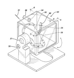
As indicated above, the vaginal model 20 is installed in the pressure
chamber 30. This installation is shown in greater detail in Figs. 4-6. The in
vitro vaginal model 20 is attached to the bottom floor 30a and back wall 30b
of
pressure chamber 30 by a series of flanges and/or clamps. The vaginal lumen
21 is shown in the vaginal model 20. Upper portions of the in vitro vaginal
model 20 are positioned at an angle a to the bottom floor 30a of the pressure
chamber 30. To accurately model the location and orientation of an average
human vagina, the vaginal model is installed to provide an angle a between the
vaginal lumen proximate to the cervical port 24 and the bottom floor 30a of
about 40 .
11
CA 02789482 2012-08-09
WO 2011/100393 PCT/ES2011/024291
Fig. 6 shows a side view of the vaginal model 20 installed in the
pressure chamber 30. The upper portion of the vaginal model 20 is secured to
the back wall 30b of the pressure chamber 30 such that fluid can be injected
into the vaginal lumen 21 at the top of the vaginal model 20 corresponding to
the location of the cervix via a cannula 31. The lower portion of the vaginal
model 20 is attached to an opening in the floor 30a in a manner that the labia
25 of the vaginal model 20 extend beyond the opening. Fluid that is delivered
from the cannula 31 can then flow through the vaginal lumen 21 and may exit
the vaginal model by an opening in the labia 25. In the embodiment of Fig. 6,
the fluid delivery can include a first fluid supply to deliver a relatively
steady
fluid supply via fluid delivery line 68 and a second fluid supply 33 to
deliver fluid
gushes.
During operation, the pressure chamber 30 is appropriately sealed to
maintain background air pressure, the body pressure discussed above. This
pressure is established by application of air through one or more air supply
ports 34.
Body pressure varies with activity and position (sitting/standing/lying
down). SIMA can simulate these variations in body pressures, from greater
than 0 to over 220 cm H20 (0 ¨ 3.13 psi). Body pressures for non-exerting
activities are normally in the range between about 20 cm H20 to about 50 cm
H20. On average, body pressure for sitting is about 23 cm H20, for standing is
27 cm H20 and for lying in a supine position, body pressure is about 2.4 cm
H20. Preferably, the background pressure or first pressure in the SIMA
system is maintained at greater than about 0 cm H20, such as for a supine
woman, more preferably, the background pressure is maintained at greater
than about 20 cm H20. Preferably, the background pressure is less than about
50 cm H20. Thus a preferred first pressure range would be between greater
than about 0 cm H20 and about 50 cm H20. A more preferred range would be
between about 20 cm H20 and about 50 cm H20. The first pressure range is
maintained relatively constant during a test simulation. Preferably, the first
pressure range or background pressure range is maintained within about 5 cm
12
CA 02789482 2012-08-09
WO 2011/100393 PCT/US2011/024291
H20, more preferably, within about 2 cm H20 and most preferably, within about
1 cm H20.
As shown in Fig. 6, dynamic intra-abdominal pressure is provided by air
flowing through flexible hose 35 and nozzle 36 onto the anterior vaginal
surface
22 of a central portion of the in vitro vaginal model 20. As stated
previously,
the anterior vaginal surface 22 was molded as closely as possible to the
actual
nulliparous vagina imaged.
The pressure chamber 30 is mounted on a pivoting support to permit the
system to model a woman in a range or orientations from standing to supine or
lying flat on her back. This is shown in Fig. 4 showing the pressure chamber
30 mounted on a pivot 37 with a release pin 38 to maintain the desired
orientation.
As detailed above and shown in the figures, the vagina can be
characterized as a cone-shaped structure with the proximal end (upper
fornices) being wider than the distal end (introitus). The anterior and
posterior
walls of the vagina are also collapsed together, even with an intra-vaginal
device in place. These walls however can also open and close, depending on
the woman's positioning, muscle structure, and activity. Intra-vaginal
pressures
help fluid move between these walls by compressing the walls together. The
vagina is also curved in the sagittal (side) view. The vagina extends
generally
vertically from the introitus to a "vaginal flexure" at which it begins to
curve to
an angle a of about 40 from the horizontal. This vaginal flexure 27 is about
5
cm above the introitus in the SIMA model based upon the subject from which
the vaginal model 20 was derived. This difference could affect how an intra-
vaginal device is placed in the vagina and consequently the perceived wearing
comfort of the device. Factors that affect the vaginal flexure of the vagina
could
include genetics, muscle structure and strength as well as ligaments and
tendons joining the muscle structure, and pelvic bone structure. In a
preferred
embodiment shown in Figs. 3-6, the vaginal model contains a casting of a
nulliparous vagina, which has the dimensions of 9 cm in length and 5 cm in
width (at the widest location at the proximal end).
13
CA 02789482 2012-08-09
WO 2011/100393
PCT/ES2011/024291
Other embodiments may contain primiparous and/or mulitparous vagina,
which differs from a nulliparous vagina in mainly the width and supporting
structures (angle) of the vagina. MRI studies of mulitiparous women showed
that the vaginal width at the widest part of the vagina (proximal end, near
fornices) ranges from 3.5 to 5.7 cm. Supporting structures to the vagina can
decrease in strength with damage to the pelvic floor muscles due to child
birth,
genetics, surgery, increased weight gain, and other factors. SIMA model can
simulate this change in support structure by changing the angle of the in
vitro
vaginal model. In addition, the length of the vagina can vary widely from
woman to woman. In other embodiments, SIMA system can simulate these
anatomical differences in order to better understand how these factors affect
menstrual fluid flow and its interaction with a tampon or the vaginal anatomy
and an incontinence device.
Any materials may be used to create the final vaginal model. Materials
that may be molded but retain a certain flexibility are preferred. Other
materials
useful for making the vaginal model may include but are not limited to
silicones
(including room-temperature vulcanizing silicone rubber), polyethylene,
castable polyurethane, plasticized polyvinyl chloride, styrene-butadiene,
thermoplastic elastomers, rubber latex, and the like. Preferred materials
include thermoplastic elastomeric materials such as Santoprene TM brand
thermoplastic vulcanates (TPV) (Supplier: Exxon Mobil Chemical, Houston,
Texas, USA). In one preferred embodiment, the vaginal model is made from a
30:60 blend of DS-302 and DS-303 (California Medical Innovations, Pomona,
California). Materials that can be molded but are clear after curing are also
preferred.
The materials may be heated until liquefied and then poured into the
mold. The filled mold may be placed into an oven for curing for a period of
time. The vaginal model is then released from the mold. Once it has cooled, it
is mounted into the support platform.
The molded labia provide a resistance normally seen in women during
insertion of a medical device. The vaginal model also provides realistic
14
CA 02789482 2012-08-09
WO 2011/100393 PCT/US2011/024291
resistance to the medical device in that the model is provided at an angle (as
in
a female body) and the interior walls are a flat cone-like shape. The material
chosen to make the vaginal model is important as the material may affect the
flexibility of the molded labia and molded vagina. A medical doctor
(urologist)
performed a pelvic examination of the in vitro SIMA vagina and found it to be
realistic to a typical female patient.
Examples
The following examples will detail how the vaginal model described
above is used.
Example 1
An example of SIMA was constructed for this example using the diagram
shown in Fig. 2. The following is a discussion of the details and components.
In one preferred embodiment shown schematically in Fig. 7, the
pressure chamber 30, fluid pump 54, and GUI terminal 56 were located within a
safety hood 70. In addition to the safety hood 70, associated compartments
held an air control panel 40, electrical control panel 50, local computer,
e.g., a
PC 52, and a printer 72. In this example, the pressure chamber 30 was
situated within the hood 70 for safety purposes even though safety pressure
relief valves, e.g., 66 were used.
As in Fig. 2, the pressure chamber 30 provided a closed environment
which allowed the in vitro vaginal model 20 to be subjected to pressure
replicating background body pressure and dynamic intra-abdominal pressure.
Body pressure was provided by maintaining the interior of pressure chamber 30
under constant pressure. Intra-abdominal pressure was introduced as air
pressure directed on the anterior vaginal surface 22 on the in vitro vaginal
model 20. Both of these pressures were provided by the air control panel 40
and associated controls discussed below. The body pressure air line 58 of Fig.
2 supplied air to air supply port 34 of Fig.4-6. The dynamic pressure air line
60
CA 02789482 2012-08-09
WO 2011/100393 PCT/US2011/024291
of Fig. 2 supplied air to the flexible air supply hose 35 and nozzle 36 for
directing the dynamic intra-abdominal pressure onto the anterior vaginal
surface 22 of Fig. 4-6.
As shown in Fig. 2, a pressure transducer 64 was connected to the air
solenoid valve 62 to measure air pressure of the intra-abdominal pressure
simulation elements. This pressure transducer measured both the air pressure
within pressure chamber (modeling the background body pressure) and the
dynamic (inter-abdominal) pressure or impulse pressure provided to the vaginal
model 20 via dynamic pressure air line 60.
Fig. 8 is a schematic diagram of the air control panel 40, which may be
located below the rest of the SIMA apparatus. The air control panel 40 had a
pressurized air supply 41 connected to a pressure regulator 42. Downstream
of the pressure regulator 42, a solenoid valve 43 delivered pressurized air to
either of two proportional valves. A first proportional valve 44 having an
integral pressure sensor for pressure regulation related to the body pressure
system and delivered body pressure to the pressure chamber 30 via body
pressure air line 58 (Fig. 2) to air supply port 34 (Figs. 4-6). By means of a
proportional valve, the pressure was regulated to provide constant body
pressure and to release the pressure once testing is complete.
A second proportional valve 45 having an integral pressure sensor for
pressure regulation related to the intra-abdominal pressure system and
delivered pressurized air via air line 46 to an accumulator tank 47. Again,
the
proportional valve regulates the pressure in the accumulator tank 47 to
provide
constant intra-abdominal pressure supply and to release pressure once testing
is complete. The accumulator tank 47 was connected to the pressure chamber
solenoid air valve (cough valve) 62 (Fig. 2) at the pressure chamber 30 via
the
dynamic pressure air line 60. The pressure chamber solenoid air valve 62 can
be switched "On" and "Off' at defined time intervals to deliver the intra-
abdominal pressure pulses to the in vitro vaginal model 20. A pressure relief
valve 48 was located at exit of air accumulating tank 47 and was used as
protection in case of over pressurization of the system.
16
CA 02789482 2012-08-09
WO 2011/100393 PCT/US2011/024291
The SIMA pressurization system allows for precise control of air
pressure inside the pressure chamber 30 (body pressure). As air was
introduced into the pressure chamber 30 to simulate dynamic intra-abdominal
pressure, the air pressure in the pressure chamber 30 (the body pressure)
increased momentarily. Once the intra-abdominal pressure delivery ended, the
body pressure gradually readjusted, automatically, to the original body
pressure
setting. Pressure relief valve 66 (Fig. 2), located on top of pressure chamber
30 provided additional protection in case of over pressurization of the
chamber
30.
As shown in Fig. 9, a fluid pump 54 to deliver simulated menstrual fluid
was located near the pressure chamber 30. While any pump can be used, it is
important to precisely control the delivery of the fluid. For example, the
pump
was able to provide the fluid at a steady rate or in spurts (to imitate
gushes).
Fluid from reservoir 54a was delivered by the fluid pump 54 through tubing 68.
As shown in Fig. 9A, junction 54b linked tubing 68 to cannula 31, which
provided fluid into the cervical port 24 of the in vitro vaginal model 20.
Junction 54b also had a second fluid input port 54c that accommodated a
syringe for alternative fluid injection (independent or in conjunction with
fluid
delivery via 68).
One example of a suitable fluid pump is made by Watson-Marlow Model
520 Di with Pump Head Model 505L, which was used for this example. While
for medical devices such as tampon, an artificial menstrual fluid is
preferred,
other fluids may be substituted.
As shown in Figure 4, the pressure chamber 30 was mounted on a pivot
37. This allowed the pressure chamber 30 to rotate such that the in vitro
vaginal model 20 could be oriented as if in a standing position, sitting or
supine
position. In an alternative embodiment (not shown), a secondary pivoting
means may rotate the box to mimic side turning or sleeping. In the
embodiment shown in the figures, the pivoting means rotated the entire
pressure chamber 30 until the desired position at which point release pin 38
17
CA 02789482 2012-08-09
WO 2011/100393 PCT/US2011/024291
was engaged. Upon completion of the test, release pin 38 was disengaged to
permit the pressure chamber 30 to return to its original position.
In vitro vaginal model 20 included vaginal lumen 21, cervical port 24,
and labia 25. Intra-abdominal pressure nozzle 36 was disposed adjacent to the
central portion of anterior vaginal surface 22 of the in vitro vaginal model
20
(Fig. 4-6).
Synthetic menstrual fluid was delivered through the cervical port 24 via
cannula 31, and it flowed within the vaginal lumen 21, and out through the
labia
25 (Figs. 4-6). During the testing of a medical device, the device was
inserted
into the vaginal model 20 prior to introducing the synthetic menstrual fluid.
In the embodiment shown in the figures, the temperature was room
temperature but additional controls may be put into place to elevate or lower
the temperature of the interior of the box, which would include the vaginal
model and fluid.
SIMA allows for two modes of operation ¨ manual and automatic. These
modes are further discussed in Examples 2 and 3.
Another advantage to a system like SIMA is the ability to capture data. It
is also possible to monitor the test by video recording devices and/or taking
photographs during the testing as the vaginal model can be transparent.
Whenever testing commences, the data acquisition system can also
commence automatically. The data acquisition system is capable of, but not
limited to, recording of events at specified sampling rate and recording the
date, time, test name, user name, body pressure, intra-abdominal pressure at
accumulating tank, intra-abdominal pressure delivered to the vaginal model,
menstrual pump flow and menstrual pump flow events. During the recording
period, the data is saved to the PC hard drive under an assigned file name.
Data is saved in comma-delimited format, also referred to as comma-separated
values (CSV) format and can be imported into an Excel spreadsheet. Data can
be imported into an Excel spreadsheet to enable further data visualization,
data
management, data archiving, graphical representation and report generation.
18
CA 02789482 2012-08-09
WO 2011/100393 PCT/US2011/024291
During the data acquisition process, the acquired data can be viewed in
real-time on the Graphical User Interface (GUI) terminal. It is possible to go
forward and backward in time using the arrows to either side of the pause
button. Pressing the pause button again will bring the trend screen back into
real-time monitoring. The capability also exists to perform print screen
function.
Example 2 ¨ Manual Mode
Step A - Prior to using the apparatus, artificial menstrual fluid is prepared
according to patent publication US 20070219520. The computer, monitor, and
fluid pump is turned on. The internal vaginal cavity is cleaned of any
residual
menstrual fluid from previous testing by using a cotton swab and the pump is
flushed with water until the hosing is clean. Next, the vaginal cavity is
primed
with artificial menstrual fluid prior to testing. The fluid pump is calibrated
and
the cannula is placed within the cervix opening of the vaginal model. The box
is then closed and set into the desired testing position (sitting, standing,
or
supine). A tampon is weighed without the cello wrap (example: commercial
o.b. tampon regular absorbency made by McNeill Consumer Products). A
small amount (-0.1-0.2 grams) of KY gel is placed on the tip of the tampon
(to
facilitate insertion). The tampon is weighed tampon with KY and the weight
recorded. The tampon is inserted into the opening of the labial portion. In
some cases, it is preferred to place the tampon to the left side of the vagina
as
tampons are typically placed on the lateral wall of the vagina (either left or
right). The placement of the tampon is recorded, as measured from the cervix.
In a preferred embodiment, the tampon is placed about 10 mm below the
cervix.
Step B - On the touch screen controls, the "Manual mode" of operation is
selected. The desired body pressure is selected (in cm H20). In a preferred
embodiment, 27 cm H20 was selected to simulate a standing position. The
green switch "On" (next to the heading "Body Pressure") is depressed to
engage body pressure. The cough pressure is selected (in cm H20). In one
preferred example, 146 cm H20 was selected. Additionally, the "cough on
19
CA 02789482 2012-08-09
WO 2011/100393 PCT/US2011/024291
time" and "cough full cycle time" is selected. The "cough on time" represents
the duration of the single cough and in a preferred embodiment, 1.0 second
was selected. The "cough full cycle time" represents the time period during
which the cough will occur. In a preferred embodiment, 10 seconds is selected.
These settings will provide a 1 (one) second cough every 10 seconds for a
cough pressure of 146 cm H20. The green switch "ON" (Next to the heading
Cough Pressure) is depressed to engage the cough pressure. Next the
menstrual flow in mL/Min is entered. In a preferred embodiment, 1 mL/Min is
entered. The "Flow On Time" is entered, which is the duration of the flow. In
a
preferred embodiment, 10 seconds is selected. The "flow full cycle time" is
entered. In one preferred embodiment, 20 seconds is selected. This setting
will provide 10 seconds of fluid flow every 20 seconds. The pump calibration
value is entered and the green "ON" button (next to the heading "Menstrual
Flow") depressed to engage the menstrual flow.
Step C - The start button is depressed to start testing. Video recording
and/or digital photographs may be obtained during testing. To stop testing,
the
Stop button is depressed.
Step D - At the conclusion of the test, the tampon is carefully removed and
the stain pattern and weight is recorded. The internal vaginal anatomy of the
vaginal model is cleaned by using a cotton swab and the fluid pump/hosing
flushed with water.
Example 3 ¨ Automatic Mode
Repeat Step A from Example 2
Step B - On the touch screen controls, press the "Automatic mode" of
operation. This mode of operation allows a user to re-run a previously set up
"recipe" or enter the parameters for a new one. As used herein, the term
"recipe" shall mean the combination of pressures (body, intra-abdominal), time
sequences, and flow, all of which happen at preset times intervals. If this is
a
first time recipe, the user inputs the information and creates a recipe name
or
CA 02789482 2012-08-09
WO 2011/100393
PCT/US2011/024291
number, which is then saved. For example, the conditions of Step B of the
manual mode are inputted and saved as a new recipe. Parameters such as
body pressure and step length (length of time you would like to run the
entered
body pressure in seconds) are entered. Likewise, the cough pressure recipe is
selected, which includes the cough pressure in cm H20, cough on time in
seconds, full cycle time in seconds, count (number of times you would like to
run the cycle), and the step length (count x full cycle time). The menstrual
flow
recipe is entered in mUmin, flow on time in seconds, full cycle time in
seconds,
count (number of times the cycle is to run), and the step length (count x full
cycle time). The save button is engaged at the top of the screen to save this
recipe.
Step C ¨ The testing is begun by pressing the start button. Video
recording and/or digital photographs may be obtained during testing. The
system completes its cycle automatically, or it is manually stopped, as
appropriate.
Step D - At the conclusion of the test, the tampon is carefully removed
and the stain pattern and weight is recorded. The internal vaginal anatomy of
the vaginal model is cleaned by using a cotton swab and the fluid pump/hosing
flushed with water.
The user may also edit or run a previously entered "recipe", which would
include other cycles, trends, etc.
For both the manual and automatic mode of running SIMA, the
instantaneous pressure may be manually repeated or automatically
programmed to repeat at certain time intervals. For example, SIMA may be
used to study the effects of coughing every 60 seconds, each cough being 1
second in duration. This means that the vaginal model of SIMA may be under
a "body pressure" of 30 cm H20 for 60 seconds, with the "intra-abdominal
pressure" reaching 150 cm H20 for 1 second. This trending seeks to replicate
the body dynamics of a menstruating or incontinent woman when she coughs ¨
a body action which is intense and brief.
21
CA 02789482 2012-08-09
WO 2011/100393
PCT/US2011/024291
Example 4 ¨ Use with external sanitary products
The SIMA system includes the external labial anatomy, which was
casted from a woman, is made out of a soft, transparent, and stretchable
material. The movement of fluid along the labia and gushes are important
aspects in understanding how external sanitary napkins interact with the
anatomy. Fluid movement and gushes can be simulated in the SIMA system.
The SIMA system is setup with the same parameters used in any of
Examples 1-3. Body pressure is applied to the system at 27 cm H20, cough
pressure is set to 146 cm H20 (with a cough on time of 1 second and full cycle
time of 10 seconds), and menstrual fluid flow is set to 1 mL/min with the flow
on
for 10 seconds over a 20 second cycle. Gushes (3mL) are also introduced to
the system after 3 minutes of flow using a syringe. A Stayfree0 regular
ultrathin napkin is manually applied against the SIMA system and the fluid
movement and interaction with the anatomy is observed. Time to leak and
add-on (in grams) are recorded. Observations are made on how the fluid
moves along the body and interacts with the napkin. These observations are
important in developing new insights into the absorbent system of napkins and
how leakage can occur.
Example 5 ¨ Use with intravaqinal incontinence devices
The SIMA system includes the vaginal anatomy which was replicated
from a 3D vaginal computer model. The 3D model was reconstructed from 2D
MRI. Having the actual geometry of the vaginal anatomy is an important
aspect in understanding how intra-vaginal devices such as incontinence
pessaries fit and stay in place in the vagina.
The SIMA system is set up with the same pressure parameters used in
Examples 1-3. Body pressure is applied to the system at 27 cm H20 and
22
CA 02789482 2012-08-09
WO 2011/100393 PCT/US2011/024291
cough pressure is set to 146 cm H20 (with a cough on time of 1 second and full
cycle time of 10 seconds). Menstrual fluid flow is turned off for incontinence
device testing. The incontinence device is inserted into the vagina and the
device's ability to stay in place is recorded and observed.
The specification and embodiments above are presented to aid in the
complete and non-limiting understanding of the invention disclosed herein.
Since many variations and embodiments of the invention can be made without
departing from its spirit and scope, the invention resides in the claims
hereinafter appended.
23