Note: Descriptions are shown in the official language in which they were submitted.
CA 02790134 2012-08-16
WO 2011/100841 PCT/CA2011/050096
1
PD-1 MODULATION AND USES THEREOF FOR MODULATING HIV REPLICATION
CROSS REFERENCE TO RELATED APPLICATION
This application claims the benefit of United States Provisional Patent
Application
serial No. 61/304,864 filed on February 16, 2010, which is incorporated herein
by reference in
its entirety.
TECHNICAL FIELD
The present invention generally relates to the modulation of Human
Immunodeficiency
Virus (HIV) infection, and more particularly to methods and compositions for
inhibiting or
enhancing HIV replication.
BACKGROUND ART
Human Immunodeficiency Virus-1 (HIV-1) is the etiologic agent that is
responsible for
Acquired Immunodeficiency Syndrome (AIDS), a syndrome characterized by
depletion of CD4+
T lymphocytes and collapse of the immune system. HIV-1 infection is pandemic
and HIV-
associated diseases have become a world-wide health problem. Upon infection,
HIV integrates
into the cellular genome of an infected cell. HIV-1 infection then leads to
two different
scenarios: productive infection and latent infection. Productive infection
occurs most frequently
and leads to death of the infected cell after release of progeny virus. During
latent infection,
which is rare, HIV genes are not expressed after proviral integration,
resulting in an infected cell
that is characterized by transcriptionally silent HIV genes. These fully
replication-competent HIV
can persist dormant in cells for several years and then become reactivated
(Chun et al., 1995,
Nat Med 1(12):1284-1290; Chun et al., 1997, Proc Natl Acad Sci USA
94(24):13193-13197).
Current treatments of HIV infection typically seek to block one or more steps
involved
in the production of viral particles. Treatment options involve administration
of reverse
transcriptase inhibitors, inhibitors of viral protease, fusion, entry, or
integration inhibitors in
different combinations to block multiple steps in the viral life cycle. This
approach, termed highly
active antiviral therapy (HAART) has greatly decreased morbidity and mortality
in people
infected with HIV (Palella et al., 1998, N Engl J Med 338(13):855-860).
However, there are
several concerns about HAART regimens, including serious side effects of the
drugs,
complexity of the regimens, requirement of lifelong adherence and development
of drug
resistance (particularly in cases of non-compliance).
Furthermore, studies have shown that HAART is not effective in completely
eradicating
HIV in patients. In most cases, a rapid rebound in viremia occurs upon
discontinuation of
HAART, even after several years of successful treatment with undetectable
viral loads (Davey
et al., 1999, Proc Natl Acad Sci USA 96(26):15109-15114; Cohen and Fauci,
2001, Adv Intern
CA 02790134 2012-08-16
WO 2011/100841 PCT/CA2011/050096
2
Med 46: 207-246). It is believed that this rebound in viremia is due, at least
in part, to the
reactivation of latent HIV that persists in a small fraction of resting memory
CD4+ T cells.
Although the frequency of latently-infected CD4+ T cells (typically referred
to as the HIV
reservoir) is very low, this latent population of HIV serves as a source of
virus for reseeding the
infection after HAART discontinuation.
There is thus a need for novel strategies for modulating HIV replication, and
for the
treatment of associated conditions such as AIDS.
The present description refers to a number of documents, the content of which
is
herein incorporated by reference in their entirety.
SUMMARY OF THE INVENTION
The present invention relates to the modulation of Human Immunodeficiency
Virus
(HIV) infection, and more particularly to methods, compositions, uses and kits
for inhibiting or
enhancing HIV replication.
In a first aspect, the present invention provides a method for inhibiting
Human
Immunodeficiency Virus (HIV) replication in a cell comprising contacting said
cell with a
Programmed Death-1 (PD-1) agonist.
In another aspect, the present invention provides a use of a PD-1 agonist for
inhibiting
HIV replication in a cell.
In another aspect, the present invention provides a use of a PD-1 agonist for
the
preparation of a medicament for inhibiting HIV replication in a cell.
In another aspect, the present invention provides a use of a PD-1 inhibitor
for
increasing HIV replication in a cell.
In another aspect, the present invention provides a use of a PD-1 inhibitor
for the
preparation of a medicament for increasing HIV replication in a cell.
In another aspect, the present invention provides a method for increasing HIV
replication in a cell comprising contacting said cell with a PD-1 inhibitor.
In another aspect, the present invention provides a use of a PD-1 inhibitor
for
increasing HIV replication in a cell.
In another aspect, the present invention provides a use of a PD-1 inhibitor
for the
preparation of a medicament for increasing HIV replication in a cell.
In another aspect, the present invention provides a method for reducing or
eliminating
a latent HIV reservoir in a cell comprising: (a) performing the method for
increasing HIV
replication in a cell defined above; and contacting said cell with one or more
antiretroviral
agents.
CA 02790134 2012-08-16
WO 2011/100841 PCT/CA2011/050096
3
In another aspect, the present invention provides a method for decreasing the
number
of latently HIV-infected cells in a subject, said method comprising
administering to said subject
an effective amount of: (a) a PD-1 inhibitor; and (b) one or more
antiretroviral agents.
In another aspect, the present invention provides a use of (i) a PD-1
inhibitor and (ii)
one or more antiretroviral agents for eliminating a latent HIV reservoir in a
cell.
In another aspect, the present invention provides a use of (i) a PD-1
inhibitor and (ii)
one or more antiretroviral agents for the preparation of a medicament for
eliminating a latent
HIV reservoir in a cell.
In another aspect, the present invention provides a use of (i) a PD-1
inhibitor and (ii)
one or more antiretroviral agents for decreasing the number of latently HIV-
infected cells in a
subject.
In another aspect, the present invention provides a use of (i) a PD-1
inhibitor and (ii)
one or more antiretroviral agents for the preparation of a medicament for
decreasing the
number of latently HIV-infected cells in a subject.
In another aspect, the present invention provides a composition for inhibiting
HIV
replication in a cell, said composition comprising a PD-1 agonist and a
carrier.
In another aspect, the present invention provides a composition for inhibiting
HIV
replication in an HIV-infected, said composition comprising a PD-1 agonist and
a carrier.
In another aspect, the present invention provides a composition for increasing
HIV
replication in a cell, said composition comprising a PD-1 inhibitor and a
carrier.
In another aspect, the present invention provides a composition for reducing
or
eliminating a latent HIV reservoir in a cell, said composition comprising a PD-
1 inhibitor, one or
more antiretroviral agents, and a pharmaceutically acceptable carrier.
In another aspect, the present invention provides a composition for decreasing
the
number of latently HIV-infected cells in a subject, said composition
comprising a PD-1 inhibitor,
one or more antiretroviral agents, and a carrier.
In an embodiment, the above-mentioned cell is a CD4+ T cell. In an embodiment,
the
above-mentioned agonist is a PD-1 ligand. In a further embodiment, the above-
mentioned PD-1
ligand comprises a PD-L1 polypeptide or an extracellular domain thereof
(having PD-1 agonist
activity). In a further embodiment, the above-mentioned PD-1 ligand comprises
an extracellular
domain of PD-L1 (having PD-1 agonist activity) linked to an antibody Fc
domain. In an
embodiment, the above-mentioned antibody Fc domain is a human IgG1 domain.
In an embodiment, the above-mentioned PD-1 inhibitor blocks the interaction
between
PD-1 and a PD-1 ligand. In a further embodiment, the above-mentioned PD-1
ligand is PD-L1.
In an embodiment, the above-mentioned PD-1 inhibitor is an anti-PD-1 antibody
or
antigen-binding fragment thereof.
CA 02790134 2012-08-16
WO 2011/100841 PCT/CA2011/050096
4
In another embodiment, the above-mentioned cell is a latently HIV-infected
cell. In a
further embodiment, the above-mentioned latently-infected cell is a CD4+ T
cell.
In another aspect, the present invention provides a method for determining
whether a
test compound may be useful for inhibiting HIV replication, said method
comprising:
(a) contacting a cell expressing PD-1 or a functional variant or fragment
thereof with
said test compound;
(b) determining whether PD-1 activity is increased in the presence of said
test
compound relative to the absence thereof;
wherein an increase in said activity in the presence of said of said test
compound relative to the
absence thereof is indicative that said test compound may be useful for
inhibiting HIV
replication.
In another aspect, the present invention provides a method for determining
whether a
test compound may be useful for decreasing the number of latently HIV-infected
cells in a
subject, said method comprising:
(a) contacting a cell expressing PD-1 or a functional variant or fragment
thereof with
said test compound;
(b) determining whether PD-1 activity is decreased in the presence of said
test
compound relative to the absence thereof;
wherein a decrease in said activity in the presence of said of said test
compound relative to the
absence thereof is indicative that said test compound may be useful for
decreasing the number
of latently HIV-infected cells in a subject.
In another aspect, the present invention provides a method for determining
whether a
test compound may be useful for increasing HIV replication in a cell, said
method comprising:
(a) contacting a cell expressing PD-1 or a functional variant or fragment
thereof with
said test compound;
(b) determining whether PD-1 activity is decreased in the presence of said
test
compound relative to the absence thereof;
wherein a decrease in said activity in the presence of said of said test
compound relative to the
absence thereof is indicative that said test compound may be useful for
increasing HIV
replication in a cell.
In another aspect, the present invention provides a method for obtaining a
cell
population enriched in latently HIV-infected cells, the method comprising:
contacting said cell
population with an agent binding to PD-1; and isolating/purifying the cells on
which the ligand is
bound, thereby obtaining a cell population enriched in latently HIV-infected
cells.
Other objects, advantages and features of the present invention will become
more
apparent upon reading of the following non-restrictive description of specific
embodiments
thereof, given by way of example only with reference to the accompanying
drawings.
CA 02790134 2012-08-16
WO 2011/100841 PCT/CA2011/050096
BRIEF DESCRIPTION OF DRAWINGS
In the appended drawings:
FIGs. 1A-1C show the frequency of CD4+ T cells expressing PD-1 in HIV-infected
subjects. FIG. 1A: Correlation between the frequency of CD4+ T cells
expressing PD-1 and the
5 frequency of CD4+ T cells harbouring HIV integrated DNA in a cohort of 32
HIV-infected
subjects receiving suppressive HAART. FIG. 1B: Frequency of CD4+ T cells
expressing PD-1 in
HIV negative controls (circles; n= 8), HIV-infected subjects receiving
suppressive HAART
(squares; n = 9) and HIV-infected untreated subjects (triangles; n = 10). FIG.
1C: Frequency of
naive (CD45RA+ CCR7+ CD27+, TN), central memory (CD45RA- CCR7+ CD27+, TcM),
transitional memory (CD45RA- CCR7- CD27+, TTM) and effector memory (CD45RA-
CCR7-
CD27-, TEM) CD4+ T cells expressing PD-1 measured in CD4+ T cells from 9
virally suppressed
subjects. PD-1 expression was measured by flow cytometry in total CD4+ T cells
(FIGs. 1A and
1B) or in gated memory CD4 T cells subsets using the CD45RA, CCR7, and CD27
markers
(FIG. 11C).
FIG. 2A and 2B shows the frequency of PD-1hi (left bar of each pair) and PD-11
(right
bar of each pair) cells harbouring HIV DNA and integrated HIV DNA in untreated
HIV-infected
subjects (FIG. 2A) and virally-suppressed subjects (FIG. 2B). Memory CD4+ T
cell subsets
(TcM, TTM and TEM) from 2 untreated, viremic subjects and 2 HAART-treated,
virally-suppressed
subjects were sorted according to their relative expression of PD-1. Sorted
cells were subjected
to ultrasensitive quantitative PCR to measure the frequency of cells
harbouring HIV DNA and
integrated HIV DNA.
FIG. 3 shows the effect of PD-1 triggering on HIV replication in primary CD4+
T cells.
CD4+ T cells from 4 viremic donors were isolated by magnetic negative
selection and activated
with beads coated with anti-CD3 + anti-CD28 antibodies and with the Fc-PD-L1
chimera, or the
appropriate isotype (IgG2) control (NS = non stimulated). Cell supernatants
were collected after
3 (d3), 6 (d6) and 9 (d9) days of culture, and viral replication was measured
by p24 ELISA;
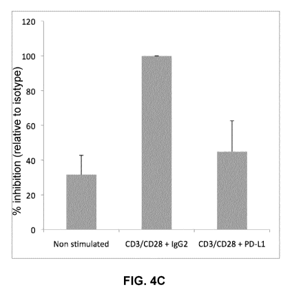
FIGs. 4A-4C show the effect of PD-1 triggering on early HIV replication in
primary
CD4+ T cells. CD4+ T cells from 7 viremic donors were isolated by negative
selection and
activated with beads coated with anti-CD3 + anti-CD28 antibodies and with the
Fc-PD-L1
chimera, or the appropriate isotype (IgG2) control. Cell supernatants were
collected after 24
hours of stimulation, and viral particles were pelleted by
ultracentrifugation. After extraction of
viral RNA, viral production was measured by ultrasensitive real time RT-PCR.
FIGs. 4A and 4B
show the raw data obtained in five representative donors, and FIG. 4C shows
the mean values
and standard deviations (SD) obtained from 7 independent experiments,
expressed as a
percentage of viral production relative to the positive control (anti-CD3 +
anti-CD28 antibodies
and isotype (IgG2) control).
CA 02790134 2012-08-16
WO 2011/100841 PCT/CA2011/050096
6
FIGs. 5A and 5B show the effect of PD-1 triggering on "early" (24h, FIG. 5A)
or "late"
(3, 6 and 9 days, FIG. 5B) HIV replication in primary CD4+ T cells in the
presence of
antiretroviral molecules (ARV). CD4+ T cells from 6 viremic donors were
isolated by negative
selection and activated with beads coated with anti-CD3 + anti-CD28 antibodies
and with the
Fc-PD-L1 chimera, or the appropriate isotype (IgG2) control, in the presence
of antiretroviral
molecules (ARV). FIG. 5A: Cell supernatants were collected after 24 hours of
stimulation, and
viral particles were pelleted by ultracentrifugation. After extraction of
viral RNA, viral production
was measured by ultrasensitive real time RT-PCR. The data obtained in 4
representative
subjects are depicted. FIG. 5B: Cell supernatants were collected after 3 (d3),
6 (d6) and 9 (d9)
days of culture, and viral replication was measured by p24 ELISA. Circles: non-
stimulated (NS)
+ ARV; triangles: anti-CD3 + anti-CD28 antibodies + Fc-PD-L1 chimera; squares:
anti-CD3 +
anti-CD28 antibodies + isotype (IgG2) control. The data obtained in 2
representative subjects
are depicted;
FIG. 6 shows the effect of PD-1 triggering in primary CD4+ T cells expressing
high (top
panel) or low (bottom panel) levels of PD-1. Memory CD4 T cells (CD3+ CD4+
CD45RA-) from 2
untreated subjects were sorted according to their relative expression of PD-1
and activated with
beads coated with anti-CD3 + anti-CD28 antibodies and with the Fc-PD-L1
chimera, or the
appropriate isotype (IgG2) control. Cell supernatants were collected after 3
days of culture, and
viral replication was measured by p24 ELISA;
FIG. 7 shows the effect of blocking the PD-1/PD-L1 interaction on viral
production in
CD4+ T cells. CD4+ T cells from 3 viremic donors were isolated by negative
selection and
incubated with a monoclonal anti-PD-1 antibody (ONO-4538), a fully human IgG4
(Medarex
Inc.; Cat. No. MDX-1106). The anti-PD-1 human monoclonal antibody MDX-1106
binds to PD-1
and prevents the interaction with its ligands PD-L1 and PD-L2. Cell
supernatants were collected
after 3 days and viral replication was measured by p24 ELISA;
FIGs. 8A and 8B show the amino acid (SEQ ID NO: 2) and nucleotide (SEQ ID NO:
1)
sequences, respectively, of human PD-1. The signal peptide is indicated in
italics in the amino
acid sequence, and the coding region is indicated in bold in the nucleotide
sequence;
FIGs. 9A and 9B show the amino acid (SEQ ID NO: 4) and nucleotide (SEQ ID NO:
3)
sequences, respectively, of human PD-L1. The signal peptide is indicated in
italics in the amino
acid sequence, and the coding region is indicated in bold in the nucleotide
sequence;
FIGs. 10A and 10B show the amino acid (SEQ ID NO: 14) and nucleotide (SEQ ID
NO: 13) sequences, respectively, of human PD-L2. The signal peptide is
indicated in italics in
the amino acid sequence, and the coding region is indicated in bold in the
nucleotide sequence;
FIG. 11 shows an amino sequence alignment of mouse and human PD-L1 and PD-L2
(from Latchman et al., 2001, Nature Immunology 2: 261-268);
CA 02790134 2012-08-16
WO 2011/100841 PCT/CA2011/050096
7
FIG. 12 shows an amino sequence alignment of the ectodomains of mouse and
human
PD-1.
DISCLOSURE OF INVENTION
Inhibition of HIV replication
In the studies described herein, the present inventors have shown that
modulating PD-
1 activity has an effect on HIV replication in primary CD4+ T cells obtained
from chronic HIV-
infected subjects. More specifically, they have shown that engagement of PD-1
using its natural
ligand PD-L1 results in an inhibition of HIV replication in activated primary
CD4+ T cells from
HIV-infected subjects.
Accordingly, in a first aspect, the present invention provides a method for
inhibiting HIV
replication in a cell comprising contacting said cell with a PD-1 agonist. The
present invention
also provides a method for treating HIV infection, as well as treating a
related condition such as
AIDS, in a subject, comprising administering to said subject an effective
amount of a PD-1
agonist. The present invention also provides a use of a PD-1 agonist for
inhibiting HIV
replication in a cell, or for the preparation of a medicament for inhibiting
HIV replication in a cell.
The present invention also provides a use of a PD-1 agonist for treating HIV
infection in a
subject (as well as treating a related condition such as AIDS), or for the
preparation of a
medicament for treating HIV infection in a subject (as well as treating a
related condition such
as AIDS). The present invention also provides a composition for inhibiting HIV
replication in a
cell and/or for treating HIV infection in a subject (as well as treating a
related condition such as
AIDS), said composition comprising a PD-1 agonist and a pharmaceutically
acceptable carrier
or excipient. In an embodiment, the above-mentioned cell is a latently HIV-
infected cell. In
another embodiment, the above-mentioned cell is a CD4+ T cell, in a further
embodiment a
memory CD4+ T cell, in a further embodiment a particular subset of memory CD4+
T cell, such
as a central memory (CD45RA- CCR7+ CD27+, TOM), transitional memory (CD45RA-
CCR7-
CD27+, TTM) or effector memory (CD45RA- CCR7- CD27-, TEM) CD4+ T cell. In
another
embodiment, the above-mentioned cell expresses PD-1.
PD-1, a member of the immunoglobulin (Ig) superfamily, is highly upregulated
on
activated lymphocytes and monocytes. It interacts with its two known ligands
PD-L1 (B7-H1)
and PD-L2 (B7-DC). PD-L1 is constitutively expressed on splenic T cells, B
cells, monocytes,
macrophages and dendritic cells (DCs), and its expression can be induced by
activation of T
lymphocytes, monocytes, macrophages and DCs. PD-L2 is expressed on non-
lymphoid tissues
and is upregulated on monocytes and DCs after activation.
Human PD-1 is a Type I membrane protein of 268 amino acids (precursor = 288
amino
acids) comprising an extracellular portion (from about residues 21 to 170)
that includes an IgV
domain (from about residues 35 to 145), a transmembrane domain (from about
residues 171 to
CA 02790134 2012-08-16
WO 2011/100841 PCT/CA2011/050096
8
191 and an intracellular tail (from about residues 192 to 288). The
intracellular tail contains two
phosphorylation sites located in an immunoreceptor tyrosine-based inhibitory
motif (residues
221 to 226) and an immunoreceptor tyrosine-based switch motif (residues 246 to
251). These
two motifs are involved in the recruitment of the phosphatases SHP-1 and SHP-
2, which at
least in part mediates the inhibitory activity of PD-1 (Sheppard et al., 2004,
FEBS Letters,
574(1-3): 37-41). The amino acid and nucleotide sequences of human PD-1 are
shown in FIGs.
8A and 8B, respectively.
"PD-1 agonist" as used herein refers to any agent capable of
inducing/triggering the
PD-1 signalling pathway in a cell. It includes agents that binds to PD-1
(e.g., to the extracellular
portion of PD-1) and triggers an intracellular signal, such as a natural or
synthetic PD-1 ligand
(e.g., an agonistic antibody, as described in PCT publications Nos. WO
04/056875, WO
10/029434 and WO 10/029435). In an embodiment, the above-mentioned PD-1
agonist is a
natural PD-1 ligand (e.g., PD-L1, PD-L2), or a functional variant or fragment
thereof (a variant
or fragment exhibiting PD-L1 or PD-L2 activity). In a further embodiment, the
above-mentioned
natural PD-1 ligand is PD-L1 or a functional variant or fragment thereof.
Human PD-L1 is a Type I membrane protein of 272 amino acids (precursor = 290
amino acids) comprising an extracellular portion (from about residues 19 to
238) that includes
an IgV domain (from about residues 18 to 130), a transmembrane domain (from
about residues
239 to 261 and a short intracellular tail (from about residues 262 to 290).
The amino acid and
nucleotide sequences of human PD-L1 are shown in FIGs. 9A and 9B,
respectively. Human
PD-L2 is a Type I membrane protein of 253 amino acids (precursor = 273 amino
acids)
comprising an extracellular portion of about 201 amino acids that includes an
IgV domain
(about residues 35 to 120) and a C-like Ig domain (about residues 137 to 193),
a
transmembrane domain of about 24 residues (about residues 221-241) and a short
intracellular
tail of about 28 residues. The amino acid and nucleotide sequences of human PD-
L2 are
shown in FIGs. 10A and 10B, respectively.
Functional variants and fragments of PD-L1 or PD-L2 as used herein refers to
variants
(PD-L1/PD-L2 mutants having one or more substitutions, deletions and/or
additions relative to
native PD-L1/PD-L2) or fragments of PD-L1/PD-L2 (e.g., the extracellular
portion of PD-L1/PD-
L2), which retain the activity of native PD-L1/PD-L2, such as the ability to
bind PD-1 and to
trigger a signal through PD-1. In an embodiment, the above-mentioned PD-1
agonist comprises
a fragment of PD-L1/PD-L2, such as the extracellular fragment of PD-L1/PD-L2.
In a further
embodiment, the above-mentioned PD-L1/PD-L2 fragment comprises the IgV domain.
In
another embodiment, the above-mentioned PD-L1 fragment comprises one or more
of residues
19, 20, 26, 54, 56, 66, 113, 115, 117, and 121-125 of the IgV domain of PD-L1.
In an embodiment, the above-mentioned PD-L1/PD-L2 derivative is a PD-L1/PD-L2,
or
a fragment thereof (e.g., the extracellular fragment of PD-L1/PD-L2), linked
to an Fc portion of
CA 02790134 2012-08-16
WO 2011/100841 PCT/CA2011/050096
9
an antibody (directly or via a linker), such as the Recombinant Human B7H1/PD-
L1 Fc Chimera
commercially available from R&D SystemsTM (Cat. No. 156-B7), which comprises
residues
Phe19 to Thr167 of human PD-L1 linked to residues Pro100 to Lys330 of human
IgG1 via a
linker (sequence: DIEGRMD, SEQ ID NO: 11), or the Recombinant Human PD-L2 Fc
Chimera
commercially available from R&D SystemsTM (Cat. No. 1224-PL), which comprises
residues
Leu20 to Pro219 of human PD-L2 linked to residues Pro100 to Lys330 of human
IgG1 via a
linker (sequence: IEGRMD, SEQ ID NO: 12).
The domains and residues of human PD-1 and PD-L1 involved in their interaction
is
described in for example Lin et al., Proc. Natl. Acad. Sci. 2008 105(8): 3011-
3016. The IgV
domains of PD-1 (from about residues 35 to 145, and more particularly residues
64, 66, 68, 73-
76, 78, 90, 122, 124, 126, 128, 130-132, 134 and 136) and PD-L1 (from about
residues 18 to
130, and more particularly residues 19, 20, 26, 54, 56, 66, 113, 115, 117, and
121-125) are
involved in the interaction. Similarly, the domains and residues of mouse PD-1
and PD-L1
involved in their interaction is described in for example Lazar-Molnar et al.,
Proc. Natl. Acad.
Sci. 2008 105(30): 10483-10488. The IgV domains of murine PD-1 (more
particularly residues
31, 33, 35, 40, 42, 43, 45, 50, 95, 99 and 103) and murine PD-L2 (more
particularly residues
21, 28, 56, 60, 101, 110, 112, 113 and 114) are involved in the interaction.
It may be expected
that the most or all corresponding residues of human PD-1 and PD-L2 (which may
be readily
identified by sequence comparison/alignment, FIGs. 11 and 12) also interact
with each others
(Lin et al., 2008, supra; Lazar-Molnar et al., 2008, supra). Based on this
knowledge, the skilled
person would be able to identify/prepare active (which may be used as
agonists) and/or inactive
(which may be used as PD-1 inhibitors) fragments and/or variants of PD-1/PD-
L1/PD-L2, as
well as compounds/agents (e.g., peptides, antibodies, small molecules) capable
of blocking the
PD-1 - PD-L1/PD-L2 interaction.
As used herein, the terms "treat", "treating", and "treatment" include
inhibiting the
condition or disease, i.e., arresting or reducing the development or
progression of the condition
or disease or its clinical symptoms; or relieving the condition or disease,
i.e. causing regression
of the condition or disease or its clinical symptoms. Treatment means any
manner in which the
symptoms or pathology of a condition, disorder, or disease are ameliorated or
otherwise
beneficially altered.
In further embodiments, the methods of the invention are for preventing a
condition or
disease, i.e., causing the clinical symptoms of the condition or disease not
to develop in a
subject that may be predisposed to the condition or disease but does not yet
experience any
symptoms of the condition or disease, or reducing the onset of the condition
or disease, or
symptoms thereof (or severity thereof). Prevention encompasses prophylaxis.
Preferably, the subject in need of such treatment or prevention is a mammal,
more
preferable a human.
CA 02790134 2012-08-16
WO 2011/100841 PCT/CA2011/050096
Increase of HIV replication / reactivation of the latent HIV reservoir
The present inventors have further shown that an increase in HIV replication
was
observed following incubation of primary CD4+ T cells with an antibody
blocking the interaction
5 between PD-1 and PD-L1. Accordingly, in another aspect, the present
invention provides a
method for increasing HIV replication in a cell comprising contacting said
cell with a PD-1
inhibitor. The present invention also provides a use of a PD-1 inhibitor for
increasing HIV
replication in a cell, or for the preparation of a medicament for increasing
HIV replication in a
cell.
10 The present invention also provides a method for reactivating HIV
replication in a
latently HIV-infected cell, said method comprising contacting said cell with a
PD-1 inhibitor. The
present invention also provides a use of a PD-1 inhibitor for reactivating HIV
replication in a
latently HIV-infected cell, or for the preparation of a medicament for
reactivating HIV replication
in a latently HIV-infected cell.
As used herein, the term "PD-1 inhibitor" includes any compound able to
directly or
indirectly affect the regulation of PD-1 by reducing for example the
expression of PD-1 (i.e.,
transcription and/or the translation) or its natural ligands PD-L1/PD-L2, or a
PD-1 activity. It
includes intracellular (e.g., agents that block a PD-1-associated signalling
molecule or pathway,
such as SHP-1 and SHP-2) as well as extracellular PD-1 inhibitors. Without
being so limited,
such inhibitors include siRNA, antisense molecules, proteins, peptides, small
molecules,
antibodies, etc.
In an embodiment, the above-mentioned PD-1 inhibitor blocks/inhibits the
interaction
between PD-1 and a PD-1 ligand (e.g., PD-L1, PD-L2). Such inhibitor may
target, for example,
the IgV domain of PD-1 and/or PD-L1 and/or PD-L2, such as one or more of the
residues
involved in the interaction, as discussed above.
In an embodiment, the above-mentioned PD-1 inhibitor is a blocking antibody,
such as
an anti-PD-1 or anti-PD-L1/PD-L2 antibody. Blocking anti-PD-1 and/or anti-PD-
L1/PD-L2
antibodies are well known in the art and are described, for example, in
Goldberg et al., Blood
110(1): 186-192 (2007), Thompson et al., Clin. Cancer Res. 13(6): 1757-1761
(2007), Chen Y
et al., Hybridoma (Larchmt) 29(2):153-60 2010); U.S. Patent Application
Publication Nos. US
2003/0039653, US 2004/0213795, US 2006/0110383, US 2007/0065427 and US
2007/0122378 as well as in PCT publication Nos. WO 04/056875, WO 06/121168, WO
08/156712, WO 09/114335, WO 10/036959 and WO 10/089411, as well as antibody
MDX-1106
(ONO-4538) tested in clinical studies for the treatment of certain
malignancies (Brahmer et al.,
J Clin Oncol. 2010 28(19): 3167-75, Epub 2010 Jun 1). Other blocking
antibodies may be
readily identified and prepared by the skilled person based on the known
domain of interaction
between PD-1 and PD-L1/PD-L2, as discussed above. For example, a peptide
corresponding
CA 02790134 2012-08-16
WO 2011/100841 PCT/CA2011/050096
11
to the IgV region of PD-1 or PD-L1/PD-L2 (or to a portion of this region)
could be used as an
antigen to develop blocking antibodies using methods well known in the art.
By "anti-PD-1 antibody" or "anti-PD-L1" or "anti-PD-L2" in the present context
is meant
an antibody capable of detecting/recognizing (i.e. binding to) a PD-1, PD-L1
or PD-L2 protein or
a PD-1, PD-L1 or PD-L2 protein fragment. In an embodiment, the above-mentioned
antibody
inhibits the biological activity of PD-1, such as PD-1 - PD-L1/PD-L2
interaction or PD-1-
mediated T cell inhibition. In another embodiment, the PD-1 or PD-L1/PD-L2
protein fragment is
an extracellular domain of PD-1 or PD-L1/PD-L2 (e.g., the IgV domain).
In an embodiment, the antibody specifically binds to (interacts with) a
polypeptide (e.g.,
the polypeptide of SEQ ID NO: 2, 4 or 14) and displays no substantial binding
to other naturally
occurring proteins other than the ones sharing the same antigenic determinants
as a PD-1 or
PD-L1/PD-L2 polypeptide. The term antibody or immunoglobulin is used in the
broadest sense,
and covers monoclonal antibodies (including full-length monoclonal
antibodies), polyclonal
antibodies, multispecific antibodies, and antibody fragments so long as they
exhibit the desired
biological activity. Antibody fragments comprise a portion of a full length
antibody, generally an
antigen binding or variable region thereof. Examples of antibody fragments
include Fab, Fab',
F(ab')2, and Fv fragments, diabodies, linear antibodies, single-chain antibody
molecules, single
domain antibodies (e.g., from camelids), shark NAR single domain antibodies,
and multispecific
antibodies formed from antibody fragments. Antibody fragments can also refer
to binding
moieties comprising CDRs or antigen binding domains including, but not limited
to, VH regions
(VH, VH-VH), anticalins, PepBodies, antibody-T-cell epitope fusions
(Troybodies) or
Peptibodies. Additionally, any secondary antibodies, either monoclonal or
polyclonal, directed
to the first antibodies would also be included within the scope of this
invention.
In general, techniques for preparing antibodies (including monoclonal
antibodies and
hybridomas) and for detecting antigens using antibodies are well known in the
art (Campbell,
1984, In "Monoclonal Antibody Technology: Laboratory Techniques in
Biochemistry and
Molecular Biology", Elsevier Science Publisher, Amsterdam, The Netherlands)
and in Harlow et
al., 1988 (in: Antibody A Laboratory Manual, CSH Laboratories). The term
antibody
encompasses herein polyclonal, monoclonal antibodies and antibody variants
such as single-
chain antibodies, humanized antibodies, chimeric antibodies and
immunologically active
fragments of antibodies (e.g., Fab and Fab' fragments) which inhibit or
neutralize their
respective interaction domains and/or are specific thereto. In an embodiment,
the antibody is a
monoclonal antibody.
Polyclonal antibodies are preferably raised in animals by multiple
subcutaneous (s.c.),
intravenous (i.v.) or intraperitoneal (i.p.) injections of the relevant
antigen (e.g., PD-1 or PD-
L1/PD-L2 polypeptide or a fragment thereof) with or without an adjuvant. It
may be useful to
conjugate the relevant antigen to a protein that is immunogenic in the species
to be immunized,
CA 02790134 2012-08-16
WO 2011/100841 PCT/CA2011/050096
12
e.g., keyhole limpet hemocyanin, serum albumin, bovine thyroglobulin, or
soybean trypsin
inhibitor using a bifunctional or derivatizing agent, for example,
maleimidobenzoyl
sulfosuccinimide ester (conjugation through cysteine residues), N-
hydroxysuccinimide (through
lysine residues), glutaraldehyde, succinic anhydride, SOC12, or R'N=C=NR,
where R and R1 are
different alkyl groups.
Animals may be immunized against the antigen (e.g., PD-1 or PD-L1/PD-L2
polypeptide or a fragment thereof, such as the IgV domain or a fragment
thereof), immunogenic
conjugates, or derivatives by combining the antigen or conjugate (e.g., 100 pg
for rabbits or 5
pg for mice) with 3 volumes of Freund's complete adjuvant and injecting the
solution
intradermally at multiple sites. One month later the animals are boosted with
the antigen or
conjugate (e.g., with 1/5 to 1/10 of the original amount used to immunize) in
Freund's complete
adjuvant by subcutaneous injection at multiple sites. Seven to 14 days later
the animals are
bled and the serum is assayed for antibody titer. Animals are boosted until
the titer plateaus.
Preferably, for conjugate immunizations, the animal is boosted with the
conjugate of the same
antigen, but conjugated to a different protein and/or through a different
cross-linking reagent.
Conjugates also can be made in recombinant cell culture as protein fusions.
Also, aggregating
agents such as alum are suitably used to enhance the immune response.
Monoclonal antibodies may be made using the hybridoma method first described
by
Kohler et al., Nature, 256: 495 (1975), or may be made by recombinant DNA
methods (e.g.,
U.S. Patent No. 6,204,023). Monoclonal antibodies may also be made using the
techniques
described in U.S. Patent Nos. 6,025,155 and 6,077,677 as well as U.S. Patent
Application
Publication Nos. 2002/0160970 and 2003/0083293.
In the hybridoma method, a mouse or other appropriate host animal, such as a
rat,
hamster or monkey, is immunized (e.g., as hereinabove described) to elicit
lymphocytes that
produce or are capable of producing antibodies that will specifically bind to
the antigen used for
immunization. Alternatively, lymphocytes may be immunized in vitro.
Lymphocytes then are
fused with myeloma cells using a suitable fusing agent, such as polyethylene
glycol, to form a
hybridoma cell.
The hybridoma cells thus prepared are seeded and grown in a suitable culture
medium
that preferably contains one or more substances that inhibit the growth or
survival of the
unfused, parental myeloma cells. For example, if the parental myeloma cells
lack the enzyme
hypoxanthine guanine phosphoribosyl transferase (HGPRT or HPRT), the culture
medium for
the hybridomas typically will include hypoxanthine, aminopterin, and thymidine
(HAT medium),
which substances prevent the growth of HGPRT-deficient cells.
In an embodiment, the above-mentioned antibody is raised against an
extracellular
domain of a PD-1 or PD-L1/PD-L2 polypeptide (i.e. an extracellular domain of a
PD-1 or PD-
L1/PD-L2 polypeptide is used for immunization). In a further embodiment, the
above-mentioned
CA 02790134 2012-08-16
WO 2011/100841 PCT/CA2011/050096
13
antibody is raised against a PD-1 or PD-L1/PD-L2 polypeptide fragment
comprised in the IgV
domain of a PD-1 or PD-L1/PD-L2 polypeptide.
In an embodiment, the above-mentioned antibody blocks or interferes with PD-1 -
PD-
L1 interaction, for example by competing for the PD-L1/PD-L2 binding domain on
PD-1 (or vice-
versa) or by sterically hindering the PD-L1/PD-L2 binding domain on PD-1 (or
vice-versa). In
another embodiment, the above-mentioned antibody binds to an epitope located
in the IgV
domain of a PD-1 or PD-L1/PD-L2 polypeptide.
PD-1 or PD-L1/PD-L2 inhibitors may also be in the form of non-antibody-based
scaffolds, such as avimers (Avidia); DARPins (Molecular Partners); Adnectins
(Adnexus),
Anticalins (Pieris) and Affibodies (Affibody). The use of alternative
scaffolds for protein binding
is well known in the art (see, for example, Binz and Pluckthun, 2005, Curr.
Opin. Biotech. 16: 1-
11).
In another embodiment, the PD-1 inhibitor is a PD-L1 or PD-L2 polypeptide,
especially
a soluble portion of PD-L1 or PD-L2, that binds to PD-1 without triggering
inhibitory signal
transduction, such as those described in U.S. Patent No. 6,803,192 and PCT
publication No.
WO 10/027423.
In another embodiment, the above-mentioned PD-1 inhibitor is an antisense or
RNAi-
based inhibitory molecule.
As used herein "antisense molecule" is meant to refer to an oligomeric
molecule,
particularly an antisense oligonucleotide for use in modulating the activity
or function of nucleic
acid molecules encoding a PD-1 polypeptide (e.g., the polypeptide of SEQ ID
NO: 2) or its
ligands PD-L1 or PD-L2 (e.g., the polypeptide of SEQ ID NOs: 4 or 14),
ultimately modulating
the amount of PD-1 and/or PD-L1 produced in cells (e.g., immune cells,
latently HIV-infected
cells). This is accomplished by providing oligonucleotide molecules which
specifically hybridize
with one or more nucleic acids encoding PD-1 and/or PD-L1. As used herein, the
term "nucleic
acid encoding a PD-1 (or PD-L1) polypeptide" encompasses DNA encoding said
polypeptide,
RNA (including pre-mRNA and mRNA) transcribed from such DNA, and also cDNA
derived
from such RNA (e.g., a nucleic acid comprising the coding sequence of the
nucleotide
sequence set forth in SEQ ID NO: 1 or 3). The specific hybridization of an
oligomeric compound
with its target nucleic acid interferes with the normal function of the
nucleic acid. The overall
effect of such interference with target nucleic acid function is modulation of
the expression of
PD-1 and/or PD-L1. In the context of the present invention, "modulation" means
either an
increase (stimulation) or a decrease (inhibition) in the expression of a gene.
In the context of this invention, "hybridization" means hydrogen bonding
between
complementary nucleoside or nucleotide bases. Terms "specifically
hybridizable" and
"complementary" are the terms which are used to indicate a sufficient degree
of
complementarity or precise pairing such that stable and specific binding
occurs between the
CA 02790134 2012-08-16
WO 2011/100841 PCT/CA2011/050096
14
oligonucleotide and the DNA or RNA target. It is understood in the art that
the sequence of an
antisense compound need not be 100% complementary to that of its target
nucleic acid to be
specifically hybridizable. An antisense compound is specifically hybridizable
when binding of
the compound to the target DNA or RNA molecule interferes with the normal
function of the
target DNA or RNA to cause a loss of utility, and there is a sufficient degree
of complementarity
to avoid non-specific binding of the antisense compound to non-target
sequences under
conditions in which specific binding is desired, i.e., under physiological
conditions in the case of
in vivo assays or therapeutic treatment, and in the case of in vitro assays,
under conditions in
which the assays are performed. Such conditions may comprise, for example, 400
mM NaCl,
40 mM PIPES pH 6.4, 1 mM EDTA, at 50 to 70 C for 12 to 16 hours, followed by
washing. The
skilled person will be able to determine the set of conditions most
appropriate for a test of
complementarity of two sequences in accordance with the ultimate application
of the hybridized
nucleotides.
In the context of this invention, the term "oligonucleotide" refers to an
oligomer or
polymer of ribonucleic acid (RNA) or deoxyribonucleic acid (DNA) or mimetics
thereof. This
term includes oligonucleotides composed of naturally-occurring nucleobases,
sugars and
covalent internucleoside (backbone) linkages as well as oligonucleotides
having non-naturally-
occurring portions which function similarly. Such modified or substituted
oligonucleotides are
often preferred over native forms because of desirable properties such as, for
example,
enhanced cellular uptake, enhanced affinity for nucleic acid target and
increased stability in the
presence of nucleases. Examples of modified nucleotides include a 2'-O-methyl
modified
nucleotide, a nucleotide comprising a 5'-phosphorothioate group, a terminal
nucleotide linked to
a cholesteryl derivative, a 2'-deoxy-2'-fluoro modified nucleotide, a 2'-deoxy-
modified
nucleotide, a locked nucleotide, an abasic nucleotide, a 2'-amino-modified
nucleotide, a 2'-alkyl-
modified nucleotide, a morpholino nucleotide, a phosphoramidate and a non-
natural base
comprising nucleotide.
Methods to produce antisense molecules directed against a nucleic acid are
well
known in the art. The antisense molecules of the invention may be synthesized
in vitro or in
vivo.
Reagents and kits for performing RNAi are available commercially from for
example
Ambion Inc. (Austin, TX, USA), New England Biolabs Inc. (Beverly, MA, USA) and
Invitrogen
(Carlsbad, CA, USA).
The antisense molecule may be expressed from recombinant viral vectors, such
as
vectors derived from adenoviruses, adeno-associated viruses, retroviruses,
herpesviruses, and
the like. Such vectors typically comprises a sequence encoding an antisense
molecule of
interest (e.g., a dsRNA specific for PD-1 and/or PD-L1) and a suitable
promoter operatively
linked to the antisense molecule for expressing the antisense molecule. The
vector may also
CA 02790134 2012-08-16
WO 2011/100841 PCT/CA2011/050096
comprise other sequences, such as regulatory sequences, to allow, for example,
expression in
a specific cell/tissue/organ, or in a particular intracellular
environment/compartment. Methods
for generating, selecting and using viral vectors are well known in the art.
Antisense molecules (siRNA and shRNA) inhibiting the expression of human PD-1
are
5 commercially available, for example from Origene (TG310561) and from Sigma-
Aldrich (Cat. No
TRCN0000083508 to TRCN0000083512, and EHU146521). Also, several providers
(e.g.,
InvivoGen, Qiagen, Ambion, Inc.) offer custom-made antisense synthesis
services. PD-1 siRNA
are also described in Borkner et al., Cancer Immunol Immunother. 2010
59(8):1173-83, Epub
2010 Mar 27. Similarly, antisense molecules (siRNA and shRNA) inhibiting the
expression of
10 human PD-L1 are commercially available, for example from Santa Cruz
Biotechnology Inc.
(Cat. Nos. sc-39699). PD-L1 siRNA are described in Breton et al., J Clin
Immunol. 2009 29(5):
637-45. Epub 2009 Jun 27; Hobo et al., Blood, 2010, 116(22): 4501-4511.
In another embodiment, the above-mentioned PD-1 inhibitor is an agent that
blocks the
interaction between PD-1 and one or more signalling molecules involved in
mediating the PD-1
15 inhibitory signal, such as SHP-1 and SHP-2. In an embodiment, the agent
targets the
immunoreceptor tyrosine-based inhibitory (ITIM) motif (residues 221 to 226)
and/or the
immunoreceptor tyrosine-based switch (ITSM) motif (residues 246 to 251) of PD-
1, and blocks
the recruitment of SHP-1 and/or SHP-2.
As noted above, latent HIV persists in a small fraction of resting memory CD4+
T cells
in HAART-treated subjects. This HIV reservoir, which is not eliminated/purged
by antiretroviral
therapy, serves as a source of virus for reseeding the infection after HAART
discontinuation.
The results described herein demonstrate that PD-1 contributes to the
inhibition of viral
production in primary CD4+ T cells, and that blocking PD-1
stimulates/increases viral replication
in these cells, and therefore that PD-1 blocking is useful for reactivating
HIV replication in
latently-infected cells, thus permitting elimination of HIV using
antiretroviral drugs.
Accordingly, in another aspect, the present invention provides a method for
reducing or
eliminating a latent HIV reservoir in a cell comprising:
(a) performing the above-mentioned method for increasing or reactivating HIV
replication in a cell (e.g., a latently HIV-infected cells); and
(b) contacting the cell with one or more antiretroviral agents.
In another aspect, the present invention provides a method for decreasing the
number
of latently HIV-infected cells in a subject, said method comprising
administering to said subject
an effective amount of:
(a) a PD-1 inhibitor; and
(b) one or more antiretroviral agents.
A PD-1 inhibitor may thus be co-administered (at the same time, or
sequentially) with
any antiretroviral drugs, such as antiretroviral drugs commonly used in HAART
regimen.
CA 02790134 2012-08-16
WO 2011/100841 PCT/CA2011/050096
16
Typically, HAART usually involves a combination of (e.g., at least three)
nucleoside reverse
transcriptase inhibitors and frequently includes a protease inhibitor, or
alternatively a non-
nucleoside reverse transcriptase inhibitor. In an embodiment, the PD-1
inhibitor is administered
prior to the antiretroviral agents. In another embodiment, the PD-1 inhibitor
is administered to a
patient already undergoing antiretroviral therapy.
Pharmaceutical compositions
In an embodiment, the composition of the present invention is a pharmaceutical
composition and comprises a pharmaceutically acceptable carrier or excipient.
As used herein
"pharmaceutically acceptable carrier" or "excipient" includes any and all
solvents, buffers,
dispersion media, coatings, antibacterial and antifungal agents, isotonic and
absorption
delaying agents, and the like that are physiologically compatible.
Pharmaceutically acceptable
carriers include sterile aqueous solutions or dispersions and sterile powders
for the
extemporaneous preparation of sterile injectable solutions or dispersion. The
use of such media
and agents for pharmaceutically active substances is well known in the art
(Rowe et al.,
Handbook of pharmaceutical excipients, 2003, 4th edition, Pharmaceutical
Press, London UK).
Except insofar as any conventional media or agent is incompatible with the
active compound,
use thereof in the pharmaceutical compositions of the invention is
contemplated. The carrier
can be suitable, for example, for intravenous, parenteral, subcutaneous,
intramuscular,
intracranial, intraorbital, ophthalmic, intraventricular, intracapsular,
intraspinal, intrathecal,
epidural, intracisternal, intraperitoneal, intranasal or pulmonary (e.g.,
aerosol) administration.
Therapeutic formulations may be in the form of liquid solutions or suspension;
for oral
administration, formulations may be in the form of tablets or capsules; and
for intranasal
formulations, in the form of powders, nasal drops, or aerosols.
Examples of formulations suitable for oral administration are (a) liquid
solutions, such
as an effective amount of active agent(s)/composition(s) suspended in
diluents, such as water,
saline or PEG 400; (b) capsules, sachets or tablets, each containing a
predetermined amount
of the active ingredient, as liquids, solids, granules or gelatin; (c)
suspensions in an appropriate
liquid; and (d) suitable emulsions. Tablet forms can include one or more of
lactose, sucrose,
mannitol, sorbitol, calcium phosphates, corn starch, potato starch,
microcrystalline cellulose,
gelatin, colloidal silicon dioxide, talc, magnesium stearate, stearic acid,
and other excipients,
colorants, fillers, binders, diluents, buffering agents, moistening agents,
preservatives, flavoring
agents, dyes, disintegrating agents, and pharmaceutically compatible carriers.
Lozenge forms
can comprise the active ingredient in a flavor, e.g., sucrose, as well as
pastilles comprising the
active ingredient in an inert base, such as gelatin and glycerin or sucrose
and acacia
emulsions, gels, and the like containing, in addition to the active
ingredient, carriers known in
the art.
CA 02790134 2012-08-16
WO 2011/100841 PCT/CA2011/050096
17
Formulations for parenteral administration may, for example, contain
excipients, sterile
water, or saline, polyalkylene glycols such as polyethylene glycol, oils of
vegetable origin, or
hydrogenated napthalenes. Biocompatible, biodegradable lactide polymer,
lactide/glycolide
copolymer, or polyoxyethylene-polyoxypropylene copolymers may be used to
control the
release of the compounds. Other potentially useful parenteral delivery systems
for
compounds/compositions of the invention include ethylenevinyl acetate
copolymer particles,
osmotic pumps, implantable infusion systems, and liposomes. Formulations for
inhalation may
contain excipients, (e.g., lactose) or may be aqueous solutions containing,
for example,
polyoxyethylene-9-lauryl ether, glycocholate and deoxycholate, or may be oily
solutions for
administration in the form of nasal drops, or as a gel.
For preparing pharmaceutical compositions from the compound(s)/composition(s)
of
the present invention, pharmaceutically acceptable carriers are either solid
or liquid. Solid form
preparations include powders, tablets, pills, capsules, cachets,
suppositories, and dispersible
granules. A solid carrier can be one or more substance, which may also act as
diluents,
flavoring agents, binders, preservatives, tablet disintegrating agents, or an
encapsulating
material.
In powders, the carrier is a finely divided solid, which is in a mixture with
the finely
divided active component. In tablets, the active component is mixed with the
carrier having the
necessary binding properties in suitable proportions and compacted in the
shape and size
desired. The powders and tablets may typically contain from 5% or 10% to 70%
of the active
compound/composition. Suitable carriers are magnesium carbonate, magnesium
stearate, talc,
sugar, lactose, pectin, dextrin, starch, gelatin, tragacanth, methylcellulose,
sodium
carboxymethylcellulose, a low melting wax, cocoa butter, and the like. The
term "preparation" is
intended to include the formulation of the active compound with encapsulating
material as a
carrier providing a capsule in which the active component with or without
other carriers, is
surrounded by a carrier, which is thus in association with it. Similarly,
cachets and lozenges are
included. Tablets, powders, capsules, pills, cachets, and lozenges can be used
as solid dosage
forms suitable for oral administration.
Liquid form preparations include solutions, suspensions, and emulsions, for
example,
water or water/propylene glycol solutions. For parenteral injection, liquid
preparations can be
formulated in solution in aqueous polyethylene glycol solution.
Aqueous solutions suitable for oral use are prepared by dissolving the active
compound(s)/composition(s) in water and adding suitable colorants, flavors,
stabilizers, and
thickening agents as desired. Aqueous suspensions suitable for oral use can be
made by
dispersing the finely divided active component in water with viscous material,
such as natural or
synthetic gums, resins, methylcellulose, sodium carboxymethylcellulose, and
other well-known
suspending agents.
CA 02790134 2012-08-16
WO 2011/100841 PCT/CA2011/050096
18
Formulations to be used for in vivo administration are preferably sterile.
This is readily
accomplished, for example, by filtration through sterile filtration membranes.
The amount of the pharmaceutical composition (e.g., a PD-1 agonist, a PD-1
inhibitor)
which is effective in the prevention and/or treatment of a particular disease,
disorder or
condition (e.g., HIV infection and/or HIV-related disease) will depend on the
nature and severity
of the disease, the chosen prophylactic/therapeutic regimen (i.e., compound,
protein, cells), the
target site of action, the patient's weight, special diets being followed by
the patient, concurrent
medications being used, the administration route and other factors that will
be recognized by
those skilled in the art. The dosage will be adapted by the clinician in
accordance with
conventional factors such as the extent of the disease and different
parameters from the
patient. Typically, 0.001 to 1000 mg/kg of body weight/day will be
administered to the subject.
In an embodiment, a daily dose range of about 0.01 mg/kg to about 500 mg/kg,
in a further
embodiment of about 0.1 mg/kg to about 200 mg/kg, in a further embodiment of
about 1 mg/kg
to about 100 mg/kg, in a further embodiment of about 10 mg/kg to about 50
mg/kg, may be
used. The dose administered to a patient, in the context of the present
invention should be
sufficient to effect a beneficial prophylactic and/or therapeutic response in
the patient over time.
The size of the dose also will be determined by the existence, nature, and
extent of any
adverse side-effects that accompany the administration. Effective doses may be
extrapolated
from dose response curves derived from in vitro or animal model test systems.
For example, in
order to obtain an effective mg/kg dose for humans based on data generated
from rat studies,
the effective mg/kg dosage in rat may be divided by six.
In an embodiment, the above-mentioned treatment comprises the
use/administration of
more than one (i.e. a combination of) active/therapeutic agent (e.g., PD-1
agonists, PD-1
inhibitors). The combination of therapeutic agents and/or compositions of the
present invention
may be administered or co-administered (e.g., consecutively, simultaneously,
at different times)
in any conventional dosage form. Co-administration in the context of the
present invention
refers to the administration of more than one therapeutic in the course of a
coordinated
treatment to achieve an improved clinical outcome. Such co-administration may
also be
coextensive, that is, occurring during overlapping periods of time. For
example, a first agent
may be administered to a patient before, concomitantly, before and after, or
after a second
active agent is administered. The agents may in an embodiment be
combined/formulated in a
single composition and thus administered at the same time. In an embodiment,
the one or more
active agent(s) of the present invention is used/administered in combination
with one or more
agent(s) currently used to prevent or treat HIV infection and/or HIV-
associated diseases, for
example antiretroviral drugs including reverse transcriptase inhibitors
(nucleoside and non-
nucleoside) such as Efavirenz, Zidovudine (AZT), Lamivudine (3TC), Tenofovir
and
Emtricitabine, protease inhibitors such as Saquinavir, Ritonavir, Indinavir,
Nelfinavir and
CA 02790134 2012-08-16
WO 2011/100841 PCT/CA2011/050096
19
Amprenavir, and integrase inhibitors such as Raltegravir. In an embodiment,
the above-
mentioned PD-1 agonist or PD-1 inhibitor is administered/used in combination
with drugs
commonly used in HAART regimens.
Kits/packages for the treatment of HIV infection
The invention further provides kits or packages comprising the above-mentioned
agent
(e.g., PD-1 agonist or PD-1 inhibitor) or composition together with
instructions for its use for
treating HIV infection or HIV/related diseases and/or for decreasing the
number of latently HIV-
infected cells in a subject. The kit may further comprise, for example,
containers, buffers, a
device (e.g., syringe) for administering the agent, or a composition
comprising same, to a
subject. The instruction may also comprise warnings of possible side effects
and drug-drug or
drug-food interactions.
Screening methods
The present invention also relates to methods for identifying agents that may
be useful
for modulating HIV replication based on PD-1 modulation.
In another aspect, the present invention provides a method for determining
whether a
test compound may be useful for modulating HIV replication, said method
comprising:
(a) contacting a PD-1 or a functional variant or fragment thereof with said
test
compound;
(b) determining whether said test compound binds to said PD-1, functional
variant or
fragment thereof;
wherein the binding of said test compound to said PD-1, functional variant or
fragment thereof
is indicative that said test compound may be useful for modulating HIV
replication.
In an embodiment, the above-mentioned binding is determined by assessing
whether
said test compound inhibits or interferes with the binding of a PD-1 ligand
(i.e., competes with
said PD-1 ligand for binding to PD-1). In an embodiment, the above-mentioned
PD-1 ligand is
PD-L1 or PD-L2, or a variant or fragment thereof comprising a PD-1-binding
domain.
In another embodiment, the above-mentioned method further comprises
determining
whether said test compound (which binds to PD-1) inhibits or increases PD-1
activity, for
example using the method described below. An inhibition of PD-1 activity would
be indicative
that said test compound is a PD-1 inhibitor and thus may be used to stimulate
HIV replication
whereas an increase in PD-1 activity would be indicative that said test
compound is a PD-1
agonist and thus may be used to inhibit HIV replication.
In another aspect, the present invention provides a method for determining
whether a
test compound may be useful for inhibiting HIV replication, said method
comprising:
CA 02790134 2012-08-16
WO 2011/100841 PCT/CA2011/050096
(a) contacting a cell expressing PD-1 or a functional variant or fragment
thereof with
said test compound;
(b) determining whether PD-1 activity and/or expression is increased in the
presence of said test compound relative to the absence thereof;
5 wherein an increase in said activity and/or expression in the presence of
said of said test
compound relative to the absence thereof is indicative that said test compound
may be useful
for inhibiting HIV replication.
In another aspect, the present invention provides a method for determining
whether a
test compound may be useful for increasing or stimulating HIV replication in a
cell, said method
10 comprising:
(a) contacting a cell expressing PD-1 or a functional variant or fragment
thereof with
said test compound;
(b) determining whether PD-1 activity and/or expression is decreased in the
presence of said test compound relative to the absence thereof;
15 wherein a decrease in said activity and/or expression in the presence of
said of said test
compound relative to the absence thereof is indicative that said test compound
may be useful
for increasing or stimulation HIV replication in a cell.
In another aspect, the present invention provides a method for determining
whether a
test compound may be useful (when used in combination with an antiretroviral
agent) for
20 decreasing the number of latently HIV-infected cells in a subject, said
method comprising:
(a) contacting a cell expressing PD-1 or a functional variant or fragment
thereof with
said test compound;
(b) determining whether PD-1 activity and/or expression is decreased in the
presence of said test compound relative to the absence thereof;
wherein a decrease in said activity and/or expression in the presence of said
of said test
compound relative to the absence thereof is indicative that said test compound
may be useful
for decreasing the number of latently HIV-infected cells in a subject.
A homolog, variant and/or fragment of PD-1 which retains activity (i.e. a
functional
homolog, variant or fragment) may also be used in the uses and methods of the
invention.
Homologs include protein sequences, which are substantially identical to the
amino acid
sequence of a PD-1 (e.g., FIG. 8), sharing significant structural and
functional homology with a
PD-1. Variants include, but are not limited to, proteins or peptides, which
differ from a PD-1
(e.g., FIG. 8) by any modifications, and/or amino acid substitutions,
deletions or additions (e.g.
fusion with another polypeptide). Modifications can occur anywhere including
the polypeptide
backbone, (i.e. the amino acid sequence), the amino acid side chains and the
amino or carboxy
termini. Such substitutions, deletions or additions may involve one or more
amino acids.
Fragments include a fragment or a portion of a PD-1 or a fragment or a portion
of a homolog or
CA 02790134 2012-08-16
WO 2011/100841 PCT/CA2011/050096
21
variant of a PD-1 which retains PD-1 activity. As noted above, the domains and
residues of
human PD-1 and PD-L1 involved in their interaction is described in Lin et al.,
supra, and include
the IgV domain of PD-1 (from about residues 35 to 145, and more particularly
residues 64, 66,
68, 73-76, 78, 90, 122, 124, 126, 128, 130-132, 134 and 136). Based on this
knowledge, the
skilled person would be able to easily identify/prepare functionally active
fragments and/or
variants of PD-1, for example fragments and/or variants comprising the IgV
domain of PD-1 or
in which one or more (or all) of the above-mentioned residues are conserved,
that could be
used in the methods of the invention.
"Homology" and "homologous" and "homolog" refer to sequence similarity between
two
peptides or two nucleic acid molecules. Homology can be determined by
comparing each
position in the aligned sequences. A degree of homology between nucleic acid
or between
amino acid sequences is a function of the number of identical or matching
nucleotides or amino
acids at positions shared by the sequences. As the term is used herein, a
nucleic acid
sequence is "homologous" to or is a "homolog" of another sequence if the two
sequences are
substantially identical and the functional activity of the sequences is
conserved (as used herein,
the term 'homologous' does not infer evolutionary relatedness). Two nucleic
acids or amino acid
sequences are considered "substantially identical" if, when optimally aligned
(with gaps
permitted), they share at least about 50% sequence similarity or identity, or
if the sequences
share defined functional motifs. In alternative embodiments, sequence
similarity in optimally
aligned substantially identical sequences may be at least 60%, 70%, 75%, 80%,
85%, 90% or
95%, e.g., with the sequences depicted in the instant Figures. As used herein,
a given
percentage of homology between sequences denotes the degree of sequence
identity in
optimally aligned sequences. An "unrelated" or "non-homologous" sequence
shares less than
40% identity, though preferably less than about 25% identity, with the
sequences depicted in
the instant Figures.
Substantially complementary nucleic acids are nucleic acids in which the
complement of
one molecule is substantially identical to the other molecule. Two nucleic
acid or protein
sequences are considered substantially identical if, when optimally aligned,
they share at least
about 70% sequence identity. In alternative embodiments, sequence identity may
for example
be at least 75%, at least 80%, at least 85%, at least 90%, or at least 95%,
e.g., with the
sequences depicted in the instant Figures. Optimal alignment of sequences for
comparisons of
identity may be conducted using a variety of algorithms, such as the local
homology algorithm
of Smith and Waterman, 1981, Adv. Appl. Math 2: 482, the homology alignment
algorithm of
Needleman and Wunsch, 1970, J. Mol. Biol. 48: 443, the search for similarity
method of
Pearson and Lipman, 1988, Proc. Natl. Acad. Sci. USA 85: 2444, and the
computerised
implementations of these algorithms (such as GAP, BESTFIT, FASTA and TFASTA in
the
Wisconsin Genetics Software Package, Genetics Computer Group, Madison, WI,
U.S.A.).
CA 02790134 2012-08-16
WO 2011/100841 PCT/CA2011/050096
22
Sequence identity may also be determined using the BLAST algorithm, described
in Altschul et
al., 1990, J. Mol. Biol. 215:403-10 (using the published default settings).
Software for
performing BLAST analysis may be available through the National Center for
Biotechnology
Information (through the internet at www.ncbi.nlm.nih.gov/). The BLAST
algorithm involves first
identifying high scoring sequence pairs (HSPs) by identifying short words of
length W in the
query sequence that either match or satisfy some positive-valued threshold
score T when
aligned with a word of the same length in a database sequence. T is referred
to as the
neighbourhood word score threshold. Initial neighbourhood word hits act as
seeds for initiating
searches to find longer HSPs. The word hits are extended in both directions
along each
sequence for as far as the cumulative alignment score can be increased.
Extension of the word
hits in each direction is halted when the following parameters are met: the
cumulative alignment
score falls off by the quantity X from its maximum achieved value; the
cumulative score goes to
zero or below, due to the accumulation of one or more negative-scoring residue
alignments; or
the end of either sequence is reached. The BLAST algorithm parameters W, T and
X determine
the sensitivity and speed of the alignment. The BLAST program may use as
defaults a word
length (W) of 11, the BLOSUM62 scoring matrix (Henikoff and Henikoff, 1992,
Proc. Natl. Acad.
Sci. USA 89: 10915-10919) alignments (B) of 50, expectation (E) of 10 (or 1 or
0.1 or 0.01 or
0.001 or 0.0001), M=5, N=4, and a comparison of both strands. One measure of
the statistical
similarity between two sequences using the BLAST algorithm is the smallest sum
probability
(P(N)), which provides an indication of the probability by which a match
between two nucleotide
or amino acid sequences would occur by chance. In alternative embodiments of
the invention,
nucleotide or amino acid sequences are considered substantially identical if
the smallest sum
probability in a comparison of the test sequences is less than about 1,
preferably less than
about 0.1, more preferably less than about 0.01, and most preferably less than
about 0.001.
An alternative indication that two nucleic acid sequences are substantially
complementary is that the two sequences hybridize to each other under
moderately stringent,
or preferably stringent, more preferably highly stringent conditions.
Hybridization to filter-bound
sequences under moderately stringent conditions may, for example, be performed
in 0.5 M
NaHPO4, 7% sodium dodecyl sulfate (SDS), 1 mM EDTA at 65 C, and washing in 0.2
x
SSC/0.1 % SDS at 42 C (see Ausubel, et al. (eds), 1989, Current Protocols in
Molecular
Biology, Vol. 1, Green Publishing Associates, Inc., and John Wiley & Sons,
Inc., New York, at
p. 2.10.3). Alternatively, hybridization to filter-bound sequences under
stringent conditions may,
for example, be performed in 0.5 M NaHPO4, 7% SDS, 1 mM EDTA at 65 C, and
washing in
0.1 x SSC/0.1 % SDS at 68 C (see Ausubel, et al. (eds), 1989, supra).
Hybridization conditions
may be modified in accordance with known methods depending on the sequence of
interest
(see Tijssen, 1993, Laboratory Techniques in Biochemistry and Molecular
Biology --
Hybridization with Nucleic Acid Probes, Part I, Chapter 2 "Overview of
principles of
CA 02790134 2012-08-16
WO 2011/100841 PCT/CA2011/050096
23
hybridization and the strategy of nucleic acid probe assays", Elsevier, New
York). Generally,
stringent conditions are selected to be about 5 C lower than the thermal
melting point for the
specific sequence at a defined ionic strength and pH.
The assay may in an embodiment be performed using an appropriate host cell
comprising PD-1 activity. Such a host cell may be prepared by the introduction
of a nucleic acid
encoding PD-1 (e.g., comprising the nucleotide sequence set forth in FIG. 8B,
or the coding
sequence thereof, or a functional fragment/variant thereof having PD-1
activity) into the host
cell and providing conditions for the expression of PD-1. Such host cells may
be prokaryotic or
eukaryotic, bacterial, yeast, amphibian or mammalian. In an embodiment, the
above-mentioned
nucleic acid encoding PD-1 is linked to transcriptional regulatory sequences,
for example in an
expression vector.
"Transcriptional regulatory sequence" or "transcriptional regulatory element"
as used
herein refers to DNA sequences, such as initiation and termination signals,
enhancers, and
promoters, splicing signals, polyadenylation signals which induce or control
transcription of
protein coding sequences with which they are operably linked. A first nucleic
acid sequence is
"operably-linked" with a second nucleic acid sequence when the first nucleic
acid sequence is
placed in a functional relationship with the second nucleic acid sequence. For
instance, a
promoter is operably-linked to a coding sequence if the promoter affects the
transcription or
expression of the coding sequences. Generally, operably-linked DNA sequences
are
contiguous and, where necessary to join two protein coding regions, in reading
frame.
However, since enhancers generally function when separated from the promoters
by several
kilobases and intronic sequences may be of variable lengths, some
polynucleotide elements
may be operably-linked but not contiguous. As used herein, a transcriptional
regulatory
element "normally" associated with for example a PD-1 gene refers to such an
element or a
functional portion thereof derived from sequences operably-linked to for
example a PD-1 gene
in its naturally-occurring state (i.e., as it occurs in a genome in nature).
In another embodiment,
the construct may comprise an in frame fusion of a suitable reporter gene
within the open
reading frame of a PD-1 gene. The reporter gene may be chosen as such to
facilitate the
detection of its expression, e.g. by the detection of the activity of its gene
product. Such a
reporter construct may be introduced into a suitable system capable of
exhibiting a change in
the level of expression of the reporter gene in response to exposure a
suitable biological
sample. Such an assay would also be adaptable to a possible large scale, high-
throughput,
automated format, and would allow more convenient detection due to the
presence of its
reporter component.
PD-1 activity and/or expression may be measured using various methods well
known
in the art.
CA 02790134 2012-08-16
WO 2011/100841 PCT/CA2011/050096
24
Expression levels may in general be detected by either detecting nucleic acids
(e.g.,
mRNA) from the cells and/or detecting expression products (e.g., polypeptides
or proteins).
Suitable methods or techniques for measuring/quantitating or detecting nucleic
acids
include, but are not limited to, polymerase chain reaction (PCR), reverse
transcriptase-PCR
(RT-PCR), in situ PCR, quantitative PCR (q-PCR), in situ hybridization,
Southern blot, Northern
blot, sequence analysis, microarray analysis, detection of a reporter gene, or
other DNA/RNA
hybridization platforms. The term "quantifying" or "quantitating" when used in
the context of
quantifying transcription levels of a gene can refer to absolute or to
relative quantification.
Absolute quantification may be accomplished by inclusion of known
concentration(s) of one or
more target nucleic acids and referencing the hybridization intensity of
unknowns with the
known target nucleic acids (e.g., through generation of a standard curve).
Alternatively, relative
quantification can be accomplished by comparison of hybridization signals
between two or
more genes, or between two or more treatments to quantify the changes in
hybridization
intensity and, by implication, transcription level.
Methods to measure protein expression levels are well known in the art.
Examples of
such methods include, but are not limited to: Western blot, immunoblot, enzyme-
linked
immunosorbant assay (ELISA), radioimmunoassay (RIA), immunoprecipitation,
surface
plasmon resonance, chemiluminescence, fluorescent polarization,
phosphorescence,
immunohistochemical analysis, matrix-assisted laser desorption/ionization time-
of-flight
(MALDI-TOF) mass spectrometry, microcytometry, microarray, microscopy, flow
cytometry, and
assays based on a property of the protein including but not limited to DNA
binding, ligand
binding (e.g., binding to PD-L1 and/or PD-L2), or interaction with other
protein partners.
PD-1 activity may be determined using methods well known in the art. For
example,
PD-1 activity may be determined by measuring the expression of one or more
gene(s) (at the
nucleic acid and/or polypeptide level) whose expression is modulated by PD-1
activity, such as
CD55, NFKB2, FAM65A, DIP, STS-1, TPST2, E4F1, CST7, GNG4, CD70, BACH2, REL,
PAM,
KIAA0831, LOC197322, IL2RA, IL13, LPIN1, CBFA2T3, KRT1, MT1A, ANKRD5, NQO1,
KLF6,
CENPE, SMOX, FBXO34, LZTS1, LAMP3, SPEN, SH2B3, TNF, BAT2D1, ZYX, SPTBNI,
ATP1B1, SLA, PLAU, SOCS1, OSGINI, BRD2, VGF, PTPN6, TNFSFI4, IL2, CD97, RPL28,
CSF2, CCAR1, RPL7L1, CD83, MIDN, BCL2L1, LUZP1, VHL, CCL20, PCNT, SPRY1,
RUNX3,
BCL2A1, MBP, RHOU, RDH10, HTR2B, DDEF1, GZMB, TJAP1, MACF1, RCBTB2, RGS16,
JMJD1C, SPRY1, LTB, MYH9, CLIP3, GBE1, CCDC64, PHEX, SNX26, TAGAP, FAM50A,
TRAF1, CDK5RAP2, TAF1C, KIAA1754, LRRC8C, SUPT6H, IL23A, SH2D2A, IL21R,
ATP6VOA4, TNFRSF8, MAPRE2, TMEM158, ITGA5, JAM3, BAZ1A, IL3, FOS, HES4, TIMP1,
TNS3, NFKBIA, CGA, TSC22D1, ATP1 B1, EIF4G3, ATP6V1 B2, DUSP1, SLC9A1, MEF2D,
SNAPC4, GPR171, CD27, ALDOC, TNFRSF21, DPP9, SRRM2, METT11D1, CD69, IRX5,
TBC1D10C, KLF6, PLAGL2, KLF2, PRR14, BIRC3, FSCN1, IGFBP2, LTBP4, USP11,
CA 02790134 2012-08-16
WO 2011/100841 PCT/CA2011/050096
BHLHB2, ARC, PPP1R15A, AUTS2, RXRA, MARVELD3, ARG2, SETD2, CENPF, ADORA2A,
FOSB, EGR2, LAIR2, CBX6, PHACTR4, CCL4L1, ULK1, PTPN22, GNL3L, ZCCHC6, PRKCH,
MFSD2, BIRC3, TMEM187, C6orf190, ITPR3, ADM, MT2A, EOMES, POU2AF1, NFATC1,
C1orf165, ZFP36, BCL9, NOTCH1, POLE, LY96, CREBBP, EGR4, ACVR1, PFKFB4, NR4A2,
5 MYC, CCL1, CXCR3, ICOS, MAG1 and/or FXYD5, as disclosed in PCT publication
No. WO
09/067812. For example, PD-1 engagement has been shown to be associated with
decreased
IL-2 levels. Therefore, the effect on a test compound on PD-1 activity may be
determined by
measuring the levels of IL-2 mRNA or polypeptide in PD-1-expressing cells in
the presence and
absence of the test compound. A decrease in IL-2 levels in the presence of the
compound
10 would be indicative that the compound is a PD-1 agonist (and thus may be
useful for inhibiting
HIV replication), whereas an increase in IL-2 levels in the presence of the
compound would be
indicative that the compound is a PD-1 inhibitor (and thus may be useful for
reactivating HIV
replication in latently HIV-infected cells).
Also, given the known effect of PD-1 engagement on cell proliferation (e.g., T
cell
15 proliferation), the effect on a test compound on PD-1 activity may be
determined by measuring
the proliferation of the PD-1-expressing cells (using well known methods such
as 3H-thymidine
incorporation or CFSE dilution) in the presence and absence of the test
compound. A decrease
in proliferation in the presence of the compound would be indicative that the
compound is a PD-
1 agonist (and thus may be useful for inhibiting HIV replication), whereas an
increase in
20 proliferation in the presence of the compound would be indicative that the
compound is a PD-1
inhibitor (and thus may be useful for reactivating HIV replication in latently
HIV-infected cells).
In an embodiment, the above-mentioned PD-1-expressing cell endogenously
expresses PD-1. In another embodiment, the above-mentioned PD-1-expressing
cell
recombinantly expresses PD-1 (i.e., has been transfected or transformed with a
nucleic acid
25 encoding PD-1, or has been genetically modified to induce the
expression/overexpression of
endogenous PD-1). In another embodiment, the above-mentioned PD-1-expressing
cell is a T
cell, in a further embodiment a CD4+ T cell.
Screening assay systems may comprise a variety of means to enable and optimize
useful assay conditions. Such means may include but are not limited to:
suitable buffer
solutions, for example, for the control of pH and ionic strength and to
provide any necessary
components for optimal activity and stability (e.g., protease inhibitors),
temperature control
means for optimal activity and/or stability, of PD-1, and detection means to
enable the detection
of its activity. A variety of such detection means may be used, including but
not limited to one or
a combination of the following: radiolabelling, antibody-based detection,
fluorescence,
chemiluminescence, spectroscopic methods (e.g., generation of a product with
altered
spectroscopic properties), various reporter enzymes or proteins (e.g.,
horseradish peroxidase,
green fluorescent protein), specific binding reagents (e.g.,
biotin/(strept)avidin), and others.
CA 02790134 2012-08-16
WO 2011/100841 PCT/CA2011/050096
26
The screening methods mentioned herein may be employed either with a single
test
compound or a plurality or library (e.g., a combinatorial library) of test
compounds. In the latter
case, synergistic effects provided by combinations of compounds may also be
identified and
characterized. In certain embodiments, one or a plurality of the steps of the
screening/testing
methods of the invention may be automated.
Enrichment of latently HIV-infected cells
The data presented herein indicates that PD-1 expressing cells are more likely
to
harbour integrated HIV DNA, a hallmark of latently HIV-infected cells.
Accordingly, in another
aspect, the present invention provides a method for enriching a cell
population in latently HIV-
infected cells, the method comprising contacting said cell population with an
agent binding to
PD-1; and isolating/purifying the cells binding to the ligand. The agent may
be any molecule
capable of specifically binding to PD-1, such as antibodies, a PD-1 ligand (PD-
L1 or a PD-1
binding fragment thereof). In an embodiment, the agent is conjugated to a
label, such as a
fluorescent label, that permits the detection and purification of cells on
which the agent is bound
using commonly used techniques (e.g., fluorescent activated cell sorting
(FACS) or any other
affinity-based cell enrichment technique). In an embodiment, the bound agent
may be indirectly
detected, for example using a second agent that specifically recognizes the
first agent (e.g., a
secondary antibody). Such second agent is typically labelled to allow the
detection of the
complex. In another embodiment, the method further comprise contacting the
cell population
with one or more markers. For example, FIG. 1C shows that the effector memory
cell
population (CD45RA- CCR7- CD27-, TEM) contains a higher proportion of PD-1
expressing cell
as compared to other cell subsets naive (CD45RA+ CCR7+ CD27+, TN), central
memory
(CD45RA- CCR7+ CD27+, TOM) and transitional memory (CD45RA- CCR7- CD27+, TTM).
Therefore, the above-mentioned may further comprises contacting the cell with
an agent that
binds to CD45RA, CCR7 and/or CD27.
MODE(S) FOR CARRYING OUT THE INVENTION
The present invention is illustrated in further details by the following non-
limiting
examples.
Example 1: Materials and Methods
Human subjects. Ten HIV-chronically infected subjects enrolled in this study
and
signed informed consent approved by the Royal Victoria Hospital and the CR-
CHUM hospital
review board. None of these subjects received antiretroviral therapy at the
time of study.
Plasma viremia were measured by the AmplicorTM HIV-1 monitor ultrasensitive
Method
(Roche). All subjects underwent leukapheresis to collect large numbers of
PBMCs.
CA 02790134 2012-08-16
WO 2011/100841 PCT/CA2011/050096
27
Stimulation of CD4+ T cells. PBMCs from HIV-infected donors were isolated from
whole blood by density gradient centrifugation (Ficoll) and resuspended in
RPMI supplemented
with 10% Fetal Bovine Serum (FBS). CD4+ T cells were isolated by negative
selection on a
RobosepTM (Stemcell Technologies - EasySepTM Human CD4+ T cell enrichment kit,
Cat. No.
19052). Purified CD4+ T cells (more than 90% pure, as determined by flow
cytometry) were
distributed at 1x10⁶ cells/ml in 48 well plates in 1 ml of RPMI supplemented
with 10% FBS and
Penicillin (100 U/ml) + Streptomycin (100 pg/ml). Purified mouse anti-CD3 (BD
BioSciences,
Cat. No. 555330), purified mouse anti-CD28 (BD BioSciences, Cat. No. 555726),
murine IgG2a
human PD-L1 chimera (Freeman, G.J. et al. (2000) J. Exp. Med. 192:1027,
commercially
available from R&D Systems, Catalog Number: 156B7) or isotype control IgG2a
(Sigma, Cat.
No. M5409.1 MG) were covalently attached to superparamagnetic polystyrene
beads (4.5 pm
diameter) coated with a monoclonal human anti-mouse IgG antibody via a DNA
linker
(CELLectionTM Pan Mouse IgG DynabeadsTM (Invitrogen, Cat. No. 115.31D). To
prepare 2 x
107 beads, 140 ng of anti-CD3 antibody, 33 ng of anti-CD28 and 500 ng of Human
PD-L1-
murine IgG2a chimera (or isotype control) were used. Cells were stimulated
with beads at a
ratio of 1:2.
HIV-1 released virus quantification. After 24h of stimulation, 500 pl of
supernatant was
harvested and replaced with 500 pl of fresh medium. Viral particles were
pelleted by
centrifugation for 60 min at 17,000 rpm at 4 C. To generate the standard
curve, a sample of
titer-known HIV-1 was pelleted in the same run. Viral pellets were used to
extract the viral RNA
using the QlAampTM viral RNA mini kit (Qiagen, Cat. No. 52906). The purified
RNA was then
used as a matrix for a two-step quantitative real-time reverse transcription-
PCR (RT-PCR
followed by qRT-PCR). For each sample, a minimum of 2 independent replicates
(separate
wells) were performed, including the ACH2 RNA sample as a standard ranging
from 300000
copies to 3 copies. Total viral RNA (17 pl) was first treated with 1 U of
DNase in DNase I
reaction buffer 1 X for 10 min at 25 C. The DNase was inactivated with 1 pL of
25 mM EDTA for
10 min at 65 C. Total viral RNA was then reverse-transcribed into cDNA for
quantitative PCR
analysis. RT-PCR was performed in 50 pl of solution containing 22 pl of DNase
treated RNA,
0,5 pl of each Gag gene-specific primers (50 pM each), LM667 (5'-ATG CCA CGT
AAG CGA
AAC TCT GGC TAA CTA GGG AAC CCA CTG-3', SEQ ID NO: 5) and GagR (5'- AGC TCC
CTG CTT GCC CAT A-3', SEQ ID NO: 6), 2 pl of SuperscriptTM III RT/PlatinumTM
Taq mix and
1X Reaction mix (SuperscriptTM One-Step RT-PCR kit, Invitrogen, Cat. No. 10928-
042) in a
final volume of 50 pl. No-template samples were used as negative controls. The
running
conditions were as follows: reverse transcription 30 min at 50 C, denaturation
2 min at 94 C
followed by 20 cycles at 94 C for 15 sec (denaturation), 62 C for 30 s
(annealing), and 68 C for
1 min (extension). The reaction was achieved by a final elongation at 68 C for
5 min before
cooling gradually to 4 C. The cDNAs were diluted 10-fold with DNase-RNase free
water, then
CA 02790134 2012-08-16
WO 2011/100841 PCT/CA2011/050096
28
subjected to quantitative real-time PCR analysis. Quantitative Real-Time PCR
(qRT-PCR)
experiments were performed with a LightCyclerTM Carousel-based system (Roche).
Water was
included as a no-template control. All reactions were carried out in 20 pl
reaction mixtures
containing 6,4 pl of cDNAs, 0,3 pl of Taq DNA polymerase (Invitrogen), 1X
JumpstartTM mix
(Sigma), 1,8 pl MgCl2 25 mM, 0,25 pl of each Gag gene-specific primers (100 pM
each),
Lambda T (5'-ATG CCA CGT AAG CGA AAC T-3', SEQ ID NO: 7) and A55M (5'-GCT AGA
GAT TTT CCA CAC TGA CTA A-3', SEQ ID NO: 8), 0,5 pl each hybridization probes
(8 pM
each) LTR-LC (LCred640-5'-CAC TCA AGG CAA GCT TTA TTG AGG C-3'-Phosphate, SEQ
ID NO: 9) and LTR-FL (5'-CAC AAC AGA CGG GCA CAC ACT ACT TGA-3'-Fluorescein,
SEQ
ID NO: 10). The running conditions were as follows: 4 min at 95 C, followed by
50 cycles of
95 C for 10 sec (denaturation), 60 C for 10 s (annealing), and 72 C for 9 s
(extension).
Following the PCR reaction, melting curve analysis was performed to control
amplification
specificity by measuring the fluorescence intensity across the temperature
interval from 45 C to
95 C. The absence of nonspecific products or primer dimers was indicated by
observation of a
single melting peak in melting curve analysis.
Cell supernatants were also collected after 3, 6 and 9 days of stimulation,
and p24
levels were measured by an in-house sandwich ELISA using the monoclonal
antibody 183-
H12-5C (coating) and the biotinylated antibody 31.90.25, two antibodies
recognizing different
epitopes of the HIV-1 major viral core protein p24. Briefly, flat-bottom 96-
well plates (Immulon 2;
Dynatech, Ltd.) were initially coated with 183-H12-5C, a monoclonal anti-p24
antibody. After the
wells were washed and blocked with 1 % bovine serum albumin (Sigma, St. Louis,
Mo.), viral
lysates were added to the wells at various dilutions, along with samples of
known p24
concentration, in order to establish a standard curve. After a 60-min
incubation at 37 C, the
plates were washed, and a second biotinylated anti-p24 monoclonal antibody
(clone 31-90-25)
was then added. After a 45 min. incubation at 37 C, the plates were washed,
and a
spreptavidin-peroxidase conjugate (Steptavidin-HRP-40; Research Diagnostics)
was added;
this was followed by the addition of the TMB-S substrate (Cedarlane, Inc.).
After 30 min at room
temperature, the reaction was terminated by adding 1 M H3PO4, and the
absorbance was
measured at 450 nm. Unknown p24 values were calculated on the basis of
regression analysis
of p24 standards over a linear range of 2.5 to 160 pg/ml.
Example 2: PD-1 expression in HIV-infected subjects
The results depicted in FIG. 1A show that there is a correlation between the
frequency
of CD4+ T cells expressing PD-1 and the frequency of CD4+ T cells harbouring
integrated HIV
DNA in HIV-infected subjects, suggesting that PD-1 expressing cells are more
likely to harbour
integrated HIV DNA. FIG. 1B demonstrates that the frequency of cells
expressing PD-1 is
increased during HIV infection, and cannot be normalized by HAART. The
frequency of PD-1
CA 02790134 2012-08-16
WO 2011/100841 PCT/CA2011/050096
29
expressing cells in various CD4 T cells subsets, namely naive (CD45RA+ CCR7+
CD27+, TN),
central memory (CD45RA- CCR7+ CD27+, TOM), transitional memory (CD45RA- CCR7-
CD27+,
TTM) and effector memory (CD45RA- CCR7- CD27-, TEM), from 9 virally suppressed
subjects is
shown in FIG. 1 C, with TEM > TTM > TcM > TN-
The frequency of PD-1h' and PD-110 cells harbouring HIV DNA and integrated HIV
DNA
in untreated HIV infected subjects and virally suppressed subjects is depicted
in FIGs. 2A and
2B, respectively. The results shows that PD-1h' cells are enriched in total
and integrated HIV
DNA when compared to PD-1I0 cells in all memory CD4 T cell subsets, suggesting
that PD-1 "'
cells constitute a preferential reservoir for the virus.
Example 3: PD-1 triggering inhibits HIV replication in primary CD4+ T cells
The effect of PD-1 triggering on HIV replication was assessed in primary CD4+
T cells
purified from 6 viremic donors (results from 4 donors are illustrated in FIG.
3A). CD4+ T cells
were isolated by negative selection and stimulated with anti-CD3 + anti-CD28
antibodies with or
without co-triggering of PD-1 by the murine IgG2a human PD-L1 chimera. PD-1
triggering
inhibited HIV replication in primary CD4+ T cells after 3, 6 and 9 days of
stimulation (mean
percentages of inhibition with PD-L1 relative to isotype control = 95.3, 99.0
and 98.2% after 3, 6
and 9 days, respectively).
Example 4: PD-1 triggering inhibits early HIV production in primary CD4+ T
cells
Since PD-1 is a negative regulator of T cell activation, one may hypothesize
that the
inhibition of HIV replication observed in Example 3 could be attributed to the
limited activation
levels of bystander CD4+ T cells, thereby limiting the number of new target
cells available for de
novo infections. To rule out this possibility, the above experiments were
repeated, and early
HIV production was determined by ultrasensitive RT-PCR after 24 hours of
stimulation. The
results depicted at FIG. 4A and 4B indicate that early HIV production was
inhibited after PD-1
engagement in the 5 donors tested, indicating that PD-1 triggering directly
impacts on HIV
production/replication. FIG. 4C depicts the percentage of inhibition of PD-1
engagement
(relative to isotype controls) obtained in 7 donors.
In order to confirm this result, the same experiment was repeated but in the
presence
of antiretroviral molecules (2 pM zidovudine (AZT), 2 pM Lamivudine (3TC), and
200 nM
Saquinavir or Ritonavir, obtained through the AIDS Reagent program), thus
allowing the
assessment of the role of PD-1 engagement in a single round infection system.
In accordance
with the observations described above, HIV production (FIG. 5A) and early HIV
production
(FIG. 513) was inhibited after engagement of PD-1 with its ligand in the
presence of antiretroviral
molecules. FIG. 6 shows that the effect of PD-1 triggering on HIV replication
occurs only in
CA 02790134 2012-08-16
WO 2011/100841 PCT/CA2011/050096
primary CD4+ T cells expressing high levels of PD-1, confirming the role of
the PD-1 pathway in
the control of HIV replication.
Altogether, these results indicate that PD-1 engagement by an agonist (i.e. PD-
L1, a
natural PD-1 ligand) interaction directly inhibits HIV production in primary
CD4+ T cells from
5 viremic donors, and thus that PD-1 triggering could contribute to the
establishment and
maintenance of viral latency in CD4+ T cells.
Example 5: Disruption of the PD-1/PD-L1 interaction induces viral production
in primary
CD4+ T cells
10 The data presented above shows that the triggering of the PD-1 pathway
inhibits HIV
production by infected CD4+ T cells. The effect of an antibody blocking the PD-
1/PD-L1
interaction on viral production in CD4+ T cells was evaluated (FIG. 7). CD4+ T
cells from viremic
donors were isolated by negative magnetic selection as described above, and
incubated with
an anti-PD-1 antibody that prevents the interaction of PD-1 with its natural
ligand PD-L1. After 3
15 days of culture, it was observed that blocking PD-1/PD-L1 interaction
enhances the
spontaneous release of HIV-1 virions by CD4+ T cells from 3 donors. This
observation indicates
that the PD-1/PD-L1 interaction contributes to the inhibition of viral
production in primary CD4+
T cells, and thus that PD-1 inhibition could be used to reactivate HIV
production in latently HIV-
infected CD4+ T cells in virally suppressed subjects.
Although the present invention has been described hereinabove by way of
specific
embodiments thereof, it can be modified, without departing from the spirit and
nature of the
subject invention as defined in the appended claims. In the claims, the word
"comprising" is
used as an open-ended term, substantially equivalent to the phrase "including,
but not limited
to". The singular forms "a", "an" and "the" include corresponding plural
references unless the
context clearly dictates otherwise.