Note: Descriptions are shown in the official language in which they were submitted.
CA 02790322 2012-08-17
WO 2011/103451 PCT/US2011/025465
METHODS OF MANUFACTURE OF IMMUNOCOMPATIBLE CHORIONIC
MEMBRANE PRODUCTS
RELATED APPLICATIONS
001 This application claims priority to:
002 U.S. Provisional Applications Ser. No, 61/338,464 entitled "Selectively
Immunodepleted Chorionic Membranes" , filed on February 18, 2010 bearing
Docket
No. 22924US01,
003 U.S. Provisional Applications Ser. No, 61/338,489 entitled "Selectively
Immunodepleted Amniotic Membranes" , filed on February 18, 2010 bearing Docket
No.
22925US01, and
004 U.S. Provisional Applications Ser. No, 61/369,562 entitled "Therapeutic
Products Comprising Vitalized Placental Dispersions filed on July 30, 2010
bearing
Docket No 23498US01, the contents of which are hereby incorporated by
reference in
their entireties.
005 This application is being co-filed on February 18, 2011 with, and
incorporates
by reference, applications entitled:
006 "Methods of Manufacture of Immunocompatible Chorionic Membrane
Products",
007 "Immunocompatible Amniotic Membrane Products",
008 "Methods of Manufacture of Immunocompatible Amniotic Membrane
Products",
009 "Therapeutic Products Comprising Vitalized Placental Dispersions", and
0010 "Methods of Manufacture of Therapeutic Products Comprising Vitalized
Placental Dispersions."
FIELD OF THE INVENTION
0011 The present technology relates to products to facilitate wound healing
such as
placenta membrane-derived products and biologic skin substitutes. The present
technology relates to products to protect injured or damaged tissue, or as a
covering to
prevent adhesions, to exclude bacteria, to inhibit bacterial activity, or to
promote healing
1
CA 02790322 2012-08-17
WO 2011/103451 PCT/US2011/025465
or growth of tissue. The field also relates to methods of manufacturing and
methods of
use of such membrane-derived products.
BACKGROUND OF THE INVENTION
0012 Fresh or decellularized placental membranes have been used topically in
surgical applications since at least 1910 when Johns Hopkins Hospital reported
the use
of placental membrane for dermal applications. Subsequently unseparated amnion
and
chorion were used as skin substitutes to treat burned or ulcerated surfaces.
During the
1950's and 60's Troensegaard-Hansen applied boiled amniotic membranes to
chronic
leg ulcers.
0013 The human chorionic membrane (CM) is one of the membranes that exists
during pregnancy between the developing fetus and mother. It is formed by
extraembryonic mesoderm and the two layers of trophoblast and surrounds the
embryo
and other membranes. The chorionic villi emerge from the chorion, invade the
endometrium, and allow transfer of nutrients from maternal blood to fetal
blood.
0014 Both fresh and frozen CMs have been used for wound healing therapy. When
fresh CM is used, there is increased risk of disease transmission. According
to
published reports, fresh placental tissue, for example, chorionic tissue
exhibits cell
viability of 100%, however within 28 days of storage above 0 C diminished cell
viability
to 15 to 35%. Freezing over a time of 3 weeks reduced cell viability to 13 to
18%,
regardless of the temperature or medium. As the CM is believed to be
immunogenic, it
has not been used in commercial wound healing products.
0015 Two placental tissue graft products containing living cells, Apligraf and
Dermagraft, are currently commercially available. Both Apligraf and Dermagraft
comprise ex vivo cultured cells. Neither Apligraf nor Dermagraft comprise stem
cells.
Furthermore, neither Apligraf nor Dermagraft comprise Insulin-like Growth
Factor
Binding Protein-1 (IGFBP-1) and adiponectin, which are key factors in the
natural
wound healing process. In addition, neither Apligraf nor Dermagraft exhibit a
protease-
to-protease inhibitor ratio favorable for wound healing. As wound healing is a
multi-
factorial biological process, many factors are needed to properly treat a
wound;
products having non-native cellular populations are less capable of healing
wounds
relative to a product having an optimal population of cells representing the
native array.
It would represent an advance in the art to provide a chorion-derived biologic
skin
2
CA 02790322 2012-08-17
WO 2011/103451 PCT/US2011/025465
substitute comprising a population of cells representing the native array of
factors,
including, for example, growth factors and cytokines.
0016 Apligraf is a living, bi-layered skin substitute manufactured using
neonatal
foreskin keratinocytes and fibroblasts with bovine Type I collagen. As used in
this
application, Apligraf refers to the product available for commercial sale in
November
2009.
0017 Dermagraft is cryopreserved human fibroblasts derived from newborn
foreskin tissue seeded on extracellular matrix. According to its product
literature,
Dermagraft requires three washing steps before use which limits the practical
implementation of Dermagraft as a skin substitute relative to products that
require less
than three washing steps. As used in this application, Dermagraft refers to
the product
available for commercial sale in November 2009.
0018 Engineered skin substitutes such as Apligraf and Dermagraft do not
provide
the best potential for wound healing because they comprise sub-optimal
cellular
compositions and therefore do not provide proper wound healing. For example,
neither
Apligraf nor Dermagraft comprises stem cells and, as a result, the ratio
between
different factors secreted by cells does not enable efficient wound healing.
Additionally,
some factors that are important for wound healing, including EGF, IGFBPI, and
adiponectin are absent from both Apligraf and Dermagraft. Additionally, some
factors,
including MMPs and TIMPs, are present in proportions that differ greatly from
the
proportions found in the natural wound healing process; this difference
significantly
alters, among other things, upstream inflammatory cytokine pathways which in
turn
allows for sub-optimal micro-environments at the wound site. The present
inventors
have identified a need for the development of chorionic membrane products that
more
closely resemble natural tissue.
0019 Paquet-Fifield et al. report that mesenchymal stem cells and fibroblasts
are
important for wound healing (J Clin Invest, 2009, 119: 2795). No product has
yet been
described that comprise mesenchymal stem cells and fibroblasts.
0020 Both MMPs and TIMPs are among the factors that are important for wound
healing. However, expression of these proteins must be highly regulated and
coordinated. Excess of MMPs versus TIMPs is a marker of poor chronic wound
healing
(Liu et al, Diabetes Care, 2009, 32: 117; Mwaura et al, Eur J Vasc Endovasc
Surg,
3
CA 02790322 2012-08-17
WO 2011/103451 PCT/US2011/025465
2006, 31: 306; Trengove et al, Wound Rep Reg, 1999, 7: 442; Vaalamo et al, Hum
Pathol, 1999, 30: 795).
0021 a2-macroglobulin is known as a plasma protein that inactivates
proteinases
from all 4 mechanistic classes: serine proteinases, cysteine proteinases,
aspartic
proteinases, and metalloproteinases (Borth et al., FASEB J, 1992,6: 3345;
Baker et al.,
J Cell Sci, 2002, 115:3719). Another important function of this protein is to
serve as a
reservoir for cytokines and growth factors, examples of which include TGF,
PDGF, and
FGF (Asplin et al, Blood, 2001, 97: 3450; Huang et al, J Biol Chem, 1988; 263:
1535).
In chronic wounds like diabetic ulcers or venous ulcers, the presence of high
amount of
proteases leads to rapid degradation of growth factors and delays in wound
healing.
Thus, a placental membrane skin substitute comprising a2-macroglobulin would
constitute an advance in the art.
0022 bFGF modulates a variety of cellular processes including angiogenesis,
tissue repair, and wound healing (Presta et al., 2005, Reuss et al., 2003, and
Su et al.,
2008). In wound healing models, bFGF has been shown to increase wound closure
and
enhance vessel formation at the site of the wound (Greenhalgh et al., 1990).
0023 An in vitro cell migration assay is important for assessing the wound
healing
potential of a skin substitute. The process of wound healing is highly complex
and
involves a series of structured events controlled by growth factors (Goldman,
Adv Skin
Wound Care, 2004, 1:24). These events include increased vascularization,
infiltration by
inflammatory immune cells, and increases in cell proliferation. The beginning
stages of
wound healing revolve around the ability of individual cells to polarize
towards the
wound and migrate into the wounded area in order to close the wound area and
rebuild
the surrounding tissue. Keratinocytes are the primary cell type of the
epithelial layer.
Upon proper stimulation, they are implicated in the wound healing process
(Pastar et al,
2008 and Bannasch et al., 2000). Specifically, they proliferate and migrate
into the
wound area to promote healing. An assay capable of evaluating the wound
healing
potential of skin substitutes by examining the correlation between cell
migration and
wound healing would represent an advance in the art.
SUMMARY OF THE INVENTION
0024 The present invention provides a pharmaceutically acceptable placental
product.
4
CA 02790322 2012-08-17
WO 2011/103451 PCT/US2011/025465
0025 A placental product according to the present invention comprises an
immunocompatible chorionic membrane in a cryopreservation medium (optionally
cryopreserved) and viable native therapeutic cells and native therapeutic
factors.
0026 In some embodiments, the placental product further comprises an amniotic
membrane that is selectively devitalized.
0027 There is now provided a placental product that is selectively depleted of
substantially all immunogenic cells.
0028 There is now provided a placental product that does not contain ex vivo
cultured cells.
0029 There is now provided a placental product that comprises at least one of
Epidermal Growth Factor, IGFBPI, and Adiponectin.
0030 Optionally, the therapeutic factors include one or more of IGFBPI,
adiponectin, a2-macroglobulin, bFGF, and EGF. Optionally, the therapeutic
factors
include MMP-9 and TIMP1, wherein the ratio of MMP-9:TIMP1 is from about 7 to
about
10. Optionally, the therapeutic factors include IGFBPI, adiponectin, a2-
macroglobulin,
bFGF, EGF, MMP-9, and TIMP1. Optionally, the therapeutic factors include
IGFBPI,
adiponectin, a2-macroglobulin, bFGF, MMP-9, and TIMP1, wherein the ratio of
MMP-
9:TIMP1 is from about 7 to about 10. Optionally, the therapeutic factor is
present in a
substantial amount in comparison to the equivalent unprocessed human placental
membrane. Optionally, each placental product embodiment optionally is devoid
of ex-
vivo expanded cultured cells.
0031 The present invention also provides a method of manufacturing a placental
product comprising: obtaining a placenta, wherein the placenta comprises a
chorionic
membrane, selectively depleting the placenta of immunogenicity, and
cryopreserving
the placenta, thereby providing a placental product. According to the present
invention,
the selective depletion step comprises removing immunogenic cells (e.g. CD14+
macrophages and/or trophoblasts) and/or immunogenic factors (e.g. TNFa).
Optionally,
the selective depletion step comprises selectively immunodepleting the
placenta,
whereby the placental product is purified from immunogenic cells and/or
immunogenic
factors. Optionally, the selective depletion step comprises removing a layer
of
trophoblasts, for example, by treatment with a digestive enzyme and/or
mechanical
removal. Optionally, the selective depletion step comprises removing CD14+
macrophages by a cryoprocess wherein the placental product is incubated for a
period
CA 02790322 2012-08-17
WO 2011/103451 PCT/US2011/025465
of time (e.g. about 30-60 mins.) at a temperature above freezing (e.g. at 2-8
C), and
then freezing, whereby CD14+ macrophages are selectively killed relative to
therapeutic
cells.
0032 The present invention also provides a method of screening a placental
product for therapy comprising assaying the placental product for
immunogenicity
and/or therapeutic value. Optionally, the step of assaying the placental
product for
immunogenicity comprises a Mixed Lymphocyte Reaction (MLR) and/or
Lipopolysaccharide (LPS)-induced Tumor Necrosis Factor (TNF)-a secretion.
Optionally, the step of assaying the placental product for therapeutic value
comprises
assaying the placental product for cell migration induction.
0033 The present invention also provides a method of treating a subject
comprising
administering a placental product to the subject. Optionally, the step of
administering
comprises applying the placental product to a wound, for example, topically
applying the
placental product to a skin wound.
0034 The present inventors have identified a need for the development of
chorionic
membrane products comprising at least one of IGFBPI, and adiponectin,
providing
superior wound healing properties.
BRIEF DESCRIPTION OF THE DRAWINGS
0035 FIG. 1 depicts freezing rates of various freezing methods.
0036 FIG. 2 depicts process cell recovery as a function of cryo volume.
0037
6
CA 02790322 2012-08-17
WO 2011/103451 PCT/US2011/025465
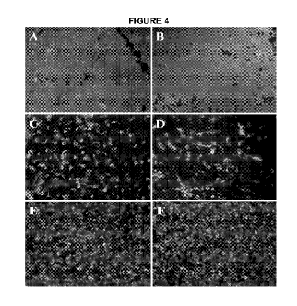
0038 FIG. 3 depicts process cell recovery as a function of refrigeration time.
0039 FIG. 4 shows representative images of the live/dead staining of the
epithelial
layer of fresh amniotic membrane.
0040 FIG. 5 depicts IL-2sR concentrations of various manufacturing
intermediates.
0041 FIG. 6 depicts IL-2sR concentrations of various manufacturing
intermediates.
0042 FIG. 7 depicts TNF a concentrations from LPS-induced secretion by
placental
tissues.
0043 FIG. 8 shows representative images of the live/dead staining of the
epithelial
layer of fresh amniotic membrane.
0044 FIG. 9 depicts a correlation between IL-2sR release and the number of
CD45+ cells.
0045 FIG. 10 depicts a correlation between the amount of CD45+ cells present
in
amnion-derived cell suspensions and immunogenicity in MLR in vitro.
0046 FIG. 11 depicts expression of EGF (A), IGFBPI (B), and Adiponectin (C) in
amniotic and/or chorionic membranes.
0047 FIG. 12 depicts expression of IFN-2a and TGF-(33 in amniotic membrane
homogenates.
0048 FIG. 13 depicts expression of BMP-2, BMP-4, PLAB, PIGF (A), and IGF-1 (B)
in amniotic membrane homogenates.
0049 FIG. 14 depicts the ratio of MMPs to TIMPs in various membrane products.
0050 FIG. 15 depicts bFGF levels in amniotic and chorionic membranes (CM).
0051 FIG. 16 depicts representative expression of bFGF in chorionic tissue
samples derived from two separate placenta donors.
0052 FIG. 17 depicts a schematic of the cell migration assay.
0053 FIG. 18 depicts the results of cell migration assay of various membrane
preparations.
0054 FIG. 19 depicts growth factor and adiponectin expression in protein
extracts
of various membrane preparations.
7
CA 02790322 2012-08-17
WO 2011/103451 PCT/US2011/025465
DETAILED DESCRIPTION OF THE INVENTION
0055 As used herein, the following definitions apply:
0056 "Examplary" (or "e.g." or "by example") means a non-limiting example.
0057 "hCMSCs: means human chorionic membrane stromal cells. hCMSCs
are generally positive for CD73, CD70, CD90, CD105, and CD166; negative for
CD45
and CD34,. hCMSCs differentiate to mesodermal lineages (osteogenic,
chondrogenic,
and adipogenic).
0058 "Selective depletion of immunogenicity" or "selective depletion of
immunogenic cells or factors" or "selective depletion" means a placental
product that
retains live therapeutic cells and/or retains therapeutic efficacy for the
treatment of
tissue injury yet is free, substantially free, or depleted of at least one of
immune cell
type (e.g. CD14+ macrophages, trophoblasts, and/or vascular-tissue derived
cells)
and/or immunogenic factor that are otherwise present in a native placenta or
chorionic
membrane.
0059 "MSC" means mesenchymal stem cells and include fetal, neonatal, adult,
or post-natal. "MSCs" include chorionic MSCs (CMSCs). MSCs generally express
one
or more of CD73, CD90, CD105, and CD166.
0060 "Native cells" means cells that are native, resident, or endogenous to
the
placental membrane, i.e. cells that are not exogenously added to the placental
membrane.
0061 "Native factors" means placental membrane factors that are native,
resident,
or endogenous to the placental membrane, i.e. factors that are not exogenously
added
to the placental membrane.
0062 "Placental products" means the instant placental products disclosed
herein.
0063 "Substantially free" means present in only a negligible amount or not
present
at all. For example, when a cell is abundant at least than about 20% or less
than about
10% or less than about 1 % of the amount in an unprocessed sample.
0064 "Substantial amount" of an element of the present invention, e.g. native
factors, therapeutic factors, or selective depletion, means a value at least
about 2% or
at least 10% in comparison to an unprocessed, not cryopreserved, fresh
membrane
sample. A substantial amount can optionally be at least about 50%.
8
CA 02790322 2012-08-17
WO 2011/103451 PCT/US2011/025465
0065 "Therapeutic cells" or "beneficial cells" means stromal cells, MSCs,
and/or
fibroblasts.
0066 "Therapeutic factors" means placenta- or chorionic membrane- derived
factors that promote wound healing. Examples include IGFBPI, adiponectin, a2-
macroglobulin, and/or bFGF. Other examples include MMP-9 and TIMP1.
0067 "Stromal cells" refers to a mixed population of cells present (optionally
in
native proportions) composed of neonatal mesenchymal stem cells and neonatal
fibroblasts. Both neonatal mesenchymal stem cells and neonatal fibroblasts are
immunoprivileged; neither express surface proteins present on immunogenic cell
types.
0068 In some embodiments, the present technology discloses placental products
for clinical use, including use in wound healing such as diabetic foot ulcers,
venous leg
ulcers, and burns. The manufacturing process optionally eliminates essentially
all
potentially immunogenic cells from the placental membrane while preserving of
specific
cells that play an important role in wound healing.
0069 In some embodiments, the present technology discloses a placental product
that is selectively devitalized. There is now provided a placental product
that is
selectively depleted of substantially all immunogenic cells. There is now
provided a
placental product that does not contain ex vivo cultured cells. There is now
provided a
placental product that comprises at least one of IGFBPI, and adiponectin.
There is now
provided a placental product that comprises IGFBPI. There is now provided a
placental
product that comprises adiponectin.
0070 In some embodiments, the present technology discloses a method of
cyropreserving a placental product that preserves the viability of specific
beneficial cells
that are the primary source of factors for the promotion of healing to the
wound healing
process while selectively depleting immunogenic cells (e.g. killing or
rendering non-
immunogenic) from the chorionic membranes.
0071 In some embodiments, the present technology discloses a bioassay to test
immunogenicity of manufactured placental products.
0072 In some embodiments, the present technology discloses a placental product
exhibiting a ratio of MMP:TIMP comparable to that exhibited in vivo. The
present
inventors have identified a need for the development of placental products
exhibiting a
ratio of MMP-9 and TIMP1 of about 7-10 to one.
9
CA 02790322 2012-08-17
WO 2011/103451 PCT/US2011/025465
0073 In some embodiments, the present technology discloses a placental product
that comprises a2-macroglobulin.
0074 The present inventors have identified a need for the development of
placental
products that comprise a2-macroglobulin.
0075 There is now provided a placental product that inactivates substantially
all
serine proteinases, cysteine proteinases, aspartic proteinases, and metal
loproteinases
present in the chorionic membrane. There is now provided a placental product
that
inactivates substantially all serine proteinases present in the chorionic
membrane.
There is now provided a placental product that inactivates substantially all
cysteine
proteinases present in the chorionic membrane. There is now provided a
placental
product that inactivates substantially all aspartic proteinases present in the
chorionic
membrane. There is now provided a placental product that inactivates
substantially all
metal loproteinases present in the chorionic membrane.
0076 In some embodiments, the present technology discloses a placental product
that comprises bFGF, optionally in a substantial amount.
0077 In some embodiments, the present technology discloses a placental product
exhibiting a protease-to-protease inhibitor ratio favorable for wound healing,
optionally
in a substantial amount.
0078 In some embodiments, the present technology discloses a cell migration
assay capable of evaluating the wound-healing potential of a placental
product.
0079 IGFBP1 and adiponectin are among the factors that are important for wound
healing. Evaluation of proteins derived from placental products prepared
according to
the presently disclosed technology reveal that bFGF is one of the major
factors secreted
in substantial higher quantities by the chorionic membrane. Additionally, the
importance
of EGF for wound healing together with high levels of bFGF detected in the
presently
disclosed chorionic membranes support selection of bFGF as a potency marker
for
evaluation of membrane products manufactured for clinical use pursuant to the
present
disclosure.
0080 The present technology discloses a cryopreservation procedure for a
placental products that selectively depletes immunogenic cells from the a
chorionic
membranes and preserves the viability of other beneficial cells (including at
least one of
mesenchymal stem cells, and fibroblasts in some embodiments and all of
mesenchymal
stem cells and fibroblasts in some embodiments) that are the primary source of
factors
CA 02790322 2012-08-17
WO 2011/103451 PCT/US2011/025465
for the promotion of healing. During the development of cryopreservation
methodology
for chorionic membranes, the inventors of the present application evaluated
key
parameters of cryopreservation including volume of cryopreservative solution,
effect of
tissue equilibration prior to freezing, and cooling rates for a freezing
procedures.
0081 Placental products, their usefulness, and their immunocompatability are
surprisingly enhanced by depletion of maternal trophoblast and selective
elimination of
CD14+ fetal macrophages. Immunocompatability can be demonstrated by any means
commonly known by the skilled artisan, such demonstration can be performed by
the
mixed Lymphocyte Reaction (MLR) and by lipopolysaccharide (LPS)-induced Tumor
Necrosis Factor (TNF)-a secretion.
0082 The instant placental products contain bFGF, optionally at a substantial
concentration.
0083 The instant placental products optionally secrete factors that stimulate
cell
migration and/or wound healing. The presence of such factors can be
demonstrated by
any commonly recognized method. Optionally, the factors are in a substantial
amount,
0084 For example, commercially available wound healing assays exist (Cell
Biolabs) and cell migration can be assessed by cell line (HMVEC, Lonza Inc.).
In one
embodiment, conditioned medium from the present placental products enhance
cell
migration.
0085 The placental products disclosed herein are useful in treating a number
of
wounds including: tendon repair, cartilage repair (e.g. femoral condyle,
tibial plateau),
ACL replacement at the tunnel/bone interface, dental tissue augmentation,
fistulas (e.g.
Crohn's disease, G-tube, tracheoesophogeal), missing tissue at adhesion
barriers (e.g.
nasal septum repair, vaginal wall repair, abdominal wall repair, tumor
resection), dermal
wounds (e.g. partial thickness burns, toxic epidermal necrolysis,
epidermolysis bullosa,
pyoderma gangrenosum, ulcers e.g. diabetic ulcers (e.g. foot), venous leg
ulcers),
surgical wounds, hernia repair, tendon repair, bladder repair, periosteum
replacement,
keloids, organ lacerations, epithelial defects, and repair or replacement of a
tympanic
membrane.
0086 The placental products disclosed herein exhibit one or more of the
following properties beneficial to the wound healing process:
a. approximate number of cells per cm2 being about 20,000 to about
200,000,
11
CA 02790322 2012-08-17
WO 2011/103451 PCT/US2011/025465
b. thickness of about 40 to about 400 pm,
c. a thin basement membrane,
d. low immunogenicity,
e. and cryopreserved/cryopreserveable,
f. human Chorionic Membrane Stromal Cells (hCMSC),
0087 The present inventors have now identified a need for the development of
placental products that do not contain ex vivo cultured cells.
0088 The present inventors have now identified a need for the development of
placental products comprising IGFBPI.
0089 The present inventors have now identified a need for the development of
placental products comprising adiponectin.
0090 The present inventors have now identified a need for the development of
placental products exhibiting a protease-to-protease inhibitor ratio favorable
for wound
healing.
0091 The present inventors have now identified a need for the development of a
method of cyropreserving placental products that preserves the viability of
specific cells
that are other beneficial cells that are the primary source of factors for the
promotion of
healing to the wound healing process while selectively depleting immunogenic
cells
from chorionic membranes.
0092 The present inventors have now identified a need for the development of a
bioassay to test immunogenicity of manufactured placental products.
0093 The present inventors have now identified a need for the development of
placental products exhibiting a ratio of MMP to TIMP comparable to that
exhibited in
vivo. The present inventors have now identified a need for the development of
placental
products exhibiting a ratio of MMP-9 and TIMP1 of about 7-10 to one.
0094 The present inventors have now identified a need for the development of
placental products that comprise a2-macroglobulin.
0095 The present inventors have now identified a need for the development of
placental products that inactivate serine proteinases, cysteine proteinases,
aspartic
proteinases, and metalloproteinases. The present inventors have now identified
a need
for the development of placental products that inactivate serine proteinases.
The
12
CA 02790322 2012-08-17
WO 2011/103451 PCT/US2011/025465
present inventors have now identified a need for the development of placental
products
that inactivate cysteine proteinases. The present inventors have now
identified a need
for the development of placental products that inactivate aspartic
proteinases. The
present inventors have now identified a need for the development of placental
products
that inactivate metalloproteinases.
0096 The present inventors have now identified a need for the development of
placental products that comprise bFGF.
0097 The present inventors have now identified a need for the development of a
cell migration assay to evaluate the potential of placental membrane products.
0098 The present inventors have now identified a need for the development of a
placental product for wound healing that comprises mesenchymal stem cells and
fibroblasts.
Placental Product
Overview
0099 One embodiment of the present invention provides a placental product
comprising a cryopreservation medium and a chorionic membrane, wherein the
chorionic membrane comprises viable therapeutic native cells and native
therapeutic
factors, and wherein the cryopreservation medium comprises a cryopreserving
amount
of a cryopreservative. According to this embodiment, the chorionic membrane is
substantially free of at least one at least one or 2 or 3 immunogenic cell
types such as:
trophoblasts, CD14+ macrophages, and vascularized tissue-derived cells.
00100 In one embodiment, the chorionic membrane comprises one or more layers
which exhibit the architecture of the native chorionic membrane (e.g. has not
been
homogenized or treated with collagenase).
00101 In one embodiment, the placental product is suitable for dermal
application to
a wound.
00102 With the teachings provided herein, the skilled artisan can now produce
the
present placental products. The present disclosure provides methods of
manufacture
that produce the technical features of the present placental products.
Accordingly, in
one embodiment, the placental product is manufactured by steps taught herein.
The
present placental products are not limited to products manufactured by the
methods
taught here. For example, products of the present invention could be produced
through
13
CA 02790322 2012-08-17
WO 2011/103451 PCT/US2011/025465
methods that rely on screening steps; e.g. steps to screen for preparations
with the
described technical features and superior properties.
00103 The present placental products comprises one or more of the following
technical features:
a. the viable therapeutic native cells are capable of differentiating into
cells of
more than one lineage (e.g. osteogenic, adipogenic and/or chonodrogenic
lineages),
b. the native therapeutic factors include IGFBPI, optionally present in a
substantial amount,
c. the native therapeutic factors include adiponectin, optionally present in a
substantial amount,
d. the native therapeutic factors include a2-macroglobulin, optionally present
in
a substantial amount,
e. the native therapeutic factors include bFGF, optionally present in a
substantial
amount,
f. the native therapeutic factors include EGF, optionally present in a
substantial
amount,
g. the native therapeutic factors include MMP-9 and TIMP1, optionally present
in
a substantial amount,
h. the native therapeutic factors include MMP-9 and TIMP1 in a ratio of about
7
to about 10,
i. the placental product does not comprise ex-vivo cultured cells,
j. the cryopreservative medium is present in an amount of greater than about
20
ml or greater than about 50 ml,
k. the cryopreservative comprises DMSO,
1. cryopreservative comprises DMSO in a majority amount,
M. the cryopreservation medium further comprises albumin, optionally wherein
the albumin is HSA,
n. the cryopreservative comprises DMSO and albumin (e.g. HSA),
14
CA 02790322 2012-08-17
WO 2011/103451 PCT/US2011/025465
o. the chorionic membrane comprises about 5,000 to about 240,000 cells/cm2
or about 20,000 to about 60,000 cells/cm2,
p. the chorionic membrane comprises 20,000 to about 200,000 cells/cm2, with a
cell viability of at least about 70%,
q. comprises at least: about 7,400 or about 15,000 or about 23,217, or about
35,000, or about 40,000 or about 47,800 of stromal cells per cm2 of the
chorionic
membrane,
r. comprises about 5,000 to about 50,000 of stromal cells per cm2 of the
chorionic membrane,
s. comprises about 4% to about 46% of viable non-culturally expanded
fibroblasts per cm2 of the placental product,
t. comprises stromal cells wherein at least: about 40%, or about 50%, or about
60%, or about 70%, or about 74.3%, or about 83.4 or about 90%, or about 92.5%
of the stromal cells are viable after a freeze-thaw cycle,
U. comprises stromal cells wherein about 40% to about 92.5% of the stromal
cells are viable after a freeze-thaw cycle,
v. the chorionic membrane has a thickness of about 40 pm to about 400 pm,
w. secretes less than about any of: 420 pg/mL, 350 pg/mL, or 280 pg/mL TNF-a
into a tissue culture medium upon placing a 2 cm x 2 cm piece of the tissue
product in a tissue culture medium and exposing the tissue product to a
bacterial
lipopolysaccharide for about 20 to about 24 hours,
X. cryopreservation and thawing, secretes less than about any of: 420 pg/mL,
350 pg/mL, or 280 pg/mL TNF-a into a tissue culture medium upon placing a 2
cm x 2 cm piece of the tissue product in a tissue culture medium and exposing
the tissue product to a bacterial lipopolysaccharide for about 20 to about 24
hours,
y. after refrigeration, cryopreservation and thawing, secretes less than about
any of: 420 pg/mL, 350 pg/mL, or 280 pg/mL TNF-a into a tissue culture medium
upon placing a 2 cm x 2 cm piece of the tissue product in a tissue culture
medium and exposing the tissue product to a bacterial lipopolysaccharide for
about 20 to about 24 hours,
CA 02790322 2012-08-17
WO 2011/103451 PCT/US2011/025465
Z. the maternal side of the chorionic membrane comprises fragments of
extracellular matrix proteins in a concentration substantially greater than
that of a
native, unprocessed chorion, optionally wherein the chorionic membrane has
been treated with Dispase II or wherein a substantial portion of the protein
fragments comprises terminal leucine or phenylalanine,
aa. further comprises an amniotic membrane,
bb. further comprises an amniotic membrane, wherein the amniotic membrane
comprises a layer of amniotic epithelial cells,
cc. further comprises an amniotic membrane, wherein the amniotic membrane
and the chorionic membrane are associated to one another in the native
configuration,
dd. further comprises an amniotic membrane, wherein the amniotic membrane
and the chorionic membrane are not attached to one another in the native
configuration,
ee. further comprises an amniotic membrane wherein the chorionic membrane
comprises about 2 to about 4 times more stromal cells relative to the amniotic
membrane,
if. does not comprise an amniotic membrane,
gg. the chorionic membrane comprises about 2 to about 4 times more stromal
cells relative to an amniotic membrane of the same area derived from the same
placenta, and
hh. is suitable for dermal application to a wound;
Cells
00104 According to the present invention, a placental product comprises native
therapeutic cells of the chorionic membrane. The cells comprise one or more of
stromal
cells, MSCs, and fibroblasts.
00105 In one embodiment, the native therapeutic cells comprise viable stromal
cells.
00106 In one embodiment, the native therapeutic cells comprise viable MSCs.
00107 In one embodiment, the native therapeutic cells comprise viable
fibroblasts.
16
CA 02790322 2012-08-17
WO 2011/103451 PCT/US2011/025465
00108 In one embodiment, the native therapeutic cells comprise viable MSCs and
viable fibroblasts.
00109 In one embodiment, the native therapeutic cells comprise viable MSCs and
viable fibroblasts.
00110 In one embodiment, the native therapeutic cells comprise viable stromal
cells
and viable epithelial cells.
00111 In one embodiment, the therapeutic native cells are viable cells capable
of
differentiating into cells of more than one lineage (e.g. osteogenic,
adipogenic and/or
chonodrogenic lineages).
00112 In one embodiment, the chorionic membrane comprises about 10,000 to
about 360,000 cells/cm2 or about 40,000 to about 90,000 cells/cm2.
00113 In one embodiment, the chorionic membrane comprises at least: about
7,400
or about 15,000 or about 23,217, or about 35,000, or about 40,000 or about
47,800 of
stromal cells per cm2 of the chorionic membrane.
00114 In one embodiment, the chorionic membrane comprises about 5,000 to about
50,000 of stromal cells per cm2 of the chorionic membrane.
00115 In one embodiment, the chorionic membrane comprises stromal cells
wherein
at least: about 40%, or about 50%, or about 60%, or about 70%, or about 74.3%,
or
about 83.4 or about 90%, or about 92.5% of the stromal cells are viable after
a freeze-
thaw cycle.
00116 In one embodiment, the chorionic membrane comprises stromal cells
wherein
about 40% to about 92.5% of the stromal cells are viable after a freeze-thaw
cycle.
00117 In one embodiment, the chorionic membrane (of the placental product)
comprises fibroblasts in about 50% to about 90% of the total cells.
00118 In one embodiment, the chorionic membrane comprises CD14+ macrophage
in an amount of less than about 5% or less than about 1 % or less than about
0.5%,
optionally as demonstrated by a substantial decrease in LPS stimulation of
TNFa
release.
00119 In one embodiment, the placental product comprises about 2 to about 4
times
more stromal cells relative to an amniotic membrane of the same area derived
from the
same placenta.
17
CA 02790322 2012-08-17
WO 2011/103451 PCT/US2011/025465
00120 In one embodiment, the placental product further comprises an amniotic
membrane, wherein the placental product contains about 2 to about 4 times more
stromal cells relative to the amniotic membrane.
00121 In one embodiment, the placental product further comprises an amniotic
membrane, wherein the amniotic membrane comprises a layer of amniotic
epithelial
cells.
00122 In one embodiment, the placental product is substantially free of
trophoblasts.
00123 In one embodiment, the placental product is substantially free of
functional
CD14+ macrophages.
00124 In one embodiment, the placental product is substantially free of
vascularized
tissue-derived cells.
In one embodiment, the placental product is substantially free of
trophoblasts, functional
CD14+ macrophages, and vascularized tissue-derived cells. Optionally, the
placental
product comprises viable stromal cells. Optionally, the placental product
comprises
viable MSCs. Optionally, the placental product comprises viable fibroblasts.
Optionally,
the placental product comprises viable MSCs and viable fibroblasts.
00125 In one embodiment, the placental product is substantially free of
maternal
decidual cells.
00126 In one embodiment, the placental product is substantially free of
maternal
leukocytes and/or trophoblast cells.
00127 In one embodiment, the chorionic membrane (of the placental product)
comprises MSCs in an amount of: about 5% to about 30%, about 5% to about 25%,
about 5% to about 20%, about 5% to about 15%, about 3% to about 12%, at least
about
5%, at least about 10%, or at least about 15%, relative to the total number of
cells in the
chorionic membrane. Optionally, at least: about 40%, about 50%, about 60%, or
about
70% of the MSCs are viable after a freeze-thaw cycle.
00128 In one embodiment, the chorionic membrane (of the placental product)
comprises fibroblasts in an amount of: about 50% to about 95%, about 60% to
about
90%, or about 70% to about 85%, relative to the total number of cells in the
chorionic
membrane. Optionally, at least: about 40%, about 50%, about 60%, or about 70%
of
the fibroblasts are viable after a freeze-thaw cycle.
18
CA 02790322 2012-08-17
WO 2011/103451 PCT/US2011/025465
00129 In one embodiment, the chorionic membrane (of the placental product)
comprises functional macrophages in an amount of less than about any of: 5%,
4%, 3%,
2%, 1 %, or 0.1 %.
00130 In one embodiment, the chorionic membrane (of the placental product)
comprises MSCs and functional macrophages in a ratio of greater than about any
of:
3:1, 4:1, 5:1, 7:1, 10:1, 12:1, or 15:1.
00131 In one embodiment, the chorionic membrane comprises fibroblasts and
functional macrophages in a ratio of greater than about any of: 14:1, 15:1,
16:1, 17:1,
28:1, 30:1, 35:1, 45:1, or 50:1
00132 In one embodiment, the chorionic membrane (of the placental product)
comprises fibroblasts and MSCs in a ratio of: about 9:2 to about 17:3.
00133 In one embodiment, the chorionic membrane (of the placental product)
comprises MSCs in an amount of: at least about 1,500 cells/cm2 , at least
about 3,000
cells/cm2, about 15,000 to about 9,000 cells/cm2, or about 3,000 to about
9,000
cells/cm2. Optionally, at least: about 40%, about 50%, about 60%, or about 70%
of the
MSCs are viable after a freeze-thaw cycle.
00134 In one embodiment, the chorionic membrane (of the placental product)
comprises fibroblasts in an amount of: at least about 7,000 cells/cm2, at
least about
14,000 cells/cm2, about 7,000 to about 51,000 cells/cm2, or about 14,000 to
about
51,000 cells/cm2. Optionally, at least: about 40%, about 50%, about 60%, or
about 70%
of the fibroblasts are viable after a freeze-thaw cycle.
00135 In one embodiment, the chorionic membrane (of the placental product)
comprises functional macrophages in an amount of: less than about 3,000
cells/cm2, or
less than about 1,000 cells/cm2.
00136 In one embodiment, the placental product is substantially free of ex-
vivo
cultured cells.
Placental Factors
00137 According to the present invention, a placental product comprises native
therapeutic factors of the chorionic membrane.
00138 In one embodiment, the factors include one or more of: IGFBPI,
adiponectin,
a2-macroglobulin, bFGF, EGF, MMP-9, and TIMP1. Optionally, the factors are
present
19
CA 02790322 2012-08-17
WO 2011/103451 PCT/US2011/025465
in amounts/cm2 that are substantially similar to that of a native chorionic
membrane or
layer thereof (e.g. 10% or 20%).
00139 In one embodiment, the factors include IGFBPI, adiponectin, a2-
macroglobulin, bFGF, EGF, MMP-9, and TIMP1. Optionally, the factors are
present in
ratios that are substantially similar to that of a native chorionic membrane
or layer
thereof. Optionally, the factors are present in amounts/cm2 that are
substantially similar
to that of a native chorionic membrane or layer thereof (e.g. 10% or 20%).
00140 In one embodiment, the factors include MMP-9 and TIMP1 in a ratio of
about
7 to about 10 (e.g about 7). Optionally, the factors are present in
amounts/cm2 that are
substantially similar to that of a native chorionic membrane or layer thereof
(e.g. 10%
or 20%).
00141 In one embodiment, the factors include one or more (e.g. a majority or
all) of
the factors listed in Table 15. Optionally, the factors are present in ratios
that are
substantially similar to that of a native chorionic membrane or layer thereof.
Optionally,
the factors are present in amounts/cm2 that are substantially similar to that
of a native
chorionic membrane or layer thereof (e.g. 10% or 20%).
00142 In one embodiment, the placental product secretes substantially less TNF-
a/
cm2 than a native, unprocessed chorionic membrane.
00143 In one embodiment, the placental product secretes substantially less TNF-
a/
cm2 than a native placental product upon stimulation by LPS or CT.
00144 In one embodiment, the placental product secretes less than about any
of:
420 pg/mL, 350 pg/mL, or 280 pg/mL TNF-a into a tissue culture medium upon
placing
a 2 cm x 2 cm piece of the tissue product in a tissue culture medium and
exposing the
tissue product to a bacterial lipopolysaccharide for about 20 to about 24
hours.
00145 In one embodiment, after cryopreservation and thawing, the placental
product
secretes less than about any of: 420 pg/mL, 350 pg/mL, or 280 pg/mL TNF-a into
a
tissue culture medium upon placing a 2 cm x 2 cm piece of the tissue product
in a tissue
culture medium and exposing the tissue product to a bacterial
lipopolysaccharide for
about 20 to about 24 hours.
00146 In one embodiment, after refrigeration, cryopreservation and thawing,
the
placental product secretes less than about any of: 420 pg/mL, 350 pg/mL, or
280 pg/mL
TNF-a into a tissue culture medium upon placing a 2 cm x 2 cm piece of the
tissue
CA 02790322 2012-08-17
WO 2011/103451 PCT/US2011/025465
product in a tissue culture medium and exposing the tissue product to a
bacterial
lipopolysaccharide for about 20 to about 24 hours.
00147 In one embodiment, the placental product further comprises an
exogenously
added inhibitor of TNF-a. Optionally, the inhibitor of TNF-a is IL-10.
00148 In one embodiment, the product has been treated with an antibiotic
Architecture
00149 A placental product of the present invention comprises one or more non-
trophoblast layers which exhibit the architecture of the native chorionic
membrane. With
the teachings provided herein, the skilled artisan will recognize placental
layers that
exhibit native architecture, for example, layers that have not been
homogenized or
treated with collagenase or other enzyme that substantially disrupts the
layer.
00150 In one embodiment, the placental product comprises a stromal layer with
native architecture.
00151 In one embodiment, the placental product comprises a basement membrane
with native architecture.
00152 In one embodiment, the placental product comprises a reticular layer
with
native architecture.
00153 In one embodiment, the placental product comprises a reticular layer and
a
basement layer with native architecture.
00154 In one embodiment, the placental product comprises a stromal layer, a
basement layer, and a reticular layer with native architecture.
00155 In one embodiment, the placental product is substantially free of
trophoblasts.
In one embodiment, the placental product comprises a basement membrane with
native
architecture and the chorionic membrane is substantially free of trophoblasts.
Optionally, the maternal side of the placental product comprises fragments of
extracellular matrix proteins in a concentration substantially greater than
that of a native
chorionic membrane. Optionally, the placental product has been treated with
Dispase
(e.g. Dispase II) and/or a substantial portion of the extracellular matrix
protein fragments
comprises terminal leucine or phenylalanine.
00156 In one embodiment, the placental product has a thickness of about 40 pm
to
about 400 pm.
21
CA 02790322 2012-08-17
WO 2011/103451 PCT/US2011/025465
00157 In one embodiment, the placental product further comprises an amniotic
membrane. Optionally, the amniotic membrane and the chorionic membrane in the
placental product are associated to one another in the native configuration.
Alternatively, the amniotic membrane and the chorionic membrane are not
attached to
one another in the native configuration.
00158 In one embodiment, the placental product does not comprise an amniotic
membrane.
Formulation
00159 According to the present invention, the placental product can be
formulated
with a cryopreservation medium.
00160 In one embodiment, the cryopreservation medium comprising one or more
cell-permeating cryopreservatives, one or more non cell-permeating
cryopreservatives,
or a combination thereof.
00161 Optionally, the cryopreservation medium comprises one or more cell-
permeating cryopreservatives selected from DMSO, a glycerol, a glycol, a
propylene
glycol, an ethylene glycol, or a combination thereof.
00162 Optionally, the cryopreservation medium comprises one or more non cell-
permeating cryopreservatives selected from polyvinylpyrrolidone, a
hydroxyethyl
starch, a polysacharide, a monosaccharides, a sugar alcohol, an alginate, a
trehalose, a
raffinose, a dextran, or a combination thereof.
00163 Other examples of useful cryopreservatives are described in
"Cryopreservation" (BioFiles Volume 5 Number 4 -Sigma-Aldrich datasheet).
00164 In one embodiment, the cryopreservation medium comprises a cell-
permeating cryopreservative, wherein the majority of the cell-permeating
cryopreservative is DMSO. Optionally, the cryopreservation medium does not
comprise
a substantial amount of glycerol.
00165 In one embodiment, the cryopreservation medium comprises DMSO.
Optionally, the cryopreservation medium does not comprise glycerol in a
majority
amount. Optionally, the cryopreservation medium does not comprise a
substantial
amount of glycerol.
22
CA 02790322 2012-08-17
WO 2011/103451 PCT/US2011/025465
00166 In one embodiment, the cryopreservation medium comprises additional
components such as albumin (e.g. HSA or BSA), an electrolyte solution (e.g.
Plasma-
Lyte), or a combination thereof.
00167 In one embodiment, the cryopreservation medium comprises 1 % to about
15% albumin by weight and about 5% to about 20% cryopreservative by volume
(e.g.
about 10%). Optionally, the cryopreservative comprises DMSO (e.g. in a
majority
amount).
00168 In one embodiment, the placental product is formulated in greater than
about
20 ml or greater than about 50 ml of cryopreservation medium. Optionally, the
cryopreservative comprises DMSO (e.g. in a majority amount). Optionally, the
cryopreservation medium does not comprise a substantial amount of glycerol.
00169 In one embodiment, the placental product is placed on nitrocellulose
paper.
00170 In one embodiment, the placenta is cut into a plurality of sections.
Optionally,
the sections are less than about 10 cm x 10 cm. Optionally, the sections are
between
about2cmx2cmand5cmx5cm.
Manufacture
Overview
00171 A placental product of the present invention can manufactured from a
placenta in any suitable manner that provides the technical features taught
herein.
According to the present invention, a placental product comprises at least an
immunocompatible chorionic membrane.
00172 In one embodiment, a placental product is manufactured by a method
comprising:
a. obtaining a placenta,
b. selectively depleting the placenta of immunogenicity; and
c. cryopreserving the placenta.
00173 In one embodiment, a placental product is manufactured by a method
comprising:
a. obtaining a placenta;
b. removing a substantial portion of trophoblasts from the placenta; and
c. cryopreserving the placenta.
23
CA 02790322 2012-08-17
WO 2011/103451 PCT/US2011/025465
00174 Optionally, the method comprises a step of removing the amniotic
membrane
or portion thereof ('an amniotic membrane') from the placenta. Optionally, the
method
comprises a step of removing an amniotic membrane from the placenta without
removing a substantial portion of amniotic epithelial cells from the placenta.
00175 Optionally, the step of removing a substantial portion of trophoblasts
from the
placenta comprises treating the placenta with a digestive enzyme such as a
protease
(e.g. dispase or dispase II), mechanically removing trophoblasts from the
placenta (e.g.
by scraping), or a combination thereof.
00176 Optionally, the method comprises a step of removing vascularized tissue
from
the placenta, for example, by lysing red blood cells, by removing blood clots,
or a
combination thereof.
00177 Optionally, the method comprises a step of treating the placenta with
one or
more antibiotics.
00178 Optionally, the method comprises a step of selective depletion of CD14+
macrophages.
00179 Optionally, the step of cryopreserving the placenta comprises freezing
the
placenta in a cryopreservation medium which comprises one or more cell-
permeating
cryopreservatives, one or more non cell-permeating cryopreservatives, or a
combination
thereof.
00180 Optionally, the step of cryopreserving the placenta comprises
refrigerating for
a period of time and then freezing, thereby selectively depleting CID 14+
macrophages.
00181 An examplary placental product of the present invention can be
manufactured
or provided with a bandage or skin substitute.
Immunocompatability and Selective Depletion
00182 In one embodiment, the invention the placental product is
immunocompatible.
Immunocompatability can be accomplished by any selective depletion step that
removes immunogenic cells or factors or immunogenicity from the placenta (or
chorionic
membrane thereof).
00183 In one embodiment, the placental product is made immunocompatible by
selectively depleting it of functional immunogenic cells. A placenta can be
made
immunocompatible by selectively removing immunogenic cells from the placenta
(or
chorionic membrane thereof) relative to therapeutic cells. For example,
immunogenic
24
CA 02790322 2012-08-17
WO 2011/103451 PCT/US2011/025465
cells can be removed by killing the immunogenic cells or by purification of
the placenta
there from.
00184 In one embodiment, the placenta is made immunocompatible by selectively
depleting trophoblasts, for example, by removal of the trophoblast layer.
00185 In one embodiment, the placenta is made immunocompatible by selective
depletion of functional CD14+ macrophages, optionally resulting in depleteion
of TNFa
upon stimulation, or a combination thereof.
00186 In one embodiment, the placenta is made immunocompatible by selective
depletion of vascularized tissue-derived cells.
00187 In one embodiment, the placenta is made immunocompatible by selective
depletion of functional CD14+ macrophages, trophoblasts, and vascularized
tissue-
derived cells.
00188 In one embodiment, the placenta product is made immunocompatible by
selective depletion of trophoblasts and/or CD14+ macrophages, optionally
resulting in
depletion of TNFa upon stimulation.
Trophoblast Removal
00189 In one embodiment, trophoblasts are depleted or removed from the
placental
product. Surprisingly, such a placental product has one or more of the
following
superior features:
a. is substantially non-immunogenic;
b. provides remarkable healing time; and
c. provides enhanced therapeutic efficacy.
00190 Trophoblasts can be removed in any suitable manner which substantially
diminishes the trophoblast content of the placental product. Optionally, the
trophoblasts
are selectively removed or otherwise removed without eliminating a substantial
portion
of one or more therapeutic components from the placenta (e.g. MSCs, placental
factors,
etc). Optionally, a majority (e.g. substantially all) of the trophoblasts are
removed.
00191 One method of removing trophoblasts comprises treating the placenta
(e.g.
chorion or amnio-chorion) with a digestive enzyme such as dispase (e.g.
dispase II) and
separating the trophoblasts from the placenta. Optionally, the step of
separating
CA 02790322 2012-08-17
WO 2011/103451 PCT/US2011/025465
comprises mechanical separation such as peeling or scraping. Optionally,
scraping
comprises scraping with a soft instrument such as a finger.
00192 One method of removing trophoblasts comprises treating the chorionic
membrane with dispase for about 30 to about 45 minutes separating the
trophoblasts
from the placenta. Optionally, the dispase is provided in a solution of about
less than
about 1 % (e.g. about 0.5%). Optionally, the step of separating comprises
mechanical
separation such as peeling or scraping. Optionally, scraping comprises
scraping with a
soft instrument such as a finger.
00193 Useful methods of removing trophoblasts from a placenta (e.g. chorion)
are
described by Portmann-Lanz et al. ("Placental mesenchymal stem cells as
potential
autologous graft for pre- and perinatal neuroregeneration"; American Journal
of
Obstetrics and Gynecology (2006) 194, 664-73), ("Isolation and
characterization of
mesenchymal cells from human fetal membranes"; Journal Of Tissue Engineering
And
Regenerative Medicine 2007; 1: 296-305.), and (Concise Review: Isolation and
Characterization of Cells from Human Term Placenta: Outcome of the First
International
Workshop on Placenta Derived Stem Cells").
00194 In one embodiment, trophoblasts are removed before cryopreservation.
Macrophage Removal
00195 In one embodiment, functional macrophages are depleted or removed from
the
placental product. Surprisingly, such a placental product has one or more of
the
following superior features:
a. is substantially non-immunogenic;
b. provides remarkable healing time; and
c. provides enhanced therapeutic efficacy.
00196 Functional macrophages can be removed in any suitable manner which
substantially diminishes the macrophage content of the placental product.
Optionally,
the macrophages are selectively removed or otherwise removed without
eliminating a
substantial portion of one or more therapeutic components from the placenta
(e.g.
MSCs, placental factors, etc). Optionally, a majority (e.g. substantially all)
of the
macrophages are removed.
00197 One method of removing immune cells such as macrophages comprises
killing the immune cells by rapid freezing rates such as 60-100 C/min.
26
CA 02790322 2012-08-17
WO 2011/103451 PCT/US2011/025465
00198 Although immune cells can be eliminated by rapid freezing rates, such a
method can also be detrimental to therapeutic cells such as stromal cells
(e.g. MSCs).
The present inventors have discovered a method of selectively killing CD14+
macrophages can be selectively killed by refrigerating the placenta for a
period of time
(e.g. for at least about 10 min such as for about 30-60 mins) at a temperature
above
freezing (e.g. incubating at 2-8 C) and then freezing the placenta (e.g.
incubating at -
80 C 5 C). Optionally, the step of freezing comprises freezing at a rate of
less than
/min (e.g. less than about 5 /min such as at about 1 /min).
00199 In one embodiment, the step of refrigerating comprises soaking the
placenta
in a cryopreservation medium (e.g. containing DMSO) for a period of time
sufficient to
allow the cryopreservation medium to penetrate (e.g. equilibrate with) the
placental
tissues. Optionally, the step of freezing comprises reducing the temperature
at a rate of
about 1 /min. Optionally, the step of freezing comprises freezing at a rate
of less than
10 /min (e.g. less than about 5 /min such as at about 1 /min).
00200 In one embodiment, the step of refrigerating comprises soaking the
placenta
in a cryopreservation medium (e.g. containing DMSO) at a temperature of about -
10-
C (e.g. at 2-8 C) for at least about any of: 10 min, 20 min, 30 min, 40 min,
or 50 min.
In another embodiment, the step of refrigerating comprises soaking the
placenta in a
cryopreservation medium (e.g. containing DMSO) at a temperature of about -10-
15 C
(e.g. at 2-8 C) for about any of: 10-120, 20-90 min, or 30-60 min. Optionally,
the step of
freezing comprises freezing at a rate of less than 10 /min (e.g. less than
about 5 /min
such as at about 1 /min).
Removal of Vascularized Tissue-Derived Cells
00201 In one embodiment, vascularized tissue-derived cells (or vascularied
tissue)
are depleted or removed from the placental product. Surprisingly, such a
placental
product has one or more of the following superior features:
a. is substantially non-immunogenic;
b. provides remarkable healing time; and
c. provides enhanced therapeutic efficacy.
00202 Vascularized tissue-derived cells can be removed in any suitable manner
which substantially diminishes such cell content of the placental product.
Optionally, the
vascularized tissue-derived cells are selectively removed or otherwise removed
without
27
CA 02790322 2012-08-17
WO 2011/103451 PCT/US2011/025465
eliminating a substantial portion of one or more therapeutic components from
the
placenta (e.g. MSCs, placental factors, etc).
00203 In one embodiment, removal of vascularized tissue-derived cells
comprises
separating the chorion from the placenta by cutting around the placental skirt
on the
side opposite of the umbilical cord. The chorion on the umbilical side of the
placenta is
not removed due to the vascularization on this side.
00204 In one embodiment, removal of vascularized tissue-derived cells
comprises
rinsing the chorionic membrane (e.g. with buffer such as PBS) to remove gross
blood
clots and any excess blood cells.
00205 In one embodiment, removal of vascularized tissue-derived cells
comprises
treating the chorionic membrane with an anticoagulant (e.g. citrate dextrose
solution).
00206 In one embodiment, removal of vascularized tissue-derived cells
comprises
separating the chorion from the placenta by cutting around the placental skirt
on the
side opposite of the umbilical cord and rinsing the chorionic membrane (e.g.
with buffer
such as PBS) to remove gross blood clots and any excess blood cells.
00207 In one embodiment, removal of vascularized tissue-derived cells
comprises
separating the chorion from the placenta by cutting around the placental skirt
on the
side opposite of the umbilical cord and treating the chorionic membrane with
an
anticoagulant (e.g. citrate dextrose solution).
00208 In one embodiment, removal of vascularized tissue-derived cells
comprises
separating the chorion from the placenta by cutting around the placental skirt
on the
side opposite of the umbilical cord, rinsing the chorionic membrane (e.g. with
buffer
such as PBS) to remove gross blood clots and any excess blood cells, and
treating the
chorionic membrane with an anticoagulant (e.g. citrate dextrose solution).
Selective depletion of immunogenicity as demonstrated by a substantial
decrease in LPS stimulation of TNFa release.
00209 In one embodiment, the placental product is selectively depleted of
immunogenicity as demonstrated by a reduction in LPS stimulated TNF-a release.
depletion d of TNF-a depleted or removed from the placental product.
00210 In one embodiment, TNF-a is depleted by killing or removal of
macrophages.
00211 In one embodiment, TNF-a is depleted by treatment with an anti- TNF-a
antibody.
28
CA 02790322 2012-08-17
WO 2011/103451 PCT/US2011/025465
00212 In one embodiment, TNF-a is functionally depleted by treatment with IL-
10,
which suppresses TNF-a secretion.
Preservation
00213 A placental product of the present invention may be used fresh or may be
preserved for a period of time. Surprisingly, cryopreservation results in
immunocompatible placental products.
00214 In one embodiment, a placental product is cryopreserved. A placental
product may be cryopreserved by incubation at freezing temperatures (e.g. a -
80 C
C) in a cryopreservative medium.
00215 Cryopreservation can comprise, for example, incubating the placental
product
at 4 C for 30-60 min, and then incubating at -80 C until use. The placental
product may
then be thawed for use. Optionally, the placental product is cryopreserved in
a manner
such that cell viability is retained surprisingly well after a freeze-thaw
cycle.
00216 In one embodiment, cryopreservation comprises storage in a
cryopreservation medium comprising one or more cell-permeating
cryopreservatives,
one or more non cell-permeating cryopreservatives, or a combination thereof.
Optionally, the cryopreservation medium comprises one or more cell-permeating
cryopreservatives selected from DMSO, a glycerol, a glycol, a propylene
glycol, an
ethylene glycol, or a combination thereof. Optionally, the cryopreservation
medium
comprises one or more non cell-permeating cryopreservatives selected from
polyvinyl pyrroIidone, a hydroxyethyl starch, a polysacharide, a
monosaccharides, a
sugar alcohol, an alginate, a trehalose, a raffinose, a dextran, or a
combination thereof.
Other examples of useful cryopreservatives are described in "Cryopreservation"
(BioFiles Volume 5 Number 4 -Sigma-Aldrich datasheet).
00217 In one embodiment, the cryopreservation medium comprises a cell-
permeating cryopreservative, wherein the majority of the cell-permeating
cryopreservative is DMSO. Optionally, the cryopreservation medium does not
comprise
a substantial amount of glycerol.
00218 In one embodiment, the cryopreservation medium comprises DMSO.
Optionally, the cryopreservation medium does not comprise glycerol in a
majority
amount. Optionally, the cryopreservation medium does not comprise a
substantial
amount of glycerol.
29
CA 02790322 2012-08-17
WO 2011/103451 PCT/US2011/025465
00219 In one embodiment, the cryopreservation medium comprises additional
components such as albumin (e.g. HSA or BSA), an electrolyte solution (e.g.
Plasma-
Lyte), or a combination thereof.
00220 In one embodiment, the cryopreservation medium comprises 1 % to about
15% albumin by weight and about 5% to about 20% cryopreservative by volume
(e.g.
about 10%). Optionally, the cryopreservative comprises DMSO (e.g. in a
majority
amount).
00221 In one embodiment, cryopreservation comprises placing the placenta on
nitrocellulose paper.
00222 In one embodiment, the placenta is cut into a plurality of sections
before
cryopreservation. Optionally, the sections are placed on nitrocellulose paper
before
refrigeration.
00223
Methods of Use
00224 The placental products (e.g. derived from chorionic tissue) of the
present
invention may be used to treat any tissue injury. A method of treatment may be
provided, for example, by administering to a subject in need thereof, a
placental product
of the present invention.
00225 A typical administration method of the present invention is topical
administration. Administering the present invention can optionally involve
administration
to an internal tissue where access is gained by a surgical procedure.
00226 Placental products can be administered autologously, allogeneically or
xenogeneically.
00227 In one embodiment, a present placental product is administered to a
subject
to treat a wound. Optionally, the wound is a laceration, scrape, thermal or
chemical
burn, incision, puncture, or wound caused by a projectile. Optionally, the
wound is an
epidermal wound, skin wound, chronic wound, acute wound, external wound,
internal
wounds, congenital wound, ulcer, or pressure ulcer. Such wounds may be
accidental or
deliberate, e.g., wounds caused during or as an adjunct to a surgical
procedure.
Optionally, the wound is closed surgically prior to administration.
CA 02790322 2012-08-17
WO 2011/103451 PCT/US2011/025465
00228 In one embodiment, a present placental product is administered to a
subject
to treat a burn. Optionally, the burn is a first-degree burn, second-degree
burn (partial
thickness burns), third degree burn (full thickness burns), infection of burn
wound,
infection of excised and unexcised burn wound, loss of epithelium from a
previously
grafted or healed burn, or burn wound impetigo.
00229 In one embodiment, a present placental product is administered to a
subject
to treat an ulcer, for example, a diabetic ulcer (e.g. foot ulcer).
00230 In one embodiment, a placental product is administered by placing the
placental product directly over the skin of the subject, e.g., on the stratum
corneum, on
the site of the wound, so that the wound is covered, for example, using an
adhesive
tape. Additionally or alternatively, the placental product may be administered
as an
implant, e.g., as a subcutaneous implant.
00231 In one embodiment, a placental product is administered to the epidermis
to
reduce rhtids or other features of aging skin. Such treatment is also usefully
combined
with so-called cosmetic surgery (e.g. rhinoplasty, rhytidectomy, etc.).
00232 In one embodiment, a placental product is administered to the epidermis
to
accelerate healing associated with a dermal ablation procedure or a dermal
abrasion
procedure (e.g. including laser ablation, thermal ablation, electric ablation,
deep dermal
ablation, sub-dermal ablation, fractional ablation, and microdermal abrasion).
00233 Other pathologies that may be treated with placental products of the
present
invention include traumatic wounds (e.g. civilian and military wounds),
surgical scars
and wounds, spinal fusions, spinal cord injury, avascular necrosis,
reconstructive
surgeries, ablations, and ischemia.
00234 In one embodiment, a placental product of the present invention is used
in a
tissue graft procedure. Optionally, the placental product is applied to a
portion of the
graft which is then attached to a biological substrate (e.g. to promote
healing and/or
attachment to the substrate). By way of non-limiting example, tissues such as
skin,
cartilage, ligament, tendon, periosteum, perichondrium, synovium, fascia,
mesenter and
sinew can be used as tissue graft.
00235 In one embodiment, a placental product is used in a tendon or ligament
surgery to promote healing of a tendon or ligament. Optionally, the placental
product is
applied to portion of a tendon or ligament which is attached to a bone. The
surgery can
be any tendon or ligament surgery, including, e.g. knee surgery, shoulder, leg
surgery,
31
CA 02790322 2012-08-17
WO 2011/103451 PCT/US2011/025465
arm surgery, elbow surgery, finger surgery, hand surgery, wrist surgery, toe
surgery,
foot surgery, ankle surgery, and the like. For example, the placental product
can be
applied to a tendon or ligament in a grafting or reconstruction procedure to
promote
fixation of the tendon or ligament to a bone.
00236 Through the insight of the inventors, it has surprisingly been
discovered that
placental products of the present invention provide superior treatment (e.g.
healing time
and/or healing strength) for tendon and ligament surgeries. Tendon and
ligament
surgeries can involve the fixation of the tendon or ligament to bone. Without
being
bound by theory, the present inventors believe that osteogenic and/or
chondrogenic
potential of MSCs in the present placental products promotes healing process
and
healing strength of tendons or ligaments. The present inventors believe that
the
present placental products provide an alternative or adjunctive treatment to
periosteum-
based therapies. For example, useful periosteum based treatments are described
in
Chen et al. ("Enveloping the tendon graft with periosteum to enhance tendon-
bone
healing in a bone tunnel: A biomechanical and histologic study in rabbits";
Arthroscopy.
2003 Mar;19(3):290-6), Chen et al. ("Enveloping of periosteum on the hamstring
tendon
graft in anterior cruciate ligament reconstruction"; Arthroscopy. 2002 May-
Jun;18(5):27E), Chang et al. ("Rotator cuff repair with periosteum for
enhancing
tendon-bone healing: a biomechanical and histological study in rabbits"; Knee
Surgery,
Sports Traumatology, Arthroscopy Volume 17, Number 12, 1447-1453), each of
which
are incorporated by reference.
00237 As non-limiting example of a method of tendon or ligament surgery, a
tendon
is sutured to and/or wrapped or enveloped in a placental membrane and the
tendon is
attached to a bone. Optionally, the tendon is placed into a bone tunnel before
attached
to the bone.
00238 In one embodiment, the tendon or ligament surgery is a graft procedure,
wherein the placental product is applied to the graft. Optionally, the graft
is an allograft,
xenograft, or an autologous graft.
00239 In one embodiment, the tendon or ligament surgery is repair of a torn
ligament or tendon, wherein the placental product is applied to the torn
ligament or
tendon.
32
CA 02790322 2012-08-17
WO 2011/103451 PCT/US2011/025465
00240 Non-limiting examples of tendons to which a placental product can be
applied
include a digitorum extensor tendon, a hamstring tendon, a bicep tendon, an
Achilles
Tendon, an extensor tendon, and a rotator cuff tendon.
00241 In one embodiment, a placental product of the present invention is used
to
reduce fibrosis by applying the placental product to a wound site.
00242 In one embodiment, a placental product of the present invention is used
as an
anti-adhesion wound barrier, wherein the placental product is applied to a
wound site,
for example, to reduce fibrosis (e.g. postoperative fibrosis).
00243 Non-limiting examples of wound sites to which the placental product can
be
applied include those that are surgically induced or associated with surgery
involving
the spine, laminectomy, knee, shoulder, or child birth, trauma related wounds
or injuries,
cardiovascular procedures, angiogenesis stimulation, brain/neurological
procedures,
burn and wound care, and ophthalmic procedures. For example, optionally, the
wound
site is associated with surgery of the spine and the stromal side of the
placental product
is applied to the dura (e.g. the stromal side facing the dura). Direction for
such
procedures, including the selection of wound sites and/or methodologies, can
be found,
for example, in WO 2009/132186 and US 2010/0098743, which are hereby
incorporated
by reference.
00244 A placental product of the present invention can optionally be used to
reduce
adhesion or fibrosis of a wound. Postoperative fibrosis is a natural
consequence of all
surgical wound healing. By example, postoperative peridural adhesion results
in
tethering, traction, and compression of the thecal sac and nerve roots, which
cause a
recurrence of hyperesthesia that typically manifests a few months after
laminectomy
surgery. Repeated surgery for removal of scar tissue is associated with poor
outcome
and increased risk of injury because of the difficulty of identifying neural
structures that
are surrounded by scar tissue. Therefore, experimental and clinical studies
have
primarily focused on preventing the adhesion of scar tissue to the dura matter
and nerve
roots. Spinal adhesions have been implicated as a major contributing factor in
failure of
spine surgery. Fibrotic scar tissue can cause compression and tethering of
nerve roots,
which can be associated with recurrent pain and physical impairment.
00245 Without being bound by theory, the present inventors believe that
placental
products taught herein are useful to reduce adhesion or fibrosis of a wound,
at least in
part, because the placental products can perform the very critical function in-
situ of
33
CA 02790322 2012-08-17
WO 2011/103451 PCT/US2011/025465
providing a immunoprivileged environment (i.e. relatively high resistance
against
immune responses) in the human development process. One advantage of the wound
dressings and processes of the present invention is that an anti-adhesion
barrier is
provided which can be used to prevent adhesions following surgery, and in
particular
following back surgery.
00246 In the preceding paragraphs, use of the singular may include the plural
except
where specifically indicated. As used herein, the words "a," "an," and "the"
mean "one
or more," unless otherwise specified. In addition, where aspects of the
present
technology are described with reference to lists of alternatives, the
technology includes
any individual member or subgroup of the list of alternatives and any
combinations of
one or more thereof.
00247 The disclosures of all patents and publications, including published
patent
applications, are hereby incorporated by reference in their entireties to the
same extent
as if each patent and publication were specifically and individually
incorporated by
reference.
00248 It is to be understood that the scope of the present technology is not
to be
limited to the specific embodiments described above. The present technology
may be
practiced other than as particularly described and still be within the scope
of the
accompanying claims.
00249 Likewise, the following examples are presented in order to more fully
illustrate
the present technology. They should in no way be construed, however, as
limiting the
broad scope of the technology disclosed herein.
00250 The presently described technology and its advantages will be better
understood by reference to the following examples. These examples are provided
to
describe specific embodiments of the present technology. By providing these
specific
examples, it is not intended limit the scope and spirit of the present
technology. It will
be understood by those skilled in the art that the full scope of the presently
described
technology encompasses the subject matter defined by the claims appending this
specification, and any alterations, modifications, or equivalents of those
claims.
34
CA 02790322 2012-08-17
WO 2011/103451 PCT/US2011/025465
EXAMPLES
00251 Other features and embodiments of the present technology will become
apparent from the following examples which are given for illustration of the
present
technology rather than for limiting its intended scope.
Example 1 Characterization of Placental Membranes
00252 Characterization of cells in placental membranes by Fluorescence
Activated
Cell Sorting (FACS) demonstrated the presence of stromal cells (Mesenchymal
Stem
Cell-like cells) in addition to fetal epithelial cells and fibroblasts in
amniotic and/or
chorionic membranes.
00253 One unique characteristic of the presently disclosed placental products
is the
presence of MSCs, which have been shown to be one of three types of cells (in
addition
to epithelial cells and fibroblasts) that are important for wound healing.
Placental
membranes secrete a variety of factors involved in wound healing such as
angiogenic
factors, factors supporting proliferation and migration of epithelial cells
and fibroblasts,
factors attracting endothelial stem cells from blood circulation to the wound
site,
antibacterial factors, and others.
00254 Evaluation of proteins secreted by examplary placental products of the
invention in comparison to Apligraf and Dermagraft demonstrated a number of
growth
factors present in the tested products that are involved in wound healing.
Examples are
Vascular Endothelial Growth Factor (VEGF), Platelet-Derived Growth Factor
(PDGF),
Transforming Growth Factor (TGF) and others. However, several unique factors
including Epidermal Growth Factor (EGF), which is one of the key factors for
wound
healing, are present in placental membranes and absent in Apligraf and
Dermagraft.
Also, placental membranes have a favorable protease-to-protease inhibitor
ratio for
wound healing. In an in vitro model of wound healing (cell migration assay,
disclosed
herein), the present inventors have demonstrated that placental membranes
secrete
factors promoting cell migration that will support wound closure.
Example 2 Examplary Manufacturing Process of a Placental Product
00255 In one embodiment, the present application discloses a procedure for
manufacturing chorionic membranes from placenta post partum.
Example 2.1 Examplary Manufacturing Process of Chorionic Membrane Product
CA 02790322 2012-08-17
WO 2011/103451 PCT/US2011/025465
00256 One method of manufacturing a placental product comprising a chorionic
membrane according to the presently disclosed manufacturing procedure is as
follows:
a. Remove umbilical cord close to placental surface,
b. Blunt dissect of the amnion to placental skirt,
c. Flip placenta over and completely remove amnion,
d. Remove chorion by cutting around placental skirt,
e. Rinse the chorionic membrane in PBS to remove red blood cells,
f. Rinse the chorionic membrane once with 11 % ACD-A solution to assist in red
blood cell removal,
g. Rinse the chorionic membrane PBS to remove ACD-A solution,
h. Treat chorion in 0.5% dispase solution at 37 C 2 C for 30-45 minutes,
optionally, during dispase incubation period, use PBS to remove any remaining
blood from the amnion,
i. When dispase treatment is complete, rinse chorion with PBS to remove
dispase
solution,
j. Gently remove trophoblast layer from the chorion, for example, by scraping
(e.g.
with finger),
k. Place chorion into a bottle containing antibiotic solution and incubate at
37 C
2 C for 24-28 hrs,
1. Remove bottle from the incubator and rinse each membrane with PBS to remove
antibiotic solution,
M. Mount chorion on reinforced nitrocellulose paper and cut to size,
n. Place each piece into an FP-90 cryobag and heat seal,
o. Add 50 mL cryopreservation solution to the bag through a syringe and remove
any air trapped within the bag with the syringe,
p. Tube seal the solution line on the FP-90 bag,
q. Place filled bag into secondary bag and heat seal,
r. Place unit into packaging carton,
s. Refrigerate at 2-8 C for 30-60 minutes, Freeze at -80 C 5 C inside a
Styrofoam
container.
00257
Example 2.2 Examplary Manufacturing Process of Product Comprising Chorionic
Membrane and Amniotic Membrane
36
CA 02790322 2012-08-17
WO 2011/103451 PCT/US2011/025465
00258 One method of manufacturing a placental product comprising a chorionic
membrane and an amniotic membrane according to the presently disclosed
manufacturing procedure is as follows:
a. Remove umbilical cord close to placental surface,
b. Blunt dissect of the amnion to placental skirt,
c. Flip placenta over and completely remove amnion,
d. Remove chorion by cutting around placental skirt,
e. Rinse both membranes in PBS to remove red blood cells,
f. Rinse both membranes once with 11 % ACD-A solution to assist in red blood
cell
removal,
g. Rinse both membranes with PBS to remove ACD-A solution,
h. Treat chorion in 0.5% dispase solution at 37 C 2 C for 30-45 minutes,
optionally, during dispase incubation period, use PBS to remove any remaining
blood from the amnion,
i. Gently remove the connective tissue layer from the amnion,
j. Place the amnion in PBS and set aside,
k. When dispase treatment is complete, rinse chorion with PBS to remove
dispase
solution,
1. Gently remove trophoblast layer from the chorion,
M. Place the amnion and chorion each into a bottle containing antibiotic
solution and
incubate at 37 C 2 C for 24-28 hrs,
n. Remove bottles from the incubator and rinse each membrane with PBS to
remove antibiotic solution,
o. Mount amnion (epithelial side up) or chorion on reinforced nitrocellulose
paper
and cut to size,
p. Place each piece into an FP-90 cryobag and heat seal,
q. Add 50 mL cryopreservation solution to the bag through a syringe and remove
any air trapped within the bag with the syringe,
r. Tube seal the solution line on the FP-90 bag,
s. Place filled bag into secondary bag and heat seal,
t. Place unit into packaging carton,
U. Refrigerate at 2-8 C for 30-60 minutes, Freeze at -80 C 5 C inside a
Styrofoam
container.
00259
37
CA 02790322 2012-08-17
WO 2011/103451 PCT/US2011/025465
Example 2.3 Examplary Placental Product Manufacturing Process
00260 One method manufacturing a placental product comprising a chorionic
membrane according to the presently disclosed manufacturing procedure was as
follows:
00261 The placenta was processed inside a biological safety cabinet. The
umbilical
cord was first removed, and the amniotic membrane was peeled from the
underlying
chorionic membrane using blunt dissection. Subsequently, the chorion was
removed by
cutting around the placental skirt on the side opposite of the umbilical cord.
The chorion
on the umbilical side of the placenta was not removed due to the
vascularization on this
side. The chorionic membrane was rinsed with phosphate buffered saline (PBS)
(Gibco
Invitrogen, Grand Island, NY) to remove gross blood clots and any excess blood
cells.
The membrane was then washed with 11 % anticoagulant citrate dextrose solution
(USP) formula A (ACD-A) (Baxter Healthcare Corp., Deerfield, IL) in saline
(Baxter
Healthcare Corp., Deerfield, IL) to remove remaining blood cells.
00262 The chorion was then incubated in 200 mL of a 0.5% dispase (BD
Biosciences, Bedford, MA) solution in Dulbecco's Modified Eagles media (DMEM)
(Lonza, Walkersville, MD) at 37 C 2 C for 30-45 minutes to digest the
connective
tissue layer between the chorion and adjacent trophoblast layer. Once the
chorion
incubation period was complete, the chorion was rinsed with PBS to remove the
dispase solution. Subsequently, the trophoblast layer was removed by gently
peeling or
scraping away these maternal decidual cells.
00263 The chorion was then disinfected in 500 mL of antibiotic solution
consisting of
gentamicin sulfate (50 pg/mL) (Abraxis Pharmaceutical Products, Schaumburg,
IL),
vancomycin HCI (50 pg/mL) (Hospira Inc., Lake Forest, IL), and amphotericin B
(2.5
pg/mL) (Sigma Aldrich, St. Louis, MO) in DMEM at 37 C 2 C for 24-28 hours.
Vented
caps were used with the incubation flasks. After the incubation period, the
membrane
was washed with PBS to remove any residual antibiotic solution.
00264 The membrane was mounted on Optitran BA-S 85 reinforced nitrocellulose
paper (Whatman, Dassel, Germany) and cut to the appropriate size prior to
packaging
into an FP-90 cryobag (Charter Medical Ltd., Winston-Salem, NC). Once the
membrane unit was placed into the FP-90 cryobag and the cryobag was heat
sealed, 50
mL of a cryopreservation solution containing 10% dimethyl sulfoxide (DMSO)
(Bioniche
Teo. Inverin Co., Galway, Ireland) and 5% human serum albumin (HSA) (Baxter,
West
38
CA 02790322 2012-08-17
WO 2011/103451 PCT/US2011/025465
Lake Village, CA) in PlasmaLyte-A (Baxter Healthcare Corp., Deerfield, IL)
were added
through the center tubing line. Any excess air was removed, and the tubing
line was
subsequently sealed.
00265 The FP-90 cryobag was placed into a mangar bag (10 in. x 6 in.) (Mangar
Industries, New Britain, PA), which was then heat sealed. The mangar bag was
placed
into a packaging carton (10.5 in. x 6.5 in. x 0.6 in.) (Diamond Packaging,
Rochester,
NY). All cartons were refrigerated at 2-8 C for 30-60 minutes prior to
freezing at -80 C
C inside a Styrofoam container.
00266
Example 2.4 Examplary Manufacturing Process of a Placental Product
Comprising Chorionic Membrane and Amniotic Membrane
00267 One method of manufacturing a placental product comprising a chorionic
membrane product and an amniotic membrane product according to the presently
disclosed manufacturing procedure was as follows:
00268 The placenta was processed inside a biological safety cabinet. The
umbilical
cord was first removed, and the amniotic membrane was peeled from the
underlying
chorionic membrane using blunt dissection. Subsequently, the chorion was
removed by
cutting around the placental skirt on the side opposite of the umbilical cord.
The chorion
on the umbilical side of the placenta was not removed due to the
vascularization on this
side. Both membranes were rinsed with phosphate buffered saline (PBS) (Gibco
Invitrogen, Grand Island, NY) to remove gross blood clots and any excess blood
cells.
The membranes were then washed with 11 % anticoagulant citrate dextrose
solution
(USP) formula A (ACD-A) (Baxter Healthcare Corp., Deerfield, IL) in saline
(Baxter
Healthcare Corp., Deerfield, IL) to remove remaining blood cells.
00269 The chorion was then incubated in 200 mL of a 0.5% dispase (BD
Biosciences, Bedford, MA) solution in Dulbecco's modified eagles media (DMEM)
(Lonza, Walkersville, MD) at 37 C 2 C for 30-45 minutes to digest the
connective
tissue layer between the chorion and adjacent trophoblast layer. During this
incubation
period, the stromal side of the amnion was cleaned by gently scraping away any
remaining connective tissue. Once the chorion incubation period was complete,
the
chorion was rinsed with PBS to remove the dispase solution. Subsequently, the
39
CA 02790322 2012-08-17
WO 2011/103451 PCT/US2011/025465
trophoblast layer was removed by gently peeling or scraping away these
maternal
decidual cells.
00270 The amnion and chorion were then each disinfected in 500 mL of
antibiotic
solution consisting of gentamicin sulfate (50 pg/mL) (Abraxis Pharmaceutical
Products,
Schaumburg, IL), vancomycin HCI (50 pg/mL) (Hospira Inc., Lake Forest, IL),
and
amphotericin B (2.5 pg/mL) (Sigma Aldrich, St. Louis, MO) in DMEM at 37 C 2
C for
24-28 hours. Vented caps were used with the incubation flasks. After the
incubation
period, the membranes were washed with PBS to remove any residual antibiotic
solution.
00271 The membranes were mounted on Optitran BA-S 85 reinforced nitrocellulose
paper (Whatman, Dassel, Germany) and cut to the appropriate size prior to
packaging
into an FP-90 cryobag (Charter Medical Ltd., Winston-Salem, NC). For the
amnion, the
stromal side was mounted towards the nitrocellulose paper. Once a membrane
unit was
placed into the FP-90 cryobag and the cryobag was heat sealed, 50 mL of a
cryopreservation solution containing 10% dimethyl sulfoxide (DMSO) (Bioniche
Teo.
Inverin Co., Galway, Ireland) and 5% human serum albumin (HSA) (Baxter, West
Lake
Village, CA) in PlasmaLyte-A (Baxter Healthcare Corp., Deerfield, IL) were
added
through the center tubing line. Any excess air was removed, and the tubing
line was
subsequently sealed.
00272 The FP-90 cryobag was placed into a mangar bag (10 in. x 6 in.) (Mangar
Industries, New Britain, PA), which was then heat sealed. The mangar bag was
placed
into a packaging carton (10.5 in. x 6.5 in. x 0.6 in.) (Diamond Packaging,
Rochester,
NY). All cartons were refrigerated at 2-8 C for 30-60 minutes prior to
freezing at -80 C
C inside a Styrofoam container.
Example 3 Quantitative Evaluation of Cell Number and Cell Viability after
Enzymatic Digestion of Placental Membranes
00273 Amnion and chorion membranes and present placental products (from above)
were evaluated for cell number and cell viability throughout the process.
These
analyses were performed on fresh placental tissue (prior to the antibiotic
treatment
step), placental tissue post antibiotic treatment, and product units post
thaw. Cells were
isolated from the placental membranes using enzymatic digestion. For the
frozen
product units, the FP-90 cryobags were first removed from the packaging
cartons and
mangar bags. Then the FP-90 cryobags were thawed for 2-3 minutes in a room
CA 02790322 2012-08-17
WO 2011/103451 PCT/US2011/025465
temperature water bath. Early experiments involved the use of a 37 C 2 C
water bath.
After thaw, the placental membranes were removed from the FP-90 cryobag and
placed
into a reservoir containing saline (Baxter Healthcare Corp., Deerfield, IL)
for a minimum
of 1 minute and a maximum of 60 minutes. Each membrane was detached from the
reinforced nitrocellulose paper prior to digestion.
00274 Amniotic membranes were digested with 40 mL of 0.75% collagenase
(Worthington Biochemical Corp., Lakewood, NJ) solution at 37 C 2 C for 20-40
minutes on a rocker. After collagenase digestion, the samples were centrifuged
at 2000
rpm for 10 minutes. The supernatant was removed, and 40 mL of 0.05% trypsin-
EDTA
(Lonza, Walkersville, MD) were added and incubated at 37 C 2 C for an
additional 5-
15 minutes on a rocker. The trypsin was warmed to 37 C 2 C in a water bath
prior to
use. After trypsin digestion, the suspension was filtered through a 100 pm
cell strainer
nylon filter to remove any debris. Centrifugation at 2000 rpm for 10 minutes
was
performed, and supernatant was removed. Cell pellets were reconstituted with a
volume
of PlasmaLyte-A that was proportional to the pellet size, and 20 pL of the
resuspended
cell suspension were mixed with 80 pL of trypan blue (Sigma Aldrich, St.
Louis, MO) for
counting. The cell count sample was placed into a hemocytometer and evaluated
using
a microscope.
00275 Chorionic membranes were digested with 25 mL of 0.75% collagenase
solution at 37 C 2 C for 20-40 minutes on a rocker. After collagenase
digestion, the
suspension was filtered through a 100 pm cell strainer nylon filter to remove
any debris.
Centrifugation at 2000 rpm for 10 minutes was performed, and supernatant was
removed. Cell pellets were reconstituted with a volume of PlasmaLyte-A that
was
proportional to the pellet size, and 20 pL of the resuspended cell suspension
were
mixed with 80 pL of trypan blue for counting. The cell count sample was placed
into a
hemocytometer and evaluated using a microscope.
00276 Placenta membranes were analyzed prior to any processing to determine
the
initial characteristics of the membranes. Table 1 contains the average cell
count per cm2
and cell viability values for the amniotic and chorionic membranes from 32
placenta lots.
00277 The average cell count per cm2 for the amniotic membrane was 91,381
cells
with a corresponding average cell viability of 84.5%. For the chorionic
membrane, the
average cell count per cm2 was 51,614 cells with a corresponding cell
viability of 86.0%.
41
CA 02790322 2012-08-17
WO 2011/103451 PCT/US2011/025465
00278 As certain methods of manufacture are taught herein to retain chorionic
membrane cells, these data further demonstrate that the present methods can
produce
placental products that comprise a chorionic membrane containing about 20,000
to
about 200,000 cells/cm2.
00279 Since the amniotic membrane consists of epithelial cells and stromal
cells,
experiments were conducted to determine the ratio of epithelial cells to
stromal cells.
Amniotic membranes from 3 placenta lots were analyzed. First, a 5 cm x 5 cm
piece of
amniotic membrane was digested with approximately 25 mL of 0.05% Trypsin-EDTA
(Lonza, Walkersville, MD) at 37 C 2 C in a water bath for 30 minutes. After
the
incubation step, epithelial cells were removed by gently scraping the cells
from the
membrane. After rinsing with PBS (Gibco Invitrogen, Grand Island, NY), the
membrane
was subsequently digested in the same manner as chorionic membrane (described
above). In addition, another intact 5 cm x 5 cm piece of amniotic membrane was
digested using the standard procedure (described above) to determine the total
number
of cells. The percentage of stromal cells was then determined by dividing the
cell count
from the amniotic membrane with the epithelial cells removed with the cell
count from
the intact membrane.
00280 Results indicate that 19% of the total cells were stromal cells.
Therefore,
approximately 17,362 stromal cells were present in amniotic membrane with
approximately 74,019 epithelial cells. These data indicated that there are
approximately
3 times more stromal cells in chorionic membranes as compared to amniotic
membranes. As certain methods of manufacture are taught herein to retain
stromal
cells, this ratio demonstrates that the present methods can produce placental
products
that comprise a chorionic membrane and amniotic membrane, wherein the
chorionic
membrane comprises about 2 to about 4 times more stromal cells relative to the
amniotic membrane.
Table 1 Cell count per cm2 and cell viability values from fresh placental
tissue
from 32 donors.
Membrane Statistics Cell Count per cm Cell Viability
Amnion Average 91,381 84.5%
SD 49,597 3.7%
42
CA 02790322 2012-08-17
WO 2011/103451 PCT/US2011/025465
Chorion Average 51,614 86.0%
SD 25,478 4.7%
00281 The second point in the manufacturing process where cell count and cell
viability values were assessed was after the antibiotic treatment step. Table
2 provides
the results from these analyses. Cell recoveries from this step for the
amniotic
membrane and the chorionic membrane were 87.7% and 70.3%, respectively.
Table 2 Cell count per cm2, cell viability, and process (antibiotic treatment)
cell
recovery values for post antibiotic placental tissue from 28 donors.
Membrane Statistics Cell Count per Cell Viability Process Cell
cm2 Recovery
Amnion Average 75,230 84.4% 87.7%
SD 46,890 4.2% 49.4%
Chorion Average 33,028 85.6% 70.3%
SD 18,595 4.4% 31.1%
Example 4 Development of a Placental Product Cryopreservation Procedure
00282 Cryopreservation is a method that provides a source of tissues and
living
cells. A main objective of cryopreservation is to minimize damage to
biological materials
during low temperature freezing and storage. Although general cryopreservation
rules
are applicable to all cells, tissues, and organs, optimization of the
cryopreservation
procedure is required for each type of biological material. The present
application
discloses a cryopreservation procedure for placental membrane products that
can
selectively deplete immunogenic cells from the placental membranes; and
preserve
viability of other beneficial cells that are the primary source of factors for
the promotion
of healing.
00283 During cryopreservation method development for placental membranes, the
present inventors evaluated key parameters of cryopreservation including
volume of
43
CA 02790322 2012-08-17
WO 2011/103451 PCT/US2011/025465
cryopreservative solution, effect of tissue equilibration prior to freezing,
and cooling
rates for a freezing procedures.
00284 Acceptance of tissue allografts in the absence of immunosuppression will
depend on the number of satellite immune cells present in the tissue.
Cryopreservation
is an approach which can be utilized to reduce tissue immunogenicity. This
approach is
based on differential susceptibility of different cell types to freezing
injury in the
presence of DMSO; leukocytes are sensitive to fast cooling rates. The freezing
rate of
1 C/min is considered optimal for cells and tissues including immune cells.
Rapid
freezing rates such as 60-100 C/min eliminate immune cells. However, this type
of
procedure is harmful to other tissue cells, which are desirable for
preservation according
to the present invention. The developed cryopreservation procedure utilized a
cryopreservation medium containing 10% DMSO, which is a key component
protecting
cells from destruction when water forms crystals at low temperatures. The
second step
of cryopreservation was full equilibration of placental membrane in the
cryopreservation
medium, which was achieved by soaking membranes in the cryopreservation medium
for 30-60 min at 4 C. This step allowed DMSO to penetrate the placental
tissues.
Although there are data in the literature showing that tissue equilibration
prior to
freezing affects survival of immune cells (Taylor & Bank, Cryobiology, 1988,
25:1), it
was an unexpected finding that 30-60 min placental membrane equilibration in a
DMSO-containing solution at 2-8 C selectively increases sensitivity of immune
cells to
freezing (in comparison to therapeutic cells) so that these type of cells are
selectively
depleated during the freezing process (e.g. 1 C/min freezing rate).
00285 For example, CD14+ macrophages are selectively killed relative to
therapeutic cells such as hMSCs and/or fibroblasts.
00286 Temperature mapping experiments were performed to analyze the
temperature profiles of potential cryopreservation conditions for the membrane
products. These results are illustrated in FIG. 1. Eight (8) FP-90 cryobags
were filled
with either 20 mL or 50 mL of cryopreservation solution, and temperature
probes were
placed inside each cryobag. The first set of parameters (conditions 1 through
4 of FIG.
1 a through FIG. 1 d, respectively) involved a 30-minute refrigeration (2-8 C)
step prior to
freezing (-80 C 5 C). In addition, the analysis involved freezing of the
cryobags either
inside a Styrofoam container or on the freezer shelf. The second set of
parameters
(conditions 5 through 8 of FIG. 1 e through FIG. 1 h, respectively) involved
direct freezing
44
CA 02790322 2012-08-17
WO 2011/103451 PCT/US2011/025465
(-80 C 5 C) of the cryobags either inside a Styrofoam container or on the
freezer
shelf. The results indicated that condition 6 and condition 2 exhibited the
most gradual
temperature decreases. Gradual temperature decreases are typically desired in
order to
preserve cell viability. The difference between condition 6 and condition 2
was that
condition 2 included a 30-minute refrigeration step. Therefore, the decrease
in
temperature from the start of freezing to -4 C, where latent heat evolution
upon freezing
occurs, was examined further. For condition 6, the rate of cooling was
approximately -
1 C/minute during this period. The rate of cooling for condition 2 was
approximately -
0.4 C/minute during the same timeframe. Therefore, condition 2 was selected
for
incorporation into a non-limiting cryopreservation process since slower rates
of cooling
are generally desired to maintain optimal cell viability.
00287 FIG. 2 depicts the effects of cryopreservation solution volume on
process
(cryopreservation) cell recovery for the chorionic membrane. The analysis of
the 10 mL
cryopreservation solution volume involved 5 placenta lots, and the analysis of
the 20 mL
cryopreservation solution volume included 3 lots. For the 50 mL
cryopreservation
solution volume, 16 placenta lots were analyzed.
00288 As depicted in FIG. 2, the 50mL volume of cryopreservation solution
volume
provided superior cell recovery compared to that of the 10ml and 20m1. Since
the 50
mL cryopreservation solution volume provided the slowest cooling rate, FIG. 2
indicates
that a cooling rate of less than about 2 C/minute or less than about 1
C/minute can
provide a superior method of manufacture according to the present invention.
00289 Experiments were conducted to evaluate different potential freezing
conditions to maximize cell recovery after the cryopreservation process.
CA 02790322 2012-08-17
WO 2011/103451 PCT/US2011/025465
00290 FIG. 3 includes these results, depicting the effects of refrigeration
time and
freezing parameters on process (cryopreservation) cell recovery for the
chorionic
membrane. Three conditions were analyzed. These conditions were also linked to
the
temperature mapping studies. The first condition involved directly freezing
the product
unit on a shelf within the freezer (-80 C 5 C). The second condition also
contained a
direct freeze, but the product unit was placed into a Styrofoam container
within the
freezer. The third condition included a refrigeration (2-8 C) period of 30
minutes prior to
the freezing step. For the amniotic membrane, 3 placenta lots were evaluated.
Two (2)
placenta lots were analyzed for the chorionic membrane. Results indicated that
the third
condition was optimal for both membrane types. As depicted in
46
CA 02790322 2012-08-17
WO 2011/103451 PCT/US2011/025465
00291 FIG. 3, a refrigeration period at least about 30 min provided the best
cell
recovery.
00292 Cryopreservation parameters are assessed for the amniotic and chorionic
membranes and summarized in Table 3 and Table 4. The evaluation of the cell
recoveries and cell viabilities from these experiments resulted in the
selection of the
final parameters for the manufacturing process. In addition, all average cell
viability
values were >_ 70%.
Table 3 Post thaw cell count per cm2, cell viability, and process
(cryopreservation) cell recovery values for the chorionic membrane.
Parameter Condition Statistics Cell Cell Process
Tested Count Viability Cell
per Recovery
cm2
Refrigerate All Average 23,217 87.3% 102.8%
at 2-8 C for conditions
SD 9,155 4.1% 65.5%
30-60 min
and freeze
at -80 C N 27 27 27
C
Dispase 30 min Average 22,354 85.7% 81.1% No decrease
treatment in process cell
SD 9,505 5.1% 32.4%
recovery for
N 24 24 24 the 45 min
treatment. A
45 min Average 27,125 90.6% 172.6%
30-45 min
SD 7,963 2.2% 101.2% range was
established.
N 6 6 6
Refrigeration 30 min Average 23,815 86.8% 102.2% The process
time interval recovery value
SD 9,681 5.2% 68.8%
47
CA 02790322 2012-08-17
WO 2011/103451 PCT/US2011/025465
N 25 25 25 was > 80% for
the 60 min
60 min Average 20,773 85.8% 84.9%
time interval.
SD 7,356 4.7% 14.4% A 30-60 min
range was
N 5 5 5 established.
Thawing 37 C Average 33,360 85.9% 114.7% No significant
temperature 2 C water difference
SD 8,497 4.0% 38.1%
bath found in
N 5 5 5 process cell
Room Average 21,298 86.8% 96.3% recovery. The
room temp
temp
SD 8,189 5.3% 67.2% condition was
water bath
selected for
N 25 25 25 logistical
reasons.
Holding 1-15 min Average 23,733 86.6% 100.6% No significant
period after difference
SD 9,674 5.1% 67.0%
transfer into found in
saline N 26 26 26 process cell
1 hr Average 20,550 87.0% 91.4% recovery.
Membranes
SD 6,575 4.8% 32.0% can be held in
saline for up to
N 4 4 4 1 hr.
Tissue size 5 cm x 5 Average 23,391 86.1% 99.6% No decrease
cm in process cell
SD 8,865 5.0% 58.7%
recovery from
N 23 23 23 the 5 cm x 5
2 cm x 2 Average 23,036 88.4% 98.7% cm product to
the2cmx2
cm
SD 11,362 5.0% 81.3% cm product.
48
CA 02790322 2012-08-17
WO 2011/103451 PCT/US2011/025465
Both sizes
were
N 7 7 7
acceptable for
use.
L -L -L -L -j L
00293 Notes: cm=centimeter; min=minutes; temp=temperature; hr=hour,
SD=standard deviation; N=number
Table 4 Post thaw cell count per cm2, cell viability, and process
(cryopreservation) cell recovery values for the amniotic membrane
Parameter Condition Statistics Cell Cell Process Comments/
Tested Count Viability Cell Conclusions
per Recovery
cm2
Refrigerate All Average 55,709 83.4% 64.2% Overall
at 2-8 C for conditions assessment
SD 45,210 4.4% 22.5%
30-60 min
and freeze
at -80 C N 32 32 32
C
Refrigeration 30 min Average 52,173 83.1% 63.7% No significant
time interval difference
SD 39,750 4.5% 21.4%
found in
N 26 26 26 process cell
60 min Average 71,033 85.0% 66.5% recovery. A
30-60 min
SD 66,525 3.9% 29.3% range was
established.
N 6 6 6
Thawing 37 C Average 48,524 83.3% 64.0% No significant
temperature 2 C water difference
SD 27,804 1.7% 34.4%
bath found in
N 7 7 7 process cell
49
CA 02790322 2012-08-17
WO 2011/103451 PCT/US2011/025465
Room Average 57,721 83.5% 64.3% recovery. The
temp room temp
SD 49,271 4.9% 19.0%
water bath condition was
selected for
N 25 25 25 logistical
reasons.
Holding 1-15 min Average 50,873 83.1% 65.0% No significant
period after difference
SD 38,969 3.9% 24.2%
transfer into found in
saline N 26 26 26 process cell
1 hr Average 76,667 85.1% 61.0% recovery.
Membranes
SD 66,565 6.2% 14.3% can be held in
saline for up to
N 6 6 6 1 hr.
Tissue size 5 cm x 5 Average 58,431 83.3% 62.8% No decrease
cm in process cell
SD 47,603 4.5% 21.7%
recovery from
N 28 28 28 the 5 cm x 5
2 cm x 2 Average 36,656 84.4% 73.9% cm product to
the2cmx2
cm
SD 13,175 3.4% 29.5% cm product.
Both sizes
were
N 4 4 4 acceptable for
use.
00294 These data indicate that a step of dispase treatment for about 30-45
minutes
provides a superior method of manufacture according to the present invention.
These
data also indicate that a step of refrigeration at 2-8 C for 30-60 min before
freezing
provides a superior method of manufacture according to the present invention.
Example 5 Qualitative Evaluation of Cell Viability by Tissue Staining
CA 02790322 2012-08-17
WO 2011/103451 PCT/US2011/025465
00295 The amniotic and chorionic membranes were stained using a LIVE/DEAD
Viability/Cytotoxicity kit (Molecular Probes Inc., Eugene, OR) to
qualitatively assess cell
viability. Staining was performed as per the manufacturer's protocol. Membrane
segments of approximately 0.5 cm x 0.5 cm were used. Evaluation of stained
membranes was performed using a fluorescent microscope. An intense uniform
green
fluorescence indicated the presence of live cells, and a bright red
fluorescence indicated
the presence of dead cells. Images of fresh amniotic and chorionic membranes
as well
as cryopreserved amniotic and chorionic membranes demonstrated that the
manufacturing process did not alter the phenotypic characteristics of the
membranes
and the proportion of viable cell types (epithelial and stromal cells) in the
membranes
post thaw.
00296 FIG. 4 shows representative images of the live/dead staining of the
epithelial
layer of fresh amniotic membrane (A); epithelial layer of cryopreserved
amniotic
membrane (B); stromal layer of fresh amniotic membrane (C); stromal layer of
cryopreserved amniotic membrane (D); fresh chorionic membrane (E); and
cryopreserved chorionic membrane (F). Live cells are green, and dead cells are
red.
Example 6 Placental Tissue Immunogenicity Testing
00297 One unique feature of the human chorion is the absence of fetal blood
vessels that prevent mobilization of leukocytes from fetal circulation. On the
fetal side,
macrophages resident in the chorioamniotic mesodermal layer represent the only
population of immune cells. Thus, fetal macrophages present in the chorion are
a major
source of tissue immunogenicity, as such the chorion is considered
immunogenic. In a
study where the amnion was used together with the chorion for plastic repair
of
conjunctival defects, the success rate was low (De Roth Arch Ophthalmol, 1940,
23:
522). Without being bound by theory, the present inventors believe that
removal of
CD14+ cells from placental membranes eliminates activation of lymphocytes in
vitro. In
addition to the presence of fetal macrophages, the present inventors believe
that
immunogenicity of chorion can be mediated by contamination of blood cells
coming
from the maternal trophoblast, which contains blood vessels. Thus, the
processing of
placental membrane for clinical use can be enhanced by purification of the
chorion from
maternal trophoblasts and selective elimination of all CD14+ fetal
macrophages.
Immunogenicity testing can be used to characterize a chorion-derived product
as safe
clinical therapeutics. For example, two bioassays can be used to test
immunogenicity
51
CA 02790322 2012-08-17
WO 2011/103451 PCT/US2011/025465
of manufactured placental products: Mixed Lymphocyte Reaction (MLR) and
Lipopolysaccharide (LPS)-induced Tumor Necrosis Factor (TNF)-a secretion.
Example 7 Mixed Lymphocyte Reaction (MLR)
00298 An MLR is a widely used in vitro assay to test cell and tissue
immunogenicity.
The assay is based on the ability of immune cells (responders) derived from
one
individual to recognize allogeneic Human Leukocyte Antigen (HLA) and other
antigenic
molecules expressed on the surface of allogeneic cells and tissues
(stimulators) derived
from another individual when mixed together in a well of an experimental
tissue culture
plate. The response of immune cells to stimulation by allogeneic cells and
tissues can
be measured using a variety of methods such as secretion of particular
cytokines (e.g.,
Interleukin (IL-2), expression of certain receptors (e.g., IL-2R), or cell
proliferation, all of
which are characteristics of activated immune cells.
00299 Placental tissue samples representing different steps of the presently
disclosed manufacturing process were used for immunogenicity testing. These
samples
included amnion with chorion and trophoblast as a starting material and
separated
choriotrophoblast, chorion, trophoblast, and amnion. Both freshly purified and
cryopreserved (final products) tissues were tested.
00300 For the MLR assay, cells from placental tissues were isolated using 280
U/mL
of collagenase type II (Worthington, Cat No. 4202). Tissues were treated with
enzyme
for 60-90 min at 37 C 2 C, and the resulting cell suspension was filtered
through a 100
pm filter to remove tissue debris. Single cell suspensions were then
centrifuged using a
Beckman, TJ-6 at 2000 rpm for 10 min and washed twice with DPBS. Supernatant
was
discarded after each wash, and cells were resuspended in 2 mL of DMEM
(Invitrogen,
Cat No. 11885) and evaluated for cell number and cell viability by counting
cells in the
presence of Trypan blue dye (Invitrogen, Cat No. 15250-061). For the MLR,
placental-
derived cells were mixed with allogeneic hPBMCs at a 1:5 ratio in 24-well
culture plates
in DMEM supplemented with 5% fetal bovine serum (FBS) and incubated for 4 days
in
the incubator containing 5% C02, 95% humidity at 37 C 2 C. Human Peripheral
Blood
Mononuclear Cells (hPBMCs) alone were used as a negative control, and a
mixture of
two sets of hPBMCs derived from two different donors was used as a positive
MLR
control. After 4 days of incubation, cells were collected from wells, lysed
using a lysis
buffer (Sigma, Cat No. C2978) supplemented with protease inhibitor cocktail
(Roche,
Cat No. 11836153001), and IL-2R was measured in cell lysates using the sIL-2R
ELISA
52
CA 02790322 2012-08-17
WO 2011/103451 PCT/US2011/025465
kit (R&D Systems, Cat No. SR2AOO) generally following the manufacturer's
protocol.
The level of IL-2R is a measure of activation of T-cells in response to
immunogenic
molecules expressed by allogeneic cells. Results of 2 out of 12 representative
experiments are shown in FIG. 5 and FIG. 6. Results presented in these figures
demonstrated that the present application discloses a process for
manufacturing of
placental membranes that result in low immunogenicity of the final chorionic
membrane
products.
00301 As depicted in FIG. 5, the manufacturing process serially reduces
immunogenicity of the placental product. Samples representing different steps
of the
manufacturing process Chorion+Trophoblast (CT), Trophoblast (T), Amnion (AM),
and
Chorion (CM) were co-cultured with hPBMCs for 4 days. IL-2sR was measured in
cell
lysates as a marker of T-cell activation. Negative control shows a basal level
of immune
cell activation: PBMCs derived from one donor were cultured alone. Positive
control: a
mixture of PBMCs derived from 2 different donors.
00302 As depicted in FIG. 6, selective depletion of immunogenicity results
from the
present cryopreservation process of producing the present placental products,
as
evidenced by the significant decrease in immunogenicity upon cryopreservation.
Example 8 LPS-Induced TNF-a Secretion by Placental Membrane Cells
00303 As described herein, fetal macrophages present in the amnion and chorion
are a major source of tissue immunogenicity. Without being bound by theory,
the
present inventors believe that removal of CD14+ cells from placental membrane
eliminates activation of lymphocytes and that depletion of allogeneic donor
tissue
macrophages decreases the level of inflammatory cytokine secretion and tissue
immunogenicity. The inventors also believe that reduction of tissue
immunogenicity can
also be reached by depletion of TNF-a with anti-TNF-a antibodies or
suppression of
TNF-a secretion by IL-10. Macrophages in fetal placental membranes respond to
bacteria by secretion of inflammatory cytokines. The secretion of TNF-a by
fresh
placental membranes in vitro in response to bacterial LPS is significantly
higher in the
chorionic membrane. Thus, the present inventors believe that immunogenicity of
placental membranes is mediated by macrophages, the amount and/or activity of
which
is higher in the chorionic membrane.
00304 According to the present invention, selective depletion of macrophages
is an
optional approach to selectively deplete immunogenicity of the amniotic and
chorionic
53
CA 02790322 2012-08-17
WO 2011/103451 PCT/US2011/025465
membranes, allowing the use of both allogeneic membranes for clinical
applications.
The assay of functional macrophages in a placental product is used here as an
assay
for immunogenicity testing (e.g. in production or prior to clinical use) based
on the facts
that: macrophages are the source of immunogenicity in chorionic membranes.
Macrophages in placenta-derived membranes respond to bacterial LPS by
secretion of
high levels of TNF-a; and TNF-a is a critical cytokine involved in immune
response and
allograft tissue rejection. Therefore, secretion of TNF-a by placenta-derived
membranes
in response to LPS is used here to characterize tissue immunogenicity and for
pre-use
screening.
Example 9 Establishment of Allowed LPS-Induced TNF-a Secretion Level by
Chorionic Membranes
00305 Data from published reports regarding the level of TNF-a, which is
associated
with the absence or an insignificant immune response in a variety of
experimental
systems, are presented in Table 5. These data indicate that a TNF-a level
below 100
pg/mL correlates with a low immune response. The ability of amniotic and
chorionic
membranes to produce TNF-a spontaneously and in response to bacteria or
bacterial
LPS in vitro has been shown by a number of investigators. Table 6 summarizes
such
data. The lowest spontaneous TNF-a secretion by amniotic membrane of about 70
pg/cm2 of the membrane was reported by Fortunato et al. (Am J Reprod Immunol,
1994,
32:184). All reports also showed that fresh placental membranes secrete large
amounts
of TNF-a in response to bacteria or bacterial LPS (Table 6), which is
attributed to the
presence of viable functional macrophages.
Table 5
Description of TNF-a levels Comments References
experimental system associated with the
absence/reduction
of immune response
IL-10-induced inhibition Mean 260 pg/mL Wang et al.,
of MLR in vitro. Transplantation,
TNF was measured in 2002, 74:772
tissue culture
54
CA 02790322 2012-08-17
WO 2011/103451 PCT/US2011/025465
supernatant by ELISA.
MLR using skin tissue Mean 100 pg/mL
explants (0.02 cm2 per
well) as stimulators in the
presence or absence of
IL-10 (skin explant
assay). Skin tissue
destruction was
assessed
microscopically, and
severity was assigned
based on
histopathological tissue
damage.
Endogeneous TNF -0.04 U/mL for the TNF activity Shalaby et al., J
production in MLR in the negative control and per mg is not Immunol, 1988,
presence or absence of MLR in the presence provided. 141:499
anti-TNF antibodies. TNF of anti-TNF
levels were assessed antibodies, which
using the WEHI-164 correlated with no or
cytotoxicity assay. significant inhibition of
lymphocyte
proliferation
TNF levels in BAL fluid of Isograft: below Unmodified Sekine et al., J
lung isografts, unmodified detection; allograft: -45 Immunol, 1997,
allograft, and alveolar pg/mL 159:4084
AM-depleted allograft:
macrophages (AM) -15 pg/mL of BAL (immunogenic)
depleted allograft in rats.
(total 75 pg/ 5 ml of
BAL)
TNF levels in MLR after -<200 pg/mL TNF Ohashi et al,
CA 02790322 2012-08-17
WO 2011/103451 PCT/US2011/025465
48 hours in the presence correlated with a Clin Immunol,
or absence of advanced complete inhibition of 2010, 134:345
glycation end products MLR
(MLR inhibitors).
TNF levels in MLR. <100 pg/mL TNF in Toungouz et al.,
MLR with HLA- Hum Immunol,
matched donors 1993, 38:221
(control, no
stimulation)
TNF activity in MLR when Negative control -20 Unit of activity Lomas et al.,
pieces of cryopreserved U of TNF activity; was calculated Cell Tissue
skin allografts (-0.2 cm2) MLR with skin as TNF in Bank, 2004,
were incubated with explants: 0-40 U; ng/mL divided 5:23.
hPBMCs for 24 hours. Positive control: 600 by OD at 570
Positive control: U nm for the
hPBMC+LPS; negative: same
hPBMC alone. experimental
well
Cytokine time course in Optimal TNF Jordan & Ritter,
MLR, including TNF. after 24 hours: J Immunol
-150 pg/mL Meth, 2002,
260:1
56
CA 02790322 2012-08-17
WO 2011/103451 PCT/US2011/025465
MLR using skin tissue For no skin Recalculation Dickinson et al.,
explants (0.02 cm2 per destruction: 0.5-1.1 per l cm2 of Cytokine, 1994,
well) as stimulators in the pg/mL for HLA skin tissue: 6:141
presence or absence of compatible lowest TNF
anti-TNF antibodies (skin responders, and 2.6- non-
explant assay). Skin 1376 pg/mL for immunogenic
tissue destruction was unmatched MLR level is 100
assessed pg/cm2
microscopically, and
severity was assigned
based on
histopathological tissue
damage.
Table 6
Description of TNF levels Comments/ References
experimental system secreted by fresh recalculations
placental of the lowest
membranes in TNF levels per
culture cm2
TNF secretion by Chorion: basal Lowest TNF Zaga et al., Biol
"fresh" amnion and 3.3+_+_0.46 ng/cm2, level for amnion Reprod, 2004,
chorion tissues (1.44 LPS-induced: 150- is 1200 pg/cm2 71:1296
cm2) incubated for 24 250 ng/cm2
hours in the presence
Amnion: basal
or absence of LPS
2.5 1.3 ng/cm2,
(500 ng/mL).
LPS-induced: -50
ng/cm2
57
CA 02790322 2012-08-17
WO 2011/103451 PCT/US2011/025465
TNF secretion by Basal -1-2.5 pg/ g Lowest TNF Zaga-Clavellina
"fresh" amnion and total protein in the level for amnion et al., Reprod
chorion tissues (1.8 medium for both is 800 pg/cm2 Biol Endocrinol,
cm diameter disks: 2.5 amnion and 2007, 5:46
cm2) incubated for 24 chorion;
hours in the presence
E. Co/i-induced:
or absence of E. Coli
in 1 mL medium. amnion --> 29.2
.
(14.5-35.3) pg and
chorion -* 53.15
(40-94.2) pg per pg
total protein
TNF secretion by Basal: - 2-64 U/mL 1 unit=-100-200 Paradowska et
"fresh" amnion and or 8-10 mg chorion; pg/mL; al., Placenta,
chorion tissues <1 U/mL for 5-7 mg Lowest TN F 1997, 18:441
(chorion 8-10 mg amnion;
level for amnion
tissue/mL; amnion 5-7
LPS-induced: >100 is <100 pg/mL
mg/mL, 0.02-0.04cm2)
U/10mg for chorion corresponding to
incubated for 20 hours
and -15-17 U/10 <2500 pg/cm2
in the presence or
mg for amnion
absence of LPS (5
pg/mL).
TNF secretion by Amnion: Basal -* Lowest TNF Fortunato et al.,
"fresh" amnion (0.57 40 pg/mL, level for fresh Am J Obstet
cm2) in 0.8 mL LPS-induced amnion is -70 Gynecol, 1996,
-*
incubated for 24 hours 410 pg/mL pg/cm2 174:1855
in the presence or
absence of LPS (50
ng/mL).
58
CA 02790322 2012-08-17
WO 2011/103451 PCT/US2011/025465
TNF secretion by Basal: Amnion -7- Amnion is 5-7 Thiex et al.,
"fresh" amnion and 13 ng/mL/g tissue); mg corresponds Reprod Biol
chorion tissues (4 Chorion -18 -0.02-0.04 cm2; Endocrinol,
cm2) incubated for 24 ng/mL/g tissue 1 g is -6 cm2; 2009, 7:117
hours in the presence Lowest TNF
LPS-induced (1000
or absence of LPS (1- ng/mL): Amnion level for amnion
1000 ng/mL) -14 ng/mL/g), is 1000 pg/cm2
Chorion -27
ng/mL/g
Example 10 LPS-Induced TNF-a Secretion Immunogenicity Assay
00306 2 cm x 2 cm pieces of placental derived membranes representing
production
intermediates and final placental products were placed in tissue culture
medium and
exposed to bacterial LPS (1 pg/mL) for 20-24 hr. After 24 hours, tissue
culture
supernatant were collected and tested for the presence of TNF-a using a TNF-a
ELISA
kit (R&D Systems) according to the manufacturer's protocol. Human hPBMCs
(SeraCare) known to contain monocytes responding to LPS by secretion of high
levels
of TNF-a were used as a positive control in the assay. hPBMCs and placental
tissues
without LPS were also included as controls in the analysis. In this assay, TNF
detected
in the culture medium from greater than 70 pg/cm2 (corresponding to 280 pg/mL)
for
both spontaneous and LPS-induced TNF-a secretion was considered immunogenic.
00307 The low levels of TNF-a and the absence of the response to LPS by AM and
CM indicates the absence of viable functional macrophages that are the major
source of
immunogenicity for amniotic and chorionic membranes. Results of this assay
showed a
correlation with the MLR data: tissues that produce high levels of TNF-a in
response to
LPS are immunogenic in the MLR assay (FIG. 7A and FIG. 7B for TNF-a secretion;
Figure 9, C - MLR).
00308 As depicted in FIG. 7A and FIG. 7B, the manufacturing process serially
reduces immunogenicity of the placental product. Samples representing
different steps
of the manufacturing process (Amnion+Chorion+Trophoblast (ACT),
Chorion+Trophoblast (CT), Amnion (AM), and Chorion (CM)) were incubated in the
59
CA 02790322 2012-08-17
WO 2011/103451 PCT/US2011/025465
presence of LPS for 24 hr, and after that tissue culture supernatants were
tested for the
TNF-a by ELISA. Tissues cultured in medium without LPS show the basal level of
TNF
a secretion. PBMCs, which are known to secrete high levels of TNF, were used
as a
positive control.
00309 Choriotrophoblast (CT), which secreted high levels of TNF- a (FIG. 7 B),
was
tested in MLR against two different PBMC donors. CT cells were co-cultured
with
PBMCs for 4 days. IL-2aR was measured in cell lysates as a marker of T-cell
activation.
Positive control: a mixture of PBMCs derived from 2 different donors.
00310 FIG. 7C shows that preparations producing high levels of TNF-a are
immunogenic. Choriotrophoblast (CT), which secreted high levels of TNF-a (FIG.
7, B),
was tested in MLR against two different PBMC donors. CT cells were co-cultured
with
PBMCs for 4 days. IL-2aR was measured in cell lysates as a marker of T-cell
activation.
Positive control: a mixture of PBMCs derived from 2 different donors.
00311
Example 11 Analysis of Placental Cells by FACS
00312 Knowing the cellular composition of chorionic membranes is important for
developing a thorough understanding of potential functional roles in wound
healing and
immunogenicity. Previous reports demonstrated that the chorion contains
multiple cell
types. In addition to fibroblasts, stromal cells were identified in the
chorion. Although
there are no fetal blood vessels within the chorionic membranes, it comprises
resident
fetal macrophages. The close proximity to maternal blood circulation and
decidua
provide a potential source of immunogenic cells (maternal leukocytes and
trophoblast
cells) and therefore are a potential source of immunogenicity. To investigate
the cellular
composition of the chorion, FACS analysis was performed.
Example 11.1 FACS Procedure: Single Cell Suspension Preparation
00313 Purified chorionic membranes were used for cellular phenotypic analysis
via
FACS. Cells from chorion were isolated using 280 U/mL collagenase type II
(Worthington, Cat No. 4202). Tissues were treated with enzyme for 60-90 min at
37 C 2 C, and the resulting cell suspension was filtered through a 100 pm
filter to
remove tissue debris. Single cell suspensions were then centrifuged using a
Beckman
TJ-6 at 2000 rpm for 10 min and washed twice with DPBS. Supernatant was
discarded
CA 02790322 2012-08-17
WO 2011/103451 PCT/US2011/025465
after each wash, and cells were resuspended in 2 mL of FACS staining buffer
(DPBS +
0.09% NaN3 +1 % FBS).
Example 11.2 Immunolabeling Cells for Specific Cellular Markers
00314 Once the single cell suspension was prepared according to Example 10.1,
a
minimum of 1x10⁵ cells in 100 pL of FACS staining buffer was treated with
antibodies
labeled with fluorescent dye. Table 7 provides descriptions of the antibodies
and the
amounts used. For cell surface markers, cells were incubated for 30 min at
room
temperature in the dark with antibodies followed by washing twice with FACS
staining
buffer by centrifugation at 1300 rpm for 5 min using a Beckman TJ-6
centrifuge. Cells
were then resuspended in 400 pL of FACS staining buffer and analyzed using a
BD
FACSCalibur flow cytometer. To assess cell viability, 10 pL of 7-AAD regent
(BD, Cat
No. 559925) was added just after the initial FACS analysis and analyzed again.
For
intracellular staining, cells were permeabilized and labeled following the
manufacturer's
recommendations (BD Cytofix/Cytoperm, Cat No. 554714) and analyzed using a BD
FACSCalibur flow cytometer.
Table 7 Description of reagents used for placental cell characterization by
FACS.
Cell marker Cat No. Volume of Cell marker Cell marker
antibody and antibody type specificity
label type solution used
IgG1 isotype- BD 559320 5 pL Cell surface Isotype control
PE
CD105-PE Caltag 20 pL Cell surface MSC marker
MHCD10504
CD166-PE BD 559263 80 pL Cell surface MSC marker
CD45-PE BD 555483 10 pL Cell surface Hematopoietic
cell marker
IgG2a isotype- BD 555574 2 pL Cell surface Isotype control
PE
CD14-PE BD 555398 20 pL Cell surface Monocyte marker
61
CA 02790322 2012-08-17
WO 2011/103451 PCT/US2011/025465
HLA-DR-PE BD 556644 20 pL Cell surface HLA class II
specific for
antigen-
presenting cells
IgG1 isotype- BD555748 5 pL Cell surface Isotype control
FITC
CD86-FITC BD 557343 20 pL Cell surface Immune co-
stimulatory
marker
CD40-FITC BD 556624 20 pL Cell surface Immune co-
stimulatory
marker
IgG1 isotype- Dako X0931 10 pL Intracellular Isotype control
unlabeled
Cytokeratin 7- Dako M7018 2 pL Intracellular Trophoblast
unlabeled marker
Rabbit anti- Dako F0261 5 pL Intracellular Secondary
mouse FITC antibody
00315
Example 12 Phenotypic Analysis of Placental Cells
00316 FACS analysis of single cell suspensions of chorionic membranes
demonstrates that both membranes contain cells expressing markers specific for
mesenchymal stem cells (refer to Table 8), implicating the presence of stromal
cells. In
addition, several immunogenic markers, which are more likely expressed on
CD14+
placental macrophages, were detected. The % ranges for different markers are
wide. It
can be explained by: 1) high variability in cell number between placenta
donors; and 2)
technical issues, which include the presence of the high and variable cellular
and tissue
debris in the cellular suspension. Although debris can be gated out, debris
particles that
are comparable with cells by size will affect the accuracy of the calculated %
for each
tested marker. In addition, Table 9 provides a FACS analysis of cells from the
chorionic
62
CA 02790322 2012-08-17
WO 2011/103451 PCT/US2011/025465
membranes that were cultured in 10% FBS in DMEM at 37 C 2 C until confluency
(passage 0 cells). These data demonstrated that cells derived from chorionic
membranes retained a phenotype similar to MSCs after culturing. In conclusion,
the
presence of stromal cells in placental tissues was confirmed by FACS analysis.
00317 As certain methods of manufacture are taught herein to retain chorionic
membrane cells, these data indicate that the present methods can produce
placental
products that comprise a chorionic membrane containing MSC-like cells that
express
CD105, CD166, C90, and/or CD73 (also referred to herein as CMSCs or MSCs).
Table 8 Characterization of the cellular composition of placental membranes
based on selective CD markers.
Marker Chorion (% range)
MSC Markers CD105 6.4-78.5
CD166 4.8-51.5
Hematopoietic Cell CD14 0.9-6.1
Markers
CD45 4.6-14.7
Immune co-stimulatory HLA-DR 0-14.7
markers
CD86 4.9-22.5
CD40 2-5.8
Trophoblast marker Cytokeratin-7 2.71-23.07
00318
Table 9 FACS analysis of cultured cells (passage 0) from placenta lot D16.
Cell Surface Marker Chorion (%)
CD45 0.53
CD166 82.62
CD105 86.73
63
CA 02790322 2012-08-17
WO 2011/103451 PCT/US2011/025465
CD49a 92.26
CD73 94.57
CD41 a -0.05
CD34 -0.25
H LA-D R -0.19
C D 19 -0.22
CD14 -0.27
CD90 98.00
Example 13 Differentiation Capacity of Cells Derived from the Chorionic
Membrane
00319 Therapeutic cells, in optional embodiments of the present invention, are
adherent, express specific cellular markers such as CD105 and lack expression
of other
markers such as CD45, and demonstrate the ability to differentiate into
adipocytes,
osteoblasts, and chondroblasts.
00320 The expression of specific cellular markers has already been described
in
Example 12. To determine if the cells within the placental product derived
from the
chorionic membrane can adhere to plastic and differentiate into one of the
lineages,
cells were isolated from the placental product derived from the chorion as
described in
this invention and cultured at 37 C 2 C and expanded.
00321 FIG. 8-A shows a representative image of passage 2 cells, demonstrating
the
ability of the cells to adhere to tissue culture plastic. As a comparison, a
representative
image of MSCs isolated and expanded from human bone marrow aspirate is shown
in
FIG 8-B.
00322 Osteogenic differentiation capacity was demonstrated by staining the
cultured
cells with alkaline phosphatase labeling following the manufacturer's
recommendations
(BCIP/NBT Alkaline Phosphatase Substrate Kit IV, Vector Laboratories Cat. No.
SK-
5400). Alkaline phosphatase is an enzyme involved in bone mineralization
(Allori et al.,
Tissue Engineering: Part B, 2008, 8:275), and its expression within cells is
indicative of
64
CA 02790322 2012-08-17
WO 2011/103451 PCT/US2011/025465
osteo-precursor cells (Majors et al., J Orthopaedic Res, 1997, 15:546).
Staining for
alkaline phosphatase is carried out through an enzymatic reaction with Bromo-4-
Chloro-
3'-Indolylphosphate p-Toluidine Salt (BCIP) and Nitro-Blue Tetrazolium
Chloride (NTP).
BCIP is hydrolyzed by alkaline phosphatase to form an intermediate that
undergoes
dimerization to produce an indigo dye. The NBT is reduced to the NBT-formazan
by the
two reducing equivalents generated by the dimerization. Together these
reactions
produce an intense, insoluble black-purple precipitate when reacted with
alkaline
phosphatase. FIG 8-C shows a representative image of passage 2 cells staining
positively for alkaline phosphatase.
Example 14 Live CD45+ FACS Analysis
00323 As CD45 is a general marker for hematopoietic cells and therefore a
marker
for the presence immunogenic cells, the presence of CD45+ cells may correlate
well
with how immunogenic a tissue may be. An initial study indeed showed a
correlation
between amount of immunogenicity as measured via an in vitro MLR assay of
placental
tissue at various stages within the manufacturing process (as described
previously), and
the amount of CD45+ cells was determined via FACS analysis. As FIG. 9
demonstrates,
membranes that trigger the expression of high levels of IL-2sR on hPBMC
responders
in MLR also contained a high percentage of CD45+ cells, indicating that
immunogenicity
of placental membranes can be correlated with the number of CD45+ cells.
Further
studies revealed, however, that quantifying CD45+ cells via FACS alone showed
high
variability that did not allow for the establishment of a safety threshold for
CD45+ cells
in placental membranes. Accordingly, the inventors evaluated whether or not
viability of
CD45+ cells is correlated with immunogenicity.
00324 To eliminate some of the variability in CD45+ measurements via FACS,
viability of CD45+ cells was assessed, as dead CD45+ cells do not contribute
to
immunogenicity. To ensure an accurate assessment of live CD45+ cells, a pilot
experiment was conducted in which a single cell suspension of amnion membrane
was
spiked in with a known concentration of live CD45+ cells (hPBMCs) ranging from
a
theoretical 1.25% to 20% (0.75-12% - actual % of the spiked cells) of the
total cell
concentration in suspension. Cells were stained with CD45-PE antibody at
determined
concentrations (refer to Table 10), incubated with 7-AAD cell viability test
reagent, and
analyzed using a BD FACS Calibur. Table 10 demonstrates that recovery of known
amounts of CD45+ cells was not correct (4th column in the table). For example,
CA 02790322 2012-08-17
WO 2011/103451 PCT/US2011/025465
although 12% of PBMCs was spiked into a single-cell suspension of amnion
membrane,
only 4.26% of CD45+ cells were recovered according to FACS analysis (>60%
difference from the actual spike). To correlate with immunogenicity, MLR was
also
performed in parallel. Briefly, single cell suspensions of amniotic membrane
spiked with
various amounts of live hPBMCs were co-cultured with another donor of PBMCs in
the
MLR. FIG. 10 depicts a correlation between the amount of CD45+ cells present
in
amnion-derived cell suspensions and immunogenicity in MLR in vitro. Table 10
and FIG.
show that the suspensions spiked with higher amounts of live CD45+ cells
resulted
in higher immunogenicity as measured by IL-2sR expression on the hPBMC
responder
donor.
Table 10 %CD45+ recovery experiments.
Sample % CD45+ Actual spike % Difference Cell
Description cells (%, based on 60% from actual suspension
(in % of cell (detected CD45+ cells in this spike immunogenicity
types in the by FAGS) hPBMC batch) (tested in MLR
mixture) and expressed
as IL-2R in
pg/mL)
100% amnion 0.65 N/A N/A 20.23
0% PBMC N/A N/A N/A 15.6 (negative
control)
100% PBMC 61.51 N/A N/A 86.31 (positive
control)
20% PBMC + 4.26 12 64.5% 24.38
80% Amnion
10% PBMC + 2.24 6 62.7% 21.17
90% Amnion
5% PBMC + 1.7 3 43.3% 16.75
95% Amnion
66
CA 02790322 2012-08-17
WO 2011/103451 PCT/US2011/025465
2.5% PBMC 1.36 1.5 Not 15.9
+97.5% calculated*
Amnion
1.25% PBMC 1.06 0.75 Not 12.27
+ 98.75% calculated*
Amnion
00325 Notes: N/A- not applicable; *Not calculated - values are close to the
method
detection limits.
Example 15 Protein Array Analyses
00326 The protein profiles of amniotic and chorionic membranes were
investigated
using a SearchLight Multiplex chemiluminescence array. The presence of
proteins in
tissue membrane extracts and secreted by tissues in culture medium was
investigated.
For comparison, two commercially available products containing living cells,
Apligraf
and Dermagraft, were assayed.
Example 15.1 Dermagraft
00327 Dermagraft membrane was thawed and washed according to the
manufacturer's instructions. Dermagraft membrane was cut into 7.5 cm2 pieces.
For
tissue lysates, one 7.5 cm2 piece of membrane was snap frozen in liquid
nitrogen
followed by pulverization using a mortar and pestle. Crushed tissue was
transferred to a
1.5 mL microcentrifuge tube and 500 pL of Lysis buffer (Cell Signaling
Technologies,
Cat No. 9803) with protease inhibitor (Roche, Cat No. 11836153001) was added
and
incubated on ice for 30 min with frequent vortexing. The sample was then
centrifuged at
16000 g for 10 min. The supernatant was collected and sent for protein array
analysis
by Aushon Biosystems. For tissue culture, one 7.5 cm2 piece of membrane was
plated
onto a well of a 12-well dish and 2 mL of DMEM +1 % HSA+
antibiotic/antimycotic were
added and incubated at 37 C 2 C for 3, 7, or 14 days. After incubation, tissue
and
culture media were transferred to a 15 mL conical tube and centrifuged at 2000
rpm for
min. Culture supernatant was collected and sent for protein array analysis by
Aushon
Biosystems.
Example 15.2 Apligraf
67
CA 02790322 2012-08-17
WO 2011/103451 PCT/US2011/025465
00328 Apligraf membrane was cut into 7.3 cm2 pieces. For tissue lysates, one
7.3
cm2 piece of membrane was snap frozen in liquid nitrogen followed by
pulverization
using a mortar and pestle. Crushed tissue was transferred to a 1.5 mL
microcentrifuge
tube and 500 pL of Lysis buffer (Cell Signaling Technologies, Cat No. 9803)
with
protease inhibitor (Roche, Cat No. 11836153001) was added and incubated on ice
for
30 min with frequent vortexing. The sample was then centrifuged at 16000 g for
10 min.
The supernatant was collected and sent for protein array analysis by Aushon
Biosystems. For tissue culture, one 7.3 cm2 piece of membrane was plated onto
a well
of a 12-well dish and 2 mL of DMEM +1 % HSA+ antibiotic/antimycotic were added
and
incubated at 37 C 2 C for 3, 7, or 14 days. After incubation, tissue and
culture media
were transferred to a 15 mL conical tube and centrifuged at 2000 rpm for 5
min. Culture
supernatant was collected and sent for protein array analysis by Aushon
Biosystems.
Example 15.3 Chorionic Membranes
00329 Chorionic membranes were isolated and packaged at -80 C 5 C according
to the manufacturing protocols disclosed herein in Example 2. Packaged
membranes
were then thawed in a 37 C 2 C water bath and washed 3 times with DPBS.
Membranes were cut into 8 cm2 pieces. For tissue lysates, one 8 cm2 piece of
membrane was snap frozen in liquid nitrogen followed by pulverization using a
mortar
and pestle. Crushed tissue was transferred to a 1.5 mL microcentrifuge tube
and 500 pL
of Lysis buffer (Cell Signaling Technologies, Cat No. 9803) with protease
inhibitor
(Roche, Cat No. 11836153001) was added and incubated on ice for 30 min with
frequent vortexing. Tissue lysate was then centrifuged at 16000 g for 10 min.
The
supernatant was collected and sent for protein array analysis by Aushon
Biosystems.
For tissue culture, one 8 cm2 piece of membrane was plated onto a well of a 12-
well
dish and 2 mL of DMEM +1 % HSA+ antibiotic/antimycotic were added and
incubated at
37 C 2 C for 3, 7, or 14 days. After incubation, tissue and culture media were
transferred to a 15 mL conical tube and centrifuged at 2000 rpm for 5 min.
Culture
supernatant was collected and sent for protein array analysis by Aushon
Biosystems.
00330 Initial testing consisted of an analysis of 36 proteins that are
important for
wound healing. The list of identified proteins is described in Table 11.
Table 11 List of selected proteins for analysis.
Protein Group Based on Comments
68
CA 02790322 2012-08-17
WO 2011/103451 PCT/US2011/025465
Functionality
Metalloproteases Matrix Metalloproteinase 1 Matrix and growth factor
(MMP1), MM P2,3,7,8,9,10,13 degradation; facilitate cell
migration.
MMP Inhibitors Tissue Inhibitors of MMPs Have angiogenic activity;
(TIMP1 and 2) can be placed in the
"angiogenic factors"
group.
Angiogenic Factors Angiotensin-2 (Ang-2); basic Majority of these factors
Fibroblast Growth Factor also have growth and
(bFGF); heparin-bound migration stimulatory
Epidermal Growth Factor activities and can be
(HB-EGF); EGF; FGF-7 (also placed in a group of
known as Keratinocyte growth factors.
Growth Factor-KGF); Platelet
derived Growth Factors
(PDGF) AA, AB, and BB;
Vascular Endothelial Growth
Factor (VEGF), VEGF-C and
VEGF-D; Neutrophil
gelatinase-associated
lipocalin (NGAL); Hepatocyte
Growth Factor (HGF);
Placenta Growth Factor
(PIGF); Pigment Epithelium
Derived Factor (PEGF);
Thrombopoetin (TPO)
Protease Inhibitor/ Protein Alpha-2-macroglobulin Inhibit protease activity;
Carrier regulate growth factor
activity.
Growth Factors See "angiogenic factors" + See "angiogenic factors."
Transforming Growth Factor
69
CA 02790322 2012-08-17
WO 2011/103451 PCT/US2011/025465
alpha (TGF-a)
Cytokines Adiponectin (Acrp-30) Affect keratinocyte
functions.
Granulocyte Colony- Protection from infections.
Stimulating Factor (G-CSF)
Interleukin1 Receptor Regulate activity of
Antagonist (IL-1 RA) inflammatory cytokine IL-
1.
Leukemia Inhibitory Factor Support angiogenic
(LIF) growth factors.
Chemokines SDF-1 beta Attracts endothelial and
other stem cells from
circulation to wound site.
Regulators of IGF Insulin-like growth factor Regulate IGF activity.
binding protein (IGFBP1,2,3)
00331
Example 15.4 Protein Expression in Present Placental Products
00332 Preliminary protein array data analyses showed that the majority of
selected
testing factors (see Table 11) were expressed in amniotic membrane, chorionic
membrane, Apligraf, and Dermagraft.
00333 Three proteins were identified as unique for the chorionic membrane
which
are undetectable in Apligraf and Dermagraft. These proteins are EGF, IGFBPI,
and
Adiponectin). FIG. 11 depicts expression of EGF (A), IGFBPI (B), and
Adiponectin (C)
in amniotic or chorionic membranes. CM75 and CM 78 are placental products of
the
present invention (e.g. cryopreserved), AM75 and AM78 are cryopreserved
amniotic
membrane products. These proteins are believed by the inventors to facilitate
the
therapeutic efficacy of the present placental products for wound healing.
00334 These data indicate that the present methods can produce placental
products
that comprise a chorionic membrane containing EGF, IGFBPI, and/or adiponectin.
CA 02790322 2012-08-17
WO 2011/103451 PCT/US2011/025465
Example 16 Wound Healing Proteins are Secreted for a Minimum of 14 days
00335 Placental products of the present invention demonstrate a durable
effect,
desirable for wound healing treatments. The extracellular matrix and presence
of viable
cells within the amniotic membrane described in this invention allow for a
cocktail of
proteins that are known to be important for wound healing to be present for at
least 14
days. Amniotic membranes were thawed and plated onto tissue culture wells and
incubated at 37 C 2 C for 3, 7, and 14 days. At each time point, a sample of
the
culture supernatant was collected and measured through protein array analysis
as
described in Example 15. Table 12 illustrates the level of various secreted
factors in
tissue culture supernatants from two donors of chorionic membranes at 3, 7 and
14
days as measured through protein array analysis.
Table 12 Levels of proteins secreted in chorion tissue culture supernatants at
different time points (pg/ml).
Da 3 Da 7
hACRP30 298.4 614.3
hAlpha2Macroglobulin 34,480.5 6,952.5
hANG2 0.0 2.0
hEGF 0.7 0.4
hFGF 84.3 13.5
hFibronectin 37,510.9 41,871.4
hHBEGF 102.6 40.0
hHGF 1,382.9 1,715.4
hIGFBPI 201.6 201.0
hIGFBP2 62.7 172.9
hIGFBP3 778.1 812.4
hIL1 ra 30,037.4 556.1
hKGF 4.2 2.4
hMMP1 32,388.5 67,665.6
hMMP10 4,016.4 4,140.1
hMMP13 13.3 0.0
hMMP2 768.8 1,230.5
hMMP3 1,294.7 2,646.0
hMMP7 14.7 43.7
hMMP8 95.9 249.4
hMMP9 10,034.6 29,201.5
hNGAL 1,968.1 2,608.9
hPDGFAA 18.6 21.8
71
CA 02790322 2012-08-17
WO 2011/103451 PCT/US2011/025465
hPDGFAB 6.2 55.5
hPDGFBB 15.1 5.2
hPEDF 9,216.2 576,962.0
hSDF1 b 85.9 15.3
hTGFa 0.0 0.0
hTGFb1 377.5 410.9
hTGFb2 11.2 20.7
hTIMP1 12,279.0 15,562.7
hTIMP2 216.7 419.6
hTSP1 223.1 0.0
hTSP2 42.7 210.7
hVEGF 53.1 45.9
hVEGFC 197.4 182.7
Example 17 Interferon 2a (IFN-2a) and Transforming Growth Factor-03 (TGF-03)
00336 Placental products described in this invention have been analyzed for
the
presence of IFN-2a and TGF-(33. Briefly, after thawing, the membranes were
homogenized and centrifuged at 16,000g to collect the resulting supernatants.
Supernatants were analyzed on a commercially available ELISA kit from MabTech
(IFN-
2a) and R&D Systems (TGF-(33).
00337 FIG. 12 shows significant expression of IFN-2a (A) and TGF-(33 (B) in
cellular
chorionic membrane homogenates.
00338 Without being bound by theory, interferon-2a and TGF-(33 may aid in the
prevention of scar and contracture formation. IFN-2a may serve a role to
decrease
collagen and fibronectin synthesis and fibroblast-mediated wound contracture.
Example 18 Tissue Reparative Proteins in Chorionic Membranes
00339 Chorionic membrane homogenates were analyzed for the presence of
proteins that are important in tissue repair.
00340 Chorionic membranes described in this invention have been analyzed for
the
presence of tissue reparative proteins. Briefly, amniotic membranes were
incubated in
DMEM + 10% FBS for 72 hrs. The membranes were then homogenized in a bead
homogenizer with the culture media. The homogenates were centrifuged, and the
supernatants were analyzed on commercially available ELISA kits from R&D
Systems.
72
CA 02790322 2012-08-17
WO 2011/103451 PCT/US2011/025465
Error! Not a valid bookmark self-reference. shows significant expression of
BMP-2,
BMP-4, PLAB, PIGF, and IGF-1 in several donors of chorionic membranes.
00341 Without being bound by theory, the inventors believe that efficacy of
the
present placental products for wound repair are due, in part, to the role of
BMPs, IGF-1,
and PIGF in the development and homeostasis of various tissues by regulating
key
cellular processes. BMP-2 and BMP-4 may stimulate differentiation of MSCs to
osteoblasts in addition to promote cell growth; placental BMP or PLAB is a
novel
member of the BMP family that is suggested to mediate embryonic development.
Insulin-like growth factor 1 (IGF-1) may promotes proliferation and
differentiation of
osteoprogenitor cells. Placental derived growth factor (PIGF) may acts as a
mitogen for
osteoblasts.
Example 19 MMPs and TIMPs
00342 Both MMPs and TIMPs are among the factors that are important for wound
healing. However, expression of these proteins must be highly regulated and
coordinated. Excess of MMPs versus TIMPS is a marker of poor chronic wound
healing.
We investigated expression of MMPs and TIMPs and its ratio in amniotic
membrane
and chorionic membrane and compared it to the expression profile in Apligraf
and
Dermagraft.
00343 Results showed that all membranes express MMPs and TIMPs; the ratio in
the thawed placental products and amniotic membranes is significantly lower.
Therefore, the placental products (optionally including chorionic membranes)
will be
more beneficial for wound healing.
00344 Accumulated data indicate that the MMP to TIMP ratio is higher in cases
of
non-healing wounds. For example, the ratio between MMP-9 and TIMP1 is
approximately 7-10 to one or good healing and 18-20 to one or higher for poor
healing.
00345 As shown in FIG. 14, analysis of the ratio between MMPs and TIMPs
secreted by placental tissues, Apligraf, and Dermagraft showed that the
chorionic
membrane products contain MMPs and TIMPs at an approximate ratio of 7, which
is
favorable for wound healing. In contrast, Dermagraft had a ratio > 20, and
Apligraf had
a ratio >200.
73
CA 02790322 2012-08-17
WO 2011/103451 PCT/US2011/025465
00346 These data indicate that the present methods can produce placental
products that comprise a chorionic membrane containing MMP-9 and TIMP1 at a
ratio
of about 7-10 to one.
Example 20 a2-Macroglobulin
00347 . a2-macroglobulin is known as a plasma protein that inactivates
proteinases
from all 4 mechanistic classes. Another important function of this protein is
to serve as a
reservoir for cytokines and growth factors, examples of which include TGF,
PDGF, and
FGF. In the chronic wounds like diabetic ulcers or venous ulcers, the presence
of high
amount of proteases leads to rapid degradation of growth factors and delays in
wound
healing. Thus, the presence of a2-macroglobulin in products designed for
chronic
wound healing will be beneficial. Results of the protein array analysis showed
that
amniotic and chorionic membranes contain a2-macroglobulin (Table 13).
00348 These data indicate that the present methods can produce placental
products
that comprise a chorionic membrane containing a2-macroglobulin.
Table 13 Expression of a2-macroglobulin in placental tissue protein extracts.
Sample a 2-macroglobulin
(pg/mL/cm2)
AM75 7
CM75 790
AM78 53042
CM78 1014
Example 21 Establishment of bFGF as a Marker for Chorionic Tissue Potency
00349 bFGF modulates a variety of cellular processes including angiogenesis,
tissue
repair, and wound healing (Presta et al., 2005, Reuss et al., 2003, and Su et
al., 2008).
In wound healing models, bFGF has been shown to increase wound closure and
enhance vessel formation at the site of the wound (Greenhalgh et al., 1990).
Evaluation
of proteins derived from chorionic membranes prepared pursuant to the
presently
disclosed manufacturing process revealed that bFGF is one of the major factors
in
74
CA 02790322 2012-08-17
WO 2011/103451 PCT/US2011/025465
placental tissue protein extracts (FIG. 15). FIG. 15 depicts expression of
bFGF by
amniotic membranes (AM) and chorionic membranes (CM) detected during the
protein
profile evaluation of placental membranes.
00350 The importance of bFGF for wound healing supports selection of bFGF as a
potency marker for evaluation of chorionic membrane products manufactured for
clinical
use pursuant to the present disclosure. A commercially available ELISA kit
from R&D
Systems was selected for evaluation of its suitability to measure bFGF
secreted by
placental membranes. ELISA method qualification experiments were designed
according to FDA and ICH guidances for bioanalytical assay validation
(Validation of
Analytical Procedures: Text and Methodology Q2 (R1), 1994; ICH Harmonized
Tripartite
Guideline and Guidance for Industry Bioanalytical Method Validation, 2001).
00351 The ELISA procedure was performed according to the manufacturer's
instructions (bFGF ELISA brochure). The evaluation of the kit was performed
prior to
measurement of bFGF in placental tissue samples. Assay performance was
assessed
by analyzing linearity, range, lower and upper limits of quantitation (LLOQ
and ULOQ),
precision, and accuracy. Experimental data suggested that the quantitation
range of
this assay was 40-1280 pg/mL bFGF. The intra- and inter-assay CVs ranged from
2.42
to 6.23% and 0.59 to 7.02%, respectively. Additionally, sample recovery
analysis
demonstrated accuracy within 20%. This assay showed dilutional linearity and
specificity. Ruggedness was demonstrated by assay insensitivity to variations
introduced by different analysts. The analytical performance of the bFGF ELISA
indicated that this assay was suitable for the measurement of bFGF secreted by
placental membranes. bFGF ELISA parameters are summarized in Table 14.
Table 14 Established ELISA parameters for measuring bFGF in placenta
homogenates.
Calibration Standard 20-1280
Range pg/mL
Assay Quantitation 40-1280
Range pg/mL
LLOQ 40 pg/mL
LOD 20 pg/mL
CA 02790322 2012-08-17
WO 2011/103451 PCT/US2011/025465
ULOQ 1280 pg/mL
bFGF Expression in Chorionic Membranes
00352 Measurement of bFGF in chorionic membrane preparations has proven to be
both reliable and reproducible. The placental tissue homogenates were prepared
using
the "bead" method as described above. Also, secretion of bFGF in tissue
culture media
was evaluated. Measurement of bFGF in multiple donors showed that this method
of
quantification was a valuable means of evaluating potency the presently
disclosed
tissue products prepared for use in a clinical setting. FIG. 16 shows
representative
expression of bFGF in chorionic tissue samples derived from two separate
placenta
donors. Results have been reproduced in multiple tissue preparations.
00353 These data indicate that the present methods can produce placental
products
that comprise a chorionic membrane containing bFGF.
Example 22 Placental Tissues Enhance Cell Migration and Wound Healing
00354 The process of wound healing is highly complex and involves a series of
structured events controlled by growth factors (Goldman, 2004). These events
include
increased vascularization, infiltration by inflammatory immune cells, and
increases in
cell proliferation. The beginning stages of wound healing revolve around the
ability of
individual cells to polarize towards the wound and migrate into the wounded
area in
order to close the wound area and rebuild the surrounding tissue. Upon proper
stimulation, several different types of cells including epithelial,
endothelial,
mesenchymal, and fibroblastic cells are implicated in the wound healing
process (Pastar
et al, 2008 and Bannasch et al., 2000). Specifically, they proliferate and
migrate into the
wound area to promote healing. Therefore, experiments were conducted to
determine if
factors secreted from amniotic and chorionic membranes produced pursuant to
the
present disclosure promote vrll migration and wound field closure. To
accomplish this, a
commercially available wound healing assay (Cell Biolabs) and a highly
accepted
human microvascular endothelial cell line (HMVEC, Lonza Inc.) were utilized.
Results
indicated that cell migration was enhanced by treatment with conditioned media
from
the placental membranes.
In Vitro Cell Migration
00355 Human microvascular endothelial cells (HMVECs) were grown under normal
cell culture conditions in defined complete media (Lonza Inc.). To assess
migration and
76
CA 02790322 2012-08-17
WO 2011/103451 PCT/US2011/025465
wound field closure, a commercially available wound healing assay was used
(Cell
Biolab). The assay principle is outlined in FIG. 17.
00356 FIG. 17 depicts the Cell Biolabs 24-well Cytoselect wound healing assay.
(Figure reproduced from Cell Biolabs).
00357 Cells were collected via trypsinization, pelleted, and counted before
being
resuspended in complete media at a density of 2x10⁵ cells/mL. 250 pL (5x10⁴
cells) of
cell suspension was then pipetted into each side of a well containing a wound
healing
insert (Cytoselect 24-well Wound Healing Assay Plate, Cell Biolabs). The cells
were
grown for 24 hours in complete media. After 24 hours, the wound inserts were
removed.
At the same time, complete media was removed and replaced with experimental
media.
Complete media and basal media were used as positive and negative controls,
respectively. To generate experimental media, placental membranes were
incubated for
3 days in DMEM with 1 % human serum albumin (HSA) in a tissue culture
incubator.
The resulting tissue and media were then placed in eppendorf tubes and spun at
high
speed in a microcentrifuge. The supernatants were collected and stored at -80
C 5 C
until use. For migration and wound healing studies, conditioned media from
placental
membranes was diluted 1:20 in basal media before being added to experimental
wells.
After 18 hours, the media was removed, and the cells were fixed for 20 min in
4%
paraformaldehyde and stained with crystal violet. The wound field in each well
was then
photographed. Wound healing was determined by the amount of wound field still
visible
at the end of the experiment when compared to control pictures taken before
conditioned media was added to the wells.
Placental Membrane Conditioned Media Supports Cell Migration and Wound
Field Closure
00358 Conditioned media from amniotic and chorionic membranes was used to
assess the potential for these membranes to promote cell migration and wound
field
closure. Conditioned media from placental chorionic membranes supported
migration of
cells into the experimental wound field. FIG. 18 depicts representative images
of
HMVECs treated with 5% conditioned media from amniotic, chorionic, or a
combination
of amniotic/chorionic tissue as well as positive and negative controls. Wound
field is 0.9
mm in width.
00359 The ability of factors from placental membranes produced pursuant to the
present disclosure to promote HMVEC migration indicated that these tissues
have the
77
CA 02790322 2012-08-17
WO 2011/103451 PCT/US2011/025465
ability to enhance wound healing. Additionally, based on the insight of the
inventors, it
has been surprisingly discovered that these tissues also enhance
revascularization
since the HMVEC cell line is derived from vascular endothelial cells.
00360 These data demonstrate that the methods of manufacture according to the
present invention produce placental products with unexpectedly superior levels
of
factors that promote wound healing.
Example 23 Analysis of Factors in Examplary Placental Tissue Products
00361 Table 15 depicts the biochemical profile of examplary placental products
of
the invention (results adjusted per cm2 after subtraction of the negative
background).
Table 15 Factors in placental tissue product (pg/cm2)
Units Apligraf Dermagraft AM75 CM75 AM78 CM78
hMMP1 pg/ml/cm 1964945.37 14818.20 2821.85 3531.81 117326.89 95.46
hMMP7 pg/ml/cm 911.54 0.00 0.00 0.00 3.96 0.00
hMMP10 pg/mI/cm 0.00 0.00 113.94 0.00 0.00 0.00
hMMP13 pg/mI/cm 21.61 0.00 0.00 0.00 0.71 0.00
hMMP3 pg/ml/cm 208281.70 180721.52 170.26 161.52 8325.17 0.00
hMMP9 pg/ml/cm 8872.28 19321.39 214.78 1455.11 630.56 57.59
hMMP2 pg/ml/cm 153341.77 19712.21 287.14 37.93 3823.38 24.44
hMMP8 pg/mI/cm 36.92 12.19 0.00 0.00 0.00 0.00
hTIMP1 pg/mI/cm 2487.18 10909.84 569.23 883.05 28743.48 97.94
hTIMP2 pg/ml/cm 7285.53 1796.56 89.29 13.72 424.06 4.83
MMP/TIMP 239.26 19.72 6.81 6.26 4.50 2.62
78
CA 02790322 2012-08-17
WO 2011/103451 PCT/US2011/025465
Example 24 Factors in Examplary Placental Products as Measured Through
Protein Array Analysis by Aushon Biosystems
00362 Table 16 depicts the biochemical profile of the lysates of examplary
placental
products of the invention (results adjusted per cm2 after subtraction of the
negative
background).
Table 16
AM75 lysate AM78 lysate CM75 lysate CM78 lysate
pg/mI pg/mI pg/mI pg/mI
hACRP30 50.8 1154.6 1213.7 225.3
hAlpha2-
Macroglobulin 1910.6 426191.6 8174.4 9968.6
hEGF 127.3 361.4 0.0 0.8
hbFGF 119.1 821.5 375.0 351.3
hGCSF 0.7 3.2 1.2 0.7
hHBEGF 127.5 168.0 15.4 84.5
hHGF 3943.7 15060.0 29979.6 50392.8
hIGFBPI 5065.0 9456.6 934.0 1443.6
hIGFBP2 12460.8 5569.7 135.9 134.6
hIGFBP3 50115.7 41551.4 4571.5 11970.2
hiL1ra 3881.0 32296.9 5168.2 525.5
hKGF 1.4 8.8 3.1 1.5
hLIF 0.0 4.2 0.0 0.0
hMMP1 9144.1 20641.2 2882.9 6582.3
hMMP10 0.0 15.5 79.3 87.5
hMMP2 2067.3 4061.9 949.5 748.8
hMMP3 0.0 36.2 0.0 0.0
79
CA 02790322 2012-08-17
WO 2011/103451 PCT/US2011/025465
hMMP7 5.1 11.4 4.5 9.1
hMMP8 0.0 0.0 0.0 0.0
hMMP9 92.2 2878.1 2676.2 1259.3
hNGAL 6900.1 6175.9 938.5 229.7
hPDGFAA 0.0 12.5 39.8 35.2
hPDGFAB 11.2 31.3 14.4 14.0
hPDGFbb 4.6 13.4 4.0 1.3
hPEDF 0.0 652.6 0.0 0.0
hTIMP1 7958.1 35955.6 50712.3 17419.9
hTIMP2 3821.8 7443.2 640.7 780.0
hVEGF 3.3 11.8 125.2 8.4
hVEGFC 46.5 150.0 123.5 51.7
hVEGFD 25.7 31.0 15.0 20.4