Note: Descriptions are shown in the official language in which they were submitted.
CA 02790328 2016-02-17
WO 2011/103456
PCT/US2011/025470
= 1630-04
System, Method, and Computer Program Product for Simulating Epicardial
Electrophysiology Procedures
10
=
FIELD OF THE INVENTION
Some of the embodiments of the invention are, but not limited thereto, in the
field of
anatomical and physiological simulation systems. More specifically, some of
the
embodiments of invention may be in the field of means and methods for
simulating
interventional procedures in such a way as to train electrophysiologists and
test
electrophysiological instrumentation using a simulator as the model. Still
more specifically,
some of the embodiments of the invention may be in the sub-field of simulating
the pressure-
sensed intrathoracic navigation of a surgical probe, instrument, device, or
other type of
medical means or instruments within a subject following sub-xyphoid insertion,
with the
intention of safely reaching the epicardial surface of the heart.
BACKGROUND OF THE INVENTION
Simulators used for medical education and training purposes do not allow for
(among
other things) the simulation of the unique pressure-frequency relationship
that has been
observed in the pericardial fluid, and which is taken advantage by the
Applicant for the safe
intrathoracic navigation of a probe onto the epircardial surface to help
enable
=
CA 02790328 2016-02-17
= WO 2011/103456 PCT/US2011/025470
1630-04
electrophysiological procedures, as described in the Applicant's related
applications: 1. PCT
International Application No. Serial No, PCT/US2008/056643, filed March 12,
2008,
entitled, "Access Needle Pressure Sensor Device and Method of Use" and
corresponding
U.S. Patent Application Serial No. 12/530,830 filed September 11,2009; 2. PCT
International Application No. Serial No. PCT/US2008/056816, filed March 13,
2008,
entitled, "Epicardial Ablation Catheter and Method of Use" and corresponding
U. S. Patent
Application Serial No. 12/530,938 filed September 11, 2009; 3. PCT
International
Application No. Serial No, PCT/US2008/057626, filed March 20, 2008, entitled,
"Electrode
Catheter for Ablation Purposes and Related Method Thereof' and corresponding
U.S. Patent
lo Application Serial No. 12/532,233 filed September 21, 2009; and
4. PCT International
Application No. Serial No. PCT/US2008/082835, filed November 7, 2008,
entitled,
"Steerable Epicardial Pacing Catheter System Placed Via the Subxiphoid
Process," and
corresponding U.S. Patent Application Serial No. 12/741,710 filed May 6,2010.
Ventricular tachycardia is an often fatal heart arrhythmia that is responsible
for
roughly 500,000 deaths per year in the US alone. Radio-frequency thermal
ablation can be
used to treat this condition, as is also the case for atrial fibrillation
which is a less lethal but
even more wide-spread condition, At present, such ablations are typically
carried out on the
endocardial surface (inside the heart) via catheterization through the femoral
artery.
However, there are significant risks associated with such procedures,
including stroke and
thermal damage to the esophagus and phrenic nerve.
In a different approach, access to the epicardial surface is gained by needle-
based sub-
xyphoid puncture, with gentle movement of the tip through the diaphragm and
into the
pericardial space. Successful positioning at the epicardial surface is then
confirmed via flush
of contrast agent within the pericardium, thus revealing the cardiac
silhouette on fluoroscopy.
Thereafter, a guidewire is placed through the needle and into the pericardium.
The needle is
then removed, and a sheath is placed over the guidewire to allow fur passage
of the ablation
catheter to treat the electrically misfiring zones of myocardial tissue.
While a safe and workable technique in skilled hands, there is a learning
curve
involved and the most significant risk associated with it is inadvertent
penetration of the right
ventricle by the access needle, a situation that calls for immediate surgical
intervention to seal
2
CA 02790328 2012-08-17
WO 2011/103456 PCT/US2011/025470
1630-04
the perforation. In order to minimize this risk, in related patent
applications (See I. PCT
International Application No. Serial No. PCT/US2008/056643, filed March 12,
2008,
entitled, "Access Needle Pressure Sensor Device and Method of Use" and
corresponding
U.S. Patent Application Serial No. 12/530,830 filed September 11, 2009; 2: PCT
International Application No. Serial No. PCT/US2008/056816, filed March 13,
2008,
entitled, "Epicardial Ablation Catheter and Method of Use" and corresponding
U. S. Patent
Application Serial No. 12/530,938 filed September 11, 2009; 3. PCT
International
Application No. Serial No. PCT/US2008/057626, filed March 20, 2008, entitled,
"Electrode
Catheter for Ablation Purposes and Related Method Thereof' and corresponding
U.S. Patent
Application Serial No. 12/532,233 filed September 21, 2009; and 4.. PCT
International
Application No. Serial No. PCT/US2008/082835, filed November 7, 2008,
entitled,
"Steerable Epicardial Pacing Catheter System Placed Via the Subxiphoid
Process," and
corresponding U.S. Patent Application Serial No. 12/741,710 filed May
6,2010.), the
Applicant has introduced the concept of pressure-frequency monitoring at the
needle's tip.
By incorporating a pressure sensor within the distal tip of the needle, the
slow steady ac
signal associated with the breathing rate of the intubated patients (typically
11 to 12 breaths
per minute) is detected while the needle is within the thorax. Then, when the
needle's tip
arrives at and enters the pericardium, a higher frequency component (at the
heart rate, 60
to 90 beats per minute) is superimposed on the lower frequency one. A real-
time spectral
analysis or beat-to-beat analysis of the signal during the access procedure
can thus provide
the clinician with a "stop/go" indicator that will keep them from advancing
the needle too far
and perforating the heart.
Accordingly, an aspect of an embodiment of the present invention provides, but
not
limited thereto, the ability to train physicians to replace the existing
qualitative approach of =
needle navigation with a decidedly quantitative one, thus making it possible
for
electrophysiologists to do this procedure more routinely.
BRIEF SUMMARY OF THE INVENTION
Pressure-sensitive instrumentation can be used to monitor a range of
physiological
measurements, including those of interest in cardiology. However, the utility
of such
3
CA 02790328 2012-08-17
WO 2011/103456
PCT/US2011/025470
1630-04
pressure-sensing systems in the clinical setting must be firmly established
and well tested
before introduction into approved routine use. Accordingly, Applicant herein
provides the
ability to mimic real patient hydrodynamic pressure waveforms outside of the
clinic to test
both instrumentation and software performance in a realistic scenario, and
also the ability to
create in vitro anatomical pressure chambers, which can be used for both
testing of devices
and clinical training.
Accordingly, an aspect of an embodiment of the present invention provides,
among
other things, an anatomical training and testing tool, in vitro system, which
creates
hydrodynamic pressures in a cavity that simulate those found in the thoracic
and pericardial
cavities of a patient as seen in epicardial access procedures. During
epicardial access for
electrophysiology procedures, providing pressure-frequency guidance would be a
novel tool
for quantitatively notifying to the clinician when they have entered the
extremely thin
pericardial target for instrumentation. However, such a procedure, and use of
fluid filled
pressure-sensing systems and their accompanying data acquisition and
processing systems,
require experience and the ability to test the devices before being brought
into the clinic.
Accordingly, an aspect of an embodiment of the present invention provides,
among other
things, an in vitro system and method for mimicking the waveforms experienced
during
epicardial access in the hopes of applying dynamic pressure chambers to
anatomical testing
tools.
Moreover, it is difficult to find effective means for creating programmable
arbitrary
pressure waveforms in a chamber or cavity to create in vitro pressure testing
scenarios, which
have high resolution, control, and flexibility. Also, it is important to be
able to add a
component of noise to the scenario, to be able to test both ideal pressure
waveforms, and non-
ideal pressure waveforms, to ensure the robust characteristics of a given
instrumentation and
software system. Various embodiments of the present invention pressure-sensing
simulation
system and method presented herein provides, among other things, all these
features as
applied to epicardial access procedures.
For instance, in order to minimize the need for and costs of in vivo
experimentation to
test access needle prototypes, validate pressure-frequency analysis
algorithms, and train
physicians in this approach, an aspect of an embodiment of the present
invention provides,
among other things, anthropomorphic simulators (and related method) for
epicardial
4
CA 02790328 2012-08-17
WO 2011/103456
PCT/US2011/025470
1630-04
procedures. Although there are mannequin-type simulators used in medical
education and
training programs today, current mannequin-type simulators do not provide for
access
techniques for epicardial procedures.
An aspect of various embodiments of the present invention system and method
provide, but are not limited thereto, novel means for simulating physiological
systems and
processes in vitro in order to test surgical devices and train practitioners
in the use of surgical
devices. Thus, various embodiments of the invention provide a more cost
effective, humane,
and repeatable means for testing instruments and simulating in vivo procedures
that are
known in the prior art.
An aspect of an embodiment of the present invention provides the ability for
the
development of new tools specifically tailored towards sub-xyphoid,
percutaneous entry and
navigation to the pericardial space. An aspect of an embodiment of the present
invention
provides the ability to have a means of economically and quickly testing the
tool. Also, an
aspect of an embodiment of the present invention provides the ability for the
development of
such a simulation model that will give a certain sense of the access procedure
to
inexperienced practitioners. Through the development of this life size model,
the feasibility
of replicating the temporal pressure characteristics in the pericardial space
and the thoracic
cage is able to be evaluated. This effort is important in the further
development of more
advanced models for the purpose of simulating epicardial access procedures and
related
operations. An aspect of an embodiment of the present invention provides the
ability for not
only replicating the pressure characteristics, but also to come as close as
possible to the real
life anatomical features including the rib cage, diaphragm, positions of the
lungs, and heart.
In an aspect of an embodiment, the present invention overcomes limitations of
the
prior art by, among other things, replicating the pressure-frequency
characteristics observable
in an in vivo surgical procedure. In another aspect of an embodiment, the
invention not only
mimics real patient pressure waveforms outside of the clinic to test both
instrumentation and
software performance in a realistic scenario, but also comes as close as
possible to the real
life anatomical features including the rib cage, diaphragm, positions of the
lungs, and heart.
In yet another aspect of embodiment, the invention may be used to replacing
the existing
qualitative approach to needle navigation in certain surgical procedures with
a decidedly
5
CA 02790328 2012-08-17
WO 2011/103456
PCT/US2011/025470
1630-04
quantitative one, thus making it possible for electrophysiologists to do this
procedure
routinely in the lab.
An aspect of an embodiment provides an in vitro model system comprising a
thoracic
cavity, lungs disposed within the thoracic cavity, the lungs configured to
contain a lung fluid
having a lung pressure-frequency profile, and a heart disposed within the
thoracic cavity, the
heart configured to contain a cardiac fluid having a cardiac pressure-
frequency profile. The
embodiment further comprises a pericardium disposed within the thoracic cavity
and
configured to at least partially surround the heart, the pericardium
configured to contain a
pericardial fluid having a pericardial pressure-frequency profile.
An aspect of an embodiment provides an in vitro model system comprising a set
of
anatomical components configured to contain at least one fluid, at least one
pressure-
frequency profile, and a model communication system for providing the at least
one pressure-
frequency profile to the at least one fluid.
An aspect of an embodiment provides an in vitro modeling method comprising
providing a thoracic cavity, providing lungs disposed within said thoracic
cavity and
containing a lung fluid, and applying a lung pressure-frequency profile to the
lung fluid. The
embodiment further comprises providing a heart disposed within said thoracic
cavity and
containing a cardiac fluid, applying a cardiac pressure-frequency profile to
the cardiac fluid,
providing a pericardium disposed within said thoracic cavity, wherein the
pericardium at least
partially surrounds the heart, and wherein said pericardium contains a
pericardial fluid, and
applying a pressure-frequency profile to the pericardial fluid.
An aspect of an embodiment provides an in vitro modeling method comprising
providing a set of anatomical components configured to contain at least one
fluid, providing
at least one pressure-frequency profile, and providing a model communication
system that
provides the at least one pressure-frequency profile to the at least one
fluid.
An aspect of an embodiment provides an in vitro model system comprising a
software
program that encodes an algorithm (e.g., computer software code, algorithmic
model,
firmware, hardware, or computer medium) which captures the unique pressure-
frequency
characteristics of a pericardial fluid, a set of in vitro anatomical and
physiological models for
6
CA 02790328 2012-08-17
WO 2011/103456
PCT/US2011/025470
1630-04
the organs and processes within the thoracic cavity of humans, and a means for
causing the
software program to communicate with and to actuate physiologic-like effects
in the models.
It should be appreciated that as discussed herein, a subject may be a human or
any
animal. It should be aµPpreciated that an animal may be a variety of any
applicable type,
including, but not limited thereto, mammal, veterinarian animal, livestock
animal or pet type
animal, etc. As an example, the animal may be a laboratory animal specifically
selected to
have certain characteristics similar to human (e.g. rat, dog, pig, monkey),
etc. It should be
appreciated that the subject may be any applicable human patient, for example.
Use of the
term "patient" to describe various subjects herein below should be understood
to be
exemplary only. It should be understood that the systems and method discussed
can apply to
any subject.
An aspect of various embodiments (system, method and computer program product)
provides, but not limited thereto, novel means for simulating physiological
systems and
processes in vitro in order to test surgical devices and train practitioners
in the use of surgical
devices. An aspect of various embodiments (system, method and computer program
product)
further provides in vitro anatomical components, such as a thorax, lungs,
heart and
pericardium, configured to contain at least one fluid having a pressure-
frequency profile that
may mimic typical pressure-frequency waveforms of in vivo anatomical fluids. A
model
communication system may be used to communicate the desired pressure-frequency
profiles
to the in vitro anatomical fluids. In a further aspect of various embodiments
(system, method
and computer program product), an access device, e.g. a surgical instrument,
configured to
sense pressure, frequency, and/or a pressure-frequency profile may be inserted
into one or
more anatomical components of the in vitro model in order to test the
instrument and/or train
a practitioner'in proper use of the instrument. An access device communication
system may
be used to communicate data to the practitioner. This data may include, for
example,
pressure-frequency data and/or the location of a portion of the access device
with respect to
the various in vitro anatomical. components.
These and other objects, along with advantages and features of various aspects
of
embodiments of the invention disclosed herein, will be made more apparent from
the
description, drawings and claims that follow.
7
CA 02790328 2012-08-17
WO 2011/103456 PCT/US2011/025470
1630-04
BRIEF DESCRIPTION OF THE DRAWINGS
The foregoing and other objects, features and advantages of the present
invention, as
well as the invention itself, will be more fully understood from the following
description of
preferred embodiments, when read together with the accompanying drawings
The accompanying drawings, which are incorporated into and form a part of the
instant specification, illustrate several aspects and embodiments of the
present invention and,
together with the description herein, serve to explain the principles of the
invention. The
drawings are provided only for the purpose of illustrating select embodiments
of the
invention and are not to be construed as limiting the invention.
Figure 1 is a schematic view of an embodiment of an in vitro model system
comprising a thoracic cavity, lungs, a heart, and a pericardium.
Figure 2 is a schematic view of an embodiment of an in vitro model system
comprising a model communication system for communicating at least one
pressure-
frequency profile to at least one anatomical component.
Figure 3 is a schematic view of an embodiment of an in vitro model system
further
comprising an access device and access device communication system.
Figure 4 is a schematic view of an embodiment of an in vitro model system.
Figure 5 is a computer-aided illustration depicting an embodiment of an access
device
that provides sub-xyphoid pericardial access to a heart.
Figure 6 is a photographic depiction of an embodiment of an in vitro model
system.
Figures 7A and 7B are photographic illustrations of an embodiment of an in
vitro
model system. Figure 7A is an exploded view. In Figure 7B is a more fully
constructed view
including a user demonstrating an embodiment of an access device.
=
Figures 8A and 8B are photographic illustrations of an embodiment of an in
vitro
model system.
8
CA 02790328 2012-08-17
WO 2011/103456
PCT/US2011/025470
1630-04
=
Figure 9 is a computer-generated graph of two example pressure-frequency
profiles.
Figure 10 is a computer-generated illustration of an interface used to create
a
pressure-frequency profile. The figure shows a time domain representations of
a cardiac
waveform component (A), a ventilation waveform (B), and a summed final
pericardial
waveform (C). Also shown is a compiled computer program (D), embodying the
final
pericardial waveform.
Figure 11 is a flow diagram of information and corresponding actions in an
embodiment of the in vitro model system.
Figure 12 is a graphical representation of a phase shift 'A' in the time
domain
between the two processes causing large amplitude differences such as 'B'
between
corresponding samples.
Figure 13 is a table of correlation coefficients relating the pressure output
waveform
from various trial runs of an embodiment of an in vitro model system to the
reference
waveform.
Figure 14 is a graphical representation of an input reference waveform and an
output
pressure waveform corresponding to a trial run of an embodiment of an in vitro
model
system.
Figure 15 is a graphical representation of linear trend mapping of the input
waveform
data set with the corresponding output pressure waveform for a trial run of an
embodiment of
an in vitro model system.
Figure 16 is a photographic depiction of an embodiment of an in vitro model
system.
Figure 16A depicts an aluminum frame of the model thorax. Figure 16B depicts
rubber bands
connected to the aluminum frame, and the position of a heart in the model.
Figure 16D
depicts an aluminum wrap and Figure 16C depicts initial layers of an outer
latex covering ,
applied to the frame.
Figure 17 is a photographic depiction of an embodiment of in vitro model
anatomical
components.
9
=
=
CA 02790328 2012-08-17
WO 2011/103456
PCT/US2011/025470
1630-04
Figure 18 is a schematic perspective view of an embodiment of a model control
system.
Figures 19A and 19B are photographic and schematic depictions, respectively,
of a
microcontroller.
Figure 20 is a schematic view of a circuit for controlling the current flow
through the
specific windings of a unipolar stepper motor.
Figure 21 is a schematic view of an aspect of an embodiment of a model
communication system.
Figure 22 is a photographic depiction of an embodiment of an in vitro model
system.
Figure 23 is a graphical representation of the compliance of an embodiment of
an in
vitro heart model, showing the relationship between internal pressure and the
change in
volume.
Figure 24 is a graphical representation of pressure versus time, as observed
by an
embodiment of an access device used during a simulated procedure.
DETAILED DESCRIPTION OF EXEMPLARY EMBODIMENTS
It should be appreciated that any of the components or modules referred to
with
regards to any of the present invention embodiments discussed herein, may be a
variety of
materials and/or composites as necessary or required. Still further, it should
be appreciated
that any of the components or modules (or combination thereof) may provide
shape, size and
volume contoured by adjusting its geometry and flexibility/rigidity according
to the target
location or anatomy (or region, including structure and morphology of any
location) being
treated.
Figure 1 shows a schematic of an embodiment of the invention comprising an in
vitro
model system 100. The model system comprises a thoracic cavity 110 that houses
lungs 120,
a heart 130, and a pericardium 140 configured to at least partially the heart.
The lungs, heart,
=
CA 02790328 2012-08-17
WO 2011/103456
PCT/US2011/025470
=
1630-04
and pericardium are configured to contain a lung fluid 121, a cardiac fluid
131, and a
pericardial fluid 141, respectively. The organ fluids may be a liquid and/or a
gas. For
example, in a non-limiting aspect of an embodiment, the lung fluid may be air
and the cardiac
and pericardial fluids may both be water. However, it should be appreciated
that the organ
fluids could comprise any known liquid or gas that could be contained within
an in vitro
model organ. In another non-limiting example, the organ fluids may have
properties that
mimic body fluids such as blood or pericardial fluid.
In the embodiment of Figure 1, the hydrodynamic pressure characteristics of
each
organ fluid is configured to vary periodically as a function of time. Thus,
the lung, cardiac,
and pericardial fluids have pressure-frequency profiles 122, 132, and 142,
respectively. The
pressure-frequency profiles describe the pressure of the fluid contained in
each organ as a
function of time. Each pressure-frequency profile has a particular spectral
structure, yielding
a corresponding amplitude and frequency in the time domain. In an aspect of an
embodiment, a pressure-frequency profile may be, for example, a sinusoidal
profile. For
example, the cardiac pressure-frequency profile may be sinusoidal. Figure 12
includes an
example of a sinusoidal pressure-frequency profile, labeled "actual sine wave"
in the figure.
Alternatively, a pressure-frequency profile may replicate any other periodic
function, such as,
for example, a square wave or triangle wave. Alternatively, a pressure-
frequency profile may
simulate or mimic a subject organ waveform, or a damped component thereof. For
example,
a lung pressure-frequency profile may mimic a subject breathing or intubation
waveform, and
a cardiac pressure-frequency profile may mimic a subject cardiac blood
pressure waveform.
The subject may be, for example, a human or any other animal. Figures 10
depicts an
illustrative non-limiting example in which the cardiac pressure-frequency
profile (A) mimics
a sinusoidal human cardiac waveform component, and the respiratory pressure-
frequency
profile (B) mimics a quasi-triangle human respiratory waveform.
It should also be appreciated that the pressure-frequency profile of a given
organ
could be expressed in terms of volume as a function of time, rather than
pressure as a
function of time. For example, one could measure the compliance of a
particular in vitro
organ, that is, the change in volume of the in vitro organ as a function of
the pressure of the
organ fluid. Figure 23 provides an illustrative example of such a compliance
function for a
model in vitro organ. The measured rate of compliance might then be used to
convert the
11
CA 02790328 2012-08-17
WO 2011/103456
PCT/US2011/025470
1630-04
pressure-frequency profile into units of volume as a function of time. Figure
9, for example,
provides an example of a pressure-frequency profile expressed as a volume as a
function of
time (Figure 9A), and a second pressure-frequency profile expressed as a
pressure as a
function of time (Figure 9B). Similarly, it should be appreciated that a
pressure-frequency
profile could be expressed in any unit of measurement where the relationship
between
pressure and the chosen unit of measurement is known. Alternatively, the
pressure-frequency
profile may even be a unitless waveform (see, for example, Figure 12) that is
later scaled to a
desired measurement, such as a desired pressure amplitude.
The organ pressure-frequency profiles may all be, for example, independent
from one
another. Alternatively, one or more of the pressure-frequency profiles may be
a dependent
function of one or more of the other pressure-frequency profiles, or damped
components
thereof. For example, in one non-limiting aspect of an embodiment, the
pericardial pressure-
frequency profile may correspond to the sum of the lung pressure-frequency
profile and a
damped component of the cardiac pressure-frequency profile. Figure 10 depicts
such an
example, in which the final pericardial pressure-frequency profile (C) is the
sum of a damped
component of the cardiac pressure-frequency profile (A) and the respiratory
pressure-
frequency profile (B). Figure 9B depicts a second example of a summed pressure-
frequency
profile.
In the non-limiting embodiment of Figure 1, each in vitro organ is depicted as
being
sealed, with no fluid connections to any fluid sources. However, it should be
appreciated that
one or more of the in vitro organs may be fluidly connected to one or more
fluid sources.
The fluid sources may be located within the thoracic cavity, or alternatively,
external to the
thoracic cavity. For example, Figures 8A and 8B depict an example embodiment
in which
the lungs 820 and heart 830 are configured to be fluidly connected to a fluid
source disposed
outside the thoracic cavity via tubes 821 and 831, respectively.
Figure 2 shows a schematic of an embodiment of the invention comprising an in
vitro
model system 200. In this non-limiting example, the model system 200 comprises
anatomical components 210, 220 and 230. Anatomical components 220 and 230 are
disposed
and partially disposed within anatomical component 210, respectively.
Components 220 and
230 are also configured to contain fluids 221 and 231, respectively. Component
220 is
depicted as being sealed, whereas component 230 is not sealed and may be
connected to a
12
CA 02790328 2012-08-17
WO 2011/103456
PCT/US2011/025470
1630-04
fluid source. However, it should be appreciated that embodiments of the
present invention
could encompass any number of anatomical components, and collectively
configured to
contain least one fluid. Anatomical components may be but need not be, for
example,
disposed within, partially disposed within, configured to surround, or
configured to partially
surround other anatomical components. Any of the anatomical components that
contain a
fluid may be, for example, sealed, partially sealed, fluidly connected to
other anatomical
components, and/or fluidly connected to fluid sources.
The embodiment of Figure 2 further comprises at least one pressure-frequency
profile.
Figure 2 depicts, for example, two distinct pressure-frequency profiles, 222
and 232.
However, it should be appreciated that embodiments of the present invention
may comprise
more or less than two pressure-frequency profiles. The number of distinct
pressure-
frequency profiles may be, for example, greater than, equal to, or less than
the number of
anatomical components and/or fluids.
The embodiment of Figure 2 further comprises a model communication system 250
for
providing at least a component of the at least one pressure frequency profile
222, 232 to the at least
one fluid 221, 231. The model communication system may, for example,
communicate the sum of
pressure-frequency profile 222 and a damped component of pressure-frequency
profile 232 to the
fluid 221 of anatomical component 220, but communicate nothing directly to the
fluid 231 of
anatomical component 230. It should be appreciated that many communication
combinations are
possible for a given set of anatomical components and pressure-frequency
profiles. For example, in
another non-limiting embodiment, the model communication system could
communicate a component
of pressure-frequency profile 232 to fluid 221, and communicate the sum of
pressure-frequency
profiles 222 and 232 to fluid 231. Moreover, figures throughout this
disclosure serve merely as an
example of exemplary embodiments of the system and components, and the
specific depictions,
contours and dimensions herein do not serve as limitations; these components
may be implemented in
a number of different ways.
Generally speaking, the function of the model communication system is to
regulate
the pressure of the fluid or fluids inside the various anatomical components.
It should be
appreciated that the fluid pressure can be regulated in a number of ways, and
that the model
communication system can thus take various forms. The only limiting
characteristic of the
model communication system is that it provides at least a component of at
least one pressure-
frequency profile to at least one fluid. The model communication system may
communicate
13
CA 02790328 2012-08-17
WO 2011/103456
PCT/US2011/025470
1630-04
with the fluid by, for example, pumping the fluid. It should be understood
that
communication lines between the model communication system and other
components, as
well as communication lines among internal components of the model
communication system
itself, may be electrical (either hardwired or wireless), mechanical,
magnetic,
electromagnetic, electromechanical, or any combination thereof. It should also
be
appreciated that the various devices, systems, components and modules
discussed herein can
also be adapted to be visible on a medical imaging modality, such as at least
one of magnetic
resonance imaging, computed tomography, fluoroscopy, or other radiological
'modalities.
In one non-limiting embodiment, the model communication system may comprise,
for
example, a controller, a motor, an actuator, and a pumping that is fluidly
connected to at least
one anatomical fluid. The controller may be, for example, a digital computer,
microcontroller, microprocessor, or other comptitationally-based means for
regulating the
behavior and performance of the model system. The controller may be configured
to receive
data representing the at least one pressure-frequency profile. The controller
may further be
configured to be in communication with the motor. For example, the controller
may be
configured to communicate to the motor any one or more of the following: one
or more
pressure-frequency profiles, a damped or un-damped component of a pressure-
frequency
profile, a scaled or un-scaled component of a pressure-frequency profile,
and/or any sum or
combination thereof. The motor may be, for example, an AC or DC electric
motor, a stepper
motor, or a gear motor. The motor may be configured to, for example, convert
the signal
from the controller into kinetic motion and communicate this motion to the
actuator, such as,
for example, a rotational or linear actuator. In turn, the actuator may be
configured to, for
example, communicate motion from the motor to the pumping mechanism. The
pumping
mechanism may be, for example, a bellows pipette, a metering pump, a
peristalitic pump, or a
piston-based pumping mechanism fluidly connected to an anatomical fluid. For
example, the
pumping mechanism may be configured to pump fluid within a fluid source that
is connected
via a tube to an aperture in an anatomical component. This embodiment is
merely one non-
limiting example of how the model communication system may regulate the
pressure of an
anatomical fluid.
Figure 18 provides a non-limiting example of such a model communication system
1850. A controller 1856 is configured to communicate data signals representing
pressure-
14
CA 02790328 2012-08-17
WO 2011/103456
PCT/US2011/025470
1630-04
frequency profiles to the two stepper motors 1855. In turn, the motors convey
the pressure-
frequency profile to the actuators 1857. Each actuator drives a pair of air
pumps 1853. The
two sets of air pumps may be configured to be in fluid contact with an
anatomical fluid or
external fluid source via fluid connection tubes 1821 and 1831. For example,
tubes 1821 and
1831 may be fluidly coupled to a lung fluid source and a cardiac fluid source,
respectively.
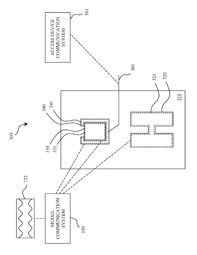
Figure 3 shows a schematic of another non-limiting embodiment of the present
invention comprising an in vitro model system 300. The model comprises the
following
anatomical components: a thoracic cavity 310, lungs 320, a heart 330, and a
pericardium
340. The lungs, heart and pericardium are configured to contain a lung fluid
321, a cardiac
fluid 331, and a pericardial fluid 341, respectively. The model system further
comprises a
model communication system 351. The model communication system 350 may be
configured to communicate at least a component of at least one pressure-
frequency waveform
322 to at least one of the lung fluid 321, the cardiac fluid 331, and/or the
pericardial fluid
341.
The embodiment of Figure 3 further comprises an access device 360 and an
access
device communication system 361. The access device may be, for example, any
one or more
of the following: a surgical instrument, a needle, a probe, a catheter, or a
minimally invasive
device. For example, the access device 360 may be configured to sense a
pressure profile, a
frequency profile, or a pressure-frequency profile. The access device may be,
for example, a
device of the type described in one or more of the following references to
Mahapatra et al.:
PCT/US2008/056643, PCT/US2008/056816, PCT/US2008/057626, and
PCT/US2008/082835. An aspect of an embodiment of the present invention
provides a
system for the access device that can serve as a guide way for introducing
other devices into
the pericardium, for instance sheath-catheters that might subsequently be
employed for
procedures in the pericardium and the epicardium of the heart. Other devices
that the present
invention device may accommodate include, but not limited thereto, the
following: ablation
catheters, guidewires, pacing leads, pacing catheters, pacemakers,
visualization and recording
devices, drugs, lumens, steering devices or systems, drug or cell delivery
catheters, fiber
endoscopes, suctioning devices, irrigation devices, electrode catheters,
needles, optical fiber
sensors, sources of illumination, vital signs sensors, and the like Theses
devices may be
developed for procedures in an integral body part or space.
CA 02790328 2012-08-17
WO 2011/103456
PCT/US2011/025470
1630-04
In an aspect of an embodiment, the pressure, frequency, or pressure-frequency
profile
sensed by the access device may be communicated to a user via an access device
communication system. For example, the access device communication system 361
may be
configured to receive a signal from the access device 360, and communicate
information to
the user via an audio and/or visual display. It should be appreciated that the
pressure related
readings and data may be received by the user, clinician, physician, or
technician or the like
by visual graphics, audible signals (such as voice or tones, for example) or
any combination
thereof. Additionally, the pressure related readings and data may be reduced
to hard copy
(e.g., paper) or computer storage medium. It should be appreciated that the
pressure related
readings and data may be transmitted not only locally, but remotely as well.
The information communicated to the user may include, for example, the
pressure
profile or pressure frequency profile itself. An example of such an access
device
communication system can be seen in Figure 6. Specifically, the access device
communication computer 650 may be configured to display pressure-frequency
waveforms as
shown. Another example communication from an access device can be seen in
Figure 24. In
this example, the tip of an access device has passed through four distinct
anatomical regions
of an in vitro model system, each region with its own unique pressure-
frequency profile. In
the example of Figure 24, these regions correspond to atmospheric pressure
(PD), an intra-
pleural space, a pericardial sac, and the interior of a heart. Additionally or
alternatively, the
information communicated to the user may also include not only a pressure-
frequency
profile, but also the actual location of a portion of the access device
relative to one or more of
the various anatomical components of the model system. For example, the access
device
and/or access device comm"unication system may be configured to recognize the
present
location of the tip of the access device based on changes in the observed
pressure-frequency
profile. Thus, in this example, the access device communication system is
capable of
communicating to the user whether the tip of the access device is presently
located, e.g., in
the thoracic cavity, in the pericardial sac, in the heart, etc.
The embodiment of Figure 3 depicts one way in which the present invention may
be
configured to test an access device or train a user of an access device. The
figure
schematically depicts an access device penetrating the thoracic cavity and
pericardium in
order to access the pericardial fluid. If the pressure-frequency profiles of
the various in vitro
16
CA 02790328 2012-08-17
WO 2011/103456
PCT/US2011/025470
1630-04
anatomical fluids are known to have certain distinct properties, or properties
that fall within
certain ranges, then the process of inserting the access device into one or
more of the
anatomical components can be used to calibrate the pressure-sensing features
of the access
device. In a similar manner, the user can implement the process of inserting
the access
device into one or more of the anatomical components in order to simulate an
in vivo
procedure. For example, sub-xyphoid pericardial ablation of a human heart can
be simulated
in part by inserting the tip of the access device through the model's thoracic
cavity and
further into the pericardium. In this example, the access device communication
system can
communicate the location of the access device to the user as described above,
thereby
training the user on how to perform a similar in vivo procedure. Examples of
using an access
device and/or access device communication system to test an access device or
train a user of
an access device can also be seen, for example, in Figures 5 and 7B.
Figure 4 shows a schematic of another non-limiting embodiment of the present
invention comprising an in vitro model system 400. In this embodiment, the
model
comprises anatomical components including a thoracic cavity 410, lungs 420 and
430, a heart
440, and a pericardium 450. The thoracic cavity 410 is sealed from the
atmosphere at both
ends 411 and 412, and may thus be configured to contain a thoracic cavity
fluid. The
thoracic cavity fluid may be supplied by an outside fluid source via a tube
413. Anatomical
fluids may also be supplied from one or more outside fluid sources to the two
lungs, heart and
pericardium via tubes 421, 431, 441, and 451 respectively. These tubes may be
configured to
extend through a surface of the thoracic cavity without breaking the seal of
the cavity.
Furthermore, the pericardium 450 may be configured to substantially surround
the heart 440,
as shown. In this manner, the pericardium may be configured to contain a
pericardial fluid
between the pericardium and the heart.
In the embodiment of Figure 4, the lungs 420 and 430 are not fluidly connected
to one
another. This arrangement is in contrast to the embodiment of Figure 1, in
which lungs 120
are fluidly connected to one another. It should be appreciated that the
present invention
encompasses embodiments in which organs may or may not be in fluid contact
with one
another. Organs that may be in fluid contact can include but are not
necessarily limited to the
lungs.
17
CA 02790328 2012-08-17
WO 2011/103456 PCT/US2011/025470
1630-04
EXAMPLES
Practice of an aspect of an embodiment (or embodiments) of the invention will
be still
more fully understood from the following examples and experimental results,
which are
presented herein for illustration only and should not be construed as limiting
the invention in
any way. =
Example and Experimental Results Set No. 1: The First Prototype
Figure 6 shows a bench-top example embodiment of an in vitro model system. The
example model system comprises a thoracic section 610 (including a sternum
611), a sub-
xyphoid access site 620, lung and cardiac fluid tubes 630, a model
communication system
cart 640, and an access device communication system, including a computer 650.
This
embodiment was designed to the scale of the adult human chest, and
incorporated two
molded balloons that served as air-inflated lungs, and a molded water-pumped
heart. The
lungs were pumped by a stepper motor-driven bellows, so that the breathing
rate and type of
inhalation waveforms used in cardiac anesthesiology could be mimicked. In this
exploratory
version of the system, the heart pump was driven at a constant rate of one
beat per minute by
a high-torque gear motor. The heart was surrounded by a thin-walled rubber
balloon to
simulate the pericardial sac, and the thin gap between the outer wall of the
heart mold and the
inner surface of the pericardial balloon was filled with water. Access
procedures could be
practiced by passing a pressure-sensing needle through the latex "skin" of the
mannequin's
sub-xyphoid region 620, then through a layer of molded rubber that served as a
surrogate for
the diaphragm, and finally into the pericardium. The chest cavity was sealed
and the thoracic
pressure was monitored by a strain gauge sensor. A laboratory computer 650 was
used to
acquire the thoracic pressures and the pressure-frequency signals in the
access needle. The
inspiration and expiration of the lungs not only mimicked the intubated state
of an
anesthetized patient, but also replicated the lifting force applied to the
heart during the
breathing cycle. This system allowed us to demonstrate the feasibility of
assembling and
operating an in vitro model system, to the point where we were able to
generate pressure-
frequency signals in the surrogate pericardial space that were similar to
those found in the
human body. Through extensive testing, we clarified a number of design and
performance
parameters.
18
CA 02790328 2012-08-17
WO 2011/103456
PCT/US2011/025470
1630-04
Example and Experimental Results Set No. 2: A Second Prototype
Figure 7A shows an exploded view of the central features of a second example
embodiment of an in vitro model system. The model system comprises a thoracic
cavity 710,
lungs 720, a heart 730, and a mannequin shell representing a patient's skin
740. In an actual
training exercise, the overlying mannequin would be covered with a surgical
drape to
simulate the patient's situation in the electrophysiology (EP) lab. As a
result, the model chest
and most of its internal components need only be anthropomorphic in function
and not
necessarily in form. In practice, this meant that we were able to redesign the
chest and its
Contents and make everything more modular for ease of assembly and use. In
this example
embodiment, the thoracic cavity was a Lucite chest box that served to hold
the two latex-
molded lungs. The relaxed-state volume of the molded heart is 220 cm3, which
is about 20%
less than the average adult heart volume of 280 cm3. That molded heart is
shown for scale
relative to the Lucite mannequin, which could be placed on top of the chest
container
during use.
Also shown in Figure 7A, to the right of the mannequin, is a second replica of
a heart,
created via rapid prototyping from an open-source Solid WorksTM design. This
second
replica was slightly oversized compared to the one in the chest case. A thin
layer of Dragon
Skin silicone rubber was cast on this second mode) in order to make the
pericardium, which
was then slipped over the latex-molded heart. The compliance of the resulting
pericardial sac
allowed for the virtual space between it and the outer wall of the heart to be
converted to an
actual one by the injection of water to mimic the pericardial fluid. Also
shown in Figure 7A is
one of the stepping motors used in the simulator. In this improved version of
the system,
both the heart and lung pumps were driven by computer-controlled stepping
motors. This
allowed us to not only simulate any anesthesia waveforms that might be needed,
but also to
simulate variable heart rates and arrhythmias. Moreover, any given heart or
lung pumping
profile could thus be easily documented, archived and repeated as necessary
for practice
purposes. In an interesting change relative to our first system, the lungs
were now water
pumped and the heart was air pumped, to insure that the correct forces were
applied to the
surrogate pericardial fluid by the components of this resealed system.
Figure 7B shows a user 760 holding a representative access device 750 in
position
above the mannequin. During use of the simulator, data acquisition for the
epicardial-access
19
CA 02790328 2012-08-17
WO 2011/103456
PCT/US2011/025470
1630-04
training procedures is handled by a program in LabVIEWO SignalExpressTM
(National
Instruments, Austin, Texas, US). This program also provided the ability to
perform a near-
real-time frequency analysis and the display the fast Fourier transform (FFT)
of a selected
window of data along with the time-domain record of the actual acquired
signal. Most
typically, the access device consisted of a fiber-optic pressure sensor (F1SO,
Quebec,
Canada) that was positioned within the tip of an 11 cm long, 17 gauge Touhy
needle. The
output signal from the sensor's pre-amplifier was acquired at a sampling rate
of 1 kHz and
processed by the data-handling program, with either the raw signal or the FFT
presented to
=
the trainee in a user-selectable window on the host computer's display.
Figure 8A and 8B show two views of a similar embodiment of an in vitro model
system 800. This particular embodiment comprises a thoracic cavity 810, lungs
820, a heart
830 which is at least partially surrounded by a pericardial sac 840. The
pericardium 840 is
attached to a diaphragm 850, and can be accessed by an access device through a
sub-xyphoid
access site 860. A 1 cm thick layer of Dragon Skin silicone rubber functions
as the
abdominal skin and muscle sheath of the model. Another such layer of the
rubber serves as
the diaphragm. The two layers are bonded together to form a "T" shape as shown
in the
figure. Both branches of this "T" are fixed onto the chest box by Lucite
frames, and the
joints are made leak free with silicone sealant. The surface area of the sub-
xyphoid injection
site is large enough to permit a grazing-incidence approach to the right
ventricle of the model
heart, in imitation of the actual clinical access procedure. Upon inflation,
the lungs expand
within the chest cavity, thus applying cyclical pressure to the pericardium
and diaphragm. As
seen in Figure 8B, the frames holding the diaphragm and sub-xyphoid injection
site have
been removed from the chest cavity and placed upside down on a table to reveal
the internal
structures. The interesting things to note are the close, full-organ fit of
the pericardial sac to
the heart and the attachment of the pericardium to the diaphragm at the apex
of the heart.
The close fit of the pericardium is meant to provide the trainee with a
realistic clinical test,
viz., attempting to snag the thin pericardial membrane at grazing incidence
(in order to
minimize the risk of perforating the heart) with and without pressure-
frequency guidance
during the training session. By using transparent Lucite as the construction
material for the
simulator's chest the trainee can do the procedure with and without visual
feedback (i.e., with
= and without the mannequin draped) in order to practice the procedure more
effectively. The
attachment of the pericardium to the diaphragm at the apex of the heart
provides a key
CA 02790328 2012-08-17
WO 2011/103456
PCT/US2011/025470
1630-04
measure of physiological fidelity by helping to hold the heart in place within
the chest while
the lungs work against it during inhalation, thus insuring that the mock
pericardial fluid is
hydrodynamically influenced by the pumping of both the heart and the lungs.
Perhaps most
significantly, since the abdominal muscle sheath, diaphragm and pericardial
sac surrogates
are thus all bonded together to form one continuous unit, it is easy to
conceive of this
assembly being made available as a single integrated replacement part from a
manufacturer
marketing it. This is an important point, since this assembly will eventually
require either
repair or replacement after a sufficiently large number of practice access
procedures have
been performed on it.
Several types of validation studies have been carried out with our improved
system.
In one of them, the stepping motor-driven pumping rates for the heart and
lungs were tuned
to the vital-function conditions that were present during an institutionally-
approved in vivo
clinical trial of epicardial access employing a canine model. The results are
shown in Figure
9. The upper trace is the measured, hydrodynamic pericardial pressure in the
canine model.
Superimposed on the high-amplitude, low-frequency (2-- 0.2 Hz) waveform shown
there is a
low-amplitude, high-frequency component 1 Hz) produced by the heart beat.
The
hydrodynamic pericardial signal measured in the simulator's mock pericardial
fluid (water)
under nominally identical conditions is shown in the lower trace. The same
periodicities are
easily discerned from visual inspection of that waveform, although the
amplitude ratios are
different for the in vivo and in vitro cases. However, during both studies we
noted that the
cardiac component of the waveform was not present either before the tip of the
access needle
had initially entered the pericardium or after it had been withdrawn from the
pericardial sac,
,thus confirming the simulator's ability to credibly represent the clinical
situation. Some
further details of our design, construction and testing efforts are presented
elsewhere.
It would not be unreasonable to introduce a version of the in vitro model
system in
which the pericardial sac was fixed to the molded heart at several locations.
This would
replicate the effect of post-surgical adhesions, which in practice reduce the
amount of fluid in
the pericardial space and thus decrease the strength of the associated
pressure-frequency
signal. It would also be possible to introduce a motional artifact in the
mannequin itself, to
mimic the movement of the chest walls during the respiration cycle. Lastly, a
significant
materials-related improvement would be achieved through the use of a substance
that was
21
CA 02790328 2012-08-17
WO 2011/103456
PCT/US2011/025470
1630-04
more fully self-healing than the silicone rubber presently employed for
abdominal sheath,
diaphragm and pericardial sac. Even when using very small gauge needles in the
access
device, that assembly eventually develops pericardial fluid leaks that are
large enough to
require either manual sealing of the penetration holes or replacement of it
altogether.
We envision using this system not only as, for example, a training tool for
electrophysiologists interested in doing epicardial procedures, but also, for
example, as a
research tool for testing new epicardial technologies. For instance, the
existing endocardial
ablation catheters are not properly configured for epicardial use. In
particular they have the
lengths and curvatures inappropriate for epicardial applications. The
simulator could serve as
a useful intermediate tool for testing specially designed epicardial ablation
catheters and
optimizing their construction and performance prior to undertaking costly in
vivo trials for
clinical commissioning. A similar situation holds for the testing of custom-
designed
epicardial pacing leads, as well.
Example and Experimental Results Set No. 3: Simulating Arbitrary Dynamic
Pressure
Waveforms for Anatomical Training and Testing Models
In an aspect of an embodiment, a LabVIEWTm virtual instrument controls the
software end of the in vitro model system, creating a range of physiological
waveforms given
numerous input parameters. The application of this simulation is towards
pressure guided
transthoracic epicardial access for electrophysiology procedures. While
reaching the
epicardium, the two pressure waveforms encountered are in the thoracic cavity,
which
mimics the respiratory wave due to local connections to respiratory
structures, and in the
pericardial cavity, which sums the thoracic wave with a damped heart component
due its
local connections to both respiratory and cardiac structures. The Lab VIEWTM
instrument
can create and mimic either of these waves, over a range of ideal and non
ideal physiological
conditions. Five different thoracic waves can be selected, which are arbitrary
waveforms that
visually mimic the five most commonly used mechanical ventilation curves in
the clinic, with
flexible options as to their duration, pause, and inspiration to expiration
ratio. For pericardial
waves, the selected respiratory wave is summed with a heart component, which
is a simple
sine wave, with options for the heart rate, heart wave amplitude, and
amplitude of white noise
22
CA 02790328 2012-08-17
WO 2011/103456
PCT/US2011/025470
1630-04
if non-ideal conditions are preferred. The front panel of the program can be
seen in Figure
10.
The virtual instrument builds the desired thoracic or pericardial electrical
waveform at
a scale indicated by a group of inputs and displays the thoracic and cardiac
components as
well as their FFT's, and the summed pericardial waveform. The sampling
frequency, or
resolution of the wave can be programmed, but reaches an upper limit depending
on the
length of the curve in the time domain, due to limited memory of the driver,
which is being
programmed. After assembling the waveform with respect to time, the program
takes the
difference between each point in time, and recompiles the difference values as
a sequence of
commands for stepper motor speed and step sizes and sends the compiled program
to a
stepper motor driver. An input for a scale up factor changes the unit less
original waveform,
to an expected amplitude of output pressure, and controls the magnitude of
each stepper
motor movement with respect to time. Due to the variability in the system, the
effect of a
given scale up factor was characterized experimentally, and is discussed
further in the
methods and results section.
The compiled program from the LabV1EWTm program is sent via RS-232 serial line
to
a Velmex driver controller, which utilizes a custom programming language to
execute stepper
motor functions on Velmex brand stepper motors. Following the directions of
the program,
the driver precisely powers and drives the stepper motor to move the proper
number of steps
at a given instantaneous speed, twisting the stepper motor clockwise or
counterclockwise.
The stepper motor is firmly mounted to a linear actuator screw with an
attached stage, which
moves laterally given a rotational torque from the twisting motor. The final
effect is the
forward and backward movement of the linear actuator stage in a manner, which
mimics the
forward and reverse displacement of the original waveform with time. The
linear actuator's
stage acts on the compliant end of a bellows pipette, which can be connected
to any male luer
slip device, including insertion sites and pressure transducers. The final
result is a sealed
pressure chamber, which increases and decreases pressure according to the
actuator stage
movement, mimicking the pressure fluctuations of a thoracic or pericardial
cavity with the
characteristics of the original program inputs. The complete flow of
information can be seen
in Figure 11.
23
CA 02790328 2012-08-17
WO 2011/103456
PCT/US2011/025470
1630-04
The performance, robustness, and accuracy of the pressure simulator to
recreate a
given waveform were assessed by methods of correlation. Two different groups
of tests were
performed using pressure instrumentation used by researchers in previous work
attached to
the bellows pipette open end. The first test was a characterization study of
the scale up
factor, to find the expected multiplier, which relates the amplitude of the
unit less reference
waveform to the amplitude of the output pressure waveform. For this test, data
from the
pressure transducer was collected in real time via serial line, sampled at a
controlled rate.
The second was a correlation test between interpolated sample reference
waveforms, and the
output waveforms. This tested the ability of the pressure simulator's ability
to truly mimic
the desired waveform as generated by the researchers' inputs. The second test
utilized an
analog output option from the pressure sensor, and data was collected at a
controlled
sampling frequency through a digital storage oscilloscope.
Due to the large number of variables and parameters in the system, as well as
given
uncertainties in the bellows pipette as a component, it was difficult to
characterize the
expected amplitude of the output pressure in comparison to the original
reference waveform
based on an analytical transfer function. Because of this, an empirical method
was designed
for characterizing a multiplier for the expected amplitude of the output
pressure waveform
given an input function and scale up factor.
A group of reference sine waveforms with different scale up factors were
statistically
compared to data acquired from a pressure transducer attached to the bellows
chamber. Both
the reference waveform and pressure data acquisition occurred at the same
sampling rate of
10 Hz, large enough to be greater than the Nyquist frequency of the waveforms,
and small
enough that a miniscule widening or narrowing of the output waveform in the
time domain
due to stepper motor imperfections would not cause a discrepancy between the
number of
points for the two waves, making statistical analysis as simple as possible.
Three different
sine waves were tested, with center frequencies of .5, I, and 1.5 Hz, all with
a peak amplitude
of 0.5 (the reference waveform is unit less). Each sine wave was tested
multiple times at
scale up factors of 50, 100, and 200. The pressure output for each trial was
plotted against
the reference waveform, and a linear best fit approximation of the two data
sets was estimated
to find the pressure multiplier given a relatively constant initial pressure
near 30 mmHg.
24
=
CA 02790328 2012-08-17
WO 2011/103456
PCT/US2011/025470
1630-04
At higher sine wave center frequencies, the change in pressure between each
point
collected every 0.1 seconds is much higher. Because there was no way to align
the starting
time for both the stepper motor and data acquisition precisely, some of the
acquired pressure
waveforms had minor phase shift deviations from the reference waveform. This
small phase
shift at high center frequencies caused major distortions in data during
statistical analysis, so
out of the 7 trials for each condition, only the 3 with the highest
correlation coefficients were
kept for data analysis, because they accurately captured the waveform at a
similar phase as
the reference waveform. An example of this phase shift can be seen in Figure
12.
The most important group of tests involved simulating different pericardial
waveforms in the pressure chamber and statistically comparing the pressure
output to the
input waveform. Upon initial construction of the system, all the thoracic
waves were tested,
as well as a range of pericardial waves, all of which visually mimicked the
input waveform,
but a quantitative comparison was imperative to characterize the system's
actual
performance. Three common ventilation curves were selected, each with large
heart
component amplitude (1/5 that of the thoracic wave), and a small heart
component amplitude
(1/20 that of the thoracic wave), visually imitating realistic cardiac
amplitudes for healthy
hearts, and unhealthy hearts with adhesions, respectively. The three
ventilation waves
included pressure controlled rectangular, flow controlled rectangular, and
flow controlled
sine waves. Breath duration, inspiration to expiration ratio, and other input
parameters were
held constant between each waveform to limit the amount of variability in the
data collection.
All waveforms had a sampling frequency of 20 Hz in the program, to create a
very smooth
and well defined wave. Each waveform was recreated four times using the exact
same
compiled waveform program with the actuator stage always at the same initial
location, with
the pressure in the output chamber monitored by a digital storage scope
sampling at 100 Hz.
The 100 Hz output waveform was then compared to the linearly interpolated
input waveform
using the equation for a linear correlation coefficient (p).
=
Equation 1: Linear Correlation Coefficient
Cov(x, y) 1 õ
= ____________________ where Cov(x, y) = ¨R
Lx,- ,ux)(y, ¨ u v)]
u n
x y
CA 02790328 2012-08-17
WO 2011/103456
PCT/US2011/025470
1630-04
For each trial, the slope (pressure multiplier), intercept (initial pressure),
and r2 value
(coefficient of determination) were calculated using a linear best-fit trend
line of the data.
The average slope was calculated for each scale up factor multiplied by the
peak amplitude of
the reference waveform, further which will be referred to as 'peak scale',
with the peak
amplitude of the reference waveforms constant at 0.5 for all trials. The
average slope
approximated the multiple which related the amplitude between the input
reference
waveform, and the output pressure waveform, given a constant initial pressure
near 30
mmHg. For the peak scales of 25, 50, and 100, the average multipliers observed
were 2.885
0.057, 5.631 0.107, and 11.347 0.122 mmHg respectively. These three values
were
placed on a plot comparing peak scale to pressure multiplier, and the
resulting linear
relationship indicated that the pressure multiplier is equal to 0.133.(peak
scale) + (0 .0272).
Using this formula, the researcher can then predict the pressure scale they
can expect to see
given the peak amplitude and scale up factor of the input waveform.
Six waveforms were tested for performance of the pressure simulator. The
waveforms were all pericardial simulations of pressure controlled rectangular
(waves 1 and
2), flow controlled sine (waves 3 and 4), and flow controlled rectangular
(waves 5 and 6)
ventilation waveforms each summed with either high or low cardiac amplitude
components,
respectively. Each waveform was run through the simulator four separate times
for four sets
of acquired pressure data. The output pressure read by the pressure
instrumentation was
acquired at 100 Hz, and statistically compared to the interpolated input
waveform as seen in
Equation 1, to assess the linearity between the two data sets. It is important
to note that the
pressure waveforms are at higher scales than physiological levels, but if
incorporated into a
larger pressure chamber, more miniscule pressures can be reproduced. However,
the
waveform itself and the dynamic capabilities of the simulator are the
important aspects of this
test. Upgrades to the pressure simulator will be discussed in the following
section.
Correlation coefficients for each of the waveform types can be seen in Figure
13.
The average correlation coefficient for the entire data set is 0.9914
0.0058, This
shows a very strong linear correlation between all of the output pressure
waveforms with the
input reference waveforms, which they are intended to duplicate. To further
justify the
results seen above, an example trial with very strong results is shown in
Figures 14 and 15.
In Figure 14, the time domain input waveform (smoother line) is graphed
alongside the
26
CA 02790328 2012-08-17
WO 2011/103456
PCT/US2011/025470
1630-04
output pressure waveform (rougher line), each on their own individual
amplitude scale for
Wave 4 (flow controlled sine ventilation wave, low cardiac component), Run 4.
Figure 15
shows the correlation graph between these two data sets, visually identifying
the linear
relationship between the two.
The ability to mimic realistic pressure waves from sealed human cavities is a
useful
practice for testing instrumentation and real time signal processing
algorithms, but is also
important to be able to develop cost effective anatomical training and testing
tools for using
such devices in an in vitro scenario. The previous results have demonstrated
the ability of
this low cost system to create chambers with fluctuating dynamic pressures
which can be
translated to a multitude of applications. Most importantly for the specific
field of epicardial
electrophysiology, this concept can be applied to anatomical structures to
create in vitro
human pressure cavities and can be applied for testing pressure guided
epicardial access
instrumentation, and more importantly, for training clinicians in this new
procedure in a safe
manner. In a broader sense, this system can be applied to a range of testing
scenarios not
only in epicardial electrophysiology, but any field which uses real time
pressure signal
monitoring and processing. While looking into the capabilities of such a
system, it is
important to note where improvements can be made to create such anatomical
models. For
example, the low volume bellows pipette can be replaced with a range of
different devices
including pumps and pistons, which can control larger amounts of pressurized
water or air
more precisely, given a stepper motor with high enough torque generation,
creating larger
and more precisely controlled dynamic chambers. As applied to anatomical
models, instead
of mathematically creating a pericardial wave by summing thoracic and cardiac
waveforms,
the separate waveforms could be created in the appropriate anatomical
structures and see the
pneumatic overlap of the pressure waves on the anatomical pericardial
structure, as it occurs
in the body.
Example and Experimental Results Set No. 2: Electro-Mechanical/Pneumatic
Device and
Method of Use for Simulating Sub-xyphoid Access for Epicardial
Electrophysiology
Procedures
In an aspect of an embodiment, a basic shape needed to be established within
which
the pressure simulations could be performed. Much consideration was given to
possible
choices ranging from a large plastic bottle, a large balloon, to a geometric
representation of
27
CA 02790328 2012-08-17
WO 2011/103456
PCT/US2011/025470
I 630-04
the thoracic cavity. Ultimately, it was chosen to create an anatomically
accurate frame on
which the enclosure can be simulated (Figure 16A). As the pressure
characteristics in the
pericardium will be influenced indirectly by the volume of the proximate lungs
as well as the
volume of the heart, we sought to come as close as possible to replicating the
real human
geometries. The thoracic cage was first to be constructed to replicate the
dimensions of an
average thorax. Aluminum rods (1 inch width) will comprise the sternum and the
general
shape of the spine. Using half inch aluminum rods vertebras 1,6, and 10 will
complete the
general shape of the thoracic cage. Over the metallic frame, 3 to 5 layers of
liquid latex
(room temperature galvanizing from TapPlastics) is applied. Using strong
rubber bands, the
vertebra will be connected, encapsulating the thoracic cavity. Using a sheet
of aluminum to
encircle the thoracic cavity, up to 20 coats of liquid latex will be applied.
The aluminum foil
will be removed and the thoracic mold dried latex will be slid into its
intended position over
the ribs. A rectangle about 4 inches in width and 6 inches in length will be
cut from the latex
shell centered on the sternum. A clear Plexiglas with the same dimensions is
glued over the
cut out. An additional 10 coats of liquid latex will seal the Plexiglas edges
so that an air tight
perimeter is established. A plastic heart model will be used as the mold for
the creation of a
heart using liquid latex. In the same way, the lungs are created with their
appropriate shape
using liquid latex and appropriately sized lung molds. Both the lungs and the
heart are
hollow and will have a single opening. The two lung balloons will be connected
by a ridged
tube representing the trachea, which will exit the cavity. Also, another
ridged tube will be
connected to the heart balloon and will exit the cavity. The heart latex
balloon is enclosed by
another balloon. This one however, does not have an access and its opening on
the top will be
tightly sealed. The heart balloon is filled with liquid connected via its exit
hose to a liquid
holding chamber. The diaphragm and a circular enclosure around the first
vertebra will seal
the thoracic cage in an air tight compartment. For certain details of
construction, see Figure
16B. After installing the plastic clear window under the sternum, the cage was
enclosed and
several layers of liquid latex applied (Figure 16C). The process of creating
the exterior
covering of the model provides a suitable skin-like surface for it.
The interior of the model is sought to be reflective of the design drawing
shown in
Figure 4. Here two large balloons on the edges of the cavity are to represent
the lungs. These
will be filled with air with modulating pressure. The heart representation can
be visualized in
the middle of the lungs. This rubber chamber is also able to modulate in
volume. However,
28
CA 02790328 2012-08-17
WO 2011/103456
PCT/US2011/025470
1630-04
it is ideally filled with liquid. The chamber surrounding the heart is filled
with a small
amount of liquid that will represent the pericardial fluid. As the shapes of
these irregular
bodies are hard to find in commercially produced products, they were
replicated using liquid
latex. As can be seen in Figure 9a, the lungs and heart were replicated using
molds. For the
heart, about 15-20 layers of liquid latex were painted on a life sized model
of a heart and then
the dried latex was removed. The form of the lungs was created by carving the
shape of each
lung on a styrofoam block. Then several layers of liquid latex were applied
(Figure 17). In
addition there are tubes (bronchi) that allow for the air flow to be
visualized. The lung and
heart molds have been successfully tested in a hydraulic system for
contractile motion.
Pressure control will be acquired by using a stepper motor (a unipolar
stepper, 3.6V,
16kgcm holding torque) to operate a linear actuator attached to an air pump.
This air pump
will not have a one way valve, but rather it will be able to both push and
pull the air column.
Two actuators will be used: one connected to the lung compartment and another
connected to
the liquid holding chamber connected to the heart. The mechanical schematic
diagram for
this device is shown in Figure 18. The design was created to be ultimately a
clear box that
can be opened. On both faces of the box are linear actuator mechanisms that
are controlled
by stepper motors. It is connected to the stepper motor, which is controlled
as described
below. Two air pumps provide the pressure variations that simulate the
pressures in the heart
and lungs, as per the mechanism of Figure 18.
A microcontroller will be used to run the stepper motors. Specifically, the
Cerebot
system by Digilent Inc. will be used to provide serial interface with a
computer for real time
instructions as well as an H-Bridge connection to the unipolar stepper motor.
An adaptive
circuit will be made using four transistors to regulate power supply to the
four leads of the
stepper motor. Also, diodes will be used to counter the kick-back current from
the stepper
motor to protect the microcontroller port. The microcontroller is equipped
with an 8-bit AVR
Microcontroller (Figures 19A & 19B) with 64K Bytes of in-system programmable
flash
memory. It is based on the ATmega64L processor. C will be used to construct
the run time
program. The Win-AVR will be used to convert the higher level C code into
machine
code/hexadecimal that will later be exported to the Cerebot (via a USB
JTAG/SPI interface)
using AVR programmer. The microcontroller will allow for preprogrammed or
variable turns
that will translate to pressure modulations.
29
CA 02790328 2012-08-17
WO 2011/103456
PCT/US2011/025470
1630-04
The open source C program was adopted to implement the connection between
computer via RS-232 COM port and the Cerebot COM port (JD) module. Ultimately,
any
PC connected to the microcontroller via an interface like Microsoft
Hyperterminal is able to
give the controller a specified commend set that allows the shifting of
voltages in the JPC
pins. The voltages can be switched from on or off varying from 0 to 3.2 V.
This is important
as the four pins are to be synchronously switched to operate the unipolar
stepper motor. The
steps of voltage were monitored via the oscilloscope. As there cannot be
enough power
delivered by these output ports to drive the stepper motor, a driver circuit
was designed, built
and tested (Figure 20). This driver circuit ultimately allows for much larger
currents
(upwards of 2 A) flowing through the stepper motor on the switch command of
several
miliamperes. As the proposed circuit diagram shows in Figure 20, power MOSFET
transistors were used to grate the four conducting wires attached to the two
unipolar stepper
windings. The winding arrangement and properties are shown in the same figure.
A stream
of synchronized pulses from the microcontroller board to the transistor gates
opens and closes
them to allow current to flow accordingly. The operation of the circuit was
successfully
tested using LEDs, but as the threshold voltage for the gating of the MOSFTETs
is one volt
higher than that given by the controller, there needs to be a base voltage of
about 1 V applied
to all the gates before enough current opened for the operation of the stepper
motor.
Nevertheless, the stepper motor can be operated with great precision.
It is expected that as a needle is inserted through the insertion port at the
sub-xyphoid
site and into the thoracic cage, leading to the pericardial space, the
physiologic pressure
waveforms will be observed. Calibration of the pressure control systems will
need to be done
to reproduce these profiles. By modulating the volume of the lungs and the
heart, we can
generate a pressure profile in the thoracic cage and the pericardial space.
These
compartments are not directly being modulated by the pressure controllers,
they are a =
consequence of them, therefore it is expected that the proposed procedure will
be able to
replicate the overlay of the thoracic and cardiac pressure waveforms.
Imperfections in the materials used may lead to inconsistent pressures. This
is most
notable in the thoracic cavity and it is important to keep an air tight
chamber. The seals
introduced by the diaphragm, upper (neck) seal and the sternum Plexiglas
window leave
ample room for air leaks. This leaking may severely dampen the pressure
waveforms in the
CA 02790328 2012-08-17
WO 2011/103456
PCT/US2011/025470
1630-04
thoracic cavity. If air leaking is a great enough hindrance to the correct
representation of the
waveforms, then a feedback loop can be established with a third air pump
connected directly
to the thoracic cavity to counteract the leakage. The additional pump,
however, can introduce
significantly great complexities. A potential problem is the stepper motor
itself, as in time it
may overheat or even disrupt the electrical components running it. Therefore,
other types of
motors can also be incorporated. Another possible problem may be the actual
air pumps
themselves, as they may also lead to some certain extent. As they leak, this
leakage is not
compensated by the control program and a bias is introduced. The bias can be
eliminated
again by using feedback loops using a stationary pressure sensor.
Those skilled in the art will recognize the many significant advantages
associated with
this general approach by considering the general and specific embodiments of
the invention
as discussed above in the drawings and their descriptions.
Figure 21 is a functional block diagram for a computer system 2100 for
implementation of an exemplary embodiment or portion of an embodiment of
present
invention (or combinations of various embodiments in whole or in part of the
present
invention). For example, a method or system of an embodiment of the present
invention may
be implemented using hardware, software or a combination thereof and may be
implemented
in one or more computer systems or other processing systems, such as personal
digit
assistants (PDAs) equipped with adequate memory and processing capabilities.
In an
example embodiment, the invention was implemented in software running on a
general
purpose computer 2100 as illustrated in Figure 21. The computer system 2100
may includes
one or more processors, such as processor 2104. The Processor 2104 is
connected to a
communication infrastructure 2106 (e.g., a communications bus, cross-over bar,
or network).
The computer system 2100 may include a display interface 2102 that forwards
graphics, text,
and/or other data from the communication infrastructure 2106 (or from a frame
buffer not
shown) for display on the display unit 2130. Display unit 2130 may be digital
and/or analog.
The computer system 2100 may also include a main memory 2108, preferably
random
access memory (RAM), and may also include a secondary memory 2110. The
secondary
memory 2110 may include, for example, a hard disk drive 2112 and/or a
removable storage
drive 2114, representing a floppy disk drive, a magnetic tape drive, an
optical disk drive, a
flash memory, etc. The removable storage drive 2114 reads from and/or writes
to a
31
CA 02790328 2012-08-17
WO 2011/103456
PCT/US2011/025470
1630-04
=
removable storage unit 2118 in a well known manner. Removable storage unit
2118,
represents a floppy disk, magnetic tape, optical disk, etc. which is read by
and written to by
removable storage drive 2114. As will be appreciated, the removable storage
unit 2118
includes a computer usable storage medium having stored therein computer
software and/or
data.
In alternative embodiments, secondary memory 2110 may include other means for
allowing computer programs or other instructions to be loaded into computer
system 2100.
Such means may include, for example, a removable storage unit 2122 and an
interface 2120.
Examples of such removable storage units/interfaces include a program
cartridge and
cartridge interface (such as that found in video game devices), a removable
memory chip
(such as a ROM, PROM, EPROM or EEPROM) and associated socket, and other
removable
storage units 2122 and interfaces 2120 which allow software and data to be
transferred from
the removable storage unit 2122 to computer system 2100.
The computer system 2100 may also include a communications interface 2124.
Communications interface 2124 allows software and data to be transferred
between computer
system 2100 and external devices. Examples of communications interface 2124
may include
a modem, a network interface (such as an Ethernet card), a communications port
(e.g., serial
or parallel, etc.), a PCMCIA slot and card, a modem, etc. Software and data
transferred via
communications interface 2124 are in the form of signals 2128 which may be
electronic,
electromagnetic, optical or other signals capable of being received by
communications
interface 2124. Signals 2128 are provided to communications interface 2124 via
a
communications path (i.e., channel) 2126. Channel 2126 (or any other
communication means
or channel disclosed herein) carries signals 2128 and may be implemented using
wire or
cable, fiber optics, blue tooth, a phone line, a cellular phone link, an RF
link, an infrared link,
wireless link or connection and other communications channels.
In this document, the terms "computer program medium" and "computer usable
medium" are used to generally refer to media or medium such as various
software, firmware,
disks, drives, removable storage drive 2114, a hard disk installed in hard
disk drive 2112, and
signals 2128. These computer program products ("computer program medium" and
"computer usable medium") are means for providing software to computer system
2100. The
computer program product may comprise a computer useable medium having
computer
32
CA 02790328 2012-08-17
WO 2011/103456
PCT/US2011/025470
1630-04
program logic thereon. The invention includes such computer program products.
The
"computer program product" and "computer useable medium" may be any computer
readable
medium having computer logic thereon.
Computer programs (also called computer control logic or computer program
logic)
are may be stored in main memory 2108 and/or secondary memory 2110. Computer
programs may also be received via communications interface 2124. Such computer
programs, when executed, enable computer system 2100 to perform the features
of the
present invention as discussed herein. In particular, the computer programs,
when executed,
enable processor 2104 to perform the functions of the present invention.
Accordingly, such
computer programs represent controllers of computer system 2100.
In an embodiment where the invention is implemented using software, the
software
may be stored in a computer program product and loaded into computer system
2100 using
removable storage drive 2114, hard drive 2112 or communications interface
2124. The
control logic (software or computer program logic), when executed by the
processor 2104,
causes the processor 1304 to perform the functions of the invention as
described herein.
In another embodiment, the invention is implemented primarily in hardware
using, for
example, hardware components such as application specific integrated circuits
(ASICs).
Implementation of the hardware state machine to perform the functions
described herein will
be apparent to persons skilled in the relevant art(s).
In yet another embodiment, the invention is implemented using a combination of
both
hardware and software.
In an example software embodiment of the invention, the methods described
above
may be implemented in SPSS control language or C + + programming language, but
could be
implemented in other various programs, computer simulation and computer-aided
design,
computer simulation environment, MATLAB, or any other software platform or
program,
windows interface or operating system (or other operating system) or other
programs known
or available to those skilled in the art.
The devices, systems, compositions, modules, computer program products, and
methods of various embodiments of the invention disclosed herein may utilize
aspecfs
33
CA 02790328 2016-02-17
WO 2011/103456
PCT/US2011/025470
1630-04
disclosed in the following references, applications, publications and patents.
I. U.S. Patent No. 6,874,501 BI, Estetter, et at., "Lung Simulator", April
5,2005.
2. U.S. Patent No. 6,428,323 BI, Pugh, C., "Medical Examination Teaching
System",
August 6, 2002.
3. U.S. Patent No. 6,273,728 B1, van Meurs, et at., "Life Support Simulation
System
Simulating Human Physiological Parameters", August 14, 2001.
4. U.S. Patent No, 5,428,323, Geissler, et at., "Device for Compensation For
Temperature-Dependent Volume Changes in a Waveguide", June 27, 1995,
5. U.S. Patent No. 7,510,398 B1, Thornton, W., "Apparatus for Simulating a
Pulse
and Heart Beat and Methods For Using Same To Train Medical Professionals",
March 31,
2009.
6. U.S. Patent Application Publication No. US 2009/0111080 Al, Chen, et at.,
"Medical Simulation System and Method", April 30, 2009.
7: U.S. Patent No. 6,921,267 B2, van Oostrom, et at., "Lung Simulator For An
Integrated Human Patient Simulator", July 26, 2005.
8. U.S. Patent No. 5,584,701, Lampotang, et at., "Self Regulating Lung For
Simulated Medical Procedures", December 17, 1996.
9. U.S. Patent No. 5,800,197, Bailey, B., "System For Trainingt Persons To
Perform
Minimally Invasive Surgical Procedures", September 1, 1998.
10. U.S. Patent No. 4,167,070, Orden, B., "Educational Lung Simulator",
September
11, 1979.
II. U.S. Patent No. 6,062,865, Bailey, B., "System For Training Persons To
Perform
Minimally Invasive Surgical Procedures", May 16, 2000.
34
CA 02790328 2012-08-17
WO 2011/103456
PCT/US2011/025470
1630-04
12. U.S. Patent No. 6,267,599 B!, Bailey, B., "System For Training Persons To
Perform Minimally Invasive Surgical Procedures", July 31, 2001.
13. U.S. Patent No. 7,021,940 B2, Morris, etal., "Patient Simulator Manikin
And
System", April 4, 2006.
14. U.S. Patent No. 6,336,812 B1, Cooper, etal., "Clinical And/Or Surgical
Training
Apparatus-, January 8, 2002.
15. U.S. Patent No. 6,234,804 B1, Yong, P., "Thoracic Training Model For
Endoscopic Cardiac Surgery", May 22, 2001.
16. U.S. Patent No. 6,007,342, Tjelsen, 0., "Pulse Device For Creating A
Simulated
Feelable And Recognition of Pulse.
17. JP Patent No. 5-27113, 4/1993
18. JP Patent No. 2990602, 10/1999
19. "A Computer-Controlled Patient Simulator", J.S. Denson, M.D., and Stephen
Abrahamson, Ph.D., JAMA (April 21, 1969), vol. 208, p. 3, pp. 504-508.
20. U.S. Patent No. 6,175,768 B1, Arndt et al., "In Vivo Simulator for
Microwave
Treatment", January 16, 2001.
21, PCT International Application No. Serial No. PCT/US2008/056643, filed
March
12, 2008, entitled, "Access Needle Pressure Sensor Device and Method of Use"
and
corresponding U.S. Patent Application Serial No. 12/530,830 filed September
11, 2009.
22. PCT International Application No. Serial No. PCT/US2008/056816, filed
March
13, 2008, entitled, "Epicardial Ablation Catheter and Method of Use" and
corresponding U.
S. Patent Application Serial No. 12/530,938 filed September 11, 2009.
23. PCT International Application No. Serial No. PCT/US2008/057626, filed
March
20, 2008, entitled, "Electrode Catheter for Ablation Purposes and Related
Method Thereof'
and corresponding U.S. Patent Application Serial No. 12/532,233 filed
September 21, 2009.
=
CA 02790328 2016-02-17
=
WO 2011/103456
PCT/US2011/025470
1630-04
24. PCT International Application No. Serial No. PCT/US2008/082835, filed
=
November 7, 2008, entitled, "Steerable Epicardial Pacing Catheter System
Placed Via the
Subxiphoid Process," and corresponding U.S. Patent Application Serial No.
12/741,710 filed
May 6,2010.
Unless clearly specified to the contrary, there is no requirement for any
particular
described or illustrated activity or element, any particular sequence or such
activities, any
particular size, speed, material, duration, contour, dimension or frequency,
or any particularly
interrelationship of such elements. Moreover, any activity can be repeated,
any activity can
be performed by multiple entities, and/or any element can be duplicated.
Further, any
activity or element can be excluded, the sequence of activities can vary,
and/or the
interrelationship of elements can vary. It should be appreciated that aspects
of the present
invention may have a variety of sizes, contours, shapes, compositions and
materials as
desired or required.
In summary, while the present invention has been described with respect to
specific
embodiments, many modifications, variations, alterations, substitutions, and
equivalents will
be apparent to those skilled in the art. The present invention is not to be
limited in scope by
the specific embodiment described herein. Indeed, various modifications of the
present
invention, in addition to those described herein, will be apparent to those of
skill in the art
from the foregoing description and accompanying drawings. Accordingly, the
invention is to
be considered as limited only by the scope of the following claims, including
all modifications and
equivalents.
Still other embodiments will become readily apparent to those skilled in this
art from
reading the above-recited detailed description and drawings of certain
exemplary
embodiments. It should be understood that numerous variations, modifications,
and
additional embodiments are possible, and accordingly, all such variations,
modifications, and
embodiments are to be regarded as being within the scope of this application.
For
example, regardless of the content of any portion (e.g., title, field,
background, summary,
abstract, drawing figure, etc.) of this application, unless clearly specified
to the contrary,
there is no requirement for the inclusion in any claim herein or of any
application claiming
priority hereto of any particular described or illustrated activity or
element, any particular
sequence of such activities, or any particular interrelationship of such
elements. Moreover,
36
CA 02790328 2016-02-17
WO 2011/103456
PCT/US2011/025470
1630-04
any activity can be repeated, any activity can be performed by multiple
entities, and/or any
element can be duplicated. Further, any activity or element can be excluded,
the sequence of
activities can vary, and/or the interrelationship of elements can vary. Unless
clearly specified
to the contrary, there is no requirement for any particular described or
illustrated activity or
element, any particular sequence or such activities, any particular size,
speed, material,
dimension or frequency, or any particularly interrelationship of such
elements. Accordingly,
the descriptions and drawings are to be regarded as illustrative in nature,
and not as
restrictive. Moreover, when any number or range is described herein, unless
clearly stated
otherwise, that number or range is approximate. When any range is described
herein, unless
clearly stated otherwise, that range includes all values therein and all sub
ranges therein.
37