Note: Descriptions are shown in the official language in which they were submitted.
WO 2011/103598
PCT/US2011/025770
PLATELET-DERIVED GROWTH FACTOR COMPOSITIONS AND METHODS
FOR THE TREATMENT OF TENDINOPATHIES
CROSS REFERENCE TO RELATED APPLICATIONS
[0001] This application claims benefit of ITS. Provisional Patent Application
No.
61/306,938, filed February 22, 2010, U.S. Provisional Patent Application
Serial No.
61/311,284, filed March 5, 2010, U.S. Provisional Patent Application Serial
No. 61/428,809,
filed December 30, 2010, and U.S. Provisional Patent Application No.
61/429,428, filed
January 3, 2011.
TECHNICAL FIELD
[0002] This invention relates to compositions and methods for the treatment of
tendinopathies, such as tenosynovitis, tendinosis or tenclinitis, including
Achilles
tendinopathy, patellar tendinoputhy, lateral epicondylitis or "tennis elbow,"
medial
epicondylitis or "golfer's elbow," plantar fasciit is, and rotator cuff
tenclinopatlay, and in
particular to methods for the treatment of tenclinopathies by administering
compositions
comprising platelet-derived growth factor (PDGF).
BACKGROUND OF THE INVENTION
[0003] A tendon is a tough band of fibrous connective tissue that usually
connects muscle
to bone. The elastic properties of tendons modulate forces during locomotion,
providing
additional stability with no active work. They also store and recover energy
at high
efficiency. Normal healthy tendons are composed primarily of parallel arrays
of type I
collagen fibers closely packed together, but also include a small amount of
elastin and of
proteoglycans. Due to their highly specialized ultrastructure, low level of
vascularization and
slow collagen turnover, tendons are very slow to heal if injured, and rarely
regain their
original strength. Partial tears heal by the rapid production of disorganized
type-III collagen,
which is weaker than normal tendon. Recurrence of injury in the damaged region
of tendon is
common.
[0004] Tendinopathies are chronic disorders or injuries of the tendons, that
appear to result
from, an imbalance between catabolic and anabolic responses that result from
gradual wear
1
CA 2790403 2017-06-22
CA 02790403 2012-08-17
WO 2011/103598
PCT/US2011/025770
and tear to the tendon from overuse or aging. The result of this imbalance is
tendon
degeneration, weakness, tearing, and pain. In contrast, acute tendon injuries
such as, for
example, tendon rupture Or detachment from the bone are quite sudden and
usually require
surgery to repair the rupture or reattach the tendon to bone. Anyone can
develop a
tendinopathy, but people who tend to make the same motions over and over again
in their
jobs, sports, or regular daily activities are more likely to develop them.
Tendinopathy usually
causes pain, stiffness, and loss of strength in the affected area.
[0005] The term tendinopathy refers to two types of tendon injury: tendinosis
and
tendinitis. The term also encompasses tenosynovitis, a tendinopathy of the
outer lining of the
tendon which occurs in certain tendons such as flexor tendons and the Achilles
tendon.
[0006] Tendinosis is a non-inflammatory injury to the tendon characterized by
intratendinous degeneration of the tendon usually in the form of microtears in
the tissue in
and around the tendon caused by overuse, leading to an increase in the number
of tendon
repair cells around the area of damage. Degeneration of the tendon is caused
by damage to or
disorganization of the collagen fibers, cells, and vascular components of the
tendon, which
can reduce the tendon's tensile strength and can lead to tendon rupture if not
treated. The
changes in collagen organization are characterized by
separation/loosening/crimping of
fibers, loss of parallel orientation, decrease in fiber diameter and decrease
in overall density
of collagen. In addition, collagen microtears can also occur that are
surrounded by
erythrocytes, fibrin, and fibronectin deposits. On the other hand, there is an
increase in type
III (reparative) collagen. These matrix organization changes can lead to
decreased
birefringence under polarized light microscopy. In addition to collagen
content and
organization, tendinosis is also characterized by an increase in mucoid ground
substance
(proteoglycans) and variation in cellular density in affected areas. Some
areas contain
abnottnally plentiful tenocytes, with rounded nuclei and ultrastructural
evidence of increased
production of proteoglycan and protein. In contrast, other areas of the
affected tendon may
contain fewer tenocytes than normal, with small, pyknotic nuclei. Another
characteristic
feature of tendinosis is proliferation of capillaries and arterioles. Several
subcategories of
tendon degeneration in tendinosis have been identified by electron microscopy:
(1) hypoxic
degeneration, (2) hyaline degeneration, (3) mucoid or myxoid degeneration, (4)
fibrinoid
degeneration, (5) lipoid degeneration, (6) calcification, and (7)
fibrocartilaginous and bony
metaplasia. These pathologies can coexist with varying prevalence, depending
on the
2
CA 02790403 2012-08-17
WO 2011/103598
PCT/US2011/025770
anatomical site and the nature of the insult that caused them (eg, hypoxia
versus mechanical
loading; acute versus chronic injury). For example, mucoid degeneration area
is characterized
by light microscopy, large mucoid patches and vacuoles between fibers.
However, lipoid
degeneration is characterized by abnomial intratendinous accumulation of lipid
that results in
disruption of collagen fiber structure. In some cases, tendinosis is
accompanied by focal
necrosis or calcification of the tendon. It is a very common reason for
chronic pain
surrounding a joint. Tendinosis is also characterized by an absence of the
initial inflammatory
response. Inflammatory cells are thought to be early stage mediators of the
repair process,
without which tendinosis can become a chronic condition.
[0007] Characteristic increases in water content and disorganization of the
collagen matrix
associated with tendinosis can be diagnosed by ultrasonography or magnetic
resonance
imaging. Symptoms can vary from simple aching and stiffness in the local area
of the tendon
to a burning sensation surrounding the entire joint around the injured tendon.
For many
patients, the pain is frequently worse during and after activity, and the
tendon and joint area
can become stiffer the following day as swelling impinges on the movement of
the tendon.
[0008] Tendinitis is an inflammatory injury to the tendon, characterized by
degeneration
like that observed in tendinosis, but also accompanied by inflammation of the
tendon
accompanied by vascular disruption and an inflammatory repair response.
Tendinitis is often
accompanied by fibroblastic and myofibroblastic proliferation, as well as
hemorrhage and
organizing granulation tissue. Generally tendinitis is referred to by the body
part involved,
such as Achilles tendinitis (affecting the Achilles tendon), or patellar
tendinitis (also known
as "jumper's knee," affecting the patellar tendon), though there are certain
exceptions, such
as lateral epicondylitis (also known as "tennis elbow," affecting the Extensor
Carpi Radialis
Brevis tendon). Symptoms can vary from aches or pains and local stiffness to a
burning
sensation surrounding the entire joint around the inflamed tendon. In some
cases, tendonitis is
characterized by swelling, sometimes accompanied by heat and redness; there
may also be
visible knots surrounding the joint. For many patients, the pain is usually
worse during and
after activity, and the tendon and joint area can become stiffer the following
day as muscles
tighten from the movement of the tendon.
[0009] Current treatments are primarily palliative in nature, with treatment
traditionally
focusing on anti-inflammatory measures, including treatment with nonsteroidal
anti-
inflammatory drugs (NSAIDs), steroid injections, and physical therapy, despite
the fact that
3
CA 02790403 2012-08-17
WO 2011/103598
PCT/US2011/025770
tendinosis tends not to be associated with an inflammatory response. More
recently, shock
wave therapy, low-level laser therapy, sclerotherapy, and other experimental
treatments have
been tested. For the most part, it appears that some treatments (e.g., NSAIDs
and cortisone
injections) offer short-term relief, while the longer-term benefit of current
treatments remains
unclear. Therefore, there is a need for improved methods of treating
tendinopathies that offer
longer-term benefits compared to existing treatment modalities.
[0010] PDGF is stored in the alpha-granules of platelets and is secreted
during tissue repair
by locally-activated cells, including macrophages, fibroblasts, and
endothelial cells. PDGF-
BB is one of the major products of the hemorrhage and inflammation of acute
tendon injury.
Platelet-derived growth factor-BB (PDGF-BB) is a wound healing protein which
is known to
be chemotactic (cell migration) and mitogenic (cell proliferation) for cells
of mesenchymal
origin, including bone (osteoblast) and tendon (tenocyte) cells. Additionally,
PDGF-BB has
been shown to up-regulate vascular endothelial growth factor (VEGF), leading
to increased
angiogenesis (revascularization), which is essential for successful
regenerative processes.
[0011] The Achilles tendon is the thickest and strongest tendon in the human
body, which
allows it to support high loads. The mechanical loading environment in which
the Achilles
tendon functions makes it prone to rupture. Achilles tendon ruptures can occur
as a result of a
variety of factors, however rupture is often associated with degenerative
changes. (Mafulli N,
Wong J, Almekinders L. Types and epidemiology of tendinopathy. Clinics in
Sports
Medicine. 2003;22:675-692). Following the repair process, ruptured Achilles
tendons
demonstrate a reduction in type I collagen and a relative increase in the
amount of type III
collagen. This change in composition leads to less cross-linking and reduced
tensile strength.
Even after healing, a ruptured Achilles tendon remains weaker due to
hypercellularity,
disorganization, and decreased collagen cross-linking (Maffulli N, Moller HD,
Evans CH.
Tendon Healing: Can it be Optimized? British Journal of Sports Medicine,
2002;36:315-316).
Controversy exists regarding the optimal treatment for Achilles tendon
ruptures, with pros
and cons to both conservative (non-operative) and surgical therapies. Non-
operative
treatment results in a higher re-rupture rate and decreased strength but
avoids the costs and
risks associated with surgery. (Inglis AE, Scott WN, Sculco TP, et al.
Ruptures of the tendo
achillis: an objective assessment of surgical and non-surgical treatment. J
Bone Joint Surg
Am. 1976;58:990-993; Nistor L. Surgical and Nonsurgical treatment of Achilles
tendon
rupture: a prospective randomized trial. J Bone Joint Surg Am 1981 63(3):394-
9; Chalmers J.
4
CA 02790403 2012-08-17
WO 2011/103598
PCT/US2011/025770
Review Article: Treatment of Achilles tendon ruptures. Journal of Orthopaedic
Surgery 200
8(1):97-99). Surgical repair carries with it the risks of surgery and
anesthesia; however it
provides increased strength, lower re-rupture rates and a earlier return to
athletic activities.
(Nistor L. Surgical and Nonsurgical treatment of Achilles tendon rupture: a
prospective
randomized trial. J Bone Joint Surg Am 1981 63(3):394-9; Rettig A, Liotta FJ,
Klootwyk TE,
Porter DA, Mieling P. Potential Risk of Rerupture in Primary Achilles Tendon
Repair in
Athletes Younger than 30 years of Age. Am J of Sports Med 2005:33(1):119-123)
Regardless of a clinician's preference for treatment of acute Achilles tendon
ruptures,
surgical repair will continue to have its place in the spectrum of treatment
of these injuries in
the active patient population. Augmentation of the biological repair process,
thereby
improving tendon healing, could potentially lead to a faster return to
activity and improved
clinical outcomes compared to current treatment modalities.
[0012] There have been several in vivo and in vitro studies regarding biologic
augmentation
of tendon healing. See e.g.: Seeherman HJ, Archambault JM, Rodeo SA, et al.
rhBMP-12
accelerates healing of rotator cuff repairs in a sheep model. .1 Bone Joint
Surg Am.
2008;90(10):2206-2219; Chan BP, Fu SC, Qin L. et al. Supplementation-time
dependence of
growth factors in promoting tendon healing. Clin Orthop Relat Res.
2006;448:240-247;
Uggen JC, Dines J, Uggen CW, et al. Tendon gene therapy modulates the local
repair
enviroment in the shoulder. J Am Osteopath Assoc. 2005;105(1):20-21; Gelbennan
R,
Thomopoulos S, Sakiyama-Elbert S, et al. The early effects of sustained
platelet-derived
growth factor administration on the functional and structural properties of
repaired
intrasynovial flexor tendons: an in vivo biomechanic study at 3 weeks in
canines. J Hand
Surg Am. 2007;32(3):373-379; Thomopoulos S, Das R, Silva MJ, et al. Enhanced
flexor
tendon healing through controlled delivery of PDGF-BB. J Orthop Res.
2009;27(9):1209-
1215; Thomopoulos 5, Zaegel M, Das R, et al. PDGF-BB released in tendon repair
using a
novel delivery system promotes cell proliferation and collagen remodeling. J
Orthop Res.
2007;25(10):1358-1368; Dines J. Grande D, Dines D. Tissue Engineering and
Rotator Cuff
Tendon Healing. J Shoulder Elbow Surg, Sept/Oct 2007: 2045-206S.
[0013] Delivering rhPDGF-BB to the site of repair in sufficient doses and over
the proper
time-course is important in achieving the desired clinical effect. Several
studies describe
sutures coated with biologics. See e.g. Rickert M, Jung M, Adiyaman M, Richter
W, Wimank
HG. Growth and differentiation factor 5 coated suture stimulates tendon
healing in an
CA 02790403 2012-08-17
WO 2011/103598
PCT/US2011/025770
Achilles tendon model in rats. Growth Factors 2001;19:115-126; Weiler A,
Forster C, Hunt
P, Falk R, Jung T, Unterhauser FN, Bergmann V. Schmidmaier G, IIaas NP. The
Influence of
Locally Applied Platelet-Derived Growth Factor¨BB on Free Tendon Graft
Remodeling
After Anterior Cruciate Ligament Reconstruction. American Journal of Sports
Medicine
2004; 32(4):881-891; Dines J, Weber L, Razzano P, et al. The Effect of Growth
Differentiation Factor-5-Coated Sutures on Tendon Repair in a Rat Model. J
Shoulder Elbow
Surg 2007;16:2155-221S; Uggen C, Dines J, McGarry M, et al. The effect of
Recombinant
Human Platelet Derived growth Factor BB coated sutures on Rotator cuff Healing
in a Sheep
Model. Arthroscopy: 2010:26(11): 1456-1462.
[0014] What is needed are improved sutures for delivery of PDC& to a tendon,
for
example, for repair of ruptured tendon such as ruptured Achilles tendons.
SUMMARY
[0015] In one aspect, provided herein is a method of treating a tendinopathy
comprising
administering to an affected site an effective amount of a composition
comprising a PDGF
and a buffer. In some embodiments, the tendinopathy is a tendinosis. In some
embodiments,
the tendinopathy is a tendinitis. In some embodiments, the tendinopathy is a
tenosynovitis. In
some embodiments, the PDGF is selected from the group consisting of PDGF-AA,
PDGF-
BB, PDGF-AB, PDGF-CC, and PDGF-DD. In some embodiments, the PDGF is PDGF-BB.
In some embodiments, the PDGF is recombinant human (rh) PDGF-BB. In some
embodiments, the effective amount of the composition comprises between about
75 pg and
about 7,500 pg of PDGF-BB per dose. In some embodiments, the effective amount
of the
composition comprises between about 500 pg to about 1,000 pg of PDGF-BB per
dose. In
some embodiments, the effective amount of the composition comprises between
about 5,000
jug to about 7,500 pg of PDGF-BB per dose. In some embodiments, the effective
amount of
the composition comprises between about 450 pg to about 3000 pg of PDGF-BB per
dose. In
some embodiments, the effective amount of the composition comprises between
about 400
lag to about 1000 pg of PDGF-BB per dose. In some embodiments, the effective
amount of
the composition comprises between about 500 pg to about 900 pg of PDGF-BB per
dose. In
some embodiments, the effective amount of the composition comprises between
about 600
1.1g to about 800 !Lig of PDGF-BB per dose. In some embodiments, the effective
amount of the
composition comprises between about 650 pg to about 750 pg of PDGF-BB per
dose. In
some embodiments, the effective amount of the composition comprises about 700
pg of
6
CA 02790403 2012-08-17
WO 2011/103598
PCT/US2011/025770
PDGF-BB per dose. In some embodiments, the composition has a volume of about
1.0 to
about 2.0 ml per dose. In some embodiments, the composition has a volume of
about 1.5 ml
per dose. In some embodiments, the buffer is selected from the group
consisting of
phosphate-buffered saline ("PBS"), sodium acetate, ammonium acetate, acetic
acid, citric
acid, sodium citrate, tris(hydroxymethyl)aminoethane ("tris"). N-2-
hydroxyethylpiperazine-
N'-2-ethanesulfonic acid ("HEPES"), 3-(N-morpholino) propanesulfonic acid
("MOPS"), 2-
(N-morpholino)ethanesulfonic acid ("MES"), N-(2-acetamido)iminodiacetic acid
("ADA"),
piperazine-N,N'-bis(2-ethanesulfonic acid) ("PIPES"), and N-(2-acetamido)-2-
aminoethanesulfonic acid ("ACES"). In some embodiments, the buffer is sodium
acetate. In
some embodiments, the sodium acetate is at a concentration between about 10 mM
and about
100 mM. In some embodiments, the sodium acetate is at a concentration of about
20 mM. In
some embodiments, the composition has a pH between about 4.0 and about 7Ø In
some
embodiments, the composition has pH of about 6. In some embodiments, the
administering is
by direct injection to the affected site. In some embodiments, the affected
site is an osseous-
tendon junction. In some embodiments, the affected site is a tendon. In some
embodiments,
the tendinopathy is selected from the group consisting of Achilles
tendinopathy, patellar
tendinopathy, lateral epicondylitis, medial epicondylitis, plantar fasciitis,
and rotator cuff
tendinopathy. In some embodiments, the tendinopathy is lateral epicondylitis.
In some
embodiments, the composition is administered as a single dose. In some
embodiments, the
composition is administered by a single injection. In some embodiments, the
composition is
administered in more than one dose. In some embodiments, the composition is
administered
by a single injection once a week for four weeks. In some embodiments, the
method results in
an increase in tendon strength of at least about 60% within about 7 days of
administration, as
compared to baseline. In some embodiments, the method results in an increase
in tendon
strength of at least about 65% within about 7 days of administration, as
compared to baseline.
In some embodiments, the method results in an increase in tendon strength of
at least about
70% within about 7 days of administration, as compared to baseline. In some
embodiments,
the method results in the tendon achieving at least about 80% of its final
strength within
about 7 days of administration, wherein final strength is measured at about 21
days after
administration. In some embodiments, the method results in the tendon
achieving at least
about 85% of its final strength within about 7 days of administration, wherein
final strength is
measured at about 21 days after administration. In some embodiments, the
method results in
the tendon achieving at least about 90% of its final strength within about 7
days of
administration, wherein final strength is measured at about 21 days after
administration.
7
CA 02790403 2012-08-17
WO 2011/103598
PCT/US2011/025770
[0016] In another aspect, provided herein is a method of treating a
tendinopathy comprising
administering to an affected site an effective amount of a composition
consisting of a PDGF
and a buffer. In some embodiments, the tendinopathy is a tendinosis. In some
embodiments,
the tendinopathy is a tendinitis. In some embodiments, the tendinopathy is a
tenosynovitis. In
some embodiments, the PDGF is selected from the group consisting of PDGF-AA,
PDGF-
BB, PDGF-AB, PDGF-CC, and PDGF-DD. In some embodiments, the PDGF is PDGF-BB.
In some embodiments, the PDGF is recombinant human (rh) PDGF-BB. In some
embodiments, the effective amount of the composition comprises between about
75 mg and
about 7,500 ILtg of PDGF-BB per dose. In some embodiments, the effective
amount of the
composition comprises between about 500 mg to about 1,000 mg of PDGF-BB per
dose. In
some embodiments, the effective amount of the composition comprises between
about 5,000
lug to about 7,500 n of PDGF-BB per dose. In some embodiments, the effective
amount of
the composition comprises between about 450 mg to about 3000 ILIg of PDGF-BB
per dose. In
some embodiments, the effective amount of the composition comprises between
about 400
Mg to about 1000 mg of PDGF-BB per dose. In some embodiments, the effective
amount of
the composition comprises between about 500 mg to about 900 mg of PDGF-BB per
dose. In
some embodiments, the effective amount of the composition comprises between
about 600
iLig to about 800 lug of PDGF-BB per dose. In some embodiments, the effective
amount of the
composition comprises between about 650 mg to about 750 mg of PDGF-BB per
dose. In
some embodiments, the effective amount of the composition comprises about 700
lug of
PDGF-BB per dose. In some embodiments, the composition has a volume of about
1.0 to
about 2.0 ml per dose. In some embodiments, the composition has a volume of
about 1.5 ml
per dose. In some embodiments, the buffer is selected from the group
consisting of
phosphate-buffered saline ("PBS"), sodium acetate, ammonium acetate, acetic
acid, citric
acid, sodium citrate, tris(hydroxymethyl)aminoethane ("tris"), N-2-
hydroxyethylpiperazine-
N'-2-ethanesulfonic acid ("HEPES"), 3-(N-morpholino) propanesulfonic acid
("MOPS"), 2-
(N-morpholino)ethanesulfonic acid ("MES"), N-(2-acetamido)iminodiacetic acid
("ADA"),
piperazine-N,N'-bis(2-ethanesulfonic acid) ("PIPES"), and N-(2-acetamido)-2-
aminoethanesulfonic acid ("ACES"). In some embodiments, the buffer is sodium
acetate. In
some embodiments, the sodium acetate is at a concentration between about 10 mM
and about
100 mM. In some embodiments, the sodium acetate is at a concentration of about
20 mM. In
some embodiments, the composition has a pH between about 4.0 and about 7Ø In
some
embodiments, the composition has pH of about 6. In some embodiments, the
administering is
by direct injection to the affected site. In some embodiments, the affected
site is an osseous-
8
CA 02790403 2012-08-17
WO 2011/103598
PCT/US2011/025770
tendon junction. In some embodiments, the affected site is a tendon. In some
embodiments,
the tendinopathy is selected from the group consisting of Achilles
tendinopathy, patellar
tendinopathy, lateral epicondylitis, medial epicondylitis, plantar fasciitis.
and rotator cuff
tendinopathy. In some embodiments, the tendinopathy is lateral epicondylitis.
In some
embodiments, the composition is administered as a single dose. In some
embodiments, the
composition is administered by a single injection. In some embodiments, the
composition is
administered in more than one dose. In some embodiments, the composition is
administered
by a single injection once a week for four weeks. In some embodiments, the
method results in
an increase in tendon strength of at least about 60% within about 7 days of
administration, as
compared to baseline. In some embodiments, the method results in an increase
in tendon
strength of at least about 65% within about 7 days of administration, as
compared to baseline.
In some embodiments, the method results in an increase in tendon strength of
at least about
70% within about 7 days of administration, as compared to baseline. In some
embodiments,
the method results in the tendon achieving at least about 80% of its final
strength within
about 7 days of administration, wherein final strength is measured at about 21
days after
administration. In some embodiments, the method results in the tendon
achieving at least
about 85% of its final strength within about 7 days of administration, wherein
final strength is
measured at about 21 days after administration. In some embodiments, the
method results in
the tendon achieving at least about 90% of its final strength within about 7
days of
administration, wherein final strength is measured at about 21 days after
administration. In
some embodiments, the method consists of administering to an affected site an
effective
amount of a composition consisting of a PDGF and a buffer.
[0017] In another aspect, provided herein is a composition for use in treating
a
tendinopathy, comprising an effective amount of a PDGF and a buffer. In some
embodiments, the tendinopathy is a tendinosis. In some embodiments, the
tendinopathy is a
tendinitis. In some embodiments, the tendinopathy is a tenosynovitis. In some
embodiments,
the PDGF is selected from the group consisting of PDGF-AA, PDGF-BB, PDGF-AB,
PDGF-
CC, and PDGF-DD. In some embodiments, the PDGF is PDGF-BB. In some
embodiments,
the PDGF is recombinant human (rh) PDGF-BB. In some embodiments, the effective
amount
comprises between about 75 ug and about 7,500 ug of PDGF-BB per dose. In some
embodiments, the effective amount comprises between about 500 ug to about
1,000 lug of
PDGF-BB per dose. In some embodiments, the effective amount comprises between
about
5,000 pg to about 7,500 ug of PDGF-BB per dose. In some embodiments, the
effective
9
CA 02790403 2012-08-17
WO 2011/103598
PCT/US2011/025770
amount comprises between about 450 pg to about 3000 pg of PDGF-BB per dose. In
some
embodiments, the effective amount comprises between about 400 pig to about
10001Ag of
PDGF-BB per dose. In some embodiments, the effective amount comprises between
about
500 pg to about 900 pig of PDGF-BB per dose. In some embodiments, the
effective amount
comprises between about 600 pg to about 800 pg of PDGF-BB per dose. In some
embodiments, the effective amount comprises between about 650 pg to about 750
pg of
PDGF-BB per dose. In some embodiments, the effective amount comprises about
700 pig of
PDGF-BB per dose. In some embodiments, the composition has a volume of about
1.0 to
about 2.0 ml per dose. In some embodiments, the composition has a volume of
about 1.5 ml
per dose. In some embodiments, the buffer is selected from the group
consisting of
phosphate-buffered saline ("PBS"), sodium acetate, ammonium acetate, acetic
acid, citric
acid, sodium citrate, tris(hydroxymethyl)aminoethane ("tris"), N-2-
hydroxyethylpiperazine-
N'-2-ethanesulfonic acid ("HEPES"), 3-(N-morpholino) propanesulfonic acid
("MOPS"), 2-
(N-morpholino)ethanesulfonic acid ("MES"), N-(2-acetamido)iminodiacetic acid
("ADA"),
piperazine-N,N'-bis(2-ethanesulfonic acid) ("PIPES"), and N-(2-acetamido)-2-
aminoethanesulfonic acid ("ACES"). In some embodiments, the buffer is sodium
acetate. In
some embodiments, the sodium acetate is at a concentration between about 10 mM
and about
100 mM. In some embodiments, the sodium acetate is at a concentration of about
20 mM. In
some embodiments, the composition has a pH between about 4.0 and about 7Ø In
some
embodiments, the composition has pH of about 6. In some embodiments, the
treating
comprises administering the composition by direct injection to the affected
site. In some
embodiments, the affected site is an osseous-tendon junction. In some
embodiments, the
affected site is a tendon. In some embodiments, the tendinopathy is selected
from the group
consisting of Achilles tendinopathy, patellar tendinopathy, lateral
epicondylitis, medial
epicondylitis, plantar fasciitis, and rotator cuff tendinopathy. In some
embodiments, the
tendinopathy is lateral epicondylitis. In some embodiments, the composition is
administered
as a single dose. In some embodiments, the composition is administered by a
single injection.
In some embodiments, the composition is administered in more than one dose. In
some
embodiments, the composition is administered by a single injection once a week
for four
weeks. In some embodiments, the treating results in an increase in tendon
strength of at least
about 60% within about 7 days of administration, as compared to baseline. In
some
embodiments, the treating results in an increase in tendon strength of at
least about 65%
within about 7 days of administration, as compared to baseline. In some
embodiments, the
treating results in an increase in tendon strength of at least about 70%
within about 7 days of
CA 02790403 2012-08-17
WO 2011/103598
PCT/US2011/025770
administration, as compared to baseline. In some embodiments, the treating
results in the
tendon achieving at least about 80% of its final strength within about 7 days
of
administration, wherein final strength is measured at about 21 days after
administration. In
some embodiments, the treating results in the tendon achieving at least about
85% of its final
strength within about 7 days of administration, wherein final strength is
measured at about 21
days after administration. In some embodiments, the treating results in the
tendon achieving
at least about 90% of its final strength within about 7 days of
administration, wherein final
strength is measured at about 21 days after administration. In some
embodiments, the
composition consists of an effective amount of a PDGF and a buffer.
[0018] In another aspect, provided herein is the use of the PDGF compositions
described
herein in connection with the methods described herein, unless otherwise noted
or as is clear
from the specific context. The PDGF compositions described herein may also be
used in the
preparation of a medicament for use in the methods described herein.
BRIEF DESCRIPTION OF THE FIGURES
[0019] Figure 1 shows the effect of rhPDGF-BB treatment on tenocyte cell
migration.
[0020] Figure 2 shows the effect of rhPDGF-BB treatment on tenocyte cell
proliferation as
measured by BrdU incorporation.
[0021] Figure 3 shows the injection site at the tendon-calcaneous junction in
the right leg.
Injections were performed with an insulin syringe using a 28.50 needle.
[0022] Figure 4 shows a representative image of a rat metatarsus-Achilles-
gastrocnemius
complex following processing of test animals for biomechanical testing.
[0023] Figure 5 shows a representative image of a sagittal section from the
lateral edge of
the calcaneous (C) - Achilles tendon (T) attachment site.
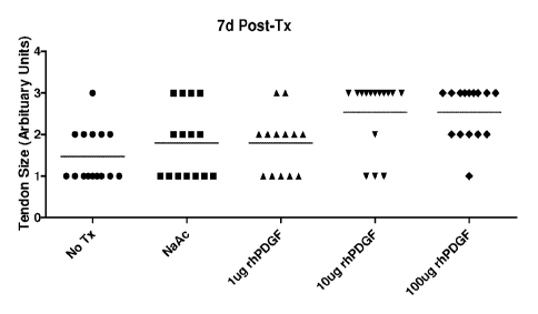
[0024] Figure 6A shows the results of a dose response study on gross
observational tendon
growth for intra-tendon application of recombinant human platelet-derived
growth factor,
isofonn BB ("rhPDGF-BB"), in the collagenase-induced rat Achilles tendon
injury model
seven days after treatment. A single injection of a medium (10.2 lig) or high
(102 jig) dose of
rhPDGF-BB produced a significant increase in tendon size seven days following
rhPDGF-BB
treatment.
11
CA 02790403 2012-08-17
WO 2011/103598
PCT/US2011/025770
[0025] Figure 6B shows the results of the same study twenty-one days following
rhPDGF-
BB treatment. The effect of a single injection of a high (102 gg) dose of
rhPDGF-BB on
tendon size was comparable to the effect of injection with sodium acetate
buffer alone
twenty-one days after treatment.
[0026] Figure 7 is a different presentation of the same data as Figures 6A and
6B, showing
the gross tendon size at 7- and 21- days post-rhPDGF-BB treatment (0 = no
growth to 3 =
severe growth).
[0027] Figure 8 shows tendon width (gm SEM) at the calcaneous insertion at 7-
and 21-
days post- rhPDGF-BB treatment.
[0028] Figure 9 shows tendon width (pm SEM) at the tendon body at 7- and 21-
days
post- rhPDGF-BB treatment.
[0029] Figure 10 shows the effect of rhPDGF-BB on rate of cellular
proliferation (cell
counts SEM) at 7- and 21- days post-rhPDGF-BB treatment.
[0030] Figure 11 shows the mechanical properties of Achilles Tendons: maximum
load to
rupture (N SEM) at 7- and 21- days post-rhPDGF-BB treatment.
[0031] Figure 12 shows mean serum rhPDGF-BB concentration-time values
following IV
dosing.
[0032] Figure 13 shows mean serum rhPDGF-BB concentration-time values
following IT
dosing.
[0033] Figure 14A shows the in vitro release profile for the amount of rhPDGF-
BB
released at each time point from 4-0 Vicryl sutures.
[0034] Figure 14B shows the in vitro cumulative release of rhPDGF-BB over 48
hours
from 4-0 Vicryl sutures (mean SEM).
[0035] Figure 14C shows the estimated in vivo cumulative dose of rhPDGF-BB
versus
initial rhPDGF-BB concentration in the suture coating solution.
[0036] Figure 14D shows the implanted 4-0 Vicryl suture lengths.
12
WO 2011/103598
PCT/US2011/025770
DETAILED DESCRIPTION
[0037]
[0038] The compositions and methods of the invention surprisingly result in
improved
treatment of tenclinopathies. In some embodiments, the compositions and
methods of the
invention result in increased strength of the tendon and an increased rate of
tendon strength
recovery. In some embodiments, the compositions and methods of the invention
result in
increased strength of the tendon. In sonic embodiments, the compositions and
methods of the
invention result in an increased rate of tendon strength recovery. For
example, as a tendon
heals after an injury, the biomechanical strength of the tendon increases as a
process of
tendon healing. Administration of a composition of the invention may result in
a more rapid
increase in tendon strength. Without wishing to be bound by theory, this more
rapid increase
in strength may he helpful in promoting healing of the tendon; provided the
load hearing does
not further increase the tendon injury, load hearing on a tendon generally
improves the
healing response of the tendon, as it generally results in improved tissue
remodeling and
reorganization. A faster initial increase in tendon strength (e.g. resulting
from administration
of a composition of the invention) may result in an ability to begin load
bearing on the tendon
more rapidly, thus further improving the tendon healing response. Without
wishing to be
bound by theory, the improvement in strength of the tendon may be caused by an
increase in
cellular proliferation and/or extracellular matrix production, and/or by an
improvement in
organization of the tissue (for example, an improvement in organization of the
extracellular
matrix).
[0039] Additionally, without wishing to he hound by theory, the inventors
surprisingly
discovered that when the compositions of the invention are administered
directly into the
tendon (e.g. by injection), the PDGF remains localized at the site of
administration (e.g. at the
site of injection). For example, as further detailed in Example 5 below, it
was unexpected that
administration of a composition consisting of PDGE in a buffer would result in
PDGE
remaining localized at the site of injection.
Definitions
[0040] As used herein, the term "treatment" refers to clinical intervention
designed to alter
the natural course of clinical pathology of the disorder being treated (e.g.,
a tenclinopathy,
1";
CA 2790403 2017-06-22
CA 02790403 2012-08-17
WO 2011/103598
PCT/US2011/025770
such as tendinosis, tendinitis, or tenosynovitis). Desirable effects of
treatment include, for
example, one or more of decreasing pain or stiffness of the affected joint or
limb, increasing
mobility and strength of the affected joint or limb, decreasing the rate of
tendinopathy
progression, decreasing inflammation, increasing the strength of the tendon,
improving the
rate of tendon strength recovery, ameliorating or palliating the disease
state, and remission or
improved prognosis. An individual is successfully "treated," for example, if
one or more
symptoms associated with a tendinopathy are mitigated or eliminated.
[0041] As used herein, the term "effective amount" refers to at least an
amount effective, at
dosages and for periods of time necessary, to achieve the desired therapeutic
or prophylactic
result. An effective amount can be provided in one or more administrations.
[0042] Reference to "about" a value or parameter herein also includes (and
describes)
embodiments that are directed to that value or parameter per se.
[0043] As used herein and in the appended claims, the singular forms "a,"
"an," and "the"
include plural reference unless the context clearly indicates otherwise. For
example, reference
to a "PDGF homodimer" is a reference to one or multiple PDGF homodimers, and
includes
equivalents thereof known to those skilled in the art, and so forth.
[0044] It is understood that all aspects and embodiments of the invention
described herein
include "comprising," "consisting," and "consisting essentially of' aspects
and embodiments.
It is to be understood that methods or compositions "consisting essentially
of' the recited
elements include only the specified steps or materials and those that do not
materially affect
the basic and novel characteristics of those methods and compositions (e.g.,
administering to
an affected site an effective amount of a composition consisting essentially
of a PDGF and a
buffer, or a composition consisting essentially of an effective amount of a
PDGF in a
buffered solution).
Platelet-Derived Growth Factor and Compositions Thereof
[0045] As used herein, the term "platelet-derived growth factor" or "PDGF"
refers to any
of four different isoforms of PDGF that activate cellular responses through
two different
receptors. Those isofonals include A (observed as a homodimer designated PDGF-
AA and as
part of a heterodimer with the B isoform designated PDGF-AB), B (observed as a
homodomer designated PDGF-BB and as part of a heterodimer with the A isofoim
14
CA 02790403 2012-08-17
WO 2011/103598
PCT/US2011/025770
designated PDGF-AB), C (observed as a homodimer designated PDGF-CC) and D
(observed
as a homodimer designated PDGF-DD). Generally herein, the term "PDGF" refers
generally
to the known PDGF homo- and heterodimers (e.g., PDGF-AA, PDGF-BB, PDGF-AB,
PDGF-CC, and PDGF-DD).
[0046] Provided herein are methods of treating tendinopathies in an individual
and
compositions for use in those methods. In general, the methods of treatment
comprise
administering a composition comprising PDGF to an affected site in an
individual who has a
tendinopathy. Specifically, the methods of treatment comprise administering a
composition
comprising PDGF and a buffer to the site of the tendinopathy. In some
embodiments, the
composition comprises a PDGF and a buffer (e.g., a buffered solution of PDGF).
[0047] In some embodiments, the compositions comprise a PDGF and a buffer. In
some
embodiments, the PDGF comprises a PDGF dimer selected from the group
consisting of
PDGF-AA, PDGF-BB, PDGF-AB, PDGF-CC, PDGF-DD, and mixtures and derivatives
thereof. In some embodiments, the PDGF dimer is a homodimer. In some
embodiments, the
PDGF homodimer is selected from the group consisting of PDGF-AA, PDGF-BB, PDGF-
CC, and PDGF-DD. In some embodiments, the PDGF homodimer is PDGF-BB. In some
embodiments, the PDGF dimer is a heterodimer. In some embodiments, the PDGF
heterodimer is PDGF-AB.
[0048] In some embodiments, PDGF can be obtained from natural sources. In some
embodiments, PDGF can be produced by recombinant DNA techniques. In some
embodiments, PDGF or fragments thereof may be produced using peptide synthesis
techniques known to one of skill in the art, such as solid phase peptide
synthesis.
[0049] When obtained from natural sources, PDGF can be derived from biological
fluids.
In some embodiments, the biological fluids can comprise any treated or
untreated fluid
associated with living organisms including blood. Biological fluids can also
comprise blood
components including platelet concentrate, apheresed platelets, platelet-rich
plasma, plasma,
serum, and fresh frozen plasma. Biological fluids can comprise platelets
separated from
plasma and resuspended in a physiological fluid or buffer.
[0050] When produced by recombinant DNA techniques, a DNA sequence encoding a
single monomer (e.g., a PDGF B-chain or A-chain) can be inserted into cultured
prokaryotic
or eukaryotic cells for expression to subsequently produce the homodimer
(e.g., PDGF-BB or
CA 02790403 2012-08-17
WO 2011/103598
PCT/US2011/025770
PDGF-AA). In some embodiments, the PDGF comprises a PDGF homodimer (e.g., PDGF-
AA, PDGF-BB, PDGF-CC. or PDGF-DD). In some embodiments, a PDGF heterodimer can
be generated by inserting DNA sequences encoding for both monomeric units of
the
heterodimer into cultured prokaryotic or eukaryotic cells and allowing the
translated
monomeric units to be processed by the cells to produce the heterodimer (e.g.,
PDGF-AB). In
some embodiments, the PDGF comprises a PDGF heterodimer (e.g., PDGF-AB).
Commercially available recombinant human PDGF-BB may be obtained commercially
from
a variety of sources, including, but not limited to Leinco Technologies, Inc.
(St. Louis, MO)
and R&D Systems, Inc. (Minneapolis, MN).
[0051] In some embodiments described herein, the PDGF comprises a recombinant
human
PDGF ("rhPDGF"). In some embodiments, the recombinant human PDGF ("rhPDGF") is
a
PDGF dimer selected from the group consisting of rhPDGF-AA, rhPDGF-BB, rhPDGF-
AB,
rhPDGF-CC, rhPDGF-DD, and mixtures and derivatives thereof. In some
embodiments, the
recombinant human PDGF is an rhPDGF homodimer. In some embodiments, the
recombinant human PDGF homodimer is selected from the group consisting of
rhPDGF-AA,
rhPDGF-BB, rhPDGF-CC, and rhPDGF-DD. In some embodiments, the recombinant
human
PDGF homodimer is rhPDGF-BB. In some embodiments, the recombinant human PDGF
is
an rhPDGF heterodimer. In some embodiments, the recombinant human PDGF
heterodimer
is rhPDGF-AB.
[0052] In some embodiments, PDGF-B comprises one or more of the following
fragments:
amino acids 1-31, 1-32, 33-108, 33-109, and/or 1-108 of the entire human B-
chain. The
complete amino acid sequence (amino acids 1-109) of the B-chain of human PDGF
is
provided in Figure 15 of U.S. Patent No. 5,516,896. It is to be understood
that the PDGF-BB
compositions of the present invention may comprise a combination of intact
humanPDGF-B
(amino acids 1-109) and fragments thereof. Other fragments of PDGF may be
employed such
as those disclosed in U.S. Patent No. 5,516,896. In some embodiments, the PDGF-
BB
comprises at least 65% of full-length human PDGF-B (amino acids 1-109). In
some
embodiments, the PDGF-BB comprises at least 75%, 80%, 85%, 90%, 95%. or 99% of
full-
length human PDGF-B (amino acid 1-109).
[0053] In some embodiments, the composition comprises a PDGF dimer (e.g., an
rhPDGF
dimer) selected from the group consisting of PDGF-AA, PDGF-BB, PDGF-AB, PDGF-
CC,
and PDGF-DD, and the composition comprises a PDGF dimer at a concentration
ranging
16
CA 02790403 2012-08-17
WO 2011/103598
PCT/US2011/025770
from about 0.01 mg/ml to about 10.0 mg/ml, from about 0.05 mg/ml to about 5.0
mg/ml,
from about 0.1 mg/ml to about 1.0 mg/ml, or from about 0.1 mg/ml to about 2.0
mg/ml, from
about 0.1 mg/ml to about 3.0 mg/ml, from about 0.1 mg/ml to about 4.0 mg/ml,
about 0.1
mg/ml to about 0.4 mg/ml, from about 0.1 mg/ml to about 5.0 mg/ml, about 0.9
mg/ml to
about 1.5 mg/ml. In some embodiments, the composition comprises a PDGF dimer
at a
concentration of about 3.4 mg/ml. In some embodiments, the composition
comprises a PDGF
dimer at a concentration of about 1.0 mg/ml. In some embodiments, the
composition
comprises a PDGF dimer at a concentration of about 0.34 mg/ml. In some
embodiments, the
composition comprises a PDGF dimer at any one of the following concentrations:
about 0.05
mg/m1; about 0.1 ing/m1; about 0.15 mg/m1; about 0.2 ing/m1; about 0.25
ing/m1; about 0.3
mg/ml: about 0.35 mg/ml; about 0.4 mg/ml; about 0.45 mg/ml: about 0.5 mg/ml,
about 0.55
mg/ml, about 0.6 mg/ml, about 0.65 mg/ml, about 0.7 mg/ml; about 0.75 mg/ml;
about 0.8
mg/m1; about 0.85 mg/ml; about 0.9 mg/ml; about 0.95 mg/ml; about 1.0 mg/ml;
about 1.5
mg/m1; about 2.0 mg/ml; about 2.5 mg/ml; about 3.0 mg/ml; about 3.5 mg/m1;
about 4.0
mg/ml; about 4.5 mg/ml; or about 5.0 mg/ml. It is to be understood that these
concentrations
are simply examples of particular embodiments, and that the concentration of
PDGF dimer
may be within any of the concentration ranges stated above.
[0054] In some embodiments, the PDGF dimer (e.g., an rhPDGF dimer) is PDGF-BB.
In
some embodiments, the composition comprises PDGF-BB at a concentration ranging
from
about 0.01 mg/ml to about 10.0 mg/ml, from about 0.05 mg/ml to about 5.0
mg/ml, from
about 0.1 mg/ml to about 1.0 mg/ml, or from about 0.1 mg/ml to about 2.0
mg/ml, from about
0.1 mg/ml to about 3.0 mg/ml, from about 0.1 mg/ml to about 4.0 mg/ml, from
about 0.1
mg/ml to about 5.0 mg/ml, about 0.1 mg/ml to about 0.4 mg/ml, about 0.9 mg/ml
to about 1.5
mg/ml. In some embodiments, the composition comprises PDGF-BB at a
concentration of
about 3.4 mg/ml. In some embodiments, the composition comprises PDGF-BB at a
concentration of about 1.0 mg/ml. In some embodiments, the composition
comprises PDGF-
BB at a concentration of about 0.34 mg/ml. In some embodiments, the
composition
comprises PDGF-BB at any one of the following concentrations: about 0.05
ing/m1; about
0.1 mg/ml; about 0.15 mg/ml; about 0.2 mg/ml; about 0.25 mg/ml; about 0.3
mg/ml; about
0.35 mg/ml; about 0.4 mg/ml; about 0.45 mg/ml; about 0.5 mg/ml, about 0.55
mg/ml, about
0.6 mg/ml, about 0.65 mg/ml, about 0.7 mg/ml; about 0.75 mg/m1; about 0.8
mg/ml; about
0.85 mg/ml; about 0.9 mg/ml; about 0.95 mg/ml; about 1.0 mg/ml; about 1.5
mg/ml; about
2.0 mg/ml; about 2.5 mg/ml; about 3.0 mg/ml; about 3.5 mg/ml; about 4.0 mg/ml;
about 4.5
17
CA 02790403 2012-08-17
WO 2011/103598
PCT/US2011/025770
mg/ml; or about 5.0 mg/ml. It is to be understood that these concentrations
are simply
examples of particular embodiments, and that the concentration of rhPDGF-BB
may be
within any of the concentration ranges stated above.
[0055] In some embodiments, the PDGF is selected from the group consisting of
PDGF-
AA, PDGF-BB, PDGF-AB, PDGF-CC, and PDGF-DD. Various amounts of PDGF may be
used in the compositions of the present invention. Amounts of PDGF that can be
used
include, but are not limited to, amounts in the following ranges: about 1 jag
to about 50 mg,
about 10 ps to about 25 mg, about 100 ps to about 10 mg. about 250 ps to about
5 mg, and
about 450 ps to about 3 mg. In some embodiments, the PDGF is PDGF-BB. Various
amounts of PDGF-BB may be used in the compositions of the present invention.
Amounts of
PDGF-BB that can be used include, but are not limited to, amounts in the
following ranges:
about 1 lag to about 50 mg, about 10 jag to about 25 mg, about 100 ps to about
10 mg, about
250 ps to about 5 mg and about 450 ps to about 3 mg.
[0056] The concentration of PDGF (e.g., rhPDGF), including PDGF-AA, PDGF-BB,
PDGF-AB, PDGF-CC, and PDGF-DD, in some embodiments of the present invention
can be
determined, for example, by using an enzyme-linked immunoassay as described in
U.S.
Patent Nos. 6,221,625; 5,747,273; and 5,290,708, or any other assay known in
the art for
determining PDGF concentration. When provided herein, the molar concentration
of rhPDGF
is determined based on the molecular weight of a PDGF homodimer (e.g., PDGF-
BB, MW
25 kDa).
[0057] In some embodiments of the present invention, the PDGF (e.g., rhPDGF)
can be in
a highly purified form. Purified PDGF, as used herein, comprises compositions
having
greater than about 95% by weight PDGF prior to incorporation in solutions of
the present
invention. The solution may be prepared using any pharmaceutically acceptable
buffer or
diluent. In some embodiments, the PDGF can be substantially purified.
Substantially purified
PDGF, as used herein, comprises compositions having about 5% to about 95% by
weight
PDC& prior to incorporation into solutions of the present invention. In one
embodiment,
substantially purified PDGF comprises compositions having about 65% to about
95% by
weight PDGF prior to incorporation into solutions of the present invention. In
some
embodiments, substantially purified PDGF comprises compositions having about
70% to
about 95%, about 75% to about 95%, about 80% to about 95%, about 85% to about
95%, or
about 90% to about 95%, by weight PDGF, prior to incorporation into solutions
of the
18
CA 02790403 2012-08-17
WO 2011/103598
PCT/US2011/025770
present invention. Purified PDGF and substantially purified PDGF may be
incorporated into
the compositions.
[0058] In a further embodiment, the PDGF can be partially purified. Partially
purified
PDGF, as used herein, comprises compositions having PDGF in the context of
platelet-rich
plasma, fresh frozen plasma, or any other blood product that requires
collection and
separation to produce PDC/F. Embodiments of the present invention contemplate
that any of
the PDGF isofotnis provided herein, including homodimers and heterodimers, can
be purified
or partially purified. Compositions of the present invention comprising PDGF
mixtures may
comprise PDGF isoforms or PDGF fragments in partially purified proportions.
Partially
purified and purified PDGF, In some embodiments, can be prepared as described
in U.S.
Application Serial No. 11/159,533 (U.S. Patent Publication No. 2006/0084602
Al).
[0059] In any of the embodiments described herein, the highly purified or
partially purified
PDGF is selected from the group consisting of PDGF-AA, PDGF-BB, PDGF-AB, PDGF-
CC, and PDGF-DD. In any of the embodiments described herein, the highly
purified or
partially purified PDGF is PDGF-BB.
Buffers
[0060] In some embodiments, the compositions comprise a PDGF and a buffer,
preferably
a pharmaceutically acceptable buffer. Buffers suitable for use in PDGF
solutions of the
present invention can comprise, but are not limited to, carbonates, phosphates
(e.g.,
phosphate-buffered saline), saline, histidine, acetates (e.g.. sodium acetate
or ammonium
acetate), acidic buffers such as acetic acid, citric acid, sodium citrate and
HC1, and organic
buffers such as lysine, Tris buffers (e.g., tris(hydroxymethyl)aminoethane), N-
2-
hydroxyethylpiperazine-N'-2-ethanesulfoni c acid (HFPES), 3-(N-morpholino)
propanesulfonic acid (MOPS), 2-(N-morpholino)ethanesulfonic acid (MES), N-(2-
acetamido)iminodiacetic acid ("ADA"), piperazine-N,N'-bis(2-ethanesulfonic
acid) (PIPES),
and N-(2-acetamido)-2-aminoethanesulfonic acid (ACES).
[0061] Buffers can be selected based on biocompatibility with PDGF and the
buffer's
ability to impede undesirable protein modification. Buffers can additionally
be selected based
on compatibility with host tissues and pharmaceutical acceptability. In some
embodiments,
the PDGF compositions comprise PDGF in sodium acetate buffer. In some
embodiments, the
PDGF in sodium acetate buffer is selected from the group consisting of PDGF-
AA, PDGF-
19
CA 02790403 2012-08-17
WO 2011/103598
PCT/US2011/025770
BB, PDGF-AB, PDGF-CC, and PDGF-DD. In some embodiments, the PDGF in sodium
acetate buffer is rhPDGF-BB.
[0062] The buffers may be employed at different molarities, for example
between about 0.1
niM to about 100 inM, about 1 inM to about 100 'RIM, about 10 mM to about 100
niM, about
1 mM to about 50 mM, about 5 mM to about 40 mM, about 10 mM to about 30 mM, or
about
15 mM to about 25 mM, or any molarity within these ranges. In some
embodiments, an
acetate buffer is employed at a molarity of about 20 mM. The buffers may be
employed at
different concentrations, for example, between about 0.01 mg/ml to about 10
mg/ml, 0.05
mg/mill to about 5 ing/ml, about 0.5 mg/m1 to about 5 mg/ml, 0.1 ing/m1 to
about 1 mg/ml, and
about 0.5 mg/ml to about 1 mg/ml, or any concentration within these ranges.
[0063] In another embodiment, solutions comprising PDGF may be formed by
solubilizing
lyophilized PDGF in water, wherein prior to solubilization the PDGF is
lyophilized from an
appropriate buffer.
[0064] Compositions comprising PDGF and a buffer according to some embodiments
of
the present invention can have a pH ranging from about 3.0 to about 8.0 or
from about 4.0 to
about 7Ø In some embodiments, the composition comprising PDGF and a buffer
has a pH
ranging from about 5.0 to about 8.0, more preferably about 5.5 to about 7.0,
most preferably
about 5.5 to about 6.5, or any value within these ranges. In some embodiments
described
herein, the PDGF composition is at a pH between about 4.0 and about 7Ø In
some
embodiments described herein, the PDGF composition is at a pH between about
5.0 and
about 7Ø In some embodiments described herein, the PDGF composition is at a
pH of about
4.0, about 5.0, about 6.0, or about 7Ø The pH of compositions comprising
PDGF and a
buffer, in some embodiments, can be compatible with the prolonged stability
and efficacy of
PDGF or any other desired biologically active agent. PDGF is generally more
stable in an
acidic environment. Therefore, in accord with some embodiments, provided
herein is an
acidic storage formulation of a PDGF composition. In accord with some
embodiments, the
composition comprising PDGF and a buffer preferably has a pH from about 3.0 to
about 7.0,
and more preferably from about 4.0 to about 6.5. The biological activity of
PDGF, however,
can be optimized in a solution having a neutral pH range. Therefore, in some
embodiments,
provided herein is a neutral pH formulation of a composition comprising PDGF
and a buffer.
In accord with this embodiment, the composition preferably has a pH from about
5.0 to about
8.0, more preferably about 5.5 to about 7.0, most preferably about 5.5 to
about 6.5.
CA 02790403 2012-08-17
WO 2011/103598
PCT/US2011/025770
[0065] The pH of solutions comprising PDGF, in some embodiments, can be
controlled by
the buffers recited herein. Various proteins demonstrate different pH ranges
in which they are
stable. Protein stabilities are primarily reflected by isoelectric points and
charges on the
proteins. The pH range can affect the conformational structure of a protein
and the
susceptibility of a protein to proteolytic degradation, hydrolysis, oxidation,
and other
processes that can result in modification to the structure and/or biological
activity of the
protein.
[0066] In some embodiments, the PDGF compositions provided herein comprise a
PDGF
selected from the group consisting of PDGF-AA, PDGF-BB, PDGF-CC, PDGF-DD, and
PDGF-AB and a buffer selected from the group consisting of PBS, sodium
acetate,
ammonium acetate, acetic acid, citric acid, sodium citrate,
tris(hydroxymethyl)aminoethane,
HEPES, MOPS, MES, ADA, PIPES, and ACES. In some embodiments, the PDGF
compositions provided herein comprise rhPDGF-BB and a buffer selected from the
group
consisting of PBS, sodium acetate, ammonium acetate, acetic acid, citric acid,
sodium citrate,
tris(hydroxymethyl)aminoethane, HEPES, MOPS, MES, ADA, PIPES, and ACES. In
some
embodiments, the PDGF composition comprises rhPDGF-BB and PBS. In some
embodiments, the PDGF composition comprises rhPDGF-BB and sodium acetate. In
some
embodiments, the PDGF composition comprises rhPDGF-BB and ammonium acetate. In
some embodiments, the PDGF composition comprises rhPDGF-BB and acetic acid. In
some
embodiments, the PDGF composition comprises rhPDGF-BB and citric acid. In some
embodiments, the PDGF composition comprises rhPDGF-BB and sodium citrate. In
some
embodiments, the PDGF composition comprises rhPDGF-BB and
tris(hydroxymethyl)aminoethane. In some embodiments, the rhPDGF composition
comprises
PDGF-BB and HEPES. In some embodiments, the PDGF composition comprises rhPDGF-
BB and MOPS. In some embodiments, the PDGF composition comprises rhPDGF-BB and
MES. In some embodiments, the PDGF composition comprises rhPDGF-BB and ADA. In
some embodiments, the PDGF composition comprises rhPDGF-BB and PIPES. In some
embodiments, the PDGF composition comprises rhPDGF-BB and ACES.
[0067] In some embodiments described herein, the buffer is at a concentration
between 1
mM and 1000 mM. In some embodiments described herein, the buffer is at a
concentration
between 10 mM and 1000 mM. In some embodiments described herein, the buffer is
at a
concentration between 100 mM and 1000 mM. In some embodiments described
herein, the
21
CA 02790403 2012-08-17
WO 2011/103598
PCT/US2011/025770
buffer is at a concentration between 5 mM and 500 mM. In some embodiments
described
herein, the buffer is at a concentration between 50 mM and 500 mM. In some
embodiments
described herein, the buffer is at a concentration between 10 mM and 100 mM.
In some
embodiments described herein, the buffer is at a concentration between 20 mM
and 200 mM.
In some embodiments described herein, the buffer is at a concentration of 10
mM, 20 mM, 30
mM, 40 mM, 50 mM, 60 mM, 70 mM, 80 mM, 90mM or 100 mM.
[0068] In some embodiments, the PDGF composition comprises rhPDGF-AA and 20 mM
sodium acetate at about pH=6Ø In some embodiments, the PDGF composition
comprises
rhPDGF-AB and 20 mM sodium acetate at about pH=6Ø In some embodiments, the
PDGF
composition comprises rhPDGF-BB and 20 mM sodium acetate at about pH=6Ø In
some
embodiments, the PDGF composition comprises rhPDGF-CC and 20 mM sodium acetate
at
about pH=6Ø In some embodiments, the PDGF composition comprises rhPDGF-DD
and 20
mM sodium acetate at about pH=6Ø
Doses and Dosing Regimens
[0069] Effective doses of PDGF identified in a rat tendon model may be
extrapolated to
effective amounts for other individuals, such as humans, based on the relative
size of
treatment area of the tendon. For example, the treatment area of a human
Achilles tendon is
approximately 69 times larger than the treatment area of a rat Achilles
tendon, so an effective
amount or dose of a PDGF for a human patient may be approximately 69 times the
effective
amount or dose of a PDGF determined in the rat tendon model.
[0070] Exemplary effective amounts or doses delivered by administration of the
PDGF
compositions provided herein include, but are not limited to, about 450 pg to
about 3,000 pg
per dose, about 1 Kg to about 10,000 pg per dose, including for example any of
about 1 pg to
about 7,500 pg per dose, about 1 pg to about 5,000 ttg per dose, about 1 pg to
about 2,500 mg
per dose, about 1 pg to about 1,000 pg per dose, about 1 pg to about 500 pg
per dose, about 1
pg to about 250 pg per dose, about 1 lig to about 100 pg per dose, about 10
lig to about
10,000 !_tg per dose, about 10 j_tg to about 7,500 pg per dose, about 10 pg to
about 5,000 ps
per dose, about 10 pg to about 2,500 [.tg per dose, about 10 pg to about 1,000
pg per dose,
about 10 ttg to about 500 ittg per dose, about 10 pg to about 250 pg per dose,
about 10 pg to
about 100 ittg per dose, about 25 pg to about 10,000 mg per dose, about 25 jug
to about 7,500
Lug per dose, about 25 pg to about 5,000 pg per dose, about 25 pg to about
2,500 pg per dose,
2')
CA 02790403 2012-08-17
WO 2011/103598
PCT/US2011/025770
about 25 pg to about 1,000 pg per dose, about 25 pg to about 500 pg per dose,
about 25 pg to
about 250 pg per dose, about 25 lig to about 100 pg per dose, about 50 pg to
about 10,000 pg
per dose, about 50 pg to about 7,500 pg per dose, about 50 pg to about 5,000
pg per dose,
about 50 pg to about 2,500 n per dose, about 50 pg to about 1,000 n per dose,
about 50 pg
to about 500 iLig per dose, about 50 pg to about 250 iLig per dose, about 50
pg to about 100 iLig
per dose, about 50 pg to about 100 pg per dose, about 75 pg to about 10,000 pg
per dose,
about 75 vg to about 7,500 g per dose, about 75 pg to about 5,000 iug per
dose, about 75 pg
to about 2.500 lug per dose, about 75 pg to about 1.000 lug per dose, about 75
pg to about 500
iLtg per dose, about 75 pg to about 250 n per dose, about 75 pg to about 125 n
per dose,
about 100 pg to about 200 pg per dose, about 200 pg to about 300 pg per dose,
about 300 pg
to about 500 pg per dose, about 500 pg to about 1,000 mg per dose, about 1,000
pg to about
2,500 2 per dose, about 1,000 pg to about 5,000 pg per dose, about 1,000 pg
to about 7,500
g per dose, about 1,000 pg to about 10,000 pg per dose, about 2,500 pg to
about 5,000 pg
per dose, about 2,500 pg to about 7,500 pg per dose, about 5,000 pg to about
7,500 pg per
dose, about 10.000 pg to about 50,000 pg per dose, about 50,000 pg to about
100,000 pg per
dose, about 100,000 mg to about 200,000 pg per dose, about 200,000 pg to about
300,000 pg
per dose, about 300,000 pg to about 400,000 pg per dose, or about 400,000 pg
to about
500,000 pg per dose.
[0071] In some embodiments, the PDGF is administered at about 400 pg to about
1000 pg
per dose, about 500 pg to about 900 pg per dose, about 600 pg to about 800 g,
about 650 pg
to about 750 pg per dose, about 700 pg per dose.
[0072] In some embodiments, the doses provided herein are administered in a
volume of 50
L. 100 L, 150 I-, 200 L, 250 I-, 300 L, 350 L, 400 L, 450 L, 500 I-,
550 L, 600
L. 650 L, 700 I-, 750 L, 800 L, 850 L, 900 L, 950 L, 1000 L Or more.
In some
embodiments, the doses provided herein are administered in a volume of 100 L,
200 L,
300 L, 400 L, 500 L. 600 L, 700 L, 800 L, 900 pt, 1000 L, 1100 L. 1200
L,
1300 L, 1400 L, 1500 L, 1600 L, 1700 L. 1800 L, 1900 L, 2000 L or
more. In
some embodiments, the doses provided herein are administered in a volume of
about 1000 L
to about 2000 L, about 1250 L to about 1750 L, about 1300 L to about 1600
L, or
about 1500 L.
23
CA 02790403 2012-08-17
WO 2011/103598
PCT/US2011/025770
[0073] The PDGF compositions provided herein may be administered in a single
daily
dose, or the total daily dose may be administered in divided dosages of, e.g.,
two, three, or
four times daily. In some embodiments, a single daily dose of the PDGF
compositions
provided herein can be administered once a day for 1, 2, 3, 4. 5, 6, 7, 8, 9,
10, 11, 12, 13, 14,
or more days. The PDGF compositions can also be administered less frequently
than daily,
for example, six times a week, five times a week, four times a week, three
times a week,
twice a week, once a week, once every two weeks, once every three weeks, once
a month,
once every two months, once every three months, once every four months, once
every five
months, or once every six months.
[0074] In some embodiments, the PDGF compositions are administered at
intervals over a
period of time. In some embodiments, the PDGF compositions are administered
once a week
for one, two, three, four, five, six or more months. In some embodiments, the
PDGF
compositions are administered twice a month for one, two, three, four, five,
six or more
months. In some embodiments, the PDGF compositions are administered monthly
for one,
two, three, four, five, six or more months.
Methods of Treating Tendinopathies
[0075] As used herein, the term "tendinopathy" refers to chronic tendon
injuries such as
tendinosis, tendinitis, and tenosynovitis. Exemplary tendinopathies include,
but are not
limited to, Achilles tendinopathy, patellar tendinopathy, lateral
epicondylitis, or "tennis
elbow," medial epicondylitis, plantar fasciitis, and rotator cuff
tendinopathy.
[0076] As used herein, the term "tendinosis" refers to a non-inflammatory
injury to the
tendon characterized by intratendinous degeneration of the tendon usually in
the form of
microtears in the tissue in and around the tendon caused by overuse, leading
to an increase in
the number of tendon repair cells around the area of damage. Degeneration of
the tendon is
caused by damage to or disorganization of the collagen fibers, cells, and
vascular components
of the tendon, which can reduce the tendon's tensile strength and can lead to
tendon rupture if
not treated. In some cases, tendinosis is accompanied by focal necrosis or
calcification of the
tendon.
[0077] As used herein, the term "tendinitis" refers to an inflammatory injury
to the tendon,
characterized by degeneration like that observed in tendinosis, but also
accompanied by
inflammation of the tendon, vascular disruption and an inflammatory repair
response.
24
CA 02790403 2012-08-17
WO 2011/103598
PCT/US2011/025770
Tendinitis is often associated with fibroblastic and myofibroblastic
proliferation, as well as
hemorrhage and organizing granulation tissue. Generally tendinitis is referred
to by the body
part involved, such as Achilles tendinitis (affecting the Achilles tendon), or
patellar tendinitis
(also known as "jumper's knee," affecting the patellar tendon), though there
are certain
exceptions, such as lateral epicondylitis (also known as "tennis elbow,"
affecting the
Extensor Carpi Radialis Brevis tendon).
[0078] Tendinopathies which may be treated by the methods of the invention
include
tendinopathies of any tendon in the human or mammalian body. In some
embodiments, the
tendinopathy is tendinosis. In some embodiments, the tendinopathy is
tendinitis. In some
embodiments, the tendinopathy is tenosynovitis.
[0079] Tendons which may be treated by the methods of the invention include
any tendon
of the human or mammalian body. Non-limiting examples of tendons include the
patellar
tendon, the anterior tibialis tendon, the Achilles tendon, the hamstring
tendon, the
semitendinosus tendon, the gracilis tendon, the abductor tendon, the adductor
tendon, the
supraspinatus tendon, the infraspinatus tendon, the subscapularis tendon, the
teres minor
tendon, the flexor tendon, the rectus femoris tendon, the tibialis posterior
tendon, and the
quadriceps femoris tendon.
[0080] In some embodiments, the tendon is a tendon of the foot or ankle. In
some
embodiments, the tendon of the foot or ankle is selected from the group
consisting of the
extensor hallucis longus, the flexor hallucis longus, the extensor digitorum
longus, the
extensor digitorum brevis, the peroneus longus, the peroneus brevis, the
flexor hallucis
brevis, the flexor digitorum longus, the posterior tibialis, the Achilles
tendon, and the plantar
fascia.
[0081] In some embodiments, the tendon is a tendon of the leg. In some
embodiments, the
tendon of the leg is selected from the group consisting of the patellar
tendon, the anterior
tibialis tendon, the Achilles tendon, the hamstring tendon, the semitendinosus
tendon, the
gracilis tendon, the abductor tendon, and the adductor tendon. In some
embodiments, the
tendon is selected from the group consisting of the flexor tendon, the rectus
femoris tendon,
the tibialis posterior tendon, and the quadriceps femoris tendon.
[0082] In some embodiments, the tendon is a tendon of the shoulder. In some
embodiments, the tendon of the shoulder is selected from the group consisting
of the
CA 02790403 2012-08-17
WO 2011/103598
PCT/US2011/025770
supraspinatus tendon, the infraspinatus tendon, the subscapularis tendon, and
the teres minor
tendon (rotator cuff complex).
[0083] In some embodiments, the tendon is a tendon of the elbow. In some
embodiments,
the tendon of the elbow is selected from the group consisting of the biceps
tendon, the triceps
tendon, the extensor carpi radialis brevis, the common extensor tendon, the
extensor
digitorum, the extensor digiti minimi, the extensor carpi ulnaris, the
supinator, the common
flexor tendon, the pronator teres, the flexor carpi radialis, the palmaris
longus, the flexor carpi
ulnaris and the digitorum superficialis. In some embodiments, the tendon is a
tendon of the
wrist. In some embodiments, the tendon of the wrist is selected from the group
consisting of
biceps tendon, the triceps tendon, the extensor carpi radialis brevis, the
common extensor
tendon, the extensor digitorum, the extensor digiti minimi, the extensor carpi
ulnaris, the
supinator, the common flexor tendon, the pronator teres, the flexor carpi
radialis, the palmaris
longus, the flexor carpi ulnaris, the digitorum superficialis, the flexor
pollicis brevis, the
flexor pollicis longus, the abductor pollicis brevis, the abductor pollicis
longus, the flexor
digitorum profundus, the flexor digitorum superficialis, the extensor pollicis
brevis, and the
extensor pollicis longus. In some embodiments, the tendon is a tendon of the
hand. In some
embodiments, the tendon of the hand is selected from the group consisting of
the flexor
pollicis brevis, the flexor pollicis longus, the abductor pollicis brevis, the
abductor pollicis
longus, the flexor digitorum profundus, the flexor digitorum superficialis,
the extensor
pollicis brevis, and the extensor pollicis longus.
[0084] In some embodiments, the tendinopathy is rotator cuff tendinopathy. In
some
embodiments, the rotator cuff tendinopathy is selected from the group
consisting of
supraspinatus tendinopathy, infraspinatus tendinopathy, subscapularis
tendinopathy, and teres
minor tendinopathy.
[0085] In some embodiments, the tendinopathy is lateral epicondylitis or
"tennis elbow" at
the extensor muscle group origin at the lateral humeral condyle insertion,
principally in the
extensor carpi radialis brevis (ECRB) tendon. In some embodiments, the subject
having
lateral epicondylitis has associated pain (e.g. for at least about six months)
as evidenced by
pain reported to be =50 on a Visual Analog Score (VAS). In some embodiments,
the subject
having lateral epicondylitis has associated pain that increases with pressure
on the lateral
epicondyle and/or resisted extension of the wrist, e.g. for at least about six
months. In some
26
CA 02790403 2012-08-17
WO 2011/103598
PCT/US2011/025770
embodiments, the tendinopathy is medial epicondylitis or "golfer's elbow" at
the interface
between the pronator teres and flexor carpi radialis origin of the medial
humeral condyle.
[0086] In some embodiments, the tendinopathy is patellar tendinopathy. In some
embodiments, the tendinopathy is Achilles tendinopathy. In some embodiments,
the
tendinopathy is plantar fasciitis. In some embodiments, the tendinopathy is
medial plantar
fasciitis. In some embodiments, the tendinopathy is lateral plantar fasciitis.
[0087] In another aspect, provided herein are methods of treating
tendinopathies
comprising administering an effective amount of a composition comprising PDGF
and a
buffer to an affected site. In some embodiments, the PDGF is selected from the
group
consisting of PDGF-AA, PDGF-BB, PDGF-AB, PDGF-CC, and PDGF-DD. In some
embodiments, the PDGF is PDGF-BB. In some embodiments, the effective amount of
the
composition comprises between about 75 pg and about 7,500 pg of PDGF-BB per
dose. In
some embodiments, the effective amount of the composition comprises between
about 500
pg to about 1,000 p.g of PDGF-BB per dose. In some embodiments, the effective
amount of
the composition comprises between about 450 ing to about 3,000 mg of PDGF per
dose. In
some embodiments, the effective amount of the composition comprises between
about 5,000
lig to about 7,500 m of PDGF-BB per dose.
[0088] In some embodiments, the buffer is selected from the group consisting
of
phosphate-buffered saline ("PBS"), sodium acetate, ammonium acetate, acetic
acid, citric
acid, sodium citrate, tris(hydroxymethyl)aminoethane ("tris"), N-2-
hydroxyethylpiperazine-
N'-2-ethanesulfonic acid ("HEPES"), 3-(N-morpholino) propanesulfonic acid
("MOPS"), 2-
(N-morpholi no)ethanesulfonic acid ("MES"), N-(2-acetamido)iminodi acetic acid
("ADA"),
piperazine-N,N'-bis(2-ethanesulfonic acid) ("PIPES"), and N-(2-acetamido)-2-
aminoethanesulfonic acid ("ACES"). In some embodiments, the buffer is sodium
acetate. In
some embodiments, the sodium acetate is at a concentration between about 10 mM
and about
100 mM. In some embodiments, the sodium acetate is at a concentration of about
20 mM. In
some embodiments, the sodium acetate is at a pH between about 4.0 and about
7Ø In some
embodiments, the sodium acetate is at about pH 6.
[0089] In some embodiments, the administering is by direct injection to the
affected site. In
some embodiments, the direct injection is accomplished using the "peppering
technique" with
or without ultrasound guidance. The "peppering technique" is an injection
method whereby
27
CA 02790403 2012-08-17
WO 2011/103598
PCT/US2011/025770
after the needle is inserted into the tender area, multiple small injections
are performed by
withdrawing, redirecting and reinserting the needle without emerging from the
skin.
[0090] In some embodiments, the affected site is an osseous-tendon junction.
In some
embodiments, the affected site is a tendon. In some embodiments, the
tendinopathy is
selected from the group consisting of Achilles tendinopathy, patellar
tendinopathy, lateral
epicondylitis, medial epicondylitis, plantar fasciitis, and rotator cuff
tendinopathy. In some
embodiments, the composition is administered by a single injection. In some
embodiments,
the composition is administered by a single injection once a week for four
weeks.
[0091] In some embodiments, the tendinopathy is tendinosis. In some
embodiments, the
tendinosis is selected from the group consisting of extensor hallucis longus
tendinosis, flexor
hallucis longus tendinosis, extensor digitorum longus tendinosis, extensor
digitorum brevis
tendinosis, peroneus longus tendinosis, peroneus brevis tendinosis, flexor
hallucis brevis
tendinosis, flexor digitorum longus tendinosis, posterior tibialis tendinosis,
Achilles tendon
tendinosis, and plantar fascia tendinosis. In some embodiments, the tendinosis
is selected
from the group consisting of patellar tendinosis, the anterior tibialis
tendinosis, the hamstring
tendinosis, semitendinosus tendinosis, gracilis tendinosis, abductor
tendinosis, and adductor
tendinosis. In some embodiments, the tendinosis is selected from the group
consisting of
flexor tendinosis, rectus femoris tendinosis, tibialis posterior tendinosis,
and quadriceps
femoris tendinosis. In some embodiments, the tendinosis is selected from the
group
consisting of supraspinatus tendinosis, infraspinatus tendinosis,
subscapularis tendinosis, and
teres minor tendinosis.
[0092] In some embodiments, the tendinosis is selected from the group
consisting of biceps
tendinosis, triceps tendinosis, extensor carpi radialis brevis tendinosis,
common extensor
tendinosis, extensor digitorum tendinosis, extensor digiti minimi tendinosis,
extensor carpi
ulnaris tendinosis, supinator tendinosis, common flexor tendinosis, pronator
teres tendinosis,
flexor carpi radians tendinosis, palmaris longus tendinosis, flexor carpi
ulnaris tendinosis and
digitorum superficialis tendinosis. In some embodiments, the tendinosis is
selected from the
group consisting of biceps tendinosis, triceps tendinosis, extensor carpi
radialis brevis
tendinosis, common extensor tendinosis, extensor digitorum tendinosis,
extensor digiti
minimi tendinosis, extensor carpi ulnaris tendinosis, supinator tendinosis,
common flexor
tendinosis, pronator teres tendinosis, flexor carpi radialis tendinosis,
palmaris longus
tendinosis, flexor carpi ulnaris tendinosis, digitorum superficialis
tendinosis, flexor pollicis
28
CA 02790403 2012-08-17
WO 2011/103598
PCT/US2011/025770
brevis tendinosis, flexor pollicis longus tendinosis, abductor pollicis brevis
tendinosis,
abductor pollicis longus tendinosis, flexor digitorum profundus tendinosis,
flexor digitorum
superficialis tendinosis, extensor pollicis brevis tendinosis, and extensor
pollicis longus
tendinosis. In some embodiments, the tendinosis is selected from the group
consisting of
flexor pollicis brevis tendinosis, flexor pollicis longus tendinosis, abductor
pollicis brevis
tendinosis, abductor pollicis longus tendinosis, flexor digitorum profundus
tendinosis, flexor
digitorum superficialis tendinosis, extensor pollicis brevis tendinosis, and
extensor pollicis
longus tendinosis.
[0093] In some embodiments, the tendinopathy is tendinitis. In some
embodiments, the
tendinitis is selected from the group consisting of extensor hallucis longus
tendinitis, flexor
hallucis longus tendinitis, extensor digitorum longus tendinitis, extensor
digitorum brevis
tendinitis, peroneus longus tendinitis, peroneus brevis tendinitis, flexor
hallucis brevis
tendinitis, flexor digitorum longus tendinitis, posterior tibialis tendinitis,
Achilles tendon
tendinitis, and plantar fascia tendinitis. In some embodiments, the tendinitis
is selected from
the group consisting of patellar tendinitis, the anterior tibialis tendinitis,
the hamstring
tendinitis, semitendinosus tendinitis, gracilis tendinitis, abductor
tendinitis, and adductor
tendinitis. In some embodiments, the tendinitis is selected from the group
consisting of flexor
tendinitis, rectus femoris tendinitis, tibialis posterior tendinitis, and
quadriceps femoris
tendinitis. In some embodiments, the tendinitis is selected from the group
consisting of
supraspinatus tendinitis, infraspinatus tendinitis, subscapularis tendinitis,
and teres minor
tendinitis.
[0094] In some embodiments, the tendinitis is selected from the group
consisting of biceps
tendinitis, triceps tendinitis, extensor carpi radialis brevis tendinitis,
common extensor
tendinitis, extensor digitorum tendinitis, extensor digiti minimi tendinitis,
extensor carpi
ulnaris tendinitis, supinator tendinitis, common flexor tendinitis, pronator
teres tendinitis,
flexor carpi radialis tendinitis, palmaris longus tendinitis, flexor carpi
ulnaris tendinitis and
digitorum superficialis tendinitis. In some embodiments, the tendinitis is
selected from the
group consisting of biceps tendinitis, triceps tendinitis, extensor carpi
radialis brevis
tendinitis, common extensor tendinitis, extensor digitorum tendinitis,
extensor digiti minimi
tendinitis, extensor carpi ulnaris tendinitis, supinator tendinitis, common
flexor tendinitis,
pronator teres tendinitis, flexor carpi radialis tendinitis, palmaris longus
tendinitis, flexor
carpi ulnaris tendinitis, digitorum superficialis tendinitis, flexor pollicis
brevis tendinitis,
29
CA 02790403 2012-08-17
WO 2011/103598
PCT/US2011/025770
flexor pollicis longus tendinitis, abductor pollicis brevis tendinitis,
abductor pollicis longus
tendinitis, flexor digitorum profundus tendinitis, flexor digitorum
superficialis tendinitis,
extensor pollicis brevis tendinitis, and extensor pollicis longus tendinitis.
In some
embodiments, the tendinitis is selected from the group consisting of flexor
pollicis brevis
tendinitis, flexor pollicis longus tendinitis, abductor pollicis brevis
tendinitis, abductor
pollicis longus tendinitis, flexor digitorum profundus tendinitis, flexor
digitorum superficialis
tendinitis, extensor pollicis brevis tendinitis, and extensor pollicis longus
tendinitis.
[0095] The methods of the invention may result in improvement in one or more
of the
following: decreasing pain of the affected joint or limb, decreasing stiffness
of the affected
joint or limb, increasing mobility of the affected joint or limb, increasing
strength of the
affected joint or limb, decreasing the rate of tendinopathy progression,
decreasing
inflammation, increasing the strength of the tendon, or improving the rate of
tendon strength
recovery. Various methods for measuring effectiveness of the treatment
include, but are not
limited to: Disabilities of the Arm, Shoulder and Hand Score (DASH), Visual
Analog Score
(VAS), and grip strength testing. In some embodiments, the treatment results
in at least a
25% reduction in pretreatment score for DASH. In some embodiments, the
treatment results
in at least a 25% reduction in pretreatment score for VAS. In some
embodiments, the
treatment produces a decrease in pain with applied pressure and/or joint
flexion. In some
embodiments, the treatment produces a decrease in pain with applied pressure
and/or joint
flexion and an increase in joint mobility. In some embodiments, the treatment
does not result
in any abnormal bone growth. In some embodiments, the treatment does not
result in any
abnormal tendon growth. In some embodiments, the treatment is safe and
tolerated by the
subject. In some embodiments, safety and tolerability of the composition is
evaluated by the
lack of an adverse event or an abnormality identified by one or more of the
following:
physical examination, vital sign measurement, laboratory test, x-ray, and/or
MR1 imaging.
[0096] In some embodiments, the treatment results in increased strength of the
tendon. In
some embodiments, the treatment results in a more rapid rate of tendon
strength recovery. In
some embodiments, the treatment results in an increase in tendon strength of
at least about
30%, at least about 40%, at least about 50%, at least about 60%, at least
about 70% within
about 4, 5, 6, 7, 8, 9, 10, 11, 12, 13, 14, 15, 16, 17, 18, 19, 20, or 21 days
of administration of
a composition of the invention, as compared to baseline. In some embodiments,
the treatment
results in an increase in tendon strength of at least about 30%, at least
about 40%, at least
CA 02790403 2012-08-17
WO 2011/103598
PCT/US2011/025770
about 50%, at least about 60%, at least about 70% within about 7 days of
administration of a
composition of the invention, as compared to baseline. In some embodiments,
the treatment
results in an increase in tendon strength of at least about 60% within about 7
days of
administration of a composition of the invention, as compared to baseline. In
some
embodiments, the treatment results in an increase in tendon strength of at
least about 65%
within about 7 days of administration of a composition of the invention, as
compared to
baseline. In some embodiments, the treatment results in an increase in tendon
strength of at
least about 70% within about 7 days of administration of a composition of the
invention, as
compared to baseline. In some embodiments, the tendon achieves at least about
60%, at least
about 70%, at least about 80%, at least about 90%, at least about 95% of its
final strength
within about 4, 5, 6, 7, 8, 9, 10, 11. 12, 13, or 14 days of administration of
a composition of
the invention, wherein final strength is measured at about 21 days after
treatment. In some
embodiments, the tendon achieves at least about 80% of its final strength
within about 7 days
of administration of a composition of the invention, wherein final strength is
measured at
about 21 days after treatment. In some embodiments, the tendon achieves at
least about 85%
of its final strength within about 7 days of administration of a composition
of the invention,
wherein final strength is measured at about 21 days after treatment. In some
embodiments,
the tendon achieves at least about 90% of its final strength within about 7
days of
administration of a composition of the invention, wherein final strength is
measured at about
21 days after treatment. Tendon strength can be measured, for example, in an
animal model,
for example in a rat collagenase model, wherein the tendon strength is the
measured load to
rupture. An example of measurement of tendon strength is described in more
detail in
Example 3.
Kits
[0097] In another aspect, provided herein are kits comprising a container
containing a
composition comprising PDGF and a buffer. In some embodiments, the kits
comprise a first
container containing a lyophilized PDGF and a second container containing a
buffer for
solubilizing the lyophilized PDGF. In some embodiments, the kits comprise a
first container
containing a lyophilized PDGF and buffer, and a second container containing
water for
solubilizing the lyophilized PDGF and buffer. In some embodiments, the buffer
is selected
from the group consisting of phosphate-buffered saline ("PBS-), sodium
acetate, ammonium
acetate, acetic acid, citric acid, sodium citrate,
tris(hydroxymethyl)aminoethane ("tris"), N-2-
31
WO 2011/103598 PCT/US2011/025770
hydroxyethylpiperazine-N'-2-ethanesulfonic acid ("TIEPES"), 3-(N-morpholino)
propanesulfonic acid ("MOPS"), 2-(N-morpholino)ethanesulfonic acid ("MES"), N-
(2-
acetamido)iminodiacetic acid ("ADA"), piperazine-N,N'-his(2-ethanesulfonic
acid)
("PIPES"), and N-(2-acetamido)-2-aminoethanesulfonic acid ("ACES"). In some
embodiments, the buffer is sodium acetate. In some embodiments, the sodium
acetate is at a
concentration between about 10 niM and about 100 nalvl. In some embodiments,
the sodium
acetate is at a concentration of about 20 mM. In some embodiments, the sodium
acetate is at
a pH between about 4.0 and about 7Ø In some embodiments, the sodium acetate
is at about
pH 6.
[0098] In some embodiments, the kits further include other materials desirable
from a
commercial, therapeutic, and user standpoint, including other buffers,
diluents, filters,
needles, syringes, and package inserts with instructions for use. As used
herein, the term
"package insert" refers to instructions customarily included in commercial
packages of
medicaments that contain information about the indications customarily
included in
commercial packages of medicaments that contain information about the
indications, usage,
dosage, administration, contraindications, other medicaments to be combined
with the
packaged product, and/or warnings concerning the use of such medicaments.
Sutures
[0099] Also provided are PDGE coated sutures, and methods of making such
sutures. The
sutures may be used, for example, in treating a tendon in an individual
(e.g.treating a tendon
tear). Suitable sutures include, for example, those made from co-polymers of
lactide and
glycolide (such as VicrylTM sutures (e.g. 4-0 VicrylTM sutures)). Suitable
coating methods include,
for example, dip coating methods, such as the dip coating method described in
Dines J,
Weber L, Razzano P, et al. The Effect of Growth Differentiation Factor-5-
Coated Sutures on
Tendon Repair in a Rat Model. J Shoulder Elbow Surg 2007;16:215S-221S), which
may
optionally be altered to eliminate the gelatin. The applicants have
surprisingly found that high
doses of PDGE may be coated onto certain types of sutures in absence of
gelatin. In some
embodiments, the suture does not comprise gelatin. In some embodiments, the
suture coating
does not comprise gelatin. In some embodiments, the suture coating does not
comprise
polylactic, polyglycolic, or poly(lactic-co-glycolic) acid. In some
embodiments, the method
of coating the suture does not comprise utilizing gelatin. In some
embodiments, the suture
coating consists essentially of PDGE. In some embodiments, the suture coating
consists of
32
CA 2790403 2017-06-22
CA 02790403 2012-08-17
WO 2011/103598
PCT/US2011/025770
PDGF and a buffer. In some embodiments, the suture coating consists of PDGF.
The sutures
may be used to treat an individual, for example, a mammal. Non-limiting
examples of
mammals which may be treated using a suture of the invention include humans,
pets (e.g.
dogs, cats, rabbits, hamsters, etc.), laboratory animals (e.g. mice, rats),
farm animals (e.g.
horses, cows, sheep, goats, etc.).
[0100] In some embodiments, the amount of PDGF loaded onto the suture is at
least about
ng PDGF/cm suture. In some embodiments, the amount of PDGF loaded onto the
suture is
at least about 100 ng PDGF/cm suture. In some embodiments, the amount of PDGF
loaded
onto the suture is at least about 1000 ng PDGF/cm suture. In some embodiments,
the amount
of PDC& loaded onto the suture is at least about 5000 ng PDGF/cm suture. In
some
embodiments, the amount of PDGF loaded onto the suture is at least about 6000
ng PDGF/cm
suture. In some embodiments, the amount of PDGF loaded onto the suture is
about 10 to
about 20,000 ng PDGF/cm suture. In some embodiments, the amount of PDGF loaded
onto
the suture is about 100 to about 10,000 ng PDGF/cm suture. In some
embodiments, the
amount of PDGF loaded onto the suture is about 500 to about 8,000 ng PDGF/cm
suture. In
some embodiments, the amount of PDGF loaded onto the suture is about 1000 to
about 8,000
ng PDGF/cm suture. In some embodiments, the amount of PDGF loaded onto the
suture is
about 4000 to about 8,000 ng PDGF/cm suture. In some embodiments, the amount
of PDGF
loaded onto the suture is about 6000 to about 7,000 ng PDGF/cm suture.
[0101] In some embodiments, the cumulative amount of PDGF released from the
suture
over 48-hours as measured in vitro is at least about 10 ng PDGF/cm suture. In
some
embodiments, the cumulative amount of PDGF released from the suture over 48-
hours as
measured in vitro is at least about 100 ng PDGF/cm suture. In some
embodiments, the
cumulative amount of Plltil-, released from the suture over 48-hours as
measured in vitro is at
least about 1000 ng PDGF/cm suture. In some embodiments, the cumulative amount
of
PDGF released from the suture over 48-hours as measured in vitro is at least
about 5000 ng
PDGF/cm suture. In some embodiments, the cumulative amount of PDGF released
from the
suture over 48-hours as measured in vitro is at least about 6000 ng PDGF/cm
suture. In some
embodiments, the cumulative amount of PDGF released from the suture over 48-
hours as
measured in vitro is about 10 to about 20,000 ng PDGF/cm suture. In some
embodiments, the
cumulative amount of PDGF released from the suture over 48-hours as measured
in vitro is
about 100 to about 10,000 ng PDGF/cm suture. In some embodiments, the
cumulative
33
CA 02790403 2012-08-17
WO 2011/103598
PCT/US2011/025770
amount of PDGF released from the suture over 48-hours as measured in vitro is
about 500 to
about 8,000 ng PDGF/cm suture. In some embodiments, the cumulative amount of
PDGF
released from the suture over 48-hours as measured in vitro is about 1000 to
about 8,000 ng
PDGF/cm suture. In some embodiments, the cumulative amount of PDGF released
from the
suture over 48-hours as measured in vitro is about 4000 to about 8,000 ng
PDGF/cm suture.
In some embodiments, the cumulative amount of PDGF released from the suture
over 48-
hours as measured in vitro is about 6000 to about 7,000 ng PDGF/cm suture.
Suitable
methods of measuring cumulative PDGF release in vitro include, for example,
the method
described in Example 7. The coated sutures of the invention may advantageously
provide for
consistent dosing in vivo.
[0102] The following examples are provided for illustrative purposes only and
are not
intended to limit the scope of the invention in any manner.
EXAMPLES
Example 1: Normal and Diseased Primary Human Tenocytes Proliferate in Response
to
rhPDGF-BB
[0103] This study determined whether rhPDGF-BB directly activated
proliferation and/or
chemotaxis of primary tenocytes derived from patients with tendinopathies.
Such findings
can support the notion of therapeutic potential of rhPDGF-BB in
tendinopathies.
Patients and Methods
Patients
[0104] Ten patients with tendinopathies were involved in this study, including
five patients
with Achilles tendinopathy and five patients with tendinopathy of the
posterior tibial tendon
(PTT). An additional five patients were involved who underwent full joint
replacement of the
knee.
Primary Cultures of Tenocytes
[0105] Tendon tissue which would otherwise be discarded was obtained from
normal and
injured tendons during reconstructive surgery procedures performed for
clinical indications.
These tissues included the tendinopathic portion of the Achilles or PTT
tendons, as well as
34
CA 02790403 2012-08-17
WO 2011/103598
PCT/US2011/025770
the healthy (non-tendinopathic) portion of the flexor digitorum longus (FDL)
tendon tissue,
Achilles tendon tissue, and Patellar tendon tissue. Primary tenocyte explant
cultures were
obtained from these tissues and tested at passages 3 to 5. Tenocyte identity
was confirmed by
assessing the expression of a tenocyte-specific gene scleraxis and genes for
collagens al(I),
a2(I), and al(III) in real-time PCR assays with specific primers.
Cell Proliferation
[0106] Tenocyte monolayers were trypsinized, resuspended in DMEM/F12 medium
containing 0.5% dialyzed fetal bovine serum, allowed to attach overnight, and
then incubated
with titrated concentrations of rhPDGF-BB for 24 hours. Changes in cell
proliferation rates
were assessed based on BrdU incorporation during DNA synthesis in cells using
a
commercially available assay (Roche Applied Science, Indianapolis, IN). Each
culture was
tested in triplicates for each dose of rhPDGF-BB.
Cell Migration
[0107] Tenocyte monolayers were trypsinized, resuspended in DMEM/F12 medium
containing 0.5% dialyzed fetal bovine serum and placed in the upper chamber of
the 96-well
ChemoTx disposable cell migration system (Neuro Probe, Gaithersburg, MD). The
lower
chambers contained titrated concentrations of rhPDGF-BB. Tenocytes were
allowed to
migrate across the membrane separating the chambers for 48 hours. 96-well
plates were then
spun down and freeze thawed three times to lyse the migrated cells. The amount
of viable
migrated cells was measured based on cytoplasmic lactate dehydrogenase (LDH)
using a
commercially available kit from Promega (Madison, WI).
Statistical Analysis
[0108] One-way ANOVA was used to determine whether stimulation with rhPDGF-BB
affects tenocyte proliferation in a dose-dependent fashion.
Results
[0109] Only tenocyte cultures but not control pulmonary fibroblast cultures or
control
primary T lymphocyte cultures expressed scleraxis mRNA, whereas tenocytes and
fibroblasts
but not lymphocytes expressed the collagen gene mRNAs.
CA 02790403 2012-08-17
WO 2011/103598
PCT/US2011/025770
[0110] In all cases, tenocytes from tendon tissues involved or not involved in
the disease
process responded to rhPDGF-BB stimulation by accelerating BrdU incorporation
(p <0.05,
one-way ANOVA). The responses were dose-dependent and were observed at 10. 50
and 150
ng/mL of rhPDGF-BB. Even though all cell cultures responded to rhPDGF-BB
stimulation,
there was significant variability among patients in the magnitude of BrdU
incorporation after
rhPDGF-BB stimulation. Incorporation of BrdU increased from a minimum of 2.1
0.2 fold
to a maximum of 10.7 0.5 fold compared to control non-stimulated cultures.
Tenocytes
from five patients responded paradoxically, with a greater increase in BrdU
incorporation at a
lower (10 ng/mL) rather than higher (50 and 150 ng/mL) concentrations of
rhPDGF-BB.
Such paradoxic response was observed in tenocytes derived from both
tendinopathic and
noimal tissues of these patients. Tenocytes derived from healthy tendons of
four patients
incorporated twice more BrdU in response to rhPDGF-BB stimulation than did
tenocytes
derived from the diseased tissues. In one patient, tenocytes from the diseased
tissue
incorporated four fold more BrdU in response to rhPDGF stimulation than did
tenocytes from
the tissue uninvolved in the disease process. Figure 2 shows the BrdU
incorporation (y-axis,
absorbance) for 0, 10, 50, and 150 ng/ml of PDGF added to the culture medium
at day 1, day
4, and day 8 for healthy and diseased tenocytes. An increase in absorbance
corresponds with
increased proliferation, with both the healthy and diseased tenocytes
responding to PDGF on
day 1.
[0111] In all cases, tenocytes were chemotactically responsive to rhPDGF-BB at
50 ng/mL
and 150 ng/mL. Tenocytes were not exposed to 10 ng/mL rhPDGF-BB for chemotaxis
experiments because of low response in pilot experiments. Again, responses
were dose-
dependent, with greater chemotaxis to 150 ng/mL than to 50 ng/mL of rhPDGF-BB.
However, tenocytes from 5 patients responded with greater chemotaxis to 50
ng/mI, than to
150 ng/mL of rhPDGF-BB, with significant decline in the number of migrated
cell (p <0.05,
two-sided Student's t-test). There was variability among patients in the
maximal chemotactic
response to rhPDGF-BB, from 1.4 0.1 to 4.0 0.5 fold increase compared to
non-
stimulated control. There was no statistically significant difference (p>
0.05) in tenocyte
chemotaxis to rhPDGF-BB within matching tenocyte cultures derived from
tendinopathic or
from healthy tendon tissues. Figure 1 shows the chemotaxis of cells (y-axis
shows increased
optical density) cultured in concentrations of 0, 50, or 150 ng/ml of PDGF.
Migration was
assessed with 1250. 2500. 5000, and 10000 initial cells.
36
CA 02790403 2012-08-17
WO 2011/103598
PCT/US2011/025770
Conclusion
[0112] The results of these experiments suggest that tenocytes derived from
healthy and
tendinopathic tissues respond to rhPDGF-BB by increasing proliferation and
chemotaxis
rates. Importantly, tenocytes from some patients showed paradoxical response
to PDGF, in
which higher doses caused less effect than lower doses. Equally important,
tenocytes from
diseased tendons were in some cases differentially responsive to PDGF versus
tenocytes from
healthy tendons, implying that proper dosing may be of paramount importance in
the clinical
setting.
Example 2: Safety studies with rhPDGF-BB
Local Injection Test.
[0113] The purpose of this study was to determine the local toxicity of rhPDGF-
BB
following an intra-Achilles tendon delivery to rats. The intra-Achilles tendon
administration
mimics the route of administration of rhPDGF-BB in the clinic for the
treatment of lateral
epicondylitis. The injection site at the Achilles tendon-calcaneous junction
mimics the
insertion site of the extensor carpi radialis brevis tendon and lateral
epicondyle bone.
[0114] The study used Sprague Dawley rats. The animals were housed at the same
lab
facility for the duration of the study. All housing and husbandry were in
accordance with the
Animal Welfare Act and the "Guide for the Care and Use of Laboratory Animals".
Rats were
fed and watered in accordance with standard protocols. Food and water were
withheld for
appropriate study related events such as anesthesia. Animals were acclimated
to the facility
for a minimum of 5 days prior to the study. This acclimation period allows the
animals to
become accustomed to the study room setting.
[0115] Three batches of sterile recombinant human PDGF-BB at different
concentrations
were used in the study: (1) 10.3 mg/ml rhPDGF-BB in 20 mM sodium acetate
buffer, pII
6.0 0.5; (2) 5.2 mg/ml rhPDGF-BB in 20 mM sodium acetate buffer, pH 6.0 0.5;
and (3) 1.7
mg/ml rhPDGF-BB in 20 mM sodium acetate buffer, pH 6.0 0.5. The vehicle
control
sample was sterile 20 mM sodium acetate buffer, pH 6.0 0.5, and was prepared
according to
standard procedures.
37
CA 02790403 2012-08-17
WO 2011/103598
PCT/US2011/025770
[0116] Standard laboratory safety procedures were employed for handling the
test and
control articles. Specifically, gloves, facemask, gown (or lab coat) and eye
protection were
worn while preparing and administering doses.
[0117] Animals were randomized into four groups, with n=60 per group, with
each group
having 30 males and 30 females. The groups each received a single intra-tendon
injection at
the osteotendinous junction of the quadriceps muscle of the following
compounds: (1) 20
mM sodium acetate; (2) 51 jig rhPDGF-BB in 20 mM sodium acetate; (3) 156 jig
rhPDGF-
BB in 20 mM sodium acetate; or (4) 515 ig rhPDGF-BB in 20 mM sodium acetate.
Injections were performed with insulin syringes equipped with a 28.5G needle.
All animals
received test article on Day 1 via a single intra-Achilles tendon injection.
Group 1 animals
received sodium acetate (Na0Ac), while Group 2-4 animals received rhPDGF-BB at
dose
levels of 36.69, 112.23, and 370.50 ii.g/inni2, respectively.
[0118] One third of each group was sacrificed at 1 day, 2 weeks, and 6 weeks
post-
rhPDGF-BB injection. Upon completion of the in-life treatment groups, animals
were
euthanized and tissues harvested in accordance with the USDA Animal Welfare
Act, The
Guide for Care and Use of Laboratory Animals (ILAR publication, 1996, National
Academy
Press), and IISS veterinary procedures. Animals were euthanized by CO?
overdose. Death
was confirmed by lack of reflexes (blinking, withdrawal, etc.).
[0119] Criteria for evaluation included clinical observations, physical
evaluations, body
weight and food consumption measurements, clinical pathology, necropsy, organ
weights,
and histopathology evaluation of the injected and noninjected hind leg ankles,
including an
examination of tendon toxicity and bone toxicity.
[0120] Animals underwent a hematological assessment, a coagulation study, and
a variety
of clinical chemistry studies. The hematological assessment included the
following measures:
a leukocyte count (WBC); an erythrocyte Count (RBC); determination of
hemoglobin (Hb),
mean corpuscular hemoglobin (MCH) levels, hematocrit (HCT), and mean
corpuscular Hb
concentration (MCHC); a platelet count; determination of mean corpuscular
volume (MCV),
and an assessment of leukocyte differential, including neutrophil, lymphcyte,
monocyte,
eosinophil, basophil, % neutrophil, % lymphocyte, % monocyte, and %
eosinophil). The
coagulation study measured activated partial thromboplastin time (APTT) and
prothrombin
time (PT). Clinical chemistry studies included analysis of the following:
alkaline
38
CA 02790403 2012-08-17
WO 2011/103598
PCT/US2011/025770
phosphatase (ALP), glucose (GLU), albumin (ALB), alanine aminotransferase
(ALT), total
bilirubin (TBIL). globulin (Glob), aspartate aminotransferase (AST),
cholesterol (CHOL),
potassium (K), gamma glutamyltransferase (GOT), triglycerides (TRIG), chloride
(Cl),
creatinine (CREAT), blood urea nitrogen (BUN), sodium (Na), inorganic
phosphorus
(PHOS), calcium (Ca), A/G ratio, and total protein (TPROT).
Results
[0121] Single intra-Achilles tendon injection of rhPDGF-BB to rats at dose
levels 36.69
lig/min2, 112.23 pg/min2, 370.50 ps/min2 had no effect on mortality or
moribundity.
Furthermore, there were no test article-associated biologically significant
differences in
clinical observations, effects on body weight, or food consumption.
[0122] On Day 2, there were no statistically or biologically significant
differences in any of
the hematology or urinalysis parameters analyzed for the any of the groups of
treated rats
when compared to the controls (Group 1). Changes were observed in leukocyte
and
coagulation parameters, but these changes were consistent with a minimal acute
inflammatory response to the injection of a foreign protein. In addition,
minimal changes
were also observed in several serum chemistry parameters. However, these
changes were
considered to be the result of individual animal variation, were not
considered to be
biologically significant, and were not associated with any organ specific
toxicity.
[0123] On Days 16 and 43, there were no biologically significant differences
in any of the
hematology, leukocyte, coagulation, or urinalysis parameters analyzed for the
any of the
groups of treated rats when compared to the controls (Group 1).
[0124] There were no test article-associated macroscopic observations; all
macroscopic
observations were considered to be incidental.
[0125] Acute hemorrhage and subacute inflammation was observed on Day 2 in the
controls and the treated groups of rats, although the frequency and severity
appeared to
greater in the treated groups of rats. On Day 16, acute hemorrhage has
subsided and subacute
inflammation was infrequent. Treated groups of rats demonstrated fibroplasia
and
neovascularization of the paratendons of the superficial flexor tendon and
calcaneal tendon in
addition to hypertrophy and hyperplasia of tenocytes of the aforementioned
tendons. By Day
39
CA 02790403 2012-08-17
WO 2011/103598
PCT/US2011/025770
43, the severity of the fibroplasia and neovascularization had ameliorated;
tenocytes still
demonstrated hypertrophy and hyperplasia in the majority of treated rats.
Second Species Local Injection Test.
[0126] The objective of this study is to determine the local toxicity of
recombinant human
platelet-derived growth factor (rhPDGF-BB) following intra-Achilles tendon
delivery to
dogs.
[0127] The study uses Beagle dogs. The animals are housed at the same lab
facility for the
duration of the study. All housing and husbandry is in accordance with the
Animal Welfare
Act and the "Guide for the Care and Use of Laboratory Animals". Dogs are fed
and watered
in accordance with standard protocols. Food and water are withheld for
appropriate study
related events such as anesthesia. Animals are acclimated to the facility for
a minimum of 5
days prior to the study. This acclimation period allows the animals to become
accustomed to
the study room setting.
[0128] Three batches of sterile recombinant human PDGF-BB at different
concentrations
are used in the study: (1) 10 mg/ml rhPDGF-BB in 20 mM sodium acetate buffer,
pH
6.0 0.5; (2) 3 mg/ml rhPDGF-BB in 20 mM sodium acetate buffer, pH 6.0 0.5; and
(3) 1
mg/ml rhPDGF-BB in 20 mM sodium acetate buffer, pH 6.0 0.5. The vehicle
control
sample is sterile 20 mM sodium acetate buffer, pH 6.0 0.5, and is prepared
according to
standard procedures.
[0129] Standard laboratory safety procedures are employed for handling the
test and
control articles. Specifically, gloves, facemask, gown (or lab coat) and eye
protection arc
worn while preparing and administering doses.
[0130] Animals are randomized into four groups, with n=24 per group, with each
group
having 12 males and 12 females. The groups each receive a single intra-tendon
injection at
the osteotendinous junction of the quadriceps muscle of the following
compositions: (1) 20
mM sodium acetate; (2) 1.5 mg rhPDGF-BB in 20 mM sodium acetate; (3) 4.5 mg
rhPDGF-
BB in 20 mM sodium acetate; or (4) 15 mg rhPDGF-BB in 20 mM sodium acetate.
Injections
are performed with insulin syringes equipped with a 28.5G needle with a fixed
dose volume
of 1.5 ml.
CA 02790403 2012-08-17
WO 2011/103598
PCT/US2011/025770
[0131] One third of each group is sacrificed at 1 day, 2 weeks, and 6 weeks
post-rhPDGF-
BB injection. Upon completion of the in-life treatment groups, animals are
euthanized and
tissues harvested in accordance with the USDA Animal Welfare Act, The Guide
for Care and
Use of Laboratory Animals (ILAR publication, 1996, National Academy Press),
and HSS
veterinary procedures. Animals are euthanized by exsanguination while under
deep
anesthesia induced with sodium pentobarbital (FatalPlus or an appropriate
alternative).
[0132] Animals undergo a hematological assessment, a coagulation study, and a
variety of
clinical chemistry studies.
[0133] The hematological assessment includes the following measures: a
leukocyte count
(WBC); an erythrocyte Count (RBC); determination of hemoglobin (Hb), mean
corpuscular
hemoglobin (MCII) levels, hematocrit (IICT), and mean corpuscular Hb
concentration
(MCHC); a platelet count; determination of mean corpuscular volume (MCV), and
an
assessment of leukocyte differential, including neutrophil, lymphcyte,
monocyte, eosinophil,
basophil, % neutrophil, % lymphocyte, % monocyte, and % eosinophil). The
coagulation
study measures activated partial thromboplastin time (APTT) and prothrombin
time (PT).
Clinical chemistry studies includes analysis of the following: alkaline
phosphatase (ALP),
glucose (GLU), albumin (ALB), alanine aminotransferase (ALT), total bilirubin
(TBIL),
globulin (Glob), aspartate aminotransferase (AST), cholesterol (CHOL),
potassium (K),
gamma glutamyltransferase (GOT), triglycerides (TRIG), chloride (Cl),
creatinine (CREAT),
blood urea nitrogen (BUN), sodium (Na), inorganic phosphorus (PHOS), calcium
(Ca), A/G
ratio, and total protein (TPROT).
[0134] Local tissue histopathology is also evaluated, including an examination
of tendon
toxicity and bone toxicity.
[0135] The rhPDGF-BB is not toxic to the dogs.
[0136] Acute Systemic Toxicity. The objective of this study is to determine
the systemic
toxicity of rhPDGF-BB administered by intravenous injection.
[0137] The study uses Sprague-Dawley rats. The animals are housed at the same
lab
facility for the duration of the study. All housing and husbandry is in
accordance with the
Animal Welfare Act and the "Guide for the Care and Use of Laboratory Animals".
Animals
are fed and watered in accordance with standard protocols. Food and water are
withheld for
41
CA 02790403 2012-08-17
WO 2011/103598
PCT/US2011/025770
appropriate study related events such as anesthesia. Animals are acclimated to
the facility for
a minimum of 5 days prior to the study. This acclimation period allows the
animals to
become accustomed to the study room setting.
[0138] Sterile recombinant human PDGF-BB at 3.0 Ing/m1 and 0.3 mg/nil in 20
:RIM
sodium acetate buffer, pH 6.0 0.5 is used in the study. On the day of dosing,
a portion of the
0.3 mg/mL solution is diluted 1:10 in 20 mM sodium acetate buffer to create a
0.03 mg/ml
solution which also is used in the study. The control sample is sterile 20 mM
sodium acetate
buffer, pH 6.0 0.5, and is prepared according to standard procedures.
[0139] Standard laboratory safety procedures are employed for handling the
test and
control articles. Specifically, gloves, facemask, gown (or lab coat) and eye
protection are
worn while preparing and administering doses.
[0140] Animals are randomized into four groups, with n=40 per group (20 males
and 20
females/group). Dose group 1 receives a single intravenous injection of 20 mM
sodium
acetate, pH 6.0 0.5 at a volume of 1.4 ml/kg; dose group 2 receives a single
intravenous
injection of 3.0 mg/ml rhPDGF-BB in 20 mM sodium acetate, pH 6.0 0.5 at a
volume of 1.4
ml/kg; dose group 3 receives a single intravenous injection of 0.3 mg/ml
rhPDGF-BB in 20
mM sodium acetate, pH 6.0 0.5 at a volume of 1.4 ml/kg; dose group 4 receives
a single
intravenous injection of 0.03 mg/ml rhPDGF-BB in 20 mM sodium acetate, pII 6.0
0.5 at a
volume of 1.4 ml/kg.
[0141] Animals are evaluated for death and other signs of severe toxicity.
During the study
duration, animals are observed for viability, clinical examinations, body
weights, food
consumption, ophthalmic examination, and clinical pathology. Upon necropsy on
day 2 and
day 14 (10 males and 10 females/group/timepoint), animals undergo a clinical
pathology
evaluation, including hematology, coagulation, serum chemistry, and
urinalysis, as well as a
full necropsy.
Example 3: Dose Response of Intra-Tendon (IT) Application of rhPDGF-BB in the
Collagenase-Induced Rat Achilles Tendon Injury Model
[0142] The objective of the study was to determine the dose-response of an
intra-tendon
application of rhPDGF-BB in a rat tendon collagenase model to validate the
reparative effect
of rhPDGF-BB on Achilles tendon injury and remodeling. We hypothesized that
intra- tendon
42
CA 02790403 2012-08-17
WO 2011/103598
PCT/US2011/025770
delivery of rhPDGF-BB will result in tendon repair by upregulating cell
proliferation and
restoring biomechanical strength of the tendon.
[0143] The recombinant human platelet-derived growth factor BB (rhPDGF-BB) is
mitogenic and chemotactic for cells of mesenchymal origin, such as
osteoblasts. tenocytes,
chondrocytes and mesenchymal stem cells. Thus, when introduced into
musculoskeletal sites
of injury, rhPDGF-BB attracts connective tissue cells and progenitors to the
treatment site,
stimulating their proliferation, resulting in increased numbers of cells which
subsequently
deposit matrix to regenerate the injured tissue(s). In addition, as shown in
Example 1 above,
tenocytes exposed to PDGF-BB showed an increase in DNA synthesis and
chemotaxis.
Collagenase-induced rat Achilles tendon injury model
[0144] There is not a single well-established model for the evaluation of
tendinopathy.
However, the collagenase-induced rat Achilles tendon injury model has been
widely used for
Achilles tendon injury. This model initiates a degenerative tendon response
regarded as being
equivalent to tendinitis, and develops tendinitis injury quickly (within 3
days) compared to
the uphill treadmill overuse model (4 months). Thus, it is a quick model to
screen for the
effect of rhPDGF-BB on tendon injury. Therefore, the model enjoys a relatively
rapid
induction period and is highly suitable and representative of clinical
tendinitis as a screen for
the therapeutic effect of rhPDGF-BB.
[0145] A total of one hundred sixty five (165) male Sprague Dawley rats were
used in this
study. They were administered a collagenase injection in their right Achilles
tendon followed
by a single injection treatment of rhPDGF-BB or control (buffer only) at the
site of injury 7
days after the collagenase injection. Collagenase and rhPDGF-BB or control
(buffer only)
was injected into right Achilles tendons of rats near the osseous-tendon
junction using insulin
syringes with 28.50 needles.
[0146] The animals were divided into 11 groups with 11=15 in each group, as
outlined in
Table 1. Studies reported in the literature utilizing this model have
historically used 8-9
animals per treatment group for biomechanical testing and 3-6 animals for
histological
analysis.
Test and Control Articles
43
CA 02790403 2012-08-17
WO 2011/103598
PCT/US2011/025770
[0147] The study control group utilized the natural reparative response of rat
tendon in the
collagenase-treated rat Achilles tendon without rhPDGF-BB as a control to
approximate the
natural healing response of an injured tendon. The study test article used
rhPDGF-BB as an
injectable drug to aid in the regeneration of tendon. The recombinant human
platelet-derived
growth factor BB (rhPDGF-BB) is mitogenic and chemotactic for cells of
mesenchymal
origin, such as osteoblasts, tenocytes, chondrocytes and mesenchymal stem
cells. Thus, when
introduced into musculoskeletal sites of injury, rhPDGF-BB attracts connective
tissue cells
and progenitors to the treatment site, stimulating their proliferation,
resulting in increased
numbers of cells which subsequently deposit matrix to regenerate the injured
tissue(s). In
addition, as shown in Example 1 above, tenocytes exposed to PDGF-BB showed an
increase
in DNA synthesis and chemotaxis.
[0148] Two batches of sterile recombinant human PDGF-BB at different
concentrations
were used in the study: (1) 3.4 mg/ml rhPDGF-BB in 20 mM sodium acetate
buffer, pH
6.0 0.5; and (2) 0.34 mg/ml rhPDGF-BB in 20 mM sodium acetate buffer, pH 6.0
0.5. The
vehicle control sample was sterile 20 mM sodium acetate buffer, pII 6.0 0.5,
prepared
according to standard procedures. Doses include: 1.02 pg, 10.2 ttg and 102 lag
rhPDGF-BB.
Two concentrations of rhPDGF-BB in Na0Ac buffer were prepared: 3.4 mg/ml and
0.34
mg/ml. When delivered at 30 rl intratendon, 102 p.g and 10.2 ittg dose levels
were achieved.
For the 1.02 lug dose, the 0.34 mg/mL solution was diluted 1:10 with 20 mM
Na0Ac buffer.
Collagenase was purchased in powder form from Sigma-Aldrich (Catalog No. C-
6885; St.
Louis, MO) and reconstituted to the desired concentration (10 mg/mL) in PBS
containing 50
mM NaH2PO4 and 150 mM NaCl at pH 7.4 0.5.
[0149] At study initiation, at least two unopened, unused vials of the 3.4
mg/ml rhPDGF-
BB, 0.34 mg/ml rhPDGF-BB, and 20 mM sodium acetate buffer test articles were
retained
under the same storage conditions (4 C) as the vials used for dosing,
stability and
concentration analysis. Stability and dose verification analyses were
performed using UV/Vis
spectrophotometry and reverse phase HPLC analyses.
[0150] Standard laboratory safety procedures were employed for handling the
test and
vehicle control articles. Specifically, gloves, facemask, gown (or lab coat)
and eye protection
were worn while preparing and administering doses.
Test System (Animals and Animal Care)
44
CA 02790403 2012-08-17
WO 2011/103598
PCT/US2011/025770
[0151] One hundred sixty five (165) male Sprague Dawley rats (Charles River
Laboratories, Inel, Wilmington, MA) were used in this study. Prior to study
selection, all
animals were screened by visual examination to ensure health and normal gait.
All animals
were selected for the study based upon their weight (approximately 315 grams
at the time of
collagenase injection). Each rat was identified by a unique number written on
their tails. Rats
were assigned randomly to each group according to their body weights.
[0152] The rats were housed at the same lab facility for the duration of the
study. All
housing and husbandry was in accordance with the Animal Welfare Act and the
"Guide for
the Care and Use of Laboratory Animals". Food and water was withheld for
appropriate
study related events such as anesthesia but was otherwise provided ad libitum.
Animals were
acclimated to the facility for a minimum of 5 days prior to the study. This
acclimation period
allowed the animals to become accustomed to the study room setting.
Experimental Design
[0153] Animals (15 per group), housed 4 per cage, were anesthetized with
isoflurane and
the hock area clipped and cleaned for injection. Collagenase (50 jul of 10
mg/ml dissolved in
PBS containing 50 mM NaH2PO4and 150 mM NaCl at pH 7.4) was injected into the
right
Achilles tendons of all rats near the osseous-tendon junction using insulin
syringes with
28.5G needles (Figure 3). Seven days post collagenase injection, treatments
with vehicle or
1.02 lug, 10.2 ug or 102 lug of rhPDGF-BB in 30 jul total volume were
administered using
insulin syringes with 28.5G needles. Animals were terminated at 7 (baseline)
14, and 28 days
for histopathologic evaluation of tendon damage and bioinechanics. In each
group of 15
animals, 6 animals were used for hi stopathology and the hind legs of 9
animals (both treated
and non-treated rear limbs) was removed, dissected, and frozen for subsequent
biomechanical
evaluation. Description of the biomechanical evaluation is provided below.
Table 1. Treatment Groups
Group '1'reatment Group Animals rhl'DGF-
Endpoint
1 Collagenase + No 15 0 Biomechanics (N=9)/
treatment
Histology (N=6)
Terminate Day 7
(Baseline)
Collagenase + No 15 0 Biomechanics (N=9)/
CA 02790403 2012-08-17
WO 2011/103598 PCT/US2011/025770
Group Treatment Group Animals rhPDGF- Endpoint
number (n) BB
treatment
Histology (N=6)
Terminate Day 14 ____________________________________________
Collagenase + No 15 0 Biomechanics
(N=9)/
treatment
Histology (N=6)
Terminate Day 28
4 Collagenase + 15 0 Biomechanics
(N=9)/
Sodium Acetate
Histology (N=6)
Terminate Day 14
11 Collagenase + 15 0 Biomechanics (N=9)/
Sodium Acetate
Histology (N=6)
Terminate Day 28
6 Collagenase + 15 102 pg Biomechanics
(N=9)/
rhPDGF-BB
Histology (N=6)
Terminate Day 14
7 Collagenase + 15 102 pg Biomechanics
(N=9)/
rhPDGF-BB
histology (N=6)
Terminate Day 28
8 Collagenase + 15 10.2 p g Biomechanics
(N=9)/
rhPDGF-BB
Histology (N=6)
Terminate Day 14
9 Collagenase + 15 10.2 pg
Biomechanics (N=9)/
rhPDGF-BB
Histology (N=6)
Terminate Day 28
I 0 Collagenase + 15 1.02 lag Biomechanics
(N=9)/
rhPDGF-BB
Histology (N=6)
Terminate Day 14
1 1 Collagenase + 15 1.02 lag Biomechanics
(N=9)/
rhPDGF-BB
Histology (N=6)
Terminate Day 28
In-Life Observations and Measurements
[0154] Animals were observed at least daily until sacrifice. Treatment of the
animals was in
accordance with the regulations outlined in the USDA Animal Welfare Act (9
CFR, Parts 1,
2, and 3) and the conditions specified in The Guide for Care and Use of
Laboratory Animals
(ILAR publication, 1996, National Academy Press), and HSS veterinary
procedures.
[0155] Body weights were recorded prior to the collagenase injection and
before sacrifice.
Food consumption was qualitative.
46
CA 02790403 2012-08-17
WO 2011/103598
PCT/US2011/025770
[0156] All animals were sacrificed at the appropriate study end points. No
unscheduled
animal deaths were observed. Upon the completion of the in-life treatment
groups, animals
were euthanized and tissues harvested in accordance with the USDA Animal
Welfare Act,
The Guide for Care and Use of Laboratory Animals (ILAR publication, 1996,
National
Academy Press), and HSS veterinary procedures. Animals were euthanized by CO2
overdose.
Death was confirmed by lack of reflexes (blinking, withdrawal, etc.).
Gross Tendon Size
[0157] Immediately prior to necropsy, ankles were scored for thickness
according to the
following systems: 0= no growth; 1 = mild growth; 2 = moderate growth; 3 =
severe growth.
Histology
[0158] At necropsy, the skin was carefully removed from the hock area with the
entire foot
submerged in 10% neutral buffered formalin (NBF) and fixed in flexion. After a
minimum of
12 hours in 10% NBF and 4-5 days in 10% formic acid to decalcify, the ankle,
with special
emphasis on the tendon-osseous junction was trimmed both medially and
laterally to achieve
an approximately 1/4 inch thick tissue block (central portion of ankle with
tendon attachment)
and placed in labeled tissue cassettes. The trimmed ankle tissue block was
processed for
paraffin embedding in the saggital orientation. Using a rotary microtome,
representative 4-6
micron thick sections were taken at 200 micron step sections until suitable
visualization of
the osseous-tendon junction was achieved and those sections were then stained
with
hematoxylin and eosin (H&E), Masson's Trichrome, and for
immunohistochemistical (IHC)
detection of proliferating cell nuclear antigen (PCNA).
[0159] All animals were bled for serum terminally by descending aorta
vacutainer while
under Isoflurane anesthesia (10 mls blood were collected). The blood was
centrifuged at
1,800g at room temperature for 10 minutes to obtain serum. Up to 1 ml of serum
was
provided in 2 ml Eppendorf0 tubes. The serum was stored at -70 C prior to
analysis.
[0160] Histopathological assessment was performed as described below.
Measurements for
different parameters were made by assessing three equidistant field columns
from each
histologic specimen (slide) using light microscopy.
[0161] The measured parameters for histopathology included:
47
CA 02790403 2012-08-17
WO 2011/103598
PCT/US2011/025770
[0162] (1) Inflammation. Inflammatory cell types (neutrophils, lymphocytes,
and
macrophages) were determined by II&E staining. Inflammation was scored as
follows: (a) 0
= no inflammation; (b) 1 = minimal inflammation (100% Mononuclear; no
neutrophils); (c) 2
= moderate inflammation (neutrophils =19%; remainder of cells mononuclear);
and (d) 3 =
marked inflammation (neutrophils =20%; remainder of cells mononuclear).
[0163] (2) Collagen organization. Organization of collagen fibrils was
assessed by H&E
and Triclu-ome staining. Collagen organization was scored as follows: (a) 0 =
collagen fibrils
are completely disorganized; (b) 1 = some alignment of the collagen fibrils
but the majority
of the bundles are highly disorganized; (c) 2 = the collagen fibrils are
highly aligned however
the bundles are still somewhat disorganized; and (d) 3 = the collagen fibrils
present in the
tissue are completely aligned and there is no disorganization of the collagen
bundles.
[0164] (3) Collagen fiber density. Collagen fiber density in the repair tissue
was assessed
by H&E and Trichrome staining. Collagen fiber density was scored as follows:
(a) 0 = low
density collagen bundles; (b) 1 = medium density collagen bundles; and (c) 2 =
highly dense
collagen bundles.
[0165] (4) Tendon vascularization at site of repair. Vascularization in the
repair tissue was
determined by H&E and Trichrome staining. Vascularization was scored as
follows: (a) 0 =
none (no vascularization present); (b) 1 = moderate; and (c) 2 = abundant.
[0166] (5) Cell proliferation. Cell proliferation was evaluated using
immunohistochemistry (IHC) for PCNA (proliferating cell nuclear antigen). An
ocular
micrometer was used to delineate an area that was 39.4x 197 um (7762 um2), or
10 x 50 units
on the micrometer. 'Three fields having this measurement were counted for each
specimen.
Proliferating cells were counted from these 3 equidistant fields.
[0167] (6) Tendon Width Measurements. Measurements of the tendon at two
different
locations were taken under the microscope and using an ocular micrometer.
Measurements
were taken of the calcaneous attachment point (non-tangential area thought to
best represent
the thickness of the attachment site) and of the tendon itself (thickest non-
tangential area of
the tendon body remote from the attachment site with associated proliferative
response).
[0168] Semi-quantitative histopathology data was analyzed using Mann-Whitney U
test or
Kruskal-Wallis test (non-parametric). Applicable data was analyzed across all
groups, using a
48
CA 02790403 2012-08-17
WO 2011/103598
PCT/US2011/025770
one-way analysis of variance (1-way ANOVA), along with the appropriate
multiple
comparison post-test.
Biomechanical Testing
[0169] 99 animals were utilized for this portion of the study. The injury-
induced tendon
was evaluated in addition to contralateral non-collagenase treated tendons. A
total of 123
biomechanical specimens were evaluated. Treatment allocations are outlined in
Table 1
above.
[0170] The leg was detached at the femur and de-sleeved to the mid-foot. Using
a scalpel,
the Achilles-gastrocnemius complex was detached from the tibia. Briefly,
starting at the
calcaneal insertion the scalpel was run along the tibia to separate the
complex and a majority
of muscle was left attached to the tendon to allow for gripping for
biomechanical testing.
After separation of the tendon, tibia was removed at the junction with the
metatarsus, leaving
the foot attached for gripping at distal end for biomechanical testing (Figure
4). The entire
metatarsus-Achilles-gastrocnemius specimen was wrapped in saline-soaked gauze
and frozen
at -20 C for storage before performing biomechanical analysis.
[0171] Samples were thawed in PBS containing proteinase inhibitors at 4 C up
to 4 hours
prior to mechanical testing. Tendon samples were patted dry, and excess muscle
was
removed to facilitate mounting of samples in grips. Bone was trimmed with
clippers to
facilitate mounting.
[0172] All mechanical testing was performed on an Instron testing frame (Model
5566)
where samples were tested while submersed in a bath containing PBS. Precise
displacement
control was applied to each specimen, and the resulting load was measured
using a 100N load
cell with load accuracy of 0.5%. All data acquisition and device control was
performed
with a personal computer, where data will be acquired at 10 Hz.
[0173] Specimens were mounted in hydraulic grips between two roughened surface
plates
and sand paper, in order to prevent slippage of the sample during pull
testing. A pre-load of
0.1N was applied to the specimens, and the length of the specimen was recorded
followed by
precycling for 10 cycles between loads of 0.1 and 1N. A uniaxial tensile
displacement was
applied to the constructs at a strain rate of 0.1 % per second, and the
resultant load was
recorded. The samples will be tested to failure in tension, until fracture is
reached and the
49
CA 02790403 2012-08-17
WO 2011/103598
PCT/US2011/025770
measured load is observed to be < 0.05N. Prior to testing, the cross sectional
area of the
specimen was measured. All specimens were tested blinded with no indication of
identifiable
group classification.
[0174] After testing, the collected data was analyzed to determine: (1) linear
stiffness and
(2) elastic modulus from the linear portion of the load-displacement or stress-
strain curve,
respectively. The maximum load and ultimate tensile strength were also
determined from the
loading data. The elastic toughness, defined as the area under the force
displacement curve
until peak load, will be calculated numerically using the Reimann sum method.
[0175] Two-way ANOVA with interaction and a Fisher's LSD post-hoc test
(p<0.05) was
used for analysis of the biomechanical data. Applicable data was analyzed
across all groups,
using a one-way analysis of variance (1-way ANOVA), along with the appropriate
multiple
comparison post-test.
[0176] Stress is the load on the specimen notnialized to the cross sectional
area of the
sample. Strain represents the change in length of the specimen normalized to
the original
length of the sample. Consequently, the stress vs. strain curve is a
notnialized version of the
load vs. displacement curve (eliminates the effect of variation in the
specimen dimensions).
For example, a longer tendon will have greater total displacement. On the
other hand, a wider
tendon can withhold more load than a narrower one. These slight variations in
the specimen
to specimen dimensions significantly affect the magnitude of load and
displacement they can
bear. When these variables are normalized by their dimensions to stress and
strain, the
analysis is no longer dependent on the size of the specimen. All properties
extracted directly
from the load vs. displacement curves are referred to as structural
properties. All properties
extracted from the normalized stress vs. strain curves are referted to as
material properties.
[0177] Linear Stiffness is a structural property that is determined from a
least-square fit of
the linear portion of the load vs. displacement curve. It represents the
tensile stiffness of the
specimen.
[0178] Elastic Modulus is a material property that is determined from a least-
square fit of
the linear portion of the stress vs. strain curve. It represents the tensile
stiffness of the tendon
material.
[0179] Maximum Load is the largest load that the specimen can withstand during
pulling.
CA 02790403 2012-08-17
WO 2011/103598
PCT/US2011/025770
[0180] Ultimate Tensile stress is the maximum load normalized by the
specimen's cross
sectional area.
[0181] Toughness is resistance of a material to fracture or break. It is
usually measured in
units of energy, and calculated as the area under the load-displacement curve
until failure.
Results
Effect of rhPDGF-BB on Gross Ankle Thickness
[0182] A dose-dependent increase in gross ankle thickness was observed seven
days post
single intra-tendon injection of rhPDGF-BB (Figure 6A and Figure 7, Day 7 Post
Tx), with
the medium (10.2 p,g) and high (102 gg) dose producing a significant increase
in tendon size
at seven days post-injection. At Day 21 Post Tx, all groups except the vehicle
control group
(the sodium acetate buffer) exhibited a decrease in gross ankle thickness,
suggesting tissue
remodeling at day 21 post-treatment (Figure 6B and Figure 7, Day 21 Post Tx).
The data in
Figures 6A, 6B, and 7 were based on an analog scale at 7- and 21- days post-
rhPDGF-BB
treatment (0 = no growth to 3 = severe growth). Data reported are means of the
ankle
thickness ( SEM) of n=6 animals per group.
Effect of rhPDGF-BB on Body Weights
[0183] At the time of treatment, the average body weight of the animals was
314.48 grams.
At 7- and 21- days post-treatment the average body weights were 396.4 and
467.0 grams,
respectively. There were no treatment related changes in body weight at either
time point.
Effect of rhPDGF-BB on Bone
[0184] Neither abnormal bone growth nor resorption was identified.
Effect of rhPDGF-BB on Tendon Width at the Calcaneous Attachment
[0185] Figure 8 shows the measured tendon width (gm SEM) at the Calcaneous
insertion
at day 7 and 21 post-treatment. Data reported are means of the tendon width (
SEM) at the
calcaneous insertion of n=6 animals per group; * p=0.01 vs. vehicle control
group. The
tendon width was measured by microscopy using an ocular micrometer.
51
CA 02790403 2012-08-17
WO 2011/103598
PCT/US2011/025770
[0186] Seven days post-treatment (Day 7 Post Tx) a significant increase (-137%
increase)
in the tendon width at the calcaneous insertion with the 32.4 mg/kg rhPDGF-BB
dose-group
as compared to vehicle was observed (Figure 8, Day 7 Post Tx). Animals treated
with 3.24
and 324 pg/kg rhPDGF-BB did not demonstrate a significant increase in tendon
width, but
trended higher as compared to vehicle control. No significant difference
between the tendon
widths of animals treated with or without rhPDGF-BB was determined at day 21
post-
treatment (Figure 8, Day 21 Post Tx). By day 21 post-treatment (Figure 8, Day
21 Post Tx),
the 32.4 lug/kg rhPDGF-BB dose group exhibited a 40% decrease in tendon width
as
compared to day 7 post-treatment, suggesting the tendon remodeled (Figure 8).
Effect of rhPDGF-BB on Tendon Width at the Tendon Mid-body
[0187] Figure 9 represents the measured tendon width (gm SEM) at the tendon
mid-body
at day 7 and 21 post-treatment. Data reported are means of tendon width (
SEM) at the
tendon body of n=6 animals per group; * p<0.05 vs. vehicle group. The tendon
width was
measured by microscopy using an ocular micrometer.
[0188] An ¨97% increase in tendon width was observed with the 32.4 lug/kg
rhPDGF-BB
dose vs. vehicle control group at day 7 post-treatment (Figure 9, Day 7 Post
Tx). A non-
significant dose-dependent increase was observed in the 3.24 and 324 ug/kg
dose groups vs.
vehicle and no treatment (Tx) groups. By day 21, no significant differences in
tendon widths
were observed across groups (Figure 9, Day 21 Post Tx). However, at day 21,
the 32.4 lug/kg
rhPDGF-BB dose group exhibited a 116% decrease in tendon width as compared to
day 7
post-treatment, suggesting the tendon remodeled.
Effect of rhPDGF-BB on Cell Proliferation
[0189] Quantitation of cell proliferation was done by cell counts of positive
PCNA
immunostained cells (Figure 10). Data reported are means of cell counts (
SEM) of n=6
animals per group; * p< 0.05 vs. vehicle. Three fields having equal
measurement were
counted for each specimen.
[0190] A dose-dependent increase in cellular density was observed in the
rhPDGF-BB
groups. An rhPDGF-BB dose-dependent increase in cell proliferation was
observed at day 7
post-treatment (Figure 10, Day 7 Post Tx); a significant increase in cellular
proliferation was
observed in the 32.4 and 324 lug/kg dose groups vs. vehicle control (Figure
10, Day 7 Post
52
CA 02790403 2012-08-17
WO 2011/103598 PCT/US2011/025770
Tx), representing a -60% and -72% increase respectively. However, by day 91,
no
significant difference in cell proliferation was observed (Figure 10, Day 21
Post Tx). A return
to vehicle control proliferation level was observed for the 32.4 and 324
lug/kg rhPDGF-BB
dose groups, indicating that the proliferative response of rhPDGF-BB was
reversible (Figure
10, Day 21 Post Tx).
Effect of rhPDGF-BB on Inflammation
[0191] Mild to moderate inflammation consisting of macrophages and mononuclear
cells
was observed across all groups at all time points (Table 2).
Table 2: Effect of rhPDGF-BB on inflammation using an analog scale (0 = no
inflammation
to 3 = severe inflammation) at 7- and 21- days following treatment.
..............................................................................
.... ... ...... ........... .......... ......
...õ............
...............................................................................
............... ,
.... ... ........ ............. ............. ........
....¨........... .. .... ..........., ...... ........,
...... ........, .......... ........, .......... ........,
,.,.. ============,.,
70ays Post:1'x:
a..AnittvaiMitubera ... ...a arotip.s,
Baseline No Ix Vehicle 314 pgaitg 32.4
mikg 324 pgifloj ,
-I 2 1 1 1 2 2
2 2 1 2 2 2 1
3 2 1 1 2 2 2
4 2 1 1 2 2 1
2 1 1 2 2 2
6 2 2 2 2 2 2
Mean 2 1.167 1.333 1/33 2 1.667
SD 0 0.408 0516 0A08 0 0.516
Er'nr"IE''M'''NTM''''''''''''''''''''''''''''''''''''''''''''''''''''''' -
'"""m''''':::.r'Irxmiii'miiiiii.xmiii'miiiiii.xiir'''''''''''''''""i
¨ 21 Days Post- Tx --- -= ---
Baseline No Tx Vehicle 3.34 pgikg 32.4 Nag
324 ptilicg
1 2 1 1 1 1 1
2 2 1 2 1 1 1
3 2 1 2 1 1 2 ,
4 2 1 1 1 1 2
5 2 2 1 1 1 2
6 2 1 1 1 1 2
MORI 2 1.167 1.333 1 1 1.667
SD 0 0.408 0.516 0 0 0.516
Effect of rhPDGF-BB on Vascularization
53
CA 02790403 2012-08-17
WO 2011/103598
PCT/US2011/025770
[0192] No significant change in vascularization was observed across all groups
and time
points (Table 3).
Table 3: Effect of rhPDGF-BB on vascularization using an analog scale (0 =
none to 2 =
severe) at 7- and 21- days following treatment.
- ..............---------'"- '
Attonat:Nki0lbor:::::::::::,:::::- .. , .:::::::::::::::::::::::.-- .. ,
...........
Baseline No Tx Vehicle 324 mike 32,41
ligikg 324 pgikg
1 1 2 2 1 2 2
2 0 2 2 2 2 1
3 2 1 1 2 2 2
4 2 1 1 2 2 2
1 2 1 2 2 2
6 2 2 2 2 2 2
Mean 1.333 t667 1.5 1.833 2 1.843
SD 0.816 0.516 0.548 0.408 0 0.408
õ.,. . .....................................
Ek., AnitnolAinraing&iii: iii .:.:i Grati
Baseline No Tx Vehicle 324 pgikg 32.4 Nag
124 pgikg
1 1 1 1 0 1 2
2 0 1 2 1 1 1
3 2 1 2 1 2 2
4 2 1 1 2 1 2
,
5 1 2 1 2 1 1
6 2 1 1 1 2 1
Mean 1.333 1.167 1.333 1.167 1.333 1.5
SD 0.816 0.408 0.516 0.753 0.516 0.548
'
Effect of rhPDGF-BB on Collagen Density
[0193] No significant change in collagen density was observed across all
groups and time
points (Table 4).
54
CA 02790403 2012-08-17
WO 2011/103598 PCT/US2011/025770
Table 4: Effect of rhPDGF-BB on collagen density using an analog scale (0 =
none to 2 =
severe) at 7- and 21- days following treatment.
KiWOMMR.RMMRRMMgRRMMRRg0-7T-)4,;,.. .w,..'7reT,-WaPMWMMPMWMMMWMMPMWP::'W
W::::'-'"""'"'"'"'":"::::::::""'"'"'":""
7!:!:!:!:!:!:!:::!::.:!:!'n""":""7"'":::!:!:r ' "'-'ul' *-:::.`.$'4:: ' '
::::: '''
::::::.::::""'4.::::.:::::::!:::!:::::::::::::::4.::::''":":::':':::::::::?:::'
:'":":'
tM AtilMaNttinbeF : --77::::':Mii2iiii': : ::::::::: .,:: Groups.
:: - - .----- :: =T'w---- - - - -: ::iiii
Baseline No Tx Vehicle 3.24 pglitg 32.4 ptilltg
324 Rag
1 1 1 2 2 1 1
2 1 1 1 1 1 2
3 1 1 2 1 1 1
4 1 1 2 1 1 2
2 1 2 1 1 2
6 1 1 1 1 1 2
Mean 1167 1 1.067 1.167 1 1.667
SD 0.4013 0 0.516 0.408 0 0.516
L ..............
11-Akiimal Nkitobr ::::,õ,,,,, õ.,,,,,, õõõ õõõ õõõ õõõ
õõõ Go..>ffsõ T: :õõ õõ :ill
Baseline No Tx Vehicle 3.24 inillaj 32.4 inilkg
324 pgikg
1 1 2 2 2 2 2
2 1 2 1 2 2 2
3 1 2 1 2 2 1
4 1 2 2 2 2 1
5 2 2 2 2 2 2
6 1 2 2 2 2 2
Mean 1167 2 1_667 2 2 1.667
SD , 0.408 0 0.516 0 0 0.516
Effect of rhPDGF-BB on Collagen Organization
[0194] No significant change in collagen organization was observed across all
groups and
time points (Table 5).
Table 5: Effect of rhPDGF-BB on collagen organization using an analog scale (0
= none to 3
= highly organized) at 7- and 21- days following treatment.
CA 02790403 2012-08-17
WO 2011/103598 PCT/US201
1/025770
....:
Baseline No Tx Vehicle 3.24pgfits 32.4 golcg
334 Wig
1 1 2 3 3 2 2
2 1 2 2 2 2 3
3 1 2 3 2 2 2
4 2 2 3 2 2 3
3 2 3 2 2 2
6 1 2 2 2 2 3
Mean t5 2 2.667 2.167 2 2.5
SD 0.837 0 0116 0.408 0 0.548
________________________________________________________________ =
.1.it...,,;::::::::::;;;77.;.; ..'i.1.6.iii:.). s. t
ti.3,11tE5.";11M11,21,.:Pir
4L-A00AN4Mh4M,AV '- ,.-,'':-:%õ.:õ.:-e-: awLgo mnt6.4-kiaffhKi
Baseline No Tx Vehicle 324 itgag 32.4 wag
324 pigikg
1 1 3 3 3 3 3
2 1 3 2 3 3 3
3 1 3 2 3 2 2
4 2 3 3 2 3 2
5 3 2 3 2 3 3
6 1 3 3 3 2 3
Mean t5 2.833 2.867 2.667 2.667 2.667 ,
S0 0.837 0A08 0.516 0.516 0.516 0.516
Biomechanics: Effect of rhPDGF-BB on Maximum Load to Rupture
[0195] In this model, the non treated group spontaneously repaired as a
function of time
based on the maximum load to rupture values (Figure 11). The mean maximum load
to
rupture values at day 7 post-treatment were significantly increased (-437c and
27%,
respectively) in the 32.4 pg/kg dose-group vs. vehicle control and the no-
treatment groups
(Figure 11, Day 7 Post Tx). By day 21 post-treatment, the maximum load to
rupture was still
significantly increased compared to the vehicle control (Figure 11, Day 21
Post Tx). At day
21 post-treatment, the mean maximum load for the 32.4 pg/kg dose-group and no
treatment
groups was similar, indicating rhPDGF-BB treatment increased the rate of
repair over no
treatment. However, the vehicle and 324 pg/kg rhPDGF-BB groups had
significantly lower
maximum load to rupture values than the 32.4 pg/kg dose-group and no treatment
group at
day 21 post-dose. Data reported are means of Maximum load to rupture ( SEM)
of n=9
animals per group; +p<0.05 vs "vehicle" group, "p<0.05 vs "no treatment"
group.
56
CA 02790403 2012-08-17
WO 2011/103598 PCT/US2011/025770
[0196] Table 6 represents the mechanical strength of the unharmed
contralateral tendons
and rhPDGF-BB treated and non-treated tendons.
Table 6: Maximum Load to Rupture values (N SEM) for Achilles Tendons at 7-,
14- and
28- days Post-collagenase injection.
Maximum Load to Rupture (N SEM)
Day 14 Day 28 Delta A
Day 7
(Day 7 (Day 21 (Compared to
Baseline
Post -Tx) Post-Tx) Vehicle)
Unharmed
17.36 19.03 27.98 0.31 (Day 7 Post-Tx)
(Contralateral Legs without
(2.18) (0.67) (1.43) 5.96 (Day 21 Post-Tx)
Collagenase Injection)
No Treatment
(With Collagenase Injection 15.68 21.06 30.86 2.34 (Day 7
Post-Tx)
without Vehicle or rhPDGF- (1.84) (1.02) (1.71) 12.14 (Day 21
Post-Tx)
BB)
Vehicle
(With Collagenase Injection 15.68 18.72 22.02
and treated with 20mM (1.84) (1.92) (1.75)
Sodium acetate)
3.24 ug/kg rhPDGF-BB
(With Collagenase injection 15.68 22.65 23.67
and treated with 3.24 pg/kg (1.84) (1.89) (1.89)
rhPDGF-BB)
32.4 ug/kg rhPDGF-BB
(With Collagenase injection 15.68 26.8 28.61 8.08 (Day 7
Post-Tx)
and treated with 32.4 pig/kg (1.84) (1.98) (1.45) 6.59 (Day 21
Post-Tx)
rhPDGF-BB)
324 pg/kg rhPDGF-BB
(With Collagenase injection 15.68 17.13 20.98
and treated with 324 p.g/kg (1.84) (1.89) (1.89)
rhPDGF-BB)
Data reported are means of Maximum load to rupture ( SEM) of n=9 animals per
group.
[0197] The mean maximum load to rupture value in the 32.4 mg/kg rhPDGF-BB
group
(26.8 N) at Day 14-post collagenase injection was increased by 41% over the
unharmed
tendons (19.03 N) at Day 14-post collagenase injection. This suggests that
biomechanical
properties in the 32.4 rig/kg rhPDGF-BB group matured faster than the normal,
developing
tendon. Further, the mean maximum load to rupture of the 32.4 pig/kg rhPDGF-BB
group
(26.8 N) at Day 14-post collagenase injection approached that of the unharmed
tendons
(27.98 N) at Day 28-post collagenase injection. The maximum load to rupture
values in the
unhatmed and no treatment (collagenase injected) groups at days 7, 14 and 28
were similar.
57
CA 02790403 2012-08-17
WO 2011/103598
PCT/US2011/025770
At day 14 (Day 7 Post Tx), the 32.4 lug/kg rhPDGF-BB group had higher maximum
load to
rupture values than the vehicle, unhamted and no treatment groups, with
increases of 43%,
41%, and 27%. respectively. However, at day 28 (Day 21 Post Tx) the maximum
load to
rupture in the unharmed, no treatment and 32.4 ug/kg rhPDGF-BB groups was
similar (Table
6).
Conclusions
In-Life Parameters
[0198] The ankle thickness results demonstrated that 7- days following rhPDGF-
BB
treatment there is a dose-dependent increase in tendon size but by 21- days
that growth has
stunted and the tissue remodels to a decreased size.
[0199] There was no significant difference in the body weights across groups
and time
points.
Microscopic Assessment
[0200] rhPDGF-1311 increased cellular proliferation, specifically fibroblastic
in nature.
Increases in cellular proliferation and microscopic tendon widths were
reversible, indicating a
possible adaptation phase to remodel the tissue.
[0201] No local adverse effects from rhPDGF-BB were observed following a
single time
intra-tendon delivery over a 3-week time period under the conditions of this
study. There was
no abnormal bone or tendon growth, and no bone resorption was identified.
Inflammation
was mononuclear in nature. The morphology of the cells was fibroblastic in
nature.
Biomechanics
[0202] The biomechanical load to rupture data indicated that rhPDGF-BB
initiated a faster
repair response in mechanical strength of the tendon when compared to the non-
rhPDGF-BB-
treated cohorts. Although there was a return to baseline for cell
proliferation and tendon
width by day 21 post-treatment, this did not result in a biomechanical loss of
tendon strength.
Summary
58
CA 02790403 2012-08-17
WO 2011/103598
PCT/US2011/025770
[0203] The biological and biomechanical outcomes in this study validated the
local safety
of rhPDGF-BB injected into the tendon. Following a onetime rhPDGF-BB
injection, there
was an initial stage of tendon growth that abated over the 21-day time period.
This study
measured tendon width under a microscope using an ocular micrometer and cell
proliferation
using PCNA immunostaining. The results from those measurements indicated that
animals
treated with 32.4 lug/kg rhPDGF-BB had an increase in microscopic tendon
widths and
cellular proliferation at 7-days post-treatment. However, by day 21 post-
treatment, tendon
widths and proliferation returned to control levels. Neither ectopic nor
abnormal bone, nor
aberrant tendon growth was observed over the 21 day time period following the
rhPDGF-BB
injection into the tendon.
[0204] The early increases in microscopic tendon width and cell proliferation
corresponded
to an increase in the mechanical strength of the tendon. Although there was a
return to
baseline for cell proliferation and tendon width by day 21 post-treatment,
this did not result in
a biomechanical loss of tendon strength, rather, the tendon biomechanical
improvement was
sustained.
[0205] The outcome from this study may be especially compelling in the clinic
to treat
lateral epicondylitis patients. In lateral epicondylitis there are
degenerative changes to the
extensor carpi-radialis brevis (ECRB) tendon that manifest as pain and
functional decrement
of the involved arm and hand to tolerate load. It is expected that increased
biomechanical
strength and structural modification of the tendon as a consequence of rhPDGF-
BB therapy
that were shown in the rat Achilles tendinopathy model described in this
study, will translate
clinically into pain abrogation and restoration of function for patients
suffering from lateral
epicondylitis.
Example 4. Pharmacokinetics of Recombinant Human Platelet-Derived Growth
Factor-
BB (rhPDGF-BB) in Sprague-Dawley Rats Following Intravenous Administration of
rhPDGF-BB
[0206] The purpose of this study was to evaluate the pharmacokinetics of
rhPDGF-BB
administered as a single intravenous dose to male Sprague-Dawley rats. This
study was
designed to assess the clearance of naïve rhPDGF-BB from body fluid (serum)
following
intravenous administration. To better understand the pharmacokinetic and
pharmcodynamic
nature of rhPDCiF-BB, systemic clearance following intravenous exposure was
determined.
59
CA 02790403 2012-08-17
WO 2011/103598
PCT/US2011/025770
We hypothesized that rhPDGF-BB will exhibit a fast rate of clearance following
intravenous
administration.
Study Design
Rat Model
[0207] Rat is a commonly used rodent model for evaluating the pharmacokinetic
and
toxicity of various classes of chemicals and a large historical database
exists for the rat. A
total of forty eight (48) male Sprague Dawley rats were used in this study.
The animals were
divided into 2 groups with 6 animals/group/timepoint (Table 7). This study was
designed to
use the fewest number of animals possible, consistent with the objective of
the study, the
scientific needs of the inventors and contemporary scientific standards. The
design provided
sufficient group sizes to allow for meaningful analysis of data. rhPDGF-BB was
administered
through intravenous delivery with insulin syringes (28.5 G) into the lateral
tail-vein.
Test and Control Articles
[0208] Test Article was 0.4 mg/ml rhPDGF-BB in 20 mM sodium acetate buffer,
pH: 6.0
+/- 0.5. Control Article was20 mM sodium acetate buffer, pH 6.0+1-0.5.
[0209] At study initiation, at least two unopened, unused vials of the 0.4
mg/ml rhPDGF-
BB and 20 mM sodium acetate (Acetate) buffer test articles were retained under
the same
storage conditions (4 C) as the vials used for dosing, stability and
concentration analysis.
Stability and dose verification analyses were performed, which consisted of
IN/Vis
spectrophotometry and reverse phase HPLC analyses (Appendix 11).
[0210] Standard laboratory safety procedures were employed for handling the
test and
control articles. Specifically, gloves, facemask, gown (or lab coat) and eye
protection were
worn while preparing and administering doses.
Test System
[0211] Forty-eight (48) male Sprague Dawley rats were used in this study. The
rats used in
this study were selected based on their weights. Average weight was
approximately 227
grams at the time of treatment. Each rat was identified by a unique tail
number that was
written with permanent marker. Food and water was withheld for appropriate
study-related
CA 02790403 2012-08-17
WO 2011/103598 PCT/US2011/025770
events such as anesthesia but was otherwise provided ad libitum. Prior to
study selection, all
animals were screened by visual examination.
Test Article Administration
[0212] Intravenous delivery was chosen to achieve 100% bioavailability and
pharmacokinetic properties of rhPDGF-BB in serum. The dose level was selected
based on
the previous dose-response efficacy study of intra-tendon application of
rhPDGF-BB in rat
tendon collagenase model (see Example 3). The maximum dose concentration to be
used in
this study is 100 pg or 0.44 ing/kg, based on rats weighing 0.227 kg.
[0213] Forty-eight rats were weighed and randomly distributed into two groups
of twenty-
four rats/group. Rats were assigned randomly to each group according to their
body weights.
[0214] Body weights were recorded at pre-dose. Group 1 animals received a
single
intravenous dose of rhPDGF-BB at 0.44 mg/kg (440 tg/kg) at a target dose
volume of 1.1
mL/kg. Group 2 animals received vehicle control (Na0Ac buffer) at a target
dose volume of
1.1 mL/kg. Intravenous injection was achieved by lateral tail vein approach
using insulin
syringes (28.5 G). Approximately 300 pl serum was collected.
Table 7. Treatment Groups
Group Treatment Route of Animal rhPDGF- Target Blood Collection
number Group Delivery s (n) BB Volume Times*
(mg/kg) (mL/kg)
1 rhPDGF- Intravenous 24 0.44 1.1 1, 5, 10, 20, 60
BB (IV) minutes, 4, 8, 24,
48, 72, 96 and
168 hours
2 Na0Ac Intravenous 24 1.1 1, 5, 10, 20, 60
(IV) minutes, 4, 8, 24,
48, 72, 96 and
168 hours
*Blood collection times:
Animals 1-6 in all groups = Baseline, 1 mm, lhr, 48hr
Animals 7-12 in all groups = Baseline, 5min, 4hr, 72 hr
Animals 13-18 in all groups = Baseline, 10 min, 8hr, 96 hr
Animals 19-24 in all groups = Baseline, 20 mm, 24 hr, 168 hr
Clinical Observations
61
CA 02790403 2012-08-17
WO 2011/103598
PCT/US2011/025770
[0215] Animals were observed at least daily until sacrifice. Body weights were
recorded
prior to the rhPDGF-BB injection and before sacrifice. Food consumption was
qualitative.
[0216] All animals were sacrificed at the appropriate study end points. Upon
the
completion of the in-life treatment groups, animals were euthanized through
CO2 overdose.
Death was confirmed by lack of reflexes (blinking, withdrawal, etc.). No gross
or
histopathology was conducted in this study. No unscheduled animal deaths were
recorded.
Serum Collection
[0217] Approximately 600 pl blood was collected in serum tubes and centrifuged
at 1,800g
at room temperature for 10 minutes to obtain serum. Approximately 300 ILL1
serum was
provided in 2 mL eppendorf tubes. The serum was stored at -70 degrees C prior
to analysis.
Serum Analysis
[0218] rhPDGF-BB serum concentrations were measured at each time point. rhPDGF-
BB
was quantified using the Quantikine ELISA kit from R&D systems.
Pharmacokinetic Analysis
[0219] A non-compartmental module of WinNonlin was used to calculate the
following
parameters: terminal half-life (t112), Tmax. Cmax. AUCO-last and CL. The
analysis were
performed using the amount of rhPDGF-BB present at different timepoints as
determined in
the serum using Quantikine FLISA kit assay.
Statistics
[0220] Statistical analyses were limited to descriptive parameters such as
means, standard
deviations, and coefficient of variation, as appropriate.
Results
[0221] Average body weight of all the animals at the time of treatment was 227
grams.
Animals received an average dose of 440.53 lug/kg rhPDGF-BB.
[0222] The mean serum rhPDGF-BB concentration-time values are illustrated in
Figure 12.
The Cmax (6161.2 ng/mL) was achieved at 1 minute post dose. Thereafter there
was a
62
CA 02790403 2012-08-17
WO 2011/103598 PCT/US2011/025770
decrease in rhPDGF-BB concentration between 5 minutes and 1 hour. rhPDGF-BB
concentrations were below level of quantitation (<0.156 ng/mL) from 1 to 168
hours.
[0223] Table 8 represents the pharmacokinetic disposition of IV administered
rhPDGF-BB
in male Sprague-Dawley rats. The average dose delivered was 440 ig/kg. Tmax
was
observed at 0.0167 hours (1 minute) with a Cmax of 6161.2 ng/mL. The AUCO-last
was
375.64 hr*ng/mL and the clearance (CL) was 17.5 mL/min/kg.
Table 8: Pharmacokinetic Data Analysis for IV dosing
Route Dose Cmax Tmax AUCO-last CL
(1-1gfkg) (ng/mL) (hours) (hr*ng/mL) (mL/min/kg)
IV 440 6161.2 0.0167 375.64 17.5
Conclusion
[0224] The intravenous (IV) administration of rhPDGF-BB resulted in a high
initial
systemic exposure. It is an intermediate clearance molecule that is rapidly
eliminated from
the blood over the first 10 minutes after dosing.
Example 5. Pharmacokinetics of rhPDGF-BB in Sprague Dawley rats following
intra-
tendon administration of rhPDGF-BB.
[0225] The purpose of this study was to evaluate the pharmacokinetics of
rhPDGF-BB
administered as a single intra-tendon dose to male Sprague-Dawley rats. This
study was
designed to assess the rhPDGF-BB systemic exposure and clearance following
intra-tendon
delivery.
Study Design
Rat Model
[0226] Rat is a commonly used rodent model for evaluating the pharmacokinetic
and
toxicity of various classes of chemicals and a large historical database
exists for the rat. A
total of thirty two (32) male Sprague Dawley rats were used in this study.
Animals were
randomly distributed into four groups of 8 animals/group (Table 9). This study
was designed
to use the fewest number of animals possible, consistent with the objective of
the study, the
63
CA 02790403 2012-08-17
WO 2011/103598
PCT/US2011/025770
scientific needs of the inventors and contemporary scientific standards. The
design provides
sufficient group sizes to allow for meaningful analysis of data.
Test and Control Articles
[0227] Test articles were as follows: (1) 3.4 mg/ml rhPDGF-BB in 20 mM sodium
acetate
buffer, pH: 6.0 +/- 0.5; and (2) 0.34 mg/ml rhPDGF-BB in 20 mM sodium acetate
buffer, pH:
6.0 +/- 0.5. The control article was 20 mM sodium acetate buffer, pH 6.0+/-
0.5.
[0228] At study initiation, at least two unopened, unused vials of the 3.4 and
0.34 mg/nil
rhPDGF-BB and 20 mM sodium acetate (Acetate) buffer test and control articles
were
retained under the same storage conditions (4 C) as the vials used for dosing,
stability and
concentration analysis. Stability and dose verification analyses were
performed, which
consisted of UV/Vis spectrophotometry and reverse phase HPLC analyses.
[0229] Doses included in this study were 1.02, 10.2 and 102 lug rhPDGF-BB. The
102 and
10.2 pg doses were achieved by 30 1 injection of 3.4 mg/ml and 0.34 mg/ml,
respectively.
For the 1.02 lug dose, 0.34 mg/ml was diluted 1:10 with Na0Ac buffer.
[0230] Standard laboratory safety procedures were employed for handling the
test and
control articles. Specifically, gloves, facemask, gown (or lab coat) and eye
protection were
worn while preparing and administering doses.
Test System
[0231] Thirty-two (32) male Sprague Dawley rats were used in this study, and
were
selected based on their weights (approximately 294 grams at the time of
injection). Rats were
assigned randomly to each group according to their body weights.
[0232] Each rat was identified by a unique number that was written on their
tails. Food and
water was withheld for appropriate study-related events such as anesthesia but
was otherwise
provided ad libitum. Prior to study selection, all animals were screened by
visual
examination.
[0233] Treatment of the animals was in accordance with FIMR' s CCP standard
procedures
that adhered to the regulations outlined in the USDA Animal Welfare Act (9
CFR, Parts 1, 2,
64
CA 02790403 2012-08-17
WO 2011/103598
PCT/US2011/025770
and 3) and the conditions specified in The Guide for Care and Use of
Laboratory Animals
(ILAR publication, 1996, National Academy Press).
Test Article Administration
[0234] The intra-Achilles tendon administration mimics the administration of
rhPDGF-BB
in the clinic. The injection site at tendon-calcaneous junction mimics the
insertional site of
tendon and bone.
[0235] The dose levels were selected based on the previous dose-response
efficacy study of
intra-tendon application of rhPDGF-BB in rat tendon collagenase model (see
Example 3).
The maximum dose concentration used in this study was 102 mg or 0.347 mg/kg,
based on
rats weighing 0.294 kg.
[0236] Thirty-two male Sprague-Dawley rats with an average weight of 294 grams
were
randomly distributed into four groups of 8 animals/group. Group 1, 2 and 3
received a single
intra-tendon average bolus dose of 347 ttg/kg, 34.7 mg/kg and 3.47 g/kg rhPDGF-
BB,
respectively, in a target delivery volume of 30 ML per animal (0.102 mL/kg) in
the right
Achilles tendon near the osteotendinous junction. Group 4 received a single
intra-tendon
injection of 20mM sodium acetate buffer in a target delivery volume of 30 I,
per animal.
Treatment was injected into the right Achilles tendon near the osteotendinous
junction using
insulin syringes (28.5 G). Animals were bled via tail vein with blank lcc
syringes (26G)
(-4000 whole blood, ¨200 1 of serum) for serum at the bleed times listed
below. rhPDGF-
BB in the serum was measured using Quantikine ELISA.
Table 9. Treatment Groups
Group Treatment Animals rhPDGF- Target Blood
Collection Times*
num- Group (n) BB (rig/kg) Volume
ber (mL/kg)
1 rhPDGF-BB 8 347 0.102 1, 5, 10, 20
minutes, 4, 8,
24, 48, 72, 96 and 168
hours
2 rhPDGF-BB 8 34.7 0.102 1, 5, 10, 20
minutes, 4, 8,
24, 48, 72, 96 and 168
hours
3 rhPDGF-BB 8 3.47 0.102 1, 5, 10, 20
minutes, 4, 8,
24, 48, 72, 96 and 168
hours
CA 02790403 2012-08-17
WO 2011/103598
PCT/US2011/025770
4 Na0Ac 8 0.102 1, 5, 10, 20 minutes, 4, 8,
(20 mM 24, 48, 72, 96 and 168
sodium hours
acetate)
*Blood collection times were:
Animals 1-4 in all groups = Baseline, 1 mm, 10min, 20min, 4hr, 24hr and 72hr
Animals 5-8 in all groups = Baseline, 5 min, 8hr, 48hr, 96hr and 168 hours
[0237] Approximately 400 ILI1 blood was collected in serum tubes and
centrifuged at 1,800g
at room temperature for 10 minutes to obtain serum. Approximately 200 p1 serum
was
provided in 2 tut eppendorf tubes. The serum was stored at -70 degrees C prior
to analyses to
measure the amount of rhPDGF-BB on the stored serum samples.
Clinical Observations
[0238] Animals were observed at least daily until sacrifice. Body weights were
recorded
prior to the rhPDGF-BB injection. Food consumption was qualitative. No
unscheduled
animal deaths were recorded.
[0239] All animals were sacrificed at the appropriate study end points. Upon
the
completion of the in-life treatment groups, animals were euthanized through
CO2 overdose.
Death was confirmed by lack of reflexes (blinking, withdrawal, etc.). No gross
or
histopathology was conducted in this study.
Serum Analysis
[0240] rhPDGF-BB serum concentrations were measured at each time point. rhPDGF-
BB
was quantified using the Quantikine Human PDGF-BB Immunoassay for the
Quantitative
Determination of Human Platelet Derived Growth Factor-BB Concentrations ELISA
kit from
R&D Systems (Minneapolis, MN) as follows.
[0241] First, all reagents in the kit were brought to room temperature before
use. Next, a
standard curve of rhPDGF-BB was prepared using the same lot of rhPDGF-BB used
in the
test samples. The rhPDGF-BB was diluted to 10 ng/ml using the diluent buffer
provided with
the kit. That solution was then serially diluted 1:2 down to a concentration
of 0.15625 ng/ml.
The samples to be tested were diluted using the same diluent buffer, such that
the
concentration values fall well within the rhPDGF-BB standard curve of 0.15625
ng/ml to 10
ng/ml.
66
CA 02790403 2012-08-17
WO 2011/103598
PCT/US2011/025770
[0242] The number of wells needed to assay all samples in duplicate was
determined, and
each 8-well strip on the microtiter plates was numbered appropriately. 100 pl
per well of
Assay Diluent RD1X was added to each well, followed by 100 .1 of standards
and samples.
The plate was then covered with an adhesive cover and incubated at room
temperature for
approximately 2 hours on an orbital shaker (set to between 50-70
rotations/minute). Each
well was aspirated and washed with 3001.1,1_, Wash Buffer. Repeat 4 times.
Next, 200 ill anti-
rhPDGF-BB Conjugate (supplied with R & D kit, no dilution necessary) was added
to each
well. The plate was covered with a new adhesive cover and incubated at room
temperature
for approximately 1.5 hours on an orbital shaker set to between 50-70
rotations/minute. The
aspiration and wash steps were repeated, and then 200 ILE1 Substrate solution
(supplied with R
& D kit, no dilution necessary) was added to each well. The samples were then
incubated at
room temperature in the dark for approximately 30 minutes on an orbital
shaker. 501..1.1 Stop
solution (supplied with R & D kit, no dilution necessary) was added to each
well and the
solution was mixed by pipetting it up-and-down 2-4 times with an 8-channel
multi-channel
pipette if color development was uneven. The optical density of each well was
determined in
a microplate reader set to 450 nm (with wavelength correction of 540 nm)
within 30 minutes
of the addition of Stop Solution. The optical density readings were exported
to Microsoft
Excel for analysis.
[0243] Mean values were calculated for each of the samples. rhPDGF-BB
concentrations
were calculated for each test sample using the standard curve on each plate.
The total amount
of protein present in each sample was calculated using rhPDGF-BB concentration
and the
total volume of each sample.
Pharmacokinetic Analysis
[0244] A non-compartmental module of WinNonlin was used to calculate the
following
parameters: terminal half-life (t112), time of maximum observed concentration
(Tmax),
maximum observed concentration (Cmax) occurring at 'max, area under the drug
concentration
v. time curve from t=0 hours to t=time of last observation (AUCo-iast), and
bioavailability
(%F). The analyses were performed using the amount of rhPDGF-BB present at
different
time points as determined in the serum using Quantikine ELISA kit assay.
[0245] Other parameters can be calculated, such as: area under the drug
concentration v.
time curve from time=0 extrapolated to infinity, based on last observed
concentration
67
CA 02790403 2012-08-17
WO 2011/103598
PCT/US2011/025770
(AUCinO, total body clearance for intravenous administration (CL), and total
body clearance
for intra-tendon administration (CL/F).
Statistics
[0246] Statistical analyses were limited to descriptive parameters such as
means, standard
deviations, and coefficient of variation, as appropriate.
Results
[0247] The average body weight of all the animals at the time of treatment was
294 grams.
Animals received an average dose of 347 mg/kg (102 mg), 34.7 mg /kg (10.2 mg)
or 3.47 mg/kg
(1.02 mg) rhPDGF-BB.
[0248] The mean serum rhPDGF-BB concentration-time values for 347 lug/kg dose
group
are illustrated in Figure 13. A decrease in concentration of rhPDGF-BB in
serum between 5
minutes and 8 hours was observed. From 8 to 168 hours the concentration of
rhPDGF-BB in
serum was below the level of quantification (<0.156 ng/mL) for the ELISA. The
rhPDGF-BB
concentration in the serum for 34.7 mg/kg and 3.47 mg/kg dose groups were
below the level of
quantification (<0.156 ng/mL) at all time points.
[0249] Table 10 represents the pharmacokinetic analysis of the parameters for
intratendon
(IT) dosing of rhPDGF-BB in male Sprague-Dawley rats for the 347 mg/kg dose
group, and
for intravenous (IV) dosing for the 440 mg/kg dose group described in Example
4. The Tmax
was observed at 0.083 hours (4.98 minutes) with a Cmax concentration of 16.3
ng/mL. The
AUCO-last was 12.58 ng*hr/mL and the bioavailability was 3.34%. The rhPDGF-BB
concentration in the serum for 34.7 mg/kg and 3.47 p.g/kg dose groups were
below the level of
quantification (<0.156 ng/mL) at all time points and thus the pharmacokinetic
parameters
were not calculated.
Table 10: Pharmacokinetic Data Analysis for IT dosing (347 g/kg dose group)
and IV
dosing (440 mg/kg dose group)
o
Route Dose Cmax Tmax AUC-last % F
(jig/kg) (ng/mL) (hours) (ng*hr/mL)
IT 347 16.3 0.0833 12.58 3.34
IV 440 6161.2 0.0167 375.64
68
CA 02790403 2012-08-17
WO 2011/103598
PCT/US2011/025770
Conclusion
[0250] The intra-tendon (IT) administration of rhPDGF-BB resulted in a rapid
initial low
systemic exposure. No detectable serum concentrations of rhPDGF-BB were
observed at
doses below 347 ug/kg rhPDGF-BB.
[0251] At the 347 ittg/kg rhPDGF-BB IT dose, approximately 3.34% of the
administered
dose reached systemic circulation, while Cmax values are only 0.26% of values
seen after IV
dosing. Therefore, rhPDGF-BB following intra-tendon administration is rapidly
cleared
following absorption into the serum. Given the low bioavailability (-3.34%) of
rhPDGF-BB
following intra-tendon administration, this surprisingly predicts that rhPDGF-
BB is retained
at the site of action.
Example 6: A Phase II Randomized, Single Ascending Dose, Double-Blinded,
Placebo
Controlled, Multi-Center Study of the Effects of rhPDGF-BB Injection on
Lateral
Epicondylitis.
[0252] The purpose of this study is to evaluate the effectiveness of rhPDGF-BB
Injection
as a treatment for lateral epicondylitis, also known as "tennis elbow."
[0253] Study Sites/Study Groups. The study is performed at up to six clinical
sites. The
study population is one hundred (100) subjects randomized into four groups
with twenty five
(25) subjects per group. Each cohort includes 5 placebo patients and 20 active
treatment
patients.
[0254] Study Population: Subjects presenting with a clinical diagnosis of
lateral
epicondylitis having failed conservative treatment for three (3) months.
[0255] Study Design. The study is a Phase II randomized, single ascending
dose, double-
blinded, placebo controlled, multi-center study. Eligible subjects meeting
inclusion criteria
for study enrollment are enrolled after obtaining informed consent for study
participation.
Subjects are followed for 24 weeks post-procedure. Randomization following
study
enrollment assigns the subject to one of the following treatment groups
following a 1:1:1:1:1
schema: (1) Dose A: Buffer alone ¨ Control Group; (2) Dose B: Buffer + 0.45 mg
rhPDGF-
BB; (3) Dose C: Buffer + 0.75 mg rhPDGF-BB; (4) Dose D: Buffer + 1.5 mg rhPDGF-
BB;
and (5) Dose E: Buffer + 3.0 mg rhPDGF-BB. The total volume for all doses is
1.5 ml of
69
CA 02790403 2012-08-17
WO 2011/103598
PCT/US2011/025770
rhPDGF-BB solution per dose (corresponding to treatment groups of 0.3 mg/ml,
0.5 mg/ml,
1.0 mg/ml, and 2.0 mg/ml of rhPDGF-BB, respectively, and corresponding to 6.4,
10.7, 21.4,
and 42.9 lug rhPDGF-BB/kg based on a 70 kg human, respectively). Buffer is 20
mM sodium
acetate, pH=6Ø
[0256] Randomization is done using a tiered escalation approach. Initial
randomization
follows a statistically powered escalation scheme assigning subjects to
treatment groups
utilizing the following pattern: the predetermined, statistically powered
number of subjects
are randomized to either Dose A (Buffer alone ¨ Control Group) or Dose B
(Buffer + 0.3
ing/m1rhPDGF-BB). Once the predetermined number is reached for this initial
dose
tolerance (Dose B), the 2"d tier randomization adds the predetermined
statistically powered
number of subjects to be randomized to either Dose A, Dose B or the additional
option of
Dose C (Buffer + 0.5 mg/ml rhPDGF-BB). This same pattern, adding the
statistically
powered number of subjects within each tier of the randomization scheme, is
repeated until
Dose D (Buffer + 1.0 mg/ml rhPDGF-BB) and Dose E ((Buffer + 2.0 mg/ml rhPDGF-
BB)
areintroduced into the randomization scheme and study enrollment targets are
reached.
[0257] A single injection of the assigned dose is administered to the tendon
using the
"peppering technique". The "peppering technique" is an injection method
whereby after the
needle is inserted into the tender area, multiple small injections are
performed by
withdrawing, redirecting and reinserting the needle without emerging from the
skin.
[0258] Subjects are evaluated for local reaction to the injection site
(redness, swelling,
itching, pain), any increase in pain to the area of chronic injury and any
sign of allergic
reaction. If subjects report an incidence of the above symptoms, the severity
and relationship
to the study drug is determined following assessment of the subject and
determination by the
investigator and documented in the assigned study eCRF. If the severity of the
symptom and
association is determined to be related to the rhPDGF-BB treatment, the
information is
immediately to the sponsor as well as appropriate regulatory bodies. A
determination is then
made whether to continue enrollment or stop subject enrollment at that time.
[0259] A pre-procedure Anterior-Posterior (AP) and Lateral x-ray is obtained
for baseline
imaging and is done up to four (4) weeks prior to the procedure. Additional AP
and Lateral x-
rays are obtained per study protocol at twenty four (24) weeks post-procedure.
Final
CA 02790403 2012-08-17
WO 2011/103598
PCT/US2011/025770
evaluation of safety is done when all subjects have completed the twenty four
(24) week
follow-up visit.
[0260] Inclusion Criteria. Subjects included in this study are those whose
symptoms can be
reproduced with resisted supination or wrist dorsiflexion, and have a clinical
diagnosis of
lateral epicondylitis and are > 21 years of age.
[0261] Exclusion Criteria. Subjects excluded from this study are those having
had previous
corticosteroid injection therapy within the past three (3) months or having
undergone surgical
intervention for the treatment of lateral epicondylitis; subjects with an
allergy to yeast-
derived products; subjects with a history of carpal tunnel syndrome; subjects
with a history of
cervical radiculopathy; and subjects who have had trauma to the affected elbow
within six (6)
months of treatment.
[0262] Duration of Study. Enrollment is approximately 9 months. Follow-up
visits include
grip-strength testing and a physical exam of the extremity for up to twenty
four (24) weeks
post-procedure. Safety endpoints are monitored until last visit at twenty-four
(24) weeks.
[0263] Primary Outcome Measures. (1) Safety. Safety and tolerability of rhPDGF-
BB are
assessed by evaluating the occurrence of adverse events. rhPDGF-BB is safe and
tolerated by
the subjects.
[0264] Secondary Outcome Measures. The following assessments are completed at
visit
two prior to the study procedure and at post-procedure study visits at four
(4), eight (8),
twelve (12) and twenty-four (24) weeks: (1) Disabilities of the Arm, Shoulder
and Hand
Score (DASH). (2) Visual Analog Score (VAS). (3) Sincerity of effort measured
by grip
strength testing. Treatment with rhPDGF-BB results in one or more beneficial
changes in the
clinical outcome for the tendon, such as an improved change from pretreatment
score (DASH
and VAS), a decrease in pain with applied pressure and/or wrist flexion,
increased mobility of
the effected extremity, and/or increased grip strength.
Example 7: Effect of rhPDGF-BB-Coated Sutures on Tendon Healing in a Rat
Model:
A Histological and Biomechanical Study.
Abstract
71
CA 02790403 2012-08-17
WO 2011/103598
PCT/US2011/025770
[0265] Introduction. Achilles tendon tears are common injuries that often
require surgical
repair. The aim of this study was twofold: (1) determine whether sutures
coated with
rhPDGF-BB could successfully deliver appropriate amounts of the factor to the
repair site,
and (2) determine if the sutures coated with rhPDGF-BB would improve healing
in a rat
achilles tendon model based on biomechanics and histology.
[0266] Methods. 4-0 Vicryl suture was coated with varying concentrations of
rhPDGF (0,
0.3, 1Ø and 10.0 mg/ml) using a dip-coating process previously described.
The 0 rhPDGF
group served as the control. ELISA was used to determine resultant
concentrations of
rhPDGF on the suture after being coated in the varying dipcoat solutions.
[0267] Rat Achilles tendons were transected and repaired acutely using one of
the four
suture types. Tendons were harvested at 4 weeks postoperatively. Histology
sections from
each specimen were scored for collagen organization and angiogenesis. Uniaxial
tensile
biomechanical analysis was performed on each specimen providing load and
extension data.
The raw data was analyzed for the Young's Modulus, Ultimate Tensile Strength
and Elastic
toughness of each specimen.
[0268] Results. The sutures were successfully coated with the rhPDGF with
higher dip-coat
concentrations resulting in larger amounts of rhPDGF on the sutures. The
histological
analysis demonstrated no significant differences in collagen score or
angiogenesis between
control and PDGF groups. The biomechanical results demonstrated a significant
difference in
ultimate tensile stress between control (1.0 0.2 MPa) and high dose PDGF
groups (1.9 0.5
MPa and 2.1 0.5 MPa). Tensile Young's Modulus was significantly higher in
PDGF 10
mg/ml group (7.22, SD 3.79) than all the other groups. This demonstrated a
positive dose
response and improved strength with PDGF coated sutures.
[0269] Conclusion. This study proved our hypothesis that rhPDGF-coated sutures
could
improve material properties of repaired tendons in a positive, dose-dependent
fashion.
Introduction
[0270] In the current study, we hypothesized that 4-0 Vicryl sutures could be
successfully
coated with reproducible amounts of rhPDGF-BB, and that augmentation of rat
Achilles
tendon repair with rhPDGF-BB-coated sutures would enhance the repair in a dose-
dependent
manner, as assessed biomechanically and histologically.
72
CA 02790403 2012-08-17
WO 2011/103598
PCT/US2011/025770
Materials and Methods
[0271] The study was carried out in two parts: 1) in vitro suture coating and
analysis and 2)
in vivo Achilles tendon repair in a rat model. The procedure was approved by
the IUCAC.
[0272] Part 1: Suture coating: Four groups of 4-0 Vicryl sutures (Ethicon,
Somerville, NJ)
were coated with: (1) 20 mM sodium acetate buffer (carrier control), (2) 0.3
mg/ml rhPDGF-
BB in buffer, (3) 1.0 mg/ml rhPDGF-BB in buffer, and (4) 10.0 mg/ml rhPDGF-BB
in buffer
using a dip-coating process, as described previously (Dines J, Weber L,
Razzano P, et al. The
Effect of Growth Differentiation Factor-5-Coated Sutures on Tendon Repair in a
Rat Model.
Shoulder Elbow Surg 2007;16:215S-221S). Briefly, after treatment with 70%
ethanol the
sutures (with needles) were submerged in sodium acetate buffer, with or
without rhPDGF-BB
(0, 0.3, 1.0, and 10.0 mg/ml), for 30 minutes and then air-dried. Unlike the
process described
by Dines et al., no gelatin was used in the coating solution. Sutures were
trimmed to 15 cm
lengths for use in the in vivo study. The remaining lengths of the trimmed
sutures were used
for in vitro analysis.
[0273] In Vitro rhPDGF-BB Release: Sutures (n=5/group) coated in solutions of
acetate
buffer (group 1) or rhPDGF-BB (group 2-4) were placed in elution buffer
(Minimum
essential medium with 2% fetal bovine serum, 1% penicillin-streptomycin, 1% I,-
Glutamine,
and 1% IIEPES buffer) and incubated at 37 C on a rocking platfomi. The elution
buffer was
fully exchanged at time points of 1, 6, 24, and 48 hours. The total rhPDGF-BB
released at
each time point was determined using a PDGF-BB ELISA (human PDGF-BB DuoSet,
R&D
Systems, Minneapolis, MN).
[0274] Part II: Study design:48 Sprague-Dawley rats (350-400 grams) were
randomized in
a blinded fashion to one of the four treatment groups (n=12 per group). The
groups were
composed of the four different concentrations of rhPDGF-BB in the dip-coating
solution
described above with a control group (0 mg/ml rhPDGF-BB) and three
experimental groups
(0.3, 1Ø and 10.0 mg/ml rhPDGF-BB initial coating concentration).
[0275] Surgical Procedure: All surgeries were performed under sterile
conditions. A 1.5
cm skin incision was made over the Achilles tendon. Blunt dissection was used
to expose the
Achilles tendon. At this point the tendon was transected with a scalpel blade
proximal to its
insertion on the calcaneus. The tendon was immediately repaired using one
modified Mason-
Allen stitch and one simple interrupted stitch using sutures from one of the
four groups.
73
CA 02790403 2012-08-17
WO 2011/103598
PCT/US2011/025770
There was no difficulty in executing either stitch due to the small size of
the rat Achilles
tendon. The skin was closed with interrupted uncoated Vicryl sutures. Animals
were allowed
to ambulate normally following the surgical repair. After four weeks the rats
were sacrificed
and tendons, including a bone block from the calcaneus and the proximal
gastroc-soleus
muscle complex, were harvested. The specimens were randomly assigned to
biomechanical
analysis (n=8/group, fresh-frozen) or histological analysis (n=4/group,
formalin-fixed).
[0276] Biomechanics: Uniaxial tensile biomechanical analysis was performed on
each
specimen using an Instron system (Model No. 5566) fitted with a 100N load cell
with load
accuracy of +1-0.5%. After thawing at 4 C for up to 4 hours in phosphate-
buffered saline
(PBS) containing protease inhibitors, samples were dissected to remove excess
muscle and
the calcaneus and gastroc-soleus ends were wrapped in 220 grit sandpaper. The
specimen was
secured between pneumatic grips and submerged in a bath of PBS. Samples were
pre-loaded
(1N) in tension and sample dimensions were measured. The tendon was then
subjected to
tensile extension at a strain rate of 0.25%/sec. Specimens were pulled to
failure and the
resulting load and extension data were collected with a personal computer,
with data acquired
at 10 Hz. After testing, the collected data was analyzed to determine (1)
linear stiffness and
(2) elastic modulus from the linear portion of the load-displacement or stress-
strain curve,
respectively. The maximum load and ultimate tensile strength were also
calculated from the
loading curves.
[0277] Histology: Tendon specimens were detached from the calcaneus at the
Achilles
tendon insertion site and the repaired Achilles tendons were processed and
embedded in
paraffin. Saggital sections from each specimen were stained with Mallory's
trichrome or
Sirius red. Slides were imaged and scored by three blinded observers using a
scoring system
for collagen organization and degree of angiogenesis, as previously described
(Dines J,
Weber L, Razzano P, et al. The Effect of Growth Differentiation Factor-5-
Coated Sutures on
Tendon Repair in a Rat Model. J Shoulder Elbow Surg 2007;16:215S-221S). Three
independent observers who were blinded to treatment group assessed each slide.
Total scores
were computed for each treatment group and compared for statistical
significance.
[0278] Statistical Analysis: The amount of rhPDGF-BB released in vitro was
analyzed
using a repeated measures ANOVA, with time as the repeated measure. The
cumulative
amount of rhPDGF-BB released and the total dose delivered versus the initial
coating
concentration were fit with a nonlinear (log-log scale) least squares fit. A
one-way ANOVA
74
CA 02790403 2012-08-17
WO 2011/103598
PCT/US2011/025770
with a Bonferroni multiple comparison test was used to determine differences
among groups
for the biomechanical parameters. Statistical analysis on the histology
grading scores was
performed using a Kniskal-Wallis test with Dunn's multiple comparison post-hoc
test.
Biomechanics data are presented as mean SEM and histology scores are
presented as
median (range). Significance was determined at p <0.05.
RESULTS
[0279] In Vitro rhPDGF-BB Release: The in vitro release data are presented as
the
amount of rhPDGF-BB released, normalized by the length of the suture (ng/cm)
(Figures 14A
and 14B). In vitro release was measured in all groups including Group 1 (0
mg/ml) which
was considered as the baseline measurement for the other groups (cumulative
release: 1.3
0.1 ng/cm). A bolus release of rhPDGF-BB from the sutures was observed after
the first hour
of incubation (Group 2: 6.29 3.0 ng/cm; Group 3: 68.81 9.3 ng/cm; and
Group 4: 5809.42
541.6 ng/cm) (Figure 14A). The initial bolus release was followed by a
continuous, gradual
release of additional rhPDGF-BB through the 48-hour time point. The cumulative
amount of
rhPDGF-BB released over the 48-hour incubation was dose-dependent with
increased
rhPDGF-BB released with higher concentrations of the dip-coating solution;
Group 4 (10
mg/ml: 6495.9 552.6 ng/cm) was significantly increased (p<0.001) relative to
Group 2 (0.3
mg/ml; 14.0 5.7 ng/cm) and Group 3 (1.0 mg/ml; 126.8 18.8 ng/cm) (Figure
14B). The
cumulative amount of rhPDGF-BB released was not significantly different
(p>0.05) among
Group 2 and Group 3. The cumulative amount of rhPDGF-BB released (Y, ng/cm)
was
logarithmically proportional (R2=0.9575) to the initial coating concentration
(X, mg/ml) by
the equation:
y = 0(1.711*log(X) + 2.102).
[0280] Surgical and Gross Observations: All animals responded well to the
surgery, with
the exception of one animal in group 3 (assigned to histology) which died
overnight
following surgery secondary to complications from anesthesia. The length of
suture used for
the repair was consistent among groups (Group 1: 4.8 0.1 cm; Group 2: 4.7
0.2 cm;
Group 3: 4.8 0.1 cm; and Group 4: 4.8 0.1 cm; p=0.94) (Figure 14D). Based
on the mean
amount of rhPDGF-BB released in vitro and the lengths of suture, the in vivo
doses (mean
SD) for each group were calculated to be: Group 1: 0 ng; Group 2: 66.0 61.2
ng; Group 3:
602.5 190.9 ng; and Group 4: 31,342.6 6774.5 ng. Similar to the cumulative
amount of
CA 02790403 2012-08-17
WO 2011/103598
PCT/US2011/025770
rhPDGF-BB released, the in vivo dose delivered (Y, ng) was logarithmically
proportional
(R2=0.9859) to the initial coating concentration (X, mg/ml) (Figure 14C) with
the equation:
Y = 10(1.732*log(X) + 2.757).
[0281] Biomechanics: Five specimens were unable to be used for biomechanical
analysis
due to an error with the Instron testing machine on that day of testing. The
resulting specimen
numbers for biomechanical analysis were: n=7 (group 1, 2, and 3) and n=6
(group 4).
[0282] No significant differences (p>0.16) in the structural properties of the
repaired
tendons (ultimate load and stiffness) were observed, however, the mean values
for the
ultimate load (Group 1: 22.1 1.8 N; Group 2: 31.6 3.5 N; Group 3: 28.0
3.7 N; Group 4:
27.6 1.8 N) and stiffness (Group 1: 6.7 0.4 N/mm; Group 2: 8.9 0.8 N/mm;
Group 3:
8.0 1.0 N/mm; Group 4: 7.9 0.7 N/mm) were consistently higher in the
groups receiving
rhPDGF-BB, relative to the control group (Group 1).
[0283] A significant decrease (p<0.05) in the cross-sectional area (CSA) was
observed in
Group 4 (0.14 0.05 cm2) compared to Group 1(0.21 0.03 cm2) and Group 2
(0.23 0.04
cm2). The CSA was also significantly decreased (p<0.05) in Group 3 (0.17
0.02 cm2)
relative to Group 2. The decrease in CSA resulted in significant differences
in the material
properties of the repaired tendons (ultimate tensile strength and elastic
modulus). There was a
significant increase (p<0.05) in the ultimate tensile strength in the 10.0
mg/ml rhPDGF-BB
group (2.1 0.2 MPa), relative to the 0 mg/nil (1.0 0.1 MPa) and 0.3 mg/m1
(1.4 0.1
MPa) rhPDGF-BB groups and in the 1.0 mg/ml (1.9 0.2 MPa) rhPDGF-BB group
compared to the 0 mg/ml group. The elastic modulus was observed to be
significantly
increased (p<0.05) in the 10.0 mg/ml (7.22 1.5 MPa) rhPDGF-BB group compared
to all of
the other groups (0 mg/ml: 3.5 0.4 MPa; 0.3 mg/ml: 4.4 0.4 MPa; 1.0 mg/ml
rhPDGF-
BB: 4.9 0.7 MPa). The 0, 0.3, and 1.0 mg/ml rhPDGF-BB groups were not
statistically
different from each other.
[0284] Histology: Histology was performed on four animals from each group (n=3
for
group 3). Each section was scored by three graders and a mean score was
determined. The
mean scores are presented as median (range). Histological analysis
demonstrated no
significant differences in collagen organization or angiogenesis scores among
groups.
76
CA 02790403 2012-08-17
WO 2011/103598
PCT/US2011/025770
However, a trend towards a more normal appearance was observed in the rhPDGF-
BB groups
compared to the control group, particularly in the collagen organization
scores.
Discussion
[0285] In this study we demonstrated that rhPDGF-BB could be successfully
coated onto
Vicryl sutures, with the amount of rhPDGF-BB released from the sutures in
vitro dependent
on the initial coating concentration. No significant differences were observed
in the
structural mechanical properties (ultimate load and stiffness) or by
histology, although there
was a trend for improvement in these properties in the rhPDGF-BB-treated
groups. A dose-
dependent response to rhPDGF-BB-coated sutures was observed for the material
biomechanical properties, as exhibited by increased material mechanical
properties (ultimate
tensile stress and elastic modulus).
[0286] Biomechanically, significant differences were observed with the highest
dose of
rhPDGF-BB for the ultimate strength and elastic modulus. The ultimate strength
(also
referred to as ultimate tensile strength or ultimate tensile stress) and
elastic modulus (also
referred to as Young's modulus) represent the structural properties (ultimate
load and
stiffness), normalized by the dimensional properties of the tendon
(displacement and CSA),
and are a measure of the quality of the tissue. In this study, the decrease in
the GSA with
increasing dose of rhPDGF-BB accounted for the increase in the material
properties. A
decrease in CSA is suggestive of a more organized tendon. Although a trend
toward
improved collagen organization with rhPDGF-BB was observed histologically,
this score did
not reach statistical significance as was observed for the CSA, potentially
due to the small
sample size allocated for histology. Regardless, the trend for improved
collagen organization,
combined with the decrease in the CSA, suggest that the highest dose of rhPDGF-
BB
promoted a better quality tendon tissue (increased ultimate strength and
elastic modulus)
relative to the control sutures.
[0287] Quantification of the applied dose of rhPDGF-BB is necessary to
evaluate the
efficiency of the suture coating process and the dose-dependent effect on
tendon healing. In
vitro analysis demonstrated a significant increase in the amount of rhPDGF-BB
released from
the highest coating concentration (10 mg/ml), however there was no significant
difference
noted at the two lower coating concentrations (0.3 and 1.0 mg/ml) even with a
9-fold
difference in the average cumulative amount of rhPDC1F-BB released (14.0 5.7
ng/cm vs.
77
CA 02790403 2012-08-17
WO 2011/103598
PCT/US2011/025770
126.8 18.8 ng/cm, respectively). This was consistent with the observation
that the ultimate
strength and elastic modulus were not significantly different among the two
lower dose
groups.
[0288] This study showed that delivery of rhPDGF-BB was able to augment the
biological
repair of the rat Achilles tendon. A limitation is this study is how the
interpretation of these
results is affected by the sample size used in this study. This study was
designed with 8
animals/group for biomechanics and 4 animals/group for histology. Due to an
error with the
Instron testing device on one day of mechanical testing, 5 specimens were not
included in the
analysis, reducing the statistical power. As a result, smaller differences in
properties such as
the ultimate load and stiffness could not be deemed significant for the
experimental groups,
even when they appeared to be increased, on average, relative to the control
group.
[0289] In the application of growth factors, where the results are often dose-
dependent,
delivery of larger doses may be required when the size of the animal
increases. Preliminary
investigations (data not shown) have exhibited a dependence of the delivered
dose on both
the initial coating concentration and the surface area of the suture,
suggesting that scaling the
dose for a larger animal is achievable.
[0290] The results demonstrated suggests that the use of rhPDGF-BB-coated
sutures in
clinical applications may have potential for improved tendon healing and
increased function
for the patient.
References
[0291] Hess G. Achilles Tendon Rupture: A review of etiology, population,
anatomy, risk
factors, and injury prevention. Foot Ankle Spec. 2010 Feb;3(1):29-32
[0292] Sode J, Obel H, Hallas H, et al. Use of Flouroquinolones and risk of
Achilles tendon
rupture, a population based cohort study. European Journal of Clinical
Pharmacology.
63(5):499-503.
[0293] Clain, M. Baxter, D.E. Achilles tendinitis. Foot Ankle Int 1992;13(8):
482-7.
[0294] Ciiddings VL, Beaupre GS, Whalen RT, Carter DR. (2000). Calcaneal
loading
during walking and running. Med Sci Sports Exerc. 32(3):627-34.
78
CA 02790403 2012-08-17
WO 2011/103598
PCT/US2011/025770
[0295] Costa MA, Wu C, Pham By, Chong AK, Pham HM, Chang J. Tissue Engineering
of flexor tendons: optimization of tenocyte proliferation using growth factor
supplementation.
Tissue Eng 2006;12:1937-1943.
[0296] Kobayashi M, Itoi E, Minagawa H, et al. Expression of growth factors in
early
phase of supraspinatus tendon healing in rabbits. J Shoulder Elbow Surg
2006;15:371-377.
[0297] Ignotz RA, Massague J, Transforming growth factor-beta stimulates the
expression
of fibronectin and collagen and their incorporation in the extracellular
matrix. J Biol Chem
1986; 261:4337-45.
[0298] Millette E, Rauch Bh, Kenagy D, Daum G, Clowes AW. Platelet derived
growth
factor-BB transactivates the fibroblast growth factorreceptor to induce
proliferation in human
smooth muscles cells. Trends Cardiov Med 2006;16:25-28.
[0299] Erikson A, Nister M, Leveen P, Westermark B, IIeldin CII, Claesson-
Welsh L.
Induction of platelelt-derived growth factor alpha and beta-receptor mRNA and
protein by
platelet derived growth factor BB. J Biol Chem 1991;266:21138-21144.
79