Note: Descriptions are shown in the official language in which they were submitted.
CA 2790419 2017-05-05
TITLE OF THE INVENTION
EPITHELIALIZATION METHODS, DRESSINGS, AND SYSTEMS
[0001J
10
BACKGROUND
100021 The disclosure herein relates generally to medical wound care systems,
and
more particularly, to epithelialization methods, dressings, and systems using
reduced pressure.
100031 Depending on the medical circumstances, reduced pressure has been used
for,
among other things, reduced-pressure therapy to encourage granulation at a
tissue site. In the
normal healing process of a wound, epithelialization (or re-epithelialization
since epithelium is
actually growing to replace lost epithelium) takes place after granulation and
can present a
number of issues.
SUMMARY
[0004] According to an illustrative, non-limiting embodiment, a method for
treating a
wound having granulation tissue in a wound bed includes the steps of deploying
an
epithelialization dressing proximate the granulation tissue in the wound bed,
causing a
compression force on the epithelialization dressing such that a plurality of
projections impinge
upon the granulation tissue, and allowing sufficient time for epithelium
tissue to form
proximate the projections. The epithelialization dressing includes a dressing
body and the
plurality of projections. Each projection has a proximal end and a distal end,
and the proximal
end is coupled to a second, tissue-facing side of the dressing body.
[0005] According to another illustrative, non-limiting embodiment, a method
for
promoting healing of a wound having a wound bed includes the steps of forming
granulation
tissue in the wound bed with a system to promote granulation, creating a
plurality of cavities
CA 02790419 2012-08-17
WO 2011/115911
PCT/US2011/028352
in the granulation tissue with an epithelialization dressing, and creating a
plurality of epithelial
columns as epithelial tissue migrates into the cavities. The epithelialization
dressing includes a
plurality of projections that create the plurality of cavities in the
granulation tissue.
[0006] According to another illustrative, non-limiting embodiment, a method of
forming simulated rete pegs in a wound between granulation tissue and
epithelium includes
providing a plurality of projections, placing the plurality of projections
proximate to the
granulation tissue, causing the plurality of projections to impinge upon the
granulation tissue,
and allowing sufficient time for epithelial migration around the plurality of
projections
whereby simulated rete pegs are formed. The simulated rete pegs help anchor
adjacent tissue
layers.
[0007] According to another illustrative, non-limiting embodiment, an
epithelialization
dressing for forming anchor points between two adjacent tissue layers includes
a dressing
body and a plurality of projections. The dressing body has a first side and a
second, tissue-
facing side. Each projection has a proximal end and a distal end, and each
proximal end is
coupled to the second, tissue-facing side of the dressing body. The
epithelialization dressing
further includes a first plurality of apertures formed on a portion of the
dressing body and sub-
features formed on the distal end of each of the plurality of projections.
[0008] According to another illustrative, non-limiting embodiment, a system
for
promoting epithelialization of a wound includes an epithelialization dressing,
a sealing
member for forming a fluid seal over the wound and epithelialization dressing,
a reduced-
pressure interface for providing reduced pressure to the epithelialization
dressing, and a
reduced-pressure source fluidly coupled to the reduced-pressure interface. The
epithelialization dressing includes a dressing body and a plurality of
projections. The dressing
body has a first side and a second, tissue-facing side. Each projection has a
proximal end and
a distal end. Each proximal end is coupled to the second, tissue-facing side
of the dressing
body. The dressing body also has a plurality of apertures formed on a portion
of the dressing
body and sub-features formed on the distal end of each of the plurality of
projections.
[0009] According to another illustrative, non-limiting embodiment, a method of
manufacturing an epithelialization dressing includes forming a dressing body,
having a first
side and a second side, from a medical-grade polymer, and forming a plurality
of projections
from a medical-grade polymer with an aspect ratio (longer dimension for an
average
2
CA 02790419 2012-08-17
WO 2011/115911
PCT/US2011/028352
projections of the plurality of projections divided by a shorter dimension for
the average
projection of the plurality of projections) in the range of 1/10 to 10. The
projections are
formed with an interior portion and a plurality of pores that fluidly couple
the interior portion
and an exterior portion of the projection. The method further includes
coupling the plurality
of projections to the second side of the dressing body.
[0010] According to another illustrative, non-limiting embodiment, an
epithelialization dressing for promoting epithelialization of a wound includes
a substantially
planar member formed from a medical-grade polymer and having a first side and
a second,
tissue-facing side and formed with a plurality of apertures operable to allow
fluid
communication between the first side and the second, tissue-facing side. The
epithelialization
dressing further includes a plurality of pegs coupled to the second, tissue-
facing side. The
pegs of the plurality of pegs have a longitudinal length in the range of 10 to
5000 microns and
have an aspect ratio (longer dimension for an average peg of the plurality of
pegs divided by a
shorter dimension for the average peg of the plurality of pegs) in the range
of 1/10 to 10.
[0011] Other features and advantages of the illustrative embodiments will
become
apparent with reference to the drawings and detailed description that follow.
BRIEF DESCRIPTION OF THE DRAWINGS
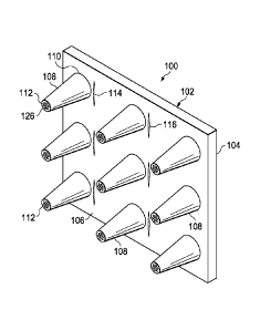
[0012] FIGURE 1 is a schematic, perspective view of an illustrative, non-
limiting
embodiment of an epithelialization dressing;
[0013] FIGURE 2 is a plan view of a portion of the epithelialization dressing
of
FIGURE 1 showing a distal end of a projection;
[0014] FIGURE 3 is a schematic, cross-sectional view of a portion of the
epithelialization dressing of FIGURE 1;
[0015] FIGURE 4 is a schematic, cross-sectional view of an illustrative
projection of
an illustrative epithelialization dressing showing a supply reservoir;
[0016] FIGURE 5 is a schematic, cross-sectional view of an illustrative
projection of
an illustrative epithelialization dressing;
[0017] FIGURE 6 is a schematic, plan view of an illustrative projection of an
illustrative epithelialization dressing;
3
CA 02790419 2012-08-17
WO 2011/115911
PCT/US2011/028352
[0018] FIGURE 7 is a schematic, cross-sectional view of an illustrative
projection of
an illustrative epithelialization dressing;
[0019] FIGURE 8 is a schematic, perspective view of an illustrative
epithelialization
dressing;
[0020] FIGURE 9 is a schematic diagram with a portion shown in cross section
of an
illustrative system for promoting granulation;
[0021] FIGURE 10 is a schematic diagram with a portion shown in cross section
of an
illustrative system for promoting epithelialization; and
[0022] FIGURE 11 is a schematic, cross-sectional view of a wound that has been
treated to promote epithelialization with an epithelialization dressing
according to one
illustrative embodiment.
DETAILED DESCRIPTION
[0023] In the following detailed description of the non-limiting, illustrative
embodiments, reference is made to the accompanying drawings that form a part
hereof. These
embodiments are described in sufficient detail to enable those skilled in the
art to practice the
invention, and it is understood that other embodiments may be utilized and
that logical
structural, mechanical, electrical, and chemical changes may be made without
departing from
the spirit or scope of the invention. To avoid detail not necessary to enable
those skilled in the
art to practice the embodiments described herein, the description may omit
certain information
known to those skilled in the art. The following detailed description is,
therefore, not to be
taken in a limiting sense, and the scope of the illustrative embodiments are
defined only by the
appended claims.
[0024] The outermost, or most superficial, layer of skin is the epidermis,
which itself
has numerous layers. The epidermis is adjacent to the dermis. The epidermis
may have
inwardly directed prolongations of the Malpighian layer that intermesh with
the dermal
papillae. These prolongations are sometimes called "rete pegs." The rete pegs
may provide
resistance against shear-induced separation of adjacent layers. In contrast,
reepithelialized
wounds often do not have as much resistance against shear.
[0025] In the healing process of wounds, the epidermis may regenerate, but the
newly
formed epithelium often, at least initially, lacks rete pegs. As such, the
newly formed
4
CA 02790419 2012-08-17
WO 2011/115911
PCT/US2011/028352
epithelium may be easily disrupted or sloughed off. Referring now primarily to
FIGURES 1-
3, an epithelialization dressing 100 may be used to promote a stronger
connection or tethering
of layers so as to function effectively as healthy rete pegs. The
epithelialization dressing 100
may induce anchor points of epithelium with tissue layers underlying the
epidermis by using
surface architecture on the epithelialization dressing 100.
[0026] Moreover, the epithelialization dressing 100 may hold or secure the
epithelium
with underlying tissues and thereby protect the epithelium from shear force
damage. In
addition, the epithelialization dressing 100 may maintain a barrier and
obviate the need for
repeated repair of the epithelium. The epithelialization dressing 100 may also
speed the
epithelialization process.
[0027] The epithelialization dressing 100 may carry out numerous functions.
For
example, the epithelialization dressing 100 may function to help manage fluids
at a wound to
promote epithelialization. As another example, the epithelialization dressing
100 may use
physical, chemical, or mechanical properties of the epithelialization dressing
100 to direct
development of underlying tissue structures to facilitate strength of the
regenerated epidermis.
As used herein, unless otherwise indicated, "or" does not require mutual
exclusivity.
[0028] The epithelialization dressing 100 may be formed with a dressing body
102
having a first side 104 and a second, tissue-facing side 106. The dressing
body 102 may take
numerous shapes, but is shown as a laminar body, i.e., having an aspect ratio
(longer
dimension divided by shorter dimension) greater than one in both a
longitudinal cross section
and lateral cross section. In other words, the dressing body 102 is shown as a
flat or
substantially flat member. Other shapes may be used for the dressing body 102,
such as a
rounded member.
[0029] The dressing body 102 has a plurality of projections 108 that may be
coupled to
the second, tissue-facing side 106 of the dressing body 102. Each projection
108 has a
proximal end 110 and a distal end 112. The proximal end 110 of each projection
108 is
coupled to the second, tissue-facing side 106 of the dressing body 102. A
plurality of
apertures 114, such as slits 116 or fenestrations, may be formed on a portion
of the dressing
body 102. The apertures 114 may be slits 116 (longitudinal openings with
substantially no
material removed) or may be round holes, square holes, or openings of any
shape that provide
for the transfer of fluids including reduced pressure.
5
CA 02790419 2012-08-17
WO 2011/115911
PCT/US2011/028352
[0030] The projections 108 or other micro-features are for placing on
granulation
tissue and function to help form epithelium columns 166 (FIG. 11) that may
function like rete
pegs. The projections 108 may take numerous shapes and sizes. The projections
108 may be
for example rods, cones, columns, ridges, grooves, waves, or other features
that form cavities
162 (FIG. 10). For example, FIGURES 1-5 present projections 108 as conical
members,
FIGURE 6 presents projections 108 that are triangular in plan view, and FIGURE
7 presents a
cross section of projections 108 as cylindrical members. FIGURE 8 presents a
perspective
view showing projections 108 that are formed like continuous bars or
orthogonal members.
The projections 108 may be randomly spaced or spaced with a pattern on the
dressing body
102. As shown in FIGURE 5, the projections may have a supply reservoir 117
formed within.
[0031] Referring now primarily to FIGURE 3, each projection 108 may have a
longitudinal length 118 (measured from the second, tissue-facing side 106) and
a lateral width
or diameter 120 at the proximal end 110 and lateral width or diameter 122 at
the distal end
112. Each projection 108 may have an aspect ratio (longitudinal length
118/average lateral
width) that is in the range of 1/10 to 10 and more typically 1/2 to 2. The
average projection of
the plurality of projections 108 may have an aspect ratio (longer dimension
for an average
projections of the plurality of projections divided by a shorter dimension for
the average
projection of the plurality of projections) in the range of 1/10 to 10 and
more typically 1/2 to 2.
In other non-limiting examples, the aspect ratio may be 3/10, 5/10, 8/10, 2,
3, 4, 5, 6, 7, 8, or
9. The aspect ratio may be adjusted to help control the level of strain placed
on underlying
tissue and to help control strain gradients. Controlling the induced strain
may help control
remodeling of the tissue and may impact stem cell differentiation. An edge
124, or leading
edge, on the distal end 112 of the projection 108 may be sharp (orthogonal, 90
degrees, or
substantially 90 degrees) or may be rounded to help control strain as well.
The projections
108 may all have the same dimensions or properties or may vary one to another.
[0032] As shown in FIGURE 2, a plurality of pores 126 or small apertures may
be
formed on the distal end 112 of the projections 108. The pores 126 may also or
alternatively
be formed at other locations on the projections 108 or dressing body 102. The
pores 126 may
facilitate removal of fluids near the epithelialization dressing 100, deliver
reduced pressure,
allow for fluid delivery, or provide for intentional ingrowth of tissue. Pores
(not shown) may
also be formed on the second, tissue-facing side 106 of the dressing body 102
between the
6
CA 02790419 2012-08-17
WO 2011/115911
PCT/US2011/028352
projections 108. With reference to FIGURE 4, the pores 126 may help deliver a
fluid or other
substance, e.g., growth factors, from the supply reservoir 117 to an area near
the projection
108.
[0033] Referring primarily to FIGURE 5, the pores 126 are shown facilitating
the
delivery of reduced pressure and the removal of fluids near the projections
108. Apertures 115
through the dressing body 102 allow reduced pressure to be delivered from the
first side 104
of the dressing body 102 to the supply reservoir 117. The pores 126, which in
this
embodiment are on the distal end and on a side portion of the projection 108,
communicate the
reduced pressure from the supply reservoir 117 to an exterior of the
projections 108. In this
embodiment, the first side 104 of the dressing body 102 is in fluid
communication with an
exterior of the projections 108. In another embodiment, the projections 108
may be formed
with a bicameral reservoir (not shown) having one portion for supplying a
substance and
another portion for providing reduced pressure.
[0034] As shown in FIGURE 6, in some embodiments, micro-scale or nano-scale
features 128, or sub-features, may be added to the projections 108 typically
on the distal end
112. The sub-features 128 may be, for example, grooves 130, ridges, or waves.
The sub-
features 128 are typically separate from the pores 126, but in other
embodiments, could
include openings as the pores on the sub-features 128. The micro-scale or nano-
scale features
128 may be able to pattern proteins that absorb to the sub-features 128 or
promote cell height
and direct orientation and migration. If the sub-features 128 are grooves,
e.g., the grooves
130, fibroblasts may attach and become oriented according to the features and
secrete their
matrix proteins in a similar pattern. This may further allow control of tissue
development.
[0035] The size and shape of the projections 108, the size and spacing of the
pores
126, or the sub-features 128 may be used to control the tissue development in
order to promote
maturation and to enhance the strength of a healing wound against shear
stress. The
projections 108 or other micro-features may guide tissue growth and
remodeling, including
cellular orientation and organization. Geometry of the projections 108 or
micro-features may
be modulated to induce specific load and strain distribution and gradients in
the tissue. These
modulations may involve aspect ratio, size, spacing, contact area (%),
curvature at contact,
alternating feature shapes and sizes, biomimetic patterns, and the overlay of
micro and sub-
7
CA 02790419 2012-08-17
WO 2011/115911
PCT/US2011/028352
features 128. The size and aspect ratio of the projections 108 may be
modulated as desired to
control stress and strain at the tissue interface.
100361 Numerous materials may be used to form the epithelialization dressing
100,
such as a medical-grade polymer, e.g., a silicone or polyurethane, or a
biological polymer, e.g.,
collagen. Other materials from which the epithelialization dressing 100 may be
formed
include bioresorbable (or resorbable) material, biologic material, or non-
resorbable material.
As used herein, "bioresorbable" includes a material that enzymatically or
chemically degrades
into a simple chemical species in vivo, and which may be removed from the body
by excretion
or metabolism. The material may be an occlusive material. The
epithelialization dressing 100
may be non-adherent to tissue growth. The epithelialization dressing 100 may
be formed from
a non-absorbable material for the dressing body 102. The projections 108 may
be formed
from a bioresorbable material. In another embodiment, the entire
epithelialization dressing
100 is formed from bioresorbable material. The second, tissue-facing surface
106 of the
epithelialization dressing 100 may be a moist surface that¨other than the
projections 108¨is
relatively smooth as compared to a foam surface. With reference to FIGURE 3,
the dressing
100 may have a depth 132 that is in the range of 10 to 5000 microns, and more
typically
between 400 and 600 microns. For example, without limitation, the depth 132
may be 400,
425, 450, 475, 500, 525, 550, 575, 600 microns or another depth.
[0037] Referring now to FIGURE 8, another illustrative, non-limiting
embodiment of
an epithelialization dressing 100 is presented. The epithelialization dressing
100 of FIGURE 8
may be formed with a dressing body 102 having a first side 104 and a second,
tissue-facing
side 106. A plurality of projections 108 may be coupled to the second, tissue-
facing side 106
of the dressing body 102. Each projection 108 has a proximal end 110 and a
distal end 112.
The proximal end 110 of each projection 108 is coupled to the second, tissue-
facing side 106
of the dressing body 102. The projections 108 form a grid that is used to
impinge upon
granulation tissue. A plurality of apertures 114 are formed on a portion of
the dressing body
102 and are shown as circular openings.
[0038] Referring now primarily to FIGURES 9-11, one illustrative, non-limiting
process for treating a wound 134 or other tissue site is presented. Referring
initially to
FIGURE 9, the wound 134 is treated with a system 103 to promote granulation. A
manifold
136 is disposed proximate the wound 134. The term "manifold" as used herein
generally
8
CA 02790419 2012-08-17
WO 2011/115911
PCT/US2011/028352
refers to a substance or structure that is provided to assist in applying
reduced pressure to,
delivering fluids to, or removing fluids from a tissue site or wound 134.
100391 The manifold 136 typically includes a plurality of flow channels or
pathways
that distribute fluids provided to and removed from the tissue site or wound
134 around the
manifold 136. In one illustrative embodiment, the flow channels or pathways
are
interconnected to improve distribution of fluids provided or removed from the
wound 134.
The manifold 136 may be a biocompatible material that is capable of being
placed in contact
with wound 134 and distributing reduced pressure to the wound 134.
[0040] Examples of manifolds 136 may include, for example, without limitation,
devices that have structural elements arranged to form flow channels, such as,
for example,
cellular foam, open-cell foam, porous tissue collections, liquids, gels, and
foams that include,
or cure to include, flow channels. The manifold 136 may be porous and may be
made from
foam, gauze, felted mat, or any other material suited to a particular
biological application. In
one embodiment, the manifold 136 is porous foam and includes a plurality of
interconnected
cells or pores that act as flow channels. The porous foam may be a
polyurethane, open-cell,
reticulated foam, such as V.A.C.0 GranuFoam0 material manufactured by Kinetic
Concepts,
Incorporated of San Antonio, Texas. Other embodiments may include "closed
cells." In some
situations, the manifold 136 may also be used to distribute fluids such as
medications,
antibacterials, growth factors, and various solutions to the wound 134. Other
layers may be
included in or on the manifold 136, such as absorptive materials, wicking
materials,
hydrophobic materials, and hydrophilic materials.
[0041] A reduced-pressure interface 138, e.g., a connector, is disposed
proximate the
manifold 136 and extends through an aperture 140 in a sealing member 142. The
sealing
member 142 forms a fluid seal over the wound 134. "Fluid seal," or "seal,"
means a seal
adequate to maintain reduced pressure at a desired site given a particular
reduced-pressure
source or subsystem involved.
[0042] The sealing member 142 may be any material that provides a fluid seal.
The
sealing member may, for example, be an impermeable or semi-permeable,
elastomeric
material. "Elastomeric" means having the properties of an elastomer.
Elastomeric material
generally refers to a polymeric material that has rubber-like properties. More
specifically,
most elastomers have ultimate elongations greater than 100% and a significant
amount of
9
CA 02790419 2012-08-17
WO 2011/115911
PCT/US2011/028352
resilience. The resilience of a material refers to the material's ability to
recover from an
elastic deformation. Examples of elastomers may include, but are not limited
to, natural
rubbers, polyisoprene, styrene butadiene rubber, chloroprene rubber,
polybutadiene, nitrile
rubber, butyl rubber, ethylene propylene rubber, ethylene propylene diene
monomer,
chlorosulfonated polyethylene, polysulfide rubber, polyurethane (PU), EVA
film, co-
polyester, and silicones. Additional, specific examples of sealing member
materials include a
silicone drape, a 3M Tegadermg drape, or a polyurethane (PU) drape such as one
available
from Avery Dennison Corporation of Pasadena, California. The sealing member
142 has a
first side 144 and a second, tissue-facing side 146.
[0043] An attachment device 148 may be used to hold the sealing member 142
against
the patient's epidermis 150 or another layer, such as a gasket or additional
sealing member.
The attachment device 148 may take numerous forms. For example, the attachment
device
148 may be a medically acceptable, pressure-sensitive adhesive that extends
about a periphery
of the sealing member 142. As additional examples, the attachment device 148
may be a
double-sided drape tape, paste, hydrocolloid, hydro gel or other sealing
devices or elements.
[0044] A reduced-pressure delivery conduit 152 may fluidly couple the reduced-
pressure interface 138 to a reduced-pressure source 154 that provides reduced
pressure. The
reduced-pressure source 154 may be any device for supplying a reduced
pressure, such as a
vacuum pump, wall suction, micro-pump, or other source. While the amount and
nature of
reduced pressure applied to a tissue site or wound 134 will typically vary
according to the
application, the reduced pressure will typically be between -5 mm Hg (-667 Pa)
and -500 mm
Hg (-66.7 kPa) and more typically between -75 mm Hg (-9.9 kPa) and -300 mm Hg
(-39.9
kPa). For example, and not by way of limitation, the pressure may be -12, -
12.5, -13, -14, -
14.5, -15, -15.5, -16, -16.5, -17, -17.5, -18, -18.5, -19, -19.5, -20, -20.5, -
21, -21.5, -22, -22.5, -
23, -23.5, -24, -24.5, -25, -25.5, -26, -26.5 kPa or another pressure.
[0045] As used herein, "reduced pressure" generally refers to a pressure less
than the
ambient pressure at a tissue site that is being subjected to treatment. In
most cases, this
reduced pressure will be less than the atmospheric pressure at which the
patient is located.
Alternatively, the reduced pressure may be less than a hydrostatic pressure at
the tissue site.
Unless otherwise indicated, values of pressure stated herein are gauge
pressures. The reduced
pressure delivered may be constant or varied (patterned or random) and may be
delivered
CA 02790419 2012-08-17
WO 2011/115911
PCT/US2011/028352
continuously or intermittently. Although the terms "vacuum" and "negative
pressure" may be
used to describe the pressure applied to the tissue site, the actual pressure
applied to the tissue
site may be more than the pressure normally associated with a complete vacuum.
Consistent
with the use herein, unless otherwise indicated, an increase in reduced
pressure or vacuum
pressure typically refers to a relative reduction in absolute pressure.
[0046] One or more devices, such as device 156, may be included on the reduced-
pressure conduit 152. For example, the device 156 may be a fluid reservoir, or
collection
member, to hold exudates and other fluids removed. Other examples of devices
156 that may
be included on the reduced-pressure delivery conduit 152 or otherwise fluidly
coupled to the
reduced-pressure delivery conduit 152 include the following non-limiting
examples: a
pressure-feedback device, a volume detection system, a blood detection system,
an infection
detection system, a flow monitoring system, or a temperature monitoring
system. Some of
these devices may be formed integrally with the reduced-pressure source 154.
[0047] The reduced pressure delivered to the wound 134 helps to fill in the
wound
defect with new tissue. The reduced pressure may promote fibroblasts to
synthesize and
develop extracellular matrix components. Granulation tissue fibroblasts
produce a matrix for
collagen deposition. After sufficient time, granulation tissue 158 (FIG. 10)
is deposited on a
bed of the wound 134. After the granulation tissue 158 has adequately
developed, the sealing
member 142 may be removed and the manifold 136 removed. In the example of
FIGURES 9
and 10, the granulation tissue 158 has grown outward from near or at a dermis
layer 151 to
above a lower portion of the epidermis 150.
[0048] Referring now primarily to FIGURE 10, a system 101 for promoting
epithelialization is presented. The epithelialization dressing 100 may placed
proximate the
granulation tissue 158 with the second, tissue-facing side 106 and projections
108 substantially
against the granulation tissue 158. A sealing member 142 is then deployed to
provide a fluid
seal over the epithelialization dressing 100. In one embodiment (not
explicitly shown), the
manifold 136 (see FIG. 9) may be placed between the epithelialization dressing
100 (see FIG.
10) and sealing member 142 to facilitate reduced pressure distribution and
fluid removal from
wound areas distal from reduced-pressure interface 138.
[0049] Referring again to the embodiment of FIGURE 10, after the reduced-
pressure
interface 138 and the sealing member 142 (this includes new interfaces and
sealing members)
11
CA 02790419 2012-08-17
WO 2011/115911
PCT/US2011/028352
are deployed, the reduced-pressure source 154 is again activated. The reduced
pressure
supplied to the epithelialization dressing 100 may achieve a number of
results.
100501 The reduced pressure delivered to the epithelialization dressing 100
may help
remove excess fluids from a surface 160 of the wound 134, which has been
partially
regenerated. Some fluids may remain to assist with signaling and to otherwise
promote re-
epithelialization. The reduced pressure may provide a compression force on the
dressing 100
to help to maintain and control contact between the dressing 100 and the
granulation tissue
158 or other tissue. The reduced pressure may be used to cause or control the
magnitude of
force causing the projections 108 to impinge on the granulation tissue 158 or
other tissue. In
some embodiments, the pressure may be varied (patterned or random) to provided
a variable
force delivered by the epithelialization dressing 100 and may thereby further
enhance
epithelialization.
[0051] Referring now primarily to FIGURES 10 and 11, fibroblasts may interact
with
the second, tissue-facing side 106 of the dressing 100 and form patterned
extracellular matrix.
Activated keratinocytes migrate from the wound edges 164 around and between
the
projections 108 and down into the cavities 162 formed by the projections 108
and form an
epithelium 168 or new epithelium tissue (FIG. 11). The portion of the matrix
going into the
cavities 162 will gradually form epithelium columns 166, which may take any
shape
corresponding substantially to the shape of the projections 108. The
epithelium 168 has an x-y
pattern around the projections 108 and a pattern in the z-direction as the
epithelium 168 moves
into the cavities 162 to form the epithelium columns 166. The cavities 162
formed by the
projections 108 will extend into the adjacent layer of granulation tissue 158
and help form the
epithelium columns 166. The epithelium columns 166 act functionally like rete
pegs between
the epidermis 150 and dermis 151 and thereby may provide anchor points
resistant against
external forces to keep the involved tissue layers adherent. The epithelium
columns 166 form
simulated rete pegs.
[0052] Referring now primarily to FIGURE 11, the wound 134 is shown after
dressing
100 has been removed. (The surface of the epithelium 168 may be more or less
flush with the
intact epidermis 150 than shown). The dressing 100 may stay in place on the
wound for a set
time of any duration and typically between one to six days. In this
illustrative, non-limiting
embodiment, the epithelium 168 covers the granulation tissue 158. The
epithelium columns
12
CA 02790419 2012-08-17
WO 2011/115911
PCT/US2011/028352
166 fill the cavities 162 and extend into the granulation tissue 158. In some
embodiments (not
explicitly shown), the epithelium columns 166 extend into a portion of the
dermis 151. The
epithelium columns 166 provide additional shear resistance for the epithelium
168.
[0053] It should be noted that while the process of FIGURES 9-11 shows the use
of
reduced-pressure treatment to promote formation of the granulation tissue 158,
the dressing
100 may be used independently of such a step. In addition, while the process
and system 100
has been described as using reduced pressure, it should be understood that the
dressing 100
may be used without reduced pressure. In this illustrative process, the
dressing 100 may
further include a hydrophilic material, e.g., hydrophilic foam or capillaries,
on the first side
104 to help remove fluids through the apertures 114 and a bolster, tape,
compression wrap, or
other device may be used to provide a compression force on the dressing 100 to
assure contact
between the dressing 100 and the tissue.
[0054] Numerous alternatives or additions may be involved with the
illustrative, non-
limiting dressing 100, system 101, or the process. Some have already been
mentioned and
other, non-exhaustive examples are now mentioned. In another illustrative
process, the
dressing 100 may be made from a reabsorbable material that degrades. The
projections 108
may include supply reservoirs 117 that hold encapsulated stem cells,
keratinocytes, growth
factors, soluble factors, or other substance. As the degradation reaches a
certain level, the
substance within the supply reservoirs 117 is delivered to the cavities 162
and tissue. The
substance then fills or helps fill the cavity 162 and helps promote further
healing.
[0055] In another illustrative, non-limiting embodiment, the dressing 100,
system 101,
or the processes may be used with other tissues. For example, in addition to
epithelial tissue,
endothelial and mucosal linings may benefit. Other embodiments may also be
used for
treating tendons, ligaments, muscles, or cartilage to add inherent resistance
to the forces that
actively work to separate these tissues.
[0056] In another illustrative, non-limiting example, the dressing 100 may be
used as
an aspect of promoting granulation and epithelialization. The dressing 100 may
include the
projections 108 with pores 126 on the distal end 112 and also all along the
side walls of the
projections 108. Ingrowth of granulation tissue 158 into the pores 126 is
promoted. If
reduced pressure is utilized, the reduced pressure may help pull tissue into
the pores 126.
Once the granulation tissue 158 has grown into and around the projections 108,
the advancing
13
CA 02790419 2012-08-17
WO 2011/115911
PCT/US2011/028352
keratinocytes may follow the granulation tissue 158 and overlay the
granulation tissue 158. In
addition or as a separate alternative, the dressing 100 may be bioresorbable
and degrade over
time. The degradation should not negatively impact keratinocyte proliferation.
In addition,
the projections 108 may have supply reservoirs 117 with active soluble factors
that enhance
local keratinocyte differentiation. The thickening of the keratinocytes in
this area may fill the
cavity 162 formed by the projections 108.
[0057] Although the present invention and its advantages have been disclosed
in the
context of certain illustrative, non-limiting embodiments, it should be
understood that various
changes, substitutions, permutations, and alterations can be made without
departing from the
scope of the invention as defined by the appended claims. It will be
appreciated that any
feature that is described in connection to any one embodiment may also be
applicable to any
other embodiment.
[0058] It will be understood that the benefits and advantages described above
may
relate to one embodiment or may relate to several embodiments. It will further
be understood
that reference to 'an item refers to one or more of those items.
[0059] The steps of the methods described herein may be carried out in any
suitable
order, or simultaneously where appropriate.
[0060] Where appropriate, aspects of any of the examples described above may
be
combined with aspects of any of the other examples described to form further
examples
having comparable or different properties and addressing the same or different
problems.
[0061] It will be understood that the above description of preferred
embodiments is
given by way of example only and that various modifications may be made by
those skilled in
the art. The above specification, examples and data provide a complete
description of the
structure and use of exemplary embodiments of the invention. Although various
embodiments
of the invention have been described above with a certain degree of
particularity, or with
reference to one or more individual embodiments, those skilled in the art
could make
numerous alterations to the disclosed embodiments without departing from the
scope of the
claims.
14