Note: Descriptions are shown in the official language in which they were submitted.
CA 02790931 2012-08-23
WO 2012/023983 PCT/US2011/001451
CLUTCH RELEASE MECHANISM FOR VASCULAR CLOSURE DEVICE
RELATED APPLICATION
[0001] This claims the benefit of U.S. Provisional Application No. 61/375,361,
filed 20 August 2010, which is hereby incorporated by reference herein in its
entirety.
TECHNICAL FIELD
[0002] The present disclosure relates generally to medical devices and more
particularly to devices for sealing punctures or incisions in a tissue wall.
BACKGROUND
[0003] Various surgical procedures are routinely carried out intravascularly
or
intraluminally. For example, in the treatment of vascular disease, such as
arteriosclerosis, it
is a common practice to invade the vessel and insert an instrument (e.g., a
balloon or other
type of catheter) to carry out a procedure within the vessel. Such procedures
usually involve
the percutaneous puncture of the vessel so that an insertion sheath may be
placed in the vessel
and thereafter instruments (e.g., catheters) may pass through the sheath to an
operative
position within the vessel. Intravascular and intraluminal procedures
unavoidably present the
problem of stopping the bleeding at the percutaneous puncture after the
procedure has been
completed and after the instruments (and any insertion sheaths used therewith)
have been
removed. Bleeding from puncture sites, particularly in the case of femoral
arterial punctures,
is typically stopped by utilizing vascular closure devices, such as those
described in U.S.
Patent Nos. 6,179,963; 6,090,130; and 6,045,569, which are incorporated herein
in their
entireties by this reference.
1
CA 02790931 2012-08-23
WO 2012/023983 PCT/US2011/001451
[0004] Typical closure devices such as the ones described in the above-
mentioned patents place a sealing plug at the tissue puncture site. Successful
deployment of
the sealing plug, however, requires that it be manually ejected from within a
device sheath
and compacted down to an outer surface of the tissue puncture using a
compaction tube. The
compaction procedure cannot commence until the device sheath (within which the
compaction tube is located) has been removed so as to expose the compaction
tube for
manual grasping. Under certain conditions, removal of the sheath prior to
compacting the
sealing plug may cause the sealing plug itself to be displaced proximally from
the tissue
puncture, hindering subsequent placement of the sealing plug, and resulting in
only a partial
seal and associated late bleeding from the tissue puncture. Accordingly, there
is a need for
improving the mechanism for deployment of the sealing plug at the site of a
tissue puncture.
SUMMARY
[0005] The present disclosure meets the above-described needs and others.
Specifically, the present disclosure provides methods and systems for closing
internal tissue
punctures. However, unlike prior systems, the present disclosure provides
automatic
compaction to a sealing plug as the closure device is retracted. In addition,
the present
disclosure allows the automatic compaction system to disengage, facilitating
full retraction of
the closure device and easy separation of the sealing plug from the remainder
of the closure
device.
[0006] In one of many possible embodiments, the present disclosure provides a
tissue puncture closure device that includes an anchor, a sealing plug, a
filament, a
compaction member, a spool, a driving plate, and a follower. The filament is
secured
between the sealing plug and the anchor. The compaction member assembly is
disposed
2
CA 02790931 2012-08-23
WO 2012/023983 PCT/US2011/001451
adjacent to the sealing plug and structured and arranged to apply an axially
directed
compressive force to automatically compact the sealing plug toward the anchor.
The
compaction member assembly has a distal end and a proximal end. The spool has
a portion
of the filament wound thereon. The driving plate is connected to the spool and
arranged to
contact and apply a force to the proximal end of the compaction member
assembly upon
rotation of the driving plate to advance the compaction member assembly. The
follower is
operable between the spool and driving plate to releasably connect the spool
and driving
plate.
[0007] The compaction member assembly may further include a compaction tube
and a coil, wherein the coil is structured and arranged to apply an axially
directed
compressive force to the compaction tube to drive the compaction tube to
automatically
compact the sealing plug toward the anchor. The spool may include a cam
portion with a
portion of the filament being wrapped around the cam portion, wherein
unwinding the
filament from the spool applies a variable rotation force to the driving
plate. The spool may
include first and second outer plates and a cam portion positioned between the
first and
second outer plates. The cam portion may define a cam surface having a
variable radius,
wherein a portion of the filament wraps around the cam portion.
[0008] The follower may be mounted to the spool and biased toward the driving
plate. The driving plate may include at least one recess sized to receive a
portion of the
follower. The compaction member assembly may include a compaction tube and a
coil
member arranged end-to-end. The compaction tube may define the distal end of
the
compaction member assembly and the coil defines the proximal end of the
compact member
assembly. The driving plate includes a coil track defined in a peripheral
surface of the driving
plate, and a portion of the compaction member assembly may be positioned in
the coil track.
3
CA 02790931 2012-08-23
WO 2012/023983 PCT/US2011/001451
[0009] Another aspect of the present disclosure relates to a tissue puncture
closure
device for partial insertion into and sealing of a tissue puncture in an
internal tissue wall
accessible through a percutaneous incision. The device includes an anchor, a
sealing plug, a
filament, a compaction member, a storage spool, a driving plate, and at least
one follower.
The anchor is disposed on a distal side of the internal tissue wall. The
sealing plug is
disposed on a proximal side of the internal tissue wall. The filament is
connected to and
anchored at a distal end to the anchor and sealing plug, and is slidable and
cinchable along the
filament toward the anchor to close the tissue puncture. The compaction member
assembly is
disposed on the filament and arranged to drive the sealing plug along the
filament distally
towards the anchor. The storage spool has a proximal end of the filament wound
thereon.
The driving plate is connected to the storage spool and configured to contact
a proximal end
of the compaction member assembly to advance the compaction member assembly.
The at
least one follower is mounted to the storage spool and biased into contact
with the driving
plate to releasably resist relative rotational movement between the storage
spool and driving
plate.
[0010] The driving plate may include at least one recess configured to receive
a
portion of the at least one follower. The at least one recess may include a
plurality of recesses
arranged in a circle. The at least one follower may move out of the at least
one recess when a
threshold torsional force applied to the storage spool by the filament is
exceeded. The storage
spool may include a cam portion about which the proximal end of the filament
is wound. The
storage spool is configured to apply a variable rotational force to the
driving plate when the
filament unwinds from the cam portion.
[0011] A further aspect of the present disclosure is directed to a method of
sealing
a tissue puncture in an internal tissue wall of a vessel that is accessible
through a
4
CA 02790931 2012-08-23
WO 2012/023983 PCT/US2011/001451
percutaneous incision. The method includes providing a closure device having
an anchor, a.
sealing plug, a filament secured between the sealing plug and the anchor, a
compaction
member assembly, a spool having a portion of the filament wound thereon, a
driving plate,
and at least one follower arranged to resist relative rotational movement
between the driving
plate and spool. A distal end of the compaction member assembly is disposed
adjacent the
sealing plug, a proximal end of the compaction member assembly is in contact
with the
driving plate, and the driving plate is connected to the spool. The method
also includes
inserting the anchor through the tissue puncture, withdrawing the closure
device from the
tissue puncture with the anchor positioned within the vessel, wherein
withdrawing the closure
device rotates the spool, and rotating the spool rotates the driving plate to
drive the
compaction member assembly and compact the sealing plug toward the anchor. In
the
method, further withdrawing the closure device until the at least one follower
disconnects
from at least one of the spool and driving plate permits relative rotation
between the spool
and driving plate.
[0012] The at least one follower may be mounted to the spool and biased into
contact with the driving plate with a biasing member. The at least one
follower may include a
plurality of followers biased into contact with the driving plate.
[0013] Another aspect of the present disclosure relates to a method of sealing
a
tissue puncture in an internal tissue wall accessible through a percutaneous
incision. The
method includes providing a tissue puncture closure device comprising an
anchor, a sealing
plug, a filament, a compaction member assembly, a driving plate, a spool
having a cam
member, and at least one follower, the filament being connected at its distal
end to the anchor,
to the sealing plug located proximal of the anchor, and to the cam member of
the spool at its
proximal end. The at least one follower is operable to releasably connect the
spool and
5
CA 02790931 2012-08-23
WO 2012/023983 PCT/US2011/001451
driving plate. The method also includes inserting the tissue puncture closure
device into the
percutaneous incision, deploying the anchor into the tissue puncture, and
automatically
compacting the sealing plug toward the anchor upon withdrawal of the tissue
puncture closure
device from the internal tissue wall puncture. Automatically compacting
includes unwinding
the filament from the spool to rotate the spool and driving plate together to
apply a variable
force to the compaction member assembly to advance a distal end of the
compaction member
assembly. The method further includes operating the at least one follower to
release the spool
from rotating with the driving plate, and cutting the filament to leave the
anchor and sealing
plug at the tissue puncture.
[0014] The step of operating the at least one follower may include applying a
withdrawal force to the tissue puncture closure device to exceed a threshold
torsional force
applied to the spool by unwinding in the filament to automatically move the at
least one
follower relative to at least one of the spool and driving plate. The driving
plate may include
a plurality of follower recesses arranged to receive the at least one follower
at different
relative rotated positions between the driving plate and spool. The tissue
puncture closure
device may include a housing and a base upon which the driving plate and spool
are mounted,
wherein the base is movable within the housing to permit ejection of the
sealing plug from the
tissue puncture closure device without compacting the sealing plug.
[0015] Additional advantages and novel features will be set forth in the
description which follows or can be learned by those skilled in the art
through reading these
materials or practicing the examples disclosed herein.
6
CA 02790931 2012-08-23
WO 2012/023983 PCT/US2011/001451
BRIEF DESCRIPTION OF THE DRAWINGS
[0016] The accompanying drawings illustrate various embodiments of the present
disclosure and are a part of the specification. The illustrated embodiments
are merely
examples and do not limit the scope of the invention.
[0017] FIG. 1 is a partial cut-away view of a tissue puncture closure device
according to the prior art.
[0018] FIG. 2 is a side view of the tissue puncture closure device of FIG. 1
engaged with a vessel according to the prior art.
[0019] FIG. 3 is a side view of the tissue puncture closure device of FIG. 1
being
withdrawn from a vessel according to the prior art to deploy a sealing plug.
[0020] FIG. 4 is a side view of the tissue puncture closure device of FIG. 1
illustrating compaction of the sealing plug according to the prior art.
[0021] FIG. 5A is an exploded perspective view of an example tissue puncture
closure device with an automatic compaction or driving mechanism according to
the present
disclosure.
[0022] FIG. 5B is another exploded perspective view of the tissue puncture
closure device of FIG. 5A.
[0023] FIG. 5C is a side view of the tissue puncture closure device of FIG. 5A
inserted through a procedure sheath and tissue puncture and engaged with a
vessel in a first
position.
[0024] FIG. 5D is a detailed inset of FIG. 5C.
[0025] FIG. 5E is a side view of the tissue puncture closure device of FIG. 5A
shown engaged with a vessel in a second position with the procedure sheath
retracted.
[0026] FIG. 5F is a detailed inset of FIG. 5E.
7
CA 02790931 2012-08-23
WO 2012/023983 PCT/US2011/001451
[0027] FIG. 5G is a side view of the tissue puncture closure device of FIG. 5A
engaged with a vessel in a third fourth position with a carrier tube retracted
to expose a
sealing plug adjacent to the tissue puncture and the sealing plug being
compacted.
[0028] FIG. 5H is a detailed inset of FIG. 5G.
[0029] FIG. 6A is a top exploded perspective view of the automatic driving
assembly of FIG. 5A.
[0030] FIG. 6B is a bottom exploded perspective view of the automatic driving
assembly of FIG. 5A.
[0031] FIG. 7 is a perspective view of the automatic driving assembly of FIGS.
6A-B.
[0032] FIG. 8 is a cross-sectional view of the automatic driving assembly of
FIG.
7 taken along cross-section indicators 8-8.
[0033] FIG. 9 is a cross-sectional view of the automatic driving assembly of
FIG.
7 taken along cross-section indicators 9-9 with a spool assembly and driving
plate connected
together.
[0034] FIG. 10 is a cross-sectional view of the automatic driving assembly of
FIG.
9 with the spool assembly and driving plate disconnected to permit relative
rotation
therebetween.
[0035] FIG. 11 is a side view of the spool assembly of the automatic driving
assembly of FIG. 7.
[0036] FIG. 12 is a cross-sectional view of the spool assembly of FIG. 11
taken
along cross-section indicators 12-12.
8
CA 02790931 2012-08-23
WO 2012/023983 PCT/US2011/001451
[0037] FIG. 13 is an exploded perspective view of another example tissue
puncture closure device with an automatic compaction or driving mechanism
according to the
present disclosure.
[0038] Throughout the drawings, identical reference numbers designate similar,
but not necessarily identical, elements.
DETAILED DESCRIPTION
[0039] As mentioned above, vascular procedures are conducted throughout the
world and require access to a vessel through a puncture. Most often, the
vessel is a femoral
artery. To close the puncture following completion of a procedure, many times
a closure
device is used to sandwich the puncture between an anchor and a sealing plug.
However,
sometimes the sealing plug is difficult to eject from the sealing device and
may not properly
seat against an exterior situs of the arteriotomy. If the plug does not seat
properly against the
arteriotomy, there is a potential for elongated bleeding.
[0040] The present disclosure describes methods and apparatuses that
facilitate
sealing plug ejection and proper placement of the sealing plug. One aspect of
the present
disclosure is directed to the use of a cam structure in a vascular closure
device as part of an
automatic or semi-automatic driving assembly. The cam structure may contact or
be coupled
to a compaction member assembly that is used to compact the sealing plug. The
compaction
member assembly may include a compaction tube that is arranged to contact the
sealing plug.
The compaction member assembly may also include a compaction tube driver
(e.g., a coiled
structure) positioned between the compaction tube and the cam structure. The
cam structure
may include at least one cam surface, and rotation of the cam structure
contacts the cam
surface with compaction tube driver to advance the compaction tube. The cam
member may
9
CA 02790931 2012-08-23
WO 2012/023983 PCT/US2011/001451
be coupled to a spool about which a portion of a suture is wound, wherein the
suture is used
to connect the sealing plug and an anchor of the vascular closure device
together. The cam
member may apply a variable driving force to the proximal end of the
compaction assembly
upon rotation of the spool. In some arrangements, the cam member is
constructed as a
driving plate that is arranged coaxially with the spool and is rotated upon
rotation of the
spool. The cam member may have a generally thin, flat construction. The cam
member
interface with the compaction assembly by, for example, contacting a proximal
end of the
compaction assembly, or may interface with an interference fit, clamp, or
other type of
interface at a location distal of a proximal end of the compaction assembly. A
clutch may be
operable between the driving plate and spool.
[0041] While the vascular instruments shown and described below include
procedure sheaths and puncture sealing devices, the application of principles
described herein
are not limited to the specific devices shown. The principles described herein
may be used
with any medical device. Therefore, while the description below is directed
primarily to
arterial procedures and certain embodiments of a vascular closure device, the
methods and
apparatus are only limited by the appended claims.
[0042] As used in this specification and the appended claims, the terms
"compact," "compaction," and "compacting" are used broadly to mean packing
down and
compressing by one or a succession of blows or taps or smooth, steady
pressure, but not by
excessive force. The terms "tamp" and "tamping" may relate to certain types or
forms of
"compaction" and "compacting." "Engage" and "engabable" are also used broadly
to mean
interlock, mesh, or contact between two devices. Likewise "disengage" or
"disengagable"
means to remove or capable of being removed from interlock, mesh, or contact.
A "tube" is
an elongated device with a passageway. The passageway may be enclosed or open
(e.g., a
CA 02790931 2012-08-23
WO 2012/023983 PCT/US2011/001451
trough). A "lumen" refers to any open space or cavity in a bodily organ,
especially in a blood
vessel. The words "including" and "having," as used in the specification,
including the
claims, have the same meaning as the word "comprising."
[0043] Referring to FIGS. 1-4, a vascular closure device 100 is shown
according
to the prior art. Some example closure devices are disclosed in U.S. Published
Patent
Application No. 2005/0085851 and U.S. Patent Nos. 7,618,438 and 7,618,436,
which
references are incorporated herein in their entireties by this reference. The
vascular closure
device 100 includes a carrier tube 102 with a filament or suture 104 extending
at least
partially therethrough. The vascular closure device 100 also includes a first
or proximal end
106 and a second or distal end 107. External to the distal end 107 of the
carrier tube 102 is an
anchor 108. The anchor may include an elongated, stiff, low profile member
including an eye
109 formed at the middle. The anchor 108 is typically made of a biologically
resorbable
polymer.
[0044] The suture 104 is threaded through the anchor 108 and back to a
collagen
pad 110. The collagen pad 110 may comprise, for example, randomly oriented
fibrous
material bound together by chemical means. The collagen pad 110 is slidingly
attached to the
suture 104 as the suture passes distally through the carrier tube 102. As the
suture traverses
the anchor 108 and reenters the carrier tube 102, the suture 104 is securely
slip-knotted
proximal to the collagen pad 110 to facilitate cinching of the collagen pad
110 when the
vascular closure device 100 is properly placed and the anchor 108 deployed
(see FIG. 4).
[0045] The carrier tube 102 typically includes a compaction member 112
disposed
therein. The compaction member 112 is slidingly mounted on the suture 104 and
may be
used by an operator to compact the collagen pad 110 toward the anchor 108 at
an appropriate
time to seal a percutaneous tissue puncture.
11
CA 02790931 2012-08-23
WO 2012/023983 PCT/US2011/001451
[0046] Prior to deployment of the anchor 108 within a vessel (e.g., an
artery), the
eye 109 of the anchor 108 rests outside the distal end 107 of the carrier tube
102. The anchor
108 may be temporarily held in place flush with the carrier tube 102 using a
bypass tube 114
that is disposed over the distal end 107 of the carrier tube 102.
[0047] The flush arrangement of the anchor 108 and carrier tube 102 allows the
anchor 108 to be inserted into a sheath such as insertion sheath 116 as shown
in FIGS. 2-4,
and eventually through a tissue (e.g., arterial) puncture 118. The insertion
sheath 116 is
shown in FIGS. 2-4 inserted through a percutaneous incision 119 and into a
vessel 128. The
bypass tube 114 (see FIG. 1) includes an oversized head 120 that prevents the
bypass tube
114 from passing through an internal passage of the insertion sheath 116. As
the vascular
closure device 100 is inserted into the insertion sheath 116, the oversized
head 120 bears
against a surface 122 of insertion sheath 116.
[0048] Further insertion of the vascular closure device 100 results in sliding
movement between the carrier tube 102 and the bypass tube 114, thereby
releasing the anchor
108 from the bypass tube 114 (see FIG. 1). The anchor 108 typically remains in
the flush
arrangement shown in FIG. 1 following release from the bypass tube 114,
limited in
movement by the insertion sheath 116.
[0049] The insertion sheath 116 may include a monofold at a second or distal
end
126 thereof. The monofold acts as a one-way valve to the anchor 108. A
monofold is
typically a plastic deformation in a portion of the insertion sheath 116 that
elastically flexes as
the anchor 108 is pushed out through the distal end 126 of the insertion
sheath 116.
Typically, after the anchor 108 passes through the distal end 126 of the
insertion sheath 116
and enters the vessel 128, the anchor 108 is no longer constrained to the
flush arrangement
with respect to the carrier tube 102 and it deploys and rotates to the
position shown in FIG. 2.
12
CA 02790931 2012-08-23
WO 2012/023983 PCT/US2011/001451
[0050]. The insertion sheath 116 may include a pair of closure device
connection
apertures (not shown) and a carrier tube aperture (not shown) at a proximal
surface 122 (see
FIG. 1). The carrier tube 102 is inserted into the carrier tube aperture and
the sheath
connection members 130 are inserted into and releasably engage with the
closure device
connection apertures when assembling the vascular closure device 100 with the
insertion
sheath 116.
[0051] Referring next to FIGS. 3-4, with the anchor 108 deployed, the vascular
closure device 100 and the insertion sheath 116 are withdrawn together,
ejecting the collagen
pad 110 from the carrier tube 102 into the percutaneous incision 119 and
exposing the
compaction member 112. With the compaction member 112 fully exposed as shown
in FIG.
4, the collagen pad 110 is manually compacted, and the anchor 108 and collagen
pad 110 are
cinched together and held in place with the self-tightening slip-knot on the
suture 102. The
tissue puncture is sandwiched between the anchor 108 and the collagen pad 110,
thereby
sealing the tissue puncture 118. The suture 104 is then cut and the
percutaneous incision 119
may be closed. The suture 104, anchor 108, and collagen pad 110 are generally
made of
resorbable materials and therefore remain in place while the tissue puncture
118 heals.
[0052] It may be difficult to eject and compact the collagen pad 110 using the
typical vascular closure device 100 described above. The insertion sheath 116
resists
deformation as the collagen pad 110 is ejected from the carrier tube and
compaction does not
commence until the insertion sheath 116 has been removed so as to expose the
compaction
member 112 for manual grasping. Under certain conditions, removal of the
insertion sheath
116 prior to compacting the collagen pad 110 causes the collagen pad 110 to
retract or
displace proximally from the tissue puncture 118, creating an undesirable gap
between the
collagen pad 110 and the tissue puncture 118.
13
CA 02790931 2012-08-23
WO 2012/023983 PCT/US2011/001451
[0053] The general structure and function of tissue puncture closure devices
used
for sealing a tissue puncture in an internal tissue wall accessible through an
incision in the
skin are well known in the art. Applications of closure devices including
those implementing
principles described herein include closure of a percutaneous puncture or
incision in tissue
separating two internal portions of a living body, such as punctures or
incisions in blood
vessels, ducts or lumens, gall bladders, livers, hearts, etc.
[0054] Referring now to FIGS. 5A-5H, an apparatus, for example a tissue
puncture closure device 200, is shown according to one embodiment of the
present disclosure.
The closure device 200 is shown in an exploded assembly view in FIGS. 5A-5B.
FIGS. 5C-
5H illustrate the closure device 200 assembled and inserted through a
procedure sheath 216
and into a lumen 232. The closure device 200 has particular utility when used
in connection
with intravascular procedures, such as angiographic dye injection, cardiac
catheterization,
balloon angioplasty and other types of recanalizing of atherosclerotic
arteries, etc. as the
closure device 200 is designed to cause immediate hemostasis of the blood
vessel (e.g.,
arterial) puncture. However, it will be understood that while the description
of the preferred
embodiments below are directed to the sealing off of percutaneous punctures in
arteries, such
devices have much more wide-spread applications and may be used for sealing
punctures or
incisions in other types of tissue walls as well. Thus, the sealing of a
percutaneous puncture
in a vessel, shown herein, is merely illustrative of one particular use of the
closure device 200
according to principles of the present disclosure.
[0055] The closure device 200 includes a first or proximal end portion 206 and
a
second or distal end portion 207. A carrier tube 202 extends from the proximal
end portion
206 to the distal end portion 207 and includes an outlet 213 at the distal end
portion 207. The
distal end portion 207 may include a slit 209.
14
CA 02790931 2012-08-23
WO 2012/023983 PCT/US2011/001451
[0056] The carrier tube 202 may be made of plastic or other material and is
designed for insertion through the procedure sheath 216. The procedure sheath
216 is
designed for insertion through a percutaneous incision 219 in a tissue layer
230 and into the
lumen 232. According to FIGS. 5B-5H, the lumen 232 comprises an interior
portion of a
vessel 228 (e.g., a femoral artery).
[0057] At the distal end portion 207 of the carrier tube 202 there is an
anchor 208
and a sealing plug 210. The anchor 208 of the present embodiment is an
elongated, stiff, low-
profile member arranged to be seated inside the vessel 228 against a vessel
wall 234
contiguous with a tissue puncture 218. The anchor 208 is preferably made of a
biologically
resorbable polymer. The sealing plug 210 is formed of a compressible sponge,
foam, or
fibrous mat made of a non-hemostatic biologically resorbable material such as
collagen, and
may be configured in any shape so as to facilitate sealing the tissue puncture
218.
[0058] The sealing plug 210 and anchor 208 are connected to one another by a
connector such as a filament or suture 204 that is also biologically
resorbable. The anchor
208, the sealing plug 210, and the suture 204 may be collectively referred to
as the "closure
elements" below. As shown in FIG. 5A, the anchor 208 is initially arranged
adjacent to and
exterior of the distal end portion 207 of the carrier tube 202, while the
sealing plug 210 is
initially disposed within the carrier tube 202. The anchor 208 is shown nested
in its low
profile configuration along the carrier tube 202 to facilitate insertion into
the lumen 232 in
FIGS. 5A-5B, and deployed abutting the vessel wall 234 in FIGS. 5C-5H.
[0059] The suture 204 extends distally from the proximal end portion 206 of
the
closure device 200 through the carrier tube 202. The suture 204 may be
threaded through one
or more perforations in the sealing plug 210, through a hole in the anchor
208, and proximally
back toward the carrier tube 202 to the sealing plug 210. The suture 204 is
preferably
CA 02790931 2012-08-23
WO 2012/023983 PCT/US2011/001451
threaded again through a perforation or series of perforations in the sealing
plug 210. The
suture 204 may also be threaded around itself to form a self-tightening slip-
knot. The suture
204 may thus connect the anchor 208 and the sealing plug 210 in a pulley-like
arrangement to
cinch the anchor 208 and the sealing plug 210 together when the carrier tube
202 is pulled
away from the anchor 208 and the sealing plug 210. The anchor 208 and the
sealing plug 210
sandwich and lock together with the suture 204, sealing the tissue puncture
218.
[0060] The carrier tube 202 may house a compaction device or compaction
member, such as a compaction tube 212, for advancing the sealing plug 210
along the suture
204 and toward the anchor 208. The compaction tube 212 is shown located
partially within
the carrier tube 202 and proximal of the sealing plug 210. The compaction tube
212,
however, may also extend through a handle or housing 252 of the closure device
200. The
compaction tube 212 is preferably an elongated tubular or semi-tubular member
that may be
rigid or flexible and formed of any suitable material. For example, according
to one
embodiment, the compaction tube 212 is made of polyurethane. The suture 204
extends
through at least a portion of the compaction tube 212. For example, as shown
in FIGS. 5A-
5H, the suture 204 extends along the compaction tube 212 between the proximal
and distal
end portions 206, 207. However, the suture 204 is not directly connected to
the compaction
tube 212. Accordingly, the suture 204 and the compaction tube 212 may slide
past one
another.
[0061] According to the embodiment of FIGS. 5A-5H, the suture 204 attaches to
an automatic driving assembly 260. The automatic driving assembly 260 may
include a base
262, a driving plate 264, a spool assembly 266, a coil 268, a release member
270, and a clutch
assembly that includes a follower 272 and a follower biasing member 274. The
automatic
driving assembly 260 may, in some arrangements, also include the compaction
tube 212 and
16
CA 02790931 2012-08-23
WO 2012/023983 PCT/US2011/001451
carrier tube 202. In other arrangements, features of the automatic driving
assembly 260, such
as the coil 268, may be eliminated or provided as a separate feature of the
tissue puncture
closure device 200.
[0062] The base 262 may include a distal end 275, a connector recess 276, a
coil
recess 278, a mounting hub 279, a spool recess 280, and first and second
release member
apertures 281, 282. The base 262 is movable within the housing 252. As shown
in FIG. 5E,
the base 262 may slide forward in the housing 252 until the distal end 275
contacts a stop,
such as an internal surface of the housing 252.
[0063] The connector recess 276 may be sized to receive a connector feature
used
to secure the carrier tube 202 to the automatic driving assembly 260. The coil
recess 278 may
be sized to receive a portion of the coil 268. The spool recess 280 may be
sized to receive at
least portions of the driving plate 264, spool assembly 266, release member
270, and other
features of the automatic driving assembly 260. The first and second release
member
apertures 281, 282 may be sized and arranged to receive portions of the
release member 270,
such as a contact portion 271 that rotates into and out of the spool recess
280 for contact with
a portion of the driving plate 264. The mounting hub 279 may be arranged to
support the
driving plate 264 and spool assembly 266 within the spool recess 280.
[0064] The driving plate 264 may include a coil track 284, a coil stop 285, a
connector aperture 286, a plate connector 287, and a plurality of follower
recesses 288 (see
FIGS. 513, 9 and 10). The coil track 284 may be sized to receive a portion of
the coil 268.
The coil track 284 may be defined around a periphery of the driving plate 264.
In one
arrangement, the coil track 284 extends around an entire periphery of the
driving plate 264.
In other arrangements, the coil track 284 may be defined by other portions of
the driving plate
17
CA 02790931 2012-08-23
WO 2012/023983 PCT/US2011/001451
264 such as, for example as a recess in a top or bottom surface of the driving
plate 264, or a
recess or track defined in a surface of the base 262 or spool assembly 266.
[0065] The coil stop 285 may be positioned in the coil track 284. The coil
stop
285 may define a contact surface against which a portion of the compaction
tube assembly
(e.g., a proximal end of the coil 268) contacts to transfer rotational forces
from the driving
plate 264 to longitudinal movement of the compaction tube assembly. Typically,
rotation of
the driving plate 264 advances the compaction tube assembly by applying a
force to a
proximal end of the compaction tube assembly (e.g., a proximal end of the coil
268 or the
compaction tube 212). In other arrangements, other features of the driving
plate 264, such as
a compression fit between the coil 268 and coil track 284, may be used to
transfer the
rotational forces of the driving plate 264 to advance the compaction tube
assembly.
[0066] The connector aperture 286 may be sized to receive a connection feature
of
the spool assembly 266. An interface defined between the driving plate 264 and
spool
assembly 266 at least in part by the connector aperture 286 may provide
alignment and
connection between the driving plate 264 and spool assembly 266.
[0067] The plate connector 287 may be used to connect the driving plate 264 to
the base 262. In one example, the plate connector 287 is insertable into the
mounting hub
279 to provide a connection between the driving plate 264 and base 262. The
plate connector
287 may be releasably or permanently connected to the base 262 via the plate
connector 287.
[0068] The driving plate 264 may include at least one follower recess 288
sized
and arranged to receive the follower 272. The follower 272 may be carried by
the spool
assembly 266 and biased toward the driving plate 264 by the biasing member
274. The
follower 272 may be configured to stay positioned in the follower recess 288
until a threshold
torsional force is applied by unwinding the suture 204 from the spool assembly
266. The
18
CA 02790931 2012-08-23
WO 2012/023983 PCT/US2011/001451
suture 204 unwinds from the spool assembly 266 by retracting the housing 252
when the
anchor 208 is retained within the vessel 228 as will be described in more
detail below.
[0069] The follower recess 288 and follower 272 may be reversed in other
embodiments so that the follower recess 288 is defined in the spool assembly
266 and the
follower 272 is carried by the driving plate 264. The follower recesses 288
may be arranged
in a circular pattern (see FIG. 5B) around the connector aperture 286. The
follower 272 may
move into and out of the follower recesses 288 as the driving plate 264 and
spool assembly
266 rotate relative to each other.
[0070] The follower recesses 288 may have different sizes and shapes that
provide
differences in the amount of tortional force applied by the spool assembly 266
that is required
to move the follower 272 out of the follower recesses 288. The follower
recesses 288 may
have a circular cross-sectional shape. The follower recesses 288 may have a
cross-sectional
shape that matches a cross-sectional shape of the follower 272. The follower
recesses 288
may have a cross-sectional shape that changes along its length. The follower
recesses 288
may be tapered. Typically, the follower recesses 288 have a depth that is less
than a total
length of the follower 272 so that the follower 272, when positioned in the
follower recesses
288, also remains at least partially positioned in the spool assembly 266.
[0071] The spool assembly 266 may include a top plate 290 defining a top
surface
291, a bottom plate 292 defining a bottom surface 293, a central aperture 294,
a follower
cavity 295, a cam member 296, and a spool connector 298. The cam member 296
defines a
cam surface 297. The top and bottom plates 290, 292 and cam member 296 may be
defined
as separate pieces that are connected together as an assembly. Alternatively,
the top and
bottom plates 290, 292 and cam member 296 may be integrally formed as a single
piece. The
cam surface 297 may be accessible around a periphery of the spool assembly 266
for
19
CA 02790931 2012-08-23
WO 2012/023983 PCT/US2011/001451
wrapping of the suture 204.. A proximal end of the suture 204 may be secured
to the spool
assembly 266 at a suture connector 205 (see FIG. 5B).
[0072] FIG. 12 illustrates an example construction for the cam member 296. The
cam member 296 may have a variable radius measured from the central aperture
294 (about
which the spool assembly 266 rotates) to the cam surface 297. The radius may
change from a
smallest size RI to larger sizes R2 and R3 as the suture wraps along the cam
surface 297.
Unwinding the suture 204 from the spool assembly 266 provides a variable
torsional force
due to the variable radius R1-R3 of the cam member 296. Many other shapes and
sizes are
possible for the cam member 296. In some arrangements, the cam surface 297,
about which
the suture 204 is wound, is defined at least in part by, for example, one or
more of the top and
bottom plates 290, 292, or a peripheral surface of the spool assembly 266,
which may be
defined by some other feature.
[0073] The spool connector 298 may be sized to extend into the connector
aperture 286 of the driving plate 264 (see FIGS. 9 and 10). In some
arrangements, the spool
assembly 266 may be connected to the driving plate 264 by an interface fit or
a snap-fit
connection between the spool connector 298 and the connector aperture 286. In
other
arrangements, the spool connector 298 and connector aperture 286 may be
reversed so that
the spool connector 298 extends from the driving plate 264 and into the
connector aperture
286 defined in the spool assembly 266.
[0074] The release member 270 may include a contact portion 271 (see FIG. 5B).
The contact portion 2 may move into and out of the spool recess 280 through
the second
release member aperture 282 upon rotation of the release member 270 to make
contact with
the driving plate 264. When contacting the driving plate 264, the release
member 270 may
limit rotation of the driving plate 264 relative to the base 262. When out of
contact with the
CA 02790931 2012-08-23
WO 2012/023983 PCT/US2011/001451
driving plate. 264, the release member 270 no longer limits rotation of the
driving plate 264 so
the driving plate 264 and spool assembly 266 may rotate to permit unwinding of
the suture
204.
[0075] The coil 268 includes a distal end 267 and a proximal end 269 (see FIG.
6). The distal end 267 may abut the compaction tube 212 (e.g., at a proximal
end of the
compaction tube 212). The proximal end 269 may abut the coil stop 285 of the
driving plate
264. The cam shape of the cam surface 297 that the suture 204 follows as the
spool assembly
266 rotates provides a variable linear force to the coil 268 through the
driving plate 264 to
advance the compaction tube 212 toward the sealing plug 210.
[0076] In some arrangements, the automatic driving assembly 260 may include
the
compaction tube 212. The compaction tube 212 and coil 268 may together define
a
compaction tube assembly. The compaction tube assembly may be positioned
proximal of
and adjacent to the sealing plug 210. The entire automatic driving assembly
260, including
the compaction tube 212, may move together longitudinally within the housing
252 as shown
by comparison of FIGS. 5C and 5E.
[0077] The automatic driving assembly 260 is located within the housing 252 at
the proximal end portion 206 of the closure device 200. Embodiments of the
automatic
driving assembly 260 may be selectively disengagable. For example, operation
of the release
member 270, which protrudes through the release member aperture 281 in the
housing 252,
may release the spool assembly 266 to permit unspooling of the suture 204.
Operating the
release member 270 may release at least some length of the suture 204 from the
housing 252.
Unspooling or release of some length of the suture 204 after compaction of the
sealing plug
210 permits the operator to withdraw the tissue puncture closure device 200
without further
compacting the sealing plug 210. With the tissue puncture closure device 200
further
21
CA 02790931 2012-08-23
WO 2012/023983 PCT/US2011/001451
withdrawn from the percutaneous incision 219, the operator is more easily able
to cut the
suture 204 at a location proximal of the sealing plug 210.
[0078] As shown in FIGS. 9-10, the driving plate 264 may be connected to the
spool assembly 266. The suture 204 is connected to and at least partially
wound about the
spool assembly 266. The driving plate 264 tends to rotate at the same angular
rate as the
spool assembly 266 as a result of the connection between the driving plate 264
and spool
assembly 266 with the spool connector 298.
[0079] Withdrawal of the closure device 200 from the tissue puncture 218 (if
the
anchor 208 is deployed and the automatic driving assembly 260 has contacted
the stop in the
housing 252 (see FIGS. 5E and 5G)) causes the suture 204 to unwind from the
spool
assembly 266. The spool assembly 266 rotates as the suture 204 unwinds and
provides a
torsional motive force that is transduced to a linear compaction force.
[0080] The torsional motive force provided by the spool assembly 266 is
transduced into the linear compaction force by the driving plate 264, coil 268
and compaction
tube 212. The driving plate 264 may be arranged coaxially with the spool
assembly 266.
When the spool assembly 266 rotates, it drives the driving plate 264, which in
turn drives the
coil 268. The coil 268 drives the compaction tube 212, which in turn compacts
the sealing
plug 210.
[0081] The compaction tube 212 is preferably tubular or semi-tubular and
partially
disposed about the suture 204 along its longitudinal axis. In some
arrangements wherein the
coil 268 also comprises the compaction tube 212, the coil 268 may comprise a
semi-tubular
shape having a generally U-shaped cross section, to provide a trough through
which the suture
204 may enter and exit laterally. An open trough construction may permit the
suture 204 and
the coil 268 to merge as the spool assembly 266 unwinds. Accordingly, with the
anchor 208
22
CA 02790931 2012-08-23
WO 2012/023983 PCT/US2011/001451
deployed, as the closure device.200 is retracted in a first, proximal
direction, the suture 204
unwinds from the spool assembly 266, which drives the driving plate 264. The
driving plate
264 drives the coil 268, and the coil 268 drives the compaction tube 212 in a
second, opposite
or distal direction. The compaction tube 212 compacts the sealing plug 210
toward the
anchor 208.
[0082] In practice, the carrier tube 202 of the closure device 200 (containing
the
closure elements described above) is inserted into the procedure sheath 216,
which is already
inserted within the vessel 228 (see FIGS. 5C-5D). As the closure device 200
and the
associated closure elements are inserted into the procedure sheath 216, the
anchor 208 passes
through and out of the distal end of the procedure sheath 216 and is inserted
into the lumen
232. As mentioned above and shown in FIGS. 5A-5B, the anchor 208 is initially
arranged
substantially flush with the carrier tube 202 to facilitate insertion of the
anchor 208 through
the percutaneous incision 219 and into the lumen 232.
[0083] After the anchor 208 passes out of the distal end of the procedure
sheath
216, the anchor 208 tends to deploy or rotate to the position shown in FIGS.
5C-5D. The
closure device 200 may be partially withdrawn from the procedure sheath 216,
catching the
anchor 208 on the distal end of the procedure sheath 216 and rotating the
anchor 208 to the
position shown in FIGS. 5C-5D. The closure device 200 preferably includes a
pair of biased
fingers 215 that are lockingly received by a matching pair of recesses 217 in
the procedure
sheath 216. The locking arrangement between the biased fingers 215 and
matching recesses
217 may fix the position of the housing 252 relative to the procedure sheath
216.
[0084] Following deployment of the anchor 208, the housing 252 and the
procedure sheath 216 are withdrawn together. Withdrawing the housing 252
causes the
anchor 208 to anchor itself within the vessel 228 against the vessel wall 234
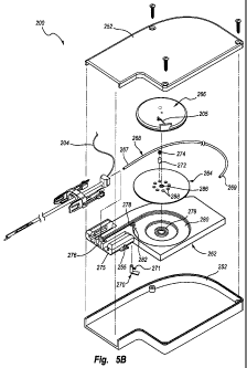
as shown in
23
CA 02790931 2012-08-23
WO 2012/023983 PCT/US2011/001451
FIGS. 5C-5D. .Further withdrawing the housing 252 causes the automatic driving
assembly
260 to slide forward in the housing 252 as shown in FIG. 5E-5F. Functionally,
the anchor
208, sealing plug 210, carrier tube 202, procedure sheath 216, and automatic
driving
assembly 260 maintain the same axial position upon this further withdrawal of
the housing
252, and the procedure sheath 216 and housing 252 move proximally (see FIGS.
5E-5F).
[0085] Referring to FIGS. 5E-5F, the distal end portion 207 of the carrier
tube
202 is exposed within the percutaneous incision 219 as the housing 252 and the
procedure
sheath 216 are retracted. The carrier tube 202 may retain its position
relative to the tissue
puncture 218 until the housing 252 and the procedure sheath 216 have been
retracted a
predetermined distance. Relative movement between the housing 252/procedure
sheath 216
and the carrier tube 202 may be facilitated by a sliding mount arrangement
between the
automatic driving assembly 260 and the housing 252. However, according to some
embodiments the automatic driving assembly 260 is fixed to the housing 252.
[0086] As shown by the combination of FIGS. 5C-5H, the automatic driving
assembly 260, which is attached to the carrier tube 202, may be free-floating
or displaceable
and slides relative to the housing 252 as the housing 252 and the procedure
sheath 216 are
retracted. However, the automatic driving assembly 260 may be initially held
in a first
position relative to the housing 252, as shown in FIG. 5C. For example, as
shown in FIG. 5C,
the tissue puncture closure device 200 may comprise a temporary holder such as
a stowage
detent 255 that releasably operates between the automatic driving assembly 260
and the
housing 252. The stowage detent 255 may include a finger 257 with a protrusion
to at least
temporarily hold the automatic driving assembly 260 in the first position
shown in FIG. 5C by
contact with a webbing structure within the housing 252. In other
arrangements, the stowage
detent 255 may be mounted to the housing 252 and releasable contact the
automatic driving
24
CA 02790931 2012-08-23
WO 2012/023983 PCT/US2011/001451
assembly 260. The stowage detent 255 may be positioned at any desired location
within the
housing 252. For example, the stowage detent 255 may connected to a bottom
surface of the
automatic driving assembly 260 and be operably positioned within a slot formed
in surface of
the housing 252. Further, at least one slot and follower member may be
positioned on the
automatic driving assembly 260 and housing 252 to assist in maintaining
relative axial
movement between the automatic driving assembly 260 and housing 252 after
release of the
stowage detent.
[0087] Although the finger 257 tends to hold or temporarily lock the automatic
driving assembly 260 in the first position shown in FIG. 5C, the finger 257
releases when a
sufficient predetermined force is applied between the housing 252 and the
automatic driving
assembly 260. For example, with the anchor 208 deployed, a retraction force
provided by a
user to the housing 252 causes the finger 257 to deflect inward and release.
Thereafter, the
finger 257 provides little resistance to sliding movement between the
automatic driving
assembly 260 and the housing 252. Accordingly, retraction of the housing 252
may retract
the procedure sheath 216, which is fixedly connected to the housing 252, but
the automatic
driving assembly 260 and the carrier tube 202 may slide relative to the
housing 252 and
therefore remain in position with respect to the tissue puncture 218 (see FIG.
5E). The
automatic driving assembly 260 may slide a predetermined distance with respect
to the
housing 252 until the automatic driving assembly 260 reaches a stop (e.g., a
distal internal
wall of the housing 252). The predetermined distance may be at least long
enough to expose
the slit 209 (see FIG. 5A) in the carrier tube 202 to facilitate later removal
of the sealing plug
210 from the carrier tube 202.
[0088] When the automatic driving assembly 260 reaches the stop, further
retraction of the housing 252 withdraws the carrier tube 202 as well, ejecting
the sealing plug
CA 02790931 2012-08-23
WO 2012/023983 PCT/US2011/001451
210 automatically. The spool assembly 266 begins to rotate to permit unwinding
of some of
the suture 204 from the spool. Typically, the driving plate 264, which rotates
with the spool
assembly 266, unwinds an amount to advance the coil 268 and compaction tube
212 and
compact the sealing plug 210, as shown in FIGS. 5G-5H. Still further
retraction of the
housing 252 further rotates the spool assembly 266 and driving plate 264 to
advance the coil
268 and compaction tube to complete compaction of the sealing plug 210.
[0089] Any further retraction of the housing 252 exceeds a threshold torsional
force between the driving plate 264 and spool assembly 266 that causes the
follower 272 to
move out of the follower recess 288 of the driving plate 264. The driving
plate 264 and spool
assembly 266 are then able to rotate relative to each other without further
compacting the
sealing plug 210. The interaction between the follower 272 and follower
recesses 288
provides a clutch function. The follower 272 and follower recesses 288 may be
referred to as
a clutch or clutch assembly of the automatic driving assembly.
[0090] Upon completion of compacting the sealing plug 210, the operator may
actuate the release member 270 to permit unwinding of the suture 204 from the
spool
assembly 266. The suture 204 may then be better exposed for cutting near the
tissue layer
230 to release the housing 252 from the anchor 208/sealing plug 210.
[0091] Unlike previous closure devices that require a separate, manual
compaction procedure following the deposition of the sealing plug 210, the
closure device
200 of the present disclosure automatically compacts the sealing plug 210 by
applying a
retracting force to the housing 252. The sealing plug 210 may be compacted
during or after
withdrawal of the carrier tube 202, reducing or eliminating any gaps that may
otherwise occur
between the sealing plug 210 and the tissue puncture 218 in the vessel 228.
26
CA 02790931 2012-08-23
WO 2012/023983 PCT/US2011/001451
[0092] .In addition, by placing tension on or pulling the suture 204 away from
the
percutaneous incision 219, the suture 204 may cinch and lock (with a slip-knot
or the like)
together the anchor 208 and the sealing plug 210, sandwiching the vessel wall
234 between
the anchor 208 and sealing plug 210. The force exerted by the compaction tube
212 and the
cinching together of the anchor 208 and sealing plug 210 by the suture 204
also causes the
sealing plug 210 to deform radially outward within the percutaneous incision
219 and
function as an anchor on the proximal side of the tissue puncture 218 as shown
in FIGS. 5G-
5H.
[0093] Many variations are possible for the features of tissue puncture
closure
device 200. In some arrangements, the coil 268 may be permanently connected to
the driving
plate 264. The driving plate 264 may be directly connected to the compaction
tube 212.
Generally, any device or construction that uses a disengagable cam structure
driven by
rotation of a spool member (about which the suture is wound) to advance a
compaction
member to compact a sealing plug falls within the spirit and scope of the
present disclosure.
[0094] Operation of the embodiment of FIGS. 5A-5H is as follows. As the
housing 252 of the closing device 200 is retracted from the percutaneous
incision 219, as
shown in FIG. 5C, the stowage detent 255 releases. The automatic driving
assembly 260 and
carrier tube 202 may remain stationary and therefore float relative to the
housing 252. The
procedure sheath 216 is retracted as the housing 252 is withdrawn, exposing
the distal end
portion 207 of the carrier tube 202. The automatic driving assembly 260
eventually contacts
a stop (or, in some embodiments, the automatic driving assembly is fixed), and
further
retraction causes the automatic driving assembly 260 and carrier tube 202 to
retract as well.
As the automatic driving assembly 260 retracts, the suture 204, which is
threaded through
27
CA 02790931 2012-08-23
WO 2012/023983 PCT/US2011/001451
the anchor 208, unwinds. from the spool assembly along a cam suture path and
causes
rotation of the spool assembly 266 and driving plate 264 with a variable
rotation force.
[0095] As the driving plate 264 rotates, the coil 268 is advanced to drive and
advance the compaction tube 212. In some arrangements, the coil 268 may be
long enough
and constructed such that the coil 268 functions as the compaction tube 212.
The compaction
tube 212 compacts the sealing plug 210. Therefore, as the closing device 200
is retracted
from the percutaneous incision 219, the procedure sheath 216 may be retracted
(see FIGS.
5E-5F), the carrier tube 202 may be retracted, and the sealing plug 210 is
automatically
compacted (see FIGS. 5G-5H). The sealing plug 210 is more likely to create a
sufficient
arterial seal without a gap relative to the anchor 208, as may otherwise occur
with a separate
manual compaction procedure.
[0096] Compaction of the sealing plug 210 may be confirmed by further
retraction
of the housing 252 until the follower 272 moves out of the follower recesses
288 to permit
relative rotation between the driving plate 264 and spool assembly 266 (also
referred to as a
clutch operation of the automatic driving assembly 260). This relative
rotation may be
signaled to the operator of the tissue puncture closure device 200 with a
tactile or audible
"click" or other indicator. The clutch action between the driving plate 264
and spool
assembly 266 may limit the possibility of over compaction of the sealing plug
210 into the
vessel 228.
[0097] When the sealing plug 210 has been sufficiently compacted, the
automatic
driving assembly 260 may be disengaged, enabling further retraction of the
closure device
200 without additional compaction. The automatic driving assembly 260 may be
advantageously disabled by activating the release member 270 out of contact
with the driving
plate 264. Activating the release member 270 allows the suture 204 to at least
partially
28
CA 02790931 2012-08-23
WO 2012/023983 PCT/US2011/001451
unwind from the spool assembly 266 without driving the compaction tube 212.
Unwinding
the spool assembly 266 exposes a sufficient length of the suture 204 distal of
the compaction
tube 212 to allow an operator to cut the suture 204 and separate the sealing
plug 210 and
anchor 208 from the remainder of the closure device 200.
[0098] In an alternative construction shown in FIG. 13, an automatic driving
assembly 360 includes a plurality of followers 372 carried by a spool assembly
366 that
interface with a plurality of follower recesses 388 defined in a driving plate
364. The
automatic driving assembly 360 includes a base 362 having a distal end 375, a
connector
recess 376, a coil recess 378, a mounting hub 379, a spool recess 380, and
first and second
release member apertures 381, 382. The operation and function of the base 362
may be the
same or similar to the base 262 described herein. The driving plate 364 may a
connector
aperture 386, a plurality of follower recesses 388, and other features that
are the same or
similar with the same or similar function as the driving plate 264 described
herein. The spool
assembly 366 may include a plurality of follower cavities 395 receptive of the
plurality of
followers 372 and biasing members 374. The spool assembly 366 may include
other features
that are the same or similar with the same or similar function as the spool
assembly 266
described herein.
[0099] The use of a plurality of followers 372 that operate independently to
move
into and out of corresponding follower recesses 388 of the driving plate 364
may provide
certain advantages. For example, but without limitation, the use of multiple
followers 372
may provide improved consistency in the threshold level of torsional force
required to move
the followers 372 out of the follower recesses 388 to permit relative rotation
between the
driving plate 364 and spool assembly 366. Further, the use of multiple
followers 372 may
provide improved safety and assurance of operability during use. The use of
multiple
29
CA 02790931 2012-08-23
WO 2012/023983 PCT/US2011/001451
followers 372 and/or multiple follower recesses 388 may provide additional
precision and
control of the amount of free rotation of the spool assembly 366 relative to
the driving plate
364 after one or more of the followers 372 move out of one or more of the
follower recesses
388.
[0100] The preceding description has been presented only to illustrate and
describe exemplary embodiments of the present disclosure. It is not intended
to be exhaustive
or to limit the invention to any precise form disclosed. Many modifications
and variations are
possible in light of the above teaching. It is intended that the scope of the
invention be
defined by the following claims.