Note: Descriptions are shown in the official language in which they were submitted.
CA 02791243 2012-10-02
MASK FOR USE IN RESPIRATORY MONITORING
FIELD OF THE DISCLOSURE
[0001] The present disclosure relates to a method and apparatus for the
analysis of breathing
cycles and the monitoring, identifying and/or. determining the inspiration
phase and expiration.
phase of breathing cycles.
BACKGROUND
[0002] Respiratory disorders are known to disturb sleep patterns. For example,
recurrent
apneas and hypopnea lead to intermittent hypoxia that provokes arousals and
fragmentation of
sleep, which in turn may lead to restless sleep, and excessive daytime
sleepiness. Repetitive
apnoeas and intermittent hypoxia may also elicit sympathetic nervous system
activation, oxidative
stress and elaboration of inflammatory mediators which may cause repetitive
surges in blood
pressure at night and increase the risk of developing daytime hypertension,
atherosclerosis, heart
failure, and stroke independently from other risks. There remains a need for
improved methods
for monitoring, identifying and/or determining breathing cycles, in order to
obviate these risks.
SUMMARY OF THE GENERAL INVENTIVE CONCEPT
[0003] In an exemplary embodiment, there is provided a mask to be worn by a
subject on its
face for use in respiratory monitoring, the mask comprising: at least one
transducer responsive to
sound and airflow for generating a data signal representative thereof, and a
support structure
shaped and configured to rest on the subject's face and thereby delineate a
nose and mouth area
thereof, and comprising two or more outwardly projecting limbs that, upon
positioning the mask,
converge into a transducer supporting portion for supporting said at least one
transducer at a
TR]-MP2/PCT-CDADIV
CA 02791243 2012-10-02
distance from said area, thereby allowing for monitoring via said at least one
transducer of both
sound and airflow produced by the subject while breathing.
BRIEF DESCRIPTION OF THE FIGURES
[0004] Several embodiments of the present disclosure will be provided, by way
of examples
only, with reference to the appended drawings, wherein:
[0005] Figure 1 is a plot of an exemplary microphone response curve of an
exemplary
embodiment;
[0006] Figure 2a is side view of an exemplary embodiment of a microphone and
transducer
set-up on an individual wherein the microphone is attached to a face mask
located on the front of
an individual's face;
[0007] Figure 2b is side view of an exemplary embodiment of a 2-microphone and
transducer
set-up on an individual wherein the microphones are attached to a face mask
located on the front
of an individual's face;
[0008] Figure 3 is a schematic computer system in accordance with an apparatus
for
transforming breathing sounds in inspiration and expiration phases;
[0009] Figure 4 is a block diagram of a computer system in accordance with the
apparatus of
figure 4;
[0010] Figure 5 is a digitized raw data wave plot representative of breathing
sound amplitude
versus time;
[0011] Figure 6a is an exemplary set-up of Respiratory Inductance
Plethysmogrphy (RIP) on
an individual and the microphone and transducer equipment of figures 2a and
2b;
2
TRI-MP2/PCT-CDADI V
CA 02791243 2012-10-02
[0012] Figure 6b is an exemplary plot of 25-second long recording of breathing
sounds and
simultaneous RIP signals from a representative individual wherein the dashed
line indicates the
separation of inspiration and expiration cycles;
[0013] Figure 7a is a representative digitized raw data breathing sound
amplitude versus time
plot of a single breathing cycle with the three phases of respiration;
[0014] Figure 7b is a representative frequency spectrum of the inspiration
phase of figure 7a;
[0015] Figure 7c is a representative frequency spectrum of the expiration
phase of figure 7a;
[0016] Figure 8a is a representative plot of the average frequency magnitude
spectrum and
standard deviations of breathing sounds for inspiration in an individual;
[0017] Figure 8b is a -representative plot of the average frequency magnitude
spectrum and
standard deviations of breathing sounds for expiration in an individual;
[0018] Figure 9 is a flow diagram of the method for monitoring, identifying
and determining
the breathing phases from breathing sound data;
[0019] Figure IOa is representative amplitude versus time plot of breathing
sound data and
simultaneous RIP data; and
[0020] Figure IOb is a comparative plot of the RIP data of figure I Oa and the
breathing phases
found using the method of figure 9 for monitoring, identifying and determining
breathing phases
wherein the positive values of the dashed line represent inspiration and the
negative values of the
dashed line represent expiration.
DESCRIPTION OF THE PREFERRED EMBODIMENTS
[0021] It should be understood that the disclosure is not limited in its
application to the details
of construction and the arrangement of components set forth in the following
description or
3
TRI-MP2/PCT-CDADIV
CA 02791243 2012-10-02
illustrated in the drawings. The disclosure is capable of other embodiments
and of being practiced
or of being carried out in various ways. Also, it is to be understood that the
phraseology and
terminology used herein is for the purpose of description and should not be
regarded as limiting.
The use of "including," "comprising," or "having" and variations thereof
herein is meant to
encompass the items listed thereafter and equivalents thereof as well as
additional items. Unless
limited otherwise, the terms "connected," "coupled," and "mounted," and
variations thereof herein
are used broadly and encompass direct and indirect connections, couplings, and
mountings. In
addition, the terms "connected" and "coupled" and variations thereof are not
restricted to physical
or mechanical or electrical connections or couplings. Furthermore, and as
described in subsequent
paragraphs, the specific mechanical or electrical configurations illustrated
in the drawings are
intended to exemplify embodiments of the disclosure. However, other
alternative mechanical or
electrical configurations are possible which are considered to be within the
teachings of the instant
disclosure. Furthermore, unless otherwise indicated, the term "or" is to be
considered inclusive.
[0022] With reference to the disclosure herein and the appended figures, a
method for
monitoring, identifying and/or determining characteristics of an individual's
breathing, including
breathing phases thereof, is henceforth described using a processed acoustic
signal data stream
collected and/or recorded waveform data. In one example, the waveform data is
collected from or
is associated with breathing sounds and other sounds from one or more
microphones or other
sound wave collecting equivalents thereof.
[0023] In this case, the system and method may involve the use of a control
unit, in which
some or all of its associated components are computer implemented that may be
provided in a
number of forms. They may be embodied in a .software program configured to run
on one or more
4
TRl-MP2/PCT-CDADIV
CA 02791243 2012-10-02
general purpose computers, such as a personal computer, or on a single custom
built computer,
such as a programmed logic controller (PLC) which is dedicated to the function
of the system
alone. The system may, alternatively, be executed on a more substantial
computer mainframe.
The general purpose computer may work within a network involving several
general purpose
computers, for example those sold under the trade names APPLE or IBM, or
clones thereof, which
are programmed with operating systems known by the trade names WINDOWSTM,
LINUXTM,
MAC O/STM or other well known or lesser known equivalents of these. The system
may involve
pre-programmed software using a number of possible languages or a custom
designed version of a
programming software sold under the trade name ACCESS or other programming
software. The
computer network may be a wired local area network, or a wide area network
such as the Internet,
or a combination of the two, with or without added security, authentication
protocols, or under
"peer-to-peer" or "client-server" or other networking architectures. The
network may also be a
wireless network or a combination of wired and wireless networks. The wireless
network may
operate under frequencies such as those dubbed `radio frequency' or "RF" using
protocols such as
the 802.11, TCP/IP, BLUE TOOTH and the like, or other well known Internet,
wireless, satellite
or cell packet protocols. Also, the present method may also be implemented
using a
microprocessor-based, battery powered device.
100241 FIG. 3 shows a general computer system on which embodiments may be
practiced. The
general computer system comprises information relay module (1.1). In some
embodiments, the
information relay module (1.1) comprises a means for providing audible cues,
such as speakers.
In some embodiments, the information relay module is comprised of a display
device or module
(1.1). with a display screen (1.2). Examples of display device are Cathode Ray
Tube (CRT)
TRI-MP2/PCT-CDAD1V
CA 02791243 2012-10-02
devices, Liquid Crystal Display (LCD) Devices etc. The general computer system
can also have
other additional output devices like a printer. The cabinet (1.3) houses the
additional basic
components of the general computer system such as the microprocessor, memory
and disk drives.
In a general computer system the microprocessor is any commercially available
processor of
which x86 processors from Intel and 680X0 series from Motorola are examples.
Many other
microprocessors are available. The general computer system could be a single
processor system or
may use two or more processors on a single system or over a network. The
microprocessor for its
functioning uses a volatile memory that is a random access memory such as
dynamic random
access memory (DRAM) or static memory (SRAM). The disk drives are the
permanent storage
medium used by the general computer system. This permanent storage could be a
magnetic disk, a
flash memory and a tape. This storage could be removable like a floppy disk or
permanent such as
a hard disk. Besides this the cabinet (1.3) can also house other additional
components like a
Compact Disc Read Only Memory (CD-ROM) drive, sound card, video card etc. The
general
computer system also includes various input devices such as, for example, a
keyboard (1.4) and a
mouse (1.5). The keyboard and the mouse are connected to the general computer
system through
wired or wireless links. The mouse (1.5) could be a two-button mouse, three-
button mouse or a
scroll mouse. Besides the said input devices there could be other input
devices like a light pen, a
track ball, etc. The microprocessor executes a program called the operating
system for the basic
functioning of the general computer system. The examples of operating systems
are UNIXTM,
WINDOWSTM and OS XTM. These operating systems allocate the computer system
resources to
various programs and help the users to interact with the system. It should be
understood that the
6
TRI-MP2/PCT-CDADIV
CA 02791243 2012-10-02
disclosure is not limited to any particular hardware comprising the computer
system or the
software running on it.
[00251 FIG. 4 shows the internal structure of the general computer system of
FIG. 3. The
general computer system (2.1) includes various subsystems interconnected with
the help of a
system bus (2.2). The microprocessor (2.3) communicates and controls the
functioning of other
subsystems. Memory (2.4) helps the microprocessor in its functioning by
storing instructions and
data during its execution. Fixed Drive (2.5) is used to hold the data and
instructions permanent in
nature like the operating system and other programs. Display adapter (2.6) is
used as an interface
between the system bus and the display device (2.7), which is generally a
monitor. The network
interface (2.8) is used to connect the computer with other computers on a
network through wired
or wireless means. The system is connected to various input devices like
keyboard (2.10) and
= mouse (2.11) and output devices like a printer (2.12) or speakers. Various
configurations of these
subsystems are possible. It should also be noted that a system implementing
exemplary
embodiments may use less or more number of the subsystems than described
above. The computer
screen which displays the recommendation results can also be a separate
computer system than
that which contains components such as database 360 and the other modules
described above.
[00261 The method, in accordance with the instant disclosure, provides a
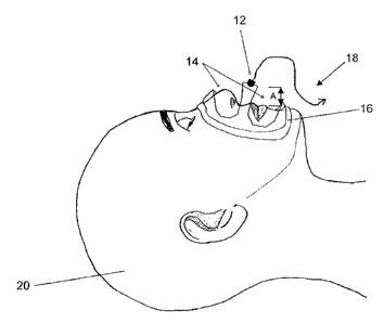
microphone 12
located in a position proximal to an individual's mouth as shown in FIGS. 2a
and 2b, in this case
by a dimension A of approximately 3 cm in front of the individual's face. The
microphone 12
may be configured to communicate with the microprocessor by way of an
interface or other data
acquisition system, via a signal transducing link or data path 18 to provide
one or more data
collection modules with the microphone 12. Thus, such data collection modules
and the
7
TRI-MP2fPCT-CDAD1V
CA 02791243 2012-10-02
microphone are operable to collect breathing sounds emanating from the
individual's mouth and
nose, during the inspiration and/or expiration phases of breathing. For
example, an exemplary
microphone response curve is shown in FIG. 1. The acoustic signal data
breathing sounds
collected from the individual may be comprised of both airflow sounds from the
individual's
breathing applying air pressure to the microphone diaphragm and actual
breathing sounds resultant
from the individual's breathing being recorded and/or collected by the
microphone 12.
Furthermore, the acoustic signal data breathing sounds collected from the
individual may be, in
another exemplary embodiment, comprised of substantially only actual sounds
resultant from the
individual's breathing being recorded and/or collected by the microphone 12.
In still yet another
embodiment, the acoustic signal data breathing sounds collected from the
individual may be
comprised of substantially only airflow sounds resultant from the individual's
breathing applying
air pressure to the microphone diaphragm and being recorded and/or collected
by the microphone
12. As used hereinafter, term "airflow sounds" refers to the air pressure
resultant from an
individual's breathing being applied to and causing the microphone's diaphragm
to move such
that the microphone collects and produces data for the audio recording.
10027] The microphone 12, for example, may be coupled in or to a loose fitting
full face mask
16 as shown in FIGS. 2a and 2b. Furthermore, the face mask 16 may include at
least one opening
14 to allow for ease of breathing of an individual 20. For example, the
microphone 12 may be in a
fixed location with a spacing of dimension "A", of about 3 cm in front of the
individual's face as
shown schematically in FIG. 2a; however other distances in front of the
individual's face may be
desirable in some embodiments. The microphone 12, in this case, is embedded in
a respiratory
mask 16 which is modified by cutting away material so as produce opening 14
such that only a
8
TR -MP2/PCT-CDADIV
CA 02791243 2012-10-02
structural frame portion remains to keep the microphone 12 in a fixed location
relative the nostrils
and the mouth of an individual 20. In one example, the audio signals from the
microphone may
be digitized using an audio signal digitizing module and digitized sound data
to be transferred via
transducing link 18 to the computer using a USB preamplifier and audio
interface (M-Audio,
Model Fast Track Pro USB) with a sampling rate of 22,050 Hz and resolution of
16 bits. Although
various types of audio interfaces may be used, in the instant exemplary
embodiment, an external
audio interface provides suitable results over the other types of audio
adapters, for example, built-
in audio adapters due to the superior signal to noise (S/N) ratio of the
external adaptor which is
about 60 dB at I kHz. Sound recordings may then be passed through a 4th order
band-stop digital
filter with a centre frequency of about 60 Hz to suppress line interference.
Other structures may
also be used to locate the microphone in position, as including support
structures positioned
against a plurality of locations on the individual or stationed adjacent the
individual as required.
[00281 Furthermore, in another exemplary embodiment, a two microphone system
may be
useful. In such a system, as shown in.FIG 2b, one of the microphones, a first
microphone 12b,
may be configured to collect actual breathing sounds and airflow sounds
whereas the other
microphone, a second microphone 12c may be configured to collect substantially
only actual
breathing sounds. In this embodiment, the waveform sounds and/or data
collected from the
second microphone 12c may be subtracted or filtered from the waveform sounds
collected from
the first microphone 12b, thereby resulting in a waveform data stream of
substantially only airflow
sounds. The airflow sounds may be resultant of pressure air from an
individual's breathing being
collected as applied to the diaphragm of a microphone as noted above.
Subsequently, the airflow
9
TR1-MP2/PCT-CDADIV
CA 02791243 2012-10-02
sounds may then be used as a waveform amplitude acoustic data stream in
accordance with the
forgoing method.
[0029] A raw acoustic data stream of breathing sounds, as shown in a
representative plot, for
example in FIG. 5, is then collected for each of a plurality of respiratory
phases to form a
bioacoustics signal recording, wherein the acoustic data stream is
subsequently transformed.
[0030] As will be described below, in at least one embodiment, a method and an
apparatus are
provided to monitor, identify and determine the inspiratory and/or expiratory
phases of the
respiratory cycle of an individual 20 from the frequency characteristics
breathing sounds. It is
understood that a numerical comparative analysis of the frequency spectrum as
transformed from
waveform amplitude data of breathing sounds and/or airflow sounds of an
individual 20 may be
useful to differentiate between the inspiration and expiration phases of
breathing.
DATA ACQUISITION
[0031] Data were collected from 10 consecutive men and women at least 18 years
of age
referred for overnight polysomnography (PSG). The subjects' characteristics
are shown in Table
1. Breath sounds were recorded by a cardoid condenser microphone (Audi-
Technica condenser
microphone, Model PRO 35x). The microphone's cardioid polar pattern reduces
pickup of sounds
from the sides and rear, improving isolation of the sound source. The
microphone 12 used for
recording breath sounds has a relatively flat frequency response up to 2000 Hz
as shown in FIG. 1.
Furthermore, the microphone 12, as used herein has a higher output when sound
is perpendicular
to the microphone's diaphragm as shown by the solid line in FIG. 1, which
helps reduce low
frequency ambient noise interference. The microphone 12 was embedded in the
centre of a loose
fitting full face mask 16 modified to reduce airflow resistance and eliminate
dead space by way of
TRI-MP2/PCT-CDADIV
CA 02791243 2012-10-02
large openings 14 as shown in FIGS. 2a and 2b. The microphone 12 attached to
the face mask 16,
and is located in front of the individual's face. The mask 16 provides a
structural frame portion to
keep the microphone in a fixed location, at a dimension A of approximately 3
cm in front of the
individual's face, so as to record breathing sounds to an audio recording
device, such as a
computer as described above, to make an audio recording. thereof. In some
exemplary
embodiments, the audio recording of breathing. sounds may be made and recorded
in analog
format prior to digitizing the audio recording. However, in other embodiments
the audio
recording of breathing sounds may be digitized in real-time. Furthermore, in
some exemplary
embodiments, the processing of the audibly recorded waveform data or acoustic
signal data may
be performed in real-time, so as to provide substantially instantaneous
information regarding an
individual's breathing. In an exemplary embodiment, digitized sound data were
transferred to a
computer using a USB preamplifier and audio interface (M-Audio, Model
MobilePre USB) with a
sampling Tate of 22,050 Hz and resolution of 16 bits. Although various types
of audio interfaces
may be used, in the instant exemplary embodiment, an external audio interface
was preferred over
a built-in audio adapter due to the better signal to noise (SIN) ratio of the
external audio interface,
which was 91 dB. FIG. 5 shows a 25-second waveform amplitude recording plot.
However, in
other exemplary embodiments, it may be desirable to record breathing sounds
for a time period of
from about 10 seconds to 8 hours. In some exemplary embodiments it may be
desirable to record
breathing sounds for a time period of from about 10 second to about 20
minutes. In other
exemplary embodiments, it may be desirable to record breathing sounds for
greater than 20
minutes.
BREATHING ACOUSTICS ANALYSIS
II
TRI-MP21PCT-CDADIV
CA 02791243 2012-10-02
[0032] In an exemplary embodiment, full night breath sound recordings were
displayed on a
computer screen similar to the computer screen 1.2 of FIG. 3. A representative
raw acoustic data
waveform plot, as may be shown on a computer screen 1.2, is provided in FIG. 5
for a 25-second
recording. Each increase in amplitude represents a single breath. The
individual phases of a
breathing cycle are not readily resolvable in FIG. 5 owing to the time scale
being too large to
resolve single breath details. For example, FIG. 7a more clearly shows the
inspiration and
expiration phases of a breathing cycle in a waveform amplitude versus time
plot. The recordings
were visually scanned to identify periods of regular breathing. After visual
scanning, the
recordings were played back for auditory analysis.
[0033] Sequences of normal breaths that did not have signs of obstructive
breathing such as
snoring and interruptions, or other irregularities such as tachypnea (rapid
breathing), or
hyperventilation (deep breathing) were then included in the subsequent
frequency analysis. This
process was repeated to select three random parts of an individual's sleep. If
a portion of the
recording fulfilled the aforementioned inclusion criteria, then 3 to 4
consecutive breaths were
selected from that portion. A total of 10 breaths were selected from each
individual. During the
process of selecting the individual's breathing sound portions, the
investigator did not have a
previous knowledge of the sleep stage. Therefore, the investigator was blind
to the sleep stage of
an individual while selecting the analyzed breaths except for knowing that
sampling started after
the onset of sleep. The real-time stamp of each breath was registered in order
to retrieve the sleep
stage in which it took place in afterwards. Subsequently, the investigator
listened to these
breathing sounds again to divide each breath into its inspiratory, expiratory
and interbreath phases.
Each phase was labeled manually.
12
TRJ-MP2/PCT-CDADI V
CA 02791243 2012-10-02
[0034] The data array of each breathing phase was passed through a hamming
window and a
2048-point Fast Fourier Transform (FFT) of the windowed data with 50% overlap
was calculated.
The resultant frequency spectrum was displayed on a computer screen for visual
analysis. The
frequency spectra of the interbreath pauses were also calculated and
incorporated in the analysis to
control for the effect of ambient noise. Careful visual examination of spectra
revealed that during
inspiration, the amplitude of signals above 400 Hz was consistently higher
than during expiration.
Therefore, it was determined that the bands ratio (BR) of frequency magnitude
between 400 to
ki 1000 Hz, to frequency magnitude between 10 to 400 Hz is higher in the
inspiration phase as
compared to the expiration phase. The BR of each breathing cycle was then
calculated using
equation (1).
1000Ha 400x=
BR= FFT(f)l Y FFT(f) (1)
400Hz 10 Hz
[0035] Using equation (1), the numerator represents the sum of FFT higher
frequency
magnitude bins which lie between 400 and 1000 Hz, and the denominator
represents the sum of
FFT lower frequency magnitude bins which lie between 10 and 400 Hz. Bins
bellow 10 Hz were
not included to avoid any DC contamination (referring to drift from a base
line), and frequencies
above 1000 Hz were not included since preliminary work (not shown) revealed
insignificant
spectral power at frequencies above 1000 Hz. Therefore, the computation may
also be reduced.
To verify repeatability of the results, BR was calculated for 3 to 4
successive breaths in the
included sequence and for a total of three sequences from different parts of
the individual's sleep.
A total of 100 breaths were collected from the 10 subjects. The mean number of
breaths per
subject was 10 0.
13
TRI-MP2IPCT-CDADIV
CA 02791243 2012-10-02
SLEEP STAGING
[0036] Sleep stages were recorded during the course of the night using
standard
polysomnographic techniques that included electro-encephalography (EEG),
electro-oculography
and submental electro-myography (Rechtschaffen A and Kales A 1968,4 Manual of
Standardized
Terminology, Techniques and Scoring System for Sleep Stages of Human Subjects.
(Los Angeles:
UCLA Brain Information Service/Brain Research Institute). The corresponding
sleep stage for the
selected breath samples was determined from the PSG recording (not shown).
STATISTICAL ANALYSIS
[0037] Data are expressed as mean SD unless otherwise stated. A Wilcoxon
Signed Ranks
Test was performed using SPSS statistical package (SPSS, Chicago, Illinois).
This test compares
two related variables drawn from non-normally distributed populations. One-
sample sing test was
performed using Minitab 15 statistical package (Minitab Inc., State College,
PA).
COMPARISION OF BANDS RATIO TO RESPIRATORY INDUCTANCE
PLETHYSMOGRAPHY
SUBJECTS
[00381 Healthy subjects at least 18 years of age were recruited with no
history of respiratory
or cardiopulmonary disease in addition to being free from prescribed
medications. Data were
collected from 15 subjects, 6 men and 9 women, healthy volunteers. Individuals
used in the study
were recruited by advertisement and were divided randomly intro 2 groups with
5 subjects in one
group (test group) and 10 in the other (validation group). The data from the 5
subjects in the test
group were used to examine acoustic characteristics of breathing phases, which
were then
incorporated into a method having an algorithm as described below. The
resultant method _ was
14
TRI-MP2/PCT-CDADI V
CA 02791243 2012-10-02
tested on the data of 10 subjects in the validation group to determine the
validity of the method for
determining the inspiration and expiration phases of an individual's breathing
sounds.
BREATH SOUND RECORDING
[0039] Breath sounds were recorded using a unidirectional, electret condenser
microphone
(Knowles Acoustics, Model MB6052USZ-2). The microphone's unidirectional
pattern reduces the
pickup of sounds from the sides and rear thereby improving isolation of the
sound source. The
microphone 12 was embedded in a respiratory mask 16 that was modified by
cutting away
material so as to produce' opening 14 such that only a structural frame
remained to keep the
microphone 12 in a fixed location relative the nostrils and the mouth of an
individual 20 at a
dimension "A" of approximately 3 cm in front of the individual's face as shown
in FIG. 2a. The
audio signal was digitized using an audio signal digitizing module and
digitized sound data were
transferred via transducing link 18 to a computer using a USB preamplifier and
audio interface
(M-Audio, Model Fast Track Pro USB) with a sampling rate of 22,050 Hz and
resolution of 16
bits. Although various types of audio interfaces may be used, in the instant
exemplary
embodiment, an external audio interface was preferred over the other types of
audio adapters, for
example, built-in audio adapters due to the superior signal to noise (S/N)
ratio of the external
adaptor which was about 60 dB at I kHz. Sound recordings were then passed
through a 4th order
band-stop digital filter with a centre frequency of about 60 Hz to suppress
line interference.
RESPIRATORY INDUCTANCE PLETHYSMOGRAPHY
[0040] Respiratory inductance plethysmography (RIP), (Respitrace Ambulatory
Monitoring
Inc., White Plains, NY, USA) was used to monitor respiratory pattern of
individuals and the
timing of the breathing phases. In contrast to other breathing monitoring
apparatus such as
TR1-MP2/PCT-CDAD! V
CA 02791243 2012-10-02
pneumotacography, RIP has the advantage of being applied away from the face of
an individual to
allow capture of breathing phases. Briefly, RIP is a system comprising two
flexible sinusoidal
wires. Each wire is embedded in stretchy fabric band. One band 28 is placed
around the chest of
an individual and the other band 30 is placed around the abdomen of the
individual as shown in
FIG. 6a. The inductance of each band changes upon rib cage and abdomen
displacements and
generates a voltage signal proportional to its inductance. The signals from
the RIP bands 28 and
30 were digitized at 150 Hz and stored in a computer memory as substantially
describe above with
reference to FIGS. 3 and 4. The electrical sum of the ribcage and abdominal
signals is displayed
on a readable medium, for example a computer screen or a physical plot, and
provides the total
thoracoabdominal displacement. The thoracoabdominal displacement recorded from
the RIP
system reflects changes of tidal volume during respiration.
:100411 In order to compare the inspiration and expiration phases of an
individual's breathing
to RIP, the microphone 12, as noted above, was coupled to a modified mask 16
in front of the.
subject's face. Simultaneously, the RIP bands 28 and 30 were placed around the
subject's chest
and abdomen to measure thoracoabdominal motion as noted above. Recording were
captured
from both the microphone 12 and the RIP bands 28 and 30 simultaneously to
assess the timing of
breath sounds against the RIP waveform data.
STUDY PROTOCOL
[00421 Individuals were studied in the supine position and were instructed to
breathe
normally. Microphone holding frame 16 was placed on individual's face. Each
individual was
asked to breath for two minutes at their regular breathing rate. In order to
mimic all possible
breathing conditions, the individuals were asked to breath through their nose
only for half of the
16
TR1-MP2/PCT-CDADI V
CA 02791243 2012-10-02
experiment time, and through their nose while mouth was slightly open in the
other half.
Incomplete breaths at the beginning and end of recording were discarded and
all the breaths in
between were included in the analysis.
ANALYSIS OF BREATH ACOUSTICS
[0043] In a first stage, spectral variables of breath sounds that characterize
the inspiratory and
expiratory phase components of a respiratory cycle were determined. The data
of five subjects, 3
females and 2 males was chosen randomly from total 15 subjects and used to
study the frequency
characteristics of the acoustic signals of different respiratory phases.
Inspiratory and expiratory
segments of breath sounds were determined and extracted from the acoustic data
by comparing it
to the inspiratory (rising edge) and expiratory (falling edge) of the RIP
trace as shown in FIG. 6b.
A 25-second long recording of breath sounds and simultaneous summed
thoracoabdominal RIP
.signals from a representative subject is shown, for example, in FIG. 6b.
Dashed vertical lines are
-shown to separate inspiration and expiration phases of the second cycle at
32.
[0044] The first 10 complete breaths of each subject were analyzed, which
yielded a total of
50 inspirations and 50 expirations acoustic data sets from the 5 subjects.
Subsequently, the
frequency spectrum of each phase was calculated separately using Welch's
method (i.e. the
average of a 2048-point Fast Fourier Transform (FFT) of sliding hamming
windows with 50%
overlap). FFT arrays were normalized in amplitude in order to compare the
relative changes in
power spectrum among resultant spectral arrays.
[00451 Using variables derived from frequency spectra of the 5 test
individual's noted above,
the inspiratory and expiratory phases of the breathing cycle were determined
for the remaining 10
individuals in order to test the validity of the method. Furthermore, the
method was tested for the
17
TR[-MP2/PCT-CDAD[V
CA 02791243 2012-10-02
ability to determine breathing phases from acoustic data independently from
other inputs. The data
analysis was performed with Matlab R2007b software package (Mathworks, Natick,
Massachusetts).
RESULTS
[00461 The characteristics of the individuals in this study are shown in Table
1. A total of 100
breaths were sampled from 10 patients with a mean number of 10 breaths per
subject. Seventy
percent of the breaths analyzed were from non-rapid-eye movement sleep (NREM),
and 18% from
rapid eye movement sleep (REM), and 12% while patients were awake according to
the
polysomnographic criteria.
Table 1. Characteristics of subjects.
Subject Age (years) Sex Body Mass Index
Subject 1 51 F 39.1
Subject 2 43 M . 25.6
Subject 3 49 M 23.7
Subject 4 27 M 36.8
Subject 5 64 M 26.3
Subject 6 60 M 33.0
Subject 7 68 F 28.5
Subject 8 31 M 30.3
Subject 9 48 F 31.6
Subject 10 56 M 26.7
18
TRI-MP2/PCT-CDADIV
CA 02791243 2012-10-02
[00471 The bands ratio (BR) value was calculated for the inspiration phase
bands ratio (BRi)
24, the expiration phase bands ratio (BRe) 26, and the interbreath pause bands
ratio (BRp) 22
using equation 1. Inspiration and expiration showed consistent patterns of
their frequency spectra
as depicted in FIG. 7a for a given breathing cycle:
[00481 As shown in a representative example in FIG. 7b, there was a sharp
narrow band of
harmonics usually below 200Hz for inspiration. The spectrum exhibited a valley
between 200Hz
and 400Hz and a peak again after 400Hz as shown in FIG. 7b. Another variation
of the
inspiratory. spectrum was the same initial narrow band followed by a
relatively smooth spectrum
without the 400 Hz drop (not shown). The expiratory spectrum, as shown in a
representative
example in FIG. 7c, on the other hand, formed a wider band that spanned
frequencies up to 500Hz
and whose power dropped off rapidly above this frequency. The inspiratory
spectrum (FIG. 7b)
showed a peak close to the line frequency. The spectrum of the interbreath
pause (not shown) was
inconsistent and showed random variations without any consistent pattern. To
rule out the effect
of line frequency on inspiration bands ratio (BRi), a Wilcoxon signed rank
test was used to test the
relation between BRi and bands ratio interbreath pause (BRp). The test was
significant (p<0.001),
thus it was determined that BRi is different from BRp and that line
interference does not
significantly contribute to the frequency spectrum of inspiration.
[00491 The relationship between BRi and BRe was examined using the Wilcoxon
Signed
Ranks. Test. The test showed that a BRi is not equal to BRe (P<0.001) with 95%
of breathes
having BRi greater than BRe. Since minute differences between BRi and BRe
might be attributed
to randomness, two thresholds of 50% and 100% difference between BRi and BRe
were tested.
The ratio BRi/BRe was calculated for each breath. By taking the ratio, BRi and
BRe may be
19
TRI-MP2/PCT-CDADIV
CA 02791243 2012-10-02
treated as dependant pairs. These ratios were then tested for being greater
than 1.5 (50%
difference) and greater than 2 (100% difference). The one-sample sign test
showed that BRi/BRe
is greater than 1.5 (p<0.001) and greater than 2 (p<0.001). In order to
account for potential
'differences between subjects in the analysis, the mean BRi/BRe was calculated
for each individual
subject as displayed in Table 2. The one-sample sign test of the median was
significant for mean
BRi/BRe greater than 1.5 (p=0.001) and significant for mean BRi/BRe greater
than 2 (p=0.001).
Breaths that were drawn when subjects were polysomnographically awake did not
differ
significantly in terms of BRi/BRe from the rest of breaths (p=0.958) and,
therefore, were included
in the aforementioned analysis.
Table 2. Mean BRi/BRe for the subjects.
Mean BRi/BRe
Subject
(value SD)
Subject 1 1.66 0.60
Subject 2 2.30 1.33
Subject 3 2.43 0.71
Subject 4 3.17+1.17
Subject 5 2.67 1.60
Subject 6 3.86 2.65
Subject 7 23.01 9.65
Subject 8 14.99 8.86
Subject 9. 15.66 9.42
Subject 10 11.56 2.60
TRI-MP2/PCT-CDADI V
CA 02791243 2012-10-02
[00501 The sensitivity of this method was tested for each of the two cut-offs.
Out of 100
breath samples, 90 had BRi 50% greater than BRe, and 72 had BRi 100% greater
than BRe
thereby giving an overall sensitivity of 90% and 72% respectively.
10051] A total of 346 breaths met the inclusion criteria. The average number
of breaths per
individual was 23.0 7.79. Only the first 10 complete breaths were used to
study the spectral
frequency characteristics from the 5 individuals in the test group. From the
validation group 218
breaths (i.e. 436 phases) were included in the analysis with an average of
21.8 8.2 breaths per
subject.
ANALYSIS OF BREATH SOUNDS
[0052] Data obtained from the test group of 5 individuals yielded 100 arrays.
of FFT
magnitude bins normalized in amplitude with one half being from inspiratory
acoustic inputs or
phases and the other half from expiratory acoustic inputs or phases. The
average spectrum of all
normalized arrays belonging to the inspiration and expiration phases with the
corresponding
standard deviation are shown in FIGS. 8a and 8b respectively. FIGS. 8a and 8b
demonstrate that
the frequency spectra of the 2 phases have different energy distributions. The
mean inspiratory
spectrum, shown in FIG. 8a peaked between 30 Hz and 270 Hz. The spectrum
exhibited flatness
between 300 Hz and 1100 Hz before the next major peak with a center frequency
of 1400 Hz_ The
expiratory spectrum, on the other hand, peaked between 30 to 180 Hz as shown
in FIG. 8b. Its
power dropped off exponentially until 500 Hz after which it flattened at low
power.
[00531 The signal power above 500 Hz was consistently higher in inspiration
than expiration.
Since the ratio of frequency magnitudes between 500 to 2500 Hz, the higher
frequency magnitude
bins, to frequency magnitude between 0 to 500 Hz, the lower frequency
magnitude bins, is higher
21
TR[-MP2/PCT-CDAD( V
CA 02791243 2012-10-02
during the inspiration phase than during the expiration phase for each
breathing cycle, frequency
ratio can be used to differentiate the two phases of the breathing cycle. This
ratio is presented in
equation (2) as the frequency bands ratio (BR).
2500Hz 500Hz
BR Y FFT(f)l Y. FFT(f) (2)
500Hz 0Hz
[0054] The numerator of equation (2) represents the sum of FFT higher
magnitude bins
between 500 to 2500 Hz, and the denominator represents the sum of FFT lower
magnitude bins
below 500 Hz. BR was calculated for each of the six curves shown in FIGS. 8a
and 8b which
include the curve of the mean and the positive and negative standards
deviation for both
inspiration and expiration. These results are presented in Table 3:
Table 3. BR calculated for inspiration and expiration spectra.
Inspiration BR Expiration BR
Mean inspiration spectrum 2.27 Mean expiration spectrum 0.15
Mean inspiration spectrum+ 2.34 Mean expiration spectrum + 0.21
Std Std
Mean inspiration spectrum - 2.14 Mean expiration spectrum - 0.02
Std Std
[00551 The numbers in Table 3 represent the BR which is a ratio calculated
from various
curves.
[00561 Table 3 shows that the mean BR for inspiration (BRi) is 15.1 times
higher than mean
BR for expiration (BRe). BRi is higher than that for BRe. For example, by
comparing the two
extremes, `BR for mean inspiration - Std', and `BR for mean expiration + Std',
as noted in Table 3
and shown in FIGS. 8a and 8b, BRi may be 10.2 time greater than that for BRe.
However, other
22
TRI-MP2/PCT-CDADI V
CA 02791243 2012-10-02
predetermined multipliers may be acceptable for determining the inspiration
and expiration phases
of breathing. For example, the multiplier maybe from about 1 to about to about
20. Therefore, the
frequency-based variable BR may be used to distinguish the various phases of a
given breathing
cycle.
[00571 In order to validate the results of the procedure as found using the
test group, the BR
parameters as determined above were utilized to track the breathing phases in
the individuals in
the validation group.. A method that depends on past readings of acoustic data
was developed to
predict the current phase. A flow diagram of this method is shown
schematically in FIG. 9. For
example, a benefit of using past values rather than post-processed statistics
is that the technique
can be adopted for real-time implementation. According to this exemplary
embodiment, the
acoustic data stream is segmented into 200 *ms segments. However, it may be
desirable for the
segments to be of a length greater than or less 200 ms. For example the
segments may be from
about 50 ins to about I second. Preferably, the segments are from about 100 ms
to about 300 ms.
Each segment is then treated as described above in relation to the test group.
For example,
Welch's method was applied to calculate frequency spectrum and it's BR, a
first bands ratio (first
BR). Subsequently the mean BR of the past 1.4 seconds (7 segments x 200 ms) or
the mean of all
the past BR's, whichever is greater, was calculated. Each newly found BR, said
first BR, was then
compared with the past BR average or mean bands ratio. If the first BR is
greater than the mean
BR by at least a predetermined multiplier, then it is labeled as. inspiration.
The predetermined
multiplier may be from about 1.1 to about 10. Preferably the multiplier is
from about I to about 5.
Most preferably, the multiplier is from about 1.5 to 2. For example, if the
first BR is twice the
past 1.4 seconds BR average (mean BR) then it is labeled as inspiration.
Likewise, if the first BR
23
TRI-MP2/PCT-CDADI V
CA 02791243 2012-10-02
is less than mean BR by at least a predetermined multiplier, then it is
labeled as expiration.
Therefore, for example, a segment is labeled as expiration if the
corresponding BR is 2 times
below the average of the past two segments. FIG. 10a shows an exemplary
representative plot of
an embodiment of all BR values calculated from the acoustic data with the
corresponding RIP for
comparison. Visual examination shows that there is a correlation between BR
waveform and its
RIP counterpart. Averaging of the BR's is performed in order to smooth out
intra-phase
oscillations in BR such as in the case of the BR curve at time 5-10 seconds
seen in FIG. 10a
[00581 The method was tested prospectively on the breathing acoustic data of
10 subjects in
the validation group. The breathing phases found using the presently described
method as applied
to the data of FIG. 10a are shown in FIG. 10b. With reference to FIG. 10b, the
dashed line
represents the respiratory or breathing phases found utilizing the currently
described method. Out
of 436 breathing phases, 425 breathing phases were labeled correctly, 8 phases
were partially
detected, and 3 phases were labeled as being the opposite phases. Therefore,
utilizing the method,
about 97.4% of the breathing phases were detected correctly using acoustic
data as compared with
RIP trace.
100591 With reference to FIG. 10b, the breathing cycles are shown as a
processed wave
amplitude versus time plot. The processed wave amplitude data are shown by the
dashed line and
indicate the respiration phase of an individual's breathing. In an exemplary
embodiment, the
processed wave amplitude versus time plot may be displayed on a display module
such as that
shown in FIG. 3 at 1.1. The processed wave amplitude versus time plot may also
be, in some
exemplary embodiments, provided to an operator by way of an information relay
or relaying
module in a printed form or other suitable form, for example audio cues, such
that the breathing of
24
TRI-MP2/PCT-CDArI V
CA 02791243 2012-10-02
an individual may be monitored in accordance with the method by an operator.
In some
exemplary embodiments, the information relay module may display or provide the
processed data
in terms or inspiration and/or expiration indicia.
[00601 The frequency spectrum of inspiration may be characterized by a narrow
band below
200 Hz, a trough starting from about 400 Hz to about 600 Hz. In the exemplary
embodiments
noted herein, the trough begins at about 400 Hz in one, the first, embodiment
(FIG. 7b) and at
about 500 Hz in another, second, embodiment (FIG. 8a). A wider but shorter
peak above may be
seen at about 400 Hz to about 600 Hz. The peak is seen at about 400 Hz in the
first embodiment
(FIG. 7b) and at about 500 Hz in the second embodiment (FIG. 8a). In the
embodiments noted
herein, a smooth frequency distribution is noted after the decline of the
initial narrow peak (FIGS.
7b and 8a). However, it maybe desirable in order embodiment to utilize various
other frequencies
and frequency ranges, for example by way of illustration and not limitation,
greater than or less
than about 400 Hz or 500 Hz.
100611 Expiration, on the other hand, may be characterized by a wider peak
with a relatively
sharp increase from about 10 to 50 Hz and a smooth drop from about 50 to 400
Hz as seen in the
first embodiment shown in FIG. 7c or in the second exemplary embodiment as
shown in FIG. 8b,
above about 500 Hz. There is a relatively sparse frequency content above about
400 Hz in the first
exemplary embodiment of FIG. 7c and likewise in the exemplary second
embodiment of FIG. 8b
above about 500 Hz. A cut-off point of 400 Hz in the first exemplary
embodiment and 500 Hz in
the second exemplary embodiment was chosen to distinguish between inspiration
and expiration
phases based upon these observations. Although recordings of breathing sounds
have frequency
content up to 10 kHz, most of the power lies below 2 kHz, and therefore higher
frequencies may
TR1-MP2/PCT-CDADI V
CA 02791243 2012-10-02
not be required to be considered. Additionally, frequencies below 10 Hz may
also be excluded in
order to avoid the effect of baseline shift (DC component). Therefore, a
considering the
aforementioned factors a simple ratio between the sums of magnitudes of bins
of higher frequency
(above about 400 Hz in the first. embodiment and above about 500 Hz in the
second embodiment)
to those of lower frequency (about 10 Hz to about 400 Hz in the first
embodiment and about 0 Hz
to about 500 Hz in the second embodiment) distinguished the inspiration phase
from the
expiration phase of breathing. However, as the preceding embodiments are for
exemplary
purposes only and should not be considered limiting, other frequency ranges
may be utilized.
Additionally, the method may be fine tuned and/or modified as desired
according to the location
and type of the microphone.
[00621 As shown by way of the exemplary embodiments disclosed herein
expiration may have
a lower BR value than inspiration. Therefore the ratio of BRi/BRe for each
breathing cycle was
calculated in order to determine the intra-breath relationship between BRi and
BRe. BRi/BRe was
surprisingly found to be significantly greater than one. In other words, for
each individual breath
BRi is significantly higher than BRe. Since this exemplary method employs
relative changes in
spectral characteristics, it is not believed to susceptible to variations in
overall signal amplitude
that result from inter-individual variations.
[0063] The sensitivity of the exemplary method in certain embodiments is about
90% and
72% for 1.5-fold and 2-fold difference between the two phases respectively.
However, there may
be a trade-off between sensitivity and robustness; choosing a higher frequency
cut-off may make
the method more specific and less susceptible to noise but sensitivity may
decrease.
26
TRI-MP2/PCT-CDADI V
CA 02791243 2012-10-02
[0064] As disclosed herein, a method for monitoring breathing by examining BR
variables of
short segments of breathing acoustic data is provided. The data was divided
into 200 ms segments
with subsequent Welch's method applied on each segment. However, longer or
shorter segments
may be desirable in various applications. The method involves applying FFT's
on each segment
and averaging the resultant arrays. Averaging FFT results within the segment
further provides a
random-noise-cancelling effect. The method of utilizing BRi/BRe in order to
determine the
breathing phase sound data a showed correlation with thoracoabdominal movement
as seen in
FIGS. 10a and 10b. Therefore, the currently provided method may be useful for
monitoring,
identifying and determining the breathing cycle phases of an individual. The
method may, for
example, be utilized for monitoring, identifying and determining the breathing
phase from a pre-
recorded audio track, or the method may also be utilized, for example for real-
time monitoring of
breathing.
[0065] For example, in a real-time breathing monitoring situations, BR
variables may be
examined in sequence and each BR variable is compared with a predetermined
number of
preceding BR values or preceding BR values. The preceding BR variables may be
subject to a
moving averaging window with the length of a breathing phase, which is
approximately, for
example 1.4 seconds. However, a longer or shorter window may be utilized as
required: Although
in one exemplary embodiment, there is shown a 10-15 fold difference in the BR
between the
breathing phases, a lower threshold may be considered. For example, since the
moving averaging
window incorporates transitional BR points between the inspiration and
expiration phases which
dilute the BR average of a pure breathing phase a greater or less fold-
difference than that noted
herein in the exemplary embodiments may be observed. Accordingly, an empirical
threshold of 2
27
TRI-MP2/PCT-CDADI V
CA 02791243 2012-10-02
was chosen for the testing and illustration purposes of an example of the
present method. Utilizing
the method as provided herein, about 97.4% of the breathing phases were
classified correctly.
[0066] The method and apparatus as defined herein may be useful for
determining the
breathing phases in sleeping individuals as well as being useful for
determining the breathing
phases of awake individuals. It provides a numerical method for distinguishing
each phase by a
comparison of segments of the frequency spectrum. The present exemplary method
may, if
desired, be used for both real-time and offline (recorded) applications. In
both cases (online and
offline) phase monitoring may be accomplished by tracking fluctuations of BR
variables.
[0067] The present exemplary method may be applied to other applications which
require
close monitoring of respiration such as in intensive care medicine,
anesthesia, patients with trauma
or severe infection, and patients undergoing sedation for various medical
procedures. The present
exemplary method and apparatus provides the ability of integrating at least
one microphone, and a
transducing link with a medical mask thereby eliminating the need to attach a
standalone
transducer on the patients' body to monitor respiration. The present exemplary
method may also
be used for accurate online breathing rate monitoring and for phase-oriented
inhaled drug delivery,
for classification of breathing phases during abnormal types of breathing such
as snoring,
obstructive sleep apnoea, and postapnoeic hyperventilation.
[00.68] Thus, the present method may thus be useful to classify breathing
phases using
acoustic data gathered from in front of the mouth and nostrils distal to the
air outlets of an
individual. A numerical method for distinguishing each phase by simple
comparison of the
frequency spectrum is provided. Furthermore, a method which employs relative
changes in
spectral characteristics, and thus it is not susceptible to variations in
overall.signal amplitude that
28
TRI-MP2/PCT-CDADI V
CA 02791243 2012-10-02
result from inter-individual variations is provided and may be applied in real-
time and recorded
applications and breathing phase analysis.
[00691 While the present disclosure has been described for what are presently
considered the
preferred embodiments, the disclosure is not so limited. To the contrary, the
disclosure is intended
to cover various modifications and equivalent arrangements.
29
TRI-MP2JPCT-CDADI V