Note: Descriptions are shown in the official language in which they were submitted.
CA 02791654 2013-05-09
- 1 -
ULTRA-SENSITIVE DETECTION OF MOLECULES OR PARTICLES USING
BEADS OR OTHER CAPTURE OBJECTS
Field of the Invention
Described are systems and methods for detecting analyte molecules or particles
in
a fluid sample and in some cases, determining a measure of the concentration
of the
molecules or particles in the fluid sample.
15
Background of the Invention
Methods and systems that are able to quickly and accurately detect and, in
certain
cases, quantify a target analyte molecule in a sample are the cornerstones of
modem
analytical measurements. Such systems and/or methods are employed in many
areas
such as academic and industrial research, environmental assessment, food
safety,
medical diagnosis, and detection of chemical, biological, and/or radiological
warfare
agents. Advantageous features of such techniques may include specificity,
speed, and
sensitivity.
Most current techniques for quantifying low levels of analyte molecules in a
sample use amplification procedures to increase the number of reporter
molecules in
order to be able to provide a measurable signal. For example, these known
processes
include enzyme-linked immunosorbent assays (ELISA) for amplifying the signal
in
antibody-based assays, as well as the polymerase chain reaction (PCR) for
amplifying
target DNA strands in DNA-based assays. A more sensitive but indirect protein
target
amplification technique, called immunoPCR (see Sano, T.; Smith, C. L.; Cantor,
C.
R. Science 1992, 258, 120-122), makes use of oligonucleotide markers, which
can
subsequently be amplified using PCR and detected using a DNA hybridization
assay
CA 02791654 2012-08-30
WO 2011/109364- 2 - PCT/US2011/026645
(see Nam, J. M.; Thaxton, C. S.; Mirkin, C. A. Science 2003; 301, 1884-1886;
Niemeyer, C. M.; Adler, M.; Pignataro, B.; Lenhert, S.; Gao, S.; Chi, L. F.;
Fuchs, H.;
Blohm, D. Nucleic Acids Research 1999, 27,4553-4561; and Zhou, H.; Fisher, R.
J.;
Papas, T. S. Nucleic Acids Research 1993, 21, 6038-6039). While the immuno-PCR
method permits ultra low-level protein detection, it is a complex assay
procedure, and
can be prone to false-positive signal generation (see Niemeyer, C. M.; Adler,
M.;
Wacker, R. Trends in Biotechnology 2005, 23,208-216).
One feature of typical known methods and/or systems for detecting or
quantifying low concentrations of a particular analyte in solution is that
they are
based on ensemble responses in which many analyte molecules give rise to a
measured signal. Most detection schemes require that a large number of
molecules
are present in the ensemble for the aggregate signal to be above the detection
threshold. This requirement limits the sensitivity of most detection
techniques and
the dynamic range (i.e., the range of concentrations that can be detected).
Many of
the known methods and techniques are further plagued with problems of non-
specific binding, which is the binding of analyte molecules or particles to be
detected or reporter species non-specifically to sites other than those
expected. This
leads to an increase in the background signal, and therefore limits the lowest
concentration that may be accurately or reproducibly detected.
Accordingly, improved methods for detecting and, optionally, quantifying
analyte molecules or particles in a fluid sample are needed, especially in
samples
where such molecules or particles are present at very low concentration.
Summary of the Invention
Described herein are systems and methods for detecting analyte molecules or
particles in a fluid sample and in some cases, determining a measure of the
concentration
of the molecules or particles in the fluid sample. The subject matter of the
present
invention involves, in some cases, interrelated products, alternative
solutions to a
particular problem, and/or a plurality of different uses of one or more
systems and/or
articles.
In some embodiments, a method for determining a measure of the concentration
of analyte molecules or particles in a fluid sample comprises exposing a
plurality of
capture objects that each include a binding surface having affinity for at
least one type of
analyte molecule or particle, to a solution containing or suspected of
containing the at
CA 02791654 2013-05-09
- 3 -
least one type of analyte molecules or particles, immobilizing analyte
molecules or
particles with respect to the plurality of capture objects such that at least
some of the
capture objects associate with at least one analyte molecule or particle and a
statistically
significant fraction of the capture objects do not associate with any analyte
molecule or
particle, spatially segregating at least a portion of the capture objects
subjected to the
immobilizing step into a plurality of separate locations, addressing at least
a portion of
the plurality of locations subjected to the spatially segregating step and
determining the
number of said locations containing at least one analyte molecule or particle,
and determining
a measure of the concentration of analyte molecules or particles in the fluid
sample based
at least in part on the number of locations determined to contain at least one
analyte
molecule or particle.
In some embodiments, a method for determining a measure of the concentration
of analyte molecules or particles in a fluid sample comprises exposing a
plurality of
capture objects that each include a binding surface having affinity for at
least one type of
analyte molecule or particle, to a solution containing or suspected of
containing the at
least one type of analyte molecules or particles to form capture objects
comprising at
least one immobilized analyte molecule or particle, mixing the capture objects
prepared
in the exposing step to a plurality of binding ligands such that at least some
of the
capture objects associate with a single binding ligand and a statistically
significant
fraction of the capture objects do not associate with any binding ligand,
spatially
segregating at least a portion of the capture objects subjected to the mixing
step into a
plurality of locations, addressing at least a portion of the plurality of
locations subjected
to the spatially segregating step and determining the number of locations
containing a
binding ligand, and determining a measure of the concentration of analyte
molecules or
particles in the fluid sample based at least in part on the number of
locations determined
to contain a binding ligand.
In some embodiments, a method for determining a measure of the concentration
of analyte molecules or particles in a fluid sample comprises providing a
substrate
comprising a plurality of locations, at least a portion of which locations
contain a bead,
wherein with respect to the total number of beads present on the substrate,
the ratio of
beads comprising at least one analyte molecule or particle to beads comprising
no
analyte molecules or particles is between about 8:1 and about 1:10,000,000,
addressing
at least a portion of the plurality of locations, wherein during the
addressing step at least
two of the plurality of locations is addressed at least partially
concurrently, detecting at
CA 02791654 2012-08-30
WO 2011/109364- 4 - PCT/US2011/026645
each addressed location the presence or absence of a bead and whether, if
present, the
bead comprises any analyte molecules or particles, and determining a measure
of the
concentration of analyte molecules or particles in the fluid sample at least
in part by
determining the number of locations addressed containing a bead comprising at
least one
analyte molecule or particle.
In some embodiments, a method for determining a measure of the concentration
of analyte molecules or particles in a fluid sample comprises providing a
substrate
comprising a plurality of locations, at least a portion of which contain a
bead, wherein
with respect to the total number of beads present on the substrate, the ratio
of beads
comprising at least one analyte molecule or particle associated with a binding
ligand to
beads comprising no analyte molecules or particles associated with a binding
ligand is
between about 8:1 and about 1:10,000,000, addressing at least a portion of the
plurality
of locations, wherein during the addressing step at least two of the plurality
of locations
is addressed at least partially concurrently, detecting at each addressed
location the
presence or absence of a bead and whether, if present, the bead comprises any
analyte
molecules or particles associated with a binding ligand, and determining a
measure of the
concentration of analyte molecules or particles in the fluid sample at least
in part by
determining the number of locations addressed containing a bead comprising at
least one
analyte molecule or particle associated with a binding ligand.
In some embodiments, an article or kit comprises a plurality of beads having
an
average diameter between about 0.1 micrometer and about 100 micrometers, and a
substrate comprising a plurality of reaction vessels, wherein the average
depth of the
reaction vessels is between about 1.0 times and about 1.5 times the average
diameter of
the beads and the average diameter of the reactions vessels is between about
1.0 times
and about 1.9 times the average diameter of the beads.
In some embodiments, a method for determining a measure of the concentration
of analyte molecules or particles in a fluid sample comprises exposing a
plurality of
capture objects that each include a binding surface having affinity for at
least one type of
analyte molecule or particle, to a solution containing or suspected of
containing the at
least one type of analyte molecules or particles, wherein at least some of the
capture
objects become associated with at least one analyte molecule or particle,
mixing the
plurality of capture objects prepared in the exposing step to a plurality of
binding ligands
comprising an enzymatic component such that a statistically significant
fraction of the
capture objects associated with at least one analyte molecule or particle
associate with a
CA 02791654 2012-08-30
WO 2011/109364- 5 - PCT/US2011/026645
single binding ligand, spatially segregating at least a portion of the capture
objects
subjected to the mixing step into a plurality of separate locations,
determining a measure
of the concentration of analyte molecules or particles in the fluid sample
based at least in
part by addressing at least a portion of the plurality of locations subjected
to the spatially
segregating step to determine the presence of the enzymatic component or a
product of a
reaction involving the enzymatic component.
In some embodiments, a method for determining a measure of the concentration
of analyte molecules or particles in a fluid sample comprises immobilizing a
plurality of
analyte molecules or particles with respect to a plurality of beads, spatially
segregating at
least a portion of the plurality of beads into a plurality of separate
locations, and
addressing at least some of the plurality of locations and determining the
number of
locations containing a bead, and further determining the number of said
locations
containing a bead and an analyte molecule or particle, and determining a
measure of the
concentration of analyte molecules or particles in the fluid sample based at
least in part
on the ratio of the number of locations containing a bead and an analyte
molecule and
particle, to the number of locations containing a bead.
In some embodiments, a method for determining a measure of the concentration
of analyte molecules or particles in a fluid sample comprises immobilizing a
plurality of
analyte molecules or particles with respect to a plurality of beads, spatially
segregating at
least a portion of the plurality of beads into a plurality of separate
locations, addressing
at least some of the plurality of locations and determining the number of
locations
containing a bead, further determining the number of said locations containing
a bead
and an analyte molecule or particle, and determining a measure of the
concentration of
analyte molecules or particles in the fluid sample based at least in part on
the ratio of the
number of locations containing a bead and an analyte molecule and particle, to
the
number of locations containing a bead but not containing any analyte molecules
or
particles.
In some embodiments, a method for determining a measure of the concentration
of analyte molecules or particles in a fluid sample comprises providing a
plurality of
capture objects that each are associated with either a single analyte molecule
or particle
or are free of any analyte molecules or particles, individually addressing at
least a portion
of the capture objects and determining the number of said capture objects
associated with
an analyte molecule or particle, and determining a measure of the
concentration of
analyte molecules or particles in the fluid sample based at least in part on
the number of
CA 02791654 2013-05-09
- 6 -
capture objects subjected to the addressing step determined to be associated
with an
analyte molecule or particle.
Brief Description of the Drawings
Other aspects, embodiments, and features of the invention will become apparent
from the following detailed description when considered in conjunction with
the
accompanying drawings. The accompanying figures are schematic and are not
intended
to be drawn to scale. For purposes of clarity, not every component is labeled
in every
figure, nor is every component of each embodiment of the invention shown where
illustration is not necessary to allow those of ordinary skill in the art to
understand the
invention,
FIG. 1 is a schematic flow diagram depicting one embodiment of steps (A-D) for
performing an exemplary method of the present invention;
FIG. 2 is a schematic flow diagram depicting one embodiment of steps (A-D) for
performing an exemplary method of the present invention;
FIG. 3 is a schematic diagram depicting one embodiment of a portion of a
method of the present invention;
FIG. 4A is a schematic flow diagram depicting one embodiment of steps (A-C)
for performing an exemplary method of the present invention;
FIG. 4B is a schematic flow diagram depicting one embodiment of steps (A-D)
for performing an exemplary method of the present invention;
FIG. 4C is a schematic diagram depicting one embodiment for performing an
exemplary method of the present invention;
FIG. 5 is a schematic flow diagram depicting one embodiment of steps (A-C) for
performing an exemplary method of the present invention;
FIG. 6 is a schematic flow diagram depicting an embodiment of a method (steps
A-D) for the formation of a plurality of reaction vessels through mating of a
substrate
and a sealing component and depicting examples of the size (E, F) of a sealing
component relative to a substrate;
FIG. 7 depicts an experimental set-up for detection using light, according to
one
embodiment of the present invention;
CA 02791654 2012-08-30
WO 2011/109364- 7 - PCT/US2011/026645
FIG. 8 shows a fiber optic array that has been sealed with a sealing
component,
according to one embodiment;
FIG. 9A shows a schematic diagram depicting a method of indirectly detecting
an
analyte molecule associated with a capture object, according to some
embodiments;
FIG. 9B shows a schematic diagram depicting a method of indirectly detecting
an
analyte molecule immobilized with respect to a capture object using a binding
ligand,
according to some embodiments;
FIGS. 10A and 10B show schematic diagrams depicting some embodiments of
steps for crosslinking a binding ligand and an analyte molecule, according to
some
methods of the present invention;
FIGS. 11A and 11B show non-limiting examples of a system employing an
optical detection system of the present invention according to some
embodiments;
FIG. 12 is a schematic block diagram showing a system employing a fiber optic
assembly with an optical detection system according to an embodiment of the
invention;
FIG. 13 shows a graph of a schematic calibration curve that may be used to
determine the concentration of analyte molecules or particles in a fluid
sample, according
to some embodiments of the present invention;
FIG. 14 show plots of the fraction of capture objects determined to be
associated
with an analyte molecule comprising A) PSA, B) TNF-alpha, or C) DNA, versus
the
concentration of analyte molecules in a fluid sample, according to an
exemplary
embodiment;
FIG. 15 shows a plot of the log of the fraction of capture objects determined
to be
associated with an analyte molecule versus the log of the concentration of
analyte
molecules in a fluid sample, according to an exemplary embodiment;
FIG. 16 shows a graph of the number of reaction vessels comprising a bead
versus the total number of beads provided to the reaction vessels, according
to a non-
limiting embodiment;
FIGS. 17A-17C show non-limiting images of beads contained in arrays
comprising a plurality of reaction vessels;
FIG. 18A shows a non-limiting fluorescence image of an array containing beads,
FIG. 18B shows an enlargement of the image from FIG. 18A;
FIGS. 19A and 19B show graphs of the number of reaction vessels determined to
contain an analyte molecule versus the concentration of analyte molecules in a
fluid
sample, according to certain embodiments;
CA 02791654 2012-08-30
WO 2011/109364- 8 - PCT/US2011/026645
FIG. 19C shows a graph of the total fluorescence read-out versus the
concentration of analyte molecules in a fluid sample, according to an
exemplary
embodiment;
FIG. 20 shows a plot of the %Poisson Noise against the experimental variance
over three measurements from the experimental data shown in FIG. 19B.
FIG. 21 shows a plot of the fraction of capture objects determined to be
associated with an analyte molecule versus the concentration of binding ligand
provided,
at two concentrations of analyte molecules, according to an exemplary
embodiment;
FIG. 22 shows a plot of the fraction of capture objects determined to be
associated with an analyte molecule versus the concentration of binding ligand
per
capture object provided, at two concentrations of analyte molecules, according
to an
exemplary embodiment;
FIG. 23 shows a plot of the fraction of capture objects determined to be
associated with an analyte molecule versus the concentration of binding ligand
provided,
at two concentrations of analyte molecules, according to an exemplary
embodiment;
FIG. 24 shows a plot of the total chemiluminescence versus the concentration
of
binding ligand provided, according to an exemplary embodiment;
FIG. 25A and 25B show schematic diagrams depicting one embodiment of steps
for performing one method of the present invention;
FIG. 25C shows an image of beads contained in a plurality of reaction vessels,
according to an exemplary embodiment;
FIG. 25D shows a fluorescence image of an array comprising a plurality of
beads,
some of which are associated with an analyte molecule following carrying out a
method
of the present invention, according to an exemplary embodiment.
FIG. 26 shows a plot of the optical density versus the concentration of TNF-
alpha, according to an exemplary embodiment;
FIG. 27 shows a plot of the concentration of PSA determined for a plurality of
human subjects;
FIG. 28 shows a histogram of the average fluorescence intensity of reaction
vessels in an assay method, according to one embodiment of the present
invention;
FIG. 29A shows a fluorescence image taken at a first wavelength of a portion
of
an array of reaction vessels containing beads;
CA 02791654 2012-08-30
WO 2011/109364- 9 - PCT/US2011/026645
FIG. 29B is a fluorescence image taken at the first wavelength of a portion of
an
array of reaction vessels containing beads, wherein the beads are associated
with
fluorescent entities;
FIG. 29C is a fluorescence image of a portion of the array from FIG. 29B taken
at
a second wavelength;
FIG. 29D shows a plot of (i) beads and (ii) beads associated with fluorescent
entities, at varying concentrations of analyte molecules;
FIGS. 30A and 30B shows plots of dissociation of immunocomplexes over time,
according to some embodiments;
FIG. 31 shows plot of average analyte molecules per bead versus concentration
of
analyte molecules, according to some embodiments.
Detailed Description
Described herein are systems and methods that may in certain embodiments be
employed for the detection and/or quantification of analyte molecules,
particles (such as,
for example, cells, cell organelles and other biological or non-biological
particulates),
and the like, in a sample. The subject matter of the present invention
involves, in some
cases, interrelated products, alternative solutions to a particular problem,
and/or a
plurality of different uses of one or more systems and/or articles. It should
be
understood, that while much of the discussion below is directed to analyte
molecules,
this is by way of example only, and other materials may be detected and/or
quantified,
for example, analytes in particulate form. Some exemplary analyte molecules
and
particles are described herein.
The systems and methods of the present invention in certain instances may help
reduce the negative effects of non-specific binding on detection sensitivity
when
compared to typical conventional systems and methods for performing similar
assays.
Non-specific binding is the binding or association in a non-specific fashion
of one
component of an assay with another component of the assay with which it is not
desirable that it interact. For example, association, binding, or
immobilization of a
binding ligand with a substrate or assay material as opposed to with an
analyte molecule
or particle to which it has binding specificity. Non-specific binding may lead
to false
positive signals. Non-specific binding may not only affect the accuracy of the
assay
measurement, but may also limit the lowest level of detection. Therefore,
certain
methods and/or systems of the present invention that provide improvements in
the level
CA 02791654 2012-08-30
WO 2011/109364- 10 - PCT/US2011/026645
of non-specific binding, may allow for the detection and/or quantification of
analyte
molecules in a sample at a lower detection limit as compared to typical
conventional
technologies. In addition, certain embodiments of the methods and/or systems
of the
present invention may also allow for the detection and/or quantification of
analyte
molecules in certain samples in which such analyte molecules have previously
been
undetected and/or unquantifiable because of the very low concentration in
which they are
present.
Certain methods of the present invention may be useful for characterizing
analyte
molecules in a sample. In some cases, the methods may be useful for detecting
and/or
quantifying analyte molecules in a fluid sample which is suspected of
containing at least
one type of analyte molecule, since, as explained in more detail below, the
inventive
assays may be designed such that the number (or equivalently fraction) of
interrogated
locations (e.g., wells, reaction sites, areas on a surface, etc.) which
contain a capture
object (e.g., bead, surface, etc. providing a capture surface) comprising an
analyte
molecule - or, more generally, the number or fraction of interrogated capture
objects of a
total interrogated population comprising an analyte molecule - can be
correlated to the
concentration of analyte molecules in the fluid sample. Certain embodiments of
present
invention thus can provide a measure of the concentration of analyte molecules
in a fluid
sample based at least in part on the number or fraction of locations, e.g., on
a substrate,
which contain a capture object associated with an analyte molecule. In some
cases, this
number/fraction may be related to the total number of locations comprising a
capture
object (e.g., with or without an associated analyte molecule or labeling
agent) and/or to
the total number of locations interrogated. Specific methods and calculations
of how to
quantify analyte molecules in a fluid sample using embodiments of the
invention are
discussed more below.
In certain embodiments, a method for detection and/or quantifying analyte
molecules (or particles) in a sample comprises immobilizing a plurality of
analyte
molecules with respect to a plurality of capture objects that each include a
binding
surface having affinity for at least one type of analyte molecule (or
particle). For
example, the capture objects may comprise a plurality of beads comprising a
plurality of
capture components (e.g., an antibody having specific affinity for an analyte
molecule of
interest, etc.). At least some of the capture objects (e.g., at least some
associated with at
least one analyte molecule) may be spatially separated/segregated into a
plurality of
locations, and at least some of the locations may be addressed/interrogated. A
measure
CA 02791654 2012-08-30
WO 2011/109364- 11 - PCT/US2011/026645
of the concentration of analyte molecules in the fluid sample may be
determined based
on the information received when addressing the locations. In some cases, a
measure of
the concentration may be based at least in part on the number of locations
determined to
contain a capture object that is or was associated with at least one analyte
molecule. In
other cases and/or under differing conditions, a measure of the concentration
may be
based at least in part on an intensity level of at least one signal indicative
of the presence
of a plurality of analyte molecules and/or capture objects associated with an
analyte
molecule at one or more of the addressed locations.
In some embodiments, the number/fraction of locations containing a capture
object but not containing an analyte molecule may also be determined and/or
the
number/fraction of locations not containing any capture object may also be
determined.
In such embodiments, a measure of the concentration of analyte molecule in the
fluid
sample may be based at least in part on the ratio of the number of locations
determined to
contain a capture object associated with an analyte molecule to the total
number of
locations determined to contain a capture object not associated with an
analyte molecule
and/or a measure of the concentration of analyte molecule in the fluid sample
may be
based at least in part on the ratio of the number of locations determined to
contain a
capture object associated with an analyte molecule to the number of locations
determined
to not contain any capture objects. In yet other embodiments, a measure of the
concentration of analyte molecules in a fluid sample may be based at least in
part on the
ratio of the number of locations determined to contain a capture object and an
analyte
molecule to the total number of locations addressed and/or analyzed.
In certain embodiments, at least some of the plurality of capture objects
(e.g., at
least some associated with at least one analyte molecule) are spatially
separated into a
plurality of locations, for example, a plurality of reaction vessels in an
array format. The
plurality of reaction vessels may be formed in, on and/or of any suitable
material, and in
some cases, the reaction vessels can be sealed or may be formed upon the
mating of a
substrate with a sealing component, as discussed in more detail below. In
certain
embodiments, especially where quantization of the capture objects associated
with at
least one analyte molecule is desired, the partitioning of the capture objects
can be
performed such that at least some (e.g., a statistically significant fraction)
of the reaction
vessels comprise at least one or, in certain cases, only one capture object
associated with
at least one analyte molecule and at least some (e.g., a statistically
significant fraction) of
the reaction vessels comprise an capture object not associated with any
analyte
CA 02791654 2012-08-30
WO 2011/109364- 12 - PCT/US2011/026645
molecules. The capture objects associated with at least one analyte molecule
may be
quantified in certain embodiments, thereby allowing for the detection and/or
quantification of analyte molecules in the fluid sample by techniques
described in more
detail herein.
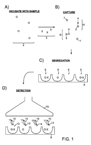
An exemplary embodiment of an inventive assay method is illustrated in FIG. 1.
A plurality of capture objects 2, are provided (step (A)). In this particular
example, the
plurality of capture objects comprises a plurality of beads. The beads are
exposed to a
fluid sample containing a plurality of analyte molecules 3 (e.g., beads 2 are
incubated
with analyte molecules 3). At least some of the analyte molecules are
immobilized with
respect to a bead. In this example, the analyte molecules are provided in a
manner (e.g.,
at a concentration) such that a statistically significant fraction of the
beads associate with
a single analyte molecule and a statistically significant fraction of the
beads do not
associate with any analyte molecules. For example, as shown in step (B),
analyte
molecule 4 is immobilized with respect to bead 5, thereby forming complex 6,
whereas
some beads 7 are not associated with any analyte molecules. It should be
understood, in
some embodiments, more than one analyte molecule may associate with at least
some of
the beads, as described herein. At least some of the plurality of beads (e.g.,
those
associated with a single analyte molecule or not associated with any analyte
molecules)
may then be spatially separated/segregated into a plurality of locations. As
shown in step
(C), the plurality of locations is illustrated as substrate 8 comprising a
plurality of
wells/reaction vessels 9. In this example, each reaction vessel comprises
either zero or
one beads. At least some of the reaction vessels may then be addressed (e.g.,
optically or
via other detection means) to determine the number of locations containing an
analyte
molecule. For example, as shown in step (D), the plurality of reaction vessels
are
interrogated optically using light source 15, wherein each reaction vessel is
exposed to
electromagnetic radiation (represented by arrows 10) from light source 15. The
light
emitted (represented by arrows 11) from each reaction vessel is determined
(and/or
recorded) by detector 15 (in this example, housed in the same system as light
source 15).
The number of reaction vessels containing an analyte molecule (e.g., reaction
vessels 12)
is determined based on the light detected from the reaction vessels. In some
cases, the
number of reaction vessels containing a bead not associated with an analyte
molecule
(e.g., reaction vessel 13), the number of wells not containing a bead (e.g.,
reaction vessel
14) and/or the total number of wells addressed may also be determined. Such
CA 02791654 2012-08-30
WO 2011/109364- 13 -
PCT/US2011/026645
determination(s) may then be used to determine a measure of the concentration
of
analyte molecules in the fluid sample.
A statistically significant fraction of capture objects that contain at least
one
analyte molecule (or no analyte molecules) will typically be able to be
reproducibly
detected and quantified using a particular system of detection and will
typically be above
the background noise (e.g., non-specific binding) that is determined when
carrying out
the assay with a sample that does not contain any analyte molecules, divided
by the total
number of objects (or locations) addressed. A "statistically significant
fraction" as used
herein for the present embodiments, may be estimated according to the Equation
1:
n 3,17t (Eq. 1)
wherein n is the number of determined events for a selected category of
events. That is,
a statistically significant fraction occurs when the number of events is
greater than three
times square root of the number of events. For example, to determine a
statistically
significant fraction of the capture objects not associated with any analyte
molecules or
particles, n is the number of capture objects detected that are not associated
with any
analyte molecules or particles. As another example, to determine a
statistically
significant fraction of the capture objects associated with at least one
analyte molecule, n
is the number of capture objects detected that are determined to be associated
with an
analyte molecule.
In some embodiments, the statistically significant fraction of capture objects
(e.g., beads) associated with at least one analyte molecule (or a single
analyte molecule
in some cases where the ratio of mixing capture objects to analyte molecules
would lead,
statistically, to only zero or one analyte molecule associate with each
capture object) to
the total number of capture objects (e.g., beads) is less than about 1:2, less
than about
1:3, less than about 1:4. is less than about 2:5, less than about 1:5, less
than about 1:10,
less than about 1:20, less than about 1:100, less than about 1:200, or less
than about
1:500. Therefore, in such embodiments, the fraction of capture objects (e.g.,
beads) not
associated with any analyte molecules to the total number of capture objects
(e.g., beads)
is at least about 1:100, about 1:50, about 1:20, about 1:10, about 1:5, about
1:4, about
1:3, about 1:2, about 1:1, about 2:1, about 3:1, about 4:1, about 5:1, about
10:1, about
20:1, about 50:1, about 100:1, or the like.
In some embodiments, the percentage of capture objects (e.g., beads)
associated
with at least one analyte molecule (or a single analyte molecule in some cases
where the
ratio of mixing capture objects to analyte molecules would lead,
statistically, to only zero
CA 02791654 2012-08-30
WO 2011/109364- 14 - PCT/US2011/026645
or one analyte molecule associate with each capture objects) is less than
about 50%, less
than about 40%, less than about 30%, less than about 20%, less than about 10%,
less
than about 5%, less than about 2%, less than about 1%, less than about 0.5%,
less than
about 0.01%, or the like, the total number of capture objects. In some
embodiments, the
percentage of capture objects (e.g., beads) not associated with an analyte
molecule to the
total number of capture objects (e.g., beads) is at least about 30%, at least
about 40%, at
least about 50%, at least about 60%, at least about 70%, at least about 80%,
at least about
90%, at least about 95%, at least about 98%, or the like, the total number of
capture
objects.
In some embodiments, prior to spatially separating the plurality of capture
objects, the capture objects may be exposed to a plurality of binding ligands
which have
an affinity for at least one type of analyte molecule (or particle). A
"binding ligand," as
used herein, is any molecule, particle, or the like which specifically binds
to or otherwise
specifically associates with an analyte molecule to aid in the detection of
the analyte
molecule. Binding ligands may be particularly useful in embodiments where at
least
some of the capture objects are associated with respect to more than one
analyte
molecule (e.g., two, three, four, five, or more, analyte molecules). In some
cases, the
binding ligand may be provided in a manner (e.g., at a concentration level)
such that a
statistically significant fraction of the capture objects comprising at least
one analyte
molecule associate with at least one binding ligand (or in some cases, a
single binding
ligand) and a statistically significant fraction of the capture objects (e.g.,
capture objects
either associated with at least one analyte molecule or not associated with
any analyte
molecules) do not associate with any binding ligand.
A statistically significant fraction of the locations that contain a capture
object
(e.g., bead) associated with at least one analyte molecule and a single
binding ligand is
greater than or equal to the minimum number of locations that can be
reproducibly
determined to contain an capture object (e.g., bead) associated with a single
binding
ligand with a particular system of detection (i.e., substantially similar
results are obtained
for multiple essentially identical fluid samples comprising the capture
objects associated
with an analyte molecule and/or binding ligand) and that is above the
background noise
(e.g., non-specific binding) that is determined when carrying out the assay
with a sample
that does not contain any analyte molecules and/or binding ligands, divided by
the total
number of locations. The statistically significant fraction of locations that
contain a
capture object associated with at least one analyte molecule and a single
binding ligand
CA 02791654 2012-08-30
WO 2011/109364- 15 - PCT/US2011/026645
can be determined according to Equation 1. The ratio of the number of capture
objects to
analyte molecules and/or binding ligands which may be provided such that
substantially
all of the capture objects are associated with zero or a single analyte
molecule may be
calculated using a Poisson distribution adjustment, as described herein.
In some embodiments, the statistically significant fraction of capture objects
(e.g., beads) associated with at least one analyte molecule and at least one
binding ligand
to the total number of capture objects (e.g., beads) is less than about 1:2,
less than about
1:3, less than about 1:4. is less than about 2:5, less than about 1:5, less
than about 1:10,
less than about 1:20, less than about 1:100, less than about 1:200, or less
than about
1:500. In some cases, the statistically significant fraction of capture
objects (e.g., beads)
associated not associated with any binding ligand to the total number of
capture objects
at least about 1:100, about 1:50, about 1:20, about 1:10, about 1:5, about
1:4, about 1:3,
about 1:2, about 1:1, about 2:1, about 3:1, about 4:1, about 5:1, about 10:1,
about 20:1,
about 50:1, about 100:1, or the like.
In some embodiments, the percentage of capture objects (e.g., beads)
associated
with at least one analyte molecule and at least one binding ligand to the
total number of
capture objects (e.g., beads) is less than about 50%, less than about 40%,
less than about
30%, less than about 20%, less than about 10%, less than about 5%, less than
about 2%,
less than about 1%, less than about 0.5%, less than about 0.01%, or less. In
some
embodiments, the percentage of capture objects (e.g., beads) not associated
with any
binding ligand to the total number of capture objects is at least about 30%,
at least about
40%, at least about 50%, at least about 60%, at least about 70%, at least
about 80%, at
least about 90%, at least about 95%, at least about 98%, or greater.
A non-limiting example of an embodiment where a capture object is associated
with more than one analyte molecule is illustrated in FIG. 2. A plurality of
capture
objects 20 are provided (step (A)). In this example, the plurality of capture
objects
comprises a plurality of beads. The plurality of beads is exposed to a fluid
sample
containing plurality of analyte molecules 21 (e.g., beads 20 are incubated
with analyte
molecules 21). At least some of the analyte molecules are immobilized with
respect to a
bead. For example, as shown in step (B), analyte molecule 22 is immobilized
with
respect to bead 24, thereby forming complex 26. Also illustrated is complex 30
comprising a bead immobilized with respect to three analyte molecules and
complex 32
comprising a bead immobilized with respect to two analyte molecules.
Additionally, in
some cases, some of the beads may not associate with any analyte molecules
(e.g., bead
CA 02791654 2012-08-30
WO 2011/109364- 16 - PCT/US2011/026645
28). The plurality of beads from step (B) is exposed to a plurality of binding
ligands 31.
As shown in step (C), a binding ligand associates with some of the analyte
molecules
immobilized with respect to a bead. For example, complex 40 comprises bead 34,
analyte molecule 36, and binding ligand 38. The binding ligands are provided
in a
manner such that a statistically significant fraction of the beads comprising
at least one
analyte molecule become associated with at least one binding ligand (e.g.,
one, two,
three, etc.) and a statistically significant fraction (i.e. as determined by
Equation 1 above)
of the beads comprising at least one analyte molecule do not become associated
with any
binding ligands. At least a portion of the plurality of beads from step (C)
are then
spatially separated into a plurality of locations. As shown in step (D), in
this example,
the locations comprise a plurality of reaction vessels 41 on a substrate 42.
The plurality
of reaction vessels may be exposed to the plurality of beads from step (C)
such at each
reaction vessel contains zero or one beads. The substrate may then be analyzed
to
determine the number of reaction vessels containing a binding ligand (e.g.,
reaction
vessels 43), wherein in the number may be related to a measure of the
concentration of
analyte molecules in the fluid sample. In some cases, the number of reaction
vessels
containing a bead and not containing a binding ligand (e.g., reaction vessel
44), the
number of reaction vessels not containing a bead (e.g., reaction vessel 45),
and/or the
total number of reaction vessels addressed/analyzed may also be determined.
Such
determination(s) may then be used to determine a measure of the concentration
of
analyte molecules in the fluid sample.
The foregoing exemplary methods may be performed using a number of
different assay formats, different reaction conditions, and/or detection
systems in
different embodiments of the invention, several examples of which are
described
below. Additional components and/or method steps may be utilized as a
substitute for
and/or in combination with the exemplary methods and components described
herein
within the scope of the invention. It should be understood, while certain of
the
discussion herein focuses on a plurality of locations comprising a plurality
of
wells/reaction vessels in a substrate, this is by no means limiting and other
materials
may be used to segregate capture objects/molecules into a plurality of
spatially distinct
locations (e.g., regions in/on a hydrogel, points/regions on the surface of a
planar
substrate, etc.). As another example, while much of the discussion herein
focuses on a
plurality of capture objects comprising a plurality of beads, this is by no
means limiting
CA 02791654 2012-08-30
WO 2011/109364- 17 - PCT/US2011/026645
and in other embodiments the capture objects may take other physical forms
(e.g.,
nanotubes, disks, rings, microfluidic droplets, etc.).
Exemplary Assay Formats
The inventive assays may be carried our according to a very wide variety of
basic
protocols and formats. The particular format chosen can be based on the nature
of the
analyte molecules, the nature of the fluid sample containing the analyte
molecules, and
the availability and properties of binding partners of the analyte as well as
other factors.
Several exemplary basic formats were discussed previously in the context of
the
discussion of FIGS. 1-2. As would be apparent to those skilled in the art with
the benefit
of the teachings provided by the present disclosure, the invention may
alternatively be
performed according to protocols/formats not specifically described in the
specific,
exemplary embodiments illustrated in this detailed description, but which do
not require
undue burden or experimentation to practice.
As described above, an exemplary basic assay format/protocol comprises
exposing a plurality of capture objects (e.g., beads) configured to capture an
analyte
molecule or particle to a sample containing or suspected of containing such
analyte
molecules (or particles). At least some of the analyte molecules may become
immobilized with respect to a capture object. The plurality of capture objects
may each
include a binding surface having affinity for at least one type of analyte
molecule. At
least a portion of the capture objects may then be spatially segregated into a
plurality of
locations (e.g., reaction vessels/wells). Based at least in part on a
determination of the
number of locations comprising a capture object comprising at least one
analyte
molecule, a measure of the concentration of analyte molecules may be
determined.
Various other aspects of this basic assay format will now be discussed,
including
numerous considerations regarding the materials, concentrations, solutions,
steps, and
the like.
In certain embodiments, a plurality of capture objects is exposed to a sample
containing or suspected of containing at least one type of analyte molecules,
wherein
the plurality of capture objects comprises a binding surface having an
affinity for the at
least one type of analyte molecule. In some cases, the binding surface may
comprise a
plurality of capture components. A "capture component", as used herein, is any
molecule, other chemical/biological entity, or solid support modification
disposed upon a
solid support that can be used to specifically attach, bind or otherwise
capture a target
CA 02791654 2012-08-30
WO 2011/109364- 18 - PCT/US2011/026645
molecule or particle (e.g., an analyte molecule), such that the target
molecule/particle
becomes immobilized with respect to the capture component and the support. The
immobilization, as described herein, may be caused by the association of an
analyte
molecule with a capture component on the surface of the capture object. As
used
herein, "immobilized" means captured, attached, bound, or affixed so as to
prevent
dissociation or loss of the target molecule/particle, but does not require
absolute
immobility with respect to either the capture component or the object.
The number of analyte molecules which are immobilized with respect to a
capture object may depend on the ratio of the total number of analyte
molecules in the
sample versus at least one of the total number, size, and/or surface density
of capture
components of capture objects provided. In some embodiments, the number of
molecules or particles immobilized with respect to a single capture object may
follow a
standard Poisson distribution. In some cases, a statistically significant
number of the
capture objects associate with a single analyte molecule and a statistically
significant
number of capture objects do not associate with any analyte molecules. The
total
number of capture objects provided may be between about 10,000 and about
10,000,000, between about 50,000 and about 5,000,000, or between about 100,000
and
about 1,000,000. In some cases, the total number of capture objects provided
is at least
about 10,000, at least about 50,000, at least about 100,000, at least about
1,000,000, at
least about 5,000,000, or at least about 10,000,000. In some cases, the ratio
of the
number of analyte molecules in the fluid sample to capture objects provided is
between
about 10:1 and about 1:10,000,000, between about 8:1 and about 1:10,000,000,
between
about 10:1 and about 2:1, between about 2:1 and about 1:10, or less than about
1:10 (e.g.,
about 1:20, about 1:30, etc.). The ratio of analyte molecules in the fluid
sample to
capture objects provided may affect the assay steps and/or analysis carried
out to
determine a measure of the concentration of analyte molecules in the fluid
sample, as
described herein in the Quantification section.
In some cases, substantially all of the analyte molecules provided in the
sample
may become immobilized with respect to a capture object. That is, greater than
about
90%, greater than about 95%, greater than about 97%, greater than about 98%,
or greater
than about 99% of the analyte molecules in the sample may become immobilized
with
respect to a capture object. In some cases, however, only a fraction of the
analyte
molecules in the sample may become immobilized with respect to a capture
object. That
is, in some cases, between about 1% and about 90%, between about 10% and about
90%,
CA 02791654 2012-08-30
WO 2011/109364- 19 - PCT/US2011/026645
between about 20% and about 80%, or between about 30% and about 70% of the
analyte
molecules provided in the sample are immobilized with respect to a capture
object. In
some embodiments, at least about 10%, about 20%, about 30%, about 40%, about
50%,
about 60%, about 70%, about 80%, or about 90%, or about 95% of the analyte
molecules
are immobilized with respect to a capture object.
In some formats of the assay, following immobilization, the plurality of
capture
objects (e.g., at least some of which are associated with at least one analyte
molecule)
may be exposed to a plurality of binding ligands. At least some of the analyte
molecules
immobilized with respect to a capture object may associate with a binding
ligand. The
number of binding ligands which associate with a capture object (e.g., via an
analyte
molecule) may depend on the ratio of the total number of analyte molecules
immobilized with respect to a single capture object versus the total number of
binding
ligands exposed to the capture objects. For example, in embodiments where
substantially all of the capture objects are associated with either zero or
one analyte
molecules, conditions may be selected such that substantially all of the
analyte
molecules associate with a single binding ligand, therefore each capture
object
associated with a single analyte molecule becomes associated with a single
binding
ligand (e.g., via the analyte molecule). Thus, the number of locations (e.g.,
reaction
vessels) which contain a single analyte molecule may be determined by
determining
the number of locations (e.g., reaction vessels) which comprise a binding
ligand. In
such embodiments (e.g., where zero or at least one analyte molecules are
associated
with each capture object), the ratio of binding ligands provided (e.g., in a
mixing step)
to the total number of analyte molecules immobilized with respect to a capture
object
may be about 20:1, about 10:1, about 5:1, about 2:1, or about 1:1.
In some embodiments, however, a single capture object may be associated with
zero, one, or more than one (e.g., two, three, four, etc.) analyte molecules.
In such
embodiments, the binding ligand may be provided at a concentration such that a
statistically significant fraction of the capture objects comprising at least
one analyte
molecule associate with only a single binding ligand and a statistically
significant
fraction of the capture objects comprising at least one analyte molecule do
not
associate with any binding ligand. In other embodiments, however, the binding
ligands
may be provided at a concentration such that a statistically significant
fraction of the
capture objects comprising at least one analyte molecule associate with at
least one
binding ligand (e.g., one, two, three, etc.) and a statistically significant
fraction of the
CA 02791654 2012-08-30
WO 2011/109364- 20 - PCT/US2011/026645
capture objects comprising at least one analyte molecule do not associate with
any
binding ligand. The concentration of analyte molecules in the fluid sample may
then
be determined, either with an analysis based at least in part of the number of
locations
containing a capture object associated with a binding ligand (e.g., by
relating the
concentration of analyte molecules in the fluid sample to the number of
locations
comprising a binding ligand), and/or an analysis based at least in part on an
intensity
reading of a signal indicative of the number of binding ligands at the
addressed
locations (e.g., in embodiments where at least some of the capture objects
comprise
more than one analyte molecule and/or more than one binding ligand, as
described
herein). In such embodiments (e.g., wherein more than one analyte molecule may
be
immobilized with respect to each capture object), the ratio of the number of
binding
ligands provided in solution to the number of analyte molecules immobilized
with
respect to a capture object may be about 1:50, about 1:40, about 1:30, about
1:20, about
1:10, about 1:5, about 1:3, about 1:2, about 1:1, or the like. In some cases,
the ratio of
the number of binding ligands provided in solution may be calculated based on
the
number of capture objects provided. In some cases, the ratio of binding
ligands
provided to the number of capture objects is about 1:50, about 1:40, about
1:30, about
1:20, about 1:10, about 1:5, about 1:3, about 1:2, about 1:1, or the like. In
other cases,
the ratio the number of capture objects to the number of binding ligands
provided is
about 1:50, about 1:40, about 1:30, about 1:20, about 1:10, about 1:5, about
1:3, about
1:2, or the like. In some embodiments, the quantification determination may
comprise
a Poisson distribution adjustment, as described herein.
In some embodiments, the concentration of binding ligand used in an assay may
be selected as to minimize certain events which may occur when an excess of
binding
ligand is present, for example, non-specific binding of the binding ligand. In
some cases,
if the concentration of binding ligand is too high, an increase in background
readings
may occur due to non-specific interactions (e.g., with the capture objects,
reaction
vessels, etc.). In some cases, the concentration of binding ligand may be
selected (or
estimated, in the case of an unknown concentration of analyte molecule) such
that a only
a fraction of the analyte molecules immobilized with respect to a capture
object associate
with a binding ligand (e.g., about 0.1%, about 1%, about 2%, about 3%, about
4%, about
5%, about 10%, about 20%, about 30%, about 40%, about 50%, or more). This may
be
especially useful in embodiments where the percentage of capture objects which
associate with at least one analyte molecule is relatively high (e.g., greater
than about
CA 02791654 2012-08-30
WO 2011/109364- 21 - PCT/US2011/026645
20%, greater than about 30%, greater than about 40%, greater than about 50%,
greater
than about 60%, greater than about 70%, greater than about 80%, greater than
about
90%, or more). By providing the binding ligand at a lower concentration, in
some cases,
not every analyte molecule immobilized with respect to a capture object will
associate
with a binding ligand, which can be advantageous for quantification, for
example when
the presence of a binding ligand is required for detection, and especially
when using a
digital/binary read-out technique. For example, if the percentage of capture
objects
associated with an analyte molecule is about 50% or greater, a reduced number
of
binding ligands may be provided such that less than all of the immobilized
analyte
molecules associate with a binding ligand. In other cases, the percentage of
binding
ligands that associate with an analyte molecule may be reduced by decreasing
the
incubation time with the analyte molecule (e.g., limit the time of exposure
such that only
a fraction of the immobilized analyte molecules associate with an analyte
molecule).
The total number of analyte molecules/binding ligands/capture objects/etc. in
a
solution may be determined using calculations with knowledge of the
concentration of
the analyte molecules/binding ligands/capture objects/etc. in solution. For
example,
the total number of binding ligands in a solution may be determined according
to
Equation 2:
#of binding ligands = N Ax[binding ligandlxvolume (Eq. 2)
wherein NA is Avogadro's number (6.022 x 10²³ mo1-1), [binding ligandl is the
concentration of the binding ligand in solution in moles per liter, and volume
is the
total volume of solution in liters employed. Similar calculations may be
carried out for
other components (e.g., analyte molecules (e.g., in a calibration sample),
capture
objects, etc.).
Following immobilization of a plurality of analyte molecules with respect to a
plurality of capture objects and, in some cases, association of a binding
ligand to at least
some the immobilized analyte molecules, at least a portion of the capture
objects may be
spatially segregated into a plurality of locations. The percentage of capture
objects
which are spatially segregated into the plurality of locations may vary
depending on
numerous factors including, but not limited to, the ratio of the number of
capture objects
versus the total number of locations, the method of spatially segregating the
capture
objects, and/or the length of the time the capture objects are exposed to the
locations. In
some cases, at least about 0.5%, at least about 1%, at least about 2%, at
least about 5%,
CA 02791654 2012-08-30
WO 2011/109364- 22 - PCT/US2011/026645
at least about 10%, at least about 20%, at least about 30%, at least about
40%, at least
about 50%, at least about 60%, at least about 70%, at least about 80%, at
least above
90%, or more, of the capture objects are spatially segregated into the
plurality of
locations. In some cases, between about 0.1% and about 50%, between about 0.1%
and
about 30%, between about 0.5% and about 20%, between about 0.5% and about 10%,
between about 0.5% and about 5%, between about 1% and about 10%, or about
0.5%,
about 1%, about 2%, about 4%, about 5%, about 10%, about 20%, about 30%, about
50%, about 70%, or about 90% of the capture objects are spatially segregated
into the
plurality of locations. Following spatially segregating at least a portion of
the capture
objects into a plurality of locations, at least a portion of the locations may
be addressed.
The number of locations addressed may be about 0.5%, about 1%, about 2%, about
3%,
about 5%, about 10%, about 20%, about 30%, about 40%, about 50%, about 60%,
about
70%, about 80%, about 90%, about 95%, or more, of the total number of
locations.
The portion of locations may be addressed to determine the number of locations
containing an analyte molecule, or in some cases, a binding ligand. In some
cases, the
number of locations containing a capture object not associated with an analyte
molecule
(or a binding ligand), the number of locations containing and/or not
containing a capture
object, and/or the total number of locations analyzed/determined may also be
determined. A measure of the concentration of analyte molecules in the fluid
sample
may be determined at least in part on the number of locations determined to
contain an
analyte molecule (or binding ligand). In some cases the measure of the
concentration of
analyte molecules in the fluid sample may be based at least in part on the
ratio of the
number of locations containing a capture object associated with an analyte
molecule to
the total number of locations addressed or the total number of locations
addressed that
contain a capture object. In other cases, a measure of the concentration of
analyte
molecules in the fluid sample may be based at least in part on the ratio of
the number of
locations containing a capture object associated with an analyte molecule to
the number
of locations containing a capture object not associated with an analyte
molecule.
Specific methods and calculations which may be used to determine the measure
of the
concentration of analyte molecules in the fluid sample are discussed in more
detail
below.
The ratios, percentages, and other parameters described herein with respect to
the
amount/quantity/ratio of a first component to a second component (for example,
analyte
molecules/capture objects, binding ligands/capture objects, binding
ligands/analyte
CA 02791654 2012-08-30
WO 2011/109364- 23 - PCT/US2011/026645
molecules, capture objects/locations, precursor labeling agents/binding
ligands, etc.) may
be adjusted as desired to yield a desired ratio of analyte molecules/binding
ligands
captured per capture object, and/or may be controlled or determined using no
more than
routine experimentation, calculations (in some cases, including accounting for
Poisson
distributions), screening tests, etc., given the teaching and guidance
provided by the
present specification. For example, if the number of capture objects provided
is known
(e.g., as determined using a similar formula as given in Equation 1), the
number of
binding ligands that need to be provided may be determined based on the
desired ratio of
capture objects to binding ligands, and hence, the amount of moles of binding
ligand that
should be provided may be determined. As another example, in the case of an
unknown
concentration of analyte molecules, if a first assay method indicates that a
significant
number of capture objects comprise more than one analyte molecule (e.g., all
or a
significant number of locations are determined to contain an analyte molecule
or there is
less than a statistically significant number of beads determined to be free of
analyte
molecules), the fluid sample may be diluted and/or the number of capture
objects may be
increased such that the number of capture objects comprising at least one
analyte
molecule may be decreased.
Other aspects of the assay will now be discussed in detail. It should be
understood, that none, a portion of, or all of the following steps may be
performed at
least once during the certain exemplary assay formats described herein. Non-
limiting
examples of additional steps not described which may be performed include, but
are not
limited to, washing and/or exposure to additional binding ligands, precursor
labeling
agents, and/or labeling agents, etc.
In some embodiments, the plurality of capture objects (e.g., at least some of
which are associated with at least one analyte molecule) may be exposed to at
least one
additional reaction component prior to, concurrent with, and/or following
spatially
separating at least some of the plurality of capture objects into a plurality
of locations. In
some cases, the capture objects may be exposed to a plurality of binding
ligands. In
certain embodiments, a binding ligand may be adapted to be directly detected
(e.g., the
binding ligand comprises a detectable molecule or moiety) or may be adapted to
be
indirectly detected (e.g., including a component that can convert a precursor
labeling
agent into a labeling agent), as discussed more below. More than one type of
binding
may be employed in any given assay method, for example, a first type of
binding ligand
and a second type of binding ligand. In one example, the first type of binding
ligand is
CA 02791654 2013-05-09
- 24 -
able to associate with a first type of analyte molecule and the second type of
binding
ligand is able to associate with the first binding ligand. In another example,
both a first
type of binding ligand and a second type of binding ligand may associate with
the same
or different epitopes of a single analyte molecule, as described below.
Certain binding ligands can comprise a component that is able to facilitate
detection, either directly or indirectly. A component may be adapted to be
directly
detected in embodiments where the component comprises a measurable property
(e.g., a
fluorescence emission, a color, etc.). A component may facilitate indirect
detection, for
example, by converting a precursor labeling agent into a labeling agent (e.g.,
an agent
that is detected in an assay). A "precursor labeling agent" is any molecule,
particle, or
the like, that can be converted to a labeling agent upon exposure to a
suitable converting
agent (e.g., an enzymatic component). A "labeling agent" is any molecule,
particle, or
the like, that facilitates detection, by acting as the detected entity, using
a chosen
detection technique.
In some embodiments, at least one binding ligand comprises an enzymatic
component. In some embodiments, the analyte molecule may comprise an enzymatic
component. The enzymatic component may convert a precursor labeling agent
(e.g., an
enzymatic substrate) into a labeling agent (e.g., a detectable product). A
measure of the
concentration of analyte molecules in the fluid sample can then be determined
based at
least in part by determining the number of locations containing a labeling
agent (e.g., by
relating the number of locations containing a labeling agent to the number of
locations
containing an analyte molecule). Non-limiting examples of enzymes or enzymatic
components include horseradish peroxidase, beta-galactosidase, and alkaline
phosphatase. Other non-limiting examples of systems or methods for detection
include
embodiments where nucleic acid precursors are replicated into multiple copies
or
converted to a nucleic acid that can be detected readily, such as the
polymerase chain
reaction (PCR), rolling circle amplification (RCA), ligation, Loop-Mediated
Isothermal
Amplification (LAMP), etc. Such systems and methods will be known to those of
ordinary skill in the art, for example, as described in "DNA Amplification:
Current
Technologies and Applications," Vadim Demidov et al., Taylor &Francis, 2004
As an example of an assay method which comprises the use of a precursor
labeling agent, as shown in FIG. 3, substrate 100 comprising a plurality of
locations is
provided, wherein the locations comprise reaction vessels. In reaction vessel
101 (e.g.,
location), analyte molecule 102 is immobilized with respect to bead 103 (e.g.,
capture
CA 02791654 2013-05-09
- 25 -
object). Binding ligand 104 is associated with analyte molecule 102. Binding
ligand
104 comprises an enzymatic component (not shown). Precursor labeling agent 106
is
converted to labeling agent 108 (upon exposure to the enzymatic component).
Labeling
agent 108 is detected using methods described herein. In contrast, reaction
vessel 111
contains analyte molecule 112 immobilized with respect to bead 110. In this
reaction
vessel, analyte molecule 112 is not associated with a binding ligand
comprising an
enzymatic component. Therefore, precursor labeling agent 114 is not converted
to a
labeling agent in the reaction vessel. Thus this reaction vessel would give a
different
signal as compared to reaction vessel 101 where the precursor labeling agent
was
converted to a labeling agent. In some cases, there may also be reaction
vessels which
contain a bead not associated with an analyte molecule, for example, reaction
vessel 121
contains bead 116. Additionally, some of the reaction vessels may not comprise
any
bead, for example, reaction vessel 123. Reaction vessels 121 and 123 may give
different
signals as compared to reaction vessel 101 as there would be no labeling agent
present.
However, reaction vessels 121 and 123 may contain precursor labeling agent
117. More
than one precursor labeling agent may be present in any given reaction vessel.
In certain embodiments, solubilized, or suspended precursor labeling agents
may be employed, wherein the precursor labeling agents are converted to
labeling agents
which are insoluble in the liquid and/or which become immobilized within/near
the
location (e.g., within the reaction vessel in which the labeling agent is
formed). Such
precursor labeling agents and labeling agents and their use is described in
commonly
owned U.S. Patent Application Pub. No. U.S. 2010-0075862 Al, entitled "High
Sensitivity
Determination of the Concentration of Analyte molecules in a Fluid Sample," by
Duffy,
et al., filed September 23, 2008.
In some embodiments, during the assay, at least one washing step may be
carried
out. In one instance, a plurality of capture objects may be washed after
exposing the
capture objects to one or more solutions comprising analyte molecules, binding
ligands,
precursor labeling agents, or the like. For example, following immobilization
of the
analyte molecules with respect to a plurality of capture objects, the
plurality of capture
objects may be subjected to a washing step thereby removing any analyte
molecules not
specifically immobilized with respect to a capture object. In certain
embodiments, the
wash solution is selected so that it does not cause appreciable change to the
configuration
of the capture objects and/or analyte molecules and/or does not disrupt any
specific
binding interaction between at least two components of the assay (e.g., a
capture
CA 02791654 2012-08-30
WO 2011/109364- 26 - PCT/US2011/026645
component and an analyte molecule). In other cases, the wash solution may be a
solution
that is selected to chemically interact with one or more assay components. As
will be
understood by those of ordinary skill in the art, a wash step may be performed
at any
appropriate time point during the inventive methods.
In some embodiments, assay methods may be carried out that do not comprise the
use of a plurality of capture objects comprising a binding surface for at
least one type of
analyte molecule and/or a plurality of locations to which the capture objects
may be
spatially separated. For example, an assay according to the invention in
certain
embodiments may use any suitable method which is capable of isolating single
analyte
molecules and/or capture objects associated with one or more analyte molecules
such
that they can be individually addressed for detection. For example, an assay
method may
comprise providing a plurality of capture objects which are each associated
with either a
single analyte molecule or are free of any analyte molecules. At least a
portion of the
capture objects may be individually addressed to determine the number of the
capture
objects associated with an analyte molecule or particle. Based at least in
part on the
number of capture objects determined to be associated with an analyte
molecule, a
measure of the concentration of analyte molecules or particles in a fluid
sample may be
determined.
FIG. 4A illustrates a non-limiting embodiment where single analyte molecules
are spatially segregating into a plurality of droplets. In FIG. 4A, plurality
of analyte
molecules 70 are provided, as shown in step (A). In this example, analyte
molecules 70
are capable of being optically detected (e.g., the analyte molecules may be
directly
detected using optical interrogation). At least some of the plurality of
analyte molecules
70 are contained within liquid droplets 72 (e.g., using microfluidic
techniques) which
comprise fluid 71, as shown in step (B). Additionally, some droplets may be
present
which do not contain any analyte molecules (e.g., droplets 74 comprising fluid
71).
Plurality of droplets 75 are substantially surrounded by fluid 73 which is
substantially
immiscible with fluid 71. Plurality of droplets 75 can be optically
interrogated by
feeding droplets into column 74 such that each droplet passes by an optical
detection
system (e.g., comprising light source 76 and detector 78) single file, as
shown in step
(C). Each droplet may be determined to contain an analyte molecule when there
is a
change in the optical single (e.g., a change in optical signal due to the
presence of an
analyte molecule in the droplet).
CA 02791654 2012-08-30
WO 2011/109364- 27 - PCT/US2011/026645
As another example, as illustrated in FIG. 4B, plurality of analyte molecules
80
are provided, as shown in step (A). In this example, analyte molecules 70 are
not
capable of being optically detected, and must be indirectly detection (as
described
herein). Plurality of analyte molecules 80 are exposed to a plurality of
binding ligands
82, such that at least one binding ligand associates with a significant
portion of the
analyte molecules, as shown in step (B), to form complex 83, as shown in step
(B). In
this example, each binding ligand 82 comprises enzymatic component 84. At
least a
portion of complexes 83 may be contained in droplets 85 (e.g., using
microfluidic
techniques), as shown in step (C), which comprise liquid 79. Additionally,
some
droplets may be present which do not contain any complexes (e.g., droplets 86
comprising fluid 79). Plurality of droplets 91 are substantially surrounded by
fluid 87
which is substantially immiscible with fluid 79. Droplets 85 and 86 may
additionally
comprise precursor labeling agent 86, which is converted to labeling agent 88
upon
exposure to enzymatic component 84, as indicated by arrow 87. Plurality of
droplets 91
can be optically interrogated by feeding the plurality of droplets into column
89 such that
each droplet passes by an optical detection system (e.g., comprising light
source 90 and
detector 92) single file, as shown in step (D). Each droplet may be determined
to contain
an analyte molecule when there is a change in the optical single (e.g., a
change in optical
signal due to the presence of a labeling agent in the droplet).
As yet another example, FIG. 4C illustrates and embodiment where single
analyte
molecules 282 are associated with respect to objects 280 via capture
components 274.
Additionally, in this example, the immobilized analyte molecules are
associated with
binding ligand 284. The droplets can be optically interrogated by feeding
droplets into
column 287 such that each droplet passed by the optical detection system
(e.g.,
comprising light source 286 and detector 288) single file. Each droplet may be
determined to contain a binding ligand when there is a change in the optical
single (e.g.,
a change in optical signal due to the presence of a binding ligand in the
droplet).
The following sections provide additional information regarding method steps,
materials, and parameters that may be used to practice the assay methods
described
above.
Capture Objects and Spatial Locations for Capture Object Segregation
In some embodiments, the method and systems of the present invention utilize a
plurality of capture objects that each includes a binding surface having
affinity for at
CA 02791654 2012-08-30
WO 2011/109364- 28 - PCT/US2011/026645
least one type of analyte molecule. The plurality of capture objects may be
configured to
be able to be spatially segregated from each other, that is, the capture
objects may be
provided in a form such that the capture objects are capable of being
spatially separated
into a plurality of locations. For example, the plurality of capture objects
may comprise
a plurality of beads (which can be of any shape, e.g., sphere-like, disks,
rings, cube-like,
etc.), a dispersion or suspension of particulates (e.g., a plurality of
particles in suspension
in a fluid), nanotubes, or the like. In some embodiments, the plurality of
capture objects
is insoluble or substantially insoluble in the solvent(s) or solution(s)
utilized in the assay.
In some cases, the capture objects are solid or substantially solid (e.g., is
essentially free
of pores), however, in some cases, the plurality of capture objects may be
porous or
substantially porous, hollow, partially hollow, etc. The plurality of capture
objects may
be non-absorbent, substantially non-absorbent, substantially absorbent, or
absorbent. In
some cases, the capture objects may comprise a magnetic material, which as
described
herein, may facilitate certain aspect of the assay (e.g., washing step). In
some cases, a
capture object surface may also comprise a protective or passivating layer
that can
reduce or minimize non-specific binding events (e.g., analyte molecules,
binding ligands,
etc.).
In some embodiments, the capture objects each include a binding surface having
affinity for at least one type of analyte molecule of interest. The portion of
the capture
object which comprises a binding surface may be selected or configured based
upon the
physical shape/characteristics and properties of the capture objects (e.g.,
size, shape), and
the format of the assay. In some embodiments, substantially all of the outer
surfaces of
the capture objects form the binding surfaces. A binding surface having an
affinity for at
least one type of analyte molecule may be formed via the association of a
plurality of
capture components with a capture object. In some cases, an analyte molecule
may
associate with a capture component (e.g., become immobilized with respect to)
via
formation of at least one chemical bond and/or physical adsorption, or
combination
thereof. Non-limiting examples of types of chemical bonds include ionic bonds,
covalent bonds (e.g., carbon-carbon, carbon-oxygen, oxygen-silicon, sulfur-
sulfur,
phosphorus-nitrogen, carbon-nitrogen, metal-oxygen, or other covalent bonds),
hydrogen
bonds (e.g., between hydroxyl, amine, carboxyl, thiol, and/or similar
functional groups),
dative bonds (e.g., complexation or chelation between metal ions and
monodentate or
multidentate ligands), Van der Waals interactions, or the like. Capture
components
which are useful or potentially useful for practicing certain aspects and
embodiments of
CA 02791654 2012-08-30
WO 2011/109364- 29 - PCT/US2011/026645
the invention are discussed in more detail below. At least some of the analyte
molecules,
upon exposure to the plurality of capture objects comprising a plurality of
capture
components, become immobilized with respect to a capture component. In certain
embodiments, substantially all of the plurality of analyte molecules in the
fluid sample
tested may become immobilized with respect to the capture components (and
hence, the
capture objects).
Without wishing to be bound by any theory, the use in certain embodiments of
the invention of a capture step in which a plurality of capture objects having
a large
surface area for binding are exposed to a fluid sample containing the analyte
molecules
or particles such that the analyte molecules/particles become immobilized with
respect to
the capture objects may facilitate an increase in the speed and/or efficiency
of the assay
for detection and quantification of concentration of the analyte in the sample
compared
to assays where the analyte molecules themselves are segregated for detection
without
being exposed to and immobilized with respect to a capture object. This
increase in
binding speed and efficiency may be further enhanced if the solution in which
the
plurality of analyte molecules and capture objects are incubated for capture
is agitated
(e.g., stirred) to increase the collision frequency and mass transfer rate
between the
plurality of capture objects (e.g., plurality of beads) and the analyte
molecules (e.g., as
contrasted with a substrate comprising a stationary surface (e.g., a
microtiter plate)).
The plurality of capture objects for analyte capture may be of any suitable
size or
shape. Non-limiting examples of suitable shapes include spheres, cubes,
ellipsoids,
tubes, sheets, and the like. In certain embodiments, the average diameter (if
substantially
spherical) or average maximum cross-sectional dimension (for other shapes) of
a capture
object may be greater than about 0.1 um (micrometer), greater than about 1 um,
greater
than about 10 um, greater than about 100 um, greater than about 1 mm, or the
like. In
other embodiments, the average diameter of a capture object or the maximum
dimension
of a capture object in one dimension may be between about 0.1 um and about 100
um,
between about 1 um and about 100 um, between about 10 um and about 100 um,
between about 0.1 um and about 1 mm, between about 1 um and about 10 mm,
between
about 0.1 um and about 10 um, or the like. The "average diameter" or "average
maximum cross-sectional dimension" of a plurality of capture objects, as used
herein, is
the arithmetic average of the diameters/maximum cross-sectional dimensions of
the
capture objects. Those of ordinary skill in the art will be able to determine
the average
diameter/maximum cross-sectional dimension of a population of capture objects,
for
CA 02791654 2012-08-30
WO 2011/109364- 30 - PCT/US2011/026645
example, using laser light scattering, microscopy, sieve analysis, or other
known
techniques. For example, in some cases, a Coulter counter may be used to
determine the
average diameter of a plurality of beads.
The capture objects for analyte capture may be fabricated from one or more
suitable materials, for example, plastics or synthetic polymers (e.g.,
polyethylene,
polypropylene, polystyrene, polyamide, polyurethane, phenolic polymers, or
nitrocellulose etc.), naturally derived polymers (latex rubber,
polysaccharides,
polypeptides, etc), composite materials, ceramics, silica or silica-based
materials, carbon,
metals or metal compounds (e.g., comprising gold, silver, steel, aluminum,
copper, etc.),
inorganic glasses, silica, and a variety of other suitable materials. Non-
limiting examples
of potentially suitable configurations include beads (e.g., magnetic beads),
tubes (e.g.,
nanotubes), plates, disks, dipsticks, or the like.
In some cases, the capture objects may be selected such that the capture
object
itself is detectable by the system. This may be useful in embodiments where
the fraction
or percentage of capture objects associated with an analyte molecule is to be
determined
(e.g., when the total number of capture objects interrogated and detected is
used to
determine the fraction of capture objects associated with an analyte
molecule). For
example, a capture object may be characterized as having an emission or
absorption
spectrum that can be exploited for detection so that capture objects may be
interrogated
to determine which spatial location contains a capture object. The properties
of the
emission spectrum (e.g., wavelength(s), intensity, etc.), may be selected such
that the
emission produced by the capture objects does not substantially alter and/or
interfere
with any other emission from components used in the assay (e.g., the emission
of any
labels used to determine the presence or absence of an analyte molecule). In
some cases,
dye molecules may be associated with a capture object using the binding
ligands present
on the surface of the capture object (e.g., a dye molecule may associate via a
bond or
interaction with a capture component). In some cases, between about 1 and
about
50,000, or between about 1000 and about 50.000, or between about 1000 and
about
20.000, or between about 10,000 and about 20,000, or between about 1,000 and
about
10,000, or between about 10,000 and about 30,000, or between about 1 and about
10,000
dye molecules (e.g., fluorescent dye molecules) are associated with each
capture object.
See, for example, Example 21. In some cases, dye molecules might first be
associated
with the capture object, before the binding ligands are attached to the
capture object.
This approach reduces the chances that the affinity of the binding ligands are
reduced by
CA 02791654 2012-08-30
WO 2011/109364- 31 - PCT/US2011/026645
attachment of dye molecules. In some cases, dye molecules are attached to an
inert,
"blocking" protein (e.g., bovine serum albumin) and the dye-labeled inert
protein is used
to block the beads after binding ligands are attached to the capture object.
It should be
understood, that in some cases, the capture objects may be detected and
quantified using
white light (e.g., as described herein, for example using bright field, dark
field and/or
phase contrast imaging).
In some embodiments, more than one type of capture object for analyte capture
may be employed. In some cases, each type of capture object may include a
surface with
differing binding specificity. In these embodiments, more than one type of
analyte
molecule may be quantified and/or detected in a single, multiplexed assay
method. For
example, the plurality of capture objects for analyte capture may comprise a
plurality of
a first type of capture object comprising a binding surface having an affinity
for a first
type of analyte molecule and a plurality of a second type of capture objects
comprising a
binding surface having an affinity for a second type of analyte molecule. Upon
exposure
to a sample containing the first type of analyte molecule and the second type
of analyte
molecule, the first type of analyte molecule becomes immobilized with respect
to the
first type of capture object and the second type of analyte molecule becomes
immobilized with respect to the second type of capture object. The first type
of capture
object and the second type of capture object may be encoded to be
distinguishable from
each other (e.g., to facilitate differentiation upon detection) by including a
differing
detectable property. For example, each type of capture object may have a
differing
fluorescence emission, a spectral reflectivity, shape, a spectral absorption,
or an FT1R
emission or absorption. In a particular embodiment, each type of capture
object may
comprise one or more dye compounds (e.g., fluorescent dyes) but at varying
concentration levels, such that each type of capture object has a distinctive
signal (e.g.,
based on the intensity of the fluorescent emission). Upon spatially
segregating the
capture objects after the capture step into a plurality of locations for
detection, a location
comprising a first type of capture object associated with a first type of
analyte molecule
may be distinguished from a location comprising a second type of capture
object
associated with a second type of analyte molecule via detection of the
differing property.
The number of locations comprising each type of capture object and/or the
number of
capture objects associated with an analyte molecule may be determined,
enabling a
determination of a measure of the concentration of both the first type of
analyte molecule
CA 02791654 2012-08-30
WO 2011/109364- 32 - PCT/US2011/026645
and the second type of analyte molecules in the fluid sample based at least in
part on
these numbers.
For example, as illustrated in FIG. 5, step (A) a plurality of a first type of
capture
object 132 comprising a first type of capture component 134 and a plurality of
a second
type of capture object 136 comprising a second type of capture component 138
are
provided. The plurality of capture objects is exposed to a fluid sample
comprising a
plurality of a first type of analyte molecule 140 and a second type of analyte
molecule
142. As shown in step (B), at least some of the first type of analyte molecule
140 may
associate with a capture object of the first type 132 via capture component
134 and at
least some of the second type of analyte molecule 142 may associate with a
capture
object of the second type 136 via capture component 138. Some of the first
type of
capture objects and second type of capture objects may not associate with any
first type
of or second type of analyte molecules. At least some of the plurality of
capture objects
from step (B) may be spatially segregated into a plurality of locations
(represented by
reaction vessels 142 formed in substrate 146, as shown in step (C). Some of
the reaction
vessels may not comprise any capture objects. The plurality of reaction
vessels may then
be analyzed (e.g., for example, as illustrated in FIG. 1, step (D)) to
determine the number
of reaction vessels containing a first type of capture object associated with
a first type of
analyte molecule (e.g., reaction vessel 144) and the number of reaction
vessels
containing a second type of capture object associated with a second type of
analyte
molecule (e.g., reaction vessel 145). Additionally, the number of locations
containing a
first type of capture object or a second type of capture object not associated
with any
analyte molecules may also be determined. A measure of the concentration of
the first
type (or second type) of analyte molecule may be determined at least in part
based on the
number of the first type (or second type) of analyte molecule detected.
Alternatively, a
measure of the concentration of either first type (or second type) of analyte
molecule in
the fluid sample may be based on the ratio of the number of reaction vessels
comprising
the first type (or second type) of capture object associated with a first type
(or second
type) of analyte molecule to the number of reactions vessels comprising the
first type (or
second type) of capture objects not associated with any analyte molecules.
Additional
methods for determining the concentration of the first and/or second types of
analyte
molecules in the fluid sample may be carried out using methods similar to
those
described herein for samples comprising a single type of analyte molecule.
Using optical
detection, the first type of capture object may have a maximum wavelength of
emission
CA 02791654 2012-08-30
WO 2011/109364- 33 - PCT/US2011/026645
at a first wavelength and the second type of capture object may maximum
wavelength of
emission at a second wavelength, and therefore, allow for the reaction vessels
which
contain a first type of capture object to be distinguished from the reaction
vessels which
contain a second type of capture object.
Alternatively, or in combination, with use of coded capture objects for
multiplexing, as described above, in some multiplexed assays, similar capture
object
types may be employed that may, in certain cases, each include capture
components
specific for multiple types of analytes. In certain such assays, a first type
of binding
ligand, e.g., having affinity for a first analyte molecule type, and a second,
third, etc. type
of binding ligand having affinity for a second, third, etc., respectively type
of analyte
molecule type and configured to be detectably distinguishable from each other
(e.g.,
through use of differing detectable markers, enzymatic components and/or
labeling
agents, etc.) may be used in conjunction with the assay, and the
detection/quantification
of the different types of binding ligands may be correlated to the
presence/concentration
of different types of analyte molecules in the fluid sample.
In a particular embodiment, the plurality of capture objects for analyte
capture
comprises a plurality of beads. The beads may each comprise a plurality of
capture
components via which a plurality of analyte molecules may be immobilized. The
plurality of capture components may be present on the surface of the beads. In
some
embodiments, the beads may be magnetic beads. The magnetic property of the
beads
may help in separating the beads from a solution (e.g., comprising a plurality
of unbound
analyte molecules) and/or during washing step(s) (e.g., to remove excess fluid
sample,
labeling agents, etc.). Potentially suitable beads, including magnetic beads,
are available
from a number of commercial suppliers. As noted above, there are many other
examples
of potentially suitable capture objects for analyte capture including
nanotubes (e.g.,
carbon nanotubes), microfluidic droplets (e.g., droplets of a first fluid
substantially
surrounded by a second fluid), etc.
Those of ordinary skill in the art will be aware of methods and techniques for
exposing a plurality of capture objects to a fluid sample containing or
suspected of
containing an analyte molecule or particle for initial analyte capture. For
example, the
plurality of capture objects may be added (e.g., as a solid, as a solution)
directly to a fluid
sample. As another example, the fluid sample may be added to the plurality of
capture
objects (e.g., in solution, as a solid). In some instances, the solutions may
be agitated
(e.g., stirred, shaken, etc.).
CA 02791654 2012-08-30
WO 2011/109364- 34 - PCT/US2011/026645
Following immobilization of the analyte molecules with respect to a plurality
of
capture objects, the capture objects may be subjected to at least one wash
step. The wash
step may aid in the removal of any unbound molecules (e.g., analyte molecules,
or other
reaction components) from the solution. For example, referring to FIG. 1,
following
immobilization of analyte molecules 4 with beads 6, as shown in step (B), a
wash step
may be performed to remove any unbound analyte molecules not immobilized with
respect to an analyte molecule. As another example, referring to FIG. 2,
following
association of binding ligands 31 with analyte molecules 36, as shown in step
(C), a
wash step may be performed to remove any unbound binding ligands. The wash
step
may be performed using any suitable technique known to those of ordinary skill
in the
art, for example, by incubation of the capture objects with a wash solution
followed by
centrifuging the solution comprising the capture objects and decanting off the
liquid, or
by using filtration techniques. In embodiments where the plurality of capture
objects
comprises a plurality of magnetic beads, the beads may be isolated from the
bulk
solution with aid of a magnet.
The plurality of capture objects subsequent to the capture step (e.g., at
least some
associated with at least one analyte molecule) may be exposed to one or more
additional
reagents, prior to spatially segregating the plurality of capture objects into
a plurality of
locations for detection. For example, as noted previously, the capture objects
may be
exposed a plurality of binding ligands, at least some of which may associate
with an
immobilized analyte molecule. The capture objects may be exposed to more than
one
type of binding ligand (e.g., a first type of binding ligand and a second,
third, etc. type of
a binding ligand), as noted above. The association of a binding ligand with an
immobilized analyte molecule may aid in the detection of the analyte
molecules, as
described herein.
In some embodiments, in addition to a plurality of capture objects for analyte
capture, a plurality of control objects may also be provided and/or employed.
A control
object(s) may be useful for a variety of purposes including, but not limited
to,
identification of the orientation of the plurality of locations (e.g., in the
case where the
plurality of locations is formed as an array of reaction sites, reaction
vessels, etc.), to
help determine the quality of the assay, and/or to help calibrate the
detection system
(e.g., optical interrogation system), as described below. It should be
understood, that
more than one type of control object may be present in any assay format (e.g.,
a first type
of control object to determine quality of the assay and a second type of
control object to
CA 02791654 2012-08-30
WO 2011/109364- 35 - PCT/US2011/026645
act as a location marker), or a single type of control object may have more
than one of
the above-described functions.
In some cases, the control objects used to identify the orientation of the
plurality
of locations (e.g., reaction vessels, sites, etc.) on an array (e.g., function
as location
marker(s) for an array). For example, a control object may be randomly or
specifically
distributed on an array, and may provide one or more reference locations for
determining
the orientation/position of the array. Such a feature may be useful when
comparing
multiple images of a portion of the array at different time intervals. That
is, the positions
of control objects in the array may be used to register the images. In some
cases, the
control objects may be use to provide reference locations in embodiments where
a
plurality of images of small overlapping regions are being combined to form a
larger
image.
The presence of control objects in an assay may provide information regarding
the quality of the assay. For example, if a location is found to contain a
control object
comprising an enzymatic component but no labeling agent is present (e.g., the
product of
which would be present upon exposure of a control object comprising an
enzymatic
component to a precursor labeling agent), this gives an indication that some
aspect of the
assay may not be functioning properly. For example, the quality of the
reagents may be
compromised (e.g., concentration of precursor labeling agent is too low,
decomposition
of the precursor labeling agent, etc.), and/or perhaps not all of the
locations were exposed
to the precursor labeling agent.
In some embodiments, the control objects may be used to calibration the
detection system. For example, the control objects may output an optical
signal which
may be used to calibration an optical detection system. In some embodiments,
the
control objects can be characterized and doped with a particular
characteristic (e.g.,
fluorescence, color, absorbance, etc.) which can act as a quality control
check for the
detection system performance.
In some cases, the control objects may be used to standardize or normalize the
system to account for variations of the performance and/or characteristics of
different
system components in different assays, over the course of time, etc. (e.g.,
detection
system, arrays, reagents, etc.) between different potion of an array used in a
test, and/or
between two different arrays. For example, experimental set-up, parameters
and/or
variations may lead to changes the intensity of a signal (e.g., fluorescence
signal)
produced from a single array at different time points, or between at least two
arrays at
CA 02791654 2012-08-30
WO 2011/109364- 36 - PCT/US2011/026645
simultaneous or different time points. In addition, in a single array,
different portions of
the array may produce different background signals. Such variations may lead
to
changes in calibration signals (e.g., determination of an average bead signal)
between
arrays, portions of and array or at multiple times, which can lead to
inaccurate
determinations in some cases. Non-limiting examples of parameters that may
cause
variation include labeling agent concentration, temperature, focus, intensity
of detection
light, depth and/or size of the locations in an array, etc. To account for the
effects of
some or all of such variations, in some embodiments, a plurality of control
objects may
be utilized. In certain instances, such control objects are essentially free
of association
with analyte molecules or particles. In certain embodiments, less than about
20%, about
10%, about 5%, about 1%, etc. of the control objects are associated with
analyte
molecules or particles. The control objects may be distinguishable from the
capture
objects (e.g., each may produce a distinguishable signal) and the system may
be
configured such that any analyte molecules associated with a control object
are not
accounted for in the concentration determination of the analyte molecules. The
signals
from the control objects may be used to normalize the interrogation values
between
different arrays, or in areas of a single array. For example, because the
signals from the
control objects should be approximately equal between arrays and/or about a
single
array, the control object signals may be normalized to an appropriate value
and the
signals of the non-control objects (e.g., the capture objects associated with
an analyte
molecule) may be adjusted accordingly.
The control object may be provided with the plurality of capture objects for
analyte capture prior to exposure to a fluid sample containing analyte
molecules, or may
be added at another point in the assay (e.g., following exposure to the
plurality of analyte
molecules and/or binding ligands, and/or prior to spatially segregating the
plurality of
capture objects into a plurality of locations). The control objects may be
distinguishable
from the capture objects using techniques known to those of ordinary skill in
the art. For
example, in some embodiments, the control objects may comprise a unique
property
(e.g., are encoded) as compare to the capture objects comprising a binding
surface for the
analyte molecules. For example, the control object may have a different
fluorescence
emission, a spectral reflectivity, shape, a spectral absorption, or an FTIR
emission or
absorption, as compared to the capture objects. The percentage of control
objects to total
number of objects (e.g., capture objects and control objects) in the assay may
be about
CA 02791654 2012-08-30
WO 2011/109364- 37 - PCT/US2011/026645
0.0001%, about 0.0005%, about 0.001%, about 0.005%, about 0.01%, about 0.05%,
about 0.1%, about 0.5%, about 1%, about 5%, or the like.
In some embodiments, the control objects are configured as negative binding
controls and may be of a similar shape and size as the capture objects used
for
immobilizing analyte molecules, however, the control objects may lack a
binding surface
for the analyte molecules (e.g., a plurality of capture components). For
example, the
control objects may comprise a plurality of beads, and the capture objects for
immobilizing the analyte molecules may comprise the same or similar beads,
additionally comprising at least one surface comprising a plurality of capture
components.
In one embodiment, the control objects may comprise a positive control and
include an enzymatic component. A precursor labeling agent may be converted to
a
labeling agent upon exposure to the enzymatic component. In some cases, the
enzymatic
component may be the same as the enzymatic component being used to detect the
analyte
molecules in a fluid sample (e.g., comprised in another component of the
assay, for
example, an enzymatic component comprised in a binding ligand, an analyte
molecule,
etc.). In such embodiments, the control object may be distinguishable from the
capture
objects such that the reaction vessels having a positive signal may be
analyzed to
determine whether the reaction vessel comprises a control object (e.g., having
a first
detectable signal) or a capture object (e.g., having a second detectable
signal
distinguishable from the first detectable signal). In other cases, the
enzymatic
component may be the different than an enzymatic component being used to
detect the
analyte molecules in a fluid sample (e.g., comprised in another component of
the assay,
for example, an enzymatic component comprised in a binding ligand, an analyte
molecule, etc.). In this embodiment, the control object may or may not be
distinguishable from the capture objects. Both a first type and a second type
of precursor
labeling agent may be provided to the reaction vessels, and the first type of
precursor
labeling agent may be converted to a first type of labeling agent upon
exposure to the
enzymatic component associated with the control beads and the second type of
precursor
labeling agent may be converted to a second type of labeling agent upon
exposure to the
other enzymatic component (e.g., comprised in the binding ligand/analyte
molecule/etc.).
The reaction vessels containing the first type of labeling agent correspond to
the reaction
vessels containing a control object and reaction vessels containing a second
type of
labeling agent correspond to the reaction vessels which contain a binding
ligand/analyte
CA 02791654 2012-08-30
WO 2011/109364- 38 - PCT/US2011/026645
molecule/etc. The plurality of locations containing a control bead may be
observed to
analyze, for example, the effectiveness of the enzymatic conversion reaction
A variety of methods may be used to prepare the controls objects, for example,
similar methods described herein for the capture objects (e.g., beads
comprising a
plurality of capture components). In some cases, some of the control objects
may
comprise an enzymatic component. The control objects may be prepared such that
1) the
majority of control objects each comprise at least one enzymatic component
(e.g., one,
two, three, four, etc.) or 2) some of the control objects comprise a single
enzymatic
component and the remainder of the control objects do not comprise any
enzymatic
component. In the first case, during formation of the control objects, the
ratio of
enzymatic components provided in solution to objects may be greater than 1:1,
greater
than about 2:1, greater than about 5:1, greater than about 10:1, or the like.
In such cases,
it would be expected that after partitioning the control objects on a
substrate, each
location comprising a control object would give a positive signal on exposure
to a
precursor labeling agent. In the second case, during formation of the control
objects, the
ratio of enzymatic components provided in solution to objects may be less than
about
1:5, less than about 1:10, less than about 1:20, less than about 1:50, or the
like. In such
cases, it would be expected that ratio of the number of locations comprising a
control
object and giving a positive signal to the number of locations comprising a
control object
not giving a positive signal would be approximately similar to the ratio of
enzymatic
components to objects during formation of the control objects and/or may
follow a
Poisson distribution.
As described above, following immobilization of a plurality of analyte
molecules
with respect to the plurality of capture objects in the analyte capture step,
at least a
portion of the capture objects may be spatially segregated into a plurality of
locations, for
example on a substrate. For example, each of capture objects of the portion of
capture
objects which are spatially segregated may be positioned in and/or associated
with a
location (e.g., a spot, region, well, etc. on the surface and/or in the body
of a substrate)
that spatially distinct from the locations in which each of the other capture
objects are
located, such that the capture objects and locations can be individually
resolved by an
analytical detection system employed to address the locations. As an example,
each of a
potion of the capture objects may be spatially segregated into an array of
reaction vessels
on a substrate, such that statistically only zero or one capture objects are
located in at
least some of the reaction vessels and in certain cases in essentially each
reaction vessel.
CA 02791654 2012-08-30
WO 2011/109364- 39 - PCT/US2011/026645
Each location may be individually addressable relative to the other locations.
Additionally, the locations may be arranged such that a plurality of locations
may be
addressed substantially simultaneously, as described herein, while still
permitting the
ability to resolve individual locations and capture objects.
It should be understood, that while much of the discussion herein focusing on
locations containing a single capture object, this is by no means limiting,
and in some
embodiments, more than one capture object may be contained at a single
location. In
such embodiments, the ratio of capture objects to analyte molecules may be
such that
following spatial segregation of the plurality of capture objects into the
plurality of
locations, a statistically significant fraction of the locations contain no
analyte molecules
and a statistically significant fraction of locations contain at least one
analyte molecule.
That is, while a single location may contain a plurality of capture objects,
in some cases,
none of the capture objects are associated with any analyte molecules and only
a single
one of the capture objects in an addressed location is associated with at
least one analyte
molecule.
As noted above, in some embodiments, the plurality of locations comprises a
plurality of reaction vessels/wells on a substrate. The reactions vessels, in
certain
embodiments, may be configured to receive and contain only a single capture
object used
for analyte capture. The plurality of capture objects can be partitioned
across a plurality
of such reaction vessels (e.g., configured as an array of reaction vessels on
a substrate),
in some cases, to facilitate determination of a measure of the concentration
of analyte
molecules in a fluid sample by means discussed in further detail below and in
the
examples.
In some embodiments of the present invention, the plurality of reaction
vessels
may be sealed e.g., after the introduction of the capture objects used for
analyte capture,
for example, through the mating of the second substrate and a sealing
component. The
sealing of the reaction vessels may be such that the contents of each reaction
vessel
cannot escape the reaction vessel during the remainder of the assay. In some
cases, the
reaction vessels may be sealed after the addition of the capture objects and,
optionally, a
precursor labeling agent to facilitate detection of the analyte molecules. For
embodiments employing precursor labeling agents, by sealing the contents in
some or
each reaction vessel, a reaction to produce the detectable labeling agents can
proceed
within the sealed reaction vessels, thereby producing a detectable amount of
labeling
agents that is retained in the reaction vessel for detection purposes.
CA 02791654 2012-08-30
WO 2011/109364- 40 - PCT/US2011/026645
The plurality of locations comprising a plurality of reaction vessels may be
formed using a variety of methods and/or materials. In some cases, the
plurality of
reaction vessels is formed as an array of depressions on a first surface. In
other cases,
however, the plurality of reaction vessels may be formed by mating a sealing
component
comprising a plurality of depressions with a substrate that may either have a
featureless
surface or include depressions aligned with those on the sealing component.
Any of the
device components, for example, the substrate or sealing component, may be
fabricated
from a compliant material, e.g., an elastomeric polymer material, to aid in
sealing. The
surfaces may be or made to be hydrophobic or contain hydrophobic regions to
minimize
leakage of aqueous samples from the microwells.
In some cases, the sealing component may be capable of contacting the exterior
surface of an array of microwells (e.g., the cladding of a fiber optic bundle
as described
in more detail below) such that each reaction vessel becomes sealed or
isolated such that
the contents of each reaction vessel cannot escape the reaction vessel.
According to one
embodiment, the sealing component may be a silicone elastomer gasket that may
be
placed against an array of microwells with application of substantially
uniform pressure
across the entire substrate. In some cases, the reaction vessels may be sealed
after the
addition of the plurality of capture objects used for analyte capture and,
optionally, any
precursor labeling agent molecule that may be used to facilitate detection of
the analyte
molecule.
A non-limiting example of the formation of a plurality of reaction vessels
containing assay solution on/in a substrate is depicted in FIG. 6. FIG. 6,
panel (A) shows
a surface comprising a plurality of microwells 139, which have been exposed to
an assay
solution 141 (e.g., a solution containing the capture objects used for analyte
capture
and/or control objects obtained after performance of the analyte capture
step(s) and any
washing step(s)), and a sealing component 143. Sealing component 143 in this
example
comprises a substantially planar bottom surface. Mating of substrate 139 with
sealing
component 143 forms a plurality of sealed reaction vessels 145. The areas
between the
reaction vessels 148 may be modified to aid in the formation of a tight seal
between the
reaction vessels.
A second embodiment is shown in FIG. 6, panel (B), in which sealing component
162 comprising a plurality of microwells 163 is mated with a substantially
planar surface
158 which has been exposed to assay solution 162, thereby forming a plurality
of
reaction vessels 164.
CA 02791654 2012-08-30
WO 2011/109364- 41 - PCT/US2011/026645
In a third embodiment, as shown in FIG. 6, panel (C), substrate surface 166
comprising a plurality of microwells 167 is mated with sealing component 170
also
comprising a plurality of microwells 171. In this embodiment, the microwells
in the
substrate and the microwells in the sealing components are substantially
aligned so each
reaction vessel 172 formed comprises a portion of the microwell from the
sealing
component and a portion of a microwell from the substrate. In FIG. 6, panel
(D), the
microwells are not aligned such that each reaction vessel comprises either a
microwell
from the sealing component 173 or a microwell from the substrate 175.
The sealing component may be essentially the same size as the substrate or may
be different in size. In some cases, the sealing component is approximately
the same size
as the substrate and mates with substantially the entire surface of the
substrate. In other
cases, as depicted in FIG. 6, panel (E), the sealing component 176 is smaller
than the
substrate 174 and the sealing component only mates with a portion 178 of the
substrate.
In yet another embodiment, as depicted in FIG. 6, panel (F), the sealing
component 182
is larger than the substrate 180, and only a portion 184 of the sealing
component mates
with the substrate 180.
In some embodiments, the reaction vessels may all have approximately the same
volume. In other embodiments, the reaction vessels may have differing volumes.
The
volume of each individual reaction vessel may be selected to be appropriate to
facilitate
any particular assay protocol. For example, in one set of embodiments where it
is
desirable to limit the number of capture objects used for analyte capture
contained in
each vessel to a small number, the volume of the reaction vessels may range
from
attoliters or smaller to nanoliters or larger depending upon the nature of the
capture
objects, the detection technique and equipment employed, the number and
density of the
wells on the substrate and the expected concentration of capture objects in
the fluid
applied to the substrate containing the wells. In one embodiment, the size of
the reaction
vessel may be selected such only a single capture object used for analyte
capture can be
fully contained within the reaction vessel. In accordance with one embodiment
of the
present invention, the reaction vessels may have a volume between about 1
femtoliter
and about 1 picoliter, between about 1 femtoliters and about 100 femtoliters,
between
about 10 attoliters and about 100 picoliters, between about 1 picoliter and
about 100
picoliters, between about 1 femtoliter and about 1 picoliter, or between about
30
femtoliters and about 60 femtoliters. In some cases, the reaction vessels have
a volume
of less than about 1 picoliter, less than about 500 femtoliters, less than
about 100
CA 02791654 2012-08-30
WO 2011/109364- 42 - PCT/US2011/026645
femtoliters, less than about 50 femtoliters, or less than about 1 femtoliter.
In some cases,
the reaction vessels have a volume of about 10 femtoliters, about 20
femtoliters, about 30
femtoliters, about 40 femtoliters, about 50 femtoliters, about 60 femtoliters,
about 70
femtoliters, about 80 femtoliters, about 90 femtoliters, or about 100
femtoliters.
In embodiments where the plurality of capture objects used for analyte capture
comprise a plurality of beads and the plurality of locations comprise a
plurality of
reaction vessels having a shape that is essentially that of a circular
cylinder, the size of
the reaction vessels may be based upon the size of the beads and may be
designed so as
to ensure that the number of wells containing more than a single bead is
minimal. In
some cases, the maximum permissible well diameter may be calculated according
to
Equation 3:
2* BeadRadius + V(3* BeadRadius 2 - WellDepth 2 2* WellDepth * BeadRadius)
(Eq. 3)
and/or the maximum permissible well depth may be calculated according to
Equation 4:
BeadRadius + A/(4* BeadRadius*WellDiameter ¨WellDiameter 2 ) (Eq. 4)
The minimum permissible well depth and the minimum permissible well diameter
to
assure that a single bead can be contained in the well, in most embodiments,
will not be
less than the average diameter of the bead. Having a properly sized reaction
vessel
which allows for no more than a single bead to be present in a reaction vessel
may
provide better ability to resolve individual beads allowing for more accuracy
with regard
to determining a measure of the concentration of analyte molecules in a fluid
sample by
the means described in more detail below and in the Examples. For example, if
the
reaction vessels are too large, more than one bead may be able to fit in the
reaction
vessel, which may lead to an increase in the number of reaction vessels
containing
multiple analyte molecules, which may introduce inaccuracy in a concentration
determination using an algorithm/statistical model based on single molecule
detection
(see below). In some cases, however, it may be desirable to have more than one
bead fit
in a reaction vessel. On the other hand, in the reaction vessel is too small,
a bead may
not be able to fit in the reaction vessel, thereby preventing proper sealing
of the reaction
vessel (e.g., in embodiments where the reaction vessel is sealed) and/or may
lead to
difficulties in addressing individual locations (e.g., in embodiments where a
labeling
agent is produced for detection, the labeling agent may disperse away from the
reaction
vessel it is produced in). In such cases, there may be false positives (e.g.,
a reaction
vessel which does not contain an analyte molecule may be determined to contain
an
analyte molecule based on the labeling agent which has diffused away from the
location
CA 02791654 2012-08-30
WO 2011/109364- 43 - PCT/US2011/026645
in which it was produced) which may lead to imprecise determination of a
measure of
the concentration of analyte molecules in a fluid sample.
In some embodiments, the average depth of the reaction vessels is between
about
1.0 and about 1.7 times, between about 1.0 times and about 1.5 times, between
about 1.0
times and about 1.3 times, or between about 1.1 times and about 1.4 times the
average
diameter of the beads. In some embodiments, the average diameter of the
reactions
vessels is between about 1.0 times and about 1.9 times, between about 1.2
times and
about 1.7 times, between about 1.0 times and about 1.5 times, or between about
1.3 times
and about 1.6 times the average diameter of the beads. In a particular
embodiment, the
average depth of the reaction vessels is between about 1.0 times and about 1.5
times the
average diameter of the beads and the average diameter of the reactions
vessels is
between about 1.0 times and about 1.9 times the average diameter of the beads.
The total number of locations and/or density of the locations employed in an
assay (e.g., the number/density of reaction vessels in an array) can depend on
the
composition and end use of the array. For example, the number of reaction
vessels
employed may depend on the number of capture objects employed, the suspected
concentration range of the assay, the method of detection, the size of the
capture objects,
the type of detection entity (e.g., free labeling agent in solution,
precipitating labeling
agent, etc.). Arrays containing from about 2 to many billions of reaction
vessels (or total
number of reaction vessels) can be made by utilizing a variety of techniques
and
materials. Increasing the number of reaction vessels in the array can be used
to increase
the dynamic range of an assay or to allow multiple samples or multiple types
of analyte
molecules to be assayed in parallel. The array may comprise between one
thousand and
one million reaction vessels per sample to be analyzed. In some cases, the
array
comprises greater than one million reaction vessels. In some embodiments, the
array
comprises between about 1,000 and about 50,000, between about 1,000 and about
1,000,000, between about 1,000 and about 10,000, between about 10,000 and
about
100,000, between about 100,000 and about 1,000,000, between about 100,000 and
about
500,000, between about 1,000 and about 100,000, between about 50,000 and about
100,000, between about 20,000 and about 80,000, between about 30,000 and about
70,000, between about 40,000 and about 60,000, or the like, reaction vessels.
In some
embodiments, the array comprises about 10,000, about 20,000, about 50,000,
about
100,000, about 150,000, about 200,000, about 300,000, about 500,000, about
1,000,000,
or more, reaction vessels.
CA 02791654 2013-05-09
=
- 44 -
The array of reaction vessels may be arranged on a substantially planar
surface or
in a non-planar three-dimensional arrangement. The reaction vessels may be
arrayed in a
regular pattern or may be randomly distributed. In a specific embodiment, the
array is a
regular pattern of sites on a substantially planar surface permitting the
sites to be
addressed in the X-Y coordinate plane.
In some embodiments, the reaction vessels are formed in a solid material. As
will be appreciated by those in the art, the number of potentially suitable
materials in
which the reaction vessels can be formed is very large, and includes, but is
not limited to,
glass (including modified and/or functionalized glass), plastics (including
acrylics,
polystyrene and copolymers of styrene and other materials, polypropylene,
polyethylene,
polybutylene, polyurethanes, cyclic olefin copolymer (COC), cyclic olefin
polymer
(COP), Teflon , polysaccharides, nylon'tor nitrocellulose, etc.), elastomers
(such as
poly(dimethyl siloxane) and poly urethanes), composite materials, ceramics,
silica or
silica-based materials (including silicon and modified silicon), carbon,
metals, optical
fiber bundles, or the like. In general, the substrate material may be selected
to allow for
optical detection without appreciable autofluorescence. In certain
embodiments, the
reaction vessels may be formed in a flexible material.
A reaction vessel in a surface (e.g., substrate or sealing component) may be
formed using a variety of techniques known in the art, including, but not
limited to,
photolithography, stamping techniques, molding techniques, etching techniques,
or the
like. As will be appreciated by those of the ordinary skill in the art, the
technique used
can depend on the composition and shape of the supporting material and the
size and
number of reaction vessels.
In a particular embodiment, an array of reaction vessels is formed by creating
microwells on one end of a fiber optic bundle and utilizing a planar compliant
surface as
a sealing component. In certain such embodiments, an array of reaction vessels
in the
end of a fiber optic bundle may be formed as follows. First, an army of
microwells is
etched into the end of a polished fiber optic bundle. Techniques and materials
for
forming and etching a fiber optic bundle are known to those of ordinary skill
in the art.
For example, the diameter of the optical fibers, the presence, size and
composition of
core and cladding regions of the fiber, and the depth and specificity of the
etch may be
varied by the etching technique chosen so that microwells of the desired
volume may be
formed. In certain embodiments, the etching process creates microwells by
preferentially etching the core material of the individual glass fibers in the
bundle such
CA 02791654 2012-08-30
WO 2011/109364- 45 - PCT/US2011/026645
that each well is approximately aligned with a single fiber and isolated from
adjacent
wells by the cladding material. Potential advantages of the fiber optic array
format is
that it can produce thousands to millions of reaction vessels without
complicated
microfabrication procedures and that it can provide the ability to observe and
optically
address many reaction vessels simultaneously.
Each microwell may be aligned with an optical fiber in the bundle so that the
fiber optic bundle can carry both excitation and emission light to and from
the wells,
enabling remote interrogation of the well contents. Further, an array of
optical fibers
may provide the capability for simultaneous or non-simultaneous excitation of
molecules
in adjacent vessels, without signal "cross-talk" between fibers. That is,
excitation light
transmitted in one fiber does not escape to a neighboring fiber.
Alternatively, the equivalent structures of a plurality of reaction vessels
may be
fabricated using other methods and materials that do not utilize the ends of
an optical
fiber bundle as a substrate. For example, the array may be a spotted, printed
or
photolithographically fabricated substrate produced by techniques known in the
art; see
for example W095/25116; W095/35505; PCT US98/09163; U.S. Patent Nos.
5,700,637,
5,807,522, 5,445,934, 6,406,845, and 6,482,593. In some cases, the array may
be
produced using molding, embossing, and/or etching techniques as will be known
to those
of ordinary skill in the art.
In certain embodiments, the present invention provides a system equipped with
a
mechanical platform that applies a sealing component to a substrate. The
platform may
be positioned beneath a stage on the system. After the chosen reaction
components have
been added to an array of reaction vessels, the sealing component may be mated
with the
array. For example, the sealing component may be sandwiched between a flat
surface
(such as, for example, a microscope slide) and the array of reaction vessels
using
uniform pressure applied by the mechanical platform.
A non-limiting embodiment is illustrated in FIG. 7. A sealing component 300 is
placed on top of mechanical platform 302. The assay solution 304 is placed on
top of the
sealing component 300. The mechanical platform is moved upwards towards the
array
306 (e.g., fiber optic array) such that uniform pressure is applied. As shown
in FIG. 8,
the sealing component 300 forms a tight seal with the array 306. In other
instances,
varying pressure may be applied to the sealing component to form a tight seal
between
the sealing component and the array. The system may also comprise additional
CA 02791654 2012-08-30
WO 2011/109364- 46 - PCT/US2011/026645
components 312 that may be utilized to analyze the array (e.g., microscope,
computer,
etc.) as discussed more herein.
The plurality of capture objects used for analyte capture may be spatially
separated into the plurality of reaction vessels using any of a wide variety
of techniques
known to those of ordinary skill in the art. In some cases, the plurality of
reaction
vessels may be exposed to a solution containing the plurality of capture
objects. In some
cases, force may be applied to the solution and/or capture objects, thereby
aiding in the
spatial separation of the capture objects from the fluid phase and/or the
deposition of the
capture objects in the vessels. For example, after application of an assay
solution
containing the capture objects to a substrate containing the reaction vessels,
the substrate
and solution may be centrifuged to assist in depositing the capture objects in
the reaction
vessels. In embodiments where the capture objects (e.g., beads) are magnetic,
a magnet
may be used to aid in containing the capture objects in the reaction vessels.
In some
cases, when the plurality of reaction vessels is formed on the end of a fiber
optic bundle
(or another planar surface), a material (e.g., tubing) may be placed around
the edges of
the surface of the array comprising the plurality of reaction vessel to form a
container to
hold the solution in place while the capture objects settle in the reaction
vessels or are
placed into the reaction vessels (e.g., while centrifuging). Following
placement of the
capture objects into at least some of the reaction vessels, the surrounding
material may
be removed and the surface of the array may be washed and/or swabbed to remove
any
excess solution/capture objects.
In some embodiments, the substrate does not include wells or reaction vessels
forming the plurality of reaction vessels but uses/provides other means to
spatially
segregate the plurality of capture objects used for analyte capture. In some
cases, a
patterned substantially planar surface may be employed, wherein the patterned
areas
form a plurality of locations. In some cases, the patterned areas may comprise
substantially hydrophilic surfaces which are substantially surrounded by
substantially
hydrophobic surfaces. A plurality of capture objects (e.g., beads) may be
substantially
surround by a substantially hydrophilic medium (e.g., comprising water), and
the beads
may be exposed to the pattern surface such that the beads associate in the
patterned areas
(e.g., the locations), thereby spatially segregating the plurality of beads.
For example, in
one such embodiment, a substrate may be or include a gel or other material
able to
provide a sufficient barrier to mass transport (e.g., convective and/or
diffusional barrier)
to prevent capture objects used for analyte capture and/or precursor labeling
agent and/or
CA 02791654 2012-08-30
WO 2011/109364- 47 - PCT/US2011/026645
labeling agent from moving from one location on or in the material to another
location so
as to cause interference or cross-talk between spatial locations containing
different
capture objects during the time frame required to address the locations and
complete the
assay. For example, in one embodiment, a plurality of capture objects is
spatially
separated by dispersing the capture objects on and/or in a hydrogel material.
In some
cases, a precursor labeling agent may be already present in the hydrogel,
thereby
facilitating development of a local concentration of the labeling agent (e.g.,
upon
exposure to a binding ligand or analyte molecule carrying an enzymatic
component). As
still yet another embodiment, the capture objects may be confined in one or
more
capillaries. In some cases, the plurality of capture objects may be absorbed
or localized
on a porous or fibrous substrate, for example, filter paper. In some
embodiments, the
capture objects may be spatially segregated on a uniform surface (e.g., a
planar surface),
and the capture objects may be detected using precursor labeling agents which
are
converted to substantially insoluble or precipitating labeling agents that
remain localized
at or near the location of where the corresponding capture object is
localized. The use of
such substantially insoluble or precipitating labeling agents is described
herein.
Articles and Kits
In some embodiments of the present invention, an article or kit for
determining a
measure of the concentration of analyte molecules or particles in a fluid
sample is
provided. The article or kit may comprise a plurality of beads and a substrate
comprising
a plurality of reaction vessels. The reaction vessels may be configured to
receive and
contain the capture objects. The plurality of beads in certain embodiment have
an
average diameter between about 0.1 micrometer and about 100 micrometers and
the size
of the reaction vessels may be selected such that only either zero or one
beads is able to
be contained in single reaction vessels. In some cases, the average depth of
the reaction
vessels is between about 1.0 times and about 1.5 times the average diameter of
the beads
and the average diameter of the reactions vessels is between about 1.0 times
and about
1.9 times the average diameter of the beads. In certain embodiments, the beads
may
have an average diameter between about between about 1 micrometer and about 10
micrometers, between about 1 micrometer and about 5 micrometers, or any range
of
sizes described herein.
The plurality of beads provided may have a variety of properties and
parameters,
as described herein. For example, the beads may be magnetic. The plurality of
beads
CA 02791654 2012-08-30
WO 2011/109364- 48 - PCT/US2011/026645
may comprise a binding surface (e.g., a plurality of capture components)
having an
affinity for at least one type of analyte molecule or particle.
The plurality of reaction vessels may be formed in any suitable substrate, as
described herein. In a particular embodiment, the plurality of reaction
vessels is formed
on the end of a fiber optic bundle. The fiber optic bundle may be prepared
(e.g., etched)
according to methods known to those of ordinary skill in the art and/or
methods
described herein. In other embodiments, the plurality of reactions vessels is
formed in a
plate or similar substantially planar material (e.g., using lithography or
other known
techniques). Exemplary suitable materials are described herein.
The average depth of the plurality of reaction vessels may be between about
1.0
and about 1.5 times, or between about 1.1 and about 1.3 times the average
diameter of
the beads, or any range described herein. The average diameter of the
plurality of
reaction vessels may be between about 1.0 times and about 1.9 times, or
between about
1.3 times and about 1.8 times the average diameter of the beads, or any range
described
herein. The average depth and/or the average diameter of the plurality of
reaction
vessels may be chosen such that no more than one bead is able to be contained
in a
reaction vessels. Methods for calculating maximum depths and maximum diameters
to
facilitate single bead loading are described herein. The average volume of the
plurality
of reaction vessels may be between about 10 attoliters and about 100
picoliters, between
about 1 femtoliter and about 1 picoliter, or any desired range. The substrate
may
comprise any number of reaction vessels, for example, between about 1,000 and
about
1,000,000 reaction vessels, between about 10,000 and about 100,000 reaction
vessels, or
between about 100,000 and about 300,000 reaction vessels, or any other desired
range.
The article or kit may comprise any number of additional components, some of
which are described in detail herein. In some cases, the article or kit may
further
comprise a sealing component configured for sealing the plurality of reaction
vessels. In
certain embodiments, the plurality of reaction vessels may be formed upon the
mating of
at least a portion of a sealing component and at least a portion of the second
substrate, as
shown in FIGS. 7A-7F and as discussed in more detail herein. As another
example, the
kit may also provide solutions for carrying out an assay method as described
herein.
Non-limiting example of solutions include solutions containing one or more
types of
binding ligands and precursor labeling agents. In some cases, the article or
kit may
comprise at least one type of control bead.
CA 02791654 2012-08-30
WO 2011/109364- 49 - PCT/US2011/026645
In some embodiments, the kit may optionally include instructions for use of
the
plurality of beads and the plurality or reaction vessels (and any additional
components
provided). That is, the kit can include a description of use of the beads and
reaction
vessels, for example, for use with a system to determine a measure of the
concentration
of analyte molecules (or particles) in a fluid sample. As used herein,
"instructions" can
define a component of instruction and/or promotion, and typically involve
written
instructions on or associated with packaging of the invention. Instructions
also can
include any oral or electronic instructions provided in any manner such that a
user of the
kit will clearly recognize that the instructions are to be associated with the
kit.
Additionally, the kit may include other components depending on the specific
application, as described herein. As used herein, "promoted" includes all
methods of
doing business including methods of education, hospital and other clinical
instruction,
scientific inquiry, drug discovery or development, academic research,
pharmaceutical
industry activity including pharmaceutical sales, and any advertising or other
promotional activity including written, oral and electronic communication of
any form,
associated with the invention.
Capture Components
In some embodiments of the present invention, the surface of the capture
objects
may provide a binding surface having an affinity for at least one type of
analyte molecule
or particle. In some embodiments, the binding surface may comprise at least
one type of
capture component. Generally, the capture component allows the attachment of a
molecule, particle, or complex to a solid support (that is, a surface of a
capture object)
for the purposes of immobilization, detection, quantification, and/or other
analysis of the
molecule, particle, or complex. A capture component is used in the present
invention, in
some cases, to immobilize an analyte molecule with respect to a capture object
(e.g., a
bead).
As will be appreciated by those in the art, the composition of the capture
component will depend on the composition of the analyte molecule. Capture
components for a wide variety of target molecules are known or can be readily
found or
developed using known techniques. For example, when the target molecule is a
protein,
the capture components may comprise proteins, particularly antibodies or
fragments
thereof (e.g., antigen-binding fragments (Fabs), Fab' fragments, pepsin
fragments, F(ab')2
fragments, full-length polyclonal or monoclonal antibodies, antibody-like
fragments,
CA 02791654 2012-08-30
WO 2011/109364- 50 - PCT/US2011/026645
etc.), other proteins, such as receptor proteins, Protein A, Protein C, etc.,
or small
molecules. In some cases, capture components for proteins comprise peptides.
For
example, when the target molecule is an enzyme, suitable capture components
may
include enzyme substrates and/or enzyme inhibitors. In some cases, when the
target
analyte is a phosphorylated species, the capture component may comprise a
phosphate-
binding agent. For example, the phosphate-binding agent may comprise metal-ion
affinity media such as those describe in U.S. Patent No. 7,070,921 and U.S.
Patent
Application No. 20060121544. In addition, when the target molecule is a single-
stranded nucleic acid, the capture component may be a complementary nucleic
acid.
Similarly, the target molecule may be a nucleic acid binding protein and the
capture
component may be a single-stranded or double-stranded nucleic acid;
alternatively, the
capture component may be a nucleic acid-binding protein when the target
molecule is a
single or double stranded nucleic acid. Alternatively, as is generally
described in U.S.
Patents 5,270,163, 5,475,096, 5,567,588, 5,595,877, 5,637,459, 5,683,867,
5,705,337,
and related patents, nucleic acid "aptamers" may be developed for capturing
virtually
any target molecule. Also, for example, when the target molecule is a
carbohydrate,
potentially suitable capture components include, for example, antibodies,
lectins, and
selectins. As will be appreciated by those of ordinary skill in the art, any
molecule that
can specifically associate with a target molecule of interest may potentially
be used as a
capture component.
For certain embodiments, suitable target analyte molecule/capture component
pairs can include, but are not limited to, antibodies/antigens,
receptors/ligands,
proteins/nucleic acid, nucleic acids/nucleic acids, enzymes/substrates and/or
inhibitors,
carbohydrates (including glycoproteins and glycolipids)/lectins and/or
selectins,
proteins/proteins, proteins/small molecules; small molecules/small molecules,
etc.
According to one embodiment, the capture components are portions (particularly
the
extracellular portions) of cell surface receptors that are known to
multimerize, such as
the growth hormone receptor, glucose transporters (particularly GLUT 4
receptor), and
T-cell receptors and the target analyte molecules are one or more receptor
target ligands.
In a particular embodiment, the capture component may be attached to the
surface of a capture object via a linkage, which may comprise any moiety,
functionalization, or modification of the binding surface and/or capture
component that
facilitates the attachment of the capture component to the surface. The
linkage between
the capture component and the surface may comprise one or more chemical or
physical
CA 02791654 2012-08-30
WO 2011/109364- 51 - PCT/US2011/026645
(e.g., non-specific attachment via van der Waals forces, hydrogen bonding,
electrostatic
interactions, hydrophobic/hydrophilic interactions; etc.) bonds and/or
chemical linkers
providing such bond(s). In certain embodiments, the capture component
comprises a
capture extender component. In such embodiments, the capture component
comprises a
first portion that binds the analyte molecule and a second portion that can be
used for
attachment to the binding surface.
In certain embodiments, a capture object surface may also comprise a
protective
or passivating layer that can reduce or minimize non-specific attachment of
non-capture
components (e.g., analyte molecules, binding ligands) to the binding surface
during the
assay which may lead to false positive signals during detection or to loss of
signal.
Examples of materials that may be utilized in certain embodiments to form
passivating
layers include, but are not limited to: polymers, such as poly(ethylene
glycol), that repel
the non-specific binding of proteins; naturally occurring proteins with this
property,
such as serum albumin and casein; surfactants, e.g., zwitterionic surfactants,
such as
sulfobetaines; naturally occurring long-chain lipids; and nucleic acids, such
as salmon
sperm DNA.
The method of attachment of the capture component to a capture object surface
depends of the type of linkage employed and may potentially be accomplished by
a wide
variety of suitable coupling chemistries/techniques known to those of ordinary
skill in
the art. The particular means of attachment selected will depend on the
material
characteristics of the capture object surface and the nature of the capture
component. In
certain embodiments, the capture components may be attached to the capture
object
surface through the use of reactive functional groups on each. According to
one
embodiment, the functional groups are chemical functionalities. That is, the
binding
surface may be derivatized such that a chemical functionality is presented at
the binding
surface which can react with a chemical functionality on the capture component
resulting
in attachment. Examples of functional groups for attachment that may be useful
include,
but are not limited to, amino groups, carboxy groups, epoxide groups,
maleimide groups,
oxo groups, and thiol groups. Functional groups can be attached, either
directly or
through the use of a linker, the combination of which is sometimes referred to
herein as a
"crosslinker." Crosslinkers are known in the art; for example, homo-or hetero-
bifunctional crosslinkers as are well known (e.g., see 1994 Pierce Chemical
Company
catalog, technical section on crosslinkers, pages 155-200, or "Bioconjugate
Techniques"
by Greg T. Hermanson, Academic Press, 1996). Non-limiting example of
crosslinkers
CA 02791654 2012-08-30
WO 2011/109364- 52 - PCT/US2011/026645
include alkyl groups (including substituted alkyl groups and alkyl groups
containing
heteroatom moieties), esters, amide, amine, epoxy groups and ethylene glycol
and
derivatives. A crosslinker may also comprise a sulfone group, forming a
sulfonamide.
According to one embodiment, the functional group is a light-activated
functional
group. That is, the functional group can be activated by light to attach the
capture
component to the capture object surface. One example is PhotoLinkTM technology
available from SurModics, Inc. in Eden Prairie, MN.
In some cases, the capture object may comprise streptavidin-coated surfaces
and
the capture component may be biotinylated. Exposure of the capture component
to the
streptavidin-coated surfaces can cause association of the capture component
with the
surface by interaction between the biotin component and streptavidin.
In certain embodiments, attachment of the capture component to the binding
surface may be effected without covalently modifying the binding surface of a
capture
object. For example, the attachment functionality can be added to the binding
surface by
using a linker that has both a functional group reactive with the capture
component and a
group that has binding affinity for the binding surface. In certain
embodiments, a linker
comprises a protein capable of binding or sticking to the binding surface; for
example, in
one such embodiment, the linker is serum albumin with free amine groups on its
surface.
A second linker (crosslinker) can then be added to attach the amine groups of
the
albumin to the capture component (e.g., to carboxy groups).
According to one embodiment in which a chemical crosslinker is used to attach
the capture components to the capture object, the analyte molecule may be
captured on
the binding surface of a capture object using a capture component attached via
chemical
crosslinking in the following manner. First, the binding surface is
derivatized with a
functional group, such as, an amine group. Next, a crosslinker and the capture
component are placed in contact with the binding surface such that one end of
the
crosslinker attaches to the amine group and the capture component attaches to
the other
end of the crosslinker. In this way, capture components comprising proteins,
lectins,
nucleic acids, small organic molecules, carbohydrates can be attached.
One embodiment utilizes proteinaceous capture components. As is known in the
art, any number of techniques may be used to attach a proteinaceous capture
component
to a wide variety of solid surfaces. "Protein" or "proteinaceous" in this
context includes
proteins, polypeptides, peptides, including, for example, enzymes, and
antibodies. A
wide variety of techniques are known to add reactive moieties to proteins, for
example,
CA 02791654 2012-08-30
WO 2011/109364- 53 - PCT/US2011/026645
the method outlined in U.S. Patent No. 5,620,850. The attachment of proteins
to
surfaces is known, for example, see Heller, Acc. Chem. Res. 23:128 (1990), and
many
other similar references.
In some embodiments, the capture component (or binding ligand) may comprise
Fab' fragments. The use of Fab' fragments as opposed to whole antibodies may
help
reduce non-specific binding between the capture component and the binding
ligand. In
some cases, the Fc region of a capture component (or binding ligand) may be
removed
(e.g., proteolytically). In some cases, an enzyme may be used to remove the Fc
region
(e.g., pepsin, which may produce F(ab')2 fragments and papain, which may
produce Fab
fragments). In some instances, the capture component may be attached to a
binding
surface using amines or may be modified with biotin (e.g., using NHS-biotin)
to
facilitate binding to an avidin or streptavidin coated capture object surface.
F(ab')2
fragments may be subjected to a chemical reduction treatment (e.g., by
exposure to 2-
mercaptoethylamine) to, in some cases, form two thiol-bearing Fab' fragments.
These
thiol-bearing fragments can then be attached via reaction with a Michael
acceptor such as
maleimide. For example, the Fab' fragments may then be treated with a reagent
(e.g.,
maleimide-biotin) to attach at least one biotin moiety (i.e., biotinylated) to
facilitate
attachment to streptavidin-coated surfaces as described above.
Certain embodiments utilize nucleic acids as the capture component, for
example
for when the analyte molecule is a nucleic acid or a nucleic acid binding
protein, or when
the it is desired that the capture component serve as an aptamer for binding a
protein, as
is well known in the art.
According to one embodiment, each binding surface of a capture object
comprises a plurality of capture components. The plurality of capture
components, in
some cases, may be distributed randomly on the binding surface like a "lawn."
Alternatively, the capture components may be spatially segregated into
distinct region(s)
and distributed in any desired fashion.
Binding between the capture component and the analyte molecule, in certain
embodiments, is specific, e.g., as when the capture component and the analyte
molecule
are complementary parts of a binding pair. In certain such embodiments, the
capture
component binds both specifically and directly to the analyte molecule. By
"specifically
bind" or "binding specificity," it is meant that the capture component binds
the analyte
molecule with specificity sufficient to differentiate between the analyte
molecule and
other components or contaminants of the test sample. For example, the capture
CA 02791654 2012-08-30
WO 2011/109364- 54 - PCT/US2011/026645
component, according to one embodiment, may be an antibody that binds
specifically to
some portion of an analyte molecule (e.g., an antigen). The antibody,
according to one
embodiment, can be any antibody capable of binding specifically to an analyte
molecule
of interest. For example, appropriate antibodies include, but are not limited
to,
monoclonal antibodies, bispecific antibodies, minibodies, domain antibodies,
synthetic
antibodies (sometimes referred to as antibody mimetics), chimeric antibodies,
humanized
antibodies, antibody fusions (sometimes referred to as "antibody conjugates"),
and
fragments of each, respectively. As another example, the analyte molecule may
be an
antibody and the capture component may be an antigen.
According to one embodiment in which an analyte particle is a biological cell
(e.g., mammalian, avian, reptilian, other vertebrate, insect, yeast,
bacterial, cell, etc.), the
capture component may be a ligand having specific affinity for a cell surface
antigen
(e.g., a cell surface receptor). In one embodiment, the capture component is
an adhesion
molecule receptor or portion thereof, which has binding specificity for a cell
adhesion
molecule expressed on the surface of a target cell type. In use, the adhesion
molecule
receptor binds with an adhesion molecule on the extracellular surface of the
target cell,
thereby immobilizing or capturing the cell. In one embodiment in which the
analyte
particle is a cell, the capture component is fibronectin, which has
specificity for, for
example, analyte particles comprising neural cells.
In some embodiments, as will be appreciated by those of ordinary skill in the
art,
it is possible to detect analyte molecules using capture components for which
binding to
analyte molecules is not highly specific. For example, such systems/methods
may use
different capture components such as, for example, a panel of different
binding ligands,
and detection of any particular analyte molecule is determined via a
"signature" of
binding to this panel of binding ligands, similar to the manner in which
"electronic
noses" work. This may find particular utility in the detection of certain
small molecule
analytes. In some embodiments, the binding affinity between analyte molecules
and
capture components should be sufficient to remain bound under the conditions
of the
assay, including wash steps to remove molecules or particles that are non-
specifically
bound. In some cases, for example in the detection of certain biomolecules,
the binding
constant of the analyte molecule to its complementary capture component may be
between at least about 104 and about 106 M-1, at least about 105 and about 109
M-1, at
least about 107 and about 109 M-1, greater than about 109 M-1, or the like.
For example,
CA 02791654 2012-08-30
WO 2011/109364- 55 - PCT/US2011/026645
typical affinities for IgG antibodies for their antigens are in the range 105-
1010m-1.
The
affinity of biotin for streptavidin is 1015 M-1.
In certain embodiments, the capture component is chosen to be able to bind to
a
corresponding binding partner associated with or attached to the analyte
molecule. For
example, the capture component according to one embodiment is a chemical
crosslinker
as described above able to bind to proteins generally. According to one
embodiment,
every protein molecule in a fluid sample comprises an analyte molecule that
attaches to
such a chemical crosslinker. In another example, the capture component
comprises
streptavidin, which binds with high affinity to biotin, and thus captures any
analyte
molecules to which biotin has been attached. Alternatively, the capture
component may
be biotin, and streptavidin may be attached to or associated with the analyte
molecules
such that the analyte molecules can be captured by the biotin.
According to one embodiment, the binding surfaces of a capture object may be
functionalized with capture components in the following manner. First, the
surface of a
capture object is prepared for attachment of the capture component(s) by being
modified
to form or directly bind to the capture components, or a linker may be added
to the
binding surface of the capture object such that the capture component(s)
attaches to the
binding surface of the capture object via the linker. In one embodiment, the
binding
surfaces of the capture object are derivatized with a chemical functionality
as described
above. Next, the capture component may be added, which binds to and is
immobilized
by the chemical functionality.
Exemplary Target Analytes
As will be appreciated by those in the art, a large number of analyte
molecules
and particles may be detected and, optionally, quantified using methods and
systems of
the present invention; basically, any analyte molecule that is able to be made
to become
immobilized with respect to a capture object (e.g., via a binding surface
comprising a
plurality of capture components) can be potentially investigated using the
invention.
Certain more specific targets of potential interest that may comprise an
analyte molecule
are mentioned below. The list below is exemplary and non-limiting.
In some embodiments, the analyte molecule may be an enzyme. Non-limiting
examples of enzymes include, an oxidoreductase, transferase, kinase,
hydrolase, lyase,
isomerase, ligase, and the like. Additional examples of enzymes include, but
are not
limited to, polymerases, cathepsins, calpains, amino-transferases such as, for
example,
CA 02791654 2012-08-30
WO 2011/109364- 56 - PCT/US2011/026645
AST and ALT, proteases such as, for example, caspases, nucleotide cyclases,
transferases, lipases, enzymes associated with heart attacks, and the like.
When a
system/method of the present invention is used to detect the presence of viral
or bacterial
agents, appropriate target enzymes include viral or bacterial polymerases and
other such
enzymes, including viral or bacterial proteases, or the like.
In other embodiments, the analyte molecule may comprise an enzymatic
component. For example, the analyte particle can be a cell having an enzyme or
enzymatic component present on its extracellular surface. Alternatively, the
analyte
particle is a cell having no enzymatic component on its surface. Such a cell
is typically
identified using an indirect assaying method described below. Non-limiting
example of
enzymatic components are horseradish peroxidase, beta-galactosidase, and
alkaline
phosphatase.
In yet other embodiments, the analyte molecule may be a biomolecule. Non-
limiting examples of biomolecules include hormones, antibodies, cytokines,
proteins,
nucleic acids, lipids, carbohydrates, lipids cellular membrane antigens and
receptors
(neural, hormonal, nutrient, and cell surface receptors) or their ligands, or
combinations
thereof. Non-limiting embodiments of proteins include peptides, polypeptides,
protein
fragments, protein complexes, fusion proteins, recombinant proteins,
phosphoproteins,
glycoproteins, lipoproteins, or the like. As will be appreciated by those in
the art, there
are a large number of possible proteinaceous analyte molecules that may be
detected or
evaluated for binding partners using the present invention. In addition to
enzymes as
discussed above, suitable protein analyte molecules include, but are not
limited to,
immunoglobulins, hormones, growth factors, cytokines (many of which serve as
ligands
for cellular receptors), cancer markers, etc. Non-limiting examples of
biomolecules
include PSA and TNF-alpha.
In certain embodiments, the analyte molecule may be a host-translationally
modified protein (e.g., phosphorylation, methylation, glycosylation) and the
capture
component may be an antibody specific to a post-translational modification.
Modified
proteins may be captured with capture components comprising a multiplicity of
specific
antibodies and then the captured proteins may be further bound to a binding
ligand
comprising a secondary antibody with specificity to a post-translational
modification.
Alternatively, modified proteins may be captured with capture components
comprising
an antibody specific for a post-translational modification and then the
captured proteins
CA 02791654 2012-08-30
WO 2011/109364- 57 - PCT/US2011/026645
may be further bound to binding ligands comprising antibodies specific to each
modified
protein.
In another embodiment, the analyte molecule is a nucleic acid. A nucleic acid
may be captured with a complementary nucleic acid fragment (e.g., an
oligonucleotide)
and then optionally subsequently labeled with a binding ligand comprising a
different
complementary oligonucleotide.
Suitable analyte molecules and particles include, but are not limited to small
molecules (including organic compounds and inorganic compounds), environmental
pollutants (including pesticides, insecticides, toxins, etc.), therapeutic
molecules
(including therapeutic and abused drugs, antibiotics, etc.), biomolecules
(including
hormones, cytokines, proteins, nucleic acids, lipids, carbohydrates, cellular
membrane
antigens and receptors (neural, hormonal, nutrient, and cell surface
receptors) or their
ligands, etc), whole cells (including prokaryotic (such as pathogenic
bacteria) and
eukaryotic cells, including mammalian tumor cells), viruses (including
retroviruses,
herpesviruses, adenoviruses, lentiviruses, etc.), spores, etc.
The fluid sample containing or suspected of containing an analyte molecule may
be derived from any suitable source. In some cases, the sample may comprise a
liquid,
fluent particulate solid, fluid suspension of solid particles, supercritical
fluid, and/or gas.
In some cases, the analyte molecule may be separated or purified from its
source prior to
determination; however, in certain embodiments, an untreated sample containing
the
analyte molecule may be tested directly. The source of the analyte molecule
may be
synthetic (e.g., produced in a laboratory), the environment (e.g., air, soil,
etc.), a
mammal, an animal, a plant, or any combination thereof. In a particular
example, the
source of an analyte molecule is a human bodily substance (e.g., blood, serum,
plasma,
urine, saliva, tissue, organ, or the like). The volume of the fluid sample
analyzed may
potentially be any amount within a wide range of volumes, depending on a
number of
factors such as, for example, the number of capture objects used/available,
the number of
locations us/available, etc. In a few particular exemplary embodiments, the
sample
volume may be about 0.01 ul, about 0.1 uL, about 1 uL, about 5 uL, about 10
uL, about
100 uL, about 1 mL, about 5 mL, about 10 mL, or the like. In some cases, the
volume of
the fluid sample is between about 0.01 uL and about 10 mL, between about 0.01
uL and
about 1 mL, between about 0.01 uL and about 100 uL, or between about 0.1 uL
and
about 10 uL.
CA 02791654 2012-08-30
WO 2011/109364- 58 - PCT/US2011/026645
In some cases, the fluid sample may be diluted prior to use in an assay. For
example, in embodiments where the source of an analyte molecule is a human
body fluid
(e.g., blood, serum), the fluid may be diluted with an appropriate solvent
(e.g., a buffer
such as PBS buffer). A fluid sample may be diluted about 1-fold, about 2-fold,
about 3-
fold, about 4-fold, about 5-fold, about 6-fold, about 10-fold, or greater,
prior to use. The
sample may be added to a solution comprising the plurality of capture objects,
or the
plurality of capture objects may be added directly to or as a solution to the
sample.
Binding Ligands and Precursor Labeling Agents/Labeling Agent
Binding ligands may be selected from any suitable molecule, particle, or the
like,
as discussed more below, able to associate with an analyte molecule and/or to
associate
with another binding ligand. Certain binding ligands can comprise an entity
that is able
to facilitate detection, either directly (e.g., via a detectable moiety) or
indirectly. A
component may facilitate indirect detection, for example, by converting a
precursor
labeling agent molecule into a labeling agent molecule (e.g., an agent that is
detected in
an assay). In some embodiments, the binding ligand may comprise an enzymatic
component (e.g., horseradish peroxidase, beta-galactosidase, alkaline
phosphatase, etc).
A first type of binding ligand may or may not be used in conjunction with
additional
binding ligands (e.g., second type, etc.), as discussed herein.
In some embodiments, the plurality of capture objects, at least some of which
comprise at least one analyte molecule, may be exposed to a plurality of
binding ligands
such that a binding ligand associates with at least some of the plurality of
analyte
molecules. In embodiments where a statistically significant fraction of the
capture
objects are associated with a single analyte molecule and a statistically
significant
fraction of the capture objects are not associated with any analyte molecules
(e.g., where
the number of analyte molecules is less than the total number of capture
objects), a
binding ligand may associate with substantially all of the analyte molecules
immobilized
with respect to a capture object. In some cases, greater than about 80%,
greater than
about 85%, greater than about 90%, greater than about 95%, greater than about
97%,
greater than about 98%, greater than about 99%, or more, analyte molecules may
become
associated with a binding ligand.
In other embodiments where substantially all of the capture objects comprise
at
least one analyte molecule (e.g., in embodiments where the number of analyte
molecules
is about equal to or greater than the number of capture objects provided), the
capture
CA 02791654 2012-08-30
WO 2011/109364- 59 - PCT/US2011/026645
objects may be exposed to the binding ligand such that a statistically
significant fraction
of the capture objects associate with at least one binding ligand (or in
certain
embodiments substantially only a single binding ligand) and a statistically
significant
fraction of the capture objects do not associate with any binding ligand. In
some cases,
the capture objects may be exposed to the binding ligands such that at least
some of the
capture objects associate with at least one binding ligand and a statistically
significant
fraction of the capture objects do not associate with any binding ligand. A
screening test
to determine an appropriate amount of binding ligand to use for a desired
degree of
binding ligand loading (e.g. to facilitate selection of an appropriate
quantity of binding
ligand to use for a particular situation) may be performed with calibration
standards
containing a known concentration of analyte molecule and varying quantities of
binding
ligand using a Poisson analysis. In certain embodiments, it is determined
whether the
analyte is essentially fully labeled or only partially labeled with binding
ligand. The
percentage active analyte molecules (i.e. those associated with binding
ligand) detected
can be converted to the percentage analyte molecules associated with zero,
one, two etc.
binding ligands using Poisson distribution adjustment as described elsewhere
herein.
In some embodiments, more than one type of binding ligand may be used. In
some embodiments, a first type of binding ligand and a second type of binding
ligand
may be provided. In some instances, at least two, at least three, at least
four, at least five,
at least eight, at least ten, or more, types of binding ligands may be
provided. When a
plurality of capture objects, some of which are associated with at least one
analyte
molecule, are exposed to a plurality of types of binding ligand, at least some
of the
plurality of immobilized analyte molecules may associate with at least one of
each type
of binding ligand. The binding ligands may be selected such that they interact
with each
other in a variety of different manners. In a first example, the first type of
binding ligand
may be able to associate with an analyte molecule and the second type of
binding ligand
may be able to associate with the first type of binding ligand. In such
embodiments, the
first type of binding ligand may comprise a first component which aids in
association of
the analyte molecule and a second component which aids in association of the
second
type of binding ligand with the first type of binding ligand. In a particular
embodiment,
the second component is biotin and the second type of binding ligand comprises
an
enzyme or an enzymatic component which associates with the biotin.
As another example, both the first type of binding ligand and the second type
of
binding ligand may associate directly with an analyte molecule. Without being
bound by
CA 02791654 2013-05-09
- 60 -
theory or any particular mechanism, the association of both the first type and
the second
type of binding ligand may provide additional specificity and reliability in
performing an
assay, by identifying only locations which are determined to contain both the
first type of
binding ligand and/or the second type of binding ligand (e.g., either through
direct or
indirect detection) as containing an analyte molecule. Such assay methods may
reduce
the number of false positives caused by non-specific binding as locations that
are found
to only have a single type of binding ligand (e.g., only the first type of
labeling agent or
the second type of labeling agent) would be not be considered or counted as a
location
comprising an analyte molecule. The first type of binding ligand may comprise
a first
type of enzymatic component and the second type of binding ligand may comprise
a
second type of enzymatic component which differs from the first type of
enzymatic
component. A capture object comprising an analyte molecule, the first type of
binding
ligand, and the second type of binding ligand may be exposed to a first type
of precursor
labeling agent which is converted to a first type of labeling agent (e.g.,
comprising a first
measurable property) upon exposure to the first type of enzymatic component
and a
second type of precursor labeling agent which is converted to a second type of
labeling
agent (e.g., comprising a measurable property which is distinguishable from
the first
measurable property) upon exposure to the second type of enzymatic component.
Therefore, only locations which are determined to contain the first type of
labeling agent
and the second type of labeling agent are determined to contain an analyte
molecule. As
another example, the first type of binding ligand and the second type of
binding ligand
may each incorporate a component (e.g., such as a DNA label) and a third type
of
binding ligand may comprise two components complimentary to the components of
the
first type and second type of binding ligands (e.g., two types of
complimentary DNA
labels), wherein the third type of binding ligand also comprises an molecule
or moiety
for direct or indirect detection (e.g., the presence of the third type of
binding ligand in a
reaction vessel is required to determine the presence or absence of an analyte
molecule in
a location). When both the first type of binding ligands and the second types
of binding
ligands are present in substantially close proximity to each other (e.g., via
association
with an analyte molecule) association of the third type of binding ligand may
occur, thus
allowing detection of the analyte molecule. More information regarding the use
of more
than one type of binding ligand in a manner which may reduce certain negative
affects
associated with non-specific binding, are described in commonly owned U.S.
Patent
Application Pub. No. U.S. 2011-0212462 Al, entitled "Ultra-Sensitive Detection
of Molecules
CA 02791654 2013-05-09
- 61 -
using Dual Detection Methods" by Duffy et al., filed March 24, 2010, and
WO 2011/109372 , entitled "Ultra-Sensitive Detection of Molecules using
Dual Detection Methods" by Duffy et al., filed March 1, 2011.
Detection
The plurality of capture objects, some of which comprise at least one analyte
molecule and/or at least one binding ligand can be detected and/or quantified,
and the
detection and/or quantification can be related to the presence and,
optionally, the quantity
and/or concentration of analyte molecules/particles in the sample being
tested. In some
embodiments, the plurality of capture objects may be detected and/or
quantified by
spatially segregating the plurality of capture objects into a plurality of
locations. In some
embodiments, the plurality of locations comprises a plurality of reaction
vessels (e.g., in
an array). In some cases, a detector may be configured to detect the capture
objects in or
at a plurality of locations (e.g., an array of reaction vessels). In some
embodiments, the
capture objects may be able to produce or be made to produce a detectable
signal, for
example, fluorescence emission, which may aid in the detection of the capture
objects.
In some cases, the capture objects may be detected using scattering
techniques, as
described herein.
In some embodiments, the number of capture objects spatially segregated may be
substantially equal to the number of capture objects exposed to a fluid sample
containing
or suspected of containing analyte molecules. In some embodiments, however,
the
number of capture objects spatially segregated into a plurality of locations
may be
substantially less than the number of capture objects exposed to a fluid
sample
containing or suspected of containing analyte molecules. In some cases, about
1%, about
2%, about 3%, about 5%, about 10%, about 15%, about 20%, about 30%, about 40%,
about 50%, about 60%, about 70%, about 80%, or more, of the capture objects
exposed
to a fluid sample arc spatially segregated into a plurality of locations. In
some instances,
between about 1% and about 99%, between about 10% and about 90%, between about
20% and about 80%, between about 30% and about 70%, between about 50% and
about
90%, between about 1% and about 50%, between about 5% and about 40%, or
between
about 10% and about 30% of the capture objects exposed to a fluid sample are
spatially
segregated into a plurality of locations.
CA 02791654 2012-08-30
WO 2011/109364- 62 - PCT/US2011/026645
The analyte molecules (or binding ligands) which are spatially segregated may
be
detected and/or quantified directly or indirectly. In the case of direct
detection, the
analyte molecules may comprise a molecule or moiety that may be directly
interrogated
and/or detected, for example, a fluorescent entity (e.g., a fluorescent
moiety, fluorescent
bead, fluorescent antibody, etc.), a metal nanoparticle or nanocluster (e.g.,
a gold
nanocluster or nanoparticle, silver nanocluster or nanoparticle), a quantum
dot (e.g.,
CdSe quantum dot, CdTe quantum dot, etc.), and radioactive isotopes. In
embodiments
where the assay comprises the use of one or more types of binding ligands, the
binding
ligands may comprise a molecule(s) or moiety(ies) that may be directly
interrogated
and/or detected. A location that comprises such an analyte molecule or binding
ligand
which comprises a moiety that may be directly interrogated and/or detected can
be
made to emit a signal upon interrogation of the location.
In some embodiments, non-enzymatic detection methods may be employed.
Non-enzymatic detection methods will be known to those of ordinary skill in
the art.
Non-limiting examples include Raman scattering, electromagnetic radiation
resonance
methods (e.g., whispering gallery modes), spectroscopy (e.g., infrared, atomic
spectroscopies), absorbance, piezoelectric transduction (e.g., quartz crystal
microbalance
(QCM)), circular dichroism, electron microscopies (e.g., scanning electron
microscopy
(SEM), x-ray photoelectron microscopy (XPS)), scanning probe microscopies
(e.g.,
atomic force microscopy (AFM), scanning tunneling microscopy (STM)), light
scattering; surface plasmon resonance (SPR), evanescent wave detection,
optical
interferometry and other methods based on measuring changes in refractive
index,
electrical transduction methods, such as conduction and capacitance; magnetic
transduction effects (e.g., magnetoresistive effect), calorimetry (e.g.,
differential
scanning calorimetry (DSC)), diffraction; nuclear magnetic resonance (NMR),
electron
paramagnetic resonance (EPR), mass spectroscopy (e.g., matrix assisted laser
desorption
and ionization (MALDI)), fluorescence technologies (e.g., fluorescence
resonance
energy transfer (FRET), time-resolved fluorescence (TRF), fluorescence
polarization
(FP)), and luminescent oxygen channeling (LOCI).
In some embodiments, the plurality of analyte molecules (or binding ligands)
are indirectly detected. The indirect approach can include, for example,
exposing an
analyte molecule, or a binding ligand associated with an analyte molecule, to
a precursor
labeling agent, wherein the precursor labeling agent is converted into a
labeling agent
upon exposure to the analyte molecule or the binding ligand associated with an
analyte
CA 02791654 2013-05-09
- 63 -
molecule. The labeling agent may comprise a molecule or moiety that can be
interrogated and/or detected. The presence or absence of an analyte molecule
or binding
ligand at a location may then be determined by determining the presence or
absence of a
labeling agent at/in the location. For example, the analyte molecule may
comprise an
enzymatic component and the precursor labeling agent molecule may be a
chromogenic, fluorogenic, or chemiluminescent enzymatic precursor labeling
agent
molecule which is converted to a chromogenic, fluorogenic, or chemiluminescent
product (each an example of a labeling agent) upon exposure to the converting
agent. In
this instance, the precursor labeling agent may be an enzymatic label, for
example, a
chromogenic, fluorogenic, or chemiluminescent enzymatic precursor labeling
agent, that
upon contact with the enzymatic component, is converted into a labeling agent,
which is
detectable. In some cases, the chromogenic, fluorogenic, or chemiluminescent
enzymatic precursor labeling agent is provided in an amount sufficient to
contact every
location. In some embodiments, an electrochemilutninescent precursor labeling
agent is
converted to an electrochemiluminescent labeling agent. In some cases, the
enzymatic
component may comprise beta-galactosidase, horseradish peroxidase, or alkaline
phosphatase.
As will be understood by those of ordinary skill in the art, a variety of
appropriate
chromogenic, fluorogenic, or chemiluminescent enzymatic precursor labeling
agents
may be selected for conversion by many different enzymes. Thus, any known
chromogenic, fluorogenic, or chemiluminescent enzyme precursor labeling agent
capable of producing a labeling agent in a reaction with a particular enzyme
can
potentially be used in the present invention as the precursor labeling agent
in
embodiments where the analyte molecule or a binding ligand associated with an
analyte molecule comprises an enzymatic component. For example, many
chromogenic, fluorogenic, or chemiluminescent precursor labeling agent
suitable for
use an enzymatic precursor labeling agent molecule are disclosed in The
Handbook -A
Guide to Fluorescent Probes and Labeling Technologies, Haugland, R.P., Tenth
Ed.,
Invitrogen, 2005, Chapter 10.
In another embodiment, the analyte molecule may be a protein and the binding
ligand may comprise a component which is capable of binding both to the
analyte
molecule and an enzymatic component. Exposure of the precursor labeling agent
molecule to the enzymatic component bound to the binding ligand may convert
the
precursor labeling agent molecule to a chromogenic, fluorogenic, of
chemiluminescent
labeling agent molecule that may be detected.
CA 02791654 2012-08-30
WO 2011/109364- 64 - PCT/US2011/026645
Two non-limiting examples of indirect detection of an analyte molecule are
illustrated in FIGS. 9A and 9B. In FIG. 9A, a location 150 (in this
embodiment,
represented by a reaction vessel) is provided which comprises capture object
152 (in
this embodiment, represented by a bead). Analyte molecule 154 is immobilized
with
respect to capture object 152 via capture component 156. The reaction vessel
is
exposed to precursor labeling agent 158, which upon exposure to analyte
molecule 154,
is converted to labeling agent molecule 160, as indicated by arrow 159. As
another
example, in FIG. 9B, location 170 (in this embodiment, represented by a
reaction
vessel) is provided which comprises capture object 172 (in this embodiment,
represented by a bead). Analyte molecule 174 is immobilized with respect to
capture
object 172 via capture component 176, and binding ligand 177 is associated
with
analyte molecule 174. The reaction vessel is exposed to precursor labeling
agent 158,
which upon exposure to binding ligand 177, is converted to a labeling agent
molecule
180, as indicated by arrow 179.
In some embodiments, a plurality of locations may be addressed and/or a
plurality of capture objects and/or species/molecules/particles of interest
may be detected
substantially simultaneously. "Substantially simultaneously" when used in this
context,
refers to addressing/detection of the locations/capture
objects/species/molecules/particles
of interest at approximately the same time such that the time periods during
which at
least two locations/capture objects/species/molecules/particles of interest
are
addressed/detected overlap, as opposed to being sequentially
addressed/detected, where
they would not. Simultaneous addressing/detection can be accomplished by using
various techniques, including optical techniques (e.g., CCD detector).
Spatially
segregating capture objects/species/molecules/particles into a plurality of
discrete,
resolvable locations, according to some embodiments facilitates substantially
simultaneous detection by allowing multiple locations to be addressed
substantially
simultaneously. For example, for embodiments where individual
species/molecules/particles are associated with capture objects that are
spatially
segregated with respect to the other capture objects into a plurality of
discrete, separately
resolvable locations during detection, substantially simultaneously addressing
the
plurality of discrete, separately resolvable locations permits individual
capture objects,
and thus individual species/molecules/particles (e.g., analyte molecules) to
be resolved.
For example, in certain embodiments, individual molecules/particles of a
plurality of
molecules/particles are partitioned across a plurality of reaction vessels
such that each
CA 02791654 2012-08-30
WO 2011/109364- 65 - PCT/US2011/026645
reaction vessel contains zero or only one species/molecule/particle. In some
cases, at
least about 80%, at least about 85%, at least about 90%, at least about 95%,
at least about
96%, at least about 97%, at least about 98%, at least about 99%, at least
about 99.5% of
all species/molecules/particles are spatially separated with respect to other
species/molecules/particles during detection. A plurality of
species/molecules/particles
may be detected substantially simultaneously within a time period of less than
about 1
second, less than about 500 milliseconds, less than about 100 milliseconds,
less than
about 50 milliseconds, less than about 10 milliseconds, less than about 1
millisecond,
less than about 500 microseconds, less than about 100 microseconds, less than
about 50
microseconds, less than about 10 microseconds, less than about 1 microsecond,
less than
about 0.5 microseconds, less than about 0.1 microseconds, or less than about
0.01
microseconds, less than about 0.001 microseconds, or less. In some
embodiments, the
plurality of species/molecules/particles may be detected substantially
simultaneously
within a time period of between about 100 microseconds and about 0.001
microseconds,
between about 10 microseconds and about 0.01 microseconds, or less.
In some embodiments, techniques may be used to prevent or reduce dissociation
of an analyte molecule from a capture component and/or capture object, and/or
to
prevent or reduce dissociation of a binding ligand from an analyte molecule
and/or
another binding ligand. As will be known to those of ordinary skill in the
art, some
reversible affinity interactions between selected analyte molecules, capture
components,
and/or binding ligands (e.g., between an antibody and an antigen) are governed
by
thermodynamics. Accordingly, at some point during certain assay methods, some
dissociation may occur between an analyte molecule and a capture component
and/or a
binding ligand, and/or between a binding ligand and an analyte molecule and/or
another
binding ligand. This may result in a reduced number of analyte molecules
(e.g.,
immunocomplexes) being detected than are actually present. The dissociation
constant
of a particular pair of components (e.g., antibody-antigen pair), washing
and/or fluid
exposure, time between exposure and interrogation, and/or other factors, may
affect the
degree to which a dissociation event alters determination of analyte molecules
and/or
particles. Accordingly, certain techniques may be used to reduce the effects
of
dissociation processes.
In a first embodiment, dissociation may be reduced or eliminated by removing
fluids from the assay locations (e.g., wells) following spatial segregation of
a plurality of
analyte molecules (e.g., associated with a capture object via a capture
component and/or
CA 02791654 2012-08-30
WO 2011/109364- 66 - PCT/US2011/026645
associated with at least one binding ligand) into a plurality of such
locations. That is, all
or substantially all of the fluid surrounding or substantially contained in or
at the
locations may be removed. For example. the fluid may be removed by air and/or
vacuum drying. Removal of the fluid may reduce or eliminate dissociation.
Immediately prior to interrogation of the locations, a fluid may be added to
the locations
thereby rehydrating the complexes to facilitate interrogation using a
detector.
In a specific example, a plurality of analyte molecules is associated (e.g.,
via a
capture object) with beads (one embodiment of capture objects). The beads, and
thus the
analyte molecules, are optionally associated with at least one binding ligand
and/or
exposed to a material (e.g., a buffer) to keep the analyte molecules and/or
binding
ligands hydrated (e.g., immunocomplexes may be exposed to a buffer containing
a
certain carbohydrate). Following loading of the beads into a plurality of
wells (e.g.,
locations), excess solution (e.g., buffers) is removed by air and/or vacuum
drying. In this
embodiment, by eliminating the contact of the beads with bulk solution, the
dissociation
of the analyte molecules and/or binding ligands is essentially eliminated or
substantially
reduced as compared to a similar system where the fluid is not removed. In
this case, the
wells comprising the beads may be stored (e.g., for 1 min, 2 min, 5 min, 10
min, 20 min,
30 min, 1 hour, 2 hours, 3 hours, 4 hours, 6 hours, 12 hours, or more) with
none or
substantially none (e.g., less than about 20%, less than about 15%, less than
about 10%,
less than about 5%, less than about 4%, less than about 3%, less than about
2%, less than
about 1%, less than about 0.5%, less than about 0.3%, less than about 0.1%,
less than
about 0.01%, or less) of the analyte molecules (and/or binding ligands)
dissociating.
Immediately prior to or close in time to the time when interrogations of the
wells are
performed (e.g., about 1 second, about 5 seconds, about 10 seconds, about 30
seconds,
about 1 minute, about 2 minutes, or about 5 minutes prior), a solution may be
provided to
the wells, optionally followed by sealing (e.g., prior to interrogation). See,
for example,
Example 22.
In a second embodiment, dissociation may be reduced or eliminated by
crosslinking an analyte molecule with a capture component, and/or crosslinking
a
binding ligand with an analyte molecule and/or a second binding ligand. For
example,
an analyte molecule comprising an antigen may be crosslinked with a binding
ligand
and/or capture component comprising an antibody. Crosslinking methods and
techniques that may be employed are known to those of ordinary skill in the
art. In some
cases, following association of an analyte molecule with a capture component
(e.g.,
CA 02791654 2012-08-30
WO 2011/109364- 67 - PCT/US2011/026645
associated with a capture object) and/or association of a binding ligand with
the analyte
molecule and/or a second binding ligand, the complex may be exposed to a
crosslinking
reagent (e.g., 1-ethy1-3-[3-dimethylaminopropyl1carbodiimide (i.e., EDC),
glutaraldehyde, etc.), thereby promoting the desired crosslinking reaction.
The
crosslinking reagent may be selected according to the properties of the
reagents used for
assay, and various factors may be considered including the length of the
crosslinking
reagent and/or the necessary functional groups when selecting a crosslinking
agent, as
would be understood by those skilled in the art. Excess crosslinking reagent
may
optionally be removed and/or unreacted crosslinking groups may be quenched
prior to
interrogation and/or prior exposure to a labeling agent precursor or other
reagent. The
assay may be then carried out according to the methods as described herein.
See, for
example, Example 23.
A non-limiting example of a crosslinking reaction is shown in FIG. 10A. In
this
figure, bead 502 comprising capture component 504 (e.g., capture antibodies)
is
associated with analyte molecule 506. Analyte molecule 506 is also associated
with
binding ligand 508. Complex 507 is exposed to crosslinking reagent 510 and a
reaction
occurs as indicated by arrow 511. First crosslinking reagent 512 forms a
crosslink
between analyte molecule 506 and capture component 504 and crosslinking
reagent 514
forms a crosslink between analyte molecule 506 and binding ligand 508.
As another example, the binding ligand may comprise a crosslinking component
capable of forming a crosslink between two components (e.g., an analyte
molecule and a
binding ligand, or a capture component and an analyte molecule, or a first
binding ligand
and a second binding ligand), as shown in FIG. 10B. In this figure, bead 522
comprising
capture component 524 (e.g., capture antibody) is associated with analyte
molecule 526.
Analyte molecule 526 is also associated with binding ligand 528 comprising
crosslinking
component 530. Upon exposure to an external stimulus (e.g., UV-light, chemical
stimulus, etc.), as indicated by arrow 531, crosslink 532 forms between
analyte molecule
526 and binding ligand 524. Non-limiting examples of crosslinking components
useful
for this purpose include N-succinimidy1-6-[4'-azido-2'-nitropheny1amino1)
hexanoate
(i.e., SANPAH) and succinimidyl 6-(4,4'-azipentanamido)hexanoate (i.e., LC-
SDA).
During the step of the method where the locations into which the capture
objects/analyte molecules have been segregated are addressed, any of a variety
of
parameters may be determined. In some embodiments, the number of locations
which
comprise a capture object and an analyte molecule (or binding ligand) is
determined.
CA 02791654 2012-08-30
WO 2011/109364- 68 -
PCT/US2011/026645
The number of locations which comprise a capture object but do not comprise an
analyte
molecule (or binding ligand) may also be determined. In some cases, the number
of
locations which are addressed which do not contain a capture object may also
be
determined. In still yet other cases, the total number of locations addressed
may also be
determined. A single interrogation or multiple interrogations of any subset or
all of the
locations ultimately addressed may be made at any given time to facilitate one
or all of
the above described determinations. For example, a first determination may be
completed under a first range of wavelengths (e.g., white light) to determine
the number
of locations comprising a capture object, wherein the locations are not
distinguished as to
whether an analyte molecule (or binding ligand) is associated with the capture
object,
and a second determination of the same or some subset of the locations may be
completed under a second range of wavelengths (e.g., fluorescence) to
determine the
number of locations which comprise a capture object associated with an analyte
molecule
(or binding ligand). Exemplary detection methods are described below.
Detection Methods
In some embodiments, in the systems/methods in which the species to be
detected
are partitioned across a plurality of locations, the locations can be
interrogated using a
variety of techniques, including techniques known to those of ordinary skill
in the art.
In a specific embodiment of the present invention, the locations are optically
interrogated. The locations exhibiting changes in their optical signature may
be
identified by a conventional optical train and optical detection system.
Depending on the
detected species (e.g., labeling agent molecules, particles, etc.) and the
operative
wavelengths, optical filters designed for a particular wavelength may be
employed for
optical interrogation of the locations.
In embodiments where optical interrogation is used, the system may comprise
more than one light source and/or a plurality of filters to adjust the
wavelength and/or
intensity of the light source. For example, in some cases, a first
interrogation of the
locations may be conducted using light of a first range of wavelengths (e.g.,
white light
in embodiments where the capture objects are not fluorescent, or a wavelength
range
where the capture objects fluoresce), whereas a second interrogation is
conducted using
light of a second, differing range of wavelengths, such that the plurality of
detectable
molecules fluoresce. An exemplary system configuration is provided below (see
FIG.
11).
CA 02791654 2012-08-30
WO 2011/109364- 69 - PCT/US2011/026645
In some embodiments, the optical signal from a plurality of locations is
captured
using a CCD camera Other non-limiting examples of camera imaging types that
can be
used to capture images include charge injection devices (CIDs), complimentary
metal
oxide semiconductors (CMOS s) devices, scientific CMOS (sCMOS) devices, and
time
delay integration (TDI) devices, as will be known to those of ordinary skill
in the art.
The camera may be obtained from a commercial source. CIDs are solid state, two
dimensional multi pixel imaging devices similar to CCDs, but differ in how the
image is
captured and read. For examples of CIDs, see U.S. Patent No. 3,521,244 and
U.S. Patent
No. 4,016,550. CMOS devices are also two dimensional, solid state imaging
devices but
differ from standard CCD arrays in how the charge is collected and read out.
The pixels
are built into a semiconductor technology platform that manufactures CMOS
transistors
thus allowing a significant gain in signal from substantial readout
electronics and
significant correction electronics built onto the device. For example, see
U.S. Patent No.
5,883,830. sCMOS devices comprise CMOS imaging technology with certain
technological improvements that allows excellent sensitivity and dynamic
range. TDI
devices employs a CCD device that allows columns of pixels to be shifted into
and
adjacent column and allowed to continue gathering light. This type of device
is typically
used in such a manner that the shifting of the column of pixels is synchronous
with the
motion of the image being gathered such that a moving image can be integrated
for a
significant amount of time and is not blurred by the relative motion of the
image on the
camera. In some embodiments, a scanning mirror system coupled with a
photodiode or
photomultiplier tube (PMT) could be used to for imaging.
In one embodiment, the plurality of locations is formed directly as a
plurality of
reaction vessels in an end of a fiber optic bundle. According to one
embodiment, the
array of reaction vessels for the present invention can be used in conjunction
with an
optical detection system such as the system described in U.S. Publication No.
2003/0027126. For example, according to one embodiment, the array of reaction
vessels
of the present invention is formed in one end of a fiber optic assembly
comprising a fiber
optic bundle constructed of clad fibers so that light does not mix between
fibers.
FIGS. 11A and 11B show non-limiting examples of a system of the present
invention according to some embodiments. The system comprises a light source
452,
excitation filter 454, dichromatic mirror 458, emission filter 460, objective
470, and
array 472. Light 453 given off from light source 452 is passed through
excitation filter
454. The light reflects off dichromatic mirror 458, passes through objective
470 and
CA 02791654 2012-08-30
WO 2011/109364- 70 - PCT/US2011/026645
shines on array 472. In some cases, stray light 464 may be reduced by a stray
light
reducing function 468, such as an iris or aperture. Light 471 emitted from the
array
passes through objective 470 and emission filter 460. Light 462 is observed.
The system
may comprise additional components (e.g., additional filters, mirrors,
magnification
devices, etc.) as needed for particular applications, as would be understood
by those of
ordinary skill in the art.
The system shown in FIG. 11A may additionally comprise components which aid
in the determination of the number of reaction vessels which contain a capture
object
(e.g., using white light). Alternatively, the additional components may be
used to
determine the total number of locations and/or provide spatially information
regarding
the position of the locations (e.g., containing or not containing a capture
object), which
may help corroborate signals observed under different light regimes (e.g.,
fluorescence,
white light) corresponding with the position of a location (e.g., a mask may
be created).
In FIGS. 11A and 11B, excitation light is emitted from source 452 and
collimated
into a beam 453. The excitation filter 454 may be configured to transmit only
the
wavelength band that excites the fluorophore (e.g., 575 nm +/- 10 nm for
resorufin).
The excitation light is reflected downward by the dichroic filter 458 and
excites the
substrate 472 containing the sample through the objective lens 470. The image
light is
collected by the objective lens 470, collimated into a beam 471 and
transmitted through
the dichroic filter 458. Only the image light corresponding to the
fluorescence
wavelength band (e.g., 670 nm +/- 30 nm for resorufin) is transmitted through
the
emission filter 460. The remaining collimated beam 462 contains only the
emitted
fluorescence wavelengths which will subsequently be imaged through the camera
system.
The same system may be used to determine the positioning of the locations
containing sample (e.g., reaction vessels). The array comprising the reaction
vessels
containing capture objects may be illuminated with a "bright field" white
light
illumination. The array may be illuminated (e.g., using light source 475 shown
in FIG.
11A) by directing a pseudo-collimated white light (e.g., white light LED) onto
the array
surface from an angle (e.g., 01 in FIG. 11A may be about 20 degrees, about 25
degrees,
about 30 degrees, about 35 degrees, about 40 degrees, or greater) just outside
the
numerical aperture of the collection objective. Light that hits the surface of
the array 472
(e.g., light 476) is reflected (and scattered) off the surface, collimated
471, and collected
CA 02791654 2012-08-30
WO 2011/109364- 71 - PCT/US2011/026645
by the objective lens (470). The collimated beam is subsequently imaged
through the
camera system.
The same system may also be used to determine which locations contain a
capture object (e.g., bead). Any particular bead may or may not be associated
with an
analyte molecule and/or binding ligand. The array may be illuminated (e.g.,
using light
source 473 as shown in FIG. 11A) with a "dark field" white light illumination.
The array
may be illuminated by aiming a pseudo-collimated white light (e.g., white
light LED
473) onto the array surface from an angle (e.g., 02 in FIG. 11A is about 65
degrees, about
70 degrees, about 75 degrees, about 80 degrees, about 85 degrees)
substantially outside
the numerical aperture of the collection objective. Light that hits the
surface of the array
472 (e.g., light 474) is reflected (and scattered) off the surface, collimated
471, and
collected by the objective lens 470. The collimated beam is subsequently
imaged by the
camera system.
In some embodiments, an optical detection system may be employed, for
example, as described in U.S. Publication No. 2003/0027126. In an exemplary
system,
light returning from an array of reaction vessels formed at the distal end of
a fiber optic
bundle is altered via use of a magnification changer to enable adjustment of
the image
size of the fiber's proximal or distal end. The magnified image is then
shuttered and
filtered by a shutter wheel. The image is then captured by charge coupled
device (CCD)
camera. A computer may be provided that includes and executes imaging
processing
software to process the information from the CCD camera and also optionally
may be
configured to control shutter and filter wheels. As depicted in U.S.
Publication No.
20030027126, the proximal end of the bundle is received by a z-translation
stage and x-y
micropositioner.
For example, FIG. 12 shows a schematic block diagram of a system employing a
fiber optic assembly 400 with an optical detection system. The fiber optic
assembly 400
that comprises a fiber optic bundle or array 402 that is constructed from clad
fibers so
that light does not mix between fibers. An array of reaction vessels 401 is
formed
at/attached to the bundle's distal end 412, with the proximal end 414 being
operatively
connected with a z-translation stage 416 and x-y micropositioner 418. These
two
components act in concert to properly position the proximal end 414 of the
bundle 402
for a microscope objective lens 420. Light collected by the objective lens 420
is passed
to a reflected light fluorescence attachment with three pointer cube slider
422. The
attachment 422 allows directs light from a 75 watt Xe lamp 424 through the
objective
CA 02791654 2012-08-30
WO 2011/109364- 72 - PCT/US2011/026645
lens 420 to be coupled into the fiber bundle 402. The light from source 424 is
condensed
by condensing lens 426, then filtered and/or shuttered by filter and shutter
wheel 428,
and subsequently passes through a ND filter slide 430. Light returning from
the distal
end 412 of the bundle 402 passes through the attachment 422 to a magnification
changer
432 which enables adjustment of the image size of the fiber's proximal or
distal end.
Light passing through the magnification changer 432 is then shuttered and
filtered by a
second wheel 434. The light is collected by a charge coupled device (CCD)
camera 436.
A computer 438 executes imaging processing software to process the information
from
the CCD camera 436 and also optionally controls other components of the
system,
including but not limited to the first and second shutter and filter wheels
428, 434.
An array of reaction vessels used to practice some embodiments of the present
invention may be integral with or attached to the distal end of the fiber
optic bundle
using a variety of compatible processes. In some cases, microwells are formed
at the
center of each individual fiber of the fiber optic bundle and the microwells
may or may
not be sealed. Each optical fiber of the fiber optic bundle may convey light
from the
single microwell formed at the center of the fiber's distal end. This feature
enables the
interrogation of the optical signature of individual reaction vessels to
identify
reactions/contents in each microwell. Consequently, by collecting the image of
the end
of the bundle with the CCD array, the optical signatures of the reaction
vessels may be
individually interrogated and/or imaged substantially simultaneously.
Quantification
According to some embodiments of the present invention, the methods, systems,
and/or devices are used to determine the presence and/or a measure of the
concentration
of analyte molecules (or particles) in a fluid sample based at least in part
on detecting
and/or quantifying at least some of a plurality of capture objects used to
capture the
analyte molecules (and optionally at least one binding ligand). In certain
embodiments
where concentration is determined, a correlation and/or calibration relating
the number
(or fraction/percentage) of locations containing a capture object comprising
at least one
analyte molecule (and/or at least one binding ligand) to the
quantity/concentration of
analyte molecules in the fluid sample is employed. In some cases, the
concentration of
the analyte molecules in a fluid sample may be linearly proportional to the
number/fraction of locations containing a capture object comprising at least
one analyte
molecule (and/or at least one binding ligand). In other cases, the measure of
CA 02791654 2012-08-30
WO 2011/109364- 73 - PCT/US2011/026645
concentration of the analyte molecules in a fluid sample may be related to the
number/fraction of locations containing a capture object associated with at
least one
analyte molecule (and/or at least one binding ligand) by a non-linear
relationship. In
some embodiments, a measure of the concentration of analyte molecules in a
fluid
sample may be determined at least in part using a calibration curve developed
using
samples containing known concentrations of target analyte molecules. Methods
to
determine a measure of the concentration of analyte molecules in a fluid
sample are
discussed more below.
Certain embodiments of present invention are distinguished by the ability to
detect and/or quantify low numbers/concentrations of capture objects
comprising at least
one analyte molecule (and/or at least one binding ligand) and may be well
suited to
determine a measure of the concentration of analyte molecules in a fluid
sample
containing very low concentrations of the analyte molecule. This capability
may be
facilitated, in certain embodiments, at least in part by spatially isolating
individual
capture objects, including at least some comprising at least one analyte
molecule (and/or
at least one binding ligand), for example, by partitioning a plurality of such
capture
objects across an array of locations (e.g., reaction vessels), and then
detecting their
presence in the reaction vessels. The presence of a capture object comprising
at least one
analyte molecule (and/or at least one binding ligand) in a reaction vessel, in
some
embodiments, can be determined and the number of such reaction vessels counted
in a
binary fashion. That is, in embodiments where a location (e.g., a reaction
vessel) is
found to contain at least one capture object associated with at least one
analyte molecule
(and/or binding ligand), the location is counted as one. In embodiments where
a location
(e.g., a reaction vessel) is found to contain a capture object, the location
is counted as
zero. For example, wells that are counted as "ones" may be determined by
detecting the
presence of a detectable molecule or particle in a reaction vessel that, as
described above,
indicates the presence of a capture object comprising at least one analyte
molecule
(and/or at least one binding ligand) in the well.
In embodiments where a fluid sample containing or suspected of containing is
contacted with a plurality of capture objects such that any analyte molecules
present in
the sample are immobilized with respect to the plurality of capture objects
such that a
statistically significant fraction (e.g., as described above) of the capture
objects associate
with a single analyte molecule and a statistically significant fraction of the
capture
objects do not associate with any analyte molecules (e.g., as shown in FIG. 1,
step (B)), a
CA 02791654 2012-08-30
WO 2011/109364- 74 - PCT/US2011/026645
determination of a measure of the concentration of analyte molecules in the
fluid sample
may be carried out as follows. First, at least a portion of the capture
objects (at least
some of which have a single analyte molecule immobilized) are spatially
segregated into
a plurality of locations (e.g., as shown in FIG. 1, step (C)). The number of
locations that
contain an analyte molecule immobilized with respect to a capture object is
determined,
either directly (e.g., by detection of the analyte molecule itself (e.g., see
FIG. 1, step (D))
or indirectly (e.g., by detection of a binding ligand associated with the
analyte molecule,
by detection of a labeling agent (e.g., formed via conversion of a precursor
labeling agent
upon exposure to an analyte molecule see FIG. 4A), etc.). In some embodiments,
a
measure of the concentration of analyte molecules in a fluid sample is
determined at least
in part on the determination of the number of the plurality of locations that
contain an
analyte molecule (e.g., reaction vessels 12 in FIG. 1, step (D)). In certain
such
embodiments, a measure of the concentration of analyte molecules in the fluid
sample is
determined at least in part by comparison of this measured parameter to a
calibration
standard and/or by using a Poisson and/or Gaussian distribution analysis of
the number
of locations that would be expected to contain an analyte molecule.
In some embodiments the number of locations which comprise a capture object
not associated with an analyte molecule may also be determined (e.g., reaction
vessel 13
in FIG. 1, step (D)). In such cases, a measure of the concentration of analyte
molecules
in a fluid sample may be determined based at least in part on the ratio of
locations
comprising an analyte molecule immobilized with respect to a capture object,
to the
number of locations comprising a capture object not associated with an analyte
molecule.
In some cases, the number of locations which do not comprise a capture object
may also
be determined (e.g., reaction vessel 14 in FIG. 1, step (D)). In such cases, a
measure of
the concentration of analyte molecules in a fluid sample may be determined
based at
least in part on the ratio of locations comprising an analyte molecule
immobilized with
respect to a capture object to the number of locations not comprising a
capture object
and/or the number of locations not comprising an analyte molecule ¨ whether or
not such
location contains a capture object (in either case above or elsewhere, the
denominator for
the ratio/fraction may or may not include the positive ("on" or "one")
locations added to
the nil ("off' or "zero") locations depending upon preference). In yet other
cases, the
total number of locations addressed/analyzed may be determined (e.g., reaction
vessels
12, 13, and 14 in FIG. 1, step (D)) and a measure of the concentration of
analyte
molecules in a fluid sample may be based on the ratio of the locations
comprising an
CA 02791654 2012-08-30
WO 2011/109364- 75 - PCT/US2011/026645
analyte molecule immobilized with respect to a capture object to the total
number of
locations addressed/analyzed.
It should be understood, that in some assay methods, a measure of the
concentration of analyte molecules in a fluid sample may be carried out using
more than
one type of analysis (e.g., a first analysis based on the number of locations
comprising an
analyte molecule immobilized with respect to a capture object, and a second
analysis
based on the ratio of the ratio/fraction of locations comprising an analyte
molecule
immobilized with respect to a capture component, to the total number of
locations
comprising a capture object, etc.). In such embodiments, the second analysis
may be
used as a quality control measure (e.g., to confirm that the first analysis
provided a
reasonable result) and/or the two analysis results may be averaged.
In some embodiments, the determination of a measure of the concentration of
analyte molecules in a fluid sample being tested may be carried out using a
similar
analysis as described above, but by determining the number of reaction vessels
which
comprise a binding ligand as opposed to the number of reaction vessels which
comprise
an analyte molecule immobilized with respect to a capture object. As described
herein,
in some cases, following immobilization of a plurality of analyte molecules to
a plurality
of capture objects, the plurality of capture objects may be exposed to at
least one type of
binding ligand such that at least some of the immobilized analyte molecules
associate
with at least one binding ligand (e.g., see FIG. 2, step (B). This assay
method may be
especially useful in embodiments where more than one analyte molecule is
expected to
become associated with each capture object, but binary quantification may
still be
desired. In some cases, the binding ligand may be provided at a concentration
such that
at least some of the capture objects containing at least one analyte molecule
do not
associate with any binding ligands (e.g., see FIG. 2, step (C). In such
embodiments, the
number of locations containing a capture object associated with a binding
ligand (e.g.,
via an analyte molecule) can replace the number of locations containing a
capture object
associated with an analyte molecule in the analysis and methods described
above.
A measure of the concentration of analyte molecules or particles in a fluid
sample
may be determined using a variety of calibration techniques, and the
particular technique
resulting in the most accuracy and reliability can depend on the relative
number/concentration of analyte molecules in the sample to the
number/concentration of
capture objects exposed to the sample (and or, for embodiments using binding
ligands,
the relative number/concentration of binding ligands to the
number/concentration of
CA 02791654 2012-08-30
WO 2011/109364- 76 - PCT/US2011/026645
capture objects exposed to each other during/after capture of the analyte
molecules by
the capture objects). Non-limiting examples of concentration determination
methods that
may be useful in particular analyte concentration regimes include the above
described
binary read-out methods, and/or methods in which the relative positive signal
intensity
measured for the locations ("intensity read-out methods") is employed. Either
or both of
the above methods ¨ or alternative methods ¨ may further employ a comparison
of the
measured parameter with a calibration curve.
It is currently believed that the most accurate method of determination may
depend at least in part on the concentration of analyte molecules contained in
the fluid
sample. For example, in embodiments in which the concentration of the analyte
molecules in the sample being tested results in a statistically significant
fraction of
locations to which that capture objects are partitioned comprising a single
analyte
molecule or binding ligand and a statistically significant fraction of
locations not
comprising any analyte molecules or binding ligands (e.g., at or approaching a
regime
where essentially no locations comprise more than one analyte molecule or
binding
ligand), a binary read-out method may be particularly useful, and in some
cases, may be
used in conjunction with a calibration curve. In other embodiments, where a
larger
number of locations comprise more than one analyte molecule and/or more than
one
binding ligand, a determination based at least in part on an intensity read-
out may
provide a more accurate measure of the concentration of analyte molecules in a
fluid
sample. Such a determination may also be used in conjunction with a
calibration curve.
In certain embodiments, the fraction of locations (e.g., the statistically
significant
fraction) which comprises at least one capture object associated with an
analyte molecule
and/or binding ligand is less than about 50%, less than about 40%, less than
about 25%,
less than about 10%, less than about 5%, less than about 1%, less than about
0.5%, or
less than about 0.1% of the total number of locations containing a capture
object. In
such embodiments, a measure of the concentration of analyte molecules in the
fluid
sample may be determined using a binary read-out method. In some cases, the
percentage of locations which do not contain a capture object associated with
an analyte
molecule and/or binding ligand is at least about 20%, at least about 40%, at
least about
50%, at least about 60%, at least about 70%, at least about 75%, at least
about 80%, at
least about 90%, or at least about 95%, at least about 99%, at least about
99.5%, at least
about 99.9%, or greater, of the total number of locations.
CA 02791654 2013-05-09
- 77 -
While the discussion below focuses primarily on the use of a binary read-out
system (e.g., based on counting the number of "on" and "off' locations) for
ultra low
level detection capability, this is by no means limiting, and the inventive
methods and
assays may also in certain embodiments employ instead of or in addition to a
binary
quantification protocol , one based in measurement of intensity (i.e. an
intensity read-out
method) (e.g., to extend dynamic range). As noted, in some cases, the
detection systems
and quantification methods may be configured so that the system can use either
or both
of a binary read-out determination and an intensity read-out determination,
depending on
the assay format and/or the concentration of analyte molecules in the fluid
sample. For
example, the method and/or system may be able to determine a base parameter
from a
first measurement and decide to use either a binary read-out determination or
an intensity
read-out determination depending on the result of the first determination, as
described in
more detail below and as is described in commonly owned U.S. Patent
Application
Pub. No. U.S. 2011-0212537 Al entitled "Methods and systems for extending
dynamic
range in assays for the detection of molecules or particles" by Rissin et al.,
filed March 24, 2010, and WO 2011/109379 entitled "Methods and systems
for extending dynamic range in assays for the detection of molecules or
particles" by
Rissin et al., filed March 1, 2011.
According to one embodiment, the quantification method of the present
invention can be performed as follows. A fluid sample containing or suspected
of
containing an analyte molecule of interest is contacted with a plurality of
capture objects
and, optionally, one or more binding ligands and the capture objects are
partitioned
across an array of locations, such as reaction vessels/wells (as described
previously). In
some embodiments, where a binary read-out method is desired to be used for
determination, in the step of contacting the fluid sample with the capture
objects, the
relative amounts/concentrations of fluid sample and capture object containing
solution
are selected (e.g., based on a known or estimated/suspected approximate
concentration
range of analyte molecules in the sample) so that the ratio of analyte
molecules in the
fluid sample to total number of capture objects provided to the solution will
be less than
about 1:5, less than about 1:10, less than about 1:12, less than about 1:15,
less than about
1:20, less than about 1:50, less than about 1:100, or less. With such ratios,
at least some
of the capture objects statistically will be expected associate with a single
analyte
molecule and the majority of the remainder of the capture objects will not
associate with
CA 02791654 2012-08-30
WO 2011/109364- 78 - PCT/US2011/026645
any analyte molecules. The number of capture objects associating with multiple
analyte
molecules under such conditions may be low enough to be neglected, such that
capture
object determined to comprise an analyte molecule can be assumed to comprise a
single
analyte molecule. Under such conditions, an analysis system configured to
perform a
binary read out quantification may be used to determine the number of
locations which
comprise a capture object associated with an analyte molecule by any detection
method
as described herein. The number of locations which comprise a capture object
associated
with an analyte molecule is then counted (e.g., FIG. 1, step (D), the total
number of
reaction vessels comprising an analyte molecules is two, e.g., reaction
vessels 12) and, in
some cases, the fraction of the total number of locations containing a capture
object
which contain a capture object associated with an analyte molecule is
calculated (e.g., in
FIG. 1, total number of reaction vessels comprising a capture object is three,
reactions
vessels 12 and 13; thus, fraction of the total number of locations comprising
a capture
object associated with an analyte molecule is 2:3). Utilization of a zero (no
analyte
molecule detected) or one (an analyte molecule detected) response, in
conjunction with
using an array with a large number of locations can permit a determination of
bulk
concentrations of analyte molecules in the sample by counting the actual
number of
molecules contained in the volume of sample partitioned across and contained
in the
locations. In some cases, the analyte molecule may be detected indirectly
(e.g., the read-
out is accomplished by counting the number of locations containing at least
one labeling
agent molecule, wherein the labeling agent has been converted from a precursor
labeling
agent upon exposure to an analyte molecule). In instances where a large number
of
locations (e.g., at least about 10,000 locations) are substantially
simultaneously
interrogated, the ratio of locations comprising an analyte molecule associated
with a
capture object to total number of locations determined (e.g., in some cases,
the locations
which contain a capture object associate with or not associated with any
analyte
molecules) may be at least about 1:100, at least about 1:1000, at least about
1:10,000 or
less. Utilizing an array with a large number of locations (e.g., at least
about 10,000, at
least about 50,000, at least about 100,000, at least about 500,000, etc.) may
provide a
statistically significant signal even at this low ratio.
In some assays, a Poisson distribution adjustment may be applied to numbers
and/or ratios determined by a binary read-out method to facilitate and/or
improve
accuracy of determining a concentration of analyte molecules in a fluid
sample. For
example, in embodiments where the ratio of analyte molecules in the fluid
sample to the
CA 02791654 2012-08-30
WO 2011/109364- 79 - PCT/US2011/026645
total number of capture objects contacted with the fluid sample is greater
than about
1:10, greater than about 1:5, greater than about 1:4, greater than about 1:3,
or greater
than about 1:2, or between about 1:10 and about 1:2, between about 1:5 and
about 1:2,
the number of analyte molecules immobilized per capture may be zero or one,
with a
greater proportion containing more than one than for the regime described in
the
paragraph above. In some such cases, performance and accuracy of the
concentration
determinations may be improved over use of an assumption that all positive
locations
contain only a single analyte molecule (as described in the paragraph above)
by
employing a Poisson distribution adjustment to predict the number of locations
expected
to contain 0, 1, 2, 3, 4, etc., analyte molecules per capture object.
A Poisson distribution describes the likelihood of a number of events
occurring if
the average number of events is known. If the expected number of occurrences
is ji, then
the probability (Pli(v)) that there are exactly v occurrences (v being a non-
negative
integer, v = 0, 1, 2, ...) may be determined by Equation 5:
(Eq. 5)
In some embodiment of the present invention, ji is equal to the fraction of
the number of
locations determined to contain an analyte molecule associated with an analyte
to the
total number of capture objects detected (e.g., either associated with or not
associated
with any analyte molecules), and v is the number of capture objects associated
with a
certain number of analyte molecules (e.g., the number of capture objects
associated with
either 0, 1, 2, 3, etc. analyte molecule). By determining ji from
interrogating the array of
locations during an assay, the concentration of analyte molecules in the
sample can be
determined using a Poisson distribution adjustment. For example, in an assay
using the
binary mode of measurements where capture objects associated with 1, 2, 3, 4,
etc.
analyte molecules are not distinguished from each other (e.g., where v = 1, 2,
3, 4 are not
differentiated from each other) and the wells (e.g., locations, reaction
vessels) are simply
characterized as "on" wells, then occurrences of v = 0 can by determined
definitively as
the number of "off' wells. (Pli(0)) may be calculated according to Equation 6:
(F-1
Pi=e
t-7-P
k\O!,
(Eq. 6)
and the number of expected occurrences, ji, may be determined based on a
rearrangement of Equation 5, as given in Equation 7:
CA 02791654 2012-08-30
WO 2011/109364- 80 - PCT/US2011/026645
it = ¨
(Eq. 7).
The number of occurrences of capture objects associated with no analyte
molecules (or
binding ligands), Pli(0), is equal to 1 minus the total number of capture
objects with all
other occurrences (e.g., capture objects associated at least one analyte
molecule or
binding ligand) then id. is given by Equation 8:
N inn beT Of Anal 1' tE o.s
= __________________________________ = inCi ¨ fraction of "on" wells)
Number of C
(Eq. 8).
Rearranging Equation 8, the total number of analyte molecules in the fluid
sample
contained in the locations interrogated containing a capture object can be
determined
using Equation (9):
ten .J eincay to molem I = - frzotian cf
"De WeI iS)KinArTi.51- &,f Capture E.C:t7 (Eq. 9).
Therefore, the total number of molecules can be determined from the fraction
of "on"
wells for a given number of wells containing capture objects, and a measure of
the
concentration of analyte molecules in the fluid sample may be based at least
in part on
this number (as well as, e.g., any dilutions of the sample during the assay,
the number
and volume of the wells containing capture objects interrogated, etc.). The
number of
capture objects with 1, 2, 3, 4 etc. associated analyte molecules can also be
determined
by calculating PI(1), Pli(3) etc. from the id. determined and Equation 5.
As a non-limiting example of use of a Poisson distribution adjustment, in an
assay where 26% of 50,000 capture objects interrogated were "on" (i.e.,
contained one or
more analyte molecules and/or binding ligands) then the total number of
analyte
molecules present is calculated as ¨1n(1-0.26)x50,000 = 15,056 molecules. Of
these
15,056 molecules, using id. = ¨1n(1-0.26) = 0.3011 in the Eq. 5 for v = 1,
11,141 capture
objects are calculated to have 1 analyte molecule, 1,677 capture objects 2
analyte
molecules, 168 capture objects 3 analyte molecules, 13 capture objects 4
analyte
molecules, and 1 capture object 5 analyte molecules. A similar analysis may be
applied
to embodiments where a statistically significant fraction of the spatially
separated
capture objects are associated with at least one binding ligand and a
statistically
significant fraction of spatially separated capture objects are not associated
with any
binding ligands.
In some embodiments, wherein the ratio of locations comprising a capture
object
associated with at least one analyte molecule and/or a binding ligand to
locations
containing a capture object free of any analyte molecule/binding ligand is
high (e.g.,
greater than about 1:2, greater than about 1:1, greater than about 2:1,
greater than about
CA 02791654 2012-08-30
WO 2011/109364- 81 - PCT/US2011/026645
4:1, greater than about 8:1, or greater), the determination of the
concentration of analyte
molecules in the fluid sample may be based at least in part on an intensity
read-out
determination. In such an embodiment, the total intensity of the array (e.g.,
total
fluorescence) may be determined and a measure of the concentration of analyte
molecules in the fluid sample is based at least in part on this determination.
In some embodiments, a measure of the concentration of analyte molecules or
particles in the fluid sample may be determined at least in part by comparison
of a
measured parameter to a calibration standard. In some cases, a calibration
curve may be
used, similar to as described herein, wherein the total intensity is
determined for a
plurality of samples comprising the analyte molecule at a known concentration
using a
substantially similar assay format. For example, the number and/or fraction of
locations
that comprise a capture object associated with an analyte molecule (e.g.,
based on a
binary read-out), or alternatively, the total intensity of the array, may be
compared to a
calibration curve to determine a measure of the concentration of the analyte
molecule in
the fluid sample. The calibration curve may be produced by completing the
assay with a
plurality of standardized samples of known concentration under similar
conditions used
to analyze test samples with unknown concentrations. A calibration curve may
relate the
fraction of the capture objects determined to be associated with an analyte
molecule
and/or binding ligand with a known concentration of the analyte molecule. The
assay
may then be completed on a sample containing the analyte molecule in an
unknown
concentration, and number/fraction of capture objects determined to be
associated with
an analyte molecule and/or binding ligand may be compared to the calibration
curve, (or
a mathematical equation fitting same) to determine a measure of the
concentration of the
analyte molecule in the fluid sample.
In one exemplary embodiment for performing a calibration, four standardized
fluid samples comprising an analyte molecule in varying concentration (w, x,
y, and z)
are used. An assay (e.g., immobilizing the analyte molecules with respect to a
plurality
of capture objects, optionally exposing the capture objects to at least one
type of binding
ligand, partitioning at least a portion of the capture objects into a
plurality of discrete,
separately addressable locations, detecting at least a portion of the capture
objects, etc.)
is carried out for each calibration sample, and the number/fraction of capture
objects
comprising an analyte molecule and/or binding ligand (b, c, d, and e) is
determined. A
plot/equation/look-up table, etc. is produced relating the values b, c, d, and
e to
concentrations w, x, y, and z, respectively, as depicted in FIG. 13. The assay
may be
CA 02791654 2012-08-30
WO 2011/109364- 82 - PCT/US2011/026645
then be carried out under substantially identical conditions on a fluid sample
containing
an analyte molecule of unknown concentration, wherein the resulting value of
number/fraction of capture objects comprising an analyte molecule and/or
binding ligand
determined to detection of the capture objects is f. This value (f) may be
plotted on the
graph and a measure of the unknown concentration of the target analyte in the
fluid
sample (t) may be determined. In some cases, the calibration curve may have a
limit of
detection, wherein the limit of detection is the lowest concentration of
analyte molecules
in a fluid sample that may be accurately determined. In some cases, the r2
value of the
calibration curve may be greater than about 0.5, greater than about 0.75,
greater than
about 0.8, greater than about 0.9, greater than about 0.95, greater than about
0.97, greater
than about 0.98, greater than about 0.99, greater than about 0.9999, or about
1. Values b,
c, d, and e may be based on the absolute number of measured locations/capture
objects
associated with an analyte molecule (or binding ligand), or a ratio of the
number of
locations containing a capture object associated with an analyte molecule (or
binding
ligand) to the number of locations containing a capture object not associated
with any
analyte molecules or a ratio of the number of locations containing a capture
object
associated with an analyte molecule (or binding ligand) to the number of
locations
containing a capture object or a ratio of the number of locations containing a
capture
object associated with an analyte molecule (or binding ligand) to the total
number of
locations addressed, etc. Any number of calibration standards may be used to
develop
the calibration curve (e.g., about 2, 3, 4, 5, 6, 7, 8, 9, 10, or more,
calibration standards).
In some embodiments, the concentration of analyte molecules in the fluid
sample
may be determined through use of a calibration curve using an assay system
employing a
computer. The computer may execute software that may use the data collected to
produce the calibration curve and/or to determine a measure of the
concentration of
analyte molecules in a test fluid sample from such calibration curve. For
example, a
fluorescence image of an array comprising the plurality of capture objects
partitioned
across the array may be collected and analyzed using image analysis software
(e.g., IP
Lab, BD Biosciences). The analysis software may automatically calculate the
number of
locations that have fluorescence intensity over the background intensity
(e.g., a number
that correlates to the number of locations which comprise an analyte
molecule). The
number of locations which comprise fluorescence intensity over the background
intensity
may be divided by the total number of locations addressed, for example, to
determine the
fraction of locations which comprise an analyte molecule. The active location
fraction
CA 02791654 2012-08-30
WO 2011/109364- 83 -
PCT/US2011/026645
may be compared to a calibration curve to determine a measure of the
concentration of
analyte molecules in the fluid sample.
In certain embodiments, it may be possible to increase both the dynamic range
and the sensitivity of the assay by expanding the number of locations into
which the
capture objects are partitioned and/or by adjusting the ratio of capture
objects (e.g.
beads) to analyte molecules in the initial capture step. In certain cases,
decreasing or
increasing the analyte-to bead ratio may result in more dynamic range. In some
cases,
as the volume of a sample increases, detecting small numbers of analyte
molecule with
accuracy, may, in some cases, become more challenging for example, due to
limitations
of equipment, time constraints, etc. For example, to achieve the same
efficiencies in
larger volume sample (e.g., 1 mL, 10 mL) as achieved with a smaller volume
sample
(e.g., 100 i.th), more beads (e.g., 10 and 100 times more beads, respectively)
may be
necessary, and thus, the beads may need to be spatially segregated into larger
number of
locations, wherein the larger number of locations may require an increased
imaging area.
For the capture step, the choice of bead concentration may depend on several
competing factors. For example, it can be advantageous if sufficient beads are
present to
capture most of the target analyte from thermodynamic and kinetic
perspectives. As an
exemplary illustration, thermodynamically, 200,000 beads in 100 i.th that each
have
about 80,000 capture components (e.g. antibodies) bound to correlates to an
antibody
concentration of about 0.3 nM, and the antibody-protein equilibrium at that
concentration
may give rise to a relatively high capture efficiency of target analyte
molecules in certain
cases (e.g. >70%). Kinetically, for 200,000 beads dispersed in 100 i.th, the
average
distance between beads can be estimated to be about 80 i.tm. Proteins the size
of TNF-cc
and PSA (17.3 and 30 kDa, respectively), as exemplary analyte molecules, for
example,
will typically tend to diffuse 80 i.tm in less than 1 min, such that, over a 2
hour
incubation, capture of such analyte molecules will tend not to be limited
kinetically. In
addition, it can also be advantageous to provide sufficient beads loaded onto
the arrays to
limit Poisson noise to a desired or acceptable amount. Considering as an
example a
situation where 200,000 beads in a in 10 i.th volume are loaded onto an array,
typically
about 20,000-30,000 beads may become trapped in femtoliter sized wells of the
array.
For a typical background signal (e.g. due to non specific binding, etc.) of 1%
active
beads, this loading would be expected to result in a background signal of 200-
300 active
beads detected, corresponding to a coefficient of variation (CV) from Poisson
noise of 6-
7%, which may be acceptable in typical embodiments. However, bead
concentrations
CA 02791654 2012-08-30
WO 2011/109364- 84 - PCT/US2011/026645
above certain concentrations may be undesirable in certain cases in that they
may lead to:
a) increases in non-specific binding that may reduce signal-to-background;
and/or b)
undesirably low ratios of analyte-to-bead such that the fraction of active
beads is too low,
resulting in high CVs from Poisson noise. In certain embodiments, considering
a
balance of factors such as those discussed above, providing about 200,000 to
1,000,000
beads per 100 i.th of test sample may be desirable or, in certain cases
optimal, for
performing certain assays of the invention.
For embodiments of the inventive assay employing one or more binding ligand(s)
to label the captured analyte molecules, it may be advantageous to, in certain
instances,
adjust the concentrations used to yield desirable or optimal performance. For
example,
considering an embodiment involving an analyte molecule that is a protein
(captured
protein) and employing a first binding ligand comprising a detection antibody
and a
second binding ligand comprising an enzyme conjugate (e.g. SI3G), the
concentrations of
detection antibody and enzyme conjugate (SI3G) used to label the captured
protein may
in some cases be limited or minimized to yield an acceptable background signal
(e.g. 1%
or less) and Poisson noise. The choice of the concentrations of detection
antibody and
enzyme conjugate (SI3G) used to label the captured protein can be factors in
improving
the performance of or optimizing certain of the inventive assay methods. In
certain
cases, it may be desirable for only a fraction of the capture proteins to be
labeled so as to
avoid saturating signals produced by the assay. For example, for a particular
assay
where background levels observed are equivalent to ¨1-2 fM of target protein,
such that
the ratio of analyte to bead may be about 0.3-0.6, the number of active beads
may be in
the range of about 25-40% if every protein was labeled with an enzyme, which
may be
higher than desirable in some cases. To produce background signals that may be
closer
to a lower end of the dynamic range for a digital detection assay¨ considering
e.g. that
in certain cases 1% active beads may provide a reasonable noise floor for
background in
digital detection assays of the invention¨appropriate labeling of the captured
protein
can potentially be achieved by kinetic control of the labeling steps, either
by limiting or
minimizing the concentrations of both labeling reagents or by using shorter
incubation
times. For example, in an embodiment where label concentrations are minimized,
use of
a standard ELISA incubation time may provide acceptable results; e.g. using a
total assay
time of ¨6 h. This length of time may be acceptable for testing that tolerates
a daily
turnaround time for samples. For shorter turnaround times of, for example, <1
hour
CA 02791654 2012-08-30
WO 2011/109364- 85 - PCT/US2011/026645
(e.g., for point-of-care applications), the assay could be performed with
shorter
incubations with higher concentrations of labels.
In some embodiments, accuracy of a particular method of determining
concentration with the inventive assays may be compromised, i.e. both above
and below
25 In some embodiments, the concentration of analyte molecules or particles
in the
fluid sample that may be substantially accurately determined is less than
about 5000 fM,
less than about 3000 fM, less than about 2000 fM, less than about 1000 fM,
less than
about 500 fM, less than about 300 fM, less than about 200 fM, less than about
100 fM,
less than about 50 fM, less than about 25 fM, less than about 10 fM, less than
about 5
CA 02791654 2012-08-30
WO 2011/109364- 86 - PCT/US2011/026645
analyte molecule which may be determined in solution substantially accurately)
is about
100 fM, about 50 fM, about 25 fM, about 10 fM, about 5 fM, about 2 fM, about 1
fM,
about 500 aM (attomolar), about 100 aM, about 50 aM, about 10 aM, about 5 aM,
about
1 aM, about 0.1 aM, about 500 zM (zeptomolar), about 100 zM, about 50 zM,
about 10
zM, about 5 zM, about 1 zM, about 0.1 zM, or less. In some embodiments, the
concentration of analyte molecules or particles in the fluid sample that may
be
substantially accurately determined is between about 5000 fM and about 0.1 fM,
between about 3000 fM and about 0.1 fM, between about 1000 fM and about 0.1
fM,
between about 1000 fM and about 0.1 zM, between about 100 fM and about 1 zM,
between about 100 aM and about 0.1 zM. The concentration of analyte molecules
or
particles in a fluid sample may be considered to be substantially accurately
determined if
the measured concentration of the analyte molecules or particles in the fluid
sample is
within about 10% of the actual (e.g., true) concentration of the analyte
molecules or
particles in the fluid sample. In certain embodiments, the measured
concentration of the
analyte molecules or particles in the fluid sample may be within about 5%,
within about
4%, within about 3%, within about 2%, within about 1%, within about 0.5%,
within
about 0.4%, within about 0.3%, within about 0.2% or within about 0.1%, of the
actual
concentration of the analyte molecules or particles in the fluid sample. In
some cases,
the measure of the concentration determined differs from the true (e.g.,
actual)
concentration by no greater than about 20%, no greater than about 15%, no
greater than
10%, no greater than 5%, no greater than 4%, no greater than 3%, no greater
than 2%, no
greater than 1%, or no greater than 0.5%. The accuracy of the assay method may
be
determined, in some embodiments, by determining the concentration of analyte
molecules in a fluid sample of a known concentration using the selected assay
method.
In some embodiments, more than one of the above described types of analysis
and quantification methods may be employed with the same system and in a
single assay.
For example, in embodiments where the analyte molecules are present at lower
concentration ranges, single analyte molecules can be detected, and the data
may be
analyzed using a digital analysis method (binary quantification). In some
cases using
binary quantification, as previously described, the data may be processed
using a Poisson
distribution adjustment. At higher concentration ranges (e.g., where it may
become
challenging or inaccurate to perform binary quantification), the data may be
analyzed
using an analog analysis method, based, for example on measured relative
signal
intensities (intensity read-out determination). In certain embodiments, the
results of the
CA 02791654 2012-08-30
WO 2011/109364- 87 - PCT/US2011/026645
two analysis methods (digital and analog) may both be utilized by a single
assay
system/protocol of the invention by linking the two methods using a single
calibration
curve. For example, in some embodiments, at low concentration levels (e.g., in
the
digital/binary concentration range), a measure of the concentration of analyte
molecules
in a fluid sample may be determined at least in part by counting beads as
either "on"
(e.g., by determining if the reaction vessel contains a bead associated with
an analyte
molecule) or "off' (e.g., by determining if the reaction vessel contains a
bead not
associated with any analyte molecule). At low ratios of analyte molecules to
beads (e.g.,
less than about 1:5, less than about 1:10, less than about 1:20, etc.),
substantially all of
the beads are associated with either zero or a single analyte molecule. In
this range, the
percentage of active beads (e.g., "on" reaction vessels) increases linearly
with increasing
analyte concentration, and a digital analysis method may be most suitable.
As the analyte concentration increases, however, more of the beads will
associate
with more than one analyte molecule. Therefore, as the analyte concentration
increases
(but still in the digital range), the percentage of active beads in a
population generally
will not be linearly related to the bulk analyte concentration as some of the
beads may
associate with more than one analyte molecule. In these concentration ranges,
the data
may be advantageously analyzed using a digital analysis method with the above
described Poisson distribution adjustment. The above described non-linear
effect can be
accounted for using Poisson distribution adjustment across substantially the
concentration range in which there remains a statistically significant
fraction of beads not
associated with any analyte molecules or particles in the sample. For example,
ranges of
percentage of active beads (i.e. "on" beads divided by total beads multiplied
by 100%)
for which a digital analysis method may be able to accurately determine a
measure of the
concentration, include up to about 20% active beads, up to about 30% active
beads, up to
about 35% active beads, up to about 40% active beads, up to about 45% active
beads, up
to about 50% active beads, up to about 60% active beads, up to about 70%
active beads,
up to about 80% active beads, or more. In many cases when operating in the
ranges
above, using a Poisson distribution adjustment will improve accuracy.
Above a certain active bead percentage (i.e., where there is no longer a
statistically significant fraction of beads present in the population that are
not associated
with any of analyte molecules or particles, or, potentially advantageously for
situations
where there may be a statistically significant fraction of beads present in
the population
that are not associated with any of analyte molecules or particles but that
result in active
CA 02791654 2013-05-09
- 88 -
bead percentages above a certain level ¨ e.g., greater than or substantially
greater than
about 40%) (or active location percentage, in embodiments where beads are not
employed)) improvements in accuracy and/or reliability in the determination of
analyte
molecule concentration may potentially be realized by employing an intensity
measurement based analog determination and analysis rather than or
supplementary to a
digital/binary counting/Poisson distribution adjustment as previously
described, At
higher active bead percentages, the probability of an active bead (e.g.,
positive reaction
vessel) being surrounded by other active beads (e.g., positive reaction
vessels) is higher
and may in certain assay set ups create certain practical challenges to
exclusively using
the digital/binary determination method. For example, in certain embodiments,
leakage
of a detectable component into a reaction vessel from an adjacent reaction
vessel may
occur to some extent. Use of an analog, intensity level based technique in
such situations
can potentially yield more favorable performance. In an intensity measurement
based
analog determination and analysis, the association of multiple analyte
molecules at
relatively high concentrations with single beads is quantified. The intensity
of at least
one signal from the plurality of reaction vessels which contain at least one
analyte
molecule may be determined. In some cases, the intensity is determined as the
total
overall intensity for the reaction vessels containing at least one analyte
molecule (e.g.,
the intensity of the reaction vessels in determined as a whole). In other
cases, the
intensity of each reaction vessel with a signal may be determined and
averaged, giving
rise to an average bead signal (ABS).
According to certain embodiments, an inventive assay system may include a link
between the results/parameters of the two analysis methods/systems (i.e.
digital and
analog), for example, with the aid of a calibration curve, so that the system
is able to
operate in multiple modes of quantification depending on the signal relating
to the
number/fraction of "on" beads detected. Such systems can have substantially
expanded
dynamic ranges in certain cases. Further description of such systems which can
combine
and use more than one quantification method for a single assay is provided in
commonly
owned U.S. Patent Application Pub. No. U.S. 2011-0212537 Al entitled "Methods
and
systems for extending dynamic range in assays for the detection of molecules
or particles"
by Rissin et al., filed March 24, 2010, and WO 2011/109379,
entitled "Methods and systems for extending dynamic range in assays for
the detection of molecules or particles" by Rissin et al., filed March 1,
2011.
CA 02791654 2012-08-30
WO 2011/109364- 89 - PCT/US2011/026645
The following examples are included to demonstrate various features of the
invention. Those of ordinary skill in the art should, in light of the present
disclosure, will
appreciate that many changes can be made in the specific embodiments which are
disclosed while still obtaining a like or similar result without departing
from the scope of
the invention as defined by the appended claims. Accordingly, the following
examples
are intended only to illustrate certain features of the present invention, but
do not
necessarily exemplify the full scope of the invention.
Example 1
This following example describes materials used in Examples 2-19. Optical
fiber
bundles were purchased from Schott North America (Southbridge, MA). Non-
reinforced
gloss silicone sheeting was obtained from Specialty Manufacturing (Saginaw,
MI).
Hydrochloric acid, anhydrous ethanol, and molecular biology grade Tween-20
were
purchased from Sigma-Aldrich (Saint Louis, MO). 2.8-um (micrometer)-diameter
tosyl-
activated magnetic beads were purchased from Invitrogen (Carlsbad, CA). 2.7-um-
diameter carboxy-terminated magnetic beads were purchased from Varian, Inc.
(Lake
Forest, CA). Monoclonal anti-human TNF-a capture antibody, polyclonal anti-
human
TNF-a detection antibody, and recombinant human TNF-a were purchased from R&D
systems (Minneapolis, MN). Monoclonal anti-PSA capture antibody and monoclonal
detection antibody were purchased from BiosPacific (Emeryville, CA); the
detection
antibody was biotinylated using standard methods. 1-ethyl-3-(3-
dimethylaminopropyl)
carbodiimide hydrochloride (EDC), N-hydroxysulfosuccinimide (NHS), and
SuperBlock T-20 Blocking Buffer were purchased from Thermo Scientific
(Rockford,
IL). DNA was purchased from Integrated DNA Technologies (Coralville, IA)
and/or
purified DNA was ordered from Integrated DNA Technologies (Coralville, IA).
Streptavidin-13-ga1actosidase (SG) was purchased from Invitrogen or conjugated
in
house using standard protocols. Resorufin-13-D-ga1actopyranoside (RGP) was
purchased
from Invitrogen (Carlsbad, CA). The fiber polisher and polishing consumables
were
purchased from Allied High Tech Products (Rancho Dominguez, CA).
Example 2
The following describes a non-limiting example of the preparation of 2.8-um-
diameter magnetic beads functionalized with protein capture antibody. 600 uL
(microliter) of 2.8-um-diameter tosyl-activated magnetic bead stock (1.2 x 10⁹
beads)
CA 02791654 2012-08-30
WO 2011/109364- 90 - PCT/US2011/026645
was washed three times in 0.1 M sodium borate coating buffer pH 9.5. 1000 ug
(microgram) of capture antibody was dissolved in 600 uL of sodium borate
coating
buffer. 300 uL of 3M ammonium sulfate was added to the antibody solution. The
600
uL of bead solution was pelleted using a magnetic separator and the
supernatant was
removed. The antibody solution was added to the beads and the solution was
allowed to
mix at 37 C for 24 hours. After incubation, the supernatant was removed and
1000 uL
of PBS buffer containing 0.5% bovine serum and 0.05% Tween-20 was added to the
beads. The beads were blocked overnight (-8 hours) at 37 C. The functionalized
and
blocked beads were washed three times with 1 ml PBS buffer containing 0.1%
bovine
serum and 0.05% Tween-20. Finally, 1 mL of PBS containing 0.1% bovine serum,
0.05% Tween-20, and 0.02% sodium azide was added to the functionalized and
blocked
beads. 50 uL aliquots were stored at 4 C for later use.
Example 3
The following describes a non-limiting example of the preparation of 2.7-um-
diameter magnetic beads functionalized with protein capture antibody. 500 uL
of 2.7-
um-diameter carboxy-terminated magnetic beads stock (1.15 x 10⁹ beads) was
washed
twice in 0.01 M sodium hydroxide, followed by three washes in deionized water.
Following the final wash, the bead solution was pelleted and the wash solution
was
removed. 500 uL of a freshly prepared 50 mg/mL solution of NHS in 25 mM MES,
pH
6.0, was added to the bead pellet and mixed. Immediately, a 500 uL of a
freshly
prepared 50 mg/mL solution of EDC in 25 mM MES, pH 6.0, was added to the bead
solution and mixed. The solution was then allowed to mix for 30 min at room
temperature. After activation, the beads were washed twice with 25 mM MES at
pH 5Ø
Meanwhile, 1000 uL of 25 mM MES at pH 5.0 was used to dissolve 1000 ug of
capture
antibody. The antibody solution was then added to the activated beads and the
coupling
reaction was allowed to proceed for 3 hours at room temperature. After
incubation, the
supernatant was removed using the magnetic separator, and 1000 uL of 100 mM
Tris-
HC1 (pH 7.4) was added and allowed to mix at room temperature for one hour to
block
any remaining reactive sites. Finally, the functionalized beads were stored in
1 mL of
SuperBlock blocking buffer, and 0.02% sodium azide was added to the
functionalized
and blocked beads. 50 uL aliquots were stored at 4 C for later use.
CA 02791654 2012-08-30
WO 2011/109364- 91 - PCT/US2011/026645
Example 4
The following describes a non-limiting example of the preparation of 2.7-um-
diameter magnetic beads functionalized with DNA. 120 tit of 2.7-um-diameter
carboxy-terminated magnetic beads was washed three times with 0.01 M NaOH,
followed by deionized water for another three times. 500 uL of freshly
prepared 50
mg/mL NHS in cold 25 mM MES (pH 6) was added to the pellet of beads after the
final
wash, and the beads were re-suspended by vortexing briefly. 500 uL of freshly
prepared
50 mg/mL EDC solution in cold 25 mM MES (pH 6) was immediately added to this
bead suspension and mixed for 30 min. After activation, the beads were washed
three
times with cold 25 mM MES (pH 5). DNA capture probe with amine modification at
5'
end (5'-NH2/C12-GTT GTC AAG ATG CTA CCG TTC AGA G-3' (SEQ ID NO. 1))
was dissolved in nuclease-free water to make a 2.6 mM stock solution. 60 tit
of the
DNA stock was added to 600 uL of the coupling buffer that contains 0.1 M
sodium
phosphate and 0.5 M NaC1, pH 8. The resulting DNA solution was added to the
washed
beads and mixed for 3 hours at room temperature. The bead suspension was
vortexed
every 30 min during the reaction. After incubation, the DNA supernatant was
removed
and 1 mL of 100 mM Tris-HCL (pH 7.4) was added to the pellet and mixed for 1
hour to
inactivate the remaining binding sites on the beads. Finally, the beads were
washed in
Tris-EDTA (TE) buffer and 0.05% Tween-20 for three times, and stored in TE
buffer
containing 0.05% Tween-20 and 0.02% sodium azide at 4 C.
Example 5
The following describes a non-limiting example of the capture of proteins on
magnetic beads and formation of enzyme-labeled immunocomplex. Test solutions
containing the protein of interest were incubated with suspensions of magnetic
beads
functionalized with capture antibody (e.g., see Example 2) for 1 h at 37 C.
The beads
were then separated and washed three times in PBS. The beads were resuspended
and
incubated with solutions containing detection antibodies for 30 min at 37 C.
The beads
were then separated and washed three times in PBS. The beads were incubated
with
solutions containing SG (e.g., target analyte) for 30 min at 37 C, separated,
and
washed six times in PBS and 0.1% Tween-20. The beads were then resuspended in
10
uL of PBS in order to load into the wells of the fiber bundle arrays.
CA 02791654 2012-08-30
WO 2011/109364- 92 - PCT/US2011/026645
Example 6
The following describes a non-limiting example of the capture of DNA on
magnetic beads and formation of enzyme-labeled complex (FIG. 14). Beads
functionalized with DNA capture probe (e.g., see Example 4) that is specific
to the
complementary target DNA of interest were incubated with solutions containing
the
target DNA (5'-TT GAC GGC GAA GAC CTG GAT GTA TTG CTC C TCT GAA
CGG TAG CAT CTT GAC AAC-3' (SEQ ID NO. 2)) (e.g., target analyte) for 2 hrs.
After incubation, the DNA target solution was removed and the beads were
washed three
times in 0.2x SSC buffer containing 0.1% Tween-20. The beads were then
resuspended
and mixed with 10 nM biotinylated signal probe (5'-TAC ATC CAG GTC TTC GCC
GTC AA/Biotin/-3' (SEQ ID NO. 3)) (e.g., first type of binding ligand) that is
also
specific to the target DNA for 1 hr. The beads were then washed three times in
0.2x
SSC buffer containing 0.1% Tween-20 after removing the signal probe. A
solution 10
pM containing SG (e.g., second type of binding ligand comprising an enzymatic
component) was then added to the bead pellet, resuspended, and mixed for 1 hr.
The
beads were then separated and washed six times in 5x PBS buffer containing
0.1%
Tween-20. The beads were then resuspended in 10 tit of PBS and loaded onto a
femtoliter well array.
Example 7
The following example describes the capture of biotin-labeled DNA on magnetic
beads and formation of enzyme-labeled complex, according to a non-limiting
embodiment (see FIG. 15). Beads functionalized with DNA capture probe that is
specific to DNA of interest were incubated with 1 uM target DNA-biotin (5'-
biotin-C
TCT GAA CGG TAG CAT CTT GAC AAC-3' (SEQ ID NO. 4)) overnight (16 hrs) in
TE buffer containing 0.5M NaC1 and 0.01% Tween-20. After incubation, the DNA
target solution was removed and the beads were washed three times in PBS
buffer
containing 0.1% Tween-20. The bead stock was distributed into a microtiter
plate giving
400,000 beads per well in 100 uL. The buffer was aspirated from the microtiter
plate
wells, the beads were resuspended and incubated with various concentrations of
SG in
Superblock containing 0.05% Tween-20 for 5 hr. In some cases, the beads were
resuspended every 30 min during the incubation. The beads were then separated
and
washed six times with 5x PBS buffer containing 0.1% Tween-20. Finally, the
beads
were resuspended in 10 uL of PBS containing 0.1% Tween-20. In some
embodiments,
CA 02791654 2012-08-30
WO 2011/109364- 93 -
PCT/US2011/026645
the beads were then separated and washed six times with 5x PBS buffer
containing 0.1%
Tween-20. For detection of enzyme, the beads were either: a) resuspended in 20
tit of
PBS containing 0.1% Tween-20, and 10 tit aliquots were loaded onto two
femtoliter
well arrays for detection, or; b) resuspended in 100 tit of 100 iiM RGP in
PBS,
incubated for 1 h at room temperature, and read on a fluorescence plate reader
(Infinite
M200, Tecan).
Example 8
The following describes a non-limiting example of the preparation of
microwells
arrays. Optical fiber bundles approximately 5-cm long were sequentially
polished on a
polishing machine using 30-, 9-, and 1-micron-sized diamond lapping films. The
polished fiber bundles were chemically etched in a 0.025 M HC1 solution for
130
seconds, and then immediately submerged into water to quench the reaction. To
remove
impurities from etching, the etched fibers were sonicated for 5 s and washed
in water for
5 min. The fibers were then dried under vacuum and exposed to air plasma for 5
min to
clean and activate the glass surface. The arrays were silanized for 30 minutes
in a 2%
solution of silane to make the surfaces hydrophobic.
Example 9
The following describes a non-limiting example of the loading of beads into
microwells. To apply the solution of beads to the etched wells in a fiber
bundle, clear
PVC tubing (1/16" I.D. 1/8" 0.D.) and clear heat shrink (3/16" ID) were cut
into
approximately 1 cm long. A piece of PVC tubing was first put onto the etched
and
functionalized end of a fiber bundle to create a reservoir to hold the bead
solution,
followed by the application of heat shrink around the interface between the
PVC tubing
and fiber bundle to provide a tight seal. 10 uL of the concentrated bead
solution was
pipetted into the reservoir created by the PVC tubing. The fiber bundle was
then
centrifuged at 3000 rpm (-1333 g) for 10 minutes to force the beads into the
etched
wells. The PVC tubing/heat shrink assembly was removed after centrifugation.
The
distal end of the fiber bundle was dipped in PBS solution to wash off excess
bead
solution, followed by swabbing the surface with deionized water.
CA 02791654 2012-08-30
WO 2011/109364- 94 - PCT/US2011/026645
Example 10
The following describes a non-limiting example of the detection of beads and
enzyme-labeled beads in microwell arrays. A custom-built imaging system
containing a
mercury light source, filter cubes, objectives, and a CCD camera was used for
acquiring
fluorescence images. Fiber bundle arrays were mounted on the microscope stage
using a
custom fixture. A droplet of p-galactosidase substrate (100 iiM RPG) was
placed on the
silicone gasket material, and put into contact with the distal end of the
fiber array. The
precision mechanical platform moved the silicone sheet into contact with the
distal end
of the etched optical fiber array, creating an array of isolated femtoliter
reaction vessels.
Fluorescence images were acquired at 577 nm with an exposure time 1011 ms.
Five
frames (at 30 seconds per frame) were taken for each fiber bundle array. The
fluorescent
images were analyzed using image analysis software to determine the presence
or
absence of enzymatic activity within each well of the microwell array. The
data was
analyzed using a developed image processing software using MathWorks MATLAB
and
MathWorks Image Processing toolbox. The software aligned acquired image
frames,
identified reaction vessel positions, located reaction vessels with beads and
measured the
change in reaction vessel intensity over a predefined time period. Reaction
vessels
containing beads with sufficient intensity growth over all data frames were
counted and
the final number of active reaction vessels wes reported as a percentage of
all identified
reaction vessels.
As well as fluorescence, the arrays were imaged with white light to identify
those
wells that contain beads. After acquiring the fluorescence images, the distal
(sealed) end
of the fiber bundle arrays were illuminated with white light and imaged on the
CCD
camera. Due to scattering of light by the beads, those wells that contained a
bead
appeared brighter in the image than wells without beads. Beaded wells were
identified
using this method by software.
Example 11
The following describes a non-limiting example of the loading of beads into an
array of microwells (FIG. 16). Arrays of 50,000 microwells were prepared as
described
above. 2.8 um beads were prepared as described above. 10-uL solutions
containing
different numbers of beads (from 80,000 to 2 million beads) were prepared as
described
above. Beads were loaded into the arrays of microwells as described above. The
array
loaded with a solution comprising 2 million beads was imaged using scanning
electron
CA 02791654 2012-08-30
WO 2011/109364- 95 - PCT/US2011/026645
microscopy (SEM). SEM showed that >99% of the 50,000 wells contained a bead,
and
each of these well only contained a single bead. The arrays loaded with 80,000
to
200,000 beads were imaged using white-light microscopy and image analysis was
used
to identify wells that contained a bead. The number of beads per array was
determined
over three arrays and plotted as a function of number of beads in solution
(FIG. 16B).
From FIG. 16B, in this embodiment, the number of beads loaded is a fraction of
those
provided in solution and not every well contains a bead at these assay-
relevant bead
loading concentrations. In some cases, the presence of a bead in a well (using
white light
images) may be correlated to those wells that contain enzymatic activity. In
such cases,
the read-out may be ratiometric (%active beads) and normalized for variation
in bead
loading.
Example 12
The following describes a non-limiting example of bead filling as a function
of
well depth (FIG. 17). In some embodiments, a single bead containing single
analyte
molecules can be delivered into a microwell so that they can be spatially
isolated and
sealed. To achieve this situation, the well depth and width may be carefully
controlled to
parameters optimized for a given bead diameter. FIGS. 17A-17C show SEM images
of
beads loaded as described above into arrays of microwells where the well depth
was
controlled by etching for different times. On average, the wells etch at a
rate of
approximately 1.5 to 1.7 i.tm per minute. Therefore, wells of 3.25 um depth
are produced
in about 115 to 130 s. For a well depth of 2.5 um (FIG. 17A), very few beads
are
retained in the microwells as they are too shallow and detection of single
analytes may
be poor. At 3-um depth (FIG. 17B), SEM images show good filling of single
beads into
single wells, and a low occurrence of two beads in one well; this array may
seal well and
allow large numbers of single beads to be interrogated for the presence of a
single
analyte. For 3.5-um deep wells (FIG. 17C), many of the wells contain two beads
as well
as those that contain one. The presence of a second bead above the plane of
the array
may deteriorate the sealing of the array as described above and may denigrate
the quality
of single bead isolation. These experiments indicated that an optimal well
depth for 2.8-
um diameter beads, in this embodiment, is between about 3 and about 3.25 um.
While
this range is optimal, it is also possible to perform the inventive
measurements using well
depths of 3.6 um, i.e., the upper limit as indicated by Eq.(4).
CA 02791654 2012-08-30
WO 2011/109364- 96 - PCT/US2011/026645
Example 13
The following example describes the comparison of a non-limiting method of the
present invention versus a conventional plate reader for detecting enzyme (see
FIGS.
18A, 18B, and 19). Biotin-DNA beads were prepared as described above. These
beads
were then incubated with a low concentration of SG such that beads
statistically
contained either zero or one enzymes. These beads were loaded into microwells,
sealed,
and imaged as described above; FIG. 18A and 18B show representative images. In
some
cases, an increase in sensitivity to enzyme label that comes from isolating
single beads is
observed as compared to traditional bulk measurements. Beads coated with DNA
were
prepared, incubated with biotinylated DNA, and then incubated with various
concentrations of SG (from 350 aM to 320 fM) as described above. Enzymes on
these
beads were then measured in two ways. First, the beads were loaded into
microwell
arrays, sealed and imaged as described above. The fraction of active wells was
determined as described above and is plotted as a function of the
concentration of SG in
FIGS. 19A, the lower range expanded in FIG. 19B. Second, the beads were
incubated
with 100 uL of RPG in a microtiter plate for one hour and read on a
fluorescence plate
reader. The fluorescence signal as a function of the concentration of SG is
plotted in
FIG. 19C. The lower limit of detection (LOD) of the inventive method (defined
as the
concentration at which the signal rises above three standard deviations over
the
background) in this experiment was 384 zM. The LOD of the bulk measurement on
the
plate reader was 14.5 fM. The single molecule array approach of the present
invention,
therefore, provided an increase of 37,760-fold in sensitivity to enzyme label
over the
plate reader. It should be noted that at the concentrations tested
statistically only zero or
single analytes should be detected on beads; for example, the ratio of enzymes
to beads
at 350 aM was 21,070/400,000 = 0.053.
Example 14
The following example illustrates the precision of detection in a method as
described herein, in a non-limiting embodiment. Detection of single molecules
may
allow for high precision. In theory, the lowest variance in the measurement is
the
Poisson noise associated with counting small numbers of events. In this non-
limiting
example, the %Poisson Noise is given by \iN/N, where N is the number of active
(enzyme-associated) beads. FIG. 20 shows a plot of the %Poisson Noise against
the
experimental variance over three measurements (%CV) from the experimental data
in
CA 02791654 2012-08-30
WO 2011/109364- 97 - PCT/US2011/026645
FIG. 19B. As can be seen, the imprecision of the measurement (%CV) tracks
closely
with the Poisson Noise, suggesting that the Poisson noise may limit the
precision of the
methods in some cases.
Example 15
The following non-limiting example describes the detection of PSA in serum
(FIG. 21). 2.8-um-diameter beads coated in anti-PSA antibody were prepared as
described above. These beads were incubated with 25% bovine serum or 25%
bovine
serum spiked with 50 fM PSA. The beads were then labeled with anti-PSA
detection
antibody and three different concentrations of SG (1, 10, or 100 pM). The
beads were
then loaded into microwell arrays, sealed and imaged as described above. Image
analysis was used to determine the fraction of beads that contained an enzyme.
These
data show that the invention can be used to detect low concentrations of
proteins in
serum by performing ELISAs on single beads and detecting single enzyme labels.
Because of the high efficiency of the capture of analyte using beads in this
invention, the
concentration of enzyme label used can be varied to only label a fraction of
the analytes
captured on the beads in order to optimize the signal-to-background and
dynamic range
of the measurement. In this data set, 1 pM of enzyme label gave an optimal
signal-to-
background ratio.
Example 16
The following non-limiting example describes the detection of TNF-cc (FIG.
22).
2.8-um-diameter beads coated in anti-TNF-cc antibody were prepared as
described above.
These beads were incubated with 25% bovine serum or 25% bovine serum spiked
with
100 fM TNF-cc. The beads were then labeled with anti-TNF-cc detection antibody
(e.g.,
first binding ligand) and two different concentrations of SG (1 or 100 pM)
(e.g., second
binding ligand comprising an enzymatic component). The beads were then loaded
into
microwell arrays, sealed and imaged as described above. Image analysis was
used to
determine the fraction of beads that contained an enzyme. These data show that
the
invention can be used to detect low concentrations of TNF-cc in serum by
performing
ELISAs on single beads and detecting single enzyme labels. As in the previous
example,
the amount of enzyme label can be varied to ensure that the measurement
detects only
single enzyme labels on the beads and optimize the signal-to-background ratio.
In this
CA 02791654 2012-08-30
WO 2011/109364- 98 - PCT/US2011/026645
particular example, the backgrounds are very low so the signal-to-background
ratio is
optimal at an enzyme label concentration of 1 pM and 100,000 beads.
Example 17
The following non-limiting example describes the detection of DNA in buffer
(FIG. 23). 2.7-um-diameter beads functionalized with a capture sequence of DNA
were
prepared as described as above. These beads were then incubated with various
concentrations of target DNA and then labeled with a biotinylated signal probe
DNA
sequence as described above. The beads were then labeled by incubating with
various
concentrations of SG (1, 10, or 100 pM). The beads were then loaded into
microwell
arrays, sealed and imaged as described above. Image analysis was used to
determine the
fraction of beads contained an enzyme. These data show that the invention can
be used
to detect low concentrations of DNA by forming sandwich-like complexes on
single
beads and detecting single enzyme labels. As in the case of protein detection,
the
amount of enzyme label can be varied to ensure that there are statistically
one or zero
enzymes per bead even in the case where there are more than one target DNA
molecules
captured. This allows the dynamic range and signal-to-background of the single
molecule measurement to be optimized. In this case, 10 pM of SG gave the
optimal
signal-to-background.
Comparative Example 18
The following non-limiting example describes a method using detection
comprising chemiluminscence from alkaline phosphatase (FIG. 24). Different
concentrations of 10 uL of alkaline phosphatase were mixed with 90 uL of a
solution
containing the most sensitive chemiluminescent substrate available (APS-5;
Lumigen
Inc.) in a microtiter plate and incubated for 5 mins. The microtiter plate was
then read in
chemiluminescence mode of a plate reader. FIG. 24 shows a plot of
chemiluminescence
as a function of concentration of alkaline phosphatase. The lowest
concentration of
enzyme that could be detected above background was 100 aM and the calculated
limit of
detection was 50 aM, close to the reported value of 30 aM. Single molecule
detection of
I3-ga1actosidase on beads in this invention (LOD = 220 zM) is, therefore, more
than 100
times more sensitive than chemiluminscent detection of alkaline phosphatase,
the most
sensitive enzyme label system that is commercially available.
CA 02791654 2012-08-30
WO 2011/109364- 99 - PCT/US2011/026645
Example 19
The clinical use of protein biomarkers for the differentiation of healthy and
disease states, and for monitoring disease progression, requires the
measurement of low
concentrations of proteins in complex samples. Certain current immunoassays
can
measure proteins at concentrations above 10-12 M, whereas the concentration of
the
majority of proteins important in cancer, neurological disorders, and the
early stages of
infection are thought to circulate in the range from 10-16 to 10-12 M. For
example: a 1
mm3 tumor composed of a million cells that each secrete 5000 proteins into 5 L
of
circulating blood translates to ¨2 x 10-15 M (or 2 femtomolar, fM); early HIV
infection
with sera containing 2-3000 virions equates to concentrations of p24 antigen
ranging
from 60 x 10-18 M (60 attomolar, aM) to 15 x 10-15 M (15 femtomolar). Attempts
to
develop protein-based detection methods capable of detecting these
concentrations have
focused on the replication of nucleic acid labels on proteins, or on measuring
the bulk,
ensemble properties of labeled protein molecules. Sensitive methods for
detecting
proteins have, however, lagged behind those for nucleic acids, such as the
polymerase
chain reaction (PCR), limiting the number of proteins in the proteome that
have been
detected in blood. The isolation and detection of single protein molecules
provides the
most direct method for measuring extremely low concentrations of proteins,
although the
sensitive and precise detection of single protein molecules has proven
challenging. The
following describes a non-limiting exemplary method for detecting thousands of
single
protein molecules simultaneously using the same reagents as the gold standard
for
detecting proteins, namely, the enzyme-linked immunosorbent assay (ELISA). The
method can detect proteins in serum at attomolar concentrations and may enable
the
measurement of a single molecule in blood.
The method makes use of arrays of femtoliter-sized reaction chambers (FIG. 25)
that can isolate and detect single enzyme molecules. In the first step, a
sandwich
antibody complex is formed on microscopic beads, and the bound complexes are
labeled
with an enzyme reporter molecule, as in a conventional bead-based ELISA. When
assaying samples containing extremely low concentrations of protein, the ratio
of protein
molecules (and the resulting enzyme label complex) to beads is small
(typically less than
1:1) and, as such, the percentage of beads that contain a labeled
immunocomplex follows
a Poisson distribution, leading to single immunocomplexes on individual beads.
For
example, if 50 aM of a protein in 0.1 mL (3000 molecules) was captured on
200,000
beads, then 1.5% of the beads would have one protein molecule and 98.5% would
have
CA 02791654 2012-08-30
WO 2011/109364- 100 - PCT/US2011/026645
zero protein molecules (FIG. 25B). It is typically not possible to detect
these low
numbers of proteins using conventional detection technology (e.g., a plate
reader),
because the fluorophores generated by each enzyme diffuse into a large assay
volume
(typically 0.1-1 mL), and it takes hundreds of thousands of enzyme labels to
generate a
fluorescence signal above background (FIG. 15A). The method of this Example
enables
the detection of very low concentrations of enzyme labels by confining the
fluorophores
generated by individual enzymes to extremely small volumes (-50 fL), leading
to a high
local concentration of fluorescent product molecules. To achieve this
localization in an
immunoassay, in the second step of the method the immunoassay beads are loaded
into
an array of femtoliter-sized wells (FIG. 25B). The loaded array is then sealed
against a
rubber gasket in the presence of a droplet of fluorogenic enzyme substrate,
isolating each
bead in a femtoliter reaction chamber. Beads possessing a single enzyme-
labeled
immunocomplex generate a locally high concentration of fluorescent product in
the 50-
fL reaction chambers. By using standard fluorescence imaging on a microscope,
it is
possible to detect single enzyme molecules, and to image tens to hundreds of
thousands
of immunocomplexes substantially simultaneously. By isolating the enzymes
associated
with each immunocomplex, each complex can give rise to a high measurable
signal
which can result in substantially improved sensitivity over bulk measurements.
The
protein concentration in the test sample, in some cases, is determined by
simply counting
the number of wells containing both a bead and fluorescent product relative to
the total
number of wells containing beads (FIG. 25D). The concentration is then
determined
digitally rather than by using the total analog signal.
FIG. 25A shows the capturing and labeling single protein molecules on beads
using standard ELISA reagents. FIG. 25B shows the loading of beads into
femtoliter
microwell arrays for isolation and detection of single molecules. FIG. 25C
shows an
SEM image of a small section of a femtoliter well array after bead loading.
2.7-iim-
diameter beads were loaded into an array of wells with diameters of 4.5 p.m
and depths
of 3.25 p.m. FIG. 25D shows a fluorescence image of a small section of the
femtoliter
well array after signals from single molecules are generated. While the
majority of
femtoliter chambers contain a bead from the assay, only a fraction of those
beads possess
catalytic enzyme activity, indicative of a single, bound protein. The
concentration of
protein in bulk solution may be correlated to the percentage or number of
beads that have
bound a protein molecule. The exemplary assay was capable of providing
linearity over
¨4.5 logs for 50,000 beads.
CA 027 91 654 2012-08-30
WO 2011/109364- 101 - PCT/US2011/026645
FIG. 15 shows the digitization of enzyme-linked complexes can increase
sensitivity substantially compared to bulk, ensemble measurements. FIG. 15
shows a
log-log plot of signal output (% active beads for assay comprising the use of
capture
objects; Relative Fluorescence Units (r.f.u.) for plate reader) as a function
of the
concentration of S I3G. SI3G concentrations for the ensemble readout ranged
from 3 fM to
300 fM, with a detection limit of 15 x 10-15 M (15 fM; line (i)). For the
exemplary assay
according to the current invention, SI3G concentrations ranged from 350 zM to
7 fM,
demonstrating a linear response over 4.5 logs, with a detection limit of 220 x
10-21 M
(220 zM; line (ii)). Error bars are based on the standard deviation over three
replicates
for both technologies. LODs were determined from the signal at three standard
deviations above background. Table 1 provides information regarding the
imprecision of
this exemplary assay according to the current invention relating to Poisson
noise of
counting single events. The intrinsic variation (Poisson noise) of counting
single active
beads is given by -\in. Comparing the Poisson noise associated coefficient of
variation
(%CV) with the %CV for this exemplary assay according to the current invention
over
three measurements shows that the imprecision of the assay is determined only
by
counting error. This observation suggests that this assays according to the
current
invention, in some cases, may have imprecision <20% as long as at least 25
active beads
are detected, equating to an enzyme concentration of 4.5 aM.
Table 1.
Ave rage #
[SbG] single Average % Measurement Poisson
(aM) complexes active %CV %CV
0 1 0.0016% 87% 122%
0.35 3 0.0086% 75% 55%
0.7 5 0.0099% 63% 46%
3.5 22 0.0413% 10% 21%
7 38 0.0713% 15% 16%
35237 0.4461% w 7%
- '1
70 385 0.8183% 5% 5%
350 1787 3.3802% 2% 2%
700 4036 7.5865% 5% 2%
3500 15634 30.6479% 3%
7000 24836 44.5296% 1%
To quantify the potential sensitivity that may be achieved by singulating
enzyme-
labeled molecules compared to conventional ensemble measurements, a model
sandwich
assay was developed to capture enzyme molecules on beads; the population of
beads
were either singulated and read using methods of the current invention, or
read as an
CA 02791654 2012-08-30
WO 2011/109364- 102 - PCT/US2011/026645
ensemble population on a conventional plate reader. Beads were functionalized
with
DNA capture molecules, and subsequently saturated with biotinylated
complementary
DNA target molecules in a one-step hybridization (see Methods section below).
These
beads were used to capture various concentrations of an enzyme conjugate,
streptavidin-
I3-ga1actosidase (SI3G), commonly used as a label in ELISA. While the
exemplary assay
according to the current invention and conventional assay incubations were
performed
under the substantially similar conditions, in these example the assays
diverged at the
bead readout step. For the comparative conventional assay, beads were read out
in 100
uL on a fluorescence plate reader after 1 h incubation with 100 iiM resorufin-
I3-D-
galactopyranoside (RGP), a fluorogenic substrate for I3-ga1actosidase. The
detection
limit of the capture assay on the microtiter plate reader was 15 fM of SI3G
(FIG. 15). For
single molecule detection according to the present invention, the beads were
loaded into
the femtoliter arrays and, after sealing a solution of RGP into the wells of
the array,
signal generated from single enzymes accumulated in the reaction chambers for
2.5 min,
with fluorescent images acquired every 30 s. A white light image of the array
was
acquired at the end of the experiment. These images were analyzed to identify
wells that
contained beads (from the white light image) and determine which of those
beads had an
associated enzyme molecule bound (from time-lapsed fluorescent images). FIG.
15
shows a log-log plot of the percentage of beads that contained an enzyme as a
function of
bulk SI3G concentration. The limit of detection (LOD) for the assay according
to the
invention was 220 zeptomolar (13 molecules in 100 tit, or 22 yoctomoles),
corresponding to an improvement in sensitivity over the plate reader of a
factor of
68,000, showing that singulation can result in a dramatic increase in
sensitivity over
conventional ensemble measurements. The concentration of S I3G detected using
the
assay of the present invention was a factor of 140 lower than
chemiluminescence
detection of alkaline phosphatase (30 aM), the current most sensitive ELISA
system.
The high thermodynamic and kinetic efficiency that may be achieved for the
present
process (Table 2) can enable the detection of very low numbers of enzyme
labels and
indicates that the measurement of a single labeled molecule from blood is a
possibility.
CA 02791654 2012-08-30
WO 2011/109364- 103 - PCT/US2011/026645
Table 2. Calculations of the capture efficiency of enzyme label from 0.35 aM
to 7000
aM for the assay performed according to the invention (FIG. 15).
Column A Column B Column C Column D Column
E Column F
n. E zyme/bead Background
Average % activeTotal # of Calculated # of
ratio from
corrected # ofCapture
beads observed enzymes on enzymes in 100-
[SPG] Poisson enzymes on Efficiency
(aM) (Figure 2)
distribution 400,000 beads
beads j.iLsample
0 0.0016% 0.000016 7
0.35 0.0086% 0.000086 34 28 21 132%
0.7 0.0099% 0.000099 40 33 42 79%
3.5 0.0413% 0.000413 165 159 211 75%
7 0.0713% 0.000713 285 279 421 66%
35 0.4461% 0.004471 1789 1782 2107 85%
70 0.8183% 0.008217 3287 3280 4214 78%
350 3.3802% 0.034387 13755 13748 21070 65%
700 7.5865% 0.078897 31559 31552 42140 75%
3500 30.6479% 0.365974 146390 146383 210700 69%
7000 44.5296% 0.589320 235728 235722 421400 56%
In Table 2, for the experiments, on average 50,000 beads (-12.5%) were
interrogated out
of the 400,000 incubated with the solutions of enzyme. The low fraction of
beads
detected was limited by the number of reaction vessels used in this example
(50,000
wells). By accounting for bead loss, the number of active beads observed
experimentally
(Column A) can be used to estimate the total number of active beads out of the
400,000
used (Column B). After background subtraction (Column C) and applying a
Poisson
distribution adjustment based on the distribution of 0, 1, 2, 3, 4, etc.,
enzyme molecules
per bead, the total number of molecules captured on beads can be determined
(Column
D). The ratio of this number to the total number of enzymes in 100 uL at the
start of the
experiment (Column E; volume x concentration x Avogadro's number) yields a
capture
efficiency (Column F). The overall efficiency of capture and detection of
enzyme using
the present assay is high (65-85%) and in some embodiments, may be minimally
limited
in this experiment by the slow diffusion of the large S I3G conjugate (MW ¨
515 kDa).
A sandwich-based assay was developed for two clinically-relevant protein
biomarkers, namely prostate specific antigen (PSA) and tumor necrosis factor-a
(TNF-
a). These assays show that the high enzyme label sensitivity of the assay of
this
Example translates to highly sensitive (<1 fM) assays suitable for detecting
proteins in
blood. An assay for DNA was also developed to show that the assay of this
Example can
be used to directly detect single nucleic acid molecules without requiring
replication of
the target. All assays were performed as outlined in FIG. 25A and FIG. 25B,
apart from
the DNA assay where a capture sequence and a biotinylated signal sequence were
used in
place of capture and detection antibodies, respectively. FIGS. 14A-14C show
data from
CA 02791654 2012-08-30
WO 2011/109364- 104 - PCT/US2011/026645
the assays for A) PSA, B) TNF-a, and C) DNA. The human forms of the proteins
were
spiked into 25% bovine serum to be representative of clinical test samples; a
four-fold
dilution factor was used which may reduce matrix effects in immunoassays. DNA
was
detected in buffer to be representative of purified nucleic acid preparation
techniques.
Using digital detection to detect PSA in 25% serum, an LOD of 46 aM (1.5
fg/mL) was
attained, equating to 184 aM in whole serum. A leading commercial PSA assay
(ADVIA Centaur, Siemens) reports an LOD of 3 pM (0.1 ng/mL) in human serum,
and
ultra-sensitive assays have been reported with LODs in the range of 10-30 fM.
The
single molecule assay of the present Example was, therefore, more sensitive
than the
commercial assay by a factor of 15,000, and more sensitive than other ultra-
sensitive
methods by a factor of at least 50. The detection limit in the TNF-a
determination was
148 aM (2.5 fg/mL), corresponding to 590 aM in whole serum. The highest
sensitivity
commercially-available ELISA for TNF-a has an LOD of 21 fM (0.34 pg/mL) in
serum
(Table 1). The assay of the present Example, therefore, imparted an
improvement over
the most sensitive TNF-a assay of a factor of 35. The LOD of the digital DNA
sandwich
assay was 135 aM, corresponding to about 8000 copies. The ability of certain
assays of
the present invention to potentially measure much lower concentrations of
proteins
compared to conventional techniques arises from the extremely low background
signals
that may be achieved by digitizing the detection of proteins.
FIGS. 14A-14C shows the attomolar detection of proteins in serum and DNA in
buffer using digital detection. Plots of % active beads against analyte
concentration for:
(FIG. 14A) human PSA spiked into 25% serum, (FIG. 14B) human TNF-a spiked into
25% serum, and (FIG. 14C) DNA in buffer. The bottom row of plots shows the low-
end
of the concentration range. Assays were performed by sequentially incubating
specific
capture beads with target solution, biotinylated detector, and SI3G conjugate.
After
completion of the assay, beads were loaded into femtoliter well arrays and
interrogated
for the presence of single immunocomplexes.
Background in the inventive digital detection immunoassays may arise, at least
in part from non-specific binding (NSB) of detection antibody and enzyme
conjugate to
the capture bead surface (Table 3). Because the inventive assays can provide
improved
label sensitivity over conventional assays (FIG. 15), significantly less
detection antibody
(-1 nM) and enzyme conjugate (1-50 pM) can be used to detect binding events
compared to conventional assays (labeling reagent concentrations ¨10 nM). The
decreased label concentration may result in substantially reduced NSB to the
capture
CA 02791654 2012-08-30
WO 2011/109364- 105 - PCT/US2011/026645
surface, resulting in much lower background signals and lower LODs. For
example, in
the TNF-a and PSA determinations as described above, the NSB levels were
equivalent
to the signal produced by 1.8 fM and 1.2 fM of target protein, respectively.
The highest
sensitivity commercial TNF-a assay has an NSB level equivalent to 85 fM of TNF-
a
(FIG. 26), a factor of 50 higher. The ability to reduce backgrounds in certain
assays of
the present invention by lowering the concentration of labeling reagents can
translate to
immunoassays with improved sensitivity over conventional assays.
Table 3. NSB dropout data.
NSB Dropout Experiment (Si MoA PSA assay)
Average SD CV % NSB
No dAb; No PSA 0.110% 0.162% 147% from SbG 20%
No SbG; No PSA 0.000% 0.000% --- from dAb 80%
NSB 0.541% 0.194% 36% Total NSB 100%
In Table 3, a dropout experiment isolating the sources of NSB in the PSA
digital assay.
By comparing 'no detection antibody NSB' (No dAb) and 'no S I3G NSB' (No SI3G)
to
total NSB (NSB), the contribution of detection antibody and S I3G to the NSB
of the
assay was determined.
To demonstrate the unique diagnostic measurements that could be afforded by
detecting single molecules of a protein in human clinical samples, PSA was
measured in
serum samples from patients who had undergone radical prostatectomy (RP). PSA
is a
serum biomarker for prostate cancer used as both a screening tool and to
monitor for the
biochemical recurrence of patients who have undergone radical prostatectomy.
After
radical prostatectomy, the vast majority of PSA is eliminated, and levels fall
below the
detection limit of standard commercial assays (3 pM or 0.1 ng/mL). Regular
monitoring
of these patients for return of PSA can detect recurrence of prostate cancer,
but several
years may pass post-surgery for biochemical recurrence to be detected by
current
immunoanalyzers. The ability to accurately quantify PSA levels in post-
prostatectomy
patients at very low concentrations (<3 fM or 100 fg/mL) may provide early
indication
of recurrence should PSA levels increase. FIG. 27 shows PSA levels measured
using the
assay of this Example in the serum of thirty patients (age 60-89) who had
undergone
radical prostatectomy and whose blood was collected at least 6 weeks post-
surgery. The
PSA levels in the sera of all 30 patients were below the detection limit of
commercial
assays. Here, whole serum samples were diluted 1:4 in buffer and measured
using the
CA 02791 654 2012-08-30
WO 2011/109364- 106 - PCT/US2011/026645
PSA digital assay of this Example (FIG. 14A). PSA was successfully detected in
all 30
patients using the present assay. Nine of the thirty samples fell below the
LOD of the
previously highest sensitivity PSA assay. These data demonstrate a potential
clinical
utility of certain inventive assays for measuring proteins in serum at
concentrations well
below the capability of current technology. Table 4 summarizes the patient
results.
FIG. 27 shows digital detection of PSA in serum samples of patients who had
undergone radical prostatectomy. The concentrations of PSA were determined
using the
assay of the present Example in serum samples from RP patients (circles (iv)),
healthy
control samples (circles (ii)), and Bio-Rad PSA control samples (circles (i)).
RP patient
samples were obtained from SeraCare Life Sciences (Milford, MA) and all had
undetectable PSA levels as measured by a leading clinical diagnostic assay
(ADVIA
Centaur); line (iv) represents the detection limit of the ADVIA Centaur PSA
assay (100
pg/mL or 3 pM). All 30 patient samples were above the detection limit of the
inventive
PSA digital assay, shown by the line (v) (0.00584 pg/mL or 184 aM), with the
lowest
patient PSA concentrations measured at 0.014 pg/mL (420 aM) using the present
assay.
Patient samples with the lowest PSA levels were detectable, but approached the
LOD of
the assay resulting in a high dose %CV. The present assay was validated for
specificity
to PSA using control standards (Bio-Rad) and serum from healthy individuals
(ProMedDx) that had been assayed using the ADVIA Centaur PSA assay (See Table
5).
Table 4. Summary of Patient Results
Patient [PSA] Dose Patient [PSA] Dose Patient [PSA] Dose
ID (pg/mL) %CV ID (pg/mL) %CV ID (pg/mL) %CV
5600 9.39 6% 5590 0.22 22% 5580 0,30 4%
5599 0.75 10% 5589 0.85 17% 5579 1,22 15%
5598 2,71 12% 5588 2,33 3% 5578 0,090 91%
5597 1,79 12% 5587 1,06 13% 5577 1.92 6%
5596 2,46 17% 5586 1,29 22% 5576 0.014 286%
5595 032 21% 5585 0,49 84% 5575 0,79 63%
5594 1.63 15% 5584 0,056 136% 5574 1,62 20%
5593 1.15 12% 5583 1.33 26% 5573 0,22 32%
5592 3,46 9% 5582 4,76 9% 5572 1.04 20%
5591 0,21 25% 5581 L57 31% 5571 0.24 21%
CA 02791654 2012-08-30
WO 2011/109364- 107 - PCT/US2011/026645
Table 5.
Assay Method of the
Centaur (ng/mL)
present invention
Bio-Rad Control 1 0.838 1.06 0.21
Bio-Rad Control 2 2.47 2.66 0.36
Normals
ProMedDx S376 2.1 1.60
ProMedDx S378 2.3 1.70
ProMedDx S381 2.9 2.14
ProMedDx S388 4.1 3.95
4
ProMedDx S395 0.93 0.63
ProMedDx S396 0.9 0.77
ProMedDx S397 1.2 0.66
Table 5 shows the specificity of the present assay was confirmed using PSA
samples
from Bio-Rad (controls) and ProMedDx (serum from healthy individuals) that had
previously been tested on a commercial immunoanalyzer (ADVIA Centaur,
Siemens).
The PSA concentrations of the healthy serum samples determined using the
exemplary
method were (24 12)% lower than those originally determined on the ADVIA
Centaur.
There are two possible explanations for this systematic bias between the two
technologies. First, the ADVIA Centaur values were obtained on fresh serum,
whereas
values for the present assay were obtained after the sera had been frozen for
extended
periods of time and had experienced a freeze-thaw cycle. Second, there may be
differences between the PSA calibrators used to generate the calibration
curves that
would result in differences in PSA concentrations determined. Complex PSA from
the
World Health Organization (WHO) was used as calibrators; the ADVIA Centaur
calibration PSA is unknown.
By isolating and detecting single immunocomplexes formed in ELISA, certain
assays of the invention can impart sensitivity, precision, and dynamic range
improvements over standard readout methods and other known ultra-sensitive
approaches. While certain of the inventive assays can provide sensitivity
below the
detectable limit of standard and ultra-sensitive readout formats in serum,
potentially two
more logs of sensitivity may be available based on the enzyme label LOD (FIG.
15).
The ability to isolate and interrogate single molecules on individual beads
according to
certain embodiments of the invention may facilitate distinguishing true
antibody-antigen
binding events from non-specifically bound complexes. Identifying and
differentiating
these two populations may permit the detection of a single biomarker molecule
in a
human serum sample.
CA 02791654 2012-08-30
WO 2011/109364- 108 - PCT/US2011/026645
Capture of proteins on magnetic beads and formation of enzyme-labeled
immunocomplex (FIG. 14 and 27). Beads functionalized with an antibody to the
target
protein were prepared according to the manufacturer's instructions. Test
solutions
containing the protein of interest were incubated with suspensions of 200,000
magnetic
beads for 2 h at room temperature. The beads were then separated and washed
three
times in PBS and 0.1% Tween-20. The beads were resuspended and incubated with
solutions containing detection antibody (typically, 1-3 nM) for 45 min at room
temperature. The beads were then separated and washed three times in PBS and
0.1%
Tween-20. The beads were incubated with solutions containing SI3G (1-50 pM)
for 30
min at room temperature, separated, and washed six times in PBS and 0.1% Tween-
20.
The beads were then resuspended in 10 tit of PBS and loaded onto a femtoliter
well
array.
Example 20
FIG. 28 shows a histogram of the average fluorescence intensity of reaction
vessels in an experiment of the present invention. A representative set of
images from
the experiment were analyzed to determine populations of reaction vessels
that: (i)
contained no beads; (ii) contained a bead (from white light scatter) but no
enzyme (no
increase in fluorescence intensity); and, (iii) contained a bead (from white
light scatter)
and an enzyme (increasing fluorescence intensity over four consecutive images,
and an
overall increase in fluorescence of at least 20%). Specifically, line (i)
represents data
from empty reaction vessels, line (ii) represents reaction vessels with a bead
but no
enzyme ("off' beads), and line (iii) represents reaction vessels with both a
bead and
enzymatic activity ("on" beads).
Example 21
The following example describes determination of assay locations comprising a
capture object using capture objects (e.g., beads) which are fluorescent.
In some cases, it may be advantageous to determine the presence or absence of
beads in a location (e.g., well in this example) using methods other than dark
field
detection methods. In some cases, dark field detection methods may not be
ideal
because 1) the scattering light source may send a non-uniform field of light
to the beaded
array surface and thus, the intensity distribution of scattered light produced
by wells
comprising beads may be broad and beaded wells that are in the extreme high or
extreme
CA 02791654 2012-08-30
WO 2011/109364- 109 - PCT/US2011/026645
low intensity region of the distribution may not be accurately determined to
contain a
bead.; 2) small differences in scatter from beaded and empty wells may result
in
detection algorithms based on local, well-to-well differences in scattering
lacking desired
accuracy at high bead occupancy because the well-to-well differences are to
small to be
consistently detected; and 3) the use of dark field scatter to detect beads
may eliminate
using different bead types on the same array for multiplexing purposes
Microspheres of many different colors and fluorescent emission wavelengths are
commercially available and some were employed in an assay as described herein.
Emission wavelengths were chosen so there was no spectral overlap with readout
wavelengths for detecting resorufin dye produced by single enzyme molecule
catalysis
(570/590 excitation/emission) which were used in this example as the detected
entity,. In
experiments investigating commercially available fluorescent beads (e.g., FITC
labeled
and Qdot 525 labeled beads), the amount of fluorescent dye loaded into the
beads, in
some cases, was so high that the bead fluorescence bled into the resorufin
channel
(570/590). It is important in some embodiments to have low background
fluorescence
over the wavelengths of detection of the analyte molecules because the amount
of
fluorescence product in the wells (e.g., from the analyte molecules) may be
relatively
small.
Accordingly, in this example, beads were loaded (e.g., as described below)
with
varying quantities of dyes so that the beads could be easily counted by the
imaging
system, while keeping the amount of dye low enough so it did not interfere
with the
current background level of the system. In this example, each bead surface
comprised
about 100,000 antibody molecules (e.g., capture components), and only a very
small
fraction of these antibodies are then used to associate an analyte molecule or
particle.
For example, if about 10 analyte molecules associate with a bead on average,
only 10
capture antibody molecules (0.01 % of capture components) have bound a target
molecule which are then detected by the system. Due to the large fraction of
unused
antibodies available on the bead surface, the dye labeling approach in this
example
employed covalent attachment of dye molecules directly to the antibodies on
the bead
surface.
Alexa Fluor 488 sulfodichlorophenol ester (AF-488 SDP ester) is an amine
reactive dye molecule purchased from Invitrogen that was used in this example.
Antibody coated beads were incubated with various molar ratios of the AF-488
SDP
ester. Antibody:AF-488 SDP ester molar ratios from 1:27 to 1:800 were tested,
all of
CA 02791654 2012-08-30
WO 2011/109364- 110 - PCT/US2011/026645
which gave bright signal in the Alexa 488 channel. FIG. 29A shows the antibody
beads
modified at a 1:27 ratio. The beads were seen in the Alexa 488 channel, while
the
background resorufin channel fluorescence of the AF-488 modified beads appears
to be
comparable to the standard unmodified antibody beads. Specifically, FIG. 29A
shows
an image of a portion of the standard array using the resorufin channel;
FIG. 29B an
image of a portion of the Alexa 488 modified 1:27 molar ratio beads using the
resorufin
channel; and FIG. 29C an image of a portion of the Alexa 488 modified 1:27
molar ratio
beads using the Alexa 488 channel.
Table 6 shows the relative intensities of both the Alexa 488 modified beads
and
the standard unmodified beads in both fluorescent channels. The standard beads
give a
similar background fluorescence in both channels, and the Alexa 488 beads give
a bright
signal in the Alexa channel (-400 counts) while maintaining a resorufin
background
signal that is comparable to the standard beads.
Table. 6
Upright Imager Results
standard beads
average stdev %cv
Alexa intensity beaded 98.37 8.13 8%
Alexa intensity empty 52.14 8.22 16%
Net signal 46.23
average stdev %cv
Resorufin intensity
beaded 134.16 16.28 12%
Resorufin intensity
empty 97.09 3.86 4%
Net signal 37.08
A1exa488 labeled beads
average stdev %cv
Alexa intensity beaded 469.85 89.04 19%
Alexa intensity empty 64.53 1.44 2%
Net signal 405.32
Resorufin intensity
beaded 114.93 5.45 5%
Resorufin intensity
empty 85.06 3.79 4%
Net signal 29.86
By comparing the intensity of the modified beads to an Alexa-488 calibration
curve, each bead was calculated to have approximately 5,000 Alexa-488 dye
molecules
CA 02791654 2012-08-30
WO 2011/109364- 111 - PCT/US2011/026645
bound. With such a low number of dye molecules on the bead surface,
experiments were
performed to assess if the Alexa-488 labeling procedure negatively impacted
the
antibody beads in a protein detection assay. The antibody beads that were
labeled with
Alexa-488 dye specifically bind Tau protein, for a protein relevant to
Alzheimer's
disease. Control beads and Alexa-488 beads with Tau specific capture antibody
were
compared in an assay similar to those described in Examples 3, 5, 8, 9, and 10
above but
with anti-Tau antibodies The results shown in FIG. 29D suggest that the Alexa-
488
conjugation has little or no negative impact on bead performance in the assay.
Specifically, FIG. 29D shows results comparing signal from control beads and
Alexa-
488 labeled beads for the Tau assay.
While the initial assay results show that the Alexa-488 conjugation has little
or no
negative impact on the performance of the antibody beads used in the assay, it
was also
determined whether the Alexa-488 labeling resulted in a bead counting
advantage over
unlabeled assay beads. The results of this determination are summarized in
Table 7. The
results were determined using two modes of analysis of a single strip using
Alexa-488
conjugated beads in the Tau assay. This strip was analyzed using the standard
dark field
method, and also the Alexa-488 detection method. Results comparing the dark
field
method to the fluorescence detection method for counting beads reveal an
improvement
in the Alexa-488 detection method over the dark field method. Across the
arrays,
anywhere from 1300 to 6500 beads were not counted using dark field imaging.
Table 7.
Strip 242, beads counted using dark field imaging
Conc. T. Pos. PreMorph Beads % Active
0.00 pg/m L 586 25806 2.271
0.00 pg/m L 532 28584 1.861
0.00 pg/m L 685 31899 2.147
6.25 pg/m L 14969 28625 52.293
6.25 pg/m L 15988 26330 60.722
6.25 pg/m L 17456 30850 56.583
25.00 pg/mL 32482 32741 99.209
25.00 pg/mL 28028 28155 99.549
Strip 242, beads counted using Alexa channel
Conc. T. Pos. PreMorph Beads % Active
bead increase % increase
0.00 pg/m L 735 31890 2.305 6084 23.58%
0.00 pg/m L 553 29954 1.846 1370 4.79%
0.00 pg/m L 756 35299 2.142 3400 10.66%
6.25 pg/m L 17277 33045 52.283 4420 15.44%
6.25 pg/m L 19735 32528 60.671 6198 23.54%
6.25 pg/m L 21086 37385 56.402 6535 21.18%
25.00 pg/mL 36222 36497 99.247 3756 11.47%
25.00 pg/mL 33144 33245 99.696 5090 18.08%
CA 02791654 2012-08-30
WO 2011/109364- 112 - PCT/US2011/026645
Example 22
In the certain embodiments of assay methods of the present invention, there
may
be a variation in the elapsed time between addition of detector antibody and
interrogation
of certain arrays or regions of arrays versus interrogation of other arrays or
regions. In
certain cases, dissociation that may occur over such time intervals may result
in
inaccuracies in later made versus earlier made determinations of analyte
molecule
concentrations. This example investigated the effect of measures to reduce
dissociation
on the time variant accuracy of assay determinations. In this example, the
assay
comprised the following steps:
1. Immunocomplexes of enzyme-labeled PSA molecules (capture anti-PSA, PSA in
serum, biotinylated detection anti-PSA, and SG) were formed on magnetic
beads and washed using methods similar to those described in Examples 3 and 5.
Magnetic beads presenting immunocomplexes were exchanged into buffer
containing a certain carbohydrate (e.g., sucrose, tehalose, etc.), or any
other
molecular species that can help maintain hydration state of proteins.
2. After loading beads into the microwells of fiber bundle arrays using
methods
similar to those described in Example 9, excess of buffer was removed by
air/vacuum drying.
3. The fiber bundle with beads loaded were stored for various periods of time.
4. Right before signal generating/array read out, the dried fiber bundles were
dipped
in PBS buffer, wiped with a swab, dipped in PBS buffer again, and then dipped
in
a solution containing 100 iiM RGP. The arrays were then sealed and read out
using methods similar to those described above in Example 10.
Dissociation of the biotinylated detection antibody from the captured analyte
molecule, in particular, can cause a continual signal drop over time. In
certain cases,
dissociation can account for more than a 50% signal drop over the course of
interrogation of an array or series of arrays during conduct of an assay
(e.g., see FIG.
30A). FIG. 30A shows data from the similar experiments carried out over four
different
days. In these experiments, immunocomplexes of enzyme-labeled PSA were
prepared
simultaneously on beads in 64 wells of a 96-well plate that were arranged in 8-
well
columns. The beads from each column were then tested on a strip of 8 arrays
using
single molecule detection (Example 10) at increasing times (shown on the x-
axis). Each
data point is the average concentration of PSA determined from a column of 8
wells by
comparison to a calibration curve that was obtained before the first column
was tested. .
CA 02791654 2012-08-30
WO 2011/109364- 113 - PCT/US2011/026645
This signal drop over time (which is manifested as a drop in the concentration
of PSA
that is determined) may be more pronounced in assays using proteins as
reagents/targets
than in assays involving nucleic acids (e.g. DNA). The decrease of signal fits
to a
dissociation model in certain cases.
By carrying out drying of the wells in the present Example, the dissociation
effects was eliminated and/or reduced by up to 90% (e.g., see FIG. 30B).
Those of ordinary skill in the art will be aware that while traditional
immunoassays (e.g., ELISA) can suffer from signal loss due to dissociation of
detection
entities, in general, they do not require special treatment for the
dissociation of
immunocomplex because the plates are read out very quickly. For example,
within a
minute or so, a microplate reader can generate signal for a full 96-well
plate. The
difference in dissociation between well 1 and well 96 within this time frame
is
negligible. However, for assays like the methods described herein wherein
interrogation
of the assay sites may in certain cases take a longer time period,
dissociation may affect
the precision and/or accuracy of the methods. Accordingly, the process of
drying the
assay sites can prevent loss of an analyte molecule and/or binding ligand and
reduce
effects of dissociation.
In some embodiments, additional components may be provided which maintain
the native configuration and/or biological activity of analyte molecules
(e.g., proteins) in
the microwells and which can protect the immunocomplexes and/or labeling
agent(s)
(e.g. enzyme(s)) during the drying/rehydration process. For example, routine
screening
tests may be used to identify agents that can keep an antibody/antigen/enzyme
complex
intact and/or maintain enzyme activity after drying/rehydration. Without such
agents,
drying may denature and/or deactivate enzymes or other components of the
assay. For
example, screening of known nonionic kosmotropes revealed that carbohydrates
help
keep 90-100% of the immunocomplex signal after drying/rehydration treatment.
Carbohydrates and other molecules may be used to maintain antibody active
during
dehydration in protein array assay and/or may be used to keep protein activity
during
dehydration/lyophilization.
Example 23
The following example describes the crosslinking of a binding ligand to an
analyte molecule to aid in the prevention of disassociation of the binding
ligand from the
analyte molecule. In this assay, enzyme labeled PSA immunocomplexes were
formed on
CA 02791654 2013-05-09
- 114 -
beads using methods similar to those described in Example 3, 5, and 22, using
concentrations of PSA of 0, 0.1, and 1 pg/mL spiked into serum. One set of
beads were
reacted with 10 mg/mL of EDC for 10 minutes to cross-link the detection
antibody to
other proteins associated with the beads, e.g., PSA, capture antibody and
blocking
proteins. Control beads were not reacted with EDC. Control bead were then read
using
methods similar to those described above in Example 10 at t = 0 and then 2
hours later.
The cross-linked beads were also read using methods similar to as described in
Example
at about 2 hours after crosslinking The result showed that after 2 hours the
average
number of enzymes per bead on the control beads was reduced because of
dissociation of
10 the detection antibody. The crosslinked beads, however, maintained the
high AEB even
after 2 hours. These data suggest that crosslinking the detection antibody to
proteins
irreversibly associated with the beads substantially eliminated the
dissociation effects. In
addition, crosslinking boosted the signal, also as shown in FIG. 31 (EDAC-
treated; (ii))
relative to the 0 hour wait (i) presumably because of the extra dissociation
time the
control beads experienced before being read.
While several embodiments of the present invention have been described and
illustrated herein, those of ordinary skill in the art will readily envision a
variety of other
means and/or structures for performing the functions and/or obtaining the
results and/or
one or more of the advantages described herein, and each of such variations
and/or
modifications is deemed to be within the scope of the present invention, More
generally,
those skilled in the art will readily appreciate that all parameters,
dimensions, materials,
and configurations described herein are meant to be exemplary and that the
actual
parameters, dimensions, materials, and/or configurations will depend upon the
specific
application or applications for which the teachings of the present invention
is/are used.
Those skilled in the art will recognize, or be able to ascertain using no more
than routine
experimentation, many equivalents to the specific embodiments of the invention
described herein. It is, therefore, to be understood that the foregoing
embodiments are
presented by way of example only and that, the scope of the claims should not
be limited by the
preferred embodiments set forth in the examples, but should be given the
broadest interpretation
consistent with the description as a whole. The present invention is directed
to each individual
feature, system, article, material, kit, and/or method described herein. In
addition, any
combination of two or more such features, systems, articles, materials, kits,
and/or
CA 02791654 2012-08-30
WO 2011/109364- 115 - PCT/US2011/026645
methods, if such features, systems, articles, materials, kits, and/or methods
are not
mutually inconsistent, is included within the scope of the present invention.
The indefinite articles "a" and "an," as used herein in the specification and
in the
claims, unless clearly indicated to the contrary, should be understood to mean
"at least
one."
In the claims, as well as in the specification above, all transitional phrases
such as
"comprising," "including," "carrying," "having," "containing," "involving,"
"holding,"
and the like are to be understood to be open-ended, i.e., to mean including
but not limited
to. Only the transitional phrases "consisting of' and "consisting essentially
of' shall be
closed or semi-closed transitional phrases, respectively.
What is claimed: