Note: Descriptions are shown in the official language in which they were submitted.
SURGICAL GENERATOR FOR ULTRASONIC AND ELECTROSURGICAL DEVICES
[0001] Various embodiments are directed to surgical devices, and
generators for
supplying energy to surgical devices, for use in open or minimally invasive
surgical
environments.
[0002] Ultrasonic surgical devices, such as ultrasonic scalpels, are
finding increasingly
widespread applications in surgical procedures by virtue of their unique
performance
characteristics. Depending upon specific device configurations and operational
parameters,
ultrasonic surgical devices can provide substantially simultaneous transection
of tissue and
homeostasis by coagulation, desirably minimizing patient trauma. An ultrasonic
surgical device
may comprise a handpiece containing an ultrasonic transducer, and an
instrument coupled to
the ultrasonic transducer having a distally-mounted end effector (e.g., a
blade tip) to cut and
seal tissue. In some cases, the instrument may be permanently affixed to the
handpiece. In
other cases, the instrument may be detachable from the handpiece, as in the
case of a
disposable instrument or an instrument that is interchangeable between
different handpieces.
The end effector transmits ultrasonic energy to tissue brought into contact
with the end effector
to realize cutting and sealing action. Ultrasonic surgical devices of this
nature can be
configured for open surgical use, laparoscopic, or endoscopic surgical
procedures including
robotic-assisted procedures.
[0003] Ultrasonic energy cuts and coagulates tissue using temperatures
lower than
those used in electrosurgical procedures and can be transmitted to the end
effector by an
ultrasonic generator in communication with the handpiece. Vibrating at high
frequencies (e.g.,
55,500 times per second), the ultrasonic blade denatures protein in the tissue
to form a sticky
coagulum. Pressure exerted on tissue by the blade surface collapses blood
vessels and allows
the coagulum to form a haemostatic seal. A surgeon can control the cutting
speed and
coagulation by the force applied to the tissue by the end effector, the time
over which the force
is applied and the selected excursion level of the end effector.
[0004] The ultrasonic transducer may be modeled as an equivalent circuit
comprising
first branch having a static capacitance and a second "motional" branch having
a serially
connected inductance, resistance and capacitance that define the
electromechanical properties
of a resonator. Known ultrasonic generators may include a tuning inductor for
tuning out the
static capacitance at a resonant frequency so that substantially all of
generator's drive signal
current flows into the motional branch. Accordingly, by using a tuning
inductor, the generator's
drive signal current represents the motional branch current, and the generator
is thus able to
1
CA 2791681 2018-10-22
control its drive signal to maintain the ultrasonic transducer's resonant
frequency. The tuning
inductor may also transform the phase impedance plot of the ultrasonic
transducer to improve
the generator's frequency lock capabilities. However, the tuning inductor must
be matched with
the specific static capacitance of an ultrasonic transducer at the operational
resonance
frequency. In other words, a different ultrasonic transducer having a
different static capacitance
requires a different tuning inductor,
[0005] Additionally, in some ultrasonic generator architectures, the
generator's drive
signal exhibits asymmetrical harmonic distortion that complicates impedance
magnitude and
phase measurements. For example, the accuracy of impedance phase measurements
may be
reduced due to harmonic distortion in the current and voltage signals.
[0006] Moreover, electromagnetic interference in noisy environments
decreases the
ability of the generator to maintain lock on the ultrasonic transducer's
resonant frequency,
increasing the likelihood of invalid control algorithm inputs.
[0007] Electrosurgical devices for applying electrical energy to tissue in
order to treat
and/or destroy the tissue are also finding increasingly widespread
applications in surgical
procedures. An electrosurgical device may comprise a handpiece and an
instrument having a
distally-mounted end effector (e.g., one or more electrodes). The end effector
can be positioned
against the tissue such that electrical current is introduced into the tissue.
Electrosurgical
devices can be configured for bipolar or monopolar operation. During bipolar
operation, current
is introduced into and returned from the tissue by active and return
electrodes, respectively, of
the end effector. During monopolar operation, current is introduced into the
tissue by an active
electrode of the end effector and returned through a return electrode (e.g., a
grounding pad)
separately located on a patient's body. Heat generated by the current flow
through the tissue
may form haemostatic seals within the tissue and/or between tissues and thus
may be
particularly useful for sealing blood vessels, for example. The end effector
of an electrosurgical
device may also comprise a cutting member that is movable relative to the
tissue and the
electrodes to transect the tissue.
[0008] Electrical energy applied by an electrosurgical device can be
transmitted to the
instrument by a generator in communication with the handpiece. The electrical
energy may be
in the form of radio frequency ("RE') energy. RF energy is a form of
electrical energy that may
be in the frequency range of 300 kHz to 1 MHz. During its operation, an
electrosurgical device
can transmit low frequency RE energy through tissue, which causes ionic
agitation, or friction, in
effect resistive heating, thereby increasing the temperature of the tissue.
Because a sharp
boundary may be created between the affected tissue and the surrounding
tissue, surgeons can
2
CA 2791681 2018-10-22
operate with a high level of precision and control, without sacrificing un-
targeted adjacent tissue.
The low operating temperatures of RF energy may be useful for removing,
shrinking, or
sculpting soft tissue while simultaneously sealing blood vessels. RF energy
may work
particularly well on connective tissue, which is primarily comprised of
collagen and shrinks when
contacted by heat.
[0009] Due to their unique drive signal, sensing and feedback needs,
ultrasonic and
electrosurgical devices have generally required different generators.
Additionally, in cases
where the instrument is disposable or interchangeable with a handpiece,
ultrasonic and
electrosurgical generators are limited in their ability to recognize the
particular instrument
configuration being used and to optimize control and diagnostic processes
accordingly.
Moreover, capacitive coupling between the non-isolated and patient-isolated
circuits of the
generator, especially in cases where higher voltages and frequencies are used,
may result in
exposure of a patient to unacceptable levels of leakage current.
[0010] Furthermore, due to their unique drive signal, sensing and feedback
needs,
ultrasonic and electrosurgical devices have generally required different user
interfaces for the
different generators. In such conventional ultrasonic and electrosurgical
devices, one user
interface is configured for use with an ultrasonic instrument whereas a
different user interface
may be configured for use with an electrosurgical instrument. Such user
interfaces include
hand and/or foot activated user interfaces such as hand activated switches
and/or foot activated
switches. As various embodiments of combined generators for use with both
ultrasonic and
electrosurgical instruments are contemplated in the subsequent disclosure,
additional user
interfaces that are configured to operate with both ultrasonic and/or
electrosurgical instrument
generators also are contemplated.
[0011] Additional user interfaces for providing feedback, whether to the
user or other
machine, are contemplated within the subsequent disclosure to provide feedback
indicating an
operating mode or status of either an ultrasonic and/or electrosurgical
instrument. Providing
user and/or machine feedback for operating a combination ultrasonic and/or
electrosurgical
instrument will require providing sensory feedback to a user and
electrical/mechanical/electro-
mechanical feedback to a machine. Feedback devices that incorporate visual
feedback devices
(e.g., an LCD display screen, LED indicators), audio feedback devices (e.g., a
speaker, a
buzzer) or tactile feedback devices (e.g., haptic actuators) for use in
combined ultrasonic and/or
electrosurgical instruments are contemplated in the subsequent disclosure.
3
CA 2791681 2018-10-22
SUMMARY
[0012] Various embodiments of a generator to communicate a drive signal to
a surgical
device are disclosed. In one embodiment, the generator may comprise a power
amplifier to
receive a time-varying drive signal waveform. The drive signal waveform may be
generated by
a digital-to-analog conversion of at least a portion of a plurality of drive
signal waveform
samples. An output of the power amplifier may be for generating a drive
signal. The drive
signal may comprise one of: a first drive signal to be communicated to an
ultrasonic surgical
device, a second drive signal to be communicated to an electrosurgical device.
The generator
may also comprise a sampling circuit to generate samples of current and
voltage of the drive
signal when the drive signal is communicated to the surgical device.
Generation of the samples
may be synchronized with the digital-to-analog conversion of the drive signal
waveform samples
such that, for each digital-to-analog conversion of a drive signal waveform
sample, the sampling
circuit generates a corresponding set of current and voltage samples. The
generator may also
comprise at least one device programmed to, for each drive signal waveform
sample and
corresponding set of current and voltage samples, store the current and
voltage samples in a
memory of the at least one device to associate the stored samples with the
drive signal
waveform sample. The at least one device may also be programmed to, when the
drive signal
comprises the first drive signal: determine a motional branch current sample
of the ultrasonic
surgical device based on the stored current and voltage samples, compare the
motional branch
current sample to a target sample selected from a plurality of target samples
that define a target
waveform, the target sample selected based on the drive signal waveform
sample, determine an
amplitude error between the target sample and the motional branch current
sample, and modify
the drive signal waveform sample such that an amplitude error determined
between the target
sample and a subsequent motional branch current sample based on current and
voltage
samples associated with the modified drive signal waveform sample is reduced.
[0013] In one embodiment, the generator may comprise a memory and a device
coupled to the memory to receive for each of a plurality of drive signal
waveform samples used
to synthesize the drive signal, a corresponding set of current and voltage
samples of the drive
signal. For each drive signal waveform sample and corresponding set of current
and voltage
samples, the device may store the samples in a memory of the device to
associate the stored
samples with the drive signal waveform sample. Also, for each drive signal
waveform sample
and corresponding set of current and voltage samples, the device may, when the
drive signal
comprises a first drive signal to be communicated to an ultrasonic surgical
device, determine a
4
CA 2791681 2018-10-22
motional branch current sample of the ultrasonic surgical device based on the
stored samples,
compare the motional branch current sample to a target sample selected from a
plurality of
target samples that define a target waveform, the target sample selected based
on the drive
signal waveform sample, determine an amplitude error between the target sample
and the
motional branch current sample, and modify the drive signal waveform sample
such that an
amplitude error determined between the target sample and a subsequent motional
branch
current sample based on current and voltage samples associated with the
modified drive signal
waveform sample is reduced.
[0014] Embodiments of a method for determining motional branch current in
an
ultrasonic transducer of an ultrasonic surgical device over multiple
frequencies of a transducer
drive signal are also disclosed. In one embodiment, the method may comprise,
at each of a
plurality of frequencies of the transducer drive signal, oversampling a
current and voltage of the
transducer drive signal, receiving, by a processor, the current and voltage
samples, and
determining, by the processor, the motional branch current based on the
current and voltage
samples, a static capacitance of the ultrasonic transducer and the frequency
of the transducer
drive signal.
[0015] Embodiments of a method for controlling a waveform shape of a
motional branch
current in an ultrasonic transducer of a surgical device are also disclosed.
In one embodiment,
the method may comprise generating a transducer drive signal by selectively
recalling, using a
direct digital synthesis (DDS) algorithm, drive signal waveform samples stored
in a look-up table
(LUT), generating samples of current and voltage of the transducer drive
signal when the
transducer drive signal is communicated to the surgical device, determining
samples of the
motional branch current based on the current and voltage samples, a static
capacitance of the
ultrasonic transducer and a frequency of the transducer drive signal,
comparing each sample of
the motional branch current to a respective target sample of a target waveform
to determine an
error amplitude, and modifying the drive signal waveform samples stored in the
LUT such that
an amplitude error between subsequent samples of the motional branch current
and respective
target samples is reduced.
[0016] In accordance with various embodiments, a surgical generator for
providing a
drive signal to a surgical device may comprise a first transformer and a
second transformer.
The first transformer may comprise a first primary winding and a first
secondary winding. The
second transformer may comprise a second primary winding and a second
secondary winding.
The surgical generator may further comprise a generator circuit to generate
the drive signal.
The generator circuit may be electrically coupled to the first primary winding
to provide the drive
CA 2791681 2018-10-22
signal across the first primary winding. The surgical generator may also
comprise a patient-side
circuit electrically isolated from the generator circuit. The patient-side
circuit may be electrically
coupled to the first secondary winding. Further, the patient-side circuit may
comprise first and
second output lines to provide the drive signal to the surgical device. In
addition, the surgical
generator may comprise a capacitor. The capacitor and the second secondary
winding may be
electrically coupled in series between the first output line and ground.
[0017] Also, in accordance with various embodiments, a surgical generator
for providing
a drive signal to a surgical device may comprise a first transformer, a
patient-side circuit, and a
capacitor. The first transformer may comprise a primary winding, a first
secondary winding, and
a second secondary winding. A polarity of the first secondary winding relative
to the primary
winding may be opposite the polarity of the second secondary winding. The
generator circuit
may generate the drive signal and may be electrically coupled to the first
primary winding to
provide the drive signal across the first primary winding. The patient-side
circuit may be
electrically isolated from the generator circuit and may be electrically
coupled to the first
secondary winding. Also, the patient-side circuit may comprise first and
second output lines to
provide the drive signal to the surgical device. The capacitor and second
secondary winding
may be electrically coupled in series between the first output line and
ground.
[0018] Additionally, in accordance with various embodiments, a surgical
generator for
providing a drive signal to a surgical device may comprise, a first
transformer, a generator
circuit, a patient-side circuit and a capacitor. The first transformer may
comprise a primary
winding and a secondary winding. The generator circuit may generate the drive
signal and may
be electrically coupled to the first primary winding to provide the drive
signal across the first
primary winding. The patient-side circuit may be electrically isolated from
the generator circuit
and may be electrically coupled to the secondary winding. Further, the patient-
side circuit may
comprise first and second output lines to provide the drive signal to the
surgical device. The
capacitor may be electrically coupled to the primary winding and to the first
output line.
[0019] In accordance with various embodiments, a surgical generator for
providing a
drive signal to a surgical device may comprise a first transformer, a
generator circuit, a patient-
side circuit, as well as first, second and third capacitors. The first
transformer may comprise a
primary winding and a secondary winding. The generator circuit may generate
the drive signal
and may be electrically coupled to the first primary winding to provide the
drive signal across the
first primary winding. The patient-side circuit may be electrically isolated
from the generator
circuit and may be electrically coupled to the secondary winding. Further, the
patient-side circuit
may comprise first and second output lines to provide the drive signal to the
surgical device. A
6
CA 2791681 2018-10-22
first electrode of the first capacitor may be electrically coupled to the
primary winding. A first
electrode of the second capacitor may be electrically coupled to the first
output line and a
second electrode of the second capacitor may be electrically coupled to a
second electrode of
the first capacitor. A first electrode of the third capacitor may be
electrically coupled to the
second electrode of the first capacitor and the second electrode of the second
capacitor. A
second electrode of the third capacitor may be electrically coupled to ground.
[0020] Various embodiments of surgical device control circuits are also
disclosed. In
one embodiment, the control circuit may comprise a first circuit portion
comprising at least one
first switch. The first circuit portion may communicate with a surgical
generator over a
conductor pair. The control circuit may also comprise a second circuit portion
comprising a data
circuit element. The data circuit element may be disposed in an instrument of
the surgical
device and transmit or receive data. The data circuit element may implement
data
communications with the surgical generator over at least one conductor of the
conductor pair.
[0021] In one embodiment, the control circuit may comprise a first circuit
portion
comprising at least one first switch. The first circuit portion may
communicate with a surgical
generator over a conductor pair. The control circuit may also comprise a
second circuit portion
comprising a data circuit element. The data circuit element may be disposed in
an Instrument of
the surgical device and transmit or receive data. The data circuit element may
implement data
communications with the surgical generator over at least one conductor of the
conductor pair.
The first circuit portion may receive a first interrogation signal transmitted
from the surgical
generator in a first frequency band. The data circuit element may communicate
with the
surgical generator using an amplitude-modulated communication protocol
transmitted in a
second frequency band. The second frequency band may be higher than the first
frequency
band.
[0022] In one embodiment, the control circuit may comprise a first circuit
portion
comprising at least one first switch. The first circuit portion may receive a
first interrogation
signal transmitted from a surgical generator over a conductor pair. The
control circuit may also
comprise a second circuit portion comprising at least one of a resistive
element and an inductive
element disposed in an instrument of the device. The second circuit portion
may receive a
second interrogation signal transmitted from the surgical generator over the
conductor pair. The
second circuit portion may be frequency-band separated from the first circuit
portion. A
characteristic of the first interrogation signal, when received through the
first circuit portion, may
be indicative of a state of the at least one first switch. A characteristic of
the second
7
CA 2791681 2018-10-22
interrogation signal, when received through the second circuit portion, may
uniquely identify the
instrument of the device.
[0023] In one embodiment, the control circuit may comprise a first circuit
portion
comprising a first switch network and a second switch network. The first
switch network may
comprise at least one first switch, and the second switch network may comprise
at least one
second switch. The first circuit portion may communicate with a surgical
generator over a
conductor pair. The control circuit may also comprise a second circuit portion
comprising a data
circuit element. The data circuit element may be disposed in an instrument of
the surgical
device and may transmit or receive data. The data circuit element may be in
data
communication with the surgical generator over at least one conductor of the
conductor pair.
[0024] In accordance with various embodiments, a surgical generator for
providing a
drive signal to a surgical device may comprise a surgical generator body
having an aperture.
The surgical generator may also comprise a receptacle assembly positioned in
the aperture.
The receptacle assembly may comprise a receptacle body and a flange having an
inner wall
and an outer wall. The inner wall may be comprised of at least one curved
section and at least
one linear section. The inner wall may define a cavity. A central protruding
portion may be
positioned in the cavity and may comprise a plurality of sockets and a magnet.
An outer
periphery of the central protruding portion may comprise at least one curved
section and at least
one linear section.
[0025] In accordance with various embodiments, a surgical instrument may
comprises
an electrical connector assembly. The electrical connector assembly may
comprise a flange
defining a central cavity and a magnetically compatible pin extending into the
central cavity.
The electrical connector assembly may comprise a circuit board and a plurality
of electrically
conductive pins coupled to the circuit board. Each of the plurality of
electrically conductive pins
may extending into the central cavity. The electrical connector assembly may
further comprise a
strain relief member and a boot.
[0026] In accordance with various embodiments, a surgical instrument
system may
comprise a surgical generator comprising a receptacle assembly. The receptacle
assembly
may comprise at least one curved section and at least one linear portion. The
surgical
instrument system may comprise a surgical instrument comprising a connector
assembly and
an adapter assembly operatively coupled to the receptacle assembly and the
connector
assembly. The adapter assembly may comprise a distal portion contacting the
receptacle
assembly. The distal portion may comprise a flange with the flange having at
least one curved
section and at least one linear portion. The adapter assembly may comprise a
proximal portion
8
CA 2791681 2018-10-22
contacting the connector assembly. The proximal portion may define a cavity
dimensioned to
receive at least a portion of the connector assembly. The adapter assembly may
further
comprise a circuit board.
[0027] In accordance with various embodiments, methods may be utilized
(e.g., in
conjunction with surgical instruments) to accomplish various surgical
objectives. For example,
methods to control electrical power provided to tissue via first and second
electrodes may
comprise providing a drive signal to the tissue via the first and second
electrodes and
modulating a power provided to the tissue via the drive signal based on a
sensed tissue
impedance according to a first power curve. The first power curve may define,
for each of a
plurality of potential sensed tissue impedances, a first corresponding power.
The methods may
also comprise monitoring a total energy provided to the tissue via the first
and second
electrodes. When the total energy reaches a first energy threshold, the
methods may comprise
determining whether an impedance of the tissue has reached a first impedance
threshold. The
methods may further comprise, conditioned upon the impedance of the tissue
failing to reach
the first impedance threshold, modulating the power provided to the tissue via
the drive signal
based on the sensed tissue impedance according to a second power curve. The
second power
curve may define, for each of the plurality of potential sensed tissue
impedances, a second
corresponding power.
[0028] In accordance with various embodiments, methods for controlling
electrical
power provided to tissue via first and second electrodes may comprise
providing a drive signal
to the tissue via the first and second electrodes and determining a power to
be provided to the
tissue. The determining may comprise receiving an indication of a sensed
tissue impedance;
determining a first corresponding power for the sensed tissue impedance
according to a power
curve; and multiplying the corresponding power by a multiplier. The power
curve may define a
corresponding power for each of a plurality of potential sensed tissue
impedances. The
methods may further comprise modulating the drive signal to provide the
determined power to
the tissue and, conditioned upon the impedance of the tissue failing to reach
a first impedance
threshold, increasing the multiplier as a function of the total energy
provided to the tissue.
[0029] In accordance with various embodiments, methods for controlling
electrical
power provided to tissue via first and second electrodes may comprise
providing a drive signal
to the tissue via the first and second electrodes and determining a power to
be provided to the
tissue. The determining may comprise receiving an indication of a sensed
tissue impedance;
determining a first corresponding power for the sensed tissue impedance
according to a power
curve; and multiplying the corresponding power by a first multiplier to find a
determined power.
9
CA 2791681 2018-10-22
The power curve may define a corresponding power for each of a plurality of
potential sensed
tissue impedances. The methods may further comprise modulating the drive
signal to provide
the determined power to the tissue and monitoring a total energy provided to
the tissue via the
first and second electrodes. In addition, the methods may comprise, when the
total energy
reaches a first energy threshold, determining whether the impedance of the
tissue has reached
a first impedance threshold; and, conditioned upon the impedance of the tissue
not reaching the
first impedance threshold, increasing the first multiplier by a first amount.
[0030] In accordance with various embodiments, methods for controlling
electrical
power provided to tissue via a surgical device may comprise providing a drive
signal to a
surgical device; receiving an indication of an impedance of the tissue;
calculating a rate of
increase of the impedance of the tissue; and modulating the drive signal to
hold the rate of
increase of the impedance greater than or equal to a predetermined constant.
[0031] In accordance with various embodiments, methods for controlling
electrical
power provided to tissue via a surgical device may comprise providing a drive
signal. A power
of the drive signal may be proportional to a power provided to the tissue via
the surgical device.
The methods may also comprise periodically receiving indications of an
impedance of the tissue
and applying a first composite power curve to the tissue, wherein applying the
first composite
power curve to the tissue comprises. Applying the first composite power curve
to the tissue
may comprise modulating a first predetermined number of first composite power
curve pulses
on the drive signal; and for each of the first composite power curve pulses,
determining a pulse
power and a pulse width according to a first function of the impedance of the
tissue The
methods may also comprise applying a second composite power curve to the
tissue. Applying
the second composite power curve to the tissue may comprise modulating at
least one second
composite power curve pulse on the drive signal; and for each of the at least
one second
composite power curve pulses, determining a pulse power and a pulse width
according to a
second function of the impedance of the tissue.
[0032] In accordance with various embodiments, a generator is provided to
generate a
drive signal to a surgical device. The generator includes an ultrasonic
generator module to
generate a first drive signal to drive an ultrasonic device, an
electrosurgery/radio frequency (RF)
generator module to generate a second drive signal to drive an electrosurgical
device, and a
foot switch coupled to each of the ultrasonic generator module and the
electrosurgery/RF
generator module. The foot switch is configured to operate in a first mode
when the ultrasonic
device is coupled to the ultrasonic generator module and the foot switch is
configured to operate
CA 2791681 2018-10-22
in a second mode when the electrosurgical device is coupled to the
electrosurgery/RF generator
module.
[0033] In accordance with various embodiments, a generator is provided
that includes a
user interface to provide feedback in accordance with the operation of any one
of the ultrasonic
device and the electrosurgical device in accordance with a predetermined
algorithm.
FIGURES
[0034] The novel features of the various embodiments are set forth with
particularity in
the appended claims. The described embodiments, however, both as to
organization and
methods of operation, may be best understood by reference to the following
description, taken
in conjunction with the accompanying drawings in which:
[0035] Figure 1 illustrates one embodiment of a surgical system comprising
a generator
and various surgical instruments usable therewith;
[0036] Figure 2 illustrates one embodiment of an example ultrasonic device
that may be
used for transection and/or sealing;
[0037] Figure 3 illustrates one embodiment of the end effector of the
example ultrasonic
device of Figure 2.
[0038] Figure 4 illustrates one embodiment of an example electrosurgical
device that
may also be used for transection and sealing;
[0039] Figures 5, 6 and 7 illustrate one embodiment of the end effector
shown in Figure
4;
[0040] Figure 8 is a diagram of the surgical system of Figure 1;
[0041] Figure 9 is a model illustrating motional branch current in one
embodiment;
[0042] Figure 10 is a structural view of a generator architecture in one
embodiment;
[0043] Figures 11A-11C are functional views of a generator architecture in
one
embodiment;
[0044] Figure 12 illustrates a controller for monitoring input devices and
controlling
output devices in one embodiment;
[0045] Figure 13 illustrates structural and functional aspects of one
embodiment of the
generator;
[0046] Figures 14-32 and 33A-33C illustrate embodiments of control
circuits;
[0047] Figure 33D-33I illustrate embodiments of cabling and adaptor
configurations for
connecting various generators and various surgical instruments;
11
CA 2791681 2018-10-22
[0048] Figure 34 illustrates one embodiment of a circuit 300 for active
cancellation of
leakage current.
[0049] Figure 35 illustrates one embodiment of a circuit that may be
implemented by the
generator of Figure 1 to provide active cancellation of leakage current;
[0050] Figure 36 illustrates an alternative embodiment of a circuit that
may be
implemented by the generator of Figure 1 to provide active cancellation of
leakage current;
[0051] Figure 37 illustrates an alternative embodiment of a circuit that
may be
implemented by the generator of Figure 1 to provide active cancellation of
leakage current;
[0052] Figure 38 illustrates yet another embodiment of a circuit that may
be
implemented by the generator of Figure 1 to provide active cancellation of
leakage current;
[0053] Figure 39 illustrates an embodiment of a circuit that may be
implemented by the
generator of Figure 1 to provide cancellation of leakage current;
[0054] Figure 40 illustrates another embodiment of a circuit that may be
implemented by
the generator of Figure 1 to provide cancellation of leakage current;
[0055] Figure 41 illustrates a receptacle and connector interface in one
embodiment;
[0056] Figure 42 is an exploded side view of the receptacle assembly in
one
embodiment;
[0057] Figure 43 is an exploded side view of the connector assembly in one
embodiment;
[0058] Figure 44 is a perspective view of the receptacle assembly shown in
Figure 41;
[0059] Figure 45 is a exploded perspective view of the receptacle assembly
in one
embodiment;
[0060] Figure 46 is a front elevation view of the receptacle assembly in
one
embodiment;
[0061] Figure 47 is a side elevation view of the receptacle assembly in
one embodiment;
[0062] Figure 48 is an enlarged view of a socket in one embodiment;
[0063] Figure 49 is a perspective view of the connector assembly in one
embodiment;
[0064] Figure 50 is an exploded perspective view of the connector assembly
in one
embodiment;
[0065] Figure 51 is a side elevation view of a connector body in one
embodiment;
[0066] Figure 52 is perspective view of the distal end of a connector body
in one
embodiment;
[0067] Figure 53 is perspective view of the proximal end of a connector
body in one
embodiment;
12
CA 2791681 2018-10-22
[0068] Figure 54 illustrates a ferrous pin in one embodiment;
[0069] Figure 55 illustrates electrically conductive pins and a circuit
board in one
embodiment;
[0070] Figure 56 illustrates a strain relief member in one embodiment;
[0071] Figure 57 illustrates a boot in one embodiment;
[0072] Figure 58 illustrates two adaptor assemblies in accordance with
various non-
limiting embodiments;
[0073] Figure 59 illustrates a surgical generator in one embodiment;
[0074] Figure 60 illustrates a connector assembly connected to an adaptor
assembly in
one embodiment;
[0075] Figure 61 illustrates an adaptor assembly inserted into a
receptacle assembly of
a surgical generator in one embodiment;
[0076] Figure 62 illustrates a connector assembly connected to an adaptor
assembly in
one embodiment;
[0077] Figure 63 illustrates a perspective view of a back panel of a
generator in one
embodiment;
[0078] Figure 64 illustrates a back panel of a generator in one
embodiment;
[0079] Figures 65 and 66 illustrate different portions of a back panel of
a generator in
one embodiment;
[0080] Figure 67 illustrates a neural network for controlling a generator
in one
embodiment;
[0081] Figure 68 illustrates measured temperature versus estimated
temperature output
by a surgical instrument controlled by a generator in one embodiment;
[0082] Figure 69 illustrates one embodiment of a chart showing example
power curves;
[0083] Figure 70 illustrates one embodiment of a process flow for applying
one or more
power curves to a tissue bite;
[0084] Figure 71 illustrates one embodiment of a chart showing example
power curves
that may be used in conjunction with the process flow of Figure 70;
[0085] Figure 72 illustrates one embodiment of a chart showing example
common
shape power curves that may be used in conjunction with the process flow of
Figure 70;
[0086] Figure 73A illustrates one embodiment of a routine that may be
performed by a
digital device of the generator of Figure 1 to act upon a new tissue bite;
[0087] Figure 73B illustrates one embodiment of a routine that may be
performed by a
digital device of the generator of Figure 1 to monitor tissue impedance;
13
CA 2791681 2018-10-22
[0088] Figure 73C illustrates one embodiment of a routine that may be
performed by a
digital device of the generator of Figure 1 to provide one or more power
curves to a tissue bite;
[0089] Figure 74 illustrates one embodiment of a process flow for applying
one or more
power curves to a tissue bite;
[0090] Figure 75 illustrates one embodiment of a block diagram describing
the selection
and application of composite load curves by the generator of Figure 1;
[0091] Figure 76 illustrates shows a process flow illustrating one
embodiment of the
algorithm of Figure 75, as implemented by the generator of Figure 1;
[0092] Figure 77 illustrates one embodiment of a process flow for
generating a first
composite load curve pulse;
[0093] Figure 78 illustrates one embodiment of a pulse timing diagram
illustrating an
example application of the algorithm of Figure 76 by the generator of Figure
1;
[0094] Figure 79 illustrates a graphical representation of drive signal
voltage, current
and power according to an example composite load curve;
[0095] Figures 80-85 illustrate a graphical representations of example
composite load
curves; and
[0096] Figure 86 illustrates one embodiment of a block diagram describing
the
application of an algorithm for maintaining a constant tissue impedance rate
of change.
DESCRIPTION
[0097] Before explaining various embodiments of surgical devices and
generators in
detail, it should be noted that the illustrative embodiments are not limited
in application or use to
the details of construction and arrangement of parts illustrated in the
accompanying drawings
and description. The illustrative embodiments may be implemented or
incorporated in other
embodiments, variations and modifications, and may be practiced or carried out
in various
ways. Further, unless otherwise indicated, the terms and expressions employed
herein have
been chosen for the purpose of describing the illustrative embodiments for the
convenience of
the reader and are not for the purpose of limitation thereof. Also, it will be
appreciated that one
or more of the following-described embodiments, expressions of embodiments
and/or
examples, can be combined with any one or more of the other following-
described
embodiments, expressions of embodiments and/or examples.
[0098] Various embodiments are directed to improved ultrasonic surgical
devices,
electrosurgical devices and generators for use therewith. Embodiments of the
ultrasonic
surgical devices can be configured for transecting and/or coagulating tissue
during surgical
14
CA 2791681 2018-10-22
procedures, for example. Embodiments of the electrosurgical devices can be
configured for
transecting, coagulating, scaling, welding and/or desiccating tissue during
surgical procedures,
for example.
[0099] Embodiments of the generator utilize high-speed analog-to-digital
sampling (e.g.,
approximately 200x oversampling, depending on frequency) of the generator
drive signal
current and voltage, along with digital signal processing, to provide a number
of advantages and
benefits over known generator architectures. In one embodiment, for example,
based on
current and voltage feedback data, a value of the ultrasonic transducer static
capacitance, and a
value of the drive signal frequency, the generator may determine the motional
branch current of
an ultrasonic transducer. This provides the benefit of a virtually tuned
system, and simulates
the presence of a system that is tuned or resonant with any value of the
static capacitance (e.g.,
Co in Figure 9) at any frequency. Accordingly, control of the motional branch
current may be
realized by tuning out the effects of the static capacitance without the need
for a tuning inductor.
Additionally, the elimination of the tuning inductor may not degrade the
generator's frequency
lock capabilities, as frequency lock can be realized by suitably processing
the current and
voltage feedback data.
[00100] High-speed analog-to-digital sampling of the generator drive signal
current and
voltage, along with digital signal processing, may also enable precise digital
filtering of the
samples. For example, embodiments of the generator may utilize a low-pass
digital filter (e.g., a
finite impulse response (FIR) filter) that rolls off between a fundamental
drive signal frequency
and a second-order harmonic to reduce the asymmetrical harmonic distortion and
EMI-induced
noise in current and voltage feedback samples. The filtered current and
voltage feedback
samples represent substantially the fundamental drive signal frequency, thus
enabling a more
accurate impedance phase measurement with respect to the fundamental drive
signal
frequency and an improvement in the generator's ability to maintain resonant
frequency lock.
The accuracy of the impedance phase measurement may be further enhanced by
averaging
falling edge and rising edge phase measurements, and by regulating the
measured impedance
phase to 00.
[00101] Various embodiments of the generator may also utilize the high-
speed analog-to-
digital sampling of the generator drive signal current and voltage, along with
digital signal
processing, to determine real power consumption and other quantities with a
high degree of
precision. This may allow the generator to implement a number of useful
algorithms, such as,
for example, controlling the amount of power delivered to tissue as the
impedance of the tissue
CA 2791681 2018-10-22
changes and controlling the power delivery to maintain a constant rate of
tissue impedance
increase.
[00102] Various embodiments of the generator may have a wide frequency
range and
increased output power necessary to drive both ultrasonic surgical devices and
electrosurgical
devices. The lower voltage, higher current demand of electrosurgical devices
may be met by a
dedicated tap on a wideband power transformer, thereby eliminating the need
for a separate
power amplifier and output transformer. Moreover, sensing and feedback
circuits of the
generator may support a large dynamic range that addresses the needs of both
ultrasonic and
electrosurgical applications with minimal distortion.
[00103] Various embodiments may provide a simple, economical means for the
generator
to read from, and optionally write to, data circuit (e.g., a single-wire bus
device, such as a 1-
wire protocol EEPROM) disposed in an instrument attached to the handpiece
using existing
multi-conductor generator/handpiece cables. In this way, the generator is able
to retrieve and
process instrument-specific data from an instrument attached to the handpiece.
This may
enable the generator to provide better control and improved diagnostics and
error detection.
Additionally, the ability of the generator to write data to the instrument
makes possible new
functionality in terms of, for example, tracking instrument usage and
capturing operational data.
Moreover, the use of frequency band permits the backward compatibility of
instruments
containing a bus device with existing generators.
[00104] Disclosed embodiments of the generator provide active cancellation
of leakage
current caused by unintended capacitive coupling between non-isolated and
patient-isolated
circuits of the generator. In addition to reducing patient risk, the reduction
of leakage current
may also lessen electromagnetic emissions.
[00105] These and other benefits of embodiments of the present invention
will be
apparent from the description to follow.
[00106] It will be appreciated that the terms "proximal" and "distal" are
used herein with
reference to a clinician gripping a handpiece. Thus, an end effector is distal
with respect to the
more proximal handpiece. It will be further appreciated that, for convenience
and clarity, spatial
terms such as "top" and "bottom" may also be used herein with respect to the
clinician gripping
the handpiece. However, surgical devices are used in many orientations and
positions, and
these terms are not intended to be limiting and absolute.
[00107] Figure 1 illustrates one embodiment of a surgical system 100
comprising a
generator 102 configurable for use with surgical devices. According to various
embodiments,
the generator 102 may be configurable for use with surgical devices of
different types, including,
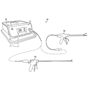
16
CA 2791681 2018-10-22
for example, the ultrasonic surgical device 104 and electrosurgical or RF
surgical device 106.
Although in the embodiment of Figure 1 the generator 102 is shown separate
from the surgical
devices 104, 106, in certain embodiments the generator 102 may be formed
integrally with
either of the surgical devices 104, 106 to form a unitary surgical system.
[00108] Figure 2 illustrates one embodiment of an example ultrasonic device
104 that
may be used for transection and/or sealing. The device 104 may comprise a hand
piece 116
which may, in turn, comprise an ultrasonic transducer 114. The transducer 114
may be in
electrical communication with the generator 102, for example, via a cable 122
(e.g., a multi-
conductor cable). The transducer 114 may comprise piezoceramic elements, or
other elements
or components suitable for converting the electrical energy of a drive signal
into mechanical
vibrations. When activated by the generator 102, the ultrasonic transducer 114
may cause
longitudinal vibration. The vibration may be transmitted through an instrument
portion 124 of
the device 104 (e.g., via a waveguide embedded in an outer sheath) to an end
effector 126 of
the instrument portion 124.
[00109] Figure 3 illustrates one embodiment of the end effector 126 of the
example
ultrasonic device 104. The end effector 126 may comprise a blade 151 that may
be coupled to
the ultrasonic transducer 114 via the wave guide (not shown). When driven by
the transducer
114, the blade 151 may vibrate and, when brought into contact with tissue, may
cut and/or
coagulate the tissue, as described herein. According to various embodiments,
and as illustrated
in Figure 3, the end effector 126 may also comprise a clamp arm 155 that may
be configured for
cooperative action with the blade 151 of the end effector 126. With the blade
151, the clamp
arm 155 may comprise a set of jaws 140. The clamp arm 155 may be pivotally
connected at a
distal end of a shaft 153 of the instrument portion 124. The clamp arm 155 may
include a clamp
arm tissue pad 163, which may be formed from TEFLON or other suitable low-
friction material.
The pad 163 may be mounted for cooperation with the blade 151, with pivotal
movement of the
clamp arm 155 positioning the clamp pad 163 in substantially parallel
relationship to, and in
contact with, the blade 151. By this construction, a tissue bite to be clamped
may be grasped
between the tissue pad 163 and the blade 151. The tissue pad 163 may be
provided with a
sawtooth-like configuration including a plurality of axially spaced,
proximally extending gripping
teeth 161 to enhance the gripping of tissue in cooperation with the blade 151.
The clamp arm
155 may transition from the open position shown in Figure 3 to a closed
position (with the clamp
arm 155 in contact with or proximity to the blade 151) in any suitable manner.
For example, the
hand piece 116 may comprise a jaw closure trigger 138. When actuated by a
clinician, the jaw
closure trigger 138 may pivot the clamp arm 155 in any suitable manner.
17
CA 2791681 2018-10-22
[00110] The generator 102 may be activated to provide the drive signal to
the transducer
114 in any suitable manner. For example, the generator 102 may comprise a foot
switch 120
coupled to the generator 102 via a footswitch cable 122 (Figure 8). A
clinician may activate the
transducer 114, and thereby the transducer 114 and blade 151, by depressing
the foot switch
120. In addition, or instead of the foot switch 120 some embodiments of the
device 104 may
utilize one or more switches positioned on the hand piece 116 that, when
activated, may cause
the generator 102 to activate the transducer 114. In one embodiment, for
example, the one or
more switches may comprise a pair of toggle buttons 136a, 136b, for example,
to determine an
operating mode of the device 104. When the toggle button 136a is depressed,
for example, the
ultrasonic generator 102 may provide a maximum drive signal to the transducer
114, causing it
to produce maximum ultrasonic energy output. Depressing toggle button 136b may
cause the
ultrasonic generator 102 to provide a user-selectable drive signal to the
transducer 114, causing
it to produce less than the maximum ultrasonic energy output. The device 104
additionally or
alternatively may comprise a second switch to, for example, indicate a
position of a jaw closure
trigger 138 for operating jaws 140 of the end effector 126. Also, in some
embodiments, the
ultrasonic generator 102 may be activated based on the position of the jaw
closure trigger 138,
(e.g., as the clinician depresses the jaw closure trigger 138 to close the
jaws 140, ultrasonic
energy may be applied.
[00111] Additionally or alternatively, the one or more switches may
comprises a toggle
button 136c that, when depressed, causes the generator 102 to provide a pulsed
output. The
pulses may be provided at any suitable frequency and grouping, for example. In
certain
embodiments, the power level of the pulses may be the power levels associated
with toggle
buttons 136a,b (maximum, less than maximum), for example.
[00112] It will be appreciated that a device 104 may comprise any
combination of the
toggle buttons 136a,b,c. For example, the device 104 could be configured to
have only two
toggle buttons: a toggle button 136a for producing maximum ultrasonic energy
output and a
toggle button 136c for producing a pulsed output at either the maximum or less
than maximum
power level per. In this way, the drive signal output configuration of the
generator 102 could be
continuous signals and 5 or 4 or 3 or 2 or 1 pulsed signals. In certain
embodiments, the
specific drive signal configuration may be controlled based upon, for example,
EEPROM
settings in the generator 102 and/or user power level selection(s).
[00113] In certain embodiments, a two-position switch may be provided as an
alternative
to a toggle button 136c. For example, a device 104 may include a toggle button
136a for
producing a continuous output at a maximum power level and a two-position
toggle button 136b.
18
CA 2791681 2018-10-22
In a first detented position, toggle button 136b may produce a continuous
output at a less than
maximum power level, and in a second detented position the toggle button 136b
may produce a
pulsed output (e.g., at either a maximum or less than maximum power level,
depending upon
the EEPROM settings).
[00114] In some embodiments, the end effector 126 may also comprise a pair
of
electrodes 159, 157. The electrodes 159, 157 may be in communication with the
generator 102,
for example, via the cable 122. The electrodes 159, 157 may be used, for
example, to measure
an impedance of a tissue bite present between the clamp arm 155 and the blade
151. The
generator 102 may provide a signal (e.g., a non-therapeutic signal) to the
electrodes 159, 157.
The impedance of the tissue bite may be found, for example, by monitoring the
current, voltage,
etc. of the signal.
[00115] Figure 4 illustrates one embodiment of an example electrosurgical
device 106
that may also be used for transection and sealing. According to various
embodiments, the
transection and sealing device 106 may comprise a hand piece assembly 130, a
shaft 165 and
an end effector 132. The shaft 165 may be rigid (e.g., for laparoscopic and/or
open surgical
application) or flexible, as shown, (e.g., for endoscopic application). In
various embodiments,
the shaft 165 may comprise one or more articulation points. The end effector
132 may
comprise jaws 144 having a first jaw member 167 and a second jaw member 169.
The first jaw
member 167 and second jaw member 169 may be connected to a clevis 171, which,
in turn,
may be coupled to the shaft 165. A translating member 173 may extend within
the shaft 165
from the end effector 132 to the hand piece 130. At the hand piece 130, the
shaft 165 may be
directly or indirectly coupled to a jaw closure trigger 142 (Figure 4).
[00116] The jaw members 167, 169 of the end effector 132 may comprise
respective
electrodes 177, 179. The electrodes 177, 179 may be connected to the generator
102 via
electrical leads 187a, 187b (Figure 5) extending from the end effector 132
through the shaft 165
and hand piece 130 and ultimately to the generator 102 (e.g., by a
multiconductor cable 128).
The generator 102 may provide a drive signal to the electrodes 177, 179 to
bring about a
therapeutic effect to tissue present within the jaw members 167, 169. The
electrodes 177, 179
may comprise an active electrode and a return electrode, wherein the active
electrode and the
return electrode can be positioned against, or adjacent to, the tissue to be
treated such that
current can flow from the active electrode to the return electrode through the
tissue. As
illustrated in Figure 4, the end effector 132 is shown with the jaw members
167, 169 in an open
position. A reciprocating blade 175 is illustrated between the jaw members
167, 169.
19
CA 2791681 2018-10-22
[00117] Figures 5, 6 and 7 illustrate one embodiment of the end effector
132 shown in
Figure 4. To close the jaws 144 of the end effector 132, a clinician may cause
the jaw closure
trigger 142 to pivot along arrow 183 from a first position to a second
position. This may cause
the jaws 144 to open and close according to any suitable method. For example,
motion of the
jaw closure trigger 142 may, in turn, cause the translating member 173 to
translate within a bore
185 of the shaft 165. A distal portion of the translating member 173 may be
coupled to a
reciprocating member 197 such that distal and proximal motion of the
translating member 173
causes corresponding distal and proximal motion of the reciprocating member.
The
reciprocating member 197 may have shoulder portions 191a, 191b, while the jaw
members 167,
169 may have corresponding cam surfaces 189a, 189b. As the reciprocating
member 197 is
translated distally from the position shown in Figure 6 to the position shown
in Figure 7, the
shoulder portions 191a, 191b may contact the cam surfaces 189a, 189b, causing
the jaw
members 167, 169 to transition to the closed position. Also, in various
embodiments, the blade
175 may be positioned at a distal end of the reciprocating member 197. As the
reciprocating
member extends to the fully distal position shown in Figure 7, the blade 175
may be pushed
through any tissue present between the jaw members 167, 169, in the process,
severing it.
[00118] In use, a clinician may place the end effector 132 and close the
jaws 144 around
a tissue bite to be acted upon, for example, by pivoting the jaw closure
trigger 142 along arrow
183 as described. Once the tissue bite is secure between the jaws 144, the
clinician may
initiate the provision of RF or other electro-surgical energy by the generator
102 and through the
electrodes 177, 179. The provision of RF energy may be accomplished in any
suitable way.
For example, the clinician may activate the foot switch 120 (Figure 8) of the
generator 102 to
initiate the provision of RF energy. Also, for example, the hand piece 130 may
comprise one or
more switches 181 that may be actuated by the clinician to cause the generator
102 to begin
providing RF energy. Additionally, in some embodiments, RF energy may be
provided based
on the position of the jaw closure trigger 142. For example, when the trigger
142 is fully
depressed (indicating that the jaws 144 are closed), RF energy may be
provided. Also,
according to various embodiments, the blade 175 may be advanced during closure
of the jaws
144 or may be separately advanced by the clinician after closure of the jaws
144 (e.g., after a
RF energy has been applied to the tissue).
[00119] Figure 8 is a diagram of the surgical system 100 of Figure 1. In
various
embodiments, the generator 102 may comprise several separate functional
elements, such as
modules and/or blocks. Different functional elements or modules may be
configured for driving
the different kinds of surgical devices 104, 106. For example an ultrasonic
generator module
CA 2791681 2018-10-22
108 may drive an ultrasonic device, such as the ultrasonic device 104. An
electrosurgery/RF
generator module 110 may drive the electrosurgical device 106. For example,
the respective
modules 108, 110 may generate respective drive signals for driving the
surgical devices 104,
106. In various embodiments, the ultrasonic generator module 108 and/or the
electrosurgery
/RF generator module 110 each may be formed integrally with the generator 102.
Alternatively,
one or more of the modules 108, 110 may be provided as a separate circuit
module electrically
coupled to the generator 102. (The modules 108 and 110 are shown in phantom to
illustrate
this option.) Also, in some embodiments, the electrosurgery/RF generator
module 110 may be
formed integrally with the ultrasonic generator module 108, or vice versa
[00120] In accordance with the described embodiments, the ultrasonic
generator module
108 may produce a drive signal or signals of particular voltages, currents,
and frequencies, e.g.
55,500 cycles per second (Hz). The drive signal or signals may be provided to
the ultrasonic
device 104, and specifically to the transducer 114, which may operate, for
example, as
described above. In one embodiment, the generator 102 may be configured to
produce a drive
signal of a particular voltage, current, and/or frequency output signal that
can be stepped with
high resolution, accuracy, and repeatability.
[00121] In accordance with the described embodiments, the electrosurgery/RF
generator
module 110 may generate a drive signal or signals with output power sufficient
to perform
bipolar electrosurgery using radio frequency (RF) energy. In bipolar
electrosurgery applications.
The drive signal may be provided, for example, to the electrodes 177, 179 of
the electrosurgical
device 106, for example, as described above. Accordingly, the generator 102
may be
configured for therapeutic purposes by applying electrical energy to the
tissue sufficient for
treating the tissue (e.g., coagulation, cauterization, tissue welding, etc.).
[00122] The generator 102 may comprise an input device 145 (Figure 1)
located, for
example, on a front panel of the generator 102 console. The input device 145
may comprise
any suitable device that generates signals suitable for programming the
operation of the
generator 102. In operation, the user can program or otherwise control
operation of the
generator 102 using the input device 145. The input device 145 may comprise
any suitable
device that generates signals that can be used by the generator (e.g., by one
or more
processors contained in the generator) to control the operation of the
generator 102 (e.g.,
operation of the ultrasonic generator module 108 and/or electrosurgery/RF
generator module
110). In various embodiments, the input device 145 includes one or more of
buttons, switches,
thumbwheels, keyboard, keypad, touch screen monitor, pointing device, remote
connection to a
general purpose or dedicated computer. In other embodiments, the input device
145 may
21
CA 2791681 2018-10-22
comprise a suitable user interface, such as one or more user interface screens
displayed on a
touch screen monitor, for example. Accordingly, by way of the input device
145, the user can
set or program various operating parameters of the generator, such as, for
example, current (I),
voltage (V), frequency (f), and/or period (T) of a drive signal or signals
generated by the
ultrasonic generator module 108 and/or electrosurgery/RF generator module 110.
[00123] The generator 102 may also comprise an output device 147 (Figure 1)
located,
for example, on a front panel of the generator 102 console. The output device
147 includes one
or more devices for providing a sensory feedback to a user. Such devices may
comprise, for
example, visual feedback devices (e.g., an LCD display screen, LED
indicators), audio feedback
devices (e.g., a speaker, a buzzer) or tactile feedback devices (e.g., haptic
actuators).
[00124] Although certain modules and/or blocks of the generator 102 may be
described
by way of example, it can be appreciated that a greater or lesser number of
modules and/or
blocks may be used and still fall within the scope of the embodiments.
Further, although various
embodiments may be described in terms of modules and/or blocks to facilitate
description, such
modules and/or blocks may be implemented by one or more hardware components,
e.g.,
processors, Digital Signal Processors (DSPs), Programmable Logic Devices
(PLDs), Application
Specific Integrated Circuits (ASICs), circuits, registers and/or software
components, e.g.,
programs, subroutines, logic and/or combinations of hardware and software
components.
[00125] In one embodiment, the ultrasonic generator drive module 108 and
electrosurgery/RF drive module 110 may comprise one or more embedded
applications
implemented as firmware, software, hardware, or any combination thereof. The
modules 108,
110 may comprise various executable modules such as software, programs, data,
drivers,
application program interfaces (APIs), and so forth. The firmware may be
stored in nonvolatile
memory (NVM), such as in bit-masked read-only memory (ROM) or flash memory. In
various
implementations, storing the firmware in ROM may preserve flash memory. The
NVM may
comprise other types of memory including, for example, programmable ROM
(PROM), erasable
programmable ROM (EPROM), electrically erasable programmable ROM (EEPROM), or
battery
backed random-access memory (RAM) such as dynamic RAM (DRAM), Double-Data-Rate
DRAM (DDRAM), and/or synchronous DRAM (SDRAM).
[00126] In one embodiment, the modules 108, 110 comprise a hardware
component
implemented as a processor for executing program instructions for monitoring
various
measurable characteristics of the devices 104, 106 and generating a
corresponding output drive
signal or signals for operating the devices 104, 106. In embodiments in which
the generator
102 is used in conjunction with the device 104, the drive signal may drive the
ultrasonic
22
CA 2791681 2018-10-22
transducer 114 in cutting and/or coagulation operating modes. Electrical
characteristics of the
device 104 and/or tissue may be measured and used to control operational
aspects of the
generator 102 and/or provided as feedback to the user. In embodiments in which
the generator
102 is used in conjunction with the device 106, the drive signal may supply
electrical energy
(e.g., RF energy) to the end effector 132 in cutting, coagulation and/or
desiccation modes.
Electrical characteristics of the device 106 and/or tissue may be measured and
used to control
operational aspects of the generator 102 and/or provided as feedback to the
user. In various
embodiments, as previously discussed, the hardware components may be
implemented as
DSP, PLD, ASIC, circuits, and/or registers. In one embodiment, the processor
may be
configured to store and execute computer software program instructions to
generate the step
function output signals for driving various components of the devices 104,
106, such as the
ultrasonic transducer 114 and the end effectors 126, 132.
[00127] Figure 9 illustrates an equivalent circuit 150 of an ultrasonic
transducer, such as
the ultrasonic transducer 114, according to one embodiment. The circuit 150
comprises a first
"motional" branch having a serially connected inductance L5, resistance R, and
capacitance Cs
that define the electromechanical properties of the resonator, and a second
capacitive branch
having a static capacitance Co. Drive current Ig may be received from a
generator at a drive
voltage Vg, with motional current in, flowing through the first branch and
current Ig¨ lin flowing
through the capacitive branch. Control of the electromechanical properties of
the ultrasonic
transducer may be achieved by suitably controlling /g and Vg. As explained
above, known
generator architectures may include a tuning inductor Lt (shown in phantom in
Figure 9) for
tuning out in a parallel resonance circuit the static capacitance Co at a
resonant frequency so
that substantially all of generator's current output /g flows through the
motional branch. In this
way, control of the motional branch current /,õ is achieved by controlling the
generator current
output lg. The tuning inductor L, is specific to the static capacitance Cod an
ultrasonic
transducer, however, and a different ultrasonic transducer having a different
static capacitance
requires a different tuning inductor L. Moreover, because the tuning inductor
Lt is matched to
the nominal value of the static capacitance Co at a single resonant frequency,
accurate control
of the motional branch current /n, is assured only at that frequency, and as
frequency shifts
down with transducer temperature, accurate control of the motional branch
current is
compromised.
[00128] Various embodiments of the generator 102 may not rely on a tuning
inductor Lt
to monitor the motional branch current /rn. Instead, the generator 102 may use
the measured
value of the static capacitance CO in between applications of power for a
specific ultrasonic
23
CA 2791681 2018-10-22
surgical device 104 (along with drive signal voltage and current feedback
data) to determine
values of the motional branch current 47., on a dynamic and ongoing basis
(e.g., in real-time).
Such embodiments of the generator 102 are therefore able to provide virtual
tuning to simulate
a system that is tuned or resonant with any value of static capacitance Co at
any frequency, and
not just at a single resonant frequency dictated by a nominal value of the
static capacitance Co.
[00129] Figure 10 is a simplified block diagram of one embodiment of the
generator 102
for proving inductorless tuning as described above, among other benefits.
Figures 11A-11C
illustrate an architecture of the generator 102 of Figure 10 according to one
embodiment. With
reference to Figure 10, the generator 102 may comprise a patient isolated
stage 152 in
communication with a non-isolated stage 154 via a power transformer 156. A
secondary
winding 158 of the power transformer 156 is contained in the isolated stage
152 and may
comprise a tapped configuration (e.g., a center-tapped or non-center tapped
configuration) to
define drive signal outputs 160a, 160b, 160c for outputting drive signals to
different surgical
devices, such as, for example, an ultrasonic surgical device 104 and an
electrosurgical device
106. In particular, drive signal outputs 160a, 160c may output a drive signal
(e.g., a 420V RMS
drive signal) to an ultrasonic surgical device 104, and drive signal outputs
160b, 160c may
output a drive signal (e.g., a 100V RMS drive signal) to an electrosurgical
device 106, with
output 160b corresponding to the center tap of the power transformer 156. The
non-isolated
stage 154 may comprise a power amplifier 162 having an output connected to a
primary winding
164 of the power transformer 156. In certain embodiments the power amplifier
162 may
comprise a push-pull amplifier, for example. The non-isolated stage 154 may
further comprise a
programmable logic device 166 for supplying a digital output to a digital-to-
analog converter
(DAC) 168, which in turn supplies a corresponding analog signal to an input of
the power
amplifier 162. In certain embodiments the programmable logic device 166 may
comprise a
field-programmable gate array (FPGA), for example. The programmable logic
device 166, by
virtue of controlling the power amplifier's 162 input via the DAC 168, may
therefore control any
of a number of parameters (e.g., frequency, waveform shape, waveform
amplitude) of drive
signals appearing at the drive signal outputs 160a, 160b, 160c. In certain
embodiments and as
discussed below, the programmable logic device 166, in conjunction with a
processor (e.g.,
processor 174 discussed below), may implement a number of digital signal
processing (DSP)-
based and/or other control algorithms to control parameters of the drive
signals output by the
generator 102.
[00130] Power may be supplied to a power rail of the power amplifier 162 by
a switch-
mode regulator 170. In certain embodiments the switch-mode regulator 170 may
comprise an
24
CA 2791681 2018-10-22
adjustable buck regulator, for example. The non-isolated stage 154 may further
comprise a
processor 174, which in one embodiment may comprise a DSP processor such as an
Analog
Devices ADSP-21469 SHARC DSP, available from Analog Devices, Norwood, MA, for
example.
In certain embodiments the processor 174 may control operation of the switch-
mode power
converter 170 responsive to voltage feedback data received from the power
amplifier 162 by the
processor 174 via an analog-to-digital converter (ADC) 176. In one embodiment,
for example,
the processor 174 may receive as input, via the ADC 176, the waveform envelope
of a signal
(e.g., an RF signal) being amplified by the power amplifier 162. The processor
174 may then
control the switch-mode regulator 170 (e.g., via a pulse-width modulated (PWM)
output) such
that the rail voltage supplied to the power amplifier 162 tracks the waveform
envelope of the
amplified signal. By dynamically modulating the rail voltage of the power
amplifier 162 based on
the waveform envelope, the efficiency of the power amplifier 162 may be
significantly improved
relative to a fixed rail voltage amplifier schemes.
[00131] In certain embodiments and as discussed in further detail in
connection with
Figure 13, the programmable logic device 166, in conjunction with the
processor 174, may
implement a direct digital synthesizer (DDS) control scheme to control the
waveform shape,
frequency and/or amplitude of drive signals output by the generator 102. In
one embodiment,
for example, the programmable logic device 166 may implement a DDS control
algorithm 268
by recalling waveform samples stored in a dynamically-updated look-up table
(LUT), such as a
RAM LUT which may be embedded in an FPGA. This control algorithm is
particularly useful for
ultrasonic applications in which an ultrasonic transducer, such as the
ultrasonic transducer 114,
may be driven by a clean sinusoidal current at its resonant frequency. Because
other
frequencies may excite parasitic resonances, minimizing or reducing the total
distortion of the
motional branch current may correspondingly minimize or reduce undesirable
resonance
effects. Because the waveform shape of a drive signal output by the generator
102 is impacted
by various sources of distortion present in the output drive circuit (e.g.,
the power transformer
156, the power amplifier 162), voltage and current feedback data based on the
drive signal may
be input into an algorithm, such as an error control algorithm implemented by
the processor
174, which compensates for distortion by suitably pre-distorting or modifying
the waveform
samples stored in the LUT on a dynamic, ongoing basis (e.g., in real-time). In
one embodiment,
the amount or degree of pre-distortion applied to the LUT samples may be based
on the error
between a computed motional branch current and a desired current waveform
shape, with the
error being determined on a sample-by sample basis. In this way, the pre-
distorted LUT
samples, when processed through the drive circuit, may result in a motional
branch drive signal
CA 2791681 2018-10-22
having the desired waveform shape (e.g., sinusoidal) for optimally driving the
ultrasonic
transducer. In such embodiments, the LUT waveform samples will therefore not
represent the
desired waveform shape of the drive signal, but rather the waveform shape that
is required to
ultimately produce the desired waveform shape of the motional branch drive
signal when
distortion effects are taken into account.
[00132] The non-isolated stage 154 may further comprise an ADC 178 and an
ADC 180
coupled to the output of the power transformer 156 via respective isolation
transformers 182,
184 for respectively sampling the voltage and current of drive signals output
by the generator
102. In certain embodiments, the ADCs 178, 180 may be configured to sample at
high speeds
(e.g., 80 Msps) to enable oversampling of the drive signals. In one
embodiment, for example,
the sampling speed of the ADCs 178, 180 may enable approximately 200x
(depending on drive
frequency) oversampling of the drive signals. In certain embodiments, the
sampling operations
of the ADCs 178, 180 may be performed by a single ADC receiving input voltage
and current
signals via a two-way multiplexer. The use of high-speed sampling in
embodiments of the
generator 102 may enable, among other things, calculation of the complex
current flowing
through the motional branch (which may be used in certain embodiments to
implement DDS-
based waveform shape control described above), accurate digital filtering of
the sampled
signals, and calculation of real power consumption with a high degree of
precision. Voltage and
current feedback data output by the ADCs 178, 180 may be received and
processed (e.g., FIFO
buffering, multiplexing) by the programmable logic device 166 and stored in
data memory for
subsequent retrieval by, for example, the processor 174. As noted above,
voltage and current
feedback data may be used as input to an algorithm for pre-distorting or
modifying LUT
waveform samples on a dynamic and ongoing basis. In certain embodiments, this
may require
each stored voltage and current feedback data pair to be indexed based on, or
otherwise
associated with, a corresponding LUT sample that was output by the
programmable logic device
166 when the voltage and current feedback data pair was acquired.
Synchronization of the LUT
samples and the voltage and current feedback data in this manner contributes
to the correct
timing and stability of the pre-distortion algorithm.
[00133] In certain embodiments, the voltage and current feedback data may
be used to
control the frequency and/or amplitude (e.g., current amplitude) of the drive
signals. In one
embodiment, for example, voltage and current feedback data may be used to
determine
impedance phase. The frequency of the drive signal may then be controlled to
minimize or
reduce the difference between the determined impedance phase and an impedance
phase
setpoint (e.g., 00), thereby minimizing or reducing the effects of harmonic
distortion and
26
CA 2791681 2018-10-22
correspondingly enhancing impedance phase measurement accuracy. The
determination of
phase impedance and a frequency control signal may be implemented in the
processor 174, for
example, with the frequency control signal being supplied as input to a DDS
control algorithm
implemented by the programmable logic device 166.
[00134] In another embodiment, for example, the current feedback data may
be
monitored in order to maintain the current amplitude of the drive signal at a
current amplitude
setpoint. The current amplitude setpoint may be specified directly or
determined indirectly
based on specified voltage amplitude and power setpoints. In certain
embodiments, control of
the current amplitude may be implemented by control algorithm, such as, for
example, a
proportional-integral-derivative (PID) control algorithm, in the processor
174. Variables
controlled by the control algorithm to suitably control the current amplitude
of the drive signal
may include, for example, the scaling of the LUT waveform samples stored in
the programmable
logic device 166 and/or the full-scale output voltage of the DAC 168 (which
supplies the input to
the power amplifier 162) via a DAC 186.
[00135] The non-isolated stage 154 may further comprise a processor 190 for
providing,
among other things user interface (UI) functionality. In one embodiment, the
processor 190 may
comprise an Atmel AT91SAM9263 processor having an ARM 926EJ-S core, available
from
Atmel Corporation, San Jose, California, for example. Examples of Ul
functionality supported
by the processor 190 may include audible and visual user feedback,
communication with
peripheral devices (e.g., via a Universal Serial Bus (USB) interface),
communication with the
footswitch 120, communication with an input device 112 (e.g., a touch screen
display) and
communication with an output device 147 (e.g., a speaker). The processor 190
may
communicate with the processor 174 and the programmable logic device (e.g.,
via serial
peripheral interface (SPI) buses). Although the processor 190 may primarily
support Ul
functionality, it may also coordinate with the processor 174 to implement
hazard mitigation in
certain embodiments. For example, the processor 190 may be programmed to
monitor various
aspects of user input and/or other inputs (e.g., touch screen inputs,
footswitch 120 inputs,
temperature sensor inputs) and may disable the drive output of the generator
102 when an
erroneous condition is detected.
[00136] In certain embodiments, both the processor 174 and the processor
190 may
determine and monitor the operating state of the generator 102. For the
processor 174, the
operating state of the generator 102 may dictate, for example, which control
and/or diagnostic
processes are implemented by the processor 174. For the processor 190, the
operating state of
the generator 102 may dictate, for example, which elements of a user interface
(e.g., display
27
CA 2791681 2018-10-22
screens, sounds) are presented to a user. The processors 174, 190 may
independently
maintain the current operating state of the generator 102 and recognize and
evaluate possible
transitions out of the current operating state. The processor 174 may function
as the master in
this relationship and determine when transitions between operating states are
to occur. The
processor 190 may be aware of valid transitions between operating states and
may confirm if a
particular transition is appropriate. For example, when the processor 174
instructs the
processor 190 to transition to a specific state, the processor 190 may verify
that requested
transition is valid. In the event that a requested transition between states
is determined to be
invalid by the processor 190, the processor 190 may cause the generator 102 to
enter a failure
mode.
[00137] The non-isolated stage 154 may further comprise a controller 196
for monitoring
input devices 145 (e.g., a capacitive touch sensor used for turning the
generator 102 on and off,
a capacitive touch screen). In certain embodiments, the controller 196 may
comprise at least
one processor and/or other controller device in communication with the
processor 190. In one
embodiment, for example, the controller 196 may comprise a processor (e.g., a
Mega168 8-bit
controller available from Atmel) configured to monitor user input provided via
one or more
capacitive touch sensors. In one embodiment, the controller 196 may comprise a
touch screen
controller (e.g., a QT5480 touch screen controller available from Atmel) to
control and manage
the acquisition of touch data from a capacitive touch screen.
[00138] In certain embodiments, when the generator 102 is in a "power off'
state, the
controller 196 may continue to receive operating power (e.g., via a line from
a power supply of
the generator 102, such as the power supply 211 discussed below). In this way,
the controller
196 may continue to monitor an input device 145 (e.g., a capacitive touch
sensor located on a
front panel of the generator 102) for turning the generator 102 on and off.
When the generator
102 is in the power off state, the controller 196 may wake the power supply
(e.g., enable
operation of one or more DC/DC voltage converters 213 of the power supply 211)
if activation of
the "on/off" input device 145 by a user is detected. The controller 196 may
therefore initiate a
sequence for transitioning the generator 102 to a "power on" state.
Conversely, the controller
196 may initiate a sequence for transitioning the generator 102 to the power
off state if
activation of the "on/off' input device 145 is detected when the generator 102
is in the power on
state. In certain embodiments, for example, the controller 196 may report
activation of the
"on/off' input device 145 to the processor 190, which in turn implements the
necessary process
sequence for transitioning the generator 102 to the power off state. In such
embodiments, the
28
CA 2791681 2018-10-22
controller 196 may have no independent ability for causing the removal of
power from the
generator 102 after its power on state has been established.
[00139] In certain embodiments, the controller 196 may cause the generator
102 to
provide audible or other sensory feedback for alerting the user that a power
on or power off
sequence has been initiated. Such an alert may be provided at the beginning of
a power on or
power off sequence and prior to the commencement of other processes associated
with the
sequence.
[00140] In certain embodiments, the isolated stage 152 may comprise an
instrument
interface circuit 198 to, for example, provide a communication interface
between a control circuit
of a surgical device (e.g., a control circuit comprising handpiece switches)
and components of
the non-isolated stage 154, such as, for example, the programmable logic
device 166, the
processor 174 and/or the processor 190. The instrument interface circuit 198
may exchange
information with components of the non-isolated stage 154 via a communication
link that
maintains a suitable degree of electrical isolation between the stages 152,
154, such as, for
example, an infrared (IR)-based communication link. Power may be supplied to
the instrument
interface circuit 198 using, for example, a low-dropout voltage regulator
powered by an isolation
transformer driven from the non-isolated stage 154.
[00141] In one embodiment, the instrument interface circuit 198 may
comprise a
programmable logic device 200 (e.g., an FPGA) in communication with a signal
conditioning
circuit 202. The signal conditioning circuit 202 may be configured to receive
a periodic signal
from the programmable logic device 200 (e.g., a 2 kHz square wave) to generate
a bipolar
interrogation signal having an identical frequency. The interrogation signal
may be generated,
for example, using a bipolar current source fed by a differential amplifier.
The interrogation
signal may be communicated to a surgical device control circuit (e.g., by
using a conductive pair
in a cable that connects the generator 102 to the surgical device) and
monitored to determine a
state or configuration of the control circuit. As discussed below in
connection with Figures 16-
32, for example, the control circuit may comprise a number of switches,
resistors and/or diodes
to modify one or more characteristics (e.g., amplitude, rectification) of the
interrogation signal
such that a state or configuration of the control circuit is uniquely
discernable based on the one
or more characteristics. In one embodiment, for example, the signal
conditioning circuit 202
may comprise an ADC for generating samples of a voltage signal appearing
across inputs of the
control circuit resulting from passage of interrogation signal therethrough.
The programmable
logic device 200 (or a component of the non-isolated stage 154) may then
determine the state
or configuration of the control circuit based on the ADC samples.
29
CA 2791681 2018-10-22
[00142] In one embodiment, the instrument interface circuit 198 may
comprise a first data
circuit interface 204 to enable information exchange between the programmable
logic device
200 (or other element of the instrument interface circuit 198) and a first
data circuit disposed in
or otherwise associated with a surgical device. In certain embodiments and
with reference to
Figures 33E-33G, for example, a first data circuit 206 may be disposed in a
cable integrally
attached to a surgical device handpiece, or in an adaptor for interfacing a
specific surgical
device type or model with the generator 102. In certain embodiments, the first
data circuit may
comprise a non-volatile storage device, such as an electrically erasable
programmable read-
only memory (EEPROM) device. In certain embodiments and referring again to
Figure 10, the
first data circuit interface 204 may be implemented separately from the
programmable logic
device 200 and comprise suitable circuitry (e.g., discrete logic devices, a
processor) to enable
communication between the programmable logic device 200 and the first data
circuit. In other
embodiments, the first data circuit interface 204 may be integral with the
programmable logic
device 200.
[00143] In certain embodiments, the first data circuit 206 may store
information pertaining
to the particular surgical device with which it is associated. Such
information may include, for
example, a model number, a serial number, a number of operations in which the
surgical device
has been used, and/or any other type of information. This information may be
read by the
instrument interface circuit 198 (e.g., by the programmable logic device 200),
transferred to a
component of the non-isolated stage 154 (e.g., to programmable logic device
166, processor
174 and/or processor 190) for presentation to a user via an output device 147
and/or for
controlling a function or operation of the generator 102. Additionally, any
type of information
may be communicated to first data circuit 206 for storage therein via the
first data circuit
interface 204 (e.g., using the programmable logic device 200). Such
information may comprise,
for example, an updated number of operations in which the surgical device has
been used
and/or dates and/or times of its usage.
[00144] As discussed previously, a surgical instrument may be detachable
from a
handpiece (e.g., instrument 124 may be detachable from handpiece 116) to
promote instrument
interchangeability and/or disposability. In such cases, known generators may
be limited in their
ability to recognize particular instrument configurations being used and to
optimize control and
diagnostic processes accordingly. The addition of readable data circuits to
surgical device
instruments to address this issue is problematic from a compatibility
standpoint, however. For
example, designing a surgical device to remain backwardly compatible with
generators that lack
the requisite data reading functionality may be impractical due to, for
example, differing signal
CA 2791681 2018-10-22
schemes, design complexity and cost. Embodiments of instruments discussed
below in
connection with Figures 16-32 address these concerns by using data circuits
that may be
implemented in existing surgical instruments economically and with minimal
design changes to
preserve compatibility of the surgical devices with current generator
platforms.
[00145] Additionally, embodiments of the generator 102 may enable
communication with
instrument-based data circuits, such as those described below in connection
with Figures 16-32
and Figures 33A-33C. For example, the generator 102 may be configured to
communicate with
a second data circuit (e.g., data circuit 284 of Figure 16) contained in an
instrument (e.g.,
instrument 124 or 134) of a surgical device. The instrument interface circuit
198 may comprise
a second data circuit interface 210 to enable this communication. In one
embodiment, the
second data circuit interface 210 may comprise a tri-state digital interface,
although other
interfaces may also be used. In certain embodiments, the second data circuit
may generally be
any circuit for transmitting and/or receiving data. In one embodiment, for
example, the second
data circuit may store information pertaining to the particular surgical
instrument with which it is
associated. Such information may include, for example, a model number, a
serial number, a
number of operations in which the surgical instrument has been used, and/or
any other type of
information. Additionally or alternatively, any type of information may be
communicated to
second data circuit for storage therein via the second data circuit interface
210 (e.g., using the
programmable logic device 200). Such information may comprise, for example, an
updated
number of operations in which the instrument has been used and/or dates and/or
times of its
usage. In certain embodiments, the second data circuit may transmit data
acquired by one or
more sensors (e.g., an instrument-based temperature sensor). In certain
embodiments, the
second data circuit may receive data from the generator 102 and provide an
indication to a user
(e.g., an LED indication or other visible indication) based on the received
data.
[00146] In certain embodiments, the second data circuit and the second data
circuit
interface 210 may be configured such that communication between the
programmable logic
device 200 and the second data circuit can be effected without the need to
provide additional
conductors for this purpose (e.g., dedicated conductors of a cable connecting
a handpiece to
the generator 102). In one embodiment, for example, information may be
communicated to and
from the second data circuit using a 1-wire bus communication scheme
implemented on existing
cabling, such as one of the conductors used transmit interrogation signals
from the signal
conditioning circuit 202 to a control circuit in a handpiece. In this way,
design changes or
modifications to the surgical device that might otherwise be necessary are
minimized or
reduced. Moreover, as discussed in further detail below in connection with
Figures 16-32 and
31
CA 2791681 2018-10-22
Figures 33A-330, because different types of communications can be implemented
over a
common physical channel (either with or without frequency-band separation),
the presence of a
second data circuit may be "invisible" to generators that do not have the
requisite data reading
functionality, thus enabling backward compatibility of the surgical device
instrument.
[00147] In certain embodiments, the isolated stage 152 may comprise at
least one
blocking capacitor 296-1 connected to the drive signal output 160b to prevent
passage of DC
current to a patient. A single blocking capacitor may be required to comply
with medical
regulations or standards, for example. While failure in single-capacitor
designs is relatively
uncommon, such failure may nonetheless have negative consequences. In one
embodiment, a
second blocking capacitor 296-2 may be provided in series with the blocking
capacitor 296-1,
with current leakage from a point between the blocking capacitors 296-1, 296-2
being monitored
by, for example, an ADC 298 for sampling a voltage induced by leakage current.
The samples
may be received by the programmable logic device 200, for example. Based on
changes in the
leakage current (as indicated by the voltage samples in the embodiment of
Figure 10), the
generator 102 may determine when at least one of the blocking capacitors 296-
1, 296-2 has
failed. Accordingly, the embodiment of Figure 10 may provide a benefit over
single-capacitor
designs having a single point of failure.
[00148] In certain embodiments, the non-isolated stage 154 may comprise a
power
supply 211 for outputting DC power at a suitable voltage and current. The
power supply may
comprise, for example, a 400 W power supply for outputting a 48 VDC system
voltage. The
power supply 211 may further comprise one or more DC/DC voltage converters 213
for
receiving the output of the power supply to generate DC outputs at the
voltages and currents
required by the various components of the generator 102. As discussed above in
connection
with the controller 196, one or more of the DC/DC voltage converters 213 may
receive an input
from the controller 196 when activation of the "on/off' input device 145 by a
user is detected by
the controller 196 to enable operation of, or wake, the DC/DC voltage
converters 213.
[00149] Figure 13 illustrates certain functional and structural aspects of
one embodiment
of the generator 102. Feedback indicating current and voltage output from the
secondary
winding 158 of the power transformer 156 is received by the ADCs 178, 180,
respectively. As
shown, the ADCs 178, 180 may be implemented as a 2-channel ADC and may sample
the
feedback signals at a high speed (e.g., 80 Msps) to enable oversampling (e.g.,
approximately
200x oversampling) of the drive signals. The current and voltage feedback
signals may be
suitably conditioned in the analog domain (e.g., amplified, filtered) prior to
processing by the
ADCs 178, 180. Current and voltage feedback samples from the ADCs 178, 180 may
be
32
CA 2791681 2018-10-22
individually buffered and subsequently multiplexed or interleaved into a
single data stream
within block 212 of the programmable logic device 166. In the embodiment of
Figure 13, the
programmable logic device 166 comprises an FPGA.
[00150] The multiplexed current and voltage feedback samples may be
received by a
parallel data acquisition port (PDAP) implemented within block 214 of the
processor 174. The
PDAP may comprise a packing unit for implementing any of a number of
methodologies for
correlating the multiplexed feedback samples with a memory address. In one
embodiment, for
example, feedback samples corresponding to a particular LUT sample output by
the
programmable logic device 166 may be stored at one or more memory addresses
that are
correlated or indexed with the LUT address of the LUT sample. In another
embodiment,
feedback samples corresponding to a particular LUT sample output by the
programmable logic
device 166 may be stored, along with the LUT address of the LUT sample, at a
common
memory location. In any event, the feedback samples may be stored such that
the address of
an LUT sample from which a particular set of feedback samples originated may
be
subsequently ascertained. As discussed above, synchronization of the LUT
sample addresses
and the feedback samples in this way contributes to the correct timing and
stability of the pre-
distortion algorithm. A direct memory access (DMA) controller implemented at
block 216 of the
processor 174 may store the feedback samples (and any LUT sample address data,
where
applicable) at a designated memory location 218 of the processor 174 (e.g.,
internal RAM).
[00151] Block 220 of the processor 174 may implement a pre-distortion
algorithm for pre-
distorting or modifying the LUT samples stored in the programmable logic
device 166 on a
dynamic, ongoing basis. As discussed above, pre-distortion of the LUT samples
may
compensate for various sources of distortion present in the output drive
circuit of the generator
102. The pre-distorted LUT samples, when processed through the drive circuit,
will therefore
result in a drive signal having the desired waveform shape (e.g., sinusoidal)
for optimally driving
the ultrasonic transducer.
[00152] At block 222 of the pre-distortion algorithm, the current through
the motional
branch of the ultrasonic transducer is determined. The motional branch current
may be
determined using Kirchoff's Current Law based on, for example, the current and
voltage
feedback samples stored at memory location 218 (which, when suitably scaled,
may be
representative of Ig and Vg in the model of Figure 9 discussed above), a value
of the ultrasonic
transducer static capacitance Co (measured or known a priori) and a known
value of the drive
frequency. A motional branch current sample for each set of stored current and
voltage
feedback samples associated with a LUT sample may be determined.
33
CA 2791681 2018-10-22
[00153] At block 224 of the pre-distortion algorithm, each motional branch
current sample
determined at block 222 is compared to a sample of a desired current waveform
shape to
determine a difference, or sample amplitude error, between the compared
samples. For this
determination, the sample of the desired current waveform shape may be
supplied, for example,
from a waveform shape LUT 226 containing amplitude samples for one cycle of a
desired
current waveform shape. The particular sample of the desired current waveform
shape from the
LUT 226 used for the comparison may be dictated by the LUT sample address
associated with
the motional branch current sample used in the comparison. Accordingly, the
input of the
motional branch current to block 224 may be synchronized with the input of its
associated LUT
sample address to block 224. The LUT samples stored in the programmable logic
device 166
and the LUT samples stored in the waveform shape LUT 226 may therefore be
equal in
number. In certain embodiments, the desired current waveform shape represented
by the LUT
samples stored in the waveform shape LUT 226 may be a fundamental sine wave.
Other
waveform shapes may be desirable. For example, it is contemplated that a
fundamental sine
wave for driving main longitudinal motion of an ultrasonic transducer
superimposed with one or
more other drive signals at other frequencies, such as a third order harmonic
for driving at least
two mechanical resonances for beneficial vibrations of transverse or other
modes, could be
used.
[00154] Each value of the sample amplitude error determined at block 224
may be
transmitted to the LUT of the programmable logic device 166 (shown at block
228 in Figure 13)
along with an indication of its associated LUT address. Based on the value of
the sample
amplitude error and its associated address (and, optionally, values of sample
amplitude error for
the same LUT address previously received), the LUT 228 (or other control block
of the
programmable logic device 166) may pre-distort or modify the value of the LUT
sample stored at
the LUT address such that the sample amplitude error is reduced or minimized.
It will be
appreciated that such pre-distortion or modification of each LUT sample in an
iterative manner
across the entire range of LUT addresses will cause the waveform shape of the
generator's
output current to match or conform to the desired current waveform shape
represented by the
samples of the waveform shape LUT 226.
[00155] Current and voltage amplitude measurements, power measurements and
impedance measurements may be determined at block 230 of the processor 174
based on the
current and voltage feedback samples stored at memory location 218. Prior to
the
determination of these quantities, the feedback samples may be suitably scaled
and, in certain
embodiments, processed through a suitable filter 232 to remove noise resulting
from, for
34
CA 2791681 2018-10-22
example, the data acquisition process and induced harmonic components. The
filtered voltage
and current samples may therefore substantially represent the fundamental
frequency of the
generator's drive output signal. In certain embodiments, the filter 232 may be
a finite impulse
response (FIR) filter applied in the frequency domain. Such embodiments may
use the fast
Fourier transform (FFT) of the output drive signal current and voltage
signals. In certain
embodiments, the resulting frequency spectrum may be used to provide
additional generator
functionality. In one embodiment, for example, the ratio of the second and/or
third order
harmonic component relative to the fundamental frequency component may be used
as a
diagnostic indicator.
[00156] At block 234, a root mean square (RMS) calculation may be applied
to a sample
size of the current feedback samples representing an integral number of cycles
of the drive
signal to generate a measurement /rms representing the drive signal output
current.
[00157] At block 236, a root mean square (RMS) calculation may be applied
to a sample
size of the voltage feedback samples representing an integral number of cycles
of the drive
signal to determine a measurement Vim, representing the drive signal output
voltage.
[00158] At block 238, the current and voltage feedback samples may be
multiplied point
by point, and a mean calculation is applied to samples representing an
integral number of
cycles of the drive signal to determine a measurement Pr of the generator's
real output power.
[00159] At block 240, measurement Pa of the generator's apparent output
power may be
determined as the product Vrms-irms=
[00160] At block 242, measurement Z, of the load impedance magnitude may be
determined as the quotient Vrmsfirms.
In certain embodiments, the quantities Vrma, Pr, Pa and Zm determined at
blocks 234, 236,
238, 240 and 242 may be used by the generator 102 to implement any of number
of control
and/or diagnostic processes. In certain embodiments, any of these quantities
may be
communicated to a user via, for example, an output device 147 integral with
the generator 102
or an output device 147 connected to the generator 102 through a suitable
communication
interface (e.g., a USB interface). Various diagnostic processes may include,
without limitation,
handpiece integrity, instrument integrity, instrument attachment integrity,
instrument overload,
approaching instrument overload, frequency lock failure, over-voltage, over-
current, over-power,
voltage sense failure, current sense failure, audio indication failure, visual
indication failure,
short circuit, power delivery failure, blocking capacitor failure, for
example.
[00161] Block 244 of the processor 174 may implement a phase control
algorithm for
determining and controlling the impedance phase of an electrical load (e.g.,
the ultrasonic
CA 2791681 2018-10-22
transducer) driven by the generator 102. As discussed above, by controlling
the frequency of
the drive signal to minimize or reduce the difference between the determined
impedance phase
and an impedance phase setpoint (e.g., 00), the effects of harmonic distortion
may be minimized
or reduced, and the accuracy of the phase measurement increased.
[00162] The phase control algorithm receives as input the current and
voltage feedback
samples stored in the memory location 218. Prior to their use in the phase
control algorithm,
the feedback samples may be suitably scaled and, in certain embodiments,
processed through
a suitable filter 246 (which may be identical to filter 232) to remove noise
resulting from the data
acquisition process and induced harmonic components, for example. The filtered
voltage and
current samples may therefore substantially represent the fundamental
frequency of the
generator's drive output signal.
[00163] At block 248 of the phase control algorithm, the current through
the motional
branch of the ultrasonic transducer is determined. This determination may be
identical to that
described above in connection with block 222 of the pre-distortion algorithm.
The output of
block 248 may thus be, for each set of stored current and voltage feedback
samples associated
with a LUT sample, a motional branch current sample.
[00164] At block 250 of the phase control algorithm, impedance phase is
determined
based on the synchronized input of motional branch current samples determined
at block 248
and corresponding voltage feedback samples. In certain embodiments, the
impedance phase is
determined as the average of the impedance phase measured at the rising edge
of the
waveforms and the impedance phase measured at the falling edge of the
waveforms.
[00165] At block 252 of the of the phase control algorithm, the value of
the impedance
phase determined at block 222 is compared to phase setpoint 254 to determine a
difference, or
phase error, between the compared values.
[00166] At block 256 of the phase control algorithm, based on a value of
phase error
determined at block 252 and the impedance magnitude determined at block 242, a
frequency
output for controlling the frequency of the drive signal is determined. The
value of the frequency
output may be continuously adjusted by the block 256 and transferred to a DDS
control block
268 (discussed below) in order to maintain the impedance phase determined at
block 250 at the
phase setpoint (e.g., zero phase error). In certain embodiments, the impedance
phase may be
regulated to a 0 phase setpoint. In this way, any harmonic distortion will be
centered about the
crest of the voltage waveform, enhancing the accuracy of phase impedance
determination.
[00167] Block 258 of the processor 174 may implement an algorithm for
modulating the
current amplitude of the drive signal in order to control the drive signal
current, voltage and
36
CA 2791681 2018-10-22
power in accordance with user specified setpoints, or in accordance with
requirements specified
by other processes or algorithms implemented by the generator 102. Control of
these quantities
may be realized, for example, by scaling the LUT samples in the LUT 228 and/or
by adjusting
the full-scale output voltage of the DAC 168 (which supplies the input to the
power amplifier
162) via a DAC 186. Block 260 (which may be implemented as a PID controller in
certain
embodiments) may receive as input current feedback samples (which may be
suitably scaled
and filtered) from the memory location 218. The current feedback samples may
be compared to
a "current demand" /d value dictated by the controlled variable (e.g.,
current, voltage or power)
to determine if the drive signal is supplying the necessary current. In
embodiments in which
drive signal current is the control variable, the current demand Id may be
specified directly by a
current setpoint 262A (Isp). For example, an RMS value of the current feedback
data
(determined as in block 234) may be compared to user-specified RMS current
setpoint Isp to
determine the appropriate controller action. If, for example, the current
feedback data indicates
an RMS value less than the current setpoint /se), LUT scaling and/or the full-
scale output voltage
of the DAC 168 may be adjusted by the block 260 such that the drive signal
current is
increased. Conversely, block 260 may adjust LUT scaling and/or the full-scale
output voltage of
the DAC 168 to decrease the drive signal current when the current feedback
data indicates an
RMS value greater than the current setpoint I.
[00168] In embodiments in which the drive signal voltage is the control
variable, the
current demand Id may be specified indirectly, for example, based on the
current required
maintain a desired voltage setpoint 262B (V5p) given the load impedance
magnitude Zn,
measured at block 242 (e.g. /d = Vsp/Zm). Similarly, in embodiments in which
drive signal power
is the control variable, the current demand id may be specified indirectly,
for example, based on
the current required to maintain a desired power setpoint 262C (Psp) given the
voltage Vrms
measured at blocks 236 (e.g. / P -11/
=d = = si - rms)=
[00169] Block 268 may implement a DDS control algorithm for controlling the
drive signal
by recalling LUT samples stored in the LUT 228. In certain embodiments, the
DDS control
algorithm be a numerically-controlled oscillator (NCO) algorithm for
generating samples of a
waveform at a fixed clock rate using a point (memory location)-skipping
technique. The NCO
algorithm may implement a phase accumulator, or frequency-to-phase converter,
that functions
as an address pointer for recalling LUT samples from the LUT 228. In one
embodiment, the
phase accumulator may be a D step size, modulo N phase accumulator, where D is
a positive
integer representing a frequency control value, and N is the number of LUT
samples in the LUT
228. A frequency control value of 0=1, for example, may cause the phase
accumulator to
37
CA 2791681 2018-10-22
sequentially point to every address of the LUT 228, resulting in a waveform
output replicating
the waveform stored in the LUT 228. When D>1, the phase accumulator may skip
addresses in
the LUT 228, resulting in a waveform output having a higher frequency.
Accordingly, the
frequency of the waveform generated by the DDS control algorithm may therefore
be controlled
by suitably varying the frequency control value. In certain embodiments, the
frequency control
value may be determined based on the output of the phase control algorithm
implemented at
block 244. The output of block 268 may supply the input of (DAC) 168, which in
turn supplies a
corresponding analog signal to an input of the power amplifier 162.
[00170] Block 270 of the processor 174 may implement a switch-mode
converter control
algorithm for dynamically modulating the rail voltage of the power amplifier
162 based on the
waveform envelope of the signal being amplified, thereby improving the
efficiency of the power
amplifier 162. In certain embodiments, characteristics of the waveform
envelope may be
determined by monitoring one or more signals contained in the power amplifier
162. In one
embodiment, for example, characteristics of the waveform envelope may be
determined by
monitoring the minima of a drain voltage (e.g., a MOSFET drain voltage) that
is modulated in
accordance with the envelope of the amplified signal. A minima voltage signal
may be
generated, for example, by a voltage minima detector coupled to the drain
voltage. The minima
voltage signal may be sampled by ADC 176, with the output minima voltage
samples being
received at block 272 of the switch-mode converter control algorithm. Based on
the values of
the minima voltage samples, block 274 may control a PWM signal output by a PWM
generator
276, which, in turn, controls the rail voltage supplied to the power amplifier
162 by the switch-
mode regulator 170. In certain embodiments, as long as the values of the
minima voltage
samples are less than a minima target 278 input into block 262, the rail
voltage may be
modulated in accordance with the waveform envelope as characterized by the
minima voltage
samples. When the minima voltage samples indicate low envelope power levels,
for example,
block 274 may cause a low rail voltage to be supplied to the power amplifier
162, with the full
rail voltage being supplied only when the minima voltage samples indicate
maximum envelope
power levels. When the minima voltage samples fall below the minima target
278, block 274
may cause the rail voltage to be maintained at a minimum value suitable for
ensuring proper
operation of the power amplifier 162.
[00171] Figures 33A-33C illustrate control circuits of surgical devices
according to various
embodiments. As discussed above in connection with Figure 10, a control
circuit may modify
characteristics of an interrogation signal transmitted by the generator 102.
The characteristics
of the interrogation signal, which may uniquely indicate a state or
configuration of the control
38
CA 2791681 2018-10-22
circuit, can be discerned by the generator 102 and used to control aspects of
its operation. The
control circuits may be contained in an ultrasonic surgical device (e.g., in
handpiece 116 of
ultrasonic surgical device 104), or in an electrosurgical device (e.g., in
handpiece 130 of
electrosurgical device 106).
[00172] Referring to the embodiment of Figure 33A, control circuit 300-1
may be
connected to the generator 102 to receive an interrogation signal (e.g., a
bipolar interrogation
signal at 2kHz) from the signal conditioning circuit 202 (e.g., from generator
terminals HS and
SR (Figure 10) via a conductive pair of cable 112 or cable 128). The control
circuit 300-1 may
comprise a first branch that includes series-connected diodes D1 and D2 and a
switch SW1
connected in parallel with D2. The control circuit 300-1 may also comprise a
second branch
that includes series-connected diodes D3, D4 and D5, a switch SW2 connected in
parallel with
D4, and a resistor R1 connected in parallel with D5. In certain embodiments
and as shown, D5
may be a Zener diode. The control circuit 300-1 may additionally comprise a
data storage
element 302 that, together with one or more components of the second branch
(e.g., D5, R1),
define a data circuit 304. In certain embodiments, the data storage element
302, and possibly
other components of the data circuit 304, may be contained in the instrument
(e.g., instrument
124, instrument 134) of the surgical device, with other components of the
control circuit 300-1
(e.g., SW1, SW2, D1, D2, D3, D4) being contained in the handpiece (e.g.,
handpiece 116,
handpiece 130). In certain embodiments, the data storage element 302 may be a
single-wire
bus device (e.g., a single-wire protocol EEPROM), or other single-wire
protocol or local
interconnect network (LIN) protocol device. In one embodiment, for example,
the data storage
element 302 may comprise a Maxim DS28EC20 1-Wire EEPROM, available from
Maxim
Integrated Products, Inc., Sunnyvale, CA. The data storage element 302 is one
example of a
circuit element that may be contained in the data circuit 304. The data
circuit 304 may
additionally or alternatively comprise one or more other circuit elements or
components capable
of transmitting or receiving data. Such circuit elements or components may be
configured to, for
example, transmit data acquired by one or more sensors (e.g., an instrument-
based
temperature sensor) and/or receive data from the generator 102 and provide an
indication to a
user (e.g., an LED indication or other visible indication) based on the
received data.
[00173] During operation, an interrogation signal (e.g., a bipolar
interrogation signal at
2kHz) from the signal conditioning circuit 202 may be applied across both
branches of the
control circuit 300-1. In this way, the voltage appearing across the branches
may be uniquely
determined by the states of SW1 and SW2. For example, when SW1 is open, the
voltage drop
across the control circuit 300-1 for negative values of the interrogation
signal will be sum of the
39
CA 2791681 2018-10-22
forward voltage drops across D1 and D2. When SW1 is closed, the voltage drop
for negative
values of the interrogation signal will be determined by the forward voltage
drop of D1 only.
Thus, for example, with a forward voltage drop of 0.7 volts for each of D1 and
D2, open and
closed states of SW1 may correspond to voltage drops of 1.4 volts and 0.7
volts, respectively.
In the same way, the voltage drop across the control circuit 300-1 for
positive values of the
interrogation signal may be uniquely determined by the state of SW2. For
example, when SW2
is open, the voltage drop across the control circuit 300-1 will be the sum of
the forward voltage
drops across D3 and D4 (e.g., 1.4 volts) and the breakdown voltage of D5
(e.g., 3.3 volts).
When SW2 is closed, the voltage drop across the control circuit 300-1 will be
the sum of the
forward voltage drop across D3 and the breakdown voltage of D5. Accordingly,
the state or
configuration of SW1 and SW2 may be discerned by the generator 102 based on
the
interrogation signal voltage appearing across the inputs of the control
circuit 300-1 (e.g., as
measured by an ADC of the signal conditioning circuit 202).
[00174] In certain embodiments, the generator 102 may be configured to
communicate
with the data circuit 304, and, in particular, with the data storage element
302, via the second
data circuit interface 210 (Figure 10) and the conductive pair of cable 112 or
cable 128_ The
frequency band of the communication protocol used to communicate with the data
circuit 304
may be higher than the frequency band of the interrogation signal. In certain
embodiments, for
example, the frequency of the communication protocol for the data storage
element 302 may
be, for example, 200 kHz or a significantly higher frequency, whereas the
frequency of the
interrogation signal used to determine the different states of SW1 and SW2 may
be, for
example, 2 kHz. Diode D5 may limit the voltage supplied to the data storage
element 302 to a
suitable operating range (e.g., 3.3-5V).
[00175] As explained above in connection with Figure 10, the data circuit
304, and, in
particular, the data storage element 302, may store information pertaining to
the particular
surgical instrument with which it is associated. Such information may be
retrieved by the
generator 102 and include, for example, a model number, a serial number, a
number of
operations in which the surgical instrument has been used, and/or any other
type of information.
Additionally, any type of information may be communicated from the generator
102 to the data
circuit 304 for storage in the data storage element 302. Such information may
comprise, for
example, an updated number of operations in which the instrument has been used
and/or dates
and/or times of its usage.
[00176] As noted above, the data circuit 304 may additionally or
alternatively comprise
components or elements other than the data storage element 302 for
transmitting or receiving
CA 2791681 2018-10-22
data. Such components or elements may be configured to, for example, transmit
data acquired
by one or more sensors (e.g., an instrument-based temperature sensor) and/or
receive data
from the generator 102 and provide an indication to a user (e.g., an LED
indication or other
visible indication) based on the received data.
[00177] Embodiments of the control circuit may comprise additional
switches. With
reference to the embodiment of Figure 33B, for example, control circuit 300-2
may comprise a
first branch having a first switch SW1 and a second switch SW2 (for a total of
three switches),
with each combination of SW1 and SW2 states corresponding to a unique voltage
drop across
the control circuit 300-2 for negative values of the interrogation signal. For
example, the open
and closed states of SW1 add or remove, respectively, the forward voltage
drops of D2 and D3,
and the open and closed states of SW2 add or remove, respectively, the forward
voltage drop of
D4. In the embodiment of Figure 33C, the first branch of control circuit 300-3
comprises three
switches (for a total of four switches), with the breakdown voltage of Zener
diode D2 being used
to distinguish changes in the voltage drop resulting from the operation of SW1
from voltage
changes resulting from the operation of SW2 and SW3.
[00178] Figures 14 and 15 illustrate control circuits of surgical devices
according to
various embodiments. As discussed above in connection with Figure 10, a
control circuit may
modify characteristics of an interrogation signal transmitted by the generator
102. The
characteristics of the interrogation signal, which may uniquely indicate the
state or configuration
of the control circuit, can be discerned by the generator 102 and used to
control aspects of its
operation. The control circuit 280 of Figure 14 may be contained in an
ultrasonic surgical device
(e.g., in handpiece 116 of ultrasonic surgical device 104), and the control
circuit 282 of Figure
15 may be contained in an electrosurgical device (e.g., in handpiece 130 of
electrosurgical
device 106).
[00179] Referring to Figure 14, control circuit 280 may be connected to the
generator 102
to receive an interrogation signal (e.g., a bipolar interrogation signal at
2kHz) from the signal
conditioning circuit 202 (e.g., from generator terminals HS and SR (Figure 10)
via a conductive
pair of cable 112). The control circuit 280 may comprise a first switch SW1 in
series with a first
diode D1 to define a first branch, and a second switch SW2 in series with a
second diode D2 to
define a second branch. The first and second branches may be connected in
parallel such that
the forward conduction direction of D2 is opposite that of Dl. The
interrogation signal may be
applied across both branches. When both SW1 and SW2 are open, the control
circuit 280 may
define an open circuit. When SW1 is closed and SW2 is open, the interrogation
signal may
undergo half-wave rectification in a first direction (e.g., positive half of
interrogation signal
41
CA 2791681 2018-10-22
blocked). When SW1 is open and SW2 is closed, the interrogation signal may
undergo half-
wave rectification in a second direction (e.g., negative half of interrogation
signal blocked).
When both SW1 and SW2 are closed, no rectification may occur. Accordingly,
based on the
different characteristics of the interrogation signal corresponding to the
different states of SW1
and SW2, the state or configuration of the control circuit 280 may be
discerned by the generator
102 based on a voltage signal appearing across the inputs of the control
circuit 280 (e.g., as
measured by an ADC of the signal conditioning circuit 202).
[00180] In certain embodiments and as shown in Figure 14, the cable 112 may
comprise
a data circuit 206. The data circuit 206 may comprise, for example, a non-
volatile storage
device, such as an EEPROM device. The generator 102 may exchange information
with the
data circuit 206 via the first data circuit interface 204 as discussed above
in connection with
Figure 10. Such information may be specific to a surgical device integral
with, or configured for
use with, the cable 112 and may comprise, for example, a model number, a
serial number, a
number of operations in which the surgical device has been used, and/or any
other type of
information. Information may also be communicated from the generator 102 to
the data circuit
206 for storage therein, as discussed above in connection with Figure 10. In
certain
embodiments and with reference to Figures 33E-33G, the data circuit 206 may be
disposed in
an adaptor for interfacing a specific surgical device type or model with the
generator 102.
[00181] Referring to Figure 15, control circuit 282 may be connected to the
generator 102
to receive an interrogation signal (e.g., a bipolar interrogation signal at
2kHz) from the signal
conditioning circuit 202 (e.g., from generator terminals HS and SR (Figure 10)
via a conductive
pair of cable 128). The control circuit 282 may comprise series-connected
resistors R2, R3 and
R4, with switches SW1 and SW2 connected across R2 and R4, respectively. The
interrogation
signal may be applied across at least one of the series-connected resistors to
generate a
voltage drop across the control circuit 282. For example, when both SW1 and
SW2 are open,
the voltage drop may be determined by R2, R3 and R4. When SW1 is closed and
SW2 is open,
the voltage drop may be determined by R3 and R4. When SW1 is open and SW2 is
closed, the
voltage drop may be determined by R2 and R3. When both SW1 and SW2 are closed,
the
voltage drop may be determined by R3. Accordingly, based on the voltage drop
across the
control circuit 282 (e.g., as measured by an ADC of the signal conditioning
circuit 202), the state
or configuration of the control circuit 282 may be discerned by the generator
102.
[00182] Figure 16 illustrates one embodiment of a control circuit 280-1 of
an ultrasonic
surgical device, such as the ultrasonic surgical device 104. The control
circuit 280-1, in addition
to comprising components of the control circuit 280 of Figure 14, may comprise
a data circuit
42
CA 2791681 2018-10-22
284 having a data storage element 286. In certain embodiments, the data
storage element 286,
and possibly other components of the data circuit 284, may be contained in the
instrument (e.g.,
instrument 124) of the ultrasonic surgical device, with other components of
the control circuit
280-1 (e.g., SW1, SW2, D1, D2, D3, D4, Cl) being contained in the handpiece
(e.g., handpiece
116). In certain embodiments, the data storage element 286 may be a single-
wire bus device
(e.g., a single-wire protocol EEPROM), or other single-wire protocol or local
interconnect
network (LIN) protocol device. In one embodiment, for example, the data
storage element 286
may comprise a Maxim DS28EC20 1-Wire EEPROM, available from Maxim Integrated
Products, Inc., Sunnyvale, CA.
[00183] In certain embodiments, the generator 102 may be configured to
communicate
with the data circuit 284, and, in particular, with the data storage element
286, via the second
data circuit interface 210 (Figure 10) and the conductive pair of the cable
112. In particular, the
frequency band of the communication protocol used to communicate with the data
circuit 284
may be higher than the frequency band of the interrogation signal. In certain
embodiments, for
example, the frequency of the communication protocol for the data storage
element 286 may
be, for example, 200 kHz or a significantly higher frequency, whereas the
frequency of the
interrogation signal used to determine the different states of SW1 and SW2 may
be, for
example, 2 kHz. Accordingly, the value of capacitor Cl of the data circuit 284
may be selected
such that the data storage element 286 is "hidden" from the relatively low
frequency of the
interrogation signal while allowing the generator 102 to communicate with the
data storage
element 286 at the higher frequency of the communication protocol. A series
diode D3 may
protect the data storage element 286 from negative cycles of the interrogation
signal, and a
parallel Zener diode D4 may limit the voltage supplied to the data storage
element 286 to a
suitable operating range (e.g., 3.3-5V). When in the forward conduction mode,
D4 may also
clamp negative cycles of the interrogation signal to ground.
[00184] As explained above in connection with Figure 10, the data circuit
284, and, in
particular, the data storage element 286, may store information pertaining to
the particular
surgical instrument with which it is associated. Such information may be
retrieved by the
generator 102 and include, for example, a model number, a serial number, a
number of
operations in which the surgical instrument has been used, and/or any other
type of information.
Additionally, any type of information may be communicated from the generator
102 to the data
circuit 284 for storage in the data storage element 286. Such information may
comprise, for
example, an updated number of operations in which the instrument has been used
and/or dates
and/or times of its usage. Moreover, because the different types of
communications between
43
CA 2791681 2018-10-22
the generator 102 and the surgical device may be frequency-band separated, the
presence of
the data storage element 286 may be "invisible" to generators that do not have
the requisite
data reading functionality, thus enabling backward compatibility of the
surgical device.
[00185] In certain embodiments and as shown in Figure 17, the data circuit
284-1 may
comprise an inductor L1 to provide isolation of the data storage element 286
from the states of
SW1 and SW2. The addition of L1 may additionally enable use of the data
circuit 284-1 in
electrosurgical devices. Figure 18, for example, illustrates one embodiment of
a control circuit
282-1 that combines the control circuit 282 of Figure 15 with the data circuit
284-1 of Figure 17.
[00186] In certain embodiments, a data circuit may comprise one or more
switches to
modify one or more characteristics (e.g., amplitude, rectification) of an
interrogation signal
received by the data circuit such that a state or configuration of the one or
more switches is
uniquely discernable based on the one or more characteristics. Figure 19, for
example,
illustrates one embodiment of a control circuit 282-2 in which the data
circuit 284-2 comprises a
switch SW3 connected in parallel with D4. An interrogation signal may be
communicated from
the generator 102 (e.g., from the signal conditioning circuit 202 of Figure
10) at a frequency
sufficient for the interrogation signal to be received by the data circuit 284-
2 via Cl but blocked
from other portions of the control circuit 282-2 by L1. In this way, one or
more characteristics of
a first interrogation signal (e.g., a bipolar interrogation signal at 25 kHz)
may be used to discern
the state of SW3, and one or more characteristics of a second interrogation
signal at a lower
frequency (e.g., a bipolar interrogation signal at 2 kHz) may be used to
discern the states of
SW1 and SW2. Although the addition of SW3 is illustrated in connection with
the control circuit
282-2 in an electrosurgical device, it will be appreciated that SW3 may be
added to a control
circuit of an ultrasonic surgical device, such as, for example, the control
circuit 280-2 of Figure
17.
[00187] Additionally, it will be appreciated that switches in addition to
SW3 may be added
to a data circuit. As shown in Figures 20 and 21, for example, embodiments of
the data circuit
284-3 and 284-4, respectively, may comprise a second switch SW4. In Figure 20,
voltage
values of Zener diodes D5 and D6 may be selected such that their voltage
values sufficiently
differ to allow reliable discrimination of the interrogation signal in the
presence of noise. The
sum of the voltages values of D5 and D6 may be equal to or less than the
voltage value of D4.
In certain embodiments, depending upon the voltages values of D5 and D6, it
may be possible
to eliminate D4 from the embodiment of the data circuit 284-3 illustrated in
Figure 20.
[00188] In certain cases, the switches (e.g., SW1-SW4) may impede the
ability of the
generator 102 to communicate with the data storage element 286. In one
embodiment, this
44
CA 2791681 2018-10-22
issue may be addressed by declaring an error if the states of the switches are
such that they will
interfere with communication between the generator 102 and the data storage
element 286. In
another embodiment, the generator 102 may only permit communication with the
data storage
element 286 when determined by the generator 102 that the states of the
switches will not
interfere with the communication. Because the states of the switches may be
unpredictable to
an extent, the generator 102 may make this determination on a recurring basis.
The addition of
L1 in certain embodiments may prevent interference caused by switches external
to the data
circuit (e.g., SW1 and SW2). For switches contained within the data circuit
(e.g., SW3 and
SW4), isolation of the switches by frequency band separation may be realized
by the addition of
a capacitor C2 having a capacitance value significantly smaller than C1 (e.g.,
C2<<C1).
Embodiments of data circuits 284-5, 284-6, 284-7 comprising 02 are shown in
Figures 22-24,
respectively.
[00189] In any of the embodiments of Figures 16-24, depending on the
frequency
response characteristics of D4, it may be desirable or necessary to add a fast
diode in parallel
with D4 and pointing in the same direction.
[00190] Figure 25 illustrates one embodiment of a control circuit 280-5 in
which
communication between the generator 102 and a data storage element is
implemented using an
amplitude-modulated communication protocol (e.g., amplitude-modulated 1-Wire
protocol,
amplitude-modulated LIN protocol). Amplitude modulation of the communication
protocol on a
high-frequency carrier (e.g., 8 MHz or higher) substantially increases
frequency band separation
between low frequency interrogation signals (e.g., interrogation signals at 2
kHz) and the native
"baseband" frequency of the communication protocol used in the embodiments of
Figures 16-
24. The control circuit 280-5 may be similar to the control circuit 280-1 of
Figure 16, with the
data circuit 288 comprising an additional capacitor 03 and resistor R5, which,
in conjunction
with D3, demodulate the amplitude-modulated communication protocol for receipt
by the data
storage element 286. As in the embodiment of Figure 16, D3 may protect the
data storage
element 286 from negative cycles of the interrogation signal, and D4 may limit
the voltage
supplied to the data storage element 286 to a suitable operating range (e.g.,
3.3-5V) and clamp
negative cycles of the interrogation signal to ground when in the forward
conduction mode. The
increased frequency separation may allow Cl to be somewhat small relative to
the
embodiments of Figures 16-24. Additionally, the higher frequency of the
carrier signal may also
improve noise immunity of communications with the data storage element because
it is further
removed from the frequency range of electrical noise that may be generated by
other surgical
devices used in the same operating room environment. In certain embodiments,
the relatively
CA 2791681 2018-10-22
high frequency of the carrier in combination with the frequency response
characteristics of D4
may make it desirable or necessary to add a fast diode in parallel with D4 and
pointing in the
same direction.
[00191] With the addition of an inductor L1 to prevent interference with
data storage
element 286 communications caused by switches external to the data circuit 288
(e.g., SW1
and SW2), the data circuit 288 may be used in control circuits of
electrosurgical instruments, as
shown in the embodiment of the data circuit 288-1 of Figure 26.
[00192] With the exception of C2 and R3, and the more likely need for D7,
the
embodiments of Figures 25 and 26 are similar to the "baseband" embodiments of
Figures 16-
24. For example, the manner in which switches may be added to the data
circuits of Figures
19-21 is directly applicable to the embodiments of Figures 25 and 26
(including the possibility of
eliminating D4 from the modulated-carrier equivalent of the Figure 20).
Modulated-carrier
equivalents of the data circuits embodied in Figures 22-24 may simply require
the addition of an
appropriately-sized inductor L2 in series with C2 in order to isolate the
interrogation frequency
for the additional switches (e.g., SW3, SW4) to an intermediate frequency band
between the
carrier frequency and the lower interrogation frequency for switches external
to the data circuit.
An embodiment of one such data circuit 282-7 is shown in Figure 27.
[00193] In the embodiment of Figure 27, any interference with the
generators ability to
communicate with the data storage element 286 caused by states of SW1 and SW2
may be
addressed as described above in connection with the embodiments of Figures 19-
24. For
example, the generator 102 may declare an error if switch states will prevent
communication, or
the generator 102 may only permit communication when determined by the
generator 102 that
the switch states will not cause interference.
[00194] In certain embodiments, the data circuit may not comprise a data
storage
element 286 (e.g., an EEPROM device) to store information. Figures 28-32
illustrate
embodiments of control circuits that utilize resistive and/or inductive
elements to modify one or
more characteristics of an interrogation signal (e.g., amplitude, phase) such
that a state or
configuration of the control circuit may be uniquely discerned based on the
one or more
characteristics.
[00195] In Figure 28, for example, the data circuit 290 may comprise an
identification
resistor R1, with the value of Cl selected such that R1 is "hidden" from a
first low frequency
interrogation signal (e.g., an interrogation signal at 2kHz) for determining
the states of SW1 and
SW2. By measuring the voltage and/or current (e.g., amplitude, phase) at the
inputs of the
control circuit 280-6 resulting from a second interrogation signal within a
substantially higher
46
CA 2791681 2018-10-22
frequency band, the generator 102 may measure the value of R1 through Cl in
order to
determine which of a plurality of identification resistors is contained in the
instrument. Such
information may be used by the generator 102 to identify the instrument, or a
particular
characteristic of the instrument, so that control and diagnostic processes may
be optimized.
Any interference with the generator's ability to measure R1 caused by states
of SW1 and SW2
may be addressed by declaring an error if switch states will prevent
measurement, or by
maintaining the voltage of the second higher-frequency interrogation signal
below the turn-on
voltages of D1 and D2. Such interference may also be addressed by adding an
inductor in
series with the switch circuitry (L1 in Figure 29) to block the second higher-
frequency
interrogation signal while passing the first, lower-frequency interrogation
signal. The addition of
an inductor in this manner may also enable the use of the data circuit 290 in
control circuits of
electrosurgical instruments, as shown in the embodiment of the data circuit
290-2 of Figure 30.
[00196] In certain embodiments, multiple capacitors Cl for allowing
interrogation at
multiple frequencies could be used to differentiate between a larger number of
distinct R1
values for a given signal-to-noise ratio, or for a given set of component
tolerances. In one such
embodiment, inductors may be placed in series with all but the lowest value of
C1 to create
specific pass bands for different interrogation frequencies, as shown in the
embodiment of the
data circuit 290-3 in Figure 31.
[00197] In embodiments of control circuits based on the control circuit 280
of Figure 14,
identification resistors may be measured without the need for frequency band
separation.
Figure 32 illustrates one such embodiment, with R1 selected to have a
relatively high value.
[00198] Figures 33D-33I illustrate embodiments of multi-conductor cables
and adaptors
that may be used to establish electrical communication between the generator
102 and a
handpiece of a surgical device. In particular, the cables may transmit the
generator drive signal
to surgical device and enable control-based communications between the
generator 102 and a
control circuit of the surgical device. In certain embodiments, the cables may
be integrally
formed with the surgical device or configured for removable engagement by a
suitable
connector of the surgical device. Cables 112-1, 112-2 and 112-3 (Figures 33E-
33G,
respectively) may be configured for use with an ultrasonic surgical device
(e.g., ultrasonic
surgical device 104), and cable 128-1 (Figure 33D) may be configured for use
with an
electrosurgical device (e.g., electrosurgical device 106). One or more of the
cables may be
configured to connect directly with the generator 102, such as cable 112-1,
for example. In
such embodiments, the cable may comprise a data circuit (e.g., data circuit
206) for storing
information pertaining to the particular surgical device with which it is
associated (e.g., a model
47
CA 2791681 2018-10-22
number, a serial number, a number of operations in which the surgical device
has been used,
and/or any other type of information). In certain embodiments, one or more of
the cables may
connect to the generator 102 via an adaptor. For example, cables 112-2 and 112-
3 may
connect to the generator 102 via a first adaptor 292 (Figure 331), and cable
128-1 may connect
to the generator 102 via a second adaptor 294 (Figure 33H). In such
embodiments, a data
circuit (e.g., data circuit 206) may be disposed in the cable (e.g., cables
112-2 and 112-3) or in
the adaptor (e.g., second adaptor 294).
[00199] In various embodiments, the generator 102 may be electrically
isolated from the
surgical devices 104, 106 in order to prevent undesired and potentially
harmful currents in the
patient. For example, if the generator 102 and the surgical devices 104, 106
were not
electrically isolated, voltage provided to the devices 104, 106 via the drive
signal could
potentially change the electrical potential of patent tissue being acted upon
by the device or
devices 104, 106 and, thereby, result in undesired currents in the patient. It
will be appreciated
that such concerns may be more acute when the using a ultrasonic surgical
device 104 that is
not intended to pass any current though tissue. Accordingly, the remainder of
the description of
active cancellation of leakage current is described in terms of a ultrasonic
surgical device 104.
It will be appreciated, however, that the systems and methods described herein
may be
applicable to electrosurgical devices 106 as well.
[00200] According to various embodiments, an isolation transformer, such as
the isolation
transformer 156, may be used to provide electrical isolation between the
generator 102 and the
surgical device 104. For example, the transformer 156 may provide isolation
between the non-
isolated stage 154 and the isolated stage 152 described above. The isolated
stage 154 may be
in communication with the surgical device 104. The drive signal may be
provided by the
generator 102 (e.g., the generator module 108) to the primary winding 164 of
the isolation
transformer 156 and provided to the surgical device 104 from the secondary
winding 158 of the
isolation transformer. Considering the non-idealities of real transformers,
however, this
arrangement may not provide complete electrical isolation. For example, a real
transformer
may have stray capacitance between the primary and secondary windings. The
stray
capacitance may prevent complete electrical isolation and allow electrical
potential present on
the primary winding to affect the potential of the secondary winding. This may
result in leakage
currents within the patient.
[00201] Contemporary industry standards, such as the International
Electrotechnical
Commission (IEC) 60601-1 standard limit allowable patient leakage current to
10 pA or less.
Leakage current may be passively reduced by providing a leakage capacitor
between the
48
CA 2791681 2018-10-22
secondary winding of the isolation transformer and ground (e.g., earth
ground). The leakage
capacitor may operate to smooth changes in patient-side potential coupled from
the non-
isolated side via the stray capacitance of the isolation transformer and
thereby reduce leakage
current. As the voltage, current, power and/or frequency of the drive signal
provided by the
generator 102 increase, however, the leakage current may also increase. In
various
embodiments, induced leakage current may increase beyond the capability of a
passive leakage
capacitor to keep it below 10 pA and/or other leakage current standards.
[00202] Accordingly, various embodiments are directed to systems and
methods for
actively cancelling leakage current. Figure 34 illustrates one embodiment of a
circuit 800 for
active cancellation of leakage current. The circuit 800 may be implemented as
a part of or in
conjunction with the generator 102. The circuit may comprise an isolation
transformer 802
having a primary winding 804 and a secondary winding 806. The drive signal 816
may be
provided across the primary winding 804, generating an isolated drive signal
across the
secondary winding 806. In addition to the isolated drive signal, stray
capacitance 808 of the
isolation transformer 802 may couple some component of the potential of the
drive signal
relative to ground 818 to the secondary winding 806 on the patient side.
[00203] A leakage capacitor 810 and active cancellation circuit 812 may be
provided, as
shown, connected between the secondary winding 806 and ground 818. The active
cancellation circuit 812 may generate an inverse drive signal 814 that may be
about 180 out of
phase with the drive signal 816. The active cancellation circuit 812 may be
electrically coupled
to the leakage capacitor 810 to drive the leakage capacitor to a potential
that, relative to ground
818, is about 180 out of phase with the drive signal 816. Accordingly,
electrical charge on the
patient-side secondary winding 806 may reach ground 818 via the leakage
capacitor 810
instead of through the patient, reducing leakage current. According to various
embodiments,
the leakage capacitor 810 may be designed to meet adequate, industry,
government and/or
design standards for robustness. For example, the leakage capacitor 810 may be
a Y-type
capacitor complying with the IEC 60384-14 standard and/or may comprise
multiple physical
capacitors in series.
[00204] Figure 35 illustrates one embodiment of a circuit 820 that may be
implemented
by the generator 102 to provide active cancellation of leakage current. The
circuit 820 may
comprise a generator circuit 824 and a patient-side circuit 822. The generator
circuit 824 may
generate and/or modulate the drive signal, as described herein. For example,
in some
embodiments, the generator circuit 824 may operate similar to the non-isolated
stage 154
49
CA 2791681 2018-10-22
described above. Also, for example, the patient-side circuit 822 may operate
similar to the
isolated state 152 described above.
[00205] Electrical isolation between the generator circuit 824 and the
patient-side circuit
822 may be provided by an isolation transformer 826. The primary winding 828
of the isolation
transformer 826 may be coupled to the generator circuit 824. For example, the
generator circuit
824 may generate the drive signal across the primary winding 828. The drive
signal may be
generated across the primary winding 828 according to any suitable method. For
example,
according to various embodiments, the primary winding 828 may comprise a
center tap 829 that
may be held to a DC voltage (e.g., 48 volts). The generator circuit 824 may
comprise output
stages 825, 827 that are, respectively, coupled to the other ends of the
primary winding 828.
Output stages 825, 827 may cause currents corresponding to the drive signal to
flow in the
primary winding 828. For example, positive portions of the drive signal may be
realized when
the output stage 827 pulls its output voltage lower than the center tap
voltage, causing the
output stage 827 to sink current from across the primary winding 828. A
corresponding current
may be induced in the secondary winding 830. Likewise, negative portions of
the drive signal
may be implemented when the output state 827 pulls its output voltage lower
than the center tap
voltage, causing the output stage 825 to sink an opposite current across the
primary winding
828. This may induce a corresponding, opposite current in the secondary
winding 830. The
patient-side circuit 822 may perform various signal conditioning and/or other
processing to the
isolated drive signal, which may be provided to a device 104 via output lines
821, 823.
[00206] An active cancellation transformer 832 may have a primary winding
834 and a
secondary winding 836. The primary winding 834 may be electrically coupled to
the primary
winding 828 of the isolation transformer 826 such that the drive signal is
provided across the
winding 834. For example, the primary winding 834 may comprise two windings
843, 845. A
first end 835 of the first winding 845 and a first end 839 of the second
winding 843 may be
electrically coupled to the center tap 829 of the winding 828. A second end
841 of the first
winding 845 may be electrically coupled to the output stage 827, while a
second end 837 of the
second winding 843 may be electrically coupled to the output state 825. The
secondary winding
836 of the cancellation transformer 832 may be coupled to ground 818 and to a
first electrode of
a cancellation capacitor 840. The other electrode of the cancellation
capacitor 840 may be
coupled to the output line 823. An optional load resistor 838 may also be
electrically coupled in
parallel across the secondary winding 836.
[00207] According to various embodiments, the secondary winding 836 of the
active
cancellation transformer may be wound and/or wired to the other components
840, 838, 818,
CA 2791681 2018-10-22
such that its polarity is opposite the polarity of the primary winding 834.
For example, an
inverse drive signal may be induced across the secondary winding 836. Relative
to ground 818,
the inverse drive signal may be 180 out of phase with the drive signal
provided across the
primary winding 834 of the active cancellation transform 832. In conjunction
with the load
resistor 838, the secondary winding 836 may provide the inverse drive signal
at the cancellation
capacitor 840. Accordingly, charge causing leakage potential appearing at the
patient-side
circuit 822 due to the drive signal may be drawn to the cancellation capacitor
840. In this way,
the capacitor 840, secondary winding 836 and load resistor 838 may sink
potential leakage
current to ground 818, minimizing patient leakage current.
[00208] According to various embodiments, the parameters of the components
832, 838,
840 may be selected to maximize leakage current cancellation and, in various
embodiments, to
lessen electromagnetic emissions. For example, the active cancellation
transformer 832 may
be made from materials and according to a construction that allows it to match
the frequency,
temperature, humidity and other characteristics of the isolation transformer
826. Other
parameters of the active transformer 832 (e.g., number of turns, turn ratios,
etc.) may be
selected to achieve a balance between minimizing output-induced current,
electromagnetic
(EM) emissions and leakage current due to applied external voltage. For
example, the circuit
820 may be configured to meet the IEC 60601 or other suitable industry or
government
standards. The value of the load resistor 838 may be similarly chosen. In
addition, the
parameters of the cancellation capacitor 840 (e.g., capacitance, etc.) may be
selected to match,
as well as possible, the characteristics of the stray capacitances responsible
for the inducing
leakage current.
[00209] Figure 36 illustrates an alternate embodiment of a circuit 842 that
may be
implemented by the generator 102 to provide active cancellation of leakage
current. The circuit
842 may be similar to the circuit 820, however, the secondary winding 836 of
the active
cancellation transformation 832 may be electrically coupled to the output line
823. The
cancellation capacitor 823 may be connected in series between the secondary
winding 836 and
ground 818. The circuit 842 may operate in a manner similar to that of the
circuit 820.
According to various embodiments, (e.g., when the active cancellation
transformer 832 is a
step-up transformer), the total working voltage, for example, as defined in
IEC 60601-1, may be
minimized.
[00210] Figure 37 illustrates an alternate embodiment of a circuit 844 that
may be
implemented by the generator 102 to provide active cancellation of leakage
current. The circuit
844 may omit the active cancellation transformer 832 and replace it with a
second secondary
51
CA 2791681 2018-10-22
winding 846 of the isolation transformer 826. The second secondary winding 846
may be
connected to the output line 823. The cancellation capacitor 840 may be
connected in series
between the second secondary winding 846 and ground. The second secondary
winding may
be wound and or wired with a polarity opposite that of the primary winding 828
and the
secondary winding 830. Accordingly, when the drive signal is present across
the primary
winding 828, the inverse drive signal, as described above, may be present
across the
secondary winding 846. Accordingly, the circuit 844 may cancel leakage current
in a manner
similar to that described above with respect to the circuits 820 and 842.
Omitting the active
cancellation transformer 832, as shown in circuit 844, may reduce part count,
cost and
complexity.
[00211] Figure 38 illustrates yet another embodiment of a circuit 848 that
may be
implemented by the generator 102 to provide active cancellation of leakage
current. The circuit
848 may be configured to cancel extraneous currents in the patient side
circuit 822 due to
capacitive coupling, as described above, as well as other external effects
such as, for example,
frequency-specific effects (e.g., 60 Hz or other frequency noise from power
supplies), path
effects, load effects, etc. Instead of being electrically coupled to ground
818, the cancellation
capacitor 840, as shown in the circuit 848, may be coupled to an correction
control circuit 851.
The circuit 851 may comprise a digital signal processor (DSP) 850 or other
processor. The
DSP 850 may receive inputs 858 (e.g., via an analog-to-digital converter). The
inputs 858 may
be values tending to indicate external effects that may cause additional
leakage current.
Examples of such inputs may be, for example, power supply parameters, load
data such as
impedance, impedance or other values describing the path from the circuit 848
to the device
104, etc. Based on the inputs 858, the DSP 85 may derive a cancellation
potential that, when
provided to the cancellation capacitor 840, may cancel patient-side currents
due to the external
effects. The cancellation potential may be provided, digitally, to digital-to-
analog converter 852,
which may provide an analog version of the cancellation potential to the
cancellation capacitor
840. Accordingly, the voltage drop across the cancellation capacitor 840 may
be a function of
the inverse drive signal, present across the second secondary winding 846 and
the cancellation
potential found by the circuit 851.
[00212] The circuit 848 is shown with the active cancellation transformer
832 omitted and
the capacitor 840 and second secondary winding 846 in the configuration of the
circuit 844. It
will be appreciated, however, that the correction control circuit 851 may be
utilized in any of the
configurations described herein (e.g., 820, 842, 844, etc.). For example, the
correction control
circuit 851 may be substituted for ground 818 in any of the circuits 820, 842,
844.
52
CA 2791681 2018-10-22
[00213] Figure 39 illustrated an embodiment of a circuit 860 that may be
implemented by
the generator 102 to provide cancellation of leakage current. According to the
circuit 860, the
cancellation capacitor 840 may be connected between the primary winding 828 of
the isolation
transformer 826 and the output line 823 (e.g., the common output line). In
this way, the inverse
of the drive signal may appear across the cancellation capacitor 840, bringing
about a similar
leakage current cancellation effect to those described above.
[00214] Figure 40 illustrates another embodiment of a circuit 862 that may
be
implemented by the generator 102 to provide cancellation of leakage current.
The circuit 862
may be similar to the circuit 860 with the exception that the cancellation
capacitor may be
connected between the output line 823 (e.g., the common output line) and two
additional
capacitors 864, 866. Capacitor 864 may be connected between the cancellation
capacitor 840
and the primary winding 828 of the isolation transformer 826. Capacitor 866
maybe connected
between the cancellation capacitor 840 and ground 818. The combination of the
capacitors
864, 866 may provide a radio frequency (RF) path to ground that may enhance
the RF
performance of the generator 102 (e.g., by decreasing electromagnetic
emissions).
[00215] A surgical generator, such as the generator 102 schematically
illustrated in
Figure 10, for example, may be electrically coupled to a variety of surgical
instruments. The
surgical instruments may include, for example, both RF-based instruments and
ultrasonic-based
devices. Figure 41 illustrates a receptacle and connector interface 900 in
accordance with one
non-limiting embodiment. In one embodiment, the interface 900 comprises a
receptacle
assembly 902 and a connector assembly 920. The connector assembly 920 may be
electrically
coupled to the distal end of a cable 921 that is ultimately connected to a
handheld surgical
instrument, for example. Figure 59 illustrates a surgical generator 1050 in
accordance with one
non-limiting embodiment. The surgical generator 1050 may comprise a surgical
generator body
1052 that generally includes the outer shell of the generator. The surgical
body 1052 may
define an aperture 1054 for receiving a receptacle assembly, such as the
receptacle assembly
1058 illustrated in Figure 59. Referring now to Figures 41 and 59, the
receptacle assembly 902
may comprise a seal 906 to generally prevent fluid ingress into the surgical
generator 1050 by
was of the aperture 1054. In one embodiment, the seal 906 is an epoxy seal.
[00216] Figure 42 is an exploded side view of the receptacle assembly 902
in accordance
with one non-limiting embodiment. The receptacle assembly 902 may include a
variety of
components, such as a magnet 212, for example. The receptacle assembly 902 may
also
comprise a plurality of sockets 908 that may be arranged in a generally
circular formation, or
any other suitable formation. Figure 48 is an enlarged view of a socket 908 in
accordance with
53
CA 2791681 2018-10-22
one non-limiting embodiment. In one embodiment, the socket 908 is bifurcated
and the
receptacle assembly 902 includes nine bifurcated sockets 908, while greater or
few sockets
may be utilized in other embodiments. Each of the sockets 908 may define an
inner cavity 910
for receiving electrically conductive pins, as discussed in more detail below.
In some
embodiments, various sockets 908 will be mounted within the receptacle
assembly 902 at
different elevations such that certain sockets are contacted prior to other
sockets when a
connector assembly is inserted into the receptacle assembly.
[00217] Figure 43 is an exploded side view of the connector assembly 920 in
accordance
with one non-limiting embodiment. The connector assembly 920 may comprise, for
example, a
connector body 922 that includes an insertion portion 924 that is sized to be
received by the
receptacle assembly 902, as described in more detail below. The connector
assembly 920 may
comprise a variety of other components, such as a ferrous pin 926, a circuit
board 928, and a
plurality of electrically conductive pins 930. As shown in Figure 54, the
ferrous pin 926 may be
cylindrical. In other embodiments, the ferrous pin 926 may be other shapes,
such as
rectangular, for example. The ferrous pin 926 may be steel, iron, or any other
magnetically
compatible material that is attracted to magnetic fields or that may be
magnetizable. The
ferrous pin 926 may also have a shoulder 927, or other type of laterally
extending feature.
Referring now to Figure 55, the electrical conductive pins 930 may be affixed
to and extend from
the circuit board 928. The circuit board 928 may also include device
identification circuitry, such
as the circuits illustrated in Figures 33E-33G, for example. Thus, in various
embodiments, the
circuit board 928 may carry EEPROM, resistors, or any other electrical
components. In some
embodiments, portions of the circuit board 928 may be potted, or otherwise
encapsulated, to
improve the sterility of the surgical device and assist in water resistance.
[00218] Referring again to Figure 43, the connector assembly 920 may also
include a
strain relief member 932. As shown in Figure 56, the strain relief member 932
generally
accepts cable loading to prevent that loading from being applied to the
circuit board 928 and/or
the sockets 908. In some embodiments, the strain relief member 932 may include
an alignment
notch 934 to aid in assembly. Referring again to Figure 43, the connector
assembly 920 may
also include a boot 936 that is coupled to the connector body 922. Figure 57
illustrates the boot
936 in accordance with one non-limiting embodiment. The boot 936 may generally
serve as
bend relief for an associated cable and assist in sealing the connector
assembly 920. In some
embodiments, the boot 936 may snap onto the connector body 922. For autoclave
applications,
the boot 936 may be an overmolded component. In other embodiments, other
attachment
techniques may be used, such as adhesives or spin welding, for example.
54
CA 2791681 2018-10-22
[00219] Figure 44 is a perspective view of the receptacle assembly 902
shown in Figure
41. Figure 45 is an exploded perspective view of the receptacle assembly 902.
Figure 46 is a
front elevation view of the receptacle assembly 902. Figure 47 is a side
elevation view of the
receptacle assembly 902. Referring to Figures 44-47, the receptacle assembly
902 may
comprise a flange 950. The flange 950 may have an inner wall 952 and an outer
wall 954.
Spanning the inner wall 952 and the outer wall 954 is a flange surface 956.
The inner wall 952
may include at least one curved portion and at least one linear portion. The
inner wall 952 of
the flange 950 defines a cavity 960 having a unique geometry. In one
embodiment, the cavity
960 is defined by about 270 degrees of a circle and two linear segments that
are tangential to
the circle and intersect to form an angle G. In one embodiment, angle G is
about 90 degrees.
In one embodiment, a central protruding portion 962 having an outer periphery
964 is positioned
in the cavity 960. The central protruding portion 962 may have a central
surface 966 that
defines a recess 968. The magnet 912 (Figure 42) may be positioned proximate
the recess
968. As illustrated, the sockets 908 may be positioned through apertures 972
defined by the
central surface 966 of the central protruding portion 962. In embodiments
utilizing a circular
arrangement of sockets 908, the magnet 912 may be positioned internal to the
circle defined by
the sockets. The receptacle body 904 may also define a rear recess 976 (Figure
47). The rear
recess 976 may be sized to receive the seal 906. The flange face 966 may be
slanted at an
angle 13 (Figure 47). As illustrated in Figure 61, a face of the body 1052 of
the surgical
generator 1050 also may be slanted at the angle p as well.
[00220] Figure 49 is a perspective view of the connector assembly 920 and
Figure 50 is
an exploded perspective view of the connector assembly 920. Figure 51 is a
side elevation
view of the connector body 922 with Figures 52 and 53 illustrating perspective
views of the
distal and proximal ends, respectively, of the connector body 922. Referring
now to Figures 49-
53, connector body 922 may have a flange 980. The flange 980 may comprise at
least one
curved portion and at least one linear portion.
[00221] The adapter assemblies 1002 and 1004 may comprise substantially the
similar
components that are contained by the connector body 922 (Figure 50). For
example, the
adapter assemblies 1002 and 1004 may each house a circuit board with device
identification
circuitry. The adapter assemblies 1002 and 1004 may also each house one of a
ferrous pin and
a magnet to aid in the connection with the surgical generator. An outer wall
982 of the flange
980 may generally be shaped similarly to the inner wall 952 of the receptacle
assembly 902
(Figure 46). An inner wall 984 of the flange 980 may be shaped similarly to
the outer periphery
964 of the central protruding portion 962. The connector body 922 may also
have a wall 988
CA 2791681 2018-10-22
that includes a plurality of apertures 990. The apertures 990 may be sized to
receive the
electrically conductive pins 930 and the ferrous pin 926. In one embodiment,
the shoulder 927
of the ferrous pin 926 is sized so that it can not pass through the aperture
990. In some
embodiments, the ferrous pin 926 may be able to translate with respect to the
wall 988. When
assembled, the shoulder 927 of the ferrous pin 926 may be positioned
intermediate the wall 988
and the circuit board 928. The ferrous pin 926 may be positioned such that it
encounters the
magnetic field of the magnet 912 when the connector assembly 920 is inserted
into the
receptacle assembly 902. In some embodiments, a proper connection will be
denoted by an
audible click when the ferrous pin 926 translates to the wall 988 and strikes
the magnet 912. As
is to be appreciated, various components may be positioned intermediate the
ferrous pin 926
and the magnet 912, such as a washer, for example, to reduce incidental wear
to the interfacing
components. Additionally, in some embodiments the magnet 912 may be coupled to
the
connector assembly 920 and the ferrous pin 926 may be coupled to the
receptacle assembly
902.
[00222] Figure 58 illustrates two adaptor assemblies 1002 and 1004 in
accordance with
various non-limiting embodiments. The adaptor assemblies 1002 and 1004 allow
of connector
assemblies having various geometries to be electrically coupled to a
receptacle assembly of a
surgical generator. Adaptor assembly 1002 is configured to accommodate a
surgical instrument
having connector assembly 1006 and adaptor assembly 1004 is configured to
accommodate a
surgical instrument having a connector assembly 1008. In one embodiment, the
connector
assembly 1006 is associated with an RF-based surgical device via a cable 1060
and the
connector assembly 1008 is associated with an ultrasonic-based device via a
cable 1062. As is
to be appreciated, other embodiments of adaptor assemblies may accommodate
surgical
instruments have connector assemblies different than those illustrated in
Figure 58. Figure 59
illustrates the adaptor assembly 1002 after being inserting into the
receptacle assembly 1058 of
a surgical generator 1050 in accordance with one non-limiting embodiment.
Figure 60
illustrates the connector assembly 1006 after being inserted into the adaptor
assembly 1002
and therefore electrically coupled to the surgical generator 1050. Similarly,
Figure 61 illustrates
the adaptor assembly 1004 after being inserted into the receptacle assembly
1058 of a surgical
generator 1050 in accordance with one non-limiting embodiment. Figure 62
illustrates the
connector assembly 1008 after being inserted into the adaptor assembly 1004.
Accordingly,
while connector assemblies 1006 and 1008 each having different geometries,
both may be used
with the surgical generator 1050.
56
CA 2791681 2018-10-22
[00223] Referring to Figures 58-62, in one embodiment, the adaptor assembly
1002 has
a distal portion 1010 that comprises a flange 1012. The flange 1012 is
configured to be inserted
into the receptacle assembly 1058 of the surgical instrument 1050 and may be
similar to the
flange 980 illustrated in Figure 52, for example. Any number of electrically
conductive pins, or
other connection components, may be positioned in the distal portion to engage
the receptacle
assembly 1058. In one embodiment, the adaptor assembly 1002 also has a
proximal portion
1014 that defines a cavity 1016. The cavity 1016 may be configured to accept a
particular
connector assembly, such as connector assembly 1006. As is to be appreciated,
the proximal
portion 1014 may be configured appropriately based on the type of connector
assembly with
which it will be used. In one embodiment, the adaptor assembly 1006 has a
distal portion 1020
that comprises a flange 1022. The flange 1022 is configured to be inserted
into the receptacle
assembly 1058 of the surgical instrument 1050 and may be similar to the flange
980 illustrated
in Figure 52, for example. The adaptor assembly 1004 also has a proximal
portion 1024 that
defines a cavity 1026. In the illustrated embodiment, the central portion 1028
is positioned in
the cavity 1026 and is configured to accept the connector assembly 1008.
[00224] Figure 63 illustrates a perspective view of a back panel 1100 of a
generator 1102
in accordance with one non-limiting embodiment. The generator 1102 may be
similar to
generator 102 illustrated in Figure 10, for example. The back panel 1100 may
comprise various
input and/or output ports 1104. The back panel 1110 may also comprise an
electronic paper
display device 1106. The electronic paper display device 1106 may be based on
electrophoresis in which an electromagnetic field is applied to a conductive
material such that
the conductive material has mobility. Micro particles having conductivity are
distributed between
thin-type flexible substrates, and positions of the micro particles (or toner
particles) are changed
due to the change of the polarities of an electromagnetic field, whereby data
is displayed. The
technical approach to realize the electronic paper may be accomplished using
any suitable
technique, such as liquid crystals, organic elect luminescence (EL),
reflective film reflection-
type display, electrophoresis, twist balls, or mechanical reflection-type
display, for example.
Generally, electrophoresis is a phenomenon in which, when particles are
suspended in a
medium (i.e., a dispersion medium), the particles are electrically charged,
and, when an electric
field is applied to the charged particles, the particles move to an electrode
having opposite
charge through the dispersion medium. Further discussion regarding electronic
paper display
devices may be found in U.S. Patent Number 7,751,115 entitled ELECTRONIC PAPER
DISPLAY DEVICE, MANUFACTURING METHOD AND DRIVING METHOD THEREOF.
57
CAN_DMS: \123581269l1
CA 2791681 2019-09-10
[00225] Figure 64 illustrates the back panel 1110 illustrated in Figure 63.
Figures 65 and
66 provide enlarged views of the back panel 1110. Referring to Figures 64-66,
the electronic
paper display device 1106 may display a variety of information, such a serial
number, a part
number, patent numbers, warning labels, port identifiers, instructions, vendor
information,
service information, manufacturer information, operational information, or any
other type of
information. In one embodiment, the information displayed on the electronic
paper display
device 1106 may be changed or updated through connecting a computing device to
a
communication port (e.g., a USB port) of the generator 1102.
[00226] As shown in Figure 66, in some embodiments, the back panel 1100 may
comprise an interactive portion 1108. In one embodiment, the interactive
portion 1108 allows a
user to input information to the generator 1102 using input devices, such as
buttons 1110. The
interactive portion 1108 may also display information that is simultaneously
displayed on a front
panel (not shown) of the generator 1102.
[00227] In a surgical procedure utilizing an ultrasonic surgical device,
such as the
ultrasonic surgical device 104, the end effector 126 transmits ultrasonic
energy to tissue brought
into contact with the end effector 126 to realize cutting and sealing action.
The application of
ultrasonic energy in this manner may cause localized heating of the tissue.
Monitoring and
controlling such heating may be desirable to minimize unintended tissue damage
and/or to
optimize the effectiveness of the cutting and sealing action. Direct
measurement of ultrasonic
heating requires temperature sensing devices in or near the end effector 126.
Although sensor-
based measurements of ultrasonic heating is technically feasible, design
complexity and other
considerations may make direct measurement impractical. Various embodiments of
the
generator 102 may address this problem by generating an estimate of
temperature or heating
resulting from an application of ultrasonic energy.
[00228] In particular, one embodiment of the generator 102 may implement an
artificial
neural network to estimate ultrasonic heating based on a number of input
variables 1218.
Artificial neural networks are mathematical models that learn complex,
nonlinear relationships
between inputs and outputs based on exposure to known input and output
patterns, a process
commonly referred to as "training." An artificial neural network may comprise
a network of
simple processing units, or nodes, connected together to perform data
processing tasks. The
structure of an artificial neural network may be somewhat analogous to the
structure of
biological neural networks in the brain. When an artificial neural network is
presented with an
input data pattern, it produces an output pattern. An artificial neural
network may be trained for
a specific processing task by presentation of large amounts of training data.
In this way, the
58
CA 2791681 2018-10-22
artificial neural network may modify its structure by changing the "strength"
of communication
between nodes to improve its performance on the training data.
[00229] Figure 67 illustrates one embodiment of an artificial neural
network 1200 for
generating an estimated temperature Tõ, resulting from an application of
ultrasonic energy
using an ultrasonic surgical device, such as the ultrasonic surgical device
104. In certain
embodiments, the neural network may be implemented in the processor 174 and/or
the
programmable logic device 166 of the generator 102. The neural network 1200
may comprise
an input layer 1202, one or more nodes 1204 defining a hidden layer 1206, and
one or more
nodes 1208 defining an output layer 1210. For the sake of clarity, only one
hidden layer 1206 is
shown. In certain embodiments, the neural network 1200 may comprise one or
more additional
hidden layers in a cascaded arrangement, with each additional hidden layer
having a number of
nodes 1204 that may be equal to or different from the number of nodes 1204 in
the hidden layer
1206.
[00230] Each node 1204, 1208 in the layers 1202, 1210 may include one or
more weight
values w 1212, a bias value b 1214, and a transform function f 1216. In Figure
67, the use of
different subscripts for these values and functions is intended to illustrate
that each of these
values and functions may be different from the other values and functions. The
input layer 1202
comprises one or more input variables p 1218, with each node 1204 of the
hidden layer 1206
receiving as input at least one of the input variables p 1218. As shown in
Figure 67, for
example, each node 1204 may receive all of the input variables p 1218. In
other embodiments,
less than all of the input variables p 1218 may be received by a node 1204.
Each input variable
p 1218 received by a particular node 1204 is weighted by a corresponding
weight value w 1212,
then added to any other similarly weighted input variables p 1218, and to the
bias value b 1214.
The transform function f 1216 of the node 1204 is then applied to the
resulting sum to generate
the node's output. In Figure 67, for example, the output of node 1204-1 may be
given as fi(nd,
where ni = 0111,iM + w1,2P2 "' wid-pd +
[00231] A particular node 1208 of the output layer 1210 may receive an
output from one
or more of the nodes 1204 of the hidden layer 1206 (e.g., each node 1208
receives outputs
f/(),f2(), fi()_ from respective nodes 1204-1, 1204-2, ..., 1204-i in
Figure 67), with each
received output being weighted by a corresponding weight value w 1212 and
subsequently
added to any other similarly weighted received outputs, and to a bias value b
1214. The
transform function f 1216 of the node 1208 is then applied to the resulting
sum to generate the
node's output, which corresponds to an output of the neural network 1200
(e.g., the estimated
temperature Test in the embodiment of Figure 67). Although the embodiment of
the neural
59
CA 2791681 2018-10-22
network 1200 in Figure 67 comprises only one node 1208 in the output layer
1210, in other
embodiments the neural network 1200 may comprise more than one output, in
which case the
output layer 1210 may comprise multiple nodes 1208.
[00232] In certain embodiments, the transform function f 1216 of a node
1204, 1208 may
be a nonlinear transfer function. In one embodiment, for example, one or more
of the transform
functions f 1216 may be a sigmoid function. In other embodiments, the
transform functions f
1216 may include a tangent sigmoid, a hyperbolic tangent sigmoid, a
logarithmic sigmoid, a
linear transfer function, a saturated linear transfer function, a radial basis
transfer function, or
some other type of transfer function. The transform function f 1216 of a
particular node 1204,
1208 may be the same as, or different from, a transform function f 1216 in
another node 1204,
1208.
[00233] In certain embodiments, the input variables p 1218 received by the
nodes 1204
of the hidden layer 1206 may represent, for example, signals and/or other
quantities or
conditions known or believed to have an effect on the temperature or heating
resulting from an
application of ultrasonic energy. Such variables may comprise, for example,
one or more of:
drive voltage output by the generator 102, drive current output by the
generator 102, drive
frequency of the generator output 102, drive power output by the generator
102, drive energy
output by the generator 102, impedance of the ultrasonic transducer 114, and
time duration over
which ultrasonic energy is applied. Additionally, one or more of the input
variables p 1218 may
be unrelated to outputs of the generator 102 and may comprise, for example,
characteristics of
the end effector 126 (e.g., blade tip size, geometry, and/or material) and a
particular type of
tissue targeted by the ultrasonic energy.
[00234] The neural network 1200 may be trained (e.g., by changing or
varying the weight
values w 1212, the bias values b 1214, and the transform functions f 1216)
such that its output
(e.g., estimated temperature Tõ, in the embodiment of Figure 67) suitably
approximates a
measured dependency of the output for known values of the input variables p
1218. Training
may be performed, for example, by supplying known sets of input variables p
1218, comparing
output of the neural network 1200 to measured outputs corresponding to the
known sets of input
variables p 1218, and modifying the weight values w 1212, the bias values b
1214, and/or the
transform functions f 1216 until the error between the outputs of the neural
network 1200 and
the corresponding measured outputs is below a predetermined error level. For
example, the
neural network 1200 may be trained until the mean square error is below a
predetermined error
threshold. In certain embodiments, aspects of the training process may be
implemented by the
CA 2791681 2018-10-22
neural network 1200 (e.g., by propagating errors back through the network 1200
to adaptively
adjust the weight values w 1212 and/or the bias values b 1214).
[00235] Figure 68 illustrates a comparison between estimated temperature
values Test
and measured temperature values Tm for an implementation of one embodiment of
the neural
network 1200. The neural network 1200 used to generate Test in Figure 68
comprised six input
variables p 1218: drive voltage, drive current, drive frequency, drive power,
impedance of the
ultrasonic transducer, and time duration over which ultrasonic energy was
applied. The hidden
layer 1206 comprised 25 nodes, and the output layer 1210 comprised a single
node 1208.
Training data was generated based on 13 applications of ultrasonic energy to
carotid vessels.
Actual temperature (Tm) was determined based on IR measurements over a 250-
sample range
for varying values of the input variables p 1218, with estimated temperatures
Test being
generated by the neural network 1200 based on corresponding values of the
input variables p
1218. The data shown in Figure 68 was generated on a run that was excluded
from the training
data. The estimated temperatures Test demonstrate a reasonably accurate
approximation of the
measured temperatures Tm in the region of 110-190 F. It is believed that
inconsistencies in
estimated temperatures Test appearing in certain regions, such as the region
following 110 F,
may be minimized or reduced by implementing additional neural networks
specific to those
regions. Additionally, inconsistencies in the data that may skew the trained
output of the neural
network 1200 may be identified and programmed in as special cases to further
improve
performance.
[00236] In certain embodiments, when the estimated temperature exceeds a
user-defined
temperature threshold Tth, the generator 102 may be configured to control the
application of
ultrasonic energy such that the estimated temperature Test is maintained at or
below the
temperature threshold Tth. For example, in embodiments in which the drive
current is an input
variable p 1218 to the neural network 1200, the drive current may be treated
as a control
variable and modulated to minimize or reduce the difference between Test and
Tth. Such
embodiments may be implemented using a feedback control algorithm (e.g., a RID
control
algorithm), with Tth being input to the control algorithm as a setpoint, Test
being input to the
algorithm as process variable feedback, and drive current corresponding to the
controlled output
of the algorithm. In cases where the drive current serves as the control
variable, suitable
variations in drive current value should be represented in the sets of input
variables p 1218 used
to train the neural network 1200. In particular, the effectiveness of drive
current as a control
variable may be reduced if the training data reflects constant drive current
values, as the neural
network 1200 may reduce the weight values w 1212 associated with drive current
due to its
61
CA 2791681 2018-10-22
apparent lack of effect on temperature. It will be appreciated that input
variables p 1218 other
than drive current (e.g., drive voltage) may be used to minimize or reduce the
difference
between Test and Tth=
[00237] According to various embodiments, the generator 102 may provide
power to a
tissue bite according to one or more power curves. A power curve may define a
relationship
between power delivered to the tissue and the impedance of the tissue. For
example as the
impedance of the tissue changes (e.g., increases) during coagulation, the
power provided by
the generator 102 may also change (e.g., decrease) according to the applied
power curve.
[00238] Different power curves may be particularly suited, or ill-suited,
to different types
and/or sizes of tissue bites. Aggressive power curves (e.g., power curves
calling for high power
levels) may be suited for large tissue bites. When applied to smaller tissue
bites, such as small
vessels, more aggressive power curves may lead to exterior searing. Exterior
searing may
reduce the coagulation/weld quality at the exterior and can also prevent
complete coagulation of
interior portions of the tissue. Similarly, less aggressive power curves may
fail to achieve
hemostasis when applied to larger tissue bites (e.g., larger bundles).
[00239] Figure 69 illustrates one embodiment of a chart 1300 showing
example power
curves 1306, 1308, 1310. The chart 1300 comprises an impedance axis 1302
illustrating
increasing potential tissue impedances from left to right. A power axis 1304
illustrates
increasing power from down to up. Each of the power curves 1306, 1308, 1310
may define a
set of power levels, on the power axis 1304, corresponding to a plurality of
potential sensed
tissue impedances, in the impedance axis 1302. In general, power curves may
take different
shapes, and this is illustrated in Figure 69. Power curve 1306 is shown with a
step-wise shape,
while power curves 1308, 1310 are shown with curved shapes. It will be
appreciated that power
curves utilized by various embodiments may take any usable continuous or non-
continuous
shape. The rate of power delivery or aggressiveness of a power curve may be
indicated by its
position on the chart 1300. For example, power curves that deliver higher
power for a given
tissue impedance may be considered more aggressive. Accordingly, between two
power
curves, the curve positioned highest on the power axis 1304 may be the more
aggressive. It will
be appreciated that some power curves may overlap.
[00240] The aggressiveness of two power curves may be compared according to
any
suitable method. For example, a first power curve may be considered more
aggressive than a
second power curve over a given range of potential tissue impedances if the
first power curve
has a higher delivered power corresponding to at least half of the range of
potential tissue
impedances. Also, for example, a first power curve may be considered more
aggressive than a
62
CA 2791681 2018-10-22
second power curve over a given range of potential tissue impedances if the
area under the first
curve over the range is larger than the area under the second curve over the
range.
Equivalently, when power curves are expressed discretely, a first power curve
may be
considered more aggressive than a second power curve over a given set of
potential tissue
impedances if the sum of the power values for the first power curve over the
set of potential
tissue impedances is greater than the sum of the power values for the second
power curve over
the set of potential tissue impedances.
[00241] According to various embodiments, the power curve shifting
algorithms described
herein may be used with any kind of surgical device (e.g., ultrasonic device
104, electrosurgical
device 106). In embodiments utilizing a ultrasonic device 104, tissue
impedance readings may
be taken utilizing electrodes 157, 159. With an electrosurgical device, such
as 106, tissue
impedance readings may be taken utilizing first and second electrodes 177,
179.
[00242] In some embodiments, an electrosurgical device 104 may comprise a
positive
temperature coefficient (FTC) material positioned between one or both of the
electrodes 177,
179 and the tissue bite. The PTC material may have an impedance profile that
remains
relatively low and relatively constant until it reaches a threshold or trigger
temperature, at which
point the impedance of the PTC material may increase. In use, the FTC material
may be placed
in contact with the tissue while power is applied. The trigger temperature of
the PTC material
may be selected such that it corresponds to a tissue temperature indicating
the completion of
welding or coagulation. Accordingly, as a welding or coagulation process is
completed, the
impedance of the FTC material may increase, bringing about a corresponding
decrease in
power actually provided to the tissue.
[00243] It will be appreciated that during the coagulation or welding
process, tissue
impedance may generally increase. In some embodiments, tissue impedance may
display a
sudden impedance increase indicating successful coagulation. The increase may
be due to
physiological changes in the tissue, a FTC material reaching its trigger
threshold, etc., and may
occur at any point in the coagulation process. The amount of energy that may
be required to
bring about the sudden impedance increase may be related to the thermal mass
of the tissue
being acted upon. The thermal mass of any given tissue bite, in turn, may be
related to the type
and amount of tissue in the bite.
[00244] Various embodiments may utilize this sudden increase in tissue
impedance to
select an appropriate power curve for a given tissue bite. For example, the
generator 102 may
select and apply successively more aggressive power curves until the tissue
impedance
reaches an impedance threshold indicating that the sudden increase has
occurred. For
63
CA 2791681 2018-10-22
example, reaching the impedance threshold may indicate that coagulation is
progressing
appropriately with the currently applied power curve. The impedance threshold
may be a tissue
impedance value, a rate of change of tissue impedance, and/or a combination of
impedance
and rate of change. For example, the impedance threshold may be met when a
certain
impedance value and/or rate of change are observed. According to various
embodiments,
different power curves may have different impedance thresholds, as described
herein.
[00245] Figure 70 illustrates one embodiment of a process flow 1330 for
applying one or
more power curves to a tissue bite. Any suitable number of power curves may be
used. The
power curves may be successively applied in order of aggressiveness until one
of the power
curves drives the tissue to the impedance threshold. At 1332, the generator
102 may apply a
first power curve. According to various embodiments, the first power curve may
be selected to
deliver power at a relatively low rate. For example, the first power curve may
be selected to
avoid tissue searing with the smallest and most vulnerable expected tissue
bites.
[00246] The first power curve may be applied to the tissue in any suitable
manner. For
example, the generator 102 may generate a drive signal implementing the first
power curve.
The power curve may be implemented by modulating the power of the drive
signal. The power
of the drive signal may be modulated in any suitable manner. For example, the
voltage and/or
current of the signal may be modulated. Also, in various embodiments, the
drive signal may be
pulsed. For example, the generator 102 may modulate the average power by
changing the
pulse width, duty cycle, etc. of the drive signal. The drive signal may be
provided to the first and
second electrodes 177, 179 of the electrosurgical device 106. Also, in some
embodiments the
drive signal implementing the first power curve may be provided to an
ultrasonic generator 114
of the ultrasonic device 104 described above.
[00247] While applying the first power curve, the generator 102 may monitor
the total
energy provided to the tissue. The impedance of the tissue may be compared to
the impedance
threshold at one or more energy thresholds. There may be any suitable number
of energy
thresholds, which may be selected according to any suitable methodology. For
example, the
energy thresholds may be selected to correspond to known points where
different tissue types
achieve the impedance threshold. At 1334, the generator 102 may determine
whether the total
energy delivered to the tissue has met or exceeded a first energy threshold.
If the total energy
has not yet reached the first energy threshold, the generator 102 may continue
to apply the first
power curve at 1332.
[00248] If the total energy has reached the first energy threshold, the
generator 102 may
determine whether the impedance threshold has been reached (1336). As
described above, the
64
CA 2791681 2018-10-22
impedance threshold may be a predetermined rate of impedance change (e.g.,
increase) a
predetermined impedance, or combination of the two. If the impedance threshold
is reached,
the generator 102 may continue to apply the first power curve at 1332. For
example, reaching
the impedance threshold in the first power curve may indicate that the
aggressiveness of the
first power curve is sufficient to bring about suitable coagulation or
welding.
[00249] In the event that the impedance threshold is not reached at 1336,
the generator
102 may increment to the next most aggressive power curve at 1338 and apply
the power curve
as the current power curve at 1332. When the next energy threshold is reached
at 1334, the
generator 102 again may determine whether the impedance threshold is reached
at 1336. If it
is not reached, the generator 102 may again increment to the next most
aggressive power curve
at 1338 and deliver that power curve at 1332.
[00250] The process flow 1330 may continue until terminated. For example,
the process
flow 1330 may be terminated when the impedance threshold is reached at 1336.
Upon
reaching the impedance threshold, the generator 102 may apply the then-current
power curve
until coagulation or welding is complete. Also, for example, the process flow
1330 may
terminate upon the exhaustion of all available power curves. Any suitable
number of power
curves may be used. If the most aggressive power curve fails to drive the
tissue to the
impedance threshold, the generator 102 may continue to apply the most
aggressive power
curve until the process is otherwise terminated (e.g., by a clinician or upon
reaching a final
energy threshold).
[00251] According to various embodiments, the process flow 1330 may
continue until the
occurrence of a termination threshold. The termination threshold may indicate
that coagulation
and/or welding is complete. For example, the termination threshold may be
based on one or
more of tissue impedance, tissue temperature, tissue capacitance, tissue
inductance, elapsed
time, etc. These may be a single termination threshold or, in various
embodiments, different
power curves may have different termination thresholds. According to various
embodiments,
different power curves may utilize different impedance thresholds. For
example, the process
flow 1330 may transition from a first to a second power curve if the first
power curve has failed
to drive the tissue to a first tissue impedance threshold and may,
subsequently, shift from the
second to a third power curve if the second power curve has failed to drive
the tissue to a
second impedance threshold.
[00252] Figure 71 illustrates one embodiment of a chart 1380 showing
example power
curves 1382, 1384, 1386, 1388 that may be used in conjunction with the process
flow 1330.
Although four power curves 1382, 1384, 1386, 1388 are shown, it will be
appreciated that any
CA 2791681 2018-10-22
suitable number of power curves may be utilized. Power curve 1382 may
represent the least
aggressive power curve and may be applied first. If the impedance threshold is
not reached at
the first energy threshold, then the generator 102 may provide the second
power curve 1384.
The other power curves 1386, 1388 may be utilized, as needed, for example in
the manner
described above.
[00253] As illustrated in Figure 71, the power curves 1382, 1384, 1386,
1388 are of
different shapes. It will be appreciated, however, that some or all of a set
of power curves
implemented by the process flow 1330 may be of the same shape. Figure 72
illustrates one
embodiment of a chart showing example common shape power curves 1392, 1394,
1396, 1398
that may be used in conjunction with the process flow of Figure 70. According
to various
embodiments, common shape power curves, such as 1392, 1394, 1396, 1398 may be
constant
multiples of one another. Accordingly, the generator 102 may implement the
common shape
power curves 1392, 1394, 1396, 1398 by applying different multiples to a
single power curve.
For example, the curve 1394 may be implemented by multiplying the curve 1392
by a first
constant multiplier. The curve 1396 may be generated by multiplying the curve
1392 by a
second constant multiplier. Likewise, the curve 1398 may be generated by
multiplying the curve
1392 by a third constant multiplier. Accordingly, in various embodiments, the
generator 102
may increment to a next most aggressive power curve at 1338 by changing the
constant
multiplier.
[00254] According to various embodiments, the process flow 1330 may be
implemented
by a digital device (e.g., a processor, digital signal processor, field
programmable gate array
(FPGA), etc.) of the generator 102. Examples of such digital devices include,
for example,
processor 174, programmable logic device 166, processor 190, etc.). Figures
73A-73C illustrate
process flows describing routines that may be executed by a digital device of
the generator 102
to generally implement the process flow 1330 described above. Figure 73A
illustrates one
embodiment of a routine 1340 for preparing the generator 102 to act upon a new
tissue bite.
The activation or start of the new tissue bite may be initiated at 1342. At
1344, the digital device
may point to a first power curve. The first power curve, as described above,
may be the least
aggressive power curve to be implemented as a part of the process flow 1330.
Pointing to the
first power curve may comprise pointing to a deterministic formula indicating
the first power
curve, pointing to a look-up table representing the first power curve,
pointing to a first power
curve multiplier, etc.
[00255] At 1346, the digital device may reset an impedance threshold flag.
As described
below, setting the impedance threshold flag may indicate that the impedance
threshold has
66
CA 2791681 2018-10-22
been met. Accordingly, resetting the flag may indicate that the impedance
threshold has not
been met, as may be appropriate at the outset of the process flow 1330. At
1348, the digital
device may continue to the next routine 1350.
[00256] Figure 73B illustrates one embodiment of a routine 1350 that may be
performed
by the digital device to monitor tissue impedance. At 1352, load or tissue
impedance may be
measured. Tissue impedance may be measured according to any suitable method
and utilizing
any suitable hardware. For example, according to various embodiments, tissue
impedance may
be calculated according to Ohm's law utilizing the current and voltage
provided to the tissue. At
1354, the digital device may calculate a rate of change of the impedance. The
impedance rate
of change may likewise be calculated according to any suitable manner. For
example, the
digital device may maintain prior values of tissue impedance and calculate a
rate of change by
comparing a current tissue impedance value or values with the prior values.
Also, it will be
appreciated that the routine 1350 assumes that the impedance threshold is a
rate of change. In
embodiments where the impedance threshold is a value, 1354 may be omitted. If
the tissue
impedance rate of change (or impedance itself) is greater than the threshold
(1356), then the
impedance threshold flag may be set. The digital device may continue to the
next routing at
1360.
[00257] Figure 73C illustrates one embodiment of a routine 1362 that may be
performed
by the digital device to provide one or more power curves to a tissue bite. At
1364, power may
be delivered to the tissue, for example, as described above with respect to
1334 of Figure 70.
The digital device may direct the delivery of the power curve, for example, by
applying the
power curve to find a corresponding power for each sensed tissue impedance,
modulating the
corresponding power onto a drive signal provided to the first and second
electrodes A20, A22,
the transducer 114, etc.
[00258] At 1366, the digital device may calculate the total accumulated
energy delivered
to the tissue. For example, the digital device may monitor the total time of
power curve delivery
and the power delivered at each time. Total energy may be calculated from
these values. At
1368, the digital device may determine whether the total energy is greater
than or equal to a
next energy threshold, for example, similar to the manner described above with
respect to 1334
of Figure 70. If the next energy threshold is not met, the current power curve
may continue to
be applied at 1378 and 1364.
[00259] If the next energy threshold is met at 1368, then at 1370, the
digital device may
determine whether the impedance threshold flag is set. The state of the
impedance threshold
flag may indicate whether the impedance threshold has been met. For example,
the impedance
67
CA 2791681 2018-10-22
threshold flag may have been set by the routine 1350 if the impedance
threshold has been met.
If the impedance flag is not set (e.g., the impedance threshold is not met),
then the digital device
may determine, at 1372, whether any more aggressive power curves remain to be
implemented.
If so, the digital device may point the routine 1362 to the next, more
aggressive power curve at
1374. The routine 1362 may continue (1378) to deliver power according to the
new power
curve at 1364. If all available power curves have been applied, then the
digital device may
disable calculating and checking of accumulated energy for the remainder of
the tissue
operation at 1376.
[00260] If the impedance flag is set at 1370 (e.g., the impedance threshold
has been
met), then the digital device may disable calculating and checking of
accumulated energy for the
remainder of the tissue operation at 1376. It will be appreciated that, in
some embodiments,
accumulated energy calculation may be continued, while 1370, 1372, 1374, and
1376 may be
discontinued. For example, the generator 102 and/or digital device may
implement an
automated shut-off when accumulated energy reaches a predetermined value.
[00261] Figure 74 illustrates one embodiment of a process flow 1400 for
applying one or
more power curves to a tissue bite. For example, the process flow 1400 may be
implemented
by the generator 102 (e.g., the digital device of the generator 102). At 1402,
the generator 102
may deliver a power curve to the tissue. The power curve may be derived by
applying a
multiplier to a first power curve. At 1404, the generator 102 may determine if
the impedance
threshold has been met. If the impedance threshold has not been met, the
generator 102 may
increase the multiplier as a function of the total applied energy. This may
have the effect of
increasing the aggressiveness of the applied power curve. It will be
appreciated that the
multiplier may be increased periodically or continuously. For example, the
generator 102 may
check the impedance threshold (1404) and increase the multiplier (1406) at a
predetermined
periodic interval. In various embodiments, the generator 102 may continuously
check the
impedance threshold (1404) and increase the multiplier (1406). Increasing the
multiplier as a
function of total applied energy may be accomplished in any suitable manner.
For example, the
generator 102 may apply a deterministic equation that receives total received
energy as input
and provides a corresponding multiplier value as output. Also, for example,
the generator 102
may store a look-up table that comprises a list of potential values for total
applied energy and
corresponding multiplier values. According to various embodiments, the
generator 102 may
provide a pulsed drive signal to tissue (e.g., via one of the surgical devices
104, 106).
According to various embodiments, when the impedance threshold is met, the
multiplier may be
held constant. The generator 102 may continue to apply power, for example,
until a termination
68
CA 2791681 2018-10-22
threshold is reached. The termination threshold may be constant, or may depend
on the final
value of the multiplier.
[00262] In some embodiments utilizing a pulsed drive signal, the generator
102 may
apply one or more composite load curves to the drive signal, and ultimately to
the tissue.
Composite load curves, like other power curves described herein, may define a
level of power to
be delivered to the tissue as a function of a measured tissue property or
properties (e.g.,
impedance). Composite load curves may, additionally, define pulse
characteristics, such as
pulse width, in terms of the measured tissue properties.
[00263] Figure 75 illustrates one embodiment of a block diagram 1450
describing the
selection and application of composite load curves by the generator 102. It
will be appreciated
that the block diagram 1450 may be implemented with any suitable type of
generator or surgical
device. According to various embodiments, the block diagram 1450 may be
implemented
utilizing an electrosurgical device, such as the device 106 described above
with respect to
Figures 4-7. Also, in various embodiments, the block diagram 1450 may be
implemented with a
ultrasonic surgical device, such as the surgical device 104 described above
with respect to
Figures 2-3. In some embodiments, the block diagram 1450 may be utilized with
a surgical
device having cutting as well as coagulating capabilities. For example, an RF
surgical device,
such as the device 106, may comprise a cutting edge, such as the blade 175 for
severing tissue
either before or during coagulation.
[00264] Referring back to Figure 75, an algorithm 1452 may be executed, for
example by
a digital device of the generator 102 to select and apply composite load
curves 1456, 1458,
1460, 1462. The algorithm 1452 may receive a time input from a clock 1454 and
may also
receive loop input 1472 from sensors 1468. The loop input 1472 may represent
properties or
characteristics of the tissue that may be utilized in the algorithm 1452 to
select and/or apply a
composite load curve. Examples of such characteristics may comprise, for
example, current,
voltage, temperature, reflectivity, force applied to the tissue, resonant
frequency, rate of change
of resonant frequency, etc. The sensors 1468 may be dedicated sensors (e.g.,
thermometers,
pressure sensors, etc.) or may be software implemented sensors for deriving
tissue
characteristics based on other system values (e.g., for observing and/or
calculating voltage,
current, tissue temperature, etc., based on the drive signal). The algorithm
1452 may select
one of the composite load curves 1456, 1458, 1460, 1462 to apply, for example
based on the
loop input 1472 and/or the time input from the clock 1454. Although four
composite load curves
are shown, it will be appreciated that any suitable number of composite load
curves may be
used.
69
CA 2791681 2018-10-22
[00265] The algorithm 1452 may apply a selected composite load curve in any
suitable
manner. For example, the algorithm 1452 may use the selected composite load
curve to
calculate a power level and one or more pulse characteristics based on tissue
impedance (e.g.,
currently measured tissue impedance may be a part of, or may be derived from,
the loop
input)or resonant frequency characteristics of a ultrasonic device 104.
Examples of pulse
characteristics that may be determined based on tissue impedance according to
a composite
load curve may include pulse width, ramp time, and off time.
[00266] At set point 1464, the derived power and pulse characteristics may
be applied to
the drive signal. In various embodiments, a feedback loop 1474 may be
implemented to allow
for more accurate modulation of the drive signal. At the output of the set
point 1464, the drive
signal may be provided to an amplifier 1466, which may provide suitable
amplification. The
amplified drive signal may be provided to a load 1470 (e.g., via sensors
1468). The load 1470
may comprise the tissue, the surgical device 104, 106, and/or any cable
electrically coupling the
generator 102 with the surgical device 104, 106 (e.g., cables 112, 128).
[00267] Figure 76 illustrates shows a process flow illustrating one
embodiment of the
algorithm 1452, as implemented by the generator 102 (e.g., by a digital device
of the generator
102). The algorithm 1452 may be activated at 1476. It will be appreciated that
the algorithm
1452 may be activated in any suitable manner. For example, the algorithm 1452
may be
activated by a clinician upon actuation of the surgical device 104, 106 (e.g.,
by pulling or
otherwise actuating a jaw closure trigger 138, 142, switch, handle, etc.).
[00268] According to various embodiments, the algorithm 1452 may comprise a
plurality
of regions 1478, 1480, 1482, 1484. Each region may represent a different stage
of the cutting
and coagulation of a tissue bite. For example, in the first region 1478, the
generator 102 may
perform an analysis of initial tissue conditions (e.g., impedance). In the
second region 1480, the
generator 102 may apply energy to the tissue in order to prepare the tissue
for cutting. In the
third or cut region 1482, the generator 102 may continue to apply energy while
the surgical
device 104, 106 cuts the tissue (e.g., with the electrosurgical device 106,
cutting may be
performed by advancing the blade A18). In the fourth or completion region
1484, the generator
102 may apply energy post-cut to complete coagulation.
[00269] Referring now to the first region 1478, the generator 102 may
measure any
suitable tissue condition or conditions including, for example, current,
voltage, temperature,
reflectivity, force applied to the tissue, etc. In various embodiments, an
initial impedance of the
tissue may be measured according to any suitable manner. For example, the
generator 102
may modulate the drive signal to provide a known voltage or currency to the
tissue. Impedance
CA 2791681 2018-10-22
may be derived from the known voltage and the measured current or vice versa.
It will be
appreciated that tissue impedance may alternately or additionally be measured
in any other
suitable manner. According to the algorithm 1452, the generator 102 may
proceed from the first
region 1478 to the second region 1480. In various embodiments, the clinician
may end the
algorithm 1452 in the first region 1478, for example, by deactivating the
generator 102 and/or
the surgical device 104, 106. If the clinician terminates the algorithm 1542,
RF (and/or
ultrasonic) delivery may also be terminated at 1486.
[00270] In the second region 1480, the generator 102 may begin to apply
energy to the
tissue via the drive signal to prepare the tissue for cutting. Energy may be
applied according to
the composite load curves 1456, 1458, 1460, 1462, as described below. Applying
energy
according to the second region 1480 may comprise modulating pulses onto the
drive signal
according to some or all of the composite load curves 1456, 1458, 1460, 1462.
In various
embodiments, the composite load curves 1456, 1458, 1460, 1462 may be
successively applied
in order of aggressiveness (e.g., to accommodate various types of tissue-
volume clamped in the
instrument jaws).
[00271] The first composite load curve 1456 may be applied first. The
generator 102
may apply the first composite load curve 1456 by modulating one or more first
composite load
curve pulses onto the drive signal. Each first composite load curve pulse may
have a power
and pulse characteristics determined according to the first composite load
curve and
considering measured tissue impedance. Measured tissue impedance for the first
pulse may be
the impedance measured at the first region 1478. In various embodiments, the
generator 102
may utilize all or a portion of the first composite load curve pulses to take
additional
measurements of tissue impedance or resonant frequency. The additional
measurements may
be used to determine the power and other pulse characteristics of a subsequent
pulse or
pulses.
[00272] Figure 77 illustrates one embodiment of a process flow 1488 for
generating a first
composite load curve pulse. The process flow 1488 may be executed by the
generator 102
(e.g., by a digital device of the generator 102), for example, as a part of
the algorithm 1452. At
1490, the generator 102 may calculate a pulse width (Tpw). The pulse width may
be determined
considering the most recent measured tissue impedance (Z) and according to the
first
composite load curve 1456.
[00273] At 1492, the generator 102 may ramp the power of the drive signal
up to a pulse
power (Purnit) over a ramp time (tramp), thereby applying the pulse to the
tissue. The pulse power
may be determined, again, considering the most recent measured tissue
impedance (Z) and
71
CA 2791681 2018-10-22
according to the first composite load curve 1456. The ramp time may be
determined according
to the composite load curve considering tissue impedance or may be constant
(e.g., constant for
all first composite load curve pulses, constant for all pulses, etc.). The
generator 102 may apply
the pulse power to the drive signal in any suitable manner including, for
example, modulating a
current and/or voltage provided by the drive signal. According to various
embodiments, the
drive signal may be an alternating current (A/C) signal, and therefore the
pulse itself may
comprise multiple cycles of the drive signal.
[00274] The drive signal may be held at the pulse power for the pulse width
at 1494. At
the conclusion of the pulse, the drive signal may be ramped down, at 1496,
over a fall time
(ifall)= The fall time may be determined according to the first composite load
curve considering
tissue impedance, or may be constant (e.g., constant for all first composite
load curve pulses,
constant for all pulses, etc.). It will be appreciated that, depending on the
embodiment, the
ramp time and fall time may or may not be considered part of the pulse width.
At 1498, the
generator 102 may pause for an off time (Toff). Like the ramp time and fall
time, the off time may
be determined according to the first composite load curve considering tissue
impedance, or may
be constant (e.g., constant for all first composite load curve pulses,
constant for all pulses, etc.).
[00275] At the completion of the off time, the generator 102 may repeat the
process flow
1488 as long as the first composite load curve 1456 is applied. According to
various
embodiments, the generator 102 may apply the first composite load curve 1456
for a
predetermined amount of time. Accordingly, the process flow 1488 may be
repeated until the
predetermined amount of time has elapsed (e.g., as determined based on the
time input
received from the clock 1454). Also, in various embodiments, the first
composite load curve
may be applied for a predetermined number of pulses. Because the applied pulse
width varies
according to measured tissue impedance, the total time that the first
composite load curve is
applied may also vary with measured tissue impedance. According to various
embodiments,
the first composite load curve 1456 (as well as the other composite load
curves 1458, 1460,
1462) may specify decreasing pulse widths as tissue impedance increases.
Therefore, a higher
initial tissue impedance may lead to less time spent in the first composite
load curve.
[00276] Upon completion of the first composite load curve 1456, the
generator 102 may
successively apply the remaining consolidated load curves 1458, 1460, 1462
throughout the
application of the second region 1480. Each load curve 1458, 1460, 1462 may be
applied in a
manner similar to that of the load curve 1456 described above. For example,
pulses according
to a current load curve may be generated until the completion of that load
curve (e.g., the
expiration of a predetermined amount of time or a predetermined number of
pulses). The
72
CA 2791681 2018-10-22
predetermined number of pulses may be the same for each composite load curve
1456, 1458,
1460, 1462 or may be different. According to various embodiments, pulses
according to the
load curves 1458, 1460, 1462 may be generated in a manner similar to process
flow 1488,
except that pulse power, pulse width and, in some embodiments, ramp time, fall
time, and off
time, may be derived according to the current composite load curve.
[00277] The second region 1480 may be terminated upon the occurrence of
various
events. For example, if the total RF application time has exceeded a timeout
time, then the
generator 102 may end the tissue operation by terminating RF (and/or
ultrasonic) delivery at
1486. Also, various events may cause the generator 102 to transition from the
second region
1480 to the third region 1482. For example, the generator 102 may transition
to the third region
1482 when the tissue impedance (Z) exceeds a threshold tissue impedance
(Zterm) and RF
energy has been delivered for at least more than a minimum time (Tstaft). The
threshold tissue
impedance may be an impedance and/or an impedance rate of change indicating
that the tissue
bite is adequately prepared for cutting by the blade 175.
[00278] According to various embodiments, if the final load curve 1462 is
completed in
the second region 1480 before completion of the second region 1480, then the
final power curve
1462 may be continuously applied, for example, until the tissue impedance
threshold is met, the
maximum second region time is reached and/or the timeout time is reached.
Also, it will be
appreciated that, with some tissue cuts, the second region 1480 may be
completed before all
available consolidated load curves 1456, 1458, 1460, 1462 are executed.
[00279] At the third region 1482, the generator 102 may continue to
modulate pulses onto
the drive signal. Generally, third region pulses may be modulated onto the
drive signal
according to any suitable manner including, for example, that described above
with reference to
the process flow 1488. The power and pulse characteristics of the third region
pulses may be
determined according to any suitable method and, in various embodiments, may
be determined
based on the composite load curve that was being executed at the completion of
the second
region 1480 (the current load curve). According to various embodiments, the
current load curve
may be utilized to determine the pulse power of third region pulses, while the
pulse
characteristics (e.g., pulse width, ramp time, fall time, off time, etc.) may
be constant regardless
of composite load curve. In some embodiments, the third region 1482 may
utilize a third-region-
specific composite load curve that may be one of the load curves 1456, 1458,
1460, 1462
utilized in the second region 1480, or may be a different composite load curve
(not shown).
[00280] The generator 102 may continue to execute the third region 1482
until receiving
an indication that the tissue cut is complete. In embodiments utilizing
surgical implements
73
CA 2791681 2018-10-22
having a blade, such as 175, the indication may be received when the blade 175
reaches its
distal-most position, as shown in Figure 6. This may trip a knife limit sensor
(not shown)
indicating that the blade 175 has reached the end of its throw. Upon receiving
the indication
that the tissue cut is complete, the generator 102 may continue to the fourth
region 1484. It will
also be appreciated that, in some embodiments, the generator 102 may
transition from the third
region 1482 directly to RF (and/or ultrasonic) termination at 1486, for
example, if the timeout
time has been reached.
[00281] In the fourth region 1484, the generator 102 may provide an energy
profile
designed to complete coagulation of the now-cut tissue. For example, according
to various
embodiments, the generator 102 may provide a predetermined number of pulses.
The pulses
may be provided in a manner similar to that described above with respect to
the process flow
1488. The power and pulse characteristics of the pulses may be determined
according to any
suitable manner. For example, power and pulse characteristics of the fourth
region pulses may
be determined based on the current composite load curve, the third-region-
specific load curve,
or a fourth-region-specific composite load curve. In some embodiments, power
may be
determined based on the current composite load curve, while pulse
characteristics may be
fourth region-specific. Also, according to various embodiments, the power and
pulse
characteristics of fourth region pulses may be determined independent of the
current composite
load curve.
[00282] Figure 78 illustrates one embodiment of a pulse timing diagram 1474
illustrating
an example application of the algorithm 1452 by the generator 102 (e.g., by a
digital device of
the generator 102). A first region pulse 1502 is shown in the first region
1478. The first region
pulse 1502 may be utilized, as described, to measure an initial tissue
impedance. At the
completion of the first region pulse (1509), second region 1480 may begin with
second region
pulses 1504 applied. The second region pulses 1504 may be applied according to
the various
composite load curves 1456, 1458, 1460, 1462, for example, as described
herein. In the
example diagram 1474, the second region 1480 concludes at 1510 when the tissue
reaches the
threshold impedance (Zterm). The third region 1482 is then implemented, with
third region pulses
1506, as described above, applied until a knife limit signal is received at
1512. At that point, the
fourth region 1484 may commence, with fourth region pulses 1508, as described
above, applied
until cycle completion at 1514.
[00283] According to various embodiments, the generator 102 may implement a
user
interface in conjunction with the algorithm 1452. For example, the user
interface may indicate
the current region of the algorithm. The user interface may be implemented
visually and/or
74
CA 2791681 2018-10-22
audibly. For example, the generator 102 may comprise a speaker for generating
audible tones
or other audible indication. At least one audible indication may correspond to
the second region
1480. The third and fourth regions 1482, 1484 may also have region-specific
audible
indications. According to various embodiments, the first region 1478 may have
a region-specific
audible indication as well. According to various embodiments, the audible
indications may
comprise pulsed tones generated by the generator 102. The frequency of the
tones and/or the
pitch of the tones themselves may indicate the current region. In addition to,
or instead of, the
audible indications, the generator 102 may also provide a visual indication of
the current region
(e.g., on output device 147). It will be appreciated that the clinician may
utilize the described
user interface to properly use the generator 102 and associated surgical
devices 104, 106. For
example, the indication of the second region 1480 may let the clinician know
that tissue
treatment has begun. The indication of the third region 1482 may let the
clinician know that the
tissue is ready for the cutting operation. The indication of the fourth region
1484 may let the
clinician know that the cutting operation is complete. The cessation of the
indication and/or a
final indication may indicate that the total cutting/coagulation operation is
complete.
[00284] Figure 79 illustrates a graphical representation of drive signal
voltage, current
and power according to an example load curve 1520. In the chart 1520, drive
signal voltage is
represented by line 1522, drive signal current is represented by line 1524 and
drive signal
power is represented by line 1526. Pulse width is not indicated in Figure 79.
In various
embodiments, the values for voltage 1522, current 1524 and power 1526
indicated by the graph
1520 may represent possible values within a single pulse. Accordingly, the
load curve 1520
may be expressed as a composite load curve by adding a curve (not shown)
indicating a pulse
width as a function of tissue impedance or another tissue condition. As shown
for the load
curve 1520, the maximum voltage 1522 is 100 Volts Root Mean Square (RMS), the
maximum
current is 3 Amps RMS and the maximum power is 135 Watts RMS.
[00285] Figures 79-84 illustrate graphical representations of various
example composite
load curves 1530, 1532, 1534, 1536, 1538, 1540. Each of the composite load
curves 1530,
1532, 1534, 1536, 1538, 1540 may indicate both pulse power and pulse width in
terms of
measured tissue impedance. The composite load curves 1530, 1532, 1534, 1536
may be
implemented either in isolation or as part of a pattern of successively more
aggressive
composite load curves, as described above with respect to the algorithm 1452.
[00286] Figure 80 illustrates a graphical representation of a first example
composite load
curve 1530. The composite load curve 1530 may have a maximum pulse power of 45
Watts
RMS and a maximum pulse width of 0.35 seconds. In Figure 80, the power as a
function of
CA 2791681 2018-10-22
tissue impedance is indicated by 1542, while the pulse width as a function of
tissue impedance
is indicated by 1544. Table 1 below illustrates values for the composite load
curve 1530 for
tissue impedances from 00 to 4750.
Table 1
V
Load, Lim, Lim, Lim, PW,
Ohms RMS RMS W Sec
0-24 85 1.4 45 0.35
25-49 85 1.4 45 0.35
50-74 85 1.4 45 0.3
75-99 85 1.4 45 0.3
100-124 85 1.4 45 0.25
125-149 85 1.4 45 0.25
150-174 85 1.4 45 0.2
175-199 85 1.4 45 0.2
200-224 85 1.4 44 0.15
225-249 85 1.4 40 0.15
250-274 85 1.4 36 0.1
275-299 85 0.31 24 0.1
300-324 85 0.28 22 0.1
325-349 85 0.26 20 0.1
350-374 85 0.25 19 0.1
375-399 85 0.22 18 0.1
400-424 85 0.21 17 0.1
425-449 85 0.2 16 0.1
450-475 85 0.19 15 0.1
475+ 85 0.15 14 0.1
In various embodiments, the composite load curve 1530 may be suited to smaller
surgical
devices and/or smaller tissue bites.
[00287] Figure 81 illustrates a graphical representation of a second
example composite
load curve 1532. The composite load curve 1532 may have a maximum pulse power
of 45
Watts RMS and a maximum pulse width of 0.5 seconds. In Figure 81, the power as
a function
of tissue impedance is indicated by 1546, while the pulse width as a function
of tissue
76
CA 2791681 2018-10-22
impedance is indicated by 1548. Table 2 below illustrates values for the
composite load curve
1532 for tissue impedances from 00 to 4750.
Table 2
Load, V Lim, I Lim, P Lim, PW,
Ohms RMS RMS W Sec
0-24 85 3 45 0.5
25-49 85 2 45 0.5
50-74 85 1.4 45 0.5
75-99 85 1.1 45 0.5
100-124 85 0.9 45 0.5
125-149 85 0.7 45 0.5
150-174 85 0.55 45 0.5
175-199 85 0.48 45 0.5
200-224 85 0.42 32 0.5
225-249 85 0.38 28 0.5
250-274 85 0.33 26 0.3
275-299 85 0.31 24 0.3
300-324 85 0.28 22 0.25
325-349 85 0.26 20 0.25
350-374 85 0.25 19 0.25
375-399 85 0.22 18 0.25
400-424 85 0.21 17 0.25
425-449 85 0.2 16 0.25
450-475 85 0.19 15 0.25
475+ 85 0.15 14 0.25
The composite load curve 1532 may be targeted at small, single vessel tissue
bites and,
according to various embodiments, may be a first composite power curve applied
in region two
1480.
[00288] Figure 82 illustrates a graphical representation of a third example
composite load
curve 1534. The composite load curve 1534 may have a maximum pulse power of 60
Watts
RMS and a maximum pulse width of 2 seconds. In Figure 82, the power as a
function of tissue
impedance is indicated by 1550, while the pulse width as a function of tissue
impedance is
77
CA 2791681 2018-10-22
indicated by 1552. Table 3 below illustrates values for the composite load
curve 1534 for tissue
impedances from 00 to 4750.
Table 3
Load, V Lim, I Lim, P Lim, PVV,
Ohms RMS RMS W Sec
0-24 85 3 60 2
25-49 85 3 60 2
50-74 100 3 60 2
75-99 100 3 60 2
100-124 100 3 60 2
125-149 100 3 60 2
150-174 100 3 55 0.5
175-199 100 3 50 0.5
200-224 85 0.42 32 0.3
225-249 85 0.38 28 0.3
250-274 85 0.33 26 0.3
275-299 85 0.31 24 0.3
300-324 85 0.28 22 0.25
325-349 85 0.26 20 0.25
350-374 85 0.25 19 0.25
375-399 85 0.22 18 0.25
400-424 85 0.21 17 0.25
425-449 85 0.2 16 0.25
450-475 85 0.19 15 0.25
475+ 85 0.15 14 0.25
The composite load curve 1534 may be more aggressive than the prior curve 1532
by virtue of
its generally higher power. The composite load curve 1534 may also, initially,
have higher pulse
widths than the prior curve 1532, although the pulse widths of the composite
load curve 1534
may begin to drop at just 1500. According to various embodiments, the
composite load curve
1536 may be utilized in the algorithm 1542 as a load curve implemented
sequentially after the
composite load curve 1532.
78
CA 2791681 2018-10-22
[00289] Figure 83 illustrates a graphical representation of a fourth
example composite
load curve 1536. The composite load curve 1536 may have a maximum pulse power
of 90
Watts RMS and a maximum pulse width of 2 seconds. In Figure 83, the power as a
function of
tissue impedance is indicated by 1554, while the pulse width as a function of
tissue impedance
is indicated by 1556. Table 4 below illustrates values for the composite load
curve 1536 for
tissue impedances from 00 to 4750.
Table 4
V
Load, Lim, I Lim, Lim, PW,
Ohms RMS RMS W Sec
0-24 85 3 90 2
25-49 85 3 90 2
50-74 100 3 90 2
75-99 100 3 90 2
100-124 100 3 80 2
125-149 100 3 65 2
150-174 100 3 55 0.5
175-199 100 3 50 0.5
200-224 85 0.42 32 0.3
225-249 85 0.38 28 0.3
250-274 85 0.33 26 0.3
275-299 85 0.31 24 0.3
300-324 85 0.28 22 0.25
325-349 85 0.26 20 0.25
350-374 85 0.25 19 0.25
375-399 85 0.22 18 0.25
400-424 85 0.21 17 0.25
425-449 85 0.2 16 0.25
450-475 85 0.19 15 0.25
475+ 85 0.15 14 0.25
The composite load curve 1536 may, again, be more aggressive than the prior
curve 1534 and,
therefore, may be implemented sequentially after the curve 1534 in the
algorithm 1452. Also,
79
CA 2791681 2018-10-22
according to various embodiments, the composite load curve 1536 maybe suited
to larger tissue
bundles.
[00290] Figure 84 illustrates a graphical representation of a fifth example
composite load
curve 1538. The composite load curve 1538 may have a maximum pulse power of
135 Watts
RMS and a maximum pulse width of 2 seconds. In Figure 84, the power as a
function of tissue
impedance is indicated by 1558, while the pulse width as a function of tissue
impedance is
indicated by 1560. Table 5 below illustrates values for the composite load
curve 1538 for tissue
impedances from 00 to 4750.
Table 5
V I
Load, Lim, Lim, Lim, PW,
Ohms RMS RMS W Sec
0-24 85 3 135 2
25-49 85 3 135 2
50-74 100 3 135 2
75-99 100 3 100 2
100-124 100 3 80 2
125-149 100 3 65 2
150-174 100 3 55 0.5
175-199 100 3 50 0.5
200-224 85 0.42 32 0.3
225-249 85 0.38 28 0.3
250-274 85 0.33 26 0.3
275-299 85 0.31 24 0.3
300-324 85 0.28 22 0.25
325-349 85 0.26 20 0.25
350-374 85 0.25 19 0.25
375-399 85 0.22 18 0.25
400-424 85 0.21 17 0.25
425-449 85 0.2 16 0.25
450-475 85 0.19 15 0.25
475+ 85 0.15 14 0.25
CA 2791681 2018-10-22
The composite load curve 1538 may be used sequentially after the prior curve
1536 in the
algorithm 1452.
[00291] Figure 85 illustrates a graphical representation of a sixth example
composite load
curve 1540. The composite load curve 1540 may have a maximum pulse power of 90
Watts
RMS and a maximum pulse width of 2 seconds. In Figure 85, the power as a
function of tissue
impedance is indicated by 1562, while the pulse width as a function of tissue
impedance is
indicated by 1564. Table 6 below illustrates values for the composite load
curve 1540 for tissue
impedances from 00 to 4750.
Table 6
V I P
Load, Lim, Um, Lim, PW,
Ohms RMS RMS W Sec
0-24 85 3 90 2
25-49 85 3 90 2
50-74 100 3 90 2
75-99 100 3 90 2
100-124 100 3 80 2
125-149 100 3 65 2
150-174 100 3 55 . 0.5
175-199 100 3 50 0.5
200-224 85 0.42 32 0.3
225-249 85 0.38 28 0.3
250-274 85 0.33 26 0.3
275-299 85 0.31 24 0.3
300-324 85 0.28 22 0.25
325-349 85 0.26 20 0.25
350-374 85 0.25 19 0.25
375-399 85 0.22 18 0.25
400-424 85 0.21 17 0.25
425-449 85 0.2 16 0.25
450-475 85 0.19 15 0.25
475+ 85 0.15 14 0.25
81
CA 2791681 2018-10-22
The composite power curve 1540 is less aggressive than the prior power curve
1538.
According to various embodiments, the composite power curve 1540 may be
implemented in
the algorithm 1452 sequentially after the curve 1538. Also, in some
embodiments, the
composite power curve 1540 may be implemented in the algorithm 1452 as a third
or fourth
region-specific composite power curve.
[00292] As described above, the various composite power curves used in the
algorithm
1452 may each be implemented for a predetermined number of pulses. Table 7
below
illustrates the number of pulses per composite power curve for an example
embodiment utilizing
the power curves 1532, 1534, 1536, 1540 sequentially in the algorithm 1452.
Table 7
Composite Load Curve Number of Pulses
1532 4
1534 2
1536 2
1538 8
1540 n/a
The last composite power curve 1540 is shown without a corresponding number of
pulses. For
example, the composite power curve 1540 may be implemented until the clinician
terminates
the operation, until the timeout time is reached, until the threshold tissue
impedance is reached,
etc.
[00293] According to various embodiments, the generator 102 may provide
power to a
tissue bite in a manner that brings about a desired value of other tissue
parameters. Figure 86
illustrates one embodiment of a block diagram 1570 describing the application
of an algorithm
1572 for maintaining a constant tissue impedance rate of change. The algorithm
1572 may be
implemented by the generator 102 (e.g., by a digital device of the generator
102). For example,
the algorithm 1572 may be utilized by the generator 102 to modulate the drive
signal. Sensors
1574 may sense a tissue condition, such as tissue impedance and/or a rate of
change of tissue
impedance. The sensors 1574 may be hardware sensors or, in various embodiments
may be
software implemented sensors. For example, the sensors 1574 may calculate
tissue
impedance based on measured drive signal current and voltage. The drive signal
may be
provided by the generator 102 to the cable/implement/load 1576, which may be
the electrical
82
CA 2791681 2018-10-22
combination of the tissue, the surgical device 104,106 and a cable 112, 128
electrically coupling
the generator 102 to the device 104, 106.
[00294] The generator 102, by implementing the algorithm 1572, may monitor
the
impedance of the tissue or load including, for example, the rate of change of
impedance. The
generator 102 may modulate one or more of the voltage, current and/or power
provided via the
drive signal to maintain the rate of change of tissue impedance at a
predetermined constant
value. Also, according to various embodiments, the generator 102 may maintain
the rate of
change of the tissue impedance at above a minimum impedance rate of change.
[00295] It will be appreciated that the algorithm 1572 may be implemented
in conjunction
with various other algorithms described herein. For example, according to
various
embodiments, the generator 102 may sequentially modulate the tissue impedance
to different,
increasingly aggressive rates similar to the method 1330 described herein with
reference to
Figure 70 herein. For example, a first impedance rate of change may be
maintained until the
total energy delivered to the tissue exceeds a predetermined energy threshold.
At the energy
threshold, if tissue conditions have not reached a predetermined level (e.g.,
a predetermined
tissue impedance), then the generator 102 may utilize the drive signal to
drive the tissue to a
second, higher impedance rate of change. Also, in various embodiments, tissue
impedance
rates of change may be used in a manner similar to that described above with
respect to
composite load curves. For example, instead of utilizing plurality of
composite load curves, the
algorithm 1452 of Figure 75 may call for applying a plurality of rates of
tissue impedance
change. Each rate of tissue impedance change may be maintained for a
predetermined amount
of time and/or a predetermined number of pulses. The rates may be successively
applied in
order of value (e.g., rates may successively increase). In some embodiments,
however, the
driven rates of tissue impedance change may peak, and then be reduced.
[00296] As previously discussed, in one embodiment, the generator 102 may
comprise
an ultrasonic generator module 108 to drive an ultrasonic device, such as the
ultrasonic device
104 be by providing a drive signal to the transducer 114 in any suitable
manner. For example,
the generator 102 may comprise a foot switch 120 coupled to the generator 102
via a footswitch
cable 122 (Figure 8). Control circuits may be employed to detect the presence
of the foot switch
120 and the ultrasonic generator module 108 such that the operation of the
foot switch 120 is
corresponds with the activation of the ultrasonic generator module 108.
Accordingly, a clinician
may activate the transducer 114, and thereby the transducer 114 and blade 151,
by depressing
the foot switch 120. In one embodiment, the foot switch 120 may comprise
multiple pedals,
where each pedal may be activated to perform a particular function. In one
embodiment, the
83
CA 2791681 2018-10-22
foot switch 120 may comprise a first pedal, for example, to enable and/or
control the ultrasonic
mode of the generator 120 and a second pedal, for example, to enable and/or
control the
electrosurgical mode of the generator 120. Accordingly, the generator 102 may
configure the
foot switch 120 pedals depending upon which drive mode of the generator 102 is
active.
[00297] As previously discussed, the drive mode of the generator 102 may be
determined
by the device identification. The characteristics of the interrogation signal
may uniquely indicate
the state or configuration of a control circuit, which can be discerned by the
generator 102 and
used to control aspects of its operation. Any of the control circuits 280, 280-
1 to 280-9, 282,
and 282-1 to 282-8 previously descried in connection with Figures 14-32 may be
configured to
set or indicate the drive mode of the generator 102, for example. Such control
circuits may be
contained, for example, in an ultrasonic surgical device (e.g., in a handpiece
of an ultrasonic
surgical device) or may be contained in an electrosurgical device (e.g., in a
handpiece of an
electrosurgical device).
[00298] In one embodiment, the generator 102 may be configured to operate
in ultrasonic
mode using the ultrasonic generator module 108. Accordingly, the first pedal
of the foot switch
120 enables and/or controls the operation of the ultrasonic generator module
108. During
ultrasonic mode operation, the first pedal (e.g., the left pedal) may be
configured to activate the
ultrasonic output of the ultrasonic generator module 108 to generate a drive
signal that
corresponds to a minimum (Pm,n) ultrasonic power level (e.g., minimum energy
setting) and the
second pedal (e.g., the right pedal) may be configured to activate the
ultrasonic generator
module 108 to generate a drive signal that corresponds to a the maximum (Pmõ)
ultrasonic
power level (e.g., maximum energy setting). It will be appreciated that either
the first or second
pedal may be configured to activate the ultrasonic generator module 108 to
generate a drive
signal that corresponds to the minimum (Pmia) or maximum (Pmax) ultrasonic
power levels,
without limitation. Therefore, either the left or right pedal may be assigned
as the first pedal,
and the other pedal may be assigned as the second pedal. In embodiments where
the foot
switch 120 comprises more than two pedals, each of the pedals may be assigned
a
predetermined function. In some embodiments, one or more of the foot pedals
may be
deactivated or ignored by the generator 102 DSP software or logic (e.g.,
executable
instructions, hardware devices, or combinations thereof).
[00299] In one embodiment, the generator 102 is configured in ultrasonic
mode and the
foot switch 120 comprises two pedals, e.g., first and second or left and
right. During ultrasonic
foot switch 120 activation mode, when a valid activation of the first pedal
(e.g., left pedal) of the
foot switch 120 is detected and an ultrasonic surgical device 104 (Figure 8)
is connected to the
84
CA 2791681 2018-10-22
ultrasonic generator module 108, the generator 102 DSP software is configured
to drive the
ultrasonic transducer 114 of the ultrasonic surgical device 104 at the maximum
power level, for
example. For ultrasonic transducers 114, the maximum power (Pmax) level can be
obtained at
the maximum drive current (Imu). In embodiments where the foot switch 120
comprises more
than two pedals, any two of the multiple pedals may be configured in
accordance with the above
functionality.
[00300] In one embodiment, during ultrasonic foot switch 120 activation
mode, when a
valid activation of the second pedal (e.g., right pedal) of the foot switch
120 is detected and an
ultrasonic surgical device 104 (Figure 8) is connected to the ultrasonic
generator module 108,
the generator 102 DSP software or logic is configured to drive the ultrasonic
transducer 114 of
the ultrasonic surgical device 104 at the minimum power level, for example.
For ultrasonic
transducers 114, the maximum power level can be obtained at the minimum drive
current.
[00301] In another embodiment, the generator 102 the generator 102 may
comprise an
electrosurgery/RF generator module 110 to drive the electrosurgical device 106
in any suitable
manner. Control circuits may be employed to detect the presence of the foot
switch 120 and the
electrosurgery/RF generator module 110 such that the operation of the foot
switch 120 is
corresponds with the activation of the electrosurgery/RF generator module 110.
Accordingly,
the generator 102 may be configured to operate in electrosurgical mode (e.g.,
bi-polar RF
mode) using the electrosurgery/RF generator module 110 and the foot switch 120
may comprise
two pedals, e.g., first and second or left and right. The first pedal of the
foot switch 120 enables
and/or controls the operation of the electrosurgery/RF generator module 110.
During
electrosurgical mode operation, the first pedal (e.g., the left pedal) may be
configured to activate
the bi-polar RF output of the electrosurgery/RF generator module 110 and the
second pedal
(e.g., the right pedal) switch may be ignored and may be referred to as an
inactive switch. It will
be appreciated that the inactive switch (e.g., the right pedal) may be used by
the user for input
to the generator 102 other than power level. For example, tapping the inactive
switch pedal
may be a way for the user to acknowledge or clear a fault, among other
functions, as well as
other inputs from the user to the generator by way of the inactive switch
pedal. In embodiments
where the foot switch 120 comprises more than two pedals, any two of the
multiple pedals may
be configured in accordance with the above functionality.
[00302] In one embodiment, during electrosurgical foot switch 120
activation mode when
an electrosurgical or RF surgical device 106 (Figure 8) is connected to the
electrosurgery/RF
generator module 110, the generator 102 DSP and user interface (UI) software
or logic is
configured to ignore the status of the second pedal of the foot switch 120,
which is the
CA 2791681 2018-10-22
maximum activation switch. Accordingly, there is no change in power delivery
and no change in
visual or audible feedback to the user when the second pedal of the foot
switch 120 is activated.
Therefore, when the electrosurgery/RF generator module 110 transitions from
inactive to active
of the Max switch-input on the second pedal of the foot switch 120 while all
other activation
switch inputs are inactive, the electrosurgery/RF generator module 110 ignores
the switch input
from the second pedal of the foot switch 120 and does not sound or emit a
tone.
[00303] In one embodiment, during electrosurgical foot switch 120
activation mode when
an electrosurgical or RF surgical device 106 (Figure 8) is connected to the
electrosurgery/RF
generator module 110, the generator 102 DSP software is configured to drive
the electrosurgical
or RF device 106 in accordance with a predetermined algorithm specific to the
electrosurgical or
RF device 106. Various algorithms are discussed in the present disclosure.
[00304] As previously discussed, according to various embodiments, the
generator 102
may implement a user interface in conjunction with the algorithm 1452
described in connection
with Figure 75. One embodiment of a user interface now will be described with
reference to
Figures 75 and 76 where Figure 75 illustrates a process flow illustrating one
embodiment of the
algorithm 1452, as implemented by the generator 102 (e.g., by a digital device
of the generator
102) and Figure 76 illustrates shows a process flow illustrating one
embodiment of the algorithm
1452, as implemented by the generator 102 (e.g., by a digital device of the
generator 102). The
algorithm 1452 may be activated at 1476. It will be appreciated that the
algorithm 1452 may be
activated in any suitable manner. For example, the algorithm 1452 may be
activated by a
clinician upon actuation of the surgical device 104, 106 (e.g., by pulling or
otherwise actuating a
jaw closure trigger 138, 142, switch, handle, etc.).
[00305] Accordingly, in one embodiment, the algorithm 1452 may be
implemented to
control the operation of the electrosurgery/RF generator module 110 to control
the
electrosurgical or RF surgical device 106 (Figure 8). Accordingly, in one
embodiment of the
algorithm 1452, a first activation tone occurs during the activation of the
electrosurgical or RF
surgical device 106 that transitions to a second activation tone that occurs
when the impedance
and/or energy conditions/threshold(s) are met and then transitions to a third
activation tone that
occurs when the impedance and/or energy conditions/threshold(s) and an end
effector 132
(e.g., knife) position condition are all met. The tone from the second
activation tone to third
activation tone is reset upon opening of the jaws as determined by impedance
as described with
reference to the algorithm 1452 as described and illustrated in connection
with Figure 76 used
to drive the electrosurgical or RF surgical device 106.
86
CA 2791681 2018-10-22
[00306] Accordingly, turning now to Figure 76, in one embodiment the
algorithm 1452
controls the activation of various audible feedback during certain power
delivery regions and
transitions between regions. In one embodiment, multiple audible feedback is
provided based
on operational cycle and region of the electrosurgery/RF generator module 110
of the generator
102. In one embodiment, for example, a first audio tone (Audio Tone-I) is
emitted when the
electrosurgical (RF) power delivery cycle begins. The first audio tone is
emitted, for example,
whenever the electrosurgery/RF generator module 110 of the generator 102
enters the first
region 1478 and continues in the second region 1480.
[00307] A second audio tone (Audio Tone-II) is emitted when the tissue
impedance
threshold is reached. In one embodiment, the second tone is emitted, for
example, upon the
electrosurgery/RF generator module 110 of the generator 102 entering the third
region 1482. In
one aspect, the second tone may be latched such that if a transition occurs
from the third region
1482 to the second region 1480, the second tone is continued to be emitted.
[00308] A third audio tone (Audio Tone-Ill) is emitted when the
electrosurgical (RF) power
delivery cycle is complete. In one embodiment, when the generator 102 DSP
software and/or
logic determines that a "cycle complete" status has been reached, the third
tone is emitted upon
completion of a fourth energy pulse in the fourth region 1484.
[00309] Still with reference to Figure 76, in one embodiment, the algorithm
1452 may be
employed when marching on tissue, which is defined as opening the jaws of the
end effector
132 (Figure 8) without releasing the energy activation, re-grasping on tissue,
and then
continuing energy activation. To accommodate the marching on tissue
functionality, the
algorithm 1452 may be implemented as follows. While in the second region 1480
or third region
1482, if the tissue impedance gmagl IZjaws_openl, the algorithm 1452 re-enters
the first region
1478 and implements the first region 1478 applicable Power (P), Current (/),
Voltage (V) limits.
In one embodiment, while re-entering the first region 1478 Ttimeout is not
reset and the Tstart timer
and the energy accumulation parameter is reset. In one embodiment, when re-
entering the first
region 1478, the algorithm 1452 shall cause the first audio tone (Audio Tone-
I) to be emitted
and the logic to switch again to the second audio tone (Audio Tone-II) is kept
the same as
previously discussed.
[00310] It will be appreciated that the previously described sequence of
tones, first,
second, and third audio tones, may be applied to indicate energy delivery to
tissue when the
generator 102 is configured to operate in ultrasonic mode using the ultrasonic
generator module
108. For example, the first audio tone (Audio Tone-I) is emitted when the
ultrasonic energy
delivery cycle begins, the second audio tone (Audio Tone-II) is emitted when
the tissue
87
CA 2791681 2018-10-22
impedance threshold is reached, and the third audio tone (Audio Tone-Ill) is
emitted when the
ultrasonic energy delivery cycle is complete.
[00311] Although
the various embodiments of the devices have been described herein in
connection with certain disclosed embodiments, many modifications and
variations to those
embodiments may be implemented. For example, different types of end effectors
may be
employed. Also, where materials are disclosed for certain components, other
materials may be
used. The foregoing description and following claims are intended to cover all
such modification
and variations.
88
CA 2791681 2018-10-22