Note: Descriptions are shown in the official language in which they were submitted.
CA 02791975 2012-09-04
WO 2011/097477
PCT/US2011/023744
TITLE OF THE INVENTION
ECOS Critically Regulates The Expansion and Function of Inflammatory Human
Th 1 7 Cells
BACKGROUND OF THE INVENTION
CD4+ T cells are important in regulating immunity to pathogens,
allergic responses, asthma, and immunity to self or tumor tissues (Zhu et al.,
2010
Annu. Rev. Immunol. 28;445-489; Muranski etal., 2009 N. P. Restifo, Curr.
Opin,
Immunol. 21:200-208; Zhu et al., 2008 Blood 112:1557-1569). Depending on the
microenvironmental cues present, naïve CD4+ T cells may differentiate into one
of
several T helper (TH) cell lineages, including THI, TH2, Th17, TH22, and
regulatory
T (Treg) cells (O'Shea et al., 2010 Science 327;1098-1102; Murphy et al., 2010
Nat.
immunol. 11:674-680), TI-I1 and TH2 cells are effector cells that express T-
bet and
GATA-3, respectively (Zhu ct al., 2010 Annu, Rev. Immunol. 28:445-489). In
contrast, Treg cells suppress effector T cell functions and are essential for
regulating
autoimmune responses (Tang et al., 2006 Immunol. Rev. 212:217-237), and the
recently described TH22 cells secrete inter1eukin-22 (11,-22) and might be a
subset of
skin-homing cells responsible for inflammation (Duhen et al., 2009 Nat,
Immunol.
10:857-863; Trifari et al., 2009 Nat. 'minutia 10:864-871). Th17 cells augment
host defense, have a major role in mucosal immunity, enhance a number of
autoimmune diseases, and release cytokines, including IL-I 7A and IL-17F (Korn
ct
al., 2009 Amin, Rev, Immunol. 27:485-517). The contribution of Th17 cells to
tumor
immunity varies, showing the potential for both antitumorigenic and
protumorigenic
activity (Zon et al., 2010 Nat. Rev. Immunol. 10;248-256). Therefore,
identification
of the mechanisms that control Th17 responses is essential to understand tumor
immunity. The functions of cytokines (for example, transforming growth
factor¨I3
(TGF-p), 1L-6, IL-lb, IL-21, and IL-23) and transcription factors (such as
RORC2
and RORa) in human Th17 cell development are distinct from TH1 and TI-12
effector
cells (Zhou et al., 2009 Curr, Opin. Immunol. 21:146-152; Manel et al., 2008
Nat,
Immunol. 9:641-649; Yang et al., 2008 Nature 454;350-352; Volpe et al., 2008
Nat.
Immunol. 9:650-657). Further, natural agonists for the aryl hydrocarbon
receptor
(AHR) augment murine 1h17 cell differentiation (Veldhoen et al., 2009 J. Exp.
Med.
206:43-49). However, the specific costimulatory pathways that may influence
Th17
generation and stability remain to be elucidated.
CA 02791975 2012-09-04
WO 2011/097477 PCT/US2011/023744
Antigen-specific and antigen-nonspecific costimulatory signals from
antigen-presenting cells (APCs) are necessary for the activation,
differentiation, and
function of T lymphocytes (Greenwald et al., 2005 Anna. Rev. Immunol, 23:515-
548). CD28 is considered to be the primary co-signaling molecule on CD4+ T
cells
because of its early expression, and it is often used to generate IL-I
7¨producing
lymphocytes (1Vlanel et al., 2008 Nat. Immunol. 9:641-649; Yang et al., 2008
Nature
454:350-352; Volpe et al., 2008 Nat. Immunol. 9:650-657; Acosta-Rodriguez et
al.,
2007 Nat. Immunol. 8:942-949; Acosta-Rodriguez et al., 2007 Nat. Immunol.
8:639-
646; Wilson et al., 2007 Nat. Immunol. 8:950-957). However, in addition to
CD28,
signaling via the inducible costimulator (ICOS, also called CD278) is required
for
optimal cytokine secretion, because both molecules are essential for optimal
1L-17A
secretion by marine Th17 cells (Park et al., 2005 Nat. Immunol. 6:1133-1141).
Recent findings in marine models have revealed that ICOS amplifies Th17
responses
by inducing the expression of the transcription factor c-MAP and therefore
transactivating IL-21 production (Banquet et al., 2009 Nat, Immunol. 10:167-
175).
Although both CD28 and ICOS are important for the generation of
marine Th17 cells, their particular roles in regulating key genes in human
Th17 cells
remain to be identified. The present invention satisfies this need in the art.
SUMMARY OF THE INVENTION
The invention provides a composition comprising a first agent that is
capable of providing a primary activation signal to a T cell and a second
agent that is
capable of activating ICOS on said T cell,
In one embodiment, the comprising is a solid phase surface. In another
embodiment, the composition is a human cell line. In yet another embodiment,
the
human cell line is selected from the group consisting of K562, U937, 721.221,
T2,
and Cl R cells.
In one embodiment, the cell is genetically modified to express a human
Fcy receptor. In another embodiment, the Fey receptor is selected from the
group
consisting of CD32, CD64, and any combination thereof.
In one embodiment, the first agent binds CD3 or a component of the
TCR/CD3 complex. In another embodiment, the second agent is anti-ICOS antibody
or ICOS-L.
2
CA 02791975 2012-09-04
WO 2011/097477
PCT/US2011/023744
In another embodiment, the cell is further genetically modified to
express said second agent. In another embodiment, the cell is further modified
to
express a cytokine. In yet another embodiment, the cytokine is selected from
the
group consisting of IL-113, IL-2, 1L-6, 1L-23 and any combination thereof.
In another embodiment, the cell is further modified to express an
inhibitory molecule that inhibits a eytokine that interferes with Th17
differentiation
process. Preferably, the cytokine that interferes with TI117 differentiation
process is
selected from the group consisting of IlNy, IL-4, and any combination thereof
The present invention also includes a method for activating or
stimulating a population of T cells, The method comprises: 1) providing a
population
of cells wherein at least a portion thereof comprises T cells; 2) contacting
the
population of cells with a composition comprising a first agent that is
capable of
providing a primary activation signal to the T cells and a second agent that
is capable
of activating ICOS on said T cells.
In one embodiment, contacting the population of cells with a
composition comprising a first agent that is capable of providing a primary
activation
signal to the T cells and a second agent that is capable of activating ICOS on
the T
cells is in the presence of a Th-17 polarizing agent.
In one embodiment, the Th-17 polarizing agent is selected from the
group consisting of IL-113, IL-6, neutralizing anti-IFINly, anti-IL-4, and any
combination thereof,
In one embodiment, the T cells are CD4+ T cells.
In another embodiment, the T cells are umbilical cord T cells.
In another embodiment, the T cells are peripheral T cells.
In one embodiment, the T cells secrete heightened levels of IL-17A,
1L-17F and CCL20 after at least one, two, three, four, five, six, seven, or
eight rounds
of stimulation as compared with cells costimulated with CD28.
In one embodiment, the T cells secrete elevate levels of IFNI?, TNFu,
and IL-21 as compared with CD28 costimidation.
In another embodiment, the T cells are contacted with an antigen, hi
one embodiment, the antigen is a tumor antigen.
The present invention includes a method of immunotherapy
comprising administering an ICOS stimulated T cell to a patient in need
thereof. In
one embodiment, the ICOS stimulated T cell has been contacted with a first
agent that
3
CA 02791975 2012-09-04
WO 2011/097477
PCT/US2011/023744
is capable of providing a primary activation signal to T cells and a second
agent that is
capable of activating 1COS on T cells in the presence of a Th-I 7 polarizing
agent.
In one embodiment, the Th-17 polarizing agent is selected t'rom the
group consisting of IL-113, 1L-6, neutralizing anti-IFNI, anti-IL-4, and any
combination thereof.
In one embodiment, the first agent binds CD3 or a component of the
TCR/CD3 complex. In another embodiment, the second agent is anti-ICOS antibody
or ICOS-L.
In one embodiment, the Th17 has been contacted with an antigen,
The present invention also provides a population of cultured expanded
Th17 cells exhibiting antitumor activity, wherein the antitumor activity is
retained
long term and wherein the cells are expanded to a number sufficient for
effective
therapy in a mammal.
The invention also provides a method of regulating a Th17 cell in a
mammal, The method comprises administering to the mammal an effective amount
of composition comprising a first agent that is capable of providing a primary
activation signal to a T cell and a second agent that is capable of activating
ICOS on
said T
BRIEF DESCRIPTION OF THE DRAWINGS
For the purpose of illustrating the invention, there are depicted in the
drawings certain embodiments of the invention. However, the invention is not
limited
to the precise arrangements and instrumentalities of the embodiments depicted
in the
drawings.
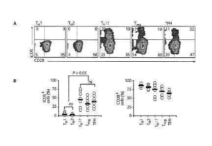
Figure 1, comprising Figures IA through IC, is a series of images
depicting distinct expression and function of !COS and CD28 on human CD4+ T
cell
subsets. Figure IA is an image demonstrating that the expression of ICOS and
CD28
costimulatory molecules was assessed on resting human peripheral blood CD4+ T
cell
subsets, consisting of CXCR3+CCR4-CCR64- T1 1, CCR4+CXCR3-CCR6- TH 2,
.. CCR4+CCR6+ Th17, CD25+CD1271oFoxP3+ Treg, and CXCR5+CD45R0+ TFH
cells, Figure 1B is an image depicting flow cytometrie quantification of ICOS
and
CD28 on different subsets from several normal donors (n 7). Horizontal bars
indicate mean; ns = not significant. Figure IC is an image depicting cytokines
IL-2
(1), 1L-4 (ii), IFN-y (iii), 1L-10 (iv), IL-22 (v), IL-17A (vi), IL-I 7F
(vii), CCL20 (viii),
4
CA 02791975 2012-09-04
WO 2011/097477
PCT/US2011/023744
and 1L-21 (ix) secreted from various sorted cells activated with antibodies to
CD3/CD28 or CD3/ICOS beads and measured on day 3 by ELISA. Statistics were
corrected for multiple comparisons with the ANOVA Seheffd test. TM¨follicular
helper T.
Figure 2, comprising Figures 2A through 2G, is a series of images
demonstrating that ICOS augments cytokine production by human Th17 cells.
Figure
2A is an image demonstrating that IL-1 7F production was assessed by
peripheral
blood CD4+ T cells differentiated to a Th17 phenotype with Th17-polarizing
conditions (IL-6, IL-lb, 1L-23, neutralizing IFN-y, and neutralizing IL-4
antibodies in
serum containing TGF-13, a eytokine required for inducing Th17
differentiation) and
activated with either aAPCs expressing CD86, CD80, CD70, 1COSL, OX4OL, or 4-
IBBL or with beads bearing antibodies to CD3 and CD28 on day 3 by ELISA.
Figure
2B is an image demonstrating that IL-17F production was assessed by peripheral
blood CD4+ T cells cultured with or without Th17-polarizing conditions and
activated
with aAPC engineered to express ICOSL or with beads bearing antibodies to
CD3/ICOS on day 3. Figure 2C to 2G depicts measurements of (C) IL-17F, (D) IL-
17A, (E) 1L-2, (F) 1L-22, and (G) IL-10 secretion or expression by Thl 7-
polarized
CD4+ T cells activated with beads bearing antibodies to CD3, CD28, and/or 1COS
on
day 3 using ELISA or reverse transcription PCR (RT-PCR).
Figure 3, comprising Figures 3A through 3G, is a series of images
demonstrating that ICOS is critical for the expansion of human Th17 cells.
Figures
3A and 3B depict the frequency and absolute number, respectively of
CCR4 CCR6+CD4+ T cells over time assessed by flow cytometry from peripheral
blood CD4+ T cells cultured in Th17-polarizing conditions and activated with
antibodies to CD3/CD28 or CD3/ICOS beads. Figure 3C is an image demonstrating
that CD27 and CD62L expression was measured on day 10 on these cells with flow
cytometry. Figure 3D demonstrate that on the days indicated, CD28- or ICOS-
engaged Th17-polarized CD4+ T cells were stimulated with PMA-ionomyein and the
frequency of cells secreting IL-17A and IFN-y was assessed via flow cytometry.
Figure 3E is an image demonstrating that the frequency of CD28- or ICOS-
engaged
Th17-polarized cells coprodueing 1L-17A and/or IFN-y was determined at the end
of
their primary expansion (ranging from days 9 to 14) in several different
normal
5
CA 02791975 2012-09-04
WO 2011/097477
PCT/US2011/023744
donors (n = 8). Figures 3F and 36 demonstrate expression of RORC2 and T-bet,
respectively, in these treated cells measured using RT-PCR on days 3 and 10.
Figure 4, comprising Figures 4A through 4F, is a series of image
demonstrating that 1COS drives rapid Th17 cell differentiation from naïve UCB
CD4+ T cells. Figures 4A through 4C, is a series of image demonstrating that
UCB
CD45RA+CD25¨CD4+ T cells were cultured with Th17-polarizing conditions and
expanded with antibodies to CD3/CD28, CD3/1COS, or CD3/CD28/ICOS beads.
Starting on day 3, IL-2 (50 1U/m1) was added to the cultures. Cultures were
stimulated with PMA-ionomycin (TONO) and the intracellular expression of IL-
17A,
IFN-y, IL-2, and TNF-a and the extracellular expression of IL-23R and CD 161
were
assessed on day 11. Cells from Figure 4A to Figure 4C were reactivated with
antibodies to CD3-coupled beads bearing antibodies to CD28 and/or TCOS.
Figures
4D to 4F demonstrate that cultures were restimulated with PMA-ionomyein and
the
intracellular expression of IL-17A, 1FN-y, 1L-2, and TNF-a and the
extracellular
expression of IL-23R and CD161 were assessed on day 18.
Figure 5, comprising Figures 5A through 5L, is a series of image
demonstrating that CD28 and ICOS differentially regulate e-MAF, RORC2, and T-
bet
expression in UCB Th17 cells. UCB CD4+ T cells were cultured in Th17-
polarizing
conditions and expanded with antibodies to CD3/CD28 or CD3/ICOS beads. 1L-2
(50
IU/m1) was added on day 3. Figures 5A and 513 demonstrate that on day 5, mRNA
expression of c-MAF and 1L-21 in CD28- or ICOS-stimulated cells was measured
by
RT-PCR. Figure 5C demonstrate that on day 5, IL-17F production in CD28-
stimulated cells cultured with exogenous IL-21 and 1L-2 neutralization was
measured
by EL1SA. Figures 5D through 5L demonstrate that on the days indicated, RORC2,
1-bet, FoxP3, ALIR, 1L-22, IL-10, and 1L-17A production in CD28- or 1COS-
stimulated cells was measured by flow cytometry and RT-PCR.
Figure 6, comprising Figures 6A through 6E, is a series of images
demonstrating that human Th17 cells originate from ICOS+CD161+CD4+ T cell
precursors. Figure 6A demonstrates that CD45RA, CD31, CD127, CD62L, and
CD27 expression was assessed on ICOS+CD 61+CD4+ and ICOS¨CD161+CD4+ T
cells from the UCB via flow cytometry. Figure 6B is an image demonstrating
that IL-
17F, CCL20, TFN-y, 1L-4, IL-22, and 1L-10 secretion by sorted ICOS+CD161+CD4+
and ICOS¨CD1611 CD4+ T cells cultured with Th17-polarizing conditions and
6
CA 02791975 2012-09-04
WO 2011/097477
PCT/US2011/023744
expanded with antibodies to CD3/CD28 or CD3/1COS beads was assessed on day 4
by EL1SA, Figure 6C is an image depicting the frequency and absolute number of
CD161+ cells cultured with Th17-polarizing conditions and expanded with
antibodies
to CD3/CD28- or CD3/ICOS-coated beads that were determined on day 4 or on the
days indicated, respectively. Figure 6D is an image depicting RORC2, 1L-23R,
AHR,
and FoxP3 mRNA expression in sorted ICOS+CD161+CD4+ and
ICOS¨CD161+CD4+ T cells cultured with Th17-polarizing conditions and expanded
with antibodies to CD3/CD28- or CD3/ICOS-coated beads that were assessed on
day
7 by RT-PCR. Figure 6E is an image demonstrating that on day 7,
ICOS+CD161+CD4+ and 1COS¨CD161+CD4+ T cells cultured in media alone or in
TH1-, TH2-, Th17-, and Treg-polarizing conditions and expanded with antibodies
to
CD3/CD28- or CD3/1COS-eoated beads were then stimulated with PMA-ionomyein,
and IL- 17A secretion was assessed by flow eytometry.
Figure 7, comprising Figures 7A through 7F, is a series of images
demonstrating that1COS augments T cell¨mediated tumor immunity. As shown
schematically, human CD4+ and CD8+ T cells were stimulated with antibodies to
CD3/CD28 or CD3/ICOS beads and cultured with or without Th 1 7-polarizing
conditions. One day later, bead-activated T cells were genetically redirected
with a
CAR that binds mesothelin. After their primary expansion, the genetically
redirected
cells (two administrations, 8 x 10⁶ cells total) were infused into mice
bearing a large
human mesothel in (M108) tumor pre-established for 61 days (n = 8 mice per
group).
Figures 7A through 7D demonstrate that tumor growth was measured in mice
infused
with genetically redirected cells expanded with the ICOS or CD28 signal with
or
without Th 17-polarizing conditions. Tumor growth was analyzed with a linear
mixed-effects model and by applying a conservative Bonferroni correction
approach
(mean SEM). Figure 7E demonstrates that redirected T cells were isolated
from the
mouse spleens (on day 43) and cultured with irradiated aAPCs bearing
mesothelin.
IL-17A and 1FN-y secretion was analyzed by flow eytometry 24 hours later.
Figure
7F demonstrates that the absolute number of CD4+ andCD8+ T cells was
determined
in the blood and spleen on days 21 and 43, respectively.
Figure 8 is an image demonstrating that UCB CD45RA+CD25-CD4+
T cells contain few CD161+1L-23R+ cells. The expression of CD161 and IL-23R
7
CA 02791975 2012-09-04
WO 2011/097477
PCT/US2011/023744
surface markers on CD45RA+CD25-CD4+ T cells was assessed on human umbilical
cord blood cells using flow cytometry.
Figure 9 is an image demonstrating that ICOS induces c-MAF and IL-
21. PB CD4+ T cells were cultured in Th17 polarizing conditions (IL-1 [3, IL-
6, IL-
23, plus neutralizing anti-IFNI and anti-IL-4) and activated with anti-CD3
beads
bearing either anti-CD28 or anti-ICOS antibodies. After their primary
expansion, their
c-114AF and IL-21 expression mRNA levels was assessed by RT-PCR.
Figure 10 is an image demonstrating that CD28 induces expression of
the aryl hydrocarbon receptor, PB CD4+ T cells were programmed toward a Th17
phenotype and activated with anti-CD3 beads bearing either anti-CD28 or anti-
1COS
antibodies, After their primary expansion, their mRNA expression level of AHR
relative to I3-actin was assessed by RT-PCR.
Figure 11 is an image demonstrating that exogenous TGF-13 augments
the inflammatory potential of human TH 17 cells. PB CD4+ T cells were
programmed toward a TH 17 phenotype and activated with anti-CD3 beads bearing
either anti-CD28 or 2 anti-ICOS antibodies in media containing serum and the
indicated supplemental TGF-13 (from 0.1-10 ng/m1) was added to the culture on
day 1.
IL-17 A secretion by cells was measured on day 5 post-activation by ELISA.
Figure 12 is an image demonstrating that ICOS+CD161+CD4+ T cells
from UCB constitutively express RORC2 and 11,23R, CD4+, ICOS+CD161+CD4+
and ICOS-CD161+CD4+ T cells were sorted and their mRNA expression level of
RORC2 and 1L-23R relative to 13-actin was measured by RT-PCR.
Figure 13, comprising Figures 13A through 13D, is a series of images
demonstrating that ICOS+CDI61 -l-CD4+ T cells are imprinted as Th17 cells,
CD4+
and 1COS+CD161 +CD4+ T cells from UCB were sorted and cultured in various
polarizing conditions as indicated. The frequency of IFN-7+ (Figure 13A), IL-
4+
(Figure 13B), 1L-17 A+ (Figure 13C) or FoxP3+ (Figure 13D) cells was measured
after their primary expansion with anti-CD3 beads bearing anti-CD28 or anti-
ICOS
antibodies. As a control, companion control cultures of bulk UCB CD4 T cells
were
stimulated with antiCD3/CD28 beads, Cytokines and FoxP3 were measured by flow
cytometry or ELISA on day 7 of culture post-stimulation with PMA/ionomycin,
DETAILED DESCRIPTION OF THE INVENTION
8
CA 02791975 2012-09-04
WO 2011/097477
PCT/US2011/023744
The present invention provides compositions and methods for their use
to expand in vitro or in vivo a desired T cell, activate and/or expand
specific T cell
subsets, identify stimulatory molecules, co-stimulatory molecules, and
combinations
thereof; that can promote expansion of specific T cell subsets, as well as
numerous
therapeutic uses relating to expansion and stimulation of T cells. Preferably,
the T
cell is Th17.
The present invention is based on the discovery that human Th17 cell
proliferation and function vary dramatically depending upon whether they
receive
CD28 or ICOS costimulation. The disclosure presented herein demonstrates that
ICOS costimulation specifically promotes the outgrowth and augments the
function of
peripheral Th17 cells, In contrast, CD28 costimulation abrogates the effect of
ICOS.
The results presented herein demonstrate that costimulation of naive precursor
cells
from human cord blood with ICOS in the presence of Th17 polarizing agents
support
the generation and expansion of Th17 cells, as indicated by their capacity to
secrete
heightened levels of IL-17A, 1L-17F and CCL20. ICOS costimulation not only can
elevate Th17 cells to produce Th17-associated cytokines, but also elevate
secretion of
1FiN7, TNFct and 1L-2 I as compared with CD28 costimulation.
In one embodiment, ICOS costimulation on T cells can be
accomplished by contacting the T cell with an artificial antigen presenting
cell
(aAPC) that comprises a molecule capable of activating ICOS on the T
In another embodiment, the aAPC comprising a molecule capable of
activating ICOS on T cells can further be engineered to comprise a cytokine
that
promotes Th [7 differentiation. Such Th17 differentiation eytokines includes
but are
not limited to 1L-2, IL-6, and IL-1.
In yet another embodiment, the aAPC comprising a molecule capable
of activating ICOS on T cells can also be engineered to comprise an inhibitory
molecule that can block a cytokine that interferes with the Th17
differentiation
process. For example, the aAPC can be engineered to secrete a neutralizing
antibody
than can inhibit a cytokine that interferes with Th17 differentiation. A
cytokine that
interferes with Th17 differentiation process includes but is not limited to
IFNy and IL-
4.
Of clinical importance, the Th17 cells generated according to the
methods of the invention can be used in adoptive transfer immunotherapy. That
is,
human T cells expanded in the presence of ICOS costimulation mediate superior
9
CA 02791975 2012-09-04
WO 2011/097477
PCT/US2011/023744
regression of established human tumors compared with an otherwise identical T
cell
expanded in the presence of CD28. In one embodiment, cells engineered to be
able to
activate ICOS on T cells can be used to boost and expand Th17 cells in vivo as
a form
of vaccination.
Definitions
Unless defined otherwise, all technical and scientific terms used herein
have the same meaning as commonly understood by one of ordinary skill in the
art to
which the invention pertains. Although any methods and materials similar or
equivalent to those described herein can be used in the practice for testing
of the
present invention, the preferred materials and methods are described herein.
In
describing and claiming the present invention, the following terminology will
be used.
It is also to be understood that the terminology used herein is for the
purpose of describing particular embodiments only, and is not intended to be
limiting.
The articles "a" and "an" arc used herein to refer to one or to more
than one (i.e., to at least one) of the grammatical object of the article. By
way of
example, "an element" means one element or more than one element.
An "amino acid" as used herein is meant to include both natural and
synthetic amino acids, and both D and L amino acids. "Standard amino acid"
means
any of the twenty L-amino acids commonly found in naturally occurring
peptides.
"Nonstandard amino acid residues" means any amino acid, other than the
standard
amino acids, regardless of whether it is prepared synthetically or derived
from a
natural source. As used herein, "synthetic amino acid" also encompasses
chemically
modified amino acids, including but not limited to salts, amino acid
derivatives (such
as amides), and substitutions. Amino acids contained within the peptides, and
particularly at the earboxy- or amino-terminus, can be modified by
methylation,
amidation, acetylation or substitution with other chemical groups which can
change a
peptide's circulating half life without adversely affecting activity of the
peptide.
Additionally, a disulfide linkage may he present or absent in the peptides.
"About" as used herein when referring to a measurable value such as
an amount, a temporal duration, and the like, is meant to encompass variations
of
20% or +10%, more preferably 5%, even more preferably +1%, and still more
preferably +0.1% from the specified value, as such variations are appropriate
to
perform the disclosed methods.
CA 02791975 2012-09-04
WO 2011/097477
PCT/US2011/023744
The term "antigen" or "Ag" as used herein is defined as a molecule
that provokes an immune response. This immune response may involve either
antibody production, or the activation of specific immunologically-competent
cells, or
both. The skilled artisan will understand that any macromolecule, including
virtually
all proteins or peptides, can serve as an antigen. Furthermore, antigens can
be derived
from recombinant or genomic DNA. A skilled artisan will understand that any
DNA,
which comprises a nucleotide sequences or a partial nucleotide sequence
encoding a
protein that elicits an immune response therefore encodes an "antigen" as that
term is
used herein. Furthermore, one skilled in the art will understand that an
antigen need
not be encoded solely by a full length nucleotide sequence of a gene. It is
readily
apparent that the present invention includes, but is not limited to, the use
of partial
nucleotide sequences of more than one gene and that these nucleotide sequences
are
arranged in various combinations to elicit the desired immune response.
Moreover, a
skilled artisan will understand that an antigen need not be encoded by a
"gene" at all.
It is readily apparent that an antigen can be generated synthesized or can be
derived
from a biological sample. Such a biological sample can include, but is not
limited to a
tissue sample, a tumor sample, a cell or a biological fluid.
The term "antibody," as used herein, refers to an inununoglobulin
molecule which specifically binds with an antigen. Antibodies can be intact
immunoglobulins derived from natural sources or from recombinant sources and
can
be immunoreactive portions of intact immunoglobulins. Antibodies are typically
tetramers of immunoglobul in molecules. The antibodies in the present
invention may
exist in a variety of forms including, for example, polyelonal antibodies,
monoclonal
antibodies, Fv, Fab and F(ab)2, as well as single chain antibodies and
humanized
antibodies (Harlow et al., 1999, In: Using Antibodies: A Laboratory Manual,
Cold
Spring Harbor Laboratory Press, NY; Harlow et al., 1989, In: Antibodies: A
Laboratory Manual, Cold Spring Harbor, New York; Houston et al., 1988, Proc.
Natl.
Acad. Sei. USA 85:5879-5883; Bird et al., 1988, Science 242:423-426).
The term "agent", "ligand", or "agent that binds a cell surface moiety",
as used herein, refers to a molecule that binds to a defined population of
cells. The
agent may bind any ecll surface moiety, such as a receptor, an antigenic
determinant,
or other binding site present on the target cell population. The agent may be
a protein,
peptide, antibody and antibody fragments thereof, fusion proteins, synthetic
molecule,
an organic molecule (e.g., a small molecule), a carbohydrate, or the like.
Within the
11
CA 02791975 2012-09-04
WO 2011/097477
PCT/US2011/023744
specification and in the context of T cell stimulation, antibodies and natural
ligands
are used as prototypical examples of such agents.
The terms "agent that binds a cell surface moiety" and "cell surface
moiety", as used herein, are used in the context of a ligand/anti-ligand pair.
Accordingly, these molecules should be viewed as a complementary/anti-
complementary set of molecules that demonstrate specific binding, generally of
relatively high affinity.
As used herein, the term "autologotts" is meant to refer to any material
derived from the same individual to which it is later to be re-introduced into
the
individual,
"Allogeneie" refers to a graft derived from a different animal of the
same species.
"Xenogeneic" refers to a graft derived from an animal of a different
species.
The term "cancer" as used herein is defined as disease characterized by
the rapid and uncontrolled growth of aberrant cells. Cancer cells can spread
locally or
through the bloodstream and lymphatic system to other parts of the body.
Examples
of various cancers include but are not limited to, breast cancer, prostate
cancer,
ovarian cancer, cervical cancer, skin cancer, pancreatic cancer, colorectal
cancer,
renal cancer, liver cancer, brain cancer, lymphoma, leukemia, lung cancer and
the
like.
A "coding region" of a gene consists of the nucleotide residues of the
coding strand of the gene and the nucleotides of the non-coding strand of the
gene
which are homologous with or complementary to, respectively, the coding region
of
an mRNA molecule which is produced by transcription of the gene.
A "coding region" of an mRNA molecule also consists of the
nucleotide residues of the mRNA molecule which are matched with an anti-codon
region of a transfer RNA molecule during translation of the mRNA molecule or
which encode a stop codon. The coding region may thus include nucleotide
residues
corresponding to amino acid residues which are not present in the mature
protein
encoded by the mRNA molecule (e.g., amino acid residues in a protein export
signal
sequence).
"Encoding" refers to the inherent property of specific sequences of
nucleotides in a polynucleotide, such as a gene, a cDNA, or an mRNA, to serve
as
12
CA 02791975 2012-09-04
WO 2011/097477
PCT/US2011/023744
templates for synthesis of other polymers and macromolecules in biological
processes
having either a defined sequence of nucleotides (1.e., rRNA, tRNA and mRNA) or
a
defined sequence amino acids and the biological properties resulting
therefrom.
Thus, a gene encodes a protein if transcription and translation of mRNA
corresponding to that gene produces the protein in a cell or other biological
system.
Both the coding strand, the nucleotide sequence of which is identical to the
mRNA
sequence and is usually provided in sequence listings, and the non-coding
strand, used
as the template for transcription of a gene or cDNA, can be referred to as
encoding the
protein or other product of that gene or cDNA.
Unless otherwise specified, a "nucleotide sequence encoding an amino
acid sequence" includes all nucleotide sequences that are degenerate versions
of each
other and that encode the same amino acid sequence. Nucleotide sequences that
encode proteins and RNA may include introits.
"Effective amount" or "therapeutically effective amount" are used
interchangeably herein, and refer to an amount of a compound, formulation,
material,
or composition, as described herein effective to achieve a particular
biological result.
Such results may include, but are not limited to, the inhibition of virus
infection as
determined by any means suitable in the art.
As used herein "endogenous" refers to any material from or produced
inside an organism, cell, tissue or system.
As used herein, the term "exogenous" refers to any material introduced
from or produced outside an organism, cell, tissue or system.
The term "expression" as used herein is defined as the transcription
and/or translation of a particular nucleotide sequence driven by its promoter.
"Expression vector" refers to a vector comprising a recombinant
polynucleotide comprising expression control sequences operatively linked to a
nucleotide sequence to he expressed. An expression vector comprises sufficient
cis-
acting elements for expression; other elements for expression can be supplied
by the
host cell or in an in vitro expression system. Expression vectors include all
those
known in the art, such as cosmids, plasm ids (e.g., naked or contained in
liposomes)
and viruses (e.g., lentiviruses, retroviruses, adenoviruses, and adeno-
associated
viruses) that incorporate the recombinant polynucleotide.
As used herein, the term "fragment," as applied to a nucleic acid, refers
to a subsequence of a larger nucleic acid. A "fragment" of a nucleic acid can
be at
13
CA 02791975 2012-09-04
WO 2011/097477
PCT/US2011/023744
least about 15 nucleotides in length; for example, at least about 50
nucleotides to
about 100 nucleotides; at least about 100 to about 500 nucleotides, at least
about 500
to about 1000 nucleotides, at least about 1000 nucleotides to about 1500
nucleotides;
or about 1500 nucleotides to about 2500 nucleotides; or about 2500 nucleotides
(and
any integer value in between).
As used herein, the term "fragment," as applied to a protein or peptide,
refers to a subsequence of a larger protein or peptide. A "fragment" of a
protein or
peptide can be at least about 20 amino acids in length; for example at least
about 50
amino acids in length; at least about 100 amino acids in length, at least
about 200
amino acids in length, at least about 300 amino acids in length, and at least
about 400
amino acids in length (and any integer value in between).
"Homologous" as used herein, refers to the subunit sequence identity
between two polymeric molecules, e.g., between two nucleic acid molecules,
such as,
two DNA molecules or two RNA molecules, or between two polypeptide molecules,
When a subunit position in both of the two molecules is occupied by the same
monomeric subunit; e.g., if a position in each of two DNA molecules is
occupied by
adenine, then they are homologous at that position. The homology between two
sequences is a direct function of the number of matching or homologous
positions;
e.g., if half (e.g., five positions in a polymer ten subunits in length) of
the positions in
two sequences are homologous, the two sequences are 50% homologous; if 90% of
the positions (e.g., 9 of 10), are matched or homologous, the two sequences
are 90%
homologous. By way of example, the DNA sequences 5'-ATTGCC-3' and 5'-
TATGGC-3' share 50% homology,
The term "immunoglobulin" or "Ig", as used herein is defined as a
class of proteins, which function as antibodies. The five members included in
this
class of proteins are IgA, IgG, IgM, IgD, and IgE. IgA is the primary antibody
that is
present in body secretions, such as saliva, tears, breast milk,
gastrointestinal
secretions and mucus secretions of the respiratory and genitourinary tracts.
IgG is the
most common circulating antibody. IgM is the main immunoglobulin produced in
the
primary immune response in most mammals. It is the most efficient
immunoglobulin
in agglutination, complement fixation, and other antibody responses, and is
important
in defense against bacteria and viruses, 1gD is the immunoglobulin that has no
known
antibody function, but may serve as an antigen receptor. IgE is the
immunoglobulin
14
CA 02791975 2012-09-04
WO 2011/097477
PCT/US2011/023744
that mediates immediate hypersensitivity by causing release of mediators from
mast
cells and basophils upon exposure to allergen.
As used herein, an "instructional material" includes a publication, a
recording, a diagram, or any other medium of expression which can be used to
communicate the usefulness of the compositions and methods of the invention.
The
instructional material of the kit of the invention may, for example, be
affixed to a
container which contains the nucleic acid, peptide, and/or composition of the
invention or be shipped together with a container which contains the nucleic
acid,
peptide, and/or composition. Alternatively, the instructional material may be
shipped
separately from the container with the intention that the instructional
material and the
compound be used cooperatively by the recipient,
"Isolated" means altered or removed from the natural state. For
example, a nucleic acid or a peptide naturally present in a living animal is
not
"isolated," but the same nucleic acid or peptide partially or completely
separated from
the coexisting materials of its natural state is "isolated." An isolated
nucleic acid or
protein can exist in substantially purified form, or can exist in a non-native
environment such as, for example, a host cell.
An "isolated nucleic acid" refers to a nucleic acid segment or fragment
which has been separated from sequences which flank it in a naturally
occurring state,
i.e., a DNA fragment which has been removed from the sequences which are
normally
adjacent to the fragment, i.e., the sequences adjacent to the fragment in a
genome in
which it naturally occurs. The term also applies to nucleic acids which have
been
substantially purified from other components which naturally accompany the
nucleic
acid, i.e., RNA or DNA or proteins, which naturally accompany it in the cell.
The
term therefore includes, for example, a recombinant DNA which is incorporated
into a
vector, into an autonomously replicating plasm id or virus, or into the
genomic DNA
of a prokaryote or eukaryote, or which exists as a separate molecule (i.e., as
a cDNA
or a genomic or cDNA fragment produced by PCR or restriction enzyme digestion)
independent of other sequences. It also includes a recombinant DNA which is
part of
a hybrid gene encoding additional polypeptide sequence.
In the context of the present invention, the following abbreviations for
the commonly occurring nucleic acid bases are used. "A" refers to adenosine,
"C"
refers to cytosine, "G" refers to guanosine, "T" refers to thymidine, and
"I.I" refers to
urid inc.
CA 02791975 2012-09-04
WO 2011/097477
PCT/US2011/023744
Unless otherwise specified, a "nucleotide sequence encoding an amino
acid sequence" includes all nucleotide sequences that are degenerate versions
of each
other and that encode the same amino acid sequence. The phrase nueleotide
sequence
that encodes a protein or an RNA may also include introns to the extent that
the
nucleotide sequence encoding the protein may in some version contain an
intron(s).
As used herein, the term "modulate" is meant to refer to any change in
biological state, i.e. increasing, decreasing, and the like.
=
The term "operably linked" refers to functional linkage between a
regulatory sequence and a heterologous nucleic acid sequence resulting in
expression
of the latter. For example, a first nucleic acid sequence is operably linked
with a
second nucleic acid sequence when the first nucleic acid sequence is placed in
a
functional relationship with the second nucleic acid sequence. For instance, a
promoter is operably linked to a coding sequence if the promoter affects the
transcription or expression of the coding sequence. Generally, operably linked
DNA
sequences are contiguous and, where necessary to join two protein coding
regions, in
the same reading frame.
"Parenteral" administration of an immunogenic composition includes,
e,g,, subcutaneous (sc.), intravenous (Iv.), intramuscular (i.m,), or
intrasternal
injection, or infusion techniques.
The term "polynueleotide" as used herein is defined as a chain of
nucleotides. Furthermore, nucleic acids are polymers of nucleotides. Thus,
nucleic
acids and polynueleotides as used herein are interchangeable. One skilled in
the art
has the general knowledge that nucleic acids are polynucleotides, which can be
hydrolyzed into the monomeric "nucleotides." The monomeric nucleotides can be
hydrolyzed into nucleosides. As used herein polynucleotides include, but are
not
limited to, all nucleic acid sequences which are obtained by any means
available in
the art, including, without limitation, recombinant means, i.e., the cloning
of nucleic
acid sequences from a recombinant library or a cell genome, using ordinary
cloning
technology and PCRTNI, and the like, and by synthetic means.
As used herein, the terms "peptide," "polypeptide," and "protein" are
used interchangeably, and refer to a compound comprised of amino acid residues
covalently linked by peptide bonds. A protein or peptide must contain at least
two
amino acids, and no limitation is placed on the maximum number of amino acids
that
can comprise a protein's or peptide's sequence. Polypeptides include any
peptide or
16
CA 02791975 2012-09-04
WO 2011/097477
PCT/US2011/023744 =
protein comprising two or more amino acids joined to each other by peptide
bonds.
As used herein, the term refers to both short chains, which also commonly are
referred
to in the art as peptides, oligopeptides and oligomers, for example, and to
longer =
chains, which generally are referred to in the art as proteins, of which there
are many
types. "Polypeptides" include, for example, biologically active fragments,
substantially homologous polypeptides, oligopeptides, homodimers,
heterodimers,
variants of polypeptides, modified polypeptides, derivatives, analogs, fusion
proteins,
among others. The polypeptides include natural peptides, recombinant peptides,
synthetic peptides, or a combination thereof.
The term "promoter" as used herein is defined as a DNA sequence
recognized by the synthetic machinery of the cell, or introduced synthetic
machinery,
required to initiate the specific transcription of a polynueleotide sequence.
As used herein, the term "promoter/regulatory sequence" means a
nucleic acid sequence which is required for expression of a gene product
operably
linked to the promoter/regulatory sequence. In some instances, this sequence
may be
the core promoter sequence and in other instances, this sequence may also
include an
enhancer sequence and other regulatory elements which are required for
expression of
the gene product, The promoter/regulator>, sequence may, for example, be one
which expresses the gene product in a tissue specific manner,
A "constitutive" promoter is a nucleotide sequence which, when
operably linked with a polynucleotide which encodes or specifies a gene
product,
causes the gene product to be produced in a cell under most or all
physiological
conditions of the cell.
An "inducible" promoter is a nucleotide sequence which, when
operably linked with a polynucleotide which encodes or specifies a gene
product,
causes the gene product to be produced in a cell substantially only when an
inducer
which corresponds to the promoter is present in the cell,
A "tissue-specific" promoter is a nucleotide sequence which, when
operably linked with a polynueleotide encodes or specified by a gene, causes
the gene
product to be produced in a cell substantially only if the cell is a cell of
the tissue type
corresponding to the promoter.
The term "RNA" as used herein is defined as ribonucleic acid.
The term "recombinant DNA" as used herein is defined as DNA
produced by joining pieces of DNA from different sources.
17
CA 02791975 2012-09-04
WO 2011/097477
PCT/US2011/023744
The term "recombinant polypeptide" as used herein is defined as a
polypeptide produced by using recombinant DNA methods.
The term "subject" is intended to include living organisms in which an
immune response can be elicited (e.g,, mammals).
As used herein, a "substantially purified" cell is a cell that is
essentially free of other cell types. A substantially purified cell also
refers to a cell
which has been separated from other cell types with which it is normally
associated in
its naturally occurring state. In some instances, a population of
substantially purified
cells refers to a homogenous population of cells. In other instances, this
term refers
simply to cell that have been separated from the cells with which they are
naturally
associated in their natural state. In some embodiments, the cells are cultured
in vitro.
In other embodiments, the cells are not cultured in vitro.
The term "T-helper" as used herein with reference to cells indicates a
sub-group of lymphocytes (a type of white blood cell or leukocyte) including
different
cell types identifiable by a skilled person. In particular, T-helper cell
according to the
present disclosure include effector Th cells (such as Th I, Th2 and T1117).
These Th
cells secrete cytokines, proteins or peptides that stimulate or interact with
other
leukocytes.
The term "therapeutic" as used herein means a treatment and/or
prophylaxis. A therapeutic effect is obtained by suppression, remission, or
eradication of a disease state.
The term "transfeeted" or "transformed" or "transduced" as used
herein refers to a process by which exogenous nucleic acid is transferred or
introduced into the host cell. A "transfected" or "transformed" or
"transduced" cell
is one which has been transfected, transformed or transduced with exogenous
nucleic
acid. The cell includes the primary subject cell and its progeny.
The phrase "under transcriptional control" or "operatively linked" as
used herein means that the promoter is in the correct location and orientation
in
relation to a polynucleotide to control the initiation of transcription by RNA
poiymerase and expression of the polynueleotide.
"Variant" as the term is used herein, is a nucleic acid sequence or a
peptide sequence that differs in sequence from a reference nucleic acid
sequence or
peptide sequence respectively, but retains essential properties of the
reference
molecule. Changes in the sequence of a nucleic acid variant may not alter the
amino
CA 02791975 2012-09-04
WO 2011/097477
PCT/US2011/023744
acid sequence of a peptide encoded by the reference nucleic acid, or may
result in
amino acid substitutions, additions, deletions, fusions and truncations.
Changes in the
sequence of peptide variants are typically limited or conservative, so that
the
sequences of the reference peptide and the variant are closely similar overall
and, in
many regions, identical. A variant and reference peptide can differ in amino
acid
sequence by one or more substitutions, additions, deletions in any
combination, A
variant of a nucleic acid or peptide can be a naturally occurring such as an
allelic
variant, or can be a variant that is not known to occur naturally. Non-
naturally
occurring variants of nucleic acids and peptides may be made by mutagenesis
techniques or by direct synthesis.
A "vector" is a composition of matter which comprises an isolated
nucleic acid and which can be used to deliver the isolated nucleic acid to the
interior
of a cell, Numerous vectors are known in the art including, but not limited
to, linear
polynucleotides, polynueleotides associated with ionic or amphiphilic
compounds,
plasmids, and viruses. Thus, the term "vector" includes an autonomously
replicating
plasmid or a virus. The term should also be construed to include non-plasmid
and
non-viral compounds which facilitate transfer of nucleic acid into cells, such
as, for
example, polylysine compounds, liposomes, and the like. Examples or viral
vectors
include, but are not limited to, adenoviral vectors, adeno-associated virus
vectors,
retroviral vectors, and the like.
By the term "stimulation," is meant a primary response induced by
binding of a stimulatory molecule (e.g., a TCR/CD3 complex) with its cognate
ligand
thereby mediating a signal transduction event, such as, but not limited to,
signal
transduction via the TCR/CD3 complex. Stimulation can mediate altered
expression
of certain molecules, such as downregulation of TGF-13, and/or reorganization
of
cytoskeletal structures, and the like.
"Activation", as used herein, refers to the state of a T cell that has been
sufficiently stimulated to induce detectable cellular proliferation.
Activation can also
be associated with induced cytokine production, and detectable effector
functions.
The term "activated T cells" refers to, among other things, '1' cells that are
undergoing
cell division.
By the term "specifically binds," as used herein, is meant an antibody,
or a ligand, which recognizes and binds with a cognate binding partner (e.g.,
a
stimulatory and/or costimulatory molecule present on a T cell) protein present
in a
19
CA 02791975 2012-09-04
WO 2011/097477
PCT/US2011/023744
sample, but which antibody or ligand does not substantially recognize or bind
other
molecules in the sample.
A "stimulatory ligand," as used herein, means a ligand that when
present on an antigen presenting cell (e.g., an aAPC, a dendritic cell, a B-
cell, and the
like) can specifically bind with a cognate binding partner (referred to herein
as a
"stimulatory molecule") on a T cell, thereby mediating a primary response by
the T
cell, including, but not limited to, activation, initiation of an immune
response,
proliferation, and the like. Stimulatory ligands are well-known in the art and
encompass, inter al/a, an MHC Class I molecule loaded with a peptide, an anti-
CD3
antibody, a superagonist anti-CD28 antibody, and a superagonist anti-CD2
antibody.
A "stimulatory molecule," as the term is used herein, means a
molecule on a Teell that specifically binds with a cognate stimulatory ligand
present
on an antigen presenting cell (e.g., an aAPC of the invention, among others).
"Loaded" with a peptide, as used herein, refers to presentation of an
antigen in the context of an MHC molecule. "Loaded" as used herein also means
the
binding of an antibody to an Fe binding receptor on a cell, such as CD32
and/or
CD64.
A "co-stimulatory signal", as used herein, refers to a signal, which in
combination with a primary signal, such as TCR/CD3 ligation, leads to T cell
proliferation and/or upregulation or downregulation of key molecules.
A "co-stimulatory molecule" refers to the cognate binding partner on a
T cell that specifically binds with a co-stimulatory ligand, thereby mediating
a co-
stimulatory response by the T cell, such as, but not limited to,
proliferation. Co-
stimulatory molecules include, but are not limited to an MHC class I molecule,
BTLA
and a Toll ligand receptor.
"Co-stimulatory ligand," as the term is used herein, includes a
molecule on an antigen presenting cell (e.g., an aAPC, dendritie cell, B cell,
and the
like) that specifically binds a cognate co-stimulatory molecule on a T cell,
thereby
providing a signal which, in addition to the primary signal provided by, for
instance,
binding of a TCR/CD3 complex with an MHC molecule loaded with peptide,
mediates a T cell response, including, but not limited to, proliferation,
activation,
differentiation, and the like. A co-stimulatory ligand can include, but is not
limited to,
CD7, B7-1 (CD80), B7-2 (CD86), PD-L1, PD-L2, 4-1BBL, OX4OL, inducible
costimulatory ligand (ICOS-L), intercellular adhesion molecule (ICAM), CD3OL,
CA 02791975 2012-09-04
WO 2011/097477
PCT/US2011/023744
CD40, CD70, CD83, HLA-G, MICA, MICB, HVEIV1, lymphotoxin beta receptor,
37TR6, ILT3, 1LT4, IIVEM, an agonist or antibody that binds Toll ligand
receptor arid
a ligand that specifically binds with B7-H3. A co-stimulatory ligand also
encompasses, inter cilia, an antibody that specifically binds with a co-
stimulatory
molecule present on a I cell, such as, but not limited to, CD27, CD28, 4-BB,
0X40,
CD30, CD40, PD-1, ICOS, lymphocyte function-associated antigen-1 (LFA-I), CD2,
CD7, LIGHT, NKG2C, B7-H3, and a ligand that specifically binds with CD83.
Description
The present invention is partly based on the observation that the nature
of costimulation during CD4+ T cell activation critically regulates human Th17
cell
differentiation, For example, ICOS, but not CD28, was found to be necessary
for
optimal expansion and function of human Th17 cells. Surprisingly, CD28
ligation
abrogated the effects of ICOS costimulation. Of clinical relevance,
genetically
reprogrammed human Th17 cells expanded with 'COS mediated superior regression
of human tumors compared to cells expanded with CD28. These findings reveal a
key role for ICOS signaling in human Th17 cell development and suggest new
therapeutic approaches.
The invention relates to the surprising discovery that ICOS
costimulation of Th17 cells resulted in significantly higher levels of IL17F,
CCL20,
and IL-21 production compared to the levels of 1L-17F, CCL20, and IL-21
produced
from an otherwise identical cell costimulated with CD28. In some instances,
ICOS
costimulation also resulted in elevated IL-17A secretion compared with the
level of
IL-17A secretion from an otherwise identical cell costimulated with CD28. In
some
instances, ICOS-stimulated Th17 cells also produced substantially greater
amounts of
IFNy compared to CD28-stimulated Th I cells, a subset previously thought to be
a
dominant source of IFNy production.
Accordingly, the present invention includes compositions and methods
for generating a population of human Th17 cells having unique inflammatory
characteristics, For example, the ICOS-stimulated Th17 cells secrete high
levels of
IL-17 and CCL20 as well as produce elevated levels of IFNy and IL-21 compared
to
CD28-stimulated Th 1 cells, The present invention is based on the unexpected
discovery that ICOS, but not CD28, costimulation preferentially expands Th17
cells.
21
WO 2011/097477
PCT/US2011/023744
ICOS-costimulation provides a means to culture expand Th17 and maintain long-
term
culture of Th17 cells.
The present invention provides compositions and methods for their use
to expand a Th17 cells as well as numerous therapeutic uses relating to
expansion and
stimulation of Th17 cells.
In one embodiment, the invention provides compositions and methods
for generating therapeutic amounts of Th17 cells from peripheral or umbilical
cord
blood (UCB). In some instances, Th17 cells are generated from naïve precursor
cells.
Preferable, the naïve precursor cells are CD45RA+CD25- cells.
Composition
=
The invention pertains to compositions comprising an agent that
provides a costimulatory signal to a T cell for T cell expansion (e.g.,
TCOSL). In
some instances, the costimulatory signal is provided to a T cell in
combination with
an agent that provides a primary activation signal to the T cell (e.g., a
TCR/CD3
complex). For example, an agent that provides a primary activation signal to
the T
cell is an anti-CD3 antibody.
In some instances, the agent (primary, costimulatory, or combination
thereof) is preferably attached to beads. Compositions of the invention can
also
include those comprising more than one type of agent coupled to different
solid phase
surfaces (i.e., an agent that provides a primary T cell activation signal
coupled to a
first solid phase surface and an agent that provides a costimulatory signal
coupled to a
second solid phase surface).
Alternatively, the agent (primary, costimulatory, or combination
thereof) is in the context of being displayed on an artificial antigen
presenting cell
(aAPC). Accordingly, the invention includes any means of promoting ICOS
engagement of T cells using either a solid phase surface (e.g., beads) or a
cell (e.g.,
aAPC). That is, there is extensive knowledge in the art regarding the events
and
molecules involved in activation and induction of T cell. However, the
invention is
based on the unexpected discovery that TCOS engagement, but not CD28
costimulation, preferentially expands cells having a Th17 phenotype.
The extensive disclosure is provided in WO 03/057171 and
US2003/01,17869.
More specifically, a primary signal, usually mediated via the T cell
receptor/CD3
22
CA 2791975 2017-07-05
CA 02791975 2012-09-04
WO 2011/097477
PCT/US2011/023744
complex on a T cell, initiates the T cell activation process. Additionally,
numerous
co-stimulatory molecules present on the surface of a T cell are involved in
regulating
the transition from resting T cell to cell proliferation. Such co-stimulatory
molecules,
also referred to as "co-stimulators", which specifically bind with their
respective
ligands, include, but are not limited to, CD28 (which binds with B7-1 [CD801,
B7-2
[CD86j), PD-1 (which binds with ligands PD-Ll and PD-L2), B7-H3, 4- IBB (binds
the ligand 4-1BBL), 0X40 (binds ligand OX4OL), ICOS (binds ligand ICOS-L), and
LFA (binds the ligand ICAM). Thus, the primary stimulatory signal mediates T
cell
stimulation, but the co-stimulatory signal is then required for T cell
activation, as
demonstrated by proliferation.
T cell activation can be accomplished by stimulating the T cell
TCR/CD3 complex or via stimulation of the CD2 surface protein. An anti-CD3
monoclonal antibody can be used to activate a population of T cells via the
TCR/CD3
complex, Although a number of anti-human CD3 monoclonal antibodies are
commercially available, OKT3 prepared from hybridoma cells obtained from the
American Type Culture Collection or monoclonal antibody G19-4 is preferred.
Similarly, binding of an anti-CD2 antibody will activate T cells. Stimulatory
forms of
anti-CD2 antibodies are known and available.
A primary activation signal can also be delivered to a T cell through
use of a combination of a protein kinase C (PKC) activator such as a phorbol
ester
(e.g., phorbol myristate acetate) and a calcium ionophore (e.g., ionomycin
which
raises cytoplasmic calcium concentrations). The use of these agents bypasses
the
TCR/CD3 complex but delivers a stimulatory signal to T cells. These agents are
also
known to exert a synergistic effect on T cells to promote T cell activation
and can be
used in the absence of antigen to deliver a primary activation signal to T
cells.
Although stimulation of the TCR/CD3 complex or CD2 molecule is
required for delivery of a primary activation signal in a T cell, a number of
molecules
on the surface of T cells, termed accessory or costimulatory molecules have
been
implicated in regulating the transition of a resting T cell to blast
transformation, and
subsequent proliferation and differentiation. Thus, in addition to the primary
activation signal provided through the TCR/CD3 complex, induction of T cell
responses requires a second, costimulatory signal, One such costimulatory or
accessory molecule, CD28, is believed to initiate or regulate a signal
transduction
pathway that is distinct from those stimulated by the TCR complex. However,
the
23
CA 02791975 2012-09-04
WO 2011/097477
PCT/US2011/023744
invention is based on the discovery that ICOS, but not CD28 eostimulation,
preferentially expands cells having a Th17 phenotype. Moreover, combined CD28
and 1COS costimulation does not potentiate, but rather specifically reduces
Th17
phenotype. This discovery was surprising because of the extensive use of CD28
in
the art to expand Th17.
Accordingly, the invention relates to the use of compositions that can
promote ICOS costimulation on T cells. Any agent that can induce stimulation
of the
ICOS molecule is encompassed by the invention, In addition, binding homologues
of
a natural ligand, whether native or synthesized by chemical or recombinant
technique,
can also be used in accordance with the invention. Ligands useful for
stimulating an
ICOS can be used in soluble form, attached to the surface of a cell, or
immobilized on
a solid phase surface as described herein. Anti-ICOS antibodies or fragments
thereof
are also useful in stimulating ICOS molecule.
In a specific embodiment of the invention, activated T cells are
contacted with a stimulatory form of a natural ligand for ICOS for
costimulation. The
natural ligand of ICOS is referred in the art as ICOSL, A "stimulatory form of
a
natural ligand for ICOS" is a form of a natural ligand that is able to bind to
ICOS and
costimulate the T cell. Costimulation can be evidenced by proliferation and/or
cytokine production by T cells that have received a primary activation signal,
such as
stimulation through the C1)3/ICR complex or through CD2.
In a preferred embodiment of the invention, an ICOSL molecule is
localized on the surface of a cell. This can be accomplished by transfecting a
cell
with a nucleic acid encoding the ICOSL molecule in a form suitable for its
expression
on the cell surface or alternatively by coupling a ICOSL molecule to the cell
surface.
Alternatively, an anti-ICOS antibody can be "loaded" to the cell surface of an
aAPC.
That is, the skilled artisan would understand, based upon the disclosure
provided
herein, that an aAPC comprising an antibody can be produced, as exemplified
elsewhere herein, by introducing a nucleic acid encoding a human Fey receptor
(e.g.,
CD32 or (2D64), into the aAPC. The CD32 and/or CD64 expressed on the aAPC
surface can then be "loaded" with any desired antibody that binds with CD32
and/or
CD64, including, but not limited to, antibody that specifically binds CD3 and
antibody that specifically binds with ICOS.
One of ordinary skill in the art will recognize that any agent, including
an anti-ICOS antibody or fragment thereof capable of cross-linking the ICOS
24
CA 02791975 2012-09-04
WO 2011/097477
PCT/US2011/023744
molecule, or a natural ligand for ICOS can be used to stimulate T cells. In
particular,
human ICOS ligand can be cloned from the appropriate cell into the pcDNA3 or
other
suitable vectors and be transfected into an aAPC.
Moreover, the invention encompasses an aAPC wherein a nucleic acid
encoding the antibody ligand of interest, optionally linked to an IRES
sequence, is
transduced and expressed on the surface of the aAPC thereby eliminating the
need for
expression of CD32 and/or CD64 and loading thereof, Thus, the present
invention
includes an aAPC transduced with a nucleic acid encoding at least one antibody
that
specifically binds with a molecule associated with a primary activation signal
and
ICOS, among others, as well as an aAPC transduced with CD32 and/or CD64 and
loaded with at least one antibody that specifically binds with the afore-
mentioned
molecules.
Soluble Forms of TCOSL as Costimulator
The natural ligands of ICOS can also be presented to T cells in soluble
form, Soluble forms of ICOSL molecules include natural ICOSL molecules, a
fragment thereof, or modified form of the full length or fragment of the ICOSL
molecule that is able to bind to ICOS and costimulate the T cell.
Costimulation can
be evidenced by proliferation and/or cyotkine production by T cells that have
received
a primary activation signal. Modifications of ICOSL molecules include
modifications
that preferably enhance the affinity of binding of ICOSL molecules to ICOS
molecules, but also modifications that diminish or do not affect the affinity
of binding
of ICOSL molecules to ICOS molecules, Modifications of ICOSL molecules also
include those that increase the stability of a soluble form of a ICOSL
molecule. The
modifications of TCOS molecules are usually produced by amino acid
substitutions,
but can also be produced by linkage to another molecule,
In one specific embodiment, the soluble form Ian ICOSL molecule is
a fusion protein containing a first peptide consisting of an ICOSL molecule,
or
fragment thereof and a second peptide corresponding to a moiety that alters
the
solubility, binding, affinity, stability, or valency (i.e., the number of
binding sites
available per molecule) of the first peptide. Preferably, the first peptide
includes an
extracellular domain portion of an ICOSL molecule that interacts with ICOS and
is
able to provide a costimulatory signal as evidenced by stimulation of
proliferation of
CA 02791975 2012-09-04
WO 2011/097477
PCT/US2011/023744
T cells or secretion of cytokines from the T cells upon exposure to the ICOSL
fusion
protein and a primary T cell activation signal.
Fusion proteins within the scope of the invention can be prepared by
expression of a nucleic acid encoding the fusion protein in a variety of
different
.. systems. Typically, the nucleic acid encoding an ICOSL fusion protein
comprises a
first nucleotide sequence encoding a first peptide consisting of an ICOSL
molecule or
a fragment thereof and a second nucleotide sequence encoding a second peptide
corresponding to a moiety that alters the solubility, binding, stability, or
valency of
the first peptide, such as an immunoglobulin constant region. Nucleic acid
encoding a
peptide comprising an immunoglobulin constant region can be obtained from
human
immunoglobulin mRNA present in B lymphocytes. It is also possible to obtain
nucleic acid encoding an immunoglobulin constant region from B cell genomic
DNA.
For example, DNA encoding Cy! or Cy4 can be cloned from either a cDNA or a
genomic library or by polymerase chain reaction (PCR) amplification in
accordance
.. standard protocols. A preferred nucleic acid encoding an immunoglobulin
constant
region comprises all or a portion of the following: the DNA encoding human Cy
I
(Takahashi, N. S. et al. (1982) Cell 29:671-679), the DNA encoding human Cy2;
the
DNA encoding human C73 (Huck, S., et al. (1986) Nucl. Acid Res. 14:1779); and
the
DNA encoding human Cy4. When an immunoglobulin constant region is used in the
.. ICOSL fusion protein, the constant region can be modified to reduce at
least one
constant region mediated biological effector function. For example, DNA
encoding a
Cyl or Cy4 constant region can be modified by PCR mutagenesis or site directed
mutagenesis. Protocols and reagents for site directed mutagenesis systems can
be
obtained commercially from Amersham International PLC, Amersham, UK.
In one embodiment the first and second nucleotide sequences are
linked (i.e., in a 5' to 3' orientation by phosphodiester bonds) such that the
translational frame of the ICOSL protein or fragment thereof and the 1gC
(i.e., Fe
fragment that comprises the hinge, CH2, and CH3 regions of human IgG) coding
segments are maintained (i.e., the nucleotide sequences are joined together in-
frame).
Thus, expression (i.e., transcription and translation) of the nucleotide
sequence
produces a functional ICOSLIg fusion protein. The nucleic acids of the
invention can
be prepared by standard recombinant DNA techniques. For example, an ICOSLIg
fusion protein can be constructed using separate template DNAs encoding 1COSL
and
26
WO 2011/097477
PCT/US2011/023744
an immtmoglobulin constant region. The appropriate segments of each template
DNA
can be amplified by polymerase chain reaction (PCR) and ligated in frame using
standard techniques. A nucleic acid of the invention can also be chemically
synthesized using standard techniques. Various methods of chemically
synthesizing
polydeoxynucleotides are known, including solid-phase synthesis which has been
automated in commercially available DNA synthesizers (See e.g., Itakura et al.
U.S.
Pat. No, 4,598)049; Caruthers et al. U.S. Pat. No. 4,458,066; and ltakura U.S.
Pat,
Nos. 4,401,796 and 4,373,071).
The following is a description of molecular biology techniques
applicable for generating soluble ICOSL. However, these molecular biology
techniques can be applied to generate ICOSL presented in the context of any
form
encompassed by the present invention (e.g., displayed on a solid phase
support,
aAPC, and the like).
The nucleic acids encoding ICOSL molecules or TCLOSLIg fusion
proteins can be inserted into various expression vectors, which in turn direct
the
synthesis of the corresponding protein in a variety of hosts, particularly
eucaryotic
cells, such as mammalian or insect cell culture and procaryotic cells, such as
E. coll.
Expression vectors within the scope of the invention comprise a nucleic acid
as
described herein and a promoter operably linked to the nucleic acid. Such
expression
vectors can be used to transfect host cells to thereby produce fusion proteins
encoded
by nucleic acids as described herein. An expression vector of the invention,
as
described herein, typically includes nucleotide sequences encoding an 1COSL
molecule or 1COSLIg fusion protein operably linked to at least one regulatory
sequence.
An expression vector of the invention can be used to transfect cells,
either procaryotic or eucaryotic (e.g., mammalian, insect or yeast cells) to
thereby
produce fusion proteins encoded by nucleotide sequences of the vector.
Expression in
procaryotes is most often carried out in E. coil with vectors containing
constitutive or
inducible promoters. Certain E. coli expression vectors (so called fusion-
vectors) are
designed to add a number of amino acid residues to the expressed recombinant
protein, usually to the amino terminus of the expressed protein. Such fusion
vectors
typically serve three purposes: 1) to increase expression of recombinant
protein; 2) to
increase the solubility of the target recombinant protein; and 3) to aid in
the
purification of the target recombinant protein by acting as a ligand in
affinity
27
CA 2791975 2017-07-05
CA 02791975 2012-09-04
WO 2011/097477
PCT/US2011/023744
purification. Examples of fusion expression vectors include pGEX (Amrad Corp.,
Melbourne, Australia) and pMAL (New England Biolabs, Beverly, Mass.) which
fuse
giutathione S-tranferase and maltose E binding protein, respectively, to the
target
recombinant protein. Accordingly, an ICOSL molecule or ICOSLIg fusion gene may
be linked to additional coding sequences in a procaryotic fusion vector to aid
in the
expression, solubility or purification of the fusion protein. Often, in fusion
expression
vectors, a proteolytic cleavage site is introduced at the junction of the
fusion moiety
and the target recombinant protein to enable separation of the target
recombinant
protein from the fusion moiety subsequent to purification of the fusion
protein. Such
enzymes, and their cognate recognition sequences, include Factor Xa, thrombin
and
enterokinase.
One strategy to maximize expression of an ICOSL molecule or
ICOSLIg fusion protein in E. coil is to express the protein in a host
bacterium with an
impaired capacity to proteolytically cleave the recombinant protein
((Jottesman, S.,
Gene Expression Technology: Methods in Enzymology 185, Academic Press, San
Diego, Calif. (1990) 119-128). Another strategy is to alter the nucleotide
sequence of
the ICOSL molecule or ICOSLIg fusion protein construct to be inserted into an
expression vector so that the individual codons for each amino acid would be
those
preferentially utilized in highly expressed E. eoli proteins (Wada et al.,
(1992) Nue.
Acids Res, 20:2111-2118). Such alteration of nucleic acid sequences are
encompassed by the invention and can be carried out using standard DNA
synthesis
techniques.
Alternatively, an ICOSL molecule or ICOSLTg fusion protein can be
expressed in a eucaryotic host cell, such as mammalian cells (e.g., Chinese
hamster
ovary cells (Cl-TO) or NSO cells), insect cells (e.g., using a baculovirus
vector) or yeast
cells. Other suitable host cells are known to those skilled in the art.
Eucaryotic,
rather than procaryotic, expression of an ICOSL molecule or ICOSLIg fusion
protein
may be preferable since expression of eucaryotic proteins in eucaryotic cells
can lead
to partial or complete glycosylation and/or formation of relevant inter- or
intra-chain
disulfide bonds of a recombinant protein. For expression in mammalian cells,
the
expression vector's control functions are often provided by viral material.
Vector DNA can be introduced into procaryotic or eucaryotic cells via
conventional transformation or transfection techniques such as calcium
phosphate or
calcium chotoride co-precipitation, DEAE-dextran-mediated transfection,
lipofection,
28
CA 02791975 2012-09-04
WO 2011/097477
PCT/US2011/023744
or electroporation. Suitable methods for transforming host cells can be found
in
Sambrook et at (Molecular Cloning: A Laboratory Manual, 2nd Edition, Cold
Spring
Harbor Laboratory press (2001)), and other laboratory textbooks.
For stable transfection of mammalian cells, it is known that, depending
upon the expression vector and transfection technique used, only a small
faction of
cells may integrate DNA into their genomes. In order to identify and select
these
integrants, a gene that encodes a selectable marker (e.g., resistance to
antibiotics) is
generally introduced into the host cells along with the gene of interest.
Preferred
selectable markers include those which confer resistance to drugs, such as
G4118,
hygromycin and methotrexate. Nucleic acid encoding a selectable marker may be
introduced into a host cell on the same plasmid as the gene of interest or may
be
introduced on a separate plasmic!. Cells containing the gene of interest can
be
identified by drug selection (e.g., cells that have incorporated the
selectable marker
gene will survive, while the other cells die). The surviving cells can then be
screened
for production of ICOSL molecules or ICOSLIg fusion proteins by, for example,
immunoprecipitation from cell supernatant with an anti-ICOSL monoclonal
antibody.
ICOSL molecule or ICOSLIg fusion proteins produced by recombinant
technique may be secreted and isolated from a mixture of cells and medium
containing the protein. Alternatively, the protein may be retained
eytoplasmically and
the cells harvested, lysed and the protein isolated. A cell culture typically
includes
host cells, media and other byproducts. Suitable mediums for cell culture are
well
known in the art. Protein can be isolated from cell culture medium, host
cells, or both
using techniques known in the art for purifying proteins,
For T cell costimulation, the soluble form of the natural ligand for
ICOSL is added to the T cell culture in an amount sufficient to result in
costimulation
of activated T cells, The appropriate amount of soluble ligand to be added
will vary
with the specific ligand, but can be determined by assaying different amounts
of the
soluble ligand in T cell cultures and measuring the extent of costimulation by
proliferation assays or production of cytokines, as described in the Examples.
Coupling of the Natural Ligands to a Solid Phase Surface
In another embodiment of the invention, a natural ligand of ICOS can
be presented to T cells in a form attached to a solid phase surface, such as
beads. The
ICOSL molecules, fragments thereof or modified forms thereof capable of
binding to
29
CA 02791975 2012-09-04
WO 2011/097477
PCT/US2011/023744
ICOS and costimulating the T cells can be prepared as described for the
soluble
ICOSL forms. These molecules can then be attached to the solid phase surface
via
several methods. For example the ICOSL molecules can be crosslinked to the
beads
via covalent modification using tosyl linkage. In this method, ICOSL molecules
or
ICOSL fusion proteins are in 0.05M borate buffer, pH 9.5 and added to tosyl
activated
magnetic immunobeads (Dynal Inc., Great Neck, N.Y.) according to
manufacturer's
instructions. After a 24 hr incubation at 22 C., the beads are collected and
washed
extensively. It is not mandatory that immunmagnetic beads be used, as other
methods
are also satisfactory. For example, the ICOSL molecules may also be
immobilized on
polystyrene beads or culture vessel surfaces, Covalent binding of the ICOSL
molecules or ICOSLIg fusion proteins to the solid phase surface is preferable
to
adsorption or capture by a secondary monoclonal antibody. ICOSLIg fusion
proteins
can be attached to the solid phase surface through anti-human IgG molecules
bound to
the solid phase surface. These beads can then be incubated with the ICOSLIg
fusion
proteins in an appropriate buffer such as PBS for about an hour at 5 C., and
the
uncoupled ICOSLIg proteins removed by washing the beads in a buffer, such as
PBS.
It is also possible to attach the ICOSL molecules to the solid phase
surface through an avid in- or streptavidin-biotin complex. In this particular
=
embodiment, the soluble ICOSL molecule is first crosslinked to biotin and then
reacted with the solid phase surface to which avidin or streptavid in
molecules are
bound. It is also possible to crossl ink the ICOSL molecules with avidin or
streptavidin and to react these with a solid phase surface that is covered
with biotin
molecules,
The amount of ICOSL molecules attached to the solid phase surface
can be determined by FACS analysis if the solid phase surface is that of beads
or by
ELISA if the solid phase surface is that of a tissue culture dish, Antibodies
reactive
with the ICOSL molecules can be used in these assays.
In a specific embodiment, the stimulatory form of an ICOSL molecule
is attached to the same solid phase surface as the agent that stimulates the
TCFJCD3
complex, such as an anti-CD3 antibody. In addition to anti-CD3, other
antibodies that
bind to receptors that mimic antigen signals may be used, for example, the
beads or
other solid phase surface may be coated with combinations of anti-CD2 and an
ICOSL molecule.
WO 2011/097477
PCT/US2011/023744
In a typical experiment, ICOSL-coated beads or beads coated with
1COSL molecules and an agent that stimulates the TCR/CD3 complex will be added
at a ratio of 3 beads per T cell. However, the ratio can be adjusted to
provide a
desirable result.
Artificial Antigen Presenting Cell (aAPC)
The invention encompasses an aAPC wherein the co-stimulatory
ligand is a cognate binding partner that specifically binds with a co-
stimulatory
molecule, as well as where the ligand is an antibody that specifically binds
with a
costimulatory molecule, and any combination thereof, such that a single aAPC
can
comprise both nucleic acids encoding costimulatory ligands and/or antibodies
specific
for costimulatory molecules present on the T cell, and any combination
thereof. The
extensive disclosure regarding aAPCs is provided in WO 03/057171 and
US2003/0147869 .
However, the present invention is based on the surprising discovery that ICOS
costimulation rather than CD28 costimulation preferentially expands cells with
a
Th17 phenotype.
The invention also encompasses an aAPC comprising a nucleic acid
encoding an antigen of interest. A wide plethora of antigens are included,
such as, but
not limited to, tumor antigens, e.g., telomerase, melanoma antigen recognized
by T
cells (MART-I), melanoma antigen-encoding genes, 1,2, and 3 (MAGE-1, -2, -3),
melanoma GP100, carcinoembryonle antigen (CEA), breast cancer antigen HER-
2/Neu, serum prostate specific antigen (PSA), Wilm's Tumor I (WT- I), mucin
antigens (MUC-1, -2, -3, 4), and B cell lymphoma idiotypes. This is because,
as
demonstrated by the data disclosed elsewhere herein, K562-based aAPC
comprising
an antigen, can process and present the antigen in the context of MI-IC (where
the cell
is also transduced with a nucleic acid encoding a MI-1C class I or class II
molecule)
thereby producing antigen-specific T cells and expanding a population thereof.
The
data disclosed demonstrate that T-cells expanded with anti-CD3/ICOS beads or
anti-
CD3/1COSL expressing aAPC, and then genetically modified with a chimeric
immunoreceptor to confer specificity for mesothelin-expressing tumors
exhibited
antitumor activity. Thus, aAPCs can be used to expand and produce sufficient
antigen specific T cells in order to administer the T cells to a patient in
need thereof
thus providing an immunovaccine treatment directed against tumor cells bearing
the
31
CA 2791975 2017-07-05
CA 02791975 2012-09-04
WO 2011/097477
PCT/US2011/023744
antigen. Alternatively, the aAPCs can be administered directly to the patient
as
another form of immunovaccination. Therefore, an antigen of interest can be
introduced into an aAPC of the invention, wherein the aAPC then presents the
antigen
in the context of the MCH Class I or Il complex, i.e., the MEC molecule is
"loaded"
.. with the antigen, and the aAPC can be used to produce an antigen-specific T
cell.
Alternatively, the aAPC can be used to expand the T cells in vitro or in vivo.
and the
expanded T cell can be further modified to become antigen specific.
In one embodiment, the invention includes a T cell that has been
expanded with at least by ICOS costimulation and the expanded T cell is
further
modified to render the ICOS costimulated T cell antigen specific.. For
example, an
ICOS costimulated T cell may become Ag-specific in vitro, e.g., genetically
modified
with the ICOS costimulated T cell to confer specificity for a desired antigen.
The
ICOS costimulated T cell may be transfeeted with a vector which allows for the
expression of a specific antigen by the ICOS costimulated T cell.
In another embodiment, the invention uses1COSL aAPC to boost T
cells in vivo. The T cells may have previously been engineered in vitro and
after
infusion to a patient, boosted with the 1COSL aAPC vaccination. Alternatively
, the
ICOSL aAPC may be loaded with antigens and used as a priming vaccine to
stimulate
a Th17 response.
As discussed elsewhere herein, vectors may be prepared to include a
specific polynueleotide which encodes and expresses a protein to which an
immunogenic response is desired. As discussed elsewhere herein, various
methods
can be used for transfecting a polynucleotide into a host cell. The methods
include,
but are not limited to, calcium phosphate precipitation, lipofeetion, particle
bombardment, microinjection, electroporation, colloidal dispersion systems
(i.e.
macromolecule complexes, nanocapsules, microspheres, beads, and lipid-based
systems including oil-in-water emulsions, micelles, mixed micelles, and
Liposomes).
A polynueleotide encoding an antigen can be cloned into an expression
vector and the vector can be introduced into an ICOS costimulated T cell
.. to otherwise generate an ICOS costimulated antigen specific T cell. Various
types of
vectors and methods of introducing nucleic acids into a cell are discussed
elsewhere
herein. For example, a vector encoding an antigen may be introduced into a
host cell
by any method in the art. For example, the expression vector can be
transferred into a
host cell by physical, chemical or biological means. See, for example,
Sambrook et
32
CA 02791975 2012-09-04
WO 2011/097477
PCT/US2011/023744
al. (2001, Molecular Cloning: A Laboratory Manual, Cold Spring Harbor
Laboratory,
New York), and in Ausubel et al. (1997, Current Protocols in Molecular
Biology,
John Wiley & Sons, New York).
The antigen of interest may be derived from a virus, a fungus, or a
.. bacterium. The antigen may be a self-antigen or an antigen associated with
a disease
selected from the group consisting of an infectious disease, a cancer, an
autoimmune
disease.
In certain embodiments, an immune response may be promoted by
introducing the 1COS costimulated antigen specific T cell into a mammal. For
an
.. antigenic composition to be useful as a vaccine, the antigenic composition
must
induce an immune response to the antigen in a cell, tissue or mammal (e.g., a
human),
As used herein, an "immunological composition" may comprise an antigen (e.g.,
a
peptide or polypeptide), a nucleic acid encoding an antigen (e.g., an antigen
expression vector), a cell expressing or presenting an antigen or cellular
component.
.. In particular embodiments the antigenic composition comprises or encodes
all or part
of any antigen described herein, or an immunologically functional equivalent
thereof.
In the context of the present invention, "tumor antigen" or
"hyperporoliferative disorder antigen" or "antigen associated with a
hyperproliferative
disorder" refer to antigens that are common to specific hyperproliferative
disorders,
.. In certain aspects, the hyperproliferative disorder antigens of the present
invention are
derived from, cancers including but not limited to primary or metastatic
melanoma,
thymoma, lymphoma, sarcoma, lung cancer, liver cancer, non-Hodgkin's lymphoma,
Hodgkins lymphoma, leukemias, uterine cancer, cervical cancer, bladder cancer,
kidney cancer and adenocarcinomas such as breast cancer, prostate cancer,
ovarian
.. cancer, pancreatic cancer, and the like.
In one embodiment, the tumor antigen of the present invention
comprises one or more antigenic cancer epitopes immunologically recognized by
tumor infiltrating lymphocytes (TEL) derived from a cancer tumor of a mammal.
Malignant tumors express a number of proteins that can serve as target
antigens for an immune attack. These molecules include but are not limited to
tissue-
specific antigens such as MART-I, tyrosinase and GP 100 in melanoma and
prostatic
acid phosphatase (PAP) and prostate-specific antigen (PSA) in prostate cancer.
Other
target molecules belong to the group of transformation-related molecules such
as the
oncogene TIER-2/Neu/ErbB-2. Yet another group of target antigens are onco-
fetal
33
CA 02791975 2012-09-04
WO 2011/097477
PCT/US2011/023744
antigens such as careinoembryonie antigen (CEA). In B-cell lymphoma the tumor-
specific idiotype immunoglobulin constitutes a truly tumor-specific
immunoglobulin
antigen that is unique to the individual tumor. B-cell differentiation
antigens such as
CD19, CD20 and CD37 are other candidates for target antigens in B-cell
lymphoma.
Some of these antigens (CEA, HER-2, CD 19, CD20, idiotype) have been used as
targets for passive immtmotherapy with monoclonal antibodies with limited
success.
The tumor antigen and the antigenic cancer epitopes thereof may be
purified and isolated from natural sources such as from primary clinical
isolates, cell
lines and the like. The cancer peptides and their antigenic epitopes may also
be
obtained by chemical synthesis or by recombinant DNA techniques known in the
arts.
Techniques for chemical synthesis are described in Steward et al. (1969);
Bodansky et
al. (1976); Meienhofer (1983); and Schroder et al. (1965). Furthermore, as
described
in Renkvist et al. (2001), there are numerous antigens known in the art. The
following tables describe T cell-defined epitopes encoded by tumor antigens,
and only
those tumor antigens recognized by T cells (either cytotoxic CD8+ or helper
CD4+)
are listed. Although analogs or artificially modified epitopes are not listed,
a skilled
artisan recognizes how to obtain or generate them by standard means in the
art. Other
antigens, identified by antibodies and as detected by the Serex technology
(see Sahin
et al. (1997) and Chen eta!, (2000)), are identified in the database of the
Ludwig
Institute for Cancer Research.
Sources of T Cells
Prior to expansion, a source of'! cells is obtained from a subject.
Examples of subjects include humans, dogs, cats, mice, rats, and transgenic
species
thereof. Preferably, the subject is a human. T cells can be obtained from a
number of
sources, including peripheral blood mononuclear cells, bone marrow, lymph node
tissue, spleen tissue, and tumors. In certain embodiments of the present
invention,
any number of T cell lines available in the art, may be used. In certain
embodiments
of the present invention, T cells can be obtained from a unit of blood
collected from a
subject using any number of techniques known to the skilled artisan, such as
fieoll
separation. In one preferred embodiment, cells from the circulating blood of
an
individual are obtained by apheresis or leukapheresis. The apheresis product
typically
contains lymphocytes, including I cells, monoeytes, granulocytes, B cells,
other
nucleated white blood cells, red blood cells, and platelets. In one
embodiment, the
34
CA 02791975 2012-09-04
WO 2011/097477
PCT/US2011/023744
cells collected by apheresis may be washed to remove the plasma fraction and
to place
the cells in an appropriate buffer or media for subsequent processing steps.
In one
embodiment of the invention, the cells are washed with phosphate buffered
saline
(PBS). In an alternative embodiment, the wash solution lacks calcium and may
lack
magnesium or may lack many if not all divalent cations. After washing, the
cells may
be resuspended in a variety of biocompatible buffers, such as, for example, Ca-
free,
Mg-flee PBS. Alternatively, the undesirable components of the apheresis sample
may
be removed and the cells directly resuspended in culture media.
In another embodiment, T cells are isolated from peripheral blood by
lysing the red blood cells and depleting the monoeytes, for example, by
centrifugation
through a PERCOLIT" gradient. Alternatively, T cells can be isolated from
umbilical cord. In any event, a specific subpopulation of T cells can be
further
isolated by positive or negative selection techniques.
Enrichment of a T cell population by negative selection can be
accomplished using a combination of antibodies directed to surface markers
unique to
the negatively selected cells. A preferred method is cell sorting and/or
selection via
negative magnetic immunoadherence or flow eytometry that uses a cocktail of
monoclonal antibodies directed to cell surface markers present on the cells
negatively
selected. For example, to enrich for CD4+ cells by negative selection, a
monoclonal
antibody cocktail typically includes antibodies to CD14, CD20, CD] 1 b, CD16,
HLA-
DR, and CD8.
For isolation of a desired population of cells by positive or negative
selection, the concentration of cells and surface (e.g., particles such as
beads) can be
varied. In certain embodiments, it may be desirable to significantly decrease
the
volume in which beads and cells arc mixed together (i.e., increase the
concentration of
cells), to ensure maximum contact of cells and beads. For example, in one
embodiment, a concentration of 2 billion cells/m1 is used. In one embodiment,
a
concentration of! billion cells/m1 is used. In a further embodiment, greater
than 100
million cells/nil is used. In a further embodiment, a concentration of cells
of 10, 15,
20, 25, 30, 35, 40, 45, or 50 million cells/m1 is used. In yet another
embodiment, a
concentration of cells from 75, 80, 85, 90, 95, or 100 million cells/ml is
used. In
further embodiments, concentrations of 125 or 150 million cells/m1 can be
used.
Using high concentrations can result in increased cell yield, cell activation,
and cell
expansion.
CA 02791975 2012-09-04
WO 2011/097477
PCT/US2011/023744
In a related embodiment, it may be desirable to use lower
concentrations of cells. By significantly diluting the mixture of T cells and
surface
(e.g,, particles such as beads), interactions between the particles and cells
is
minimized. This selects for cells that express high amounts of desired
antigens to be
bound to the particles. For example, CD4+ T cells express higher levels of
CD28 and
are more efficiently captured than CD81- T cells in dilute concentrations.
T cells for stimulation can also be frozen after the washing step, which
does not require the monoeyte-removal step. While not wishing to be bound by
theory, the freeze and subsequent thaw step provides a more uniform product by
removing granulocytes and to some extent monocytes in the cell population,
After the
washing step that removes plasma and platelets, the cells may be suspended in
a
freezing solution. While many freezing solutions and parameters are known in
the art
and will be useful in this context, in a non-limiting example, one method
involves
using PBS containing 20% DMS0 and 8% human serum albumin, or other suitable
cell freezing media. The cells are then frozen to -80 C at a rate of 1 per
minute and
stored in the vapor phase of a liquid nitrogen storage tank. Other methods of
controlled freezing may be used as well as uncontrolled freezing immediately
at -
C or in liquid nitrogen.
20 Stimulation of a Cell Population
As noted herein, the present invention provides compositions and
methods for stimulating a cell population by binding moieties on the surfaces
of the
cells in that population. Contacting a cell population with an agent (e.g., a
ligand)
that binds to a cell surface moiety can stimulate the cell population. The
ligand may
be in solution but also may be attached to a surface. Ligation of cell surface
moieties,
such as a receptor, may generally induce a particular signaling pathway.
The methods of the present invention relate to the stimulation of a
target cell by introducing a ligand or agent that binds to a cellular moiety,
thereby
inducing a cellular event. Binding of the ligand or agent to the cell may
trigger a
signaling pathway that in turn activates particular phenotypic or biological
changes in
the cell. The stimulation of a target cell by introducing a ligand or agent
that binds to
a cellular moiety as described herein may upregulate or downregulate any
number of
cellular processes leading to particular phenotypic or biological changes in
the cell.
36
CA 02791975 2012-09-04
WO 2011/097477
PCT/US2011/023744
The activation of the cell may enhance normal cellular functions or initiate
normal
cell functions in an abnormal cell, The method described herein provides
stimulation
by contacting the cells with the ligand or agent that binds a cell surface
moiety.
Stimulation of a cell may be enhanced or a particular cellular event may be
stimulated
by introducing a second agent or ligand that ligates a second cell surface
moiety. This
method may be applied to any cell for which ligation of a cell surface moiety
leads to
a signaling event. The invention further provides means for selection or
culturing the
stimulated cells.
In one embodiment, umbilical cord blood cells are stimulated
according to the present invention related to ICOS costimulation. For example,
umbilical cord blood cells can be stimulated with either anti-CD3/anti-ICOS
beads or
with ICOSL-expressing aAPCs, in the presence of Th17-polarizing cytokines. An
example of Th17-polarizing eytokines include but is not limited to IL-6, IL-
113 and IL-
23 eytokines and neutralizing IFIN17 and IL-4 antibodies. Accordingly, the
present
invention provides a means to expand Th17 precursor cells. This aspect of the
invention is based on the unexpected finding that [COS eostimulation of CD4+ T
cells
in the presence of Th17-polarizing eytokines resulted in elevated secretion of
IL-17A,
while virtually none of the cells engaged with CD28 produced IL-17A.
In one particular embodiment of the invention, a T cell may be
stimulated by contacting an agent with a cell surface moiety on the T cell, In
one
aspect of the present invention, antibodies to CD3 and ICOS are loaded onto an
aAPC. In another aspect of the present invention, any ligand that binds the
TCR/CD3
complex and initiates a primary stimulation signal may be utilized as a
primary
activation agent loaded onto or expressed by the aAPC. Any ligand that binds
ICOS
and initiates the ICOS signal transduction pathway, thus causing co-
stimulation of the
cell with a CD3 ligand and enhancing activation of a population of T cells, is
an ICOS
ligand and accordingly, is a co-stimulatory agent within the context of the
present
invention.
In other aspects of the present invention, T cells can be exposed to a
bead comprising a first agent that binds the TCR/CD3 complex and initiates a
primary
stimulation signal and a second agent that binds 'COS and initiates the ICOS
signal
transduction pathway, thus causing co-stimulation of the cell with a CD3
ligand and
enhancing activation of a population of T cells,
37
CA 02791975 2012-09-04
WO 2011/097477
PCT/US2011/023744
Cells stimulated by the methods of the present invention are activated
as shown by the induction of signal transduction, expression of cell surface
markers
and/or proliferation. Markers appropriate for Th17 cells include but are not
limited to
their capacity to secrete heightened levels of IL-17A, IL-17F and CCL20.
Moreover,
cells generated and expanded according to the ICOS costimulation method of the
invention not only exhibit elevated production of Th17-associated cytokines
but also
exhibit elevated secretion of IFNy, TNFt.t and IL-21 compared to CD28
costimulated
cells.
In the context of generating Th17 cells by way of stimulating 1COS on
T cells, an aAPC can be engineered to comprise a first agent that binds to
TCR/CD3
complex of the T cell and a second agent that binds ICOS, the aAPC can further
be
engineered to comprise a cytokine that promotes Th17 differentiation.
Exemplary
Th17 differentiating cytokines include but are not limited to 1L-2, 1L-6, IL-
23, and IL-
Accordingly, in certain aspects, the present invention includes aAPC
that have been genetically modified to express stimulatory agents, co-
stimulatory
agents, and/or cytokines as well as other polypept ides. The invention
encompasses an
aAPC transduced with a nucleic acid encoding at least one cytokine. The aAPC
can
be engineered to express and secrete any desirable cytokine the promotes Th17
differentiation using the methods disclosed herein or known methods in the art
for
genetically modifying a cell.
Thus, the invention encompasses a cytokine, including a full-length,
fragment, homologue, variant or mutant of the cytokine. A cytokine includes a
protein that is capable of affecting the biological function of another cell.
A
.. biological function affected by a cytokine can include, but is not limited
to, cell
growth, cell differentiation or eel] death. Preferably, a cytokine of the
present
invention is capable of binding to a specific receptor on the surface of a
cell, thereby
affecting the biological function of a cell. Preferably, the cytokine promotes
Th17
differentiation,
A preferred cytokine includes, among others, a hematopoietic growth
factor, an interleukin, an interferon, an immtmoglobulin superfamily molecule,
a
tumor necrosis factor family molecule and/or a chemokine. A cytokine of the
invention includes but is not limited to granulocyte macrophage colony
stimulating
factor (GM-CSF), tumor necrosis factor alpha (TNFa), tumor necrosis factor
beta
38
CA 02791975 2012-09-04
WO 2011/097477
PCT/US2011/023744
(TNFI3), macrophage colony stimulating factor (IVI-CSF), interleukin-1 I),
interieukin-2 (IL-2), interleukin-4 (IL-4), interleukin-5 (IL-5), interleukin-
6 (1L-6),
interlcukin- 10 (IL-10), interleukin- 12 (IL-12), interleukin- 15 (IL-15),
interleukin-2
(IL-21), interleukin-23 (IL-23), interferon alpha (1FNa), interferon beta
(IFN13),
interthron gamma (1FNy), and IGIF, among many others. A more preferred
cytokine
of the invention includes a cytokine that promotes Th17 differentiation
including but
not limited to 1L-2, 1L-6, IL-1 (e.g., IL-Ip). One skilled in the art would
appreciate,
once armed with the teachings provided herein, that the invention encompasses
any
Th17 differentiation promoting cytokine, such as those known in the art, as
well as
any discovered in the future.
In addition to engineering an aAPC to comprise a Th17 differentiation
promoting cytokine, the aAPC can be engineered to comprise an inhibitory
molecule
that can block a cytokine that interferes with the Th17 differentiation
process. For
example, the aAPC can be engineered to secrete a neutralizing antibody than
can
inhibit a cytokine that interferes with Th17 differentiation. A cytokine that
interferes
with Th17 differentiation process includes but is not limited to 1FNy and 1L-
4.
When the aAPC has been engineered to express a desired cytokine that
promotes Th17 differentiation and/or inhibitor of a cytokine that interferes
with Th17
differentiation, the invention provides a method for activating and/or
stimulating a
.. population of T cells to promote Th17 differentiation in the absence of
exogenously
added cytokines. Further, such Th17 differentiation may occur in vivo.
In another embodiment, ICOS stimulated cells of the invention can be
further manipulated to be antigen specific. For example, ICOS stimulated cells
can be
further genetically redirected to exhibit antitumor activity. To one
embodiment, T
cells are subjected to ICOS costimulation in the presence of Th17 polarizing
cytokines (IL- I 1L-6, IL-23, and neutralizing antibodies against IL-4 and
IFNy).
These ICOS stimulated cells, upon genetic redirection, can mediate superior
tumor
regression compared with cells traditionally expanded with CD28. For example,
T
cells arc expanded with anti-CD3/IC0SL, and then genetically modified with a
chimeric immunoreceptor to confer specificity for a desired tumor antigen.
This
aspect of the invention is based on the discovery that, under Th17 polarizing
conditions, ICOS signaling promotes the generation of inflammatory human T
cells
with an antitumor capacity exceeding those generated with CD28. The benefits
of
39
WO 2011/097477
PCT/1JS2011/023744
1COS signaling over CD28 was an unexpected discover), because prior to the
present
invention, the CD28 costimulatory molecule was considered the preferred used
to
expand human 1' cells,
Those of ordinary skill in the art will readily appreciate that the cell
stimulation methodologies described herein may be carried out in a variety of
environments (i.e., containers). For example, such containers may be culture
flasks,
culture bags, or any container capable of holding cells, preferably in a
sterile
environment. In one embodiment of the present invention a bioreaetor is also
useful.
For example, several manufacturers currently make devices that can be used to
grow
cells and be used in combination with the methods of the present invention.
See for
example, patents covering bioreactors such as U.S. Pat. Nos. 6,096,532;
5,985,653;
5,888,807; 5,190,878 .
Cell Populations
T helper cells (also known as effector T cells or Th cells) are a sub-
group of lymphocytes (a type of white blood cell or leukocyte) that plays an
important
role in establishing and maximizing the capabilities of the immune system and
in
particular in activating and directing other immune cells. Different types of
Th cells
have been identified that originate in outcome of a differentiation process
and are
associated with a specific phenotype, Following T cell development, matured,
naive
(meaning they have never been exposed to the antigen to which they can
respond) T
cells leave the thymus and begin to spread throughout the body. Naive T cells
can
differentiate into a T-helper 1 (Th1), T-helper 2 (Th2), T-helper 17 (Th17) or
regulatory T cell (Treg) phenotype.
Each of these Th cell types secretes cytokines, proteins or peptides that
stimulate or interact with other leukocytes, including Th cells, However, each
cell
type has a peculiar phenotype and activity that interferes and often conflict
with the
other.
Thl, Th2, and Th17 (inflammatory T-helper or inflammatory Th),
promote inflammation responses trough secretion of pro-inflammatory eytokines,
such as IL-1, 1L-6, TNF-a, IL-17, IL21, IL23, and/or through activation and/or
inhibition of other T cell including other Th cells (for example Th l cell
suppresses
1h2 and Th17, Th2 suppresses Thl and Th 7). Tregs instead, are a component of
the
CA 2791975 2017-07-05
CA 02791975 2012-09-04
WO 2011/097477
PCT/US2011/023744
immune system that suppresses biological activities of other cells associated
to an
immune response. In particular, Tregs can secrete immunosuppressive eytokines
TGF-13 and Interleukin 10, and are known to be able to limit or suppress
inflammation.
The present invention is based on the discovery that ICOS
costimulation attributes to expansion of Th17 cells. For example, CD4+ T cells
were
activated in the presence of Th17-polarizing cytokines with ICOS costimulation
exhibited characteristics of Th17. Such methodologies can be used
therapeutically in
an ex vivo setting to activate and stimulate cells for infusion into a patient
or could be
used in vivo, to induce cell signaling events on a target cell population.
Th17 cells or otherwise cells exhibiting Th17 cell phenotype may have
a variety of specific phenotypic properties, depending on the conditions
employed.
Such phenotypic properties include production of IL-17A and 1FN7. Moreover,
cells
expanded according to the methods of the invention continue to produce both IL-
17A
and IFNy event after their primary expansion, In some instances, cells engaged
with
ICOS coexpressed both RORyt and T-bet, transcription factors that regulate
Th17 and
Th I cell development, respectively. In some instances, umbilical cord T cells
engaged with ICOS coexpressed IL-23R and CD161 on their cell surface,
phenotypic
markers associated with umbilical cord Th17 cells. In some instances, ICOS
stimulated cells expressed RORyt.
In one embodiment, the invention provides a purified population of
ICOS+CD28+ umbilical cord blood Th17 precursor cells that secret elevated
levels of
CCL20, IL-17F and IFNy upon ICOS engagement compared with CD28 engagement.
ICOS engagement not only augmented the function of ICOS+CD28+ precursor Th17
cells but also promoted their expansion. This new subset of CD4 cells from
umbilical
cord blood is believed to be recent thymic emigrants, which express ICOS
constitutively, and are imprinted as Th17 cells via ICOS engagement. This new
subset of CD4 cells is exhibits inflammatory characteristics with an antitumor
capacity. Moreover, the disclosure presented herein demonstrate that ICOS
signaling
promotes the generation of inflammatory human T cells with an antitumor
capacity
exceeding those generated with CD28. The cells of the present invention can be
used
in clinical applications for the design of immunotherapies for patients with
cancer,
infectious disease and autoimmunity.
41
CA 02791975 2012-09-04
WO 2011/097477
PCT/US2011/023744
T cell populations of the present invention may also be antigen-specific
T cells, for example, tumor-antigen-specific T cells. In certain embodiments,
antigen-
specific T cells can be generated according to the ICOS stimulation methods of
the
present invention. In certain embodiments, antigen-specific T cells can be
administered to a mammal in need thereof as an anti-tumor therapy.
Therapy
The invention encompasses an aAPC wherein the co-stimulatory
igand is a cognate binding partner that specifically binds with a co-
stimulatory
molecule, as well as where the ligand is an antibody that specifically binds
with a
costimulatory molecule, and any combination thereof, such that a single aAPC
can
comprise both nucleic acids encoding costimulatory ligands and/or antibodies
specific
for costimulatory molecules present on the T cell, and any combination
thereof.
Preferably, the aAPC comprises a ligand for ICOS. This is because the present
invention is based on the surprising discovery that ICOS costimulat ion rather
than
CD28 costimulation preferentially expands cells with a Th17 phenotype.
In one embodiment, the invention encompasses using an aAPC that is
capable of activating ICOS on a T cell to boost T cells in vivo, For example,
the
invention includes useing ICOSL aAPC to boost T cells in vivo. The T cells may
have previously been engineered in vitro and after infusion to a patient,
boosted with
the ICOSL aAPC vaccination, Alternatively, the ICOSL aAPC may be loaded with
antigens and used as a priming vaccine to stimulate a Th 1 7 response.
In another aspect of the invention, a method of activating antigen
specific T cells is provided. The method comprises culturing T cells with a
first agent
that is capable of providing a primary activation signal to the T cell (e.g.,
anti-CD3
antibody) and a second agent that is capable of activating ICOS on the T cell
(anti-
ICOS antibody). Preferably, the T cells are cultured in the presence of Th17
polarizing cytokines when the T cells are stimulated with a first agent that
is capable
of providing a primary activation signal to the T cell (e.g., anti-CD3
antibody) and a
second agent that is capable of activating ICOS on the T cell (anti-ICOS
antibody).
The ICOS stimulated T cells are then genetically redirected with a desired
chimeric
antigen receptor that recognizes a tumor antigen. Thus, one embodiment of the
invention includes generating an ICOS stimulated T cell population prior to
contacting the T cell with an antigen.
42
CA 02791975 2012-09-04
WO 2011/097477
PCT/US2011/023744
In certain embodiments, a population of T cells is first contacted with
antigen, and then subjected to ICOS stimulation according to the invention. In
one
particular embodiment, the antigen-specific T cells are induced by vaccination
of a
patient with a particular antigen, either alone or in conjunction with an
adjuvant or
pulsed on dendritic cells. Antigen-specific cells for use in expansion using
the ICOS
stimulation method of the invention may also be generated in vitro.
Another aspect of the present invention provides a method for
expanding antigen specific T cells, comprising contacting a population of T
cells with
an antigen for a time sufficient to induce activation of T cells specific to
said antigen;
contacting said population of antigen-specific T cells ex vivo according to
the ICOS
stimulation method of the invention under conditions and for time sufficient
to induce
proliferation of T cells specific to said antigen, thereby expanding antigen-
specific T
cells. In one embodiment, the antigen is a tumor antigen. In another
embodiment, the
antigen is pulsed on or expressed by an antigen-presenting cell. In a further
embodiment the population of T cells is contacted with said antigen in vivo.
In yet
another embodiment, the population of T cells is contacted with said antigen
ex vivo.
In another embodiment, the method comprises at least one round of peptide-
MI.1C
termer sorting of said antigen-specific T cells. In certain embodiments, the
method
of the present invention further comprises at least one round of peptide-MHC
tetramer
magnetic selection of said antigen-specific T cells.
Another aspect of the present invention provides a method for the
treatment of cancer comprising administering to a cancer patient antigen-
specific T
cells expanded according to the methods provided herein,
The cells generated according to the present invention can also be used
.. to treat autoimmune diseases. Examples of autoimmune disease include but
are not
limited to, Acquired Immunodeficiency Syndrome (AIDS, which is a viral disease
with an autoimmune component), alopecia areata, ankylosing spondylitis,
antiphospholipid syndrome, autoimmune Addison's disease, autoimmune hemolytic
anemia, autoimmune hepatitis, autoimmune inner ear disease (AIED), autoimmune
lymphoproliferative syndrome (ALPS), autoimmune thromboeytopenic purpura
(ATP), Behcet's disease, card iomyopathy, celiac sprue-dermatitis
hepetiformis;
chronic fatigue immune dysfunction syndrome (CFIDS), chronic inflammatory
demyelinating polyneuropathy (CIPD), cicatricial pemphigold, cold agglutinin
disease, crest syndrome, Crohn's disease, Degos disease, dermatomyositis-
juvenile,
43
CA 02791975 2012-09-04
WO 2011/097477
PCT/US2011/023744
discoid lupus, essential mixed cryoglobulinemia, fibromyalgia-fibromyositis,
Graves'
disease, Guillain-Barre syndrome, Hashimoto's thyroiditis, idiopathic
pulmonary
fibrosis, idiopathic thrombocytopenia purpura (ITP), IgA nephropathy, insulin-
dependent diabetes mellitus, juvenile chronic arthritis (Still's disease),
juvenile
rheumatoid arthritis, Meniere's disease, mixed connective tissue disease,
multiple
sclerosis, myasthenia gravis, pernacious anemia, polyarteritis nodosa,
polychondritis,
polyglandular syndromes, polymyalgia rheumatica, polymyositis and
dermatomyositis, primaty agammaglobtdinemia, primary biliary cirrhosis,
psoriasis,
psoriatic arthritis, Raynaud's phenomena, Reiter's syndrome, rheumatic fever,
rheumatoid arthritis, sarcoidos is, sclerodertna (progressive systemic
sclerosis (PSS),
also known as systemic sclerosis (SS)), Sjogren's syndrome, stiff-man
syndrome,
systemic lupus erythematosus, Takayasu arteritis, temporal arteritis/giant
cell arteritis,
ulcerative colitis, uveitis, vitiligo and Wegener's granalomatosis.
The cells generated according to the present invention can also be used
to treat inflammatory disorders. Examples of inflammatory disorders include
but are
not limited to, chronic and acute inflammatory disorders. Examples of
inflammatory
disorders include Alzheimer's disease, asthma, atopic allergy, allergy,
atherosclerosis,
bronchial asthma, eczema, glomerulonephritis, grail vs. host disease,
hemolytic
anemias, osteoarthritis, sepsis, stroke, transplantation of tissue and organs,
vasculit is,
diabetic retinopathy and ventilator induced lung injury.
The present invention also provides methods for preventing, inhibiting,
or reducing the presence of a cancer or malignant cells in an animal, which
comprise
administering to an animal an anti-cancer effective amount of the anti-tumor
cells of
the invention,
The cancers contemplated by the present invention, against which the
immune response is induced, or which is to be prevented, inhibited, or reduced
in
presence, may include but are not limited to melanoma, non-Hodgkin's lymphoma,
Hodgkin's disease, leukemia, plasmocytoma, sarcoma, glioma, thymoma, breast
cancer, prostate cancer, cob-rectal cancer, kidney cancer, renal cell
carcinoma,
pancreatic cancer, esophageal cancer, brain cancer, lung cancer, ovarian
cancer,
cervical cancer, multiple myelonm, hepatocellular carcinoma, nasopharyngeal
carcinoma, ALL, AML, CIVIL, CLL, and other neoplasms known in the art.
Alternatively, compositions as described herein can be used to induce
or enhance responsiveness to pathogenic organisms, such as viruses, (e.g.,
single
44
CA 02791975 2012-09-04
WO 2011/097477
PCT/US2011/023744
stranded RNA viruses, single stranded DNA viruses, double-stranded DNA
viruses,
HIV, hepatitis A, B, and C virus, HSV, CMV, EBV, HPV), parasites (e.g,,
protozoan
and metazoan pathogens such as Plasmodia species, Leishmania species,
Schistosoma
species, Trypanosoma species), bacteria (e.g., Mycobacteria, Salmonella,
Streptococci, E coil, Staphylococci), fungi (e.g., Candida species,
Aspergillus
species) and Pneumocystis carinii.
The immune response induced in the animal by administering the
subject compositions of the present invention may include cellular immune
responses
mediated by CD8+ T cells, capable of killing tumor and infected cells, and
CD4+ T
cell responses. Humoral immune responses, mediated primarily by B cells that
produce antibodies following activation by CD4+ cells, may also be induced. A
variety of techniques may be used for analyzing the type of immune responses
induced by the compositions of the present invention, which are well described
in the
art; e.g., Coligan et al., Current Protocols in Immunology, John Wiley & Sons
Inc.,
1994.
When "an immunologically effective amount," "an anti-tumor effective
amount," "a tumor-inhibiting effective amount," or "therapeutic amount" is
indicated,
the precise amount of the compositions of the present invention to be
administered
can be determined by a physician with consideration of individual differences
in age,
weight, tumor size, extent of infection or metastasis, and condition of the
patient. It
can generally he stated that a pharmaceutical composition comprising the
subject cells
of the invention, may be administered at a dosage to be determined during
appropriate
clinical trials. Cells of the invention may also be administered multiple
times at these
dosages. The optimal dosage and treatment regime for a particular patient can
readily
be determined by one skilled in the art of medicine by monitoring the patient
for signs
of disease and adjusting the treatment accordingly.
Cells of the invention can be administered in dosages and routes and at
times to be determined in appropriate clinical trials. Cell compositions may
be
administered multiple times at dosages within these ranges. The cells of the
invention
may be combined with other methods. The cells of the invention for
administration
may be autologous, allogeniec or xenogenie to the patient undergoing therapy.
If
desired, the treatment may also include administration of mitogens (e.g., PHA)
or
lymphokines, eytokines, and/or ehemokines (e.g., GM-CSF, IL-4, IL-I3, Flt3-L,
WO 2011/097477
PCTMS2011/023744
RANTES, etc.) as described herein to enhance induction of the
immune
response.
The administration of the cells of the invention may be carried out in
any convenient manner. The cells of the present invention may be administered
to a
patient subcutaneously, intradermally, intramuscularly, by intravenous (i.v,)
injection,
or intraperitoneally. In some instances, the cells of the invention are
administered to a
patient by intradermal or subcutaneous injection. In other instances, the
cells of the
invention are administered by i.v. injection. In other instances, the cells of
the
invention are injected directly into a tumor or lymph node.
The cells of the invention can also be administered using any number
of matrices. The present invention utilizes such matrices within the novel
context of
acting as an artificial lymphoid organ to support, maintain, or modulate the
immune
system, typically through modulation of T cells. Accordingly, the present
invention
can utilize those matrix compositions and formulations which have demonstrated
utility in tissue engineering. Accordingly, the type of matrix that may be
used in the
compositions, devices and methods of the invention is virtually limitless and
may
include both biological and synthetic matrices. In one particular example, the
compositions and devices set forth by U.S. Pat. Nos. 5,980,889; 5,913,998;
5,902,745;
5,843,069; 5,787,900; or 5,626,561 are utilized.
Matrices comprise features commonly associated
with being biocompatible when administered to a mammalian host. Matrices may
be
formed from natural and/or synthetic materials, The matrices may be non-
biodegradable in instances where it is desirable to leave permanent structures
or
removable structures in the body of an animal, such as an implant; or
biodegradable.
The matrices may take the form of sponges, implants, tubes, telfa pads,
fibers, hollow
fibers, lyophilized components, gels, powders, porous compositions, 03'
nanoparticles.
In addition, matrices can be designed to allow for sustained release of seeded
cells or
produced cytokine or other active agent. In certain embodiments, the matrix of
the
present invention is flexible and elastic, and may be described as a semisolid
scaffold
that is permeable to substances such as inorganic salts, aqueous fluids and
dissolved
gaseous agents including oxygen.
A matrix is used herein as an example of a biocompatible substance,
However, the current invention is not limited to matrices and thus, wherever
the term
46
CA 2791975 2017-07-05
CA 02791975 2012-09-04
WO 2011/097477
PCT/US2011/023744
matrix or matrices appears these terms should be read to include devices and
other
substances which allow for cellular retention or cellular traversal, are
biocompatible,
and are capable of allowing traversal of macromolecules either directly
through the
substance such that the substance itself is a semi-permeable membrane or used
in
conjunction with a particular semi-permeable substance.
In one aspect of the present invention, the cells of the invention can be
used in vivo as an adjuvant as described in U.S. Pat. No. 6,464,971 In a
further
embodiment, the cells of the invention can be used as a vaccine to induce an
immune
response in vivo against an antigen of interest such as those described herein
(e.g.,
tumor antigens, viral antigens, autoantigens, etc). In one embodiment the
cells of the
invention can be used to generate an immune response in vivo, either
administered
alone or in combination with other immune regulators and in combination with
other
known therapies.
EXAMPLES
The invention is now described with reference to the following
Examples. These Examples are provided for the purpose of illustration only and
the
invention should in no way be construed as being limited to these Examples,
but
rather should be construed to encompass any and all variations which become
evident
as a result of the teaching provided herein.
Human T helper 17 (Th17) cells regulate host defense, autoimmunity,
and tumor immunity. Although cytokines that control human Th17 cell
development
have been identified, the costimulatory molecules important for Th17 cell
generation
are unknown. The present invention is partly based on the discovery that the
inducible costimulator (ICOS) was critical for the differentiation and
expansion of
human ThI7 cells. Human cord blood contained a subset of CD161+CD4+ T cells
that were recent emigrants from the thymus, expressed ICOS constitutively, and
were
imprinted as Th17 cells through ICOS signaling. ICOS stimulation induced e-
MAP,
RORC2, and T-bet expression in these cells, leading to increased secretion of
interleukin-21 (IL-21), 1L-17, and interferon-y (IFN-y) compared with cells
stimulated
with CD28. Conversely, CD28 ligation abrogated ICOS costimulation, dampening
RORC2 expression while promoting the expression of the aryl hydrocarbon
receptor,
which led to reduced secretion of IL-17 and enhanced production of IL-22
compared
47
CA 02791975 2012-09-04
WO 2011/097477
PCT/US2011/023744
with cells stimulated with ICOS. Moreover, 1COS promoted the robust expansion
of
IL-17+IFN-y+ human T cells, and the antitumor activity of these cells after
adoptive
transfer into mice bearing large human tumors was superior to that of cells
expanded
with CD28. The therapeutic effectiveness of ICOS-expanded cells was associated
with enhanced functionality and engraftment in vivo. These findings reveal a
vital
role for 1COS signaling in the generation and maintenance of human Th 17 cells
and
suggest that components of this pathway could be therapeutically targeted to
treat
cancer or chronic infection and, conversely, that interruption of this pathway
may
have utility in multiple sclerosis and other autoimmune syndromes. These
findings
have provided the rationale for designing new clinical trials for tumor
immunotherapy.
The materials and methods employed in the experiments disclosed
herein are now described,
Cell purification
Blood samples were obtained from the Human Immunology Core of
the University of Pennsylvania. Peripheral bloodCD4+Teellswerenegatively
isolated
and >95% pure adult subsets of 1141, TH2, Th17, Treg, and TFH CD4+ T cells
were
further purified as described (Acosta-Rodriguez et al., 2007 Nat, Immunol.
8:639-
646; Liu et al., 2006 J. Exp. Ivied. 203:1701-1711; Rasheed et al., 2006 Eur.
J.
Immunol. 36: I 892-1903).
T cell activation with beads or aAPCs
For stimulation, 1 x 10⁶ CD4+ T cells were cultured with either 3 x
106 activating beads coated with antibodies to CD3, CD28, and/or ICOS or with
0.5
106 CD32-transduced aAPCs bearing CD80, CD86, CD70, ICOSL, OX4OL, or 4-
IBBL. The methods of aAPC generation and '1' cell expansion are described
elsewhere (Parry et al., 2009 J. Immunol, 171:166-174; Suhoski et al., 2007
Mol,
Titer. 15:981-988), Cultures were monitored for cell volume and enumerated via
Coulter Multisizer 3 (Beckman Coulter).
Cell culture and T111,11-12, Th17, and Treg cell polarization
48
CA 02791975 2012-09-04
WO 2011/097477
PCT/US2011/023744
Cells were cultivated in RPMI 1640 culture media as described
previously in a 37 C and 5% CO2 incubator (Turka et al., 1990 J. Immunol.
144:1646-1653). For polarization experiments, cells were seeded with antibody-
coated beads or aAPCs. 1L-2 (50 to 100 Ill/m1) was added at day 3 and media
were
replaced as described previously (Suhoski et al., 2007 Mol. Ther. 15:981-988;
Maus
et al., 2002 Nat. Biotechnol. 20:143-148). For Th17 cell polarization, as
indicated,
IL-lb (10 ng/ml), IL-6 (10 ng/m1),11-23 (20 ng/ml), and neutralizing
antibodies (10
mg/ml) against IL-4 and IFN-y (eBioscience) were added at day 0 and maintained
throughout the experiment. Experiments were conducted with fetal calf serum
containing endogenous sources of TGF-p. In experiments indicated, IL-21 (25
ng/ml)
(eBioscience) and an antibody to 11-2 (5 mg/m1) (R&D Systems) were added to
Th17-polarized T cells.
For TH1 cell polarization, IL-12 (5 ng/ml) and neutralizing antibodies
against 1L-4 (eBioscience) were added at day 0. For TH2 cell polarization, IL-
4 (5
ng/ml) and neutralizing antibodies against IFN-y (eBioscience) were added at
day 0
and maintained throughout the experiment. For Treg cell polarization, TGF-p (5
ng/ml) and rapamycin (50 ng/ml) were added at day 0 and maintained throughout
the
experiment. Cells and supernatant were harvested at various days throughout
short-
and long-term primary and secondary cultures for intracellular staining and/or
ELISA.
Real-time polymerase chain reaction
RNA was extracted with the RNAqueous isolation kit (Ambion), and
then complementary DNA (cDNA) was transcribed with 'Script cDNA Synthesis
(Bio-Rad) and used as a template for Taqman polymerase chain reaction (FCR)
from
the specified samples. Expression ofRORC2, Tbx21(T-bet), FoxP3, AHR, c-MAF,
IL-17A, IL-21, and IL-23R was assessed with specific primers and probes
(Applied
Biosystems) via the Applied Biosystems 7500 Fast System. Gene expression was
normalized to expression of the human gene b-actin. Relative quantitation was
performed with umnanipulated CD4+ T cells as a reference,
Surface and Intracellular Staining
For intracellular cytokine staining, cells were incubated for 5 hours with
PMA(20 ng/ml) (Sigma) and ionotnyein (2 ing/m1) (Sigma) and GolgiStop (BD).
49
CA 02791975 2012-09-04
WO 2011/097477
PCT/US2011/023744
Surface staining was performed, followed by intracellular staining, as
described
previously, with an LSR II (BD Bioseiences) flow eytometer and FlowJo software
(Tree Star Inc.), RORC2, T-bet, and FoxP3 were stained with FoxP3 staining
buffers
(eB ioscience),
Mice
The University of Pennsylvania Institutional Animal Care and Use Committee
approved all animal experiments. NSG mice were purchased from The Jackson
Laboratory and bred in the vivarium at the University of Pennsylvania. The
mice
were housed under specific pathogen-free conditions ininicroisolator cages and
given
ad libitum access to autoclaved food and acidified water.
In vivo assessment of anti-mesothelin CART cells
A chimeric anti-mesothelin single-chain variable fragment (scFv)
fusion protein containing the 4-1 BB and T cell receptor z (TCRz) signaling
domains
was generated as described previously (Carpenito et al., 2009 Proc, Natl.
Acad. Sei.
U.S.A. 106:3360-3365), M108 xenograft tumors were established as described
previously (Carpenito et al., 2009 Proc. Natl. Acad. SO. U.S.A. 106:3360-3365)
in
NSG mice before adoptive transfer of Th17 cells. Tumors were measured with
calipers, and their area was calculated by multiplying the length by the
width.
Statistical Analysis
Tumor growth data were analyzed by life table methods with a linear
mixed-effects model via a conservative Bonferroni correction approach. Values
of P
<0.005 were considered statistically significant. Other data were analyzed by
analysis of variance (ANOVA) Scheffe test or Student's t test. Values of P =
0.05
were considered statistically significant.
The results of the experiments disclosed herein are now described.
Example 1: ICOS and CD28 have distinct effects on human CD4+ T cell subsets
ICOS was originally identified as a molecule expressed on T cells only
after activation (Hutloff et al., 1999 Nature 397:263-266). Constitutive
expression of
ICOS was later found on a subpopulation of resting murine effector memory T
cells,
CA 02791975 2012-09-04
WO 2011/097477
PCT/US2011/023744
Treg cells, and follicular helper T (TFH) cells (Burmeister et al., 2008 J.
Immunol.
180;774-782; Ito et at., 2008 Immunity 28:870-880; King et at., 2008 Annu.
Rev.
Immunol. 26:741-766). Given the recent identification of human Th17 cells,
experiments were designed to examine whether ICOS was also constitutively
expressed on these cells. Resting peripheral blood CD4+ T cells were sorted
into
various subsets based on their expression of chemokine receptors and other
cell
surface molecules. This strategy yielded THI (CXCR3+CCR4¨CCR6--), TH2
(CCR4+CXCR3¨CCR6¨), Th17 (CCR4+CCR6+), Treg (CD25+CD1271o), and TFH
(CXCR5+CD45R0+) subsets (Acosta-Rodriguez et al., 2007 Nat, Immunol. 8:639-
646; Liu et al., 2006 J. Exp. Med. 203;1701-1711; Rasheed et al., 2006 Eur. J.
Intim:not. 36:1892-1903). Surprisingly, 40% of cells in the Th17 subset
constitutively expressed ICOS, whereas the TH1 and TH2 subsets did not express
ICOS (Figures IA and 1B). As expected, Treg and TFH subsets constitutively
expressed ICOS (Burmeister et al., 2008 J. Immunol. 180:774-782; Ito et al.,
2008
Immunity 28:870-880; King et al., 2008 Annu. Rev. Immunol. 26:741-766),
whereas
all subsets constitutively expressed CD28 at high levels (Figures IA and 1B).
Given that human T cell subsets constitutively express varying
amounts of ICOS and CD28, the next set of experiments was designed to evaluate
the
functional effects of signaling via these particular molecules on each subset.
Thus,
subsets were sorted as described above and then stimulated with antibodies to
CD3/CD28 or CD3/ICOS beads. IL-2, IL-4, interferon-y (IFN-y), IL-10, IL-22, IL-
17A, IL-17F, CCL20, and IL-21 production was measured by enzyme-linked
immunosorbent assay (ELBA) (Figure 1C).
As expected, all subsets except Treg cells secreted substantial amounts
of IL-2 after CD28 costimulation (Figure IC, i). In contrast, ICOS
costimulation did
not trigger 1L-2 secretion, corroborating previous finding that CD28, but not
ICOS, =
mediates IL-2 production by T cells (Riley et al., 2005 Blood 105;13-21; Parry
et at.,
2009 J. hniminol. 171:166-174). Furthermore, CD28, but not ICOS, induced IL-4
production by TH2 cells (Figure 1C, ii). IL-10 and IL-22 secretion was
triggered by
both CD28 and ICOS costimulation in a subset-specific manner, although in most
subsetsCD28costimulation induced higher amounts of these cytokines (Figure IC,
iv
and v). In contrast, ICOS costimulation of Th17 cells resulted in
significantly higher
production of IL-17A, IL-17F, CCL20, and IL-21 compared with CD28
costimulation
CA 02791975 2012-09-04
WO 2011/097477
PCT/US2011/023744
(Figure IC, vi to ix). Notably, ICOS-stimulatedTh17 cells also produced
greater
amounts of IFN-y than CD28-stimulated Till cells, a subset reported to be a
dominant
source of IFN-y secretion (Figure IC, iii). Although ICOS costimulation
augments
Th17 cell function, it is interesting that this signal did not amplify TH1 or
TH2 cell
function, likely because these cells lack ICOS.
Example 2: ICOS drives human Th17 cell differentiation
Costimulatory molecules play critical roles in initiating T cell
responses (Greenwald et al., 2005 Annu. Rev. Immunol. 23:515. 548; Smith et
al.,
1994 Cell 76:959-962), but their individual influence on human Th17
functionality
remains unknown. To understand their respective impact onTh17 function,
peripheral
blood CD4+ T cells were activated with OKT3-loaded artificial APCs (aAPCs)
engineered to express CD86, CD80, CD70, 1COSL, OX4OL, or 4-1BBL and then
cultured the cells in Th17-polarizing conditions (IL-6, IL-lb, IL-23,
neutralizing IFN-
y, and neutralizing 1L-4 antibodies in serum containing endogenous sources of
TGF-
13). Only ICOS costimulation reproducibly induced IL-17F secretion (Figure
2A),
supporting the notion that ICOS might play a unique role in human Th17 cell
development.
The next set of experiments was designed to assess whether ICOS
engagement alone might be sufficient to induce IL- I 7F secretion by bulk
unpolarized
CD4+ T cells. It was observed that ICOS engagement was not sufficient to
promote
significant IL-17F production in the absence of Th 1 7-polarizing conditions.
However, in the presence ofTh17-polarizing conditions, ICOS induced IL-I 7F
secretion from bulk CD4+Tcells (Figure 211). Delivery of the ICOS signal via
either
beads or aAPCs was equally effective at inducing 1L-17F secretion (Figure 2B),
Thus, although ICOS was sufficient to augment IL-17F secretion in already
differentiated CCR4+CCR6+ Th17 cells (Figure IC), it was not capable of
inducing
IL-17F secretion by bulk CD4+ T cells in the absence of Th17-polarizing
conditions
(Figure 2B). This inability to detect IL-17F may be, in part, due to the low
frequency
of Th17 cells in bulk CD4+ T cells ICOS and CD28 costimulation are both
required
for the differentiation of murine Th17 cells (Park et al., 2005 Nat. Irmnunol.
6:1133-
1141). Therefore, it is suspected that they would also augment human Th17
function
in combination. Conversely, the addition of CD28 with ICOS markedly reduced IL-
52
CA 02791975 2012-09-04
WO 2011/097477
PCT/US2011/023744
I 7F secretion (Figure 2C) and IL-17A messenger RNA (mRNA) expression (Figure
2D). Yet, combining these signals did not exert a similar "veto effect" on IL-
2, IL-10,
or IL-22 secretion (Figure 2, E to G). These data are surprising given that
CD28 is
often used to expand human Th17 cells,
Example 3: ICOS expands the population of IL-17A+1FN-y+ human CD4+ T cells
Although ICOS augmented human Th17 cell function at early time
points (day 3 after activation), it remained unclear whether ICOS supported
their
long-term development. To address this question, the frequency and absolute
numbers of CCR4+CCR6+CD/1+ T cells were measured throughout their primary
expansion. At baseline, the frequency of CCR4+CCR6+CD4+ T cells was ¨16%
(Figure 3A). However, a progressive decrease in the frequency of these cells
was
observed in the CD28-costimulated culture. In contrast, the frequency of
CCR4+CCR6+CD4+ T cells was stable, and even increased slightly, in the ICOS-
costimulated culture, The selective outgrowth of these cells by ICOS was
apparent
when their absolute numbers were compared to those expanded with CD28 (Figure
3B). In the ICOS-stimulated culture, the number of CCR4+CCR6+CD4+ T cells
increased by more than 30-fold, whereas in the CD28-stimulated culture, their
number
increased for 5 days and then returned to baseline. Cultures driven by CD28
had a
greater frequency of cells with a central memory¨like (CD62LhiCD27hi)
phenotype,
as reported (Bondanza et al., 2006 Blood 107:1828-1836), whereas ICOS-driven
cultures contained a higher frequency of cells with an effector memory¨like
(CD62LloCD271o) phenotype (Figure 3C), The next set of experiments was
designed
to evaluate the effects of CD28 or ICOS on human Th17 cell function over time.
In
cultures costimulated with CD28, Th17-polarized CD4+ T cells produced IL-17A
after the first 5 to 7 days of expansion (Figure 3D), consistent with previous
reports.
However, the frequency of CD28-engaged Th17-polarized cells producing 1L-17A
or
both IL-17A and IEN-y declined nearly to baseline levels by the end of their
primary
expansion. In contrast, the frequency of these cells increased over time in
ICOS-
costimulated cultures (Figure 3D), a finding reproduced in several independent
cultures. Cells engaged with ICOS eoexpressed both transcription factors RORC2
and T-bet (Figures 3F and 30), master regulators of Th17 and TH1
differentiation, at
greater mRNA concentrations than cells engaged with CD28 over time. Thus, ICOS
53
CA 02791975 2012-09-04
WO 2011/097477
PCT/US2011/023744
expands the population of IL-17A+IFN-y+CD4+ T cells (Figure 3E) and this
correlates to induction of RORC2 and T-bet.
Example 4: ICOS and CD28 have distinct roles in development of Th17 cells
derived
from cord blood
The above data indicated that ICOS preferentially expands effector
human Th17 cells, but these data did not discern whether ICOS supports their
development from naïve CD4+ T cells. Bauquet and coworkers reported that ICOS
was crucial for the expansion but not the development of murine Th17 cells
(Banquet
et al., 2009 Nat. Immunol. 10:167-175), Thus, the next set of experiments was
designed to determine whether naïve CD4+ T cells preferentially differentiate
into
Th17 cells via ICOS signaling. To test this, naïve CD45RA+CD25-CD4+ T cells
from umbilical cord blood (UCB) were sorted, cultured in Th17-polarizing
conditions,
and activated with an antibody to CD3 beads bearing antibodies to CD28 and/or
ICOS, The function and phenotype of the cultures were assessed after primary
(day
11) and secondary (day 18) stimulation (Figure 4 scheme), IL-17A, IFN-y, IL-2,
and
tumor necrosis factor a (TNF- a) were measured after phorbol 12-myristate 13-
acetate (PMA)¨ionomycin activation. It was observed that >40% of cells engaged
with ICOS produced IL-17A alone or IFN-y alone and that ¨20% of ICOS engaged
cells secreted both cytokines. In contrast, few cells engaged with CD28
produced IL-
I 7A (Figures 4A and 4B). 0D28 was indeed functional under these conditions
because ¨10% of these cells produced IFN-y and >50% of these cells produced IL-
2
Mier CD28 or CD28 plus ICOS costimulation (Figures 4A and 4C). Yet, only ¨10%
of cells secreted IL-2 after ICOS costimulation alone (Figure 4B). Combining
CD28
with ICOS costimulation prevented IL-17A production, and IFN-y was produced by
these cells at similar levels to CD28 stimulation alone (Figure 4C). Primary
engagement of cells with ICOS but not CD28 induced substantial TNF-a and IL-
17A
coexpression. CD16 I expression was assessed as well, because human Th17 cells
originate from CD161+CD4+ T cell precursors in UCB (Cosmi et al., 2008 J. Exp.
Med. 205:1903-1916). Nearly half of cells engaged with ICOS coexpressed CD161
and 1L-23 receptor (IL-23R) (Figure 4B), whereas <5% of cells engaged with
CD28
or CD28 plus ICOS were 1L-23R¨ and CD161-positive (Figures 4A and 4C) and
resting CD4+CD45RA+CD25¨ T cells contain <0.5% of these cells (Figure 8).
54
CA 02791975 2012-09-04
WO 2011/097477
PCT/US2011/023744
Examination of cells after secondary expansion revealed that cells originally
stimulated with ICOS continued to secrete high amounts of IL-17A, IFNI, and
TNF-
ct, and this was independent of the mode of secondary costimulation (Figure
4E).
Likewise, ¨30% of these cells continued to coexpress IL-23R and CD161.
However,
virtually no UCB Th17-polarized cells initially stimulated with CD28 or with
CD28
plus ICOS secreted IL-17A, even after a restimulation with ICOS (Figures 4D
and
4F). Thus, CD28 costimulation does not block 11.-17A secretion after primary
induction by unopposed ICOS costimulation (Figures 4B and 4E). These data
suggest
an important role for ICOS in programming Th17 development from naïve human
UCB CD4+ T cells.
Example 5: ICOS augments human Th17 function by inducing c-MAP and IL-21
The next set of experiments was designed to investigate the
mechanisms underlying enhanced human Th17 cell functionality via ICOS. In
mice,
.. ICOS induces the transcription factor c-MAF, which, in turn, transactivates
IL-21 and
augments Th17 function (Banquet et al., 2009 Nat. Immunol, 10:167-175). The
next
experiments were performed to evaluate whether ICOS also induces c-MAF in
human
Th17 cells, given that ICOS increases 1L-21 secretion (Figure 1C, ix). Human
UCB
CD4+ T cells polarized toward a Th17 phenotype expressed considerably higher
mRNA concentrations of c-MAF and IL-21 upon ICOS versus CD28 costimulation
(Figures 5A and 5B). Similar results were observed in peripheral blood human
Th17
cells (Figure 9). Thus, ICOS induced greater amounts of c-MAF expression than
CD28, corresponding with increased IL-21 expression by ICOS-stimulated human
Th17 cells. Without wishing to be bound by any particular theory, it is
believed that
IL-21 induced by ICOS was partially responsible for enhanced human Th17 cell
functionality. Thus, it was assessed whether adding exogenous IL-21 to CD28-
stimulated Th17-polarized UCB CD4+ '1' cells would increase their potential to
secrete IL- I 7F.
Consistent with previous studies (Yang et at., 2008 Nature 454:350-
352), adding IL-21 toCD28-stimulated Th 17-polarized UCB cells modestly
increased
their capacity to secrete IL-17F but not to the level attained by ICOS-
stimulated
Th17-polarized UCB cells (Figure 5C). Given that Th17 cells costimtdated with
CD28 secrete significantly higher amounts of IL-2 than those stimulated with
ICOS, it
CA 02791975 2012-09-04
WO 2011/097477
PCT/US2011/023744
is believed that 1L-2 might be responsible for the reduced functionality
observed in
CD28-stimulated Th17-polarized UCB cells.
Indeed, IL-17F production was increased in the cultures where IL-2
was neutralized. Furthermore, exogenous 1L-21 together with IL-2
neutralization in
the culture of CD28-stimulated Th17-polarized UCH cells further increased IL-
17F
production, but it still did not induce IL-17F secretion to a level comparable
to that
elicited by ICOS stimulation (Figure SC). Thus, in addition to e-MAF¨mediatecl
IL-
21 production, other factors are likely involved in mediating the ICOS-
enhanced
function of human Th17-polarized UCB cells.
Example 6: ICOS induces RORC2 expression
To better understand the mechanisms underlying how ICOS signaling
augmented the functionality of human Th17 cells, experiments were performed to
investigate how ICOS regulates the cell expression of RORC2 (RORgt), T-bet
(Tbx21), and FoxP3, master regulators of Th17, Till, and Treg cells (Zhu et
al., 2010
Amu, Rev. Immunol. 28:445-489), respectively. Thus, RORC2, T-bet, and FoxP3
were measured in naïve UCB CD25¨CD4+ T cells cultured in Th17-polarizing
conditions over time via flow cytometry. At baseline, the cells expressed
virtually no
RORC2, 1-bet, or FoxP3; there was a transient activation-associated increase
in their
expression in each culture at 3 to 5 days after stimulation (Figures 5D and
5E).
However, by the end of their primary expansion, it was observed that >75% of
ICOS-
stimulated cells expressed RORC2 (Figures SD and 5E, days 7 to 10). In
contrast, the
frequency of CD28-expanded cells expressing RORC2, T-bet, and FoxP3
progressively declined (Figures SD and 5E). Likewise, ICOS induced greater
mRNA
expression of RORC2 and 1-bet than CD28 (Figures 5F and 5G), whereas CD28
induced greater yet transient mRNA expression of FoxP3 than ICOS in these
cells
(Figure 5H).
Similar to peripheral blood data (Figure 2F and Figure 10), CD28
induced higher expression of the AHR transcripts than ICOS (Figure 50, likely
resulting in their heightened production of LL-22 (Figure 5J). These data are
consistent with findings in mice showing that AHR correlates with 1L-22
production
by T cells (Veldhoen et al., 2009 J. Exp. Med. 206:43-49; Veldhoen et at.,
2008
Nature 453:106-109). IL-10 expression was comparable in cells stimulated with
either CD28 or ICOS (Figure 5K), whereas IL-17A expression was significantly
56
CA 02791975 2012-09-04
WO 2011/097477
PCT/US2011/023744
higher in cells stimulated with ICOS versus CD28 over time (Figure 5L). RORC2
transcripts were stably induced at high amounts throughout the culture
compared to T-
IM and FoxP3 transcripts in cells stimulated with ICOS (Figures 5F to 51-1).
Without wishing to be bound by any particular theory, it is believed
that the amounts of IL-17A with CD28 costimulation might be low because the
cells
were differentiated in serum without the addition of TGF-f3. Indeed, titrating
TGF-13
into the culture over a 3-log 10 range of concentration increased the amount
of IL-17A
produced by Th17-polarized CD4+ T cells expanded with the CD28 signal but not
to
the amounts reached by ICOS-stimulated cells (Figure 11). These data
underscore the
notion that CD28-costimulated T cells are composed of Th17 cells that have not
reached their full inflammatory potential. Further, they reveal the importance
of the
availability of TGF-13 in the microenvironment as well as CD28 "veto
signaling"
(Figures 2C and 2D), which have the potential to regulate the inflammatory
potential
of Th17 cells.
Example 7: UCB CD161+CD4+ T cells constitutively express ICOS
Given that Th17 cells originate from a CD161+CD4+UCBT cell
precursor (Cosmi et al., 2008 J. Exp. Med. 205:1903-1916) and that ICOS is
critical
for augmenting their function, experiments were performed to investigate
whether
these cells express ICOS constitutively. Similar to peripheral blood
CCR4+CCR6+CD4+ Th17 cells (Figures IA and 1B), ¨50% of resting CD161+CD4+
cord blood T cells expressed ICOS (Figure 6A). Thus, the next experiments were
performed to investigate whether CD161+CD4+ T cells that constitutively
express
ICOS were phenotypically different from ICOS¨CD161+CD4+ T cells.
Given that ICOS+ cells from peripheral blood arc largely effector
memory cells, it was hypothesized that ICOS+CD161+CD4+ cord blood T cells
would be a more differentiated subset than ICOS¨CD161+CD4+Tcells,
Unexpectedly, ICOS+CD161+CD4+ and ICOS¨CD161+CD4+Tcells shared a similar
naïve phenotype (Figure 6A), as indicated by comparable high expression of
CD45RA, CD127, CD62L, and CD27, and bright expression of CD31, which is
typical of recent thymic emigrants (Kohler et al., 2009 Blood 114:290-298),
Example 8: ICOS+CD161+CD4+ T cells are imprinted as Th17 cells via ICOS
signaling
57
CA 02791975 2012-09-04
WO 2011/097477
PCT/US2011/023744
The next set of experiments was designed to investigate whether
CD1611 CD4 I cord blood Tcells that express ICOS differentiate into human Th17
cells via ICOS signaling. Thus, experiments were performed to examine the
function
ofICOS+CD161+CD4+ versus ICOS¨CD161+CD4+ T cells sorted from UCB that
were stimulated with antibodies to either CD3/CD28 or CD3/ICOS beads under
Th 17-polarizing conditions.
ICOS+CD161+CD4+ T cells secreted higher amounts of IL-17F,
CCL20, and IFN-7 upon ICOS engagement compared to ICOS¨CD161+CD4+ T cells
(Figure 6B). In contrast, CD28 engagement mediated slightly greater secretion
of IL-
10 and 1L-22 by ICOS+CD161+CD4+ than by ICOS¨CD16 I +CD4+ T cells. Further,
CD28 engagement induced 1L-4 secretion by ICOS¨CD161+CD4+ T cells. Notably,
ICOS but not CD28 engagement promoted the sustained expansion of
ICOS+CD161+CD4+ T cells, as indicated via their greater frequency and overall
yields (Figure 6C).
It has been reported that CD161+CD4+ T cells constitutively express
RORC2 and IL-23R and that Th17-polarizing conditions further up regulate
expression of these molecules (Cosmi et al., 2008 J. Exp, Med. 205:1903-1916).
Given the results presented herein, it is believed that CD161+CD4+ T cells
that
constitutively express ICOS would express higher mRNA amounts of RORC2 and IL-
23R than ICOS¨CD161+CD4+ T cells, Moreover, without wishing to be bound by
any particular theory, it is believed that ICOS engagement would further
increase
RORC2 and IL-23R mRNA expression in ICOS+CD161+CD4+ T cells. Indeed,
resting ICOS+CD161+CD4+ UCB T cells expressed higher mRNA amounts of
RORC2 and IL-23R than resting ICOS¨CD161+CD4+ or bulk UCB T cells (Figure
12). Furthermore, ICOS engagement induced greater expression of RORC2 and IL-
23R mRNA in ICOS+CD161+CD4+ versus 161CD 1-FCD4+ T cells (Figure
6D), corresponding with their increased IL-17F and CCL20 secretion (Figure
6B), In
contrast, CD28 engagement induced higher mRNA expression amounts of AHR in
1COS+CD161+CD4+ T cells (Figure 6D), consistent with their enhanced IL-22
production (Figure 6B). Thus, in addition to CD161, ICOS might be a surface
marker
for UCB CD4+Teells that develop into Th17 cells.
Given that costimulation of ICOS+CD161+CD4+ T cells with ICOS
specifically induced RORC2 and IL-17A, it is believed that these cells were
imprinted
58
CA 02791975 2012-09-04
WO 2011/097477
PCT/US2011/023744
as Th17 cells via the ICOS signal, and consequently, even in the presence of
TiI-,
TH2-, and Treg-polarizing conditions, these cells would continue to secrete IL-
17A
and resist differentiation into TH1, T112, or Treg cells, respectively. To
test this
notion, ICOS+CD161+CD4+ and ICOS¨CD161+CD4+ T cells were sorted and
stimulated with antibodies to CD3/CD28- or CDPICOS-uoated beads, and then
cultured in media alone or in THI-, T112-, Th17-, and Treg-polarizing
conditions.
ICOS costimulation of ICOS+CD161+CD4+ T cells induced IL-17A secretion even
under Till-, TH2-, or Treg-polarizing conditions, although at varying amounts
(Figure 6E), In contrast, costimulation through CD28 induced modest amounts of
IL-
17A secretion, even in the presence of Th17-polarizing conditions (Figure 6E),
Conditions that polarize bulk UCB CD4+ T cells toward a TH1, TH2, Th17, and
Treg
cell phenotype were effective because they promoted IFN-y, 1L-4, IL-17A
secretion,
or FoxP3 expression, respectively (Figure 13). In contrast, ICOS costimulation
of
1COS+CD161+CD4+ T cells was unable to elicit IL-4 secretion and failed to
promote
FoxP3 expression when cultured in conditions that fostered their TH2 or Treg
development (Figure 13). Regardless of the T cell subset¨polarizing conditions
and
the mode of costimulation, it was observed that less than 5% of
TCOS¨CD161+CD4+
T cells produced 1L-17A (Figure 6E). Thus, the results presented herein
indicate that
cells with the potential to differentiate intoTh17 cells are largely confined
to the
ICOS+ subset of CD161+CD4+ UCB T cells and are rapidly imprinted as Th17 cells
via ICOS signaling.
Example 9: 1COS augments T cell¨mediated tumor immunity
It has been reported that genetically redirected peripheral blood T cells
expanded with antibodies to CD3/CD28 beads mediate robust antitumor effects
after
infusion into mice bearing human tumor xenografts (Carpenito et at., 2009
Proc. Nati.
Acad. Sci, U.S.A. 106:3360-3365). Given the present finding that 1COS
costimulation in the presence of Thl 7-polarizing conditions generates 1L-
17A+IFN-
T lymphocytes in vitro, experiments were designed to investigate how these
cells,
upon genetic redirection, would affect the growth of human tumors. To test
this
question, bulk peripheral blood T cells were expanded with antibodies to
CD3/CD28
or CD3/1COS beads in the presence or absence of Th17-polarizing conditions and
genetically modified them with a chimeric antigen receptor (CAR) to confer
59
CA 02791975 2012-09-04
WO 2011/097477
PCT/US2011/023744
specificity for mesothelin-expressing tumors (Figure 7 scheme). NOD/scid/IL-
2Rgnull mice were injected in the flank with the human mesothelioma cell line
M108
and were injected intratumorally with the redirected cells beginning on day 61
afler
tumor challenge.
It was observed that mice treated with ICOS-stimulated T cells
polarized with Th17 cytokines experienced superior tumor regression compared
with
all other treatment groups (P <0.005; Figure 7D versus Figures 7A to 7C). Only
cells
stimulated with ICOS in the presence of 1117-polarizing conditions were able
to
mediate regression of large tumors (Figure 7D). Cells stimulated by CD28 alone
or
by CD28 plus Th17-polarizing conditions were able to slow tumor progression,
but
were unable to mediate long-lasting tumor regression (Figure 7, A to C). The
therapeutic effectiveness of polarized cells stimulated with ICOS may be a
consequence of their enhanced IFN-y secretion upon antigen recognition ex vivo
(Figure 7E) and increased engraftment in vivo (Figure 7F). The results
presented
.. herein identify ICOS and its downstream signaling pathways as a target for
the
development of cancer immtmotherapy to modify Th17 cell function and numbers.
Example 10: The Inducible Costimulator (ICOS) Is Critical for the Development
of
Human Th17 Cells
Phylogenetie studies indicate that the co-signaling molecule ICOS
arose as a duplication of CD28 and that this event was coincident with the
appearance
of high-affinity metnoty antibody responses (Bernard et al., 2007 Immunol,
31:255-
271). Although many aspects of the ICOS and CD28 paralogs are conserved, a
number of important differences have emerged. For example, the expression
pattern
of human and mouse CD28 in thymus and peripheral T cells is considerably
different
(Riley et al., 2005 Blood 105:13-21; Turku et al,, 19901 Immunol, 144:1646-
1653;
Gross et al., 1992 J. Immunol. 149:380-388). A difference between ICOS
expression
in human and mouse CD4+ T cells has been uncovered, where, unlike humans, ICOS
is not expressed on recent thymic emigrants in the mouse (Burmeister et al.,
2008 J.
Immunol. 180:77L1-782). ICOS-deficient humans have few TFH cells (Bossaller et
al., 2006 J. Immunol. 177:4927-4932) and impaired TI-II, TH2, and Th17
responses
(Takahashi et al., 2009 J. Immunol, 182:5515-5527), suggesting that ICOS
signaling
has nonredundant roles for the homeostasis of multiple humanCD4+ Teel!
subsets.
CA 02791975 2012-09-04
WO 2011/097477
PCT/US2011/023744
The results presented herein suggest that some of these differences between
mice and
humans could be a result of the earlier expression of ICOS during lymphocyte
ontogeny in humans than in mice.
The results presented herein suggest that CD28 and ICOS ligands, in
concert with the eytokine milieu, critically dictate the fate of Th17 cells.
Previous
studies have shown that CD28 costimulation can provide short-term expansion of
Th17 cells, and our results are consistent with those findings. However, using
an
ICOS-based culture system, conditions that permit sustained expansion of human
Th17 cells have been identified. Given that 1COSL is constitutively expressed
in
many tissues, and ICOSL overexpression can result in autohnmunity (Tafuri et
al.,
2001 Nature 409:105-109; Yu et al., 2007 Nature 450:299-303), the results
presented
herein raise a question as to how Th17 cell expansion is controlled. The
results
presented herein may address this paradox in that CD28 ligands temper the
growth
and inflammatoty potential of Th17 cells. These data are particularly
interesting in
light of recent data describing a new human T cell lineage called T1122 cells,
which
are characterized by their ability to produce IL-22 but nominal amounts of IL-
17A
and IFNif (Duhen et al., 2009 Nat. Immunol, 10:857-863). The results presented
herein suggest that CD28 may transition Th17 cells into TH22 cells, whereas
ICOS
transitions them into THI/Th17 cells. The results presented herein support the
idea
that the fate of T cell subsets, particularly Th17 cells, appears more
flexible in humans
than previously appreciated (Murphy et al., 2010 Nat. Immunol. 11:674-680).
There are several therapeutic implications from these findings. A
number of autoimmune and inflammatory conditions are associated with increased
Th17 cells and their associated eytokines. For example, skin lesions in
psoriasis show
substantial up-regulation of CCL20 and CCR6 (Homey et al., 2000 J. Immunol.
164:6621-6632). In multiple sclerosis, a subset of patients has disease that
is
dominated by Th17 cells, and this biomarker predicts the lack of response to
subsequent therapy with IFN-13 (Axtell etal., 2010 Nat. Med. 16:406-412). The
relative balance of APCs with ligands for ICOS and CD28 is likely to play a
role in
the homeostasis of pathogenic and regulatory Th17 cell populations. Thus,
modulation of ICOS function may have therapeutic utility in certain autoimmune
disorders.
61
CA 02791975 2012-09-04
WO 2011/097477
PCT/US2011/023744
Th17 cells can also promote antitumor immunity in mice and humans
(Zou et al., 2010 Nat. Rev. Immunol. 10:248-256; Martin-Orozco et al., 2009
Immunity 3 787-798; Muranski eta!,, 2008 Blood 112:362-373). For adoptive
therapy, the use of defined cell culture conditions to control CD28 and 1COSL
availability may permit the selective growth or depletion of Th17 cells to
abrogate
chronic inflammation or enhance antitumor immunity, as demonstrated here. 1COS
stimulation can be used to generate clinically relevant numbers of human Th17
lymphocytes with potent antitumor activities. New tumor immunotherapy clinical
trials are currently being designed on the basis of the findings reported here
that will
test the antitumor effects of genetically reprogrammed Th17 cells.
Example 11: Pharmaceutical Compositions and Modes of Administration
The following experiments were designed to culture Th17 lymphocytes
to obtain extensive in vitro or ex vivo expansion of these cells, while at the
same time
maintaining Good Manufacturing Practices (GMP) conditions. Under these
conditions, it is desirable to culture expand Th17 cells and maintain their
function in
order to preserve their therapeutic properties.
By applying the presently disclosed concepts and mechanisms relating
to the development of Th17 cells, the cells of the invention can be isolated
from a
biological sample for ex vivo treatment and long-term, culture-expansion. The
expanded cells can then be administered to a patient in need thereof for
treating a
disease. The availability of large numbers of cultured Th17 cells enable more
detailed
immunological, biochemical, and molecular characterization of these cells.
More
importantly, because the present methods are adaptable for GMT' conditions,
clinical
testing is feasible, and the cultured Th17 cells may be permitted as a novel
form of
cell therapy.
The present disclosure also provides pharmaceutical compositions
which include a therapeutically effective amount of purified Th17 cells, alone
or with
a pharmaceutically acceptable carrier. Furthermore, the pharmaceutical
compositions
or methods of treatment can be administered in combination with other
therapeutic
treatments, such as chemotherapeutic agents and/or antimicrobial agents, or
vaccines.
The amount of purified Th17 cells effective in the treatment of a
particular disorder or condition depends on the nature of the disorder or
condition, and
can be determined by standard clinical techniques. In addition, assays can be
62
WO 2011/097477
PCT/US2011/023744
employed to identify optimal dosage ranges. The precise dose to be employed in
the
formulation will also depend on the route of administration, and the
seriousness of the
disease or disorder, and should be decided according to the judgment of the
practitioner and each subject's circumstances, Effective doses can be
extrapolated
from dose-response curves derived from in vitro or animal model test systems.
The disclosure also provides a pharmaceutical pack or kit comprising
one or more containers filled with one or more of the ingredients of the
pharmaceutical compositions. Optionally associated with such container(s) can
be a
notice, in the form prescribed by a governmental agency regulating the
manufacture,
use or sale of pharmaceuticals or biological products, which notice reflects
approval
by the agency of manurileture, use or sale for human administration.
Instructions for
use of the composition can also be included.
While the invention has been disclosed with reference to specific
embodiments, it is apparent that other embodiments and variations of this
invention
may be devised by others skilled in the art without departing from the true
spir[t and
scope of the invention. The appended claims are intended to be construed to
include
all such embodiments and equivalent variations.
63
CA 2791975 2017-07-05