Note: Descriptions are shown in the official language in which they were submitted.
CA 02792213 2012-09-05
WO 2011/107747 PCT/GB2011/000304
Genetically encoded photo control
Field Of The Invention
The invention relates to the provision of useful caging groups, their
use in a method of site-specific introduction in proteins and the uses
thereof.
Background Of The Invention
Biologically active compounds may be protected with photo-removable
protecting groups, altering important functionality in the molecule so as
io to block its biological efficacy. One mode of protecting such groups is
known as caging. De-caging, for example by irradiation of the system,
removes the protective (caging) group and restores the intrinsic property
of the molecule.
Precise photochemical control of protein function can be achieved
through the site-specific introduction of caging groups.1=2 Chemical and
enzymatic methods, including in vitro translation3 and chemical ligation4
have been used to photocage proteins in vitro. These methods have
been extended to allow the introduction of caged proteins into cells by
permeabilizations or microinjection,6 but cellular delivery remains
challenging.
Recently ortho-nitrobenzyl (ONB) caged versions of several amino acids
have been genetically encoded in response to the amber stop codon.7=8
The ONB group is stable under physiological conditions, but is readily
removed with UV light of 250-365 nm.
The application of ONB disadvantageously uses the lower part of the UV
light range of 250-365 nm for efficient photolysis which is toxic to cells
because it leads to photoreactions of nucleic acids, destruction of
disulphides and other cellular damage, which may occur when a simple
ONB group is used to cage lysine.8
Lysine residues are key determinants for nuclear localization sequences,9
are the target of key post-translational modifications1 including
ubiquitination, meehylation, and acetylation, and are key residues in
1
CA 02792213 2012-09-05
WO 2011/107747 PCT/GB2011/000304
many important enzyme active sites. However, the application of ONB
caging to lysine residues is further disadvantageous because the
photolysis products of an ONB caged lysine residue leads to an
undesired condensation of the E-amino group of lysine.
Thus there is a problem in the art of providing an efficient caging
molecule for lysine. It is a further problem to provide a method and/or a
system to allow it to be incorporated site-specifically in proteins. It is a
further problem to provide a method of producing said proteins whilst
alleviating the present problems of cellular delivery of caged proteins.
lo Summary Of The Invention
The present invention relates to a caged lysine molecule in which the
caging group is induced by electron donating substituents to decage
efficiently by irradiation with UV light above 340nm.
The invention further relates to an orthogonal pyrollysyl-tRNA synthetase
with mutations in up to 5 positions according to Table I wherein the
mutations are present at residues M241, A267, Y271, L274 and C313, and
the resulting orthogonal pyrollysyl-tRNA synthetase/tRNA pair therefrom.
Another aspect of the invention relates to an in vitro method of
incorporating the caged lysine amino acids according to the invention
in a protein in a eukaryotic cell, wherein the method comprises the
following steps:
i) introducing an amber codon at the desired site in, or
replacing a specific codon in, the nucleotide sequence encoding
the protein
ii) introducing the expression system as described herein into
the cell
iii) growing the cells in a medium with the caged lysine as
described herein present in the medium.
A still further aspect of the invention relates to the use of caged lysine
3o amino acid according to the invention in determining or altering at least
one property of a protein by UV light irradiation above 340nm.
2
CA 02792213 2012-09-05
WO 2011/107747 PCT/GB2011/000304
The invention relates to a caged lysine molecule in which the caging
group is induced by electron donating substituents to decage efficiently
at irradiation of UV light above 340nm. Suitably the caging group
decages efficiently at irradiation of UV light above 355 nm, preferably
365nm.
Suitably the photolysis byproducts will not undergo condensation with
the c-amino group of lysine.
Suitably the caged lysine is according to Formula (I)
CH3 0 NH2
O '~ UN CO H
H
O NO2
(I)
or salts thereof.
In another aspect, the invention relates to a protein in which the caged
lysine as described above has been incorporated into its amino acid
sequence. Suitably the incorporation is site-specific. Suitably the
incorporation of caged lysine is replacing a lysine amino acid. Suitably
said replaced lysine amino acid was present in the naturally occurring
sequence.
Suitably the protein is linked to a labelling molecule. Suitably the
labelling molecule is a fluorescent protein.
In another aspect, the invention relates to a pyrollysyl-tRNA synthetase
(an orthogonal pyrollysyl-tRNA synthetase) with mutation(s) in one to five
positions according to Table I wherein the mutation(s) are present at one
to five residues selected from M241, A267, Y271, L274 and C313. Suitably
3
CA 02792213 2012-09-05
WO 2011/107747 PCT/GB2011/000304
the orthogonal pyrollysyl-tRNA synthetase comprises four mutations,
wherein the mutations are M241 F, A267S, Y271 C and L274M.
In another aspect, the invention relates to an orthogonal pyrollysyl-tRNA
synthetase/tRNA pair wherein the orthogonal pyrollysyl-tRNA synthetase
is an orthogonal pyrollysyl-tRNA synthetase as described above. Suitably
the orthogonal tRNA is PyItRNACUA.
In another aspect, the invention relates to an expression system in
to eukaryotic cells for expressing orthogonal pyrollysyl-tRNA
synthetase/tRNA pair as described above which comprises:
a nucleic acid such as a plasmid where PyItRNACUA expression is under
the control of a U6 promoter downstream of a CMV enhancer
a nucleic acid such as a plasmid comprising the orthogonal pyrollysyl-
tRNA synthetase as described above under control of a CMV enhancer.
An in vitro method of incorporating a caged lysine amino acid as
described above into a protein in a cell, wherein the method comprises
the following steps:
introducing, or replacing a specific codon with, an orthogonal codon
such as an amber codon at the desired site in a nucleotide sequence
encoding the protein
introducing the expression system as described above into the cell
growing the cells in a medium with the caged lysine as described above
present in the medium.
Suitably the amber codon replaces a codon for lysine in the nucleotide
sequence encoding the protein.
4
CA 02792213 2012-09-05
WO 2011/107747 PCT/GB2011/000304
In another aspect, the invention relates to a caged lysine amino acid as
described above for use in determining at least one property of a
protein by UV light irradiation above 340nm.
In another aspect, the invention relates to a caged lysine amino acid as
described above for use in altering at least one property of a protein by
UV light irradiation above 340nm.
In another aspect, the invention relates to a caged lysine amino acid as
described above, wherein the altering of the at least one property allows
measurement of the kinetics of the biological effect that result therefrom.
In another aspect, the invention relates to a caged lysine amino acid as
described above, wherein the at least one property of the protein is the
localisation of the protein in a eukaryotic cell.
Suitably the protein is in a eukaryotic cell.
Suitably the protein is in a human body.
Suitably the protein is in vitro.
The invention is now described by numbered paragraphs:
Paragraph 1. A caged lysine, wherein the caged lysine is according
to Formula (I)
CH3 O NH2
0 O (CO2H
01() N02
(I)
or salts thereof.
5
CA 02792213 2012-09-05
WO 2011/107747 PCT/GB2011/000304
Paragraph 2. A polypeptide comprising a caged lysine according
to paragraph 1.
Paragraph 3. A polypeptide according to paragraph 2 wherein said
caged lysine is present at a position in the polypeptide corresponding
to a lysine residue in the wild type polypeptide.
Paragraph 4. A polypeptide according to paragraph 2 or
paragraph 3 which is a nucleotide triphosphate binding protein.
Paragraph 5. A polypeptide according to paragraph 4 which is a
kinase.
Paragraph 6. A polypeptide according to paragraph 5 wherein the
caged lysine is present in the catalytic site of said kinase.
Thus the invention provides a photoactivatable kinase. The invention
also relates to a method of photoactivating a kinase comprising
decaging a caged lysine residue in the catalytic domain of said
kinase.
Suitably the caged lysine is present at the conserved lysine residue of
the catalytic site of a kinase, such as a residue corresponding to K97
of MEK.
Suitably the kinase is a member of a MAP kinase cascade. Suitably
the kinase is a MEK (MAPKK).
Paragraph 7. A polypeptide according to paragraph 6 wherein
decaging of the lysine permits kinase activity of said polypeptide.
6
CA 02792213 2012-09-05
WO 2011/107747 PCT/GB2011/000304
Paragraph 8. A method of making a polypeptide comprising a
caged lysine according to paragraph 1, said method comprising
arranging for the translation of a RNA encoding said polypeptide,
wherein said RNA comprises an orthogonal codon,
wherein said translation is carried out in the presence of tRNA
recognising said orthogonal codon and capable of being charged
with caged lysine according to paragraph 1, and in the presence of
a tRNA synthetase capable of charging said tRNA with caged lysine
according to paragraph 1, and in the presence of caged lysine
according to paragraph 1.
Paragraph 9. A method according to paragraph 8 wherein the
tRNA synthetase comprises pyrollysyl-tRNA synthetase with mutations
relative to the wild type sequence in one to five positions according
to Table I wherein the mutation(s) are present at positions
corresponding to one to five residues selected from M241, A267, Y271,
L274 and C313
Paragraph 10. A method according to paragraph 9 wherein the
tRNA synthetase comprises four mutations, wherein the mutations are
M241 F, A267S, Y271 C and L274M
Paragraph 11. A method according to any of paragraphs 8 to
10 wherein the orthogonal codon is an amber codon (TAG).
Paragraph 12. A method according to paragraph 11 wherein
the orthogonal tRNA is PyltRNACUA
Paragraph 13. A method of making a polypeptide comprising
caged lysine according to paragraph 1, said method comprising
modifying a nucleic acid encoding said polypeptide to provide an
amber codon at one or more position(s) corresponding to the
7
CA 02792213 2012-09-05
WO 2011/107747 PCT/GB2011/000304
position(s) in said polypeptide where it is desired to incorporate
caged lysine according to paragraph 1.
Paragraph 14. A method according to paragraph 13 wherein
modifying said nucleic acid comprises mutating a codon for lysine to
an amber codon (TAG).
Paragraph 15. A homogenous recombinant polypeptide
according to paragraph 2, wherein said polypeptide is made by a
method according to any of paragraphs 8 to 14.
Paragraph 16. A pyrollysyl-tRNA synthetase with mutations
relative to the wild type sequence in one to five positions according
to Table I wherein the mutation(s) are present at positions
corresponding to one to five residues selected from M241, A267, Y271,
L274 and C313.
Paragraph 17. The orthogonal pyrollysyl-tRNA synthetase
according to paragraph 16, comprising four mutations, wherein the
mutations are M241 F, A267S, Y271 C and L274M.
Paragraph 18. An orthogonal pyrollysyl-tRNA synthetase/tRNA
pair wherein the orthogonal pyrollysyl-tRNA synthetase is an
orthogonal pyrollysyl-tRNA synthetase according to paragraph 16 or
17 and
wherein the orthogonal tRNA is PyltRNAcUA.
Description of the drawings
The invention will now be described in relation to the drawings in
which:
Figure 1 - 1 H NMR spectrum of compound 4
Figure 2 - 1 H NMR spectrum of compound 1
8
CA 02792213 2012-09-05
WO 2011/107747 PCT/GB2011/000304
Figure 3 - A. anti His-tag immunoblot of cell extracts from E. coil cells
expressing PCKRS/PyItRNAcuA and myoglobin with an amber codon at
position 4 (pMyo4TAGHis6) in the presence or absence of 1 mM
photocaged lysine 1. B. Coomassie stained gel of Ni-NTA purified sfGFP-
his6 from cells containing either the MbPyIRS/PyItRNAcuA pair and grown
with E-Boc-lysine (BocK) (1 mM), or the PCKRS/PyItRNAcuA pair and grown
with 1 (5 mM). The unnatural amino acid was introduced into sfGFP in
response to an amber codon at position 145. The yield of sfGFP-his6
obtained by incorporation of 1 using the PCKRS/PyItRNAcuA pair was 1
io mg/L, which is comparable with the yield obtained with BocK, known to
be efficiently incorporated using the MbPyIRS/PyItRNAcuA pair'6. C. ESI-MS
analysis of myoglobin produced by PCKRS/PyItRNAcuA (with 2 mM 1)
revealed a mass of 18634 Da (peak A; expected mass 18631.7 Da). A
second peak corresponding to myoglobin with a free lysine is also
detected (peak B; obtained mass 18396 Do, expected mass 18395.7 Da).
Since genetic and protein expression experiments indicated that protein
expression is amino acid dependent this peak may result from the
decaging of the incorporated 1 during sample preparation, where we
cannot exclude light. D. MS/MS fragmentation of tryptic peptide derived
from sfGFP(145-1) (the peptide sequence is shown above the spectrum;
MH+ peptide mass 2145.972 Do). The spectrum confirms the
incorporation of 1 at codon 145. The fragmentation sites are illustrated
above the spectrum. Fragments with asterisk (*) do not contain the
caged group due to the use of a MALDI laser at 355 nm which decages
the sample. E. ESI-MS analysis of myoglobin produced by
PCKRS/PyItRNAcuA (with 2 mM 1) after photolysis for 0 min, I min and 5
min with 365 nm light (A: caged protein mass 18633.0 1.8 Da, expected
mass 18631.7 Da ; B: uncaged protein mass 18395.4 0.7 Da, expected
mass 18395.7).
Figure 4 - 1. Genetic incorporation of a photocaged lysine in
mammalian cells. A. Photocaged lysine 1. B,C. The PCKRS/PyItRNAcuA
pair allows for the specific incorporation of 1 (1 mM) in response to an
Y
CA 02792213 2012-09-05
WO 2011/107747 PCT/GB2011/000304
amber codon in HEK293 cells; B. Fluorescence confocal micrographs of
HEK293 cells expressing mCherry-TAG-egfp-ha and PCKRS/ PyItRNAcuA
without and with 1; C. Immunoblot (IB) of cells from B with anti-HA. D.
mCherry-EGFP-HA incorporating 1 expressed in HEK293 cells was purified
by anti-HA immunoprecipitation for subsequent MS/MS analysis. The
spectrum of the MS/MS fragmentation of a tryptic peptide derived from
the purified protein confirms the incorporation of 1 at the expected site.
Fragments labeled with an asterisk (*) result from decaging of peptide
fragments during the MS/MS.
to Figure 5 - The PCKRS/PyItRNAcuA pair allows the specific incorporation of
1 (1 mM) in response to an amber codon into proteins in HEK293 cells;
HEK293 cells were transfected with mCherry-TAG-egfp-ha and
PCKRS/PyItRNAcuA in the presence or absence of 1 mM 1. Anti-HA, anti-
DsRed and anti-Flag immunoblots of the experiment are shown. The anti-
HA immunoblot shows the expression level of full-length mCherry-GFP-HA,
the anti-Ds-Red immunoblot shows the relative amount of truncated
protein, and the anti-flag immunoblot show the expression level of PCKRS
possessing a N-terminal flag-tag. Control experiments where PCKRS is
absent or/and PyItRNAcuA is absent or is replaced by hTyrtRNAcuA are
also shown.
Figure 6 - The MbPyIRS/PyItRNAcuA and MmPyIRS/PyItRNAcuA pairs
(MbPyIRS is from M. barkeri and MmPyIRS is form M. mazei) allow the
specific incorporation of E-Boc-lysine (BocK) (2 mM) in response to an
amber codon into proteins in HEK293 cells; A. Fluorescence confocal
micrographs of HEK293 cells expressing mCherry-TAG-egfp-ha and
MbPyIRS /PyItRNAcuA in the presence or absence of 2 mM BocK (green:
EGFP fluorescence, red: mCherry fluorescence). B. Fluorescence
confocal micrographs of HEK293 cells expressing mCherry-TAG-egfp-ha
and MmPyIRS/PyItRNAcuA in the presence or absence of 2 mM BocK
(green: EGFP fluorescence, red: mCherry fluorescence). C. anti-HA and
anti-Flag immunoblots of the experiment shown in A. and B.. The anti-flag
immunoblot shows the expression level of MbPyIRS and MmPyIRS
CA 02792213 2012-09-05
WO 2011/107747 PCT/GB2011/000304
possessing a N-terminal flag-tag. Control experiments where PyItRNAcuA is
absent or is replaced by hTyrtRNAcuA are also shown. Boc = tert-
butyloxycarbonyl.
Figure 7 - Photo-control of protein localization. A. Bipartite nuclear
localization signal (NLS) of nucleoplasmin: the lysine in bold was mutated
to alanine (NLS-A) or replaced by an amber stop codon (NLS-*). B. The
PCKRS/ PyItRNAcuA pair allows the specific incorporation of 1 (1 mM) in
response to the amber codon in nls-*-gfp-ha (lanes 2 and 3). Controls:
expression of WTNLS-GFP-HA (lane 1), NLS-A-GFP-HA (lane 5), expression of
io NLS-*-Y-GFP (Y incorporation using hTyr-tRNAcuA) (lane 4), non-
transfected cells (lane 6). C. Fluorescence confocal micrographs
showing the cellular localization of the GFP fusions; photolysis: 1 s, 365
nm, 1.2 mW/cm2. D. Ratio F(n/c) of the mean nuclear and cytoplasmic
GFP fluorescence before and 4 min after photolysis in the case of NLS-*-
1-GFP-HA (data represents mean SD of 27 cells, see Figure 10 for
representative examples). E. Kinetic analysis of the nuclear import
process: the graph shows the normalized F(n/c) in function of time
(mean SD of 4 cells). A half-time of 20 s was determined. Scale bars 10
m.
Figure 8 - Photocontrol of p53 localization. A. Bipartite nuclear
localization signal of p53 (NLSp53): the lysine K305 in bold was mutated to
alanine (NLSp53-K305A) or replaced by an amber stop codon (NLSp53-
K305A*). B. The PCKRS/ PyItRNAcuA pair allows the specific incorporation
of 1 (1 mM) in response to the amber codon in p53-K305*-EGFP-HA in
HEK293 cells (lane 2 and 3). Controls: expression of p53-EGFP-HA and
p53-K305A-EGFP-HA (lane 1 and 5), expression of p53-K305*-Y-EGFP-HA (Y
incorporation using hTyr-tRNAcuA) (lane 4), non-transfected cells (lane 6).
C. Fluorescence confocal micrographs showing the cellular localization
of the EGFP fusions of wild-type p53, p53-K305A. D. Confocal
micrographs showing the cellular localization of the EGFP fusions before
and 50 min after photolysis (5 s; 365 nm; 1.2 mW/cm2). E. Ratio F(n/c) of
the mean nuclear and cytoplasmic EGFP fluorescence before and 30
CA 02792213 2012-09-05
WO 2011/107747 PCT/GB2011/000304
min after photolysis in the case of p53-K305*-1-EGFP-HA (data represents
mean SD of 7 cells). Scale bars 10 m.
Figure 9 - A. Fluorescence confocal micrographs showing the cellular
localization of the EGFP fusions of wild-type p53, p53-K305A, p53-K305*-Y
(Y incorporation using hTyr-tRNAcuA), p53-K305*-BocK (BocK incorporation
using MbPyIRS/PyItRNAcuA) and p53-K305*-1 (incorporation of 1 using
PCKRS/ PyItRNAcuA). B. p53-K305*-BocK localization before and 50 min
after photolysis (5 s; 365 nm; 1.2 mW/cm2). C. Examples of p53-K305*-1-
EGFP relocalization after photolysis (5 s; 365 nm; 1.2 mW/cm2). The time in
to minutes after photolysis is indicated on each frame. A 16-color scale is
used to show the EGFP fluorescence. D. Kinetic analysis of p53
relocalization. The ratio F(n/c) of the mean nuclear and cytoplasmic GFP
fluorescence is given in function of time for two different examples. Scale
bars indicate 10 m.
Figure 10 - Representative confocal micrographs showing the cellular
localization of NLS*1-GFP fusions (incorporation of 1 using
PCKRS/PyItRNAcuA) before and 4 min after photolysis (1-2 s; 365 nm; 1.2
mW/cm2). Scale bars indicate 10 m.
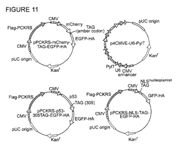
Figure 11 Maps of the main plasmids used
Figure 12 shows a caged lysine and an application of the invention.
Figure 13 shows alternative caged lysines applicable in the invention.
Figure 14. Isolating a sub-network in MAP kinase signalling via genetically
encoding of
a photocaged lysine in the MEK1 active site. (a) Schematic of the MAP kinase
signaling
pathway and its photo-activable sub-network. (b) Caging a near-universally
conserved
lysine in the MEKI active site inactivates the enzyme by sterically blocking
ATP binding.
Decaging with light rapidly removes the caging group and activates the kinase
(figures
created using Pymol and MEKI structure PDB: 1S9J). (c) Structure of the photo-
caged
lysine 1, that can be genetically encoded by the PCKRS/tRNAcUA pair, allowing
the
incorporation of 1 into proteins in response to an amber codon.
12
CA 02792213 2012-09-05
WO 2011/107747 PCT/GB2011/000304
Figure 15. Specific phosphoryiation and activation of ERK2 upon photo-
activation of the
caged MEKI. (a) HEK293ET cells co-transfected with plasmids encoding PCKRS,
pyrrolysyl tRNACUA, C-MEK1-AN-HA and EGFP-ERK2 (either TEY, lanes 7 and 8; or
AAA,
lanes 9 and 10) were grown in medium supplemented with 2 mM of amino acid 1
(lanes
8 and 10) or without (lanes 7 and 9) for 24 h. As controls, cells were
transfected with
plasmids encoding PCKRS, EGFP-ERK2 (TEY or AAA) and either: pyrrolysyl tRNACUA
and
A-MEKI-AN-HA (lanes 1 and 2); or pyrrolysyl tRNACUA and D-MEKI-AN-HA (lanes 3
and 4);
or tyrosine tRNATYrCUA and C-MEK 1-AN-HA (lanes 5 and 6, the incorporation of
Tyr in
response to the amber codon in C-MEKI-AN-HA gene via the use of the amber
suppressor tyrosine tRNATYrCUA leads to an inactive MEKI named D*-MEK1-AN-HA).
(b)
HEK293ET cells co-transfected with plasmids encoding PCKRS, pyrrolysyl
tRNACUA,
EGFP-ERK2 and either A-MEK1-AN-HA (lane 2), or D-MEKI-AN-HA (lanes 3-6), or
C-MEK1-AN-HA (lanes 7-10) were grown in medium supplemented with 2 mM of 1 and
0.1% FBS for 24 h. Cells expressing D-MEK1-AN-HA and C-MEKI-AN-HA were
illuminated
with a 365 nm LED lamp for 60 s. Cells were lysed 1, 10 and 60 min after
illumination. (c)
HEK293ET cells co-transfected with plasmids encoding PCKRS, pyrrolysyl
tRNACUA,
EGFP-ERK2 and either A-MEKI-AN-HA (lane 2), or D-MEKI-AN-HA (lanes 3, 5, 6, 9,
10, 13,
14, 17, 18) or C-MEK 1-AN-HA (lanes 4, 7, 8, 11, 12, 15, 16, 19, 20) were
grown in medium
supplemented with 2 mM of amino acid 1 and 0.1% FBS for 24 h. Cells expressing
D-MEK1-AN-HA and C-MEK1-AN-HA were illuminated with a 365 nm LED lamp for 5 s
(lanes 5-8), 15 s (lanes 9-12), 30 s (lanes 13-16) and 60 s (lanes 17-20).
Cells were lysed 1
and 10 min after illumination. (d) HEK293ET cells co-transfected with plasmids
encoding
PCKRS, pyrrolysyl tRNACUA, EGFP-ERK2 and either C-MEK1-AN-HA (lanes 1-4), or
D-MEKI-AN-HA (lanes 5-8) or A-MEKI-AN-HA (lanes 9-12) were grown in medium
supplemented with 2 mM of amino acid 1 and 0.1% FBS for 24 h. Before
illumination,
cells were incubated with 0 or 10 M of U0126 for 30 min. When indicated,
cells were
illuminated for 60 s with a 365 nm LED lamp. Cells were lysed 10 min after
illumination.
(a-d) Cell lysates were resolved by SDS-PAGE, followed by immunoblotting (IB)
with the
indicated antibodies.
13
CA 02792213 2012-09-05
WO 2011/107747 PCT/GB2011/000304
Figure 16 EGFP-ERK2 nuclear translocation upon EGF stimulation. (a) Montage
showing
EGFP-ERK2 sub-cellular fluorescence at different time points after activation
of co-
expressed wt-MEKI by addition of 100 ng/ml EGF. Scale bars represent 5 pm. (b)
The
graph shows the normalized F(n/c) as a function of time after activation (mean
SD of
seven representative cells). (c) The graph shows F(nlc) of seven independent
experiments as a function of time after activation. (d) The graph shows
normalized
F(n/c) of seven independent experiments as a function of time after
activation.
Figure 17. Nuclear translocation of EGFP-ERK2 upon photo-activation of caged
MEK1.
(a) HEK293ET cells co-transfected with plasmids encoding PCKRS, pyrrolysine
tRNAcUA,
and either C-MEKI-DD / EGFP-ERK2-TEY (cases 1 and 2), or D-MEKI-DD / EGFP-ERK2-
TEY
(case 3), or C-MEK1-DD / EGFP-ERK2-AAA (case 4) were grown in medium
supplemented with 2 mM of amino acid 1 and 0.1% FBS for 24 h. In case 2, cells
were
pre-incubated with 10 M of U0126. EGFP fluorescence of a representative cell
before
and 10 min after illumination (2 s, 365 nm, 1 mW/cm2) is shown in each case.
The
diagrams show the fluorescence intensity along the dotted lines before (black)
and
after (grey) illumination. Scale bars represent 10 pm. (b) Quantitative
analysis of
EGFP-ERK2 nuclear translocation. The graph on the left shows the ratio F(n/c)
of the
mean nuclear and cytoplasmic EGFP fluorescence before (white bars) and 10 min
after
illumination (black bars) in the cases shown in (a). For each case, mean
standard
deviation (SD) of ten representative cells is shown. The graph on the right
shows the
difference of F(n/c) before and 10 min after illumination (AF(n/c) =
F(n/C)aRer -
F(n/C)before) in the cases shown in (a). For each case, data from ten
representative cells
are represented as box-and-whisker plot (the ends of the whiskers represent
the
minimum and maximum of all the data).
Figure 18. Kinetics of EGFP-ERK2 nuclear translocatlon upon photo-activation
of the
caged MEK1. (a) Montage showing EGFP-ERK2 sub-cellular fluorescence at
different
14
CA 02792213 2012-09-05
WO 2011/107747 PCT/GB2011/000304
time points after photo-activation (2 s, 365 nm, 1 mW/cm2) of co-expressed C-
MEKI-DD.
Scale bars represent 5 pm. (b) The graph shows the normalized F(n/c) as a
function of
time after photo-activation (mean SD of ten representative cells). In grey
line is shown
as a comparison the normalized F(n/c) observed when cells were stimulated with
EGF
and presented in Figure 16b. (c) The graph shows F(n/c) of ten experiments as
a
function of time after activation. (d) The graph shows normalized F(n/c) of
ten
independent experiments as a function of time after activation. (e,f)
Comparison of the
cell-to-cell variability observed in EGFP-ERK2 nuclear translocation upon
stimulation with
EGF (data shown on Figure 16b-d, n = 7 cells) and upon photo-activation of C-
MEKI-DD
(data shown in b-d, n = 10 cells). The two graphs show respectively (e) the
half-time of
the translocation process upon activation (ti/2) and (f) the change in F(n/c)
observed
(zF(n/c) = max(F(n/c)) - min(F(n/c))). The data are represented as box-and-
whisker plot
(the ends of the whiskers represent the minimum and maximum of all the data).
(g)
Montage showing representative EGFP-ERK2 sub-cellular fluorescence at
different time
points early after photo-activation (2 s, 365 nm, 1 mW/cm2) of co-expressed C-
MEKI-DD
(see also Movie Si). Scale bars represent 10 pm. (h) Kinetics of translocation
early after
photo-activation. Normalized F(n/c) as a function of time after photo-
activation (mean
SD of ten representative cells) is shown. Data were fitted with a sigmoidal
function.
Figure 19. ERK2 nucleocytoplasmic shuttling. (a) HEK293ET cells co-expressing
C-MEK1-
DD and EGFP-ERK2 were illuminated (2 s, 365 nm, 1 mW/cm2), then 8 minutes
after
illumination, U0126 (10 M) was added to block the activity of photoactivated
C-MEKI-DD and unveil EGFP-ERK2 efflux from the nucleus. The bottom montage
shows
representative EGFP-ERK2 sub-cellular fluorescence at different times after
illumination
and post-illumination blockage with U0126. The top montage shows as a
reference the
EGFP-ERK2 sub-cellular fluorescence at different times after photo-activation
without
addition of U01 26. Scale bars represent 5 pm. (b) The graph presents the
normalized
F(n/c) as a function of time after illumination (mean SD of ten
representative cells).
CA 02792213 2012-09-05
WO 2011/107747 PCT/GB2011/000304
The arrow indicates the time when U0126 was added. As a comparison, the
normalized
F(n/c) without addition of the inhibitor is presented in Figure 18b is plotted
as a grey line.
Figure 20. (a) Montage showing EGFP-ERK2 (top) and EGFP- ERK2A4 (bottom) sub-
cellular fluorescence at different time points after photo-activation (2 s,
365 nm,
1 mW/cm2) of co-expressed C-MEKI-DD. Scale bars represent 5 pm. (b) The graph
shows the kinetics of nuclear translocation of EGFP-ERK2A4 upon photo-
activation of C-
MEK 1-DD (mean SD for ten representative cells). In grey line is shown as a
comparison
the kinetics of nuclear translocation of EGFP-ERK2 shown in Figure 18b. (c)
The plot
shows the maximum of F(n/c) from the experiments shown in (a) (mean SD for
ten
representative cells). (d,e) HEK293ET cells co-expressing C-MEK1-DD and either
EGFP-
ERK2 or EGFP-ERK204 were illuminated with a LED lamp for 1 minute and lysed
after 1, 5,
10, 15, 20 and 30 minutes. (d) Cell lysates were resolved by SDS-PAGE,
followed by
immunoblotting (IB) with the indicated antibodies. (e) The phosphorylation of
the EGPF-
ERK2 mutants observed in (d) was quantified and normalized by their expression
level,
and plotted as a function of time (representative data from three independent
data
sets).
Figure 21. (a) HEK293ET cells co-transfected with plasmids encoding PCKRS,
pyrrolysine tRNAcuAand C-MEKI-AN-HA (lanes 3 and 4) were grown in medium
supplemented with 1 mM I (lane 4) or without (lane 3) for 24 h. As controls,
cells
were co-transfected with plasmids encoding PCKRS and either: pyrrolysine
tRNAcuA
and A-MEK 1-AN-HA (lane 1); or pyrrolysine tRNAcuA and D-MEK 1-AN-HA (lane
2); or tyrosine tRNATyr
CUA and C-MEKI-AN-HA (lane 5; the incorporation of Tyr in
response to the amber codon in C-MEKI-AN-HA gene via the use of the amber
suppressor tyrosine tRNATyr
CUA leads to an inactive dead MEKI named
D*-MEKI-AN-HA). Cell lysates were resolved by SDS-PAGE, followed by
immunoblotting (IB) with the indicated antibodies. (b) Immunoblot comparing
the
expression level of the different MEKI-AN-HA mutants with endogenous MEK.
Figure 22. Cells were co-transfected with plasmids encoding PCKRS, EGFP-ERK2
and either: pyrrolysine tRNAcuA and A-MEKI-AN-HA (lanes I and 2); or
pyrrolysine tRNAcuA and D-MEKI -AN-HA (lanes 3 and 4); or pyrrolysine tRNAcuA
only (lanes 5 and 6); or tyrosine tRNATyT
cu, and C-MEKI-AN-HA (lanes 7 and 8; the
incorporation of Tyr in response to the amber codon in C-MEKI-AN-HA gene via
the
use of the amber suppressor tyrosine tRNATyr
16
CA 02792213 2012-09-05
WO 2011/107747 PCT/GB2011/000304
CUA leads to an inactive dead MEKI
named D*-MEKI-AN-HA); or pyrrolysine tRNAcuA and C-MEKI-AN-HA (lanes 9
and 10). Lanes 11 and 12 show mock non-transfected cells. After transfection,
HEK293ET cells were grown in medium supplemented with 2 mM I and 0.1% FBS
for 24 h. When indicated, cells were illuminated with a 365 nm LED lamp for 60
s,
and lysed 60 min after illumination. Cell lysates were resolved by SDS-PAGE,
followed by immunoblotting (IB) with the indicated antibodies.
Figure 23. (a) HEK293ET cells co-transfected with plasmids encoding PCKRS,
pyrrolysine tRNAcuAand C-MEKI-DD-HA (lanes 3 and 4) were grown in medium
supplemented with 2 mM I (lane 4) or without (lane 3) for 24 h. As controls,
cells
were co-transfected with plasmids encoding PCKRS and either: pyrrolysine
tRNAcUA
and A-MEK 1-DD-HA (lane 1); or pyrrolysine tRNAcuA and D-MEK 1-DD-HA (lane
2); or tyrosine tRNATyr
CUA and C-MEKI-DD-HA (lane 5; the incorporation of Tyr in
response to the amber codon in C-MEK 1-DD-HA gene via the use of the amber
suppressor tyrosine tRNATyr
CUA leads to an inactive dead MEKI named
D*-MEKI-DD-HA). (b) HEK293ET cells co-transfected with plasmids encoding
PCKRS, pyrrolysine tRNAcuA, EGFP-ERK2 and either A-MEK I -AN-HA (lane 1), or
D-MEKI-AN-HA (lanes 2-3) or C-MEKI-AN-HA (lanes 4-5), or A-MEKI-DD-HA
(lane 6), or D-MEKI-DD-HA (lanes 7-8) or C-MEK1-DD-HA (lanes 9-10) were
grown in medium supplemented with 2 mM 1 and 0.1 % FBS for 24 h. When
indicated, cells expressing D-MEK I -AN or DD)-HA and C-MEK I -(AN or DD)-HA
were illuminated with a 365 nm LED lamp for 60 s. Cells were lysed 10 min
after
illumination. (a-b) Cell lysates were resolved by SDS-PAGE, followed by
immunoblotting (IB) with the indicated antibodies.
Detailed description of the invention
The present invention relates to a caged lysine molecule in which the
caging group is induced by electron-donating substituents to decage
efficiently at irradiation of UV light above 340nm. The effect of the
electron donation to the caging group allows the caging group to be
decaged efficiently when irradiated with light above 340nm. Preferably
the UV irradiation is above 355 nm, preferably between 360 and 370 nm,
even more preferably about 365nm. It is clear to the person skilled in the
art that the advantage with respect to other caging molecules is the
efficiency of photolysis of the caged molecule, when irradiated at these
higher UV wavelengths. As shown in Fig. 3 and Example 3, after 5
minutes, essentially the entire population of caged protein is de-caged
by UV irradiation at 365nm.
It is further preferable if the caging group is constructed so that upon
photolysis, the by products of the photolysis do not react in a
17
CA 02792213 2012-09-05
WO 2011/107747 PCT/GB2011/000304
condensation reaction with the e-amino group of lysine. A preferred
embodiment is when the caged lysine according to the present
invention is according to Formula (I)
CH3 0 NH2
0 0 N CO2H
H
03() N02
(I)
or salts thereof.
Another aspect of the invention is the caged lysine as described above
when incorporated into the amino acid sequence of a protein. The
advantage, as discussed below, is that it allows the determination
1o and/or alteration of a specific property in a protein. It is preferable
that
the incorporation be site-specific, as this advantageously allows
determination/alteration of a specific property of the protein due to the
presence of the caged lysine in a specific point of the protein.
The site-specific incorporation of the caged lysine amino acid may be at
any point in the polypeptide sequence. This is typically accomplished by
site specific mutation of the nucleotide sequence of a nucleic acid
encoding the polypeptide of interest, followed by transcription (if
necessary) and translation of that nucleic acid into polypeptide. The
incorporation may be by replacement of an existing codon or may be
by insertion of a codon. Typically the codon used to specify the caged
lysine will be the amber codon TAG (CUA). However, of course if a tRNA
synthetase-tRNA pair used for incorporation comprises a tRNA
recognising a different codon (or a quadruplet codon), then the
corresponding cognate codon of that tRNA synthetase-tRNA pair will be
used in place of the amber codon. The amber codon is a preferred
example of a suitable orthogonal codon by which genetic incorporation
may be easily achieved, but is not intended to limit or to exclude the use
18
CA 02792213 2012-09-05
WO 2011/107747 PCT/GB2011/000304
of other codon(s) provided that a suitable system for charging the
cognate tRNA of any such other codon(s) can be employed.
It is further preferable that the site-specific incorporation of the caged
lysine amino acid in the amino acid sequence of the protein be as
replacement of a lysine residue present in the wild-type sequence of the
protein. The advantage of said protein is that it allows empirical
determination the intrinsic properties of that lysine residue and therefore
the biological effect(s) of the protein mediated by (or influenced by)
that lysine once the irradiation and resulting de-caging occurs.
to In one preferred embodiment, the protein according to the invention as
described above is further linked to a labelling molecule. The labelling
molecule can be any molecule which a person skilled in the art can use
under experimental circumstances to determine some biologically
relevant property or function of the protein. Some examples of such
molecules are radioactive elements, fluorescent or luminescent markers.
The method of linking the protein to the labelling molecule depends
entirely on the type of labelling molecule used and the choice is well
within the person skilled in the art's expertise. In a preferred example of
said system, the labelling molecule is a fluorescent protein, such as GFP,
fused to the C-terminal of the protein with the caged lysine incorporated
in it. Said example is preferred as the method of linking the protein is
easily achieved by incorporating a nucleotide sequence encoding the
GFP protein into a plasmid which encodes the protein with the caged
amino acid. In said preferred example, the resulting protein expressed in
the cell is easily visualised.
Another aspect of the invention is a method, such as an in vitro method,
of incorporating the caged lysine amino acids genetically and site-
specifically into the protein of choice, suitably in a eukaryotic cell. One
advantage of incorporating it genetically by said method is that it
obviates the need to deliver the proteins comprising the caged amino
acid into a cell once formed, since in this embodiment they may be
19
CA 02792213 2012-09-05
WO 2011/107747 PCT/GB2011/000304
synthesised directly in the target cell. The method comprises the
following steps:
i) introducing, or replacing a specific codon with, an
orthogonal codon such as an amber codon at the desired site in
the nucleotide sequence encoding the protein
ii) introducing an expression system of orthogonal pyrollysyl-tRNA
synthetase/tRNA pair in the cell
iii) growing the cells in a medium with the caged lysine
according to the invention.
io Step (i) entails or replacing a specific codon with an orthogonal codon
such as an amber codon at the desired site in the genetic sequence of
the protein. This can be achieved by simply introducing a construct, such
as a plasmid, with the nucleotide sequence encoding the protein,
wherein the site where the caged lysine is desired to be
introduced/replaced is altered to comprise an orthogonal codon such
as an amber codon. This is well within the person skilled in the art's ability
and examples of such are given here below.
Step (ii) requires an orthogonal expression system to specifically
incorporate the caged lysine amino acid at the desired location (e.g.
the amber codon). Thus a specific orthogonal tRNA synthetase such as
an orthogonal pyrollysyl-tRNA synthetase and a specific corresponding
orthogonal tRNA pair which are together capable of charging said tRNA
with the caged lysine are required.
Thus another aspect of the invention is the provision of an orthogonal
tRNA synthetase such as a pyrollysyl-tRNA synthetase for the caged lysine
according to the invention. Said orthogonal pyrollysyl-tRNA synthetase
are suitably wild-type Pyrollysyl-tRNA synthetase with mutation(s) in up to
5 positions as defined in Table I wherein the mutation(s) are present at
residues M241, A267, Y271, L274 and C313. In a preferred embodiment,
the orthogonal pyrollysyl-tRNA synthetase is clone 7 of Table I, i.e.
wherein the mutations are M241 F, A267S, Y271 C and L274M, which has
CA 02792213 2012-09-05
WO 2011/107747 PCT/GB2011/000304
the advantage of being found to be the most efficient synthetase clone
as defined in Table I.
The orthogonal pyrollysyl-tRNA synthetase according to the invention
needs to be associated with an orthogonal tRNA to constitute an
expression system to be able to execute step (ii) of the method above.
The use of PyIT, the gene encoding PyltRNAcUA, lacks the consensus
internal RNA polymerase III promoter sequences found in eukaryotic
tRNAs and is well known in the art as an orthogonal tRNA system to be
used in an orthogonal pyrollysyl-tRNA synthetase/tRNA pair14. It requires
to an external promoter for transcription. Preferably tRNA expression is
under the control of a U6 promoter downstream of a CMV enhancer,15
enabling efficient transcription of PyIT.
Thus a preferred expression system to be used in step (ii) of the method
above comprises:
a. a plasmid where PyItRNACUA expression is under the control
of a U6 promoter downstream of a CMV enhancer
b. a plasmid comprising the orthogonal pyrollysyl-tRNA
synthetase as described herein under control of a CMV
enhancer.
In another aspect of the invention, the caged lysine according to the
invention can be used for determining or altering at least one property of
a protein by UV light irradiation above 340nm. Preferably the irradiation is
above 355nm, more preferably between 360 and 370 nm, even more
preferably about 365nm.
The advantage of such uses is that the mode of switching from caged to
de-caged is efficiently achieved, both in the sense of time and
percentage of protein with caged lysines being de-caged, and that
such a system is non-invasive and not toxic to cells.
Advantageously such a system can be used for determination or
alteration of at least one property of a protein in a eukaryotic cell, even
within a human body.
21
CA 02792213 2012-09-05
WO 2011/107747 PCT/GB2011/000304
Said at least one property of the protein may be a biochemical property
of the protein which is present in the wild-type protein and not present
when the caged amino acid is present. This may be the sole biological
function of the protein, or may be one or more of several properties of
the protein. An example where the property is not the sole biological
function of the protein is the NLS sequences present in a tumour
suppressor p53. The property and its effects on the protein can vary
according to the size, shape of the protein and also importantly the
position of incorporation of the caged lysine in the polypeptide chain.
io Thus when used for determination upon de-caging by photolysis, the
invention enables the operator to study how the property impeded by
the caged lysine residue affects the biological effect of the protein upon
de-caging. In its use in altering a property of a protein, the biological
effects resulting from the alteration may be known and therefore
studied, or may be unknown in which case the invention may be
advantageously applied to the determination or inference of such
properties. An example of application of the invention to a known
property would be the desire to release a caged lysine placed in a
localisation sequence so as to allow the uncaged sequence to then
localise the protein in the appropriate cellular compartment, thereby
permitting kinetic studies or other observations to be carried out.
It is preferable for the caged lysine to replace a lysine present in the wild-
type protein. This is preferable as it allows determination, through de-
caging, of the intrinsic function of the protein in the cell as the protein
reverts to its wild-type structure on de-caging.
When being used for alteration, it is assumed that the biological effects
resulting from the alteration are known and/or desired. Such use of the
caged lysine as alternator (switch) can be actuated to study the kinetics
of proteins resulting from the de-caging. One example is when the
protein folding is disturbed by the presence of a caged lysine. In such a
case, the de-caging of the caged amino acid would allow protein
folding to occur again, thus allowing one to measure the kinetics of
22
CA 02792213 2012-09-05
WO 2011/107747 PCT/GB2011/000304
protein folding that result from the de-caging. Another example is the
incorporation of a caged lysine amino acid in a localisation sequence.
This would disrupt the proper localisation of the protein until the de-
caging, allowing one to measure the kinetics of protein localisation.
Thus, embodiments of the invention in which the properties of the protein
of interest are altered by decaging are sometimes referred to as
'switching' or 'alternation' (i.e. moving to an alternate form of the
protein in the decaged state).
It is further contemplated that the use described here above regarding
io the altering (alternating) of at least one property of a protein by UV
light
irradiation above 340nm may be used for therapeutic purposes. The
alternation by de-caging may allow a protein, which previously was
undesired to be localised/have a certain function/ be fully folded, to
then localise/have a certain function/ be fully folded and thus have a
certain therapeutic function. Examples of proteins where such a situation
may occur are membrane proteins, especially expression of known
cluster of differentiation proteins or for example antibodies or proteins
belonging to the complement immune system.
Caged Lysine Species
Alternative caged lysines other than that of Formula I may be used in the
invention. Examples of useful caged lysine compounds are shown in
figure 13.
Possible compounds are reviewed in Mayer et al. Angew. Chem. Int. Ed.
45, 4900-4921 (2006).
Compounds shown in Figure 13 are specifically described in the following
citations. The sections of these citations describing the compounds
shown in Figure 13 are specifically incorporated herein by reference, in
particular for the details of structure or production of the corresponding
compound(s) shown in Figure 13:
23
CA 02792213 2012-09-05
WO 2011/107747 PCT/GB2011/000304
compound 4: Momotake et al. Nat. Meth. 3, 35-40 (2006)
compound 5: Walbert et at. Helv. Chim. Acta 84, 1601-1611 (2001)
compound 6: Singh et al. Bioconjug. Chem. 13, 1286-1291 (2002)
compound 7: Furuta et al. Proc. Nat. Acad. Sci. 96, 1193-1200 (1999);
Suzuki et al. Org. Lett. 5, 4867-4870 (2003); Hagen et at. ChemBioChem 4,
434-442 (2003).
compound 8: Fedoryak et at. Org. Lett. 4, 3419-3422 (2002).
compound 9: Park et at. JACS 119, 2453-2463 (1997); Zhang et at. JACS
121, 5625-5632 (1999); Conrad II et at. Org Lett 2, 1545-1547 (2000).
1o compound 10: Atemnkeng et at. Org Lett 5, 4469-4471 (2003).
compound 11: Klan et al. Photochem Photobiol Sci 1, 920-923 (2002);
Klan et at. Org Lett 2, 1569-1571 (2000)
Most suitably the caged lysine is as shown in Formula I.
Reference Sequences
The Methanosarcina barkeri PylT gene encodes the MbtRNAcUA tRNA.
The Methanosarcina barkeri PyIS gene encodes the MbPyIRS tRNA
synthetase protein. When particular amino acid residues are referred to
using numeric addresses, the numbering is taken using MbPyIRS
(Methanosarcina barkeri pyrrolysyl-tRNA synthetase) amino acid
sequence as the reference sequence (i.e. as encoded by the publicly
available wild type Methanosarcina barkeri PyIS gene Accession number
Q46E77):
MDKKPLDVLI SATGLWMSRT GTLHKIKHYE VSRSKIYIEM ACGDHLVVNN
SRSCRTARAF RHHKYRKTCK RCRVSDEDIN NFLTRSTEGK TSVKVKVVSA
PKVKKAMPKS VSRAPKPLEN PVSAKASTDT SRSVPSPAKS TPNSPVPTSA
PAPSLTRSQL DRVEALLSPE DKISLNIAKP FRELESELVT RRKNDFQRLY
TNDREDYLGK LERDITKFFV DRDFLEIKSP ILIPAEYVER MGINNDTELS
24
CA 02792213 2012-09-05
WO 2011/107747 PCT/GB2011/000304
KQIFRVDKNL CLRPMLAPTL YNYLRKLDRI LPDPIKIFEV GPCYRKESDG
KEHLEEFTMV NFCQMGSGCT RENLESLIKE FLDYLEIDFE IVGDSCMVYG
DTLDIMHGDL ELSSAVVGPV PLDREWGIDK PWIGAGFGLE RLLKVMHGFK
NIKRASRSES YYNGISTNL
This is to be used as is well understood in the art to locate the residue of
interest. This is not always a strict counting exercise - attention must be
paid to the context. For example, if the protein of interest is of a slightly
different length, then location of the correct residue in that sequence
to correseponding to (for example) Y271 may require the sequences to be
aligned and the equivalent or corresponding residue picked, rather than
simply taking the 271st residue of the sequence of interest. This is well
within the ambit of the skilled reader.
Mutating has it normal meaning in the art and may refer to the
substitution or truncation or deletion of the residue, motif or domain
referred to. Mutation may be effected at the polypeptide level e.g. by
synthesis of a polypeptide having the mutated sequence, or may be
effected at the nucleotide level e.g. by making a nucleic acid encoding
the mutated sequence, which nucleic acid may be subsequently
translated to produce the mutated polypeptide. Where no amino acid
is specified as the replacement amino acid for a given mutation site,
suitably a randomisation of said site is used, for example as described
herein in connection with the evolution and adaptation of tRNA
synthetase of the invention. As a default mutation, alanine (A) may be
used. Suitably the mutations used at particular site(s) are as set out
herein.
A fragment is suitably at least 10 amino acids in length, suitably at least
25 amino acids, suitably at least 50 amino acids, suitably at least 100
amino acids, suitably at least 200 amino acids, suitably at least 250
amino acids, suitably at least 300 amino acids, suitably at least 313
CA 02792213 2012-09-05
WO 2011/107747 PCT/GB2011/000304
amino acids, or suitably the majority of the tRNA synthetase polypeptide
of interest.
Polynucleotides of the invention can be incorporated into a
recombinant replicable vector. The vector may be used to replicate the
nucleic acid in a compatible host cell. Thus in a further embodiment, the
invention provides a method of making polynucleotides of the invention
by introducing a polynucleotide of the invention into a replicable vector,
introducing the vector into a compatible host cell, and growing the host
1o cell under conditions which bring about replication of the vector. The
vector may be recovered from the host cell. Suitable host cells include
bacteria such as E. coli.
Preferably, a polynucleotide of the invention in a vector is operably
linked to a control sequence that is capable of providing for the
expression of the coding sequence by the host cell, i.e. the vector is an
expression vector. The term "operably linked" means that the
components described are in a relationship permitting them to function
in their intended manner. A regulatory sequence "operably linked" to a
coding sequence is ligated in such a way that expression of the coding
sequence is achieved under condition compatible with the control
sequences.
Vectors of the invention may be transformed or transfected into a
suitable host cell as described to provide for expression of a protein of
the invention. This process may comprise culturing a host cell
transformed with an expression vector as described above under
conditions to provide for expression by the vector of a coding sequence
encoding the protein, and optionally recovering the expressed protein.
The vectors may be for example, plasmid or virus vectors provided with
an origin of replication, optionally a promoter for the expression of the
26
CA 02792213 2012-09-05
WO 2011/107747 PCT/GB2011/000304
said polynucleotide and optionally a regulator of the promoter. The
vectors may contain one or more selectable marker genes, for example
an ampicillin resistance gene in the case of a bacterial plasmid. Vectors
may be used, for example, to transfect or transform a host cell.
Control sequences operably linked to sequences encoding the protein
of the invention include promoters/enhancers and other expression
regulation signals. These control sequences may be selected to be
compatible with the host cell for which the expression vector is designed
io to be used in. The term promoter is well-known in the art and
encompasses nucleic acid regions ranging in, size and complexity from
minimal promoters to promoters including upstream elements and
enhancers.
Protein Expression and Purification
Host cells comprising Oolynucleotides of the invention may be used to
express proteins of the invention. Host cells may be cultured under
suitable conditions which allow expression of the proteins of the
invention. Expression of the proteins of the invention may be constitutive
such that they are continually produced, or inducible, requiring a
stimulus to initiate expression. In the case of inducible expression, protein
production can be initiated when required by, for example, addition of
an inducer substance to the culture medium, for example
dexamethasone or IPTG.
Proteins of the invention can be extracted from host cells by a variety of
techniques known in the art, including enzymatic, chemical and/or
osmotic lysis and physical disruption.
The following non-limiting examples are illustrative of the present
invention:
27
CA 02792213 2012-09-05
WO 2011/107747 PCT/GB2011/000304
In all examples, the caged lysine according to Formula (I) is either
denoted as such or as compound 1.
We teach photocaging of lysine to control protein localization, post-
translational modification and enzymatic activity. The photochemical
control of these important functions mediated by lysine residues in
proteins has not previously been demonstrated in living cells. Here we
synthesize 1, and evolve a pyrrolysyl-tRNA synthetase/tRNA pair to
genetically encode the incorporation of this amino acid in response to
1o an amber codon in mammalian cells. To exemplify the utility of this
amino acid we cage the nuclear localization sequences (NLSs) of
nucleoplasmin and the tumor suppressor p53 in human cells, thus mis-
localizing the proteins in the cytosol. We trigger protein nuclear import
with a pulse of light allowing us to directly quantify the kinetics of nuclear
import.
Example -1 - Synthesis of caged lysine according to Formula I
The nitrobenzyl caged lysine 1 was prepared by reacting N -Boc-lysine
with the chloroformate 3 in a basic THE/H20 solution at 0 C providing 4 in
82% yield, followed by deprotection with TFA in CH2CI2 in 95% yield
(Scheme S1). The chloroformate 3 was generated through an acylation
of the alcohol 3 (synthesized according to ref 19) with triphosgene in THE
in the presence of Na2CO3, followed by evaporation of the volatiles and
a direct reaction without further purification. The presence of Na2CO3
prevented dehydration of 2 to the corresponding styrene.
28
CA 02792213 2012-09-05
WO 2011/107747 PCT/GB2011/000304
NHBoc
CH3 CH3 O -
O OH triphosgene O O )( CI H2N CO2H
O I N 2CO3, 0 I H20lrHF, NaHCO3 THF
NO2 N02
2 quant. 3 82%
CH3 0 NHBoc CH3 0 NH2
0 N C02H ~- O I 0 N C02H
O T
DCM
N02 95% 0 N02
4
Scheme Si
Synthetic Protocols
(2S)-2-(tert-Butoxycarbonylamino)-6-{[1-(6-nitrobenzo[d] [1,3]dioxol-5-
yl)ethoxy]carbonylamino}hexanoic acid (4). 1-(6-
Nitrobenzo[d] [1,3]dioxol-5-yl)ethanol (2) (500 mg, 2.36 mmol) was
dissolved in THE (5 mL), containing Na2CO3 (247 mg, 2.36 mmol), and
cooled to 0 C. To the solution was added triphosgene (701 mg, 2.36
mmol) and the reaction was kept stirring for 12 h at r.t. The reaction was
Io filtered and the volatiles were subsequently evaporated without heating
and the residue dried under vacuum, to give NPOC chloroformate 3 in
quantitative conversion (644 mg, 2.36 mmol). To a solution of N-Boc-
lysine (500 mg, 2.02 mmol) in THE/1 M NaOH (aq.) (1:4 mixture, 8 mL
total), at 0 C, was added NPOC-a-methyl chloroformate 3 (496 mg, 1.82
mmol). After the reaction was stirred for 12 h, at r.t., the aqueous layer
was washed with Et20 (5 mL) and subsequently acidified with ice-cold 1
M HCI (20 mL) to pH 1 and extracted with EtOAc (30 mL). The organic
layer was dried over Na2SO4, filtered, and the volatiles were evaporated,
affording 4 as a yellow foam in 82% yield (720 mg, 1.49 mmol). 1H NMR
(300 MHz, CDC13) 8 = 1.18-1.81 (m, 18 H), 3.08 (br s, 2 H), 4.23 (br s, 1 H),
5.11-5.38 (m, 1 H), 6.07 (s, 2 H), 6.20-6.36 (m, 1 H), 6.99 (s, 1 H), 7.40 (s,
1 H).
13C NMR (75 MHz, CHCI3) 8 = 22.3, 22.6, 28.5, 29.4, 32.3, 40.8, 53.3, 69.1,
80.4, 103.3, 105.4, 105.8, 136.7, 141.5, 147.2, 152.6, 155.7, 156.1, 176.4.
HRMS: m/z calcd for C21H29N3010 [M+Na]+: 506.1745; found: 506.1748.
see Figure 1 for' H NMR spectrum)
29
CA 02792213 2012-09-05
WO 2011/107747 PCT/GB2011/000304
(2S)-2-Amino-6-([1-(6-nitrobenzo[d][1,3]dioxol-5-yl)ethoxy]
carbonylamino} hexanoic acid TFA salt (1). Compound 4 (720 mg, 1.49
mmol) was dissolved in DCM:TFA (1:1 mixture, 14 mL total) and the
reaction was allowed to stir for 40 min. The volatiles were subsequently
evaporated and the residue was redissolved in MeOH (5 mL) and
precipitated into Et20 (250 mL), giving 1 as a white solid in 95% yield (679
mg, 1.42 mmol). 1H NMR (300 MHz, D20) 8 = 1.08-1.40 (m, 7 H), 1.63-1.88
(m, 2 H), 2.80-2.88 (m, 2 H), 3.83-3.97 (m, 1 H), 5.91-6.00 (m, 3 H), 6.92 (s,
1
H), 7.28 (s, 1 H). 13C NMR (75 MHz, D20) 8 = 21.1, 21.7, 28.5, 29.5, 39.9,
52.7,
io 68.8, 103.6, 104.4, 105.7, 136.1, 140.6, 146.9, 152.7, 156.9, 171.9. HRMS:
m/z
calcd for C15H21N306 [M+H]+: 384.1402; found: 384.1403. (see Figure 2 for
1 H NMR spectrum)
Example 2 - Synthesis of an orthogonal pyrrolysyl-tRNA synthetase/tRNA
pair for caged lysine according to Formula (I)
To evolve the orthogonal MbPyIRS/PyltRNAcUA pair11 for the incorporation
of the caged lysine 1 in response to an amber codon, a library of 108
mutants of MbPyIRS was created in which 5 positions (M241, A267, Y271,
L274, C313) in the binding pocket of the pyrrolysine ring were
randomized to all possible amino acids.
pBKAcKRS3amp20 was used as a template in the generation of a library
of MbPyIRS mutants. Three rounds of inverse PCR21 were performed to
randomize codons for M241, A267, Y271, L274 and C313 to all 20 natural
amino acids in this library. The following primers were used in each round
of PCR reactions:
= (round 1) PyISM241 f (5'-
GCGCAGGTCTCAGAACGTNNKGGCATTAACAACGACACCGAAC
TGAGCAAAC-3') and PyISM241 r (5'-
GCGCAGAGTAGGTCTCAGTTCCACATATTCCGCCGGAATCAGAA
TC-3');
= (round 2) PyISAYLf (5'-GCGCAGGTCTCAATGCTGN
NKCCGACCCTGNNKAACTATN NKCGTAAACTGGATCGTATTCTGCC
CA 02792213 2012-09-05
WO 2011/107747 PCT/GB2011/000304
GGGC-3') and PyISAYLr (5'-
GCGCAGAGTAGGTCTCAGCATCGGACGCAGGCACAGGTTTTTAT
C-3');
= (round 3) PyISC313f (5'-GCGCAGGAAAGGTCTCAAACTTTN
NKCAAATGGGCAGCGGCTGCACCCGTGAAAAC-3') and
PyISC3l 3r (5'-
GCGCAGAGTAGGTCTCAAGTTAACCATGGTGAATTCTTCCAGGTGT
TCTTTG-3').
The PCR product in each round was first digested with Dpnl and Bsal, re-
circularized by ligation and used to transform electrocompetent DH10B.
The reisolated plasmids served as template for the next round of
mutagenesis. Transformation of electro-competent DH 10B with the
ligation of the third round of mutagenesis produced 108 transformants,
covering the theoretical diversity of the library (2x10⁷) by more than 99%.
Selection of mutants specific for 1 was carried out as described for the
evolution of a synthetase specific for acetyl-lysine'1.
Three rounds of alternating positive and negative selection on this library
in E. coli were performed, as previously described.11.12 Clones that
survived the selection were transformed with a plasmid encoding the
chloramphenicol resistance gene with an amber codon at a permissive
position. The best clones allowed cells to survive on media containing up
to 300 pg/ml chloramphenicol in the presence of 1 (1 mM), but did not
survive at 50 pg/ml in the absence of 1. This demonstrates that the
selected synthetases have a high specificity for 1, and do not
incorporate any of the common 20 amino acids. The most active
synthetase contained the mutations M241 F, A267S, Y271 C, and L274M
with respect to wild-type MbPyIRS. This synthetase was named
Photocaged Lysyl-tRNA Synthetase (PCKRS) and was further
characterized (see Table 1 for all isolated MbPyIRS sequences).
31
CA 02792213 2012-09-05
WO 2011/107747 PCT/GB2011/000304
Example 3 - Demonstration of de-caning upon irradiation with 365nm
light in myoglobin in vitro
1. Expression and purification of myoglobin
To express myoglobin with an incorporated unnatural amino acid, we
transformed E. coli DH10B cells with pBKamp-PCKRS and pMyo4TAGPyIT-
his6. Cells were recovered in 1 mL of LB media for 1 h at 37 C, before
incubation (16 h, 37 C, 250 r.p.m.) in 100 mL of LB containing ampicillin
(100 pg/mL) and tetracycline (25 pg/mL). 20 mL of this overnight culture
was used to inoculate 1 L of LB supplemented with ampicillin (50 pg/mL),
io tetracycline (12 pg/mL) and 2 mM of 1. Cells were grown (37 C, 250
r.p.m.), and protein expression was induced at OD6oo -0.6, by addition of
arabinose to a final concentration of 0.2%. After 3 h of induction, cells
were harvested. Proteins were extracted by sonication at 4 C. The
extract was clarified by centrifugation (20 min, 21,000 g, 4 C), 300 pL of
Nit+-NTA beads (Qiagen) were added to the extract, the mixture was
incubated with agitation for 1 h at 4 C. Beads were collected by
centrifugation (10 min, 1000 g). The beads were twice resuspended in 50
mL wash buffer and spun down at 1000 g. Subsequently, the beads were
resuspended in 20 ml of wash buffer and transferred to a column. Protein
was eluted in 1 ml of wash buffer supplemented with 250 mM imidazole
and was then re-buffered to 20 mM ammonium bicarbonate using, a
sephadex G25 column.
sfGFP-his6 incorporating an unnatural amino acid (BocK or 1) in response
to an amber codon at position 145 (psfGFP145TAGPyIT-his6) was
expressed and purified following the same protocol.
2. Protein mass spectrometry
Protein total mass was determined on an LCT time-of-flight mass
spectrometer with electrospray ionization (ESI, Micromass). Proteins were
rebuffered in 20 mM of ammonium bicarbonate and mixed 1:1 with
formic acid (1% in methanol/H20 = 1:1). Samples were injected at 10
pl/min and calibration was performed in positive ion mode using horse
heart myoglobin. 60 scans were averaged and molecular masses
32
CA 02792213 2012-09-05
WO 2011/107747 PCT/GB2011/000304
obtained by deconvoluting multiply charged protein mass spectra using
MassLynx version 4.1 (Micromass). Theoretical masses of wild-type
proteins were calculated using Protparam
(http://us.expasy.org/tools/protporam.html), and theoretical masses for
unnatural amino acid containing proteins were adjusted manually.
For MS/MS analysis of sfGFP(145-1), the gel band was washed, alkylated,
and in-gel digested with trypsin. 1 l of digest mixture was premixed with
1 41 of CHCA matrix (3mg/ml in 60% MeCN/ 0.1% TFA) and 1 l was
applied onto a stainless steel target. The spectrum was acquired with an
Ultraflex III TOF/TOF mass spectrometer (Bruker Daltonics, Bremen,
Germany). A m/z 2145.972 fragment that matched to peptide modified
with 1 was manually selected for further MS/MS fragmentation. The
fragmentation ion series confirmed the identity and modification site of
the peptide LEYN(1)NSHNVYITADK.
For the analysis of the uncaging process, purified myoglobin was
photolysed at 365 nm with a high power LED source module at 365 nm
(Black-led-365, Prizmatix). Protein total mass was then determined as
described above.
Expression of myo4TAGhis6>>=12 in the presence of PCKRS/PyItRNAcuA Was.
efficient and dependent on the addition of 1. Electrospray ionization
mass spectrometry (ESI-MS) and MS-MS sequencing confirm the
incorporation of 1 at a single genetically encoded site (Figure 3). We
confirmed that myoglobin containing 1 is efficiently decaged upon
irradiation with 365 nm light in vitro (Figure 3E).
Example 4 - Demonstration that the orthogonal pair PCKRS/PyItRNAcuA is
functional in human cells
To demonstrate that the PCKRS/PyItRNAcuA pair is functional in human
embryonic kidney (HEK293) cells, we examined the red and green
fluorescence of cells containing mCherry-TAG-egfp-ha (this reporter
contains an N-terminal mCherry gene, a linker containing an amber stop
codon, a C-terminal enhanced GFP gene, and the HA-tag coding
33
CA 02792213 2012-09-05
WO 2011/107747 PCT/GB2011/000304
sequence), PCKRS and PyItRNAcuA, in the presence and absence of 1
Protocol
1. culture, transfection and immunoblot analysis
Adherent human embryonic kidney (HEK)-293 cells were cultured at 37
C in a 5% C02 atmosphere in DMEM+GIutaMAX-1 medium (Gibco)
supplemented with 10% FBS and lx pen-strep solution. Cells were
transiently transfected with Genejuice (Novagen) according to the
manufacturer's protocol. Double transfections were performed using
equal amount of both plasmids. Before transfection, medium was
io replaced by fresh antibiotic-free medium supplemented, when
necessary, with the unnatural amino acid (see figure legends for
concentrations). Cells were analyzed 24 h after transfection. For western
blot analysis, cells were washed with cold PBS, then lysed with universal
lysis buffer (Roche) at 4 C for 10 min. Western Blots were performed using
antibodies against HA-tag (Sigma), Flag-tag (Cell signaling), Ds-Red
(Clontech) or p53 (Abcam).
2. Mass spectrometry analysis
HEK293 cells in a 100 mm petri dish were transfected with mCherry-TAG-
egfp-ha and PCKRS/PyItRNAcuA and grown in presence of 2 mM 1 for 24
h. Cells were lysed and the full length mCherry-l-EGFP-HA was pulled-
down using the ProFoundTM Mammalian HA Tag IP/Co-IP Kit (Pierce)
according to manufacturer's protocol. The protein sample was purified
by SDS-PAGE. The protein band of interest was excised from a
Coomassie-blue stained gel, washed, alkylated, and in-gel digested with
trypsin. A portion of the in-gel digest peptide mixture was separated by
nanoscale liquid chromatography (Dionex) on reverse phase C18
column (150 X 0.075 mm ID, flow rate 0.2 l/min). The eluate was
introduced directly into a LTQ-Orbitrap-XL (Thermo Scientific) mass
spectrometer. The spectra were searched against the protein sequence
AQASPWH1QLAMVSK (residues 243 to 257 of mCherry-l-EGFP-HA) using
in-house MASCOT MS/MS Ions search (www.matrixscience.com). The
identity and modification site was confirmed by manual inspection of the
34
CA 02792213 2012-09-05
WO 2011/107747 PCT/GB2011/000304
fragmentation series.
Microscopy
For imaging' cells expressing mCherry-TAG-EGFP-HA, cells were seeded
and transfected in 24-well plates. Laser-scanning confocal microscopy
was performed using a Bio-Rad Radiance 2100 system mounted on a
Nikon Eclipse TE300 inverted microscope equipped with a Plan Fluor
ELWD 20x/0.45 objective. Fluorescence emission was measured between
515-530 nm for EGFP (excitation wavelength: 488 nm) and above 560 nm
for mCherry (excitation wavelength: 543 nm).
io For live cell imaging, cells were seeded and transfected in N-Dish (Ibidi).
Live cells were imaged at room temperature with a Zeiss LSM 710 Laser
Scanning Microscope equipped with a Plan Apochromat 63x/1.4 oil
immersion objective. Cells were illuminated for 1-5 s (power: 1.2 mW/cm2)
with an EXFO X-Cite 120 XL System employing a 120-watt metal halide
lamp with a UV filter (filter setting- excitation G 365, beam splitter FT 395,
emission BP 445/50), and imaged at room temperature (excitation: 488
nm, emission: 500-560 nm). Microscope settings: for cell images, scan
resolution 512x512, averaging 8, scan zoom 3x, scanning speed 10; for
real-time imaging, scan resolution 512x512, averaging 1, scan zoom 5x or
3x, scanning speed 8. The mean nuclear (Fn) and cytoplasmic (Fc)
fluorescence intensities were quantified using Image) software to enable
the F(n/c) ratio to be determined according to the formula: F(n/c)=(Fn-
Fb)/(Fc-Fb), where Fb is the mean background fluorescence intensity.
Plasmids
The plasmid pCR2.1 /htRNATVrCUA for expressing human Tyr-tRNAcUA in
mammalian cell was a kind gift from Ashton Cropp (University of
Maryland). The genes of MbPyIRS and MmPyIRS, codon optimized for
expression in mammalian cells, was purchased from GeneArt.
1. Construction of pMbPyIRS-mCherry-TAG-EGFP-HA and pPCKRS-
mCherry-TAG-EGFP-HA
We constructed single plasmids that enable the expression of MbPyIRS or
CA 02792213 2012-09-05
WO 2011/107747 PCT/GB2011/000304
PCKRS (with an N-terminal Flag-tag) with mCherry-TAG-EGFP (with a C-
terminal HA-tag), both under the control of a CMV promoter. To do so,
we built a first plasmid pmCherry-TAG-EGFP-HA (allowing the expression
of mCherry-TAG-GFP-HA) by generating the EGFP-HA sequence by PCR
using pEGFP-N1 (Clonetch) as template and primers
mGFPHindamf/AG27, and by then introducing the PCR product in
pmCherry-C1 (Clontech) using Hindlll and BamHl restriction sites. A
multiple cloning site (MCS) was then introduced upstream of the CMV
promoter in pmCherry-TAG-EGFP-HA by amplifying the vector backbone
1o with primers 3367bkf/3367bkr, and by then digesting the PCR product
with Sacll and religating, giving plasmid pMCS-mCherry-TAG-EGFP-HA.
We then amplified by PCR the sequence of Flag-MbPyIRS flanked with an
upstream CMV promoter and a downstream sequence containing a
polyA site with primers KpnpvuKSf/AgesacKSr and using as a template a
plasmid initially built by introducing Flag-MbPyIRS gene (codon-optimized
for mammalian cell expression) into the BamHl and Hindlll sites of
pCDNA4/TO (Invitrogen). The resulting fragment was then ligated
between Pvul and Sacll sites within pMCS-mCherry-TAG-EGFP-HA, giving
plasmid pMbPyIRS-mCherry-TAG-EGFP-HA. The plasmid pPCKRS-mCherry-
TAG-EGFP-HA containing Flag-PCKRS instead of Flag-MbPyIRS was
generated by cloning Flag-PCKRS gene (codon-optimized for
mammalian cell expression) into the Aflll and EcoRl sites of pMbPyIRS-
mCherry-TAG-EGFP-HA. Mutations within PCKRS were introduced by PCR:
two fragments were generated using primers AG40/AG43 and
AG42/AG41, and MbPyIRS gene as template, and then assembled by
overlapping PCR using primers AG40/AG41.
2. Construction of p4CMVE-U6-PyIT
We constructed a plasmid, p4CMVE-U6-PylT, allowing the expression of
PyltRNAcUA in mammalian cells. The expression is driven by a U6 promoter
with an upstream CMV enhancer. We first amplified by PCR the CMV
enhancer sequence (CMVE) from the CMV promoter of pmCherry-Cl
(Clontech) (from 1 to 484) with primers AG16/AG17, digested the PCR
36
CA 02792213 2012-09-05
WO 2011/107747 PCT/GB2011/000304
product with BamHl and BgIII, and ligated the digested product in
pSIREN-Shuttle (Clontech) using Bglll site, giving pCMVE-U6. We then
generated by PCR a sequence made of PyItRNAcuA DNA sequence, 5'-
GGAAACCTGATCATGTAGATCGAATGGACTCTAAATCCGTTCAGCCGGG
TTAGATTCCCGGGGTTTCCG-3', flanked with the 5'-leader 5'-
AGATCTTCTAGACTCGAA-3', and the 3'-trailer 5'-GACAAGTGCGGTTTTT-
3', using primers AG30/AG20 and a plasmid containing PyItRNAcuA
sequence as template. The PCR product was then digested with BamHl
and Mfel, and ligated in pCMVE-U6 using BamHl and EcoRl sites, giving
1o pCMVE-U6-PyIT. Then we generated a cluster of 2 times CMVE-U6-PyIT, by
cutting the CMV-U6-PyIT sequence from pCMVE-U6-PylT with Spel and
EcoRl, and by then ligating the resulting fragment into the Nhel and
EcoRl sites of pCMVE-U6-PyIT, giving p2CMVE-U6-PyIT. The plasmid
p4CMV-U6-CMV, containing a cluster of 4 times CMVE-U6-PyIT, was
generated by cutting the cluster of 2 times CMVE-U6-PyIT in p2CMVE-U6-
PyIT with Spel and EcoRl, and by then ligating the resulting fragment into
the Nhel and EcoRl sites of p2CMVE-U6-PyIT.
Results
As expected, mCherry fluorescence was detected with or without 1, but
EGFP fluorescence was observed only upon addition of 1 (1 mM) (Figure
4). This confirms that mammalian synthetases do not aminoacylate
PyItRNAcuA appreciably in human cells,13 and is consistent with the
suppression of the amber codon by the PCKRS/PyItRNAcuA pair using 1.
Control experiments lacking PyItRNAcuA or PCKRS demonstrate that both
are required for amino acid incorporation (Figure 5). Western blot
analysis (Figure 4C) shows that the efficiency of incorporation of 1 using
the PCKRS/PyItRNAcuA pair is comparable to the efficiency of
incorporation of tyrosine using a human tyrosyl-tRNAcuA (hTyrtRNAcuA),
which is efficiently aminoacylated by the endogenous human tyrosyl-
tRNA synthetase. Similar results were obtained = with the
MbPyIRS/PyItRNAcuA pair and s-Boc-protected lysine, a known substrate
of PyIRS'3,16 (Figure 6). The site-specific incorporation of 1 into mCherry-
37
CA 02792213 2012-09-05
WO 2011/107747 PCT/GB2011/000304
EGFP-HA in mammalian cells was further confirmed by MS/MS
sequencing (Figure 4D).
Example 5 - Demonstration of the utility of photochemical control by
caged lysine in studying nuclear Import processes
To demonstrate the applicability of 1 for functional studies in mammalian
cells, we first investigated its utility for photochemically controlling
nuclear import processes. Specifically, we investigated the kinetics of
nuclear import driven by the classical bipartite nuclear localization signal
io (NLS) of hucleoplasmin17 by caging one of the lysine residues involved in
importin-a binding (Figure 7A). We generated constructs allowing the
expression of GFP-HA with an N-terminal wild-type NLS (nls-gfp-ha), with
an NLS mutant where the targeted lysine was replaced by an alanine
(nls-A-gfp-ha), and with an NLS mutant where the target lysine was
replaced by an amber codon (nIs-*-gfp-ha).
The protocol were followed as above and the following was also
followed:
Construction of pPCKRS-NLS-GFP-HA, pPCKRS-NLS-GCC-GFP-HA,
pPCKRS-NLS-TAG-GFP-HA
' Plasmids were obtained by ligating PCR fragments for NLS-GFP-HA, NLS-
GCC-GFP-HA or NLS-TAG-GFP-HA into the Nhel and Bsshl sites of pPCKRS-
p53-EGFP-HA (see Example 6). As template was used a plasmid provided
by Murray Stewart (MRC Laboratory of Molecular Biology, Cambridge
UK) containing the nucleoplasmin NLS fused to GFP. The PCR fragment of
NLS-GFP-HA was obtained by using primers AG95/AG96. The PCR
fragment of NLS-GCC-GFP-HA was obtained by using primers
AG95/AG96 to assemble two fragments (generated with primers
AG95/AG98 and AG99/AG96) by overlapping PCR. The PCR fragment of
NLS-TAG-GFP-HA was obtained by using primers AG95/AG96 to assemble
two fragments (generated with primers AG95/AG97 and AG99/AG96) by
overlapping PCR.
38
CA 02792213 2012-09-05
WO 2011/107747 PCT/GB2011/000304
Results
Expression of full length NLS-GFP-HA protein from nls-*-gfp-ha is
dependent on the addition of the PCKRS/PyltRNACUA pair and 1,
demonstrating the incorporation of 1 in response to the amber codon
(Figure 7B). We next confirmed by fluorescence imaging that the
photocaged lysine 1 blocks the NLS function as efficiently as the alanine
mutation, leading to partial relocalization of the GFP fusions to the
cytoplasm (Figure 7C). GFP is still present in the nucleus because of
passive diffusion. Upon photolysis of NLS-*-1-GFP-HA (1 s; 365 nm; 1.2
mW/cm2), we observed nuclear import of cytoplasmic GFP, as a result of
the decaging and subsequent nuclear import of GFP (Figure 7C,D).
Quantification of 27 representative cells shows a 3.75-fold increase in the
ratio of nuclear to cytoplasmic protein following photolysis (Figure 7D).
Real-time fluorescence microscopy following photolysis allowed us to
measure a half-time of -20 s for the import of cytosolic GFP (Figure 7E,).
Irradiation of cells expressing NLS-A-GFP-HA did not lead to any GFP
relocalization (Figure 7C). Similar results were obtained when suppressing
the amber codon in nls-*-gfp-ha with hTyrtRNAcUA (not shown). These
results demonstrate that the relocalization is fast and results from specific
decaging of 1 upon photolysis.
Example 6 - Demonstration of the utility of photochemical control by
caned lysine in studying more complicated nuclear import processes
regulated by numerous pathways
To begin investigating the utility of the photocaging approach in more
complicated systems and to investigate the effect of caging one lysine
in a system that is regulated by numerous pathways, we next used the
photocaged lysine 1 to control the nuclear import of the tumor
suppressor p53. p53 nuclear import is carried out by a bipartite nuclear
localization signal (NLS) and K305 is a crucial determinant of nuclear
localization18 (Figure 8A).
39
CA 02792213 2012-09-05
WO 2011/107747 PCT/GB2011/000304
We generated constructs allowing the expression of p53 with a C-
terminal EGFP-HA tag (p53-egfp-ha) and p53 mutants with either the
mutation K305A (p53-K305A-egfp-ha) or an amber codon (p53-K305*-
egfp-ha). The same protocols as above were used and the following was
also used:
Construction of pPCKRS-p53-EGFP-HA, pPCKRS-p53-305GCC-EGFP-HA,
pPCKRS-p53-305TAG-EGFP-HA and pMbPyIRS- p53-305TAG-EGFP-HA
Plasmids were obtained by ligating PCR fragments for p53-EGFP-HA, p53-
305GCC -EGFP-HA or p53-305TAG-EGFP-HA into the Nhel and Mfel sites
io of pPCKRS-mCherry-TAG-EGFP-HA or pMbPyIRS-mCherry-TAG-EGFP-HA.
The PCR fragment of p53-EGFP-HA was obtained by using primers
AG52/AG55 to assemble two fragments by overlapping PCR: a p53
fragment (generated using primers AG52/AG53, and p53 cDNA as
template) and a GFP-HA fragment (generated using primers
AG54/AG55, and pmCherry-TAG-EGFP-HA as template). The PCR
fragment of p53-K305A-EGFP-HA was obtained by using primers
AG52/AG55 to assemble three fragments by overlapping PCR: two
fragments from p53 (generated using primers AG52/AG58 and
AG56/AG53, and p53 cDNA as template) and the GFP-HA fragment
described above. The PCR fragment of p53-305TAG-GFP-HA was
obtained by the same strategy using primer AG57 instead of AG58.
Results
The production of full-length p53-EGFP-HA protein from p53-K305*-egfp-
ha is dependent on the addition of the PCKRS/PyItRNAcuA pair and 1,
confirming the incorporation of 1 in response to the amber codon TAG at
position 305 of p53. Western blots (Figure 8B) demonstrate that the levels
of p53 containing 1 were comparable to, but slightly lower than,
endogenous p53 levels.
We confirmed by fluorescence imaging that p53-EGFP-HA is localized in
the nucleus and that p53-K305A-EGFP-HA is mainly localized in the
cytosol, as previously reported18 (Figure 8C). When p53-K305*-egfp-ha is
expressed together with the PCKRS/PyItRNAcuA pair in presence of 1 (1
CA 02792213 2012-09-05
WO 2011/107747 PCT/GB2011/000304
mM), we observed that p53 is mainly localized to the cytosol (Figure 8D
and Figure 9). This demonstrates that the function of the p53 NLS signal
has been effectively abrogated through introduction of a single caged
lysine. Upon photolysis (5 s; 365 nm; 1.2 mW/cm2) we observe progressive
nuclear import of cytoplasmic p53, as a result of the decaging and
subsequent nuclear import of p53 (Figure 8D, Figure 9). In line with the
greater complexity of this system we observe greater cell-to-cell
variability in nuclear import than in the nucleoplasmin case. In control
experiments we incorporated s-Boc-protected lysine or tyrosine in
to response to the amber codon at position 305 of p53. These p53 variants
were localized to the cytoplasm and did not localize to the nucleus
following photolysis (Figure 8D, Figure 9), confirming that the re-
localization results from the specific decaging of 1.
In conclusion we have demonstrated the synthesis and site-specific
genetic incorporation of the new photocaged lysine 1 into proteins in
human cells. We have used this amino acid to cage nuclear localization
signals and measure the kinetics of nuclear import via the
photochemical control of protein localization in human cells using a
rapid pulse of non-photodamaging UV irradiation.
Example 7: Engineered light-activated kinases enable temporal
dissection of signalling networks in living cells
The activation of user-defined kinases with high temporal resolution inside
living cells
would accelerate our understanding of signal transduction. We report a general
strategy for creating light-activated kinases. Photo-activatable MEK1 allows
the
specific, rapid, and receptor independent activation of a sub-network within
MAP
kinase signalling. Time-lapse microscopy allowed us to observe ERK2
translocation
following MEK1 photo-activation with high temporal resolution In single
mammalian
cells. The photo-activated sub-network exhibits much less cell-to-cell
variability than
the EGF stimulated pathway. While ERK2 nuclear levels rise upon exposure to
EGF,
41
CA 02792213 2012-09-05
WO 2011/107747 PCT/GB2011/000304
before returning to pre-stimulus levels, the photo-activated sub-network
results in
sustained levels of nuclear ERK2. The MAP kinase pathway upstream of MEK1
introduces a delay prior to ERK2 translocafon, but does not limit the kinetics
of
translocation once initiated. ERK2 accumulation in the nucleus following MEK1
photo-
activation exhibits a sigmoidal time course, consistent with non-processive
(distributive), dual-phosphorylation of ERK2 by MEK1 being rate-determining
for nuclear
import.
Introduction
Organisms survive, develop and respond to environmental changes by
temporally and spatially regulating complex signalling networks. Understanding
the
dynamic processes by which signalling networks transmit information in normal
physiology and in disease is an important goal.
Protein kinases are arguably the most important class of signalling proteins.
This
large class of enzymes (containing more than 500 members (Manning et al.,
2002))
transfer the gamma phosphate from ATP to specific tyrosine, threonine or
serine
residues on a target protein. Almost every biological process is regulated by
phosphorylation - including metabolic processes, cell-cycle progression,
cytoskeletal
rearrangement, organelle trafficking, membrane transport, muscle contraction,
growth,
apoptosis and differentiation, immunity and learning and memory (Manning et
al.,
2002)
As our understanding of connectivity in kinase mediated networks expands
(Breitkreutz et al.) it is becoming increasingly clear that signal
transduction pathways
are complex, dynamic, multistep processes that may crosstalk, feed-back, feed-
forward and contain elementary steps that operate at very different rates. The
complexity of signalling networks make it difficult to assign the molecular
cause and
effect for events between a pathway's extracellular inputs and its outputs. We
realized
that the ability to rapidly and specifically target the activation of a single
kinase within
the cell should make it possible to dissect the cell's signalling network into
simpler sub-
42
CA 02792213 2012-09-05
WO 2011/107747 PCT/GB2011/000304
networks (Figure 14a). These simpler sub-networks may be more amenable to
study,
and may allow us to directly observe the kinetics of events that are un-
resolved within
the context of an entire network.
Several methods have been reported to control the activity of protein kinases,
including induced dimerization (Spencer et al., 1993), controlled degradation
(Banaszynski et al., 2006), engineered allosteric activation (Karginov et al.,
2010),
chemical rescue of an inactivating mutation (Qiao et al., 2006) and selective
inhibition
of sensitized kinases (Bishop et al., 2000). While these methods have
contributed
substantially to our knowledge of kinase function (Burkard et al., 2007; Choi
et al., 2008;
Justman et al., 2009; Kim et al., 2008; Larochelle et al., 2006; Li et al.,
2009; Ventura et al.,
2006), they a) may be limited to specific kinases, b) inactivate rather than
activate
kinases, c) may not allow regulation of kinase catalytic activity independent
of other
roles the kinase may play - such as acting as a scaffold or an anchor, and d)
do not
allow the study of rapid processes that occur within seconds following
activation of a
kinase's catalytic activity.
A potentially attractive strategy for rapidly activating protein function
inside
living cells involves replacing a key amino acid in the protein with a photo-
caged
version of the amino acid, leading to an inactive protein. Upon illumination
of the
protein, the photo-cage is removed and the native function of the protein is
restored
(Deiters, ; Deiters, 2009; Lawrence, 2005; Lee et al., 2009). Chemical and
enzymatic
methods including native chemical ligation and in vitro translation have been
used to
introduce photo-caging groups into proteins in vitro (Endo et al., 2004; Ghosh
et al.,
2004; Pellois et al., 2004). These approaches have been extended for the
introduction
of caged proteins into eukaryotic cells by permealization (Hahn and Muir,
2004) or
microinjection (Pellois and Muir, 2005), but these methods remain challenging.
In one
case a serine phosphorylation site in S. cerevisiae was masked using a
photocaged
serine installed into Pho4 using an evolved leucyl-tRNA synthetase/tRNAcuA
pair that
incorporates a photo-caged serine in response to the amber codon in yeast
(Lemke et
al., 2007), but this approach- which regulates substrate availability rather
than kinase
43
CA 02792213 2012-09-05
WO 2011/107747 PCT/GB2011/000304
activity- has not been demonstrated in mammalian cells. Moreover, this
approach
cannot be used to study tyrosine phosphorylation, or the majority of processes
that are
regulated by multiple phosphorylations, including combinations of tyrosine
phosphorylation, threonine phosphorylation and serine phosphorylation.
We recently reported an evolved variant of the M. barked pyrrolysyl-tRNA
synthetase/tRNAcun, the photocaged lysyl-tRNA synthetase/RNAcUA
(PCKRS/tRNAcUA)
pair, that directs the incorporation of the photocaged amino acid 1 (Figure
14c) into
proteins in mammalian cells in response to the amber stop codon (Gautier et
al., 2010).
We incorporated this amino acid into nuclear localization sequences of
proteins,
blocking their nuclear import function, and mis-localizing the proteins to the
cytosol.
Upon decaging with a one-second pulse of light, 1 was converted to lysine on
the
protein - restoring its nuclear localization sequence - and we were able to
follow the
kinetics of nuclear localization in real time (Gautier et al., 2010).
Since many protein kinases can be constitutively activated by deletions in
their
regulatory domains or by mutations of their phosphorylation (activation) sites
to
negatively charged amino acids (Cowley et al., 1994; Huang et al., 1997;
Mansour et
al., 1994; Minden et al., 1994; Raingeaud et al., 1995), we realized that it
may be
possible to create a kinase that is poised for photo-activation by
simultaneously
introducing activating mutations into the kinase and photo-caging a key
residue in the
active site of the enzyme.
Protein kinases contain a near universally conserved lysine residue in their
ATP
binding pocket that anchors and orientates ATP (Manning et al., 2002).
Modelling 1 in
place of the conserved lysine in several kinase active sites revealed that the
bulky
caging group should prevent ATP binding but may be accommodated within the
active site without perturbing the kinase structure (Figure 14b).
Here we report a general strategy for creating kinases that can be activated
with light, and apply our approach to provide new insight into a conserved MAP
kinase
pathway, which is important in cell proliferation, survival, differentiation,
apoptosis,
motility and metabolism. We create a version of MEK 1 kinase, that can be
photo-
44
CA 02792213 2012-09-05
WO 2011/107747 PCT/GB2011/000304
activated with a 1-2 second pulse of light, allowing the specific, rapid, and
receptor
independent activation of a sub-network in which MEK 1 phosphorylates ERK 1 /2
on both
a threonine and a tyrosine residue, leading to ERK1/2 accumulation in the
nucleus and
the phosphorylation and activation of transcription factors important for
neural
differentiation in PC12 cells and cell cycle re-entry and initiation of DNA
synthesis in
fibroblasts (Dikic et al., 1994; Lenormand et al., 1993; Traverse et al.,
1994).
Time-lapse microscopy allowed us to follow ERK2 nuclear accumulation with
high temporal resolution following MEK 1 photo-activation in single cells.
These
experiments revealed that the photo-activated sub-network exhibits much less
cell-to-
cell variability than the EGF stimulated pathway. While EGF stimulation
results in exact
adaptation (Cohen-Saidon et al., 2009) (a phenomenon in which ERK2 nuclear
levels
rise upon exposure to EGF, but then return to pre-stimulus levels), the pool
of photo-
actived MEK] acts as a stationary stimulus that maintains high levels of ERK2
in the
nucleus for long periods of time. Our results reveal that the MAP kinase
pathway
upstream of MEK1 introduces a delay prior to ERK2 translocation, but does not
limit the
kinetics of translocation once initiated. The accumulation of ERK2 in the
nucleus
following photo-activation of MEK1 is sigmoidal with time, consistent with the
non-
processive (distributive) (Burack and Sturgill, 1997; Salazar and Hofer,
2009), dual-
phosphorylation of ERK2 by MEKI being rate-determining for nuclear import.
Results
Controlling a MAP kinase sub-network via MEK-1 photo-activation
To create a MEK) mutant that could be activated upon illumination, we first
constructed a constitutively active MEK1 mutant, A-MEKI-4N (A denotes active),
in
which residues 30-49 are deleted (Mansour et al., 1994). We replaced lysine
K97, a near-
universally conserved lysine crucial for ATP binding and catalysis, by the
photocaged
lysine 1 in A-MEKI-AN by replacing the codon for K97 with an amber stop codon -
creating mekl-AN-97TAG - and directed the incorporation of 1 in response to
this
codon using the evolved PCKRS/tRNACuA pair (Gautier et al., 2010). This
produced
CA 02792213 2012-09-05
WO 2011/107747 PCT/GB2011/000304
C-MEKI-AN, in which C denotes that the catalytic residue K97 is caged by
genetically
incorporating amino acid 1.
Immunoblotting showed that the level of C-MEKI-AN protein in human
embryonic kidney (HEK) 293T cells transfected with mekl-AN-97TAG and
expressing the
PCKRS/tRNACUA pair when grown in the presence of 1 (2 mM) was comparable with
that obtained when tyrosine was incorporated instead of 1 using a human
tyrosine
amber suppressor tRNATYrCUA (Figure 15a). Similarly, the level of C-MEKI-AN
was
comparable to that of A-MEKI-AN and the D-MEK1-AN mutant (D denotes dead),
which contains a known mutation, K97M, that abolishes kinase catalytic
activity (Figure
15a and Figure 21). Taken together these observations demonstrate that the
photo-
caged kinase can be produced at levels comparable to the wild-type kinase, and
that
the levels of these proteins in transfected cells are comparable to those of
endogenous
MEK1 (Figure 21 b), which is present in cells at micromolar concentrations.
MEKI is highly specific for the downstream extracellular signal-regulated
protein
kinases ERK1 and ERK2, and has no other known substrates (Shaul and Seger,
2007).
MEK1 phosphorylates two regulatory residues in ERK1/2, a threonine and a
tyrosine,
both part of a conserved Thr-Glu-Tyr (TEY) motif (Payne et al., 1991). To
verify that the
introduction of the photocaged lysine 1 prevents kinase activity, C-MEKI-AN
was co-
expressed with ERK2 fused to an enhanced green fluorescence protein (EGFP-
ERK2) in
resting HEK293ET cells. Immunoblotting revealed no phosphorylation of the TEY
motif in
both endogenous ERKI /2 and EGFP-ERK2 (Figure 15a), showing that the caged
lysine 1
blocks the catalytic activity of C-MEKI-AN as efficiently as the K97M mutation
in
D-MEK 1-AN.
To activate C-MEKI-AN we illuminated cells producing the protein for one
minute using a 365 nm light-emitting diode (LED) lamp placed underneath the
culture
plate. This method allows us to illuminate a sufficient number of cells for
western blot
analysis with monochromatic light that avoids sample heating. Immunoblotting
revealed phosphorylation of EGFP-ERK2 and endogenous ERK2 one minute after
illumination of cells, with a maximum phosphorylation around 10 minutes after
46
CA 02792213 2012-09-05
WO 2011/107747 PCT/GB2011/000304
illumination (Figure 15b and Figure 22). We observed more phosphorylated EGFP-
ERK2
than phosphorylated endogenous ERK1/2. This is consistent with the fact that
only a
subset of cells contain EGFP-ERK2 and C-MEKI-AN, via transfection, while
essentially all
cells contain endogenous ERK 1 /2. These observations suggest that
phosphorylation
directly results from uncaged co-expressed C-MEK 1-AN rather than by an
illumination-
induced cellular stress response that would affect all cells and activate the
endogenous MAP kinase pathway leading to phosphorylation of ERK. Control
experiments replacing C-MEKI-AN with the inactive kinase D-MEK1-AN (Figure 15b
and
Figure 22) or with no protein (Figure 22) led to no detectable phosphorylation
of
EGFP-ERK2 or endogenous ERKI /2 after illumination. These control experiments
further
demonstrate that illumination does not activate the endogenous MAP kinase
pathway
and lead to phosphorylation of ERK. EGFP-ERK2 levels in transfected cells are
lower-
than or equal to that of the endogenous ERKs.
Illumination of cells co-expressing C-MEKI-AN and EGFP-ERK2 for increasing
times led to increased phosphorylation of EGFP-ERK2 (Figure 15c),
demonstrating that it
is possible to control, with high precision, the cellular concentration of
active kinase by
simply adjusting the illumination time.
To demonstrate that photo-activation of C-MEKI-AN led to phosphorylation of
ERK1/2 substrates we probed the phosphorylation state of p90 ribosomal S6
kinase
p90RSK and the transcription factor Elk-l, two downstream substrates of ERKI
/2.
Illumination of resting cells co-expressing C-MEKI-AN and EGFP-ERK2 led to an
increase
of endogenous phosphorylated p90RSK and Elk-1, which was not observed when
D-MEKI-AN was used instead of C-MEKI-AN or when the MEKI inhibitor U0126 was
added (Figure 15d). These data demonstrate that photo-activation of C-MEKI-AN
allows us to specifically activate a sub-network of the MAP kinase pathway in
the cell,
in which ERK 1 /2 is phosphorylated and subsequently phosphorylates p90RSK and
Elk-1.
EGF stimulation leads to ERK translocation following a long lag-phase with
high cell-to-
cell variability
47
CA 02792213 2012-09-05
WO 2011/107747 PCT/GB2011/000304
Specifically activating a sub-network within the entire cell might allow us to
observe the kinetics of processes in the sub-network directly that would be
difficult to
observe when the whole pathway is activated. Aside from ifs role as an
activator,
MEK1 also acts as a cytoplasmic anchor protein for ERK1/2 (Fukuda et al.,
1997;
Rubinfeld et al., 1999). Upon dual phosphorylation, ERKI/2 detaches from MEK1
and its
other cytoplasmic anchors and translocates into the nucleus (Khokhlatchev et
al., 1998;
Rubinfeld et al., 1999), where it regulates gene expression by phosphorylating
transcription factors (Brunet et al., 1999; Chen et al., 1992; Kim et al.,
2000; Lenormand
et al., 1993). Dephosphorylation of nuclear ERK1/2 returns it to the nucleus
(Ando et al.,
2004; Costa et al., 2006; Volmat et al., 2001). In vitro, it is known that
MEK1 mediated
phosphorylation of ERKI/2 on its two phosphorylation sites is non-processive
(distributive), and distributive phosphorylation of ERK 1 /2 leads to
ultrasensitive, switch-
like, sigmoidal kinetics for formation of the di-phosphorylated form (Burack
and Sturgill,
1997; Ferrell and Bhatt, 1997; Markevich et al., 2004; Salazar and Hofer,
2009). However
in vivo, where scaffolds and other proteins may organize and regulate kinases
(Bashor
et al., 2008; Malleshaiah et al.), it is unknown if sigmoidal kinetics for
this elementary step
are conserved, and whether these phosphorylations, or steps upstream of MEK,
control
ERKI/2 translocation (Lidke et al., 2010). We set out to compare the kinetics
of a)
ERK1/2 translocation following receptor mediated stimulation of the whole
pathway to
the kinetics of b) ERK translocation in the photoactivated sub-network, with
the goal of
providing insight into how this pathway controls the kinetics of ERKI/2
translocation, a
key regulatory step in controlling ERK responsive transcription factors.
When cells expressing wild-type MEK1 and EGFP-ERK2 are stimulated with 100
ng/ml of epidermal growth factor (EGF) the entire MAP kinase pathway is
activated
and EGFP-ERK2 is translocated to the nucleus. Quantification of fluorescence
time-
lapse microscopy images demonstrates that the nuclear accumulation of EGFP-
ERK2
exhibits sigmoidal kinetics, with a lag phase of 3 minutes prior to rapid
nuclear
accumulation of EGFP-ERK2 (Figure 16a,b). We observe substantial cell-to-cell
variability in both the t1/2 for import and the maximal ratio of nuclear to
cytoplasmic
48
CA 02792213 2012-09-05
WO 2011/107747 PCT/GB2011/000304
EGFP-ERK2, and, as previously reported (Cohen-Saidon et al., 2009), we observe
less
variability in the timing of nuclear import than in the fraction of ERK2 in
the nucleus
following stimulation (Cohen-Saidon et al., 2009) (Figure 16c,d). The nuclear
accumulation of EGFP-ERK2 peaks and then dissipates to pre-stimulus levels, a
well
known effect in cellular systems known as exact adaptation (Cohen-Saidon et
al.,
2009). This effect is not understood in molecular detail for EGF signalling,
but must
involve cellular processes that compensate for EGF stimulation at some - as
yet
undefined - point or points in the pathway. Our experiments using transfected
cells
reproduce the previous observations on cell-to-cell variability following EGF
stimulation
using stable cell lines with endogenous MEK1 and a fluorescently tagged MEK2
produced from the endogenous promoter at.endogenous levels (Cohen-Saidon et
al.,
2009). This further confirms that our experiments reflect the endogenous
situation.
C-MEKI photo-activation leads to rapid ERK translocatlon, which Is highly
reproducible from cell to cell
To characterize the kinetics of ERK2 nuclear translocation upon light-
activation
of MEKI, we required a new photo-activatable MEK1. C-MEK1-AN could not be used
to
sequester ERK2 in the cytoplasm because the N-terminal sequence (residues 30-
49)
deleted for rendering MEK 1 constitutively active also contains the nuclear
export
sequence (NES, residues 33-44) responsible for MEKI cytoplasmic localization
(Fukuda et
al., 1996). We created a new caged MEK] with an N-terminal NES by photo-caging
the
conserved lysine K97 of a constitutively active MEKI in which Ser218 and
Ser222, that
are normally phosphorylated by Raf to activate MEKI, are substituted with Asp
residues
(A-MEK1-DD), mimicking the phosphorylated state (Mansour et al., 1994). We
confirmed
by immunoblotting experiments that the new photoactivatable caged MEKI
(C-MEK1-DD) functions as efficiently as C-MEK1-AN (Figure 23).
Confocal fluorescence imaging showed that C-MEKI-DD retains EGFP-ERK2 in
the cytoplasm, demonstrating that its anchoring function is maintained, while
its
catalytic activity is eliminated (Figure 17a). Upon illumination with a
microscope metal
halide lamp equipped with a UV filter (2 s, 365 nm, 1 mW/cm2), cells under
resting
49
CA 02792213 2012-09-05
WO 2011/107747 PCT/GB2011/000304
conditions that contain cytoplasmic C-MEK1-DD and EGFP-ERK2 rapidly
accumulated
EGFP-ERK2 in the nucleus (Figure 17a,b). A 4-fold increase in the ratio of the
nuclear to
cytoplasmic EGFP fluorescence F(n/c) within ten minutes was observed (Figure
17b).
Adding 10 pM of MEK1 inhibitor U0126 before illumination significantly blocked
nuclear
accumulation (Figure 17a,b). Likewise, when resting cells co-expressing EGFP-
ERK2 with
wild-type wt-MEK1 or with the catalytically dead mutant D-MEKI-DD were
illuminated,
no nuclear translocation was observed (Figure 17a,b). These experiments show
that
EGFP-ERK2 is translocated in resting cells upon phosphorylation by light-
activated
C-MEKI-DD. Furthermore, when C-MEKI-DD was co-expressed with an EGFP-ERK2
mutant in which the phosphorylation site (TEY) was mutated to AAA (EGFP-ERK2-
AAA),
no translocation of EGFP-ERK2-AAA was observed upon illumination (Figure
17a,b), in
accordance with nuclear translocation driven by dual phosphorylation of the
TEY motif.
These data show that activation of ERK2 by MEKI-induced phosphorylation of the
TEY
motif is sufficient for triggering ERK2 nuclear translocation.
Real time measurements of EGFP-ERK2 nuclear translocation show that the
translocation process in the sub-network is initiated much faster after
photoactivation of
C-MEKI-DD than for EGF-stimulated activation of the whole pathway (t1/2 = 1.5
min vs
4.5 min, Figure 18b).
Photoactivation of C-MEKI-DD in contrast to EGF stimulation, allows the
nuclear
accumulation of EGFP-ERK2 to be sustained for long periods (Figure 18a,b,
Figure 16b).
This shows that the pool of photo-activated C-MEKI-DD acts as a stationary
stimulus,
and that, unlike EGF stimulation of the whole pathway, the activity of the sub-
network is
not subject to exact adaptation. These observations suggest that the molecular
targets
that are adaptively regulated in the EGF response, and the regulators that act
on these
targets, cannot both be located between MEK1 and ERK2 in the MAPK network.
We observed much less cell-to-cell variability for the rate of translocation
and
the increase of nuclear fluorescence when photo-activating C-MEK1-DD than when
stimulating wild-type MEK1 with EGF (Figure 18c-f), showing that the photo-
activated
sub-network is more less sensitive to cell-to-cell variability than the whole
network
CA 02792213 2012-09-05
WO 2011/107747 PCT/GB2011/000304
activated by the external stimuli. The robustness of such sub-networks should
aid
quantitative measurement.
The stationary stimulus for nuclear import of ERKI/2 is counteracted by
nuclear
dephosphorylation of ERKI/2 and its export (Ando et al., 2004; Costa et al.,
2006;
Volmat et al., 2001). To unveil this compensating process and visualize ERK2
nuclear
export, we induced EGFP-ERK2 nuclear translocation by C-MEKI-DD
photoactivation,
and then blocked MEKI-induced ERK2 nuclear translocation by addition of the
MEKI
inhibitor U0126. This caused a rapid loss of EGFP nuclear fluorescence (t112 <
3 min)
(Figure 19a,b, Supplementary movie Si), in accordance with a rapid
dephosphorylation-induced nuclear export of the imported ERK2 (Ando et al.,
2004;
Costa et al., 2006). These data demonstrate that action of U0126 on MEKI is
sufficient to
account for its effects on the MAP kinase pathway.
The kinetics of ERK translocatiion are regulated by MEKI mediated
phosphorylation
Fluorescence time-lapse microscopy, with high temporal resolution, reveals a
sigmoidal curve for EGFP-ERK2 translocation. The sigmoidal curves for EGF
stimulated
translocation (Figure 16b) and photo-activated translocation (Figure 18g,h and
Supplementary movie S2) have comparable slopes once translocation is initiated
(as
judged by the slope when 50% of the net translocation has occurred), but the
initial lag
phase in the EGF stimulated experiment is much longer than in the photo-
activated
experiment (Compare Figure 18g,h and Figure 16b). These observations suggest
that
steps upstream of MEKI in the pathway function to introduce a delay prior to
activating
the translocation process, but do not significantly affect the translocation
rate once
translocation has begun. The rate of translocation is therefore set between
MEKI and
ERK2 in the pathway. The sigmoidal curve for the sub-network (Figure 18h), is
consistent
with the distributive dual phosphorylation of ERK1/2 by MEKI, previously only
observed
in vitro, operating in vivo to determine the rate of translocation in cells
(Ferrell and
Bhatt, 1997; Salazar and Hofer, 2009). We followed the accumulation of
phosphorylated ERK1 /2 by western blot following activation of the sub-network
with an
51
CA 02792213 2012-09-05
WO 2011/107747 PCT/GB2011/000304
LED lamp for 1 minute. Phosphorylated ERK1/2 accumulated rapidly, but the time-
resolution of this method does not allow us to directly observe the lag-phase
in the
formation of the doubly phosphorylated species (figure 20 d,e).
To further characterize the net kinetics of EGFP-ERK2 translocation and their
relationship with the rate of EGFP-ERK2 phosphorylation, we studied the rate
of
translocation of the ERK2-A4 mutant that contains the deletion 0174-177 and
that has
been reported to have an altered nuclear import rate, but an unchanged export
rate
(Lidke et at., 2010). When we photoactivated cells co-expressing this mutant
with C-
MEK1-DD we observed nuclear translocation, but with slower kinetics (t1/2 = 6
min)
(Figure 20a,b), accompanied by a lower level of nuclear accumulation (Figure
20c).
The slower nuclear accumulation suggests that the rate of nuclear import is
now only
slightly faster than the rate of export. Both the lag time following
photoactivation and
the net rate of import (as judged by the slope when 50% of the net
translocation has
occurred) have changed from the wild-type case. The coupled change in lag time
and net rate of import is consistent with the distributive dual
phosphorylafion of ERK 1 /2
being the rate-limiting step in its nuclear import in the wild-type case. Our
data suggest
that the slower translocation for this mutant may be due to slower
phosphorylation, as
has been recently proposed (Lidke et al., 2010). If we consider that the rates
of import
and export are driven by the rates of the respective phosphorylation and
dephosphorylation, the slower nuclear import and diminished nuclear
accumulation
observed for EGFP-ERK2-04 should be accompanied by a slower and diminished
accumulation of phosphorylated EGFP-ERK2-o4. Immunoblotting of cells co-
expressing
EGFP-ERK2-o4 and C-MEK1-DD and illuminated with a LED lamp for 1 minute
revealed
that the apparent rate of phosphorylation of EGFP-ERK2-i\4 is slower and that
the
degree of phosphorylation of EGFP-ERK2-A4 is less than that of EGFP-ERK2
(Figure 20d-e).
This experiment directly correlates the slower and diminished nuclear
accumulation of
EGFP-ERK2-M4 with a slower phosphorylation, providing further evidence that
nuclear
translocation of ERK1/2 is controlled by the rate of double phosphorylation by
MEK1.
52
CA 02792213 2012-09-05
WO 2011/107747 PCT/GB2011/000304
Discussion
In conclusion, we have demonstrated a strategy for creating kinases that can
be activated in living mammalian cells by a 1-2 second light pulse. We have
shown
that MEK1 catalytic activity can be inactivated by genetically encoding the
photocaged lysine 1, while maintaining its anchoring function. We have
demonstrated
that the amount of activated MEKI can be controlled by the duration of
illumination
and that our method allows us to turn-on a sub-network within MAP kinase
signalling
independent of the activation of the pathway through extracellular receptors.
We have demonstrated that the sub-network activation kinetics show less cell-
to-cell variability upon photoactivation than EGF mediated activation
kinetics. This
observation may reflect an evolutionarily conserved role for the MEKI sub-
network in
reproducibly transmitting signals resulting from diverse extracellular inputs,
without
distorting these signals by introducing additional noise. Such a model might
allow the
transcriptional program initiated upon ERK 1 /2 import to accurately respond
to the level
and intensity of extracellular stimulation.
Unlike the EGF activated pathway the sub-network is not controlled by exact
adaptation. This suggests that cell-to-cell variation may result from
processes upstream
of MEK activation and that the pathway downstream of MEK is not sufficient to
elicit the
adaptive response in ERK2 translocation elicited by EGF stimulation.
By comparing EGF activation and sub-network activation, we conclude that
steps prior to MEKI create a delay of several minutes in initiating ERK1/2
translocation
following EGF stimulation - leading to switch-like activation of MEK1, but do
not
dramatically alter the transport kinetics of ERK1 /2 once transport is
initiated, revealing
that steps prior to MEKI are not rate determining for ERK1/2 translocation.
Finally, we
show that the rate of ERK1/2 translocation following MEK1 photo-activation
displays a
lag phase, and that mutations that affect the phosphorylation of ERK1/2
directly also
directly affect the kinetics of ERK1/2 nuclear accumulation following MEKI
activation.
These results suggest that the distributive dual phosphorylation of ERK1/2 by
MEK1 is rate
determining for ERK transport in this MAP kinase pathway.
53
CA 02792213 2012-09-05
WO 2011/107747 PCT/GB2011/000304
The methodology presented here can be used to provide very high temporal
and spatial resolution (Levskaya et al., 2009) to address the effects of
spatial and
temporal kinase activation in cells. Due to the substantial conservation of
the targeted
lysine residue, the light-activation method reported here should be generally
and
readily applicable to creating photoactivated versions of other kinases.
Moreover, by
applying the lysine photocaging to each kinase in a pathway, precise
quantitative
insights into the kinetics of kinase networks, and the substrates of
individual kinases will
be possible. Such quantitative insights in specifically and rapidly activated
single-cell
sub-networks will shed further light on the molecular pathways that lead to
cell-to-cell
variability and robustness and adaptation, and should allow us to rapidly
constrain the
experimental parameters in quantitative models of signal transduction
(Aldridge et al.,
2006; Asthagiri and Lauffenburger, 2001; Barkai and Leibler, 1997; Fujioka et
al., 2006). It
will also be possible to extend our approach to the wide range of other
proteins that
utilize NTP binding, allowing the temporal and spatial dependence of a wide
range of
biological processes to be manipulated and investigated.
Materials and methods
Reagents - The photo-caged lysine 1 was prepared as previously described
(Gautier
et al., 2010). TPA (12-O-tetradecanoylphorbol-13-acetate) was purchased from
Cell
Signaling. MEK inhibitor U0126 was purchased from Promega. Recombinant human
epidermal growth factor (EGF) was purchased from Gibco. Western Blots were
performed using antibodies against HA-tag (Sigma), Flag-tag (Cell Signaling),
p44/42
MAPK (ERKI/2) (Cell Signaling), phospho-p44/42 MAPK (ERK1/2) (T202/Y204) (Cell
Signaling), phospho-Elkl (S383) (Cell signaling), phospho-p90RSK (S380) (Cell
Signaling).
DNA constructs - The plasmids p4CMVE-U6-PyIT (allowing the expression of the
pyrrolysyl tRNACUA in mammalian cells) and pPCKRS-mCherry-TAG-EGFP-HA were
described previously(Gautier et al., 2010). The plasmid pCR2.1 /htRNATYrCUA
for expressing
the human tyrosine amber suppressor tRNATYrCUA in mammalian cell was a kind
gift from
T. Ashton Cropp (University of Maryland). The gene encoding MEK] mutant fused
to HA
54
CA 02792213 2012-09-05
WO 2011/107747 PCT/GB2011/000304
tag (MEK1-HA) was ligated into Nhel and BssHll sites in the previously
reported
pPCKRS-p53-EGFP-HA plasmid(Gautier et al., 2010), allowing the simultaneous
expression of MEK1-HA and the photo-caged lysyl-tRNA synthetase PCKRS.
Plasmids for
expressing the different MEKI mutants were obtained by PCR mutagenesis and
sequences were verified by DNA sequencing. A-MEKI-AN contains deletion 030-49;
C-
MEKI-iN contains deletion A30-49 and mutation K97TAG; D-MEKI-AN contains
deletion
A30-49 and mutation K97M; A-MEKI-DD contains mutations S218D and S222D;
C-MEK 1-DD contains mutations S218D, S222D and K97TAG; D-MEK 1-ON contains
mutations S218D, S222D and K97M; MEK I -K97M-HA contains mutation K97M. ERK2
gene
was ligated into Pstl and Kpnl sites downstream of the enhanced green
fluorescent
protein (EGFP) gene in pEGFP-C (Clontech). Plasmids for expressing ERK2
mutants were
obtained by PCR mutagenesis and sequences were verified by DNA sequencing.
ERK2-AAA contains mutations T185A, E186A and. Y187A. ERK2-o4 contains the
deletion
0174-177.
Cell culture and transfection - Human embryonic kidney 293ET cells were grown
at
37 C in 5% C02 atmosphere in DMEM+GlutaMAX-1 medium (Gibco) supplemented with
10% fetal bovine serum (FBS) and lx pen-strep solution for 24 hours before
transfection.
Cells were transiently transfected with Genejuice (Novagen) according to the
manufacturer's protocol. Cells were serum-starved (DMEM with 0.1% FBS) and
grown
with 2 mM of the photocaged lysine 1 for 24 h before analysis. Growth medium
was
replaced with fresh 1-free DMEM supplemented with 0.1% FBS before photo-
activation
experiments.
Photo-activation - Cells grown in 24-well plates were illuminated with a high
power LED
source module at 365 nm (Black-led-365, Prizmatix) placed underneath the
plate, and
then harvested for immunoblotting analysis.
Immunoblotting - Cells were washed with ice-cold phosphate buffer saline
(PBS), then
lysed with ice-cold universal lysis buffer (Roche) supplemented with protease
inhibitors
cocktail (Roche), 1 mM sodium vanadate, 5 mM sodium fluoride and 10 mM EDTA.
CA 02792213 2012-09-05
WO 2011/107747 PCT/GB2011/000304
Samples were resolved by SDS-PAGE and analyzed by immunoblotting with
appropriate
antibodies after transferring to nitrocellulose membranes.
Live cell imaging - Live cells grown in p-Dish (Ibidi) were imaged at room
temperature
with an inverted Zeiss LSM 710 Laser Scanning Microscope equipped with a Plan
Apochromat 63x/1.4 oil immersion objective. Photo-activation was performed for
approximately 2 s (power: 1 mW/cm2) with an EXFO X-Cite 120 XL System
employing a
120 W metal halide lamp with a UV filter (filter setting- excitation G 365,
beam splitter FT
395, emission BP 445/50). EGFP was excited with a 488 nm argon laser, and
emission was
collected between 500-560 nm. The mean nuclear (Fn) and cytoplasmic (Fc)
fluorescence intensities were quantified using ImageJ software to enable the
F(n/c)
ratio to be determined according to the formula: F(n/c)=(Fn-Fb)/(Fc-Fb), where
Fb is
the mean background fluorescence intensity.
Example 7A: Light-activated kinases enable the temporal dissection of
signalling networks in living cells
List of supplementary movies
Movie S1. Unveiling of ERK2 nucleocytoplasmic shuttling.
HEK293 cells cotransfected with plasmids encoding PCKRS, pyrrolysine tRNACUA,
EGFP-
ERK2 and C-MEKI-DD were grown in medium supplemented with 2 mM 1 and 0.1% FBS.
Cells were illuminated using 365 nm light (2 s, 1 mW/cm2) at time t = 0 min
and
U0126 (10 pM) was added after 8 min. On the right is shown the EGFP
fluorescence
of a representative cell. On the left is shown as a control the EGFP
fluorescence of a
representative cell non-treated with U0126. The graph shows normalized F(n/c)
vs.
time (mean of 10 representative experiments) with addition of U0126 after 8
min
(black) and without addition of U0126 (grey).
Movie S2. Kinetics of early EGFP-ERK2 nuclear translocation upon
photoactivation of the caged MEKI.
HEK293 cells co-transfected with plasmids encoding PCKRS, pyrrolysine tRNACUA,
EGFP-
ERK2 and C-MEKI-DD were grown in medium supplemented with 2 mM 1 and 0.1% FBS.
56
CA 02792213 2012-09-05
WO 2011/107747 PCT/GB2011/000304
Cells were illuminated using 365 nm light (2 s, 1 mW/cm2) at time t = 0 s. The
EGFP
fluorescence of a representative cell is followed. Scale bar represents 10 pm.
The graph
on the right shows normalized F(n/c) vs. time (mean of 10 representative
experiments).
We refer to Figure 21, Figure 22 and Figure 23.
57
CA 02792213 2012-09-05
WO 2011/107747 PCT/GB2011/000304
References to Example 7:
Aldridge, B. B., Burke, J. M., Lauffenburger, D. A., and Sorger, P. K. (2006).
Physicochemical modelling of cell signalling pathways. Not Cell Biol 8, 1195-
1203.
Ando, R., Mizuno, H., and Miyawaki, A. (2004). Regulated fast
nucleocytoplasmic
shuttling observed by reversible protein highlighting. Science 306, 1370-1373.
Asthagiri, A. R., and Lauffenburger, D. A. (2001). A computational study of
feedback
effects on signal dynamics in a mitogen-activated protein kinase (MAPK)
pathway
model. Biotechnol Prog 17, 227-239.
Banaszynski, L. A., Chen, L. C., Maynard-Smith, L. A., Ooi, A. G., and
Wandless, T. J.
(2006). A rapid, reversible, and tunable method to regulate protein function
in living
cells using synthetic small molecules. Cell 126, 995-1004.
Barkai, N., and Leibler, S. (1997). Robustness in simple biochemical networks.
Nature
387, 913-917.
Bashor, C. J., Heiman, N. C., Yan, S., and Lim, W. A. (2008). Using engineered
scaffold
interactions to reshape MAP kinase pathway signaling dynamics. Science 319,
1539-
1543.
Bishop, A. C., Ubersax, J. A., Petsch, D. T., Matheos, D. P., Gray, N. S.,
Blethrow, J.,
Shimizu, E., Tsien, J. Z., Schultz, P. G., Rose, M. D., et al. (2000). A
chemical switch for
inhibitor-sensitive alleles of any protein kinase. Nature 407, 395-401.
Breitkreutz, A., Choi, H., Sharom, J. R., Boucher, L., Neduva, V., Larsen, B.,
Lin, Z. Y.,
Breitkreutz, B. J., Stark, C., Liu, G., et al. A global protein kinase and
phosphatase
interaction network in yeast. Science 328, 1043-1046.
Brunet, A., Roux, D., Lenormand, P., Dowd, S., Keyse, S., and Pouyssegur, J.
(1999).
Nuclear transoocation of p42/p44 mitogen-activated protein kinase is required
for
growth factor-induced gene expression and cell cycle entry. EMBO J 18, 664-
674.
Burack, W. R., and Sturgill, T. W. (1997). The activating dual phosphorylation
of MAPK by
MEK Is nonprocessive. Biochemistry 36,5929-5933.
Burkard, M. E., Randall, C. L., Larochelle, S., Zhang, C., Shokat, K. M.,
Fisher, R. P., and
Jailepalli, P. V. (2007). Chemical genetics reveals the requirement for Polo-
like kinase 1
activity in positioning RhoA and triggering cytokinesis In human cells. Proc
Nati Acad
Sci 104, 4383-4388.
Chen, R. H., Sarnecki, C., and Blenis, J. (1992). Nuclear localization and
regulation of
erk- and rsk-encoded protein kinases. Mol Cell Bioi 12, 915-927.
Choi, D. S., Wei, W., Deitchman, J. K., Kharazia, V. N., Lesscher, H. M.,
McMahon, T.,
Wang, D., Qi, Z. H., Sleghart, W., Zhang, C., et al. (2008). Protein kinase
Cdelta regulates
ethanol intoxication and enhancement of GABA-stimulated tonic current. J
Neurosci 28,
11890-11899.
Cohen-Saidon, C., Cohen, A. A., Sigal, A., Liron, Y., and Alon, U. (2009).
Dynamics and
variability of ERK2 response to EGF in individual living cells. Mol Cell 36,
885-*893.
Costa; M., Marchi, M., Cardareili, F., Roy, A., Beitram, F., Maffei, L., and
Ratio, G. M.
(2006). Dynamic regulation of ERK2 nuclear translocation and mobility in
living cells. J
Cell Sci 119, 4952-4963.
Cowley, S., Paterson, H., Kemp, P., and Marshall, C. J. (1994). Activation of
MAP kinase
kinase Is necessary and sufficient for PC12 differentiation and for
transformation of NIH
3T3 cells. Cell 77, 841-852.
Deiters, A. Principles and applications of the photochemical control of
cellular
processes. Chembiochem 11, 47-53.
Deiters, A. (2009). Light activation as a method of regulating and studying
gene
expression. Curr Opin Chem Biol 13, 678-686.
Dikic, I., Schlessinger, J., and Lax, I. (1994). PC12 cells overexpressing the
insulin
receptor undergo insulin-dependent neuronal differentiation. Curr Biol 4, 702-
708.
Endo, M., Nakayama, K., Kaida, Y., and Majima, T. (2004). Design and synthesis
of
photochemically controllable caspase-3. Angew Chem Int Ed 43, 5643-5645.
58
CA 02792213 2012-09-05
WO 2011/107747 PCT/GB2011/000304
Ferrell, J. E., Jr., and Bhatt, R. R. (1997). Mechanistic studies of the dual
phosphorylation
of mitogen-activated protein kinase. J Biol Chem 272, 19008-19016.
Fujioka, A., Terai, K., Itoh, R. E., Aoki, K., Nakamura, T., Kuroda, S.,
Nishida, E., and
Matsuda, M. (2006). Dynamics of the Ras/ERK MAPK cascade as monitored by
fluorescent probes. J Biol Chem 281, 8917-8926.
Fukuda, M., Gotoh, I., Gotoh, Y., and Nishida, E. (1996). Cytoplasmic
localization of
mitogen-activated protein kinase kinase directed by its NH2-terminal, leucine-
rich short
amino acid sequence, which acts as a nuclear export signal. J Biol Chem 271,
20024-
20028.
Fukuda, M., Gotoh, Y., and Nishida, E. (1997). Interaction of MAP kinase with
MAP kinase
kinase: its possible role In the control of nucleocytoplasmic transport of MAP
kinase.
EMBO J 16, 1901-1908.
Gautier, A., Nguyen, D. P., Lusic, H., An, W., Deiters, A., and Chin, J. W.
(2010).
Genetically encoded photocontrol of protein localization in mammalian cells. J
Am
Chem Soc 132, 4086-4088.
Ghosh, M., Song, X., Mouneimne, G., Sidanl, M., Lawrence, D. S., and
Condeelis, J. S.
(2004). Cofilin promotes actin polymerization and defines the direction of
cell motility.
Science 304, 743-746.
Hahn, M. E., and Muir, T. W. (2004). Photocontrol of Smad2, a
multiphosphorylated cell-
signaling protein, through caging of activating phosphoserines. Angew Chem Int
Ed 43,
5800-5803.
Huang, S., Jiang, Y., Li, Z., Nishida, E., Mathias, P., Lin, S., Ulevitch, R.
J., Nemerow, G. R.,
and Han, J. (1997). Apoptosis signaling pathway in T cells is composed of
ICE/Ced-3
family proteases and MAP kinase kinase 6b. Immunity 6, 739-749.
Justman, Q. A., Serber, Z., Ferrell, J. E., Jr., EI-Samad, H., and Shokat, K.
M. (2009). Tuning
the activation threshold of a kinase network by nested feedback loops. Science
324,
509-512.
Karginov, A. V., Ding, F., Kota, P., Dokholyan, N. V., and Hahn, K. M. (2010).
Engineered
allosteric activation of kinases In living cells. Nat Biotechnol 28, 743-747.
Khokhlatchev, A. V., Canagarajah, B., Wilsbacher, J., Robinson, M., Atkinson,
M.,
Goldsmith, E., and Cobb, M. H. (1998). Phosphorylation of the MAP kinase ERK2
promotes Its homodimerization and nuclear translocation. Cell 93, 605-615.
Kim, J. S., Lilley, B. N., Zhang, C., Shokat, K. M., Sanes, J. R., and Zhen,
M. (2008). A
chemical-genetic strategy reveals distinct temporal requirements for SAD-1
kinase in
neuronal polarization and synapse formation. Neural Dev 3, 23.
Kim, K., Nose, K., and Shibanuma, M. (2000). Significance of nuclear
relocalization of
ERK1/2 in reactivation of c-fos transcription and DNA synthesis in senescent
fibroblasts. J
Blol Chem 275, 20685-20692.
Larochelle, S., Batliner, J., Gamble, M. J., Barboza, N. M., Kraybill, B. C.,
Blethrow, J. D.,
Shokat, K. M., and Fisher, R. P. (2006). Dichotomous but stringent substrate
selection by
the dual-function Cdk7 complex revealed by chemical genetics. Nat Struct Mol
Biol 13,
55-62.
Lawrence, D. S. (2005). The preparation and to vivo applications of caged
peptides and
proteins. Curr Opln Chem Blol 9, 570-575.
Lee, H. M., Larson, D. R., and Lawrence, D. S. (2009). Illuminating the
chemistry of life:
design, synthesis, and applications of "caged" and related photoresponsive
compounds. ACS Chem Biol 4,409-427.
Lemke, E. A., Summerer, D., Gelerstanger, B. H., Brittain, S. M., and Schultz,
P. G. (2007).
Control of protein phosphorylation with a genetically encoded photocaged amino
acid. Nat Chem Biol 3, 769-772.
Lenormand, P., Sardet, C., Pages, G., L'Allemain, G., Brunet, A., and
Pouyssegur, J.
(1993). Growth factors induce nuclear translocation of MAP kinases (p42mapk
and
p44mapk) but not of their activator MAP kinase kinase (p45mapkk) to
fibroblasts. J Cell
Biol 122,1079-1088.
Levskaya, A., Weiner, O. D., Lim, W. A., and Voigt, C. A. (2009).
Spatlotemporal control
of cell signalling using a Tight-switchable protein interaction. Nature 461,
997-1001.
59
CA 02792213 2012-09-05
WO 2011/107747 PCT/GB2011/000304
LI, S., Makovets, S., Matsuguchl, T., Blethrow, J. D., Shokat, K. M., and
Blackburn, E. H.
(2009). Cdkl -dependent phosphorylation of Cdc13 coordinates telomere
elongation
during cell-cycle progression. Cell 136, 50-61.
Lidke, D. S., Huang, F., Post, J. N., Rieger, B., Wilsbacher, J., Thomas, J.
L, Pouyssegur, J.,
Jovin, T. M., and Lenormand, P. (2010). ERK nuclear translocation is
dimerization-
independent but controlled by the rate of phosphorylation. J Biol Chem 285,
3092-3102.
Malleshaiah, M. K., Shahrezaei, V., Swain, P. S., and Michnick, S. W. The
scaffold protein
SteS directly controls a switch-like mating decision in yeast. Nature 465, 101-
105.
Manning, G., Whyte, D. B., Martinez, R., Hunter, T., and Sudarsanam, S.
(2002). The
protein kinase complement of the human genome. Science 298, 1912-1934.
Mansour, S. J., Matten, W. T., Hermann, A. S., Candia, J. M., Rong, S.,
Fukasawa, K.,
Vande Woude, G. F., and Ahn, N. G. (1994). Transformation of mammalian cells
by
constitutively active MAP kinase kinase. Science 265, 966-970.
Markevich, N. I., Hoek, J. B., and Kholodenko, B. N. (2004). Signaling
switches and
bistability arising from mulfisite phosphorylation in protein kinase cascades.
J Cell Biol
164, 353-359.
Minden, A., Lin, A., McMahon, M., Lange-Carter, C., Derijard, B., Davis, R.
J., Johnson, G.
L., and Karin, M. (1994). Differential activation of ERK and JNK mitogen-
activated protein
kinases by Raf-1 and MEKK. Science 266, 1719-1723.
Payne, D. M., Rossomando, A. J., Martino, P., Erickson, A. K., Her, J. H.,
Shabanowitz, J.,
Hunt, D. F., Weber, M. J., and Sturgill, T. W. (1991). Identification of the
regulatory
phosphorylaflon sites in pp42/mitogen-activated protein kinase (MAP kinase).
EMBO J
10,885-892.
Pellois, J. P., Hahn, M. E., and Muir, T. W. (2004). Simultaneous triggering
of protein
activity, and fluorescence. J Am Chem Soc 126, 7170-7171.
Pellois, J. P., and Muir, T. W. (2005). A Ligation and photorelease strategy
for the
temporal and spatial control of protein function in living cells. Angew Chem
Int Ed 44,
5713-5717.
Qiao, Y., Molina, H., Pandey, A., Zhang, J., and Cole, P. A. (2006). Chemical
rescue of a
mutant enzyme in living cells. Science 311, 1293-1297.
Raingeaud, J., Gupta, S., Rogers, J. S., Dickens, M., Han, J., Ulevltch, R.
J., and Davis, R. J.
(1995). Pro-inflammatory cytokines and environmental stress cause p38 mltogen-
activated protein kinase activation by dual phosphorylation on tyrosine and
threonine.
J Biol Chem 270, 7420-7426.
Rubinfeid, H., Hanoch, T., and Seger, R. (1999). Identification of a
cytoplasmic-retention
sequence In ERK2. J Biol Chem 274, 30349-30352.
Salazar, C., and Hofer, T. (2009). Multisite protein phosphorylation--from
molecular
mechanisms to kinetic models. FEBS J 276, 3177-3198.
Shaul, Y. D., and Seger, R. (2007). The MEK/ERK cascade: from signaling
specificity to
diverse functions. Biochim Blophys Acta 1773, 1213-1226.
Spencer, D. M., Wandless, T. J., Schreiber, S. L, and Crabtree, G. R. (1993).
Controlling
signal transduction with synthetic ilgands. Science 262, 1019-1024.
Traverse, S., Seedorf, K., Paterson, H., Marshall, C. J., Cohen, P., and
Ullrlch, A. (1994).
EGF triggers neuronal differentiation of PC12 cells that overexpress the EGF
receptor.
Cuff Biol 4, 694-701.
Ventura, J. J., Hubner, A., Zhang, C., Flaveil, R. A., Shokat, K. M., and
Davis, R. J. (2006).
Chemical genetic analysis of the time course of signal transduction by JNK.
Mol Cell 21,
701-710.
Volmat, V., Camps, M., Arkinstall, S., Pouyssegur, J., and Lenormand, P.
(2001). The
nucleus, a site for signal termination by sequestration and inactivation of
p42/p44 MAP
kinases. J Cell Sci 114, 3433-3443.
CA 02792213 2012-09-05
WO 2011/107747 PCT/GB2011/000304
Primer list
mGFPHindamf
5' -GCTCAAGCTTCACCATGGCACTAGCAATTAGC CATGGTGAGCAAG
GGCGAGGAGCTGTTCACCG-3'
AG27
5' -TCCGGTGGATCCTTATCATTAAGCGTAATCTGGAACATCGTATGGGTAC
ATCTTGTACAGCTCGTCCATGC-3'
3367bkf
5'-GAAGGTACCCGATCGCCGCGGACCGGTTTAATTAAGCGGCCGCTA
GTTATTAATAGTAATCAATTACGGGGTCATTAGTTCATAGCC-3'
3367bkr
5'-ATAGCGGCCGCTTAATTAAACCGGTCCGCGGCGATCGGGTACCAT
GCATGGCGGTAATACGGTTATCCACAGAATCAGGGGATAACGCAGGA
AAGAAC-3'
KpnpvukSf
5'-GAAGGTACCCGATCGACTAGTTATTAATAGTAATCAATTACGGGGTCAT
TAG-3'
AgesacKSr
5'-GAAACCGGTCCGCGGAAGCCATAGAGCCCACCGCATCCCCAGC
ATG-3'
AG40
5'-TAAACTTAAGCTTGCCACCATGGACTACAAGGACGAC-3'
3o AG43
5'-GGCACAGGTTCTTGTCCACCCGGAAAATCTGCTTGGACAGCTCGGTG
TCGTTGTTGATGCCGAACCGCTCCACGTACTC-3'
AG42
5'-GGTGGACAAGAACCTGTGCCTGCGGCCTATGCTGAGCCCCACCCT
GTGCAACTACATGCGGAAACTGGACAGAATC-3'
AG41
5'-ATCTGCAGAATTCCACCACACTGGACTAGTGGATCCTTATC-3'
AG16
5'-ATGCTAGGATCCTTAATTAAACTAGTCTAGTTATTAATAGTAATCAATTACG
G-3'
AG 17
5' -ATGCTAAGATCTGTCCCGTTGATTTTGGTGCC-3'
61
CA 02792213 2012-09-05
WO 2011/107747 PCT/GB2011/000304
AG30
5'-ATGCTAGGATCCAGATCTTCTAGACTCGAAGGAAACCTG-3'
AG20
5'-ATGCTACAATTGCCGCGGGAATTCGCTAGCAAAAACCGCACTTGTC
CGGAAACC-3'
AG52
5'-CAGATCCGCTAGCACCGGTGCGATCGCACCATGGAGGAGCCGCA
1o GTCAGATCCTAG-3'
AG53
5'-GCTCGAGATCTGAGTCCGGATGGCGCGCCGTCTGAGTCAGGCCCTT
CTG-3'
AG54
5'-GGCGCGCCATCCGGACTCAGATCTCGAGCTCAAGC-3'
AG55
5'-TAAACAAGTTAACAACAACAATTGCATTC-3'
AG58
5'-GTTGTTGGGCAGTGCTCGGGCAGTGCTCCCTGGGGGCAGCTCGTG
GTG-3'
AG56
5'-CGAGCACTGCCCAACAACACCAG-3'
AG57
5'-GTTGTTGGGCAGTGCTCGCTAAGTGCTCCCTGGG-3'
AG95
5'-AGATCCGCTAGCACCGGTGCGATCGCACCATGGCTAGCATGACTG
GTGGACAG-3'
AG96
5'-CGGATGGCGCGCCTTATCATTAAGCGTAATCTGGAACATCGTATGGG
TACATCTCGAGGCAGCCGGATCCTTTG-3'
4o AG97
5'-TTGATCCAGTTTCTTTTTCTACGCCTGGCCC-3'
AG98
5'-TTGATCCAGTTTCTTTTTGGCCGCCTGGCCC-3'
AG99
5'-AAAAAGAAACTGGATCAAG-3'
62
CA 02792213 2012-09-05
WO 2011/107747 PCT/GB2011/000304
References
1) (a) Deiters, A. Chembiochem 2010, 1 1, 47-53; (b) Lee, H. M.; Larson, D.
R.; Lawrence, D. S. ACS Chem. Biol. 2009, 4, 409-427; (c) Deiters, A. Curr.
Opin. Chem. Biol. 2009, 13, 678-686; (d) Young, D. D.; Deiters, A. Org.
Biomol. Chem. 2007, 5, 999-1005.
(2) Mayer, G.; Heckel, A. Angew. Chem. Int. Ed. 2006,45,4900-4921.
(3) Endo, M.; Nakayama, K.; Kaida, Y.; Majima, T. Angew. Chem. Int. Ed.
2004, 43, 5643-5645.
(4) Pellois, J. P.; Hahn, M. E.; Muir, T. W. J. Am. Chem. Soc. 2004, 126, 7170-
7171.
(5) Hahn, M. E.; Muir, T. W. Angew. Chem. Int. Ed. 2004, 43, 5800-5803.
(6) Pellois, J. P.; Muir, T. W. Angew. Chem. Int. Ed. 2005, 44, 5713-5717.
(7) (a) Wu, N.; Deiters, A.; Cropp, T. A.; King, D.; Schultz, P. G. J. Am.
Chem. Soc. 2004, 126, 14306-14307; (b) Deiters, A.; Groff, D.; Ryu, Y. H.;
Xie, J. M.; Schultz, P. G. Angew. Chem. Int. Ed. 2006, 45, 2728-2731; (c)
Lemke, E. A.; Summerer, D.; Geierstanger, B. H.; Brittain, S. M.; Schultz, P.
G. Nat. Chem. Biol. 2007, 3, 769-772.
(8) Chen, P. R.; Groff, D.; Guo, J. T.; Ou, W. J.; Cellitti, S.; Geierstanger,
B.
H.; Schultz, P. G. Angew. Chem. Int. Ed. 2009, 48, 4052-4055.
(9) Stewart, M. Nat. Rev. Mol. Cell. Biol. 2007, 8,195-208.
(10) Yang, X. J. Oncogene 2005,24,1653-1662.
(11) Neumann, H.; Peak-Chew, S. Y.; Chin, J. W. Nat. Chem. Biol. 2008, 4,
232-234.
(12) Chin, J. W.; Martin, A. B.; King, D. S.; Wang, L.; Schultz, P. G. Proc.
Natl. Acad. Sci. 2002, 99, 11020-11024.
(13) Mukai, T.; Kobayashi, T.; Hino, N.; Yanagisawa, T.; Sakamoto, K.;
Yokoyama, S. Biochem. Biophys. Res. Commun. 2008, 371, 818-822.
(14) Galli, G.; Hofstetter, H.; Birnstiel, M. L. Nature 1981, 294, 626-631.
(15) Xia, X. G.; Zhou, H. X.; Ding, H. L.; Affar, E. B.; Shi, Y.; Xu, Z. S.
Nucl.
Acids Res. 2003, 31, e100.
(16) Nguyen, D. P.; Lusic, H.; Neumann, H.; Kapadnis, P. B.; Deiters, A.;
Chin, J. W. J. Am. Chem. Soc. 2009, 131, 8720-8721.
63
CA 02792213 2012-09-05
WO 2011/107747 PCT/GB2011/000304
(17) Robbins, J.; Dilworth, S. M.; Laskey, R. A.; Dingwall, C. Cell 1991, 64,
615-623.
(18) (a) Liang, S. H.; Clarke, M. F. J. Biol. Chem. 1999, 274, 32699-32703;
(b)
O'keefe, K.; Li, H. P.; Zhang, Y. P. Mol. Cell. Biol. 2003, 23, 6396-6405.
(19) Lusic, H.; Deiters, A. Synthesis 2006,2147-2150.
(20) Neumann, H.; Hancock, S. M.; Buning, R.; Routh, A.; Chapman, L.;
Somers, J.; Owen-Hughes, T.; van Noort, J.; Rhodes, D.; Chin, J. W.
Molecular Cell 2009, 36, 153-163.
(21) Rackham, 0.; Chin, J. W. Nat. Chem. Biol. 2005, 1, 159-166.
64
CA 02792213 2012-09-05
WO 2011/107747 PCT/GB2011/000304
Table I
Sequences of PyIRS variants selected for the specific incorporation of the
caged lysine according to the invention.
Clones Mutations
241 267 271 274 313
PyIRS M A Y L C
I Y G C M C
2 M A Y V V
3 F A A I A
4 M A T V C
5 M S I V C
6 F S C M A
7 F S C M C
8 F S T M C
9 F A T I A
F S T M C
II M A A V C
12 M A A V A
13 M T I M S
14 M G A M A
F A C I A
16 M A I M A
17 M S A A A