Note: Descriptions are shown in the official language in which they were submitted.
CA 02792529 2012-09-07
WO 2011/112773 PCT/US2011/027834
MODULAR STIMULATOR FOR TREATMENT OF BACK PAIN,
IMPLANTABLE RF ABLATION SYSTEM AND METHODS OF USE
I. Field Of The Invention
[0001] This application relates to apparatus and methods for treating back
pain by
combining circuitry for providing neuro-muscular electrical stimulation (NMES)
therapy
with circuitry for providing analgesic stimulation, performance monitoring and
feedback,
and/or selective ablation.
11. Background Of The Invention
[0002] The human back is a complicated structure including bones, muscles,
ligaments,
tendons, nerves and other structures. The spinal column consists of
interleaved vertebral
bodies and intervertebral discs. These joints are capable of motion in several
planes including
flexion-extension, lateral bending, axial rotation, longitudinal axial
distraction-compression,
anterior-posterior sagittal translation, and left-right horizontal
translation. The spine provides
connection points for a complex collection of muscles that are subject to both
voluntary and
involuntary control.
[0003] Muscles provide mechanical stability to the spinal column. Cross
sectional
images of the spine demonstrate that the total area of the cross sections of
the muscles
surrounding the spinal column is much larger than the spinal column itself.
Additionally, the
muscles have much larger lever arms than those of the intervertebral disc and
ligaments. The
motor control system sends signals down nerves to activate the muscles of the
back in concert
to maintain spine stability.
[0004] The multifidus is the largest and most medial of the lumbar back
muscles. It
1
CA 02792529 2012-09-07
WO 2011/112773 PCT/US2011/027834
consists of a repeating series of fascicles which stem from the laminae and
spinous processes
of the vertebrae, and exhibit a substantially similar pattern of attachments
caudally. These
fascicles are arranged in five overlapping groups such that each of the five
lumbar vertebrae
gives rise to one of these groups. At each segmental level, a fascicle arises
from the base and
caudolateral edge of the spinous process, and several fascicles arise, by way
of a common
tendon, from the caudal tip of the spinous process. Although confluent with
one another at
their origin, the fascicles in each group diverge caudally to assume separate
attachments to
the mamillary processes, the iliac crest, and the sacrum. Some of the deep
fibers of the
fascicles which attach to the mamillary processes attach to the capsules of
the facet joints
next to the mamillary processes. All the fasicles arriving from the spinous
process of a given
vertebra are innervated by the medial branch of the dorsal ramus that issues
from below that
vertebra.
100051 Normally, load transmission in the spinal column is painless, with
the muscles
acting in concert with the ligaments and bones preventing excessive relative
movements of
the structures. The neutral zone is the range of intervertebral motion,
measured from a
neutral position, within which the spinal motion is produced with a minimal
internal
resistance. Over time, dysfunction of the spinal stabilization system can lead
to instability
and abnormal movement of the spine, resulting in overloading of structures
when the spine
moves beyond its neutral zone. High loads can lead to inflammation, disc
degeneration,
ligament damage, facet joint degeneration, and muscle fatigue, all of which
can result in pain.
100061 For patients believed to have back pain due to instability,
clinicians first offer a
group of therapies that attempts to minimize the abnormal range of motion that
leads to the
pain. If this group of therapies does not work, then the next group of
therapies aims to block
the pain produced by the abnormal range of motion.
100071 Common conservative methods of attempting to reduce abnormal motion
aim to
improve muscle strength and control and include core abdominal exercises, use
of a stability
ball, and Pilates. If conservative methods of preventing abnormal movement are
ineffective,
surgical approaches may be used.
100081 Spinal fusion is the standard surgical treatment for chronic back
pain. One or
more vertebrae are surgically fused together to prevent relative motion.
Following fusion,
motion is reduced across the vertebral motion segment. Dynamic stabilization
implants are
2
CA 02792529 2012-09-07
WO 2011/112773
PCT/US2011/027834
intended to reduce abnormal motion and load transmission of a spinal motion
segment,
without fusion. Total disc replacement and artificial nucleus prostheses also
aim to improve
spine stability and load transmission while preserving motion.
100091 If pain persists after physical therapy or surgical intervention to
prevent the
abnormal motion that leads to pain, few options are available for relief.
100101 One option is a technique referred to as "RF rhizotomy", in which
radio frequency
("RF") energy is used to ablate the medial branch of the dorsal ramus that
contains the
afferent fibers responsible for transmitting pain signals from the facet
joint. There are several
devices available for performing this treatment, such as those offered by
Baylis Medical Inc.
(Montreal, Canada). While this technique can be effective, it provides only
short term relief
as nerve fibers may regenerate over time, and generally the procedure must be
repeated
approximately every six months to maintain effective pain control. The
electrical parameters
for RF ablation of nerves differ amongst various suppliers.
100111 Another option for pain relief is Transcutaneous Electrical Nerve
Stimulation
(TENS). This technology provides low energy electrical signals delivered via
externally
applied skin pad electrodes. While the exact mechanism of action is still
subject to some
controversy, it is generally believed that the electrical energy blocks the
signals in the
afferent nerve fibers that transmit the pain signals to the brain.
100121 A modification to this approach is to use percutaneous wires
connected to
electrodes placed nearer to the nerves (PENS or Percutaneous Electrical Nerve
Stimulation).
A wide variety of PENS or TENS stimulation parameters have been published,
including
high-frequency (HF; >10 Hz), low-frequency (LF; <10 Hz), variable-frequency
(VF) and
acupuncture-like (AL), which employs very low-frequency, high-amplitude
stimulation. The
intensity of the TENS or PENS stimulation (voltage or current) is generally
adjusted to a
level which achieves analgesia without causing irritation or pain from the
stimulation itself.
One such PENS device is described in U.S. Patent 6,671,557.
100131 Implantable devices for electrical stimulation of peripheral nerves
for control of
pain have been described. For example, U.S. Patent 7,324,852 B2 describes an
implantable
electrical stimulation device with a plurality of electrodes that are
implanted subcutaneously
and arc stimulated in a pre-determined pattern to provide pain relief.
3
CA 02792529 2012-09-07
WO 2011/112773 PCT/US2011/027834
[0014] A Spinal Cord Stimulator (SCS) is an implanted electrical
stimulation device with
one or more electrodes that are placed adjacent or near to the spinal cord,
with the goal of
blocking the pain signals from being transmitted via the spinal cord to the
brain. Although
SCS was originally designed and approved for radicular pain (sciatica), the
technique is
increasingly being used for lower back pain. Spinal cord stimulators may be
self-powered
(i.e., contain a primary battery or cell) or may include a rechargeable
battery (i.e., a
secondary battery or cell), as described for example, in U.S. Patent
6,516,227.
[0015] The key drawback with all of the previously known electrical
stimulation
techniques that seek to block the pain signals (TENS, PENS, SCS and RF
Ablation of the
nerves) is that relief, if obtained, is usually only temporary, and repeated
or continuous
therapies are needed.
100161 U.S. Patent Application Publication No. U S2008/0228241 to Sachs,
assigned to
the assignee of the present invention, describes an implanted electrical
stimulation device that
is designed to restore neural drive and rehabilitate the multifidus muscle.
Rather than
masking pain signals while the patient's spinal stability potentially
undergoes further
deterioration, the stimulator system described in that application is designed
to reduce the
propensity for instability of the spinal column, which in turn is expected to
reduce persistent
or recurrent pain.
[0017] While the stimulator system described in the Sachs application seeks
to
rehabilitate the multifidus and restore neural drive, it does not provide
relief of the pain
during the application of the therapy. Thus, it is possible that for some
patients the
effectiveness of the therapy may be hindered by the continuation of pain,
which may interfere
with restoration of neural drive to the muscle or impede the patient's ability
to tolerate the
therapy. In addition, it is possible that as the tone of the multifidus muscle
improves during
use of the stimulator system described in the Sachs application, it may be
desirable to reduce
the stimulus amplitude, frequency or duration, or stimulation intervals.
[0018] In view of the foregoing, it would be desirable to augment the
stimulator system
described in the Sachs application with additional therapeutic modalities,
such as the ability
to alleviate pain during and between muscle stimulation. It therefore may be
desirable to
provide pain blocking stimulation to afferent nerve fibers simultaneously with
muscle
stimulation pulses, or at other times.
4
CA 02792529 2012-09-07
WO 2011/112773 PCT/US2011/027834
[0019] It further may be desirable, depending upon the severity of the pain
experienced
by a patient and the degree to which it interferes with rehabilitation of the
multifidus muscle,
to provide pain blocking by selectively ablating afferent nerve fibers in
conjunction with the
stimulation therapy described in the Sachs application.
[0020] It also would be desirable to combine the rehabilitative stimulation
therapy
described in Sachs with a capability to monitor muscle performance during the
stimulation
therapy, and to adjust the applied stimulation pulses to account for changes
in the muscle tone
and neural drive. In addition, it would be desirable to detect the duration,
frequency and
strength of muscle contractions to further reduce the patient's perception of
pain resulting
from the muscle stimulation therapy, for example, to avoid spasm.
Summary Of The Invention
[0021] In view of the drawbacks of previously-known methods and apparatus
for treating
back pain, the stimulator system of the present invention provides a
neuromuscular electrical
stimulation system designed to rehabilitate spinal stability and restore
neural drive, while
providing additional therapeutic modalities, such as the ability to alleviate
pain during and
between muscle stimulation intervals. In accordance with the principles of the
present
invention, an implantable neuromuscular electrical stimulation system is
provided that
includes one or more of a number of additional therapeutic modalities: a
module that
provided analgesic stimulation; a module that monitors muscle performance and
adjusts the
muscle stimulation regime; and/or a module that provides longer term pain
relief by
selectively and if necessary repeatedly ablating afferent nerve fibers.
[0022] Accordingly, one embodiment of the stimulator system of the present
invention
combines circuitry to stimulate and rehabilitate the multifidus muscle with
circuitry to
stimulate afferent nerves to alleviate back pain during and between muscle
stimulation
intervals. The analgesic pulse regime may be applied to afferent nerve fibers
simultaneously
with muscle stimulation pulses, or at other times.
[0023] In an alternative embodiment, circuitry to stimulate and
rehabilitate the multifidus
muscle may be combined with circuitry that achieves pain blocking by
selectively and
repeatedly ablating afferent nerve fibers.
[0024] In still another embodiment, circuitry to stimulate and rehabilitate
the multifidus
CA 02792529 2012-09-07
WO 2011/112773 PCT/US2011/027834
muscle may be combined with circuitry to monitor muscle performance during the
stimulation therapy, and to adjust the applied stimulation pulses to account
for changes in the
muscle tone and neural drive. For example, such performance feedback circuitry
may detect
the duration, frequency and strength of muscle contractions to further reduce
the patient's
perception of pain resulting from the muscle stimulation therapy, for example,
to avoid
spasm.
[0025] It should be appreciated that while the foregoing additional
modalities are
described in the context of a neuromuscular electrical stimulation system,
such as described
in the foregoing Sachs application, such modules may be packaged separately or
in other
combinations for applications other than treating back pain. For example, the
RF ablation
module may be implemented as a standalone implantable system for selectively
ablating
unresectable tumors located in the liver, brain, thyroid, pancreas, kidney,
lung, breast, or
other body structures, thereby avoiding the need for repeated reoperations.
Alternatively, the
RF ablation module may be combined with the analgesic stimulation module, such
that the
analgesic module provides continual pain relief while the RF ablation module
provides
intermittent ablation of selected afferent nerve fibers or tissue. As an
additional example, the
analgesic stimulator module may be combined with the performance feedback
module, to
provide an implantable stimulator that monitors muscle exertion and may adjust
the
stimulatory regime applied to the afferent nerves to maintain patient comfort.
[0026] The implantable electrical stimulation system of the present
invention includes an
implantable housing connected to at least one or more electrodes placed in
appropriate
anatomical locations and connected by leads to the housing. Feedthroughs
(preferably
hermetically sealed) connect the leads to the internal electronic circuitry.
Stimulation
electrodes may be logically connected in pairs to a stimulation channel
designed to supply the
stimulation regime needed for the therapeutic modality chosen for that
electrode pair. The
stimulator system may be arranged so that a different therapeutic modality may
be applied to
selected electrode pairs simultaneously. For example, the stimulator may apply
neuromuscular electrical stimulation to the medial branch of the dorsal ramus
to effect
contraction and rehabilitation of the multifidus muscle, while simultaneously
applying
electrical stimulation to a different arrangement of electrodes placed
adjacent to the spinal
cord to effect spinal cord stimulation to relieve pain.
[0027] In general, the stimulator system includes an implantable housing
including a
6
CA 02792529 2012-09-07
WO 2011/112773 PCT/US2011/027834
controller, a memory, a power source (e.g., battery or cell), a telemetry
system (e.g.,
transceiver), one or more modules containing therapeutic circuitries (e.g.,
muscle stimulation,
analgesic stimulation, performance feedback or RF ablation) coupled to the
electrodes via an
electrode switching circuit, and one or more sensors. The controller
preferably comprises a
processor, nonvolatile memory for storing firmware, implant identification
information, and
system and environmental data, and volatile memory that serves as a buffer for
computations
and instructions during execution and firmware updating. The controller
preferably is
coupled to battery, transceiver, electrode switching circuit, therapeutic
module circuitries and
sensors to monitor system status and to activate the various therapeutic
module circuitries in
accordance with the programming stored in the memory. The battery (or cell)
can be a
primary or secondary (rechargeable) configuration that preferably uses long-
lasting lithium
chemistry (e.g., lithium-ion or lithium polymer). If rechargeable, the battery
is coupled to an
inductive charging circuit, thereby enabling the battery to be periodically
coupled to an
external control system for charging. A radio frequency transceiver preferably
is employed
in the device for transmitting system information to, and receiving
information from, the
external control system, including system performance data, logged
physiological data,
commands, and firmware upgrades.
100281 The stimulator system further comprises an external control system
that may be
coupled to the stimulator housing to supply power to the power source, to
program/reprogram
the controller, and to download system parameters and data stored within the
memory. The
external control system may be configured to transfer energy to the power
source via
inductive coupling. In a preferred embodiment, the external control system
comprises a
housing containing a controller, radio transceiver, inductive charging circuit
and power
source. The controller is coupled to the inductive charging circuit, power
source, radio
transceiver, and memory for storing information to be transmitted between the
external
control system and the implantable housing. The external control system may
include a data
port, such as a USB port or Bluetooth wireless connection, that permits the
external control
system to be coupled to a conventional computer, such as a personal computer
or laptop
computer, to configure the stimulation programs input to the stimulator and to
review and
analyze data received from the stimulator.
100291 The stimulator system further may comprise monitoring and control
software,
configured to run on a conventional personal computer, laptop computer, "smart
phone" or
7
other computational device that enables the patient's physician to configure
and monitor
operation of the external control system and stimulator. The software may
include routines for
controlling any of a number of parameters associated with operation of the
various therapeutic
module circuitries incorporated in the stimulator. The software further may be
configured, for
example, to send immediate commands to the stimulator to start or stop muscle
or analgesic
stimulation, to perform RF ablation, or to take a current reading of muscle
activity and adjust
the stimulation regime(s), or to change the electrodes used to apply
stimulation. Finally, the
software may be configured to download data collected from the stimulator and
stored on the
external control system, such as during a patient visit to the physician's
office.
[0030] Methods of operating the stimulator system of the present invention
also are provided.
The implantable portion of the stimulator may be placed subcutaneously using
interventional
radiologic techniques including radiographic imaging or ultrasound, while the
electrode leads
may be placed using surgical, percutancous, or minimally invasive techniques.
The stimulator
preferably is programmed using radio frequency coupling of the transceivers in
the stimulator
and the external control system, while power is supplied to the battery of the
stimulator by
coupling the inductive charging circuits of the stimulator and external
control system.
Additional details of methods of implanting and operating a stimulator system
in accordance
with the present invention are described below.
[0030a] In a further embodiment of the present invention there is provided a
therapeutic
electrical stimulation system comprising: an implantable housing; one or more
electrode leads
coupled to the implantable housing and having a plurality of electrodes,
wherein at least one of
the one or more electrode leads is configured to be implanted adjacent to a
spinal tissue; a first
circuitry module disposed within the implantable housing and operatively
coupled to at least
one of the one or more electrode leads, wherein the first circuitry module is
configured to
deliver electrical stimulation to a selected first subset of the plurality of
electrodes to cause
contraction of a spinal muscle associated with the spinal tissue; a second
circuitry module,
adapted for long term pain relief, disposed within the implantable housing and
operatively
coupled to at least one of the one or more electrode leads, wherein the second
circuitry module
is configured to perform a function selected from a group consisting of:
analgesic stimulation
of afferent nerve fibers associated with the spinal tissue; muscle performance
monitoring;
stimulation of efferent nerve fibers associated with the spinal tissue to
reduce spasm, and radio
frequency (RF) ablation; and a controller disposed within the implantable
housing and
8
CA 2792529 2018-03-22
operatively coupled to the first circuitry module and the second circuitry
module, wherein the
controller is configured to cause simultaneous delivery of the electrical
stimulation via the first
circuitry module and performance of the function via the second circuitry
module.
IV. Brief Description Of The Drawings
[0031] FIG. 1 is a schematic view of an exemplary embodiment of a stimulator
system
constructed in accordance with the principles of the present invention.
[0032] FIG. 2 is a side view of the implantable portion of the stimulator
system of FIG. 1.
[0033] FIG. 3 is a generalized block diagram of the stimulator of FIG. 2.
[0034] FIG. 4 is a schematic diagram of a first embodiment of the stimulator
of FIG. 3, wherein
the stimulator is configured to deliver both neuromuscular stimulation and
analgesic
stimulation to afferent nerve fibers.
[0035] FIG. 5 is a schematic diagram of a second embodiment of the stimulator
of FIG. 3
wherein the stimulator is configured to deliver neuromuscular stimulation,
monitor the effects
8a
CA 2792529 2018-03-22
CA 02792529 2012-09-07
WO 2011/112773 PCT/US2011/027834
of the applied stimulation, and adapt the stimulation regime to improve muscle
toning and
reduce patient discomfort.
[0036] FIG. 6 is a schematic diagram of an alternative embodiment of the
apparatus of
the present invention that provides a selective ablation capability.
[0037] FIG. 7 is a schematic diagram of a further alternative embodiment of
the
stimulator of the present invention that includes neuromuscular stimulation,
pain reduction,
performance feedback and selective nerve ablation capabilities.
[0038] FIGS. 8A and 8B are, respectively, a plan view and detailed view of
an exemplary
electrode constructed in accordance with the principles of the present
invention.
V. Detailed Description Of The Invention
System Overview
[0039] Referring to FIG. 1, an overview of an exemplary stimulator system
constructed in
accordance with the principles of the present invention is provided. In FIG.
1, components of
the system arc not depicted to scale on either a relative or absolute basis.
Stimulator system
comprises implantable stimulator 20 and external control system 30. In the
illustrated
embodiment, software may be installed and run on a conventional laptop
computer, and used
by the patient's physician to program external control system 30 and/or to
provide
programming that is communicated by external control system 30 to stimulator
20. During
patient visits, external system 30 may be coupled, either wirelessly or using
a cable, to the
physician's computer to download for review data stored on stimulator 20, or
to adjust the
operational parameters of the stimulator.
[0040] In FIG. 1 implantable stimulator 20 is connected to a plurality of
electrode leads.
Illustratively, electrode lead 21 is connected to electrode pair 22, which is
situated close to or
around a peripheral nerve N where the nerve enters skeletal muscle SM, which
may be a
multifidus muscle. Electrode pair 22 may deliver neuromuscular electrical
stimulation
("NMES") pulses to nerve N that induce contraction of muscle SM to effect
contraction of
the muscle, and restoration of neural control and rehabilitation of the
muscle, as described in
the aforementioned U.S. Patent Application Publication No. US2008/0228241 to
Sachs.
Electrode lead 23 is illustratively disposed with electrode pair 24 adjacent
or near to
peripheral nerve P, such that electrical stimulation may be applied to achieve
pain control in
9
CA 02792529 2012-09-07
WO 2011/112773 PCT/US2011/027834
the region served by the peripheral nerves. Electrode lead 25 illustratively
includes
quadripolar electrode array 26, which is placed near spinal cord S in a manner
well known to
one skilled in the art to deliver Spinal Cord Stimulation therapy that reduces
or blocks the
transmission of pain signals to the patient's brain B.
100411 Implantable stimulator 20 is controlled by, and optionally powered
by, external
control system 30, which communicates with stimulator 20 via antenna 31, which
may
comprise an inductive coil configured to transmit power and communicate
information in a
bidirectional manner across skin SK. The technology for antenna 31 is well
known to one
skilled in the art and may include a magnet, a coil of wire, a longer range
telemetry system
(such as using MICS), or technology similar to a pacemaker programmer.
Alternatively, coil
30 may be used to transmit power only, and separate radio frequency
transmitters may be
provided in external control system 30 and stimulator 20 for establishing
directional data
communication.
100421 Referring now to FIG. 2, an exemplary embodiment of implantable
stimulator 20
coupled to electrode lead 27 is described. As is common with other active
implantable
medical devices, the stimulator electronics are housed in a hermetically
sealed metal housing
28. Housing 28 may comprise titanium or other biocompatible material, and
includes
connector block 29 that permits allows electrode lead 27 to be electrically
coupled to the
electronics within housing 28. While only one electrode lead 27 is shown
coupled to
connector block 29, it should be understood that multiple leads may connected
to connector
block 29, as shown in FIG. 1. Electrode lead 27 contains a plurality of
electrodes 27a-27d
that may be used for multiple purposes, as described in detail below. The
construction of
electrode lead, the electrode design and manufacture, and connector block 29
are all well
known to those skilled in the art. As will also be understood by one of skill
in the art, an
electrode lead may contain more or fewer than four electrodes, as described in
detail below
with respect to FIGS. 8A and 8B.
100431 With respect to FIG. 3, a generalized schematic diagram of the
internal functional
components of implantable stimulator 20 is now described. Stimulator 20
includes controller
40, telemetry system 41 coupled to antenna 42 (which may be inside or external
to the
hermetic housing), power supply 43, electrode switching array 44, system
sensors 45, and
therapeutic circuitry modules 46 and 47. Electrode switching array 44 is
selectably coupled
to terminal array 48, which is housed in connector block 29 and enables
stimulator 20 to be
CA 02792529 2012-09-07
WO 2011/112773 PCT/US2011/027834
coupled to one or more electrode leads, as shown in FIG. 1.
[0044] Controller 40 may comprise a commercially available microcontroller
unit
including a programmable microprocessor, volatile memory, nonvolatile memory
such as
EEPROM for storing programming, and nonvolatile storage, e.g., Flash memory,
for storing a
log of system operational parameters and patient data. Controller 40 is
coupled to telemetry
system 41 that permits transmission of energy and data between implantable
stimulator 20
and external control system 30. Controller 40 also is coupled to therapeutic
circuitry modules
46 and 47 that provide any of a number of complimentary therapeutic
stimulation, analgesic,
feedback or ablation treatment modalities as described in detail below.
Controller 40 further
may be coupled to electrode switching array 44 so that any set of electrodes
of the electrode
leads may be selectably coupled to therapeutic circuitry modules 46 and 47. In
this way, an
appropriate electrode set may be chosen from the entire selection of
electrodes implanted in
the patient's body to achieve a desired therapeutic effect. Electrode
switching array 44
preferably operates at high speed, thereby allowing successive stimulation
pulses to be
applied to different electrode combinations.
[0045] Power supply 43 powers the electrical components of implantable
stimulator 20,
and may comprise a primary cell or battery, a secondary (rechargeable) cell or
battery or a
combination of both. Alternatively, power supply 43 may not include a cell or
battery, but
instead comprise a capacitor that stores energy transmitted through the skin
via a
Transcutaneous Energy Transmission System (TETs), e.g., by inductive coupling.
Stimulator
20 may be programmed and/or controlled by, and may upload stored system and
operational
data to external control system 30 via telemetry system 41. In a preferred
embodiment,
power supply 43 comprises a lithium ion battery.
[0046] System sensors 45 may comprise one or more sensors that monitor
operation of
the systems of implantable stimulator 20, and log data relating to system
operation as well as
system faults, which may be stored in a log for later readout using the
external control
system. Sensors 45 may include, for example, a humidity sensor to measure
moisture within
housing 28, which may provide information relating to the state of the
electronic components,
or a temperature sensor, e.g., for measuring battery temperature during
charging to ensure
safe operation of the battery. System sensors 45 also may include a 3-axis
accelerometer for
determining whether the patient is active or asleep and to sense overall
activity of the patient,
which may be a surrogate measure for clinical parameters (e.g., more activity
implies less
11
CA 02792529 2012-09-07
WO 2011/112773 PCT/US2011/027834
pain), and/or a heart rate or breathing rate (minute ventilation) monitor,
e.g., which may be
obtained using one or more of the electrodes disposed on the electrode leads.
Data from the
system sensors may be logged by controller 40 and stored in nonvolatile memory
for later
transmission to external controller 30 via telemetry system 41.
100471 If system sensor 45 includes an accelerometer, it may be used to
determine the
orientation of stimulator 20, and by inference the orientation of the patient,
at any time. For
example, after implantation, external control system 30 may be used to take a
reading from
the implant, e.g., when the patient is lying prone, to calibrate the
orientation of the
accelerometer. If the patient is instructed to lie prone during therapy
delivery, then the
accelerometer may be programmed to record the orientation of the patient
during stimulation,
thus providing information on patient compliance.
100481 Implantable stimulator 20 illustratively includes two therapeutic
circuitry modules
46 and 47, although more or fewer circuitry modules may be employed in a
particular
embodiment depending upon its intended application. As described in greater
detail below
with respect to further embodiments, therapeutic circuitry modules 46 and 47
may be
configured to provide different types of stimulation, either to induce muscle
contractions or to
block pain signals in afferent nerve fibers, to monitor muscle contractions
induced by
stimulation and vary the applied stimulation regime as needed to obtain a
desired result, or to
selectively and intermittently ablate nerve fibers to control pain and thereby
facilitate muscle
rehabilitation As shown in FIG. 3, the therapeutic circuitry modules are
coupled to and
controlled by controller 40.
100491 Typical stimulation parameters provided for different requirements
are
summarized below, and will be well known to those skilled in the art:
For neuromuscular electrical stimulation (NMES):
= Bipolar electrode pairs
= Biphasic rectangular charge balanced
= 0.5-500 ms pulse width (adjustable to control intensity)
= 10-30Hz (to achieve tetanic contraction)
= Constant current, <50mA (<50Y)
12
CA 02792529 2012-09-07
WO 2011/112773
PCT/US2011/027834
For PENS type stimulation:
= Multiple bipolar electrode system
= Biphasic pulses
= 20-40 I-1z (including possibility of variable frequency over time of
application
of therapy)
= Constant current (typically 5 ¨ 20 mA)
For Spinal Cord Stimulation:
= Multiple electrode configurations
= Biphasic rectangular charge balanced
= Typically 500psec pulse width
= Current control (preferred) or voltage control, typically up to 10mA into
a
11(n load
For Radio Frequency Ablation:
= 450-500KHz
= RF heating energy
100501 Embodiments comprising specific combinations of therapeutic
circuitry modules
in accordance with the principles of the present invention are described
below.
Combination Stimulator for Neuromuscular Electrical Stimulation and Pain
Relief
100511 Referring now to FIG. 4, a first embodiment of a neuromuscular
electrical
stimulation is described, which provides both stimulation to improve muscle
tone and neural
drive, while also providing stimulation to block or reduce transmission of
pain along afferent
nerve fibers. In the schematic of FIG. 4, implantable stimulator 50 includes
controller 51,
telemetry system 52 coupled to antenna 53, power supply 54, electrode
switching array 55,
system sensors 56, and NMES circuitry module 57 and analgesic stimulation
circuitry
module 58. Electrode switching array 55 is selectably coupled to terminal
array 59, which is
coupled to the connector block 29 (see FIG. 2) and enables stimulator 50 to be
coupled to one
or more electrode leads.
100521 Each of components 51 to 59 operates in the manner described above
for the
13
CA 02792529 2012-09-07
WO 2011/112773
PCT/US2011/027834
embodiment of FIG. 3. More specifically, controller 51 preferably includes a
programmable
microprocessor, volatile memory, nonvolatile memory, and nonvolatile storage,
and is
coupled to and controls operation of telemetry system 52, NMES circuitry
module 57,
analgesic stimulation circuitry module 58, and electrode switching array 55.
Power supply
54 powers the electrical components of implantable stimulator 50, and may
comprise a
primary cell or battery, a secondary cell or battery, a combination of both or
neither. In the
latter case, power supply 54 may comprise a capacitor that stores energy
transmitted through
the skin via TETS. Stimulator 50 may be programmed and/or controlled by, and
may upload
stored system and operational data to external control system 30 via telemetry
system 52.
System sensors 56 may comprise one or more sensors that monitor operation of
stimulator
50, as well as patient parameters, such as movement, heart rate, etc., and may
log data
relating to these parameters for later readout using the external control
system.
100531 In the embodiment of FIG. 4, NMES circuitry module is configured to
provide
stimulatory pulses to the nerves innervating, or directly to the muscle fiber
of, the multifidus
or other selected muscle group to cause a predetermined series of muscle
contractions in
during a predetermined number of sessions to enhance muscle tone and improve
neural drive
in the muscle, as described in the above published application to Sachs, U.S.
Patent
Application Publication No. US 2008/0228241, the entirety of which is
incorporated herein
by reference.
100541 Some patients receiving stimulator 50 may experience back pain due
to previous
injury and/or loss of muscle tone, while other patients may find the
contractions induced by
operation of the NMES circuitry to be unpleasant. Accordingly, stimulator 50
further
includes analgesic stimulation circuitry module 58 to block or reduce pain
associated with the
previous injury or muscle contractions induced by the NMES therapy. As
depicted in FIG. 1,
in one preferred application of stimulator 50 (corresponding to stimulator 20
in FIG. 1),
electrode pair 22 is situated on the medial branch of the dorsal ramus to
deliver NMES pulses
that cause muscle contraction to effect restoration of neural drive to and
rehabilitation of the
multifidus muscle. Analgesic stimulation circuitry module 58 may
simultaneously be
coupled to electrode pair 34, via electrode lead 23, and quad electrode 26,
via electrode lead
25, to block or reduce pain signals generated in spinal cord S or peripheral
nerve P. In
addition, electrode pair 22 also may be used, e.g., by controller 51 switching
electrode
switching array 55 to couple electrode pair 22 to analgesic stimulation
circuitry module 58,
14
CA 02792529 2012-09-07
WO 2011/112773 PCT/US2011/027834
to deliver higher frequency stimulation to block afferent pain signals. In
this manner, it is
expected that NMES therapy may be provided while reducing patient discomfort
and pain
associated with any pre-existing injury.
100551 Stimulator 50 and the electrodes also may be configured such that
one set of
electrodes is used to simulate the tissues on one side of the body, and
another set of
electrodes is used to simulate tissues on the other side of the body. In this
manner, the
stimulator and electrode system can be configured to deliver unilateral or
bilateral
stimulation, or a combination of electrodes stimulating tissues in no
particular geometric
arrangement.
100561 Alternatively, a plurality of electrodes may be implanted on or
adjacent to the
medial branch of the dorsal ramus, such that one pair delivers NMES via
circuitry module 57
to effect contraction of the multifidus muscle, and another pair
simultaneously or
successively delivers higher frequency stimulation via circuitry module 58 to
block the pain
signals in the afferent fibers. The pairs of electrodes may include one or
more common
electrodes. The timing of the different electrical stimulation delivered
offers several options.
For example, the pain blocking stimulation may occur simultaneously with the
NMES
stimulation, may be multiplexed with the NMES stimulation (i.e., time wise
interleaved so
that stimulation pulses are not delivered simultaneously on both electrode
pairs), in an
alternating manner (i.e., NMES then pain blocking and so on), or episodically,
such as NMES
for a period without pain blocking stimulation, and then pain blocking
stimulation when the
NMES is not being delivered.
100571 In a preferred embodiment intended for clinical applications, NMES
stimulation is
applied to the multifidus in sessions, typically one hour per day over a
period of a few weeks.
Such a regime is similar to conventional strength training by physical
exercise which
typically follows a similar time course. In preparation for the sessions of
NMES strength
training, stimulator 50 may be used to apply SCS therapy to block or dampen
the pain signals
which may arise from the NMES exercise regime. In this way, the desired
therapeutic effect
of restoration of neural drive and rehabilitation of the multifidus may occur
without
substantial pain or discomfort. For patients afflicted with severe back or
radicular pain,
stimulator 50 offers the capability to apply SCS therapy at the same time as
NMES
rehabilitation therapy for the multifidus.
CA 02792529 2012-09-07
WO 2011/112773 PCT/US2011/027834
[0058] In one embodiment, the patient may have access to external control
system 30,
and can thus activate implantable stimulator 50 in accordance with a
rehabilitation plan
developed jointly with his or her physician. In this case, controller 51 may
be programmed to
provide a delay of specified duration between activation of the stimulator and
initiation of the
stimulation pulses. This delay allows the patient to assume a comfortable
position before the
stimulation is applied, e.g., by lying prone. The external control system also
may include a
multi-functional user interface, including a range of patient operated inputs
(e.g., buttons,
knobs, touch screen, etc.) that allows activation or suspension of different
types of
stimulation.
[0059] In another embodiment, implantable stimulator 50 may be programmed
to ramp
up and ramp down the strength and duration of the stimulation pulses. This can
be done in at
least one of two manners. In the first manner, the stimulation pulse intensity
is increased
gradually (e.g., over 0.5 to 1 second) to a programmed maximum value to elicit
the desired
muscle contraction and then ramped down slowly. In this way, the muscle
contraction has a
smooth on and off sensation for the patient. In the second manner, the
therapeutic dose (i.e.,
the number of contractions of a therapy period) are programmed to increase
gradually until
the desired level is achieved and then decrease gradually to zero, in much the
same way that a
good muscle strength training regime provides a stretching or warm-up phase
and cool-down
phase. In this mode of operation, stimulator 50, via either input to the
external control system
or at a pre-determined time, and following the stimulation delay (if any),
ramps us the
stimulation amplitude from a low level (e.g., beginning at zero) to a pre-
determined
maximum level over a pre-determined period of time. Likewise, upon conclusion
of the
stimulation therapy period, stimulator 50 ramps the amplitude down from the
pre-determined
maximum level to a low level. It is expected that this embodiment, which
provides a gradual
increase and decrease of stimulation intensity, will provide a more
comfortable experience
for some patients.
[0060] As discussed above, implantable stimulator 50 preferably contains
nonvolatile
memory for storage, and is programmed to log data during the therapy session,
along with
internal parameters of the device. Such data logging may also record data from
system
sensors 56, which may be downloaded from stimulator 50 using the external
control system,
to provide an indication of the effectiveness of the therapy. For example, if
the sensors
include a three axis accelerometer, then a patient's overall activity level on
an hourly, daily,
16
CA 02792529 2012-09-07
WO 2011/112773 PCT/US2011/027834
or weekly basis may be logged, for example, by recording an integral of all
accelerometer
measurements. The sensors also may include circuitry for determining heart
rate, and such
circuitry may be used to record the patient's maximum heart rate as a measure
of overall
activity.
[0061] In clinical use, the stimulator 50 is implanted subcutaneously, and
system sensors
55 may be used to record and log baseline (i.e., pre-therapy) patient
parameters such as total
activity and maximum heart rate. The therapy then is enabled, and the data
logging may be
used to assess progress of the therapy and the patient's change in status. For
example, if the
accelerometer shows increased overall activity, this would indicate that the
pain, which was
previously inhibiting activity, had been ameliorated. Such data may be used by
the physician
to adjust the therapy by adjusting the programming of stimulator 50 using
external control
system 30, and/or such information may be provided to the patient as
encouraging feedback.
Stimulator for Neuromuscular Stimulation with Performance Feedback
[0062] Referring now to FIG. 5, another embodiment of a stimulation system
constructed
in accordance with the principles of the present invention is described, in
which the
implantable stimulator provides a NMES stimulator therapy and further has the
capability to
monitor the progress of the therapy and to revise the therapy regime to
reflect changes in the
muscle characteristics resulting from the therapy. Such revision may be made
by way of a
physician periodically reprogramming the NMES parameters using external
control system
30, or alternatively the NMES stimulation parameters may be adjusted
dynamically and
automatically modified to keep the muscle contraction at a certain
predetermined efficacious
and tolerable level. In some embodiments, stimulator 60 may provide a closed
loop feedback
system, in which the system instantaneously responds to physiological changes
affecting the
stimulation characteristics of the muscle.
[0063] Although a primary application of the inventive technology is to
improve stability
of the spine, it also may be advantageously applied in other areas of muscle
rehabilitation,
e.g.:
= Restoration of function of leg muscles to allow standing and walking in
paraplegic
patients (referred to as Functional Electrical Stimulation (FES));
= Rehabilitation of injured or weakened muscles following surgery or
correction of
17
CA 02792529 2012-09-07
WO 2011/112773
PCT/US2011/027834
osteoarthritis, such as rehabilitation of the quadriceps after knee surgery;
= Restoration of neural drive and rehabilitation of muscles that are part
of the
stabilizing system in the back, including the lumbar multifidus;
= Providing stimulation to effect breathing (diaphragm and/or intercostal
muscles);
and
= Providing mechanical muscle power to perform a bodily function, for
example, as
in cardiomyoplasty.
[0064] The implantable NMES stimulator described in the above-incorporated
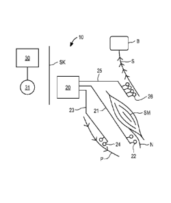
Sachs
application discusses that the parameters for electrical stimulation may be
programmed into
the stimulator following testing by the physician of stimulation thresholds.
Therapy
parameters such as duration, frequency and strength of contraction also may be
programmed
into the stimulator according to the patient's needs, and the stage of therapy
delivery. In
some cases it is expected that the programmed parameters may need to be
changed, for
example during the course of the therapy program as the muscle becomes
rehabilitated.
[0065] Stimulator 60 of FIG. 5 is designed to improve the NMES performance
and
reduce the need for frequent reprogramming by monitoring muscle performance
during
therapy, and adjusting the stimulation parameters accordingly. More
specifically,
implantable stimulator 60 includes controller 61, telemetry system 62 coupled
to antenna 63,
power supply 64, electrode switching array 65, system sensors 66, and NMES
circuitry
module 67 and muscle performance monitoring circuitry module 68. Electrode
switching
array 65 is selectably coupled to terminal array 69, which is coupled to the
connector block
29 (see FIG. 2) and enables stimulator 60 to be coupled to one or more
electrode leads.
Electrode switching array 65 also may include connection 69a to the housing of
stimulator
60, so that the housing functions as an electrode.
[0066] Each of components 61 to 67 and 69 operates in the manner described
above for
the embodiment of FIG. 3. Controller 61 preferably includes a programmable
microprocessor, volatile memory, nonvolatile memory, and nonvolatile storage,
and is
coupled to and controls operation of telemetry system 62, NMES circuitry
module 67, muscle
performance monitoring circuitry module 68, and electrode switching array 65.
Power
supply 64 powers the electrical components of implantable stimulator 60, and
may comprise
18
CA 02792529 2012-09-07
WO 2011/112773 PCT/US2011/027834
a primary cell or battery, a secondary cell or battery, a combination of both
or neither. In the
latter case, power supply 64 may comprise or include a capacitor that stores
energy
transmitted through the skin via a Transcutaneous Energy Transmission System
("TETS").
Stimulator 60 may be programmed and/or controlled by, and may upload stored
system and
operational data to external control system 30 via telemetry system 62. System
sensors 66
may comprise one or more sensors that monitor operation of stimulator 60, as
well as patient
parameters, such as movement, heart rate, etc., and may log data relating to
these parameters
for later readout using the external control system.
[0067] In accordance with one aspect of the present invention, stimulator
60 further
comprises muscle performance monitoring circuitry module 68 coupled to
controller, and
designed to monitor one or more parameters of muscle performance. The measured
parameters may be used to automatically modify the therapy delivered by NMES
circuitry
module 67, and/or to provide stored and telemetered information via telemetry
system 62 and
external control system 30 that enable the physician to modify the parameters.
In one
preferred embodiment, muscle performance monitoring circuitry module 68 may be
coupled
through electrode switching array 65 to selected electrodes coupled to
terminal array 69 to
measure electrical parameters of the tissue, such as impedance, or evoked
potential from the
stimulation. Circuitry module 68 may in addition be coupled to system sensor
66, for
example, to obtain data from an accelerometer or other movement transducer,
and/or
temperature or pressure. Circuitry module 68 also may be configured to receive
inputs from
other types of body sensors such as are known in the art, including those
monitoring chemical
properties (e.g., pH sensor, etc.).
[0068] Circuitry module 68 preferably includes at least one listening
amplifier configured
for electromyography (EMG). EMG is an electrical signal produced by muscle
when it
contracts, and the strength (power) of the EMG is an indicator of strength of
muscle
contraction. Configuration of an amplifier for measurement of EMG, e.g., gain,
frequency
response, impedance, etc., is well known to those skilled in the art. As
described in Stokes,
Ian A F, Sharon M Henry, and Richard M Single, "Surface EMG electrodes do not
accurately
record from lumbar multifidus muscles," Clinical Biomechanics (Bristol, Avon)
18, no. 1
(January 2003): 9-13, it is known that certain muscles, such as the deep
fibers of the lumbar
multifidus, surface EMG provides an unreliable signal. Accordingly, the
implantable
electrode leads used with stimulator 60 advantageously are expected to provide
a useful EMG
19
CA 02792529 2012-09-07
WO 2011/112773 PCT/US2011/027834
signal.
100691 In another embodiment, circuitry modules 67 and 68 may be configured
to
perform impedance measurements, in a manner similar to that described in U.S.
Patent
6,406,421 B1 to Grandjean et al. As is well known, an electrical impedance
measurement
may be performed by injecting a current through one pair of electrodes, and
measuring
voltage through a different pair of electrodes disposed approximately along
the same
geometric path. See, e.g., Rutkove, S.B., "Electrical impedance myography:
Background,
current state, and future directions", Muscle & Nerve 40, No. 6 (December
2009): 936-46. In
one implementation, a first pair of electrodes consisting of the stimulator
housing (via
connection 69a) and one or more of electrodes disposed on an electrode lead
may be used to
inject current into the tissue (e.g., from NMES circuitry module 67), while
voltage is
measured by circuitry module between the stimulator housing and a different
set of one or
more of electrodes on the electrode leads. Alternatively, the same set of
electrodes (including
the stimulator housing) may be used for both injecting current and measuring
the resulting
voltage.
100701 The foregoing impedance measurements may be of direct current (DC)
or
alternating current (AC). With AC impedance measurement, additional useful
information
may be obtained such as phase, frequency spectrum, and changes in parameters.
The
electrical impedance so measured is an indication of the tissue volume and
tissue
organization (anisotropy) between the measurement electrodes, as reported in
Garmirian et
at., "Discriminating neurogenic from myopathic disease via measurement of
muscle
anisotropy", Muscle Nerve, 2009 January; 39 (1): 16-24. See also, Miyatani,
M., et al.,
"Validity of estimating limb muscle volume by bioelectrical impedance", J.
Applied Physio.
(Bethesda, Md. 1985) 91, no. 1 (July 2001): 386-94. Accordingly, judicious
placement of the
electrodes and the stimulator housing will ensure that only the tissue of
interest (e.g., the
target muscle) is in the path of the injected and measured voltage. As a
muscle contracts, its
dimensions change, and this will generate a change in electrical impedance.
Thus,
measurement of electrical impedance may be used as a surrogate measure of
muscle
contraction.
100711 In another embodiment, circuitry module 68 may include or be coupled
to a
transducer that senses mechanical motion, such as vibration, acceleration or
deflection, and
may include piezoelectric polymers (e.g., PVDF) placed on a lead. The signal
from such a
CA 02792529 2012-09-07
WO 2011/112773
PCT/US2011/027834
transducer provides a surrogate measure of muscle contraction. In a further
alternative
embodiment, circuitry module 68 may include or be coupled to a transducer that
senses
pressure, such as a MEMS pressure sensors disposed on a lead, and which thus
provides a
surrogate measure of muscle contraction.
100721 In yet another embodiment, stimulator 60 is configured to sense EMG
from more
than one muscle, using multiple electrode leads or multiple electrodes on a
single lead that
passes through more than one muscle. In this case, the listening amplifier of
circuitry module
68 is multiplexed to listen for EMGs from more than one muscle. Alternatively,
circuitry
module 68 may include multiple listening amplifiers that are arranged to
simultaneously
listen to EMGs from more than one muscle. It is well-known, for example from
Jaap van
Dieen et al., "Trunk Muscle Recruitment Patterns," Spine Vol. 28, Number 8 pg
834-841,
that the relative timing and amplitude of EMGs in trunk muscles during the
performance of
specific tasks is different between healthy individuals and patients
experiencing low back
pain due to spinal instability. In patients with spinal instability,
recruitment patterns of the
trunk muscles may be altered to compensate for the lack of spinal stability.
The amplitude
and timing of EMGs measured from multiple trunk muscles therefore may be used
to
diagnose the presence and degree of spinal instability, as well as the change
of spinal
instability during a course of therapy. The EMG data may be used to
automatically modify
treatment parameters, or such data may be stored for later review by the
physician to assist in
diagnosis and revision of the therapy parameters.
100731 In the embodiment of FIG. 5, muscle performance monitoring circuitry
module 68
is configured to measure muscle contraction induced by NMES circuitry module
67, and to
modify the therapeutic parameters as muscle performance changes. In
particular, the initial
therapeutic parameters, such as dose and duration of therapy session, are
established and
programmed into stimulator 60 using external control system 30. Between
therapy sessions,
muscle performance may be monitored continuously or periodically using
circuitry module
68. When the change in measured muscle performance exceeds a predetermined
physician
selected threshold, circuitry module 68 may instruct controller 61 to modify
the parameters
for subsequent NMES therapy sessions. For example, if the monitoring
parameters reveal
that the muscle mass has increased, indicative of muscle rehabilitation, or
contractility has
decreased, then the therapy dose may be automatically reduced some pre-
determined amount
as previously programmed by the physician.
21
CA 02792529 2012-09-07
WO 2011/112773 PCT/US2011/027834
100741 In an alternative embodiment, muscle performance may be used to
inhibit muscle
contraction. For example, in certain types of low back pain, pain is caused by
spasm of
certain muscles in the back. Such spasm is accompanied by continuous increase
in EMG
activity. In accordance with one aspect of the present invention, NMES
stimulation may be
used to inhibit muscle contraction by configuring the listening amplifier of
circuitry module
68 to continuously or periodically measure EMG. If the EMG satisfies
conditions indicating
that muscle spasm has occurred, then NMES circuitry module is directed by
controller 61 to
apply stimulation to the nerve innervating the muscle in spasm to block
conduction of signals
from the nervous system which cause the muscle spasm, thereby preventing
spasm. The
stimulation provided by NMES circuitry module may be inhibited from time to
time to allow
circuitry module 68 to assess from the EMG signal if the muscle is still in
spasm; if spasm
has ceased, then application stimulation by NMES circuitry module 67 is
terminated.
100751 In an alternative embodiment, muscle performance monitoring
circuitry module
68 may be configured to measure a combination of EMG and tissue impedance to
confirm
that a muscle is in spasm, thereby improving the safety and reliability of the
measurement.
Muscle performance monitoring circuitry module 68 also may be used to track
changes in
activity and health of the muscle in response to neural activity. In other
words, the amount of
muscle contraction as determined by impedance measurement of tissue volume may
be
correlated to the amount of electrical activity in the muscle as determined by
EMG. Further
still, the electrodes and muscle performance monitoring circuitry module 68
may be
configured to record electrical signals from the nerves as well as the muscle,
such that a
measurement of the EMG (and/or tissue volume) in response to neural activity
may be used
as an indication of the health of the muscle.
100761 Muscle performance monitoring circuitry module 68 also may employ
measurement of the change in muscle mass in response to NMES of the nerve to
adjust the
electrical stimulation parameters. In this case, an empirically derived
transfer function may
be determined that relates electrical stimulation parameters, such as current,
pulse width,
frequency and duration, to the strength of contraction of the muscle. Over
time, this transfer
function may change, for example, as a result of electrode changes from
movement or tissue
ingrowth. Thus, the strength of muscle contraction may be used to
automatically adjust the
electrical parameters of the NMES stimulation provided by circuitry module 67
to achieve a
desired muscle contraction.
22
CA 02792529 2012-09-07
WO 2011/112773 PCT/US2011/027834
Stimulator with RF Ablation Capability
[0077] Referring to FIG. 6, in accordance with another aspect of the
present invention,
an implantable RF ablation device is described. Although a primary application
of the
inventive technology is pain reduction in connection with improving stability
of the spine, the
inventive technology may be advantageously applied in other areas, for
example:
= RF rhizotomy, in which a sensory nerve is ablated to prevent sensory
signals (e.g.,
pain) from reaching the brain, such as rhizotomy of the medial branch of the
dorsal ramus in patients with facet joint pain;
= RF ablation of unresectable tumors located in the liver, brain,
musculoskeletal
system, thyroid and parathyroid glands, pancreas, kidney, lung, and breast, in
which it is difficult to achieve complete tumor necrosis, leading to
recurrence of
the tumors and necessitating repeated RF ablation; and
= Treatment of tumors in which the root of the tumor is located in tissue
that is
considered too risky for surgical intervention, such as tumors with roots in
the
digestive tract, uterine wall or certain oesophageal tumors, and for which
regular
repeat surgery is required to remove new growths.
[0078] The field of RF ablation is well developed, and parameters suitable
for ablating
nerve fibers and other tissues, such as RF energy, and attendant issues is
well known to those
of ordinary skill in the art. See, e.g., Gazelle et al., "Tumor ablation with
radio-frequency
energy", Radiology, December 2000: 217(3): 633-46 and Haemerrich et al,
"Thermal tumour
ablation: devices, clinical applications and future directions", Int. J.
Hyperthermia, 2005 Dec;
21(8):755-60. To the inventors' knowledge, however, no one has suggested an RF
ablation
device that is configured to be chronically implanted and capable of
performing repeated RF
ablation.
[0079] Referring now to FIG. 6, implantable device 70 is described, which
is intended for
chronic implantation to perform serial RF ablations in scenarios where it is
necessary to
repeat RF ablation of tissue in a particular region of the body after certain
periods of time.
The components of device 70 correspond closely to those described above with
respect to the
embodiment of FIG. 3, and includes controller 71, telemetry system 72 coupled
to antenna
73, power supply 74, electrode switching array 75, system sensors 76, and
terminal array 77.
23
CA 02792529 2012-09-07
WO 2011/112773 PCT/US2011/027834
As in the preceding embodiments, electrode switching array 75 is selectably
coupled to
terminal array 77, which is coupled to the connector block 29 (see FIG. 2)
that accepts one or
more implantable electrode leads. Electrode switching array 75 also may
include connection
77a to the housing of device 70, so that the housing functions as an
electrode. In accordance
with this aspect of the present invention, device 70 further comprises RF
ablation circuitry
module 78, as further described below.
[0080] Each of components 71 to 77 operates in the manner described above
for the
embodiment of FIG. 3. Controller 71 preferably includes a programmable
microprocessor,
volatile memory, nonvolatile memory, and nonvolatile storage, and is coupled
to and controls
operation of telemetry system 72, electrode switching array 75 and RF ablation
circuitry
module 78. Power supply 74 powers the electrical components of device 70, and
may
comprise a primary cell or battery, a secondary cell or battery, a combination
of both, or
neither. In the latter case, power supply 74 may comprise or include a
capacitor (such as a
super capacitor of technology known to those skilled in the art) that stores
energy transmitted
through the skin via TETS. Device 70 may be programmed and/or controlled by,
and may
upload stored system and operational data to external control system 30 via
telemetry system
72. System sensors 76 may comprise one or more sensors that monitor operation
of device
70, as well as patient parameters, such as tissue impedance, and may log data
relating to these
parameters for later readout using the external control system.
[0081] In accordance with this aspect of the present invention, device 70
further
comprises RF ablation circuitry module 78 coupled to controller, and designed
to periodically
ablate tissue or nerve fibers using RF energy. Accordingly, controller 71 may
be configured
to control operation of the telemetry system 72 to receive energy wirelessly
from external
control system 30 and store that energy in power supply 74, and may be
configured to
communicate the amplitude of received power back to the external control
system via
telemetry system 72 or via modulation of the impedance of the antenna 73. To
ensure that
RF ablation is only carried out at the direction of the external control
system, device 70 may
not include battery or capacitor, but instead may be arranged so that it is
energized only when
in communication with the external control system.
[0082] Expected energy requirements for the RF ablation circuitry module
are in a range
of about 1-40 watts, depending upon the intended application. TETS systems
with this power
capacity are well known to those skilled in the art and have been used, for
example, with
24
CA 02792529 2012-09-07
WO 2011/112773 PCT/US2011/027834
artificial hearts or Left Ventricular Assist Devices (LVADs). However, the
physical volume
and other requirements of a high power TETS system may preclude its use in
applications
where the available surgical locations are limited. Thus, in an alternative
embodiment, the
TETS system may be of lower power capacity than the requirements of the RF
generator, and
device 70 may include an energy storage element, such as a super capacitor or
low impedance
secondary (rechargeable) cell, for powering RF ablation circuitry module 78.
In use, the
TETS may operate continuously, such that a signal is generated when there is
adequate
energy stored in the implantable device to deliver the RF ablation energy at
the desired power
and for the desired time. As an example, a TETS system capable of transferring
1W may be
used to supply RF energy delivery of 5W with 20% duty cycle.
100831 In this embodiment, telemetry system 72 enables communications
between the
external control system and device 70, allowing the implantable device to
receive device and
RF ablation operating parameters, as well as communicate logged information
such as
impedance between electrodes, temperature data and battery status to the
external control
system. Telemetry system 71 also may provide programming to controller 71 to
reconfigure
the operative electrodes through which ablation energy is supplied using
electrode switching
array 75, thereby allowing any electrode of a plurality of electrodes to be
configured as a
cathode, an anode or unconnected. The housing of device 70 also may be
configured as an
electrode via connection 77a of terminal array 77. The foregoing capabilities
provide
flexibility in the location of ablation lesions and allow the physician to
compensate for
electrode movement after implantation.
100841 System sensors 76 advantageously may be used to monitor the
temperature of the
tissue near the electrodes thru which energy for ablation is delivered.
Typical tissue
temperatures for RF ablation range from 50C to 130C, depending on the type of
tissue being
ablated and the time allocated to the ablation. System sensors 76 may
comprise, e.g.,
temperature sensors disposed within the device housing, or alternatively may
measure the
temperature of the connection to the electrode leads, and use that data to
infer or predict the
tissue temperature. Temperature sensors may also be incorporated into the
leads and placed
closer to the tissue targeted for ablation. System sensors 76 may be used in a
passive
(measuring) mode, or alternatively may comprise part of a feedback control
system that
continually or intermittently adjusts power delivered by the RF ablation
circuitry module so
that the temperature of the ablated tissue is maintained between desired
limits for safety and
CA 02792529 2012-09-07
WO 2011/112773
PCT/US2011/027834
efficacy.
[0085] Referring now to FIG. 7, an implantable stimulator illustratively
incorporating all
of the therapeutic circuitry modules described for the preceding embodiments
is described.
Implantable stimulator 80 corresponds to stimulator 20 of FIG. 1, and is
programmed and
controlled and/or powered by external control system 30. Stimulator 80 is
intended for use,
for example, in a stimulator that provides NMES stimulation, analgesic
stimulation to block
or reduce afferent pain signals in a nerve, and permits periodic nerve
ablation (such as
rhizotomy). Further in accordance with this aspect of the present invention,
stimulator 80
includes muscle performance monitoring circuitry that supports testing of
nerve fibers prior
to rhizotomy, which to guide proper selection of the ablation electrodes.
[0086] Stimulator 80 of FIG. 7 includes controller 81, telemetry system 82
coupled to
antenna 83, power supply 84, electrode switching array 85, system sensors 86,
terminal array
87, NMES circuitry module 88, analgesic stimulation circuitry module 89,
muscle
performance monitoring circuitry module 90, and RF ablation circuitry module
91. As in the
preceding embodiments, electrode switching array 85 is selectably coupled to
terminal array
87 under the control of controller 81, and enables any one or more of the
therapeutic circuitry
modules of stimulator 80 to be selectably coupled to selected electrodes of
one or more
electrode leads. Electrode switching array 85 also may include connection 87a
to the housing
of stimulator 80, so that the housing also may serve as an electrode.
[0087] Each of components 81 to 87 operates in the manner described above
for the
embodiment of FIG. 3. Controller 81 preferably includes a programmable
microprocessor,
volatile memory, nonvolatile memory, and nonvolatile storage, and is coupled
to and controls
operation of telemetry system 82, electrode switching array 85, NMES circuitry
module 88,
analgesic stimulation circuitry module 89, muscle performance monitoring
circuitry module
90, and RF ablation circuitry module 91. Power supply 84 powers the electrical
components
of implantable stimulator 80, and may comprise a primary cell or battery, a
secondary cell or
battery, a combination of both, or neither, as discussed above. Stimulator 80
may be
programmed and/or controlled by, and may upload stored system and operational
data to
external control system 30 via telemetry system 82. System sensors 86 may
comprise one or
more sensors that monitor operation of stimulator 80, as well as various
patient parameters as
discussed above.
26
CA 02792529 2012-09-07
WO 2011/112773 PCT/US2011/027834
100881 In accordance with this aspect of the present invention, stimulator
80 further
comprises NMES circuitry module 88 and analgesic stimulation circuitry module
89, as
described above with respect to the embodiment of FIG. 4, muscle performance
monitoring
circuitry module 90 as described above with respect to the embodiment of FIG.
5, and RF
ablation circuitry module 91 as described above with respect to the embodiment
of FIG. 6. In
this manner, a patient in need of spinal muscle rehabilitation and restoration
of neural drive
may have the full range of therapeutic modalities available. In particular,
stimulator 80 as
initially implanted by the physician, may be programmed to provide NMES
stimulation and
stimulation to block pain signals in afferent nerves. As muscle strength and
contractility
improve over the course of the therapy, the muscle performance monitoring
circuitry module
90 may measure the progress of the therapy and adjust the NMES stimulation
parameters or
circumvent spasm. In addition, depending upon the patient's reported condition
and
measurement data provided by the muscle performance monitoring circuitry
module 90, the
physician may periodically activate RF ablation circuitry module 91 to
denervate selected
nerve fibers.
Electrode Lead Systems
100891 In view of the capabilities of the various implantable stimulators
described herein,
it may be advantageous to provide an electrode lead specially configured for
use with such
stimulators. Referring to FIGS. 8A and 8B, electrode leads configured to
provide NMES
stimulation to a nerve to cause muscle contraction; to stimulate a nerve to
inhibit pain signals
from propagating to the brain; to stimulate a nerve to inhibit motor nerve
signals thereby
reducing or stopping contraction of a muscle (e.g., in spasm); to record
electrical signals such
as electromyography or tissue impedance; or for performing in situ RF ablation
are now
described.
100901 With respect to FIG. 8A, electrode lead 100 carrying electrodes 101a
to 101f is
described. The number of electrodes may be as few as 1 and as many as may be
realistically
placed within the target anatomical space. Electrode configurations commonly
used in the art
include 1 (for unipolar stimulation), 2, 4 (peripheral nerve stimulation), 8,
16 (spinal cord
stimulators) or up to 22 electrodes (cochlear implants). For the purpose of
this disclosure,
distal-most electrode 101a will be referred to as electrode #1, electrode 101b
will be electrode
#2 and so on moving proximally along lead 100 up to the total number of
electrodes.
27
CA 02792529 2012-09-07
WO 2011/112773
PCT/US2011/027834
[0091] When employed with an implantable stimulator as described herein
that provides
multiple independent current outputs, electrode lead 100 is capable of
delivering multiple
therapies simultaneously, in an overlaid fashion or staggered. Electrodes 101a
to 101f may
be sized and positioned relative to each other to allow for generation of a
voltage field
tailored to the specific type of stimulation, sensing or ablation desired for
the given therapies.
100921 In one embodiment, electrode lead 100 is placed parallel to a target
nerve in a
caudal to cranial orientation (with the cranial direction being the direction
tending towards
afferent neural activity). Then so positioned, electrodes 1 and 2, which are
most cranial, may
be sized and spaced to allow for optimal blocking of afferent pain signals
being transmitted
along the nerve (for example the pain signals being carried from the facet
joint along the
medial branch). More caudally, electrodes 3 and 4 may be sized and spaced to
allow for
optimal recruitment of large fiber motor neurons. Because the action
potentials required for
activation of a muscle travel efferently, these potentials are not blocked by
the more cranial
blocking action of electrodes 1 and 2. Finally, electrodes 5 and 6, placed
most caudally, may
be sized and positioned for sensing and recording of muscle recruitment
through capturing
the EMG signal of the muscle, which may be processed, for example, by the
muscle
performance monitoring circuitry module as described above with respect to the
embodiment
of FIG. 4. Such an arrangement therefore allows for simultaneous blocking of
pain arising
from the facet joint, stimulation of the motor fibers of the nerve eliciting
muscle contraction,
and sensing of the elicited response (which would enable a closed loop system,
improving
device longevity and recruitment efficiency) without any of the stimulation
pulses negatively
impacting the performance of the others.
[0093] With respect to FIG. 8B, alternative electrode lead 110 carrying
electrodes 111a to
111g is described. Distal-most electrode 111a again will be referred to as
electrode #1,
electrode 11 lb will be electrode #2 and so on moving proximally along lead
111. In the
embodiment of FIG. 8B, a blocking action of electrodes 1 and 2 may be used to
mute the
sensory perception of stimulation. In this manner, NMES stimulation therapy of
the motor
fibers in patients may be achieved where the patients would otherwise not
tolerate the
stimulation because of the resulting bi-directional action potential generated
by neural
stimulation. It also may be possible to use electrodes 5 and 6, which will
likely be placed
intramuscularly, to record the volume EMG signal in the muscle. Changes in
this signal over
time may provide an indication of the degree to which motor control has been
compromised
28
CA 02792529 2012-09-07
WO 2011/112773 PCT/US2011/027834
due to injury. When such data are compared over time during the period after a
therapy
regime has been completed, the data may be used as a positive indicator that
additional
therapy may be required to maintain spinal stability.
100941 While various illustrative embodiments of the invention are
described above, it
will be apparent to one skilled in the art that various changes and
modifications may be made
therein without departing from the invention. The appended claims are intended
to cover all
such changes and modifications that fall within the true spirit and scope of
the invention.
29