Note: Descriptions are shown in the official language in which they were submitted.
CA 02792717 2012-09-10
WO 2011/128368 PCT/EP2011/055800
Immunoglobulin aggregate removal
Herein is reported a method for the separation of dimeric and oligomeric
immunoglobulin from monomeric immunoglobulin by selective adsorption to
underivatized controlled pore glass.
Background of the Invention
Proteins and especially immunoglobulins play an important role in today's
medical
portfolio. Polypeptides for use in pharmaceutical applications are mainly
produced
in mammalian cells such as CHO cells, NSO cells, Sp2/0 cells, COS cells, HEK
cells, BHK cells, PER.C6 cells, and the like.
Due to their chemical and physical properties, such as molecular weight and
domain architecture including secondary modifications, the downstream
processing
of immunoglobulins is very complicated. For example, are not only for
formulated
drugs but also for intermediates in downstream processing (DSP) concentrated
solutions required to achieve low volumes for economic handling and
application
storage. The down stream processing of biotechnologically produced
immunoglobulins in general comprises three chromatography steps: a frist
affinity
chromatography step using e.g. Protein A, to remove non-immunoglobulin
molecules, normally followed by two ion exchange chromatography steps, whereof
the last step is a so called polishing step to remove DNA and HCP
contaminants.
The purified immunoglobulin is obtained in a low concentration solution
requiring
a concentration step prior to formulating the antibody into the pharmaceutical
formulation. Due to the non-natural conditions required during the down stream
processing the normally monomeric immunoglobulin tends to form dimers,
oligomer and higher order aggregates. These aggregates do not possess the
intended antigen-binding activity of the monomeric immunoglobulin and have to
be removed.
Ghose, S., et al. (Biotechnol. Bioeng. 87 (2004) 413-423) report preparative
protein
purification on underivatized silica. Reifsnyder, D.H., et al. (J. Chrom. A
753
(1996) 73-80) report the capture of IGF-I from a crude fermentation broth and
a
specific elution using a combination of ethanol and NaCl. Lifsics, M.R. and
Williams, R.C.Jr (Biochem. 23 (1984) 2866-2875) report a molecular sieve
chromatography in 8 M urea on controlled-pore glass for separating monomeric
and aggregated forms of a protein from bovine neurofilaments. Ghose, S., et
al.
CA 02792717 2012-09-10
WO 2011/128368 PCT/EP2011/055800
-2-
(Abstracts of Papers, 224th ACS National Meeting, Boston, MA, United States,
August 18-22, 2002 (2002), BIOT-317 Publisher: American Chemical Society,
Washington, D. C.) report the use of underivatized naked silica gel as a
preparative
stationary phase for process purification of proteins.
In US 4,606,825 a method of separating and recovering immunoglobulin G using
controlled pore glass bearing non-cross-linked covalently bound polyethylene
imine functions is reported. A method for separating a polypeptide monomer
from
a mixture comprising dimers and/or multimers using an ion exchange
chromatography resin and a gradient elution is reported in US 2002/0010319.
Mizutani, T. and Mizutani, A., J. Chrom. 168 (1979) 143-150 report the
comparison of elution patterns of proteins chromatographed on controlled-pore
glass and carboxymethyl cellulose. The isolation and purification of the
enzyme
myeloperoxidase using a chromatography with carboxymethylated controlled pore
glass is reported in DE 39 07 162 Al.
Summary of the Invention
It has been found that underivatized controlled pore glass (uCPG) surfaces
selectively bind dimeric and oligomeric, i.e. aggregated, immunoglobulin of
class
G (IgG) present in a solution at a pH value of from pH 5 to pH 7.5. By
applying of
from 50 m2 to 150 m2 uCPG surface per gram of total IgG, up to 95 % of the
soluble aggregates are bound to the particles (as determined by SE-HPLC).
Concomitantly, only about 10 % to 20 % of the monomer is adsorbed to the
surface. This can be achieved simply by batch-wise adding the uCPG to the
solution comprising monomer and aggregates and thereafter removing the uCPG
with the bound aggregates by centrifugation or filtration. Incubation of the
protein
over 6 days with uCPG did not result in the formation of aggregates. Moreover,
no
detectable changes to protein secondary or tertiary structure could be
observed after
the incubation with uCPG.
Thus, an aspect as reported herein is a method for obtaining a polypeptide in
monomeric form or aggregated form wherein the method comprises the following
steps
a) providing a solution comprising the polypeptide in monomeric form
and in aggregated form,
b) incubating the solution with underivatized controlled pore glass at a pH
value of from pH 4 to pH 6, and
CA 02792717 2012-09-10
WO 2011/128368 PCT/EP2011/055800
-3-
c) recovering the polypeptide from the incubated solution and thereby
obtaining the polypeptide in monomeric form,
or the following steps
a) providing a solution comprising the polypeptide in monomeric form
and in aggregated form,
b) incubating the solution with underivatized controlled pore glass at a pH
value of from pH 4 to pH 6,
c) removing the controlled pore glass from the solution,
d) incubating the removed controlled pore glass with a solution of a pH
value of from pH 2 to pH 3 or of from pH 7 to pH 8 and thereby
obtaining the polypeptide in aggregated form.
Another aspect as reported herein is a method for producing a polypeptide
comprising
a) providing a eukaryotic cell comprising a nucleic acid encoding the
polypeptide,
b) cultivating the cell to express the polypeptide,
c) recovering the polypeptide from the cells or the cultivation medium,
d) incubating a solution comprising the recovered polypeptide with
underivatized controlled pore glass at a pH value of from pH 4 to pH 6,
and
e) recovering the polypeptide from the incubated solution and thereby
producing the polypeptide.
In one embodiment the underivatized controlled pore glass is underivatized
controlled pore glass beads. In a further embodiment underivatized controlled
pore
glass with a surface of 100 m2 to 150 m2 per gram of polypeptide is used. In
another embodiment the polypeptide is an immunoglobulin, or an immunoglobulin
fragment, or an immunoglobulin conjugate. In also an embodiment the pH value
is
adjusted by a buffer solution of the respective pH value. In a further
embodiment
the method is a batch method. In also an embodiment the solution comprises a
discrete amount of polypeptide and the solution is incubated with a discrete
amount
of underivatized controlled pore glass. In also an embodiment the solution is
a
buffered solution. In one embodiment the recovering is by centrifugation or
filtration. In a further embodiment the incubating is for 1 minute to 6 hours.
CA 02792717 2012-09-10
WO 2011/128368 PCT/EP2011/055800
-4-
In one embodiment all the methods may comprise as last step the step of
- purifying the polypeptide by one or more chromatographic separation
steps.
Also an aspect as reported herein is the use of underivatized controlled pore
glass
for the adsorption of high molecular weight polypeptides at a pH value of from
pH
4 to pH 6.
A further aspect as reported herein is a kit of parts comprising
a) underivatized controlled pore glass beads,
b) a buffered solution of a pH value of from pH 4 to pH 6,
c) a buffered solution of a pH value of from pH 2 to pH 3,
d) a buffered solution of a pH value of from pH 7 to pH 8.
Detailed Description of the Invention
Generally, for the separation of monomeric immunoglobulin from aggregated
immunoglobulin as well as other high molecular weight compounds commonly
chromatographic methods are employed. It has now been found that underivatized
controlled pore glass (uCPG) selectively binds dimeric and oligomeric
immunoglobulins and high molecular weight compounds present in solution e.g.
compared to underivatized SepharoseTM. The monomeric immunoglobulin can be
recovered e.g. from the flow through of a chromatography column containing
uCPG as chromatography material or from the supernatant of an incubation of a
solution with uCPG. This effect is pronounced at a pH value of about 5.0 in
buffered solutions. With approximately 50 m2 to 150 m2 uCPG surface per g of
immunoglobulin up to 95 % of the aggregated form can be removed with a yield
of
80 % to 90 % of monomeric immunoglobulin.
The application of uCPG can be used to remove dimers and oligomers from active
pharmaceutical ingredient bulks or final formulated material prior to or even
after
storage.
A "polypeptide" is a polymer of amino acid residues joined by peptide bonds,
whether produced naturally or synthetically. Polypeptides of less than about
20
amino acid residues are referred to as "peptides". A "protein" is a
macromolecule
comprising one or more polypeptide chains or at least one polypeptide chain of
more than 100 amino acid residues. A polypeptide may also comprise non-
peptidic
components, such as carbohydrate groups. Carbohydrate groups and other non-
CA 02792717 2012-09-10
WO 2011/128368 PCT/EP2011/055800
-5-
peptidic substituents may be added to a polypeptide by the cell in which the
polypeptide is produced, and will vary with the type of cell. Polypeptides are
defined herein in terms of their amino acid backbone structures; substituents
such
as carbohydrate groups are generally not specified, but may be present
nonetheless.
The term "immunoglobulin" refers to a protein comprising one or more
polypeptide(s) substantially encoded by immunoglobulin genes. The recognized
immunoglobulin genes include the different constant region genes as well as
the
immunoglobulin variable region genes. Immunoglobulins may exist in a variety
of
formats, including, for example, Fv, Fab, and F(ab)2 as well as single chains
(scFv)
or diabodies.
The term "complete immunoglobulin" denotes an immunoglobulin which
comprises two light immunoglobulin chain polypeptides (light chains) and two
heavy immunoglobulin chain polypeptides (heavy chains). Each of the heavy and
light immunoglobulin chain polypeptides contains a variable domain (variable
region, generally the amino terminal portion) comprising binding regions that
are
able to interact with an antigen. Each of the heavy and light immunoglobulin
chain
polypeptides comprises a constant region (generally the carboxyl terminal
portion).
The variable domain of an immunoglobulin light or heavy chain in turn
comprises
different segments, i.e. four framework regions (FR) and three hypervariable
regions (CDR).
The term "immunoglobulin fragment" denotes a polypeptide comprising at least
one domain selected from the variable domain (VH), the CH1 domain, the hinge-
region, the CH2 domain, the CH3 domain, or the CH4 domain of a heavy chain, or
the variable domain (VL) or the CL domain of a light chain. Also enclosed are
derivatives and variants thereof. For example, a variable domain, in which one
or
more amino acids or amino acid regions are deleted, may be present.
The term "immunoglobulin conjugate" denotes a polypeptide comprising at least
one domain of an immunoglobulin heavy or light chain conjugated via a peptide
bond to a further polypeptide. The further polypeptide can be a non-
immunoglobulin peptide, such as a hormone, or toxin, or growth receptor, or
antifusogenic peptide, or complement factor, or the like.
For the purification of recombinantly produced immunoglobulins a combination
of
different column chromatography steps can be employed. Generally a protein A
affinity chromatography is followed by one or two additional separation steps.
The
CA 02792717 2012-09-10
WO 2011/128368 PCT/EP2011/055800
-6-
final purification step is a so called "polishing step" for the removal of
trace
impurities and contaminants like aggregated immunoglobulins, residual HCP
(host
cell protein), DNA (host cell nucleic acid), viruses, or endotoxins. For this
polishing step an anion exchange material in flow-through mode can be used.
Different methods can be used for protein recovery and purification, such as
affinity chromatography with microbial proteins (e.g. protein A or protein G
affinity chromatography), ion exchange chromatography (e.g. cation exchange
(carboxymethyl resins), anion exchange (amino ethyl resins) and mixed-mode
exchange), thiophilic adsorption (e.g. with beta-mercaptoethanol and other SH
ligands), hydrophobic interaction or aromatic adsorption chromatography (e.g.
with
phenyl-sepharose, aza-arenophilic resins, or m-aminophenylboronic acid), metal
chelate affinity chromatography (e.g. with Ni(II)- and Cu(II)-affinity
material), size
exclusion chromatography, and electrophoretical methods (such as gel
electrophoresis, capillary electrophoresis).
The terms "immunoglobulin in monomeric form" and "monomeric
immunoglobulin", which can be used interchangeably, as well as grammatical
equivalents thereof denote an immunoglobulin molecule that is not associated
with
a second immunoglobulin molecule, i.e. which is neither covalently nor non-
covalently bound to another immunoglobulin molecule. The terms
"immunoglobulin in aggregated form" and "aggregated immunoglobulin", which
can be used interchangeably, and "dimeric immunoglobulin" and "multimeric
immunoglobulin" as well as grammatical equivalents of all denote an
immunoglobulin molecule which is associated, either covalently or non-
covalently,
with at least one additional immunoglobulin molecule, and which is eluted in a
single peak from a size exclusion chromatography column. The term "in
monomeric form" and grammatical equivalents thereof as used within this
application not necessarily denotes that 100 % of an immunoglobulin molecule
are
present in monomeric form. It denotes that an immunoglobulin is essentially in
monomeric form, i.e. at least 90 % of the immunoglobulin is in monomeric from,
in one embodiment at least 95 % of the immunoglobulin is in monomeric form, in
another embodiment at least 98 % of the immunoglobulin is in monomeric form,
in
a further embodiment at least 99 % of the immunoglobulin is in monomeric form,
and in a final embodiment more than 99 % of the immunoglobulin is in monomeric
form determined as peak area of a size exclusion chromatogram. The term "in
monomeric and in aggregated form" denotes a mixture of immunoglobulin
molecules not associated with other immunoglobulin molecules and of
CA 02792717 2012-09-10
WO 2011/128368 PCT/EP2011/055800
-7-
immunoglobulin molecules associated with other immunoglobulin molecules. In
this mixture neither of the monomeric form nor the aggregated form is present
exclusively. The term "high molecular weight (HMW) form" denotes polymeric,
i.e. aggregated, immunoglobulin, whereby said aggregate is still soluble in an
aqueous buffered solution.
The term "100 %" as used within this application denotes that the amount of
components other than a specified component is below the detection limit of
the
referred to analytical method under the specified conditions.
The terms "90 %", "95 %", "98 %", "99 %" as used within this application
denote
no exact values but values within the accuracy of the referred to analytical
method
under the specified conditions.
A chromatographic material comprises a core material and thereto attached
chromatographic functional groups. The core material can be an inorganic
material,
such as silica, zeolithe, hydroxyapatite, or glass, an organic material, such
as
cellulose, or agarose, or a synthetic polymeric material, such as poly
(methacrylate).
The solutions employed in the method as reported herein are in one embodiment
buffered solutions. The term "buffered solution" denotes a solution in which
changes of pH due to the addition or release of acidic or alkaline substances
is
leveled by the dissolved buffer substance. Any buffer substance with such
properties can be used. Generally pharmaceutically acceptable buffers
substances
are used. In one embodiment the buffered solution is selected from a phosphate
buffered solution consisting of phosphoric acid and/or salts thereof, or an
acetate
buffered solution consisting of acetic acid and/or salts thereof, or a citrate
buffered
solution consisting of citric acid and/or salts thereof, or a morpholine
buffered
solution, or a 2-(N-morpholino) ethanesulfonic buffered solution, or a
histidine
buffered solution, or a glycine buffered solution, or a tris (hydroxymethyl)
aminomethane (TRIS) buffered solution. In another embodiment the buffered
solution is selected from a phosphate buffered solution, or an acetate
buffered
solution, or a citrate buffered solution, or a histidine buffered solution.
Optionally
the buffered solution may comprise an additional salt, such as e.g. sodium
chloride,
sodium sulphate, potassium chloride, potassium sulfate, sodium citrate, or
potassium citrate.
CA 02792717 2012-09-10
WO 2011/128368 PCT/EP2011/055800
-8-
Underivatized chromatographic core materials, especially underivatized
controlled
pore glass, can be used to selectively adsorb polypeptides, especially
immunoglobulins in aggregated form, i.e. dimeric and oligomeric immunoglobulin
molecules.
Generally, CPG beads have a mean particle diameter of about 125 m. The
specific
surface area of the herein used CPG beads was determined to be 36 m2/g.
In Figure IA the adsorption properties of underivatized controlled pore glass,
crosslinked agarose (SepharoseTM) and poly (methacrylate) are shown. It can be
seen that polypeptides can bind to underivatized controlled pore glass on the
one
hand and on the other hand that a strong pH dependency of the binding capacity
can be seen. As can be seen in Figure lB the ProSep vA ultra medium shows the
highest adsorption of the IgG at pH 7.5. ProSep vA ultra is a functionalized
CPG
material in which a Protein A affinity ligand is coupled to the glass surface.
At a
pH of 5.0 adsorption of IgG to CPG was observed to be higher than to the
functionalized Protein A gel.
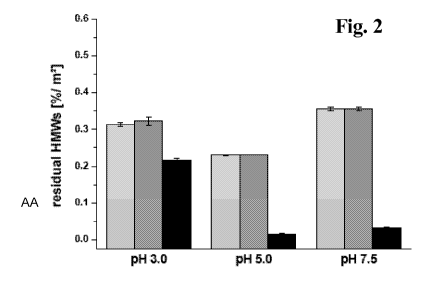
After incubation of the IgG with uCPG at different pH values of pH 3.0, 5.0
and
7.5 the supernatant was analyzed by SE-HPLC. It was observed, that after
incubation with underivatized CPG beads at pH 5.0 and 7.5, the solution was
almost fully cleared from soluble aggregates (Figure 2).
Figure 6 shows the amount of IgG adsorbed to uCPG after 12 hour incubation at
a
pH value of pH 5.0 versus the concentration of protein in the supernatant.
Saturation can be reached at relatively low protein concentrations and maximum
adsorption can be reached at 2.0 mg IgG per square meter CPG. Quantitative
desorption can be effected when the protein loaded CPG is incubated in a
buffered
solution at a pH value of pH 3Ø Adsorption of protein to silica surfaces is
in
general reported to be reversible under defined conditions. Harsh chemical
solvents
like chloroform, methanol, or 2-propanol can be used (see e.g. Manning, J.N.,
et
al., Journal of Chromatography B 487 (1989) 41-50; Stankovic, C.J., et al.,
Anal.
Biochem. 184 (1990) 100-103). Moreover chaotropic salts can be used (see e.g.
Mecs, I., et al., Arch. Virol. 81 (1984) 303-311) besides a changing of the pH
value
(see e.g. Edy, V.G., et al., J. Gen. Virol. 33 (1976) 517-521).
Maximum adsorption can be observed at pH 5.0 (Figure 7). A decrease in IgG
adsorption can be observed when the pH is raised above the isoelectric point
(IP) of
the IgG (e.g. the IP was determined to be 8.0 by using zeta potential
measurements
CA 02792717 2012-09-10
WO 2011/128368 PCT/EP2011/055800
-9-
for the IgG shown in Figure 8). The zeta-potential of the IgG can be
determined
depending on a pH value titration.
The surface charge of the nano-sized CPG depends on the pH value as well. The
IP
of the CPG can be determined to be about 4. At a pH value above pH 4 the
surface
charge of the CPG changes from positive to negative.
For example, a solution containing 6.8 % of HMWs was incubated with different
uCPG surface areas of 0.1 m2 to 10 m2 at a pH value of about 5. The residual
percentage of HMWs (high molecular weight compounds) in solution was
determined in the supernatant using SE-HPLC. In Figure 4 the amount of monomer
and HMWs in percent adsorbed to the CPG surface area per gram protein, which
was initially present before incubation, is shown. When 50 m2 CPG surface per
gram IgG is employed, 63 % of the HMWs initially present in the solution can
be
adsorbed. Nine percent of the monomer initially present in the solution is
bound to
the particles. By applying about 140 m2 CPG surface per gram protein, nearly
95 %
of HMWs can be bound, while only 22 % of monomer is adsorbed. At about 250
m2 CPG surface per gram protein almost 100 % of the HMWs can be adsorbed on
the CPG surface (see Figure 5). Between 50 m2 and 150 m2 CPG surface per gram
protein, the oligomers can be removed almost completely. The CPG surface area
present during incubation has no effect on the amount of the LMWs (low
molecular
weight compounds) remaining in solution.
Thus, soluble aggregates of immunoglobulins can be adsorbed to underivatized
CPG. The surface area of CPG available per gram protein can be used to control
the amount of aggregated and monomeric species remaining in solution after
incubation. A surface area between 100 m2 to 150 m2 per gram protein can be
used
to remove 60 % to 95 % of soluble aggregates from an IgG solution at a pH
value
of about 5, having concomitantly 80 % to 90 % monomeric IgG remaining in
solution.
The conformation of the protein was investigated before, during and after
adsorption to the surface. No protein loss due to the formation of insoluble
aggregates can be observed during six day incubation. The level of HMWs
determined by SE-HPLC decreased with increasing CPG surface area. After an
incubation of one day no further decrease in the level of soluble aggregates
can be
detected if the incubation is prolonged for 5 days. No increase of HMWs in the
CA 02792717 2012-09-10
WO 2011/128368 PCT/EP2011/055800
-10-
supernatant can be determined. In one embodiment the incubation is for 1 min.
to
24 hours. In a further embodiment the incubation is for 1 min. to 6 hours.
The exposure of IgG to uCPG did not result in conformational alterations of
the
secondary structure as determined with FT-IR spectroscopy.
An aspect as reported herein is a method for obtaining a polypeptide
comprising
the step
- incubating the polypeptide with underivatized controlled pore glass at a
pH value of from pH 4 to pH 6.
In one embodiment the underivatized controlled pore glass is used at a pH
value of
from pH 4.5 to pH 5.5. In another embodiment the uCPG is used at a pH value of
about pH 5. In another embodiment the incubating is in a buffered solution.
The term õabout" denotes that the thereafter following value is no exact value
but
is the center point of a range that is in one embodiment +/- 10 % of the
value, or in
another embodiment +/- 5 % of the value, or in a further embodiment +/- 2 % of
the value, or in an embodiment +/- 1 % of the value. If the value is a
relative value
given in percentages the term "about" also denotes that the thereafter
following
value is no exact value but is the center point of a range that is in one
embodiment
+/- 10 % of the value, or in another embodiment +/- 5 % of the value, or in a
further embodiment +/- 2 % of the value, or in an embodiment +/- 1 % of the
value,
whereby the upper limit of the range cannot exceed a value of 100 %.
In one embodiment the method for obtaining a polypeptide, especially an
immunoglobulin, in monomeric form comprises the following steps
a) incubating a solution comprising the polypeptide in monomeric form and
in aggregated form with underivatized controlled pore glass at a pH
value of from pH 4 to pH 6,
b) recovering the supernatant and thereby obtaining the polypeptide in
monomeric form from the supernatant of step a).
In another embodiment the method for obtaining a polypeptide, especially an
immunoglobulin, in aggregated form comprises the flowing steps
a) incubating a solution comprising the polypeptide in monomeric form and
in aggregated form with underivatized controlled pore glass at a pH
value of from pH 4 to pH 6,
b) recovering the incubated underivatized controlled pore glass,
CA 02792717 2012-09-10
WO 2011/128368 PCT/EP2011/055800
-11-
c) recovering the polypeptide in aggregated form from the underivatized
controlled pore glass by incubation with a solution with a pH value
differing by at least two pH units from the pH value of the solution of
step a).
In one embodiment the incubation is by adding a defined amount of
underivatized
controlled pore glass to the solution comprising the polypeptide. In also an
embodiment the incubating is by applying the solution to a chromatography
column comprising the underivatized controlled pore glass. In the first
embodiment
the polypeptide is recovered from the supernatant of the incubation. In the
second
embodiment the polypeptide is recovered from the flow-through of the column.
The recovering of the high molecular weight compounds is in both embodiments
by applying a solution with a pH value differing by at least two pH units from
the
pH value of the incubation solution to the controlled pore glass. In one
embodiment
the first method is a batch method.
In one embodiment the method comprises the following steps
- purifying a solution comprising the immunoglobulin with a Protein A
affinity chromatography,
- optionally purifying the immunoglobulin with an ion exchange
chromatography,
- incubating the obtained immunoglobulin comprising solution with
underivatized controlled pore glass at a pH value of from pH 4 to pH 6,
- recovering the supernatant of the previous step and thereby obtaining the
immunoglobulin in monomeric form.
Another aspect as reported herein is a method for producing a polypeptide,
especially an immunoglobulin, in monomeric form comprising
a) cultivating a cell comprising a nucleic acid encoding the polypeptide ,
b) recovering the polypeptide from the cell or the cultivation medium,
c) incubating the polypeptide with underivatized controlled pore glass at a
pH value of from pH 4 to pH 6,
d) recovering the supernatant of step c) and thereby producing the
polypeptide in monomeric form.
In one embodiment the incubating with the underivatized controlled pore glass
is
for 1 min. to 120 min.
CA 02792717 2012-09-10
WO 2011/128368 PCT/EP2011/055800
-12-
In Figure 3 the accumulation of high molecular weight (HMW) compounds on
underivatized controlled pore glass depending on the pH value and the buffer
compound is shown. It can be seen that underivatized controlled pore glass at
a pH
value of approximately pH 5 adsorbs high molecular weight compounds from an
immunoglobulin solution independent of the buffer substance. In one embodiment
the buffer substance is selected from acetic acid or a salt thereof, such as
sodium or
potassium acetate, and citric acid or a salt thereof, such as sodium or
potassium
citrate.
In Figure 4 the adsorption of high molecular weight (HMW) compounds on
underivatized controlled pore glass is compared to the adsorption of monomeric
immunoglobulin. It can be seen that the adsorbed amount of HMW compounds
shows an exponential dependency on the surface area of controlled pore glass
per
mass of applied polypeptide. The adsorbed amount of monomeric immunoglobulin
shows a linear dependency on the surface of controlled pore glass per mass of
applied polypeptide.
In Figure 5 a size exclusion chromatogram of differently treated solutions is
shown.
It can be seen that e.g. the use of 140 m2 CPG surface per gram of polypeptide
results in a reduction of high molecular weight compounds.
In Figure 6 the surface coverage of underivatized controlled pore glass is
shown. It
can be seen that the surface coverage reached 2 mg/m2 surface of underivatized
controlled pore glass. The adsorption is reversible by changing the pH value.
In one
embodiment the adsorbed high molecular weight compounds are recovered from
the underivatized controlled pore glass by changing the pH value from pH 5 to
a
value differing by at least two pH units, in one embodiment to pH 3.0 or to pH
7Ø
By 2d derivative UV- or IR-spectroscopy it can be shown that the adsorption
does
not affect the secondary or tertiary structure.
Due to the selective adsorption of high molecular weight compounds from
immunoglobulin preparations the use of underivatized controlled pore glass can
be
suitable for the purification of active pharmaceutical ingredients at the end
of the
down stream processing in order to remove remaining immunoglobulin in
aggregated form. The operating pH value of from pH 5 to pH 6 corresponds to
the
pH value of bulk active pharmaceutical ingredients. With underivatized
controlled
pore glass it is possible to remove oligomeric and even dimeric immunoglobulin
forms which is otherwise difficult at late stages in down stream processing.
CA 02792717 2012-09-10
WO 2011/128368 PCT/EP2011/055800
- 13 -
Additionally a handling in batch mode can be made by simply incubating
underivatized controlled pore glass beads with the bulk protein solution and
removing aggregates with the removal of the CPG e.g. by centrifugation or
filtration.
The following examples and figures are provided to aid the understanding of
the
present invention, the true scope of which is set forth in the appended
claims. It is
understood that modifications can be made in the procedures set forth without
departing from the spirit of the invention.
Description of the Figures
Figure 1 A: Comparison of the adsorption properties of underivatized
controlled pore glass (a), crosslinked agarose (Sepharose) (b) and
poly (methacrylate) (c) at pH values of 3.0, 5.0 and 7.2.
B: Adsorption of IgG to CPG matrices at pH 3, pH 5, pH 7.5
Percent protein adsorbed per square meter bead surface referring
to the protein mass initially present before incubation; adsorption
at pH 3.0 (light grey), at pH 5.0 (dark grey) and at pH 7.5 (black);
results are presented as mean values of three measurements SD.
Figure 2 Residual HMWs per square meter CPG surface; percent HMWs
determined with SE-HPLC in the supernatant referring to the
amount of HMWs initially present before incubation with the
surface; before incubation (light grey), incubation without
chromatographic surface (dark grey), and incubated with
chromatographic surface (black); results are presented as mean
values of three measurements SD.
Figure 3 Comparison of the specific accumulation of high molecular
weight (HMW) compounds on underivatized controlled pore
glass depending on the pH value and the buffer compound.
Figure 4 Adsorption of IgG monomer (plain) and HMWs (black) on CPG;
20 mg IgG containing 6.8 % HMWs were initially present before
incubation with 0.1-10 m2 CPG; experimental data is presented as
mean values of three measurements SD.
Figure 5 SE-chromatograms of the IgG solutions after incubation with up
to 350 m2 surface area of CPG per gram protein; 20 mg IgG
containing 6.8 % HMWs were initially present before incubation
with CPG (black profile); decreasing UV-signal with increasing
CA 02792717 2012-09-10
WO 2011/128368 PCT/EP2011/055800
-14-
CPG surface is indicated by changing the color from black to
light grey.
Figure 6 Adsorption and desorption isotherm of IgG on CPG particles;
adsorption (black diamonds) studies were done at pH 5.0,
desorption (plain diamonds) at pH 3.0; results are presented as
mean values of three measurements SD.
Figure 7 Adsorption of IgG on CPG particles at different pHs; experiments
were carried out in the saturation regime at a soluble protein
concentration of 2 mg/ml or higher; results are presented as mean
values of three measurements SD.
Figure 8 Zeta potential titration curves of the IgG (black) and the nano-
sized CPG particles (grey); experimental data is presented as
mean values of three measurements SD.
Example 1
Materials and Methods
Antibody
A completely purified bulk of a chimeric human Fc (IgGl)/ rat Fab antibody in
histidine buffer pH 6.0 (IgG A) was taken for the experiments. Aliquots were
dialyzed into 100 mM acetate buffer pH 3.0, 100 mM acetate buffer pH 5.0 and
200 mM tris (hydroxymethyl)-aminomethane buffer pH 7.5. The solutions were
filtrated afterwards by using a Sterivex-GV 0.22 m filter (Millipore,
Billerica,
USA).
CPG
Controlled pored glass (CPG 700) beads from Millipore (Billerica, USA) were
used.
Chemicals
All other chemicals and reagents used were at least analytical grade. Acidic
acid
was taken from Fluka (Steinheim, Germany), citric acid, hydrochloric acid,
sodium
hydroxide and sodium chloride were taken from Merck KG (Darmstadt, Germany),
tris (hydroxymethyl)-aminomethan was taken from Angus (Ibbenbueren,
Germany). L-histidine from Ajinomoto (Raleigh, USA) was used.
CA 02792717 2012-09-10
WO 2011/128368 PCT/EP2011/055800
- 15 -
Adsorption to surfaces
Adsorption of the monoclonal antibody (mAb) to CPG surface was investigated at
different pHs by incubating a defined surface with a defined protein mass.
Therefore, the bead slurry was suspended and mixed with purified water (Milli-
Q,
Millipore, Billerica, USA). The beads were collected by vacuum filtration
(0.22 m
cellulose filter discs, Sartorius, Goettingen, Germany) and afterwards rinsed
with
purified water (Milli-Q, Millipore, Billerica, USA). Subsequently, the beads
were
dried at 40 C and a defined mass was weighed representing a defined surface
as
determined by gas adsorption (BET). Afterwards, the beads were mixed with the
protein solution at a defined pH and incubated without head space on the
rotary
mixer RM5 (Froebel, Lindau, Germany) at 35 rpm for 12 hours at room
temperature. The samples were then centrifuged for 10 min. at 10,000 x g to
separate the beads from the protein solution. Protein concentration
determination
and SE-HPLC analysis were performed with the supernatant of the centrifuged
samples. Triplicate samples were prepared and the results are presented as
mean
values SD.
Adsorption isotherms of mAb on different bead surfaces were determined by
preparing samples containing 5 m2 bead surface and various concentrations of
mAb
between 0.2 mg/ml and 6.0 mg/ml. The samples were incubated over 12 hours as
described before and were centrifuged for 10 min. at 10,000 x g to separate
the
beads from the protein solution and to determine the protein concentration in
the
supernatant. The amount of protein adsorbed was determined by subtracting the
amount of the protein determined in the supernatant from the amount of protein
initially present before incubation. Triplicate samples were prepared and the
results
are presented as mean values SD.
To look at preferential adsorption of HMWs on CPG surfaces the IgG in 100 MM
acetate buffer pH 5 containing 6.8 % HMWs was incubated with 0.1-10 m2 CPG
surface. The samples were incubated for 12 hours as described before and were
centrifuged for 10 min. at 10,000 x g to separate the beads from the protein
solution
and to determine the protein concentration in the supernatant. In addition,
the
supernatant was analyzed by SE-HPLC. The amount of adsorbed HMWs and
monomer respectively was determined by subtracting the amount of HMWs
determined in the supernatant from the amount of HMWs initially present before
incubation. Triplicate samples were prepared and the results are presented as
mean
values SD.
CA 02792717 2012-09-10
WO 2011/128368 PCT/EP2011/055800
-16-
Protein concentration determination
Protein concentration was determined by using the photometric absorbance at
280
nm and 320 nm after buffer blank subtraction (UV-Vis spectrophotometer
Evolution 500, Thermo Fisher Scientific, Waltham, USA). The absorbance at 320
nm was subtracted from the absorbance at 280 nm and this absorbance value was
used to calculate the protein content according to the law of Lambert-Beer.
SE-HPLC
Size exclusion high pressure liquid chromatography (SE-HPLC) experiments were
conducted with a TSK 3000 SWXL column (Tosoh Bioseparation, Stuttgart,
Germany) on a Summit HPLC-system (Dionex, Idstein, Germany). The elution
peaks were monitored at 280 nm by the UV diode array detector UVD 170U from
Dionex (Idstein, Germany). Isocratic chromatography was conducted at room
temperature using an aqueous buffer composed of 200 mM potassium phosphate
and 250 mM potassium chloride at pH 7.0 and a flow rate of 0.5 ml/min. Each
sample contained 100 g mAb load per injection. The chromatograms were
integrated manually by using the Chromeleon software (Dionex, Idstein,
Germany). Percentage of higher molecular weight species (HMWs) including
dimers and larger soluble oligomers was determined as relative area (mAU*min)
referred to total area including the monomer peak and the peak of lower
molecular
weight species (LMWs).
FT-IR spectroscopy
FT-IR spectra were recorded with the Tensor 27 (Bruker Optics, Ettlingen,
Germany) applying the MlRacle attenuated total reflection (ATR) cell with a
Germanium crystal to investigate the secondary structure of the protein on the
bead
surface. The CPG beads were incubated at room temperature for 12 hours with a
6
mg/ml solution of IgG in acetate buffer at pH 5Ø The solutions showed a
level of
1.5 % HMWs determined by SE-HPLC before incubation. Incubation was
conducted in saturation mode. After incubation the beads were washed with
buffer.
The AquaSpec transmission cell was used to investigate the protein secondary
structure after desorption from the beads by using a 200 mM tris buffer pH
9Ø For
each spectrum which was recorded from 850-4000 cm -1 a 120-scan interferogram
was collected at a double sided acquisition mode with a resolution of 4 cm'.
The
reference spectrum of buffer and wetted beads respectively was subtracted to
obtain the protein spectrum. The spectra were edited by a vector normalization
CA 02792717 2012-09-10
WO 2011/128368 PCT/EP2011/055800
-17-
followed by the generation of the second derivative and smoothing using a 13-
point
Savitsky-Golay smoothing function applying the OPUS 6.0 software (Bruker
Optics, Ettlingen, Germany). Moreover, the absorption spectra recorded in ATR
mode were corrected concerning band intensity and band position to allow the
comparison to spectra recorded in transmission mode. Therefore, the extended
ATR-correction of the software OPUS 6.0 (Bruker Optics, Ettlingen, Germany)
according to Fringeli was used (Fringeli, U.P., Chimia 46 (1992) 200-214) to
overcome wave number dependent anomalous dispersion (Goldberg, M.E., and
Chaffotte, A.F., Protein Sci. 14 (2005) 2781-2792; Grdadolnik, J., Int. J.
Vibr.
Spec. 6, ed. 2 (2002).
Zeta potential measurements
To determine the charge of the protein and the sonicated nano-sized CPG 700 at
different pH values, electrophoretic mobility of the protein and the CPG 700
beads
was determined by performing Laser-Doppler-Velocimetry using the Malvern
Zetasizer Nano S (Malvern Instruments, Worcestershire, UK). The zeta potential
c
was calculated from the Henry's equation with assumption of uniform charge
distribution by using the Malvern DTS software (Version 5.0, Malvern
Instruments, Worcestershire, UK). For sample preparation a 5 mg/ml mAb
solution
was dialyzed into 50 mM acetate buffer pH 5.0 and titrated to a pH of 2.0
afterwards by using 0.2 M hydrochloric acid. The CPG 700 beads were suspended
in the same buffer system and the pH was adjusted to 2Ø The samples were
titrated with a 0.2 M sodium hydroxide solution from pH 2 to pH 12 by applying
the titrator MPT2 (Malvern Instruments, Worcestershire, UK). The zeta
potential
was determined in 15 steps between pH 2 and 12 in a temperature controlled
folded
capillary cell (Malvern Instruments, Worcestershire, UK) at 25 C. Each
measurement was repeated threefold and mean values SD are reported.