Note: Descriptions are shown in the official language in which they were submitted.
CA 02792927 2012-09-11
-1-
[Document Name] Specification
[Title of the Invention]
PROTEOGLYCAN-CONTAINING MICRONEEDLE ARRAY
[Technical Field]
[0001]
The present invention relates to a microneedle
array that can be used as an external preparation. More
specifically, the present invention relates to a
microneedle array that is cosmetically or pharmaceutically
effective.
[Prior Art]
[0002]
External preparations containing medicinal
properties are conventionally used so that the medicinal
properties may exhibit pharmacological activity under the
skin. Solutions, ointments, cream preparations, tape
preparations, patches, poultices, etc., are known as such
external preparations. These are topically applied or
attached, so that medicinal properties are percutaneously
absorbed to thereby exhibit desired pharmacological
activity under the skin.
[0003]
However, such external preparations in an
applied or attached form have drawbacks, i.e., they might
be removed or lost due to sweating, washing, external
pressure, and other factors while in use before the
medicinal properties are percutaneously absorbed. Another
problem of these external preparations is that the
medicinal properties are not percutaneously absorbed to a
sufficient degree because of the barrier function of the
skin, and hence they fail to exhibit the desired
pharmacological activity. Particularly, when polymer
compounds are used as medicinal properties, percutaneous
CA 02792927 2012-09-11
-2-
absorption is difficult, thus making it difficult for the
external preparations to exhibit the desired
pharmacological activity.
[0004]
Recently, as a technique for solving these
drawbacks of external preparations, microneedle arrays
having microneedles comprising medicinal properties are
being actively studied (Patent Documents 1 to 3). For
example, Patent Document 1 proposes a microneedle array
having microneedles formed of a raw material mainly
composed of collagen. According to the microneedle array
of Patent Document 1, the microneedles are inserted into
the skin surface layer and/or stratum corneum to supply
the medicinal properties contained in the microneedles
under the skin. The inserted portions of the microneedles
can dissolve or biodegrade under the skin and thus
disappear. Further, when the microneedles of Patent
Document 1, which have very fine needle parts, are
inserted into the skin surface layer and/or stratum
corneum, neither pain nor bleeding occurs, and puncture
wounds close quickly. The microneedles of Patent Document
1 are thus suitable for supplying medicinal properties
under the skin.
[0005]
At the same time, microneedle arrays are
required to have optimal designs depending on skin
diseases or skin conditions. There is thus a demand for
various kinds of microneedle arrays. However, the
microneedles of microneedle arrays used as external
preparations are required to comprehensively have the
following properties: (1) the strength to withstand
insertion into the skin surface layer and/or stratum
corneum, (2) the fineness and flexibility to cause no pain
or bleeding in the skin surface layer and/or stratum
corneum at the insertion site of the microneedles, and (3)
CA 02792927 2012-09-11
-3-
solubility or biodegradability in the body of the
microneedle portions under the skin. Accordingly, it is
very difficult to change the design of the constituent
materials of microneedle arrays, and this is particularly
true for the main constituent material of the microneedles.
For these reasons, only a few constituent materials for
microneedle arrays have been reported at present.
[0006]
In recent years, medical or cosmetic
applications of proteoglycan have attracted attention.
Proteoglycan is known as a glycoconjugate composed of a
core protein and glycosaminoglycan (acid
mucopolysaccharide) bonded thereto. Proteoglycan is the
principal constituent of the extracellular matrix, and is
present in skin tissue, cartilage tissue, bone tissue,
vascular tissue, etc. Proteoglycan is reportedly involved
with the growth and bonding of subcutaneous cells.
Moreover, proteoglycan is reportedly useful in preventing
or treating inflammatory diseases or autoimmune diseases,
preventing rejection after organ transplantation,
preventing or improving allergies, and preventing or
improving diabetes (see Patent Document 4).
[0007]
However, using proteoglycan in microneedle
arrays has not been studied. At present, there is no clue
as to whether proteoglycan can be used to form
microneedles.
[Prior Art Documents]
[Patent Documents]
[0008]
Patent Document 1: Japanese Unexamined Patent Publication
No. 2009-273872
Patent Document 2: Japanese Unexamined Patent Publication
No. 2009-254765
CA 02792927 2012-09-11
-4-
Patent Document 3: Japanese Unexamined Patent Publication
No. 2009-201956
Patent Document 4: Japanese Unexamined Patent Publication
No. 2007-131548
Patent Document 5: Japanese Unexamined Patent Publication
No. 2003-300858
[Non-Patent Document]
[0009]Non-Patent Document 1: Watanabe H, Yamada Y, Kimata K.,
Roles of aggrecan, a large chondroitin sulfate proteoglycan, in
cartilage structure and function.;J Biochem. 1998
Oct;l24(4):687-93.
Non-Patent Document 2: Ota S, Yoshihara S, Ishido K, Tanaka M,
Takagaki K, Sasaki M., Effects of proteoglycan on dextran
sulfate sodium-induced experimental colitis in rats.Ota S; Dig
Dis Sci. 2008 Dec;53(12):3176-83. Epub 2008 May 8.
Non-Patent Document 3: Mitsui T, Sashinami H, Sato F, Kijima
H, Ishiguro Y, Fukuda S, Yoshihara S, Hakamada K, Nakane A,
Salmon cartilage proteoglycan suppresses mouse experimental
colitis through induction of Foxp3+ regulatory T cells.;
Biochem Biophys Res Commun. 2010 Nov 12;402(2):209-15. Epub
2010 Oct 20.
Non-Patent Document 4: Sashinami H, Takagaki K, Nakane A.,
Salmon cartilage proteoglycan modulates cytokine responses to
Escherichia coli in mouse macrophages.; Biochem Biophys Res
Commun. 2006 Dec 29;351(4):1005-10. Epub 2006 Nov 3.
[Summary of the Invention]
[Problem to Be Solved by the Invention]
[0010]
An object of the present invention is to provide
a novel and unprecedented microneedle array. More
specifically, an object of the present invention is to
provide a novel microneedle array comprising one or more
microneedles that have the following properties:
(1) the strength to withstand insertion into the
CA 02792927 2012-09-11
-5-
skin surface layer and/or stratum corneum,
(2) the fineness and flexibility to cause no
pain or bleeding in the skin surface layer and/or stratum
corneum at the insertion site of the microneedles, and
(3) solubility or biodegradability in the body
of the microneedle portions under the skin.
[Means for Solving the Problem]
[0011]
The present inventors conducted extensive
studies to solve the above problems, and surprisingly
found that proteoglycan can be used as a base material to
form microneedles, and that a microneedle array having the
microneedles can be produced. The present inventors also
found that the microneedle array has the excellent
properties described above in items (1) to (3), and is
highly useful as an external preparation. Additionally,
the microneedle array is expected to effectively exhibit
proteoglycan-based useful pharmacological activity under
the skin.
The present invention was accomplished by
conducting further studies based on these findings.
[0012]
More specifically, the present invention
provides a microneedle array and a production method
thereof according to the following embodiments:
[0013]
(I) Microneedle Array
(I-1). A microneedle array comprising one or more
microneedles formed on the surface of a substrate, the
microneedles containing proteoglycan as a base material.
(I-2). The microneedle array according to (I-1), wherein
each of the microneedles has a konide-like shape or a
circular truncated cone shape.
(I-3). The microneedle array according to (I-1) or (1-2),
CA 02792927 2012-09-11
-6-
wherein each of the microneedles is a solid needle.
(I-4). The microneedle array according to any one of (I-1)
to (1-3), wherein each of the microneedles has a root
diameter of 120 to 400 pm, a tip diameter of 5 to 100 pm,
and a length of 100 to 5000 pm, and the pitch (the
distance from tip to tip) between adjacent microneedles is
100 to 1800 pm.
(I-5). The microneedle array according to (1-4), wherein
each of the microneedles has a length of 100 to 1600 pm or
100 to 1000 pm.
(I-6). The microneedle array according to (1-4), wherein
each of the microneedles has a length of more than 1000 pm
but not more than 5000 pm, or more than 1000 pm but not
more than 3000 pm.
(I-7). The microneedle array according to (1-4), wherein
each of the microneedles has a length of more than 1600 pm
but not more than 5000 pm, or more than 1600 pm but not
more than 3000 pm.
The microneedle arrays shown in the (1-6) and (1-7)
comprise the needles having a millimeter-order length as
above mentioned, but a micrometer-order fineness (the root
diameter and the tip diameter of needle). The microneedle
array of the present invention includes such arrays having
the above needles having a millimeter-order length and a
micrometer-order fineness.
(I-8). The microneedle array according to any one of (I-1)
to (1-7), wherein the proteoglycan content in the one or
more microneedles is 20 to 100 wt.%.
(I-9). The microneedle array according to any one of (I-1)
to (1-8), wherein the proteoglycan is chondroitin sulfate
proteoglycan.
(I-10). The microneedle array according to any one of (I-
1) to (1-9), wherein the proteoglycan is derived from fish.
(I-11). The microneedle array according to (I-10), wherein
the fish is salmon, shark or jellyfish.
CA 02792927 2012-09-11
-7-
(I-12). The microneedle array according to any one of (I-
1) to (I-11), wherein the one or more microneedles contain,
in addition to the proteoglycan, a water-soluble polymer
other than proteoglycan, or a cosmetically or
pharmaceutically acceptable medicinal property.
(I-13). The microneedle array according to any one of (I-
1) to (1-12), wherein the substrate has the same
composition as the microneedles.
[0014]
(II) Method of Producing Microneedle Array
(II-1). A method of producing the microneedle array
according to (I-1) or (I-11), the method comprising the
steps of:
pouring an aqueous solution in which
proteoglycan for forming microneedles is dissolved into a
mold in which the form of a microneedle array is recessed,
so that a microneedle part and a substrate part are
formed;
evaporating the moisture to dryness at room
temperature or by heating; and
removing the formed microneedle array from the
mold.
[0015]
(II-2). A method of producing the microneedle array
according to (I-1), the method comprising the steps of:
pouring an aqueous solution in which
proteoglycan for forming microneedles is dissolved into a
mold in which the form of a microneedle is recessed;
evaporating the moisture to dryness at room
temperature or by heating;
laminating a substrate thereon and combining or
bonding the bottom of the microneedles and the substrate;
and
removing the microneedles combined or bonded
with the substrate from the mold.
CA 02792927 2012-09-11
-8-
[0016]
(II-3). The method according to (II-1) or (11-2), wherein
the aqueous solution in which proteoglycan for forming
microneedles is dissolved contains the proteoglycan at a
concentration of about 1 to 30 wt.%, preferably about 1 to
25 wt. o.
[Effect of the Invention]
[0017]
The present invention provides a microneedle
array having one or more microneedles formed using
proteoglycan as a base material. The microneedle array of
the present invention comprises one or more microneedles
that have the above excellent properties, i.e., (1)
strength, (2) fineness and flexibility, and (3) solubility
in the body. The microneedle array, which thus has
sufficient properties for use as an external preparation,
can adequately supply proteoglycan under the skin and
underlying tissues.
[0018]
Moreover, the microneedle array of the present
invention is suitable for cosmetic or medical applications
using the action of proteoglycan. Particularly, the
microneedle array of the present invention is expected to
an anti-aging activity such as a wrinkle-smoothing effect
based on the epidermal cell growth-promoting activity of
proteoglycan.
[Brief Description of the Drawings]
[0019]
Fig. 1 shows a cross-sectional view of a mold
(1) having microneedle-like shape recesses (11) and filled
with a proteoglycan-containing aqueous solution (2).
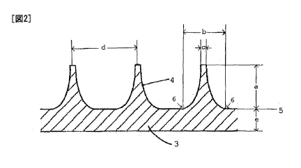
Fig. 2 shows a cross-sectional view of an
example of the microneedle array of the present invention
CA 02792927 2012-09-11
-9-
(Example 1). The microneedle array has a structure in
which a plurality of solid konide-like shape microneedles
(4) are formed on the surface of a substrate (3). In the
drawing, Sign "a" denotes the length (height) of the
microneedle formed on the substrate; Sign "b" denotes the
root diameter of the microneedle; Sign "c" denotes the tip
diameter of the microneedle; Sign "d" denotes the distance
(pitch) between tips of adjacent microneedles formed on
the substrate; and Sign "e" denotes the thickness of the
substrate. Here, the "root diameter" of the microneedle
indicates the diameter of the bottom of the microneedle
attached to the surface of the substrate. More
specifically, in the cross-sectional view shown in Fig. 2,
the "root diameter" corresponds to the distance between
the tangent points (6) of the microneedle relative to the
substrate surface (5), which is regarded as the base line.
Fig. 3 shows a perspective view and an image of
the microneedle array of the present invention used in
Example 2. (A) illustrates a perspective view (a-1) and
an image (a-2) of the microneedle array before use. (B)
illustrates a perspective view (b-1) and an image (b-2) of
the microneedle array after use. Sign "7" denotes a tape
used to fix the microneedle array to the skin of the knee.
Fig. 4 shows the results of Example 2. (A) is
an image of a site to which the microneedle array of the
present invention containing a dye has been applied and
then removed, and (B) is an image of the subcutaneous
tissue of the site to which the microneedle array has been
applied.
[Mode for Carrying Out the Invention]
[0020]
The microneedle array of the present invention
comprises one or more microneedles formed on the surface
of a substrate, the microneedles containing proteoglycan
CA 02792927 2012-09-11
-10-
as a base material. The following describes the
microneedle array of the present invention in detail.
[0021]
Proteoglycan is a general term for molecules in
which one or more glycosaminoglycans are covalently linked
to a core protein. The type of proteoglycan used in the
present invention is not particularly limited, and any of
those belonging to chondroitin sulfate proteoglycan,
dermatan sulfate proteoglycan, heparan sulfate
proteoglycan, and keratan sulfate proteoglycan can be used.
Specific examples of proteoglycan used in the present
invention include aggrecan, versican, neurocan, brevican,
decorin, biglycan, serglycin, perlecan, syndecan, glypican,
lumican, keratocan, etc. Among these, it is preferable,
in the present invention, to use chondroitin sulfate
proteoglycan, and more preferably aggrecan, as a base
material for forming microneedles.
[0022]
The source of proteoglycan used in the present
invention is not particularly limited, and any of those
derived from mammals such as humans, cows, and pigs; birds
such as chickens; fish such as sharks, salmon and
jellyfish; shellfish such as crabs and shrimps; and the
like can be used. Among these sources, it is preferable
to use proteoglycan derived from fish, more preferably
salmon, and particularly preferably salmon nasal cartilage.
[0023]
Compared with proteoglycan derived from cows,
pigs, and other higher animals, proteoglycan derived from
shark cartilage has higher transparency, and is effective
as a starting material for external skin preparations.
For the purpose of reducing dark spots, wrinkles, and
sagging, it is known to combine shark cartilage-derived
proteoglycan with a melanogenesis inhibitor and a crude
drug extract having an active oxygen eliminating effect
CA 02792927 2012-09-11
-11-
(Patent Document 5).
[0024]
The molecular weight of proteoglycan used in the
present invention is not particularly limited, and is
suitably determined. Proteoglycan having a molecular
weight of 80,000 to 3000,000 can generally be used without
limitation; preferably used is proteoglycan having a
molecular weight of 200,000 to 2500,000, and more
preferably 300,000 to 800,000.
[0025]
In the present invention, the proportion of
proteoglycan in the microneedles is not particularly
limited as long as the microneedles contain proteoglycan
as a base material. For example, the amount of
proteoglycan in the microneedles is generally 20 to 100
wt.%, preferably 50 to 100 wt.%, more preferably 70 to 100
wt.%, even more preferably 80 to 100 wt.%, and still more
preferably 90 to 100 wt.%.
[0026]
More specifically, the microneedles of the
present invention comprising proteoglycan as a base
material may be composed only of proteoglycan, or may
contain components other than proteoglycan in an amount up
to 80 wt.%, and preferably 50 wt.%, as long as the
following properties of the microneedles composed of
proteoglycan are not impaired:
(1) the strength to withstand insertion into the
skin surface layer and/or stratum corneum,
(2) the fineness and flexibility to cause no
pain or bleeding in the skin surface layer and/or stratum
corneum at the insertion site of the microneedles, and
(3) solubility or biodegradability in the body
of the microneedle portions under the skin.
In the present invention, "comprising
proteoglycan as a base material" indicates the above
CA 02792927 2012-09-11
-12-
meaning.
[0027]
As components other than proteoglycan
containable in the microneedles, water-soluble polymers
other than proteoglycan can be used. Such water-soluble
polymers may be those that can dissolve or degrade in vivo,
and specific examples thereof include polysaccharides such
as hyaluronic acid, chondroitin sulfate, glycogen, dextrin,
dextran, dextran sulfate, hydroxypropyl methylcellulose,
alginic acid, chitin, chitosan, and pullulan; proteins
such as collagen, gelatin, and hydrolysates thereof;
synthetic high polymers such as polyvinyl alcohol,
polyvinyl pyrrolidone, polyacrylic acid, and carboxyvinyl
polymer; and the like.
[0028]
When the microneedles contain a water-soluble
polymer other than proteoglycan, the amount of the water-
soluble polymer in the microneedles is generally 1 to 30
wt.%, preferably 1 to 25 wt.%, more preferably 1 to 20
wt.%, and further more preferably 1 to 10 wt.%.
[0029]
Moreover, in the present invention, the
microneedles may contain cosmetically or pharmaceutically
acceptable medicinal properties as the above other
components.
[0030]
Among such medicinal properties, examples of
cosmetically acceptable medicinal properties include
whitening agents such as ascorbic acid, sodium ascorbyl
phosphate, magnesium ascorbyl phosphate, ascorbyl
palmitate, kojic acid, rucinol, tranexamic acid, licorice
extract, and vitamin-A derivatives; anti-wrinkle agents
such as retinol, retinoic acid, retinol acetate, and
retinol palmitate; blood circulation accelerators such as
tocopheryl acetate, capsaicin, and nonylic acid
CA 02792927 2012-09-11
-13-
vanillylamide; dietary agents such as raspberry ketone,
evening primrose extract, and seaweed extract;
antimicrobial agents such as isopropyl methyl phenol,
photosensitive pigments, and zinc oxide; antiphlogistics
such as salicylic acid; vitamins such as vitamin D2,
vitamin Da, and vitamin K; and the like.
[0031]
Further, among the above medicinal properties,
pharmaceutically acceptable medicinal properties may be,
other than the cosmetically acceptable medicinal
properties described above, medicines used in the
pharmaceutical field. Specific examples of medicines
other than the aforementioned cosmetically available
medicinal properties include antipyretic analgesic
antiphlogistics, such as ibuprofen, flurbiprofen, and
ketoprofen; steroidal anti-inflammatory agents, such as
hydrocortisone, triamcinolone, and prednisolone;
vasodilators, such as diltiazem hydrochloride and
isosorbide nitrate; antiarrhythmic agents, such as
procainamide hydrochloride and mexiletine hydrochloride;
antihypertensives, such as clonidine hydrochloride,
bunitrolol hydrochloride, and captopril; local anesthetics,
such as tetracaine hydrochloride and propitocaine
hydrochloride; hormone drugs, such as propylthiouracil,
estradiol, estriol, and progesterone; antihistamines, such
as diphenhydramine hydrochloride and chlorpheniramine
maleate; anesthetics, such as pentobarbital sodium;
soporific analgesics, such as amobarbital and
phenobarbital; antiepileptic agents, such as phenytoin
sodium; antipsychotic drugs, such as chlorpromazine
hydrochloride, imipramine hydrochloride, chlordiazepoxide,
and diazepam; skeletal muscle relaxants, such as
suxamethonium hydrochloride and eperisone hydrochloride;
autonomic drugs, such as neostigmine bromide and
bethanechol chloride; antiparkinson agents, such as
CA 02792927 2012-09-11
-14-
amantadine hydrochloride; diuretics, such as
hydroflumethiazide, isosorbide, and furosemide;
vasoconstrictors, such as phenylephrine hydrochloride;
respiratory stimulants, such as lobeline hydrochloride,
dimorpholamine, and naloxone hydrochloride; narcotics,
such as morphine hydrochloride, cocaine hydrochloride, and
pethidine hydrochloride; and the like. Furthermore, in
the present invention, medicines to be added to the
microneedles may be, other than those exemplified above,
biologically active peptides and derivatives thereof, and
fragments of nucleic acids, oligonucleotides, antigen
proteins, bacteria, viruses, etc. Examples of such
biologically active peptides and derivatives thereof
include calcitonin, adrenocorticotropic hormone,
parathyroid hormone (PTH), hPTH (1-->34), insulin, secretin,
oxytocin, angiotensin, R-endorphin, glucagon, vasopressin,
somatostatin, gastrin, luteinizing hormone-releasing
hormone, enkephalin, neurotensin, atrial natriuretic
peptide, growth hormone, growth hormone-releasing hormone,
bradykinin, substance P, dynorphin, thyrotropic hormone,
prolactin, interferon, interleukin, G-CSF, glutathione
peroxidase, superoxide dismutase, desmopressin,
somatomedin, endothelin, salts thereof, etc. Examples of
the above antigen protein include HBs surface antigen, HBe
antigen, etc.
[0032]
The microneedle array of the present invention
has a structure in which one or more microneedles are
formed on the surface of a substrate. In the microneedle
array of the present invention, the larger the number of
microneedles, the higher the desired pharmacological
activity. Accordingly, the substrate is desirably
provided with a plurality of microneedles.
[0033]
The form of the microneedle may be suitably
CA 02792927 2012-09-11
-15-
determined so that it can be inserted into the skin and
dissolve in the body (in the skin and underlying tissue),
and so that pain and bleeding do not occur. For example,
the microneedle preferably has a konide-like shape, a
circular truncated cone shape, or the like. The konide-
like shape as used herein is a so-called volcano shape,
that is, a circular truncated cone whose side surface is
internally curved, as shown in Fig. 2. Moreover, the
microneedle is preferably a solid needle, rather than a
hollow needle.
[0034]
A microneedle in a konide-like shape or a circular
truncated cone shape preferably has a root diameter of
about 120 to 400 pm, and more preferably about 150 to 300
pm, because a thin microneedle supplies a smaller amount
of proteoglycan into the skin, and is easily broken when
inserted into the skin, whereas a thick microneedle is
hard to insert into the skin. The "root diameter" of the
microneedle indicates the diameter of the bottom of the
microneedle attached to the surface of the substrate.
[0035]
A microneedle in a konide-like shape or a
circular truncated cone shape preferably has a tip
diameter of about 5 to 100 pm, and more preferably about
10 to 80 pm, because a thin (sharp) microneedle is easily
broken when inserted into the skin, whereas a thick
microneedle is hard to insert into the skin, thereby
causing pain.
[0036]
A microneedle in a konide-like shape or a
circular truncated cone shape preferably has a length of
about 100 pm or more, preferably about 150 pm or more, and
more preferably about 200 pm or more, because a short
microneedle is shallowly inserted into the skin and
therefore makes it difficult to supply proteoglycan. The
CA 02792927 2012-09-11
-16-
upper limit of the length of the microneedle is not
particularly limited as long as the microneedle is not
broken when inserted into the skin. The upper limit of
the length is generally about 5000 pm or less, preferably
about 3000 pm or less, and more preferably about 1600 pm
or les. Specifically, the length of the microneedle is,
for example, about 100 to 5000 pm, preferably about 100 to
3000 pm. The microneedle is included the microneedle
having a length of about 100 to 1600 pm, preferably about
150 to 1200 pm, or more preferably about 150 to 1000 pm,
and the microneedle having a length of more than about
1000 pm but not more than 5000 pm, preferably more than
about 1600 pm but not more than 5000 pm, or more
preferably more than about 1600 pm but not more than 3000
pm.
[0037]
In the microneedle array, as for the distance
between one microneedle and an adjacent microneedle, a
shorter distance makes it difficult to insert the
microneedles into the skin, while a longer distance
results in a smaller number of microneedles per unit area,
causing an insufficient supply of proteoglycan into the
skin. From this viewpoint, the space between the
microneedles arranged in the microneedle array is
preferably such that the distance between the tip of one
microneedle and the tip of an adjacent microneedle (this
distance is referred to as the "pitch" in the present
invention) is about 100 to 1800 pm, and preferably about
150 to 1200 pm.
[0038]
The number of microneedles per unit area of the
substrate surface of the microneedle array is suitably
determined depending on the pitch described above, and
other factors. For example, the number of microneedles
per cm2 of the substrate surface of the microneedle array
CA 02792927 2012-09-11
-17-
is generally about 50 to 300, preferably about 100 to 200,
and more preferably about 120 to 160. Although the
arrangement of the plurality of microneedles in the
microneedle array is not particularly limited, they are
preferably arranged in a grid pattern.
[0039]
In the microneedle array of the present
invention, the substrate is not particularly limited, as
long as it is a film or sheet on which the microneedles
can be attached, held, or formed. The substrate may be a
film or sheet having the same composition as the
microneedles, or it may be a film or sheet having a
different composition from the microneedles. Specific
examples of films or sheets having a different composition
from the microneedles include films or sheets made of
polymethyl methacrylate, cellulose acetate, ethyl
cellulose, polyethylene resin, polypropylene resin,
ethylene-propylene copolymer, ethylene-vinyl acetate
copolymer, vinyl chloride-based resin, vinylidene chloride
resin, vinyl acetate-vinyl chloride copolymer, polyamide-
based resin, polyester resin, acrylonitrile-butadiene-
styrene copolymer, styrene-isoprene-styrene copolymer,
styrene-ethylene-butylene-styrene copolymer, urethane
resin, silicon resin, aluminum, etc. In terms of the ease
of production, the substrate is preferably formed of a
film or sheet having the same composition as the
microneedles. In this case, the substrate can be
integrally formed with the microneedles.
[0040]
The substrate is not particularly limited;
however, it is preferable that the microneedles can be
attached or held on the surface of the substrate, and that
the substrate has a thickness that allows the microneedles
to be inserted into the surface layer and/or stratum
corneum. Although the thickness of the substrate may be
CA 02792927 2012-09-11
-18-
generally determined within the range of about 50 pm or
more, for example, the thickness is preferably about 50 to
500 pm, and more preferably about 80 to 300 pm, further
more preferably about 100 to 200 pm.
[0041]
The method of producing the microneedle array of
the present invention is not particularly limited, and the
microneedle array can be produced by a known method.
Examples of the microneedle array production method of the
present invention include methods (i) and (ii) described
below.
[0042]
(i) An aqueous solution in which components for
forming the microneedles are dissolved is poured into a
mold in which the form of a microneedle array is recessed,
so that a microneedle part and a substrate part are formed.
The moisture is evaporated, and the resultant is dried at
room temperature or by heating. Then, the formed
microneedle array is removed from the mold. According to
this method, a microneedle array whose microneedles and
substrate have the same composition can be produced.
[0043]
(ii) An aqueous solution in which components for
forming the microneedles are dissolved is poured into a
mold in which the form of a microneedle array is recessed.
The moisture is evaporated, and the resultant is dried at
room temperature or by heating. Subsequently, a substrate
is laminated on the microneedles formed above, and the
bottom of the microneedles and the substrate are bonded or
combined with each other. The microneedles are then
removed, together with the substrate, from the mold.
According to this method, a microneedle array whose
microneedles and substrate have different compositions can
be produced.
[0044]
CA 02792927 2012-09-11
-19-
In the above methods (i) and (ii), the aqueous
solution in which components for forming the microneedles
are dissolved is not particularly limited as long as it
has a concentration sufficient enough to enable
proteoglycan to dissolve. For example, an aqueous
solution having a proteoglycan concentration of about 1 to
30 wt.%, preferably about 1 to 25 wt.% can be used.
[0045]
The microneedle array of the present invention
is used by applying it to the skin so that the
microneedles are inserted into the skin surface layer
and/or stratum corneum. More specifically, the
microneedle array of the present invention is attached to
the skin so that the microneedles are inserted into the
skin surface layer and/or stratum corneum, and the
microneedle array is left in this state. Thereby,
proteoglycan in the microneedles dissolves in the body
because of the subcutaneous temperature and moisture and
is eluted under the skin and underlying tissues to exert
useful pharmacological activity based on the proteoglycan.
When microneedles contain water-soluble polymers or
cosmetically or pharmaceutically acceptable medicinal
properties, as described above, these components in the
microneedles are eluted into the skin, together with the
proteoglycan, so that useful pharmacological activity
based on the proteoglycan and these components is exerted
under the skin.
[0046]
In order to more effectively achieve the
pharmacological activity of the proteoglycan, etc., it is
preferable that the microneedles inserted into the skin
surface layer and/or stratum corneum are held as they are
for generally about 30 minutes or more, preferably 30 to
300 minutes, and preferably about 60 to 180 minutes. This
allows the proteoglycan forming the microneedles to
CA 02792927 2012-09-11
-20-
sufficiently dissolve and elute under the skin.
[0047]
As described above, the microneedle array of the
present invention can supply at least proteoglycan into
the skin and underlying tissues, and is thus used for
cosmetic or medical applications that take advantage of
the proteoglycan activity.
[0048]
For example, proteoglycan is known to exhibit
promotion of epidermal cell growth (Non-patent Document 1).
Hence, the microneedle array of the present invention can
be used for cosmetic purposes, such as skin whitening,
moisturizing, and antiaging based on the proteoglycan
action.
[0049]
Proteoglycan is also known to exhibit actions of
immunostimulation, anti-inflammation, etc. Accordingly,
the microneedle array of the present invention is also
effective for medical purposes, such as skin tissue
immunological adjuvants, antiphlogistics for inflammation
of skin tissue, etc.
[Examples]
[0050]
The present invention is described in detail
below with reference to Examples. However, the present
invention should not be interpreted as being limited to
the Examples. The proteoglycan used in the following
Examples was chondroitin sulfate proteoglycan derived from
salmon nasal cartilage (eluted from salmon nasal cartilage
using acetic acid; produced by Kakuhiro Corporation,
Japan).
[0051]
Example 1 Production of Microneedle Array
An aqueous solution containing 20 wt.% of
CA 02792927 2012-09-11
-21-
proteoglycan was poured into a mold in which the form of a
microneedle array was recessed. Fig. 1 shows a cross-
sectional view of the mold (1) in which the form of a
microneedle array was recessed and the 20 wt.%
proteoglycan-containing aqueous solution (2) was poured.
More specifically, Sign 1 in Fig. 1 indicates a mold in
which concave portions (11) for forming microneedles are
formed in such a manner that a pattern of microneedles in
a predetermined shape is formed on the surface of a
photopolymer by a lithography technique (light
irradiation), followed by electroforming to transfer the
pattern of the microneedles in the predetermined shape.
The concave portions (11) for forming microneedles shown
in Fig. 1 each have a concave portion in the form of a
konide-like shape having an open end diameter
(corresponding to the root diameter of the microneedle) of
200 pm, a bottom diameter (corresponding to the tip
diameter of the microneedle) of 40 pm, and a depth
(corresponding to the length of the microneedle) of 800 pm.
The concave portions are arranged in a grid pattern at
intervals of 800 pm on the photopolymer, and 144 concave
portions are formed per cm2.
[0052]
Sign 2 in Fig. 1 indicates an aqueous solution
layer formed by pouring the 20 wt.% proteoglycan-
containing aqueous solution into the mold (1).
[0053]
The 20 wt.% proteoglycan-containing aqueous
solution poured into the mold (1) was dried in this state
in an oven at 35 C for 5 hours to evaporate the moisture.
The dried product formed on the mold (1) was then removed
from the mold (1). The microneedle array of the present
invention shown in Fig. 2 was obtained in this manner. In
the microneedle array, a number of fine solid konide-like
shape microneedles (4) are formed on the surface of the
CA 02792927 2012-09-11
-22-
substrate (3) by pouring the 20 wt.% proteoglycan-
containing aqueous solution into the concave portions (11)
for forming microneedles, followed by drying. The
substrate (3) and microneedles (4) are both composed of
proteoglycan. The produced microneedle array is in the
shape of an ellipse with a size of 6 mm (shorter axis) x
mm (longer axis), depending on the size of the
substrate (3).
[0054]
10 The microneedles (4) are each in a solid konide-
like shape with a length (Sign a) of 800 pm, a root
diameter (Sign b) of 200 pm, and a tip diameter (Sign c)
of 40 pm; and the distance (Sign d) between the tip of one
microneedle (4) and the tip of another adjacent
microneedle (4) is 800 pm. The microneedles (4) are
arranged on the substrate in a grid pattern at the above
intervals, and about 144 microneedles are formed per cm2.
The thickness (Sign e) of the substrate (3) is 200 pm.
[0055]
The microneedles of the thus-produced
microneedle array were fine needles, had good strength and
flexibility, and could be inserted into the skin surface
layer and/or stratum corneum with almost no pain, as
described above. Moreover, the microneedles, which were
composed only of proteoglycan except for residual moisture,
exhibited excellent solubility under the skin by
maintaining them under the skin for about 90 minutes after
subcutaneous insertion.
[0056]
Additionally, the microneedle array having
microneedles composed of 100% proteoglycan except for
residual moisture, as described above, is expected to be
effective for cosmetic or medical purposes based on
proteoglycan activity. The microneedle array is also
expected to be effective for cosmetic purposes, such as
CA 02792927 2012-09-11
-23-
wrinkle smoothing, based on proteoglycan activity.
[0057]
Although the microneedle array produced in
Example 1 comprises konide-like shape microneedles, a
microneedle array comprising circular truncated cone-
shape microneedles can be produced by using concave
portions (11) for forming microneedles in the form of a
circular truncated cone.
[0058]
Example 2 Usability Assessment of Microneedle Array
Using an aqueous solution containing 20 wt.% of
proteoglycan and 10 wt.% of Evans blue dye, a microneedle
array in the shape of an ellipse with a size of 8 mm
(shorter axis) x 10 mm (longer axis) (length of
microneedle: 800 pm) was produced in the same manner as in
Example 1. The produced microneedle array was cut into
pieces about 7 mm in size, and they were applied to the
knees of rats to examine how the Evans blue dye penetrated
into the surrounding tissue of the knees.
[0059]
(1) Test Animals
SD rats (Crl:CD, male, 5 weeks old, 164 to 183
g; Charles River Laboratories Japan, Inc.) were kept
overnight (lighting hours: 12 hours, non-lighting hours:
12 hours) under the conditions in which the temperature
was 23 2 C, and the humidity was 60 10%. They were
then subjected to the following experiments. The test
animals were treated in accordance with the guidelines for
animal experiments of Otsuka Pharmaceutical Co., Ltd., and
they were allowed to freely take food (MF: Oriental Yeast
Co., Ltd.) and water (tap water).
[0060]
(2) Test Method
The hair of both knees of the individual test
CA 02792927 2012-09-11
-24-
animals (n=5) was shaved with an electric shaver and then
removed with a depilatory cream. The microneedle arrays
(about 7 mm x 7 mm) produced above were applied to the
dehaired skin of the knees. More specifically, the
microneedle array was pressed and applied to the skin of
the knee so that the microneedles were inserted into the
skin, and the microneedle array was press-fixed to the
skin by taping. The microneedle arrays were applied to
the test animals in this manner, and the test animals were
secured in Bollman restraining cages to prevent movement.
[0061]
After two hours from the application of the
microneedle arrays, the test animals were released from
the cages, and euthanized under ether anesthesia.
Thereafter, the microneedle arrays were removed from the
skin, and the form of the microneedles, the surface and
subcutaneous tissue of the skin of the knees to which the
microneedle arrays were applied were observed to evaluate
the solubility of the microneedles under the skin and the
penetrance of Evans blue dye staining in the skin surface
and subcutaneous tissue.
[0062]
(3) Test Results
(3-1) Form of Microneedle
The 800-pm microneedle portions of the applied
microneedle array all disappeared. This confirmed that
the microneedles inserted into the skin subcutaneously
dissolved because of the body temperature and the
surrounding moisture.
[0063]
(3-2) Observation of Microneedle Array-Applied Site
(Knee)
In all the test animals (n=5), the surface and
subcutaneous tissue of the skin of the microneedle array-
applied site were stained by the Evans blue dye, while the
CA 02792927 2012-09-11
-25-
muscular layer was not stained. This confirmed that the
components (proteoglycan and Evans blue dye) eluted by
dissolution of the microneedles reached under the skin.
Additionally, no abnormal findings (e.g., bleeding) were
observed in the microneedle array-applied sites (skin,
subcutaneous tissue, and muscular layer) of the test
animals.