Note: Descriptions are shown in the official language in which they were submitted.
CA 02793170 2012-09-13
WO 2011/115778
PCT/US2011/027494
AEROSOLIZED DAPSONE AS A THERAPY FOR
INFLAMMATION OF THE AIRWAY AND
ABNORMAL MUCOCILIARY TRANSPORT
DESCRIPTION
BACKGROUND OF THE INVENTION
Field of the Invention
The invention generally relates to the treatment of airway inflammation and
conditions
and diseases characterized by airway inflammation. In particular, the
invention provides
aerosolized dapsone (or alternatively, aqueous formulations of dapsone) which,
when
administered in vivo, causes a decrease in airway inflammation in mammals.
Background of the Invention
Diseases associated with inflammation of the airways, particularly chronic
inflammatory
conditions such as asthma, cystic fibrosis, emphysema, and chronic obstructive
pulmonary
disorder, are frequently debilitating and complicated and costly to treat.
Current treatment
options for these diseases, which are generally characterized by neutrophil-
dominated
inflammation, include the use of steroids to suppress the overactivity of the
immune system, and
the administration of macrolide antibiotics. However, both of these treatments
have drawbacks.
Steroids suppress the immune system in general, and their use leads to a high
risk of infection
(e.g. opportunistic infection) in patients. The use of macrolide antibiotics
has contributed to the
dangerous surge in the evolution of macrolide-resistant bacteria. Clearly,
improved strategies for
treating airway inflammation are needed.
Dapsone (diamino-diphenyl sulfone), a synthetic sulfone, is successfully used
to treat
various diseases such as leprosy, Pneunweystis jiroveci (formerly P. carinii)
pneumonia and
malaria. Dapsone is also recognized as an anti-inflammatory drug and has been
used both
systemically and topically to treat skin diseases which are characterized by
-1-
CA 02793170 2012-09-13
WO 2011/115778
PCT/US2011/027494
neutrophil-dominated inflammation, e.g. dermatitis herpetiformis (Zhu et al,
2001).
Berlow etal., (1990) described the treatment of steroid-dependent asthma using
orally
administered dapsone. Patients exhibiting steroid-dependent asthma cannot be
weaned from
steroid administration without the recurrence of disease symptoms, and yet are
at risk for
developing side effects from the use of steroids, especially long term. The
results of the study
showed that 9 out of 10 patients were able to substantially reduce or stop
taking steroids while
they were taking dapsone. However, oral (and hence, systemic) administration
also resulted in
significant anemia in 9 out of 10 patients.
Chougule et al. (2008) investigated the development of spray dried liposomal
dry
powder inhaler formulations of dapsone, with the mention of possibly treating
P. carinii
infections with such a formulation. The objective of the research was to
evaluate deposition of
the spray dried formulations in vitro. The results showed that the
investigators were able to
develop a spray dried formulation of dapsone that exhibited prolonged release
(up to 16 hours)
across a cellophane membrane when evaluated using a customized diffusion cell.
Aerosol
performance was also assessed using a commercial Anderson Cascade Impactor
device.
According to the investigators, the results appeared "promising". However,
these results were
highly preliminary; no in vivo testing was attempted, and no effect on
inflammation was
demonstrated or suggested.
The prior art has thus far failed to provide a method of treating neutrophil
dominated
airway inflammation using an aerosolized dapsone foimulation.
SUMMARY OF THE INVENTION
The present invention provides a method of treating inflammation of the
airways,
particularly neutrophil-dominated inflammation, using aerosolized (or
alternatively, an aqueous)
formulations of dapsone. The present invention is the first to demonstrate
that dapsone, when
administered to a mammal in this manner, causes resolution (e.g. a decrease,
lessening or
lowering) of the symptoms associated with neutrophil-dominated inflammation in
the airways of
an afflicted individual. The present invention also includes the first
demonstration of the mode
of action of dapsone: dapsone functions as an immune modulator, rather than as
an immune
-2-
CA 02793170 2012-09-13
WO 2011/115778
PCT/US2011/027494
suppressor. Thus, the administration of dapsone in lieu of e.g. steroids to
treat inflammation is
less likely to increase the risk of infection in a patient receiving the
treatment. Further, since the
compound does not exert selective pressure on microbes, the use of dapsone
does not contribute
to the rise in antibiotic resistant bacterial strains. Significantly, as
demonstrated herein, both oral
and aerosol dapsone decreased LPS-induced intraepithelial neutrophil
accumulation, but only
treatment with aerosol dapsone restored mucociliary transport to nointal (and
at lower
concentrations that are required for oral administration). Further, this
targeted approach to the
delivery of dapsone is less likely to cause the untoward side effects that
result from oral,
systemic dapsone delivery (e.g. anemia).
BRIEF DESCRIPTION OF THE DRAWINGS
Figure IA-C. Effect of dapsone on IL-8 secretion from NHBE cells in culture.
Growth factors
were withdrawn from the culture medium 24 h before LPS or dapsone exposure,
and
supernatants were harvested 24 h after LPS stimulation. A: LPS 10 1.tg/m1
significantly
increased IL-8, and dapsone 0.3, 1 or 10 pg/m1 suppressed this effect. 13:
Dapsone 1 pg/m1 did
not influence basal IL-8 secretion at 24, 48 and 72 h. C: Dapsone 1 lig/nil
inhibited
LPS-induced IL-8 secretion to the control level at 24 and 72h. Values are
means I SE. n = 6. *P
<0.05, ***P < 0.001 compared with control (Cont). #P < 0.05, ##P < 0.01
compared with LPS
alone.
Figure 2A-D. Effect of dapsone and dexamethasone (DEX) on LPS-induced apical
(A, C) or
basolateral (B, D) IL-8 secretion from NHBE cells cultured under air-liquid
interface condition.
NHBE cells were incubated in medium with and without dapsone 1 pg/ml (A, B) or
DEX 0.1
p.g/m1 (C, D), and stimulated with LPS 10 pg/m1 for 24 h from the apical (AP)
or basolateral
(BL) side. A: AP-LPS significantly increased apical IL-8 secretion, an effect
that was inhibited
by dapsone. B: AP- and BL-LPS significantly increased basolateral IL-8
secretion, an effect that
was inhibited by dapsone. Values are means n = 6 for dapsone. ***P <0.001
compared
with control. #P < 0.05, ###P < 0.001 compared with LPS alone. C: AP-LPS
significantly
increased apical IL-8 secretion. DEX inhibited LPS-induced IL-8 secretion as
well as the basal
-3-
CA 02793170 2012-09-13
WO 2011/115778
PCT/US2011/027494
11,-8 level. D: AP- and BL-LPS significantly increased basolateral IL-8
secretion, an effect that
was inhibited by DEX. DEX also inhibited basal IL-8 level. Values are means
SE, n = 4 for
DEX. *P < 0.05, "P < 0.001 compared with control. #P < 0.05 compared with LPS
alone.
Figure 3. Effect of dapsone on LPS-induced IL-8 mRNA expression. Growth
factors were
withdrawn from the culture medium 24 h before LPS, dapsone or dexamethasone
(DEX)
exposure. NHBE cells were stimulated with LPS 10 pg/ml, dapsone 0.3-10 g/ml,
DEX 0.1
g/m1 or their combination for 4 h, and IL-8 mRNA expression was evaluated
using real-time
quantitative PCR. Dapsone 1 g/ml did not influence basal IL-8 mRNA level, but
DEX reduced
this. LPS 10 pg/m1 increased 1L-8 mRNA expression more than 5-fold, an effect
that was
inhibited by dapsone 1 and 10 g/m1 and by DEX 0.1 pg/ml. Data are expressed
as fold change
compared to control. Values are means SE. n = 4. *P < 0.05, ***P < 0.001
compared with
control. #P < 0.05, ##P < 0.01 compared with LPS alone.
Figure 4. Temporal effect of dapsone on LPS-induced MAPK activation over 24 h.
Growth
factors were withdrawn from the culture medium 24 h before LPS or dapsone
exposure.
Threonine and tyrosine phosphorylation of ERK1/2, p38 and JNK was measured by
Western
blotting. The band intensity was calculated with NIH Image J software. LPS 10
pg/m1 increased
the ratio of phospho (p)-ERK1/2 / ERK1/2, but not p-p38 / p38. p-JNK was not
detected.
Dapsone 1 pg/rial inhibited LPS-induced ERK1/2 phosphorylation at 1 h, but not
at 4 and 24 h.
Values are means SE from more than three independent experiments. *P < 0.05
compared
with control (LPS dapsone -, at each time). #P < 0.05 compared with LPS alone.
Figure 5A and B. Effect of PD98059 (MEK inhibitor) on LPS-induced ERK1/2
phosphorylation (A) and 1L-8 secretion (B). Growth factors were withdrawn from
the culture
medium 24 h before LPS exposure. PD98059, 20 M was added 1 h before LPS
stimulation.
A: LPS increased ERK1/2 phosphorylation in a dose-dependent manner at 4h, an
effect that was
abolished by PD98059. Values are means SE from four independent experiments.
*P < 0.05,
**P < 0.01 compared with control (Cont). #P < 0.05 compared with LPS alone.
B: PD98059 did not inhibit LPS-induced IL-8 secretion. Values are means +SE, n
= 6. ***P <
0.001 compared with control.
-4-
CA 02793170 2012-09-13
WO 2011/115778
PCT/US2011/027494
Figure 6A and B. Effect of dapsone treatment on LPS-induced neutrophil
accumulation in
ferret airways. Ferrets were intubated with an LPS (10 p.g)-coated
endotracheal tube for 30 min
once daily for 5 days, and dapsone was administered orally or in nebulized
form from day 4 to
day 8. Tracheas were removed on day 9, and histological analyses were
performed. The total
number of intraepithelial neutrophil was counted over 150 pAn in eight random
sites per
specimen from four different sections and averaged. A: Oral dapsone decreased
intraepithelial
neutrophil number, but not significantly. Values are means SD. n = 4. B:
Nebulized dapsone
significantly inhibited neutrophil accumulation. Values are means SD. n = 4
for vehicle and n
= 5 for dapsone. *P < 0.05 compared with vehicle (LPS +, dapsone -).
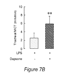
Figure 7A and B. Effect of dapsone treatment on LPS-induced inhibition of
mucociliary
transport (MCT) timed over a 3 mm segment. A: Oral dapsone increased MCT, but
not
significantly (P ¨ 0.09). Values are means + SD. n ¨ 4. B: Nebulized dapsone
significantly
increased MCT (P = 0.007) to normal levels. Values are means SD. n = 5. **P
< 0.01
compared with vehicle (LPS dapsone -).
DETAILED DESCRIPTION
The invention provides methods of treating inflammation of the airways by
administering aerosol formulations of dapsone. Without being bound by theory,
it is believed
that dapsone exerts an immunomodulatory (as opposed to an immunosuppressive)
effect by
inhibiting IL-8 and IL-13. IL-8, a member of the cysteine-X-cysteine (CXC)
chemokine family,
acts as one of the most potent neutrophil chemoattractants. Hence, attenuation
of IL-8 activity
lessens or decreases the recruitment of neutrophils to a site of inflammation,
thereby decreasing
or lowering neutrophil-dominated inflammation at the site. IL-13 is known to
induce goblet cell
hyperplasia in asthmatics, and inhibition of this process also aids in
controlling the symptoms of
airway inflammation.
The methods of the invention are advantageous compared to the use of steroids
to
counter inflammation, because steroids are immunosuppressants and, while their
use may
decrease inflammation, their use also results in immunosupression, thereby
increasing the risk
-5-
CA 02793170 2012-09-13
WO 2011/115778
PCT/US2011/027494
of infection (e.g. opportunistic infection). Likewise, the use of dapsone is
advantageous
compared to the use of macrolide antibiotics, since the use of dapsone does
not contribute to the
evolution of rnacrolide-resistant bacteria. In addition, as demonstrated
herein, while both oral
and aerosol dapsone decreased LPS-induced intraepithelial neutrophil
accumulation, but only
treatment with aerosol dapsone restored mucociliary transport to normal.
(Mucociliary
clearance, the self-clearing mechanism of the bronchi that is carried out by
cilia which are
present on the respiratory epithelium, is an indicator of the health of the
airway surface.) Thus,
the finding that aerosolized dapsone is superior to orally administered
dapsone in restoring this
important function is of great consequence.
In an alternative embodiment, the dapsone is administered to the airways via,
for
example, installation of an aqueous, physiologically acceptable carrier
comprising dapsone,
described below.
Practice of the method of the invention results in a decrease in symptoms of
inflammation in the airways of a subject treated with an aerosolized dapsone
preparation.
Administration results in inhibition of IL-8 and restoration of mucociliary
transport and
clearance. The decrease (lessening, amelioration, resolution, etc.) may be
complete (i.e.
symptoms may entirely disappear) but this need not always be the case. Those
of skill in the art
will recognize that much benefit can be accrued for a patient in whom symptoms
are only
lessened or partially ameliorated, e.g. to a level which allows the patient to
resume a normal or
near-normal level of activity. Those of skill in the art are familiar with the
assessment and
measurement of the effect of such treatments, e.g. by measuring lung capacity,
extent of airway
occlusion, blood oxygenation levels, by observing the presence/absence and/or
frequency of
symptoms (e.g. wheezing, coughing, etc.), various imaging techniques, and
others. Generally,
the practice of the methods of the invention leads to at least about a 10, 20,
30, 40, 50, 60, 70,
80, 90 or even 100% reduction in symptoms of inflammation.
The methods of the invention involve administering physiologically compatible
aerosol
compositions of dapsone (or, in an alternative embodiment, dapsone in an
aqueous carrier) to
the respiratory system of a patient or subject. The phrase "respiratory
system" is intended to
-6-
CA 02793170 2012-09-13
WO 2011/115778
PCT/US2011/027494
include all orifices and passages that participate in carrying air (usually
oxygen-rich air) to and
from the lungs and waste, CO, rich air from the lungs, as well as the lungs
themselves. For
example, included are the nose and nasal cavities, the mouth, the larynx,
trachea, bronchi and
bronchiole tubes and their branches, and alveoli small airways (e.g.
membranaceous
bronchioles, which are noncartilaginous conducting airways with a
fibromuscular wall; and
respiratory bronchioles, which are airways in which the fibromuscular wall is
partially
alveolated). Two natural orifices through which aerosolized dapsone can be
administered are the
nose and mouth, and administration via either or both of these is encompassed
by the invention.
However, the aerosols may also be delivered through surgically introduced
openings (e.g.
tracheotomies), or even directly to, for example, the lungs e.g. via
intubation.
As such, the delivery may be accomplished by using any of many known aerosol
administering devices, including but not limited to mouth inhalers (dry powder
inhalers,
metered dose inhalers, etc.), face masks, intranasal or intra-tracheal tubes,
nebulizers, etc. The
type of device that is selected will vary according to the circumstances, e.g.
whether the aerosol
is self-administered by the patient, or whether in e.g. situations of acute
attacks or crises,
delivery is carried out by medical personnel. Generally, for the treatment of
chronic disease, the
devices that are used will be suitable for patient self-administration. Such
administration may be
carried out using any of several types or styles of aerosol delivery devices
known in the art.
Exemplary devices include but are not limited to metered-dose inhalers (MDIs,
e.g. "puffers"),
in which medication is most commonly stored in solution in a pressurized
canister that contains
a propellant (e.g. fluorocarbons such as 134a or 227, pressurized air,
alkanes, etc.), although it
may also be a suspension; dry powder inhalers, (DPIs) which release a dose of
medicine as a
powder aerosol; and nebulizers, which supply the medication as an aerosol
created from an
aqueous formulation. The devices may be, for example, single-dose or multi-
dose, disposable or
reusable/refillable, etc., and may be made from a variety of materials and in
a variety of shapes,
and may operate by a variety of mechanisms (e.g. breath-hold, breath-actuated,
etc.).
Tn some embodiments, inhalation devices for use in the present invention are
breath
actuated, i.e. delivery of the aerosolized formulation is restricted o the
period of actual
-7-
CA 2793170 2017-05-04
inhalation by the patient. One such breath-actuated representative inhalation
device suitable for
use in the practice of the invention is the Aerodose' inhaler, available from
Aerogen, Inc.,
Sunnyvale, Calif. This inhaler generates an aerosol using a porous membrane
driven by a
piezoelectric oscillator. Other inhaler or nebulizer devices that may be
employed include
conventional air-jet nebulizers, for example, the PARI LC PLUS TM jet
nebulizer (PART GmbH,
Stamberg, Germany); and others that are known in the art. Various systems,
devices and
compositions for the delivery of aerosols are described, for example, in the
following issued US
patents: 7,740,463; 7,683,029; 7,497,214; 7,223,381; 7,163,014; 7,040,314;
6,932,962;
6,743,413, and 6,575,162.
As used herein, the term "aerosol" refers to a suspension of solid or liquid
particles in a
gaseous medium. Herein, this term also encompasses, for example, "mists",
"nebulized
formulations", etc. The formulations that are administered according to the
present invention are
suitable for aerosolized delivery to a patient in need thereof. Thus, the
formulations are
physiologically compatible. At a minimum, the formulations contain dapsone
plus a
physiologically/biologically compatible or suitable carrier. The amount of
dapsone in a
formulation may vary, but is generally in the range of from about Ito 99%
(wt/vol).
Formulations suitable for delivery to the lung of a patient generally comprise
either solid
particles which comprise dapsone, suspended in a gaseous medium when
delivered, or liquid
droplets comprising dapsone, suspended in a gaseous medium when delivered.
Commercial
sources of dapsone are well-known to those of ordinary skill in the art. Those
of ordinary skill in
the art are also well acquainted with the production and manufacture of such
formulations,
which may, in addition to the active agent dapsone, include one or more
additional components.
If a liquid carrier is used, it may be sterile saline or saline buffered at a
physiologically
compatible pH (e.g. from about 6,5 to 8.0, usually about 7.3-7.4). Exemplary
additional
components include but are not limited to: stabilizers, preservatives, various
organic and
inorganic pharmaceutical excipients, including various polymers, low molecular
weight
oligomers, natural products, wetting agents, and surfactants, in particular,
nonionic and ionic
surfactants. Representative examples of addtional components (which may be
surface modifiers)
-8
CA 02793170 2012-09-13
WO 2011/115778
PCT/US2011/027494
include cetyl pyridinium chloride, gelatin, casein, lecithin (phosphatides),
dextran, glycerol, gum
acacia, cholesterol, tragacanth, stearic acid, benzalkonium chloride, calcium
stearate, glycerol
monostearate, cetostearyl alcohol, cetomacrogol emulsifying wax, sorbitan
esters,
polyoxyethylene alkyl ethers (e.g., macrogol ethers such as cetomacrogol
1000),
polyoxyethylene castor oil derivatives, polyoxyethylene sorbitan fatty acid
esters (e.g., the
commercially available Tweens such as e.g., polysorbate 20, commercial name
Tweene 20 and
Tween 80 (ICI Specialty Chemicals)); polyethylene glycols (e.g., Carbowaxs
33508 and
1450 , and Carbopol 934 (Union Carbide)), dodecyl trimethyl ammonium bromide,
polyoxyethylene stearates, colloidal silicon dioxide, phosphates, sodium
dodecylsulfate,
carboxymethylcellulose calcium, hydroxypropyl cellulose (HPC, HPC-SL, and HPC-
L),
hydroxypropyl methylcellulose (HPMC), carboxymethylcellulose sodium,
methylcellulose,
hydroxyethylcellulose, hydroxypropylcellulose, hydroxypropylmethyl-cellulose
phthalate,
noncrystalline cellulose, magnesium aluminum silicate, triethanolamine,
polyvinyl alcohol
(PVA), polyvinylpyrrolidone (PVP), 4-(1,1,3,3-tetramethylbutyp-phenol polymer
with ethylene
oxide and formaldehyde (also known as tyloxapol, superione, and triton),
poloxamers (e.g.,
Pluronics F68 and F108e, which are block copolymers of ethylene oxide and
propylene
oxide); poloxamines (e.g., Tetronic 908 , also known as Poloxamine 908 , which
is a
tetrafunctional block copolymer derived from sequential addition of propylene
oxide and
ethylene oxide to ethylenediamine (BASF Wyandotte Corporation, Parsippany,
N.J.)); a charged
phospholipid such as dimyristoyl phophatidyl glycerol, dioctylsulfosuccinate
(DOSS); Tetronic
1508 (T-1508) (BASF Wyandotte Corporation), dialkylesters of sodium
sulfosuccinic acid
(e.g., Aerosol OT , which is a dioctyl ester of sodium sulfosuccinic acid
(American
Cyanamid)); Duponol P , which is a sodium lauryl sulfate (DuPont); Tritons X-
200 , which is
an alkyl aryl polyether sulfonate (Rohm and Haas); Croclestas F-110 , which is
a mixture of
sucrose stearate and sucrose distearate (Croda Inc.); p-isononylphenoxypoly-
(glycidol), also
known as Olin-lOGS or Surfactant 10-GO (Olin Chemicals, Stamford, Conn.);
Crodestas
SL-40 (Croda, Inc.); and SA9OHCO, which is
C18H37CH2(CON(CH3)--CH2(CHOH)4(CH2OH)2 (Eastman Kodak Co.);
-9-
CA 2793170 2017-05-04
=
decanoyl-N-methylglucamide; n-decylf3-D-glucopyranoside; n-decyl f3-D-
maltopyranoside;
n-dodecyl p-D-glucopyranoside; n-dodecyl P-D-maltoside; heptanoyl-N-
methylglucamide;
n-heptyl-P-D-glucopyranoside; n-heptyl p.-D-thioglucoside; n-hexyl P-D-
glucopyranoside;
nonanoyl-N-methylglucamide; n-noy113-D-glucopyranoside; outanoyl-N-
methylglucamide;
n-oetyl-P-D-glueopyranoside; octyl p-D-thioglucopyranoside; and the like.
Tyloxapol is a
particularly preferred surface modifier for pulmonary or intranasal delivery,
even more so for
nebulization therapies. Most of these compounds arc known pharmaceutical
excipients and arc
described in detail in the Handbook of Pharmaceutical Excipients, published
jointly by the
American Pharmaceutical Association and The Pharmaceutical Society of Great
Britain (The
Pharmaceutical Press, 1986). They are commercially available and/or can be
prepared
by techniques known in the art.
In addition, the dapsone aerosol may contain and be formulated with other
biologically
active components, e.g. other compounds with anti-inflammatory (or other)
properties such as
steroids, antibiotics (e.g. macrolides), decongestants, anti-cancer agents,
etc. Alternatively, the
aerosol dapsone formulation may be used in conjunction with such biologically
active
components, although they are not included in the same formulation.
The delivery schedule that is maintained by the patient (or a health care
professional that
is caring for the patient) may vary depending on several factors, e.g. the
disease or condition that
is being treated, the severity of the condition; the age, gender and weight of
the patient;
convenience of scheduling in order to achieve or maximize compliance, etc.
Generally,
administration takes place from 2-4 times daily (e.g. about every 12 hours, or
every 8 hours, or
every 4 hours), but may also be less frequent (e.g. only once per day) or more
frequent (e.g.
every 2 hours) in some cases. The duration of each administration may differ,
depending on the
amount that is to be delivered, the type of device that is used, etc.
Generally, administration
requires from about 5-30 minutes, e.g. about 5, 10, 15, 20, 25 or 30 minutes.
However, some
rapid delivery devices may accomplish delivery in only a few (e.g. 1-4)
minutes. Further, for
some patients (e.g. a patients who is experiencing an acute asthma attack)
administration may be
continuous and ongoing for a longer period of time, e.g. until the patient is
transported to a
CA 02793170 2012-09-13
WO 2011/115778
PCT/US2011/027494
hospital setting, or until the patient is stabilized in a medical setting,
etc. A skilled medical
professional (e.g. a doctor, respiratory therapist, etc.) is well aware of
these factors and will be
able to plan and adjust treatment protocols accordingly.
The amount of dapsone that is delivered per dose will also vary according to
various
factors, e.g. the disease or condition that is being treated, the severity of
the condition; the age,
gender and weight of the patient; tolerance of the patient for the treatment;
etc. Generally, the
amount administered by inhalation will range from about 0.5 to about 5 mg/kg
of body weight,
e.g. from about 0.5 to about 1.0, 1.5, 2.0, 2.5, 3.0, 3.5, 4.0, 4.5 or 5.0
mg/kg of body weight, and
frequently will be about 2 mg/kg of body weight, in a single dosing session.
Diseases which can be treated using the methods of the invention include
various
inflammations of the airways, especially neutrophil dominated inflammations,
which include
but are not limited to various obstructive lung diseases in which the
bronchial tubes become
narrowed making it hard to move air in and especially out of the lung, for
example: Chronic
Obstructive Pulmonary Disease (COPD) and asthma (ongoing chronic asthma,
severe asthma,
and asthma -flare-ups" or acute attacks, especially neutrophilic severe
asthma); cystic fibrosis;
bronchiectasis, bronchiolitis obliterans; pulmonary fibrosis (e.g. idiopathic
pulmonary fibrosis);
emphysema; acute respiratory distress syndrome; bronchitis; chronic
bronchitis; chronic
sinusitis; rhinosinusitis and chronic rhinosinusitis; chronic airway
infection; toxic inhalation
injury; etc. The inflammation can be caused by any of many triggers, including
but not limited
to tobacco smoking or exposure to second hand smoke; occupational exposure to
workplace
dusts found in coal mining, gold mining, and the cotton textile industry and
chemicals such as
cadmium, isocyanates, and fumes from welding; exposure to air pollution (e.g.
sulfur dioxide,
carbon monoxide, particulates such as soot, dust, etc.); indoor air pollution
e.g. from cooking
fire smoke and fireplace smoke; genetic susceptibility (e.g. alpha 1-
antitrypsin deficiency; and
autoimmune reactions (e.g. sustained inflammation mediated by autoantibodies
and autoreactive
T cells); allergic immune reactions and/or anaphylaxis caused by e.g. dust
mites, pet dander,
pollen, foods, insect bites or stings, etc.; and others.
The methods of the invention involve administering an aerosolized formulation
of
-11-
CA 02793170 2012-09-13
WO 2011/115778
PCT/US2011/027494
dapsone to a patient suffering from inflammation of the airways. Such patients
are generally
mammals, usually humans, but this need not always be the case. Veterinary
applications of the
technology are also contemplated.
The methods of the invention generally involve identification of a patient
that is
suffering from a disease or condition characterized or caused by airway
inflammation or
abnormal mucociliary transport (MCT) (e.g. slower than normal or basal level
MCT), especially
inflammation in which neutrophils play a role. Exemplary diseases include but
are not limited to
cystic fibrosis, bronchiectasis, bronchiolitis obliterans, emphysema, chronic
bronchitis, chronic
rhinosinusitis, toxic inhalation injury, chronic obstructive pulmonary
disease, idiopathic
pulmonary fibrosis, asthma, and chronic airway inflammation. The methods are
implemented by
administering dapsone to the airways of an affected patient, e.g. in order to
contact airway
epithelial cells, especially epithelial cilia. The methods of the invention
generally result in a
lowering or decrease in IL-8 overexpression (i.e. the methods prevent
expression of IL-8 mRNA
at levels which are above normal, control or basal levels). As a result,
disease symptoms caused
by such overexpression abate in patients suffering from diseases associated
with TL-8
overexpression. For example, mucociliary transport returns to normal or near-
normal levels.
Those of skill in the art are familiar with methods and tests for assessing,
identifying and/or
diagnosing such patients by observing and/or measuring certain parameters,
e.g. breathing
characteristics; imaging analyses of lungs and airways; blood levels of
oxygen, CO2, etc; patient
self-reporting; biopsy; sputum analysis; and others. Once a patient who is a
likely candidate for
aerosol dapsone therapy has been identified, a medical professional will
generally prescribe a
dose and/or dosing regimen for the patient, as well as providing instructions
and possibly
teaching regarding or demonstrations of the use of an inhaler. The medial
professional will then
monitor the outcome of administration of the aerosol. Doses and/or dosing
frequency may be
adjusted according to the patient's reaction or response to the therapy and
the progress that is
made toward controlling or resolving the clinical symptoms of disease. The
duration of therapy
may vary from patient to patient, or for an individual patient at different
times, and may be
short-term or long-term. Frequently, due to the chronic nature of the
conditions being treated,
-12-
CA 02793170 2012-09-13
WO 2011/115778
PCT/US2011/027494
aerosol dapsone therapy is long term and continues e.g. for weeks, or months,
or years, or even
for the remainder of the patient's life.
Those of skill in the art will recognize that delivery of an active agent to
the airways of a
patient need not always be accomplished using an aerosolized formulation. For
example,
installation of a drug (e.g. through an existing conduit such as a tracheotomy
tube, nasal tube,
etc.) may require the use of an aqueous foimulation, e.g. dapsone in a
physiologically acceptable
liquid carrier. Those of skill in the art are knowledgeable in the preparation
of such
formulations, which share many properties with aerosol preparations as
described above for
aerosols (e.g. amount of active agent and excipients present in a preparation;
pH; diseases
treated; identification, diagnosis and monitoring of patients; administration
schedules;
administration with other agents; etc.). However, such preparations are not
"dry powders" but
liquids. Routes and methods of administration of such foimulations include but
are not limited
to, for example: installation; via manually dispensed nasal mists or sprays
(e.g. delivery by
manual squeezing or pumping of a container or device); via nose drops or nasal
irrigation; etc.
EXAMPLES
EXAMPLE 1. Dapsone inhibits IL-8 secretion from human bronchial epithelial
cells stimulated
with LPS and resolves airway inflammation in the ferret
Introduction
The respiratory tract is lined with epithelial cells that separate the
internal milieu of the
host from the outside world. Airway epithelia are not only a mechanical
barrier to external
stimuli and microbes but are actively involved in the innate and acquired
immune responses and
airway inflammation. In response to bacterial invasion, mucociliary clearance
is stimulated and
inflammatory mediators and cytokines are secreted as a defense but these can
also damage the
airway. Among epithelial-derived pleiotropic cytokines, IL-8, a member of the
cysteine-X-cysteine (CXC) chemokine family, acts as one of the most potent
neutrophil
chemoattractants. Neutrophil-dominated inflammation is characteristic of
chronic obstructive
pulmonary disease (COPD), diffuse panbronchiolitis (DPB) and cystic fibrosis
(CF). IL-8 is
-13-
CA 02793170 2012-09-13
WO 2011/115778
PCT/US2011/027494
produced by airway epithelial cells. Increased IL-8 in sputum and
bronchoalveolar lavage (BAL)
fluid is associated with the severity of DPB and CF and there is increased IL-
8 gene expression
in the bronchial epithelium of subjects with severe asthma and COPD.
Pro-inflammatory cytokines, bacterial flagellin and lipopolysaccharide (LPS)
can
increase IL-8 production by normal human bronchial epithelial (NHBE) cells.
Among the many
agents present in organic dusts, LPS, is a major inducer of the inflammatory
reaction. LPS binds
to toll-like receptor 4 (TLR4), which activates intracellular signaling
pathways, including the
nuclear factor-KB (NK-KB) pathway, the phosphatidylinositol 3-kinase (PI3K)
and,
mitogen-activated protein kinase (MAPK) pathways. Three MAPK pathways
contribute to IL-8
gene expression, the extracellular-regulated protein kinase (ERK), c-Jun NH2-
terminal protein
kinase (INK), and p38 MAPK cascades. The relative degree of activation of each
of these
pathways and the functional consequences differ among cell types and
experimental systems .
Macrolides antibiotics decrease neutrophils and IL-8 concentration in BAL from
subjects
with DPB, and sputum IL-8 concentration in CF. Macrolides can inhibit IL-8
release from
airway epithelial cells in culture through inactivation of ERK. or NK-KB.
We hypothesized that dapsone would inhibit IL-8 secretion by stimulated airway
cells.
We therefore studied the effect of dapsone on IL-8 secretion from NHBE cells
stimulated with
LPS and further investigated the signaling pathways involved. We then
evaluated in the
effectiveness of dapsone in decreasing airway neutrophil recruitment and
preserving mucociliary
clearance when administered orally or as an aerosol to ferrets with airways
that had been
exposed to (inflamed by) LPS.
Material and Methods
Reagents
Dapsone (4,4'-diaminodiphenyl sulfone), LPS (Escherichia colt serotype 0111:
B4), and
all other reagents were purchased from Sigma-Aldrich Co. (St. Louis, MO)
unless otherwise
indicated. PD-98059, a MAPK/ERK kinase (MEK, an upstream kinase of ERK1/2)
inhibitor
was obtained from Calbiochem (La Jolla, CA). Phospho- and non-phospho-specific
ERK1/2,
anti-p38 MAPK, anti-SAPKANK, and phospho-specific NF-KB p65 (Ser536) as well
as
-14-
CA 02793170 2012-09-13
WO 2011/115778
PCT/US2011/027494
anti-rabbit-IgG HRP antibodies were purchased from Cell Signaling Technology
(Beverly, MA).
DMSO was used as a solvent of dapsone, and the final concentration did not
exceed 0.01%
(v/v). Preliminary in vitro experiments showed that 0.01% DMSO-medium had no
significant
effect on cell viability and IL-8 secretion for up to 72 h (data not shown).
MIBE cell culture
NTIBE cells (Lonza Walkersville, Walkersville, MD) were plated at 3,500 cells/
cm2 in
culture dishes in bronchial epithelial cell growth medium (BEGM) supplemented
with the
SingleQuot kit (Lonza) without antibiotics and cultured at 37 C in a 5% CO,
incubator. We
used endotoxin-free media (< 0.005 endotoxin units/m1) and second-passage
cells for all
experiments. Cells were grown to confluence for 6 days. Cultures without
antibiotics were then
transferred to 6-well or 35 mm dishes coated with type 1 rat-tail collagen and
seeded at 3,500
cells/ cm2. The medium was changed every 24 h. To avoid influence of growth
factors on cell
signaling and IL-8 secretion, cells were cultured in supplement-free bronchial
epithelial cell
basal medium (BEBM) for 24 h before stimulation. We evaluated cell response at
the time of
cell confluence rather than normalize to the relative number of cells because
cell maturation
could affect cell signaling and cytokine secretion, and at confluence, all
cells are at similar
growth stages.
For NHBE cell differentiation, cells were plated at 2.0 x 10⁵ cells/cm2 onto
polycarbonate inserts of 6.5-mm diameter, 0.4-pm pore size and 10-ilm
thickness (Costar
Transwell Clear, Cambridge, MA, USA) coated with type 1 rat-tail collagen, and
cultured with
serum-free DMEM/F12 medium containing ITS-A (1.0%; Invitrogen Co., Carlsbad,
CA),
epidermal growth factor (EGF) (recombinant human EGF, 0.5 ng,/m1; Invitrogen
Co.),
triiodothyronine (10 ng/ml; MP Biomedicals, Solon, OH), hydrocortisone (0.5
g/ml; MP
Bi medical s), all-trans retinoic acid (1.0 x 10⁻⁷ M; Sigma-Aldrich), bovine
serum albumin (2.0
p.g/m1; Sigma-Aldrich) and bovine pituitary extract (30 p.g/m1; Invitrogen
Co.). After achieving
confluence, the apical medium was removed, and cells were cultured with an air-
liquid-interface
(ALI) method. The culture medium was changed every 48 h, and cells were
maintained at 37 C
in a 5% CO, for 10-14 days.
-15-
CA 02793170 2012-09-13
WO 2011/115778
PCT/US2011/027494
Cytotoxicity assay
To determine the number of viable cells, formazan dye generation was measured
using
Cell Counting Kit-8 (CCK-8; Dojindo, Kumamoto, Japan). Cells treated with CCK-
8 assay
solution were incubated for 2 h and the absorbance at 450 nm was measured with
a microplate
reader. Data were expressed as % of control cells that were not exposed to
dapsone.
Measurement of IL-8 secretion
Culture supernatants were collected and centrifuged for 5 mM at 200 x g and
stored at
-20 C until assayed. IL-8 was measured by ELISA (Beckman Coulter, Inc., Brea,
CA) according
to the manufacturer's instructions. Concentrations in each sample were
obtained by
interpolation from standard curves, and calculated as the mean of the results
at the sample
dilution.
Immunoblotting
After stimulation, the plated cells were washed with cold PBS, and then lysed
on ice in a
modified radio immunoprecipitation buffer (1% Nonidet P-40, 1% sodium
deoxycholate, 150
mM NaC1, 10 mM Tris pII 7.5, 5 mM sodium pyrophosphate, 1 mM NaVO4, 5 mM NaF,
1
g/ml aprotinin, 1 Rg/m1 leupeptin, and 0.1 mM PMSF) for 15 min and then
scraped from the
dishes. DNA was sheared by passing the lysate though a 27-gauge needle, and
insoluble material
was removed by centrifugation at 20,000 g for 15 min at 4 C. The protein
concentration of the
resulting supernatant was quantified by the DC protein assay (Bio-Rad,
Hercules, CA). Equal
amounts of protein extracts were loaded on a 12% SDS-PAGE mini gel and
transferred to a
nitrocellulose membrane (Bio-Rad). Membranes were blocked with blocking buffer
(150 mM
NaC1, 20 mM Tris, and 0.1% Tween 20, pH 7.6) containing 5% nonfat dry milk at
4 C
overnight. Subsequently, membranes were rinsed and incubated with the primary
antibody:
phospho (p)-p44/42 MAPK (Thr202/Tyr204) (diluted 1:2000), p-p38 MAPK
(Thr180/Tyr182)
(diluted 1:1000), p-SAPKIJNK (Thr183/Tyr185) (diluted 1:1000), or p-NF-KB p65
rabbit
polyclonal IgG (diluted 1:1000) (Cell Signaling Technology), for 2 h at room
temperature. The
membranes were then incubated at room temperature for 1 h with the anti-rabbit
IgG HRP
secondary antibody (diluted 1:2000). Subsequently, the blots membranes were
developed with
-16-
CA 02793170 2012-09-13
WO 2011/115778
PCT/US2011/027494
LumiGLO chemiluminescent substrate peroxide (Cell Signaling Technology).
Membranes were stripped with a stripping buffer (100 mM 2-mercaptoethanol, 2%
SDS,
and 62.5 mM Tris/HC1 pH 6.7) for 30 min at 30 C. The blots were reprobed with
anti-p44/42
MAPK, anti-p38 MAPK, or anti-SAPK/JNK antibody (diluted 1:1000 for each),
followed by
anti-rabbit-igG HRP secondary antibody (diluted 1:2000). To correct small
differences in
loading for NF-KB p65, blots were stripped, and reprobed with anti-human I3-
actin antibody
(diluted 1:5000), followed by anti-mouse IgG HRP secondary antibody (diluted
1:5000).
Western blot images were scanned and analyzed on NIH Image J software (18).
Real-time quantitative polyinerase chain reaction
For the relative quantification of IL-8 mRNA expression, the expression of
glyceraldehyde-3-phosphate dehydrogenase (GAPDH) was served as an internal
control.
Ev-aGreen was used as a DNA intercalator dye to monitor amplified DNA
quantification, and
real-time quantitative PCR curves were analyzed by the CFX ManagerTM software
(Bio-Rad)
in order to obtain threshold cycle (Ct) values for each sample. Quantification
was based on a
standard curve. Appropriate IL-8 and GADPH forward and reverse primers were
used.
Animal study
Twenty adult male ferrets (weight, 1.3 to 2.0 kg) were obtained from Marshall
Farms
(Rose, NY). Ferrets were anesthetized with 40 mg/kg ketamine and 5 mg/kg
xylazine, and
exposed to endotoxin for 30 mM once daily for 5 days by intubating with a 3.0
uncuffed
endotracheal tube (ETT) coated with 10 mg of LPS mixed in 300 Ill water
soluble K-Y jelly
(Johnson & Johnson, New Brunswick, NJ) (Abanses et al., 2009). As controls, K-
Y jelly
without LPS was used in two ferrets.
Dapsone was prepared in a vehicle of 5% (v/v) ethanol/5% (v/v)
dimethylsulfoxide
(DMS0)/0.5% (w/v) methyl-cellulose solution for oral administration in a final
concentration of
3 mg,/ml. To administer in nebulized form, dapsone was dissolved in 0.67%
(v/v) DMSO/saline
at a concentration of 0.5 mg/ml. Eighteen LPS-treated ferrets were randomized
to receive 5-day
dapsone treatment starting on day 4 (after 3 days of LPS) and continuing for 5
days. For oral
administration, 2 mg/kg dapsone (n = 4) or vehicle alone (n = 4) was given
daily through a
-17-
CA 02793170 2012-09-13
WO 2011/115778
PCT/US2011/027494
nasogastric tube. This is equivalent to the oral dose used to treat persons
with skin disease. For
aerosol inhalation, ferrets were shallowly intubated with ETT, which was
placed 1 cm below the
larynx. Ferrets were treated with nebulized 0.5 mg/ml dapsone (n = 5) or 0.67%
DMSO/saline
(vehicle) (n = 5) for 15 min once daily using a jet nebulizer (PariMaster;
PARI Respiratory
Equipment Inc., Richmond, VA). Ferrets (body weight 1.5 kg) have 0.3-0.4 ml
total lung lining
fluid volume. Therefore, the dose delivered to the lung, is estimated to be
0.1 to 1.01.1g in this
system given reported nebulization efficiency.
On day 9 dapsone-treated and vehicle control animals were sacrificed and the
tracheas
removed. Each tracheal segment was fixed in 10% foimalin, embedded in
paraffin, and
processed for histological analysis using a light microscope (CKX41; Olympus,
Tokyo, Japan)
and by photography using a digital camera system (AxioCam ICe 1; Carl Zeiss,
Thomwood,
NY). The tissue was cut in 4- m thickness, and the slides were stained with
hematoxylin and
eosin. To measure the accumulation of inflammatory cells and overall severity
of inflammation,
the total number of intraepithelial neutrophil was counted over 150 pm in
eight random sites per
specimen from four different sections and averaged.
We also used an excised tracheal segment to measure tracheal mueociliary
transport
(MCT) velocity. Cilia transport mucus loaded e.g. with foreign particles and
microorganisms
towards the mouth, where it is either swallowed or expelled via coughing.
Under conditions of
inflammation, the ciliary cells suspend their transport function and bacterial
germinal
colonization, further irritation and inflammation is facilitated.
A tracheal segment was placed on a piece of gauze saturated with Ringer's
solution in a
chamber in which the relative humidity was maintained at 95 to 100% and the
temperature was
maintained at 22 C to 24 C. MCT was measured by focusing a tracheal segment
under a
microscope with gradicule (grid line) eyepiece and recording the transport
time of the leading
edge of very fine shavings of plastic that were placed on the tracheal
epithelium. The time to
transport the particle 3 mm was used to calculate the MCT (in millimeters per
minute).
Measurements were repeated five times for each tracheal segment. This study
was approved by
the Animal Care and Use Committee of Virginia Commonwealth University.
- 18-
CA 02793170 2012-09-13
WO 2011/115778
PCT/US2011/027494
Statistics
Results are expressed as means values SE or SD as appropriate. Statistical
analysis of
data was performed with the StatView 5 statistics package (SAS Institute,
Cary, NC).
Comparisons between two groups were made by unpaired Student's t test.
Multiple comparisons
were made by one-way analysis of variance using Fisher's PLSD-test, and a P
value of less than
0.05 was considered significant.
RESULTS
Effect of dapsone on NHBE cell viability.
"to confirm that the total number of cultured NHBE cells was not influenced by
dapsone
treatment, the viability of cells was evaluated using CCK-8 (Dojindo). NHBE
cells seeded onto
96-well plates (3,000 cells/well) were cultured at 37 C for 72 h (-70%
confluence). Dapsone at
concentrations of 0.3, 1 or 10 [J..g/m1 was added for 24 or 72 h. The results
showed that, for cells
treated with dapsone, the total viable cell number was similar to that of the
non-treated control
group over 72 h (not shown).
Effect of dapsone on LPS-induced IL-8 secretion.
To evaluate the effect of dapsone on IL-8 secretion from NHBE cells, cell
supernatants
were harvested 24 h after stimulation with LPS in the presence and absence of
dapsone, which
had been added at the same time as the LPS. We chose to assess cell response
at the time of cell
confluence rather than normalize to the relative number of cells, because cell
maturation could
potentially affect cell signaling and cytokine secretion, and at confluence,
all cells are at similar
growth stages. LPS at 10 1.1g/m1 significantly increased IL-8 secretion from
NHBE cells (P <
0.001), but dapsone at 0.3, 1 or 10 [..ig/m1 suppressed this (P <0.01 for
each) (Figure 1A).
Dapsone 1 jig/ml did not affect basal IL-8 secretion for 48 and 72 h (Figure
1B). In the presence
of dapsone 1 LPS-
induced IL-8 secretion was significantly and persistently decreased for
up to 72 h (Figure 1C).
Effect of dapsone in ALI-conditioned NHBE cells.
To confirm if the ILS-inhibiting effect of dapsone is seen in differentiated
cells, we used
NHBE cells cultured under ALI condition for 14 days. For cells at ALT, samples
were collected
-19-
CA 02793170 2012-09-13
WO 2011/115778
PCT/US2011/027494
from both apical and basolateral chambers of cells grown on filters. To
collect samples from the
apical chamber, 150 I of Hanks' balanced salt solution (HBSS; Lonza
Walkersville, Inc.) was
added to apical side. Apical IL-8 concentrations were expressed as values of
four-fold dilution
to be equal to the basolateral medium volume of 600 pl.
NHBE cells were stimulated with LPS from the apical (AP-LPS) or basolateral
side
(BL-LPS) for 24 h, and IL-8 secretion levels in both chambers were measured.
Dapsone was
added only to the basolateral medium. As shown in Figure 2A, apical IL-8 level
was
significantly increased by AP-LPS (P <0.001), and dapsone inhibited this
response (P <0.05).
Likewise, basolateral 1L-8 was significantly increased by AP- or BL-LPS (P <
0.001 for each)
(2B), and dapsone inhibited the both the apical and basolateral response (P <
0.001 for AP-LPS,
P < 0.01 for BL-LPS).
To compare with corticosteroids, we examined the effect of dexamethasone (DEX)
on
LPS-induced IL-8 secretion. DEX suppressed basal IL-8 level in both apical and
basolateral
sides (P < 0.05 for each) (Figure 2C, 2D). DEX also significantly inhibited AP-
LPS- or
BL-LPS-induced increase in IL-8 (P <0.05 for each).
Effect of dapsone on LPS-induced IL-8 mRNA expression.
To examine the action of dapsone on IL-8 mRNA, RNA was extracted from the
cells
after 4 h stimulation of LPS, dapsonc, DEX or their combination, and prepared
for real-time
quantitative PCR. As shown in Figure 3, 1 g,/m1 dapsone did not influence the
basal IL-8
mRNA level, but 0.1 g/m1DEX decreased this by ¨40% (P <0.05). 10 g/m1LPS
increased
IL-8 mRNA level more than 5-fold of control (P < 0.001). Dapsone at 1 and 10
g/m1
significantly inhibited LPS-induced IL-8 mRNA over expression (P < 0.05 for
each). DEX at
0.1 g/m1 also inhibited this (P <0.01).
Effect of dapsone on LPS-induced phosphorylation of MAPKs
MAPK signaling are important pathways in the synthesis of IL-8. We evaluated
the
effect of dapsone on LPS-induced phosphorylation of ERK1/2, p38, and JNK in
NHBE cells.
LPS at 10 g/inl significantly phosphorylated ERK1/2 at 1, 4, and 24 h (P
<(105 for each), but
not p38 and INK (Figure 4). Dapsone at 1 g/m1 inhibited LPS-induced ERK1/2
-20-
CA 02793170 2012-09-13
WO 2011/115778
PCT/US2011/027494
phosphorylation at 1 h (P <0.05), although this effect disappeared after 4 h.
We then assessed
the effect of PD98059 (2"-amino-3"-methoxyflavone), a selective cell-permeable
MEK
inhibitor. As shown in Figure 5A, LPS dose dependently increased ERK1/2
phosphorylation and
IL-8 secretion, and PD98059 at 20 M abolished 10 g/m1 LPS-induced ERK1/2
phosphorylation. However, this concentration of PD98059 did not inhibit IL-8
secretion (Figure
5B).
Effect of dapsone on LPS-induced phosphorylation of NF-kB p65.
Since LPS simulates TLR4 and then induces IL-8 through the NF-KB pathway, we
examined the effect of dapsone on NF-KB p65 phosphorylation in NI-IBE cells.
Growth factors
were withdrawn from culture medium 24 h before LPS or dapsone exposure. The
threonine
phosphorylation of NF-KB p65 was measured by Western blotting. The band
intensity was
calculated with NIH Image J software.
The results showed that LPS at 10 Kg/m1 significantly increased NF-KB p65
phosphorylation at 15 mm (P <0.01), and this effect persisted up to 2 h (P <
0.05). Dapsone at 1
g/m1 significantly inhibited LPS-induced NF-KB p65 phosphorylation to the
control level (P <
0.01 for 15 and 30 mm, P < 0.05 for 1 and 2 h). Further, this inhibitory
effect was
dose-dependent (P = 0.16 for 0.3 g/m1dapsone and P <0.01 for 1 and 10 g/m1
dapsone).
Effect of tracheal LPS with and without dapsone on ferret activity and weight.
Ferrets stimulated with LPS for 5 days received 5-day dapsone treatment or
vehicle
alone (n = 8 for vehicle, and n = 9 for dapsone group each time point that was
examined). There
was no measureable effect of LPS with or without dapsone treatment on ferret
activity or
appetite and no difference in weight over 9 days when comparing these two
groups (not shown).
Intraepithelial neutrophil accumulation in LPS-inflamed ferret trachea.
To evaluate in vivo effect of dapsone, we used topical LPS coated onto an
endotracheal
tube (ETT) to recruit neutrophils and inflame the trachea of anaesthetized and
spontaneously
breathing ferret (Abanses et al, 2009). Control ferrets intubated with an ETT
coated with only a
water soluble jelly (used as the LPS vehicle in the other group) showed little
few epithelial
neutrophils; less than 3 /150 in. One ferret with an LPS-inflamed airway and
treated with
- 21 -
CA 02793170 2012-09-13
WO 2011/115778
PCT/US2011/027494
nebulized vehicle was not completed because of death on day 6, and this animal
was excluded
from further analysis. LPS exposure induced marked neutrophil accumulation in
the ferret
tracheal epithelium and dapsone treatment reduced intraepithelial neutrophil
number (not
shown). Orally-administered dapsone tended to inhibit neutrophil recruitment
(P = 0.3) (Figure
6A), and nebulized dapsone significantly inhibited neutrophil accumulation (P
<0.05) (Figure
6B).
Mucociliary transport (MCT) on excised tracheal segments.
Mucociliary transport (or "mucociliary clearance", MCC) is an overall
measurement of
the health and integrity of the airway surface. MCT was timed over a 3 mm
segment. LPS
dramatically decreased MCT to 1-3 mm/minute (normal is approximately 7
mm/min). Oral
dapsone increased MCT but the increase was not significant (P = 0.09). However
aerosol
dapsone preserved MCT at near noimal velocity (6 mm/min; P = 0.007 compared
with LPS
control) as shown in Figure 7A and B
DISCUSSION
We have shown that dapsone inhibits IL-8 secretion from NHBE cells stimulated
with
LPS. Dapsone is used to treat dermatologic disorders, most notably those with
neutrophil
infiltrates. It has been postulated that dapsone impairs neutrophil chemotaxis
and function at the
sites of inflammation, apparently without increased risk of opportunistic
infections. This is
consistent with immunomodulation, but not immunosuppression.
Dapsone inhibits local production of toxic reactive oxygen species,
myeloperoxidase and
elastase, but this seems unlikely as a principal mode of action because the
clinical response to
dapsone is characterized by decreasing the neutrophil numbers. Other
investigators have shown
that dapsone may impair neutrophil chemotaxis by interfering with activation
of adhesion
molecule CD11b/CD18 in vitro. However, this seems to require a higher
concentration of
dapsone than therapeutic levels measured in vivo. The concentration of dapsone
we used in
these studies is within the ranges required for therapeutic serum levels of
0.5-5 lig/m1 (Zhu et al,
2001). We found that the lower concentration of dapsone of 0.3 ii.g/m1
inhibited LPS-induced
IL-8 secretion and this effect was not due to cell toxicity as measured by the
number of viable
-22-
CA 02793170 2012-09-13
WO 2011/115778
PCT/US2011/027494
cells. Schmidt et al. (2001) reported that dapsone, in a therapeutic
concentration, inhibited the
bullous pemphigoid IgG-mediated IL-8 release from cultured normal human
epidermal
keratinocytes (NHEK) and that dapsone did not depress basal IL-8 level. These
data are similar
to our results using NHBE cells.
Airway epithelia are functionally polarized, and there is evidence that
epithelial cells can
secrete cytokines in a bidirectional manner. To better understand the mode of
action of dapsone
in differentiated and polarized airway epithelial model, NHBE cells were
cultured at an ALT
(Kanol et al., 2001). In the presence of dapsone, IL-8 secretion induced by
LPS stimulation was
significantly reduced, while constitutive IL-8 release was not inhibited by
dapsone. Interestingly,
when stimulated apically (as would occur during an airway infection) NHBE
cells secreted IL-8
toward apical as well as basolateral sides, whereas when stimulated
basolaterally they secreted
only towards the basolateral side. Airway epithelial cells grown on filters
and stimulated by
Staphylococcus aureus at the apical side caused T cells chemotaxis towards the
apical but not
the basal side supernatant (Escotte et al, 2006). The presence of an apically
oriented
chemoattractant gradient may be necessary to drive immune cells like
neutrophils and
lymphocytes across epithelial surfaces (Chin et al, 2007). Additionally, we
tested the effect of
dexamethasone (DEX), a potent synthetic corticosteroid, in our cultured cell
system. DEX is
reported to inhibit IL-8 release from NHBE cells, and we confirmed that DEX
inhibited IL-8
mRNA expression more than dapsone. However unlike dapsone, DEX suppressed
basal IL-8
level as well as LPS-induced increase, suggesting that DEX is
immunosuppressive while
dapsone is immunomodulatory.
The intracellular signaling pathways by which dapsone inhibits IL-8 have not
been well
characterized. Because MAPK regulates IL-8 gene expression in airway
epithelial cells, we
evaluated the effect of dapsone on these pathways. We found that LPS dose
dependently
increased ERK1/2 phosphorylation, but not INK and p38, in NI-IBE cells, and
elevated
p-ERK1/2 level continued for at least 24 h. We have previously shown that
sustained activation
of ERK1/2 is required for basal IL-8 secretion from unstimulated NHBE cells,
and that a
specific MEK inhibitor PD98059 suppressed IL-8, while a pharmacological
inhibitor of INK or
-23-
CA 02793170 2012-09-13
WO 2011/115778
PCT/US2011/027494
p38 did not. Others have shown that pretreatment with PD98059 inhibits LPS-
induced
phosphorylation of ERK1/2 at 1 and 2 h. In the current study, the inhibitory
effect of PD98059
was confirmed even at 4 h. However, PD98059 did not decrease LPS-induced IL-8
secretion.
Dapsone at 1 pg/m1 inhibited p-ERK1/2 at 1 h, but not after 4 h. Taken
together, it is unlikely
that ERK1/2 inhibition alone will suppress IL-8 in LPS-stimulated cells, and
therefore that
temporal ERK1/2-inhibition by dapsone does not account for the effect on IL-8.
We then examined the effect of dapsone on NF-KB activation. NT-KB can induce
gene
expression of inflammatory mediators and cytokines in airway epithelial cells,
and LPS can
activate NF-KB via TLR4. The basic NF-KB complex is a dimer of two members of
the Rdl
family proteins, p50 and p65 (RelA). Both subunits contact DNA, but only p65
contains a
transactivation domain within the C-terminal region that directly interacts
with the transcription
apparatus. NF-KB p65 is activated by phosphorylation, which enhances its
transcriptional
activity, and is associated with nuclear translocation. We showed that LPS at
101.1,g/m1 elicited
significant NT-KB p65 phosphorylation from 15 mm to 2 h after stimulation in
NHBE cells.
Similar kinetics in response to LPS has been shown in mmine intestinal
myofibroblasts, in
which phosphorylation of NT-KB p65 was detected within 30 min of LPS treatment
and slowly
decreased over 4 h. We found that dapsone at 1 p,g/m1 inhibited LPS-induced NF-
KB p65
phosphorylation over 2 h to basal levels, suggesting that dapsone may inhibit
induction of 1L-8
secretion by blocking NF-x13 p65 phosphorylation. Also the dose-dependent
inhibitory effect on
phospho-NF-KB p65 was consistent with results of experiment in IL-8 mRNA
expression.
Accordingly, the action of dapsone is due, at least in part, by down-
regulating IL-8 at gene
transcription. However, while dapsone 0.3 [tg/mL did not significantly inhibit
NF-KB p65
activation, the same concentration of dapsone strongly inhibited IL-8 release.
More than 1 vtg/m1
of dapsone was needed to inhibit LPS-induced IL-8 mRNA expression. Schmidt et
al. (28)
speculated that dapsone inhibits IL-8 release from NHEK at the post-
transcriptional level
without affecting mRNA concentration.
These immunomodulatory effects were similar to the immunomodulatory effects of
macrolides such as erythromycin, clarithromycin and azithromycin.
Clarithromycin modulates
-24-
CA 02793170 2012-09-13
WO 2011/115778
PCT/US2011/027494
ERK1/2 phosphorylation in NHBE cells, and azithromycin inhibits NF-KB activity
in a CF cell
line; thus regulating IL-8. It is likely that dapsone also modulates airway
inflammation via
modulation of IL-8.
After inducing tracheal inflammation in ferrets using topically applied LPS,
we
evaluated the effects of five days of oral or aerosol dapsone on neutrophil
inflammation and
MCT, an integrated measure of epithelial integrity. Oral dapsone decreased
intraepithelial
neutrophil accumulation but this was not statistically significant.
Recognizing that dapsone is
effective when used topically as a cream to treat neutrophilic dermatoses, we
evaluated the
potential for a lower dose of aerosol dapsone to inhibit LPS-induced tracheal
inflammation.
Nebulized dapsone significantly inhibited airway neutrophil infiltration and
preserved or
restored MCT despite exposure to LPS. Although it is difficult to calculate
the precise dose to
the airway, even if 10% of the total dose is delivered efficiently to the
trachea (38), the dapsone
delivered was only 0.3 mg. This dose is approximately one-tenth the systemic
dose administered
orally in a 1.5 kg ferret.
In summary, dapsone did not influence unstimulated (basal) IL-8 secretion.
Apical LPS
stimulation induced both apical and basolateral IL-8, but basolateral LPS
increased only
basolateral IL-8. Dapsone inhibited polarized IL-8 secretion from ALI-
conditioned cells.
Dapsone also decreased LPS-induced IL-8 mRNA levels. LPS led to
phosphorylation of
extracellular signal regulated kinase (ERK)U2, but not p38 MAPK or c-Jun N-
terminal kinase.
LPS also induced NP-KB p65 phosphorylation, an effect that was inhibited by
dapsone. Both
oral and aerosol dapsone decreased LPS-induced intraepithelial neutrophil
accumulation but
only treatment with aerosol dapsone restored mucociliary transport to normal.
Together, these data show that dapsone inhibits IL-8 in human airway cells and
neutrophil recruitment in the inflamed mammalian trachea in vivo while
preserving MCT.
Aerosol dapsone could be a promising therapy to treat chronic inflammatory
airway diseases
such as cystic fibrosis, chronic bronchitis, or severe asthma. Thus, dapsone,
given either
systemically or especially as an aerosol, may be useful in treating
neutrophilic airway
inflammation.
-25-
CA 2793170 2017-05-04
EXAMPLE 2. IL-13 Inhibiting Properties of Dapsone
Globlet cells arc columnar epithelial cells whose sole function is to secrete
mucin, which
= dissolves in water to form mucus. Goblet cell hyperplasia is involved in
the pathological
hypersecretion exhibited by bronchial epithelial cells of asthmatics, and EL-
13 is known to pa
central role in mediating goblet cell hyperplasia in both in vivo and in vitro
models of asthma.
The ability of dapsone to inhibit goblet cell hyperplasia was tested in vitro.
An in vitro model of
goblet cell hyperplasia was developed using normal human bronchial epithelial
(NIIBE) cells
cultured under air-liquid interface (AU) conditions. Control experiments that
were analyzed
using differential cell staining and microscopy showed that dapsone (3 g/m1)
had no
measurable effect on the growth of ALT-conditioned NHBE cells (not shown).
However, when
goblet cell hyperplasia was induced using 1L-13, dapsone (101.4m1) decreased
the amount and
extent of goblet cell hyperplasia, compared to control cell cultures (not
shown). Thus, dapsone
may exert its beneficial effect on resolving inflammation by also inhibiting
IL-13 induced
goblet cell hyperplasia.
REFERENCES
Abanses JC, Arima S, Rubin BK. Vicks VapoRub induces mucin secretion,
decreases ciliary
beat frequency, and increases tracheal mucus transport in the ferret trachea.
Chest
2009;135:143-148.
Berlow, BA, Liebhaber, MI, Zeb, D and Spiegel, TM. J Allergy Clin Immunol.
1991
Mar;87(3):710-715.
Chin AC, Parkos CA. Pathobiology of neutrophil transepithelial migration:
implications in
mediating epithelial injury. Annu Rev Pathol 2007;2:111-143.
Chougule, M, Padhi, B and Misra, A. AA.PSPharmSciTech, 9(1): 47-53, January 9,
2006.
Schmidt E, Reimer S. Kruse N, Brc3eker EB, Zillilcens D. The IL-8 release from
cultured human
keratinocytes, mediated by antibodies to bullous pemphigoid autoantigen 180,
is inhibited by
dapsone. Clin Exp Immunol 2001;124:157-162.
Escotte S, Al Alam D, Le Naour R, Puchelle E, Guenounou M, Gangloff SC. T cell
chemotaxis
-26-
CA 02793170 2012-09-13
WO 2011/115778
PCT/US2011/027494
and chemokine release after Staphylococcus aureus interaction with polarized
airway
epithelium. Am J Respir Cell Mol Biol 2006;34:348-354.
Kanoh S, Kondo M, Tamaoki J, Kobayashi H, Motoyoshi K, Nagai A. Differential
regulations
between adenosine triphosphate (ATP)- and uridine triphosphate-induced Cl-
secretion in
bovine tracheal epithelium. Am J Respir Cell Mol Biol 2001;25:370-376.
Zhu Y1, Stiller MJ. Dapsone and sulfones in dermatology: overview and update.
J Am Acad
Dermatol 2001;45:420-434.
While the invention has been described in terms of its preferred embodiments,
those
skilled in the art will recognize that the invention can be practiced with
modification within the
spirit and scope of the appended claims. Accordingly, the present invention
should not be
limited to the embodiments as described above, but should further include all
modifications and
equivalents thereof within the spirit and scope of the description provided
herein.
-27-