Note: Descriptions are shown in the official language in which they were submitted.
CA 02793844 2012-09-20
WO 2011/123224 PCT/US2011/027861
INTRAOCULAR LENS
Field of the Invention
The invention is directed to an intraocular lens, and in particular, an
intraocular lens with
haptics designed to provide positional stabilization of the lens in the
lenticular capsular
bag.
Background of the Invention
A common and desirable method of treating a cataract eye is to remove the
clouded,
natural lens and replace it with an artificial intraocular lens (IOL) in a
surgical procedure
known as cataract extraction. In the extracapsular extraction method, the
natural lens is
removed from the lenticular capsular bag while leaving the posterior part of
the capsular
bag (and preferably at least part of the anterior part of the capsular bag) in
place within the
eye. In this instance, the lenticular capsular bag remains anchored to the
eye's ciliary body
through the zonular fibers. The capsular bag also continues its function of
providing a
natural barrier between the aqueous humor at the front of the eye and the
vitreous humor at
the rear of the eye.
Another trend in modem day cataract surgery is the reduction of the corneal
incision size
because larger incision sizes have been attributed to unwanted post-surgical
conditions
such as incision-induced astigmatism. IOLs and IOL inserters capable of
successfully
inserting the IOL through a sub 2.5-mm incision is desired by most cataract
surgeons.
Because the IOL undergos compression and other forces as it is passed through
the IOL
inserter, the dimensions (particularly the cross-section) of the IOL must
accordingly be
minimized. An IOL designer is thus further challenged in making an IOL that
will have the
strength and stability to remain centered in the eye, yet has a dimensional
size and
mechanical flexibility at near room temperature to pass through a sub-2.5 mm
incision
size. It will be appreciated that these are often competing design goals in
that reducing IOL
dimensions to fit within a smaller incision can result in a decrease in the
strength and
stability of the IOL in the eye.
The strength and stability of the IOL within the eye is of course crucial in
obtaining and
maintaining the intended vision correction expected by the physician and, more
importantly, the patient. Accordingly, there remains a need for an improved
IOL design
that is dimensioned to fit through a sub-2.5 mm incision, and yet, is
positionally stable in
the capsular bag for many years following the surgery. It is also, important
to the physician
1
CA 02793844 2012-09-20
WO 2011/123224 PCT/US2011/027861
that the IOL have the ability to self-center within the capsular bag to
minimize the amount
of physical manipulation of the lens following insertion of the lens.
Summary of the Invention
An intraocular lens comprises an optic portion having a peripheral edge and at
least two
haptics. Each haptic is integrated with the peripheral edge of the optic
portion by a
corresponding haptic integration region. Also, each haptic comprises a distal
segment
region and a deformation segment region. The distal segment region has an
outer distal
length bounded by a proximate endpoint and a distal endpoint on an outer
surface of the
haptic, and is scribed by a distal segment angle a of 20 to 30 . The distal
segment angle
has a segment origin that lies within a radial segment bound by a radial
distance 1.5 mm to
1.9 mm from an optic center and a segment angle 0 of from 30 to 45 from a
vertical axis.
The vertical axis extends through the distal endpoint of at least one haptic
and the optic
center.
One embodiment is directed to an intraocular lens comprising an optic portion
and two
haptics, and each haptic is integrated to a peripheral edge of the optic
portion by a haptic
integration region. The optic portion, the two haptics and the haptic
integration regions are
each formed of a hydrophobic polymeric material having a tangent modulus of
elasticity of
2 MPa to 6 MPa. Each of the haptics comprise a distal segment region and a
deformation
segment region. The distal segment region has an outer distal length bounded
by a
proximate and a distal endpoint on an outer surface of the haptic, and is
scribed by a distal
segment angle a of 20 to 30 . The distal segment angle has a segment origin
that extends a
radial distance of 1.5 mm to 1.9 mm from an optic center and a segment angle 0
of from
34 to 40 from a vertical axis. The vertical axis extends through the distal
endpoint of at
least one of the two haptics and the optic center. The deformation segment
region has an
outer deformation length bounded by a proximate deformation endpoint on an
outer
surface and the proximate endpoint of the corresponding distal segment region,
and is
scribed by a deformation segment angle (3 of 20 to 40 from the segment
origin.
Another embodiment is directed to an intraocular lens comprising an optic
portion and at
least two haptics. Each haptic is integrated to a peripheral edge of the optic
portion by a
haptic integration region, and each of the haptics comprise a distal segment
region and a
deformation segment region. A portion of the distal segment region and a
portion of the
deformation segment region combine to form an angle of contact of not less
than 50 and
2
CA 02793844 2012-09-20
WO 2011/123224 PCT/US2011/027861
not more than 70 when the haptics are diametrically stressed with an arcuate
jaw
according to ISO Test No. 11979-3(2006), which models a lenticular capsular
bag of a
human eye.
Brief Description of the Drawings
Illustrative, non-limiting embodiments of the present invention will be
described by way of
example with reference to the accompanying drawings, and in which:
FIG. 1 is a representation of an IOL of the invention;
FIG. 2A is a representation of an IOL in the prior art;
FIG. 2B is a representation of an IOL in the prior art;
FIGS. 3A and 3B are representations of IOLs in the prior art;
FIG. 4 is a photograph of an IOL of the invention implanted within a
lenticular capsular
bag of a human eye;
FIGS is a geometric representation of a radial segment;
FIG. 6 is a model representation of an IOL of the invention with a large angle
of contact;
FIG. 7A is schematic top-view representation of an IOL within the arcuate jaws
of a model
used to determine the angle of contact the haptics have with a lenticular
capsular bag;
FIG. 7B is schematic side-view representation of an IOL within the arcuate
jaws of the
model of FIG. 7A;
FIG. 8 is a model representation of the prior art IOL of FIG. 2A in a capsular
bag and the
angle of contact;
FIG. 9 is a model representation of the prior art IOL of FIG. 2B in a capsular
bag and the
angle of contact;
FIG. 10 is a model representation of the prior art IOL of FIG. 3A in a
capsular bag and the
angle of contact;
FIG. 11 is a model representation of another prior art IOL in a capsular bag
and the angle
of contact;
FIG. 12A is an anterior view of an IOL of the invention;
FIG. 12B is a cross-sectional view of the IOL of FIG. 12A;
3
CA 02793844 2012-09-20
WO 2011/123224 PCT/US2011/027861
FIG.12C is a posterior view of the IOL of FIG. 12A; and
FIG. 13 is a posterior view of an IOL of the invention.
Detailed Description of the Invention
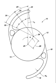
As shown in FIG. 1 an intraocular lens 10 includes an optic portion 12 having
a peripheral
edge 14 and at least two haptics 16, and each haptic 16 is integrated with the
peripheral
edge of the optic portion by a corresponding haptic integration region 18.
Each haptic 16
includes a distal segment region 20 and a deformation segment region 30. The
distal
segment region 20 has an outer distal length bound by a proximate endpoint 22
and a distal
endpoint 24. The distal segment region 20 is scribed by a distal segment angle
a of 20 to
30 having a segment origin 26 that lies within a radial segment bound by a
radial distance
28 of 1.5 mm to 1.9 mm from an optic center 25 and a segment angle 0 of from
30 to 45
from a vertical axis. As shown, the vertical axis Va extends through the
distal endpoint 24
of at least one haptic 16 and the optic center 25. As stated, the intraocular
lens 10 also
includes a deformation segment region 30 that has an outer deformation length
bounded by
a proximate deformation endpoint 32 and the proximate endpoint 22 of the
distal segment
region 20. The deformation segment region 30 is scribed by a deformation
segment angle (3
of 20 to 40 from the segment origin 26.
As stated, the at least two haptics 16 are integrated with the peripheral edge
14 of the optic
portion 12. The term "integrated" means that the haptics 16 can be formed with
the optic
portion 12 as a one-piece IOL. As an example, the IOL can be formed from a
polymeric
button. The polymeric button is then lathed to the exterior geometric shape of
the IOL
including the optic portion and haptics from a single polymeric material. The
term
"integrated" also means that the haptics 16 and optic portion 12 can be formed
of different
polymeric materials and then subsequently joined at the peripheral edge 14 of
the optic
portion 12. As an example, an IOL with an optic portion and two separately
formed haptics
that are subsequently joined is referred to in the art as a three-piece IOL.
Accordingly, one
of ordinary skill in the art understands the term "integrated" as referring to
either a one-
piece IOL formed and shaped from a single polymeric material or a multiple
piece IOL in
which the haptics are joined or attached to the optic , e.g., a three-piece
IOL, as just
described.
4
CA 02793844 2012-09-20
WO 2011/123224 PCT/US2011/027861
If the IOL is not formed or lathed from a single polymeric material the
haptics can be
joined to the optic portion by methods known in the art, for example as
described in U.S.
Pat. No. 5,217,491 to Vanderbilt. In some instances, three piece IOLs can
provide
additional functional design considerations. Typically, to facilitate movement
or warping
of the optic portion in response to the relaxation or the contraction of the
ciliary muscle
body in the eye, and therefore, in principle, provide a degree of lens
accommodation, one
can design an IOL with different polymeric materials. The design choice often
involves the
selection of a relatively stiff polymer for the haptics, e.g., a polyimide,
and a relatively soft
or compressible polymer for the optic portion.
One common shortcoming of hydrophobic acrylic IOLs presently available to
patients is
the relatively small angle of contact the haptics have with the lenticular
capsular bag. This
results in multiple problems encountered by the surgeon during implantation of
such a
lens. First, a relative stiff haptic that exhibits relatively localized
bending at a structurally
designed elbow can lead to the puncturing of the posterior capsule upon
implantation of
the IOL in the capsular bag. As shown in FIGS. 2A and 2B, the haptics 40
include a
structurally designed elbow 42. Following implantation of the IOL the
diametrically
inward force placed on the distal end 44 of the haptic by the capsular bag
causes the haptic
to bend predominantly at the designed elbow 42. The structured elbow design
limits the
degrees of mechanical freedom by which the haptic 40 can conform to the
spherical shape
of the capsular bag. The result is a relatively small angle of contact between
the haptic 40
and the capsular bag as shown in FIG. 8 and FIG. 9, respectively.
A second problem encountered by the surgeon is the localized stretching and
twisting of
the capsular bag that is believed to be caused by the stiff-arm haptics of the
prior art IOLs
of FIGS. 2A and 2B. This localized stretching and twisting can cause the
posterior capsule
to wrinkle, and the wrinkles can cause unwanted visual affects. In contrast,
the haptics of
the IOLs described herein are designed to conform to the shape of the capsular
bag, which
allows for a greater contact area between the haptics and the interior
perimeter surface of
the bag. The compression by the bag on the haptics is sufficient to stabilize
the IOL, yet
minimize localized stretching of the bag. Also, the delocalized contact
between the haptics
and the capsular bag minimizes the amount of uneven stretching of the bag
observed in the
prior art IOLs.
CA 02793844 2012-09-20
WO 2011/123224 PCT/US2011/027861
A third prior art hydrophobic IOL that is available to patients relies upon a
structural bend
in the haptic to essentially mimic the mechanical behavior of the structural
elbow present
in the two hydrophobic IOLs of FIGS. 2A and 2B. As shown in FIG. 3A and FIG.
3B a
connection portion 54 of haptic 50 that integrates with the optic 52 has a
relatively
symmetrical top profile and projects nearly perpendicular from the optic
peripheral edge
53. The haptic 50 then makes a sharp bend 56 and curves about the optic. In
fact, the angle
of contact for IOL of FIG. 3A is very similar to that of the IOL of FIG. 2A.
Applicants' IOL design provides a distinct shape or curvature to the haptics.
The design
provides a greater angle of contact between the haptics and the lenticular
capsular bag of a
human eye. As described, the haptics include a distal segment region and a
deformation
segment region. In a preferred embodiment, the haptics do not include a
structurally
designed elbow depicted in FIGS. 2A and 2B. Those in the art, however, do
understand
and recognize that an IOL of the invention can have a functional elbow, but
the design of
the elbow does not provide the lever-arm action exhibited by the haptics 40 of
the IOLs in
FIGS. 2A and 2B. By minimizing the functional importance of a structured elbow
in the
haptic design, Applicants essentially make available degrees of mechanical
freedom along
a greater contact length of the haptic that is not otherwise available with
the traditional
prior art designs. As a result, the haptics of the described IOLs will have a
relatively large
angle of contact between the haptics and the capsular bag. In most, if not
all, instances, the
described IOLs will have an angle of contact of not less than 500, or not less
than 52 and
not greater than 70 .
The design shape or curvature of the haptics in relation to the optic portion
is important in
establishing an angle of contact between the haptics and the interior
perimeter region of a
lenticular capsular bag in a human eye. By maximizing the angle of contact one
can
provide an implanted IOL with greater positional stability as well as with the
propensity of
the IOL to self-center within the capsular bag following implantation. An IOL
of the
invention that was implanted within the lenticular capsular bag of a human eye
is shown in
FIG. 4. As shown, the angle of contact between the haptics and the capsular
bag essentially
extends over the majority of the distal segment region and much of the
deformation
segment region of the haptics. One of skill would also notice from FIG. 4 a
very slight
stretching of the capsular bag that is in contact with the haptics, and that
the stretching of
the bag extends evenly over a greater haptic length - features that are not
observed in any
6
CA 02793844 2012-09-20
WO 2011/123224 PCT/US2011/027861
one of the prior art IOLs described. Again, by maximizing or extending the
angle of
contact over a greater area along the length of the haptics, Applicants have
designed an
IOL with greater positional stability and with an "auto-centering" feature not
found in
other IOLs to date.
In reference to FIG. 1, an unstressed IOL 10 can begin with the design of the
distal
segment region 20 of the haptics 16. The term "unstressed" IOL refers to an
IOL that does
not have any external compression or tension forces on the IOL. Essentially,
an unstressed
IOL can be described as an IOL placed on a bench top or stored in an IOL
package. The
distal segment region 20 is scribed by a distal segment angle a of 20 to 30
having a
segment origin 26. One vector of segment angle a extends from the segment
origin 26 to
the distal endpoint 24, and the corresponding vector extends to the proximate
endpoint 22.
Accordingly, the region of haptic 16 including and between these two endpoints
is the
distal segment region.
The segment origin 26 lies within a radial segment having a radial distance 28
of 1.5 mm
to 1.9 mm from an optic center. The optic center 25 is the center origin of
the optic portion
12 of an unstressed IOL. Referring to FIG. 5, the radial segment is bound by
segment
angles 01 and 02 of 30 and 45 from a vertical axis Va. As shown, the
vertical axis extends
through a distal endpoint 24 of a distal segment region 20 of at least one
haptic 16 and the
optic center 25. The described radial segment is also bound by radial
distances rl and r2.
The intraocular lens 10 also includes a deformation segment region 30 that is
bound by a
proximate deformation endpoint 32 on an outer surface of the haptic 16 and the
proximate
endpoint 22 of the distal segment region 20. The deformation segment region 30
is scribed
by a deformation segment angle (3 of 20 to 400 from the segment origin 26.
One vector of
segment angle P extends from the segment origin 26 to the proximate endpoint
24 of the
distal segment, and the corresponding vector extends to the proximate
deformation
endpoint 32.
In one embodiment, the segment origin lies within a radial segment having a
radial
distance r1 and r2 of 1.6 mm to 1.8 mm, respectively, from the optic center.
The radial
segment is also bound by segment angles 01 and 02 of 330 to 40 , respectively,
from the
vertical axis. Also, the distal segment angle a is from 200 to 30 , and the
deformation
segment angle P is from 20 to 32 .
7
CA 02793844 2012-09-20
WO 2011/123224 PCT/US2011/027861
In another embodiment, the segment origin lies within a radial segment having
a radial
distance r1 and r2 of 1.62 mm to 1.74 mm, respectively, from the optic center.
The radial
segment is also defined by segment angles 01 and 02 of 34 to 39 ,
respectively, from the
vertical axis. Also, the distal segment angle a is from 22 to 28 , and the
deformation
segment angle 0 is from 22 to 30 .
In still another embodiment, the segment origin lies within a radial segment
having a radial
distance r1 and r2 of 1.65 mm to 1.72 mm, respectively, from the optic center.
The radial
segment is also defined by segment angles 01 and 02 of 35 to 38 ,
respectively, from the
vertical axis. Also, the distal segment angle a is from 24 to 26 , and the
deformation
segment angle P is from 23 to 27 .
To best fit the inner perimeter surface of the lenticular capsular bag the
outer surface of the
distal segment region is preferably arcuate with a radius of curvature of 4.3
mm to 5.7 mm.
Likewise, the outer surface of the deformation segment region is preferably
arcuate with a
radius of curvature of 4.3 mm to 5.7 mm. Also, to maintain sufficient
flexibility to conform
to the inner perimeter surface of the capsular bag at least 80% of the distal
segment region
and at least 70% of the deformation segment region will preferably have a
constant width
Wh of from 0.25 mm to 0.65 mm, from 0.30 mm to 0.55 mm, or from 0.35 mm to
0.50
mm. Also, the polymeric material comprising the haptics will preferably have a
tangent
modulus of elasticity of from 2 MPa to 6 MPa. The crosssectional profile of
the haptics 16
can be any shape though a rectangular shape is preferred with Wh having the
shorter
dimension.
The term "tangent modulus of elasticity" refers to the slope of the stress-
strain diagram at
10% strain. Also, the methods and instruments used to determine a tangent
modulus of
elasticity for a particular material, particularly a polymeric material, is
well known and
understood by those in the art.
As stated, the haptic is designed to maximize the angle of contact between the
haptic,
particularly, the distal segment region and the distal portions of the
deformation segment
region, and the lenticular capsular bag. As shown in FIG. 6, corresponding
portions of the
distal segment region and deformation segment region combine to form a contact
region
with an angle of contact AC of not less than 50 with the lenticular capsular
bag of a
human eye when the haptics are stressed following implantation of the lens
into the
8
CA 02793844 2012-09-20
WO 2011/123224 PCT/US2011/027861
capsular bag. In one embodiment, the angle of contact AC is not less than 52
and not
greater than 700.
The angle of contact can be measured directly from a lens per the instructions
provided in
ISO 11979-3. FIGS. 7A and 7B are schematic representations of the mechanical
model
used to model the lenticular capsular bag of a human eye. If a physical lens
is not
available, the angle of contact can be calculated by using a finite element
model of
compression based on the same ISO standard. The finite element method is a
numerical
technique for finding approximate solutions of partial differential equations,
and several
commercially available software packages (ANSYSTM, AbaqusTM, NastranTM, etc.)
utilize
the finite element method to solve for strains incurred in solid bodies due to
applied loads
and constraints.
To determine the angles of contact for the prior art IOLs the software package
AbaqusTM has been utilized to approximate a solid model of an intraocular lens
with finite
elements and to apply compression of the outer diameter of the lens down to 10
mm per
ISO 11979-3. An image of the compressed lens is exported from AbaqusTM and
subsequently scaled and analyzed with a separate software package,
SolidWorksTM. The
intersection points of the outer haptic edge and an arc offset 0.05 mm from
the outer 10
mm compression fixture are located. The angular distance between these two
points is then
measured; this value represents the contact angle of the lens. In creating the
finite element
model of a given intraocular lens, e.g., a prior art IOL, the lens geometry
can be obtained
from issued patents containing graphics and dimensional details or by
measurements taken
directly from the lens.
The angles of contact for each of the prior art IOLs described herein are
determined using
the model described. The angle of contact in each of the prior art IOLs
described is less
than about 48 . As indicated in FIGS. 9, 10 and 11, the angle of contact for
the prior art
IOLs of FIGS, 2A, 2B and 4 were determined using the mathematical software
programs
identified above. The three described prior art IOLs are all one-piece IOLs
formed from a
single hydrophobic copolymer. The term "hydrophobic copolymer" is defined as a
copolymer with an equilibrated water content of less than 5% by weight. A
fourth prior art
IOL depicted in FIG. 11 is formed of a hydrophilic copolymer with a water
content of
about 28% by weight. The angle of contact for each of the prior art IOLs are
listed in Table
I below.
9
CA 02793844 2012-09-20
WO 2011/123224 PCT/US2011/027861
Table 1. Prior Art IOLs: Angle of Contact
FIGS. 2A/8 FIGS. 213/9 FIGS. 4/10 FIG. 11
44.6 38.10 42.8 48.3
As one might expect, the physical contact between the haptics and the inner
perimeter
surface of the capsular bag following implantation of an IOL of the invention
causes the
haptics, particularly, the distal segment region of the haptics, to move
inward toward the
optic center of the IOL. In fact, once implanted in the capsular bag of a
human eye the
segment origin translates to a point within a radial distance of 0.3 mm or
less from the
optic center. Ideally, the segment origin would actually translate to the
optic center. In any
case, the segment origin moves inward toward the optic center following
implantation of
the IOL to a radial distance of 0.3 mm or less, 0.2 mm or less or 0.1 mm or
less, from the
optic center.
In reference to the model used to determine angles of contact, as the distal
segment region
of the haptics moves inward toward the optic center as the arcuate jaws close
upon the IOL
the IOL the segment origin translates to a point within a radial distance of
0.3 mm or less
from the optic center. Ideally, the segment origin would actually translate to
the optic
center. In any case, the segment origin moves inward toward the optic center
following
implantation of the IOL to a radial distance of 0.3 mm or less, 0.2 mm or less
or 0.1 mm or
less, from the optic center, in accordance with ISO Test No. 11979-3(2006).
In one embodiment, and as shown in FIG. 12A, the optic portion of IOL 60
includes an
anterior face with a central optical zone 63, a peripheral zone 64 entirely
surrounding the
optical zone, a recessed annular zone 65 that is disposed posterior to the
peripheral zone 64
and an optic peripheral edge 69. The IOL shown in FIG. 12A also includes an
integration
region of a haptic 66 having an opening 75 thereby forming an outer
integration member
76 and an inner integration member 78. The haptic 66 of IOL 60 will exhibit an
angle of
contact of not less than 50 , or not less than 52 and not greater than 70 ,
with the lenticular
capsular bag of a human eye when the haptic is stressed following implantation
of the lens
into the capsular bag. FIG. 12B is a cross-sectional view of the optic portion
of the IOL of
FIG. 12A showing the anterior face with the central optical zone 63,
peripheral zone 64,
and the recessed annular zone 65. The posterior optical zone 68 is also shown.
FIG. 12C is
a posterior view of the IOL of FIG. 12A showing the posterior optical zone 68.
CA 02793844 2012-09-20
WO 2011/123224 PCT/US2011/027861
As shown in FIG. 13, in addition to the anterior optical features described
and shown in
FIGS. 12A, 12B and 12C, an IOL 80 includes a posterior optical zone 82, and
the at least
two haptics 86 can include a distal posterior face 87, a proximal posterior
face 88, and a
step face 85 disposed at a boundary therebetween. The distal posterior face 88
may be
substantially perpendicular to the optical axis or disposed at an angle
relative to a plane
perpendicular to the optical axis. The haptics 86 of IOL 80 will exhibit an
angle of contact
of not less than 50 , or not less than 52 and not greater than 70 , with the
lenticular
capsular bag of a human eye when the haptics are stressed following
implantation of the
lens into the capsular bag.
In one embodiment, the optic portion of each lens will have an optic diameter
from 5.7 mm
to 4.5 mm. The optic diameter of the IOL will of course depend upon the
optical power of
the lens. IOLs of less optical power, e.g., a 15 diopter lens, will typically
have a larger
optic diameter than an IOL with greater optical power, e.g., a 30 diopter
lens. A given
range of exemplary optic diameters for the IOLs is provided in Table 2.
Table 2.
power optic diameter power optic diameter
(diopter) (mm) (diopter) (mm)
15-18 5.7-5.1 21-26 5.4-4.5
17-23 5.5-4.8 24-30 5.1-4.5
In one embodiment, the optic portion of each IOL will have a center thickness
that deviates
no more than 15% from an average center thickness across an optical power
range of from
18 to 23 diopters. Also, the cross-sectional area of any one optic portion
deviates no more
than 6%, or no more than 4%, from an average cross-sectional area across the
optical
power range of 18 to 23 diopters. An exemplary sized optic portion of an IOL
with a
refractive power of 19 diopters to 22 diopters will have an optic center
thickness of 0.50
mm to 0.60 mm and a nominal cross-sectional area of 2.1 mm2 to 2.7 mm2.
The optic portion of the intraocular lens can also include a toric optical
zone (commonly
referred to as a "toric IOL"). A toric IOL can provide the functional
requirements of a lens
as well as correct refractive abnormalities of an eye associated with corneal
astigmatism.
The tonic optical zone provides cylindrical correction to compensate for
astigmatism. Also,
11
CA 02793844 2012-09-20
WO 2011/123224 PCT/US2011/027861
because astigmatism is usually associated with other refractive abnormalities,
such as
myopia (nearsightedness) or hypermetropia (farsightedness), toric IOLs can be
prescribed
with a spherical correction to correct myopic astigmatism or hypermetropic
astigmatism.
A tonic optical surface is formed on either the posterior lens surface (to
achieve a "back
surface tonic IOL") or the anterior lens surface (to form a "front surface
toric IOL").
The positional stability and self-centering characteristic of the described
IOLs becomes
very important in the designing of a tonic IOL. As stated, the unique
stabilizing features
provided by the haptics inhibit rotation of the lens within the capsular bag
following
implantation. In other words, following proper axial alignment of a toric IOL
by a surgeon
the cylindrical axis of the toric zone remains generally aligned with the axis
of the
astigmatism years after implantation.
A prescription for a toric IOL will typically specify refractive power, and
spherical
correction and cylindrical correction in relation to a base axis. Toric IOLs
prescriptions are
generally offered in 5 or 10 increments ranging from 0 to 1800. The tonic
optical zone is
easily machined into the posterior or anterior surface of the optic portion of
the IOL using
methods well known in the optical art. Also, those of ordinary skill in the
art would know
how to diagnose and prescribe a toric IOL according to the specific visual
corrective needs
of each patient.
One advantage of having an opening 75 in at least one haptic 77 depicted in
FIG. 8 is that
the surgeon can easily rotate the IOL following the implantation of the lens
by positioning
a rotating tool into the opening and rotating the lens within the capsular bag
to the proper
axial alignment. Again, the positional stability and self-centering
characteristics of the
described IOLs makes this possible and quite feasible.
One embodiment is directed to an intraocular lens comprising an optic portion
and two
haptics, and each haptic is integrated to a peripheral edge of the optic
portion by a haptic
integration region. The optic portion, the two haptics and the haptic
integration regions are
each formed of a hydrophobic polymeric material having a tangent modulus of
elasticity of
from 2 Mpa to 6 MPa. Each of the haptics comprise a distal segment region and
a
deformation segment region. The distal segment region has an outer distal
length bounded
by a proximate and a distal endpoint on an outer surface of the haptic, and is
scribed by a
distal segment angle a of 20 to 30 . The distal segment angle has a segment
origin that
12
CA 02793844 2012-09-20
WO 2011/123224 PCT/US2011/027861
extends a radial distance of 1.5 mm to 1.9 mm from an optic center and a
segment angle 0
of from 34 to 400 from a vertical axis. The vertical axis extends through the
distal
endpoint of at least one of the two haptics and the optic center. The
deformation segment
region has an outer deformation length bounded by a proximate deformation
endpoint on
an outer surface and the proximate endpoint of the corresponding distal
segment region,
and is scribed by a deformation segment angle 0 of 20 to 40 from the segment
origin.
The Optical, Polymeric Materials
The haptic design principles described above can be applied to a wide variety
of optical
polymeric materials. Non-limiting examples of such materials include those
known to be
used in IOLs. For example, the method of the present invention can be applied
to siloxy-
containing copolymers, acrylic copolymers, hydrophilic copolymers or
hydrophobic
copolymers. The terms polymer and copolymer are used interchangeably, and it
is well
understood by those of skill in the art that a copolymer is prepared from more
than one
monomeric component.
In one embodiment, a copolymeric material that can be used to make an IOL
described
herein will be hard enough to machine at room temperature, and one which is
foldable
through a controlled hydrating process. The IOL may be hydrated to a suitably
flexible
state with minimal water uptake. The relatively low water uptake allows
efficient hydration
without affecting mechanical or optical properties and results in little, if
any, change in the
dimensions or the refractive index of the lens. One exemplary copolymer with
the features
or properties described can include: a) a first monomeric component that is
selected from
an aryl acrylate or an aryl methacrylate; b) a second monomeric component
which is a
monomer having an aromatic ring with a substituent having at least one site of
ethylenic
unsaturation, and c) a third monomeric component which is a high water content
hydrogel-
forming monomer. The copolymer can further include a crosslinking agent.
The IOLs described can include a copolymer comprising: a) at least about 20%
by weight
of a first monomeric component selected from the group consisting of ethylene
glycol
phenyl ether acrylate and polyethylene glycol phenyl ether acrylate; b) at
least about 10%
by weight of a second monomeric component selected from the group consisting
of
substituted styrene and unsubstituted styrene; c) at least about 10% by weight
of a third
monomeric component selected from the group consisting of hydroxy ethyl
methacrylate,
13
CA 02793844 2012-09-20
WO 2011/123224 PCT/US2011/027861
hydroxyethoxy ethyl methacrylate, and methacrylic acid; d) less than about 10%
by weight
percent of a crosslinking agent selected from the group consisting of a
diacrylate and a
dimethacrylate. Also, the copolymer will have a refractive index greater than
about 1.50
and is foldable at normal room temperature when hydrated, and is machinable at
about
room temperature when dry.
In another embodiment, the intraocular lens comprises a cured copolymer that
is prepared
from a cationically polymerizable, branched alkene monomer, and a monomer that
includes a pendent benzocyclobutene group (herein called "BCB group"), The
cationically
polymerizable branched alkene monomer preferably contains a tertiary carbon on
the vinyl
group in the alkene. As known by those of skill in the art, tertiary
carbocations are
relatively stable due to the electron-density of the surrounding carbons that
stabilize the
positive charge of the cation. Polyisobutylene, a preferred branched alkene
monomer, as
discussed above, is an example of an alkene monomer polymerizable by cationic
chemical
means that contains a tertiary carbon. Molecules such as propene contain
secondary
carbons at the vinyl group and, as known by those of skill in the art, are not
canonically
polymerized.
Due to the strained four-membered ring, the BCP group is converted to o-
xylylene at
temperatures greater than 180 C. At such elevated temperatures, the BCB group
undergoes Diels-Alder reactions with dienophiles to form a six-membered ring,
or reacts
with itself to form an eight-membered ring. Polymers containing multiple
pendant BCB
groups per molecular chain can be thermally crosslinked with or without
dienophiles. Each
crosslink consists of a ring structure of carbon-carbon bond, which is more
thermally
stable than the sulfur bridge in vulcanized polymers and is stronger than the
Si--O bond in
silicone copolymers. The BCB crosslinking only involves heat. As long as the
polymer is
stable at the crosslinking temperature, there is no toxic chemical involved in
order to from
a cured crosslinked copolymer.
The monomer having a BCB group can be any monomer containing at least one BCB-
functional moiety. It is preferred that the monomer be cationically
polymerizable and be
compatible with the branched alkene monomer. In one embodiment, the monomer
having a
BCB group has the formula
14
CA 02793844 2012-09-20
WO 2011/123224 PCT/US2011/027861
R n \ /
wherein R is hydrogen or an alkyl group and n is an integer selected from 0,
1, 2
or 3. Suitable monomers having a BCB group include 4-vinylbenzocyclobutene,
4-(a-alkylvinyl)benzocyclobutenes such as 2-(4-benzocyclobutenyl)-propene and
2-(4-benzocyclobutenyl)-1-butene, and 4-(2-methyl-alkenyl)benzocyclobutenes
such as
2-methyl-3-(4-benzocyclobutenyl)-1-propene and 2-methyl-4-(4-
benzocyclobutenyl)-1-
butene.
Like the branched alkene, the monomer having a pendant benzocyclobutene (BCB)
group
should also be cationically polymerizable. Olefins having a secondary carbon
on the vinyl
group are cationically polymerizable in instances when the electronegativity
of the
aromatic ring adjacent to the vinyl group can stabilize the carbocation. Thus,
monomer
olefins such as 4-vinylbenzocyclobutene can be cationically polymerized, and
easily
incorporated into the polymer simply by titrating it into the reaction during
its
polymerization. This is different than, for example, the allyl-BCB, which
cannot be added
to a cationic polymerization, in part because the aromatic ring in the BCB is
not adjacent to
the vinyl group.
BCB-type monomers having and a tertiary carbon on the vinyl group are also
suitable for
cationic polymerization even if the vinyl group is not adjacent to the
aromatic ring of the
BCB. Tertiary carbons, which become quaternary carbons during polymerization,
are
stabilized by the electronegativity of the surrounding carbons. Therefore,
monomers
having tertiary carbons on the vinyl carbons can be incorporated into a
cationic
polymerized reaction much in the same manner as the alkene having a tertiary
carbon is
incorporated. 2-Methyl-3-(4-benzocyclobutenyl)-propene is an example of this
type of
compound. Also preferred are monomers that draw on the electronegativity of
both the
surrounding carbons and the aromatic ring, for example 2-(4-benzocyclobutenyl)-
propene.
These type of monomers will cationically polymerize as they are stabilized
both by the
methyl group (as in this case of 2-(4-benzocyclobutenyl)-propene) and the
aromatic ring.
In another embodiment, the optical polymeric material can be prepared as a
copolymer
from at least three monomeric components. The first monomeric component is
present in
the copolymer in an amount of at least 70% by weight, and its homopolymer will
have a
CA 02793844 2012-09-20
WO 2011/123224 PCT/US2011/027861
refractive index of at least 1.50, preferably at least 1.52 or at least 1.54.
The second
monomeric component is present in the copolymer in an amount from 3% to 20% or
from
3% to 10%, by weight, and its homopolymer will have a glass transition
temperature of
less than about 300 C, preferably less than about 220 C. The first and
second monomeric
components together represent at least 80% by weight of the copolymer. The
term
"homopolymer" refers to a polymer that is derived substantially completely
from the
respective monomeric component. Minor amounts of catalysts, initiators and the
like can
be included, as is conventionally the case, in order to facilitate the
formation of the
homopolymer.
Non-limiting examples of first and second monomeric components include
polymers
comprising units of C1-Clo alkyl methacrylates (e.g., methyl methacrylate,
ethyl
methacrylate, propyl methacrylate, butyl methacrylate, octyl methacrylate, or
2-ethylhexyl
methacrylate; preferably, methyl methacrylate to control mechanical
properties), C1-C10
alkyl acrylates (e.g., methyl acrylate, ethyl acrylate, propyl acrylate, or
hexyl acrylate;
preferably, butyl acrylate to control mechanical properties), C6-C40 arylalkyl
acrylates
(e.g., 2-phenylethyl acrylate, benzyl acrylate, 3-phenylpropyl acrylate, 4-
phenylbutyl
acrylate, 5-phenylpentyl acrylate, 8-phenyloctyl acrylate, or 2-phenylethoxy
acrylate;
preferably, 2-phenylethyl acrylate to increase refractive index), and C6-C24
arylalkyl
methacrylates (e.g., 2-phenylethyl methacrylate, 3-phenylpropyl methacrylate,
4-
phenylbutyl methacrylate, 2-phenoxyethyl methacrylate, 3,3-diphenylpropyl
methacrylate,
2-(1-naphthylethyl)-methacrylate, benzyl methacrylate, or 2-(2-naphthylethyl)
methacrylate.
Particularly useful first monomeric components include styrene, vinyl
carbazole, vinyl
naphthalene, benzyl acrylate, phenyl acrylate, 2-phenoxyethyl acrylate, 2-
phenoxyethyl
methacrylate and mixtures thereof. Particularly useful second monomeric
components
include n-butyl acrylate, n-hexyl acrylate, 2-ethylhexyl acrylate, 2-
ethoxyethyl acrylate and
mixtures thereof. The third monomeric component is best described as a cross-
linking
monomeric constituent that can form crosslinks with the first or the second
monomeric
components. Preferably, the cross-linking monomeric component is multi-
functional and
can chemically react with both the first and second monomeric components.
Preferably, the
third component is present in the copolymer in an amount of less than about 3%
by weight
of the copolymer. Examples of useful crosslinking monomeric components include
16
CA 02793844 2012-09-20
WO 2011/123224 PCT/US2011/027861
ethylene glycol dimethacrylate, propylene glycol dimethacrylate, ethylene
glycol diacrylate
and the like and mixtures thereof.
The copolymer can further include a fourth component derived from a
hydrophilic
monomeric component. This fourth component is present in an amount, from 2% to
10%
by weight of the copolymer. At times, and depending upon the hydrophobic lens
material,
the addition of a hydrophilic monomeric component can reduce the formation of
water
vacuoles, which can scatter light and cause what is referred to in the art as
"glistenings".
In another embodiment, the optical, polymeric materials can also be prepared
from
monomers having the formula:
R
H2C =C -'~ -O -~C H2}-Y -Ar
M
wherein: R is H or CH3 ; M JS 0-10;
Y is nothing, 0, S, or NR wherein R is H, CH3, Cõ H2õ+1 (n=1-10), iso OC3H7,
phenyl or benzyl; Ar is any aromatic ring, such as benzene, which can be
unsubstituted
or substituted with H, CH3, C2H5, n-C3H7, iso-C3H7, OCH3, CoH11, Cl, Br,
phenyl or
benzyl; and a cross-linking monomer having a plurality of polymerizable
ethylenically
unsaturated groups. The optical material will have a glass transition
temperature not
greater than 37 C and an elongation of at least 150%.
Exemplary monomers include, but are not limited to: 2-ethylphenoxy
methacrylate, 2-
ethylphenoxy acrylate, 2-ethylthiophenyl methacrylate, 2-ethylthiophenyl
acrylate, 2-
ethylaminophenyl methacrylate, phenyl methacrylate, benzyl methacrylate, 2-
phenylethyl
methacrylate, 3-phenylpropyl methacrylate, 4-phenylbutyl methacrylate, 4-
methylphenyl
methacrylate, 4-methylbenzyl methacrylate, 2-2-methylphenylethyl methacrylate,
2-3-
methylphenylethyl methacrylate, 2-4-methylphenylethyl methacrylate, 2-(4-
propylphenyl)ethyl methacrylate, and the like, including the corresponding
methacrylates
and acrylates.
The aryl acrylate/methacrylate based optical materials will generally comprise
a greater
mole percent of acrylate ester residues than of methacrylate ester residues.
It is preferred
that the aryl acrylate monomers constitute from about 60 mole percent to about
95 mole
percent of the polymer, while the aryl methacrylate monomers constitute from
about 5
17
CA 02793844 2012-09-20
WO 2011/123224 PCT/US2011/027861
mole percent to about 40 mole percent of the polymer. Most preferred is a
polymer
comprising about 60-70 mole percent 2-phenylethyl acrylate and about 30-40
mole percent
2-phenylethyl methacrylate.
In yet another embodiment, the optical, polymeric material can also be
prepared by
polymerizing the following monomeric components: (A)5-25% by weight of
acrylate
represented by the general formula
H
H2C=C-C -O--~CH2}-X-Ar
M
wherein Ar represents an aromatic ring of which hydrogen atom may be
substituted by a substitutional group, X represents an oxygen atom or a direct
bonding,
and in represents an integer of 1 to 5; (B)50 to 90% by weight of 2-
hydroxyethyl
(meth)acrylate; and (C) 5 to 45% by weight of a (meth)acrylate monomer though
not of
the formula that represent monomer (A) and not 2-hydroxyethyl (meth)acrylate.
Also,
the coefficient of water absorption of the homopolymer of monomer (C) is not
more than
30% by weight.
In the present invention the coefficient of water absorption is defined as the
following
equation: water absorption (%wt) = (W-Wo)/Wo x 100 wherein the value is
calculated at
25 C by using the specimen having 1 mm thickness at cutting, W represents a
weight of
the specimen in equilibrium state of water, and Wo represeents a weight of the
specimen in
a dry state.
In each of the embodiments above, the optical, polymeric materials are
prepared by
generally conventional polymerization methods from the respective monomeric
components.
18