Note: Descriptions are shown in the official language in which they were submitted.
CA 02794721 2012-09-26
Page 1 of 59
DESCRIPTION
TITLE OF THE INVENTION: ASSAY UTILIZING
IMMUNOCHROMATOGRAPHY, IMMUNOCHROMATOGRAPHIC TEST STRIP,
AND ASSAY REAGENT KIT FOR IMMUNOCHROMATOGRAPHY
TECHNICAL FIELD
[0001]
The present invention relates to a measurement method of measuring an
analyte in a sample by immunochromatography, an immunochromatographic test
strip
for measuring an analyte in a sample by immunochromatography, and a
measurement
reagent kit for immunochromatography including the immunochromatographic test
strip
and a diluting solution. The present invention particularly relates to a
technique of
measuring hemoglobin concentration and the concentration of a second analyte
in the
same sample derived from blood by using immunochromatography and correcting
the
measurement value of the second analyte with a hematocrit value obtained from
a
measurement value of hemoglobin.
BACKGROUND ART
[0002]
In the medical field, the medical research field etc., certain components in
blood (such as serum albumin, immunoglobulin, hepatitis virus, rheumatoid
factor, and
C-reactive protein) must be measured and various measurement methods have been
developed and implemented for this purpose. In frequently used methods among
these
methods, immunological assay is becoming main stream and, in the immunological
assay, blood collected and acquired from a patient or a subject (hereinafter
referred to as
whole blood) is centrifuged to acquire supernatant (serum or plasma), which is
diluted
with an appropriate buffer solution, and an antibody that specifically reacts
with an
analyte is used for detecting the analyte in the serum or plasma. Such assays
include a
method based on single radial immunodiffusion using a polyclonal antibody, as
CA 02794721 2012-09-26
Page 2 of 59
qualitative testing. Latex agglutination immunoassay and an
immunoturbidimetric
method are included as representative quantitative testing.
[0003]
The needs of "wanting to perform various tests during examination of a
patient" are recently increasing even in clinics and small hospitals and the
tests are
increasingly performed as point-of-care testing (POCT) instead of conventional
subcontract testing. The representative examples of such POCT reagents include
an
immunochromatographic lateral flow test strip. Since an operation of
separating plasma
and serum from blood is cumbersome and requires skill in the POCT field,
testing using
whole blood is desired. An assay of measuring an analyte in a whole blood
sample by
using immunochromatography is disclosed as, for example, a method as well as a
reagent and a kit using a test strip fitted with a blood cell separation
membrane (Patent
Document 1). However, when plasma separated by this procedure is directly used
for
measurement by immunochromatography based on the principle of sandwich-type
immune reaction, if the analyte is excessively present, it is problematic that
a "hook
phenomenon" (also referred to as a "prozone phenomenon") occurs and causes
apparent
reduction in value although the highly-concentrated analyte is present in the
sample.
Therefore, whole blood is normally hemolyzed and diluted for measurement so as
to
remain within a predetermined measurement range. However, in this case, the
concentration of the analyte is diluted by the blood cell volume and the
measurement
value is lowered as compared to the case of serum or plasma samples and,
therefore, the
measurement value must be corrected by using a hematocrit value (volume
percent of
red cell). The correction of the measurement value of an analyte using a
hematocrit
value (volume percent of red cell) will hereinafter also simply be referred to
as
hematocrit correction.
[0004]
The problem of the apparent reduction in value is normally corrected by
performing uniform multiplication by a correction coefficient acquired from an
average
hematocrit value of healthy individuals. However, the hematocrit value varies
among
individuals and the reference value thereof ranges from 39 to 52 % in men and
35 to
CA 02794721 2012-09-26
Page 3 of 59
48 % in women. Therefore, an accurate analyte concentration cannot be acquired
by the
correction using multiplication by a uniform coefficient. Therefore, to
perform accurate
hematocrit correction, the correction must be performed with a hematocrit
value
separately measured by using the same sample used for measuring the analyte
concentration.
[0005]
The hematocrit value is conventionally obtained by a microhematocrit method
based on a centrifuging method or by using an automated hemocytometer through
calculations from the number of red blood cells and an average red blood cell
volume.
On the other hand, another method using conversion from a measurement result
of
whole blood into a measurement value in the case of measuring serum or plasma
is
reported as a method of converting blood measurement result and, in this
method, a
hemoglobin concentration (g/L) in whole blood is measured; a numerical value
obtained
by multiplying the acquired hemoglobin concentration by about 3/10 is adopted
as a
hematocrit value (%); and the hematocrit value is used for converting a
measurement
result from whole blood into a measurement value in the case of measuring
serum or
plasma (Patent Document 2). However, since the hemoglobin concentration and
the
hematocrit value must be obtained by a method different from an
immunochromatographic assay, this method is cumbersome, requires time and
cost, and
therefore cannot satisfy needs of testing in the POCT field.
[0006]
Therefore, a method of measuring an analyte and hemoglobin at the same time
based on immunochromatography is desired; however, since the hemoglobin
concentration in whole blood is normally several dozen g/L to 200 g/L, which
is very
high concentration, 10,000- to 100,000-times dilution is required for an assay
based on
the principle of a normal sandwich-type immunoassay. In this method, a large
amount
of a diluting solution is required for performing the dilution at one step,
leading to
deterioration of measurement accuracy. A method using multistep dilution
problematically lacks practicality for a testing method in the POCT field. To
solve these
CA 02794721 2012-09-26
Page 4 of 59
problems, desired is an immunochromatographic assay that is capable of
measurement,
with a hemolytic dilution operation of whole blood by a factor of at most 50
to 400.
CITATION LIST
PATENT LITERATURE
[0007]
Patent Document 1: Published PCT Application WO 2010/001598
Patent Document 2: Japanese Laid-Open Patent Publication No. 2001-272403
SUMMARY OF INVENTION
TECHNICAL PROBLEM
[0008]
For example, a blood concentration of C-reactive protein (hereinafter also
referred to as "CRP") is equal to or less than 3 mg/L in healthy individuals,
at most
about 35 mg/L after the operation or in the case of acute bacterial infection,
and up to
about 1000 mg/L in severely injured individuals. On the other hand, a blood
concentration of hemoglobin is normally several dozen g/L to 200 g/L, which is
different in concentration by a factor of about 50 to 67000 from CRP, and is
very high
concentration for an analyte subjected to an immunoassay. Therefore, when it
is
attempted to measure the both of CRP and hemoglobin in an assay based on the
principle of sandwich-type immunochromatography, the dilution rate of a sample
must
considerably be changed depending on the analyte and, therefore, measurement
in the
same sample (sample at the same dilution rate) is very difficult.
[0009]
The greatest factors prescribing a measurement range of an immunoassay
include avidity between antigen and antibody and various environmental factors
affecting the binding reaction thereof. These environmental factors include
temperature,
time, pH, ion environment, and addition of agents producing certain effects
such as
surfactants and reaction accelerators. The measurement of the concentrations
of CRP
and hemoglobin at the same time in the same sample can be enabled in principle
by
CA 02794721 2012-09-26
Page 5 of 59
selecting antibodies having different avidities and achieving appropriate
environmental
factors such that the significant concentration gap is filled. However, it is
not easy to
prepare such a combination of antibodies and to achieve the appropriate
environmental
factors.
[0010]
In fact, when the present inventors initially considered the simultaneous
measurement of CRP and hemoglobin by sandwich type immunochromatography, 50-
to 200-fold dilution was sufficient for measuring CRP, while 2000- to 100,000-
fold
dilution was required for measuring hemoglobin. Therefore, it has been unable
to
measure CRP and hemoglobin by using the same diluted sample.
[0011]
An object of the present invention is to provide a method of measuring
concentrations of an analyte and hemoglobin in the same sample by
immunochromatography by using the same diluted sample and a method of
performing
hematocrit correction of a measurement value of the analyte by using a
measurement
value of hemoglobin and to provide a test strip and a reagent kit for
immunochromatography used in these methods.
SOLUTION TO PROBLEM
[0012]
The present inventors have found that the quantity of hemoglobin can be
determined by an immunochromatographic test strip by utilizing a phenomenon in
which the intensity of reflected light is reduced, with increasing
concentration of
hemoglobin, in colloidal gold label captured by a line of anti-hemoglobin
antibody
immobilized on an insoluble membrane support of the immunochromatographic test
strip, even in a sample obtained by diluting and hemolyzing blood by a facer
of about
100, i.e., even within the range of hemoglobin concentrations in the sample
equal to or
greater than the concentration at which the maximum signal value of hemoglobin
measurement is obtained (hereinafter also referred to as a prozone phenomenon
region),
CA 02794721 2012-09-26
Page 6 of 59
and that CRP can be measured in the diluted sample, leading to the completion
of the
present invention.
The "immunochromatographic test strip (test strip for
immunochromatography)" refers to those including at least an insoluble
membrane
support necessary for immunochromatography and further including a reagent
component, another membrane, etc., as needed.
[0013]
The present invention comprises the following.
(1) A method of measuring a sample containing at least the first analyte by
immunochromatography including the following step A, wherein the concentration
of
the first analyte in the sample is measured in the range of concentrations
equal to or
greater than the concentration at which the maximum signal value of the first
analyte is
acquired (prozone phenomenon region),
A. the step of measuring the first analyte in the sample by competitive
immunochromatography using 1) and 2) described below:
1) a conjugate in which a first antibody against the first analyte is
immobilized
to a label; and
2) an insoluble membrane support to which a second antibody against the first
analyte is immobilized (if an epitope of the first antibody against the first
analyte is
monovalent, an epitope of the second antibody is different from the first
antibody, while
if an epitope of the first antibody is multivalent, an epitope of the second
antibody may
be the same as the first antibody or the first antibody may be the same as the
second
antibody in some cases).
(2) The method of (1), wherein step A further uses 3) and 4) described below
and wherein the conjugate of 1) is contained in a pad to form a conjugate pad
and
disposed on the upstream side of the insoluble membrane support of 2),
3) a sample pad located on the upstream side of the conjugate pad and supplied
with a sample, and
4) an absorbent pad located on the downstream side of the insoluble membrane
support of 2).
CA 02794721 2012-09-26
Page 7 of 59
(3) The method of (1) or (2), wherein the first analyte is hemoglobin, and
wherein the sample is obtained by diluting and hemolyzing blood into the range
of
concentrations equal to or greater than the range of concentrations at which
the
maximum signal value of hemoglobin measurement is acquired (prozone phenomenon
region).
(4) A method of measuring a sample containing at least hemoglobin, which is
the first analyte, and the second analyte by immunochromatography including
the
following steps A to D:
A. the step of measuring the first analyte in the sample by competitive
immunochromatography using 1) and 2) described below,
1) a conjugate in which a first antibody against the first analyte is
immobilized
to a label, and
2) an insoluble membrane support to which a second antibody against the first
analyte is immobilized (if an epitope of the first antibody against the first
analyte is
monovalent, an epitope of the second antibody is different from the first
antibody, while
if an epitope of the first antibody is multivalent, an epitope of the second
antibody may
be the same as the first antibody or the first antibody may be the same as the
second
antibody in some cases);
B. the step of measuring the second analyte in the sample by
immunochromatography using 5) and 6) described below,
5) a conjugate in which a first antibody against the second analyte is
immobilized to a label, and
6) an insoluble membrane support to which a second antibody against the
second analyte is immobilized (if an epitope of the first antibody against the
second
analyte is monovalent, an epitope of the second antibody is different from the
first
antibody, while if an epitope of the first antibody is multivalent, an epitope
of the
second antibody may be the same as the first antibody or the first antibody
may be the
same as the second antibody in some cases);
C. the. step of obtaining the hematocrit value of the sample from the
measurement value of the first analyte acquired at step A; and
CA 02794721 2012-09-26
Page 8 of 59
D. the step of correcting the measurement value of the second analyte acquired
at step B by using the hematocrit value acquired at step C.
(5) The method of (4), wherein steps A and B are steps using the same
insoluble membrane support.
(6) The method of (5), wherein steps A and B are performed in the same flow
passage.
(7) The method of any one of (4) to (6), wherein the second analyte is CRP.
(8) An immunochromatographic test strip for measuring a sample containing at
least the first analyte and the second analyte by immunochromatography, said
immunochromatographic test strip comprising the following E and F:
E. a test strip for measuring the first analyte including 1) and 2) described
below,
1) a conjugate in which a first antibody against the first analyte is
immobilized
to a label, and
2) an insoluble membrane support to which a second antibody against the first
analyte is immobilized (if an epitope of the first antibody against the first
analyte is
monovalent, an epitope of the second antibody is different from the first
antibody, while
if an epitope of the first antibody is multivalent, an epitope of the second
antibody may
be the same as the first antibody or the first antibody may be the same as the
second
antibody in some cases); and
F. a test strip for measuring the second analyte including 5) and 6) described
below,
5) a conjugate in which a first antibody against the second analyte is
immobilized to a label, and
6) an insoluble membrane support to which a second antibody against the
second analyte is immobilized (if an epitope of the first antibody against the
second
analyte is monovalent, an epitope of the second antibody is different from the
first
antibody, while if an epitope of the first antibody is multivalent, an epitope
of the
second antibody may be the same as the first antibody or the first antibody
may be the
same as the second antibody in some cases).
CA 02794721 2012-09-26
Page 9 of 59
(9) The immunochromatographic test strip of (8), wherein the conjugates of 1)
of E and 5) of F are contained in the same pad to form a conjugate pad, and
wherein the
insoluble membrane support of 2) of E is the same as the insoluble membrane
support
of 5) of F.
(10) The immunochromatographic test strip of (9), wherein E and F are
disposed in the same flow passage.
(11) The immunochromatographic test strip of any one of (8) to (10), further
comprising the following G and H, wherein the conjugates of 1) of E and 5) of
F are
contained in a pad to form a conjugate pad and disposed on the upstream side
of the
insoluble membrane support of 2) of E and 6) of F,
G. a sample pad located on the upstream side of the conjugate pad and supplied
with a sample, and
H. an absorbent pad located on the downstream side of the insoluble membrane
support of 2) of E and 6) of F.
(12) The immunochromatographic test strip of any one of (8) to (11), wherein
the first analyte is hemoglobin.
(13) The immunochromatographic test strip of (12), wherein the second analyte
is CRP.
(14) An assay reagent kit for immunochromatography comprising: the
immunochromatographic test strip of (12) or (13); and a diluting solution for
hemolysis
and dilution of hemoglobin.
(15) A method of measuring a sample containing at least the first analyte and
the second analyte by immunochromatography including the following steps A and
B:
A. the step of measuring the first analyte in the sample by competitive
immunochromatography using 1) and 2) described below,
1) a conjugate in which a first antibody against the first analyte is
immobilized
to a label, and
2) an insoluble membrane support to which a second antibody against the first
analyte is immobilized (if an epitope of the first antibody against the first
analyte is
monovalent, an epitope of the second antibody is different from the first
antibody, while
CA 02794721 2012-09-26
Page 10 of 59
if an epitope of the first antibody is multivalent, an epitope of the second
antibody may
be the same as the first antibody or the first antibody may be the same as the
second
antibody in some cases); and
B. the step of measuring the second analyte in the sample by
immunochromatography using 5) and 6) described below,
5) a conjugate in which a first antibody against the second analyte is
immobilized to a label, and
6) an insoluble membrane support to which a second antibody against the
second analyte is immobilized (if an epitope of the first antibody against the
second
analyte is monovalent, an epitope of the second antibody is different from the
first
antibody, while if an epitope of the first antibody is multivalent, an epitope
of the
second antibody may be the same as the first antibody or the first antibody
may be the
same as the second antibody in some cases).
(16) The method of (15), wherein the second analyte is CRP.
[0014]
For a reason that the measurement is enabled in the prozone phenomenon
region conventionally considered to be unsuitable for quantification, the
present
inventors have presumed as follows. The hemoglobin concentration is too high
in a
blood sample diluted and hemolyzed by a factor of about 100, resulting in free
hemoglobin unable to bind with anti-hemoglobin antibody-immobilized colloidal
gold
(conjugate). Thus, the phenomenon as described above occurs since the free
hemoglobin competes with a complex of anti-hemoglobin antibody-immobilized
colloidal gold and hemoglobin when binding to the anti-hemoglobin antibody
immobilized on the insoluble membrane support.
ADVANTAGEOUS EFFECTS OF INVENTION
[0015]
According to the present invention, even in a sample containing a highly-
concentrated analyte, the concentration of the analyte can be measured within
the range
of concentrations equal to or greater than the concentration at which the
maximum
CA 02794721 2012-09-26
Page 11 of 59
signal value can be acquired (prozone phenomenon region) in the measurement
system
of the analyte, thereby considerably reducing the effort and cost required for
diluting the
sample.
The application of the present invention to the measurement of hemoglobin
enables the provision of an immunochromatographic test strip and an assay
reagent kit
for immunochromatography capable of measuring the concentrations of an analyte
and
hemoglobin in the same sample by the same immunochromatography and performing
the hematocrit correction of the measurement value of the analyte from the
measurement value of hemoglobin and, therefore, the accurate blood
concentration
measurement of the analyte can be performed in a short time with simple
operations as
compared to the conventional means, thereby meeting the social needs in the
POCT
field.
BRIEF DESCRIPTION OF DRAWINGS
[0016]
[Fig. 1] Fig. 1 shows a schematic structure of an immunochromatographic test
strip.
[Fig. 2] Fig. 2 shows a result of hemoglobin measurement of Example 1.
[Fig. 3] Fig. 3 shows a calibration curve in CRP measurement of Example 1.
[Fig. 4] Fig. 4 is a diagram of relationship between the intensity of
reflected light from a
hemoglobin measurement line and the hematocrit value of Example 1.
[Fig. 5] Fig. 5 is a diagram of correlation between the CRP concentration
acquired by
using an immunochromatographic test strip of the present invention without
hematocrit
correction in Example 1 and the CRP concentration acquired by using a
commercially
available CRP measurement kit ("Nanopia CRP" manufactured by Sekisui Medical
Co.,
Ltd.).
[Fig. 6] Fig. 6 is a diagram of correlation between the CRP concentration
acquired with
hematocrit correction in Example 1 and the CRP concentration acquired by using
the
commercially available CRP measurement kit ("Nanopia CRP" manufactured by
Sekisui Medical Co., Ltd.).
CA 02794721 2012-09-26
Page 12 of 59
[Fig. 7] Fig. 7 is a diagram of correlation between the CRP concentration when
the CRP
concentration acquired by the immunochromatographic test strip of the Example
1 is
uniformly corrected with an average hematocrit value of 44 % and the CRP
concentration acquired by using the commercially available CRP measurement kit
("Nanopia CRP" manufactured by Sekisui Medical Co., Ltd.,).
[Fig. 8] Fig. 8 is a diagram of relationship between the intensity of
reflected light from a
hemoglobin measurement line and the hematocrit value of Example 2.
[Fig. 9] Fig. 9 shows a calibration curve in CRP measurement of Example 2.
[Fig. 10] Fig. 10 is a diagram of correlation between the commercially
available CRP
measurement kit (Nanopia CRP) and the present method when HCT correction is
not
performed in Example 2.
[Fig. 11 ] Fig. 11 is a diagram of correlation between the commercially
available CRP
measurement kit (Nanopia CRP) and the present method when HCT correction is
performed in Example 2.
[Fig. 12] Fig. 12 is a diagram of the effect of capillary flow time of a
membrane on Hb
measurement.
DESCRIPTION OF EMBODIMENTS
[0017]
Description will be made of a measurement method with hematocrit correction
that is one of embodiments of the present invention in detail by taking as an
example a
measurement method when the second analyte is CRP. In this case, the first
analyte is
hemoglobin and the second analyte is CRP.
When CRP is measured with the measurement method of the present invention,
blood is used as a sample after hemolyzed and diluted to a desired
concentration at the
same time such that CRP and hemoglobin can simultaneously be measured by an
immunoassay based on the principle of immunochromatography.
The hemoglobin concentration in a sample in the measurement method of the
present invention must be within the range of concentrations equal to or
greater than the
concentration at which the maximum signal value of hemoglobin measurement is
CA 02794721 2012-09-26
Page 13 of 59
acquired (prozone phenomenon region). In other words, the hemoglobin
concentration
in a sample must be set to fall within the range of concentrations equal to or
greater than
the concentration at which the maximum signal value of hemoglobin measurement
is
acquired (prozone phenomenon region). Specifically, by changing the factor of
dilution
as appropriate, a sample is diluted to be within the range equal to or greater
than the
concentration at which the maximum signal value of hemoglobin measurement is
acquired. As a result, the hemoglobin concentration in a sample is set equal
to or greater
than the concentration at which the maximum signal value of hemoglobin
measurement
is acquired, and hemoglobin concentration can be obtained from the degree of
reduction
in a detection intensity inversely proportional to the hemoglobin
concentration. A
preliminary test can be performed in advance to obtain such a concentration at
which
the maximum signal value of hemoglobin measurement is acquired. Alternatively,
such
a concentration can be predicted in advance from a hemoglobin concentration
predicted
from the type of samples (characteristics of a patient) and a past preliminary
test result.
Specifically, a sample of blood diluted and hemolyzed by a factor of 50 to 200
can be
used for measuring CRP and hemoglobin in the same sample at the same time by
immunochromatography. Desirably, it is preferred to set the dilution rate to
100 times
since a CRP measurement range will be set to 0.2 to 20 mg/mL.
[0018]
The measurement of CRP with the measurement method of the present
invention includes the steps of dripping the sample of diluted and hemolyzed
blood onto
a sample pad of an immunochromatographic test strip, measuring the hemoglobin
concentration at a measurement part (hereinafter also referred to as a
"hemoglobin
measurement line") having an anti-hemoglobin antibody, which is an antibody
against
hemoglobin, (a second antibody to hemoglobin) immobilized to an insoluble
membrane
support, and obtaining a hematocrit value of the blood sample from the
measurement
value of hemoglobin. Specifically, the hemoglobin concentration is measured by
using a
conjugate in which a first antibody to hemoglobin is immobilized to a label
and an
insoluble membrane support to which the second antibody to hemoglobin is
immobilized (if the epitope of the first antibody to hemoglobin is monovalent,
the
CA 02794721 2012-09-26
Page 14 of 59
epitope of the second antibody is different from the first antibody, while if
the epitope
of the first antibody is multivalent, the epitope of the second antibody may
be the same
as the first antibody or the first antibody may be the same as the second
antibody in
some cases). The hemoglobin concentration may be obtained directly as a
measurement
value from the difference in the signal from the hemoglobin measurement line
on the
insoluble membrane support having the second antibody to hemoglobin
immobilized or
may be a hemoglobin concentration calculated from the difference in the
signal.
According to the measurement method of the present invention, since the
hemoglobin
concentration in the sample is measured within the range of concentrations
equal to or
greater than the concentration at which the maximum signal value of hemoglobin
measurement is acquired (prozone phenomenon region), the hemoglobin
concentration
is obtained from the degree of reduction in the absorbance or the reflected
light intensity
inversely proportional to the hemoglobin concentration.
The hematocrit value can be calculated from a correlation expression between
the measured difference in the signal or the value of hemoglobin concentration
and a
hematocrit value provided by an official method such as a centrifugal method.
[0019]
Although the immunochromatographic test strip may be a test strip for
measuring only hemoglobin, it is more desirable if the test strip is a single
immunochromatographic test strip produced to enable the simultaneous
measurement of
CRP and hemoglobin. A desirable form is an immunochromatographic test strip
described in (9) or (10) above of the present invention.
[0020]
The measurement of CRP with the measurement method of the present
invention includes the steps of dripping the sample of diluted and hemolyzed
blood on a
sample pad of the immunochromatographic test strip, and measuring the CRP
concentration at a measurement part (hereinafter also referred to as a "CRP
measurement line") having an anti-CRP antibody that is an antibody to CRP (a
second
antibody to CRP) immobilized to an insoluble membrane support. Specifically,
the CRP
concentration is measured by using a conjugate in which a first antibody to
CRP is
CA 02794721 2012-09-26
Page 15 of 59
immobilized to a label and the insoluble membrane support to which the second
antibody to CRP is immobilized (if the epitope of the first antibody to CRP is
monovalent, the epitope of the second antibody is different from the first
antibody,
while if the epitope of the first antibody is multivalent, the epitope of the
second
antibody may be the same as the first antibody or the first antibody may be
the same as
the second antibody in some cases). The CRP concentration may be obtained,
directly
as a measurement value, from the difference in the signal from the CRP
measurement
line on the insoluble membrane support having the second antibody to CRP
immobilized or may be a CRP concentration calculated from the difference in
the signal.
The dripping of a sample onto the sample pad of the immunochromatographic
test strip may be performed with any procedures capable of dripping a certain
amount of
the sample like those normally used in the clinical examination field and may
be
performed with a measuring pipette or a dropper capable of dripping a certain
amount of
a droplet. The dripping may manually be performed or an automatically
operating
device may be used.
A method of measuring a signal derived from the label may be implemented in
accordance with a known technique and, for example, if the label is colloidal
gold, an
absorbance or the intensity of reflected light may be measured. The
concentrations of
CRP and hemoglobin can concurrently be calculated by extrapolating the
difference in
the absorbance or the intensity of reflected light into a standard curve of a
sample
having a known concentration.
[0021]
The measurement of CRP with the measurement method of the present
invention includes the steps of correcting the CRP concentration measured in
the CRP
measurement line with a hematocrit value calculated from the value of
hemoglobin
measured in the hemoglobin measurement line of the test strip. In the method
of
calculating CRP through the hematocrit correction, the calculation is made as
is the case
with correction through a hematocrit value normally obtained with another
method as
follows.
CA 02794721 2012-09-26
Page 16 of 59
corrected CRP measurement value
uncorrected CRP measurement value
(1 - hematocrit value (%) calculated in the present invention/100)
[0022]
Since hemoglobin can be measured in a sample hemolyzed and diluted by a
factor of 50 to 400 by utilizing the method of measuring a hemoglobin
concentration
from the degree of reflected light intensity reduced inversely proportional to
the
hemoglobin concentration, if a coexisting second analyte can be measured at
dilution by
a factor of 50 to 400, the same sample can be used for measuring hemoglobin
and the
second analyte by immunochromatography.
[0023]
The "second analyte" capable of being subjected to the hematocrit correction
refers to a component contained in blood (whole blood) that is a substance
from which
an accurate measurement value cannot be acquired unless the hematocrit
correction is
performed. Examples of the second analyte include: inflammation-related
markers such
as C-reactive protein (CRP), IgA, IgG, and IgM; coagulation or fibrinolysis
markers
such as fibrin degradation products such as D-dimer, soluble fibrin, TAT
(thrombin-
antithrombin complex), and PIC (plasmin-plasmin inhibitor complex);
cardiovascular-
related markers such as oxidized LDL and BNP (brain natriuretic peptide);
metabolism-
related markers such as adiponectin; tumor markers such as CEA
(carcinoembryonic
antigen), AFP (a-fetoprotein), CA19-9, CA125, and PSA (prostate-specific
antigen);
infectious disease-related markers such as HBV (hepatitis B virus) and HCV
(hepatitis
C virus); allergen-specific IgE (immunoglobulin E); hormones; and drugs.
A test strip and an assay reagent kit for immunochromatography having a
mechanism of hematocrit correction of the present invention will be described
in detail.
[0024]
(Antibody)
CA 02794721 2012-09-26
Page 17 of 59
An antibody to an analyte used in the present invention may be any antibody
specifically reacting with the analyte, is not limited by a method of
manufacturing the
antibody, and may be a polyclonal antibody or a monoclonal antibody. For
example, if
the analyte is human CRP or human hemoglobin, an anti-human CRP antibody or an
anti-human hemoglobin antibody may be any antibody specifically reacting with
human
CRP or human hemoglobin, is not limited by a method of manufacturing the
antibody,
and may be a polyclonal antibody or a monoclonal antibody. A hybridoma
producing
antibody can generally be produced by the cell fusion between spleen cells of
an animal
immunized by human CRP or human hemoglobin and homologous myeloma cells in
accordance with the method of Kohler and Milstein (see Nature, Vol. 256,
p.495, 1975).
When antibodies used are monoclonal antibodies, with regard to the
relationship between the antibody immobilized to the label (first antibody)
and the
antibody immobilized to the insoluble membrane support (second antibody), if
the
epitope of the first antibody is monovalent, the epitope of the second
antibody different
from the first antibody is used and, if the epitope of the first antibody is
multivalent, the
epitope of the second antibody may be the same antibody as the first antibody
or the
first antibody may be the same antibody as the second antibody. When blood
diluted
and hemolyzed by a factor of about 100 is used as a sample, antibodies are
desirably
combined such that a CRP concentration can be measured within a range from 2
to 20
mg/L. For example, in a combination of the antibodies to CRP, it is desirable
to use a
monoclonal antibody produced by the hybridoma of the accession number FERM BP-
11344 as the first antibody immobilized to the label and a monoclonal antibody
produced by the hybridoma of the accession number FERM BP-11345 as the second
antibody immobilized to the insoluble membrane support. Alternatively, it is
desirable
to use a combination of anti-human CRP monoclonal antibodies #08210 and #08209
acquired from two hybridomas produces by using human CRP etc., as an antigen
by the
present inventors with a test method described later.
A combination of the antibodies to hemoglobin may be a combination of anti-
human hemoglobin monoclonal antibodies #69202 and #69209 acquired from two
hybridomas produces by immunizing a mouse with human hemoglobin by the present
CA 02794721 2012-09-26
Page 18 of 59
inventors, for example, or may be an appropriate combination selected as
appropriate
from commercially available anti-hemoglobin antibodies.
[0025]
(Sample Pad)
The term "sample pad" used herein refers to a part supplied with a sample and
includes any material or shape capable of absorbing a liquid sample in the
form of a pad
and allowing a liquid component and an analyte in the sample to pass through.
Specific
examples of materials suitable for the sample pad include, but not limited to,
glass
fibers, acrylic fibers, hydrophilic polyethylene materials, dry papers, pulp,
fabrics, etc. It
is preferable to use a glass fiber pad. The sample pad may additionally have
the function
of a conjugate pad described later. The sample pad may contain a blocking
reagent
commonly used for preventing or suppressing non-specific reactions
(adsorption) in the
insoluble membrane support having the antibody immobilized. For the blocking
reagent,
a reagent having no effect on a reaction system can appropriately be selected
from NEO
PROTEIN SAVER sericin, ImmunoBlockTM, Applie Block, SEA BLOCKTM/EIA/WB,
Blocking One, BSA, Blocking Peptide Fragment, Starting BlockTM (PBS) Blocking
Buffer, Smart BlockTM, and HeteroBlock, for example.
[0026]
(Label)
Materials normally known as antibody-immobilization carriers in
immunochromatography can be used for the label. For example, colloidal gold
particles,
colloidal platinum particles, color latex particles, and magnetic particles
are preferable
and the colloidal gold particles are particularly preferable.
The particle size (particle diameter) of colloidal gold particles is known to
significantly affect the sensitivity of immunochromatographic test strip and
the particle
size of colloidal gold particles used in the present invention is preferably
20 to 60 nm
and particularly preferably 30 to 45 nm. The colloidal gold particles can be
manufactured with a commonly known method, for example, by dripping and
stirring a
trisodium citrate aqueous solution in a heated tetrachloroaurate (III) aqueous
solution.
CA 02794721 2012-09-26
Page 19 of 59
The case of using the colloidal gold particles will hereinafter be described
in
detail.
[0027]
(Conjugate)
In this description, a "conjugate" refers to a label having an immobilized
antibody such as an anti-CRP antibody, an anti-hemoglobin antibody, and a
control
antibody.
[0028]
(Sensitization of Antibody to Label)
The immobilization of a first antibody to the analyte to the colloidal gold
particles, for example, the immobilization of the first antibody to CRP or
hemoglobin to
the colloidal gold particles, is normally achieved by physisorption. The
immobilization
by physisorption is performed in a system consisting of a buffer solution, and
the
antibody concentration is preferably prepared at 1 g/mL to 5 g/mL, and the
buffer
solution and pH are preferably a 2 mmol/L phosphate buffer solution (pH 6 to
7) or a 2
mmol/L borate buffer solution (pH 8 to 9) and more preferably a 2 mmol/L
phosphate
buffer solution (pH 7.0). The regions on the colloidal gold particles without
bound
antibody are preferably blocked by binding with bovine serum albumin (BSA)
etc. The
conjugate which is produced in this way and in which the first antibody is
immobilized
to the label, such as the colloidal gold particles, is dispersed and preserved
in a
preservation reagent for inhibiting denaturalization. Proteins such as bovine
serum
albumin (BSA), glycerin, sugar, etc., are used for this denaturalization
inhibiting agent.
[0029]
(Detection Reagent)
In the present invention, a "detection reagent" is a solution containing at
least
the conjugate.
The detection reagent may contain, for example, one or more stabilizers,
solubilizers, etc., so that the conjugate is maintained in a stable state to
facilitate the
specific reaction between the antibody immobilized to the conjugate and the
analyte
such as CRP and hemoglobin or to make the conjugate dissolved and fluidized
promptly
CA 02794721 2012-09-26
Page 20 of 59
and effectively when mixed with the sample. The stabilizers, solubilizers,
etc., can
include bovine serum albumin (BSA), sucrose, casein, and amino acids, for
example.
The detection reagent may contain a known sensitizer such as 2-
methacryloyloxyethyl phosphorylcholine as needed for improving detection
sensitivity.
The detection reagent may contain a chelate agent of Ca2+ ions such as EDTA
and EGTA.
The term "detection" or "measurement / assay" as used herein must be
construed in the broadest sense including verification and/or quantification
of the
presence of the analyte, for example, CRP or hemoglobin and must not be
construed in
a limited manner in any sense.
[0030]
(Conjugate Pad)
The term "conjugate pad" used herein refers to a part including a detection
reagent containing the conjugate specifically reacting with the analyte, for
example, a
detection reagent containing the conjugate in which the antibody specifically
reacting
with CRP or hemoglobin is immobilized to the label, the part having a function
of
allowing the conjugate in the detection reagent and the analyte such as CRP
and
hemoglobin to form a complex when the sample passes through the conjugate pad.
The
conjugate pad may be placed adjacent to an insoluble membrane support
described later
by itself or the conjugate pad may be placed in contact with the sample pad so
as to
accept the sample passing through the sample pad by a capillary flow and then
transporting the sample to another pad (hereinafter referred to as a "3rd
pad") in contact
with a surface not in contact with the sample pad also by a capillary flow.
The choice of
one or more parts of the conjugate pad and how the chosen parts are placed
relative to
the insoluble membrane support, the sample pad, the 3rd pad, etc., may be
changed as
appropriate.
Materials suitable for the conjugate pad include, but not limited to, paper, a
cellulose mixture, nitrocellulose, polyester, an acrylonitrile copolymer,
glass fibers, and
nonwoven fibers such as rayon. It is preferable to use a pad consisting of
nonwoven
fabric made of glass fiber.
CA 02794721 2012-09-26
Page 21 of 59
The conjugate pad may contain a "control reagent" to ensure the reliability of
the assay, such as a labeled antibody not reactive with analyte components,
and a highly
antigenic protein such as labeled KLH (keyhole limpet hemocyanin). Such a
control
reagent is a component (substance) not expected to be present in the sample
and is
selectable as appropriate. The conjugate pad may contain, for example, one or
more
stabilizers, solubilizers, etc., so that the detection reagent is maintained
in a stable state
to facilitate the specific reaction with the analyte such as CRP and
hemoglobin or to
make the conjugate dissolved and fluidized promptly and effectively when the
conjugate contacts with the sample. The stabilizers, solubilizers, etc., can
include bovine
serum albumin (BSA), sucrose, casein, and amino acids, for example. In
particular, an
anti-CRP antibody may have considerably different reactivity in the presence
and
absence of Ca2+ ions and the conjugate pad may contain a chelate agent of Ca2+
ions
such as EDTA and EGTA as appropriate so as to control the reactivity or,
conversely,
calcium salts such as CaCl2 may be added so as to add Ca2+ ions.
[0031]
(3rd Pad)
In the present invention, the 3rd pad can be placed so as to remove components
unnecessary for the measurement of the analyte (e.g., CRP or hemoglobin) out
of the
components present in the sample or the detection reagent so that components
necessary
for the measurement can smoothly progress in the insoluble membrane support.
For
example, blood cells, insoluble blood cell fractures, etc., present in
hemolyzed blood
sample are desirably removed as the components unnecessary for the measurement
of
CRP and hemoglobin. The 3rd pad may also be given an additional effect of
preliminarily removing agglutinations, among those generated by an antigen-
antibody
reaction, growing to a size unable to move to and flow smoothly in the
insoluble
membrane support. The 3rd pad may comprise any material or shape allowing the
passage of the liquid component and the analyte in the sample. Examples of the
3rd pad
include, but not limited to, pads made of glass fibers, acrylic fibers,
hydrophilic
polyethylene materials, dry papers, pulp, fabrics, etc. It is preferable to
use a blood cell
separation membrane or a similar membrane.
CA 02794721 2012-09-26
Page 22 of 59
[0032]
(Insoluble membrane support)
In the present invention, the insoluble membrane support (hereinafter also
simply referred to as the membrane) may be a conventionally known support and
may
be made of any material. The materials of the membrane include, but not
limited to,
polyethylene, polyethylene terephthalate, nylons, glass, polysaccharide such
as cellulose
and cellulose derivatives, or ceramics. Specific examples include glass fiber
filter paper
and cellulose filter paper available from Millipore, Toyo Roshi, and Whatman.
On the insoluble membrane support, the second antibody to the analyte is
immobilized to a part (measurement line) of measuring the analyte.
Appropriate selection of a pore diameter, configuration, etc., of the
insoluble
membrane support can control the speed of an immune complex of the conjugate
in
which the first antibody (e.g., an anti-CRP antibody) is immobilized to the
label such as
colloidal gold particles and the analyte (e.g., CRP) flowing through the
membrane. The
amount of labeled antibody binding to a second antibody against the analyte
immobilized to the membrane can be adjusted by controlling the speed of the
immune
complex flowing through the membrane. Thus, the pore diameter and
configuration of
the membrane are desirably optimized by considering the compatibility with the
other
constituent materials of the immunochromatographic test strip of the present
invention.
Particularly when the analyte is the first analyte such as hemoglobin present
in the
sample at higher concentration, hemoglobin is measured by utilizing a
competitive
reaction between hemoglobin not binding the conjugate and the immune complex
of
hemoglobin and the conjugate, the capillary flow time in this hemoglobin
measurement
is preferably 30 seconds/cm to 60 seconds/cm as in the examples described
later. It is
preferable to use HiFlow Plus SHF180 manufactured by Millipore etc.
[0033]
(Immobilization of Antibody to Insoluble membrane support)
A known method can be employed as a method of immobilizing a second
antibody to the analyte (e.g., CRP or hemoglobin) to the insoluble membrane
support.
For example, if an immunochromatographic test strip is of a flow-through
format (flow
CA 02794721 2012-09-26
Page 23 of 59
through-based), a second antibody is prepared as a solution at a predetermined
concentration and a certain amount of the solution is applied to the insoluble
membrane
support at a point or in a shape of a certain symbol such as "+". If an
immunochromatographic test strip is of a lateral-flow format (lateral flow-
based), a
second antibody is prepared as a solution at a predetermined concentration and
the
solution is applied to the insoluble membrane support in a line shape by using
a device
having a mechanism capable of horizontally moving while discharging the
solution
from a nozzle at a constant rate. In this case, the concentration of the
second antibody in
the solution is preferably 0.1 mg/mL to 5 mg/mL and more preferably 0.5 mg/mL
to 2
mg/mL. An immobilized amount of the second antibody on the insoluble membrane
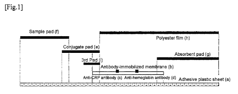
support can be optimized by adjusting an amount of the solution dripped onto
the
insoluble membrane support if the immunochromatographic test strip is of the
flow-
through format, and can be optimized by adjusting a discharge rate of the
solution from
the nozzle of the device if the immunochromatographic test strip is of the
lateral-flow
format. Particularly in the case of the lateral-flow format, the discharge
rate is
preferably 0.5 gL/cm to 2 gL/cm. The term "flow-through format (flow through-
based)"
as used herein refers to a format in which the sample etc., perpendicularly
pass through
the insoluble membrane support for flow progression and the term "lateral-flow
format
(lateral flow-based)" refers to a format in which the sample etc., move in
parallel with
the insoluble membrane support for flow progression.
In the case of the lateral-flow format, the position of application of a
second
antibody to an analyte (e.g., CRP or hemoglobin) to the insoluble membrane
support
may be placed such that the analyte, the immune complex in which the analyte
is bound
to the conjugate, etc., progress from the conjugate pad by capillary action
and
sequentially pass through the measurement sites (measurement lines) with each
of the
second antibodies applied. For example, if the analytes are CRP and hemoglobin
in a
blood sample, the arrangement is preferably made such that a CRP measurement
line
with an anti-CRP antibody applied is located upstream while a hemoglobin
measurement line with an anti-hemoglobin antibody applied is located
downstream. In
this case, it is desirable to keep a sufficient distance between the
respective
CA 02794721 2012-09-26
Page 24 of 59
measurement lines such that the signal of a label can be detected. In the case
of the
flow-through format, the position of application of the second antibody to CRP
or
hemoglobin may also be placed such that the signal of a label can be detected.
An antibody solution applied to the insoluble membrane support can normally
be prepared by using a predetermined buffer solution. The types of the buffer
solution
may include commonly used buffer solutions such as phosphate buffer solution,
Tris
buffer solution, and Good's buffer solution. The buffer solution preferably
has pH in a
range of 6.0 to 9.5, more preferably 6.5 to 8.5, further preferably 7.0 to
8Ø The buffer
solution may contain salts such as NaCl, stabilizer and antiseptic such as
sucrose, and a
preservative such as ProClin. The salts include those added for adjusting
ionic strength,
such as NaCl, as well as those added at the step of adjusting pH of the buffer
solution,
such as sodium hydroxide.
After the second antibody is immobilized to the insoluble membrane support,
the blocking can be performed by using a commonly used blocking agent in a
solution
or vapor state with a cover for the portion other than the part having the
second
antibody immobilized. In this description, an insoluble membrane support
having an
antibody immobilized as described above is also referred to as an "antibody-
immobilized membrane".
[0034]
(Absorbent pad)
The term "absorbent pad" used herein refers to a liquid-absorbing part
absorbing the sample that has migrated/passed through the insoluble membrane
support
to control the flow progression of the sample. In a lateral-flow format, the
absorbent pad
may be provided on the downstream end of the immunochromatographic test strip,
and
in a flow-through format, the absorbent pad may be provided beneath the
antibody
immobilized membrane, for example. Examples of the absorbent pad include, but
are
not limited to, filter paper. It is preferable to use 740-E manufactured by
Whatman etc.
[0035]
(Immunochromatographic test strip)
CA 02794721 2012-09-26
Page 25 of 59
In the present invention, a "immunochromatographic test strip" (hereinafter
also referred to as the "test strip") shall be a product including at least an
insoluble
membrane support having an antibody immobilized and shall be a product
containing a
reagent component as needed or a product appropriately disposed and fitted
with other
membranes etc. Other membranes may be a sample pad, a conjugate pad, an
absorbent
pad, etc. The test strip is usually formed on a solid phase support such as an
adhesive
plastic sheet. The solid phase support, as well as the adhesive component,
should be
made of a material that does not hinder the capillary flow of the sample.
Lamination
with a polyester film etc., can be performed for the purpose of increasing the
mechanical strength of the antibody immobilized membrane and preventing
evaporation
(drying) of water during the assay. The test strip may be used after being
stored in or
mounted on a container (housing) appropriate with respect to the size of the
test strip,
the manner and position of application of the sample, the immobilization
position of
antibody on the antibody-immobilized membrane, the signal detection method,
etc., and
such a stored/mounted state is referred to as a "device".
[0036]
(Same Test Strip)
The test strip for measuring the first analyte such as hemoglobin may be the
same test strip as, or a separate test strip different from, the test strip
for measuring the
second analyte such as CRP. Therefore, in the case of the same test strip, the
test strip is
made up of the same conjugate pad containing a conjugate in which a first
antibody
against the first analyte is immobilized to a label and a conjugate in which a
first
antibody against the second analyte is immobilized to a label, and the same
insoluble
membrane support having a second antibody against the first analyte and a
second
antibody against the second analyte are immobilized, as described above.
However, if
different separate test strips are used, the same sample is measured by using
a test strip
for measuring the first analyte made up of a conjugate pad containing a
conjugate in
which a first antibody against the first analyte is immobilized to a label and
an insoluble
membrane support having a second antibody against the first analyte
immobilized, and a
test strip for measuring the second analyte made up of a conjugate pad
containing a
CA 02794721 2012-09-26
Page 26 of 59
conjugate in which a first antibody against the second analyte is immobilized
to a label
and an insoluble membrane support having the second antibody against the
second
analyte immobilized. When the same test strip is used, the size can be reduced
and the
measurement can easily be performed. On the other hand, when separate test
strips are
used, a combination with a plurality of other analytes can be made whenever
necessary
and the individual test strips are thought to be more generally and frequently
used. Even
when the test strips are separated, the test strips can obviously be housed in
the same
housing to form one device.
[0037]
The immunochromatographic test strip of the present invention is preferably
used for measuring C-reactive protein (CRP) (hereinafter also referred to as a
"immunochromatographic CRP assay test strip (immunochromatographic test strip
for
CRP assay)"). The immunochromatographic CRP assay test strip may be a test
strip
including at least a membrane having an anti-CRP monoclonal antibody and an
anti-
hemoglobin antibody immobilized as well as a conjugate in which an anti-CRP
monoclonal antibody is immobilized to a label and a conjugate in which an anti-
hemoglobin antibody immobilized to a label, and may contain another reagent or
constituent element depending on the measurement condition and the sample.
In this description, the "insoluble membrane support" is also referred to as a
"solid phase" and, allowing, or a state of allowing, the insoluble membrane
support to
physically or chemically support an antigen or an antibody may be expressed as
"immobilize/immobilizing", "immobilized/immobilization", "solid-phased", "
sensitize/sensitization", or "adsorp/adsorption".
[0038]
(Sample)
The "sample" to be measured in the measurement method of the present
invention is blood (whole blood or hemolyzed whole blood).
[0039]
(Diluting Solution)
CA 02794721 2012-09-26
Page 27 of 59
The diluting solution used in the present invention has an effect of
sufficiently
hemolyzing red blood cells in a short time. The diluting solution of any
composition
may be used as long as the antigen-antibody reaction is not significantly
inhibited or,
conversely, not significantly facilitated in a measurement system of the
second analyte
such as hemoglobin and CRP causing a defect of flow progression by capillary
action
due to excessive agglutination, or the signal detection of antigen-antibody
reaction
depending on the concentration of antigen is not disabled. The diluting
solution having
such an effect may be purified water or a buffer solution having pH 6.0 to
10.0, for
example. The buffer solution may preferably be a 10 to 20 mmol/L buffer
solution, for
example, 10 to 20 mmol/L phosphate buffer solution, 10 to 20 mmol/L Tris-HC1
buffer
solution, or 10 to 20 mmol/L glycine-HC1 buffer solution. A surfactant can be
added to
these diluting solutions so as to increase the hemolytic effect and control
the flow
progression rate of the sample etc., in the membrane. Particularly, in the
system of
measuring CRP that is an example of the second analyte, if the monoclonal
antibody
produced by the hybridoma of the accession number FERM BP-11344 is used as the
first antibody immobilized to the label and the monoclonal antibody produced
by the
hybridoma of the accession number FERM BP-11345 is used as the second antibody
immobilized to the insoluble membrane support, the diluting solution can
contain
sodium alkylsulfate expressed by a general formula CH3(CH2)nOSO3Na (n=5 to 10)
to
adjust the measurement range. It is desirable to add 0.05 to 0.3 % of sodium
hexylsulfate, sodium octylsulfate, etc., to the diluting solution since a
preferable
concentration reaction curve is acquired. In this case, a desirable dilution
rate is 50 to
200 times. The diluting solution may contain a chelate agent of Ca2+ ions such
as EDTA
and EGTA.
EXAMPLES
[0040]
The present invention will specifically be described by giving examples of
measuring hemoglobin (hereinafter also referred to as "Hb") as the first
analyte and
CA 02794721 2012-09-26
Page 28 of 59
CRP as the second analyte; however, the scope of the present invention is not
limited to
these examples.
[Example 1 ]
1) Production of anti-CRP antibody-sensitized conjugate (conjugate in which
an anti-CRP monoclonal antibody is immobilized to colloidal gold particles)
and anti-
hemoglobin antibody-sensitized conjugate (conjugate in which an anti-
hemoglobin
monoclonal antibody is immobilized to colloidal gold particles)
The anti-CRP monoclonal antibody (Clone: FERM BP-11344) and the anti-
hemoglobin monoclonal antibody (Clone: #69202) were prepared to have the
following
buffer solution conditions and antibody concentrations; 1 mL of each solution
was
added to 20 mL of a 1 OD/ml colloidal gold particle solution (particle size:
40 nm); and
the mixtures were stirred at room temperature for 10 minutes. After the
addition of 2 ml
of a 10 % bovine serum albumin (BSA) aqueous solution to each of the colloidal
gold
particle-antibody mixtures, the mixtures were further stirred for 5 minutes,
and
centrifuged at 10,000 rpm at 10 degrees C for 45 minutes to obtain sediments
(the anti-
CRP antibody-sensitized conjugate and the anti-hemoglobin antibody-sensitized
conjugate). To each of the acquired conjugates, 1.2 mL of Conjugate Dilution
Buffer
(manufactured by Scripps) was added to suspend the conjugates. The absorbance
of
each of the conjugates was measured at the maximum absorption wavelength.
i) FERM BP-l 1344 (20 g/mL), 2 mmol/L phosphate buffer solution pH 7.0
ii) #69202 (80 g/mL), 2 mmol/L borate buffer solution pH 9.0
[0041]
2) Production of conjugate pad
A conjugate solution was prepared by mixing the anti-CRP antibody-sensitized
conjugate and the anti-hemoglobin antibody-sensitized conjugate produced in
(1) at 20
OD/ml and 10 OD/ml, respectively, with a 20 mmol/L Tris-HCl buffer solution
(pH
7.5) containing 1.33 % casein and 4 % sucrose. A glass fiber pad having a
certain
volume (No. 8964 manufactured by Pall Corporation) was impregnated with 1.2
volumes (relative to the volume of the pad) of the conjugate solution. The pad
was dried
at 70 degrees C for 30 minutes in a dry oven to obtain a conjugate pad. If an
additive
CA 02794721 2012-09-26
Page 29 of 59
such as a sensitizer is added as needed, a necessary amount may be added to
the
conjugate solution before performing the operation above.
[0042]
3) Production of insoluble membrane support having anti-CRP antibody and
anti-hemoglobin antibody immobilized (antibody immobilized membrane)
The anti-CRP monoclonal antibody (Clone: FERM BP-l 1345) and the anti-
hemoglobin monoclonal antibody (Clone: #69209) were prepared at 1 mg/mL as a
10
mmol/L phosphate buffer solution (pH 7.2) containing 2.5 % sucrose to apply
the anti-
CRP monoclonal antibody onto a nitrocellulose membrane (SHF180 manufactured by
Millipore) at a position (CRP measurement line) inner from one edge of the
short side
and the anti-hemoglobin monoclonal antibody on the outside (Hb measurement
line) at
an interval of about 5 mm by using an immunochromatography dispenser "XYZ3050"
(manufactured by BIO DOT) set to be 0.75 L/cm in a line shape. The membrane
was
dried at 70 degrees C for 45 minutes in a dry oven to obtain an antibody
immobilized
membrane.
[0043]
4) Production of sample pad
A glass fiber pad (manufactured by Lydall) cut to have a certain volume was
impregnated with 1.15 volumes (relative to the volume of the pad) of a 20
mmol/L Tris-
HCl buffer solution (pH 7.2) containing 24 mmol/L NaCl, 0.5 % sucrose, and 30
mmol/L ethylenediaminetetraacetic acid. The pad was dried at 70 degrees C for
45
minutes in a dry oven to obtain a sample pad.
[0044]
5) Production of test strip
On an adhesive plastic sheet (a), the antibody immobilized membrane (b)
produced in 3) was disposed and bonded such that an application portion of the
anti-
CRP antibody (c) (the CRP measurement line) was located on the upstream
portion of
the flow progression and followed by an application portion of the anti-
hemoglobin
antibody (d) (the Hb measurement line), and the 3rd pad (i) consisting of a
grass fiber
pad was further placed. The conjugate pad (e) produced in 2) was then placed
and the
CA 02794721 2012-09-26
Page 30 of 59
sample pad (f) produced in 4) was placed to overlap the conjugate pad while
the
absorbent pad (g) was placed on the end of the other side. Finally, a
polyester film (h)
was placed and laminated on the top to cover the antibody immobilized membrane
and
the absorbent pad. The structure formed by overlapping the constituting
elements as
described above was cut to produce the immunochromatographic test strip. The
test
strip was stored in/mounted on a dedicated plastic housing (having a sample
supply
window formed over the sample pad and a detection window formed over the
measurement lines, not depicted in Fig. 1) at the time of an assay to achieve
a form of
an immunochromatographic test device. Fig. 1 is a schematic of a structure of
the
immunochromatographic test strip.
[0045]
6) Hemoglobin measurement by competitive immunochromatography utilizing
competitive reaction (measurement of hemoglobin in a prozone phenomenon
region)
From one healthy individual agreed to blood collection, 5 mL of blood was
collected by using an EDTA-2Na vacuum blood collection tube. A hematocrit
value
measured by using a portion of the blood with the microhematocrit method was
46 %
and it was confirmed that the sample is within a reference value. The blood
sample was
hemolyzed and diluted with a 0.1 % sodium hexylsulfate and 10 mmol/L phosphate
buffer solution (pH 7.2) by a factor of 50 to 400, and 120 L of each of the
samples was
dripped to the sample supply window of the immunochromatographic test device
produced in 5) above to measure the reflected light intensity of the Hb
measurement line
from the detection window of the i nnunochromatographic test device after five
minutes by using an immunochromatography reader ICA-1000 (manufactured by
Hamamatsu Photonics K.K.). As a result, as depicted in Fig. 2, a decreasing
dose-
response curve was obtained indicating that the increase in the hemoglobin
concentration causes the reduction in the reflected light intensity of the
colloidal gold
particles captured by the Hb measurement line having the anti-hemoglobin
antibody
immobilized on the immunochromatographic test strip.
[0046]
7) Production of calibration curve of CRP measurement
CA 02794721 2012-09-26
Page 31 of 59
CRP calibrator A for Nanopia (manufactured by Sekisui Medical Co., Ltd.)
was diluted with a 0.1 % sodium hexylsulfate and 10 mmol/L phosphate buffer
solution
(pH 7.2) by a factor of 100, and 120 L of the sample was dripped to the
sample supply
window of the immunochromatographic test device produced in 5) to measure the
reflected light intensity of the CRP measurement line from the detection
window of the
immunochromatographic test device after five minutes by using the
immunochromatography reader ICA-1000 (manufactured by Hamamatsu Photonics
K.K.). Fig. 3 depicts a calibration curve in a range of CRP concentrations
from 1.5 to
420 mg/L.
[0047]
8) Measurement of hematocrit value with microhematocrit method
From 22 healthy individuals agreed to blood collection, twenty two (22) 5-mL
blood specimens were collected by using EDTA-2Na vacuum blood collection
tubes. A
portion of each of the blood specimens was collected by a hematocrit capillary
tube
(CEN02-0019 manufactured by Drummond) and after sealing the bottom of the
capillary tube with dedicated pate (CRITOSEAL, A422 manufactured by HELIX),
the
capillary tube was centrifuged by a hematocrit centrifuge (H-1200F
manufactured by
KOKUSAN) at room temperature at 12000 rpm for 10 minutes to measure a
hematocrit
value by using a measurement panel associated with the centrifuge.
[0048]
9) Calculation of CRP and hematocrit value using the immunochromatographic
test strip of the present invention
The CRP concentration and hemoglobin concentration of the 22 blood
specimens were measured by using the immunochromatographic test device
produced
in 5). Specifically, 2 L of each of the blood specimens was diluted and
hemolyzed with
a 0.1 % sodium hexylsulfate and 10 mmoUL phosphate buffer solution (pH 7.2) by
a
factor of 100, and 120 L of the sample was dripped to the sample supply
window of
the immunochromatographic test device to measure the reflected light
intensities of the
CRP measurement line and the Hb measurement line from the detection window of
the
immunochromatographic test device after five minutes by using the
CA 02794721 2012-09-26
Page 32 of 59
immunochromatography reader ICA-1000 (manufactured by Hamamatsu Photonics
K.K.). The reflected light intensity of the CRP measurement line was
extrapolated to the
calibration curve of Fig. 3 to obtain the CRP concentration. Fig. 4 depicts a
correlation
expression between the reflected light intensity of the Hb measurement line
and the
hematocrit value acquired in 8). This correlation expression was used for
calculating a
hematocrit value from the reflected light intensity of the Hb measurement line
of each
of the blood specimens.
Hematocrit value (%) (calculated value) _ (- 0.0788) x (reflected light
intensity of the Hb measurement line) + 68.585
[0049]
10) Hematocrit correction by the present invention
The CRP measurement value of each of the blood specimens calculated in 9)
was subjected to the hematocrit correction by the hematocrit value calculated
in 9) in
accordance with the following equation.
uncorrected CRP measurement value
corrected CRP measurement value =
(1 - calculated hematocrit value (%) /100)
[0050]
11) Verification of effect of hematocrit correction of the present invention
The CRP concentrations in plasma of the 22 blood specimens were measured
by using a commercially available kit based on the measurement principle of a
latex
agglutination reaction ("Nanopia CRP" manufactured by Sekisui Medical Co.,
Ltd.). Fig.
depicts correlation between the "CRP measurement value" of Nanopia CRP and the
CRP concentration calculated in 9), i.e., "CRP measurement value (without HCT
correction)". Fig. 6 depicts correlation between the "CRP measurement value"
of
Nanopia CRP and the hematocrit-corrected CRP concentration calculated in 10),
i.e.,
"CRP measurement value (with HCT correction, the present invention)". Fig. 7
depicts
correlation between the "CRP measurement value" of Nanopia CRP and the CRP
concentration acquired by uniformly correcting the CRP concentration
calculated in 9)
CA 02794721 2012-09-26
Page 33 of 59
with an average hematocrit value (44 %), i.e., "CRP measurement value (with
uniform
correction)".
In the case of the "CRP measurement value (without HCT correction)" not
subjected to the hematocrit correction, a slope of a correlation regression
equation
relative to the CRP measurement value measured by the commercially available
CRP
measurement kit was about 0.73 and R2 was about 0.965 (Fig. 5) while in the
case of the
"CRP measurement value (with HCT correction)" subjected to the hematocrit
correction
of the present invention, the slope was about 1.20 and R2 was about 0.989, and
the
improvement in correlation with the CRP measurement value measured in the
latex
agglutination reaction was recognized (Fig. 6).
On the other hand, in the case of the "CRP measurement value (with uniform
correction)" acquired by uniform correction with the average hematocrit value,
the slope
of the correlation regression equation was increased from about 0.73 to 1.30
and,
therefore, the y-intercept was changed from about -0.23 to about -0.41,
departing from
zero (Fig. 7).
From these results, it was demonstrated that the CRP measurement can more
accurately be performed by measuring the concentrations of CRP and hemoglobin
in the
same sample by using the immunochromatographic test strip of the present
invention,
and by performing the hematocrit correction of the CRP measurement value by
utilizing
the correlation relationship between the measurement value of hemoglobin and
the
hematocrit value in the sample.
[0051]
Test example 1: Production Method of Anti-Human CRP Monoclonal
Antibody
A combination of monoclonal antibodies other than the anti-human CRP
monoclonal antibody used in Example 1 was acquired with the following method
and
was used in Examples 2 and 3.
1) Preparation method of immunizing antigen
After human CRP (manufactured by Radioimmunoassay) was mixed at 1:1
with complete Freund's adjuvant (manufactured by Gibco), emulsion was produced
by
CA 02794721 2012-09-26
Page 34 of 59
using a connected syringe and used as an immunizing antigen. Hb was purified
by
cation exchange chromatography from hemolyzed blood of a red blood cell
fraction of
blood collected from volunteer. After this purified Hb was mixed at 1:1 with
complete
Freund's adjuvant in the same way, emulsion was produced and used as an
immunizing
antigen.
[0052]
2) Immunization and production method of hybridoma
The immunizing antigen was injected into the abdominal cavity of BALB/c
mice (50 to 100 gg per mouse). This operation (immunization) was repeated
twice every
two weeks. When five weeks had elapsed after the start of immunization, the
spleen was
extracted from mice with a higher antibody value confirmed by test blood
collection and
the cell fusion was performed by using 50 %-PEG1450 (manufactured by Sigma)
with a
common procedure. SP2/O was used for myeloma cells. The acquired fused cells
were
suspended at 2.5 X 10⁶ cells/mL (as spleen cells) in RPMI1640 medium including
HAT,
15 % fetal bovine serum, and 10 % BM-Condimed H1 Hybridoma Cloning Supplement
(Manufactured by Roche) and were dispensed onto 96-well culture plate at 0.2
mL/well.
The cells were incubated at 37 degrees C in a 5% CO2 incubator.
[0053]
3) Screening of monoclonal antibody producing hybridomas
After 7 to 10 days from the cell fusion, antigen solid-phase ELISA was
performed by using the culture supernatant in accordance with a common
procedure
("Yaku ni tatsu men-eki jikkenho" published by Kodansha, 1984) to select wells
highly
reactive with each antigen as positive wells. The cells in the positive wells
are
subcultured by a 24-well plate.
[0054]
4) Cloning and monoclonal antibody collection
The hybridomas selected by the screening were cloned by a limiting dilution
method to acquire respective hybridomas. To collect the monoclonal antibodies
produced by the hybridomas, the hybridomas were administered at an amount of
0.4 to
1.3 X 10⁶ cells into the abdominal cavity of eight-week old male BALB/c mice
subjected
CA 02794721 2012-09-26
Page 35 of 59
to the injection of 0.5 mL of pristine into the abdominal cavity two weeks
ago. The
ascites was collected every second day after one week from the administration
and was
subjected to centrifugal treatment to acquire supernatant. The supernatant was
mixed
with equal parts of an adsorption buffer solution (3 mol/L NaCl, 1.5 mol/L
Glycine-
NaOH buffer solution, pH 8.5) and then filtrated. The filtrate was passed
through a
Protein A Sepharose column equilibrated with the adsorption buffer solution
and the
antibody in the filtrate was adsorbed with the column and then eluted in a 0.1
mol/L
citrate buffer solution (pH 3.0). The eluate was neutralized with a 1 mol/L
Tris-HC1
buffer solution (pH 9.0) and dialyzed with PBS to collect the purified
antibody. Among
the monoclonal antibodies produced by the hybridomas acquired as described
above,
two types of antibodies highly reactive with CRP were used as #08209 and
#08210 for
the following examples.
[0055]
[Example 2]
1) Production of anti-CRP antibody-sensitized conjugate (conjugate in which
an anti-CRP monoclonal antibody is immobilized to colloidal gold particles)
and anti-
hemoglobin antibody-sensitized conjugate (conjugate in which an anti-
hemoglobin
monoclonal antibody is immobilized to colloidal gold particles)
The anti-CRP monoclonal antibody (Clone: #08210) and the anti-hemoglobin
monoclonal antibody (Clone: #69202) were prepared to have the following buffer
solution conditions and antibody concentrations. The CRP monoclonal antibody
(Clone:
#08210) was added as 10 mL of the antibody solution to 200 mL of a 1 OD/ml
colloidal
gold solution (particle size: 30 nm); the anti-hemoglobin monoclonal antibody
(Clone:
#69202) was added as 10 mL of the antibody solution to 200 mL of a 1 OD/ml
colloidal
gold solution (particle size: 40 nm); and the mixtures were stirred at room
temperature
for 10 minutes. After the addition of 20 ml of a 10 % bovine serum albumin
(BSA)
aqueous solution to the colloidal gold particle-antibody mixtures, the
mixtures were
further stirred for 5 minutes, and centrifuged at 10,000 rpm at 10 degrees C
for 45
minutes to obtain sediments (an anti-CRP antibody sensitized conjugate and an
anti-
hemoglobin antibody sensitized conjugate). To each of the acquired conjugates,
12 mL
CA 02794721 2012-09-26
Page 36 of 59
of Conjugate Dilution Buffer (manufactured by Scripps) was added to suspend
the
conjugates. The absorbance of each of the conjugates was measured at the
maximum
absorption wavelength.
i) #08210 (20 g/ml,), 2 mmol/L phosphate buffer solution pH 7.0
ii) #69202 (80 g/mL), 2 mmol/L borate buffer solution pH 9.0
[0056]
2) Production of conjugate pad
The anti-CRP antibody-sensitized conjugate and anti-hemoglobin antibody-
sensitized conjugate produced in (1) were mixed at 15 OD/ml and 10 OD/ml,
respectively, with a 20 mmol/L Tris-HC1 buffer solution (pH 7.5) containing a
1.33 %
casein and 4 % sucrose solution to prepare a conjugate solution. A glass fiber
pad
having a certain volume (No. 8964 manufactured by Pall Corporation) was
impregnated
with 1.2 volumes (relative to the volume of the pad) of the conjugate
solution. The pad
was dried at 70 degrees C for 30 minutes in a dry oven to obtain a conjugate
pad. If an
additive such as a sensitizer is added as needed, a necessary amount may be
added to
the conjugate solution before performing the operation above.
[0057]
3) Production of insoluble membrane support having anti-CRP antibody and
anti-hemoglobin antibody immobilized (antibody immobilized membrane)
The membrane was produced as is the case with Example 1 except that Clone
#08210 was used as the anti-CRP antibody instead of Clone FERM BP-11344.
[0058]
4) Production of sample pad
The sample pad was produced as is the case with Example 1.
[0059]
5) Production of test strip
The test strip was produced as is the case with Example 1 except that the
antibody-immobilized membrane of 3) in Example 2 was used as the antibody-
immobilized membrane. The acquired test strip was turned to a form of the
immunochromatographic test device as is the case with Example 1.
CA 02794721 2012-09-26
Page 37 of 59
[0060]
6) Production of calibration curve for hematocrit value (HCT value)
calculation
Certified Practical Reference Material for Total Hemoglobin Measurement
(JCCRM 622-1) was used to prepare 40.4 g/L, 80.7 g/L, 136.7 g/L, 185.7 g/L,
and
273.4 g/L hemoglobin reference solutions and each of the reference solutions
was
diluted with a 0.01 % Tween 20 and 10 mmol/L phosphate buffer solution (pH
7.2) by a
factor of 151. From each of the solutions, 120 L was dripped to the sample
supply
window of the immunochromatographic test device to measure the reflected light
intensity of the Hb measurement line from the detection window of the
immunochromatographic test device after five minutes by using the
immunochromatography reader ICA-1000 (manufactured by Hamamatsu Photonics
K.K.). The calibration curve for HCT value calculation was produced with the Y-
axis
indicative of an HCT value (%) acquired by multiplying the hemoglobin
concentration
(g/L) of each of the reference solutions by 2.9/10, and the X-axis indicative
of the
reflected light intensity of the Hb measurement line (Fig. 8).
[0061]
7) Production of calibration curve of CRP measurement
A dilution series of 0, 3, 10, 30, 60, 120, 210, and 420 mg/L was prepared by
diluting a 420 mg/L standard preparation of CRP calibrator D for Nanopia
(manufactured by Sekisui Medical Co., Ltd.) with a 0 mg/L standard
preparation. Each
was diluted with a 0.01 % Tween 20 and 10 mmol/L phosphate buffer solution (pH
7.2)
by a factor of 151, and 120 L of the sample was dripped to the sample supply
window
of the test device to measure the reflected light intensity of the CRP
measurement line
from the detection window of the immunochromatographic test device after five
minutes by using the immunochromatography reader ICA-1000 (manufactured by
Hamamatsu Photonics K.K.). Fig. 9 depicts a calibration curve in a range of
CRP
concentrations from 0 to 420 mg/L.
[0062]
8) Calculation of CRP and HCT value using immunochromatographic test strip
of the present invention
CA 02794721 2012-09-26
Page 38 of 59
The measurement of 200 specimens was performed by using the
immunochromatographic test device described above. Specifically, 10 L of each
blood
was diluted and hemolyzed with a 0.01 % Tween 20 and 10 mmol/L phosphate
buffer
solution (pH 7.2) by a factor of 150, and 120 gL of the sample was dripped to
the
sample supply window of the immunochromatographic test device to measure the
reflected light intensities of the CRP measurement line and the Hb measurement
line
from the detection window of the immunochromatographic test device after five
minutes by using the immunochromatography reader ICA-1000 (manufactured by
Hamamatsu Photonics K.K.).
The reflected light intensity of the Hb measurement line was extrapolated to
the calibration curve of Fig. 8 to obtain the HCT value (%) and the reflected
light
intensity of the CRP measurement line was extrapolated to the calibration
curve of Fig.
9 to obtain the CRP concentration. The CRP concentration after HCT correction
was
calculated with the following equation.
uncorrected CRP measurement value
corrected CRP measurement value =
(1 - calculated HCT value (%) /100)
[0063]
9) Verification of effect of HCT correction of the present invention
The CRP concentrations in plasma of the 200 blood specimens were measured
by using a commercially available kit based on the measurement principle of a
latex
agglutination reaction (Nanopia CRP manufactured by Sekisui Medical Co.,
Ltd.).
Fig. 10 depicts correlation between the "CRP measurement value" of Nanopia
CRP and the CRP concentration before HCT correction calculated in 8) of this
example,
i.e., "CRP measurement value (without HCT correction)". Fig. 11 depicts
correlation
between the "CRP measurement value" of Nanopia CRP and the HCT-corrected CRP
concentration calculated in 8) of this example, i.e., "CRP measurement value
(with HCT
correction, the present invention)".
CA 02794721 2012-09-26
Page 39 of 59
In the case of the "CRP measurement value (without HCT correction)" not
subjected to the hematocrit correction, a slope of a correlation regression
equation
relative to the commercially available CRP measurement kit was about 0.62 and
R2 was
about 0.930 while in the case of the "CRP measurement value (with HCT
correction)"
subjected to the hematocrit correction of the present invention, the slope was
about 0.99
and R2 was about 0.968, and the improvement in correlation was recognized.
From these results, it was demonstrated that the CRP measurement can more
accurately be performed by measuring the concentrations of CRP and hemoglobin
in the
same measurement sample according to the present invention by using a reagent
for
immunochromatography, and by performing the hematocrit correction of the CRP
measurement value by utilizing the correlation relationship between the
measurement
value of hemoglobin and the hematocrit value in the sample.
[0064]
[Example 3]
Four types of immunochromatographic test devices were produced in the same
way as 1) to 5) of Example 1, except that, when the antibody-immobilized
membrane of
3) is produced, Clone 08209 was used instead of using Clone FERM BP-11345 as
the
anti-CRP antibody and that the capillary flow time of the membrane was changed
to 19
seconds/cm (SHF75), 30 seconds/cm (SHY 120), 45 seconds/cm (SHFI 80), and 60
seconds/cm (SHF240).
Certified Practical Reference Material for Total Hemoglobin Measurement
[JCCRM 622-1L (80.7 g/L), 1 mol/L (136.7 g/L), 1H185.7 g/L)] was diluted with
a
0.01 % Tween 20 and 10 mmol/L phosphate buffer solution (pH 7.2) by a factor
of 151,
and 120 L of each of the solutions was dripped to the sample supply windows
of the
four types of the immunochromatographic test devices to measure the reflected
light
intensity of the Hb measurement line from the detection windows of the devices
after
five minutes by using the immunochromatography reader ICA-1000 (manufactured
by
Hamamatsu Photonics K.K.). The calibration curve for HCT value calculation was
produced with the Y-axis indicative of an HCT value (%) acquired by
multiplying the
CA 02794721 2012-09-26
Page 40 of 59
hemoglobin concentration (g/L) of each of the reference solutions by 2.9/10,
and the X-
axis indicative of the reflected light intensity of the Hb measurement line
(Fig. 12).
It was found out that, as the capillary flow time of the membrane decreased,
and thus, the flow rate increased, the change in the reflected light intensity
was reduced
relative to the change in the HCT value. When the capillary flow time
decreased to 19
seconds/cm, little change is made in the reflected light intensity even if the
HCT value
changed. It is thought that this result is generated because the principle of
the Hb
measurement of the present invention is based on a competitive reaction
between free
Hb not bound to the anti-Hb antibody and Hb bound to the anti-Hb antibody on
the
colloidal gold particles. It is probably thought that, if the flow rate
becomes higher, the
free Hb reaches the Hb measurement line first and fills all the Hb binding
sites of the
anti-Hb antibody on the Hb measurement line. From these results, it is known
that the
capillary flow time of the membrane is preferably 30 seconds/cm to 60
seconds/cm.
REFERENCE SIGNS LIST
[0065]
(a) adhesive plastic sheet
(b) antibody-immobilized membrane
(c) anti-CRP antibody
(d) anti-hemoglobin antibody
(e) conjugate pad
(f) sample pad
(g) absorbent pad
(h) polyester film
(i) 3rd pad
REFERENCE TO DEPOSITED BIOLOGICAL MATERIAL
[0066]
Accession numbers
FERM BP-11344
CA 02794721 2012-09-26
Page 41 of 59
FERM BP-11345
[0067]
(1) FERM BP-1 1344 (#08202 producing hybridoma)
i) Name and address of depository institution at which the biological
materials
were deposited.
International Patent Organism Depositary, National Institute of Advanced
Industrial Science and Technology
Tsukuba Central 6, 1-1-1 Higashi, Tsukuba, Ibaraki 305-8566, Japan
ii) Date of biological material deposit in the depository institution in i).
November 26, 2009
iii) Accession number for the deposition assigned by the depository
institution
in i).
FERM BP-1 1344
(2) FERM BP-11345 (#08203 producing hybridoma)
i) Name and address of depository institution at which the biological
materials
were deposited.
International Patent Organism Depositary, National Institute of Advanced
Industrial Science and Technology
Tsukuba Central 6, 1-1-1 Higashi, Tsukuba, Ibaraki 305-8566, Japan
ii) Date of biological material deposit in the depository institution in i).
November 26, 2009
iii) Accession number for the deposition assigned by the depository
institution
in i).
FERM BP-11345