Note: Descriptions are shown in the official language in which they were submitted.
CA 02794831 2015-12-18
1
Title: IMMUNOASSAY FOR FREE VITAMIN D
Field of the invention
The invention relates to an immunoassay method for assaying a
sample of blood or blood components for free vitamin D. The invention also
relates to immunoassays and kits for conducting such immunoassays including
point-of-care tests.
Background of the invention
The substances referred to as "vitamin D" encompass a group of fat-
soluble prohormones, as well as metabolites and analogues thereof. The main
forms in which vitamin D occurs in the body are vitamin D2 (ergocalciferop
and vitamin D3 (cholecalciferol). The latter is the endogenous form of vitamin
D, which humans can form in the skin under the influence of sunlight. The
former is an exogenous form of vitamin D, taken up with food. In the US,
Vitamin D2 is used as the pharmaceutical vitamin D supplement. Unless
indicated otherwise, the term Vitamin D in this disclosure refers to any form
or forms of Vitamin D, including Vitamin D metabolites such as 25-hydroxy-
Vitamin D or 1,25 dihydroxy Vitamin D.
Whilst vitamin D2 and D3 differ in the molecular structure of their
side-chains, they share the same biological activity in being prohormones,
metabolized in two steps to, ultimately, 1,25 dihydroxy vitamin D (1,25-(01-
1)2-
Vitamin D) also referred to as calcitriol, or 1,25 dihydroxy cholecalciferop.
The
preceding metabolite, 25-hydroxy vitamin D (25-(0F1)-Vitamin D) or calcidiol,
results from conversion in the liver, and is considered the storage form of
vitamin D in the body.
CA 02794831 2012-09-27
WO 2011/122948
PCT/NL2011/050219
2
Circulating vitamin D consists mainly of 25-(OH)-Vitamin D3 and
25-(OH)-Vitamin D2. Biologically, 25-(OH)-Vitamin D2 is as effective as 25-
(OH)-Vitamin D3. The half-life of 25-(OH)-Vitamin D2 in the circulation is
shorter. For clinical practice the use of a 25-(OH)-Vitamin D assay that
measures both 25-(OH)-Vitamin D3 as well as 25-(OH)-Vitamin D2 is
recommended (1).
Vitamin D has long been recognized as an important substance, the
active form of which plays a role in the formation and maintenance of bone, as
well as in other processes in the human or animal body. Thus, it serves to
increase the flow of calcium into the bloodstream, by promoting absorption of
calcium and phosphorus from food in the intestines, and re-absorption of
calcium in the kidneys; enabling normal mineralization of bone and preventing
hypocalcemic tetany. It is also necessary for bone growth and bone remodeling
by osteoblasts and osteoclasts.
Vitamin D deficiency results in impaired bone mineralization and
leads to bone softening diseases, rickets in children and osteomalacia in
adults,
and possibly contributes to osteoporosis.
Vitamin D plays a number of other roles in human health including
inhibition of calcitonin release from the thyroid gland. Calcitonin acts
directly
on osteoclasts, resulting in inhibition of bone resorption and cartilage
degradation. Vitamin D can also inhibit parathyroid hormone secretion from
the parathyroid gland, modulate neuromuscular and immune function and
reduce inflammation. Thus, it is of the essence for a person's or animal's
health
to have an adequate level of vitamin D.
Yet, excess of vitamin D (which may occur as a result of overdosing)
is toxic. Some symptoms of vitamin D toxicity are hypercalcaemia (an elevated
level of calcium in the blood) caused by increased intestinal calcium
absorption. Vitamin D toxicity is known to be a cause of high blood pressure.
Gastrointestinal symptoms of vitamin D toxicity can include anorexia, nausea,
and vomiting. These symptoms are often followed by polyuria (excessive
CA 02794831 2012-09-27
WO 2011/122948
PCT/NL2011/050219
3
production of urine), polydipsia (increased thirst), weakness, nervousness,
pruritus (itch), and eventually renal failure.
Clearly, it is important to be able to diagnose subjects for a possible
vitamin D deficiency. It is also important, particularly for subjects that are
on
vitamin D supplementation, to be able to test subjects for a potential excess
of
vitamin D. The serum level of total 25-(OH)-Vitamin D is considered to be the
primary indicator of the vitamin D status (2) However, this notion has been
disputed.
Almost all circulating 25-(OH)-Vitamin D in serum is bound by
Vitamin D Binding Protein (88%) and Albumin (12%). Vitamin D Binding
Protein (DBP) is a major component of serum, with a concentration of 250-400
mg/L of serum. Only a small portion, about 2%, of the Vitamin D binding sites
of DBP is occupied. A very small fraction, 0.04% of the 25-(OH)-Vitamin D,
circulates in the free, non-protein bound form.
The concentration of DBP is not constant in all people and can be
influenced by other factors including pregnancy, the use of oral
contraceptives,
renal disease and liver disease. Knowledge of the concentration of the DBP is
crucial for accurate assessment of the patient's true 25-(OH)-Vitamin D
status.
For example, a young woman taking oral contraceptives could have a total 25-
(OH)-Vitamin D level that was in the normal range. However, due to her
elevated DBP, the concentration of Free 25-(OH)-Vitamin D could be markedly
depressed, putting her at increased risk for clinical 25-(OH)-Vitamin D
insufficiency and all the risks that that condition entails.
It has been shown that the physiological activity of thyroid and
steroid hormones in vivo correlates better with their free, non-protein bound
fraction, than with the total concentration of the hormone in plasma.
Particularly in situations in which the level of binding proteins is elevated
or
decreased, the measurement of total circulating hormone may lead to a wrong
diagnosis. In such situations the measurement of the concentration of the free
circulating hormone provides better information. This notion is known as the
CA 02794831 2012-09-27
WO 2011/122948
PCT/NL2011/050219
4
"free hormone hypothesis". Mendel (3) suggested that the free hormone
hypothesis is "likely to be valid with respect to all tissues for the thyroid
hormones, for cortisol, and also for the hydroxylated metabolites of vitamin
D.
Bikle et al (4) tested the validity of the free hormone hypothesis for
1,25 ¨(OH)2-Vitamin D. The data suggested that free 1,25-(OH)2-Vitamin D
levels appeared to be well maintained even in subjects with liver disease and
reduced DBP levels, despite a significant decrease of the total 1,25-(OH)2-
Vitamin D.
In a subsequent study on 25-(OH)-Vitamin D the same group
recommended to measure free 25-(OH)-Vitamin D in situations with modified
concentrations of the binding protein. The author concluded that total vitamin
D metabolite measurements may be misleading in the evaluation of the
vitamin D status of patients with liver disease, and recommend that free 25-
(OH)-Vitamin D levels also be determined before making a diagnosis of
vitamin D deficiency. Bikle et al used ultrafiltration to determine the level
of
free 25-(OH)-Vitamin D (5). This method requires highly purified radiolabeled
Vitamin D and tends to overestimate the fraction of free Vitamin D.
Lauridsen et al (6) showed that women with different DBP
phenotypes have different concentrations of 1,25(OH)2VitD and 25(OH)VitD.
These authors suggest that women with Gc2-2 are Vitamin D sufficient at
lower plasma levels of 25(OH)VitD.
Some background art can be referred to regarding the determination
of free analytes.
US patent 4366143 describes an invention related to the assay of the
free portion of organic substances or ligands that are present in biological
fluids in a bound and a free form. The method essentially is a competitive
immunoassay wherein, in one step, a labeled ligand, and a specific binder are
added to a sample simultaneously. The free portion of the ligand and the
labeled ligand compete for reaction with the specific binder, and become bound
thereto in proportions which depend on the amount of the free ligand portion
CA 02794831 2012-09-27
WO 2011/122948
PCT/NL2011/050219
present in the sample. A drawback of the disclosed method is that, due to the
presence of both the specific binder and the labeled ligand, a plurality of
factors is present that are capable of disturbing the equilibrium between
bound and free ligand, which makes the method less suitable for use with a
5 free ligand that is present in a relatively low amount as is the case
with
Vitamin D. In fact, it is not disclosed how to use the assay for the
measurement of free Vitamin D.
US patent no. 4,292,296 discloses a method for the determination of
free analytes in samples containing free analytes and receptor-bound analytes.
The method involves two steps, the first being contacting a sample with an
absorbent for the analyte to remove analyte from solution. The second step
comprises contacting the absorbent-bound analyte with a labelelled analyte
analogue. Thereupon, the soluble phase is removed from the absorbent, and
the amount of label in the bound and washed-away phases are determined.
The method is described for determining the concentration of free thyroid
hormones.
US patent application 2008/0182341 is related to stabilizing agents
that are useful for the measurement of free or unbound analyte concentrations
in a fluid. It is suggested that the stabilizers prevent dissociation of the
ligand
of its binding protein. The reference employs a simultaneous assay procedure,
and lists a variety of stabilizing agents. The stabilizing agent is provided
not to
comprise an alkyl amine fluoro surfactant.
None of the prior art references specifically provides an assay for a
determination of Vitamin D that reflects the status of free Vitamin D.
It is noted that assays for Free Vitamin D have been known for
decades, but these use methods such as equilibrium dialysis or rate dialysis
as
their basis. Such methods are acceptable for researchers with highly trained
technical staff, but are ill suited for routine laboratories who need high
throughput automated tests to reach their financial goals. It is thus desired
to
CA 02794831 2012-09-27
WO 2011/122948
PCT/NL2011/050219
6
provide an assay for free Vitamin D that is capable of being automated and
which is suitable for use in point-of-care testing.
The foregoing numbered references are:
1. Hollis BW. Measuring 25-hydroxyvitamin D in a clinical environment:
challenges and needs.
Am J Clin Nutr. 2008 Aug;88(2):507S-510S.
2. Holick MF. Vitamin D: extraskeletal health.
Endocrinol Metab Clin North Am. 2010 Jun;39(2):381-400.
3 Mendel CM. The free hormone hypothesis: a physiologically based
mathematical model.
Endocr Rev. 1989 Aug;10(3):232-74.
4. Bikle D, Gee E, Halloran B, Haddad J. Free 1,25-Dihydroxyvitamin D
Levels in Serum from Normal Subjects, Pregnant Subjects, and Subjects
with Liver Disease.
J Clin Invest. 1984; 74: 1966-1971.
5. Bikle D, Gee E, Halloran B, Kowalski MA, Ryzen E, Haddad J. Assessment
of the free fraction of 25-hydroxyvitamin D in serum and its regulation by
albumin and the vitamin D-binding protein.
J Clin Endocrinol Metab. 1986 Oct;63(4):954-9.
6. Lauridsen AL, Vestergaard P, Hermann AP, Brot C, Heickendorff L,
Mosekilde L, Nexo E.
Plasma concentrations of 25-hydroxy-vitamin D and 1,25-dihydroxy-
vitamin D are related to the phenotype of Gc (vitamin D-binding protein): a
cross-sectional study on 595 early postmenopausal women.
Calcif Tissue Int. 2005 Jul;77(1):15-22.
7. van Hoof HJ, Swinkels LM, Ross HA, Sweep CG, Benraad TJ.
Determination of non-protein-bound plasma 1,25-dihydroxyvitamin D by
symmetric (rate) dialysis. Anal Biochem 1998 May 1;258(2):176-83.
CA 02794831 2012-09-27
WO 2011/122948
PCT/NL2011/050219
7
Summary of the invention
In order to better address one or more of the foregoing desires the
invention, in one aspect, presents a method for assaying a sample of blood or
blood components for the presence of free Vitamin D (including Vitamin D
metabolites such as 25-hydroxy-Vitamin D or 1,25 dihydroxy Vitamin D),
comprising
(a) adding an immobilized binding protein or antibody for 25-0H
vitamin D to the sample;
(b) mixing the sample with a diluent, said diluent comprising a
fluoroalkyl surfactant;
(c) incubating the sample for an effective amount of time to allow a
desired amount of 25-0H vitamin D to bind to the binding protein;
(d) removing the non-bound serum and serum components by
washing;
(e) subjecting the immobilized binding protein or antibody
comprising 25-0H vitamin D bound thereto, to competitive binding with a
labeled vitamin D compound;
(f) determining the concentration of labeled vitamin D compound
bound to the binding protein.
In another aspect, the invention resides in a kit for conducting the
foregoing method.
In a further aspect, the invention pertains to a method for assaying
a sample of blood or blood components for the presence of free Vitamin D,
comprising in a first step capturing Vitamin D on an immobilized binder such
as an immobilized binding protein or antibody for 25-0H vitamin D, and in a
subsequent step the captured 25-0H Vitamin D is subjected to a competitive
binding assay against a labeled vitamin D variant, wherein the capturing of
free Vitamin D is effected by sequestering an amount of bound Vitamin D so
limited as to satisfy a repetition test, wherein the sample from which the
CA 02794831 2012-09-27
WO 2011/122948
PCT/NL2011/050219
8
Vitamin D was captured is subjected to the same steps of capturing of Vitamin
D and subjecting the captured Vitamin D to the same competitive binding
assay, and wherein the measured concentration of free Vitamin D after both
competitive binding assays is substantially the same.
In a still further aspect, the invention pertains to a method for
assaying a sample of blood or blood components for the presence of free
Vitamin D, comprising in a first step capturing Vitamin D on an immobilized
binder such as an immobilized binding protein or antibody for 25-0H vitamin
D, and in a subsequent step the captured 25-0H Vitamin D is subjected to a
competitive binding assay against a labeled vitamin D variant, wherein the
0.5-5 wt.% of bound Vitamin D is captured.
In yet another aspect the method can be used for point-of-care"
testing. The latter refers to testing at or near the site of patient care,
i.e.
rather than drawing blood samples and sending these to a diagnostic
laboratory, a sample can be immediately introduced into a portable, preferably
handheld device which is able to perform the assay in as limited a number of
steps as possible, and with as limited a number of manual operations as
possible.
In another aspect, the invention pertains to the use of a fluoroalkyl
surfactant, preferably a perfluoro carboxylate surfactant, and more preferably
perfluorooctanoic acid (PFOA), as a solubility enhancer for Vitamin D in an
immunoassay for the determination of free Vitamin D.
Description of drawing
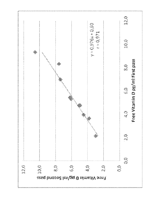
Fig. 1 represents a correlation between free Vitamin D concentration
in the first and the second pass of an assay of the invention. The line shown
results from a repeated immunoextraction using 10 samples with different
CA 02794831 2012-09-27
WO 2011/122948
PCT/NL2011/050219
9
levels of free Vitamin D. The slope of the regression line is not
significantly
different from unity.
Detailed Description of the Invention
In a broad sense, the invention concerns a two-step assay for the
determination of free Vitamin D, comprising the immuno-adsorption of non-
protein-bound 25(OH)-Vitamin D from blood or blood components, notably
serum or plasma, after which the absorbed Vitamin D is measured.
Such samples can be drawn, in any manner known in the art, from a
subject, particularly a human, in whose blood it is desired to assay the
presence of free 25-OH-Vitamin D.
Free Vitamin D refers to the circulating, unbound fraction of
Vitamin D. This relates to any form of Vitamin D, including Vitamin D2,
Vitamin D3, and the metabolites 25(OH)-Vitamin D2, 25-(OH)-Vitamin D3,
1,25(OH)2-Vitamin D2, and 1,25(OH)2-Vitamin D3. The assay can be used to
measure any of these forms of free Vitamin D, alone or in combination.
Preferably, the assay of the invention is used to measure free 25(OH)-Vitamin
D, resulting in the determination of 25(OH)-Vitamin D2 and 25-(OH)-Vitamin
D3. The terms "free" and "unbound" refer to the fraction not bound to any
protein, mainly including Vitamin D Binding Protein (VDBP or DBP).
The sample is preferably diluted before, during, or after the addition
of the binding protein. The sample diluent can be aqueous-based, and
preferably will be a buffer solution. Preferably, the buffered pH is in the
range
of from 6.0 to 8Ø The buffer includes a fluoro alkyl surfactant. It will be
understood that mixing the buffer with the sample can be done by adding the
diluent to the sample, by adding the sample to the diluent, or by
simultaneously adding the buffer and the sample to each other. For practical
reasons, it is preferred to add the diluent to the sample.
CA 02794831 2012-09-27
WO 2011/122948
PCT/NL2011/050219
Without wishing to be bound by theory, the inventors believe that
the surfactants enhances the solubility of Vitamin D.in such a way as to
result
in a limited sequestering of Vitamin D.
In another aspect, the invention pertains to a novel concept for
5 assaying free Vitamin D. A problem with assaying free Vitamin D is that
the
original concentration of free Vitamin D is very low and cannot be measured
by existing immunoassays. Existing attempts to solve this problem are either
based on the desire to avoid removal of Vitamin D from DBP (e.g. by attempts
to "stabilize" the equilibrium between free and bound Vitamin D) or just
10 involve surrendering to the fact that free DBP cannot be assayed, and
these
assays just involve displacement of Vitamin D from DBP.
In the invention, a limited sequestering of Vitamin D is foreseen,
wherein the fraction of sequestered Vitamin D is low enough, preferably 0,1-
10% of the total Vitamin D present in the sample, and preferably not exceeding
5%thereof, to still reflect the original free Vitamin D concentration.
This can be verified without undue experimentation in a simple by
the addition of Tritiated Vitamin D and the dilution buffer. Vitamin D bound
to the Vitamin D binding protein should not decrease by more than 5%.
Alternatively, sample can be absorbed to the wall of antibody coated wells.
After removing the sample, biotinylated Vitamin D is added and the free
Vitamin D, absorbed to the antibody, is quantified. The sample from which an
initial amount of Vitamin D is removed is introduced in a second well and
again incubated and quantified. The measured concentration of free Vitamin D
should be the same as the result from the first incubation.
Limited sequestering of Vitamin D is preferably achieved by the
addition of 0.05 to 0.5 % by weight of the fluoro alkyl surfactant, preferably
0.1-0.25%. Preferably the fluoro alkyl surfactant is a perfluoro alkyl
surfactant, more preferably a perfluoro carboxylate surfactant, and most
preferably perfluorooctanoic acid (PFOA). Preferably 0.1 to 0.2%, more
preferably 0.15%, of PFOA is used. Under these conditions the response of
CA 02794831 2012-09-27
WO 2011/122948
PCT/NL2011/050219
11
samples with different levels of DBP, but the same concentration of total
Vitamin D, was correlated with the concentration of free Vitamin D as
measured by symmetric dialysis.
Conceivably, in the invention use can be made of PFOA itself, or a
derivative thereof. These derivatives generally refer to derivatives
comprising
the perfluoro octanoic moiety, particularly including PFOA salts, such as
PFOA ammonium salt, and to the corresponding sulfonic acid, viz. PFOA
sulfonate, abbreviated as PFOS (perfluoro octane sulfonate, also known as
perfluoro octane sulfonic acid).
An advantage of the method of determining Free Vitamin D
(including Vitamin D metabolites) according to the invention is that it
provides an assay format that is capable of being automated. This markedly
distinguishes the assay of the invention from any pre-existing assay for the
determination of free Vitamin D.
In the assay of the invention a binding protein for 25-OH-Vitamin D
is added. Binding proteins, e.g. antibodies, for vitamin D are known in the
art,
and are widely used in the existing immunoassays for vitamin D. These same
antibodies, as well as other binding proteins, can be used in the present
invention as well. E.g., in the place of an antibody for Vitamin D an antibody
fragment can be used such as produced with phage display technology.
Suitable antibodies can be monoclonal or polyclonal antibodies. They can be
obtained in known manner, e.g. polyclonal goat anti-vitamin D, polyclonal
rabbit anti-vitamin D, or any other suitable antibody for vitamin D as known
in the art from application in immunoassays for vitamin D. Suitable antibodies
are known, e.g. from the following references: Hollis, Clin.Chem 31/11, 1815-
1819 (1985); Hollis, Clin.Chem 39/3, 529-533 (1993).
The binding proteins are preferably added in a particulate form
comprising solid carriers. Typically, the binding protein is coated on a solid
phase, e.g. on a microtiter plate. In a preferred embodiment, the binding
CA 02794831 2012-09-27
WO 2011/122948
PCT/NL2011/050219
12
protein is coated onto magnetic particles, which facilitates their separation
in
a magnetic field.
After addition of the binding protein, e.g. an antibody, the sample is
allowed to incubate. The required time will depend on circumstances such as
the concentration of the reagents, the type of binding protein, and conditions
during incubation, e.g. shaking and temperature. Generally, the incubation
time will be in a range of from 10 seconds to several hours, preferably 1
minute
to 2 hours. For automated platforms, short incubation times (10 seconds to 10
minutes, preferably 30 seconds to 30 minutes) are preferred. Basically, the
period of time is not of particular relevance, as long as one determines in a
calibration system how much of the free vitamin D is to be bound under the
circumstances, during the desired period of time. Shorter and longer periods
of
time are expressly possible, provided that proper calibration takes place.
Thus,
preferably, comparison with calibrators involves the same period of time,
under the same conditions
After the incubation period, the sample can be subjected in a known
manner to a competitive binding assay using a labeled vitamin D compound.
Numerous labeled compounds are known that are capable of serving as
competitive binding antigens in immunoassays for the determination of
vitamin D. Typical labels are radiolabels, fluorescent labels, luminescent
labels, biotin labels, gold labels, enzyme labels. Competitive binding assays
are
known to the skilled person, and do not require elucidation, notably since
this
part of the method of the invention can be carried out using any label known
to
be suitable for the determination of vitamin D. Labels that can be used are,
inter alia, those disclosed in the foregoing references on existing vitamin D
immunoassays.
With the label allowing measuring a concentration, as a result, the
concentration of free vitamin D in the sample is determined. It will be
understood that the interpretation of the values measured, is determined by a
CA 02794831 2012-09-27
WO 2011/122948
PCT/NL2011/050219
13
calibration measurement, i.e. by the response ¨ in the same assay ¨ of
calibrators.
Alternatively, the captured Vitamin D can be subjected to
quantitative analysis by means of mass spectroscopy.
The calibration for the assay of the invention can be done by
providing calibrators comprising a predetermined concentration of free 25-0H-
Vvitamin D. The fraction of free Vitamin D in these calibrators may be
determined by symmetric dialysis. In symmetric dialysis a serum sample is
loaded on one side of a dialysis cell. The other compartment is loaded with
the
same sample in which a trace amount of radiolabeled Vitamin D is added.
The rate of migration of the radiolabeled Vitamin D from one dialysis
compartment to another is directly proportional to the free fraction of
Vitamin
D(7).
The measurement of free vitamin D according to the invention relies
on the assessment of the concentration of free Vitamin D without substantially
affecting the concentration of free Vitamin.D, yet on the basis of a limited
sequesteringof Vitamin D as discussed above.
The invention, in another aspect, presents a product in the form of
an immunoassay for the determination of 25-0H vitamin D in blood or blood
components, wherein the assay makes use of a method according to any one of
the preceding embodiments. More particularly, such a product will be provided
in the form of a kit for conducting the immunoassay. Such a kit may comprise
the individual reagents involved, i.e. the binding protein and the labeled
vitamin D compound. These reagents can be provided separately, and thus
form a kit only upon their use in the assay of the invention. Preferably, the
reagents are provided together, preferably packaged together, as one kit of
parts. The kit optionally comprises a container for a sample of blood or blood
components, but as is customary this may also be provided separately.
Typically a kit comprises a binder immobilized on a solid phase and a separate
CA 02794831 2012-09-27
WO 2011/122948
PCT/NL2011/050219
14
conjugated vitamin D. Other kit components will depend, as is customary in
the art, on the label chosen, as different labels may require different
reagents.
The invention also pertains to the use of a fluoroalkyl surfactantõ
preferably a perfluoro carboxylate surfactant, and more preferably
perfluorooctanoic acid and/or perfluoroheptanoic acid, as a solubility enhance
for Vitamin D in an immunoassay for the determination of free Vitamin D.
Preferred surfactants perfluoro alkyl surfactants, more preferably
perfluoro carboxylates such as perfluoroheptanoic acid or perfluorooctanoic
acid. Most preferably, the surfactant is perfluorooctanoic acid (PFOA), or a
derivative thereof. These derivatives generally refer to derivatives
comprising
the perfluorooctanoic moiety, particularly including PFOA salts, such as PFOA
ammonium salt, and to the corresponding sulfonic acid, viz. PFOA sulfonate,
abbreviated as PFOS (perfluoro octane sulfonate, also known as perfluoro
octane sulfonic acid). The analogous derivatives of other perfluoro
carboxylate
surfactants can also be used.
It is to be understood that the invention is not limited to the
embodiments and formulae as described hereinbefore. It is also to be
understood that in the claims the word "comprising" does not exclude other
elements or steps. Where an indefinite or definite article is used when
referring to a singular noun e.g. "a" or "an", "the", this includes a plural
of that
noun unless something else is specifically stated.
The invention will be illustrated with reference to the following, non-
limiting Example and the accompanying non-limiting Figure.
EXAMPLE
Materials
Coated Microtiterplates
CA 02794831 2012-09-27
WO 2011/122948
PCT/NL2011/050219
Nunc maxisorp micotiterplates were coated with an anti-mouse IgG antibody
at a concentration of 200 ng/well. Subsequently a layer of monoclonal anti-
25(OH) vitamin D antibody was absorbed onto the anti-mouse IgG layer at a
concentration of 2 ng/well. The plates were blocked with a borate buffer
5 containing BSA and sucrose.
The sample diluent
The sample diluent consists of a 0.1M TRIS buffer of pH 8.0 containing
preservatives and 0.15% of PFOA.
The conjugate (i.e. the labeled vitamin D compound) is a biotinylated Vitamin-
10 D. The conjugate was presented at a concentration of 25 pg/ml in
0.1M Tris buffer of pH 7.5 containing 0.1% Bovine serum albumin and
preservatives.
Streptavidin-HRP and TMB were from a commercial source.
15 Protocol
The assay is performed as follows. In the well of a micotiterplate, 90 ul of
sample diluent is pipetted. Next, 10 ul of sample is added to the diluent.
This
mixture is incubated for 90 minutes at 37 C. Subsequently, the wells are
washed three times with washbuffer. 100 ul of HRPConjugate is added to the
cuvette and incubated for 30 minutes. Again the wells are washed three times
with washbuffer. A colorimetric signal is then generated by addition of Horse
Radish Peroxidase labeled streptavidin. After 20 minutes incubation at 37 C
the wells are washed three times with washbuffer. Subsequently 100 ul of
TMB solution is added to the wells. After a 20 minutes incubation at room
temperature in the dark 100 ul of stop solution is added and the absorbance is
read at 450 nm.
The signal generated in the well is inversely proportional to the
concentration
of free 25 (OH)Vitamin D in the sample or calibrator. The concentration of
free
25(OH) vitamin D in the original sample can be calculated by comparing the
signal of unknowns with the response of calibrators.
CA 02794831 2012-09-27
WO 2011/122948 PCT/NL2011/050219
16
Results
Using the assay of the invention on standard reference samples, a typical
calibration curve for the measurement of free 25(OH) Vitamin D is generated
and illustrated in the table below.
Calibrator Calibrator Calibrator Calibrator Calibrator
Calibrators A B C D E
Free 25(OH)D3 16,2 41,6
[pg/ml] 1,1 pg/ml 4,2 pg/ml 8,6 pg/ml pg/ml pg/ml
Abs. 450 nm 2,304 1,856 1,177 0,514 0,132
Abs. 450 nm 2,291 1,808 1,151 0,517 0,123
Average OD 2,298 1,832 1,164 0,516 0,128
CV% 0,40% 1,85% 1,58% 0,41% 4,99%
Binding
percentage % 100% 80% 51% 22% 6%
Samples of blood or blood components of human or animal subjects,
e.g. of patients, can be subjected to the assay of the invention. The measured
amounts of Vitamin D can be correlated with the calibration curve, and thus
interpreted.