Note: Descriptions are shown in the official language in which they were submitted.
WO 2011/120893 PCTIEP2011/054678
I
Description
Method and arrangement for forming a dental model
The invention relates to a method for forming and manufacturing a physical or
virtual dental
model as an aid in the production of a dental prosthesis, whereby one takes
into account the
shape of a tooth stump present in either the upper or lower jaw, contact
points to teeth adjacent to
the at least one tooth stump, as well the occlusal surface of at least one
antagonist tooth in the
field of motion of the dental prosthesis to be provided on the at least one
tooth stump, using
digital data of the upper and lower jaw obtained by contact-less measurements.
A multitude of arrangements and methods for the manufacture of dental
prostheses are known in
the art. In general, subsequent to the dental preparation, one creates an
impression of the tooth
stump that will accept the dental prostheses, as well as of its surroundings
and the jaw. The same
applies in the case where several tooth stumps are to be provided with dental
prostheses.
Irrespective hereof, this is usually performed using a silicone-casting
compound, even though
other materials may be used.
WO 2011/120893 PCTIEP2011/054678
2
From the imprint, which represents this situation in the patient's mouth in
negative form, one
prepares a so-called master model via a plaster cast. This model represents
the situation in the
patient's mouth in positive form. A dental technician uses his craft skills
and this model to mold
a model of the skeletal structure of the dental prostheses using wax or a
plastic curing by
polymerization at low temperature. A positive mold created in this manner
usually is used as a
basis for the dental prosthesis.
In the present day, however, the old-fashioned plaster mould is produced using
new technologies
(stereo lithography or milling) and new materials (plastic) from stereo
lithography data sets of
the occlusion pattern in combination with a digital representation of the
tooth base. In this,
removing the prepared dental stumps, for example, from the remaining model is
intended and is
realized using various connecting structures. A purely statistical final
occlusion position is
available by encoding the model of the upper and lower jaws by means of
connecting structures.
A disadvantage of the presently available method for realizing this is the
high requirements for
material, which for plaster casts is not an issue, but with the expensive
plastic materials, which
are required for stereo lithography or comparable processes, leads to cost-
related disadvantages
that limit their competitiveness. One also faces a comparatively long
manufacturing time of
several hours, which in combination with the high investment costs for rapid
prototyping
machines results in a high cost burden.
DE-B-10 2005 033 738 relates to a method and a device for the manufacture of
dental
prostheses. In this, existing design data are shown on a display together with
measured data of a
machined dental prosthesis to be manufactured.
The subject matter of DE-A-10 2006 026 776 is a method for the manufacture of
a dental
prosthesis, whereby one digitally acquires information on the configuration
and relative
positions of the jaws.
3
In accordance with DE-A-103 04 757 the manufacturing of dental prostheses
takes into
consideration data about the jaws that normally are collected from patients
during the process of
adjusting an articulator. In addition, jaw movements are simulated in a
computer.
The manufacture of dental prostheses with the help of virtual prototypes is
disclosed in EP-A-1
935 369. For this, one scans a jaw region, into which the dental prosthesis is
to be implanted.
WO-A-2008/051130 relates to a method and a device for the manufacture of a
dental model with
the help of impressions.
Known from WO-A-2008/030965 is a method for the manufacture of a dental
prosthesis, in
which one uses a virtual model of the upper and lower jaws.
The manufacture of a dental prosthesis with help of a virtual prosthesis is
described in WO-A-
03/017864, whereby one records 3D data of an upper or lower jaw, into which
the dental
prosthesis is to be implanted.
US-A-2005/0070782 describes the manufacture of dental prostheses with the help
of virtual
models. This also involves the use of a virtual articulator.
WO-A-2004/030565 describes a method to form a virtual dental model. A negative
impression of a jaw is
used to create virtual impressions of the lower and upper jaws, which are
taken into consideration together
with the spatial relation of the distance between the two jaws during
occlusion in the centric position.
An articulator is described in NL-A-7706907. Support points between the upper
part and the lower part are
designed in such a manner that the unit can be used with different distances
between the temporomandibular
joints.
3a
US-A-4,276,022 and CH-A-315737 also disclose articulators that comprise an
upper part and a lower part
and are supported with respect to each other via three support points.
The present invention is based on the objective to further develop and make
available a method
and an arrangement of the above-mentioned type in a way that facilitates a
production with
reasonable costs, for example in rapid prototyping production, whereby at the
same time
diagnostic information that can be obtained from the patients is to be
incorporated into the
manufacturing process.
With respect to the method, this objective fundamentally is met by following
at least the
following procedural steps:
a) scanning the upper and lower jaw to acquire first data,
WO 2011/120893 PCT7EP2011/054678
4
b) recording the field of motion of the temporomandibular joint during
disclusion to acquire
second data,
c) recording the contact field of the teeth during occlusion to acquire third
data,
d) computing the paths of motion between the upper and lower jaws from at
least the first
data and the third data or the first and the second data or the second and the
third data or
the first and the second and the third data,
e) employing a retaining device comprising a lower part and an upper part
adjustable
relative to the lower part, whereby connected or being connected to the upper
part is a
first element representing the upper jaw and comprising first receptacles and
connected or
being connected to the lower part is a second element representing the lower
jaw and
comprising second receptacles,
f) producing three support points or support surfaces, which are formed by the
paths of
motion, which are computed in accordance with step d) and which have been or
are being
arranged stationary relative to the lower part and upon which is supported the
upper part
via supporting elements,
g) producing the at least one stump as well as at least one facing side of the
neighboring
teeth as well as the occlusion surface of the at least one antagonist tooth,
and arranging
these in the first and second receptacles while taking into account the first,
second, and
third data, and
h) manufacturing the dental prosthesis on the at least one stump,
whereby either the procedural step b) or the step c) is carried out or both
the procedural step b)
and the procedural step c) are carried out.
The procedural steps e), f), g), and h) may be performed in a virtual manner.
WO 2011/120893 PCT/EP2011/054678
With respect to procedural step c), the occlusion should be recorded in the
centric position and
with the perimetric field of motion. It is also possible to record the
occlusion in step c) with
therapeutic correction. Also possible is a forced performance of the occlusion
in procedural step
c).
As a further development of the invention it is intended that the resiliency
field of the
temporomandibular joint and/or mandible and/or dental apparatus in order to
acquire fourth data,
whereby the resilience of the support points or surfaces is embodied taking
into account the
fourth data.
Furthermore, in a variant it is intended that associated with the supports
have been or will be
travel limiters, which serve to restrict the movement of the upper part
relative to the lower part in
accordance with the jaw movement and/or the measured resiliency field of the
temporomandibular joint, whereby in particular the jaw movement is determined
at least in
accordance with procedural step b).
It is further possible to additionally measure the resiliency of the
periodontium of at least one
tooth to determine fifth data, which are taken into account at least in
procedural step d).
Irrespective hereof, instead of procedural step c), one may use lateral
movements of the lower
jaw relative to the upper jaw and a method computing the minimum increase of
potential energy
during the movements to determine sixth data, which replaces the third data.
The invention also is characterized by the fact that the computation of the
support points or
support surfaces - from at least the first and third data or alternatively
from the sixth data - in
accordance with procedural step f) is performed using fewer degrees of freedom
of the lower jaw
relative to the upper jaw, whereby a rotation about the sagittal axis (x
axis), a rotation about the
transverse axis (y axis),
WO 2011/120893 PCT7EP2011/054678
6
and a translation along the z axis at right angles to the occlusion plane are
excluded.
As a starting point for the computation of the lateral movement, one may
choose the centric
position of the upper and lower jaw.
To determine the centric position of the upper and lower jaws, one should scan
the buccal side
surface of the teeth of the upper and lower jaws in the terminal occlusion
position.
Acquisition of the first, second, and third data takes place intra-orally.
In accordance with the invention it is possible that not only a physical
retaining device is
produced and a real production of a stump in oral surroundings, i.e. of the
neighboring teeth and
one or several antagonists, or parts thereof, takes place, but that also the
jaw regions required for
the manufacturing of the dental prosthesis are generated virtually and the
dental prosthesis is
produced virtually - taking into account the data acquired by the scanning of
the upper and lower
jaws - and that subsequently the data corresponding to the virtual dental
prosthesis are used to
produce the dental prosthesis in a CAM process, as is already done in CAD/CAM
processes
known in the art.
In accordance with the invention it also is possible to simulate the modules
of elasticity of the
individual anatomical structures, which for example are caused by the elastic
suspension of the
teeth provided by the periodontium. One also may take into account the module
of elasticity of
the temporomandibular joint. This prevents errors in the determination of the
occlusal and
interproximal contacts.
In dependence on the scope of the data acquisition performed at the patient,
the invention offers
several options.
For example, one starts by means of scanning or an equivalent no-contact
method, such as in the
system available under the name Zebris, a record of the field of motion of the
temporomandibular joint in disclusion, i.e. one determines the volume
available
WO 2011/120893 PCT/EP2011/054678
7
for movements in the absence of an occlusion. In this, the field of motion may
be determined in a
forced or in an unforced manner.
In this, the unforced performance data of the available volume of movement is
defined by the
anatomical and pathological limitations of the two temporomandibular joints,
by both the joint
articulation and the limits of the ligaments. In the unforced determination of
performance data,
the patient himself performs free jaw movements without contact, i.e. in
disclusion, and in
particular movements at the extreme limits as well as movements throughout the
available
volume, in order to fill in the performance data within the limits of motion.
The actual physiological rest position defined by the muscle action is
obtained by obtaining data
over a period of time sufficient for the deviations in the sequence of
measurement data to
stabilize.
In addition or as an alternative, a movement can be forced. This is achieved
by a dentist guiding
the lower jaw. This is often necessary to collect a sufficient amount of
required data since many
patients are not able to correctly perform terminal occlusion or excursion
movements - without
help. If the lower jaw is guided in a moderate manner, the set movements due
to the action of the
dentist may be acceptable.
From the different measurements, i.e. the difference between the fields of
motion of the forced
and the unforced procedure, one additionally may draw conclusions about
muscular
malfunctioning.
In the determination of the physiological rest position as well, a moderate
degree of guiding into
a retral contact position or a position in close vicinity thereto may be
practical.
Irrespective hereof, digital 3D data of the lower and upper jaw must be
determined in any case.
WO 2011/120893 PCT7EP2011/054678
8
Subsequently, after determining the field of motion of the temporomandibular
joint in disclusion
- in as far as this step is performed - one determines the dental contact
field during occlusion,
whereby the patient moves the lower jaw with minimal chewing effort in
occlusion relative to
the upper jaw, so that the entire occlusion region is determined. The
occlusion characteristics
thus are stored as mapping data. Data obtained in the determination of the
dental contact field
during occlusion consequently comprises the upper and lower jaw in centric
position and a field
of movement extending around the centric position.
If necessary, the field of movement can also be determined with therapeutic
correction, i.e. with
an adjustment of the bite position, e.g. by means of an occlusal splint in
preparation of an
occlusion correction in case of pathological occlusion findings. However, a
therapeutic
correction of the occlusion also may be detected with a retainer in place,
e.g. in preparation of a
bite elevation. Interferences may be determined in a comparison to the data
obtained without the
retainer.
Instead of using the field of motion of the temporomandibular joint in
occlusion one has the
option - as an alternative or in addition - to use the data determined in the
centric position of
upper and lower jaw to compute possible laterally shifted positions of the
lower jaw relative to
the upper jaw, by using a method that takes into account the minimum increase
of the potential
energy. In this, the increase in potential energy is computed for
infinitesimal motions along a
given direction. As mentioned above, the movement commences in the centric
position, which
as absolute minimum represents the smallest distance between the centers of
gravity of the upper
jaw and the lower jaw. Concatenating such infmitesimal steps of movement with
minimum
increase in energy yields the most likely relative movements, but ignores the
influence of the
temporomandibular joints, which is acceptable however in the immediate
vicinity of the centric
position. One restriction on the possible paths of motion is the ligamentary
restriction, i.e. the
limits imposed by the ligaments and cartilage of both temporomandibular
joints. This can be
assumed in the mean by estimating a most likely position of the condyles and
the dental arches
and can also be measured by means of jaw registration (Field of movement
during occlusion and
disclusion).
WO 2011/120893 PCT/EP2011/054678
9
The occlusion geometry for the upper and lower jaws is determined independent
of the applied
method, be this measuring of the field of movement of the temporomandibular
joint in disclusion
and occlusion or the determination of the paths of motion using the method of
minimum
potential energy increase, by means of intra-oral scanning and will be
available in digital form.
In this one can infer the relation of the lower jaw and upper jaw with respect
to each other from
the data of the buccal scan of the lateral side surfaces of the teeth in
contact or a scan of a check-
bite.
If the fields of motion are determined in a measuring fashion, then in another
different and
inventive variant of the invention one takes into consideration the resiliency
characteristics. In
this, the resiliency field may take into account the elasticity of
temporomandibular joints and
mandible, and additionally the resiliency of the periodontium of one tooth or
several teeth.
To determine the resiliency of temporomandibular joints and mandible the
patients carries out
the same movements that would be performed during the recording of the dental
contact field
during occlusion, this time however with chewing forces corresponding to those
applied in
regular chewing. This is achieved by letting the patient chew a partially
elastic medium such as
chewing gum. Because of the chewing forces being applied in this, the
occlusion system is
deformed in accordance with the respective modules of elasticity of the
involved anatomical
structures. Of supreme relevance in this are temporomandibular joints,
mandible, and
periodontium.
As mentioned above, these data may be supplemented by measurements of the
resiliency of other
teeth, which are determined by the force-distance profile along one spatial
direction or are
approximated by means of pulse forced response measurements. A device suitable
for this is the
commercially available Periotest system.
The data obtained in this manner can be related/associated with the data that
have been
determined by the recording of the dental contact field during occlusion, in
order to detect
relevant deformations that would be taken into consideration during the
manufacture of the
dental prosthesis.
WO 2011/120893 PCTIEP2011/054678
This provides possibilities unknown in the present state of the technology,
since the resilience of
the temporomandibular joints or resilience of individual teeth, i.e. the
resilience of the
periodontium is not known in the methods known in the art, and more
importantly their
incorporation is not possible. This will result in the loss of a significant
portion of the
information, which prevents an optimization of the dental prosthesis and can
result in the
performance of grind-to-fit measures at the patient, in order to compensate
for the inaccuracies
caused by the missing data.
A system available under the Zebris name may be used for the purpose of
determining the field
of motion of the temporomandibular joint and the dental contact field during
occlusion or
disclusion.
With respect to the resiliency data for the temporomandibular joints, the
mandible, and the
periodontium, it should be noted that these on principle are obtained only as
a whole for the
entire jaw using conventional systems.
If a dental prosthesis is to be manufactured using a real model, i.e. if a
dental technician is to
perform manual processing steps, then in addition to a digital process chain
starting with an
intra-oral digital data acquisition of the above-described type one must
additionally provide a
real aid with a functionality at least equivalent to the plaster models
presently in use.
For this it is mandatory that the following functions and characteristics be
known:
- Shape of the prepared dental stump or of the prepared dental stumps,
Contact points to the neighboring teeth,
- Chewing surfaces of the antagonist teeth within the limits of the field of
motion of the
prepared tooth or teeth,
WO 2011/120893 PCT7EP2011/054678
11
Possibility of placing upper and lower jaw into the centric position.
According to the invention, other information is available, in particular
relating to:
Dynamic occlusion, either arbitrary (as a mean) or individually.
Furthermore, according to the invention's teaching, data can be provided that
relates to:
Resiliency characteristics of at least one element out of the group
comprising:
temporomandibular joints, dental suspension in the bone, mandible, and
periodontium.
Also considered may be values concerning:
Modulus of elasticity of the bone,
which are obtained from the literature.
It is also necessary to define axes, whereby one usually defines:
X axis: sagittal axis
Y axis: transverse axis
Z axis: perpendicular to the occlusion plane
In order to be able to produce a dental prosthesis at reasonable cost when
using the new methods
and the new materials, as they were described above, whereby additionally one
dispenses with
the conventional method of impressions in the data acquisition process and
carries out contact-
free scanning instead, one key point to realize cost savings will be the
minimization of required
construction volume, material, and construction time. While retaining the
necessary optional
WO 2011/120893 PCT/EP2011/054678
12
properties, this will be made possible by using the above-described data
extraction performed at
the patient as well as the computation of the likely paths of motion, as was
described above.
Along these paths of motion, certain movement options are ruled out by dental
contacts between
the lower jaw and the upper jaw as well as by a reduction of the degrees of
freedom (rotation
about the X axis and Y axis and translation along the direction of the Z
axis). The sliding motion
of the lower jaw in dental contact with the upper jaw, which corresponds to
the measured dental
contact field during occlusion, consequently can be described using three
points of the coordinate
system of the lower jaw and the coordinate system of the upper jaw.
This provides independence from the disclusion of the jaw movement away from
the centric
position forced by the dental contact, since the necessary information is
present in the
characteristics of the movements of the jaws relative to each other, which
relates to the dental
contact field in occlusion - without necessarily having to take into account
the resiliency
characteristics -, including the information from the temporomandibular jaw.
As a result of this, one could theoretically reproduce the characteristics of
the movement in
dental contact even if no teeth were present at all, if each of these three
points is guided along a
surface that precisely corresponds to the surface that these points would have
traveled in dental
contact.
Consequently, one now has the option to use in a physical model only the
geometrical
information that is necessary for the design of the dental prosthesis, without
having to abandon
the complete statistical and dynamical occlusion information.
In a practical application this is realized via three fields of motion, which
are comparable to the
guideways of a conventional articulator but as a result of the teachings of
the invention contain
the complete information on the movement of the lower jaw relative to the
upper jaw in dental
contact.
WO 2011/120893 PCT/EP2011/054678
13
If one simplified further, at the cost of certain disadvantages, it would even
be possible to
concentrate the three fields of motion into points, and to construct a dental
prosthesis in this
manner.
Irrespective thereof, in embodying the invention it is intended that the
movement-limiting
function of the temporomandibular joint is included, in particular by so
called so-called travel
limiters, which are associated with the three-point supports.
The fields of motion and the limiters are individual component parts that can
be manufactured in
a rapid prototyping process. Receptacles of at least the fields of motion are
prefabricated in a
retaining device.
For example, for the manufacturing of an individual crown one only requires
the prepared tooth
stump, a section of the interproximal surfaces of the adjacent teeth facing
the tooth stump, as
well as a section of the occlusal surface of the antagonist teeth that is
sufficiently large to cover
the range of movements. The interproximal surfaces and the occlusion surfaces
of the antagonist
teeth may be produced using transparent material in order to facilitate direct
observation of the
contact points.
The individual components, i.e. the tooth stump, adjacent teeth, i.e. their
interproximal surface
sections, and antagonist teeth, i.e. their occlusional surface sections,
subsequently are assigned to
each other in accordance with the determined data. For this, the physical
embodiment of the
model to form the dental prosthesis using physical individual components makes
use of a
retaining device with a lower part and an upper part adjustable thereto,
whereby used with the
upper part is a first element representing the upper jaw with first
receptacles and used with the
lower part is a second element representing the lower jaw with second
receptacles. The
relationship between the first and second elements with respect to the first
and second
receptacles for the individual components is determined from the scanned data.
WO 2011/120893 PCT/EP2011/054678
14
The receptacles may be mounted as pin/hole patterns or crosshatched patterns
on base plates
serving as the first and second elements.
Consequently it becomes possible to minimize the consumption of materials as
well as the
necessary footprint and height of the individual component parts.
It should also be emphasized that the invention's teaching, i.e. the use of
minimized individual
component parts, provides the possibility to be "entirely backwards
compatible" to conventional
technology. Starting from the described minimum configuration, one has the
option to add for
example entire neighboring teeth or entire antagonist teeth, all the way to
two complete jaws. In
this case, the occlusion information provided by conventional teeth will not
be transferred into
the fields of motion. Consequently one achieves scalability of a complete
representation of the
characteristics of the temporomandibular joints and the occlusion in the three
fields of motion or
sliding fields all the way up to complete models with all teeth, whereby only
the
temporomandibular jaw information is represented by the sliding fields.
It now becomes possible to provide a quadrant model with the full
functionality of a full pair of
upper and lower models.
One also gains capability to develop the fields of motion in a manner
replicating the measured
resiliency.
Thus it is possible to map the resiliency in a mechanical articulator system.
This may be realized
by manufacturing the fields of motion from a material that matches the
measured resiliency.
But preferably one uses a component as the field of motion that consists of at
least two parts, one
of which possesses a high elasticity. By varying the layer thickness, the
resilience may be
adjusted individually, in particular in accordance with the data measured at
the patient.
WO 2011/120893 PCT/EP2011/054678
As a result of the invention's teaching one has available a physical model,
which possesses a
functional usability that is at least equal to that of conventional models but
which only comprises those
model components that are absolutely necessary to manufacture the dental
prosthesis. Especially the
savings with respect to construction time and material consumption should be
emphasized, in
particular if the manufacturing is accomplished using a rapid prototyping
process.
The invention's teaching can also be realized in a purely virtual manner, so
that one obtains in
accordance with the above-described procedure a data set for the dental
prosthesis, whereby the
prosthesis subsequently is produced in a CAM process.
The invention also is characterized by an arrangement for manufacturing a
dental prosthesis,
comprising one retaining device with a lower part and an upper part, which is
adjustable relative to the
latter and which is supported on the support points or support surfaces, which
are situated in the
regions of the corners of a triangle, which are arranged stationary relative
to the lower part or originate
in the lower part, whereby the upper part comprises a mount for a first
element representing the upper
jaw with first receptacles, and the lower part comprises a mount for a second
element representing the
lower jaw with second receptacles, whereby in the first and second receptacles
are arranged at least
one tooth stump to be equipped with the dental prosthesis, at least the facing
side surfaces of teeth
adjacent to the tooth stump, and the occlusal surface of at least one
antagonist tooth, whereby the
support points or support surfaces, the assignment of the first and the second
receptacle as well as the
positions of the at least one tooth stumps and the facing lateral surfaces and
the occlusal surface are
computed on the basis of data that were determined by means of intra-oral
measurements of lower and
upper jaws, and at least in the centric position of the jaws.
In this, it is in particular intended that one uses as data those that in
accordance with the invention's
method were determined intra-orally by recording the upper and lower jaws and
the field of motion of
the temporomandibular joints in disclusion and the contact field of the teeth
during occlusion, whereby
the data concerning the dental contact field during occlusion may be replaced
by the data computed
using a method to minimize the increase of potential energy
WO 2011/120893 PCT/EP2011/054678
16
during the movements from lateral movements of the lower jaw relative to the
upper jaw.
It is further intended that the support surfaces be surfaces of a body
connected to the lower part, with
local elasticities that correspond to the sum elasticities (resiliency) of the
temporomandibular joints
and the periodontia of the respective teeth pairs in occlusal dental contact
recomputed for the
respective support positions.
It is also possible for the supports of the upper part to contain limiting
elements that take into account
the limits of movement of the jaw. The equivalent applies with respect to the
resiliency of the jaw, so
that the limiter elements possess a corresponding elasticity.
Further details, advantages, or features of the invention are not only found
in the claims and the
characteristic features contained therein - on their own and/or in combination
- but also in the
following description of preferred embodiment variants illustrated in the
figures.
Figure 1 shows an arrangement for manufacturing a dental prosthesis,
Figure 2 shows a schematic illustration of the invention's method,
Figure 3 shows a schematic illustration of individual components for
manufacturing a dental
prosthesis,
Figures 4a, 4b show base plates to accommodate individual components,
Figures 5a, 5b show schematic illustrations of fields of motion and travel
limiters.
WO 2011/120893 PCT/EP2011/054678
17
In accordance with the teaching of the invention, one intra-orally measures
the upper and lower jaws of
a patient in order to obtain digital 3D data for the purpose of manufacturing
a dental prosthesis.
Subsequently one measures at least the tooth contact field during occlusion,
i.e. in the centric position
of the upper and lower jaw, as well as an extensive field of motion adjacent
to the centric position,
preferably intra-orally as well. From a buccal scan of the lateral surfaces of
the teeth in contact or from
the scan of a check-bite, i.e. of an element, such as a silicone element,
arranged between the lower jaw
and upper jaw positioned on top of each other, one obtains the positional
relation of the upper and
lower jaws in the centric position. The data relating thereto, possibly taking
into account data relating
to the field of motion of the temporomandibular joint in disclusion as well as
the resiliency field during
forced occlusion, possibly additionally taking into account the resiliency of
individual teeth in
accordance with the explanations provided above, are used to manufacture
fields of motion using a
rapid prototyping process. Instead of the data resulting from the dental
contact during occlusion and
the field of movement surrounding the centric position, one may also employ a
method of minimum
increase in potential energy starting from the centric position of the lower
and upper jaw.
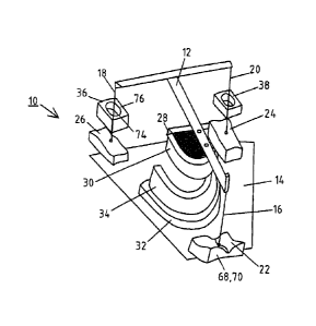
In the manufacture of the dental prosthesis one uses an arrangement 10 that
comprises an upper part 12
and a lower part 14, whereby the upper part 12 is supported in a sliding
manner via three supports 16,
18, 20 upon fields of motion 22, 24, 26, which are arranged stationary
relative to the lower part 14, in
particular on or in the latter. Preferably, the supports 16, 18, 20 possess
ends with a spheroid or
paraboloid shape, to be able to slide to the required degree on the fields of
motion 22, 24, 26.
Attached to the upper part 12, is a holding plate 28 for a first element 30
representing the upper jaw,
and attached to the lower part 14 is a holding plate 32 for a second element
34 representing the lower
jaw.
Further, at least two of the supports 16, 18, 20 (supports 18,20 in the
embodiment example), are
surrounded by travel limiters 36, 38 that reproduce the limitations of the
temporomandibular joint, i.e.
limits imposed by ligaments and cartilage.
WO 2011/120893 PCT/EP2011/054678
18
In accordance with the above-described measures pursuant to the invention one
uses digitized
measuring data and a reduction of the number of degrees of freedom, i.e.
preferably by excluding the
rotation of the lower jaw about the sagittal axis and the transverse axis and
the translation
perpendicular to the plane of occlusion, to describe the sliding motion of the
lower jaw in dental
contact with the upper jaw using 3 points of the coordinate system of the
lower jaw in the coordinate
system of the upper jaw. This motion is realized by allowing the upper part 12
to slide relative to the
lower part 14 by supporting the support elements 16, 18, 20 in the fields of
motion 22, 24, 26, which
can also be referred to as support points or support regions. In this, the
support points are situated at
the corners of a triangular column, ensuring a unique supported positioned.
Figure 2 illustrates the principle that the analysis of the motion of the
lower and upper jaw that is
measured using intra-oral scanning is used in the above-described manner (data
40), i.e. taking into
consideration at least the digital 3D data of the upper and lower jaws and the
occlusion in centric
position and the adjacent field, to compute the fields of motion 22, 24, 26 as
well as the travel limiters
36, 38 in order to subsequently use the corresponding digital data in a rapid
prototyping process to
produce the corresponding component parts that comprise the fields of motion
22, 24, 26 and the
motion limiters 36, 38.
Alternatively, the respective component parts may also be determined on the
basis of the data resulting
from the scanning of the lower and upper jaws as well from the lateral
movement of the lower and
upper jaw when using the minimum-increase-of-potential-energy method when the
centric position is
known (data 42).
Figure 3 illustrates the individual minimized design of the components needed
for the manufacturing
of the dental prosthesis. In particular, the components are: the prepared
tooth stump 44, shells or shell-
like elements 46, 48, available contact surfaces of teeth adjacent to the
tooth stump as well as a shell-
shaped component 50, which reproduces the geometry of the antagonist teeth.
The corresponding
component parts
WO 2011/120893 PCT/EP2011/054678
19
are subsequently spatially arranged relative to each other using the
corresponding digital 3D data, by
arranging them in the spatially coordinated receptacles of the first and the
second element 30, 34,
which represent the upper and lower jaw.
Starting from the above-explained minimal set consisting of the shell elements
46, 48 and the tooth
stump 44 the illustrated dental geometry can be extended all the way to a full
model, if for example a
dental technician desires to utilize die curvature information of a buccal
surface of the adjacent teeth.
Each of the elements 44, 46, 48, 50 possesses a joining element 52, 54, 56, 58
such as a peg, so that it
can be arranged positionally accurate in the receptacles of the first and
second elements 30, 34. A
hole/pin arrangement is suitable for the purpose of seating and fixing the
elements and a section
thereof is illustrated in Figure 3 whereby it is labeled by the reference
label 60 for the lower jaw and
the reference 62 for the upper jaw.
As Figures 4a and 4b illustrate, the first and second elements 30, 34, which
also can be referred to as
base plates, are provided with a hole/pin pattern 64 or a crosshatched pattern
65, which serve as
receptacles for the connecting elements 52, 54, 56, 58 of the tooth elements
44, 46, 48, 50. The joining
may be achieved via suitable friction-held mortising concepts.
According to the invention it also is possible for the fields of motion 22,
24, 26 and the motion limiters
36, 38 to replicate resiliencies that were measured additionally. In order to
realize this constructionally,
the components comprising the fields of motion 22, 24, 26 should be designed
accordingly with
respect to their materials. This is illustrated in principle in Figure 5a. For
example, a component 66
comprising the field of motion 22, 24, 26 as its surface consists of material
layers 68, 70, of which the
lower layer 70 - the layer that does not comprise the field of motion 22, 24,
26 - possesses an
elasticity
WO 2011/120893 PCT/EP2011/054678
that allows yielding to the required degree during the movements of the
support elements 16, 18, 20 on
the fields of motion 22, 24, 26, which matches the resilience.
Using individual layer thicknesses, which are computed form the data obtained
at the patient, one can
consequently represent the individual resiliency characteristics of the
temporomandibular joint all the
way to the resilience of individual teeth.
The equivalent applies to the travel limiters 36, 38, which comprise a recess
72, through which the rod-
shaped supports 18, 20 pass and which is bordered by a layer 74 of the desired
elasticity, in order to
emulate the restrictions of ligaments and cartilage of the temporomandibular
joint. The layer 74 is
contained in an outer body 76.
On the basis of the invention's teaching and the possibility to use minimal
components to model jaw
regions that are to be equipped with a dental prosthesis, one achieves
significant advantages with
respect to materials compared to conventional methods in which a complete
model of the lower and
upper jaw, as well as a quadrant model are needed. The material savings with
regard to the required
height of the tooth components are 62% compared to a complete model and 70%
compared to a
quadrant model. The volume and thus material savings are 98% compared to a
complete model and
92% compared to a quadrant model 92 %.
WO 2011/120893 PCT/EP2011/054678
21
List of reference symbols
Arrangement
12 Upper part
14 Lower part
16 Support
65 Cross-hatched pattern
18 Support 66 Component part
Support 22 Field of motion 68 Material layer
24 Field of motion 70 Material layer
72 Recess/Groove
26 Field of motion
28 Holding plate 74 Layer
First element 76 Outer body
32 Holding plate
34 Second element
36 Travel limiter
38 Travel limiter
Data
42 Data
44 Tooth stump
46 Shell element
48 Shell element
Component part
52 Connecting element
54 Connecting element
56 Connecting element
58 Connecting element
Hole/pin arrangement
62 Hole/pin arrangement
64 Hole/pin arrangement