Note: Descriptions are shown in the official language in which they were submitted.
0279.53.59 2012-1p-m.
WO 201.1/121089 KTTEP20,11/055045
Device for Interacting with Neut.()logiCal Tissue and Methods af Making and
Using the Same
100011
Field
100021 The present disclosure relates generally to field Of interacting with
biological tissue using
electrical probes, and more particularly to interacting with a .nourological
target through the use of
microelcetrode probes.
Background
100031 Neural reeording and neurostimulatiOn are eategories of inedieal
devices that are used to
interact electrically with time, In the ease of neuratrecording, physiological
measurements are
performed of neurological tisSue that eon diagnose, or treat, a patient, in
the case of ueurostimulation,
electric charge is transferred to the tissue in order to Create a therapeutic
outcome, or to generate a
diagnosis. Neural recording and neurostimulatiOn devices are Used today in the
eoehlea, the retina, the
peripheral nervenS system, the spine, the brain, and other parts of the body..
100041 hi a particular application whcm both neural recording and
neurostimulation ate Utilized,
conductive electrodes are placed in contact with deep brain structures in
order to treat certain
neurological Conditions. In the ease of stimUlating the Pcdtuicolopontine
Nucleus, for example, as
described :in U.S.. Pat No. 6,356,784, the therapy can treat the Syliiptoms of
Movement Di:Orders such
as Parkinson's disease, In the case of stimulating Brodmann Area 25, for
example, as described in
U.S. Pat, No. 7,;346,393, *therapy can treat the symptoms of Mood and Anxiety
Disorders,
100051 Qcrierally,ineural recording is performed in deep brain structures by
surgically inserting.
conductive electrodes. and amplifying neurological :signals using external
electronic equipment,
=Neurostimulation, is perfOrtried by surgically implanting conductive
electrodes in the target, and using
an implantable pulse gcnerater :to apply electrical Signals tO the conductive
electrodes,
WWI in some eases, such as described in U.S, Pat. No. 6,016,449, a system has
been developed
where both neural Mead* and neurostimulation fitnctionS are ovailoblc in a
single long term.
implantable, device.
100011 In Most teehiniOues, the electrodes used for neurril stimulation that
arc placed in contact with
tissue have been mettillic, cylindrical, with very sharp distal ends i most
cases, they only contain one:
microelectrode, which severely limits the amount of physiological information
that pan be collected i
from the patient,
100081 In other techniques, the electrodes used for neurostimulation that are
placed in contact with
tissue have been metallic cylindrical and relatively large. in "Slie (e ,t,
1.27 nun in diameter and 1.5
Main length). In Most eases, there are four or eight cylindrical electrodes
placed on a common axis.
1
CA 2795159 2018-07-26
CA 02795159 2012-10-01
WO 2011/121089 PCT/EP2011/055045
The stimulation methods are generally invasive, such as with the electrodes
used in Deep Brain
Stimulation, and the electrode lead is generally attached implantable pulse
generator.
[0009] Furthermore, advances in micromachining technology have developed whole
new
applications for medical devices, and in particular, implantable devices such
as for the treatment and
diagnosis of neurological disorders.
[0010] Advances in the imaging of tissue have elucidated the function and
anatomy of brain and
nervous tissue, permitting the development of new therapies which include
electrical stimulation
methods. A number of research groups have reported on different approaches for
imaging methods,
and the construction of implantable devices to deliver therapies. The imaging
methods are generally
extra-corporeal, and involve large and/or sophisticated equipment such as
Magnetic Resonance
Imaging systems.
[0011] One of the great challenges for clinicians delivering electrical
stimulation therapy is in
localizing the correct location for electrode placement, and then confining
the stimulation field to the
appropriate anatomical target to deliver the therapy, without inducing side
effects. Clinicians generally
combine pre-operative navigational planning derived from Magnetic Resonance
Imaging and/or
Computed Tomography scan imaging systems, with intra-operative microelectrode
recordings of
electrophysiological phenomenon to find and locate the optimal target.
[0012] Volumes of anatomical interest are commonly found using microelectrode
recording
techniques which involve invasively inserting metal tips to find the area of
interest by its
electrophysiological activity. This may be uncertain, time consuming, and
repetitive insertions may be
hazardous to patient health.
[0013] Unfortunately, there are several limitations to current practice
including uncertainty,
discomfort for the patient, and a heavy financial burden to deliver the
therapy. These factors can
render the therapy less attractive to clinicians, patients and payers.
[0014] It would be a very useful advancement in the art of neural recording
and neurostimulation
device technology and in the practice of functional neurostimulation if the
same device could image a
volume of brain tissue, and stimulate the same volume of tissue with precision
and safety.
[0015] There are many other medical applications for the present device, such
as detecting
malignant tissue within healthy tissue.
Summary
[0016] The present disclosure provides a design and method which permit the
imaging of small
volumes of tissue along with the capability of stimulating precise areas
within the volume of tissue.
The imaging method presents an advancement over conventional methods that have
relied on
expensive and low resolution systems. The stimulation method presents an
advancement over
conventional techniques which have not permitted the precise steering of
electrical fields into the
optimal tissue activation volume required to deliver effective therapy.
Combined, the inraging and
2
CA 02795159 2012-10-01
WO 2011/121089 PCT/EP2011/055045
stimulation method offers, for the first time, precise and high resolution
stimulation of tissue in
specific areas and volumes.
[0017] The disclosed devices and methods have special applications in medical
use, particularly in
the treatment of neurological disorders. Embodiments provide an unprecedented
resolution in the
imaging of tissue volumes by detecting local differences in electrical
characteristics. In this way, some
embodiments provide an imaging device, which while invasive and constrained in
use, is able to
provide a highly accurate registration of the imaged volume. The image
registration permits the
identification of anatomical structures, their surfaces and volumes, and their
electrical characteristics
such as, but not limited to, permittivity and conductivity.
101181 When combined with stimulation methods, the device permits stimulation
within specific
regions, surfaces, and volumes of the registered image. The presently
disclosed devices and methods
provide the clinician and/or surgeon a tool by which they can both visualize
the tissue of interest, and
stimulate specific areas within it. This greatly increases the accuracy and
safety of a surgery along
with an improvement in the chronic therapeutic effects of stimulation.
[0019] The use of localized tomographical imaging to determine implant
location and stimulation
volume is a unique and important advancement in the field of neurological
devices. Following the
present disclosure, for the first time, clinicians will be able to
substantially decrease the uncertainty in
device placement, and increase the specificity of the location of stimulation.
101201 The techniques described herein enjoy a number of advantages over
conventional techniques
to image tissue. Conventional methods in imaging require expensive equipment
installations and
resolution is increased by high field strengths in the case of Magnetic
Resonance Imaging, or high X-
ray dosages in the case of Computed Tomography scans. These high fields are
not compatible with
implantable devices containing metallic features, and artifacts caused by
devices translate to image
drift, errors, or decreased resolution in the registered image.
100211 By bringing the imaging device into contact with the volume of
interest, and measuring local
differences in electrical characteristics of the volume, the some embodiments
provide for images of
unprecedented resolution and fidelity.
100221 Likewise, the techniques for stimulation described herein enjoy a
number of advantages over
conventional efforts to stimulate tissue in a highly localized manner.
Conventional methods rely on
implantable devices with electrical leads often composed of cylindrical
contacts, or metal tips. Most
methods rely on stimulation volumes extending only outwards from the device,
as in the case of a
cylindrical device.
100231 One possible approach to this issue is the use of smaller electrodes,
in order to stimulate with
greater precision. However, there are practical limitations in surgery which
prevent the clinician from
precisely targeting the intended region. The image registration is often
performed before the surgery,
and subsequently navigational software is used to plan the implant trajectory
and location. One
approach is to incorporate the MM into the surgery, and perform intra-
operative imaging, however,
this is economically unviable in many hospitals, and the low field strengths
required to maintain
3
CA 02795159 2012-10-01
WO 2011/121089 PCT/EP2011/055045
compatibility with the implanted devices limit the resolution which can be
achieved. For example, a
surgeon would implant a cylindrical electrode lead after finding and
confirming the stereotactic co-
ordinates of the target site. As a more specific example, a neurosurgeon might
implant an electrode
lead in the Subthalamic Nucleus (S TN) to treat the symptoms of Parkinson's
Disease. The surgeon
might not be able to easily find the STN, and even more commonly, might not be
able to locate the
area within the STN that they seek to stimulate using electric current.
Furthermore, if the clinician
seeks to stimulate only a specific area, surface, volume, or population of
neurons or fiber bundles in,
around, or near the STN, it would not be possible using today's technology
because of the size and
geometry of existing electrode leads, which arc considerably larger than the
aforementioned targets.
100241 Thc presently disclosed devices and methods greatly improve current
practice without
fundamentally changing the surgical procedures currently in use. As an
example, a neurosurgeon
targeting the STN would implant the device using stereotactic co-ordinates
very close to the STN. The
surgeon would then deploy the several prongs from the device into and around
the STN. The imaging
method would be performed, which would provide the surgeon with a highly
localized and high
resolution image of the volume of tissue within the prongs of the device. The
image will consist of a
2D or 3D tomography of the volume of tissue. The image is constructed using
the differences in
electrical characteristics of the volume such as, but not limited to,
conductivity, permittivity,
conductivity and/or permittivity anisotropy. The image can therefore provide
information about, but
not limited to, the location and direction of fiber tracts, neural cell
density, the interface between grey
and white matter. The image is created using electrical impedance tomography
techniques which
involve a sequence of steps by which current is applied between two electrodes
and a potential
difference is preferably detected across two different electrodes, or the same
electrodes. By repeating
this procedure across all the electrodes in the periphery of the imaged
volume, an image can be
registered with the tomographic data using any one of a number of image
reconstruction techniques
and algorithms.
100251 Once the image has been registered, and the clinician can visualize
what the device's exact
location is, electrical stimulation can be applied to specific areas of the
volume using the principles of
neurostimulation and the superposition of electric fields. The clinician can
then steer the stimulation
field, and the volume of tissue activation, to particular areas of the volume.
For example, the image
might display the interface between the surface of the STN and fibers that are
projecting from it, or to
it. The clinician can then choose to stimulate this surface and the volume of
activation is directed there
by combining signals from several electrodes on the device prongs.
100261 As a result, a previously inaccessible region can be quickly located,
and stimulated, thereby
decreasing surgical times and increasing the efficacy of treatment. In
contrast, conventional devices
were limited by the geometrical arrangement and size of electrodes, and by the
lack of simultaneous or
in-situ imaging when stimulating.
100271 Another serious limitation to conventional devices is post-implantation
movement. A patient
that is reacting positively to the stimulation therapy might experience a
movement of their electrode
after implantation and thus, an immediate decrease or full halt in efficacy
and the possible introduction
4
CA 02795159 2012-10-01
WO 2011/121089 PCT/EP2011/055045
of side effects. With the present device, if a device shift occurs, the volume
of interest can be re-
imaged, and the stimulation volume can be re-directed to the proper region.
100281 The presently described devices and methods benefit from the ability of
modern
microfabrication techniques to facilitate the construction of the device.
Recent advances in surface
micromachining permit various electrode geometries consisting of favorable
materials such as
Platinum and Platinum-Iridium to be manufactured. The electrode substrates can
then be assembled
onto cut cylindrical components which consist of the prongs of the device.
This assembly is further
contained in an implantable catheter from which the prongs would extend during
surgery.
100291 In one aspect, an implantable neurological probe is disclosed
including: an elongated probe
assembly; at least one protruding shafts arranged at the distal end of the
elongated probe assembly; a
plurality of microelectrode elements arranged on the surface of the protruding
shafts; at least one
electrical contact arranged proximally along the elongated probe assembly; and
at least one electrical
conductor in electrical communication between at least one of the
microelectrode elements and the at
least one electrical contact.
100301 In some embodiments, the protruding shafts can be reversibly retracted
within the elongated
probe assembly. In some embodiments, the elongated probe shaft is configured
for insertion into a
human body using an accepted procedure for insertion of deep brain stimulation
leads. In some
embodiments, the diameter of the elongated probe assembly is between 1 mm and
3 mm.
100311 In some embodiments, at least one of the plurality of microelectrode
elements is a stimulating
electrode and at least one of the plurality of microclectrode elements is a
detecting electrode. In some
embodiments, at least one of the plurality of microelectrodes elements is both
a stimulating electrode
and a detecting electrode.
100321 In some embodiments, each microelectrode element is formed on a
conductive film, and
where each microelectrode element is embedded within two isolating substrates.
In some
embodiments, the microelectrode embedded substrate is formable into a
cylindrical assembly. In
some embodiments, the protruding shafts can be formed to bend radial from the
longitudinal axis of
the cylindrical assembly. In some embodiments, one of the protruding shafts is
longitudinal and
centered along the longitudinal axis of the cylindrical assembly. In some
embodiments, the protruding
shafts are stiffened by a supporting member. In some embodiments, the
longitudinal protruding shaft
is stiffened by a supporting member.
100331 In another aspect, a method for finding a neurological target
including: implanting a
neurological probe within a vicinity of a neurological target site, the
neurological probe including an
elongated cylindrical member, a plurality of protruding shafts, a plurality of
microelectrode elements
on each protruding shaft, at least one electrical contact arranged proximally
along the probe shaft, and
at least one electrical conductor in electrical communication between at least
one of the plurality of the
microelectrode elements and the at least one electrical contact; retracting
the protruding shafts within
the elongated cylindrical member before surgical implantation; expanding the
protruding shafts in the
vicinity of the neurological target site following implantation; recording
electrophysiological signals
from the neurological target site using at least one of the microelectrode
elements on at least one of the
CA 02795159 2012-10-01
WO 2011/121089 PCT/EP2011/055045
protruding shafts; and stimulating the neurological target using at least one
of the microelectrode
elements on at least one of the protruding shafts.
100341 In some embodiments, the protruding shafts are retracted within the
elongated cylindrical
member using a flexible pull wire situated in a lumen of the elongated
cylindrical member. In some
embodiments, the protruding shafts are expanded from within the elongated
cylindrical member using
a rigid, or semi-rigid, push rod situated in a lumen of the elongated
cylindrical member. In some
embodiments, the act of positioning the distal end of the neurological probe
includes recording neural
activity detected by at least one of the plurality of microelectrode elements
and repositioning the distal
end of the neurological probe as required, until the recorded activity is
indicative of the distal end of
the elongated probe shaft being located sufficiently at the neurological
target site.
11:0351 In some embodiments, the act of positioning the distal end of the
neurological probe includes
stimulating neural activity by applying electrical signals to at least one of
the plurality of
microelectrode elements on at least one of the plurality of protruding shafts,
performing a clinical
evaluation of the efficacy on the stimulation site in the implanted patient,
and repositioning the distal
end of the neurological probe as required, until the patient's response is
indicative of the distal end of
the elongated probe shaft being located sufficiently at the neurological
target site.
11:0361 In some embodiments, the act of positioning the distal end of the
neurological probe includes
inhibiting neural activity by applying electrical signals to at least one of
the plurality of microelectrode
elements on at least one of the plurality of protruding shafts, performing a
clinical evaluation of the
efficacy on the inhibition site in the implanted patient, and repositioning
the distal end of the
neurological probe as required, until the patient's response is indicative of
the distal end of the
elongated probe shaft being located sufficiently at the neurological target
site.
10:1371 In another aspect, a method is disclosed for finding a neurological
target including:
implanting a neurological probe within a vicinity of a neurological target
site, the neurological probe
including an elongated cylindrical member, a plurality of protruding shafts, a
plurality of
microelectrode elements on each protruding shaft, at least one electrical
contact arranged proximally
along the probe shaft, and at least one electrical conductor in electrical
communication between at
least one of the plurality of the microelectrode elements and the at least one
electrical contact;
retracting the protruding shafts within the elongated cylindrical member
before surgical implantation;
expanding the protruding shafts in the vicinity of the neurological target
site following implantation;
applying an oscillating electric current between at least two of the
microelectrode elements on at least
one of the protruding shafts; and detecting an electric voltage between at
least two of the
microelectrode elements on at least one of the protruding shafts.
11:0381 In some embodiments, the act of applying oscillating currents and
detecting electric voltages
is performed to image the electrical characteristics of the volume of
neurological tissue between the
protruding shafts.
11:0391 In another aspect, an implantable neurological probe is disclosed
including: an elongated
shaft having a distal end and an internal lumen; a support cylinder slidingly
disposed in only a distal
portion of the internal lumen; a plurality of shafts coupled to the support
cylinder and arranged to be
selectively extended from the distal end of the elongated shaft; a plurality
of microelectrode elements
disposed on each of the plurality of shafts, the microelectrode elements
including a planar substrate
6
CA 02795159 2012-10-01
WO 2011/121089 PCT/EP2011/055045
having an insulative layer and a plurality of conductive traces disposed on
the insulative layer, a stylet
removably disposed in the internal lumen and configured to contact the support
cylinder to selectively
extend the plurality of shafts during implantation; and a pull wire coupled to
the support cylinder to
selectively retract the support cylinder and plurality of shafts within the
internal lumen.
11:0401 Some embodiments include a push-pull rod which includes the pull wire
and the stylet.
10:1411 In some embodiments, the elongated shaft is configured for insertion
into a human body
using an accepted procedure for insertion of deep brain stimulation leads.
100421 In some embodiments, the diameter of the elongated shaft is between 1
mm and 3 mm.
10:1431 In some embodiments, at least one of the plurality of microelectrode
elements is a stimulating
electrode and at least one of the plurality of microelectrode elements is a
detecting electrode. In some
embodiments, at least one of the plurality of microelectrodes elements is both
a stimulating electrode
and a detecting electrode.
10:1441 In some embodiments, each microelectrode element is formed on a
conductive film, and
where each microelectrode element is embedded within two isolating substrates.
100451 In some embodiments, the microelectrode embedded substrate is formable
into a cylindrical
assembly.
100461 In some embodiments, the protruding shafts can be formed to bend
radially from the
longitudinal axis of the cylindrical assembly.
[(0471 In some embodiments, one of the protruding shafts extends and is
centered along the
longitudinal axis of the cylindrical assembly.
100481 In some embodiments, the protruding shafts are stiffened by a
supporting member. In some
embodiments, the longitudinal protruding shaft is stiffened by a supporting
member.
100491 In another aspect, an implantable neurological probe is disclosed
including: an elongated
shaft having a distal end and an internal lumen; a plurality of shafts
arranged to be selectively
extended from the distal end of the elongated shaft; and a plurality of
microelectrode elements
disposed on each of the plurality of shafts, the microelectrode elements
including a planar substrate
having an insulative layer and a plurality of conductive traces disposed on
the insulative layer. In
some embodiments, the plurality of shafts define a substantially cylindrical
volume when fully
extended.
11:0501 In some embodiments, the elongated shaft is configured for insertion
into a human body
using an accepted procedure for insertion of deep brain stimulation leads.
100511 In some embodiments, the diameter of the elongated shaft is between 1
mm and 3 mm.
110521 In some embodiments, at least one of the plurality of microelectrode
elements is a stimulating
electrode and at least one of the plurality of microelectrode elements is a
detecting electrode.
10:1531 In some embodiments, at least one of the plurality of microelectrodes
elements is both a
stimulating electrode and a detecting electrode. In some embodiments, each
microelectrode element is
formed on a conductive film, and where each microelectrode element is embedded
within two
7
CA 02795159 2012-10-01
WO 2011/121089 PCT/EP2011/055045
isolating substrates. In some embodiments, the microelectrode embedded
substrate is formable into a
cylindrical assembly. In some embodiments, the protruding shafts can be formed
to bend radially from
the longitudinal axis of the cylindrical assembly. In some embodiments, one of
the protruding shafts
extends and is centered along the longitudinal axis of the cylindrical
assembly. In some embodiments,
the protruding shafts are stiffened by a supporting member. In some
embodiments, the longitudinal
protruding shaft is stiffened by a supporting member.
11:0541 In another aspect, a method is disclosed for finding a neurological
target including:
implanting a neurological probe within a vicinity of a neurological target
site, the neurological probe
including: an elongated shaft having a distal end and an internal lumen; a
support cylinder slidingly
disposed in only a distal portion of the internal lumen; a plurality of shafts
coupled to the support
cylinder and arranged to be selectively extended from the distal end of the
elongated shaft; a plurality
of microelectrode elements disposed on each of the plurality of shafts, the
microelectrode elements
including a planar substrate having an insulative layer and a plurality of
conductive traces disposed on
the insulative layer, a sty-let removably disposed in the internal lumen and
configured to contact the
support cylinder to selectively extend the plurality of shafts during
implantation; and a pull wire
coupled to the support cylinder to selectively retract the support cylinder
and plurality of shafts within
the internal lumen. In some embodiments, the method further includes:
retracting the plurality of
shafts within the internal lumen before surgical implantation; extending the
plurality of shafts in the
vicinity of the neurological target site following implantation; recording
electrophysiological signals
from the neurological target site using at least one of the microelectrode
elements on at least one of the
protruding shafts; and stimulating the neurological target using at least one
of the microelectrode
elements on at least one of the plurality of shafts.
[0055] In some embodimens, the method includes: after the acts of recording
and stimulating,
retracting the plurality of shafts within the internal lumen and removing the
neurological probe from a
subject.
100561 In some embodiments, the protruding shafts are retracted using the pull
wire. In some
embodiments, the plurality of shafts are extended using the stylet. In some
embodiments, the
neurological probe includes a push-pull rod which includes the pull wire and
the stylet
11:0571 In some embodiments, the act of recording neurophysiological signals
includes recording
neural activity detected by at least one of the plurality of microelectrode
elements and repositioning
the distal end of the elongated shaft as required, until the recorded activity
is indicative of the distal
end of the elongated probe shaft being located sufficiently at the
neurological target site.
100581 Some embodiments include stimulating neural activity by applying
electrical signals to at
least one of the plurality of microelectrode elements on at least one of the
plurality of shafts,
performing a clinical evaluation of the efficacy on the stimulation site in
the implanted patient, and
repositioning the distal end of the elongated shaft as required, until the
patient's response is indicative
of the distal end of the elongated shaft being located sufficiently at the
neurological target site.
10:1591 Some embodiments include :inhibiting neural activity by applying
electrical signals to at least
one of the plurality of microelectrode elements on at least one of the
plurality of shafts, performing a
clinical evaluation of the efficacy on the inhibition site in the implanted
patient, and repositioning the
8
CA 02795159 2012-10-01
WO 2011/121089 PCT/EP2011/055045
distal end of elongated shaft as required, until the patient's response is
indicative of the distal end of
the elongated shaft being located sufficiently at the neurological target
site.
100601 In another aspect, a method is disclosed for finding a neurological
target including:
implanting a neurological probe within a vicinity of a neurological target
site, the neurological probe
including: an elongated shaft having a distal end and an internal lumen; a
support cylinder slidingly
disposed in only a distal portion of the internal lumen; a plurality of shafts
coupled to the support
cylinder and arranged to be selectively extended from the distal end of the
elongated shaft; a plurality
of microelectrode elements disposed on each of the plurality of shafts, the
microelectrode elements
including a planar substrate having an insulative layer and a plurality of
conductive traces disposed on
the insulative layer, a stylet removably disposed in the internal lumen and
configured to contact the
support cylinder to selectively extend the plurality of shafts during
implantation; and a pull wire
coupled to the support cylinder to selectively retract the support cylinder
and plurality of shafts within
the internal lumen. Sonic embodiments include retracting the plurality of
shafts within the internal
lumen before surgical implantation; expanding the plurality of shafts in the
vicinity of the neurological
target site following implantation; applying an oscillating electric current
between at least two of the
microelectrode elements on at least one of the plurality of shafts; and
detecting an electric voltage
between at least two of the microelectrode elements on at least one of the
plurality of shafts.
100611 Some embodiments include: after the act of detecting, retracting the
plurality of shafts within
the internal lumen and removing the neurological probe from a subject.
100621 Some embodiments include imaging the electrical characteristics of the
volume of
neurological tissue between the plurality of shafts based on the applied
oscillating electric current and
the detected electric voltage.
10:1631 In some embodiments, the neurological probe includes a push-pull rod
which includes the
pull wire and the stylet.
100641 In another aspect, a method for finding a neurological target
including: implanting a
neurological probe within a vicinity of a neurological target site, the
neurological probe including: an
elongated shaft having a distal end and an internal lumen; a plurality of
shafts arranged to be
selectively extended from the distal end of the elongated shaft; and a
plurality of microelectrode
elements disposed on each of the plurality of shafts, the microelectrode
elements including a planar
substrate having an insulative layer and a plurality of conductive traces
disposed on the insulative
layer, where the plurality of shafts define a substantially cylindrical volume
when fully extended. In
some embodiments, the method includes: retracting the plurality of shafts
within the internal lumen
before surgical implantation; extending the plurality of shafts in the
vicinity of the neurological target
site following implantation; recording electrophysiological signals from the
neurological target site
using at least one of the microelectrode elements on at least one of the
protruding shafts; and
stimulating the neurological target using at least one of the microelectrode
elements on at least one of
the plurality of shafts.
10:1651 In some embodiments, the protruding shafts are retracted using a pull
wire. In some
embodiments, he plurality of shafts are extended using a stylet. In some
embodiments, the
neurological probe includes a push-pull rod which includes the pull wire and
the stylet.
9
CA 02795159 2012-10-01
WO 2011/121089 PCT/EP2011/055045
11:0661 In some embodiments, the act of recording neurophysiological signals
includes recording
neural activity detected by at least one of the plurality of microelectrode
elements and repositioning
the distal end of the elongated shaft as required, until the recorded activity
is indicative of the distal
end of the elongated probe shaft being located sufficiently at the
neurological target site.
100671 Some embodiments include after the acts of recording and stimutating,
retracting the
plurality of shafts within the internal lumen and removing the neurological
probe from a subject
100681 Some embodiments include stimulating neural activity by applying
electrical signals to at
least one of the plurality of microelectrode elements on at least one of the
plurality of shafts.
performing a clinical evaluation of the efficacy on the stimulation site in
the implanted patient; and
repositioning the distal end of the elongated shaft as required, until the
patient's response is indicative
of the distal end of the elongated shaft being located sufficiently at the
neurological target site.
11:0691 Some embodiments include inhibiting neural activity by applying
electrical signals to at least
one of the plurality of microelectrode elements on at least one of the
plurality of shafts, performing a
clinical evaluation of the efficacy on the inhibition site in the implanted
patient, and repositioning the
distal end of elongated shaft as required, until the patient's response is
indicative of the distal end of
the elongated shaft being located sufficiently at the neurological target
site.
11:0701 In another aspect, a method for finding a neurological target
including: implanting a
neurological probe within a vicinity of a neurological target site, the
neurological probe including: an
elongated shaft having a distal end and an internal lumen; a plurality of
shafts arranged to be
selectively extended from the distal end of the elongated shaft; and a
plurality of microelectrode
elements disposed on each of the plurality of shafts, the microelectrode
elements including a planar
substrate having an insulative layer and a plurality of conductive traces
disposed on the insulative
layer,vvhere the plurality of shafts define a substantially cylindrical volume
when fully extended.
Some embodiment include retracting the plurality of shafts within the internal
lumen before surgical
implantation; expanding the plurality of shafts in the vicinity of the
neurological target site following
implantation; applying an oscillating electric current between at least two of
the microelectrode
elements on at least one of the plurality of shafts; and detecting an electric
voltage between at least
two of the microelectrode elements on at least one of the plurality of shafts.
100711 Some embodiments include imaging the electrical characteristics of the
volume of
neurological tissue between the plurality of shafts based on the applied
oscillating electric current and
the detected electric voltage.
100721 Some embodiments include: after the act of detecting, retracting the
plurality of shafts within
the internal lumen and removing the neurological probe from a subject
100731 Various embodiments may include any of the above described elements or
steps alone, or in
any suitable combination.
Brief Description of the Drawings
100741 The foregoing and other objects, features and advantages of the
invention will be apparent
from the following more particular description of preferred embodiments of the
invention, as
illustrated in the accompanying drawings in which like reference characters
refer to the same parts
throughout the different views.
CA 02795159 2012-10-01
WO 2011/121089 PCT/EP2011/055045
100751 FIG. 1 is a perspective view of one embodiment of an elongated
microelectrode assembly.
100761 FIG. 2 is a perspective view of a portion of a human anatomy
illustrating an exemplary
elongated microelectrode assembly implanted therein.
100771 FIG. 3 is a perspective view of a portion of a human anatomy
illustrating an exemplary
microelectrode structure positioned at a neurological target.
100781 FIG. 4A is a perspective view of a distal portion of the elongated
microelectrode assembly of
FIG. 1 in the expanded position.
100791 FIG. 4B is a perspective view of a distal portion of the elongated
microelectrode assembly of
FIG. 1 in the retracted position.
100801 FIG. 5 is a perspective view of a proximal portion of the elongated
microelectrode assembly
of FIG. 1.
100811 FIG. 6 is a planar view of an embodiment of a microelectrode array
film.
100821 FIG. 7 is a perspective view of the embodiment of a microelectrode
array film of FIG. 6 after
it has been assembled.
1071831 FIG. gA is a planar top view of the microelectrode array film assembly
of FIG. 7.
100841 FIG. 8B is a planar side view of the microclectrode array film assembly
of FIG. 7.
11:8:1851 FIG. 9 is a planar frontal view of the microelectrode array film
assembly of FIG. 7.
100861 FIG. 10 is a perspective view of the microelectrode array film assembly
of FIG. 7 in the
retracted position
11:0871 FIG. 11 is a planer view of the retracted microelectrode array film
assembly of FIG. 10.
100881 FIG. 12A is a perspective view of a central pin component.
11:0891 FIG. 12B is a planar side view of the central pin component of FIG.
12A.
100901 FIG. 13A is a perspective view of the outer legs component shown in the
expanded position.
11:0911 FIG. 13B is a perspective view of the outer legs component shown in
the retracted position.
11:0921 FIG. 14 is a perspective view of the microelectrode array film
assembly of FIG. 7 shown
assembled to the central pin component of FIG, 12A.
100931 FIG. 15 is a perspective view of the microelectrode assembly of FIG. 14
shown assembled to
the flexible pull wire, and a microelectronic component.
1071941 FIG. 16 is a perspective view of the microelectrode assembly of FIG.
15 shown assembled to
helical lead wires, and the outer legs component of FIG. 13A.
11
CA 02795159 2012-10-01
WO 2011/121089 PCT/EP2011/055045
11:0951 FIG. 17 is a perspective view of the microelectrode assembly of FIG.
16 shown assembled to
an outer tubing and a stiff push rod.
[0096] FIG. 18 is a close-up perspective view of the microelectrode assembly
of FIG. 17 showing
the flexible pull wire and the stiff push rod in more detail.
100971 FIG. 19A is a perspective view of the perforated end cap.
100981 FIG. 19B is a planar view of the perforated end cap.
11:0991 FIG. 20 is a cut-away perspective view of the microelectrode assembly
of FIG. 4A with
segments of the perforated end cap and outer legs component removed.
[0100] FIG. 21 is a cut-away perspective view of the retracted microelectrode
assembly of FIG. 4B
with segments of the perforated end cap and outer legs component removed.
[0101] FIG. 22 is a planar view of the microelectrode assembly demonstrating
microelectrode
elements on the same plane.
[0102] FIG. 23 is a perspective view the assembly and planes of FIG. 22.
[0103] FIG. 24 is a perspective view of an alternative embodiment of the
elongated microelectrode
assembly of FIG. 1.
[0104] FIG. 25 is a planar front view of the alternative embodiment of FIG.
24.
[0105] FIG. 26 is a planar side view of the alternative embodiment of FIG. 24.
[0106] FIG. 27 is a perspective view of an alternative embodiment of the
elongated microelectrode
assembly of FIG. 1
[0107] FIG. 28 is a planar side view of the alternative embodiment of FIG. 27.
[0108] FIG. 29 is a perspective view of an alternative embodiment of FIG. 1
where the
microelectrode arrays are placed on the outside of the protruding shafts.
[0109] FIG. 30 is a planar back view of the alternative embodiment of FIG. 29.
[0110] FIG. 31 is a planar side view of the alternative embodiment of FIG. 29
depicting separate
stimulation and recording electrodes.
[0111] FIG. 32 is a detail perspective view of the alternative embodiment of
FIG. 29.
[0112] FIG. 33 is an additional detail perspective view of the alternative
embodiment of FIG. 29.
[0113] FIG. 34 is a component of the alternative embodiment of FIG. 29.
[0114] FIG. 35 is an additional component of the alternative embodiment of
FIG. 29.
12
CA 02795159 2012-10-01
WO 2011/121089 PCT/EP2011/055045
[0115] FIG. 36 is yet an additional component of the alternative embodiment of
FIG. 29.
101161 FIG. 37 is a perspective view of an alternative embodiment of FIG. 1
where the protruding
shafts have been implemented at two different regions of the longitudinal
axis.
[0117] FIG. 38A is a planar view of the alternative embodiment of FIG. 37.
[0118] FIG. 38B is an additional planar view of the alternative embodiment of
FIG. 37.
[0119] FIG. 39A is a perspective view of the mieroelectrode array film
required in the assembly of
the alternative embodiment of FIG. 37.
[0120] FIG. 39B is a perspective view of the protruding shaft support required
in the assembly of the
alternative embodiment of FIG. 37.
[0121] FIG.40A is a perspective view of an alternative embodiment of FIG. 1
where the
microelectronic component is not required.
[0122] FIG. 40B is a perspective view of the mieroelectrode array film
required in the assembly of
the alternative embodiment of FIG. 40A.
[0123] FIG. 40C is a perspective view of an alternative embodiment of FIG. 1
where the protruding
shafts are not rigidified by the protruding shaft support.
[0124] FIG. 40D is a detail perspective view of the alternative embodiment of
FIG. 40C.
101251 FIG. 41 is a schematic of a neural recording microelectronic circuit.
[0126] FIG. 42 is a schematic of a neural stimulation microelectronic circuit.
[0127] FIG. 43 is a schematic of a combined neural recording and stimulation
microelectronic
circuit.
[0128] FIG. 44 demonstrates the Electrical Impedance Tomography method
described herein.
Detailed Description
[0129] Described herein are microelectrode array devices, and methods of
fabrication and use of the
same, to provide highly localized and efficient electrical stimulation of a
neurological target, such as
individual neurons, groups of neurons, and neural tissue as may be located in
an animal nervous
system, such as deep within a human brain. In small, difficult to find brain
targets such as the
Pedunculopontine Nucleus, or in targets that requires highly localized levels
of neural stimulation,
such as the Subthalamic Nucleus, many microelectrodes are required in the
brain region to find the
target using electrophysiological recording. A higher number of
microelectrodes will increase the
chance of fmding the neurons required for therapeutic stimulation. The
microelectrode, or group of
microelectrodes, that are closest to the target brain region will be used for
chronic, therapeutic
stimulation or inhibition.
13
CA 02795159 2012-10-01
WO 2011/121089 PCT/EP2011/055045
[0130] The stimulation can be highly localized, because the microelectrode
elements can be as small
as only 2 um or large as 2 mm in either of diameter or width. The relative
spacing between such
microelectrode elements can also be as small as only 2 um or as large as 2 mm.
Generally,
microelectrodes of about 150 um in diameter, with about a 1000 IM1 spacing are
particularly efficient
in stimulating neural tissue.
[0131] An array of such microelectrode elements may consist of one or more
such elements (e.g.,
sixteen elements), each disposed at a respective position, or site. This is in
contrast to currently
available stimulation leads, such as the Model 3387 or Model 3389 DBS leads
commercially available
from Medtronic, Inc. of Minneapolis, MN. Such commercially available devices
include relatively
large, cylindrical electrodes measuring about 1.5 mm in height, and having a
maximum of only four
electrodes in use today for deep brain stimulation.
[0132] Smaller microelectrode elements can be used to provide neurological
stimulation that is
highly localized and efficient because an array of such microelectrodes can
also be used to identify the
stimulation region of interest. For example, one or more microelectrode
elements of such an array of
microelectrode elements can be used to record neuronal activity in the
vicinity of the
detecting/recording microelectrode elements. Such refinement offered by the
relatively small size
and/or spacing of the microelectrode elements can be used to obtain a highly
localized map of
neuronal activity in the region surrounding the implant. A suitably
dimensioned microelectrode array
having multiple microelectrode elements positioned in a general vicinity of a
neurological target, can
be used to locate a precise neurological target without further repositioning.
by identifying those one
or more microelectrode elements located in a very specific region of the
neurological target. The
microelectrode array can be programmed to stimulate in a very specific region,
for example, using
only a certain number of the microelectrode elements to actively stimulate the
surrounding neurons
and/or neuronal tissue, while other electrode elements of the array remain
inactive.
[0133] In the embodiments described, the microelectrode arrays are positioned
in three dimensional
space. This has been a previous limitation of such microelectrode devices,
which were usually
implement in linear arrays, or two dimensional arrays on films. In the present
embodiment
microelectrode arrays are positioned along shafts which radiate from a central
lumen, in order to cover
as much volume in the target region with microelectrode arrays.
[0134] In some embodiments, an elongated device including such microelectrode
arrays having
elements with relatively small size and/or spacing can be used to obtain a
highly localized map of
neuronal activity in the region surrounding the implant. For example, such a
device configured with a
linear array of microelectrodes positioned along a length of a distal end of
the device can be placed
into a patient's brain. Preferably, the elements of the microelectrode array
envelop a region including
the neurological target. Neurological activity can then be independently
detected by one or more of
the microelectrode elements. The detected activity may be captured in a
recorder or display device,
allowing a clinician to identify which one or more of the microelectrode
elements is positioned closest
to the intended target. Knowing a respective location of each of the
microelectrode elements along the
device, and determining the distance to a reference, such as the patient's
skull, a precise location of the
target can be determined as the distance along a trajectory of the device,
measured from the reference
14
CA 02795159 2012-10-01
WO 2011/121089 PCT/EP2011/055045
to the particular microelectrode element. Beneficially, location of the target
can be determined
without any repositioning of the elongated device, thereby simplifying the
medical procedure and
reducing patient risk.
[0135] In some embodiments, the device is for acute intra-surgical use, being
removed after the
target has been located, being replaced with a chronic probe, positioned at
the determined target
location. Alternatively or in addition, the device itself can be left in place
as a chronic device, the
same microelectrodes, or different ones, being used to record and/or stimulate
the neurological target
over an extended period.
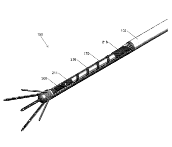
[0136] One embodiment of a microelectrode device illustrated in FIG. 1
includes an elongated
microelectrode lead assembly 100 sometimes referred to as an electrode lead.
The microelectrode lead
assembly 100 includes an external cylindrical member 102 including a
microelectrode array assembly
150 located relative to a distal end and one or more electrical contacts 106
located relative to a
proximal end. The exemplary microelectrode lead assembly 100 includes one or
more microelectrode
array shafts 160 adjacent to its distal tip. The microelectrode array assembly
150 has five protruding
shafts 160, with disc microelectrode elements disposed along an interior
surface of an extended
substrate. In the present embodiment four shafts protrude, to one of the
anterior, posterior, lateral, or
medial directions. An additional shaft protrudes along the same longitudinal
axis of the electrode lead,
referred to as the central shaft. The microelectrode lead assembly 100 also
includes eight electrically
conductive, cylindrical contacts, or contact rings (generally 106) distributed
along a longitudinal axis
of the proximal end of the assembly 100. In the exemplary embodiment, each of
the microelectrode
elements is in electrical communication with a proximal contact 106 via an
embedded microelectronic
element. In use, stimulation signals are directed from an implantable pulse
generator, or controller to
the microelectrode array. Additionally, in use, recording signals are directed
from the microelectrode
array to an implanted or external data recorder.
[0137] The microclectrode lead assembly 100 is preferably sized and shaped for
its intended
neurological application. For example, the microelectrode lead assembly 100
may be at least partially
placed within the central nervous system. Alternatively or in addition, the
microelectrode lead
assembly 100 may be at least partially placed within other parts or organs of
the body, such as the
epidural space of the spine, or other locations within the peripheral nervous
system, or within an organ
such as the liver or heart. Thus the diameter and length of the microelectrode
lead assembly 100 may
vary depending on the particular anatomical target. Additionally, the
configuration of the
microelectrode array shafts 160 is also sized and shaped for an intended
neurological target. The
number, shape, orientation, size, and spacing of the microelectrode elements
of the array can be
defined in response to the intended neurological target.
[0138] In at least some embodiments one or more of the microelectrode elements
are sized and or
spaced to record from and/or stimulate neurons. The microelectrode lead
assembly 100 can be used to
detect and/or record neuronal activity at the neurological target. Neuronal
activity naturally occurring
within the neurological target gives rise to local electromagnetic fields that
can be detected by one or
more of the microelectrode elements of the microelectrode array. For example,
electric fields
produced by neurons will polarize one or more of the microelectrode elements.
Such polarization
gives rise to an electrical potential with respect to a reference, such as
electrical ground, or another one
CA 02795159 2012-10-01
WO 2011/121089 PCT/EP2011/055045
of the microelectrode elements. Such electric activity can be further
conducted to one or more of the
cylindrical contacts 106 through the internal electrical conductors. One or
more of the cylindrical
contacts 106, in turn, can be connected to one or more additional medical
devices for further
processing of the detected electrical activity. For example, the cylindrical
contacts 106 can be coupled
to a display device or recording device for displaying and/or recording
electrical activity from the
neurological target.
101391 Alternatively or in addition, one or more of the microelectrode
elements can be used to
electrically stimulate the neurological target. For example, one or more
externally generated electrical
signals can be applied to one or more of the cylindrical contacts 106. These
electrical signals can be
conducted through the internal electrical conductors to one or more of the
microelectrode elements of
the microelectrode array. Depending on the amplitude and polarity of the
electrical signals, an
electrical field will be induced by the polarized microelectrode elements.
Electrical fields induced by
such polarization can interact with one or more neurons at the neurological
target.
[0140] Alternatively or in addition, one or more of the microelectrode
elements can be used to
perform Electrical Impedance Tomography of a neurological target or other
bodily organ. For
example, one or more externally generated electrical signals can be applied as
a current to one or more
of the microelectrode elements. Depending on the physiological characteristics
of the tissue being
imaged, and depending on the frequencies of the current signals applied, an
electrical field will be
induced in the tissue. Electrical fields induced by such polarization can be
detected by other
microelectrode elements, thereby creating a localized image of conductivity,
permittivity, and/or other
electrical characteristics.
[0141] Mechanical components of the implantable neurological lead assembly 100
include the
elongated outer cylindrical member 102, which can be a simple polymeric
cylinder, or a rigid metallic
or rigid polymeric cylinder. The outer cylindrical member 102 can vary in
length and diameter but is
generally at least about 28 cm long, (e.g., at least 20 cm long, at least 25
cm long, at least 28 cm long,
at least 30cm long, etc.) and around 1.27 mm in diameter (e.g., in the range
of 1.0-2.0 mm in
diameter).
[0142] The neurological lead 100 can be implanted near a neurological target,
such as a target brain
structure, using common neurosurgical techniques such as stereotaxy or
endoscopy. The
microelectrode lead assembly 100 can be inserted in its retracted state
without support, or within a
supporting cannula having an inner dimension slightly larger than the outer
dimension of the device.
The cannula, when used, would be removed once the microelectrode lead assembly
100 has been
suitably positioned. In some embodiments a lumen along the axis of the outer
cylindrical member 102
permits the insertion of a rigid stylet which renders the microelectrode lead
assembly 100 rigid during
surgical implantation. This is particularly helpful during insertion,
positioning and repositioning of
flexible embodiments of the microelectrode lead assembly 100. The stylet is
removed after
implantation leaving the probe in its surgical target. In some embodiments the
stylet is also a rigid
push rod, which is used to expand the microelectrode array shafts 160 into the
tissue. In some
embodiments, the microelectrode lead assembly 100 contains a flexible pull
wire which is used to pull
the microelectrode array shafts 160 back into the retracted position. In yet
additional embodiments, the
microelectrode lead assembly 100 contains only one rigid push-pull rod which
is used to both push
16
CA 02795159 2012-10-01
WO 2011/121089 PCT/EP2011/055045
and pull the microelectrode array shafts 160 in its expanded and retracted
position respectively. In yet
additional embodiments, where the microelectrode lead assembly 100 is not
intended to remain in the
patient's brain after surgery, the rigid push-pull rod may be permanently
attached to the
microelectrode array shafts 160.
[0143] A clinician can connect one or more of the microelectrode elements to a
display unit or a
recording unit through the cylindrical contacts 106. The recording unit, not
shown, allows a clinician
to identify certain regions of the brain according to their electrical
activity. In some embodiments,
such recording information can be processed automatically, through the use of
a suitably programmed
computer processor. The electrodes used to record from the brain can be the
same electrodes as those
used to stimulate tissue. The recording electrodes can also be separate from
those used to stimulate
the brain. This situation might be preferred because electrodes destined for
recording may be different
in size and design than those for stimulation.
[0144] The operator can connect the electrodes to an external stimulation
source or an implantable
source. In either instance, the source can include a pulse generator for
applying signals to the
electrode sites. The signals from such a pulse generator can be connected
directly to the electrodes, or
they can be preprocessed using electronics embedded in the device. The
electronics can filter certain
parts of the original signal. If there are more electrodes than signals, the
electronics can route or
otherwise interconnect the stimulation source as necessary.
[0145] A perspective view of the portion of a human anatomy is illustrated in
FIG. 2, showing
implantation of an exemplary elongated microelectrode probe assembly 124
position for interaction
with a neurological target located deep within the brain. A distal portion of
the microelectrode probe
assembly 124 is positioned at the neurological target 130, in this instance
located within the human
brain 132. Several exemplary microelectrode array shafts 134 protrude from the
distal portion of the
microelectrode probe assembly 124. In some embodiments the proximal end of the
microelectrode
probe assembly 124 is connected to a first medical device 128. For example,
the first medical device
128 may include an electronic assembly implanted external to the brain 132 to
minimize invasion into
the body. Alternatively or in addition, a second medical device, which again
may include an
electronic assembly such as a pulse generator 122 can be implanted at a remote
portion of the subject
body. As shown, a second electronic assembly 122 is implanted within a chest
cavity 120. When one
or more medical devices, such as the exemplary pulse generator 122 are located
remotely in this
manner, a cable 126 may also be implanted within the subject's body to
interconnect the pulse
generator 122 to the electronic assembly 128, when present or directly to
cylindrical contacts located
at the proximal end of the microelectrode probe assembly 124.
[0146] Referring now to FIG. 3, a cross-sectional view of a portion of an
anatomy 148 is shown,
illustrating an exemplary microelectrode probe assembly 140 positioned at a
neurological target 148
(e.g., subthalmic nucleus, shown). The microelectrode probe assembly 140
includes five
microelectrode array shafts, 141A, 141P, 141L, 141M, 141C (generally 141)
protruding from a
cylindrical containment structure 143. On each microelectrode array shaft 141
are three
microelectrode elements 145 distributed linearly along the microelectrode
array shaft 141. Preferably,
the microelectrode probe assembly 140, and its protruding microelectrode
electrode arrays shafts 141
17
CA 02795159 2012-10-01
WO 2011/121089 PCT/EP2011/055045
are shaped, spaced, and sized to allow one or more of the microelectrode
elements 145 to be
positioned at the neurological target 149.
[0147] As illustrated, one or more of the microelectrode elements 145 of the
microelectrode probe
assembly 140 are positioned in intimate contact with the neurological target
149. In more detail, each
microelectrode element 145 is a disc electrode along a shaft. It is understood
that some microelectrode
array shafts 141 can be in contact with the neurological target, while other
microelectrode array shafts
141 are not (as shown). Additionally, it is understood that some
microelectrode elements 145 can be in
contact with the neurological target, while other microelectrode elements 145
are not (as shown). In at
least some embodiments, one or more of the microelectrode elements 145 are
remotely accessible
from a proximal end of the probe assembly 140 via one or more electrically
conductive leads (not
shown).
[0148] In at least some embodiments, selectable microelectrode elements 145
can be activated to
record and or stimulate the target 149. For example, recordings of
neurological activity from
microelectrode elements 145 in contact with the target 149 can be used to
identify the location of the
target 149 relative to the probe assembly 140 or relative to a standard
stereotactic reference co-
ordinate. As determined form the recordings, only those microelectrode
elements 145 in contact with
the target may be activated to stimulate the target.
[0149] Any of the supporting structures described herein, such as the
supporting structure 140
illustrated here can be a ridged, or semi rigid structure, such as a polymeric
cylinder. Alternatively or
in addition, the structure can be a flexible structure, such as one or more
flexible substantially non
conducting substrate (i.e., a bi-electric ribbon) onto which the
microelectrode elements 145 are formed
as electrically conductive film layers. The one or more microelectrode
elements I 45 are in
communication with electronic circuitry (not shown) through one or more
electrical leads (not shown)
that can be routed through an internal lumen of a supporting structure 140
and/or formed using
elongated film layers along a flexible, ribbon like supporting structure 140.
[0150] In some embodiments, the microelectrode elements 145 can be placed into
the brain
generally for recording and/or stimulation of the cortex and for deep brain
stimulation and/or
recording of neurological targets including the subthalamic nucleus and the
pedunculopontine nucleus.
The microelectrode elements 145 can also be placed in other parts of the body,
such as the spine, the
peripheral nervous system for neural recording and/or neural stimulation of
such portions of an animal
anatomy. Although microelectrodes are discussed generally throughout the
various embodiments,
there is no intention to limit the upper or lower size of the microelectrodes.
The devices and methods
described herein are generally scalable, with a microelectrode size determined
according to the
intended application. For at least some of the neurological applications,
microelectrodes are
dimensioned sub-millimeter. In some embodiments, the microelectrodes are
formed as planar
structures having a diameter of about 150 vim that are arranged in a linear
array with center to center
spacing of about 1000 min. The planar structure of the microelectrodes can
have regular shapes, such
as circles, ellipses, polygons, irregular shapes, or a combination of such
regular and/or irregular
shapes.
18
CA 02795159 2012-10-01
WO 2011/121089 PCT/EP2011/055045
[0151] This probe assembly 140 is implantable near a neurological target, such
as a target brain
structure, using common neurosurgical techniques such as stereotaxy or
endoscopy. The device might
be inserted without support or within a cannula which may have an inner
dimension slightly larger
than the outer dimension of the device. Alternatively, or in addition to, the
device may have a rigid
stvlet running along its central axis with an outer diameter that is smaller
than the inner diameter of an
axial lumen in the device. When used, such a cannula, or a stylet, is
generally refracted once the
device is in position.
[0152] The operator can connect the probe assembly 140 to a recorder unit
configured to identify
certain regions of the neurological target (e.g., the brain) according to the
electrical activity detected
by the probe assembly 140. In some embodiments, the microelectrode elements
145 used to record
from the neurological target 149 can be the same microelectrodes as those used
to stimulate the target
in applications in which both recording and stimulation are accomplished.
Alternatively or in
addition, the microelectrode elements 145 used to record from the neurological
target 149 can be
separate microelectrode elements 145 from those used to stimulate the target
149. In some
embodiments, microelectrodes destined for recording (e.g., 145) may differ in
one or more of size,
shape, number, and arrangement from those microelectrodes destined for
stimulation, e.g., using
different microelectrodes.
[0153] The microelectrode elements 145 configured for stimulation can be
connected to a
stimulation source through one or more interconnecting leads. In some
embodiment, at least a portion
of the stimulation source can be extracorporeal. Alternatively or in addition,
the stimulation source
can be in vivo. Any implanted elements of the stimulation source are
preferably fabricated and/or
contained with a hermetically sealed, bio-compatible envelope. Such bio-
compatible packaging of
signal sources is well known, for example, in the area of artificial
pacemakers. The stimulation
source, when provided, may be a controllable signal generator producing a
desired signal according to
a prescribed input. For example, the signal generator may receive an input
indicative of a desired
output stimulation signal frequency. Such output stimulation signals can have
a variety of wave
forms, such as pulses, charged balanced pulses, sinusoidal, square wave,
triangle wave, and
combinations of such basic wave forms.
[0154] In some embodiments, the stimulation source includes a pulse generator
for applying signals
to the microelectrodes site. The signals from the pulse generator can be
connected directly to the
microelectrodes, or they can be preprocessed using electronics. In some
embodiments, such
preprocessing electronics are embedded within the implantable device. The
preprocessing electronics
can filter certain parts of an original signal, such as a cardiac pacemaker
signal, in order to select
preferred frequency components of the original signal that are at or near a
peak resistance frequency of
the microelectrodes. For embodiments in which there are more microelectrodes
than signals,
electronics can route the stimulation signals to preferred one or more of the
microelectrodes.
19
CA 02795159 2012-10-.01
WO 2011/121089 PCTIEP201.1/055045
101551 Referring now to FIG, 4A a. more detailed view fa distal. end of the
microelectrode probe =
assembly 100 is shown. The inicroelectrode array assembly 150 includes a
perforated end-cap 190
which: contains the protruding rnieroelectrode may shafts 160A, 1601., 160P,
160M, and IOC
(generall) 160), The microelectrode array$ Shafts 160,ate lettered A, L, P, M,
and C in order to
coincide with the anatomical eon:midi:in of Anterior, Lateral, Posterior,
Medial, and Central positions
respectively. =e'rion. mieroeleetrode :array shalt .160 contains throe
inidoele.otrodc dements 265 in n
linear arrangement. The mieroclecttode elements 265 on mierodeetrode
arrarshaft 160M are shown
and labeled 265Ma, 265Mb, and:265Me. Mieroeleetrode clement 265Ma is
thornostdiStal along
iniereelectrode.prray shaft 160M, whereas mierodectrodo clement 265Mc is the
most proximal. Eiteli
microelectrode array shaft 160 contains three microclootrode elements 265 on
its interior surface.
101561 Referring now to FIG, 4E3 a more detailed view of a distal end of the
mierbeleettodo probe.'
assembly 100 in the retracted position is shown hi this state, the protruding
mieroelwrode Shafts 160
have been retracted int0 the interior of the perforated end cap 190 and are
completely contained Within
the microdectrode array assembly 150. Also visible are the perforations 192 on
the perforated end,cap
190 Which correspond to each microelectrode array shaft 160. The perforations
192 are lettered A, L,
Isd, and C in order to coincide with the anatomical convention of Anterior,
Lateral, POStcrior,.
Medial, and Central positions respectively. The perforated end-cap 19:0 is
attached to the outer
eylindrical member 102.
[01571 Referring now to FIG, 5 a More detailed view oldie proximal end of the
microelectrode
probe assembly is shown.. The cylindrical contacts 106 are arranged along the
longitudinal axis df the
Outer eylindriCal member 102. Each of the eight cylindrical contacts 1067 106a
through 1061i, is
.electrically connected to a lead wite (pot shown) Which is in communication
with the distal end or the
microcleetrodO Iced assembly 100. In the exemplary embodiment each cylindrical
eontactiricasures
1.27 Min in diameter, and 2Min:in length. The cylindrical contacts 106 arc
spaced from each other by
insulating cylindrical Contacts 107a through 10711 (generally 107) in some
embodiments there may
only be one cylindrical contact 106., while in other embodiments there may be
two or more cylhichical
contacts 106. Generally there are between four and eight cylindrical contacts
106.
(01581 The microelectrode lead assembly 100 contains one -removable rigid push
rod 170, and one
non removable flexible pull wire 17'5 The rigid NO rod 170 is used to expand
the atterOelectrode
array assembly 150' into its expanded state, The -flexible pull wire 175 is
used to pull the
rincrocilectrode array assembly 150 back into is retracted state. As Shown,
the rigid push rod 170 is
composed of three leatates. The first feature is a hollow tigid sty:let 172
that is also used to straighten
the microelectrode lead assembly 100 during imPlantation. The second feature
is a longitudinal Slit
1.73 which permits access to the central lumen of the rigid stylet 172.
As shown; the flexible pull wire 175 has three features. The first
feature Is a fleXiblc central Wire 176 WhiCh is permanently attached to the
inicrockatode an ay
assembly 150 at the distal end. The second feature is a pull handle 178 which
the operator can 050 to
retract the microelectrode array assembly at the distal end by pulling.
Together, push rod 170 and pull rod 175 are used in order to expand and
retract the
mieroelectrode array shafts 160 and the distal end of the microelectrode lead
assembly 100.
CA 2795159 2018-07-26
CA 02795159 2012-10-01
WO 2011/121089 PCT/EP2011/055045
[0159] Referring now to FIG. 6 a more detailed view of the microelectrode
array film 200 is shown
in its non-assembled state. The microelectrode array film 200 is produced
using a sequential
production method where several films are deposited one atop the other. The
first film is a polymeric,
isolating film such as polyimide. The second film is a conductive, preferably
noble metallic film such
as platinum. The second film is structured in order to create metallic traces
and discs. The third film is
a polymeric, isolating film, such as polyimidc. The third and first films are
then structured to provide
the outline shown in FIG. 6. Embedded metallic layers are not shown, while
metallic discs and
electrical contacts are exposed. The microelectrode film shafts 260 correspond
each to one of the
microclectrode array assembly shafts 160 shown previously. The microeleetrodc
film shafts 260 are
numbered corresponding to their appropriate shaft, Anterior, Lateral,
Posterior, Medial, and Central as
260A, 260L, 260P, 260M, and 260C. The microelectrode film shafts 260 contain
the microelectrode
elements 265. The microelectrode elements 265 on microelectrode film shaft
260P are labeled as an
example. where 265Pa is the most distal microelectrode element and 265Pc is
the most proximal
microelectrode element. The length of microelectrode film shaft 260C and the
spacing of its
microelectrode elements 265 differs slightly from the other geometries because
it forms part of the
central microelectrode array shaft 160 and will not be at an angle to the
longitudinal axis of the
microelectrode lead assembly 100.
[0160] The next feature on the microelectrode array film 200 is the distal
structural cylinder 210
which is shown in its flattened state, but once assembled will be used to
stabilize the film in its final
assembly. The microelectronic platform 212 is where a subsequent
microelectronic component will be
attached. The microelectronic component is explained in detail below. It is
preferably attached to the
microelectrode array film 200 while it is still in its flattened state. On the
microelectronic platform 212
are arranged the microelectronic platform bond bands 270 which are used to
electrically communicate
the microelectrode elements 265 to external equipment through the
microelectronic component. They
are arranged in a two dimensional array. The central structural cylinder 214
which is shown in its
flattened state, but once assembled will be used to stabilize the film in its
final assembly. The helical
ribbon cable 216 which is shown in its flattened state, but once assembly will
be used to permit
movement of the microelectrode array assembly 150 within the microelectrode
lead assembly 100.
The proximal structural cylinder 218 is shown in its flattened state, but will
be attached to an internal
cylinder within the microelectrode lead assembly 100 and is the only non-
moving part of the
microelectrode array film 200. On the proximal structural cylinder 218 are the
proximal contact pads
208 which are used to communicate the elements of the microelectronic
component to lead wires that
communicate the distal portion of the microelectronic lead assembly 100 to its
proximal portion.
[0161] FIG. 7 demonstrates the microelectrode array film 200 in its assembled,
and expanded state.
The central microelectrode film shaft 260C, and the four microelectrode film
shafts 260A, 260L,
260P, 260M are shown, with their respective microelectrode elements 265 on the
interior of the
assembly. The distal structural cylinder 210 is shown curled into its
cylindrical state. The
microelectronic platform 212 is shown bent it its horizontal position. The
central structural cylinder
214 is shown curled into its cylindrical state. The helical ribbon cable 216
is shown curled and pulled
into its assembled state. The proximal structural cylinder 218 is shown curled
into its position, with
proximal contact pads 208 exposed. The microelectrode array film 200 can be
assembled into this
configuration in steps, or after assembly with subsequent components.
21
CA 02795159 2012-10-01
WO 2011/121089 PCT/EP2011/055045
[0162] FIG. 8A is a planar side view of the microelectrode array film 200 in
its assembled, and
expanded state. The important features to note in this view are the slits 211,
215, 219 in the structural
cylinders 210, 214 and 216 respectively which are present because of the
curling required to assemble
the film into its position.
[0163] FIG. 8B is a planar top view of the microelectrode array film 200 in
its assembled, and
expanded state.
[0164] FIG. 9 is a planar front view of the microelectrode array film 200 in
its assembled, and
expanded state. The position of the four angled microelectrode film shafts 260
are shown, and the
interior microelectrode elements 265 are visible.
[0165] FIG. 10 demonstrates the microelectrode array film 200 in its
assembled, and retracted state.
The central microelectrode film shaft 260C, and the four microelectrode film
shafts 260A, 260L,
260P, 260M are shown, with their respective microelectrode elements 265 on the
interior of the
assembly. These microelectrode film shafts 260 have moved from their angle
position into a closed
position. The structural cylinders 210,214 and 218 have not change in shape.
Structural cylinders 210
and 214 have not moved in position relative to each other. Structural
cylinders 210 and 214 have both
moved closer to structural cylinder 218. This movement has caused the
reversible of the helical ribbon
cable 216.
[0166] FIG. 11 demonstrates a planar side view of the microelectrode array
film 200 in its
assembled, and retracted state. The anterior microelectrode film shaft 260A
and the posterior
microelectrode film shaft 260P are in parallel positions.
[0167] Referring now to FIG. 12A, a perspective view of the central pin 185 is
shown. This pin will
be assembled in a subsequent step to the microelectrode array film 200. The
central pin has several
features including a protruding axial shaft 186, a cylindrical member 188, and
a lengthwise slit 189 on
the cylindrical member 188. The protruding axial shaft 186 has a bend 187
which permits it to be
positioned along the longitudinal axis of the cylindrical member 188.
Generally, the component is
formed from a rigid cylindrical material such as medical grade stainless steel
which has been cut by a
laser into the present shape. FIG. 12B demonstrates a side view of the central
pin 185.
[0168] Referring now to FIG. 13A, a perspective view of the expandable shaft
support 180 is shown.
The expandable shaft support 180 is composed of cylindrical member 182, from
which protrude four
semi-rigid shafts into the Anterior direction 181A, the Lateral direction
181L, the Posterior direction
181P, and the Medial direction 181M. The semi-rigid shafts 181 are expanded
radially from the
longitudinal axis of the cylindrical member 182. Generally, the component is
formed from a rigid
cylindrical material such as medical grade stainless steel which has been cut
by a laser into the present
shape. FIG. 13B demonstrates a perspective view of the expandable shaft
support 180 in its retracted
position.
[0169] Referring now to FIG. 14, the central pin 185 is shown assembled onto
the central
microelectrode film shaft 260C to form the central microelectrode array shaft
160C.
[0170] Referring now to FIG. 15, the microelectronic component 300 has been
assembled onto the
microelectronic component platform 212. Contact pads on the microelectronic
component 300 have
been attached to their respective microelectronic contact pads 270 on the
microelectrode array film.
22
CA 02795159 2012-10-01
WO 2011/121089 PCT/EP2011/055045
The proximal structural cylinder 218 has been attached and wrapped around the
internal elongated
cylindrical member 103 which extends to the proximal portion of the
microelectrode lead assembly
100. The distal portion of the central pull wire 175 is visible. It is
permanently attached to the interior
of the central support cylinder 214 and is used to pull the assembly into its
retracted position.
[0171] Referring now to FIG. 16, microelectrode array film 200 has been
assembled onto the interior
circumference of the expandable shaft support 180 forming the microelectrode
array shafts 160. In
addition, the helical lead wires 290 have been wound around the internal
cylindrical member 103 and
have been attached to their respective proximal contact pads 208.
[0172] Referring now to FIG. 17, the microelectrode array shafts 160 are shown
with the stiff push
rod 170 in contact. The stiff push rod 170 is used to push the assembly into
its expanded position.
Additionally, the assembly is shown with outer cylindrical member 102 in its
assembled position.
[0173] FIG. 18 is a close-up perspective view of the interior assembly to
demonstrate the positions
of the stiff push rod 175 and the flexible pull wire 170.
[0174] FIG. 19A is a perspective view of the perforated end-cap 190 which
demonstrates the
perforations 192 from which the microelectrode shafts will emerge. FIG. 19B is
a planar cutaway view
demonstrating the cavity 191 within the perforated end-cap 190 in which the
entire microelectrode
array shaft assembly 160 is housed.
[0175] FIG. 20 is a cut-away perspective view with several elements removed
for clarity of the
assembly in the expanded position. Part of the perforated end cap 190 and the
expandable shaft
support 180 have been removed in order to reveal the positions of the
microelectrode component 300,
and the stiff push rod 170.
[0176] FIG. 21 is a cut-away perspective view with several elements removed
for clarity of the
assembly in the retracted position. Part of the perforated end cap 190 and the
expandable shaft support
180 have been removed in order to reveal the positions of the microelectrode
component 300, and the
stiff push rod 170. Most importantly, the microelectrode array shafts 260 are
contained within the
interior of the perforated end cap 190, and the helical ribbon cable 216 has
been reversible compressed
into is retracted position.
[0177] When the microelectrodes are in use, they are placed on the same plane,
in order to improve
the operator's understanding of anatomical placement of the
electrophysiological recording, and or
stimulation. FIG. 22 is a planar view of the microelectrode assembly
demonstrating microelectrode
elements on the same plane. FIG. 23 is a perspective view the same assembly
and same planes of FIG.
22. In this embodiment, the planes are separated by 1mm, and are parallel.
This arrangement requires
that the microelectrode elements 265 on the central protruding shaft 160C have
a smaller spacing than
the microelectrode elements 265 on the anterior, lateral, posterior, medial
protruding shafts 160A,
160L, 160P, 160M. In the present embodiment, it has been chosen that the
protruding shafts make a
30' angle with the central shafts once expanded. In the expanded position, the
most distal
microelectrode elements 265 of the five protruding shafts 160 should all be on
the same plane 400a.
Additionally, the central microelectrode elements 265 of the five protruding
shafts 160 should all be
on the same plane 400b. Furthermore, the most proximal microelectrode elements
265 of the five
protruding shafts 160 should all be on the same plane 400c.
23
CA 02795159 2012-10-01
WO 2011/121089 PCT/EP2011/055045
[0178] Additional Embodiments
101791 In some embodiments the protruding shafts may be curved, or bent, into
a different angle.
This may have the advantage that the tips of the protruding shafts can cover a
greater volume. FIG. 24
demonstrates an embodiment of a distal microelectrode assembly 550 where the
protruding shafts 560
curl away from the longitudinal axis of the elongated probe. On each of the
protruding shafts are four
microelectrode elements. In some embodiments the central pin may not be
necessary, and the
embodiment in FIG. 24 does not contain said central pin. FIG. 25 is a planar
view of the same
embodiment, and FIG. 26 demonstrates an additional view.
[0180] In some embodiments it is advantageous for the protruding shafts to be
bent in such a manner
that when in the expanded state, they remain parallel to the longitudinal axis
of the elongated probe.
The alternative embodiment of a distal microelectrode assembly 650 shown in
FIG. 27 demonstrates
protruding shafts 660 that have been bent in order to remain parallel to the
longitudinal axis of the said
assembly. This creates a cylindrical volume of influence within the confines
of the device.
Additionally, the central protruding shaft 660C may consist of a single
cylindrical electrode, and not
an array of microelectrodes. FIG. 28 demonstrates this alternative embodiment
in a planar side view.
[0181] In some embodiments it is advantageous for the microelectrode array
film to be positioned on
the exterior of the protruding shafts. FIG. 29 is a perspective view of an
alternative embodiment of a
distal microelectrode assembly 750 where the microelectrode elements are
placed on the outside of the
protruding shafts. FIG. 30 demonstrates a planar back view of the alternative
embodiment. FIG. 31 is
a planar side view of the alternative embodiment of FIG. 29 depicting separate
stimulation and
recording electrodes. In some embodiments it is advantageous for recording
microelectrode elements
766 to be smaller in diameter than the stimulation microelectrode elements
765. Additionally,
stimulation microelectrode elements 765 may function advantageously with
larger effective surface
areas.
[0182] FIG. 32 is a detail perspective view of the alternative embodiment of
FIG. 29 with perforated
end-cap removed. Due to the friction that repeated retraction and expansion of
the protruding shafts
may create on the microelectrode array film, a slide guide 781 is introduced
in this embodiment.
Additionally, as shown in FIG. 33, the central pin 781 is implemented as a
sharpened cylinder, on
which a large microelectrode element 767 has been wrapped. Additionally, a
central pin support 782 is
introduced which permits alignment and added robustness of the central
protruding shaft 760C.
[0183] FIG. 34 demonstrates the required protruding shaft support 780 required
to implement the
alternative embodiment. FIG 35 demonstrates the slide guide 781, and FIG. 36
depicts the central pin
support 782.
[0184] In some embodiments it is advantageous to include protruding shafts and
different distal
distances along the longitudinal axis of the elongated microelectrode probe.
FIG. 37 is a perspective
view of an alternative embodiment where eight protruding shafts have been
implemented at two
different distal regions of the longitudinal axis. The components required to
implement this
embodiment are similar to the previous embodiments presented. Distal
microelectrode assembly 850 is
composed of an elongated perforated end cap 890 which contains the
microelectrode array film 820
24
CA 02795159 2012-10-01
WO 2011/121089 PCT/EP2011/055045
and protruding shaft support structure 880. The protruding shafts (generally
860) have been numbered
according to their proximal or distal position, as 860P or 860D in general.
The protruding shafts have
been additionally numbered according to their anatomical position, anterior,
lateral, posterior, and
medial. For example, the proximal protruding shafts (generally 860P) have been
numbered 860PA,
860PL, 860PP, and 860PM.
[0185] FIG. 38A is a planar view of the alternative embodiment of FIG. 37
which demonstrates the
microelectrode elements (generally 865) in more detail. The microelectrode
elements 865 in this
embodiment are confined to two elongated elliptical shapes per protruding
shaft 860 and are dedicated
to neural stimulation. However, it is understood, as with previous
embodiments, that the geometry,
size, and quantity of microelectrode elements can vary. Additionally, as with
previous embodiments,
the intended use of the microelectrodes can vary, such as microelectrode
elements 860 that are
designed specifically for neural recording. FIG. 38B is an additional planar
view of the same
embodiment.
[0186] FIG. 39A is a perspective view of the microelectrode array film 820
required in the assembly
of the alternative embodiment of FIG. 37. In this embodiment an extended
portion 828 is used to add
additional microelectrode array shafts 861 to the designs of previous
embodiments. It is understood to
those knowledgeable in the art that the same microfabrication and assembly
methods are used to
implement this alternative embodiment.
[0187] FIG. 39B is a perspective view of the protruding shaft support 880
required in the assembly
of the alternative embodiment of FIG. 37. As with previous embodiments, this
shaft support 880 can
be cut from a hollow cylinder of material using a laser etch process. The
microelectrode array film 820
is then assembled onto the surface of protruding shaft support 880.
[0188] In some embodiments it is advantageous to not require a microelectronic
element 300. This
may be the case when using the embodiment in a stimulation mode only, or when
using low numbers
of stimulation sites. FIG. 40A is a perspective view of an alternative
embodiment where five
protruding shafts 960 are connected directly to the fifteen electrical lead
wires 990. The distal
microelectrode assembly 950 is therefore in direct electrical communication
with the proximal
electrical contacts.
[0189] FIG. 40B is a perspective view of the microelectrode array film 920
required in the assembly
of the alternative embodiment shown in FIG. 40A. In comparison to previous
embodiments, it does
not have a microelectronic component platforni but instead the microelectrodes
are electrically
connected directly to the lead wire contact pads 908.
[0190] In some embodiments it is advantageous to not require that the
protruding shafts be rigid, and
therefore they do not need to be supported. This may be the case when using
the embodiment in
delicate tissues. FIG. 40C is a perspective view of an alternative embodiment
where five protruding
shafts 1060 are not supported by a rigid member, but only consist of the
microelectrode array film.
[0191] FIG. 40D is a detail perspective view of the internal assembly 1020 of
the alternative
embodiment shown in FIG. 40C. In comparison to previous embodiments, it does
not require a
CA 02795159 2012-10-01
WO 2011/121089 PCT/EP2011/055045
microelectronic component platform but instead the microelectrodes are
electrically connected directly
to the lead wire contact pads 1008. Furthermore, in comparison to previous
embodiments, it does not
require a rigid protruding shaft support, but this has been replaced by a
cylindrical support 1080. In the
present embodiment ten lead wires connect directly to ten microelectrode
elements where each
flexible shaft 1061 incorporates two microelectrode elements 1065.
[0192] Microelectronic Elements
[0193] When the embodiment is in used only for neural recording, the
microelectronic element 300
may be configured to only collect electrophysiologically recorded data. FIG.
41 demonstrates a
schematic of an electronic circuit that could be implemented within
microelectronic element 300.
Microelectrode elements 365 are in contact with the neurological tissue.
Microelectrode elements 365
are lettered a through n, with dots inbetween to describe a finite number of
possible microelectrode
elements 365. Generally there is at least one microelectrode element 365, and
in the present
embodiment fifteen are required. Electrophysiological signals depolarize
microelectrode elements 365
and this signal can be captured by the neural recording microelectronic
element 320. The
microelectrode element 365 chosen to perform the recording can be selected
using switchbox 321. The
signal is then routed to switchbox 322, which can chosen to either amplify
local field potentials using
amplifier 324, or spikes using spike amplifier 325. The signal may then be
encoded for transmission to
the distal end of the microelectrode lead assembly 100. Connected to the
distal end should be a
decoder 390, and a display, or data capture device, 391. In some embodiments
the circuit can be
implemented for each microelectrode element 365. Generally, the frequency
bandwidth required for
the recording is low enough that all microelectrode elements 365 can time-
share the same
amplification circuit, whilst display 391 can report the recordings
simultaneously.
[0194] When the embodiment is in used only for neural stimulation, the
microelectronic element 300
may be configured to only generate, or alternatively route, stimulation
signals. FIG. 42 demonstrates a
schematic of an electronic circuit that could be implemented within
microelectronic element 300.
Microelectrode elements 365 are in contact with the neurological tissue.
Stimulation signals are used
to stimulate or inhibit neuronal activity and the microelectronic circuit 330
can perform the generation,
or routing, of stimulation signals. The microelectrode element 365 chosen to
apply the stimulation
signal can selected using switchbox 331. In some embodiments, several switches
arc chosen in order
to apply the same signal to several microelectrode elements 365. In some
embodiments, several
unique signals are generated, or routed, and applied to at least one
microelectrode element 365. If the
stimulation signal is generated outside of the microelectronic element 300,
the signal can be
conditioned, and if necessary amplified, using signal conditioner 335. A
dedicated lead wire on
microelectrode lead assembly 100 can be reserved for this purpose.
Additionally, dedicated lead wires
on microelectrode lead assembly 100 can be reserved for supplying power to the
microelectronic
element 330, clock signals, and ground, and command signals.
[0195] In some embodiments the operator wishes to record and stimulate with
the same
microelectrode elements. To perform this method microelectronic element 300
may be implemented
with both recording and stimulation functions. FIG. 43 demonstrates a
schematic of an electronic
circuit that could be implemented within microelectronic element 300.
Microelectrode elements 365
26
CA 02795159 2012-10-01
WO 2011/121089 PCT/EP2011/055045
are in contact with the neurological tissue. Electrophysiological signals
depolarize microelectrode
elements 365 and this signal can be captured by the neural recording and
stimulation microelectronic
element 350. The microelectrode element 365 chosen to perform the recording
can be selected using
switchbox 351, and switch box 357 can be selected to the recording state. The
signal is then routed to
switchbox 358, which can chosen to either amplify local field potentials using
amplifier 354, or spikes
using spike amplifier 353. The signal may then be encoded for transmission to
the distal end of the
microelectrode lead assembly 100 using encoder 356.
[0196] Stimulation signals are used to stimulate or inhibit neuronal activity
and the neuronal
recording and stimulation microelectronic circuit 350 can perform the
generation, or routing, of
stimulation signals. The microelectrode element 365 chosen to apply the
stimulation signal can be
selected using switchbox 351. In some embodiments, several switches are chosen
in order to apply the
same signal to several microelectrode elements 365. Additionally. switchbox
357 can be in the
stimulation state. In some embodiments, several unique signals are generated,
or routed, and applied to
at least one microelectrode element 365. If the stimulation signal is
generated outside of the
microelectronic element 300, the signal can be conditioned, and if necessary
amplified, using signal
conditioner 355. A dedicated lead wire on microelectrode lead assembly 100 can
be reserved for this
purpose. Additionally, some embodiments may include high-pass filters 360, of
which each filter is
dedicated to an individual microelectrode element 365, or shared between
several microelectrode
elements 365. These high-pass filters 360 may be used in order to tune the
stimulation signal to the
peak resistance frequency of the microelectrode element 365.
[0197] Additionally, dedicated lead wires on microelectrode lead assembly 100
can be reserved for
supplying power to the neural recording and stimulation microelectronic
element 350, clock signals,
and ground, and command signals, recorded signals, and stimulation signals.
[0198] Electrical Impedance Tomography
[0199] FIG. 44 demonstrate how Electrical Impedance Tomography may be
performed using the
devices described. First, an oscillating current is passed between two
microelectrode elements 865Ac
and 865Pa. The current oscillation may be of a frequency of 1Hz ¨ 10 MHz with
a preference of 1 kHz
¨ 100 KHz. Additionally, the current oscillation may include other oscillation
frequencies.
Subsequently, an electric potential is detected between two other
microelectrode elements 865Lc and
865L. Alternatively, the electrode potential can be detected at the site of
the microelectrode elements
that generated and collected the current. This potential gives an indication
of the electrical properties
of the imaged tissue. Source and detection electrode are alternated, both in
2D space, and 3D space to
generate a volumetric and/or tomographic image of the volume contained within
the prongs. The
signals emanating and detected at the electrodes sites can change in
amplitude, frequency, and other
characteristics in order to image different tissue properties such as
conductivity, permittivity,
conductivity direction and/or anisotropy. From this electrical data an
understanding of the tissue
architecture can be obtained such as location, direction and type of neural
fibers, delineation of
different tissue types such as grey matter, white matter, and aqueducts, are
but a few examples. The
image is then reported to the clinician, additionally it can be fitted to
known anatomical data in order
27
CA 02795159 2012-10-01
WO 2011/121089 PCT/EP2011/055045
to provide a first approximation to the device location. Electrode geometries
on the prongs can vary,
including a single linear array of electrodes, or electrodes that are side-by-
side (not shown).
[0200] Conclusion
[0201] Various embodiments of micro-fabricated neurostimulation devices have
been described
herein. These embodiments are giving by way of example and are not intended to
limit the scope of
the present invention. It should be appreciated, moreover, that the various
features of the
embodiments that have been described may be combined in various ways to
produce numerous
additional embodiments. Moreover, while various materials, dimensions, shapes,
implantation
locations, etc. have been described for use with disclosed embodiments, others
besides those
disclosed may be utilized without exceeding the scope of the invention.
[0202] Although some devices described herein are identified as either acute
or chronic, it is
understood that the device may be used acutely, or chronically. They may be
implanted for such
periods, such as during a surgery, and then removed. They may implanted for
extended periods, or
even indefinitely. Similarly, any devices described herein as being chronic,
it is understood that such
devices may also be used acutely.
[0203] The present disclosure is not to be limited in terms of the particular
embodiments described
in this application, which are intended as illustrations of various aspects.
Many modifications and
variations can be made without departing from its spirit and scope, as will be
apparent to those skilled
in the art. Functionally equivalent methods and apparatuses within the scope
of the disclosure, in
addition to those enumerated herein, will be apparent to those skilled in the
art from the foregoing
descriptions. Such modifications and variations are intended to fall within
the scope of the appended
claims. The present disclosure is to be limited only by the terms of the
appended claims, along with
the full scope of equivalents to which such claims are entitled. It is to be
understood that this
disclosure is not limited to particular methods, reagents, compounds
compositions or biological
systems, which can, of course, vary. It is also to be understood that the
terminology used herein is for
the purpose of describing particular embodiments only, and is not intended to
be limiting.
[0204] With respect to the use of substantially any plural and/or singular
terms herein, those having
skill in the art can translate from the plural to the singular and/or from the
singular to the plural as is
appropriate to the context and/or application. The various singular/plural
permutations may be
expressly set forth herein for sake of clarity.
[0205] It will be understood by those within the art that, in general, terms
used herein, and especially
in the appended claims (e.g., bodies of the appended claims) are generally
intended as "open" terms
(e.g., the term "including" should be interpreted as "including but not
limited to," the term "having"
should be interpreted as "having at least," the term "includes" should be
interpreted as "includes but is
not limited to," etc.). It will be further understood by those within the art
that if a specific number of
an introduced claim recitation is intended, such an intent will be explicitly
recited in the claim, and in
the absence of such recitation no such intent is present. For example, as an
aid to understanding, the
following appended claims may contain usage of the introductory phrases "at
least one" and "one or
more" to introduce claim recitations. However, the use of such phrases should
not be construed to
imply that the introduction of a claim recitation by the indefinite articles
"a" or "an" limits any
particular claim containing such introduced claim recitation to embodiments
containing only one such
recitation, even when the same claim includes the introductory phrases "one or
more" or "at least one"
and indefinite articles such as "a" or "an" (e.g., "a- and/or "an" should be
interpreted to mean "at least
28
CA 02795159 2012-10-01
WO 2011/121089 PCT/EP2011/055045
one" or "one or more"); the same holds true for the use of definite articles
used to introduce claim
recitations. In addition, even if a specific number of an introduced claim
recitation is explicitly
recited, those skilled in the art will recognize that such recitation should
be interpreted to mean at least
the recited number (e.g., the bare recitation of "two recitations," without
other modifiers, means at
least two recitations, or two or more recitations). Furthermore, in those
instances where a convention
analogous to -at least one of A, B, and C, etc." is used, in general such a
construction is intended in
the sense one having skill in the art would understand the convention (e.g..-
a system having at least
one of A, B, and C" would include but not be limited to systems that have A
alone, B alone, C alone,
A and B together, A and C together, B and C together, and/or A, B, and C
together, etc.). In those
instances where a convention analogous to "at least one of A, B, or C, etc."
is used, in general such a
construction is intended in the sense one having skill in the art would
understand the convention (e.g.,
a system having at least one of A, B, or C- would include but not be limited
to systems that have A
alone, B alone, C alone, A and B together, A and C together, B and C together,
and/or A, B, and C
together, etc.). It will be further understood by those within the art that
virtually any disjunctive word
and/or phrase presenting two or more alternative terms, whether in the
description, claims, or
drawings, should be understood to contemplate the possibilities of including
one of the terms, either of
the terms, or both terms. For example, the phrase "A or B" will be understood
to include the
possibilities of "A" or "B" or "A and B."
[0206] In addition, where features or aspects of the disclosure are described
in terms of Markush
groups, those skilled in the art will recognize that the disclosure is also
thereby described in terms of
any individual member or subgroup of members of the Markush group.
[0207] As will be understood by one skilled in the art, for any and all
purposes, such as in terms of
providing a written description, all ranges disclosed herein also encompass
any and all possible
subranges and combinations of subranges thereof. Any listed range can be
easily recognized as
sufficiently describing and enabling the same range being broken down into at
least equal halves,
thirds, quarters, fifths, tenths, etc. As a non-limiting example, each range
discussed herein can be
readily broken down into a lower third, middle third and upper third, etc. As
will also be understood
by one skilled in the art all language such as "up to," "at least," "greater
than," "less than," and the
like include the number recited and refer to ranges which can be subsequently
broken down into
subranges as discussed above. Finally, as will be understood by one skilled in
the art, a range includes
each individual member. Thus, for example, a group having 1-3 cells refers to
groups having 1, 2, or
3 cells. Similarly, a group having 1-5 cells refers to groups having 1, 2,
3,4, or 5 cells, and so forth.
[0208] One or more or any part thereof of the techniques described herein can
be implemented in
computer hardware or software, or a combination of both. The methods can be
implemented in
computer programs using standard programming techniques following the method
and figures
described herein. Program code is applied to input data to perform the
functions described herein and
generate output information. The output information is applied to one or more
output devices such as a
display monitor. Each program may be implemented in a high level procedural or
object oriented
programming language to communicate with a computer system. However, the
programs can be
implemented in assembly or machine language, if desired. In any case, the
language can be a compiled
or interpreted language. Moreover, the program can run on dedicated integrated
circuits
preprogrammed for that purpose.
[0209] Each such computer program is preferably stored on a storage medium or
device (e.g., ROM
or magnetic diskette) readable by a general or special purpose programmable
computer, for
configuring and operating the computer when the storage media or device is
read by the computer to
perform the procedures described herein. The computer program can also reside
in cache or main
29
CA 02795159 2012-10-01
WO 2011/121089 PCT/EP2011/055045
memory during program execution. The analysis, preprocessing, and other
methods described herein
can also be implemented as a computer-readable storage medium, configured with
a computer
program. where the storage medium so configured causes a computer to operate
in a specific and
predefined manner to perform the functions described herein. In some
embodiments, the computer
readable media is tangible and substantially non-transitory in nature, e.g.,
such that the recorded
information is recorded in a form other than solely as a propagating signal.
[0210] In some embodiments, a program product may include a signal bearing
medium. The signal
bearing medium may include one or more instructions that, when executed by,
for example, a
processor, may provide the functionality described above. In sonic
implementations, signal bearing
medium may encompass a computer-readable medium, such as, but not limited to,
a hard disk drive, a
Compact Disc (CD), a Digital Video Disk (DVD), a digital tape, memory, etc. In
some
implementations, the signal bearing medium may encompass a recordable medium,
such as, but not
limited to, memory, read/write (R/W) CDs, R/W DVDs, etc. In some
implementations, signal bearing
medium may encompass a communications medium such as, but not limited to, a
digital and/or an
analog communication medium (e.g., a fiber optic cable, a waveguide, a wired
communications link, a
wireless communication link, etc.). Thus, for example, the program product may
be conveyed by an
RF signal bearing medium, where the signal bearing medium is conveyed by a
wireless
communications medium (e.g., a wireless communications medium conforming with
the IEEE 802.11
standard).
[0211] It is to be understood that any of the signals and signal processing
techniques may be digital
or analog in nature, or combinations thereof
[0212] While certain embodiments of this invention have been particularly
shown and described
with references to preferred embodiments thereof, it will be understood by
those skilled in the art that
various changes in form and details may be made therein without departing from
the scope of the
invention encompassed by the appended claims.