Note: Descriptions are shown in the official language in which they were submitted.
CA 02795289 2012-10-02
WO 2011/127405 PCT/US2011/031785
METHODS AND COMPOSITIONS FOR ENHANCED DELIVERY OF
COMPOUNDS
CROSS-REFERENCE TO RELATED APPLICATIONS
This application claims benefit of U.S. Provisional Application No.
61/322,207,
filed April 8, 2010, and U.S. Provisional Application No. 61/376,856, filed
August 25,
2010. Application No. 61/322,207, filed April 8, 2010, and Application No.
61/376,856,
filed August 25, 2010, are hereby incorporated herein by reference in their
entirety.
STATEMENT REGARDING FEDERALLY SPONSORED RESEARCH
This invention was made with government support under Grant Nos. 5 P30 CA
30199-28 and P01 CA 104898-01 awarded by the National Institutes of Health
(NIH), and
DOD/USAMRAA Grant No. PC 093283 awarded by the Department of Defense (DOD).
The government has certain rights in this invention.
FIELD OF THE INVENTION
The present invention relates generally to the field of molecular medicine,
and,
more specifically, to compositions that home to targeted cells and tissue.
BACKGROUND OF THE INVENTION
A major hurdle to advances in treating cancer is the relative lack of agents
that can
selectively target the cancer while sparing normal tissue. For example,
radiation therapy
and surgery, which generally are localized treatments, can cause substantial
damage to
normal tissue in the treatment field, resulting in scarring and loss of normal
tissue.
Chemotherapy, in comparison, which generally is administered systemically, can
cause
substantial damage to organs such as the bone marrow, mucosae, skin and small
intestine,
which undergo rapid cell turnover and continuous cell division. As a result,
undesirable
side effects such as nausea, loss of hair and drop in blood cell count often
occur when a
cancer patient is treated intravenously with a chemotherapeutic drug. Such
undesirable
side effects can limit the amount of a drug that can be safely administered,
thereby
hampering survival rate and impacting the quality of patient life.
Nanomedicine is an emerging field that uses nanoparticles to facilitate the
diagnosis and treatment of diseases. Notable early successes in the clinic
include the use
of superparamagnetic nanoparticles as a contrast agent in MRI and nanoparticle-
based
treatment systems (Desai 2006; Weissleder 1995). The first generation of
nanoparticles
used in tumor treatments rely on "leakiness" of tumor vessels for preferential
accumulation in tumors; however, this enhanced permeability and retention
(EPR) is not a
1
CA 02795289 2012-10-02
WO 2011/127405 PCT/US2011/031785
constant feature of tumor vessels (Sinek 2004) and even when present, still
leaves the
nanoparticles to negotiate the high interstitial fluid pressure in tumors
(Sinek 2004;
Boucher 1990). An attractive alternative is to target nanoparticles to
specific molecular
receptors in the blood vessels because they are readily available for binding
from the
blood stream and because tumor vessels express a wealth of molecules that are
not
significantly expressed in the vessels of normal tissues (Hoffman 2003; Oh
2004;
Ruoslahti 2002).
Glioblastomas multiforme (GBM) are the most common and lethal form of
intracranial tumors. They account for approximately 70% of the 22,500 new
cases of
malignant primary brain tumors that are diagnosed in adults in the United
States each year.
Although relatively uncommon, malignant gliomas are associated with
disproportionately
high morbidity and mortality (median survival is only 12 to 15 months).
Malignant
gliomas are among the most vascular of human tumors. However, gliomas are
among the
most difficult cancers to treat. Their location in the brain makes them
inaccessible to
numerous drugs and therapeutic compositions. Treatments that can effectively
target
gliomas are needed.
Specific targeting of nanoparticles to tumors has been accomplished in various
experimental systems (DeNardo 2005; Akerman 2002; Cai 2006), but the
efficiency of
delivery is generally low. In nature, amplified homing is an important
mechanism
ensuring sufficient platelet accumulation at sites of vascular injury. It
involves target
binding, activation, platelet-platelet binding, and formation of a blood clot.
BRIEF SUMMARY OF THE INVENTION
Disclosed are compositions and methods useful for delivering significant
amounts
of compounds of interest to targeted cells and tissues. The disclosed
compositions and
methods are useful, for example, to deliver to targeted cells and tissues an
effective
amount of compounds that are excessively toxic. For example, disclosed are
compositions
comprising a surface molecule, one or more homing molecules, and a plurality
of cargo
molecules. The cargo molecules can be, for example, excessively toxic
molecules. The
cargo molecules can be, for example, membrane perturbing molecules. As another
example, disclosed are compositions comprising a surface molecule, one or more
homing
molecules, and a plurality of membrane perturbing molecules. Also disclosed
are methods
comprising, for example, administering to a subject the disclosed
compositions.
The homing molecules can home to targets of interest, such as cells and
tissues of
interest. For example, the homing molecules can home to tumor vasculature. The
homing
2
CA 02795289 2012-10-02
WO 2011/127405 PCT/US2011/031785
molecules can selectively home to targets of interest, such as cells and
tissues of interest.
For example, the homing molecules can selectively homes to tumor vasculature.
The
composition can home to one or more of the sites to be targeted. The
composition can be
internalized in cells. The composition can penetrate tissue. The composition
can be
internalized into cells at the targeted site. The composition can penetrate
tissue at the
targeted site. The composition can, for example be internalized into cancer
cells. The
composition can, for example, penetrate tumor tissue. The composition can, for
example,
bind inside tumor blood vessels.
In some forms, one or more of the homing molecules can comprise the amino acid
sequence CGKRK (SEQ ID NO:1) or a conservative derivative thereof, the amino
acid
sequence CRKDKC (SEQ ID NO:2) or a conservative derivative thereof, or a
combination. In some forms, one or more of the homing molecule can comprise
the amino
acid sequence CGKRK (SEQ ID NO: 1) or a conservative variant thereof. In some
forms,
one or more of the homing molecules can comprise the amino acid sequence CGKRK
(SEQ ID NO:1). In some forms, one or more of the membrane perturbing molecules
can
comprise the amino acid sequence D(KLAKLAK)2 (SEQ ID NO:3) or a conservative
variant thereof, (KLAKLAK)2 (SEQ ID NO:3) or a conservative variant thereof,
(KLAKKLA)2 (SEQ ID NO:5) or a conservative variant thereof, (KAAKKAA) 2 (SEQ
ID
NO:6) or a conservative variant thereof, (KLGKKLG)3 (SEQ ID NO:7) or a
conservative
variant thereof, or a combination. In some forms, one or more of the membrane
perturbing molecules can comprise the amino acid sequence D(KLAKLAK)2 (SEQ ID
NO:3), (KLAKLAK)2 (SEQ ID NO:3), (KLAKKLA)2 (SEQ ID NO:5), (KAAKKAA) 2
(SEQ ID NO:6), (KLGKKLG)3 (SEQ ID NO:7), or a combination. In some forms, one
or
more of the membrane perturbing molecules can comprise the amino acid sequence
D(KLAKLAK)2 (SEQ ID NO:3) or a conservative variant thereof. In some forms,
one or
more of the membrane perturbing molecules can comprise the amino acid sequence
D(KLAKLAK)2 (SEQ ID NO:3).
In some forms, the composition comprises a plurality of surface molecules, a
plurality of homing molecules and a plurality of cargo molecules. In some
forms, the
composition comprises one or more surface molecules, a plurality of homing
molecules
and a plurality of cargo molecules. In some forms, the composition comprises a
plurality
of surface molecules, one or more homing molecules and a plurality of cargo
molecules.
In some forms, the composition comprises a plurality of surface molecules, a
plurality of
homing molecules and one or more cargo molecules. In some forms, the
composition
3
CA 02795289 2012-10-02
WO 2011/127405 PCT/US2011/031785
comprises one or more surface molecules, one or more homing molecules and a
plurality
of cargo molecules. In some forms, the composition comprises one or more
surface
molecules, a plurality of homing molecules and one or more cargo molecules. In
some
forms, the composition comprises a plurality of surface molecules, one or more
homing
molecules and one or more cargo molecules.
In some forms, the composition comprises a surface molecule, a plurality of
homing molecules and a plurality of cargo molecules, wherein one or more of
the homing
molecules and one or more of the cargo molecules are associated with the
surface
molecule. In some forms, the composition comprises a surface molecule, a
plurality of
homing molecules and a plurality of cargo molecules, wherein a plurality of
the plurality
of homing molecules and a plurality of the plurality of cargo molecules are
associated with
the surface molecule. In some forms, the composition comprises a surface
molecule, a
plurality of homing molecules and a plurality of cargo molecules, wherein the
homing
molecules and the cargo molecules are associated with the surface molecule.
In some forms, the composition comprises a surface molecule, wherein the
surface
molecule is multivalent for homing molecules and cargo molecules. In some
forms, the
composition comprises a surface molecule, wherein the surface molecule is
multivalent for
homing molecules and comprises one or more cargo molecules. In some forms, the
composition comprises a surface molecule, wherein the surface molecule is
multivalent for
cargo molecules and comprises one or more homing molecules. In some forms, the
composition comprises a surface molecule, wherein the surface molecule is
multivalent for
conjugates, wherein one or more of the conjugates comprise one or more homing
molecules and one or more cargo molecules. In some forms, the composition
comprises a
surface molecule, wherein the surface molecule is multivalent for conjugates,
wherein one
or more of the conjugates comprise a plurality of homing molecules and a
plurality cargo
molecules. In some forms, the composition comprises a surface molecule,
wherein the
surface molecule is multivalent for conjugates, wherein one or more of the
conjugates
comprise a homing molecule and a cargo molecule. In some forms, the
composition
comprises a surface molecule, wherein the surface molecule is multivalent for
conjugates,
wherein each of the conjugates comprises a plurality of homing molecules and a
plurality
cargo molecules. In some forms, the composition comprises a surface molecule,
wherein
the surface molecule is multivalent for conjugates, wherein each of the
conjugates
comprises a homing molecule and a cargo molecule.
4
CA 02795289 2012-10-02
WO 2011/127405 PCT/US2011/031785
In some forms, the composition comprises a surface molecule, wherein the
surface
molecule comprises one or more conjugates, wherein one or more of the
conjugates
comprise one or more homing molecules and one or more cargo molecules. In some
forms, the composition comprises a surface molecule, wherein the surface
molecule
comprises one or more conjugates, wherein one or more of the conjugates
comprise a
plurality of homing molecules and a plurality cargo molecules. In some forms,
the
composition comprises a surface molecule, wherein the surface molecule
comprises one or
more conjugates, wherein one or more of the conjugates comprise a homing
molecule and
a cargo molecule. In some forms, the composition comprises a surface molecule,
wherein
the surface molecule comprises one or more conjugates, wherein each of the
conjugates
comprises a plurality of homing molecules and a plurality cargo molecules. In
some
forms, the composition comprises a surface molecule, wherein the surface
molecule
comprises one or more conjugates, wherein each of the conjugates comprises a
homing
molecule and a cargo molecule.
In some forms, one or more of the membrane perturbing molecules can be
conjugated to one or more of the homing molecules. In some forms, one or more
of the
conjugated membrane perturbing molecules and homing molecules can be
covalently
coupled. In some forms, one or more of the covalently coupled membrane
perturbing
molecules and homing molecules can comprise fusion peptides. In some forms,
the
homing molecules can be conjugated with the surface molecule. In some forms,
one or
more of the conjugated homing molecules can be directly conjugated to the
surface
molecule. In some forms, one or more of the conjugated homing molecules can be
indirectly conjugated to the surface molecule. In some forms, one or more of
the homing
molecules can be covalently coupled to the surface molecule. In some forms,
one or more
of the covalently coupled homing molecules can be directly covalently coupled
to the
surface molecule. In some forms, one or more of the covalently coupled homing
molecules can be indirectly covalently coupled to the surface molecule. In
some forms,
the membrane perturbing molecules can be conjugated with the surface molecule.
In some
forms, one or more of the conjugated membrane perturbing molecules are
directly
conjugated to the surface molecule. In some forms, one or more of the
conjugated
membrane perturbing molecules can be indirectly conjugated to the surface
molecule. In
some forms, one or more of the membrane perturbing molecules can be covalently
coupled to the surface molecule. In some forms, one or more of the covalently
coupled
membrane perturbing molecules can be directly covalently coupled to the
surface
5
CA 02795289 2012-10-02
WO 2011/127405 PCT/US2011/031785
molecule. In some forms, one or more of the covalently coupled membrane
perturbing
molecules can be indirectly covalently coupled to the surface molecule.
In some forms, the composition can further comprise one or more
internalization
elements. In some forms, one or more of the homing molecules can comprise one
or more
of the internalization elements. In some forms, one or more of the membrane
perturbing
molecules can comprise one or more of the internalization elements. In some
forms, the
surface molecule can comprise one or more of the internalization elements not
comprised
in either the homing molecules or the membrane perturbing molecules. In some
forms, the
composition can further comprise one or more tissue penetration elements. In
some forms,
one or more of the tissue penetration elements can be comprised in an
internalization
element. In some forms, the tissue penetration element can be a CendR element.
In some forms, the surface molecule can comprise a nanoparticle. In some
forms,
the surface molecule can comprise a nanoworm. In some forms, the surface
molecule can
comprise an iron oxide nanoworm. In some forms, the surface molecule can
comprise an
iron oxide nanoparticle. In some forms, the surface molecule can comprise an
albumin
nanoparticle. In some forms, the surface molecule can comprise a liposome. In
some
forms, the surface molecule can comprise a micelle. In some forms, the surface
molecule
comprises a phospholipid. In some forms, the surface molecule comprises a
polymer. In
some forms, the surface molecule can comprise a microparticle. In some forms,
the
surface molecule can comprise a fluorocarbon microbubble.
In some forms, the composition can comprise at least 100 homing molecules. In
some forms, the composition can comprise at least 1000 homing molecules. In
some
forms, the composition can comprise at least 10,000 homing molecules. In some
forms,
the composition can comprise at least 100 membrane perturbing molecules. In
some
forms, the composition can comprise at least 1000 membrane perturbing
molecules. In
some forms, the composition can comprise at least 10,000 membrane perturbing
molecules.
In some forms, one or more of the homing molecules can be modified homing
molecules. In some forms, one or more of the homing molecules can comprise a
methylated homing molecule. In some forms, one or more of the methylated
homing
molecules can comprise a methylated amino acid segment. In some forms, one or
more of
the membrane perturbing molecules can be modified membrane perturbing
molecules. In
some forms, one or more of the membrane perturbing molecules comprise a
methylated
membrane perturbing molecule. In some forms, one or more of the methylated
membrane
6
CA 02795289 2012-10-02
WO 2011/127405 PCT/US2011/031785
perturbing molecules comprise a methylated amino acid segment. In some forms,
the
amino acid sequence is N- or C-methylated in at least one position.
In some forms, the composition can further comprise one or more moieties. In
some forms, the moieties can be independently selected from the group
consisting of an
anti-angiogenic agent, a pro-angiogenic agent, a cancer chemotherapeutic
agent, a
cytotoxic agent, an anti-inflammatory agent, an anti-arthritic agent, a
polypeptide, a
nucleic acid molecule, a small molecule, an image contrast agent, a
fluorophore,
fluorescein, rhodamine, a radionuclide, indium-111, technetium-99, carbon- 11,
and
carbon-13. In some forms, at least one of the moieties can be a therapeutic
agent. In some
forms, the therapeutic agent can be iRGD, RGD, Abraxane, paclitaxel, taxol, or
a
combination. In some forms, at least one of the moieties can be a detectable
agent. In
some forms, the detectable agent can be FAM.
In some forms, the composition can have a therapeutic effect. In some forms,
the
composition can reduce tumor growth. In some forms, the therapeutic effect can
be a
slowing in the increase of or a reduction of tumor burden. In some forms, the
therapeutic
effect can be a slowing of the increase of or reduction of tumor size. In some
forms, the
subject can have one or more sites targeted, wherein the composition can home
to one or
more of the sites targeted. In some forms, the subject can have a tumor,
wherein the
composition can have a therapeutic effect on the tumor.
Additional advantages of the disclosed method and compositions will be set
forth
in part in the description which follows, and in part will be understood from
the
description, or may be learned by practice of the disclosed method and
compositions. The
advantages of the disclosed method and compositions will be realized and
attained by
means of the elements and combinations particularly pointed out in the
appended claims.
It is to be understood that both the foregoing general description and the
following
detailed description are exemplary and explanatory only and are not
restrictive of the
invention as claimed.
BRIEF DESCRIPTION OF THE DRAWINGS
The accompanying drawings, which are incorporated in and constitute a part of
this
specification, illustrate several embodiments of the disclosed method and
compositions
and together with the description, serve to explain the principles of the
disclosed method
and compositions.
Figure 1 is a schematic diagram of an apoptotic cell.
7
CA 02795289 2012-10-02
WO 2011/127405 PCT/US2011/031785
Figures 2A, 2B and 2C show cytotoxicity of D(KLAKLAK)2CGKRK peptide in
cell lines. Figures 2A and 2B show the cytotoxicity of D(KLAKLAK)2CGKRK
peptide in
human umbilical vein endothelial cells (HUVEC) (A) and T3 (B) cells. Figure 2C
shows
the cytotoxicity of D(KLAKLAK)2CGKRK in U87 cells Cultured cells were treated
CGKRK, or D(KLAKLAK)2, or D(KLAKLAK)2CGKRK peptide. The cells were
incubated with peptide for 24 hrs and cell death was quantified by MTT assays
(n = 3).
Statistical analyses were performed with Student's t-test. Error bars, s.e.m.
Figures 3A, 3B and 3C show cytotoxicity of D(KLAKLAK)2CGKRK conjugated
with NW HUVEC cells. Cultured HUVEC cells were treated with non-targeted
D(KLAKLAK)2 conjugated NW (D(KLAKLAK)2), CREKA conjugated NW (CREKA),
CGKRK conjugated NW (CGKRK), or CGKRK-D(KLAKLAK)2 conjugated NW
(D(KLAKLAK)2-CGKRK). The cells were incubated with NW for 48 hrs without
washing (A and C) or the NW were washed after 20min (B) and cell death was
quantified
by MTT assays (n = 3). Figure 3A used rapidly proliferating HUVEC cells while
Figure
3C used synchronized HUVEC cells. Statistical analyses were performed with
Student's
t-test. Error bars, s.e.m.
Figure 4 shows cytotoxicity of CGKRK-D(KLAKLAK)2 conjugated with NW in
T3 cells. Cultured T3 cells were treated with non-targeted D(KLAKLAK)2
conjugated
NW (D(KLAKLAK)2), CREKA conjugated NW (CREKA), CGKRK conjugated NW
(CGKRK), or CGKRK-D(KLAKLAK)2 conjugated with NW (D(KLAKLAK)2-CGKRK).
The cells were incubated with NW for 48 hrs and cell death was quantified by
MTT assay.
Figures 5A and 5B show cytotoxicity of D(KLAKLAK)2CGKRK conjugated with
NW in U87 cells. Cultured U87 cells were treated with non-targeted D(KLAKLAK)2
conjugated NW (shown as KLAKLAK-NW on the graph), KAKEC (SEQ ID NO: 135)
conjugated NW (KAKEC-NW), CGKRK conjugated NW (CGKRK-NW), or CGKRK-
D(KLAKLAK)2 conjugated with NW (CIMERA-NW). The cells were incubated with NW
for 24 or 48 hrs and cell death was quantified by MTT assays. These results
are almost the
same results seen with U251 which had 50-60% cell viability.
Figure 6 shows the IC50 of D(KLAKLAK)2CGKRK peptide versus peptide on
nanoworms. NW coated with D(KLAKLAK)2CGKRK via a 5-kDa PEG-linker were
cleaved from the particles using DTT and the amount of peptide present on the
particle
was calculated to compare the amount of free peptide versus the peptide coated
nanoparticle IC50 values.
8
CA 02795289 2012-10-02
WO 2011/127405 PCT/US2011/031785
Figure 7 shows D(KLAKLAK)2CGKRK conjugated with NW induced apoptosis in
HUVEC cells. HUVEC cells were left untreated (Control) or treated for 24, 48
and 72 hrs
with an irrelevant peptide-NW (CREKA-NW; SEQ ID NO:92) or the
D(KLAKLAK)2CGKRK-NW. Cells were incubated with Annexin V-PE in a buffer
containing 7-Amino-actinomycin (7-AAD) and analyzed by flow cytometry. The
percentage of Annexin V positive cells (apoptotic cells plus end stage
apoptosis or already
dead cells) is indicated in each graph.
Figure 8 shows D(KLAKLAK)2CGKRK conjugated with NW induced apoptosis in
T3 cells. T3 cells (tumor endothelial cells) were left untreated (Control) or
treated for 24
and 48 hrs with an irrelevant peptide-NW (CREKA-NW; SEQ ID NO:92) or the
D(KLAKLAK)2CGKRK-NW. Cells were incubated with Annexin V-PE in a buffer
containing 7-Amino-actinomycin (7-AAD) and analyzed by flow cytometry. The
percentage of Annexin V positive cells (apoptotic cells plus end stage
apoptosis or already
dead cells) is indicated in each graph.
Figure 9 shows D(KLAKLAK)2CGKRK conjugated with NW inhibits HUVEC
capillary-like tube formation in vitro. Primary HUVECs were plated on growth
factor
reduced matrigel in 5% FCS medium alone (control), or containing CGKRK-NW (SEQ
ID
NO:92) (10 microg/ml), or containing D(KLAKLAK)2CGKRK-NW (5 and 10 microg/ml).
The formation of networks of capillary-like structures was viewed by phase
contrast-
microscopy at 40X magnification 24 h after plating.
Figure 10 shows caspase activity by HUVEC cells treated with
D(KLAKLAK)2CGKRK-NW. Caspase-3 activity was determined in HUVEC cells 24 h
after treatment with 3 or 10 microgram D(KLAKLAK)2CGKRK-NW using a caspase-Glo
3/7 assay kit. Two hours after reagent was added luminescence was recorded on
luminometer.
Figure 11 is a diagram of the glioblastomas multiforme (GBM) treatment with
CGKRK- D(KLAKLAK)2-NW nanoworms (EXP NUMBER 1). Mice bearing RAS-sip53
induced brain tumors (three weeks post-injection) were intravenously injected
with NW
coated with peptides through a 5-kDa polyethylene glycol spacer. The particles
were
administered every other day for 14 days (5 mg iron/kg/day, total cumulative
dose 35
mg/kg). Survival was monitored over time (n=3 per group).
Figure 12 shows GBM treatment with CGKRK- D(KLAKLAK)2-NW nanoworms
(EXP NUMBER 1). Mice bearing RAS-sip53 induced brain tumors (three weeks post-
injection) were intravenously injected with NW coated with peptides through a
5-kDa
9
CA 02795289 2012-10-02
WO 2011/127405 PCT/US2011/031785
polyethylene glycol spacer. The particles were administered every other day
for 14 days (5
mg iron/kg/day, total cumulative dose 35 mg/kg). Survival was monitored over
time (n=3
per group).
Figures 13A and 13B show GBM treatment with CGKRK- D(KLAKLAK)2-NW
nanoworms (EXP NUMBER 2). Mice bearing RAS-sip53 induced brain tumors
(injection
to the right hippocampus) were intravenously injected with NW coated with
peptides
through a 5-kDa polyethylene glycol spacer. The particles alone or co-
injection with iRGD
were administered once a week for 6 weeks (one weeks post-viral injection) or
every
other day for two weeks and a half weeks (three weeks post-viral injection ).
All mice
were monitored for luciferase signal using the IVIS system (the lentivector
contains the
luciferase reporter), only one representative mouse from the indicated groups
is shown in
the figure. Survival of the mice is being currently recorded (n=3 per group).
Figure 14 shows ALT (L-Alanine-2-Oxoglutarate Aminotransferase) levels in mice
pre and post-nanoworm treatment. Mice were bled one day before starting the
treatment
and one day following the two and a half treatment course. For the groups of
mice
injected every other day another blood collection was performed two weeks
after the last
day of treatment. The levels of ALT were tested in the serum of all the mice.
Normal
values go from 10 - 40 U/L.
Figures 15A, 15B and 15C show the GBM treatment with CGKRK-
D(KLAKLAK)2-NW nanoworms. Panel A shows a schematic of the experiment. Mice
bearing 005 brain tumor cells (10 day post-injection) were intravenously
injected with NW
coated with peptides through a 5-kDa polyethylene glycol spacer. The particles
without
and co-injection with iRGD were administered every other day for 14 days (5 mg
iron/kg/day, total cumulative dose 35 mg/kg). Panel B shows a graph of
survival. Survival
was monitored over time (n=3 per group). Panel C shows the results of mice
having
tumors induced by injecting 3 x 10⁵ 005 cells into the right hippocampus area.
The 005
cell line was derived from a lentivirally (RAS-sip53) induced brain tumor (3).
Ten days
after the tumor cell injection, the mice were intravenously injected with NW.
The NWs
were administered every other day for 14 days followed by one week gap and
continued
treatment for 14 days. All but 2 control mice have died of the tumors, whereas
all of mice
treated with CGKRK-D[KLAKLAK]2-NW are alive (top line) with no overt signs of
a
tumor (n=8 per group). The controls were: no NW (bottom line with about 15%
survival at
day 35), D[KLAKLAK]2-NW middle line with about 40% survival at day 35), and
CGKRK-NW (middle line with about 50% survival at day 35).
CA 02795289 2012-10-02
WO 2011/127405 PCT/US2011/031785
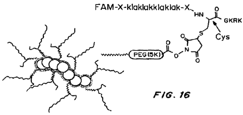
Figure 16 shows the structure of targeted theranostic NW. Aminated NW were
synthesized according to Park et al (4) and reacted with NHS-PEG(5K)-
maleimide.
Subsequently, peptides were coated on the NW through reaction between the
maleimide
group on the PEG and a cysteine thiol of the peptide. Coupling through the
side chain of
the central cysteine in the D(KLAKLAK)2CGKRK peptide gives the V-shaped
structure
depicted in the figure.
Figure 17 is a graph of FAM-CGKRK peptide binding to mitochondria in the
presence of unlabeled peptide (left panel) and a control peptide (right
panel). FAM-
CGKRK was incubated with purified mitochondria in the presence of increasing
concentrations of either unlabeled CGKRK or an unrelated peptide (CREKA; SEQ
ID
NO:92) as a control.
Figure 18 is a graph of phage binding to mitochondria. CGKRK phage and
CREKA (SEQ ID NO:92) phage (as a control) were incubated with purified
mitochondria.
Titration of bound phage shows about 80 times more binding of the CGKRK phage
than
the control. Student's t-test (c), Error bars, mean SD; n.s., * *p < 0.01; *
* *p < 0.001.
Figure 19 is a graph of adsorption (A450 nm) versus biotin-CGKRK concentration
( M). Binding of increasing amounts of biotin-labeled CGKRK peptide to
immobilized
p32 protein was detected with streptavidin coupled to horseradish peroxidase
and
normalized to nonspecific binding in the absence of p32. The affinity of the
peptide for
p32 calculated from the binding curves is also shown. The saturation curve
shown is
average of three independent experiments. Error bars, mean SD.
Figures 20A and 20B are graphs of the percent of inhibition versus non-labeled
peptide added. Figure 20A shows the results for biotin labeled CGKRK and
Figure 20B
shows the results for biotin labeled LyP-1 peptide.
Figure 21 shows Annexin V positive cells (%) when treated with various peptide
compositions 24, 48 and 72 hours. HUVEC and T3 cells were left untreated
(Control) or
treated with a concentration of 10 gg/ml of NWs coated with either a control
peptide
(CREKA; SEQ ID NO:92), D[KLAKLAK]2, or CGKRKD[KLAKLAK]2. The cells were
stained with Annexin and analyzed by flow cytometry. The total percentage of
Annexin-
positive cells (apoptotic and dead cells) is indicated.
Figure 22 shows Annexin V positive cells (%) when treated with various peptide
compositions for 30 minutes (when the particles were washed away) and the
incubation
was continued for 72 hrs. The cells were stained with Annexin and analyzed by
flow
11
CA 02795289 2012-10-02
WO 2011/127405 PCT/US2011/031785
cytometry. The total percentage of Annexin-positive cells (apoptotic and dead
cells) is
indicated.
Figure 23 shows survival (%) versus time (days) for mice bearing lenti-viral
(H-
RasVl2-sip53) induced brain tumors treated with D[KLAKLAK]2-NWs or CGKRK
D[KLAKLAK]2-NWs. Mice bearing lenti-viral (H-RasVl2-sip53) induced brain
tumors in
the right hippocampus were intravenously injected with NW coated with
peptides. The
particles were administered every other day for 18 days, starting 3 weeks post-
viral
injection. Survival curve of the non-treated and treated mice (n=8-10 per
group).
Figure 24 shows survival (%) versus time (days) for mice bearing 005 tumor
cells
treated with D[KLAKLAK]2-NWs, CGKRK-NWs, and CGKRKD[KLAKLAK]2-NWs.
Tumors were developed by transplanting 3 x 10⁵ 005 cells into the right
hippocampus of
NOD-SCID mice. Ten days post-tumor cell transplantation, the mice were
intravenously
injected with NWs. The NWs (5mg of iron/kg) were administered every other day
for 3
weeks or administered non stop for the same period of time (n=8 per group).
Survival
curves of the treated mice are shown.
Figure 25 shows survival (%) versus time (days) for mice bearing 005 tumor
cells
treated with CGKRKD[KLAKLAK]2-NWs with co-administration of cRGD or iRGD.
Mice bearing orthotopic 005 tumors implanted 10 days earlier received every
other day for
3 weeks intravenous injections of CGKRKD[KLAKLAK]2-NWs (5 mg of iron/kg) mixed
with 4 mmol/kg of cRGD or iRGD. Results for control mice and mice administered
only
iRGD are also shown. Survival curves are shown (n=8-10 per group).
Figure 26 shows inhibition of CGKRK peptide binding to p32 by anti-p32. Biotin-
CGKRK at 1 gg/ml was incubated in microtiter wells coated with purified p32,
and the
binding was detected with streptavidin coupled to horseradish peroxidase and
normalized
to nonspecific binding in the absence of p32. The anti-32 antibody was
prepared against
the full-length p32 protein (Protein Production and Analysis Facility of the
Sanford-
Burnham Medical Research Institute). The experiments were performed in
triplicate; one
of two experiments with similar results is shown.
Figures 27A, 27B, and 27C shows that CGKRKD[KLAKLAK]2-NW conjugates
induce cell death by apoptosis. HUVEC (A) and T3 (B) cells were left untreated
(Control)
or were treated with 10 gg/ml of NWs coated with CGKRK D[KLAKLAK]2-NWs for 48
(A) or 72 hours (B). In 27C, the cells were incubated with the indicated NWs,
washed to
remove excess NWs after 30 minutes, and then incubated for 72 hours. Annexin
staining
and analysis by flow cytometry were used to measure apoptosis in the cultures.
12
CA 02795289 2012-10-02
WO 2011/127405 PCT/US2011/031785
Representative images are shown indicating the percentage of Annexin-positive
cells
(apoptotic and dead cells).
Figure 28 shows toxicology analyses of mice treated with
CGKRKD[KLAKLAK]2-NWs. Blood L-alanine-2-oxoglutarate aminotransferase (ALT)
levels measured before (Pre-treatment), after completion of a 3-week treatment
course
(After treatment), and after a subsequent 2-week recovery period (2 weeks
after treatment)
are shown.
Figures 29A and 29B show toxicology analyses of mice treated with
CGKRKD[KLAKLAK]2-NWs. Possible active and innate immune responses against NW
was tested by measuring antibody (29A) and IL-6 levels (29B) in serum form
mice treated
and collected as in Figure 28.
Figure 30 shows survival curves of mice bearing intracranial U87 tumors
treated
with CGKRKD[KLAKLAK]2-NWs. Tumors were induced by injecting 5 x 10⁵ GFP-
expressing U87 cells into the right hippocampus of mice. Treatment with
intravenous
injections of CGKRK-D[KLAKLAK]2-NWs and control NWs was started 10 days after
the tumor cell injection and continued every other day for 3 weeks (n=5 per
group).
DETAILED DESCRIPTION OF THE INVENTION
The disclosed methods and compositions can be understood more readily by
reference to the following detailed description of particular embodiments and
the Example
included therein and to the Figures and their previous and following
description.
Before the present compounds, compositions, articles, devices, and/or methods
are
disclosed and described, it is to be understood that they are not limited to
specific
synthetic methods or specific recombinant biotechnology methods unless
otherwise
specified, or to particular reagents unless otherwise specified, as such may,
of course,
vary. It is also to be understood that the terminology used herein is for the
purpose of
describing particular embodiments only and is not intended to be limiting.
Definitions
As used in the specification and the appended claims, the singular forms "a,"
"an"
and "the" include plural referents unless the context clearly dictates
otherwise. Thus, for
example, reference to "a pharmaceutical carrier" includes mixtures of two or
more such
carriers, and the like.
Ranges can be expressed herein as from "about" one particular value, and/or to
"about" another particular value. When such a range is expressed, another
embodiment
includes from the one particular value and/or to the other particular value.
Similarly,
13
CA 02795289 2012-10-02
WO 2011/127405 PCT/US2011/031785
when values are expressed as approximations, by use of the antecedent "about,"
it will be
understood that the particular value forms another embodiment. It will be
further
understood that the endpoints of each of the ranges are significant both in
relation to the
other endpoint, and independently of the other endpoint. It is also understood
that there
are a number of values disclosed herein, and that each value is also herein
disclosed as
"about" that particular value in addition to the value itself. For example, if
the value "10"
is disclosed, then "about 10" is also disclosed. It is also understood that
when a value is
disclosed that "less than or equal to" the value, "greater than or equal to
the value" and
possible ranges between values are also disclosed, as appropriately understood
by the
skilled artisan. For example, if the value "10" is disclosed the "less than or
equal to 10"as
well as "greater than or equal to 10" is also disclosed. It is also understood
that the
throughout the application, data is provided in a number of different formats,
and that this
data, represents endpoints and starting points, and ranges for any combination
of the data
points. For example, if a particular data point "10" and a particular data
point 15 are
disclosed, it is understood that greater than, greater than or equal to, less
than, less than or
equal to, and equal to 10 and 15 are considered disclosed as well as between
10 and 15. It
is also understood that each unit between two particular units are also
disclosed. For
example, if 10 and 15 are disclosed, then 11, 12, 13, and 14 are also
disclosed.
In this specification and in the claims which follow, reference will be made
to a
number of terms which shall be defined to have the following meanings:
"Optional" or "optionally" means that the subsequently described event or
circumstance may or may not occur, and that the description includes instances
where said
event or circumstance occurs and instances where it does not.
Throughout this application, various publications are referenced. The
disclosures
of these publications in their entireties are hereby incorporated by reference
into this
application in order to more fully describe the state of the art to which this
pertains. The
references disclosed are also individually and specifically incorporated by
reference herein
for the material contained in them that is discussed in the sentence in which
the reference
is relied upon.
It is to be understood that the disclosed method and compositions are not
limited to
specific synthetic methods, specific analytical techniques, or to particular
reagents unless
otherwise specified, and, as such, may vary. It is also to be understood that
the
terminology used herein is for the purpose of describing particular
embodiments only and
is not intended to be limiting.
14
CA 02795289 2012-10-02
WO 2011/127405 PCT/US2011/031785
Materials
Disclosed are the components to be used to prepare the disclosed compositions
as
well as the compositions themselves to be used within the methods disclosed
herein.
These and other materials are disclosed herein, and it is understood that when
combinations, subsets, interactions, groups, etc. of these materials are
disclosed that while
specific reference of each various individual and collective combinations and
permutation
of these compounds may not be explicitly disclosed, each is specifically
contemplated and
described herein. For example, if a particular peptide is disclosed and
discussed and a
number of modifications that can be made to a number of molecules including
the peptide
are discussed, specifically contemplated is each and every combination and
permutation of
the peptides and the modifications that are possible unless specifically
indicated to the
contrary. Thus, if a class of molecules A, B, and C are disclosed as well as a
class of
molecules D, E, and F and an example of a combination molecule, A-D is
disclosed, then
even if each is not individually recited each is individually and collectively
contemplated
meaning combinations, A-E, A-F, B-D, B-E, B-F, C-D, C-E, and C-F are
considered
disclosed. Likewise, any subset or combination of these is also disclosed.
Thus, for
example, the sub-group of A-E, B-F, and C-E would be considered disclosed.
This
concept applies to all aspects of this application including, but not limited
to, steps in
methods of making and using the disclosed compositions. Thus, if there are a
variety of
additional steps that can be performed it is understood that each of these
additional steps
can be performed with any specific embodiment or combination of embodiments of
the
disclosed methods.
Disclosed are compositions useful for delivering significant amounts of
compounds of interest to targeted cells and tissues. The disclosed
compositions are useful,
for example, to deliver to targeted cells and tissues an effective amount of
compounds that
are excessively toxic. For example, disclosed are compositions comprising a
surface
molecule, one or more homing molecules, and a plurality of cargo molecules.
The cargo
molecules can be, for example, excessively toxic molecules. The cargo
molecules can be,
for example, membrane perturbing molecules. As another example, disclosed are
compositions comprising a surface molecule, one or more homing molecules, and
a
plurality of membrane perturbing molecules. As used herein, excessively toxic
compounds are compounds that too toxic when administered to a subject in
unconjugated
forms in what would be a therapeutically effective amount but for the
toxicity.
CA 02795289 2012-10-02
WO 2011/127405 PCT/US2011/031785
The homing molecules can home to targets of interest, such as cells and
tissues of
interest. For example, the homing molecules can home to tumor vasculature. The
homing
molecules can selectively home to targets of interest, such as cells and
tissues of interest.
For example, the homing molecules can selectively home to tumor vasculature.
The
composition can home to one or more of the sites to be targeted. The
composition can be
internalized in cells. The composition can penetrate tissue. The composition
can be
internalized into cells at the targeted site. The composition can penetrate
tissue at the
targeted site. The composition can, for example be internalized into cancer
cells. The
composition can, for example, penetrate tumor tissue. The composition can, for
example,
bind inside tumor blood vessels.
In some forms, one or more of the homing molecules can comprise the amino acid
sequence CGKRK (SEQ ID NO: 1) or a conservative derivative thereof, the amino
acid
sequence CRKDKC (SEQ ID NO:2) or a conservative derivative thereof, or a
combination. In some forms, one or more of the homing molecule can comprise
the amino
acid sequence CGKRK (SEQ ID NO:1) or a conservative variant thereof. In some
forms,
one or more of the homing molecules can comprise the amino acid sequence CGKRK
(SEQ ID NO: 1). In some forms, one or more of the membrane perturbing
molecules can
comprise the amino acid sequence D(KLAKLAK)2 (SEQ ID NO:3) or a conservative
variant thereof, (KLAKLAK)2 (SEQ ID NO:3) or a conservative variant thereof,
(KLAKKLA)2 (SEQ ID NO:5) or a conservative variant thereof, (KAAKKAA) 2 (SEQ
ID
NO:6) or a conservative variant thereof, (KLGKKLG)3 (SEQ ID NO:7) or a
conservative
variant thereof, or a combination. In some forms, one or more of the membrane
perturbing molecules can comprise the amino acid sequence D(KLAKLAK)2 (SEQ ID
NO:3), (KLAKLAK)2 (SEQ ID NO:3), (KLAKKLA)2 (SEQ ID NO:5), (KAAKKAA) 2
(SEQ ID NO:6), (KLGKKLG)3 (SEQ ID NO:7), or a combination. In some forms, one
or
more of the membrane perturbing molecules can comprise the amino acid sequence
D(KLAKLAK)2 (SEQ ID NO:3) or a conservative variant thereof. In some forms,
one or
more of the membrane perturbing molecules can comprise the amino acid sequence
D(KLAKLAK)2 (SEQ ID NO:3).
In some forms, the composition can comprise a plurality of surface molecules,
a
plurality of homing molecules and a plurality of cargo molecules. In some
forms, the
composition can comprise one or more surface molecules, a plurality of homing
molecules
and a plurality of cargo molecules. In some forms, the composition can
comprise a
plurality of surface molecules, one or more homing molecules and a plurality
of cargo
16
CA 02795289 2012-10-02
WO 2011/127405 PCT/US2011/031785
molecules. In some forms, the composition can comprise a plurality of surface
molecules,
a plurality of homing molecules and one or more cargo molecules. In some
forms, the
composition can comprise one or more surface molecules, one or more homing
molecules
and a plurality of cargo molecules. In some forms, the composition can
comprise one or
more surface molecules, a plurality of homing molecules and one or more cargo
molecules. In some forms, the composition comprises a plurality of surface
molecules,
one or more homing molecules and one or more cargo molecules.
In some forms, the composition can comprise a surface molecule, a plurality of
homing molecules and a plurality of cargo molecules, wherein one or more of
the homing
molecules and one or more of the cargo molecules are associated with the
surface
molecule. In some forms, the composition can comprise a surface molecule, a
plurality of
homing molecules and a plurality of cargo molecules, wherein a plurality of
the plurality
of homing molecules and a plurality of the plurality of cargo molecules are
associated with
the surface molecule. In some forms, the composition can comprise a surface
molecule, a
plurality of homing molecules and a plurality of cargo molecules, wherein the
homing
molecules and the cargo molecules are associated with the surface molecule.
In some forms, the composition can comprise a surface molecule, wherein the
surface molecule is multivalent for homing molecules and cargo molecules. In
some
forms, the composition can comprise a surface molecule, wherein the surface
molecule is
multivalent for homing molecules and comprises one or more cargo molecules. In
some
forms, the composition can comprise a surface molecule, wherein the surface
molecule is
multivalent for cargo molecules and comprises one or more homing molecules. In
some
forms, the composition can comprise a surface molecule, wherein the surface
molecule is
multivalent for conjugates, wherein one or more of the conjugates comprise one
or more
homing molecules and one or more cargo molecules. In some forms, the
composition can
comprise a surface molecule, wherein the surface molecule is multivalent for
conjugates,
wherein one or more of the conjugates comprise a plurality of homing molecules
and a
plurality cargo molecules. In some forms, the composition can comprise a
surface
molecule, wherein the surface molecule is multivalent for conjugates, wherein
one or more
of the conjugates comprise a homing molecule and a cargo molecule. In some
forms, the
composition can comprise a surface molecule, wherein the surface molecule is
multivalent
for conjugates, wherein each of the conjugates comprises a plurality of homing
molecules
and a plurality cargo molecules. In some forms, the composition can comprise a
surface
molecule, wherein the surface molecule is multivalent for conjugates, wherein
each of the
17
CA 02795289 2012-10-02
WO 2011/127405 PCT/US2011/031785
conjugates comprises a homing molecule and a cargo molecule. As used herein, a
component that is stated to be "multivalent for" one or more other components
refers to a
component that has a plurality of the other components associated with,
conjugated to
and/or covalent coupled to the first component.
In some forms, the composition can comprise a surface molecule, wherein the
surface molecule comprises one or more conjugates, wherein one or more of the
conjugates comprise one or more homing molecules and one or more cargo
molecules. In
some forms, the composition can comprise a surface molecule, wherein the
surface
molecule comprises one or more conjugates, wherein one or more of the
conjugates
comprise a plurality of homing molecules and a plurality cargo molecules. In
some forms,
the composition can comprise a surface molecule, wherein the surface molecule
comprises
one or more conjugates, wherein one or more of the conjugates comprise a
homing
molecule and a cargo molecule. In some forms, the composition can comprise a
surface
molecule, wherein the surface molecule comprises one or more conjugates,
wherein each
of the conjugates comprises a plurality of homing molecules and a plurality
cargo
molecules. In some forms, the composition can comprise a surface molecule,
wherein the
surface molecule comprises one or more conjugates, wherein each of the
conjugates
comprises a homing molecule and a cargo molecule.
In some forms, one or more of the membrane perturbing molecules can be
conjugated to one or more of the homing molecules. In some forms, one or more
of the
conjugated membrane perturbing molecules and homing molecules can be
covalently
coupled. In some forms, one or more of the covalently coupled membrane
perturbing
molecules and homing molecules can comprise fusion peptides. In some forms,
the
homing molecules can be conjugated with the surface molecule. In some forms,
one or
more of the conjugated homing molecules can be directly conjugated to the
surface
molecule. In some forms, one or more of the conjugated homing molecules can be
indirectly conjugated to the surface molecule. In some forms, one or more of
the homing
molecules can be covalently coupled to the surface molecule. In some forms,
one or more
of the covalently coupled homing molecules can be directly covalently coupled
to the
surface molecule. In some forms, one or more of the covalently coupled homing
molecules can be indirectly covalently coupled to the surface molecule. In
some forms,
the membrane perturbing molecules can be conjugated with the surface molecule.
In some
forms, one or more of the conjugated membrane perturbing molecules are
directly
conjugated to the surface molecule. In some forms, one or more of the
conjugated
18
CA 02795289 2012-10-02
WO 2011/127405 PCT/US2011/031785
membrane perturbing molecules can be indirectly conjugated to the surface
molecule. In
some forms, one or more of the membrane perturbing molecules can be covalently
coupled to the surface molecule. In some forms, one or more of the covalently
coupled
membrane perturbing molecules can be directly covalently coupled to the
surface
molecule. In some forms, one or more of the covalently coupled membrane
perturbing
molecules can be indirectly covalently coupled to the surface molecule.
In some forms, the composition can further comprise one or more
internalization
elements. In some forms, one or more of the homing molecules can comprise one
or more
of the internalization elements. In some forms, one or more of the membrane
perturbing
molecules can comprise one or more of the internalization elements. In some
forms, the
surface molecule can comprise one or more of the internalization elements not
comprised
in either the homing molecules or the membrane perturbing molecules. In some
forms, the
composition can further comprise one or more tissue penetration elements. In
some forms,
one or more of the tissue penetration elements can be comprised in an
internalization
element. In some forms, the tissue penetration element can be a CendR element.
In some forms, the surface molecule can comprise a nanoparticle. In some
forms,
the surface molecule can comprise a nanoworm. In some forms, the surface
molecule can
comprise an iron oxide nanoworm. In some forms, the surface molecule can
comprise an
iron oxide nanoparticle. In some forms, the surface molecule can comprise an
albumin
nanoparticle. In some forms, the surface molecule can comprise a liposome. In
some
forms, the surface molecule can comprise a micelle. In some forms, the surface
molecule
comprises a phospholipid. In some forms, the surface molecule comprises a
polymer. In
some forms, the surface molecule can comprise a microparticle. In some forms,
the
surface molecule can comprise a fluorocarbon microbubble.
In some forms, the composition can comprise at least 100 homing molecules. In
some forms, the composition can comprise at least 1000 homing molecules. In
some
forms, the composition can comprise at least 10,000 homing molecules. In some
forms,
the composition can comprise at least 100 membrane perturbing molecules. In
some
forms, the composition can comprise at least 1000 membrane perturbing
molecules. In
some forms, the composition can comprise at least 10,000 membrane perturbing
molecules.
In some forms, one or more of the homing molecules can be modified homing
molecules. In some forms, one or more of the homing molecules can comprise a
methylated homing molecule. In some forms, one or more of the methylated
homing
19
CA 02795289 2012-10-02
WO 2011/127405 PCT/US2011/031785
molecules can comprise a methylated amino acid segment. In some forms, one or
more of
the membrane perturbing molecules can be modified membrane perturbing
molecules. In
some forms, one or more of the membrane perturbing molecules comprise a
methylated
membrane perturbing molecule. In some forms, one or more of the methylated
membrane
perturbing molecules comprise a methylated amino acid segment. In some forms,
the
amino acid sequence is N- or C-methylated in at least one position.
In some forms, the composition can further comprise one or more moieties. In
some forms, the moieties can be independently selected from the group
consisting of an
anti-angiogenic agent, a pro-angiogenic agent, a cancer chemotherapeutic
agent, a
cytotoxic agent, an anti-inflammatory agent, an anti-arthritic agent, a
polypeptide, a
nucleic acid molecule, a small molecule, an image contrast agent, a
fluorophore,
fluorescein, rhodamine, a radionuclide, indium-111, technetium-99, carbon- 11,
and
carbon-13. In some forms, at least one of the moieties can be a therapeutic
agent. In some
forms, the therapeutic agent can be iRGD, RGD, Abraxane, paclitaxel, taxol, or
a
combination. In some forms, at least one of the moieties can be a detectable
agent. In
some forms, the detectable agent can be FAM.
In some forms, the composition can have a therapeutic effect. In some forms,
the
composition can reduce tumor growth. In some forms, the therapeutic effect can
be a
slowing in the increase of or a reduction of tumor burden. In some forms, the
therapeutic
effect can be a slowing of the increase of or reduction of tumor size. In some
forms, the
subject can have one or more sites targeted, wherein the composition can home
to one or
more of the sites targeted. In some forms, the subject can have a tumor,
wherein the
composition can have a therapeutic effect on the tumor.
The disclosed components can be associated with each other (or, in some forms,
not associated with each other) in combinations as disclosed herein. For
example, homing
molecules can be covalently coupled or non-covalently associated with surface
molecules,
homing molecules can be covalently coupled or non-covalently associated with
membrane
perturbing molecules, membrane perturbing molecules can be covalently coupled
or non-
covalently associated with surface molecules, etc. Associated components can
also be
referred to as being conjugated. Conjugation can be direct or indirect. Direct
conjugation
of components refers to covalently coupled or non-covalently associated
components
where there is no other molecule intervening between the conjugated
components.
Indirect conjugation refers to any chain of molecules and covalent bonds or
non-covalent
associations linking the components where the components are not directly
conjugated
CA 02795289 2012-10-02
WO 2011/127405 PCT/US2011/031785
(that is, there is a least one separate molecule other than the components
intervening
between the components).
Covalently coupled refers to association of components via covalent bonds. A
covalent association or coupling can be either direct or indirect. A direct
covalent
association or coupling of components refers to a covalent bond involving
atoms that are
each respectively a part of the components. Thus, in a direct covalent
association or
coupling, there is no other molecule intervening between the
associated/coupled
components. An indirect covalent association or coupling refers to any chain
of molecules
and covalent bonds linking the components where the components are not
covalently
coupled (that is, there is a least one separate molecule other than the
components
intervening between the components via covalent bonds).
As used herein, reference to components (such as a homing molecule and a
surface
molecule) as being "not covalently coupled" means that the components are not
connected
via covalent bonds (for example, that the homing molecule and the surface
molecule are
not connected via covalent bonds). That is, there is no continuous chain of
covalent bonds
between, for example, the homing molecule and the surface molecule.
Non-covalent association refers to association of components via non-covalent
bonds and interactions. A non-covalent association can be either direct or
indirect. A
direct non-covalent association refers to a non-covalent bond involving atoms
that are
each respectively connected via a chain of covalent bonds to the components.
Thus, in a
direct non-covalent association, there is no other molecule intervening
between the
associated components. An indirect non-covalent association refers to any
chain of
molecules and bonds linking the components where the components are not
covalently
coupled (that is, there is a least one separate molecule other than the
components
intervening between the components via non-covalent bonds).
Reference to components (such as a homing molecule and a surface molecule) as
not being "non-covalently associated" means that there is no direct or
indirect non-
covalent association between the components. That is, for example, no atom
covalently
coupled to a homing molecule is involved in a non-covalent bond with an atom
covalently
coupled to a surface molecule. Within this meaning, a homing molecule and a
surface
molecule can be together in a composition where they are indirectly associated
via
multiple intervening non-covalent bonds while not being non-covalently
associated as that
term is defined herein. For example, a homing molecule and a surface molecule
can be
mixed together in a carrier where they are not directly non-covalently
associated. A
21
CA 02795289 2012-10-02
WO 2011/127405 PCT/US2011/031785
homing molecule and a surface molecule that are referred to as not indirectly
non-
covalently associated cannot be mixed together in a continuous composition.
Reference to
components (such as a homing molecule and a surface molecule) as not being
"directly
non-covalently associated" means that there is no direct non-covalent
association between
the components (an indirect non-covalent association may be present).
Reference to
components (such as a homing molecule and a surface molecule) as not being
"indirectly
non-covalently associated" means that there is no direct or indirect non-
covalent
association between the components.
It is understood that components can be non-covalently associated via multiple
chains and paths including both direct and indirect non-covalent associations.
For the
purposes of these definitions, the presence a single direct non-covalent
association makes
the association a direct non-covalent association even if there are also
indirect non-
covalent associations present. Similarly, the presence of a covalent
connection between
components means the components are covalently coupled even if there are also
non-
covalent associations present. It is also understood that covalently coupled
components
that happened to lack any non-covalent association with each other are not
considered to
fall under the definition of components that are not non-covalently
associated.
Association of the components of the disclosed compositions can be aided or
accomplished via molecules, conjugates and/or compositions. Where such
molecules,
conjugates and/or compositions are other than surface molecules, homing
molecules, or
cargo molecules (such as membrane perturbing molecules, internalization
elements, tissue
penetration elements, and moieties), they can be referred to herein as
linkers. Such linkers
can be any molecule, conjugate, composition, etc. that can be used to
associate
components of the disclosed compositions. Generally, linkers can be used to
associate
components other than surface molecules to surface molecules. Useful linkers
include
materials that are biocompatible, have low bioactivity, have low antigenicity,
etc. That is,
such useful linker materials can serve the linking/association function
without adding
unwanted bioreactivity to the disclosed compositions. Many such materials are
known
and used for similar linking and association functions. Polymer materials are
a
particularly useful form of linker material. For example, polyethylene glycols
can be
used.
Linkers are useful for achieving useful numbers and densities of the
components
(such as homing molecules and membrane perturbing molecules) on surface
molecules.
For example, linkers of fibrous form are useful for increasing the number of
components
22
CA 02795289 2012-10-02
WO 2011/127405 PCT/US2011/031785
per surface molecule or per a given area of the surface molecule. Similarly,
linkers having
a branching form are useful for increasing the number of components per
surface molecule
or per a given area of the surface molecule. Linkers can also have a branching
fibrous
form.
Sufficiency of the number and composition of homing molecules in the
composition can be determined by assessing homing to the target and
effectively delivery
of the cargo molecules in a non-human animal. The composition can comprise a
sufficient
number and composition of homing molecules (modified or not) such that the
composition
homes to the target and effectively delivers the cargo molecules. In one
example,
sufficiency of the number and composition of modified and/or unmodified homing
molecules can be determined by assessing cargo delivery and/or therapeutic
effect on the
target. Sufficiency of the number and composition of membrane perturbing
molecules can
be determined by assessing membrane perturbing effect of the composition in a
non-
human animal. The composition can comprise a sufficient number and composition
of
membrane perturbing molecules (modified or not) such that the composition has
a
membrane perturbing effect on the target. In one example, sufficiency of the
number and
composition of modified and/or unmodified membrane perturbing molecules can be
determined by assessing membrane disruption, apoptosis, and/or therapeutic
effect on the
target.
The composition can comprise a sufficient density and composition of homing
molecules such that the composition homes to the target and effectively
delivers the cargo
molecules. Sufficiency of the density and composition of homing molecules can
be
determined by assessing cargo delivery and/or therapeutic effect on the target
in a non-
human animal. The composition can comprise a sufficient density and
composition of
membrane perturbing molecules such that the composition has a membrane
perturbing
effect on the target. Sufficiency of the density and composition of membrane
perturbing
molecules can be determined by assessing membrane disruption, apoptosis,
and/or
therapeutic effect on the target in a non-human animal.
The density of homing molecules and/or membrane perturbing molecules on a
surface molecule can be described in any suitable manner. For example, the
density can
be expressed as the number of homing molecules and/or membrane perturbing
molecules
per, for example, a given area, surface area, volume, unit, subunit, arm, etc.
of the surface
molecule. The density can also be relative to, for example, the area, surface
area, volume,
unit, subunit, arm, etc. of the entire surface molecule or to the area,
surface area, volume,
23
CA 02795289 2012-10-02
WO 2011/127405 PCT/US2011/031785
unit, subunit, arm, etc. of a portion of the surface molecule. For example, a
sufficient
density of homing molecule and/or membrane perturbing molecule can be present
in a
portion of the surface molecule. The presence of this dense portion can cause
clotting and
amplify the accumulation of the composition. Thus, a composition having a
sufficient
density of homing molecules and/or membrane perturbing molecules can have a
threshold
density (or above) for the entire surface molecule or for just one or more
portions of the
surface molecule. Unless otherwise stated, densities refer to average density
over the
designated portion of the surface molecule. For example, a density of 1 homing
molecule
per square nM of the surface molecule refers to an average density of the
homing
molecules over the entire surface molecule. As another example, a density of 1
homing
molecule per square nM of a portion of the surface molecule refers to an
average density
of the homing molecules over just that portion of the surface molecule.
The density can be measured or calculated in any suitable manner. For example,
the number or amount of homing molecules and/or membrane perturbing molecules
present on a surface molecule or group of surface molecules can be measured
by, for
example, detecting the level or intensity of signal produced by labeled homing
molecules
and/or membrane perturbing molecules and calculating the density based on the
structural
characteristics of the surface molecule.
The density or threshold density of homing molecules and/or membrane
perturbing
molecules can be, for example, at least 0.001, 0.002, 0.003, 0.004, 0.005,
0.006, 0.007,
0.008, 0.009, 0.01, 0.02, 0.03, 0.04, 0.05, 0.06, 0.07, 0.08, 0.09, 0.1, 0.2,
0.3, 0.4, 0.5, 0.6,
0.7, 0.8, 0.9, 1, 2, 3, 4, 5, 6, 7, 8, 9, 10, 12, 14, 16, 18, 20, 25, 30, 35,
40, 45, 50, 55, 60,
65, 70, 75, 80, 85, 90, 95, 100, 110, 120, 130, 140, 150, 160, 170, 180, 190,
200, 220, 240,
260, 280, 300, 320, 340, 360, 380, 400, 420, 440, 460, 480, 500, 550, 600,
650, 700, 750,
800, 850, 900, 950, or 1000 homing molecules and/or membrane perturbing
molecules per
square nM of the entire or a portion of the surface molecule. The composition
can also
comprise any density in between those densities listed above.
The density or threshold density of homing molecules and/or membrane
perturbing
molecules can be, for example, at least 1, 2, 3, 4, 5, 6, 7, 8, 9, 10, 12, 14,
16, 18, 20, 25,
30, 35, 40, 45, 50, 55, 60, 65, 70, 75, 80, 85, 90, 95, 100, 110, 120, 130,
140, 150, 160,
170, 180, 190, 200, 220, 240, 260, 280, 300, 320, 340, 360, 380, 400, 420,
440, 460, 480,
500, 550, 600, 650, 700, 750, 800, 850, 900, 950, 1000, 1100, 1200, 1300,
1400, 1500,
1600, 1700, 1800, 1900, 2000, 2200, 2400, 2600, 2800, 3000, 3200, 3400, 3600,
3800,
4000, 4200, 4400, 4600, 4800, 5000, 5500, 6000, 6500, 7000, 7500, 8000, 8500,
900,
24
CA 02795289 2012-10-02
WO 2011/127405 PCT/US2011/031785
9500, 10,000 homing molecules and/or membrane perturbing molecules per square
gM of
the entire or a portion of the surface molecule. The composition can also
comprise any
density in between those densities listed above.
The density or threshold density of homing molecules and/or membrane
perturbing
molecules can be, for example, at least 0.001, 0.002, 0.003, 0.004, 0.005,
0.006, 0.007,
0.008, 0.009, 0.01, 0.02, 0.03, 0.04, 0.05, 0.06, 0.07, 0.08, 0.09, 0.1, 0.2,
0.3, 0.4, 0.5, 0.6,
0.7, 0.8, 0.9, 1, 2, 3, 4, 5, 6, 7, 8, 9, 10, 12, 14, 16, 18, 20, 25, 30, 35,
40, 45, 50, 55, 60,
65, 70, 75, 80, 85, 90, 95, 100, 110, 120, 130, 140, 150, 160, 170, 180, 190,
200, 220, 240,
260, 280, 300, 320, 340, 360, 380, 400, 420, 440, 460, 480, 500, 550, 600,
650, 700, 750,
800, 850, 900, 950, or 1000 homing molecules and/or membrane perturbing
molecules per
cubic nM of the entire or a portion of the surface molecule. The composition
can also
comprise any density in between those densities listed above.
The density or threshold density of homing molecules and/or membrane
perturbing
molecules can be, for example, at least 1, 2, 3, 4, 5, 6, 7, 8, 9, 10, 12, 14,
16, 18, 20, 25,
30, 35, 40, 45, 50, 55, 60, 65, 70, 75, 80, 85, 90, 95, 100, 110, 120, 130,
140, 150, 160,
170, 180, 190, 200, 220, 240, 260, 280, 300, 320, 340, 360, 380, 400, 420,
440, 460, 480,
500, 550, 600, 650, 700, 750, 800, 850, 900, 950, 1000, 1100, 1200, 1300,
1400, 1500,
1600, 1700, 1800, 1900, 2000, 2200, 2400, 2600, 2800, 3000, 3200, 3400, 3600,
3800,
4000, 4200, 4400, 4600, 4800, 5000, 5500, 6000, 6500, 7000, 7500, 8000, 8500,
900,
9500, 10,000 homing molecules and/or membrane perturbing molecules per cubic
gM of
the entire or a portion of the surface molecule. The composition can also
comprise any
density in between those densities listed above.
The number of homing molecules and/or membrane perturbing molecules on a
surface molecule can be described in any suitable manner. For example, the
number can
be expressed as the number of homing molecules and/or membrane perturbing
molecules
per, for example, a given area, surface area, volume, unit, subunit, arm, etc.
of the surface
molecule. The number can also be relative to, for example, the area, surface
area, volume,
unit, subunit, arm, etc. of the entire surface molecule or to the area,
surface area, volume,
unit, subunit, arm, etc. of a portion of the surface molecule. For example, a
sufficient
number of homing molecule and/or membrane perturbing molecule can be present
in a
portion of the surface molecule. The presence of this dense portion can cause
clotting and
amplify the accumulation of the composition. Thus, a composition having a
sufficient
number of homing molecules and/or membrane perturbing molecules can have a
threshold
CA 02795289 2012-10-02
WO 2011/127405 PCT/US2011/031785
number (or above) for the entire surface molecule or for just one or more
portions of the
surface molecule.
The number can be measured or calculated in any suitable manner. For example,
the number or amount of homing molecules and/or membrane perturbing molecules
present on a surface molecule or group of surface molecules can be measured
by, for
example, detecting the level or intensity of signal produced by labeled homing
molecules
and/or membrane perturbing molecules and calculating the number based on the
structural
characteristics of the surface molecule.
The number or threshold number of homing molecules and/or membrane
perturbing molecules can be, for example, at least 1, 2, 3, 4, 5, 6, 7, 8, 9,
10, 12, 14, 16, 18,
20, 25, 30, 35, 40, 45, 50, 55, 60, 65, 70, 75, 80, 85, 90, 95, 100, 110, 120,
130, 140, 150,
160, 170, 180, 190, 200, 220, 240, 260, 280, 300, 320, 340, 360, 380, 400,
420, 440, 460,
480, 500, 550, 600, 650, 700, 750, 800, 850, 900, 950, 1000, 1100, 1200, 1300,
1400,
1500, 1600, 1700, 1800, 1900, 2000, 2200, 2400, 2600, 2800, 3000, 3200, 3400,
3600,
3800, 4000, 4200, 4400, 4600, 4800, 5000, 5500, 6000, 6500, 7000, 7500, 8000,
8500,
900, 9500, 10,000 homing molecules and/or membrane perturbing molecules on the
surface molecule. The composition can also comprise any number in between
those
numbers listed above.
The number or threshold number of homing molecules and/or membrane
perturbing molecules can be, for example, at least 0.001, 0.002, 0.003, 0.004,
0.005, 0.006,
0.007, 0.008, 0.009, 0.01, 0.02, 0.03, 0.04, 0.05, 0.06, 0.07, 0.08, 0.09,
0.1, 0.2, 0.3, 0.4,
0.5, 0.6, 0.7, 0.8, 0.9, 1, 2, 3, 4, 5, 6, 7, 8, 9, 10, 12, 14, 16, 18, 20,
25, 30, 35, 40, 45, 50,
55, 60, 65, 70, 75, 80, 85, 90, 95, 100, 110, 120, 130, 140, 150, 160, 170,
180, 190, 200,
220, 240, 260, 280, 300, 320, 340, 360, 380, 400, 420, 440, 460, 480, 500,
550, 600, 650,
700, 750, 800, 850, 900, 950, or 1000 homing molecules and/or membrane
perturbing
molecules per square nM of the entire or a portion of the surface molecule.
The
composition can also comprise any number in between those numbers listed
above.
The number or threshold number of homing molecules and/or membrane
perturbing molecules can be, for example, at least 1, 2, 3, 4, 5, 6, 7, 8, 9,
10, 12, 14, 16, 18,
20, 25, 30, 35, 40, 45, 50, 55, 60, 65, 70, 75, 80, 85, 90, 95, 100, 110, 120,
130, 140, 150,
160, 170, 180, 190, 200, 220, 240, 260, 280, 300, 320, 340, 360, 380, 400,
420, 440, 460,
480, 500, 550, 600, 650, 700, 750, 800, 850, 900, 950, 1000, 1100, 1200, 1300,
1400,
1500, 1600, 1700, 1800, 1900, 2000, 2200, 2400, 2600, 2800, 3000, 3200, 3400,
3600,
3800, 4000, 4200, 4400, 4600, 4800, 5000, 5500, 6000, 6500, 7000, 7500, 8000,
8500,
26
CA 02795289 2012-10-02
WO 2011/127405 PCT/US2011/031785
900, 9500, 10,000 homing molecules and/or membrane perturbing molecules per
square
gM of the entire or a portion of the surface molecule. The composition can
also comprise
any number in between those numbers listed above.
The number or threshold number of homing molecules and/or membrane
perturbing molecules can be, for example, at least 0.001, 0.002, 0.003, 0.004,
0.005, 0.006,
0.007, 0.008, 0.009, 0.01, 0.02, 0.03, 0.04, 0.05, 0.06, 0.07, 0.08, 0.09,
0.1, 0.2, 0.3, 0.4,
0.5, 0.6, 0.7, 0.8, 0.9, 1, 2, 3, 4, 5, 6, 7, 8, 9, 10, 12, 14, 16, 18, 20,
25, 30, 35, 40, 45, 50,
55, 60, 65, 70, 75, 80, 85, 90, 95, 100, 110, 120, 130, 140, 150, 160, 170,
180, 190, 200,
220, 240, 260, 280, 300, 320, 340, 360, 380, 400, 420, 440, 460, 480, 500,
550, 600, 650,
700, 750, 800, 850, 900, 950, or 1000 homing molecules and/or membrane
perturbing
molecules per cubic nM of the entire or a portion of the surface molecule. The
composition can also comprise any number in between those numbers listed
above.
The number or threshold number of homing molecules and/or membrane
perturbing molecules can be, for example, at least 1, 2, 3, 4, 5, 6, 7, 8, 9,
10, 12, 14, 16, 18,
20, 25, 30, 35, 40, 45, 50, 55, 60, 65, 70, 75, 80, 85, 90, 95, 100, 110, 120,
130, 140, 150,
160, 170, 180, 190, 200, 220, 240, 260, 280, 300, 320, 340, 360, 380, 400,
420, 440, 460,
480, 500, 550, 600, 650, 700, 750, 800, 850, 900, 950, 1000, 1100, 1200, 1300,
1400,
1500, 1600, 1700, 1800, 1900, 2000, 2200, 2400, 2600, 2800, 3000, 3200, 3400,
3600,
3800, 4000, 4200, 4400, 4600, 4800, 5000, 5500, 6000, 6500, 7000, 7500, 8000,
8500,
900, 9500, 10,000 homing molecules and/or membrane perturbing molecules per
cubic
gM of the entire or a portion of the surface molecule. The composition can
also comprise
any number in between those numbers listed above.
In some forms, the compositions not only home to tumors, but also amplify
their
own homing. Homing molecules can be used that are clot-binding compounds that
recognize clotted plasma proteins and selectively homes to tumors, where it
binds to
vessel walls and tumor stroma. Surface molecules coupled with the clot-binding
compounds can accumulate in tumor vessels or at wound sites, where they induce
additional local clotting, thereby producing new binding sites for more
particles. The
system mimics platelets, which also circulate freely but accumulate at a
diseased site and
amplify their own accumulation at that site. The clotting-based amplification
greatly
enhances cargo delivery and tumor imaging.
A. Homing Molecules
Homing molecules allow the disclosed compositions to be targeted and to home
to
desired target sites. Homing molecules generally bind preferentially to target
molecules,
27
CA 02795289 2012-10-02
WO 2011/127405 PCT/US2011/031785
cells, tissues, etc., thus resulting in an accumulation of the homing
molecules (and other
components to which they are associated) at target sites.
The term "homing molecule" as used herein, means any molecule that selectively
homes in vivo to specified target sites, such as cells or tissues, in
preference to normal or
other non-target sites, cells, or tissues. Similarly, the term "homing
peptide" or "homing
peptidomimetic" means a peptide that selectively homes in vivo to specified
target sites,
such as cells or tissues, in preference to normal or other non-target sites,
cells, or tissues. It
is understood that a homing molecule that selectively homes in vivo to, for
example,
tumors can home to all tumors or can exhibit preferential homing to one or a
subset of
tumor types.
By "selectively homes" it is meant that, in vivo, the homing molecule binds
preferentially to the target as compared to non-target. For example, the
homing molecule
can bind preferentially to certain molecules, proteins, cells, tissues, etc.
as compared to
other molecules, proteins, cells, tissues, etc. For example, the homing
molecule can bind
preferentially to tumor vasculature or one or more tumors as compared to non-
tumoral
tissue. Such a homing molecule can selectively home, for example, to tumors.
Selective
homing to, for example, certain molecules, proteins, cells, tissues, etc.
generally is
characterized by at least a two-fold greater localization the molecules,
proteins, cells,
tissues, etc. (or other target), as compared to other certain molecules,
proteins, cells,
tissues, etc. A homing molecule can be characterized by, for example, 5-fold,
10-fold, 20-
fold or more preferential localization to the target as compared to one or
more non-targets.
For example, a homing molecule can be characterized by, for example, 5-fold,
10-fold, 20-
fold or more preferential localization to tumor vasculature as compared to
vasculature of
several or many tissue types of non-tumoral tissue, or as compared to
vasculature of most
or all non-tumoral tissue. As another example, a homing molecule can be
characterized
by, for example, 5-fold, 10-fold, 20-fold or more preferential localization to
tumors as
compared to several or many tissue types of non-tumoral tissue, or as compared
to-most or
all non-tumoral tissue. Thus, it is understood that, in some cases, a homing
molecule
homes, in part, to one or more normal organs in addition to homing to the
target tissue.
Selective homing can also be referred to as targeting. The molecules,
proteins, cells,
tissues, etc. that are targeted by homing molecules can be referred to as
targeted
molecules, proteins, cells, tissues, etc.
In some forms, one or more of the homing molecules can comprise the amino acid
sequence CGKRK (SEQ ID NO: 1) or a conservative derivative thereof, the amino
acid
28
CA 02795289 2012-10-02
WO 2011/127405 PCT/US2011/031785
sequence CRKDKC (SEQ ID NO:2) or a conservative derivative thereof, or a
combination. In some forms, one or more of the homing molecule can comprise
the amino
acid sequence CGKRK (SEQ ID NO: 1) or a conservative variant thereof. In some
forms,
one or more of the homing molecules can comprise the amino acid sequence CGKRK
(SEQ ID NO: 1).
The composition can comprise a sufficient number and composition of homing
molecules (modified or not) such that the composition homes to the target and
effectively
delivers the cargo molecules. In one example, sufficiency of the number and
composition
of modified and/or unmodified homing molecules can be determined by assessing
cargo
delivery and/or therapeutic effect on the target.
Many homing molecules and homing peptides home to the vasculature of the
target
tissue. However, for the sake of convenience homing is referred to in some
places herein
as homing to the tissue associated with the vasculature to which the homing
molecule or
homing peptide may actually home. Thus, for example, a homing molecule that
homes to
tumor vasculature can be referred to herein as homing to tumor tissue or to
tumor cells.
By including or associating a homing molecule or homing peptide with, for
example, a
protein, peptide, amino acid sequence, cargo molecules, or CendR element the
protein,
peptide, amino acid sequence, cargo molecules, or CendR element can be
targeted or can
home to the target of the homing molecule or homing peptide. In this way, the
protein,
peptide, amino acid sequence, cargo molecules, or CendR element can be said to
home to
the target of the homing molecule or homing peptide. For convenience and
unless
otherwise indicated, reference to homing of a protein, peptide, amino acid
sequence, cargo
molecules, CendR element, etc. is intended to indicate that the protein,
peptide, amino acid
sequence, cargo molecules, CendR element, etc. includes or is associated with
an
appropriate homing molecule or homing peptide.
The homing molecule can selectively home to a tumor. The homing molecule can
selectively home to tumor vasculature. The homing molecule can selectively
home to one
or more particular types of tumor. The homing molecule can selectively home to
the
vasculature of one or more particular types of tumor. The homing molecule can
selectively home to one or more particular stages of a tumor or cancer. The
homing
molecule can selectively home to the vasculature of one or more particular
stages of a
tumor or cancer. The homing molecule can selectively home to one or more
particular
stages of one or more particular types of tumor. The homing molecule can
selectively
29
CA 02795289 2012-10-02
WO 2011/127405 PCT/US2011/031785
home to the vasculature of one or more different stages of one or more
particular types of
tumor.
The composition can selectively home to a tumor. The composition can
selectively
home to tumor vasculature. The composition can selectively home to one or more
particular types of tumor. The composition can selectively home to the
vasculature of one
or more particular types of tumor. The composition can selectively home to one
or more
particular stages of a tumor or cancer. The composition can selectively home
to the
vasculature of one or more particular stages of a tumor or cancer. The
composition can
selectively home to one or more particular stages of one or more particular
types of tumor.
The composition can selectively home to the vasculature of one or more
different stages of
one or more particular types of tumor.
The cargo molecule can selectively home to a tumor. The cargo molecule can
selectively home to tumor vasculature. The cargo molecule can selectively home
to one or
more particular types of tumor. The cargo molecule can selectively home to the
vasculature of one or more particular types of tumor. The cargo molecule can
selectively
home to one or more particular stages of a tumor or cancer. The cargo molecule
can
selectively home to the vasculature of one or more particular stages of a
tumor or cancer.
The cargo molecule can selectively home to one or more particular stages of
one or more
particular types of tumor. The cargo molecule can selectively home to the
vasculature of
one or more different stages of one or more particular types of tumor.
The surface molecule can selectively home to a tumor. The surface molecule can
selectively home to tumor vasculature. The surface molecule can selectively
home to one
or more particular types of tumor. The surface molecule can selectively home
to the
vasculature of one or more particular types of tumor. The surface molecule can
selectively
home to one or more particular stages of a tumor or cancer. The surface
molecule can
selectively home to the vasculature of one or more particular stages of a
tumor or cancer.
The surface molecule can selectively home to one or more particular stages of
one or more
particular types of tumor. The surface molecule can selectively home to the
vasculature of
one or more different stages of one or more particular types of tumor.
The membrane perturbing molecule can selectively home to a tumor. The
membrane perturbing molecule can selectively home to tumor vasculature. The
membrane perturbing molecule can selectively home to one or more particular
types of
tumor. The membrane perturbing molecule can selectively home to the
vasculature of one
or more particular types of tumor. The membrane perturbing molecule can
selectively
CA 02795289 2012-10-02
WO 2011/127405 PCT/US2011/031785
home to one or more particular stages of a tumor or cancer. The membrane
perturbing
molecule can selectively home to the vasculature of one or more particular
stages of a
tumor or cancer. The membrane perturbing molecule can selectively home to one
or more
particular stages of one or more particular types of tumor. The membrane
perturbing
molecule can selectively home to the vasculature of one or more different
stages of one or
more particular types of tumor.
The disclosed compositions, surface molecules, amino acid sequences, cargo
molecules, proteins or peptides can, for example, home to brain cells, brain
stem cells,
brain tissue, and/or brain vasculature, kidney cells, kidney stem cells,
kidney tissue, and/or
kidney vasculature, skin cells, skin stem cells, skin tissue, and/or skin
vasculature, lung
cells, lung tissue, and/or lung vasculature, pancreatic cells, pancreatic
tissue, and/or
pancreatic vasculature, intestinal cells, intestinal tissue, and/or intestinal
vasculature,
adrenal gland cells, adrenal tissue, and/or adrenal vasculature, retinal
cells, retinal tissue,
and/or retinal vasculature, liver cells, liver tissue, and/or liver
vasculature, prostate cells,
prostate tissue, and/or prostate vasculature, endometriosis cells,
endometriosis tissue,
and/or endometriosis vasculature, ovary cells, ovary tissue, and/or ovary
vasculature,
tumor cells, tumors, tumor blood vessels, and/or tumor vasculature, bone
cells, bone
tissue, and/or bone vasculature, bone marrow cells, bone marrow tissue, and/or
bone
marrow vasculature, cartilage cells, cartilage tissue, and/or cartilage
vasculature, stem
cells, embryonic stem cells, pluripotent stem cells, induced pluripotent stem
cells, adult
stem cells, hematopoietic stem cells, neural stem cells, mesenchymal stem
cells, mammary
stem cells, endothelial stem cells, olfactory adult stem cells, neural crest
stem cells, cancer
stem cells, blood cells, erythrocytes, platelets, leukocytes, granulocytes,
neutrophils,
eosinphils, basophils, lymphoid cells, lymphocytes, monocytes, wound
vasculature,
vasculature of injured tissue, vasculature of inflamed tissue, atherosclerotic
plaques, or a
combination.
Examples of homing molecules and homing peptides are known. Examples
include: Brain homing peptides such as: CNSRLHLRC (SEQ ID NO:8), CENWWGDVC
(SEQ ID NO:9), WRCVLREGPAGGCAWFNRHRL (SEQ ID NO:10), CLSSRLDAC
(SEQ ID NO: 11), CVLRGGRC (SEQ ID NO: 12), CNSRLQLRC (SEQ ID NO: 13),
CGVRLGC (SEQ ID NO:14), CKDWGRIC (SEQ ID NO:15), CLDWGRIC (SEQ ID
NO:16), CTRITESC (SEQ ID NO:17), CETLPAC (SEQ ID NO:18), CRTGTLFC (SEQ
ID NO:19), CGRSLDAC (SEQ ID NO:20), CRHWFDVVC (SEQ ID NO:21),
CANAQSHC (SEQ ID NO:22), CGNPSYRC (SEQ ID NO:23),
31
CA 02795289 2012-10-02
WO 2011/127405 PCT/US2011/031785
YPCGGEAVAGVSSVRTMCSE (SEQ ID NO:24), LNCDYQGTNPATSVSVPCTV
(SEQ ID NO:25); kidney homing peptides such as: CLPVASC (SEQ ID NO:26),
CGAREMC (SEQ ID NO:27), CKGRSSAC (SEQ ID NO:28), CWARAQGC (SEQ ID
NO:29), CLGRSSVC (SEQ ID NO:30), CTSPGGSC (SEQ ID NO:31), CMGRWRLC
(SEQ ID NO:32), CVGECGGC (SEQ ID NO:33), CVAWLNC (SEQ ID NO:34),
CRRFQDC (SEQ ID NO:35), CLMGVHC (SEQ ID NO:36), CKLLSGVC (SEQ ID
NO:37), CFVGHDLC (SEQ ID NO:38), CRCLNVC (SEQ ID NO:39), CKLMGEC (SEQ
ID NO:40); skin homing peptides such as: CARSKNKDC (SEQ ID NO:41), CRKDKC
(SEQ ID NO:42), CVALCREACGEGC (SEQ ID NO:43), CSSGCSKNCLEMC (SEQ ID
NO:44), CIGEVEVC (SEQ ID NO:45), CKWSRLHSC (SEQ ID NO:46),
CWRGDRKIC (SEQ ID NO:47), CERVVGSSC (SEQ ID NO:48), CLAKENVVC (SEQ
ID NO:49); lung homing peptides such as: CGFECVRQCPERC (SEQ ID NO:50),
CGFELETC (SEQ ID NO:51), CTLRDRNC (SEQ ID NO:52), CIGEVEVC (SEQ ID
NO:53), CTLRDRNC (SEQ ID NO:54), CGKRYRNC (SEQ ID NO:55), CLRPYLNC
(SEQ ID NO:56), CTVNEAYKTRMC (SEQ ID NO:57), CRLRSYGTLSLC (SEQ ID
NO:58), CRPWHNQAHTEC (SEQ ID NO:59); pancreas homing peptides such as:
SWCEPGWCR (SEQ ID NO:60), CKAAKNK (SEQ ID NO:61), CKGAKAR (SEQ ID
NO:62), VGVGEWSV (SEQ ID NO:63); intestine homing peptides such as: YSGKWGW
(SEQ ID NO:64); uterus homing peptides such as: GLSGGRS (SEQ ID NO:65);
adrenal
gland homing peptides such as: LMLPRAD (SEQ ID NO:66), LPRYLLS (SEQ ID
NO:67); retina homing peptides such as: CSCFRDVCC (SEQ ID NO:68), CRDVVSVIC
(SEQ ID NO:69); gut homing peptides such as: YSGKWGK (SEQ ID NO:70),
GISALVLS (SEQ ID NO:71), SRRQPLS (SEQ ID NO:72), MSPQLAT (SEQ ID NO:73),
MRRDEQR (SEQ ID NO:74), QVRRVPE (SEQ ID NO:75), VRRGSPQ (SEQ ID
NO:76), GGRGSWE (SEQ ID NO:77), FRVRGSP (SEQ ID NO:78), RVRGPER (SEQ
ID NO:79); liver homing peptides such as: VKSVCRT (SEQ ID NO:80), WRQNMPL
(SEQ ID NO:81), SRRFVGG (SEQ ID NO:82), ALERRSL (SEQ ID NO:83),
ARRGWTL (SEQ ID NO:84); prostate homing peptides such as: SMSIARL (SEQ ID
NO:85), VSFLEYR (SEQ ID NO:86), RGRWLAL (SEQ ID NO:87); ovary homing
peptides such as: EVRSRLS (SEQ ID NO:88), VRARLMS (SEQ ID NO:89), RVGLVAR
(SEQ ID NO:90), RVRLVNL (SEQ ID NO:91); Clot binding/homing peptide such as:
CREKA (SEQ ID NO:92), CLOT1, and CLOT2; heart homing peptides such as: CRPPR
(SEQ ID NO:93), CGRKSKTVC (SEQ ID NO:94), CARPAR (SEQ ID NO:95), CPKRPR
(SEQ ID NO:96), CKRAVR (SEQ ID NO:97), CRNSWKPNC (SEQ ID NO:98), RGSSS
32
CA 02795289 2012-10-02
WO 2011/127405 PCT/US2011/031785
(SEQ ID NO:99), CRSTRANPC (SEQ ID NO:100), CPKTRRVPC (SEQ ID NO:101),
CSGMARTKC (SEQ ID NO:102), GGGVFWQ (SEQ ID NO:103), HGRVRPH (SEQ ID
NO:104), VVLVTSS (SEQ ID NO:105), CLHRGNSC (SEQ ID NO:106),
CRSWNKADNRSC (SEQ ID NO:107), CGRKSKTVC (SEQ ID NO:108), CKRAVR
(SEQ ID NO: 109), CRNSWKPNC (SEQ ID NO:110), CPKTRRVPC (SEQ ID NO:111),
CSGMARTKC (SEQ ID NO: 112), CARPAR (SEQ ID NO: 113), CPKRPR (SEQ ID
NO: 114); tumor blood vessel homing peptide such as: CNGRC (SEQ ID NO: 115)
and
other peptides with the NGR motif (U.S. Patent Nos. 6,177,542 and 6,576,239;
U.S. Patent
Application Publication No. 20090257951); RGD peptides, and RGR peptides.
Other
homing peptides include CSRPRRSEC (SEQ ID NO: 116), CSRPRRSVC (SEQ ID
NO:117), and CSRPRRSWC (SEQ ID NO:118) (Hoffman et al., Cancer Cell, vol. 4
(2003)), F3 (KDEPQRRSARLSAKPAPPKPEPKPKKAPAKK; SEQ ID NO:119),
PQRRSARLSA (SEQ ID NO:120), and PKRRSARLSA (SEQ ID NO:121) (U.S. Patent
No. 7,544,767).
Homing molecules can also be defined by their targets. For example, numerous
antigens and proteins are known that can be useful for targeting. Any molecule
that can
bind, selectively bind, home, selectively, target, selectively target, etc.
such target
molecules can be used as a homing molecule. For example, antibodies, nucleic
acid
aptamers, and compounds that can bind to target molecules can be used as
homing
molecules. Examples of useful target molecules for homing molecules include av
integrins, av(33 integrin, av(3 5 integrin, a5(31 integrin, aminopeptidase N,
tumor
endothelial markers (TEM5), endosialin, p32, gC 1 q receptor, annexin-1,
nucleolin,
fibronectin ED-B, fibrin-fibronectin complexes, interleukin-11 receptor a, and
protease-
cleaved collagen IV. These and other examples are described and referred to in
Ruoslahti
et al., J. Cell Biology, 2010 (doi: 10.1083/jbc.200910104), which is hereby
incorporated
by reference in its entirety and specifically for its description of and
references to target
molecules.
The composition can comprise any number of homing molecules. By way of
example, the composition can comprise at least 1, 5, 10, 15, 20, 25, 50, 75,
100, 125, 150,
175, 200, 225, 250, 275, 300, 325, 350, 375, 400, 425, 450, 475, 500, 525,
550, 575, 600,
625, 650, 675, 700, 625, 750, 775, 800, 825, 850, 875, 900, 925, 950, 975,
1000, 1100,
1200, 1300, 1400, 1500, 1600, 1700, 1800, 1900, 2000, 2250, 2500, 2750, 3000,
3500,
4000, 4500, 5000, 5500, 6000, 6500, 7000, 7500, 8000, 8500, 9000, 9500,
10,000, 15,000,
20,000, 25,000, 30,000, 35,000, 40,000, 45,000, 50,000, 75,000, or 100,000, or
more
33
CA 02795289 2012-10-02
WO 2011/127405 PCT/US2011/031785
homing molecules. The composition can also comprise any number in between
those
numbers listed above.
Homing molecules can be associated with and arranged in the compositions in a
variety of configurations. In some forms, homing molecules can be associated
with,
conjugated to, and/or covalently coupled to a plurality of surface molecules.
In some
forms, homing molecules can be associated with, conjugated to, and/or
covalently coupled
to a plurality of cargo molecules. In some forms, homing molecules can be
associated
with, conjugated to, and/or covalently coupled to a plurality of cargo
molecules, wherein
the cargo molecules can be associated with, conjugated to, and/or covalently
coupled to a
plurality of surface molecules. Combinations of these combinations can also be
used.
1. Tumor-Homing Compounds
The disclosed homing molecules can be tumor-homing compounds. Tumor-
homing compounds are compounds that selectively home to tumors and tumor-
associated
tissue. Many compounds that target, bind to, and/or home to tumors are known,
most of
which can be used as tumor-homing compounds in the disclosed compositions.
Tumor-
homing compounds can each be independently selected from any known tumor-
homing
compounds.
Tumor-homing compounds can comprise the amino acid sequence CGKRK (SEQ
ID NO: 1) or a conservative derivative thereof, the amino acid sequence CRKDKC
(SEQ
ID NO:2) or a conservative derivative thereof, or a combination. Tumor-homing
compounds can comprise the amino acid sequence CGKRK (SEQ ID NO: 1) or a
conservative variant thereof. In some forms, one or more of the homing
molecules can
comprise the amino acid sequence CGKRK (SEQ ID NO:1).
Useful peptides for tumor targeting include, for example, the tumor-homing
CendR
peptide iRGD, LyP- 1, , a peptide that contains a putative CendR element and
has tumor-
penetrating properties, and RGR peptides. The LyP-1 peptide has a unique
target within
tumors; it preferentially accumulates in the hypoxic/low nutrient areas of
tumors
(Laakkonen et al., 2002; 2004; Karmali et al., 2009). CRGRRST ((SEQ ID NO:122;
RGR;
Joyce et al., 2003) is a peptide that has been successfully used in targeting
a cytokine
antibody combination into tumors (Hamzah et al., 2008). This peptide is
linear, which
simplifies the synthesis. Like LyP- 1, RGR is at least to some extent tumor
type-specific
(Joyce et al., 2003), but the tumor types recognized by the two peptides seem
to be
partially different, which may be an advantage in testing combinations with
the pan-tumor
iRGD.
34
CA 02795289 2012-10-02
WO 2011/127405 PCT/US2011/031785
Because tumors can include clot-related proteins, some clot-binding and clot-
homing compounds can also be tumor-homing compounds. Such tumor-homing clot-
binding compounds can be used as tumor-homing compounds as described herein.
Tumor-homing compounds can each be independently selected from, for example,
an
amino acid segment comprising the amino acid sequence REK, an amino acid
segment
comprising the amino acid sequence CAR (such as CARSKNKDC (SEQ ID NO: 123)),
an
amino acid segment comprising the amino acid sequence CRK (such as CRKDKC (SEQ
ID NO: 124)), a fibrin-binding peptide, a peptide that binds clots and not
fibrin (such as
CGLIIQKNEC (CLT1, SEQ ID NO:125) and CNAGESSKNC (CLT2, SEQ ID NO:126)),
a clot-binding antibody, and a clot-binding small organic molecule. A
plurality of the clot-
binding compounds can each independently comprise an amino acid segment
comprising
the amino acid sequence REK. Such peptides are also described in U.S. Patent
Application Publication No. 2008/0305101, which is hereby incorporated by
reference for
its description of such peptides. Peptides comprising amino acid sequences CAR
or CRK
are also described in U.S. Patent Application Publication No. 2009/0036349,
which is
hereby incorporated by reference for its description of such peptides.
LyP-1 are homing molecules that selectively home to tumor lymphatic
vasculature,
for example, the lymphatic vasculature of breast cancer tumors and
osteosarcomas, in
preference to normal lymphatic vasculature. LyP-1 can selectively home, for
example, to
the lymphatic vasculature of squamous carcinomas. The core LyP-l peptide has
an amino
acid sequence CGNKRTRGC (SEQ ID NO:127). LyP-1 peptides are described in U.S.
Patent Application Nos. 2004-0087499, 2007-0219134, and 2008-0014143, which
are
hereby incorporated by reference in their entirety, an specifically for their
description of
such peptides.
The clot-binding compound can also comprise a fibrin-binding peptide (FBP).
Examples of fibrin-binding peptides are known in the art (Van Rooijen N,
Sanders A
(1994) J Immunol Methods 174: 83-93; Moghimi SM, Hunter AC, Murray JC (2001)
Pharmacol Rev 53: 283-318; US Patent 5,792,742, all herein incorporated by
reference in
their entirety for their teaching concerning fibrin binding peptides).
Clot-binding peptides can also bind to proteins other than fibrin. Example
include
peptides that bind to fibronectin that has become incorporated into a clot
(Pilch et al.,
(2006) PNAS, 103: 2800-2804, hereby incorporated in its entirety for its
teaching
concerning clot-binding peptides). Examples of clot-binding peptides include,
but is not
limited to, CGLIIQKNEC (CLT1, SEQ ID NO:125) and CNAGESSKNC (CLT2, SEQ ID
CA 02795289 2012-10-02
WO 2011/127405 PCT/US2011/031785
NO: 126). The amino acid segments can also be independently selected from
amino acid
segments comprising the amino acid sequence CLT 1 or CLT2 or a conservative
variant
thereof, amino acid segments comprising the amino acid sequence CLT1 or CLT2,
or
amino acid segments consisting of the amino acid sequence CLT1 or CLT2. The
amino
acid segments can each independently comprise the amino acid sequence CLT1 or
CLT2
or a conservative variant thereof. The amino acid segments can also each
independently
comprise the amino acid sequence CLT1 or CLT2. The amino acid segment can also
consist of the amino acid sequence CLT1 or CLT2.
The amino acid segments can also each independently comprise the amino acid
sequence CARSKNKDC (SEQ ID NO:128)), and the amino acid sequence CRK (such as
CRKDKC (SEQ ID NO: 129). Peptides comprising amino acid sequences CAR or CRK
are also described in U.S. Patent Application Publication No. 2009/0036349,
which is
hereby incorporated by reference for its description of such peptides.
The composition can comprise any number of tumor-homing compounds. By way
of example, the composition can comprise at least 1, 5, 10, 15, 20, 25, 50,
75, 100, 125,
150, 175, 200, 225, 250, 275, 300, 325, 350, 375, 400, 425, 450, 475, 500,
525, 550, 575,
600, 625, 650, 675, 700, 625, 750, 775, 800, 825, 850, 875, 900, 925, 950,
975, 1000,
1100, 1200, 1300, 1400, 1500, 1600, 1700, 1800, 1900, 2000, 2250, 2500, 2750,
3000,
3500, 4000, 4500, 5000, 5500, 6000, 6500, 7000, 7500, 8000, 8500, 9000, 9500,
10,000,
15,000, 20,000, 25,000, 30,000, 35,000, 40,000, 45,000, 50,000, 75,000, or
100,000, or
more tumor-homing compounds. The composition can also comprise any number in
between those numbers listed above.
Table 1 shows examples of tumor-homing CendR peptides.
Table 1. Examples of Tumor-Homing Peptides with CendR Elements
Sequence Reference
CRKDKC (SEQ ID NO:42) Jarvinen et al., Am. J. Pathol. 171(2):702-711
(2007)
CGNKRTRGC (SEQ ID Laakkonen et al., Nature Medicine 8:751-755
NO:127) (2002)
AKVKDEPQRRSARLSAK Christian et al., JCB, 163(4): 871-878 (2003);
PAPPKPEPKPKKAPAKK U.S. Patent No. 7,544,767
(SEQ ID NO:137)
CSRPRRSEC (SEQ ID Hoffman et al., Cancer Cell, vol. 4 (2003)
36
CA 02795289 2012-10-02
WO 2011/127405 PCT/US2011/031785
NO:116)
CSRPRRSVC (SEQ ID
NO:117)
CSRPRRSWC (SEQ ID
NO:118)
CNRRTKAGC (SEQ ID Zhang et al., Cancer Res. 66(11): 5696-5706
NO:132) (2006)
CRGRRST (SEQ ID Joyce et al., 4(5):393-403 (2003)
NO: 122)
CRSRKG (SEQ ID NO: 133)
CKAAKNK (SEQ ID
NO:61)
CKGAKAR (SEQ ID
NO:62)
PQRRSARLSA (SEQ ID Porkka et al., Proc. Natl. Acad. Sci. USA
NO: 120) 99(11):7444-7449 (2002);
U.S. Patent No. 7,544,767
PKRRSARLSA (SEQ ID U.S. Patent No. 7,544,767
NO: 121)
CRGDKGPDC (SEQ ID iRGD, Sugahara et al., Cancer Cell (2009);
NO: 134) Sugahara et al. Science (2010); U.S. Patent
Application No. 12/355,672, filed January 19,
2009
Tumor-homing compounds can also be modified. Any of the modifications
described herein for homing molecules can be used with the disclosed tumor-
homing
compounds.
2. Modified Homing Molecules
The disclosed homing molecules can include modified forms of homing molecules.
The homing molecules can have any useful modification. For example, some
modifications can stabilize the homing molecule. For example, the disclosed
homing
molecules include methylated homing molecules. Methylated homing molecules are
particularly useful when the homing molecule includes a protein, peptide or
amino acid
37
CA 02795289 2012-10-02
WO 2011/127405 PCT/US2011/031785
segment. For example, a homing molecule can be a modified homing molecule,
where,
for example, the modified homing molecule includes a modified amino acid
segment or
amino acid sequence. For example, a modified homing molecule can be a
methylated
homing molecule, where, for example, the methylated homing molecule includes a
methylated amino acid segment or amino acid sequence. Other modifications can
be used,
either alone or in combination. Where the homing molecule is, or includes, a
protein,
peptide, amino acid segment and/or amino acid sequences, the modification can
be to the
protein, peptide, amino acid segment, amino acid sequences and/or any amino
acids in the
protein, peptide, amino acid segment and/or amino acid sequences. Amino acid
and
peptide modifications are known to those of skill in the art, some of which
are described
below and elsewhere herein. Methylation is a particularly useful modification
for the
disclosed homing molecules. Using modified forms of homing molecules can
increase the
effectiveness of the homing and targeting, which can increase the effect on
the target.
A plurality of modified and/or unmodified homing molecules can each be
independently selected from, for example, an amino acid segment comprising a
modified
or unmodified form of the amino acid sequence of a homing peptide, an amino
acid
segment comprising a modified or unmodified form of the amino acid sequence
CGKRK
(SEQ ID NO: 1), and an amino acid segment comprising a modified or unmodified
form of
the amino acid sequence CRKDKC (SEQ ID NO:2). A plurality of the homing
molecules
can each independently comprise an amino acid segment comprising a modified or
unmodified form of the amino acid sequence of a homing peptide.
The composition can comprise any number of modified and/or unmodified homing
molecules. By way of example, the composition can comprise at least 1, 5, 10,
15, 20, 25,
50, 75, 100, 125, 150, 175, 200, 225, 250, 275, 300, 325, 350, 375, 400, 425,
450, 475,
500, 525, 550, 575, 600, 625, 650, 675, 700, 625, 750, 775, 800, 825, 850,
875, 900, 925,
950, 975, 1000, 1100, 1200, 1300, 1400, 1500, 1600, 1700, 1800, 1900, 2000,
2250, 2500,
2750, 3000, 3500, 4000, 4500, 5000, 5500, 6000, 6500, 7000, 7500, 8000, 8500,
9000,
9500, 10,000, 15,000, 20,000, 25,000, 30,000, 35,000, 40,000, 45,000, 50,000,
75,000, or
100,000, or more modified and/or unmodified homing molecules. The composition
can
also comprise any number in between those numbers listed above.
As used herein, a "methylated derivative" of a protein, peptide, amino acid
segment, amino acid sequence, etc. refers to a form of the protein, peptide,
amino acid
segment, amino acid sequence, etc. that is methylated. Unless the context
indicates
otherwise, reference to a methylated derivative of a protein, peptide, amino
acid segment,
38
CA 02795289 2012-10-02
WO 2011/127405 PCT/US2011/031785
amino acid sequence, etc. does no include any modification to the base
protein, peptide,
amino acid segment, amino acid sequence, etc. other than methylation.
Methylated
derivatives can also have other modifications, but such modifications
generally will be
noted. For example, conservative variants of an amino acid sequence would
include
conservative amino acid substitutions of the base amino acid sequence. Thus,
reference
to, for example, a "methylated derivative" of a specific amino acid sequence
"and
conservative variants thereof"would include methylated forms of the specific
amino acid
sequence and methylated forms of the conservative variants of the specific
amino acid
sequence, but not any other modifications of derivations. As another example,
reference
to a methylated derivative of an amino acid segment that includes amino acid
substitutions
would include methylated forms of the amino acid sequence of the amino acid
segment
and methylated forms of the amino acid sequence of the amino acid segment
include
amino acid substitutions.
B. Cargo Molecules
The disclosed compositions include one or more cargo molecules. Generally, the
disclosed compositions can include a plurality of cargo molecules. The
disclosed
compositions can include a single type of cargo molecule or a plurality of
different types
of cargo molecules. Thus, for example, the disclosed compositions can include
a plurality
of different types of cargo molecules where a plurality of one or more of the
different
types of cargo molecules can be present.
Cargo molecules can be any compound, molecule, conjugate, composition, etc.
that
is desired to be delivered using the disclosed compositions. For example, the
cargo
molecules can be therapeutic agents, detectable agents, or a combination. For
example,
the cargo molecules can be membrane perturbing molecules, pro-apoptotic
molecules,
pore-generating molecules, antimicrobial molecules, mitochondria-affecting
molecules,
mitochondria-targeted molecules, or a combination. Examples of some useful
cargo
molecules are described below and elsewhere herein.
Cargo molecules can be associated with and arranged in the compositions in a
variety of configurations. In some forms, cargo molecules can be associated
with,
conjugated to, and/or covalently coupled to a plurality of surface molecules.
In some
forms, cargo molecules can be associated with, conjugated to, and/or
covalently coupled to
a plurality of homing molecules. In some forms, cargo molecules can be
associated with,
conjugated to, and/or covalently coupled to a plurality of homing molecules,
wherein the
39
CA 02795289 2012-10-02
WO 2011/127405 PCT/US2011/031785
homing molecules can be associated with, conjugated to, and/or covalently
coupled to a
plurality of surface molecules. Combinations of these combinations can also be
used.
1. Membrane Perturbing Molecules
Useful forms of cargo molecules include membrane perturbing molecules.
Membrane perturbing molecules include molecules that can disrupt membranes,
that can
form pores in membranes, that can make membranes leaky, that can be targeted
to or
affect intracellular membranes or organelles, such mitochondria or lysosomes.
Some
forms of membrane perturbing molecules can be pro-apoptotic while others can
be non-
apoptotic. Some forms of membrane perturbing molecules can be pro-apoptotic
for only
some types of cells.
In some forms, one or more of the homing molecules can comprise the amino acid
sequence CGKRK (SEQ ID NO: 1). In some forms, one or more of the membrane
perturbing molecules can comprise the amino acid sequence D(KLAKLAK)2 (SEQ ID
NO:3) or a conservative variant thereof, (KLAKLAK)2 (SEQ ID NO:3) or a
conservative
variant thereof, (KLAKKLA)2 (SEQ ID NO:5) or a conservative variant thereof,
(KAAKKAA) 2 (SEQ ID NO:6) or a conservative variant thereof, (KLGKKLG)3 (SEQ
ID
NO:7) or a conservative variant thereof, or a combination. In some forms, one
or more of
the membrane perturbing molecules can comprise the amino acid sequence
D(KLAKLAK)2 (SEQ ID NO:3), (KLAKLAK)2 (SEQ ID NO:3), (KLAKKLA)2 (SEQ ID
NO:5), (KAAKKAA) 2 (SEQ ID NO:6), (KLGKKLG)3 (SEQ ID NO:7), or a combination.
In some forms, one or more of the membrane perturbing molecules can comprise
the
amino acid sequence D(KLAKLAK)2 (SEQ ID NO:3) or a conservative variant
thereof. In
some forms, one or more of the membrane perturbing molecules can comprise the
amino
acid sequence D(KLAKLAK)2 (SEQ ID NO:3). Membrane perturbing peptides of this
type are described in Ellerby, Nature Medicine 5, 1032-1038 (1999), which is
hereby
incorporated by reference for its description of such peptides.
A plurality of modified and/or unmodified membrane perturbing molecules can
each be independently selected from, for example, an amino acid segment
comprising a
modified or unmodified form of the amino acid sequence of a homing peptide, an
amino
acid segment comprising a modified or unmodified form of the amino acid
sequence
D(KLAKLAK)2 (SEQ ID NO:3), (KLAKLAK)2 (SEQ ID NO:3), (KLAKKLA)2 (SEQ ID
NO:5), (KAAKKAA) 2 (SEQ ID NO:6), (KLGKKLG)3 (SEQ ID NO:7), or a combination.
A plurality of the membrane perturbing molecules can each independently
comprise an
CA 02795289 2012-10-02
WO 2011/127405 PCT/US2011/031785
amino acid segment comprising a modified or unmodified form of the amino acid
sequence of a homing peptide.
The composition can comprise a sufficient number and composition of membrane
perturbing molecules (modified or not) such that the composition has a
membrane
perturbing effect on the target. In one example, sufficiency of the number and
composition of modified and/or unmodified membrane perturbing molecules can be
determined by assessing membrane disruption, apoptosis, and/or therapeutic
effect on the
target.
The composition can comprise any number of modified and/or unmodified
membrane perturbing molecules. By way of example, the composition can comprise
at
least 1, 5, 10, 15, 20, 25, 50, 75, 100, 125, 150, 175, 200, 225, 250, 275,
300, 325, 350,
375, 400, 425, 450, 475, 500, 525, 550, 575, 600, 625, 650, 675, 700, 625,
750, 775, 800,
825, 850, 875, 900, 925, 950, 975, 1000, 1100, 1200, 1300, 1400, 1500, 1600,
1700, 1800,
1900, 2000, 2250, 2500, 2750, 3000, 3500, 4000, 4500, 5000, 5500, 6000, 6500,
7000,
7500, 8000, 8500, 9000, 9500, 10,000, 15,000, 20,000, 25,000, 30,000, 35,000,
40,000,
45,000, 50,000, 75,000, or 100,000, or more modified and/or unmodified
membrane
perturbing molecules. The composition can also comprise any number in between
those
numbers listed above.
Membrane perturbing molecules can be associated with and arranged in the
compositions in a variety of configurations. In some forms, membrane
perturbing
molecules can be associated with, conjugated to, and/or covalently coupled to
a plurality
of surface molecules. In some forms, membrane perturbing molecules can be
associated
with, conjugated to, and/or covalently coupled to a plurality of homing
molecules. In
some forms, membrane perturbing molecules can be associated with, conjugated
to, and/or
covalently coupled to a plurality of homing molecules, wherein the homing
molecules can
be associated with, conjugated to, and/or covalently coupled to a plurality of
surface
molecules. Combinations of these combinations can also be used.
i. Modified Membrane Perturbing Molecules
The disclosed membrane perturbing molecules can include modified forms of
membrane perturbing molecules. The membrane perturbing molecules can have any
useful modification. For example, some modifications can stabilize the
membrane
perturbing molecule. For example, the disclosed membrane perturbing molecules
include
methylated membrane perturbing molecules. Methylated membrane perturbing
molecules
are particularly useful when the membrane perturbing molecule includes a
protein, peptide
41
CA 02795289 2012-10-02
WO 2011/127405 PCT/US2011/031785
or amino acid segment. For example, a membrane perturbing molecule can be a
modified
membrane perturbing molecule, where, for example, the modified membrane
perturbing
molecule includes a modified amino acid segment or amino acid sequence. For
example,
a modified membrane perturbing molecule can be a methylated membrane
perturbing
molecule, where, for example, the methylated membrane perturbing molecule
includes a
methylated amino acid segment or amino acid sequence. Other modifications can
be used,
either alone or in combination. Where the membrane perturbing molecule is, or
includes,
a protein, peptide, amino acid segment and/or amino acid sequences, the
modification can
be to the protein, peptide, amino acid segment, amino acid sequences and/or
any amino
acids in the protein, peptide, amino acid segment and/or amino acid sequences.
Amino
acid and peptide modifications are known to those of skill in the art, some of
which are
described below and elsewhere herein. Methylation is a particularly useful
modification
for the disclosed membrane perturbing molecules. Using modified forms of
membrane
perturbing molecules can increase their effectiveness.
2. Moieties
The disclosed compositions can further comprise one or more moieties. The
cargo
molecules of the disclosed compositions can include one or more moieties. For
example,
the moieties can be independently selected from the group consisting of an
anti-angiogenic
agent, a pro-angiogenic agent, a cancer chemotherapeutic agent, a cytotoxic
agent, an anti-
inflammatory agent, an anti-arthritic agent, a polypeptide, a nucleic acid
molecule, a small
molecule, an image contrast agent, a fluorophore, fluorescein, rhodamine, a
radionuclide,
indium-111, technetium-99, carbon- 11, and carbon-13. In some forms, at least
one of the
moieties can be a therapeutic agent. Examples of therapeutic agents are
paclitaxel and
taxol. In some forms, at least one of the moieties can be a detectable agent.
As used herein, the term "moiety" is used broadly to mean a physical,
chemical, or
biological material that generally imparts a biologically useful function to a
linked or
conjugated molecule. As disclosed herein, the properties of the moiety can
also be found
in a surface molecule, or both the surface molecule and the moiety can share
one of the
traits disclosed herein. For example, the surface molecule can comprise a
detectable
agent, while the moiety can comprise a therapeutic agent. This also applies
for the homing
molecules, which can also comprise one or more of the properties of moieties
as disclosed
herein. The description of therapeutic and detectable agents herein is
intended to apply to
any of the disclosed cargo molecules, membrane perturbing molecules, moieties,
surface
molecules, or homing molecules. Thus, for example, moieties can be conjugated
to,
42
CA 02795289 2012-10-02
WO 2011/127405 PCT/US2011/031785
coupled to, or can be part of, for example, the disclosed surface molecules,
homing
molecules, membrane perturbing molecules, or compositions comprising, or
conjugates of,
surface molecules, homing molecules, and membrane perturbing molecules.
A moiety can be any natural or non-natural material including, without
limitation,
a biological material, such as a cell, phage or other virus; an organic
chemical such as a
small molecule; a radionuclide; a nucleic acid molecule or oligonucleotide; a
polypeptide;
or a peptide. Useful moieties include, but are not limited to, therapeutic
agents such as
cancer chemotherapeutic agents, cytotoxic agents, pro-apoptotic agents, and
anti-
angiogenic agents; detectable labels and imaging agents; and tags or other
insoluble
supports. Useful moieties further include, without limitation, phage and other
viruses,
cells, liposomes, polymeric matrices, non-polymeric matrices or particles such
as gold
particles, microdevices and nanodevices, and nano-scale semiconductor
materials. These
and other moieties known in the art can be components of a composition.
In some forms, the moiety can be an RGD peptide, such as iRGD. iRGD peptides
and their use are described in U.S. Patent Application Publication 2009-
0246133, which is
hereby incorporated by reference in its entirety, and specifically for its
description of the
form, structure, and use of iRGD.
i. Therapeutic Agents
The moiety can be a therapeutic agent. As used herein, the term "therapeutic
agent" means a molecule which can have one or more biological activities in a
normal or
pathologic tissue. A variety of therapeutic agents can be used as a moiety.
The therapeutic
agent can comprise a compound or composition for treating cancer. The
therapeutic agent
can comprise a compound or composition to induce programmed cell death or
apoptosis.
Membrane perturbing molecules are a form of therapeutic agent.
In some embodiments, the therapeutic agent can be a cancer chemotherapeutic
agent. As used herein, a "cancer chemotherapeutic agent" is a chemical agent
that inhibits
the proliferation, growth, life-span or metastatic activity of cancer cells.
Such a cancer
chemotherapeutic agent can be, without limitation, a taxane such as docetaxel;
an
anthracyclin such as doxorubicin; an alkylating agent; a vinca alkaloid; an
anti-metabolite;
a platinum agent such as cisplatin or carboplatin; a steroid such as
methotrexate; an
antibiotic such as adriamycin; a isofamide; or a selective estrogen receptor
modulator; an
antibody such as trastuzumab.
Taxanes are chemotherapeutic agents useful with the compositions disclosed
herein. Useful taxanes include, without limitation, docetaxel (Taxotere;
Aventis
43
CA 02795289 2012-10-02
WO 2011/127405 PCT/US2011/031785
Pharmaceuticals, Inc.; Parsippany, N.J.) and paclitaxel (Taxol; Bristol-Myers
Squibb;
Princeton, N.J.). See, for example, Chan et al., J. Clin. Oncol. 17:2341-2354
(1999), and
Paridaens et al., J. Clin. Oncol. 18:724 (2000).
A cancer chemotherapeutic agent useful with the compositions disclosed herein
also can be an anthracyclin such as doxorubicin, idarubicin or daunorubicin.
Doxorubicin
is a commonly used cancer chemotherapeutic agent and can be useful, for
example, for
treating breast cancer (Stewart and Ratain, In: "Cancer: Principles and
practice of
oncology" 5th ed., chap. 19 (eds. DeVita, Jr., et al.; J. P. Lippincott 1997);
Harris et al., In
"Cancer: Principles and practice of oncology," supra, 1997). In addition,
doxorubicin has
anti-angiogenic activity (Folkman, Nature Biotechnology 15:510 (1997);
Steiner, In
"Angiogenesis: Key principles-Science, technology and medicine," pp. 449-454
(eds.
Steiner et al.; Birkhauser Verlag, 1992)), which can contribute to its
effectiveness in
treating cancer.
An alkylating agent such as melphalan or chlorambucil also can be a useful
cancer
chemotherapeutic agent. Similarly, a vinca alkaloid such as vindesine,
vinblastine or
vinorelbine; or an antimetabolite such as 5-fluorouracil, 5-fluorouridine or a
derivative
thereof can be a useful cancer chemotherapeutic agent.
A platinum agent also can be a useful cancer chemotherapeutic agent. Such a
platinum agent can be, for example, cisplatin or carboplatin as described, for
example, in
Crown, Seminars in Oncol. 28:28-37 (2001). Other useful cancer
chemotherapeutic agents
include, without limitation, methotrexate, mitomycin-C, adriamycin, ifosfamide
and
ansamycins.
A cancer chemotherapeutic agent useful for treatment of breast cancer and
other
hormonally-dependent cancers also can be an agent that antagonizes the effect
of estrogen,
such as a selective estrogen receptor modulator or an anti-estrogen. The
selective estrogen
receptor modulator, tamoxifen, is a cancer chemotherapeutic agent that can be
used in a
composition for treatment of breast cancer (Fisher et al., J. Natl. Cancer
Instit. 90:1371-
1388 (1998)).
The therapeutic agent can be an antibody such as a humanized monoclonal
antibody. As an example, the anti-epidermal growth factor receptor 2 (HER2)
antibody,
trastuzumab (Herceptin; Genentech, South San Francisco, Calif.) can be a
therapeutic
agent useful for treating HER2/neu overexpressing breast cancers (White et
al., Annu.
Rev. Med. 52:125-141 (2001)).
44
CA 02795289 2012-10-02
WO 2011/127405 PCT/US2011/031785
Useful therapeutic agents also can be a cytotoxic agent, which, as used
herein, can
be any molecule that directly or indirectly promotes cell death. Useful
cytotoxic agents
include, without limitation, small molecules, polypeptides, peptides,
peptidomimetics,
nucleic acid-molecules, cells and viruses. As non-limiting examples, useful
cytotoxic
agents include cytotoxic small molecules such as doxorubicin, docetaxel or
trastuzumab;
antimicrobial peptides such as those described further below; pro-apoptotic
polypeptides
such as caspases and toxins, for example, caspase-8; diphtheria toxin A chain,
Pseudomonas exotoxin A, cholera toxin, ligand fusion toxins such as DAB389EGF,
ricinus communis toxin (ricin); and cytotoxic cells such as cytotoxic T cells.
See, for
example, Martin et al., Cancer Res. 60:3218-3224 (2000); Kreitman and Pastan,
Blood
90:252-259 (1997); Allam et al., Cancer Res. 57:2615-2618 (1997); and Osborne
and
Coronado-Heinsohn, Cancer J. Sci. Am. 2:175 (1996). One skilled in the art
understands
that these and additional cytotoxic agents described herein or known in the
art can be
useful in the disclosed compositions and methods.
In one embodiment, a therapeutic agent can be a therapeutic polypeptide. As
used
herein, a therapeutic polypeptide can be any polypeptide with a biologically
useful
function. Useful therapeutic polypeptides encompass, without limitation,
cytokines,
antibodies, cytotoxic polypeptides; pro-apoptotic polypeptides; and anti-
angiogenic
polypeptides. As non-limiting examples, useful therapeutic polypeptides can be
a cytokine
such as tumor necrosis factor-a (TNF-a), tumor necrosis factor-(3 (TNF-0),
granulocyte
macrophage colony stimulating factor (GM-CSF), granulocyte colony stimulating
factor
(G-CSF), interferon-a (IFN-a); interferon-y (IFN-y), interleukin-1 (IL-1),
interleukin-2
(IL-2), interleukin-3 (IL-3), interleukin-4 (IL-4), interleukin-6 (IL-6),
interleukin-7 (IL-7),
interleukin-l0 (IL-10), interleukin-12 (IL-12), lymphotactin (LTN) or
dendritic cell
chemokine 1 (DC-CK1); an anti-HER2 antibody or fragment thereof; a cytotoxic
polypeptide including a toxin or caspase, for example, diphtheria toxin A
chain,
Pseudomonas exotoxin A, cholera toxin, a ligand fusion toxin such as DAB389EGF
or
ricin; or an anti-angiogenic polypeptide such as angiostatin, endostatin,
thrombospondin,
platelet factor 4; anastellin; or one of those described further herein or
known in the art
(see below). It is understood that these and other polypeptides with
biological activity can
be a "therapeutic polypeptide."
A therapeutic agent can also be an anti-angiogenic agent. As used herein, the
term
"anti-angiogenic agent" means a molecule that reduces or prevents
angiogenesis, which is
the growth and development of blood vessels. A variety of anti-angiogenic
agents can be
CA 02795289 2012-10-02
WO 2011/127405 PCT/US2011/031785
prepared by routine methods. Such anti-angiogenic agents include, without
limitation,
small molecules; proteins such as dominant negative forms of angiogenic
factors,
transcription factors and antibodies; peptides; and nucleic acid molecules
including
ribozymes, antisense oligonucleotides, and nucleic acid molecules encoding,
for example,
dominant negative forms of angiogenic factors and receptors, transcription
factors, and
antibodies and antigen-binding fragments thereof. See, for example, Hagedorn
and
Bikfalvi, Crit. Rev. Oncol. Hematol. 34:89-110 (2000), and Kirsch et al., J.
Neurooncol.
50:149-163 (2000).
Vascular endothelial growth factor (VEGF) has been shown to be important for
angiogenesis in many types of cancer, including breast cancer angiogenesis in
vivo
(Borgstrom et al., Anticancer Res. 19:4213-4214 (1999)). The biological
effects of VEGF
include stimulation of endothelial cell proliferation, survival, migration and
tube
formation, and regulation of vascular permeability. An anti-angiogenic agent
can be, for
example, an inhibitor or neutralizing antibody that reduces the expression or
signaling of
VEGF or another angiogenic factor, for example, an anti-VEGF neutralizing
monoclonal
antibody (Borgstrom et al., supra, 1999). An anti-angiogenic agent also can
inhibit another
angiogenic factor such as a member of the fibroblast growth factor family such
as FGF-1
(acidic), FGF-2 (basic), FGF-4 or FGF-5 (Slavin et al., Cell Biol. Int. 19:431-
444 (1995);
Folkman and Shing, J. Biol. Chem. 267:10931-10934 (1992)) or an angiogenic
factor such
as angiopoietin- 1, a factor that signals through the endothelial cell-
specific Tie2 receptor
tyrosine kinase (Davis et al., Cell 87:1161-1169 (1996); and Suri et al., Cell
87:1171-1180
(1996)), or the receptor of one of these angiogenic factors. It is understood
that a variety of
mechanisms can act to inhibit activity of an angiogenic factor including,
without
limitation, direct inhibition of receptor binding, indirect inhibition by
reducing secretion of
the angiogenic factor into the extracellular space, or inhibition of
expression, function or
signaling of the angiogenic factor.
A variety of other molecules also can function as anti-angiogenic agents
including,
without limitation, angiostatin; a kringle peptide of angiostatin; endostatin;
anastellin,
heparin-binding fragments of fibronectin; modified forms of antithrombin;
collagenase
inhibitors; basement membrane turnover inhibitors; angiostatic steroids;
platelet factor 4
and fragments and peptides thereof, thrombospondin and fragments and peptides
thereof,
and doxorubicin (O'Reilly et al., Cell 79:315-328 (1994)); O'Reilly et al.,
Cell 88:277-285
(1997); Homandberg et al., Am. J. Path. 120:327-332 (1985); Homandberg et-al.,
Biochim. Biophys. Acta 874:61-71 (1986); and O'Reilly et al., Science 285:1926-
1928
46
CA 02795289 2012-10-02
WO 2011/127405 PCT/US2011/031785
(1999)). Commercially available anti-angiogenic agents include, for example,
angiostatin,
endostatin, metastatin and 2ME2 (EntreMed; Rockville, Md.); anti-VEGF
antibodies such
as Avastin (Genentech; South San Francisco, Calif.); and VEGFR-2 inhibitors
such as
SU5416, a small molecule inhibitor of VEGFR-2 (SUGEN; South San Francisco,
Calif.)
and SU6668 (SUGEN), a small molecule inhibitor of VEGFR-2, platelet derived
growth
factor and fibroblast growth factor I receptor. It is understood that these
and other anti-
angiogenic agents can be prepared by routine methods and are encompassed by
the term
"anti-angiogenic agent" as used herein.
The compositions disclosed herein can also be used at a site of inflammation
or
injury. Moieties useful for this purpose can include therapeutic agents
belonging to several
basic groups including anti-inflammatory agents which prevent inflammation,
restenosis
preventing drugs which prevent tissue growth, anti-thrombogenic drugs which
inhibit or
control formation of thrombus or thrombolytics, and bioactive agents which
regulate tissue
growth and enhance healing of the tissue. Examples of useful therapeutic
agents include
but are not limited to steroids, fibronectin, anti-clotting drugs, anti-
platelet function drugs,
drugs which prevent smooth muscle cell growth on inner surface wall of vessel,
heparin,
heparin fragments, aspirin, coumadin, tissue plasminogen activator (TPA),
urokinase,
hirudin, streptokinase, antiproliferatives (methotrexate, cisplatin,
fluorouracil,
Adriamycin), antioxidants (ascorbic acid, beta carotene, vitamin E),
antimetabolites,
thromboxane inhibitors, non-steroidal and steroidal anti-inflammatory drugs,
beta and
calcium channel blockers, genetic materials including DNA and RNA fragments,
complete
expression genes, antibodies, lymphokines, growth factors, prostaglandins,
leukotrienes,
laminin, elastin, collagen, and integrins.
Useful therapeutic agents also can be antimicrobial peptides. Thus, for
example,
also disclosed are moieties comprising an antimicrobial peptide, where the
composition is
selectively internalized and exhibits a high toxicity to the targeted area.
Useful
antimicrobial peptides can have low mammalian cell toxicity when not
incorporated into
the composition. As used herein, the term "antimicrobial peptide" means a
naturally
occurring or synthetic peptide having antimicrobial activity, which is the
ability to kill or
slow the growth of one or more microbes. An antimicrobial peptide can, for
example, kill
or slow the growth of one or more strains of bacteria including a Gram-
positive or Gram-
negative bacteria, or a fungi or protozoa. Thus, an antimicrobial peptide can
have, for
example, bacteriostatic or bacteriocidal activity against, for example, one or
more strains
of Escherichia coli, Pseudomonas aeruginosa or Staphylococcus aureus. While
not
47
CA 02795289 2012-10-02
WO 2011/127405 PCT/US2011/031785
wishing to be bound by the following, an antimicrobial peptide can have
biological
activity due to the ability to form ion channels through membrane bilayers as
a
consequence of self-aggregation.
An antimicrobial peptide is typically highly basic and can have a linear or
cyclic
structure. As discussed further below, an antimicrobial peptide can have an
amphipathic
.alpha.-helical structure (see U.S. Pat. No. 5,789,542; Javadpour et al., J.
Med. Chem.
39:3107-3113 (1996); and Blondelle and Houghten, Biochem. 31: 12688-12694
(1992)).
An antimicrobial peptide also can be, for example, a (3-strand/sheet-forming
peptide as
described in Mancheno et al., J. Peptide Res. 51:142-148 (1998).
An antimicrobial peptide can be a naturally occurring or synthetic peptide.
Naturally occurring antimicrobial peptides have been isolated from biological
sources
such as bacteria, insects, amphibians, and mammals and are thought to
represent inducible
defense proteins that can protect the host organism from bacterial infection.
Naturally
occurring antimicrobial peptides include the gramicidins, magainins,
mellitins, defensins
and cecropins (see, for example, Maloy and Kari, Biopolymers 37:105-122
(1995);
Alvarez-Bravo et al., Biochem. J. 302:535-538 (1994); Bessalle et al., FEBS
274:-151-155
(1990.); and Blondelle and Houghten in Bristol (Ed.), Annual Reports in
Medicinal
Chemistry pages 159-168 Academic Press, San Diego). An antimicrobial peptide
also can
be an analog of a natural peptide, especially one that retains or enhances
amphipathicity
(see below).
An antimicrobial peptide incorporated into the composition disclosed herein
can
have low mammalian cell toxicity when linked to the composition. Mammalian
cell
toxicity readily can be assessed using routine assays. As an example,
mammalian cell
toxicity can be assayed by lysis of human erythrocytes in vitro as described
in Javadpour
et al., supra, 1996. An antimicrobial peptide having low mammalian cell
toxicity is not
lytic to human erythrocytes or requires concentrations of greater than 100 M
for lytic
activity, preferably concentrations greater than 200, 300, 500 or 1000 M.
In one embodiment, disclosed are compositions in which the antimicrobial
peptide
portion promotes disruption of mitochondrial membranes when internalized by
eukaryotic
cells. In particular, such an antimicrobial peptide preferentially disrupts
mitochondrial
membranes as compared to eukaryotic membranes. Mitochondrial membranes, like
bacterial membranes but in contrast to eukaryotic plasma membranes, have a
high content
of negatively charged phospholipids. An antimicrobial peptide can be assayed
for activity
48
CA 02795289 2012-10-02
WO 2011/127405 PCT/US2011/031785
in disrupting mitochondrial membranes using, for example, an assay for
mitochondrial
swelling or another assay well known in the art.
An antimicrobial peptide that induces significant mitochondrial swelling at,
for
example, 50 M, 40 .M, 30 M, 20 M, 10 M, or less, is considered a peptide
that
promotes disruption of mitochondrial membranes.
Antimicrobial peptides generally have random coil conformations in dilute
aqueous solutions, yet high levels of helicity can be induced by helix-
promoting solvents
and amphipathic media such as micelles, synthetic bilayers or cell membranes.
a-Helical
structures are well known in the art, with an ideal a -helix characterized by
having 3.6
residues per turn and a translation of 1.5 A per residue (5.4 A per turn; see
Creighton,
Proteins: Structures and Molecular Properties W. H Freeman, New York (1984)).
In an
amphipathic a-helical structure, polar and non-polar amino acid residues are
aligned into
an amphipathic helix, which is a a-helix in which the hydrophobic amino acid
residues are
predominantly on one face, with hydrophilic residues predominantly on the
opposite face
when the peptide is viewed along the helical axis.
Antimicrobial peptides of widely varying sequence have been isolated, sharing
an
amphipathic a-helical structure as a common feature (Saberwal et al., Biochim.
Biophys.
Acta 1197:109-131 (1994)). Analogs of native peptides with amino acid
substitutions
predicted to enhance amphipathicity and helicity typically have increased
antimicrobial
activity. In general, analogs with increased antimicrobial activity also have
increased
cytotoxicity against mammalian cells (Maloy et al., Biopolymers 37:105-122
(1995)).
As used herein in reference to an antimicrobial peptide, the term "amphipathic
a-
helical structure" means an a-helix with a hydrophilic face containing several
polar
residues at physiological pH and a hydrophobic face containing nonpolar
residues. A polar
residue can be, for example, a lysine or arginine residue, while a nonpolar
residue can be,
for example, a leucine or alanine residue. An antimicrobial peptide having an
amphipathic
.alpha.-helical structure generally has an equivalent number of polar and
nonpolar residues
within the amphipathic domain and a sufficient number of basic residues to
give the
peptide an overall positive charge at neutral pH (Saberwal et al., Biochim.
Biophys. Acta
1197:109-131 (1994)). One skilled in the art understands that helix-promoting
amino acids
such as leucine and alanine can be advantageously included in an antimicrobial
peptide
(see, for example, Creighton, supra, 1984). Synthetic, antimicrobial peptides
having an
amphipathic a-helical structure are known in the art, for example, as
described in U.S. Pat.
No. 5,789,542 to McLaughlin and Becker.
49
CA 02795289 2012-10-02
WO 2011/127405 PCT/US2011/031785
It is understood by one skilled in the art of medicinal oncology that these
and other
agents are useful therapeutic agents, which can be used separately or together
in the
disclosed compositions and methods. Thus, it is understood that the
compositions
disclosed herein can contain one or more of such therapeutic agents and that
additional
components can be included as part of the composition, if desired. As a non-
limiting
example, it can be desirable in some cases to utilize an oligopeptide spacer
between the
surface molecule and the homing molecule and/or cargo molecules (Fitzpatrick
and
Garnett, Anticancer Drug Des. 10:1-9 (1995)).
Other useful agents include thrombolytics, aspirin, anticoagulants,
painkillers and
tranquilizers, beta-blockers, ace-inhibitors, nitrates, rhythm-stabilizing
drugs, and
diuretics. Agents that limit damage to the heart work best if given within a
few hours of
the heart attack. Thrombolytic agents that break up blood clots and enable
oxygen-rich
blood to flow through the blocked artery increase the patient's chance of
survival if given
as soon as possible after the heart attack. Thrombolytics given within a few
hours after a
heart attack are the most effective. Injected intravenously, these include
anisoylated
plasminogen streptokinase activator complex (APSAC) or anistreplase,
recombinant
tissue-type plasminogen activator (r-tPA), and streptokinase. The disclosed
compounds
can use any of these or similar agents.
Some other examples of useful therapeutic agents include nitrogen mustards,
nitrosoureas, ethyleneimine, alkane sulfonates, tetrazine, platinum compounds,
pyrimidine
analogs, purine analogs, antimetabolites, folate analogs, anthracyclines,
taxanes, vinca
alkaloids, topoisomerase inhibitors and hormonal agents. Exemplary
chemotherapy drugs
are Actinomycin-D, Alkeran, Ara-C, Anastrozole, Asparaginase, BiCNU,
Bicalutamide,
Bleomycin, Busulfan, Capecitabine, Carboplatin, Carboplatinum, Carmustine,
CCNU,
Chlorambucil, Chlomaphazine, Cholophosphamide, Cisplatin, Cladribine, CPT- 11,
Cyclophosphamide, Cytarabine, Cytosine arabinoside, Cytoxan, Dacarbazine,
Dactinomycin, Daunorubicin, Dexrazoxane, Docetaxel, Doxorubicin, DTIC,
Epirubicin,
Estramustine, Ethyleneimine, Etoposide, Floxuridine, Fludarabine,
Fluorouracil,
Flutamide, Fotemustine, Gemcitabine, Herceptin, Hexamethylamine, Hydroxyurea,
Idarubicin, Ifosfamide, Irinotecan, Lomustine, Mechlorethamine,
mechlorethamine oxide
hydrochloride, Melphalan, Mercaptopurine, Methotrexate, Mitomycin, Mitotane,
Mitoxantrone, Novembiehin, Oxaliplatin, Paclitaxel, Pamidronate, Pentostatin,
Phenesterine, Plicamycin, Prednimustine, Procarbazine, Rituximab, Steroids,
Streptozocin,
STI-571, Streptozocin, Tamoxifen, Temozolomide, Teniposide, Tetrazine,
Thioguanine,
CA 02795289 2012-10-02
WO 2011/127405 PCT/US2011/031785
Thiotepa, Tomudex, Topotecan, Treosulphan, Trimetrexate, Trofosfamide,
Vinblastine,
Vincristine, Vindesine, Vinorelbine, VP-16, and Xeloda. Alkylating agents such
as
Thiotepa and; alkyl sulfonates such as Busulfan, Improsulfan and Piposulfan;
aziridines
such as Benzodopa, Carboquone, Meturedopa, and Uredopa; ethylenimines and
methylamelamines including altretamine, triethylenemelamine,
trietylenephosphoramide,
triethylenethiophosphaoramide and trimethylolomelamine; nitroureas such as
Cannustine,
Chlorozotocin, Fotemustine, Lomustine, Nimustine, and Ranimustine; antibiotics
such as
Aclacinomysins, Actinomycin, Authramycin, Azaserine, Bleomycins, Cactinomycin,
Calicheamicin, Carabicin, Caminomycin, Carzinophilin, Chromoinycins,
Dactinomycin,
Daunorubicin, Detorubicin, 6-diazo-5-oxo-L-norleucine, Doxorubicin,
Epirubicin,
Esorubicin, Idambicin, Marcellomycin, Mitomycins, mycophenolic acid,
Nogalamycin,
Olivomycins, Peplomycin, Potfiromycin, Puromycin, Quelamycin, Rodorubicin,
Streptonigrin, Streptozocin, Tubercidin, Ubenimex, Zinostatin, and Zorubicin;
anti-
metabolites such as Methotrexate and 5-fluorouracil (5-FU); folic acid
analogues such as
Denopterin, Methotrexate, Pteropterin, and Trimetrexate; purine analogs such
as
Fludarabine, 6-mercaptopurine, Thiamiprine, and Thioguanine; pyrimidine
analogs such
as Ancitabine, Azacitidine, 6-azauridine, Carmofur, Cytarabine,
Dideoxyuridine,
Doxifluridine, Enocitabine, Floxuridine, and 5-FU; androgens such as
Calusterone,
Dromostanolone Propionate, Epitiostanol, Rnepitiostane, and Testolactone; anti-
adrenals
such as aminoglutethimide, Mitotane, and Trilostane; folic acid replenisher
such as frolinic
acid; aceglatone; aldophosphamide glycoside; aminolevulinic acid; Amsacrine;
Bestrabucil; Bisantrene; Edatraxate; Defofamine; Demecolcine; Diaziquone;
Elfornithine;
elliptinium acetate; Etoglucid; gallium nitrate; hydroxyurea; Lentinan;
Lonidamine;
Mitoguazone; Mitoxantrone; Mopidamol; Nitracrine; Pentostatin; Phenamet;
Pirarubicin;
podophyllinic acid; 2-ethylhydrazide; Procarbazine; PSK®; Razoxane;
Sizofrran;
Spirogermanium; tenuazonic acid; triaziquone; 2,2',2"-trichlorotriethylamine;
Urethan;
Vindesine; Dacarbazine; Mannomustine; Mitobronitol; Mitolactol; Pipobroman;
Gacytosine; Arabinoside ("Ara-C"); cyclophosphamide; thiotEPa; taxoids, e.g.,
Paclitaxel
(TAXOL , Bristol-Myers Squibb Oncology, Princeton, N.J.) and Doxetaxel
(TAXOTERE , Rhone-Poulenc Rorer, Antony, France); Gemcitabine; 6-thioguanine;
Mercaptopurine; Methotrexate; platinum analogs such as Cisplatin and
Carboplatin;
Vinblastine; platinum; etoposide (VP-16); Ifosfamide; Mitomycin C;
Mitoxantrone;
Vincristine; Vinorelbine; Navelbine; Novantrone; Teniposide; Daunomycin;
Aminopterin;
Xeloda; Ibandronate; CPT- 11; topoisomerase inhibitor RFS 2000;
difluoromethylomithine
51
CA 02795289 2012-10-02
WO 2011/127405 PCT/US2011/031785
(DMFO); retinoic acid; Esperamicins; Capecitabine; and pharmaceutically
acceptable
salts, acids or derivatives of any of the above. Also included are anti-
hormonal agents that
act to regulate or inhibit hormone action on tumors such as anti-estrogens
including for
example Tamoxifen, Raloxifene, aromatase inhibiting 4(5)-imidazoles, 4
Hydroxytamoxifen, Trioxifene, Keoxifene, Onapristone, And Toremifene
(Fareston); and
anti-androgens such as Flutamide, Nilutamide, Bicalutamide, Leuprolide, and
Goserelin;
and pharmaceutically acceptable salts, acids or derivatives of any of the
above. Useful
cargo molecules include, for example, doxorubicin, Herceptin, and liposomal
doxorubicin.
The cargo molecules can also comprise a boron containing compound. Boron
containing compounds have received increasing attention as therapeutic agents
over the
past few years as technology in organic synthesis has expanded to include this
atom
(Boron Therapeutics on the horizon, Groziak, M. P.; American Journal of
Therapeutics
(2001) 8, 321-328). The most notable boron containing therapeutic is the
boronic acid
bortezomib which was recently launched for the treatment of multiple myeloma.
This
breakthrough demonstrates the feasibility of using boron containing compounds
as
pharmaceutical agents. Boron containing compounds have been shown to have
various
biological activities including herbicides (Organic boron compounds as
herbicides.
Barnsley, G. E.; Eaton, J. K.; Airs, R. S.; (1957), DE 1016978 19571003),
boron neutron
capture therapy (Molecular Design and Synthesis of B- 10 Carriers for Neutron
Capture
Therapy. Yamamoto, Y.; Pure Appl. Chem., (1991) 63, 423-426), serine protease
inhibition (Borinic acid inhibitors as probes of the factors involved in
binding at the active
sites of subtilisin Carlsberg and alpha-chymotrypsin. Simpelkamp, J.; Jones,
J. B.;
Bioorganic & Medicinal Chemistry Letters, (1992), 2(11), 1391-4; Design,
Synthesis and
Biological Evaluation of Selective Boron-containing Thrombin Inhibitors.
Weinand, A.;
Ehrhardt, C.; Metternich, R.; Tapparelli, C.; Bioorganic and Medicinal
Chemistry, (1999),
7, 1295-1307), acetylcholinesterase inhibition (New, specific and reversible
bifunctional
alkylborinic acid inhibitor of acetylcholinesterase. Koehler, K. A.; Hess, G.
P.;
Biochemistry (1974), 13, 5345-50) and as antibacterial agents (Boron-
Containing
Antibacterial Agents: Effects on Growth and Morphology of Bacteria Under
Various
Culture Conditions. Bailey, P. J.; Cousins, G.; Snow, G. A.; and White, A. J.;
Antimicrobial Agents and Chemotherapy, (1980), 17, 549-553). The boron
containing
compounds with antibacterial activity can be sub-divided into two main
classes, the
diazaborinines, which have been known since the 1960's, and dithienylborinic
acid
complexes. This latter class has been expanded to include many different
diarylborinic
52
CA 02795289 2012-10-02
WO 2011/127405 PCT/US2011/031785
acid complexes with potent antibacterial activity (Preparation of
diarylborinic acid esters
as DNA methyl transferase inhibitors. Benkovic, S. J.; Shapiro, L.; Baker, S.
J.; Wahnon,
D. C.; Wall, M.; Shier, V. K.; Scott, C. P.; Baboval, J.; PCT Int. Appl.
(2002), WO
2002044184).
H. Detectable Agents
The moiety in the disclosed compositions can also be a detectable agent. A
variety
of detectable agents are useful in the disclosed methods. As used herein, the
term
"detectable agent" refers to any molecule which can be detected. Useful
detectable agents
include compounds and molecules that can be administered in vivo and
subsequently
detected. Detectable agents useful in the disclosed compositions and methods
include yet
are not limited to radiolabels and fluorescent molecules. The detectable agent
can be, for
example, any molecule that facilitates detection, either directly or
indirectly, preferably by
a non-invasive and/or in vivo visualization technique. For example, a
detectable agent can
be detectable by any known imaging techniques, including, for example, a
radiological
technique, a magnetic resonance technique, or an ultrasound technique.
Detectable agents
can include, for example, a contrasting agent, e.g., where the contrasting
agent is ionic or
non-ionic. In some embodiments, for instance, the detectable agent comprises a
tantalum
compound and/or a barium compound, e.g., barium sulfate. In some embodiments,
the
detectable agent comprises iodine, such as radioactive iodine. In some
embodiments, for
instance, the detectable agent comprises an organic iodo acid, such as iodo
carboxylic
acid, triiodophenol, iodoform, and/or tetraiodoethylene. In some embodiments,
the
detectable agent comprises a non-radioactive detectable agent, e.g., a non-
radioactive
isotope. For example, Gd can be used as a non-radioactive detectable agent in
certain
embodiments.
Other examples of detectable agents include molecules which emit or can be
caused to emit detectable radiation (e.g., fluorescence excitation,
radioactive decay, spin
resonance excitation, etc.), molecules which affect local electromagnetic
fields (e.g.,
magnetic, ferromagnetic, ferromagnetic, paramagnetic, and/or superparamagnetic
species),
molecules which absorb or scatter radiation energy (e.g., chromophores and/or
fluorophores), quantum dots, heavy elements and/or compounds thereof. See,
e.g.,
detectable agents described in U.S. Publication No. 2004/0009122. Other
examples of
detectable agents include a proton-emitting molecules, a radiopaque molecules,
and/or a
radioactive molecules, such as a radionuclide like Tc-99m and/or Xe-13. Such
molecules
can be used as a radiopharmaceutical. In still other embodiments, the
disclosed
53
CA 02795289 2012-10-02
WO 2011/127405 PCT/US2011/031785
compositions can comprise one or more different types of detectable agents,
including any
combination of the detectable agents disclosed herein.
Useful fluorescent moieties include fluorescein isothiocyanate (FITC), 5,6-
carboxymethyl fluorescein, Texas red, nitrobenz-2-oxa-1,3-diazol-4-yl (NBD),
coumarin,
dansyl chloride, rhodamine, amino-methyl coumarin (AMCA), Eosin, Erythrosin,
BODIPY , Cascade Blue , Oregon Green , pyrene, lissamine, xanthenes,
acridines,
oxazines, phycoerythrin, macrocyclic chelates of lanthanide ions such as
quantum dyeTM,
fluorescent energy transfer dyes, such as thiazole orange-ethidium
heterodimer, and the
cyanine dyes Cy3, Cy3.5, Cy5, Cy5.5 and Cy7. Examples of other specific
fluorescent
labels include 3-Hydroxypyrene 5,8,10-Tri Sulfonic acid, 5-Hydroxy Tryptamine
(5-HT),
Acid Fuchsin, Alizarin Complexon, Alizarin Red, Allophycocyanin,
Aminocoumarin,
Anthroyl Stearate, Astrazon Brilliant Red 4G, Astrazon Orange R, Astrazon Red
6B,
Astrazon Yellow 7 GLL, Atabrine, Auramine, Aurophosphine, Aurophosphine G, BAO
9
(Bisaminophenyloxadiazole), BCECF, Berberine Sulphate, Bisbenzamide,
Blancophor
FFG Solution, Blancophor SV, Bodipy Fl, Brilliant Sulphoflavin FF, Calcien
Blue,
Calcium Green, Calcofluor RW Solution, Calcofluor White, Calcophor White ABT
Solution, Calcophor White Standard Solution, Carbostyryl, Cascade Yellow,
Catecholamine, Chinacrine, Coriphosphine 0, Coumarin-Phalloidin, CY3.1 8,
CY5.1 8,
CY7, Dans (1-Dimethyl Amino Naphaline 5 Sulphonic Acid), Dansa (Diamino
Naphtyl
Sulphonic Acid), Dansyl NH-CH3, Diamino Phenyl Oxydiazole (DAO), Dimethylamino-
5-Sulphonic acid, Dipyrrometheneboron Difluoride, Diphenyl Brilliant Flavine
7GFF,
Dopamine, Erythrosin ITC, Euchrysin, FIF (Formaldehyde Induced Fluorescence),
Flazo
Orange, Fluo 3, Fluorescamine, Fura-2, Genacryl Brilliant Red B, Genacryl
Brilliant
Yellow IOGF, Genacryl Pink 3G, Genacryl Yellow 5GF, Gloxalic Acid, Granular
Blue,
Haematoporphyrin, Indo-1, Intrawhite Cf Liquid, Leucophor PAF, Leucophor SF,
Leucophor WS, Lissamine Rhodamine B200 (RD200), Lucifer Yellow CH, Lucifer
Yellow VS, Magdala Red, Marina Blue, Maxilon Brilliant Flavin 10 GFF, Maxilon
Brilliant Flavin 8 GFF, MPS (Methyl Green Pyronine Stilbene), Mithramycin, NBD
Amine, Nitrobenzoxadidole, Noradrenaline, Nuclear Fast Red, Nuclear Yellow,
Nylosan
Brilliant Flavin E8G, Oxadiazole, Pacific Blue, Pararosaniline (Feulgen),
Phorwite AR
Solution, Phorwite BKL, Phorwite Rev, Phorwite RPA, Phosphine 3R,
Phthalocyanine,
Phycoerythrin R, Polyazaindacene Pontochrome Blue Black, Porphyrin, Primuline,
Procion Yellow, Pyronine, Pyronine B, Pyrozal Brilliant Flavin 7GF, Quinacrine
Mustard,
Rhodamine 123, Rhodamine 5 GLD, Rhodamine 6G, Rhodamine B, Rhodamine B 200,
54
CA 02795289 2012-10-02
WO 2011/127405 PCT/US2011/031785
Rhodamine B Extra, Rhodamine BB, Rhodamine BG, Rhodamine WT, Serotonin, Sevron
Brilliant Red 2B, Sevron Brilliant Red 4G, Sevron Brilliant Red B, Sevron
Orange, Sevron
Yellow L, SITS (Primuline), SITS (Stilbene Isothiosulphonic acid), Stilbene,
Snarf 1,
sulpho Rhodamine B Can C, Sulpho Rhodamine G Extra, Tetracycline, Thiazine Red
R,
Thioflavin S, Thioflavin TCN, Thioflavin 5, Thiolyte, Thiozol Orange, Tinopol
CBS, True
Blue, Ultralite, Uranine B, Uvitex SFC, Xylene Orange, and XRITC.
Particularly useful fluorescent labels include fluorescein (5-
carboxyfluorescein-N-
hydroxysuccinimide ester), rhodamine (5,6-tetramethyl rhodamine), and the
cyanine dyes
Cy3, Cy3.5, Cy5, Cy5.5 and Cy7. The absorption and emission maxima,
respectively, for
these fluors are: FITC (490 nm; 520 nm), Cy3 (554 nm; 568 nm), Cy3.5 (581 nm;
588
nm), Cy5 (652 nm: 672 nm), Cy5.5 (682 nm; 703 nm) and Cy7 (755 nm; 778 nm),
thus
allowing their simultaneous detection. Other examples of fluorescein dyes
include 6-
carboxyfluorescein (6-FAM), 2',4', 1,4,-tetrachlorofluorescein (TET),
2',4',5',7',1,4-
hexachlorofluorescein (HEX), 2',7'-dimethoxy-4', 5'-dichloro-6-
carboxyrhodamine (JOE),
2'-chloro-5'-fluoro-7',8'-fused phenyl-1,4-dichloro-6-carboxyfluorescein
(NED), and 2'-
chloro-7'-phenyl- 1,4-dichloro-6-carboxyfluorescein (VIC). Fluorescent labels
can be
obtained from a variety of commercial sources, including Amersham Pharmacia
Biotech,
Piscataway, NJ; Molecular Probes, Eugene, OR; and Research Organics,
Cleveland, Ohio.
Fluorescent probes and there use are also described in Handbook of Fluorescent
Probes
and Research Products by Richard P. Haugland.
Further examples of radioactive detectable agents include gamma emitters,
e.g., the
gamma emitters In-111, 1-125 and I-131, Rhenium-186 and 188, and Br-77 (see.
e.g.,
Thakur, M. L. et al., Throm Res. Vol. 9 pg. 345 (1976); Powers et al.,
Neurology Vol. 32
pg. 938 (1982); and U.S. Pat. No. 5,011,686); positron emitters, such as Cu-
64, C-11, and
0-15, as well as Co-57, Cu-67, Ga-67, Ga-68, Ru-97, Tc-99m, In-113m, Hg-197,
Au-198,
and Pb-203. Other radioactive detectable agents can include, for example
tritium, C-14
and/or thallium, as well as Rh-105,1-123, Nd-147, Pm-151, Sm-153, Gd-159, Tb-
161, Er-
171 and/or T1-201.
The use of Technitium-99m (Tc-99m) is preferable and has been described in
other
applications, for example, see U.S. Pat. No. 4,418,052 and U.S. Pat. No.
5,024,829. Tc-
99m is a gamma emitter with single photon energy of 140 keV and a half-life of
about 6
hours, and can readily be obtained from a Mo-99/Tc-99 generator.
In some embodiments, compositions comprising a radioactive detectable agent
can
be prepared by coupling a targeting moiety with radioisotopes suitable for
detection.
CA 02795289 2012-10-02
WO 2011/127405 PCT/US2011/031785
Coupling can occur via a chelating agent such as diethylenetriaminepentaacetic
acid
(DTPA), 4,7, 1 0-tetraazacyclododecane-N- ,N',N",N"`-tetraacetic acid (DOTA)
and/or
metallothionein, any of which can be covalently attached to the targeting
moiety. In some
embodiments, an aqueous mixture of technetium-99m, a reducing agent, and a
water-
soluble ligand can be prepared and then allowed to react with a disclosed
targeting moiety.
Such methods are known in the art, see e.g., International Publication No. WO
99/64446.
In some embodiments, compositions comprising radioactive iodine, can be
prepared using
an exchange reaction. For example, exchange of hot iodine for cold iodine is
well known
in the art. Alternatively, a radio-iodine labeled compound can be prepared
from the
corresponding bromo compound via a tributylstannyl intermediate.
Magnetic detectable agents include paramagnetic contrasting agents, e.g.,
gadolinium diethylenetriaminepentaacetic acid, e.g., used with magnetic
resonance
imaging (MRI) (see, e.g., De Roos, A. et al., Int. J. Card. Imaging Vol. 7 pg.
133 (1991)).
Some preferred embodiments use as the detectable agent paramagnetic atoms that
are
divalent or trivalent ions of elements with an atomic number 21, 22, 23, 24,
25, 26, 27, 28,
29, 42, 44, 58, 59, 60, 61, 62, 63, 64, 65, 66, 67, 68, 69, or 70. Suitable
ions include, but
are not limited to, chromium(III), manganese(II), iron(II), iron(III),
cobalt(II), nickel(II),
copper(II), praseodymium(III), neodymium(III), samarium(III) and
ytterbium(III), as well
as gadolinium(III), terbiurn(III), dysoprosium(III), holmium(III), and
erbium(III). Some
preferred embodiments use atoms with strong magnetic moments, e.g.,
gadolinium(III).
In some embodiments, compositions comprising magnetic detectable agents can be
prepared by coupling a targeting moiety with a paramagnetic atom. For example,
the metal
oxide or a metal salt, such as a nitrate, chloride or sulfate salt, of a
suitable paramagnetic
atom can be dissolved or suspended in a water/alcohol medium, such as methyl,
ethyl,
and/or isopropyl alcohol. The mixture can be added to a solution of an
equimolar amount
of the targeting moiety in a similar water/alcohol medium and stirred. The
mixture can be
heated moderately until the reaction is complete or nearly complete. Insoluble
compositions formed can be obtained by filtering, while soluble compositions
can be
obtained by evaporating the solvent. If acid groups on the chelating moieties
remain in the
disclosed compositions, inorganic bases (e.g., hydroxides, carbonates and/or
bicarbonates
of sodium, potassium and/or lithium), organic bases, and/or basic amino acids
can be used
to neutralize acidic groups, e.g., to facilitate isolation or purification of
the composition.
The detectable agent can be coupled to the composition in such a way so as not
to
interfere with the ability of the homing molecule to interact with the target
site. In some
56
CA 02795289 2012-10-02
WO 2011/127405 PCT/US2011/031785
embodiments, the detectable agent can be chemically bound to, for example, the
surface
molecule, homing molecule, and/or membrane perturbing molecule. In some
embodiments, the detectable agent can be chemically bound to a moiety that is
itself
chemically bound to, for example, the surface molecule, homing molecule,
and/or
membrane perturbing molecule, indirectly linking the imaging and targeting
moieties.
C. Internalization Elements and Tissue Penetration Elements
The disclosed compositions, surface molecules, cargo molecules, peptides,
proteins, amino acid sequences, etc. can comprise one or more internalization
elements,
tissue penetration elements, or both. Internalization elements and tissue
penetration
elements can be incorporated into or fused with other peptide components of
the
composition, such as peptide homing molecules and peptide cargo molecules.
Internalization elements are molecules, often peptides or amino acid
sequences, that allow
the internalization element and components with which it is associated, to
pass through
biological membranes. Tissue penetration elements are molecules, often
peptides or
amino acid sequences, that allow the tissue penetration element and components
with
which it is associated to passage into and through tissue. "Internalization"
refers to
passage through a plasma membrane or other biological barrier. "Penetration"
refers to
passage into and through a membrane, cell, tissue, or other biological
barrier. Penetration
generally involves and includes internalization. Some molecules, such as CendR
elements, function as both internalization elements and tissue penetration
elements.
Internalization elements include, for example, cell-penetrating peptides
(CPPs) and
CendR peptides. Peptides that are internalized into cells are commonly
referred to as cell-
penetrating peptides. There are two main classes of such peptides: hydrophobic
and
cationic (Zorko and Langel, 2005). The cationic peptides, which are commonly
used to
introduce nucleic acids, proteins into cells, include the prototypic cell-
penetrating peptides
(CPP), Tat, and penetratin (Derossi et al., 1998; Meade and Dowdy, 2007). A
herpes virus
protein, VP22, is capable of both entering and exiting cells and carrying a
payload with it
(Elliott and O'Hare, 1997; Brewis et al., 2003).
1. CendR Elements
Useful forms of internalization elements and tissue penetration elements are
CendR elements. CendR elements are amino acid sequences with a C-terminal
element as
a defining feature that signals highly efficient internalization of phage and
free peptides
into cells. This internalization phenomenon has been named the "C-end rule" or
"CendR".
The CendR pathway can also be used for passage of compositions of interest
from the
57
CA 02795289 2012-10-02
WO 2011/127405 PCT/US2011/031785
vasculature and their spread into tissue. The C-terminal element can cause
spread of
compositions from the vasculature (and thus can be spread into tumor tissue
from an
intravenous injection, for example). CendR elements can also be used to
mediate passage
of compositions of interest through other CendR-capable membranes, such as
mucous
membranes and the blood-brain barrier. As used herein, "tissue penetration"
and
"penetration of tissue" refer to passage into or through a tissue beyond or
through the outer
or a first layer of cells or through a tissue membrane. Such passage or
penetration through
tissue (which can also be referred to as extravasation and tissue penetration)
can be a
function of, for example, cell internalization and passage between cells in
the tissue.
Throughout this application, when the term "tissue penetration" is used, it is
understood
that such penetration can also extend to other barriers and CendR-capable
membranes
found throughout the body, such as the blood brain barrier.
Unlike the known cell-penetrating peptides, the CendR internalizing element is
position-dependent - it is inactive when present in positions other than the C-
terminus of
the peptide. Another distinguishing feature is that the CendR element is
stereo-specific,
that is, CendR elements composed of D-amino acids are inactive. A latent CendR
peptide
can be activated by cleavage by, for example, the appropriate proteolytic
enzyme to
expose, for example, a C-terminal arginine, lysine, or lysine-glycine.
Throughout the
application, when the term "CendR element" or "C-terminal element" is used, it
is used to
describe a C-terminal arginine, a C-terminal lysine, or a C-terminal lysine-
glycine pair,
where glycine is at the furthest C-terminal position. In other words, in the
case where a
lysine is on the C terminus end, the CendR element can remain functional with
a glycine
on the C terminus side of the lysine. However, it is not necessary to have
glycine on the
end in order for the lysine residue to be functional as a C-terminal element,
so that lysine
can be present without glycine and still be functional. The converse is not
true, however,
in that glycine cannot function as a C-terminal element without the presence
of lysine
adjacent to it. Arginine does not require either lysine or glycine to function
as a C-terminal
element, as long as it remains in the furthest C-terminal position. Such CendR
elements
can be referred to as type 1 CendR elements.
The term "CendR element" or "C-terminal element" can also be used to describe
a
C-terminal histidine and amino acid sequences having the sequence X1X2X3X4,
where Xi
can be R, K or H, where X4 can be R, K, H, or KG, and where X2 and X3 can each
be,
independently, any amino acid. Such CendR elements can be referred to as type
2 CendR
elements. The X2 and X3 amino acids can be selected for specific purposes. For
example,
58
CA 02795289 2012-10-02
WO 2011/127405 PCT/US2011/031785
X2, X3, or both can be chosen to form all or a portion of a protease
recognition sequence.
This would be useful, for example, to specify or enable cleavage of a peptide
having the
CendR element as a latent or cryptic CendR element that is activated by
cleavage
following the X4 amino acid. Examples of such amino acid choices are shown in
Tables 1
and 2. The Xi, X2 and X3 amino acids can also be selected, for example, to
recruit
additional proteins to NRP-1 molecules at the cell surface. This can be
applied, for
example, to modulate the selectivity and internalization and/or tissue
penetration potency
of CendR elements (and the compositions, conjugates, proteins, and peptides
containing
CendR elements). The X2 and X3 amino acids can also be selected to prevent
protease
cleavage within the Xi -X4 motif. Optionally, certain amino acids can also be
excluded
from use for X2, X3, or both. For example, if desired, G and D can be excluded
from
simultaneous use as X2 and X3, respectively. Some type 2 CendR elements can
also be
described as R/K/HXXR/K/H (SEQ ID NO:130) and R/K/HXXKG (SEQ ID NO:131).
Examples of CendR elements include XXR/K/H, XXR/K, XXR/H, XXK/H, XXR,
XXK, XXH, XXKG, RXXR/K/H, RXXR/K, RXXR/H, RXXK/H, RXXR, RXXK,
RXXH, RXXKG, KXXR/K/H, KXXR/K, KXXR/H, KXXK/H, KXXR, KXXK, KXXH,
KXXKG, HXXR/K/H, HXXR/K, HXXR/H, HXXK/H, HXXR, HXXK, HXXH,
HXXKG, R/K/HXXR, R/KXXR, R/HXXR, K/HXXR, RXXR, KXXR, HXXR,
R/K/HXXK, R/KXXK, R/HXXK, K/HXXK, RXXK, KXXK, HXXK, R/K/HXXH,
R/KXXH, R/HXXH, K/HXXH, RXXH, KXXH, HXXH, R/K/HXXKG, R/KXXKG,
R/HXXKG, K/HXXKG, RXXKG, KXXKG, and HXXKG.
A CendR element that can be internalized into a cell can be referred to as an
internalization CendR element. A CendR element that can penetrate tissue can
be referred
to as a penetrating CendR element. A CendR element that can be internalized
into a cell
and that can penetrate tissue can be referred to as an internalization and
penetrating CendR
element. Unless the context clearly indicates otherwise, reference to "CendR
element"
refers to any of these, either individually, collectively, or in any
combination.
As used herein, "CendR composition" refers to a composition that comprises a
CendR element. The CendR element can be, for example, active, activatable, or
blocked.
For example, the CendR composition can comprise a protein or peptide
comprising an
amino acid sequence that comprises a CendR element where the amino acid
sequence is at
the C-terminal end of the protein or peptide.
As used herein, "activatable CendR element" refers to a CendR element having a
molecule, moiety, nanoparticle, compound or other composition covalently
coupled to the
59
CA 02795289 2012-10-02
WO 2011/127405 PCT/US2011/031785
CendR element, such as to the terminal carboxyl group of the C-terminal
element, where
the molecule, moiety, nanoparticle, compound or other composition can block
internalization and/or tissue penetration of the CendR composition, conjugate,
molecule,
protein, peptide, etc. and where the molecule, moiety, nanoparticle, compound
or other
composition can be removed (to expose the terminal carboxy group, for
example). For
example, the activatable CendR element can be on the C-terminal end of the
peptide, and
can prevent the CendR element from being internalized and/or from penetrating
tissue.
The molecule, nanoparticle, moiety, compound or other composition covalently
coupled to
the CendR element can be referred to as the "blocking group." For example, the
blocking
group can be coupled to the terminal carboxyl group of the C-terminal arginine
or lysine
or other C-terminal amino acid of the CendR element, to the C-terminal amino
acid of the
CendR element, or to an amino acid of the CendR element other than the C-
terminal
amino acid. The blocking group can also be coupled, or associated with a part
of a CendR
composition, conjugate, molecule, protein, peptide, etc. other than the CendR
element so
long as it can prevent the CendR element from being internalized and/or from
penetrating
tissue. A CendR composition comprising an activatable CendR element can be
referred to
as an activatable CendR composition. A CendR molecule comprising an
activatable
CendR element can be referred to as an activatable CendR molecule. A CendR
conjugate
comprising an activatable CendR element can be referred to as an activatable
CendR
conjugate. A CendR protein comprising an activatable CendR element can be
referred to
as an activatable CendR protein. A CendR peptide comprising an activatable
CendR
element can be referred to as an activatable CendR peptide.
An activatable CendR element can be blocked from internalization into a cell,
from
tissue penetration, or both. Generally, an activatable CendR element will be
blocked from
both internalization into a cell and penetration of tissue. Such activatable
CendR elements
can be referred to as activatable internalization and penetrating CendR
elements.
However, some activatable CendR elements could be blocked only from tissue
penetration
or only from internalization into a cell. Such activatable CendR elements can
be referred
to as activatable internalization CendR elements (for CendR elements that are
blocked
only from internalization into a cell) or as activatable internalization and
penetrating
CendR elements (for CendR elements that are blocked only from penetration of
tissue).
Generally, internalization CendR elements that are activatable will be
activatable
internalization CendR elements. Similarly, penetrating CendR elements that are
activatable generally will be activatable penetrating CendR elements.
Internalization and
CA 02795289 2012-10-02
WO 2011/127405 PCT/US2011/031785
penetrating CendR elements that are activatable will be activatable
internalization and
penetrating CendR elements. Removal of the blocking group will allow the CendR
element to be internalized into a cell, penetrate tissue, or both.
The cleavable bond of an activatable CendR element can be cleaved in any
suitable
way. For example, the cleavable bond can be cleaved enzymatically or non-
enzymatically. For enzymatic cleavage, the cleaving enzyme can be supplied or
can be
present at a site where the CendR element is delivered, homes, travels or
accumulates. For
example, the enzyme can be present in proximity to a cell to which the CendR
element is
delivered, homes, travels, or accumulates. For non-enzymatic cleavage, the
CendR
element can be brought into contact with a cleaving agent, can be placed in
cleaving
conditions, or both. A cleaving agent is any substance that can mediate or
stimulate
cleavage of the cleavable bond. A non-enzymatic cleaving agent is any cleaving
agent
except enzymes. Cleaving conditions can be any solution or environmental
conditions that
can mediate or stimulate cleavage of the cleavable bond. For example, some
labile bonds
can be cleaved in acid conditions, alkaline conditions, in the presence of a
reactive group,
etc. Non-enzymatic cleaving conditions are any cleaving conditions except the
presence
of enzymes. Non-agent cleaving conditions are any cleaving conditions except
the
presence of cleaving agents.
A "protease-activatable CendR element" (or "protease-activated CendR element")
refers to an activatable CendR element where the blocking group is coupled to
the CendR
element via a peptide bond and where the peptide bond can be cleaved by a
protease.
Cleavage of this peptide bond in a protease-activatable CendR element makes
the CendR
element capable of internalization into a cell and/or of tissue penetration.
In one example,
the blocking group can be coupled to the CendR element via a cleavable or
labile bond.
The cleavable bond can be cleaved by, for example, an enzyme or a chemical
compound.
Cleavage or `labilization' bond in an activatable CendR element makes the
CendR
element capable of internalization into a cell and/or of tissue penetration.
Such cleavage
or `labilization' can be referred to as activation of the CendR element. A
protease-
activatable CendR element is a form of activatable CendR element.
Proteolysis that uncovers a C-terminal element can serve as a switch that
triggers
the internalization signal. Various compositions can be internalized through
this
mechanism. For example, homing molecule-mediated accumulation can occur at a
target
site with cell type-specific proteolysis that exposes a C-terminal element
which allows for
highly specific homing systems with target-triggered internalization. This
protease-
61
CA 02795289 2012-10-02
WO 2011/127405 PCT/US2011/031785
controllable internalization system can be useful in engineering compositions
with
functions such as cell type-specific and/or tissue type-specific uptake and
the ability to
spread the compositions in tissues.
CendR elements are further described in U.S. Patent Application Publication
Nos.
2009-0226372 and 2010-0322862, which are hereby incorporated by reference in
their
entirety, and specifically for their description of the form, structure, and
use of CendR
elements and peptides.
D. Surface Molecules
The surface molecules, alternatively referred to as a surface particles,
disclosed
herein can be conjugated with homing molecules and cargo molecules in such a
way that
the composition is delivered to a target. The surface molecule can be any
substance that
can be used with the homing molecules and cargo molecules, and is not
restricted by size
or substance. Examples include, but are not limited to, nanoparticles (such as
iron oxide
nanoparticles or albumin nanoparticles), liposomes, small organic molecules,
microparticles, or microbubbles, such as fluorocarbon microbubbles. The term
surface
molecule is used to identify a component of the disclosed composition but is
not intended
to be limiting. In particular, the disclosed surface molecules are not limited
to substances,
compounds, compositions, particles or other materials composed of a single
molecule.
Rather, the disclosed surface molecules are any substance(s), compound(s),
composition(s), particle(s) and/or other material(s) that can be conjugated
with a plurality
of homing molecules and cargo molecules such that at least some of the homing
molecules
and/or cargo molecules are presented and/or accessible on the surface of the
surface
molecule. A variety of examples of suitable surface molecules are described
and disclosed
herein.
The surface molecule can be detectable, or can be a therapeutic agent such as
iRGD, RGD, or AbraxaneTM. The section herein which discusses cargo molecules
and
moieties that can be detectable or therapeutic also applies to the surface
molecule.
Surface molecules can be associated with and arranged in the compositions in a
variety of configurations. In some forms, surface molecules can be associated
with,
conjugated to, and/or covalently coupled to a plurality of homing molecules, a
plurality of
cargo molecules, or both. In some forms, surface molecules can be associated
with,
conjugated to, and/or covalently coupled to a plurality of homing molecules,
wherein the
homing molecules can be associated with, conjugated to, and/or covalently
coupled to a
plurality of cargo molecules.. In some forms, surface molecules can be
associated with,
62
CA 02795289 2012-10-02
WO 2011/127405 PCT/US2011/031785
conjugated to, and/or covalently coupled to a plurality of cargo molecules,
wherein the
cargo molecules can be associated with, conjugated to, and/or covalently
coupled to a
plurality of homing molecules. Combinations of these combinations can also be
used.
1. Nanoparticles, Microparticles, and Microbubbles
The term "nanoparticle" refers to a nanoscale particle with a size that is
measured
in nanometers, for example, a nanoscopic particle that has at least one
dimension of less
than about 100 nm. Examples of nanoparticles include paramagnetic
nanoparticles,
superparamagnetic nanoparticles, metal nanoparticles, nanoworms, fullerene-
like
materials, inorganic nanotubes, dendrimers (such as with covalently attached
metal
chelates), nanofibers, nanohoms, nano-onions, nanorods, nanoropes and quantum
dots. A
nanoparticle can produce a detectable signal, for example, through absorption
and/or
emission of photons (including radio frequency and visible photons) and
plasmon
resonance.
Microspheres (or microbubbles) can also be used with the methods disclosed
herein. Microspheres containing chromophores have been utilized in an
extensive variety
of applications, including photonic crystals, biological labeling, and flow
visualization in
microfluidic channels. See, for example, Y. Lin, et al., Appl. Phys Lett.
2002, 81, 3134; D.
Wang, et al., Chem. Mater. 2003, 15, 2724; X. Gao, et al., J. Biomed. Opt.
2002, 7, 532;
M. Han, et al., Nature Biotechnology. 2001, 19, 631; V. M. Pai, et al., Mag. &
Magnetic
Mater. 1999, 194, 262, each of which is incorporated by reference in its
entirety. Both the
photostability of the chromophores and the monodispersity of the microspheres
can be
important.
Nanoparticles, such as, for example, metal nanoparticles, metal oxide
nanoparticles, or semiconductor nanocrystals can be incorporated into
microspheres. The
optical, magnetic, and electronic properties of the nanoparticles can allow
them to be
observed while associated with the microspheres and can allow the microspheres
to be
identified and spatially monitored. For example, the high photostability, good
fluorescence
efficiency and wide emission tunability of colloidally synthesized
semiconductor
nanocrystals can make them an excellent choice of chromophore. Unlike organic
dyes,
nanocrystals that emit different colors (i.e. different wavelengths) can be
excited
simultaneously with a single light source. Colloidally synthesized
semiconductor
nanocrystals (such as, for example, core-shell CdSe/ZnS and CdS/ZnS
nanocrystals) can
be incorporated into microspheres. The microspheres can be monodisperse silica
microspheres.
63
CA 02795289 2012-10-02
WO 2011/127405 PCT/US2011/031785
The nanoparticle can be a metal nanoparticle, a metal oxide nanoparticle, or a
semiconductor nanocrystal. The metal of the metal nanoparticle or the metal
oxide
nanoparticle can include titanium, zirconium, hafnium, vanadium, niobium,
tantalum,
chromium, molybdenum, tungsten, manganese, technetium, rhenium, iron,
ruthenium,
osmium, cobalt, rhodium, iridium, nickel, palladium, platinum, copper, silver,
gold, zinc,
cadmium, scandium, yttrium, lanthanum, a lanthanide series or actinide series
element
(e.g., cerium, praseodymium, neodymium, promethium, samarium, europium,
gadolinium,
terbium, dysprosium, holmium, erbium, thulium, ytterbium, lutetium, thorium,
protactinium, and uranium), boron, aluminum, gallium, indium, thallium,
silicon,
germanium, tin, lead, antimony, bismuth, polonium, magnesium, calcium,
strontium, and
barium. In certain embodiments, the metal can be iron, ruthenium, cobalt,
rhodium, nickel,
palladium, platinum, silver, gold, cerium or samarium. The metal oxide can be
an oxide of
any of these materials or combination of materials. For example, the metal can
be gold, or
the metal oxide can be an iron oxide, a cobalt oxide, a zinc oxide, a cerium
oxide, or a
titanium oxide. Preparation of metal and metal oxide nanoparticles is
described, for
example, in U.S. Pat. Nos. 5,897,945 and 6,759,199, each of which is
incorporated by
reference in its entirety.
The nanoparticles can be comprised of cargo molecules and a carrier protein
(such
as albumin). Such nanoparticles are useful, for example, to deliver
hydrophobic or poorly
soluble compounds. Nanoparticles of poorly water soluble drugs (such as
taxane) have
been disclosed in, for example, U.S. Pat. Nos. 5,916,596; 6,506,405; and
6,537,579 and
also in U.S. Pat. Pub. No. 2005/0004002A1.
In forms, the nanoparticles can have an average or mean diameter of no greater
than about 1000 nanometers (nm), such as no greater than about any of 900,
800, 700, 600,
500, 400, 300, 200, and 100 nm. In some forms, the average or mean diameters
of the
nanoparticles can be no greater than about 200 nm. In some forms, the average
or mean
diameters of the nanoparticles can be no greater than about 150 nm. In some
forms, the
average or mean diameters of the nanoparticles can be no greater than about
100 nm. In
some forms, the average or mean diameter of the nanoparticles can be about 20
to about
400 nm. In some forms, the average or mean diameter of the nanoparticles can
be about 40
to about 200 nm. In some embodiments, the nanoparticles are sterile-
filterable.
The nanoparticles can be present in a dry formulation (such as lyophilized
composition) or suspended in a biocompatible medium. Suitable biocompatible
media
include, but are not limited to, water, buffered aqueous media, saline,
buffered saline,
64
CA 02795289 2012-10-02
WO 2011/127405 PCT/US2011/031785
optionally buffered solutions of amino acids, optionally buffered solutions of
proteins,
optionally buffered solutions of sugars, optionally buffered solutions of
vitamins,
optionally buffered solutions of synthetic polymers, lipid-containing
emulsions, and the
like.
Examples of suitable carrier proteins include proteins normally found in blood
or
plasma, which include, but are not limited to, albumin, immunoglobulin
including IgA,
lipoproteins, apolipoprotein B, alpha-acid glycoprotein, beta-2-macroglobulin,
thyroglobulin, transferin, fibronectin, factor VII, factor VIII, factor IX,
factor X, and the
like. In some embodiments, the carrier protein is non-blood protein, such as
casein,
.alpha.-lactalbumin, and .beta.-lactoglobulin. The carrier proteins may either
be natural in
origin or synthetically prepared. In some embodiments, the pharmaceutically
acceptable
carrier comprises albumin, such as human serum albumin. Human serum albumin
(HSA)
is a highly soluble globular protein of Mr 65K and consists of 585 amino
acids. HSA is the
most abundant protein in the plasma and accounts for 70-80% of the colloid
osmotic
pressure of human plasma. The amino acid sequence of HSA contains a total of
17
disulphide bridges, one free thiol (Cys 34), and a single tryptophan (Trp
214). Intravenous
use of HSA solution has been indicated for the prevention and treatment of
hypovolumic
shock (see, e.g., Tullis, JAMA 237:355-360, 460-463 (1977)) and Houser et al.,
Surgery,
Gynecology and Obstetrics, 150:811-816 (1980)) and in conjunction with
exchange
transfusion in the treatment of neonatal hyperbilirubinemia (see, e.g.,
Finlayson, Seminars
in Thrombosis and Hemostasis, 6:85-120 (1980)). Other albumins are
contemplated, such
as bovine serum albumin. Use of such non-human albumins could be appropriate,
for
example, in the context of use of these compositions in non-human mammals,
such as the
veterinary (including domestic pets and agricultural context).
Carrier proteins (such as albumin) in the composition generally serve as a
carrier
for the hydrophobic cargo molecules, i.e., the carrier protein in the
composition makes the
cargo molecules more readily suspendable in an aqueous medium or helps
maintain the
suspension as compared to compositions not comprising a carrier protein. This
can avoid
the use of toxic solvents (or surfactants) for solubilizing the cargo
molecules, and thereby
can reduce one or more side effects of administration of the cargo molecules
into an
individual (such as a human). Thus, in some embodiments, the composition
described
herein can be substantially free (such as free) of surfactants, such as
Cremophor (including
Cremophor EL® (BASF)). In some embodiments, the composition can be
substantially free (such as free) of surfactants. A composition is
"substantially free of
CA 02795289 2012-10-02
WO 2011/127405 PCT/US2011/031785
Cremophor" or "substantially free of surfactant" if the amount of Cremophor or
surfactant
in the composition is not sufficient to cause one or more side effect(s) in an
individual
when the composition is administered to the individual.
The amount of carrier protein in the composition described herein will vary
depending on other components in the composition. In some embodiments, the
composition comprises a carrier protein in an amount that is sufficient to
stabilize the
cargo molecules in an aqueous suspension, for example, in the form of a stable
colloidal
suspension (such as a stable suspension of nanoparticles). In some
embodiments, the
carrier protein is in an amount that reduces the sedimentation rate of the
cargo molecules
in an aqueous medium. For particle-containing compositions, the amount of the
carrier
protein also depends on the size and density of nanoparticles of the cargo
molecules.
Methods of making nanoparticle compositions are known in the art. For example,
nanoparticles containing cargo molecules and carrier protein (such as albumin)
can be
prepared under conditions of high shear forces (e.g., sonication, high
pressure
homogenization, or the like). These methods are disclosed in, for example,
U.S. Pat. Nos.
5,916,596; 6,506,405; and 6,537,579 and also in U.S. Pat. Pub. No.
2005/0004002A1.
Briefly, the hydrophobic carrier molecules can be dissolved in an organic
solvent,
and the solution can be added to a human serum albumin solution. The mixture
is
subjected to high pressure homogenization. The organic solvent can then be
removed by
evaporation. The dispersion obtained can be further lyophilized. Suitable
organic solvent
include, for example, ketones, esters, ethers, chlorinated solvents, and other
solvents
known in the art. For example, the organic solvent can be methylene chloride
and
chloroform/ethanol (for example with a ratio of 1:9, 1:8, 1:7, 1:6, 1:5, 1:4,
1:3, 1:2, 1:1,
2:1, 3:1, 4:1, 5:1, 6:1, 7:1, 8:1, or 9:1).
The nanoparticle can also be, for example, a heat generating nanoshell. As
used
herein, "nanoshell" is a nanoparticle having a discrete dielectric or semi-
conducting core
section surrounded by one or more conducting shell layers. U.S. Patent No.
6,530,944 is
hereby incorporated by reference herein in its entirety for its teaching of
the methods of
making and using metal nanoshells. Targeting molecules can be attached to the
disclosed
compositions and/or carriers. For example, the targeting molecules can be
antibodies or
fragments thereof, ligands for specific receptors, or other proteins
specifically binding to
the surface of the cells to be targeted.
66
CA 02795289 2012-10-02
WO 2011/127405 PCT/US2011/031785
2. Liposomes
"Liposome" as the term is used herein refers to a structure comprising an
outer
lipid bi- or multi-layer membrane surrounding an internal aqueous space.
Liposomes can
be used to package any biologically active agent for delivery to cells.
Materials and procedures for forming liposomes are well-known to those skilled
in
the art. Upon dispersion in an appropriate medium, a wide variety of
phospholipids swell,
hydrate and form multilamellar concentric bilayer vesicles with layers of
aqueous media
separating the lipid bilayers. These systems are referred to as multilamellar
liposomes or
multilamellar lipid vesicles ("MLVs") and have diameters within the range of
10 nm to
100 m. These MLVs were first described by Bangham, et al., J Mol. Biol.
13:238-252
(1965). In general, lipids or lipophilic substances are dissolved in an
organic solvent.
When the solvent is removed, such as under vacuum by rotary evaporation, the
lipid
residue forms a film on the wall of the container. An aqueous solution that
typically
contains electrolytes or hydrophilic biologically active materials is then
added to the film.
Large MLVs are produced upon agitation. When smaller MLVs are desired, the
larger
vesicles are subjected to sonication, sequential filtration through filters
with decreasing
pore size or reduced by other forms of mechanical shearing. There are also
techniques by
which MLVs can be reduced both in size and in number of lamellae, for example,
by
pressurized extrusion (Barenholz, et al., FEBS Lett. 99:210-214 (1979)).
Liposomes can also take the form of unilamnellar vesicles, which are prepared
by
more extensive sonication of MLVs, and consist of a single spherical lipid
bilayer
surrounding an aqueous solution. Unilamellar vesicles ("ULVs") can be small,
having
diameters within the range of 20 to 200 nm, while larger ULVs can have
diameters within
the range of 200 nm to 2 m. There are several well-known techniques for
making
unilamellar vesicles. In Papahadjopoulos, et al., Biochim et Biophys Acta
135:624-238
(1968), sonication of an aqueous dispersion of phospholipids produces small
ULVs having
a lipid bilayer surrounding an aqueous solution. Schneider, U.S. Pat. No.
4,089,801
describes the formation of liposome precursors by ultrasonication, followed by
the
addition of an aqueous medium containing amphiphilic compounds and
centrifugation to
form a biomolecular lipid layer system.
Small ULVs can also be prepared by the ethanol injection technique described
by
Batzri, et al., Biochim et Biophys Acta 298:1015-1019 (1973) and the ether
injection
technique of Deamer, et al., Biochim et Biophys Acta 443:629-634 (1976). These
methods
involve the rapid injection of an organic solution of lipids into a buffer
solution, which
67
CA 02795289 2012-10-02
WO 2011/127405 PCT/US2011/031785
results in the rapid formation of unilamellar liposomes. Another technique for
making
ULVs is taught by Weder, et al. in "Liposome Technology", ed. G. Gregoriadis,
CRC
Press Inc., Boca Raton, Fla., Vol. I, Chapter 7, pg. 79-107 (1984). This
detergent removal
method involves solubilizing the lipids and additives with detergents by
agitation or
sonication to produce the desired vesicles.
Papahadjopoulos, et al., U.S. Pat. No. 4,235,871, describes the preparation of
large
ULVs by a reverse phase evaporation technique that involves the formation of a
water-in-
oil emulsion of lipids in an organic solvent and the drug to be encapsulated
in an aqueous
buffer solution. The organic solvent is removed under pressure to yield a
mixture which,
upon agitation or dispersion in an aqueous media, is converted to large ULVs.
Suzuki et
al., U.S. Pat. No. 4,016,100, describes another method of encapsulating agents
in
unilamellar vesicles by freezing/thawing an aqueous phospholipid dispersion of
the agent
and lipids.
In addition to the MLVs and ULVs, liposomes can also be multivesicular.
Described in Kim, et al., Biochim et Biophys Acta 728:339-348 (1983), these
multivesicular liposomes are spherical and contain internal granular
structures. The outer
membrane is a lipid bilayer and the internal region contains small
compartments separated
by bilayer septum. Still yet another type of liposomes are oligolamellar
vesicles ("OLVs"),
which have a large center compartment surrounded by several peripheral lipid
layers.
These vesicles, having a diameter of 2-15 m, are described in Callo, et al.,
Cryobiology
22(3):251-267 (1985).
Mezei, et al., U.S. Pat. Nos. 4,485,054 and 4,761,288 also describe methods of
preparing lipid vesicles. More recently, Hsu, U.S. Pat. No. 5,653,996
describes a method
of preparing liposomes utilizing aerosolization and Yiournas, et al., U.S.
Pat. No.
5,013,497 describes a method for preparing liposomes utilizing a high velocity-
shear
mixing chamber. Methods are also described that use specific starting
materials to produce
ULVs (Wallach, et al., U.S. Pat. No. 4,853,228) or OLVs (Wallach, U.S. Pat.
Nos.
5,474,848 and 5,628,936).
A comprehensive review of all the aforementioned lipid vesicles and methods
for
their preparation are described in "Liposome Technology", ed. G. Gregoriadis,
CRC Press
Inc., Boca Raton, Fla., Vol. I, II & III (1984). This and the aforementioned
references
describing various lipid vesicles suitable for use in the invention are
incorporated herein
by reference.
68
CA 02795289 2012-10-02
WO 2011/127405 PCT/US2011/031785
3. Micelles
"Micelle" as used herein refers to a structure comprising an outer lipid
monolayer.
Micelles can be formed in an aqueous medium when the Critical Micelle
Concentration
(CMC) is exceeded. Small micelles in dilute solution at approximately the
critical micelle
concentration (CMC) are generally believed to be spherical. However, under
other
conditions, they may be in the shape of distorted spheres, disks, rods,
lamellae, and the
like. Micelles formed from relatively low molecular weight amphiphile
molecules can
have a high CMC so that the formed micelles dissociate rather rapidly upon
dilution. If
this is undesired, amphiphile molecules with large hydrophobic regions can be
used. For
example, lipids with a long fatty acid chain or two fatty acid chains, such as
phospholipids
and sphingolipids, or polymers, specifically block copolymers, can be used.
Polymeric micelles have been prepared that exhibit CMCs as low as 10-6 M
(molar). Thus, they tend to be very stable while at the same time showing the
same
beneficial characteristics as amphiphile micelles. Any micelle-forming polymer
presently
known in the art or as such may become known in the future may be used in the
disclosed
compositions and methods. Examples of micelle-forming polymers include,
without
limitation, methoxy poly(ethylene glycol)-b-poly(c-caprolactone), conjugates
of
poly(ethylene glycol) with phosphatidyl-ethanolamine, poly(ethylene glycol)-b-
polyesters,
poly(ethylene glycol)-b-poly(L-aminoacids), poly(N-vinylpyrrolidone)-bl-
poly(orthoesters), poly(N-vinylpyrrolidone)-b-polyanhydrides and poly(N-
vinylpyrrolidone)-b-poly(alkyl acrylates).
Micelles can be produced by processes conventional in the art. Examples of
such
are described in, for example, Liggins (Liggins, R. T. and Burt, H. M.,
"Polyether-
polyester diblock copolymers for the preparation of paclitaxel loaded
polymeric micelle
formulations." Adv. Drug Del. Rev. 54: 191-202, (2002)); Zhang, et al. (Zhang,
X. et al.,
"Development of amphiphilic dibiock copolymers as micellar carriers of taxol."
Int. J.
Pharm. 132: 195-206, (1996)); and Churchill (Churchill, J. R., and Hutchinson,
F. G.,
"Biodegradable amphipathic copolymers." U.S. Pat. No. 4,745,160, (1988)). In
one such
method, polyether-polyester block copolymers, which are amphipathic polymers
having
hydrophilic (polyether) and hydrophobic (polyester) segments, are used as
micelle forming
carriers.
Another type of micelle can be formed using, for example, AB-type block
copolymers having both hydrophilic and hydrophobic segments, as described in,
for
example, Tuzar (Tuzar, Z. and Kratochvil, P., "Block and graft copolymer
micelles in
69
CA 02795289 2012-10-02
WO 2011/127405 PCT/US2011/031785
solution.", Adv. Colloid Interface Sci. 6:201-232, (1976)); and Wilhelm, et
al. (Wilhelm,
M. et al., "Poly(styrene-ethylene oxide) block copolymer micelle formation in
water: a
fluorescence probe study.", Macromolecules 24: 1033-1040 (1991)). These
polymeric
micelles are able to maintain satisfactory aqueous stability. These micelles,
in the range of
approximately <200 nm in size, are effective in reducing non-selective RES
scavenging
and show enhanced permeability and retention.
Further, U.S. Pat. No. 5,929,177 to Kataoka, et al. describes a polymeric
molecule
which is usable as, inter alia, a drug delivery carrier. The micelle is formed
from a block
copolymer having functional groups on both of its ends and which comprises
hydrophilic/hydrophobic segments. The polymer functional groups on the ends of
the
block copolymer include amino, carboxyl and mercapto groups on the .alpha.-
terminal and
hydroxyl, carboxyl group, aldehyde group and vinyl group on the .omega.-
terminal. The
hydrophilic segment comprises polyethylene oxide, while the hydrophobic
segment is
derived from lactide, lactone or (meth)acrylic acid ester.
Further, for example, poly(D,L-lactide)-b-methoxypolyethylene glycol
(MePEG:PDLLA) diblock copolymers can be made using MePEG 1900 and 5000. The
reaction can be allowed to proceed for 3 hr at 160 C, using stannous octoate
(0.25%) as a
catalyst. However, a temperature as low as 130 C can be used if the reaction
is allowed to
proceed for about 6 hr, or a temperature as high as 190 C can be used if the
reaction is
carried out for only about 2 hr.
As another example, N-isopropylacrylamide ("IPAAm") (Kohjin, Tokyo, Japan)
and dimethylacrylamide ("DMAAm") (Wako Pure Chemicals, Tokyo, Japan) can be
used
to make hydroxyl-terminated poly(IPAAm-co-DMAAm) in a radical polymerization
process, using the method of Kohori, F. et al. (1998). (Kohori, F. et al.,
"Preparation and
characterization of thermally Responsive block copolymer micelles comprising
poly(N-
isopropylacrylamide-b-D,L-lactide)." J. Control. Rel. 55: 87-98, (1998)). The
obtained
copolymer can be dissolved in cold water and filtered through two
ultrafiltration
membranes with a 10,000 and 20,000 molecular weight cut-off. The polymer
solution is
first filtered through a 20,000 molecular weight cut-off membrane. Then the
filtrate was
filtered again through a 10,000 molecular weight cut-off membrane. Three
molecular
weight fractions can be obtained as a result, a low molecular weight, a middle
molecular
weight, and a high molecular weight fraction. A block copolymer can then be
synthesized
by a ring opening polymerization of D,L-lactide from the terminal hydroxyl
group of the
poly(IPAAm-co-DMAAm) of the middle molecular weight fraction. The resulting
CA 02795289 2012-10-02
WO 2011/127405 PCT/US2011/031785
poly(IPAAm-co-DMAAm)-b-poly(D,L-lactide) copolymer can be purified as
described in
Kohori, F. et al. (1999). (Kohori, F. et al., "Control of adriamycin cytotoxic
activity using
thermally responsive polymeric micelles composed of poly(N-isopropylacrylamide-
co-
N,N-dimethylacrylamide)-b-poly(D,L-lacide).- ", Colloids Surfaces B:
Biointerfaces 16:
195-205, (1999)).
Examples of block copolymers from which micelles can be prepared which can be
used to coat a support surface are found in U.S. Pat. No. 5,925,720, to
Kataoka, et al., U.S.
Pat. No. 5,412,072 to Sakarai, et al., U.S. Pat. No. 5,410,016 to Kataoka, et
al., U.S. Pat.
No. 5,929,177 to Kataoka, et al., U.S. Pat. No. 5,693,751 to Sakurai, et al.,
U.S. Pat. No.
5,449,513 to Yokoyama, et al., WO 96/32434, WO 96/33233 and WO 97/0623, the
contents of all of which are incorporated by reference. Modifications thereof
which are
prepared by introducing thereon a suitable functional group (including an
ethyleneically
unsaturated polymerizable group) are also examples of block copolymers from
which
micelles of the present invention are preferably prepared. Preferable block
copolymers are
those disclosed in the above-mentioned patents and or international patent
publications. If
the block copolymer has a sugar residue on one end of the hydrophilic polymer
segment,
as in the block copolymer of WO 96/32434, the sugar residue should preferably
be
subjected to Malaprade oxidation so that a corresponding aldehyde group may be
formed.
4. Lipids
Lipids are synthetically or naturally-occurring molecules which includes fats,
waxes, sterols, prenol lipids, fat-soluble vitamins (such as vitamins A, D, E
and K),
glycerolipids, monoglycerides, diglycerides, triglycerides,
glycerophospholipids,
sphingolipids, phospholipids, fatty acids monoglycerides, saccharolipids and
others.
Lipids can be hydrophobic or amphiphilic small molecules; the amphiphilic
nature of
some lipids allows them to form structures such as monolayers, vesicles,
micelles,
liposomes, bi-layers or membranes in an appropriate environment i.e. aqueous
environment. Any of a number of lipids can be used as amphiphile molecules,
including
amphipathic, neutral, cationic, and anionic lipids. Such lipids can be used
alone or in
combination, and can also include bilayer stabilizing components such as
polyamide
oligomers (see, e.g., U.S. Pat. No. 6,320,017, "Polyamide Oligomers", by
Ansell),
peptides, proteins, detergents, lipid-derivatives, such as PEG coupled to
phosphatidylethanolamine and PEG conjugated to ceramides (see, U.S. Pat. No.
5,885,613). In a preferred embodiment, cloaking agents, which reduce
elimination of
liposomes by the host immune system, can also be included, such as polyamide-
oligomer
71
CA 02795289 2012-10-02
WO 2011/127405 PCT/US2011/031785
conjugates, e.g., ATTA-lipids, (see, U.S. patent application Ser. No.
08/996,783, filed Feb.
2, 1998) and PEG-lipid conjugates (see, U.S. Pat. Nos. 5,820,873, 5,534,499
and
5,885,613).
Any of a number of neutral lipids can be included, referring to any of a
number of
lipid species which exist either in an uncharged or neutral zwitterionic form
at
physiological pH, including diacylphosphatidylcholine,
diacylphosphatidylethanolamine,
ceramide, sphingomyelin, cephalin, cholesterol, cerebrosides, and
diacylglycerols.
Cationic lipids, carry a net positive charge at physiological pH, can readily
be used
as amphiphile molecules. Such lipids include, but are not limited to, N,N-
dioleyl-N,N-
dimethylammonium chloride ("DODAC"); N-(2,3-dioleyloxy) propyl-N,N-N-
triethylammonium chloride ("DOTMA"); N,N-distearyl-N,N-dimethylammonium
bromide
("DDAB"); N-(2,3-dioleoyloxy)propyl)-N,N,N-trimethylammonium chloride
("DOTAP");
3.beta.-(N-(N',N'-dimethylaminoethane)-carbamoyl)cholesterol ("DC-Chol"), N-(1-
(2,3-
dioleyloxy)propyl)-N-2-(sperminecarboxamido)ethyl)-N,N-dimethyl- ammonium
trifluoracetate ("DOSPA"), dioctadecylamidoglycyl carboxyspermine ("DOGS"),
1,2-
dileoyl-sn-3-phosphoethanolamine ("DOPE"), 1,2-dioleoyl-3-dimethylammonium
propane
("DODAP"), and N-(1,2-dimyristyloxyprop-3-yl)-N,N-dimethyl-N-hydroxyethyl
ammonium bromide ("DMRIE"). Additionally, a number of commercial preparations
of
cationic lipids can be used, such as LIPOFECTIN (including DOTMA and DOPE,
available from GIBCO/BRL), LIPOFECTAMINE (comprising DOSPA and DOPE,
available from GIBCO/BRL), and TRANSFECTAM (comprising DOGS, in ethanol, from
Promega Corp.).
Anionic lipids can be used as amphiphile molecules and include, but are not
limited to, phosphatidylglycerol, cardiolipin, diacylphosphatidylserine,
diacylphosphatidic
acid, N-dodecanoyl phosphatidylethanoloamine, N-succinyl
phosphatidylethanolamine, N-
glutaryl phosphatidylethanolamine, lysylphosphatidylglycerol, and other
anionic
modifying groups joined to neutral lipids.
Amphiphatic lipids can also be suitable amphiphile molecules. "Amphipathic
lipids" refer to any suitable material, wherein the hydrophobic portion of the
lipid material
orients into a hydrophobic phase, while the hydrophilic portion orients toward
the aqueous
phase. Such compounds include, but are not limited to, fatty acids,
phospholipids,
aminolipids, and sphingolipids. Representative phospholipids include
sphingomyelin,
phosphatidylcholine, phosphatidylethanolamine, phosphatidylserine,
phosphatidylinositol,
phosphatidic acid, palmitoyloleoyl phosphatdylcholine,
lysophosphatidylcholine,
72
CA 02795289 2012-10-02
WO 2011/127405 PCT/US2011/031785
lysophosphatidylethanolamine, dipalmitoylphosphatidylcholine,
dioleoylphosphatidylcholine, distearoylphosphatidylcholine, or
dilinoleoylphosphatidylcholine. Other phosphorus-lacking compounds, such as
sphingolipids, glycosphingolipid families, diacylglycerols, and 0-
acyloxyacids, can also be
used. Additionally, such amphipathic lipids can be readily mixed with other
lipids, such as
triglycerides and sterols. Zwitterionic lipids are a form of amphiphatic
lipid.
Sphingolipids are fatty acids conjugated to the aliphatic amino alcohol
sphingosine. The fatty acid can be covalently bond to sphingosine via an amide
bond. Any
amino acid as described above can be covalently bond to sphingosine to form a
sphingolipid. A sphingolipid can be further modified by covalent bonding
through the a-
hydroxyl group. The modification can include alkyl groups, alkenyl groups,
alkynyl
groups, aromatic groups, heteroaromatic groups, cyclyl groups, heterocyclyl
groups,
phosphonic acid groups. Non-limiting examples of shingolipids are N-
acylsphingosine, N-
Acylsphingomyelin, Forssman antigen.
Saccharolipids are compounds that contain both fatty acids and sugars. The
fatty
acids are covalently bonded to a sugar backbone. The sugar backbone can
contain one or
more sugars. The fatty acids can bond to the sugars via either amide or ester
bonds. The
sugar can be any sugar base. The fatty acid can be any fatty acid as described
elsewhere
herein. The provided compositions can comprise either natural or synthetic
saccharolipids.
Non-limiting saccharolipids are UDP-3-O-((3-hydroxymyristoyl)-G1cNAc, lipid IV
A,
Kdo2-lipid A.
E. Linkers
Disclosed are linkers for associating components of the disclosed
compositions.
Such linkers can be any molecule, conjugate, composition, etc. that can be
used to
associate components of the disclosed compositions. Generally, linkers can be
used to
associate components other than surface molecules to surface molecules. Useful
linkers
include materials that are biocompatible, have low bioactivity, have low
antigenicity, etc.
That is, such useful linker materials can serve the linking/association
function without
adding unwanted bioreactivity to the disclosed compositions. Many such
materials are
know and used for similar linking and association functions. Polymer materials
are a
particularly useful form of linker material. For example, polyethylene glycols
can be
used.
Linkers are useful for achieving useful numbers and densities of the
components
(such as homing molecules and membrane perturbing molecules) on surface
molecules.
73
CA 02795289 2012-10-02
WO 2011/127405 PCT/US2011/031785
For example, linkers of fibrous form are useful for increasing the number of
components
per surface molecule or per a given area of the surface molecule. Similarly,
linkers having
a branching form are useful for increasing the number of components per
surface molecule
or per a given area of the surface molecule. Linkers can also have a branching
fibrous
form.
Linkers of different lengths can be used to bind the disclosed components to
surface molecules and to each other. A flexible linker can function well even
if relatively
short, while a stiffer linker may can be longer to allow effective exposure
and density.
The length of a linker can refer to the number of atoms in a continuous
covalent chain
between the attachment points on the components being linked or to the length
(in
nanometers, for example) of a continuous covalent chain between the attachment
points on
the components being linked. Unless the context clearly indicates otherwise,
the length
refers to the shortest continuous covalent chain between the attachment points
on the
components being linked not accounting for side chains, branches, or loops.
Due to
flexibility of the linker, all of the linkers may not have same distance from
the surface
molecule. Thus linkers with different chain lengths can make the resulting
composition
more effective (by increasing density, for example). Branched linkers bearing
multiple
components also allow attachment of more than one component at a given site of
the
surface molecule. Useful lengths for linkers include at least, up to, about,
exactly, or
between 10, 15, 20, 25, 30, 35, 40, 45, 50, 60, 70, 80, 90, 100, 120, 140,
150, 160, 180,
200, 250, 300, 350, 400, 450, 500, 550, 600, 650, 700, 750, 800, 850, 900,
950, 1,000,
2,000, 3,000, 4,000, 5,000, 6,000, 7,000, 8,000, 9,000, and 10,000 atoms.
Useful lengths
for linkers include at least, up to, about, exactly, or between 10, 15, 20,
25, 30, 35, 40, 45,
50, 60, 70, 80, 90, 100, 120, 140, 150, 160, 180, 200, 250, 300, 350, 400,
450, 500, 550,
600, 650, 700, 750, 800, 850, 900, 950, 1,000, 2,000, 3,000, 4,000, 5,000,
6,000, 7,000,
8,000, 9,000, and 10,000 nanometers. Any range of these lengths and all
lengths between
the listed lengths are specifically contemplated.
Hydrophilic or water-solubility linkers can increase the mobility of the
attached
components. Examples of water-soluble, biocompatible polymers which can serve
as
linkers include, but are not limited to polymers such polyethylene glycol
(PEG),
polyethylene oxide (PEO), polyvinyl alcohol, polyhydroxyethyl methacrylate,
polyacrylamide, and natural polymers such as hyaluronic acid, chondroitin
sulfate,
carboxymethylcellulose, and starch. Useful forms of branched tethers include
star PEO
74
CA 02795289 2012-10-02
WO 2011/127405 PCT/US2011/031785
and comb PEO. Star PEO can be formed of many PEO "arms" emanating from a
common
core.
Polyethylene glycols (PEGs) are simple, neutral polyethers which have been
given
much attention in biotechnical and biomedical applications (Milton Harris, J.
(ed)
"Poly(ethylene glycol) chemistry, biotechnical and biomedical applications"
Plenum Press,
New York, 1992). PEGs are soluble in most solvents, including water, and are
highly
hydrated in aqueous environments, with two or three water molecules bound to
each
ethylene glycol segment; this hydration phenomenon has the effect of
preventing
adsorption either of other polymers or of proteins onto PEG-modified surfaces.
Furthermore, PEGs may readily be modified and bound to other molecules with
only little
effect on their chemistry. Their advantageous solubility and biological
properties are
apparent from the many possible uses of PEGs and copolymers thereof, including
block
copolymers such as PEG-polyurethanes and PEG-polypropylenes. Appropriate
molecular
weights for PEG linkers used in the disclosed compositions can be from about
120 daltons
to about 20 kilodaltons. For example, PEGs can be at least, up to, about,
exactly, or
between 100, 150, 200, 250, 300, 350, 400, 450, 500, 600, 700, 800, 900, 1000,
1200,
1400, 1500, 1600, 1800, 2000, 2500, 3000, 3500, 4000, 4500, 5000, 5500, 6000,
6500,
7000, 7500, 8000, 8500, 9000, 9500, 10,000, 20,000, 30,000, 40,000, and 50,000
daltons.
Any range of these masses and all masses between the listed masses are
specifically
contemplated. PEGs are usually available as mixtures of somewhat heterogeneous
masses
with a stated average mass (PEG-5000, for example).
The disclosed compositions can be produced using any suitable techniques. Many
techniques, reactive groups, chemistries, etc. for linking components of the
types disclosed
herein are known and can be used with the disclosed components and
compositions.
Examples of some techniques for producing the disclosed compositions are
described in
the examples.
Protein crosslinkers that can be used to crosslink other molecules, elements,
moieties, etc. to the disclosed compositions, surface molecules, homing
molecules,
membrane perturbing molecules, internalization elements, tissue penetration
elements,
cargo compositions, CendR elements, compositions, proteins, peptides, amino
acid
sequences, etc. are known in the art and are defined based on utility and
structure and
include DSS (Disuccinimidylsuberate), DSP (Dithiobis(succinimidylpropionate)),
DTSSP
(3,3'-Dithiobis (sulfosuccinimidylpropionate)), SULFO BSOCOES (Bis[2-
(sulfosuccinimdooxycarbonyloxy) ethyl]sulfone), BSOCOES (Bis[2-
CA 02795289 2012-10-02
WO 2011/127405 PCT/US2011/031785
(succinimdooxycarbonyloxy)ethyl]sulfone), SULFO DST
(Disulfosuccinimdyltartrate),
DST (Disuccinimdyltartrate), SULFO EGS (Ethylene
glycolbis(succinimidylsuccinate)),
EGS (Ethylene glycolbis(sulfosuccinimidylsuccinate)), DPDPB (1,2-Di[3'-(2'-
pyridyldithio) propionamido]butane), BSSS (Bis(sulfosuccinimdyl) suberate),
SMPB
(Succinimdyl-4-(p-maleimidophenyl) butyrate), SULFO SMPB (Sulfosuccinimdyl-4-
(p-
maleimidophenyl) butyrate), MBS (3-Maleimidobenzoyl-N-hydroxysuccinimide
ester),
SULFO MBS (3-Maleimidobenzoyl-N-hydroxysulfosuccinimide ester), SIAB (N-
Succinimidyl(4-iodoacetyl) aminobenzoate), SULFO SIAB (N-Sulfosuccinimidyl(4-
iodoacetyl)aminobenzoate), SMCC (Succinimidyl-4-(N-maleimidomethyl)
cyclohexane-
1-carboxylate), SULFO SMCC (Sulfosuccinimidyl-4-(N-maleimidomethyl)
cyclohexane-
1-carboxylate), NHS LC SPDP (Succinimidyl-6-[3-(2-pyridyldithio) propionamido)
hexanoate), SULFO NHS LC SPDP (Sulfosuccinimidyl-6-[3-(2-pyridyldithio)
propionamido) hexanoate), SPDP (N-Succinimdyl-3-(2-pyridyldithio) propionate),
NHS
BROMOACETATE (N-Hydroxysuccinimidylbromoacetate), NHS IODOACETATE (N-
Hydroxysuccinimidyliodoacetate), MPBH (4-(N-Maleimidophenyl) butyric acid
hydrazide
hydrochloride), MCCH (4-(N-Maleimidomethyl) cyclohexane-l-carboxylic acid
hydrazide hydrochloride), MBH (m-Maleimidobenzoic acid
hydrazidehydrochloride),
SULFO EMCS (N-(epsilon-Maleimidocaproyloxy) sulfosuccinimide), EMCS (N-
(epsilon-
Maleimidocaproyloxy) succinimide), PMPI (N-(p-Maleimidophenyl) isocyanate),
KMUH
(N-(kappa-Maleimidoundecanoic acid) hydrazide), LC SMCC (Succinimidyl-4-(N-
maleimidomethyl)-cyclohexane-l-carboxy(6-amidocaproate)), SULFO GMBS (N-
(gamma-Maleimidobutryloxy) sulfosuccinimide ester), SMPH (Succinimidyl-6-(beta-
maleimidopropionamidohexanoate)), SULFO KMUS (N-(kappa-
Maleimidoundecanoyloxy)sulfosuccinimide ester), GMBS (N-(gamma-
Maleimidobutyrloxy) succinimide), DMP (Dimethylpimelimidate hydrochloride),
DMS
(Dimethylsuberimidate hydrochloride), MHBH (Wood's Reagent; Methyl-p-
hydroxybenzimidate hydrochloride, 98%), DMA (Dimethyladipimidate
hydrochloride).
Components of the disclosed compositions, such as surface molecules, homing
molecules, membrane perturbing molecules, internalization elements, tissue
penetration
elements, etc., can also be coupled using, for example, maleimide coupling. By
way of
illustration, components can be coupled to lipids by coupling to, for example,
1,2-
distearoyl-sn-glycero-3-phosphoethanolamine-N-[maleimide(polyethylene
glycol)2000;
DSPE-PEG2000-maleimide] (Avanti Polar Lipids) by making use of a free cysteine
sulfhydryl group on the component. The reaction can be performed, for example,
in
76
CA 02795289 2012-10-02
WO 2011/127405 PCT/US2011/031785
aqueous solution at room temperature for 4 hours. This coupling chemistry can
be used to
couple components of co-compositions and cargo compositions.
Components of the disclosed compositions, such as surface molecules, homing
molecules, membrane perturbing molecules, internalization elements, tissue
penetration
elements, etc., can also be coupled using, for example, amino group-
functionalized
dextran chemistry. Particles, such as, for example, nanoparticles, nanoworms,
and
micelles, can be coated with amino group functionalized dextran. Attachment of
PEG to
aminated particles increases the circulation time, presumably by reducing the
binding of
plasma proteins involved in opsonization (Moghimi et al., Pharm. Rev. 53, 283-
318
(2001)). The particles can have surface modifications, for example, for
reticuloendothelial
system avoidance (PEG) and homing (homing molecules), endosome escape (pH-
sensitive
peptide; for example, Pirollo et al., Cancer Res.67, 2938-43 (2007)), a
detectable agent, a
therapeutic compound, or a combination. To accommodate all these functions on
one
particle, optimization studies can be conducted to determine what proportion
of the
available linking sites at the surface of the particles any one of these
elements should
occupy to give the best combination of targeting and payload delivery. The
cell
internalization and/or tissue penetration of such compositions can be mediated
by the
disclosed CendR elements, amino acid sequences, peptides, proteins, molecules,
conjugates, and compositions.
The provided peptides and polypeptides can have additional N-terminal, C-
terminal, or intermediate amino acid sequences, e.g., amino acid linkers or
tags. The term
"amino acid linker" refers to an amino acid sequences or insertions that can
be used to
connect or separate two distinct peptides, polypeptides, or polypeptide
fragments, where
the linker does not otherwise contribute to the essential function of the
composition. The
term "amino acid tag" refers to a distinct amino acid sequence that can be
used to detect or
purify the provided polypeptide, wherein the tag does not otherwise contribute
to the
essential function of the composition. The provided peptides and polypeptides
can further
have deleted N-terminal, C-terminal or intermediate amino acids that do not
contribute to
the essential activity of the peptides and polypeptides.
Components can be directly or indirectly covalently bound to surface molecules
or
each other by any functional group (e.g., amine, carbonyl, carboxyl, aldehyde,
alcohol).
For example, one or more amine, alcohol or thiol groups on the components can
be reacted
directly with isothiocyanate, acyl azide, N-hydroxysuccinimide ester,
aldehyde, epoxide,
anhydride, lactone, or other functional groups incorporated onto the surface
molecules or
77
CA 02795289 2012-10-02
WO 2011/127405 PCT/US2011/031785
other components. Schiff bases formed between the amine groups on the
components and
aldehyde groups on the surface molecule or other components can be reduced
with agents
such as sodium cyanoborohydride to form hydrolytically stable amine links
(Ferreira et al.,
J. Molecular Catalysis B: Enzymatic 2003, 21, 189-199). Components can be
coupled to
surface molecules and other components by, for example, the use of a
heterobifunctional
silane linker reagent, or by other reactions that activate functional groups
on either the
surface molecule or the components.
Useful modes for linking components to surface molecules and to tother
components include heterobifunctional linkers or spacers. Such linkers can
have both
terminal amine and thiol reactive functional groups for reacting amines on
components
with sulfhydryl groups, thereby coupling the components in an oriented way.
These
linkers can contain a variable number of atoms. Examples of such linkers
include, but are
not limited to, N-Succinimidyl 3-(2-pyridyldithio)propionate (SPDP, 3- and 7-
atom
spacer), long-chain- SPDP (12-atom spacer), (Succinimidyloxycarbonyl-a-methyl-
2-(2-
pyridyldithio) toluene) (SMPT, 8-atom spacer), Succinimidyl-4-(N-
maleimidomethyl)cyclohexane-l-carboxylate) (SMCC, 11-atom spacer) and
Sulfosuccinimidyl-4-(N-maleimidomethyl)cyclohexane-l-carboxylate, (sulfo-SMCC,
11-
atom spacer), m-Maleimidobenzoyl-N hydroxysuccinimide ester (MBS, 9-atom
spacer),
N-(g-maleimidobutyryloxy)succinimide ester (GMBS, 8-atom spacer), N-(g-
maleimidobutyryloxy) sulfosuccinimide ester (sulfo-GMBS, 8-atom spacer),
Succinimidyl
6-((iodoacetyl) amino) hexanoate (SIAX, 9-atom spacer), Succinimidyl 6-(6-(((4-
iodoacetyl)amino)hexanoyl)amino)hexanoate (SIAXX, 16-atom spacer), and p-
nitrophenyl iodoacetate (NPIA, 2-atom spacer). One ordinarily skilled in the
art also will
recognize that a number of other coupling agents or links, with different
number of atoms,
may be used.
Hydrophilic spacer atoms can be incorporated into linkers to increase the
distance
between the reactive functional groups. For example, polyethylene glycol (PEG)
can be
incorporated into sulfo-GMBS. Hydrophilic molecules such as PEG have also been
shown to decrease non-specific binding (NSB) and increase hydrophilicity of
surfaces
when covalently coupled. PEG can also be used as the primary linker material.
Free amine groups of components can also be attached to surface molecules or
other components containing reactive amine groups via homobifunctional
linkers. Linkers
such as dithiobis(succinimidylpropionate) (DSP, 8-atom spacer), disuccinimidyl
suberate
(DSS, 8-atom spacer), glutaraldehyde (4-atom spacer), Bis[2-
78
CA 02795289 2012-10-02
WO 2011/127405 PCT/US2011/031785
(succinimidyloxycarbonyloxy)ethyl]sulfone (BSOCOES, 9-atom spacer), all
requiring
high pH, can be used for this purpose. Examples of homobifunctional sulfhydryl-
reactive
linkers include, but are not limited to, 1,4-Di-[3'-2'-pyridyldithio)propion-
amido]butane
(DPDPB, 16-atom spacer) and Bismaleimidohexane (BMH, 14-atom spacer). For
example, these homobifunctional linkers are first reacted with a thiolated
surface in
aqueous solution (for example PBS, pH 7.4), and then in a second step, the
thiolated
antibody or protein is joined by the link. Homo- and heteromultifunctional
linkers can
also be used.
Direct binding of components to thiol, amine, or carboxylic acid functional
groups
on surface molecules and other components be used to produce compositions
which
exhibit viral binding (due to increased density of components, for example),
resulting in
enhanced sensitivity.
As an example, when necessary to achieve high peptide coupling density,
additional amino groups can be added to the surface molecules (such as
commercially
obtained SPIO) as follows: First, to crosslink the particles before the
amination step, 3 ml
of the colloid (-10mgFe/ml in double-distilled water) was added to 5m1 of 5M
NaOH and
2 ml of epichlorohydrin (Sigma, St. Louis, MO). The mixture was agitated for
24 hours at
room temperature to promote interaction between the organic phase
(epichlorohydrin) and
aqueous phase (dextran-coated particle colloid). In order to remove excess
epichlorohydrin, the reacted mixture was dialyzed against double-distilled
water for 24
hours using a dialysis cassette (10,000 Da cutoff, Pierce, Rockford IL). Amino
groups
were added to the surface of the particles as follows: 0.02 ml of concentrated
ammonium
hydroxide (30%) was added to lml of colloid (-10 mg Fe/ml). The mixture was
agitated at
room temperature for 24 hours. The reacted mixture was dialyzed against double-
distilled
water for 24 hours. To further rinse the particles, the colloid was trapped on
a MACS
Midi magnetic separation column (Miltenyi Biotec, Auburn CA), rinsed with PBS
three
times, and eluted from the column with lml PBS.
To conjugate CGKRK peptide (and other peptides) to SPIO, the particles were re-
suspended at a concentration of 1 mg Fe/ml, and heterobifunctional linker N-[a-
maleimidoacetoxy]succinimide ester (AMAS; Pierce) was added (2.5 mg linker per
2 mg
Fe) under vortexing. After incubation at room temperature for 40 min, the
particles were
washed 3 times with 10 ml PBS on a MACS column. The peptide with free terminal
cysteine was then added (100 gg peptide per 2 mg Fe). After incubation
overnight at 4 C
the particles were washed again and re-suspended in PBS at a concentration of
0.35 mg/ml
79
CA 02795289 2012-10-02
WO 2011/127405 PCT/US2011/031785
of Fe). To quantify the number of peptide molecules conjugated to the
particles, a known
amount of stock or AMAS-activated particles was incubated with varying amounts
of the
peptide. After completion of the incubation the particles were pelleted at
100.000G using
Beckman TLA 100.3 ultracentrifuge rotor (30 min) and the amount of the unbound
peptide was quantified by fluorescence. To cleave the conjugated peptide from
the
particles, the particles were incubated at 37 C overnight at pH 10. The
concentration of
free peptide in the supernatant was determined by reading fluorescence and by
using the
calibration curve obtained for the same peptide. The fluorescence intensity of
known
amounts of particles was plotted as a function of peptide conjugation density,
and the
slope equation was used to determine conjugation density in different batches.
F. Peptides and Amino Acid Segments
In some forms, the homing molecule, cargo molecule, internalization element,
tissue penetration element, etc. can be or include a peptide, peptidomimetic,
and/or amino
acid segment. Unless the context indicates otherwise, reference herein to
"peptide" is
intended to refer also to amino acid segments, which can form a part of, or
constitute an
entire, peptide. The disclosed peptides can be in isolated form. As used
herein in
reference to the disclosed peptides, the term "isolated" means a peptide that
is in a form
that is relatively free from material such as contaminating polypeptides,
lipids, nucleic
acids and other cellular material that normally is associated with the peptide
in a cell or
that is associated with the peptide in a library or in a crude preparation.
The disclosed peptides and amino acid segments can have any suitable length.
The
disclosed peptides can have, for example, a relatively short length of less
than six, seven,
eight, nine, ten, 12, 15, 20, 25, 30, 35 or 40 residues. The disclosed
peptides also can be
useful in the context of a significantly longer sequence. Thus, the peptides
can have, for
example, a length of up to 50, 100, 150, 200, 250, 300, 400, 500, 1000 or 2000
residues. In
particular embodiments, a peptide can have a length of at least 10, 20, 30,
40, 50, 60, 70,
80, 90, 100 or 200 residues. In further embodiments, a peptide can have a
length of 5 to
200 residues, 5 to 100 residues, 5 to 90 residues, 5 to 80 residues, 5 to 70
residues, 5 to 60
residues, 5 to 50 residues, 5 to 40 residues, 5 to 30 residues, 5 to 20
residues, 5 to 15
residues, 5 to 10 residues, 10 to 200 residues, 10 to 100 residues, 10 to 90
residues, 10 to
80 residues, 10 to 70 residues, 10 to 60 residues, 10 to 50 residues, 10 to 40
residues, 10 to
30 residues, 10 to 20 residues, 20 to 200 residues, 20 to 100 residues, 20 to
90 residues, 20
to 80 residues, 20 to 70 residues, 20 to 60 residues, 20 to 50 residues, 20 to
40 residues or
CA 02795289 2012-10-02
WO 2011/127405 PCT/US2011/031785
20 to 30 residues. As used herein, the term "residue" refers to an amino acid
or amino acid
analog.
The disclosed amino acid segments can have, for example, a relatively short
length
of less than six, seven, eight, nine, ten, 12, 15, 20, 25, 30, 35 or 40
residues. The disclosed
amino acid segments also can be useful in the context of a significantly
longer sequence.
Thus, the amino acid segments can have, for example, a length of up to 50,
100, 150, 200,
250, 300, 400, 500, 1000 or 2000 residues. In particular embodiments, an amino
acid
segment can have a length of at least 10, 20, 30, 40, 50, 60, 70, 80, 90, 100
or 200
residues. In further embodiments, an amino acid segment can have a length of 5
to 200
residues, 5 to 100 residues, 5 to 90 residues, 5 to 80 residues, 5 to 70
residues, 5 to 60
residues, 5 to 50 residues, 5 to 40 residues, 5 to 30 residues, 5 to 20
residues, 5 to 15
residues, 5 to 10 residues, 10 to 200 residues, 10 to 100 residues, 10 to 90
residues, 10 to
80 residues, 10 to 70 residues, 10 to 60 residues, 10 to 50 residues, 10 to 40
residues, 10 to
30 residues, 10 to 20 residues, 20 to 200 residues, 20 to 100 residues, 20 to
90 residues, 20
to 80 residues, 20 to 70 residues, 20 to 60 residues, 20 to 50 residues, 20 to
40 residues or
to 30 residues. As used herein, the term "residue" refers to an amino acid or
amino acid
analog.
As this specification discusses various proteins, protein sequences, peptides,
peptides sequences, and amino acid sequences, it is understood that the
nucleic acids that
20 can encode those sequences are also disclosed. This would include all
degenerate
sequences related to a specific protein sequence, i.e. all nucleic acids
having a sequence
that encodes one particular protein sequence as well as all nucleic acids,
including
degenerate nucleic acids, encoding the disclosed variants and derivatives of
the protein
sequences. Thus, while each particular nucleic acid sequence may not be
written out
herein, it is understood that each and every sequence is in fact disclosed and
described
herein through the disclosed protein sequence. The disclosed peptides and
proteins can be
coupled to each other via peptide bonds to form fusion peptides and proteins.
The disclosed peptides and amino acid segments can be modified. As used
herein,
a "methylated derivative" of a protein, peptide, amino acid segment, amino
acid sequence,
etc. refers to a form of the protein, peptide, amino acid segment, amino acid
sequence, etc.
that is methylated. Unless the context indicates otherwise, reference to a
methylated
derivative of a protein, peptide, amino acid segment, amino acid sequence,
etc. does no
include any modification to the base protein, peptide, amino acid segment,
amino acid
sequence, etc. other than methylation. Methylated derivatives can also have
other
81
CA 02795289 2012-10-02
WO 2011/127405 PCT/US2011/031785
modifications, but such modifications generally will be noted. For example,
conservative
variants of an amino acid sequence would include conservative amino acid
substitutions of
the based amino acid sequence. Thus, reference to, for example, a "methylated
derivative"
of a specific amino acid sequence "and conservative variants thereof' would
include
methylated forms of the specific amino acid sequence and methylated forms of
the
conservative variants of the specific amino acid sequence, but not any other
modifications
of derivations. As another example, reference to a methylated derivative of an
amino acid
segment that includes amino acid substitutions would include methylated forms
of the
amino acid sequence of the amino acid segment and methylated forms of the
amino acid
sequence of the amino acid segment include amino acid substitutions.
Protein variants and derivatives are well understood by those of skill in the
art and
in can involve amino acid sequence modifications. For example, amino acid
sequence
modifications typically fall into one or more of three classes:
substitutional, insertional or
deletional variants. Insertions include amino and/or carboxyl terminal fusions
as well as
intrasequence insertions of single or multiple amino acid residues. Insertions
ordinarily
will be smaller insertions than those of amino or carboxyl terminal fusions,
for example,
on the order of one to four residues. Immunogenic fusion protein derivatives,
such as
those described in the examples, are made by fusing a polypeptide sufficiently
large to
confer immunogenicity to the target sequence by cross-linking in vitro or by
recombinant
cell culture transformed with DNA encoding the fusion. Deletions are
characterized by
the removal of one or more amino acid residues from the protein sequence.
Typically, no
more than about from 2 to 6 residues are deleted at any one site within the
protein
molecule. These variants ordinarily are prepared by site specific mutagenesis
of
nucleotides in the DNA encoding the protein, thereby producing DNA encoding
the
variant, and thereafter expressing the DNA in recombinant cell culture.
Techniques for
making substitution mutations at predetermined sites in DNA having a known
sequence
are well known, for example M13 primer mutagenesis and PCR mutagenesis. Amino
acid
substitutions are typically of single residues, but can occur at a number of
different
locations at once; insertions usually will be on the order of about from 1 to
10 amino acid
residues; and deletions will range about from 1 to 30 residues. Deletions or
insertions
preferably are made in adjacent pairs, i.e. a deletion of 2 residues or
insertion of 2
residues. Substitutions, deletions, insertions or any combination thereof can
be combined
to arrive at a final construct. The mutations must not place the sequence out
of reading
82
CA 02795289 2012-10-02
WO 2011/127405 PCT/US2011/031785
frame and preferably will not create complementary regions that could produce
secondary
mRNA structure.
As used herein in reference to a specified amino acid sequence, a
"conservative
variant" is a sequence in which a first amino acid is replaced by another
amino acid or
amino acid analog having at least one biochemical property similar to that of
the first
amino acid; similar properties include, for example, similar size, charge,
hydrophobicity or
hydrogen-bonding capacity. Conservative variants are also referred to herein
as
"conservative amino acid substitutions," "conservative amino acid variants,"
"conservative substitutions," and similar phrase. A "conservative derivative"
of a
reference sequence refers to an amino acid sequence that differs from the
reference
sequences only in conservative substitutions.
As an example, a conservative variant can be a sequence in which a first
uncharged
polar amino acid is conservatively substituted with a second (non-identical)
uncharged
polar amino acid such as cysteine, serine, threonine, tyrosine, glycine,
glutamine or
asparagine or an analog thereof. A conservative variant also can be a sequence
in which a
first basic amino acid is conservatively substituted with a second basic amino
acid such as
arginine, lysine, histidine, 5-hydroxylysine, N-methyllysine or an analog
thereof.
Similarly, a conservative variant can be a sequence in which a first
hydrophobic amino
acid is conservatively substituted with a second hydrophobic amino acid such
as alanine,
valine, leucine, isoleucine, proline, methionine, phenylalanine or tryptophan
or an analog
thereof. In the same way, a conservative variant can be a sequence in which a
first acidic
amino acid is conservatively substituted with a second acidic amino acid such
as aspartic
acid or glutamic acid or an analog thereof; a sequence in which an aromatic
amino acid
such as phenylalanine is conservatively substituted with a second aromatic
amino acid or
amino acid analog, for example, tyrosine; or a sequence in which a first
relatively small
amino acid such as alanine is substituted with a second relatively small amino
acid or
amino acid analog such as glycine or valine or an analog thereof. For example,
the
replacement of one amino acid residue with another that is biologically and/or
chemically
similar is known to those skilled in the art as a conservative substitution.
For example, a
conservative substitution would be replacing one hydrophobic residue for
another, or one
polar residue for another. The substitutions include combinations such as, for
example,
Gly, Ala; Val, Ile, Leu; Asp, Glu; Asn, Gln; Ser, Thr; Lys, Arg; and Phe, Tyr.
Such
conservatively substituted variations of each explicitly disclosed sequence
are included
within the mosaic polypeptides provided herein. It is understood that
conservative variants
83
CA 02795289 2012-10-02
WO 2011/127405 PCT/US2011/031785
of the disclosed amino acid sequences can encompass sequences containing, for
example,
one, two, three, four or more amino acid substitutions relative to the
reference sequence,
and that such variants can include naturally and non-naturally occurring amino
acid
analogs.
Substitutional variants are those in which at least one residue has been
removed
and a different residue inserted in its place. Examples of such substitutions,
referred to as
conservative substitutions, can generally be made in accordance with the
following Table
2.
TABLE 2:Amino Acid Substitutions
Original Residue Exemplary Conservative
Substitutions, others are known in the art.
Ala Ser
Arg Lys; Gln
Asn Gln; His
Asp Glu
Cys Ser
Gln Asn, Lys
Glu Asp
Gly Pro
His Asn;Gln
Ile Leu; Val
Leu Ile; Val
Lys Arg; Gln
Met Leu; Ile
Phe Met; Leu; Tyr
Ser Thr
Thr Ser
Trp Tyr
Tyr Trp; Phe
Val Ile; Leu
Substantial changes in function or immunological identity can be made by
selecting substitutions that are less conservative, i.e., selecting residues
that differ more
significantly in their effect on maintaining (a) the structure of the
polypeptide backbone in
84
CA 02795289 2012-10-02
WO 2011/127405 PCT/US2011/031785
the area of the substitution, for example as a sheet or helical conformation,
(b) the charge
or hydrophobicity of the molecule at the target site or (c) the bulk of the
side chain. The
substitutions which in general are expected to produce the greatest changes in
the protein
properties will be those in which (a) a hydrophilic residue, e.g. Beryl or
threonyl, is
substituted for (or by) a hydrophobic residue, e.g. leucyl, isoleucyl,
phenylalanyl, valyl or
alanyl; (b) a cysteine or proline is substituted for (or by) any other
residue; (c) a residue
having an electropositive side chain, e.g., lysyl, arginyl, or histidyl, is
substituted for (or
by) an electronegative residue, e.g., glutamyl or aspartyl; or (d) a residue
having a bulky
side chain, e.g., phenylalanine, is substituted for (or by) one not having a
side chain, e.g.,
glycine, in this case, (e) by increasing the number of sites for sulfation
and/or
glycosylation. These can be referred to a less conservative variants.
Peptides can have a variety of modifications. Modifications can be used to
change
or improve the properties of the peptides. For example, the disclosed peptides
can be N-
methylated, O-methylated, S-methylated, C-methylated, or a combination at one
or more
amino acids.
The amino and/or carboxy termini of the disclosed peptides can be modified.
Amino terminus modifications include methylation (e.g., --NHCH3 or --N(CH3)2),
acetylation (e.g., with acetic acid or a halogenated derivative thereof such
as a -
chloroacetic acid, a-bromoacetic acid, or. alpha. -iodoacetic acid), adding a
benzyloxycarbonyl (Cbz) group, or blocking the amino terminus with any
blocking group
containing a carboxylate functionality defined by RCOO-- or sulfonyl
functionality
defined by R--S02--, where R is selected from the group consisting of alkyl,
aryl,
heteroaryl, alkyl aryl, and the like, and similar groups. One can also
incorporate a
desamino acid at the N-terminus (so that there is no N-terminal amino group)
to decrease
susceptibility to proteases or to restrict the conformation of the peptide
compound. In
preferred embodiments, the N-terminus is acetylated with acetic acid or acetic
anhydride.
Carboxy terminus modifications include replacing the free acid with a
carboxamide group or forming a cyclic lactam at the carboxy terminus to
introduce
structural constraints. One can also cyclize the disclosed peptides, or
incorporate a
desamino or descarboxy residue at the termini of the peptide, so that there is
no terminal
amino or carboxyl group, to decrease susceptibility to proteases or to
restrict the
conformation of the peptide. C-terminal functional groups of the disclosed
peptides
include amide, amide lower alkyl, amide di(lower alkyl), lower alkoxy,
hydroxy, and
CA 02795289 2012-10-02
WO 2011/127405 PCT/US2011/031785
carboxy, and the lower ester derivatives thereof, and the pharmaceutically
acceptable salts
thereof.
One can replace the naturally occurring side chains of the genetically encoded
amino acids (or the stereoisomeric D amino acids) with other side chains, for
instance with
groups such as alkyl, lower (C1 6) alkyl, cyclic 4-, 5-, 6-, to 7-membered
alkyl, amide,
amide lower alkyl amide di(lower alkyl), lower alkoxy, hydroxy, carboxy and
the lower
ester derivatives thereof, and with 4-, 5-, 6-, to 7-membered heterocyclic. In
particular,
proline analogues in which the ring size of the proline residue is changed
from 5 members
to 4, 6, or 7 members can be employed. Cyclic groups can be saturated or
unsaturated, and
if unsaturated, can be aromatic or non-aromatic. Heterocyclic groups
preferably contain
one or more nitrogen, oxygen, and/or sulfur heteroatoms. Examples of such
groups include
the furazanyl, furyl, imidazolidinyl, imidazolyl, imidazolinyl, isothiazolyl,
isoxazolyl,
morpholinyl (e.g. morpholino), oxazolyl, piperazinyl (e.g., 1-piperazinyl),
piperidyl (e.g.,
1-piperidyl, piperidino), pyranyl, pyrazinyl, pyrazolidinyl, pyrazolinyl,
pyrazolyl,
pyridazinyl, pyridyl, pyrimidinyl, pyrrolidinyl (e.g., 1-pyrrolidinyl),
pyrrolinyl, pyrrolyl,
thiadiazolyl, thiazolyl, thienyl, thiomorpholinyl (e.g., thiomorpholino), and
triazolyl.
These heterocyclic groups can be substituted or unsubstituted. Where a group
is
substituted, the substituent can be alkyl, alkoxy, halogen, oxygen, or
substituted or
unsubstituted phenyl.
One can also readily modify peptides by phosphorylation, and other methods
[e.g.,
as described in Hruby, et al. (1990) Biochem J. 268:249-262].
The disclosed peptides also serve as structural models for non-peptidic
compounds
with similar biological activity. Those of skill in the art recognize that a
variety of
techniques are available for constructing compounds with the same or similar
desired
biological activity as the lead peptide compound, but with more favorable
activity than the
lead with respect to solubility, stability, and susceptibility to hydrolysis
and proteolysis
[See, Morgan and Gainor (1989) Ann. Rep. Med. Chem. 24:243-252]. These
techniques
include, but are not limited to, replacing the peptide backbone with a
backbone composed
of phosphonates, amidates, carbamates, sulfonamides, secondary amines, and N-
methylamino acids.
Molecules can be produced that resemble peptides, but which are not connected
via
a natural peptide linkage. For example, linkages for amino acids or amino acid
analogs
can include CH2NH--, --CH2S--, --CH2--CH2 --, --CH=CH-- (cis and trans), --
COCH2
--, --
CH(OH)CH2--, and --CHH2SO-(These and others can be found in Spatola, A. F. in
86
CA 02795289 2012-10-02
WO 2011/127405 PCT/US2011/031785
Chemistry and Biochemistry of Amino Acids, Peptides, and Proteins, B.
Weinstein, eds.,
Marcel Dekker, New York, p. 267 (1983); Spatola, A. F., Vega Data (March
1983), Vol.
1, Issue 3, Peptide Backbone Modifications (general review); Morley, Trends
Pharm Sci
(1980) pp. 463-468; Hudson, D. et al., Int J Pept Prot Res 14:177-185 (1979) (-
-CH2NH--,
CH2CH2); Spatola et al. Life Sci 38:1243-1249 (1986) (--CH H2--S); Hann J.
Chem. Soc
Perkin Trans. I 307-314 (1982) (--CH--CH--, cis and trans); Almquist et al. J.
Med. Chem.
23:1392-1398 (1980) (--COCH2--); Jennings-White et al. Tetrahedron Lett
23:2533 (1982)
(--COCH2--); Szelke et al. European Appln, EP 45665 CA (1982): 97:39405 (1982)
(--
CH(OH)CH2--); Holladay et al. Tetrahedron. Lett 24:4401-4404 (1983) (--
C(OH)CH2--);
and Hruby Life Sci 31:189-199 (1982) (--CH2--S--); each of which is
incorporated herein
by reference. A particularly preferred non-peptide linkage is --CH2NH--. It is
understood
that peptide analogs can have more than one atom between the bond atoms, such
as alanine, y-aminobutyric acid, and the like.
Substitutional or deletional mutagenesis can be employed to insert sites for N-
glycosylation (Asn-X-Thr/Ser) or O-glycosylation (Ser or Thr). Deletions of
cysteine or
other labile residues also can be desirable. Deletions or substitutions of
potential
proteolysis sites, e.g. Arg, can be accomplished, for example, by deleting one
of the basic
residues or substituting one by glutaminyl or histidyl residues.
Certain post-translational derivatizations can be the result of the action of
recombinant host cells on the expressed polypeptide. Glutaminyl and
asparaginyl residues
are frequently post-translationally deamidated to the corresponding glutamyl
and asparyl
residues. Alternatively, these residues are deamidated under mildly acidic
conditions.
Other post-translational modifications include hydroxylation of proline and
lysine,
phosphorylation of hydroxyl groups of Beryl or threonyl residues, methylation
of the o-
amino groups of lysine, arginine, and histidine side chains (T.E. Creighton,
Proteins:
Structure and Molecular Properties, W. H. Freeman & Co., San Francisco pp 79-
86
[1983]), acetylation of the N-terminal amine and, in some instances, amidation
of the C-
terminal carboxyl.
It is understood that one way to define the variants and derivatives of the
disclosed
amino acids sequences, amino acid segments, peptides, proteins, etc. herein is
through
defining the variants and derivatives in terms of homology/identity to
specific known
sequences. For example, specifically disclosed are variants of these and other
amino acids
sequences, amino acid segments, peptides, proteins, etc. herein disclosed
which have at
least, 70% or 75% or 80% or 85% or 90% or 95% homology to the stated sequence.
87
CA 02795289 2012-10-02
WO 2011/127405 PCT/US2011/031785
Those of skill in the art readily understand how to determine the homology of
two
proteins. For example, the homology can be calculated after aligning the two
sequences
so that the homology is at its highest level.
Another way of calculating homology can be performed by published algorithms.
Optimal alignment of sequences for comparison can be conducted by the local
homology
algorithm of Smith and Waterman Adv. Appl. Math. 2: 482 (1981), by the
homology
alignment algorithm of Needleman and Wunsch, J. MoL Biol. 48: 443 (1970), by
the
search for similarity method of Pearson and Lipman, Proc. Natl. Acad. Sci.
U.S.A. 85:
2444 (1988), by computerized implementations of these algorithms (GAP,
BESTFIT,
FASTA, and TFASTA in the Wisconsin Genetics Software Package, Genetics
Computer
Group, 575 Science Dr., Madison, WI), or by inspection.
The same types of homology can be obtained for nucleic acids by for example
the
algorithms disclosed in Zuker, M. Science 244:48-52, 1989, Jaeger et al. Proc.
Natl. Acad.
Sci. USA 86:7706-7710, 1989, Jaeger et al. Methods Enzymol. 183:281-306, 1989
which
are herein incorporated by reference for at least material related to nucleic
acid alignment.
It is understood that the description of conservative variants and homology
can be
combined together in any combination, such as embodiments that have at least
70%
homology to a particular sequence wherein the variants are conservative
variants.
As this specification discusses various amino acids sequences, amino acid
segment
sequences, peptide sequences, protein sequences, etc., it is understood that
nucleic acids
that can encode those sequences are also disclosed. This would include all
degenerate
sequences related to a specific amino acid sequence, i.e. all nucleic acids
having a
sequence that encodes one particular amino acid sequence as well as all
nucleic acids,
including degenerate nucleic acids, encoding the disclosed variants and
derivatives of the
amino acid sequences. Thus, while each particular nucleic acid sequence may
not be
written out herein, it is understood that each and every sequence is in fact
disclosed and
described herein through the disclosed amino acid sequences.
Also disclosed are bifunctional peptides, which contain the homing peptide
fused
to a second peptide having a separate function. Such bifunctional peptides
have at least
two functions conferred by different portions of the full-length molecule and
can, for
example, display anti-angiogenic activity or pro-apoptotic activity in
addition to the ability
to home to a target.
Also disclosed are isolated multivalent peptides that include at least two
subsequences each independently containing a peptide or amino acid segment.
The
88
CA 02795289 2012-10-02
WO 2011/127405 PCT/US2011/031785
multivalent peptide can have, for example, at least three, at least five or at
least ten of such
subsequences each independently containing a peptide. In particular
embodiments, the
multivalent peptide can have two, three, four, five, six, seven, eight, nine,
ten, fifteen or
twenty identical or non-identical subsequences. This is in addition to the
multiple homing
molecules and, for example, multiple membrane disrupting molecules that can
comprise
the disclosed compositions. In a further embodiment, the multivalent peptide
can contain
identical subsequences, such as repeats of a specified amino acid sequence. In
a further
embodiment, the multivalent peptide contains contiguous identical or non-
identical
subsequences, which are not separated by any intervening amino acids.
As used herein, the term "peptide" is used broadly to mean peptides, proteins,
fragments of proteins and the like. The term "peptidomimetic," as used herein,
means a
peptide-like molecule that has the activity of the peptide upon which it is
structurally
based. Such peptidomimetics include chemically modified peptides, peptide-like
molecules containing non-naturally occurring amino acids, and peptoids and
have an
activity such as selective interaction with a target of the peptide upon which
the
peptidomimetic is derived (see, for example, Goodman and Ro, Peptidomimetics
for Drug
Design, in "Burger's Medicinal Chemistry and Drug Discovery" Vol. 1 (ed. M. E.
Wolff;
John Wiley & Sons 1995), pages 803-861).
A variety of peptidomimetics are known in the art including, for example,
peptide-
like molecules which contain a constrained amino acid, a non-peptide component
that
mimics peptide secondary structure, or an amide bond isostere. A
peptidomimetic that
contains a constrained, non-naturally occurring amino acid can include, for
example, an a-
methylated amino acid; a,a.-dialkylglycine or a-aminocycloalkane carboxylic
acid; an N'-
-C' cyclized amino acid; an N''.-methylated amino acid; a 0- or y-amino
cycloalkane
carboxylic acid; an a,(3-unsaturated amino acid; a (3,(3-dimethyl or (3-methyl
amino acid; a
0-substituted-2,3-methano amino acid; an N--C or Ca--C cyclized amino acid;
a
substituted proline or another amino acid mimetic. A peptidomimetic which
mimics
peptide secondary structure can contain, for example, a non-peptidic (3-turn
mimic; y-turn
mimic; mimic of (3-sheet structure; or mimic of helical structure, each of
which is well
known in the art. A peptidomimetic also can be a peptide-like molecule which
contains,
for example, an amide bond isostere such as a retro-inverso modification;
reduced amide
bond; methylenethioether or methylene-sulfoxide bond; methylene ether bond;
ethylene
bond; thioamide bond; trans-olefin or fluoroolefin bond; 1,5-disubstituted
tetrazole ring;
ketomethylene or fluoroketomethylene bond or another amide isostere. One
skilled in the
89
CA 02795289 2012-10-02
WO 2011/127405 PCT/US2011/031785
art understands that these and other peptidomimetics are encompassed within
the meaning
of the term "peptidomimetic" as used herein.
Methods for identifying a peptidomimetic are well known in the art and
include,
for example, the screening of databases that contain libraries of potential
peptidomimetics.
As an example, the Cambridge Structural Database contains a collection of
greater than
300,000 compounds that have known crystal structures (Allen et al., Acta
Crystalloqr.
Section B, 35:2331 (1979)). This structural depository is continually updated
as new
crystal structures are determined and can be screened for compounds having
suitable
shapes, for example, the same shape as a disclosed peptide, as well as
potential
geometrical and chemical complementarity to a target molecule. Where no
crystal
structure of a peptide or a target molecule that binds the peptide is
available, a structure
can be generated using, for example, the program CONCORD (Rusinko et al., J.
Chem.
Inf. Comput. Sci. 29:251 (1989)). Another database, the Available Chemicals
Directory
(Molecular Design Limited, Information Systems; San Leandro Calif.), contains
about
100,000 compounds that are commercially available and also can be searched to
identify
potential peptidomimetics of a peptide, for example, with activity in
selectively interacting
with cancerous cells.
G. Pharmaceutical Compositions and Carriers
The disclosed compositions can be administered in vivo either alone or in a
pharmaceutically acceptable carrier. By "pharmaceutically acceptable" is meant
a
material that is not biologically or otherwise undesirable, i.e., the material
can be
administered to a subject, along with the composition disclosed herein,
without causing
any undesirable biological effects or interacting in a deleterious manner with
any of the
other components of the pharmaceutical composition in which it is contained.
The carrier
would naturally be selected to minimize any degradation of the active
ingredient and to
minimize any adverse side effects in the subject, as would be well known to
one of skill in
the art. The materials can be in solution, suspension (for example,
incorporated into
microparticles, liposomes, or cells).
1. Pharmaceutically Acceptable Carriers
The compositions disclosed herein can be used therapeutically in combination
with
a pharmaceutically acceptable carrier.
Suitable carriers and their formulations are described in Remington: The
Science
and Practice of Pharmacy (19th ed.) ed. A.R. Gennaro, Mack Publishing Company,
Easton, PA 1995. Typically, an appropriate amount of a pharmaceutically-
acceptable salt
CA 02795289 2012-10-02
WO 2011/127405 PCT/US2011/031785
is used in the formulation to render the formulation isotonic. Examples of the
pharmaceutically-acceptable carrier include, but are not limited to, saline,
Ringer's
solution and dextrose solution. The pH of the solution is preferably from
about 5 to about
8, and more preferably from about 7 to about 7.5. Further carriers include
sustained
release preparations such as semipermeable matrices of solid hydrophobic
polymers
containing the antibody, which matrices are in the form of shaped articles,
e.g., films,
liposomes or microparticles. It will be apparent to those persons skilled in
the art that
certain carriers can be more preferable depending upon, for instance, the
route of
administration and concentration of composition being administered.
Pharmaceutical carriers are known to those skilled in the art. These most
typically
would be standard carriers for administration of drugs to humans, including
solutions such
as sterile water, saline, and buffered solutions at physiological pH. The
compositions can
be administered intramuscularly or subcutaneously. Other compounds will be
administered according to standard procedures used by those skilled in the
art.
Pharmaceutical compositions can include carriers, thickeners, diluents,
buffers,
preservatives, surface active agents and the like in addition to the molecule
of choice.
Pharmaceutical compositions can also include one or more active ingredients
such as
antimicrobial agents, antiinflammatory agents, anesthetics, and the like.
The pharmaceutical composition can be administered in a number of ways
depending
on whether local or systemic treatment is desired, and on the area to be
treated.
Administration can be topically (including ophthalmically, vaginally,
rectally, intranasally),
orally, by inhalation, or parenterally, for example by intravenous drip,
subcutaneous,
intraperitoneal or intramuscular injection. The disclosed antibodies can be
administered
intravenously, intraperitoneally, intramuscularly, subcutaneously,
intracavity, or
transdermally.
Preparations for parenteral administration include sterile aqueous or non-
aqueous
solutions, suspensions, and emulsions. Examples of non-aqueous solvents are
propylene
glycol, polyethylene glycol, vegetable oils such as olive oil, and injectable
organic esters
such as ethyl oleate. Aqueous carriers include water, alcoholic/aqueous
solutions,
emulsions or suspensions, including saline and buffered media. Parenteral
vehicles
include sodium chloride solution, Ringer's dextrose, dextrose and sodium
chloride,
lactated Ringer's, or fixed oils. Intravenous vehicles include fluid and
nutrient
replenishers, electrolyte replenishers (such as those based on Ringer's
dextrose), and the
91
CA 02795289 2012-10-02
WO 2011/127405 PCT/US2011/031785
like. Preservatives and other additives can also be present such as, for
example,
antimicrobials, anti-oxidants, chelating agents, and inert gases and the like.
Formulations for topical administration can include ointments, lotions,
creams, gels,
drops, suppositories, sprays, liquids and powders. Conventional pharmaceutical
carriers,
aqueous, powder or oily bases, thickeners and the like may be necessary or
desirable.
Compositions for oral administration include powders or granules, suspensions
or
solutions in water or non-aqueous media, capsules, sachets, or tablets.
Thickeners,
flavorings, diluents, emulsifiers, dispersing aids or binders may be
desirable.
Some of the compositions can be administered as a pharmaceutically acceptable
acid- or base- addition salt, formed by reaction with inorganic acids such as
hydrochloric
acid, hydrobromic acid, perchloric acid, nitric acid, thiocyanic acid,
sulfuric acid, and
phosphoric acid, and organic acids such as formic acid, acetic acid, propionic
acid,
glycolic acid, lactic acid, pyruvic acid, oxalic acid, malonic acid, succinic
acid, maleic
acid, and fumaric acid, or by reaction with an inorganic base such as sodium
hydroxide,
ammonium hydroxide, potassium hydroxide, and organic bases such as mono-, di-,
trialkyl
and aryl amines and substituted ethanolamines.
H. Compositions with Similar Functions
It is understood that the compositions disclosed herein have certain
functions, such
as binding to clots or enhancing clot formation. Disclosed herein are certain
structural
requirements for performing the disclosed functions, and it is understood that
there are a
variety of structures which can perform the same function which are related to
the
disclosed structures, and that these structures will ultimately achieve the
same result, for
example stimulation or inhibition.
1. Kits
Disclosed herein are kits that are drawn to reagents that can be used in
practicing
the methods disclosed herein. The kits can include any reagent or combination
of reagent
discussed herein or that would be understood to be required or beneficial in
the practice of
the disclosed methods. For example, the kits can include the compositions
disclosed
herein.
J. Mixtures
Whenever the method involves mixing or bringing into contact compositions or
components or reagents, performing the method creates a number of different
mixtures.
For example, if the method includes 3 mixing steps, after each one of these
steps a unique
mixture is formed if the steps are performed separately. In addition, a
mixture is formed at
92
CA 02795289 2012-10-02
WO 2011/127405 PCT/US2011/031785
the completion of all of the steps regardless of how the steps were performed.
The present
disclosure contemplates these mixtures, obtained by the performance of the
disclosed
methods as well as mixtures containing any disclosed reagent, composition, or
component,
for example, disclosed herein.
K. Systems
Disclosed are systems useful for performing, or aiding in the performance of,
the
disclosed method. Systems generally comprise combinations of articles of
manufacture
such as structures, machines, devices, and the like, and compositions,
compounds,
materials, and the like. Such combinations that are disclosed or that are
apparent from the
disclosure are contemplated.
L. Peptide Synthesis
The compositions disclosed herein and the compositions necessary to perform
the
disclosed methods can be made using any method known to those of skill in the
art for that
particular reagent or compound unless otherwise specifically noted.
One method of producing the disclosed proteins is to link two or more peptides
or
polypeptides together by protein chemistry techniques. For example, peptides
or
polypeptides can be chemically synthesized using currently available
laboratory
equipment using either Fmoc (9-fluorenylmethyloxycarbonyl) or Boc (tent
-butyloxycarbonoyl) chemistry. (Applied Biosystems, Inc., Foster City, CA).
One skilled
in the art can readily appreciate that a peptide or polypeptide corresponding
to the
disclosed proteins, for example, can be synthesized by standard chemical
reactions. For
example, a peptide or polypeptide can be synthesized and not cleaved from its
synthesis
resin whereas the other fragment of a peptide or protein can be synthesized
and
subsequently cleaved from the resin, thereby exposing a terminal group which
is
functionally blocked on the other fragment. By peptide condensation reactions,
these two
fragments can be covalently joined via a peptide bond at their carboxyl and
amino termini,
respectively, to form an antibody, or fragment thereof. (Grant GA (1992)
Synthetic
Peptides: A User Guide. W.H. Freeman and Co., N.Y. (1992); Bodansky M and
Trost B.,
Ed. (1993) Principles of Peptide Synthesis. Springer-Verlag Inc., NY (which is
herein
incorporated by reference at least for material related to peptide synthesis).
Alternatively,
the peptide or polypeptide is independently synthesized in vivo as described
herein. Once
isolated, these independent peptides or polypeptides can be linked to form a
peptide or
fragment thereof via similar peptide condensation reactions.
93
CA 02795289 2012-10-02
WO 2011/127405 PCT/US2011/031785
For example, enzymatic ligation of cloned or synthetic peptide segments allow
relatively short peptide fragments to be joined to produce larger peptide
fragments,
polypeptides or whole protein domains (Abrahmsen L et al., Biochemistry,
30:4151
(1991)). Alternatively, native chemical ligation of synthetic peptides can be
utilized to
synthetically construct large peptides or polypeptides from shorter peptide
fragments.
This method consists of a two step chemical reaction (Dawson et al. Synthesis
of Proteins
by Native Chemical Ligation. Science, 266:776-779 (1994)). The first step is
the
chemoselective reaction of an unprotected synthetic peptide--thioester with
another
unprotected peptide segment containing an amino-terminal Cys residue to give a
thioester-linked intermediate as the initial covalent product. Without a
change in the
reaction conditions, this intermediate undergoes spontaneous, rapid
intramolecular
reaction to form a native peptide bond at the ligation site (Baggiolini M et
al. (1992) FEBS
Lett. 307:97-101; Clark-Lewis I et al., J.Biol.Chem., 269:16075 (1994); Clark-
Lewis I et
al., Biochemistry, 30:3128 (1991); Rajarathnam K et al., Biochemistry 33:6623-
30
(1994)).
Alternatively, unprotected peptide segments are chemically linked where the
bond
formed between the peptide segments as a result of the chemical ligation is an
unnatural
(non-peptide) bond (Schnolzer, M et al. Science, 256:221 (1992)). This
technique has
been used to synthesize analogs of protein domains as well as large amounts of
relatively
pure proteins with full biological activity (deLisle Milton RC et al.,
Techniques in Protein
Chemistry IV. Academic Press, New York, pp. 257-267 (1992)).
Methods
Disclosed are methods useful for delivering significant amounts of compounds o
interest to targeted cells and tissues. The disclosed methods are useful, for
example, to
deliver to targeted cells and tissues an effective amount of compounds that
are excessively
toxic. Also disclosed are methods comprising, for example, administering to a
subject the
disclosed compositions. Also disclosed are methods of detecting, measuring,
imaging, etc.
cells and tissues comprising, for example, administering to a subject the
disclosed
compositions and detecting, measuring, imaging, etc. the composition.
The homing molecules can home to targets of interest, such as cells and
tissues of
interest. For example, the homing molecules can home to tumor vasculature. The
homing
molecules can selectively home to targets of interest, such as cells and
tissues of interest.
For example, the homing molecules can selectively homes to tumor vasculature.
The
composition can home to one or more of the sites to be targeted. The
composition can be
94
CA 02795289 2012-10-02
WO 2011/127405 PCT/US2011/031785
internalized in cells. The composition can penetrate tissue. The composition
can be
internalized into cells at the targeted site. The composition can be penetrate
tissue at the
targeted site. The composition can, for example be internalized into cancer
cells. The
composition can, for example, penetrate tumor tissue. The composition can, for
example,
bind inside tumor blood vessels.
In some forms, the composition can have a therapeutic effect. In some forms,
the
composition can reduce tumor growth. In some forms, the therapeutic effect can
be a
slowing in the increase of or a reduction of tumor burden. In some forms, the
therapeutic
effect can be a slowing of the increase of or reduction of tumor size. In some
forms, the
subject can have one or more sites targeted, wherein the composition can home
to one or
more of the sites targeted. In some forms, the subject can have a tumor,
wherein the
composition can have a therapeutic effect on the tumor.
In some forms, the composition can further comprise one or more
internalization
elements. In some forms, one or more of the homing molecules can comprise one
or more
of the internalization elements. In some forms, one or more of the membrane
perturbing
molecules can comprise one or more of the internalization elements. In some
forms, the
surface molecule can comprise one or more of the internalization elements not
comprised
in either the homing molecules or the membrane perturbing molecules. In some
forms, the
composition can further comprise one or more tissue penetration elements. In
some forms,
one or more of the tissue penetration elements can be comprised in an
internalization
element. In some forms, the tissue penetration element can be a CendR element.
In some forms, the composition can further comprise one or more moieties. In
some forms, the moieties can be independently selected from the group
consisting of an
anti-angiogenic agent, a pro-angiogenic agent, a cancer chemotherapeutic
agent, a
cytotoxic agent, an anti-inflammatory agent, an anti-arthritic agent, a
polypeptide, a
nucleic acid molecule, a small molecule, an image contrast agent, a
fluorophore,
fluorescein, rhodamine, a radionuclide, indium-111, technetium-99, carbon- 11,
and
carbon-13. In some forms, at least one of the moieties can be a therapeutic
agent. In some
forms, the therapeutic agent can be iRGD, RGD, Abraxane, paclitaxel, taxol, or
a
combination. In some forms, at least one of the moieties can be a detectable
agent. In
some forms, the detectable agent can be FAM.
In some forms, the composition can have a therapeutic effect. This can be
achieved
by the delivery of therapeutic cargo molecules to the target site. The
therapeutic effect can
be a slowing in the increase of or a reduction of tumor burden. This slowing
in the
CA 02795289 2012-10-02
WO 2011/127405 PCT/US2011/031785
increase of, or reduction in the tumor burden, can be 1%, 5%, 10%, 15%, 20%,
25%, 30%,
35%, 40%, 45%, 50%, 55%, 60%, 65%, 70%, 75%, 80%, 85%, 90%, 95%,100%,150%,
200%, 300%, 400%, 500%, 600%, 700%, 800%, 900%, or 1000% or more improvement
in the increase of, or reduction in the tumor burden of, compared with a non-
treated tumor,
or a tumor treated by a different method.
The disclosed compositions can be used to treat any disease where uncontrolled
cellular proliferation occurs such as cancers. A non-limiting list of
different types of
cancers can be as follows: lymphomas (Hodgkins and non-Hodgkins), leukemias,
carcinomas, carcinomas of solid tissues, squamous cell carcinomas,
adenocarcinomas,
sarcomas, gliomas, high grade gliomas, blastomas, neuroblastomas,
plasmacytomas,
histiocytomas, melanomas, adenomas, hypoxic tumors, myelomas, AIDS-related
lymphomas or sarcomas, metastatic cancers, or cancers in general.
A representative but non-limiting list of cancers that the disclosed
compositions
can be used to treat is the following: lymphoma, B cell lymphoma, T cell
lymphoma,
mycosis fungoides, Hodgkin's Disease, myeloid leukemia, bladder cancer, brain
cancer,
nervous system cancer, head and neck cancer, squamous cell carcinoma of head
and neck,
kidney cancer, lung cancers such as small cell lung cancer and non-small cell
lung cancer,
neuroblastoma/glioblastoma, ovarian cancer, pancreatic cancer, prostate
cancer, skin
cancer, liver cancer, melanoma, squamous cell carcinomas of the mouth, throat,
larynx,
and lung, colon cancer, cervical cancer, cervical carcinoma, breast cancer,
and epithelial
cancer, renal cancer, genitourinary cancer, pulmonary cancer, esophageal
carcinoma, head
and neck carcinoma, large bowel cancer, hematopoietic cancers; testicular
cancer; colon
and rectal cancers, prostatic cancer, or pancreatic cancer.
The disclosed compositions can also be administered following decoy particle
pretreatment to reduce uptake of the compositions by reticuloendothelial
system (RES)
tissues. Such decoy particle pretreatment can prolong the blood half-life of
the particles
and increases tumor targeting.
The method can further comprise, following administering, detecting the
disclosed
compositions. The disclosed compositions can be detected by fluorescence, CT
scan, PET
or MRI. The disclosed compositions can be detected by fluorescence. The
disclosed
compositions can conjugate with tumor vasculature or a tumor in a subject.
By "treatment" is meant the medical management of a patient with the intent to
cure, ameliorate, stabilize, or prevent a disease, pathological condition, or
disorder. This
term includes active treatment, that is, treatment directed specifically
toward the
96
CA 02795289 2012-10-02
WO 2011/127405 PCT/US2011/031785
improvement of a disease, pathological condition, or disorder, and also
includes causal
treatment, that is, treatment directed toward removal of the cause of the
associated disease,
pathological condition, or disorder. In addition, this term includes
palliative treatment, that
is, treatment designed for the relief of symptoms rather than the curing of
the disease,
pathological condition, or disorder; preventative treatment, that is,
treatment directed to
minimizing or partially or completely inhibiting the development of the
associated disease,
pathological condition, or disorder; and supportive treatment, that is,
treatment employed
to supplement another specific therapy directed toward the improvement of the
associated
disease, pathological condition, or disorder.
As used herein, "subject" includes, but is not limited to, animals, plants,
bacteria,
viruses, parasites and any other organism or entity that has nucleic acid. The
subject may
be a vertebrate, more specifically a mammal (e.g., a human, horse, pig,
rabbit, dog, sheep,
goat, non-human primate, cow, cat, guinea pig or rodent), a fish, a bird or a
reptile or an
amphibian. In particular, pets and livestock can be a subject. The subject can
be an
invertebrate, such as a worm or an arthropod (e.g., insects and crustaceans).
The term does
not denote a particular age or sex. Thus, adult and newborn subjects, as well
as fetuses,
whether male or female, are intended to be covered. A patient refers to a
subject afflicted
with a disease or disorder. The term "patient" includes human and veterinary
subjects. In
the context of endometriosis and endometriosis cells, it is understood that a
subject is a
subject that has or can have endometriosis and/or endometriosis cells.
In one aspect, the compounds described herein can be administered to a subject
comprising a human or an animal including, but not limited to, a mouse, dog,
cat, horse,
bovine or ovine and the like, that is in need of alleviation or amelioration
from a
recognized medical condition.
By the term "effective amount" of a compound as provided herein is meant a
nontoxic but sufficient amount of the compound to provide the desired result.
As will be
pointed out below, the exact amount required will vary from subject to
subject, depending
on the species, age, and general condition of the subject, the severity of the
disease that is
being treated, the particular compound used, its mode of administration, and
the like.
Thus, it is not possible to specify an exact "effective amount." However, an
appropriate
effective amount can be determined by one of ordinary skill in the art using
only routine
experimentation.
The dosages or amounts of the compounds described herein are large enough to
produce the desired effect in the method by which delivery occurs. The dosage
should not
97
CA 02795289 2012-10-02
WO 2011/127405 PCT/US2011/031785
be so large as to cause adverse side effects, such as unwanted cross-
reactions, anaphylactic
reactions, and the like. Generally, the dosage will vary with the age,
condition, sex and
extent of the disease in the subject and can be determined by one of skill in
the art. The
dosage can be adjusted by the individual physician based on the clinical
condition of the
subject involved. The dose, schedule of doses and route of administration can
be varied.
The efficacy of administration of a particular dose of the compounds or
compositions according to the methods described herein can be determined by
evaluating
the particular aspects of the medical history, signs, symptoms, and objective
laboratory
tests that are known to be useful in evaluating the status of a subject in
need for the
treatment of cancer or other diseases and/or conditions. These signs,
symptoms, and
objective laboratory tests will vary, depending upon the particular disease or
condition
being treated or prevented, as will be known to any clinician who treats such
patients or a
researcher conducting experimentation in this field. For example, if, based on
a
comparison with an appropriate control group and/or knowledge of the normal
progression
of the disease in the general population or the particular individual: (1) a
subject's physical
condition is shown to be improved (e.g., a tumor has partially or fully
regressed), (2) the
progression of the disease or condition is shown to be stabilized, or slowed,
or reversed, or
(3) the need for other medications for treating the disease or condition is
lessened or
obviated, then a particular treatment regimen will be considered efficacious.
By "pharmaceutically acceptable" is meant a material that is not biologically
or
otherwise undesirable, i.e., the material can be administered to an individual
along with
the selected compound without causing any undesirable biological effects or
interacting in
a deleterious manner with any of the other components of the pharmaceutical
composition
in which it is contained.
Any of the compounds having the formula I can be used therapeutically in
combination with a pharmaceutically acceptable carrier. The compounds
described herein
can be conveniently formulated into pharmaceutical compositions composed of
one or
more of the compounds in association with a pharmaceutically acceptable
carrier. See,
e.g., Remington's Pharmaceutical Sciences, latest edition, by E.W. Martin Mack
Pub. Co.,
Easton, PA, which discloses typical carriers and conventional methods of
preparing
pharmaceutical compositions that can be used in conjunction with the
preparation of
formulations of the compounds described herein and which is incorporated by
reference
herein. These most typically would be standard carriers for administration of
compositions to humans. In one aspect, humans and non-humans, including
solutions
98
CA 02795289 2012-10-02
WO 2011/127405 PCT/US2011/031785
such as sterile water, saline, and buffered solutions at physiological pH.
Other compounds
will be administered according to standard procedures used by those skilled in
the art.
The pharmaceutical compositions described herein can include, but are not
limited
to, carriers, thickeners, diluents, buffers, preservatives, surface active
agents and the like
in addition to the molecule of choice. Pharmaceutical compositions can also
include one
or more active ingredients such as antimicrobial agents, antiinflammatory
agents,
anesthetics, and the like.
The compounds and pharmaceutical compositions described herein can be
administered to the subject in a number of ways depending on whether local or
systemic
treatment is desired, and on the area to be treated. Thus, for example, a
compound or
pharmaceutical composition described herein can be administered as an
ophthalmic
solution and/or ointment to the surface of the eye. Moreover, a compound or
pharmaceutical composition can be administered to a subject vaginally,
rectally,
intranasally, orally, by inhalation, or parenterally, for example, by
intradermal,
subcutaneous, intramuscular, intraperitoneal, intrarectal, intraarterial,
intralymphatic,
intravenous, intrathecal and intratracheal routes. Parenteral administration,
if used, is
generally characterized by injection. Injectables can be prepared in
conventional forms,
either as liquid solutions or suspensions, solid forms suitable for solution
or suspension in
liquid prior to injection, or as emulsions. A more recently revised approach
for parenteral
administration involves use of a slow release or sustained release system such
that a
constant dosage is maintained. See, e.g., U.S. Patent No. 3,610,795, which is
incorporated
by reference herein.
Preparations for parenteral administration include sterile aqueous or non-
aqueous
solutions, suspensions, and emulsions which can also contain buffers, diluents
and other
suitable additives. Examples of non-aqueous solvents are propylene glycol,
polyethylene
glycol, vegetable oils such as olive oil, and injectable organic esters such
as ethyl oleate.
Aqueous carriers include water, alcoholic/aqueous solutions, emulsions or
suspensions,
including saline and buffered media. Parenteral vehicles include sodium
chloride solution,
Ringer's dextrose, dextrose and sodium chloride, lactated Ringer's, or fixed
oils.
Intravenous vehicles include fluid and nutrient replenishers, electrolyte
replenishers (such
as those based on Ringer's dextrose), and the like. Preservatives and other
additives can
also be present such as, for example, antimicrobials, anti-oxidants, chelating
agents, and
inert gases and the like.
99
CA 02795289 2012-10-02
WO 2011/127405 PCT/US2011/031785
Formulations for topical administration can include ointments, lotions,
creams,
gels, drops, suppositories, sprays, liquids and powders. Conventional
pharmaceutical
carriers, aqueous, powder or oily bases, thickeners and the like can be
necessary or
desirable.
Compositions for oral administration can include powders or granules,
suspensions
or solutions in water or non-aqueous media, capsules, sachets, or tablets.
Thickeners,
flavorings, diluents, emulsifiers, dispersing aids or binders can be
desirable.
Examples
The following examples are put forth so as to provide those of ordinary skill
in the
art with a complete disclosure and description of how the compounds,
compositions,
articles, devices and/or methods claimed herein are made and evaluated, and
are intended
to be purely exemplary and are not intended to limit the disclosure. Efforts
have been
made to ensure accuracy with respect to numbers (e.g., amounts, temperature,
etc.), but
some errors and deviations should be accounted for. Unless indicated
otherwise, parts are
parts by weight, temperature is in C or is at ambient temperature, and
pressure is at or
near atmospheric.
A. Example 1: Nanoparticle homing to tumors
Glioblastomas multiforme (GBM) are the most common and lethal form of
intracranial tumors. They account for approximately 70% of the 22,500 new
cases of
malignant primary brain tumors that are diagnosed in adults in the United
States each year.
Although relatively uncommon, malignant gliomas are associated with
disproportionately
high morbidity and mortality (median survival is only 12 to 15 months).
Malignant
gliomas are among the most vascular of human tumors. Tumor vasculature has
proven to
be particularly well suited as a site for receptor-based targeting. It
expresses a multitude of
molecules that are not expressed in the vessels of normal tissues. A peptide,
CGKRK
(Hoffman, J.A., et al. Progressive vascular changes in a transgenic mouse
model of
squamous cell carcinoma. Cancer Cell 4, 383-391 (2003)), binds to the blood
vessels in
various kinds of tumors. Experiments showed that intravenously injected CGKRK
peptide
effectively homes to lentiviral (H-RasV l2-sip53)-induced glioma in mice. The
CGKRK
peptide was coupled to the alpha-helical amphipathic peptide D[KLAKLAK]2,
which is
toxic to eukaryotic cells if it internalized into the cells (Ellerby, H.M., et
al. Anti-cancer
activity of targeted pro-apoptotic peptides. Nature Medicine 5, 1032-1038
(1999)). The
chimeric peptide, when added to actively growing human umbilical vein
endothelial cells
(HUVEC) or U87 glioma cells colocalized with mitochondria, whereas D[KLAKLAK]2
100
CA 02795289 2012-10-02
WO 2011/127405 PCT/US2011/031785
did not. Iron oxide nanoworms (NW) coated with the chimeric D[KLAKLAK]2-CGKRK
peptide were 100-200-fold more toxic to the HUVEC and U87 cells than the free
peptide
in vitro and specifically accumulated in the blood vessels of glioma.
Treatment of
lentiviral induced glioma in mice with the D[KLAKLAK]2-CGKRK-PEG-NW inhibited
tumor growth and showed a significant survival increase compared to control-
treated
mice.
Mice bearing brain tumors (RAS-sip53 induced brain tumors into the right
hippocampus) were intravenously injected with 200 microg of FAM-labeled CGKRK
peptides and allowed to circulate for 3 hours. The mice were perfused through
the heart
with PBS, and the organs were collected. CGKRK peptide accumulated in
glioblastoma
compared to normal brain tissue. The dotted line outlines the tumor.
Magnification X200.
Cultured cells were treated CGKRK, D(KLAKLAK)2, or D(KLAKLAK)2CGKRK
peptide. The cells were incubated with peptide for 24 hrs and cell death was
quantified by
MTT assays (n = 3). Figures 2A, 2B and 2C show cytotoxicity of
D(KLAKLAK)2CGKRK
peptide in cell lines. Figures 2A and 2B show the cytotoxicity of
D(KLAKLAK)2CGKRK
peptide in HUVEC (A) and T3 (B) cells. Statistical analyses were performed
with
Student's t-test. Error bars, s.e.m.
Confocal microscopic images of HUVEC cells incubated for 2 h at 37 C with
KAKEC-NW (SEQ ID NO:135), or CGKRK-NW, or D(KLAKLAK)2-NW, or
D(KLAKLAK)2CGKRK-NW were prepared. Subcellular localization of nanoworms was
identified in HUVEC cells. There is a high sub-colocalization of CGKRK-NW and
D(KLAKLAK)2CGKRK-NW with mitochondria marker.
Confocal microscopic images of HUVEC cells incubated for 2 h at 37 C with
CGKRK-NW or D(KLAKLAK)2CGKRK-NW were prepared. Competition of subcellular
localization of nanoworms in HUVEC cells was seen with both CGKRK-NW and
D(KLAKLAK)2CGKRK-NW treated cells. I Ox non-labeled peptide-NW was added to
the
cells 15 min before adding the labeled peptide-NW. Cultured HUVEC cells were
treated
with non-targeted D(KLAKLAK)2 conjugated NW (D(KLAKLAK)2-NW), CREKA
conjugated NW (CREKA-NW), CGKRK conjugated NW (CGKRK-NW), or CGKRK-
D(KLAKLAK)2 conjugates NW (D(KLAKLAK)2-CGKRK-NW). Figures 3A and 3B show
cytotoxicity of D(KLAKLAK)2CGKRK conjugated with NW in HUVEC cells. The cells
were incubated with NW for 48 hrs without washing (A) or the NW were washed
after
20min (B) and cell death was quantified by MTT assays (n = 3). Statistical
analyses were
performed with Student's t-test. Error bars, s.e.m.
101
CA 02795289 2012-10-02
WO 2011/127405 PCT/US2011/031785
Cultured T3 cells were treated with non-targeted D(KLAKLAK)2 conjugated NW
(D(KLAKLAK)2-NW), CREKA conjugated NW (CREKA-NW), CGKRK conjugated NW
(CGKRK-NW), or D(KLAKLAK)2CGKRK conjugated with NW
(D(KLAKLAK)2CGKRK-NW). The cells were incubated with NW for 48 hrs and cell
death was quantified by MTT assay. Figure 4 shows cytotoxicity of
D(KLAKLAK)2CGKRK conjugated with NW in T3 cells.
Confocal microscopic images of U87 cells incubated for 2 h at 37 C with
CGKRK-NW, or D(KLAKLAK)2-NW, or CGKRK-D(KLAKLAK)2-NW (Chimera-NW)
were prepared. Subcellular localization of nanoworms was identified in U87
cells. There
is high sub-colocalization of CGKRK-NW and D(KLAKLAK)2CGKRK-NW with
mitochondria.
Cultured U87 cells were treated with non-targeted D(KLAKLAK)2 conjugated
NW, KAKEC (SEQ ID NO: 135) conjugated NW (KAKEC-NW), CGKRK conjugated
NW (CGKRK-NW), or CGKRK-D(KLAKLAK)2 conjugated with NW
(D(KLAKLAK)2CGKRK-NW). The cells were incubated with NW for 24 or 48 hrs and
cell death was quantified by MTT assays. Figure 5 shows cytotoxicity of
D(KLAKLAK)2CGKRK conjugated with NW in U87 cells. These results are almost the
same results seen with U251 which had 50-60% cell viability.
NW coated with D(KLAKLAK)2CGKRK via a 5-kDa PEG-linker were cleaved
from the particles using DTT and the amount of peptide present on the particle
was
calculated to compare the amount of free peptide versus the peptide coated
nanoparticle
IC50 values. Figure 6 shows the IC50 of D(KLAKLAK)2CGKRK peptide versus
peptide
on nanoworms.
HUVEC cells were left untreated (Control) or treated for 24, 48 and 72 hrs
with an
irrelevant peptide-NW (CREKA-NW) or the D(KLAKLAK)2CGKRK-NW. Cells were
incubated with Annexin V-PE in a buffer containing 7-Amino-actinomycin (7-AAD)
and
analyzed by flow cytometry. Figure 7 shows D(KLAKLAK)2CGKRK conjugated with
NW induced apoptosis in HUVEC cells. The percentage of Annexin V positive
cells
(apoptotic cells plus end stage apoptosis or already dead cells) is indicated
in each graph.
T3 cells (tumor endothelial cells) were left untreated (Control) or treated
for 24 and
48 hrs with an irrelevant peptide-NW (CREKA-NW) or the D(KLAKLAK)2CGKRK-NW.
Cells were incubated with Annexin V-PE in a buffer containing 7-Amino-
actinomycin (7-
AAD) and analyzed by flow cytometry. Figure 8 shows D(KLAKLAK)2CGKRK
conjugated with NW induced apoptosis in T3 cells. The percentage of Annexin V
positive
102
CA 02795289 2012-10-02
WO 2011/127405 PCT/US2011/031785
cells (apoptotic cells plus end stage apoptosis or already dead cells) is
indicated in each
graph.
Primary HUVECs were plated on growth factor reduced matrigel in 5% FCS
medium alone (control), or containing CGKRK-NW (10 microg/ml), or containing
D(KLAKLAK)2CGKRK-NW (5 and 10 microg/ml). The formation of networks of
capillary-like structures was viewed by phase contrast-microscopy at 40X
magnification
24 h after plating. Figure 9 shows D(KLAKLAK)2CGKRK conjugated with NW
inhibits
HUVEC capillary-like tube formation in vitro.
Caspase-3 activity was determined in HUVEC cells 24 h after treatment with 3
or
10 microgram D(KLAKLAK)2CGKRK-NW using a caspase-Glo 3/7 assay kit. Two hours
after reagent was added luminescence was recorded on luminometer. Figure 10
shows
caspase activity by HUVEC cells treated with D(KLAKLAK)2CGKRK-NW.
HUVEC cells were treated either with CREKA-NW (SEQ ID NO:92) as control
(10 g) or D(KLAKLAK)2CGKRK-NW (10 g) for 24, 48 and 72hr. Whole cell
extracts
were prepared and analyzed by Western blotting using antibodies against
cleaved caspase-
3 (exp 1) or caspase 3 (exp 2). D(KLAKLAK)2CGKRK-NW increased caspase-3
activity.
HUVEC cells were untreated (control), or treated either with CREKA-NW (10
g), or D(KLAKLAK)2CGKRK-NW (10 g) for 24 hr. Confocal microscopy images of
HUVEC cells incubated with D(KLAKLAK)2CGKRK-NW showed increased cleaved
caspase-3 in comparison to untreated or CREKA-NW (SEQ ID NO:92) treated cells.
Iron
oxide NW coated with 5K-PEG-FAM-labeled D(KLAKLAK)2CGKRK peptide were
intravenously injected (5 mg iron per kg body weight) into mice bearing RAS-
sip53
induced brain tumors (viral injections into the right hippocampus). Six hours
later post-
injection, the mice were perfused through the heart with PBS, and the organs
were
collected. Tumor sections were stained and examined by confocal microscopy.
CGKRK-
D(KLAKLAK)2-NW was shown to home to glioblastoma multiforme (GBM). Mice
bearing RAS-sip53 induced brain tumors (three weeks post-injection) were
intravenously
injected with NW coated with peptides through a 5-kDa polyethylene glycol
spacer. The
particles were administered every other day for 14 days (5 mg iron/kg/day,
total
cumulative dose 35 mg/kg). Survival was monitored over time (n=3 per group).
Figure 11
is a diagram of the GBM treatment with CGKRK- D(KLAKLAK)2-NW nanoworms (EXP
NUMBER 1).
Mice bearing RAS-sip53 induced brain tumors (three weeks post-injection) were
intravenously injected with NW coated with peptides through a 5-kDa
polyethylene glycol
103
CA 02795289 2012-10-02
WO 2011/127405 PCT/US2011/031785
spacer. The particles were administered every other day for 14 days (5 mg
iron/kg/day,
total cumulative dose 35 mg/kg). Survival was monitored over time (n=3 per
group).
Figure 12 shows GBM treatment with CGKRK- D(KLAKLAK)2-NW nanoworms (EXP
NUMBER 1).
Mice bearing RAS-sip53 induced brain tumors (injection to the right
hippocampus)
were intravenously injected with NW coated with peptides through a 5-kDa
polyethylene
glycol spacer. The particles alone or co-injection with iRGD were administered
once a
week for 6 weeks (one weeks post-viral injection), or every other day for two
weeks and a
half weeks (three weeks post-viral injection) via tail vein injection. All
mice were
monitored for luciferase signal using the IVIS system (the lentivector
contains the
luciferase reporter). Figures 13A and 13B show GBM treatment with CGKRK-
D(KLAKLAK)2-NW nanoworms (EXP NUMBER 2). Survival of the mice is being
currently recorded (n=3 per group).
Mice were bled one day before starting the treatment and one day following the
two and a half treatment course. For the groups of mice injected every other
day another
blood collection was performed two weeks after the last day of treatment. The
levels of
ALT were tested in the serum of all the mice. Normal values go from 10 - 40
U/L. Figure
14 shows ALT (L-Alanine-2-Oxoglutarate Aminotransferase) levels in mice pre
and post-
nanoworm treatment.
Mice bearing RAS-sip53 induced brain tumors (injection to the right
hippocampus)
were intravenously injected with NW coated with peptides through a 5-kDa
polyethylene
glycol spacer. Confocal immunofluorescent analysis of frozen RAS-sip53 induced
brain
tumors. One mouse from each of the indicated groups (left side) was euthanized
and
frozen sections were prepared from the brain. NW distribution after tumor
therapy
showed the presence of NW coated peptides in the tumor. Confocal images of
normal
organs from mice bearing RAS-sip53 induced brain tumors injected with FAM-
D(KLAKLAK)2CGKRK-NW were taken. The distribution of FAM-
D(KLAKLAK)2CGKRK-NW in normal organs (in the end of the treatment) showed the
presence of FAM- D(KLAKLAK)2CGKRK-NW in the kidney and spleen. The kidney and
spleen were the only non-tumor tissues that showed significant
D(KLAKLAK)2CGKRK-
NW fluorescence. Presence of the chimera peptide-NW in the spleen is due to
general
uptake of nanoparticles unrelated to the homing peptide and kidney is due to
cleavage of
the peptide from the particle.
104
CA 02795289 2012-10-02
WO 2011/127405 PCT/US2011/031785
Figures 15A and 15B show the GBM treatment with CGKRK- D(KLAKLAK)2-
NW nanoworms. Panel A shows a schematic of the experiment. Mice bearing 005
brain
tumor cells (10 day post-injection) were intravenously injected with NW coated
with
peptides through a 5-kDa polyethylene glycol spacer. The particles without and
co-
injection with iRGD were administered every other day for 14 days (5 mg
iron/kg/day,
total cumulative dose 35 mg/kg). Panel B shows a graph of survival. Survival
was
monitored over time (n=3 per group).
D(KLAKLAK)2CGKRK-NW were intravenously injected into mice bearing U87.
The particles were allowed to circulate for 6 hours (the time determined in
preliminary
experiments to be optimal for differential homing). The MR Image of
D(KLAKLAK)2CGKRK-NW in U87 T2-weighted MR images (Fast Spin Echo, TR=6.4s,
TE=69ms) shows hypointense vascular signals throughout the tumor. Nontargeted
nanoworms gave no detectable signal in these tumors after most of the
nanoparticles had
been cleared from the blood.
B. Example 2: Homing, Localization, and Effect of Homing Molecule Compositions
on Glioblastoma
This example describes examples of the disclosed tumor-homing nanoparticle
conjugates, which have been constructed based on three novel elements: (1) A
tumor-
homing peptide that specifically delivers its payload to the mitochondria of
tumor
endothelial cells and tumor cells; (2) conjugation of this homing peptide with
a pro-
apoptotic peptide that acts on mitochondria; and (3) coupling of the chimeric
peptide onto
iron oxide nanoparticles, which greatly enhances the pro-apoptotic activity.
Treatment of
glioblastoma (GBM)-bearing mice with the nanoparticles eradicated most tumors
in one
GBM model and significantly delayed tumor development in a more aggressive
model.
The iron oxide component of the nanoparticles enabled imaging of the tumors.
Finally, co-
injecting these theranostic particles with the tumor penetrating peptide iRGD
further
enhanced the therapeutic effect.
Anti-angiogenic therapy was thought to be a promising therapeutic strategy,
particularly for highly vascularized GBM tumors. However, these therapies have
not
proven effective in GMB. The new nanosystem technology disclosed herein shows
an
unprecedented efficacy in treating GBM as it eradicated most tumors in one
mouse GBM
model and greatly delayed the demise of the animals in another, more
aggressive model.
Both of these models had proven completely resistant to other treatment
modalities,
including anti-angiogenic agents.
105
CA 02795289 2012-10-02
WO 2011/127405 PCT/US2011/031785
Tumor blood vessels have in the recent years become an important therapeutic
target. As a tumor grows, the blood vessels grow with it, and this growth
primarily takes
place through angiogenesis (Hanahan, 1996; Alitalo and Ferrara). Therefore,
inhibiting
angiogenesis has become a mainstream therapeutic strategy in cancer treatment.
The
special features of tumor vasculature also enable another strategy, homing-
based
(synaphic) delivery of drugs (Ruoslahti, 2010). Tumor blood vessels express
various cell
surface and extracellular matrix proteins that normal vessels do not express
or do so at
much lower levels than tumor vessels (Hanahan, 1996; Ruoslahti, 2010). These
specific
vascular markers are readily available to bind circulating ligands, such as
peptides and
antibodies (Allen, 2004; Jain, 1986; Ruoslahti, 2002). Drugs attached to such
ligands will
become concentrated in tumor tissue, improving efficacy and allowing the
exposure of
normal tissues to be reduced (Ruoslahti, 2010).
Vascular markers can be explored in an unbiased manner by in vivo screening of
phage libraries that display random peptide sequences (Pasqualini and
Ruoslahti, 1996).
This approach has yielded a variety of homing peptides specific for tumor
vasculature and
tumor cells (Arap, 1998; Laakkonen, 2002; Sugahara, 2009). The pentapeptide
CGKRK
(Cys-Gly-Lys-Arg-Lys; SEQ ID NO: 1) was originally identified by in vivo phage
library
screening with epidermal tumors (Hoffman, 2003). It recognizes the vessels in
most
tumors and in matrigel plug angiogenesis assays (Hoffman, 2003). CGKRK is
internalized
into the target cells and can take a payload with it. Intravenously injected
CGKRK into
tumor mice specifically accumulates in the tumor localizing in both
endothelial cells and
tumor cells, but is not detectable in normal tissues (Hoffman, 2003). CGKRK
peptide was
chosen as the homing peptide for this study because of its excellent targeting
specificity,
cell internalizing properties, simple structure, and the availability of the
sulfhydryl group
in the cysteine side chain for conjugation.
The a-helical amphipathic peptide, D[KLAKLAK]2 (SEQ ID NO:3), was originally
designed as a synthetic anti-bacterial peptide that disrupts the bacterial
cell membrane, but
is less toxic to eukaryotic cells (Javadpour, 1996). However, when
internalized into
eukaryotic cells, D[KLAKLAK]2 disrupts the mitochondrial membrane, which is
similar to
the cell membrane bacteria, initiating apoptotic cell death (Ellerby, 1999).
Conjugating
D[KLAKLAK]2 with homing peptides have produced compounds with specifically
accumulate at the target of the homing peptide causing cell killing (Ellerby,
1999; Arap,
2002; Gerlag et al.). In this example, a tumor-homing D[KLAKLAK]2 compound was
made by conjugating D[KLAKLAK]2 to CGKRK.
106
CA 02795289 2012-10-02
WO 2011/127405 PCT/US2011/031785
D[KLAKLAK]2 is a highly toxic compound, even when specifically targeted to
tumors (Arap et al., 2002). Administering toxic drugs in a nanoparticle
formulation can
reduce toxicity. Examples include paclitaxel-albumin nanoparticles (AbraxaneR)
and
doxorubicin liposomes (Doxi1R), both of which are in clinical use. Other
advantages of
nanoparticles include that compounds coupled onto their surface can be
presented in a
multivalent fashion, which increases the binding efficiency at the target, and
that multiple
functions can be built into a nanoparticle. These features of nanoparticles
are used in the
disclosed compositions and in using the CGKRK- D[KLAKLAK]2 conjugate. Iron
oxide
nanoworms (NWs) are useful as the nanoparticle scaffold because, for example,
iron oxide
can be used as an MRI contrast agent, making the resulting nanoparticle a
theranostic
compound, a compound with both a therapeutic and diagnostic function.
A common disadvantage of nanoparticles as drugs is that their large size can
make
it more difficult for them to penetrate from the blood into tissues than is
the case with
simple molecules, limiting the effects to the vessels and their immediate
vicinity. Recently
discovered tumor-penetrating peptides can be used with the disclosed
compositions to
solve this problem. These peptides, an example of which is a 9-amino acid
peptide named
iRGD (CRGDKGPDC or Cys-Arg-Gly-Asp-Lys-Gly-Pro-Asp-Cys; SEQ ID NO: 134).
These peptides bind to a primary receptor (av integrins in the case of iRGD),
then are
proteolytically processed to unmask an R/KXXRK-OH motif which binds to
neuropilin- 1
activating a transport pathway across the vessel wall and through tissue
(Teesalu et al.,
2009; Sugahara et al., 2009). A payload does not have to be coupled to the
peptide to be
transported; the pathway is a bulk transport pathway that will sweep along
bystander
molecules and nanoparticles (Sugahara et al., 2010). The final element in our
CGKRK-
D[KLAKLAK]2-nanoparticle regimen was to combine the nanoparticles with iRGD in
tumor therapy.
Glioblastoma (GBM) is the most frequent primary brain tumor in adults and has
a
poor prognosis. Despite a multi-modality treatment approach, which includes
surgery,
irradiation, and chemotherapy; the median survival is only 12 months (Wen,
2008). Thus,
more effective treatments are desperately needed for this cancer. Here we use
the targeted
D[KLAKLAK]2 nanoparticles to treat experimental GBM tumors.
107
CA 02795289 2012-10-02
WO 2011/127405 PCT/US2011/031785
1. Results
i. CGKRK peptide homing to brain tumor and its co-localization with
mitochondria in cells
The CGKRK peptide recognizes endothelial cells and tumor cells in various
types
of tumors (Hoffman, 2003). CGKRK homing to GBM tumors (Marumoto, 2009; Soda,
2011) was tested for this example. Intravenously injected CGKRK strongly
accumulated
in GBM tumors, as indicated by the rhodamine label on the peptide, but not in
normal
tissues. This peptide was used as a targeting agent for the glioblastomas for
compositions
in this example.
CGKRK has the ability to become internalized into the target cells and take a
payload with it (Hoffman, 2003). To evaluate the intracellular localization of
CGKRK
peptide, live cell imaging was performed with FAM-CGKRK and found it co-
localized
with a mitochondrial marker in HUVEC and U87, human glioma cells. To determine
the
specificity of CGKRK to the mitochondria, mitochondria were isolated from
liver,
incubated with FAM-CGKRK and an excess of either non-labeled CGKRK or control
peptide (CREKA; SEQ ID NO:92). The specificity of the CGKRK peptide binding to
the
mitochondria was competitively inhibited by unlabeled CGKRK but not CREKA
(Figure
17). Furthermore, a phage binding assay to the isolated mitochondria was
performed and
an 80 fold increase in binding of CGKRK-phage compare to control was found
(Figure
18) indicated that mitochondria are the primary subcellular target organelle
of CGKRK
peptide.
ii. CGKRK peptide binds to p32 protein.
To identify the target for the CGKRK peptide in mitochondria, CGKRK peptide
coupled SulfoLink Resin was incubated with extracts from the mitochondria
purified from
mouse livers, which we have shown significantly binds CGKRK (Figures 17 and
18).
Bound proteins were eluted with excess of free CGKRK peptide (2 mM) or CREKA
peptide as a control. CGKRK bound a specific band below 36-kDa and was not
seen in the
controls. The specific band was identified as C l qBP or p32 by mass
spectrometry. The
identification of the CGKRK-binding protein as p32 was confirmed by
immunoblotting.
Saturation binding experiments gave an average binding affinity of Kd = 0.2
mg/ml
for the CGKRK-p32 interaction (Figure 19). Finally, blocking purified p32 with
full
length antibody against p32 reduced the binding of biotin CGKRK in a
concentration-
dependent manner up to 40% (Figure 26). These results indicate that indeed
CGKRK
recognizes p32 in the mitochondria.
108
CA 02795289 2012-10-02
WO 2011/127405 PCT/US2011/031785
iii. Intratumoral distribution of iron oxide nanoworms coated with
CGKRKD(KLAKLAK)2
A variety of anti-cancer drugs show an enhanced anti-tumor effect when they
are
coupled to a tumor-homing peptide (Arap, 2002;Curnis, 2000; Ellerby, 1999;
Hamzah,
2008; Karmali, 2009). It was realized that the mitochondria localization of
CGKRK
provides a way of improving the delivery of a pro-apoptotic peptide. A
targeting system
was set up that consists of 3 elements: a tumor-homing peptide (CGKRK;
Hoffman,
2003), a pro-apoptotic peptide [D[KLAKLAK]2; Ellerby, 1999 (an example of a
membrane
perturbing peptide)], and iron oxide nanoparticles dubbed nanoworms (NWs)
because of
their elongated shape (Agemy, 2010; Park, 2009). The two peptides were
synthesized as a
chimeric peptide that is covalently linked to the NWs through a 5K-
polyethylene glycol
(PEG) linker.
Intravenously injected NWs coated with the CGKRK-D[KLAKLAK]2 chimeric
peptide accumulated mainly in tumor vessels of different mouse and human GBM
xenograft model tumors (005, Human GBM spheres, and U87). The vessels of the
intact
brain did not attract CGKRK-D[KLAKLAK]2_NWs. NWs coated only with CGKRK also
accumulated in tumor vessels, whereas D(KLAKLAK)2-coated NWs did not. No
fluorescence from the various NW formulations was observed in normal tissues
of the
tumor-bearing mice, with the exception of the liver and the spleen, which take
up all
nanoparticles non-selectively.
To demonstrate use of the iron oxide component in the targeted pro-apoptotic
peptide-NW as an MRI contrast agent for clinical applications, magnetic
resonance
imaging (MRI) was performed. Magnetic resonance imaging of 005 tumors after
intravenous injection of CGKRKD[KLAKLAK]2-NWs showed hypointense vascular
signals throughout the tumor.
iv. Targeted pro-apoptotic peptide-NW induces apoptosis
To evaluate the ability of CGKRK-D[KLAKLAK]2-NWs to induce apoptosis, the
co-localization of the particles with a mitochondrial marker was assessed.
CGKRK-NWs
and CGKRK-D[KLAKLAK]2-NWs were taken up into the cells and co-localized with
mitochondria whereas only small amounts of D[KLAKLAK]2-NWs internalized and co-
localized with mitochondria. Next, the cell death affectivity of the peptide
compared to the
targeted-NWs was evaluated. To be able to quantify the amount of peptide that
is coupled
onto the NWs, FAM-CGKRK-D[KLAKLAK]2 peptide was coupled onto the NWs via a
reducible 5-kDa PEG linker. The linker was cleaved from the NWs and the amount
of
109
CA 02795289 2012-10-02
WO 2011/127405 PCT/US2011/031785
peptide present on the NWs was determined by UV-spectrophotomer using standard
curve
for the free FAM peptide and used to calculate the IC50. HUVEC cells, 005
cells trans-
differentiated to cancer endothelial cells (T3) (Soda, 2011) and U87 cells
were used.
Coupling the CGKRK-D[KLAKLAK]2 peptide to NWs increases the cytotoxicity
hundreds of times more than the monomeric peptide.
To further analyze this cytotoxicity effect it was checked whether CGKRK-
D[KLAKLAK]2-NWs can lead to cell death via apoptosis as was previously
reported for
the D[KLAKLAK]2 coupled to an internalizing peptide (Ellerby, 1999). Annexin V
staining confirmed that treatment with pro-apoptotic peptide-NWs induces cell
death
through apoptosis (Figures 21 and 27). A significant increase of apoptotic
cells was
observed after treatment with CGKRK-D[KLAKLAK]2-NWs and D[KLAKLAK]2-NWs in
HUVEC (around 60% after 48 hr), and in T3 cells (around 35% after 72 hr). The
control
particles CREKA-NWs and CGKRK-NWs showed no significant effect. Moreover, when
the particles were washed after 30 min of incubation, only CGKRK-D[KLAKLAK]2-
NWs
induced significant apoptosis in both types of cells (40-50% after 72 hr),
emphasizing the
important role of CGKRK as an internalizing peptide (Figure 22). Furthermore,
apoptotic
cell death by CGKRK-D[KLAKLAK]2-NWs induced caspase-3 cleavage.
To study the role of CGKRK-D[KLAKLAK]2-NW on angiogenic blood vessels in
vitro tube formation on HUVEC cells was tested. CGKRK-D[KLAKLAK]2-NWs
significantly reduce the ability of HUVEC to form tube like structure on
matrigel whereas
CGKRK-NWs have no significant effect. To assess the anti-angiogenic effect in
vivo,
matrigel plaque assays were employed. Matrigel/bFGF bearing Balb/c nude mice
were
treated i.v. with either PBS or CGKRK-D[KLAKLAK]2-NWs. After 14 days, the mice
were perfused with cy5-lectin, and the matrigel plugs were excised. Imaging of
the
matrigel plaque by near-infrared dye and confocal microscopy revealed that
treatment with
CGKRK-D[KLAKLAK]2-NWs leads to significant decrease in blood vessel formation
compared to the control plaque.
v. Therapeutic efficacy of targeted pro-apoptotic peptide-coated NW in
glioblastoma
Given the observations that the targeted pro-apoptotic peptide-coated NWs are
more effective in cell death than the peptide alone and that the cell death is
induced by the
same mechanism, the therapeutic effect of CGKRK-D[KLAKLAK]2-NWs in GBM was
tested. Mouse glioblastoma models were used that closely resemble human
glioblastomas
in their aggressiveness and the diffuse spreading of the tumor cells into the
normal brain
110
CA 02795289 2012-10-02
WO 2011/127405 PCT/US2011/031785
tissue (Marumoto, 2009). The model was further refined by using a single
lentiviral vector
expressing H-RasVl2 oncogene and a siRNA targeting p53 (Soda, 2011). Mice
injected
with the lentivirus in the hippocampus invariably develop glioblastomas that
have a highly
predictable course of tumorigenesis that results in the death of the mice 2 to
3 months
post-injection. Systemic CGKRK-D[KLAKLAK]2-NWs treatment for 3 weeks every
other
day cured almost all mice injected with the H-RasVl2 -sip53 lentiviral vector
compared
with mice that received PBS or D(KLAKLAK)2-NWs alone, that succumbed to the
disease
almost at the same time (Figure 23). Luciferase signal was monitored (the
lentiviral vector
contains the luciferase reporter) starting from 6 weeks post-viral injection
(that also
correlates to the end of the treatment) and at 6, 7.5, 8, 9.5, 11, and 13
weeks. Tumor was
not visualized in the mice treated with CGKRK-D[KLAKLAK]2-NWs. In addition,
H&E
staining showed a lack of detectable tumor tissue in a mouse treated with
CGKRK-
D[KLAKLAK]2-NWs compared to a control mouse that has a relative big tumor at
the end
of the treatment. Histological analysis of the CGKRK-D[KLAKLAK]2-NW treated
tumors
at the end of the study showed small tumors with a lot of particles in the
blood vessels
compared to non targeted D[KLAKLAK]2-NW that revealed big tumors and no
evidence
of particles in the tumor.
Toxicology analyses indicated some liver toxicity (nanoparticles non-
specifically
accumulate in the liver) judging from a moderate elevation in the serum level
of the liver
enzyme L-alanine-2-oxoglutarate aminotransferase (Figure 28). The values
normalized
within 2 weeks after the treatment was discontinued. The particles are not
immunogenic as
was determined by ELISA against anti-Rhodamine Abs in serum of treated mice
(Figure
29A). Minor evidence was found of macrophage activation in an IL-6 assay
(Figure 29B).
Furthermore, there is no evidence of damage to the kidney by H&E staining.
These
modest toxicities are in contrast with the severe toxicity of the monovalent
D[KLAKLAK]2
conjugates that have been used before (e.g., Arap, 2002).
The second model uses a tumor cell line (005), which was originally isolated
from
a glioblastoma tumor induced by the lentiviral method (Marumoto, 2009). Mice
transplanted with 005 tumor cells usually die 5-6 weeks post inoculation. The
tumors
retain the invasive human glioblastoma-like properties described before
(Marumoto,
2009). CGKRK-D[KLAKLAK]2-NW treatment increased the median survival time from
32 to 52 days in this experiment (Figure 24). Continuous treatment given in a
repeated
experiment with this model did not give further benefit (Figure 24). Confocal
microscopy
at the end of the treatment confirmed that many of the blood vessels in the
tumors of the
111
CA 02795289 2012-10-02
WO 2011/127405 PCT/US2011/031785
mice treated with CGKRK-D(KLAKLAK)2-NWs were filled with peptide particles.
Lectin perfusion of the 005 tumor mice at the end of the treatment showed that
the treated
tumors have almost no blood vessel perfusion, indicating that the targeted NWs
have
destroyed the vast majority of the blood vessels. In U87, a human glioblastoma
cell line,
the development of the tumor was also significantly delayed (30 days) (Figure
30).
A. Enhancement of the tumor penetration and therapeutic efficacy of
CGKRKD(KLAKLAK)2-NW by iRGD.
A peptide dubbed iRGD [sequence: CRGD K/R GP(D/E)C (SEQ ID NO:4);
CendR sequence underlined] enhances tumor penetration of iRGD-bound and,
surprisingly, co-administered compounds (Sugahara, 2009; Sugahara, 2010; U.S.
Patent
Application Publication No. 2009-0246133). This peptide comprises two active
sites: an
RGD motif (Ruoslahti, 2002) and a cryptic CendR sequence RGDK (Teesalu, 2009).
The
RGD homing motif directs the peptide to av integrins on tumor endothelium,
where the
peptide is proteolytically processed to expose the CendR motif at the C-
terminus. The
activated CendR motif binds to neuropilin-1 (NRP- 1), which mediates
extravasation,
tumor penetration, and cell entry of the C-terminally truncated peptide
(Sugahara, 2009;
Teesalu, 2009). Co-administration of iRGD with uncoupled drugs increases the
accumulation and spreading of the drug in tumor tissue, enhancing the activity
of the drug,
but not the side effects (Sugahara, 2010).
The combination of the NWs with iRGD in a 005 tumor model (NW treatment was
only partially successful) was tested in order to determine that iRGD co-
administration
system can be used to enhance the tumor penetration and therapeutic efficacy
of CGKRK-
D[KLAKLAK]2-NW. Non-labeled iRGD was intravenously co-injected with CGKRK-
D[KLAKLAK]2-NW and confocal microscopy analysis showed that NWs co-injected
with
iRGD were able to spread into the extravascular tumor tissue compared to co-
injecting
with CRGDC where the particle accumulated mainly in tumor vessels. CGKRK-
D[KLAKLAK]2-NWs co-injected with iRGD treatment increased the median survival
time
from about 50 days to greater than 80 days (Figure 25).
2. Discussion
The peptide used in this example was shown to have specific affinity for
mitochondria. Several lines of evidence show that CGKRK has the ability to
take a
payload to the mitochondria. First, live cell imaging shows colocalization
with the
mitochondria marker than phage binding assay and inhibition assay to purified
mitochondria from mouse liver show the specificity of CGKRK to mitochondria.
Second,
112
CA 02795289 2012-10-02
WO 2011/127405 PCT/US2011/031785
pull-down assays of mitochondria extracts with CGKRK peptide revealed a
specific band
identified by mass spectrometry as p32. The p32 protein, a homotrimer in
solution and
solid state, is primarily mitochondrial, but it can be found in the cytoplasm,
nuclei, and at
the cell surface (Ghebrehiwet, 1994; Braun, 2000; Dedio, 1996; Kittlesen,
2000; Mahdi,
2002; Mahdi, 2001). The tumor homing peptide Lyp-1 targets also p32 (Fogal,
2008).
CGKRK and Lyp-1 (CGNKRTRGC; SEQ ID NO: 127) could share the same binding site
on p32. The affinity binding of CGKRK is 15 time higher then Lyp-1 (Fogal,
2008),
which may be attributable to its size and linear structure which due to the
decreased steric
cloud at each of the three binding sites of the trimeric protein.
Many reports previously describe the conjugation of the pro-apototic peptide,
D[KLAKLAK]2, to different cell penetrating peptide (Arap, 2002; Fantin, 2005;
Karjalainen, 2011; Mai, 2001; Rege, 2007), antibody fragment (Marks, 2005;
Rege, 2007),
or encapsulation into nanostructures (Ko, 2009; Standley, 2010) to target
special types of
tumor. Other sequence modifications introducing more hydrophobic residues lead
to
increase internalization and the toxicity (Horton, 2009). The main limitation
of this
treatment is the high dose of D[KLAKLAK]2 needed causes kidney toxicity
presumably by
the non-proteolysable D residues in the peptide (Arap, 2002; Karjalainen,
2011). In this
study the CGKRK-D[KLAKLAK]2 peptide was coupled to NWs, which creates a
multifunctional display of the peptide and makes the peptide hundreds of times
more
effective than the monomeric peptide (NWs LC50 value was 0.05-0.15 gM compare
with
the free peptide 14-25 M) (Table 3). An untargeted multivalent display of the
D(KLAKLAK)2 on nanoparticles was reported to enhance in vitro internalization
of the
nanoparticles into cells (Standley, 2010) while the in vivo effects were not
studied.
Furthermore, results from histopathology and blood toxicity assays after tumor
treatment
indicated that the NWs do not elicit any apparent toxicity or negative health
effects.
This example shows that CGKRK-D[KLAKLAK]2_NWs induce cell death by
apoptosis in the same mechanism as the peptide conjugate D[KLAKLAK]2 alone
(Ellerby,
1999). Consistent with the cell viability results, CGKRK-D[KLAKLAK]2-NWs and
D[KLAKLAK]2_NWs induced Annexin expression whereas CGKRK-NWs did not (Figure
21). Washing the cells a short time after the particle incubation highlights
the targeting
efficacy of the moiety in this chimera. The cell death by CGKRK-D[KLAKLAK]2-
NWs
was caspase dependent obtained through pro-caspase 3 processing. Apoptosis in
proliferating endothelial cells, cancer endothelial cells and GBM cell line in
vitro cells
treated with the CGKRK-D[KLAKLAK]2-NWs indicate that CGKRK mediated delivery
113
CA 02795289 2012-10-02
WO 2011/127405 PCT/US2011/031785
into the cells and subsequent internalization into mitochondria then
D[KLAKLAK]2 motif
disturbs mitochondrial membrane to cause cell death.
CGKRK-NWs home to blood vessel in prostate cancer (Agemy, 2010).
Glioblastomas are generally highly angiogenic tumors (Chi, 2009; Jain, 2007)
thereby
serves as an ideal model for this therapy. In the current study it was found
that CGKRK
peptide shows significant homing to GBM tumor models and CGKRK-NWs home to
GBM tumor blood vessels. Conjugation of CGKRK-D[KLAKLAK]2 to iron oxide
retained its biological properties in different types of GBM including a human
model. This
example shows that CGKRK-D[KLAKLAK]2-NWs are a good contrast agent for brain
glioma and could be used to selectively improve the detection of tumor with
MRI in vivo.
3. Experimental Procedures
Cell lines and tumors. Human umbilical vein endothelial cells (HUVEC; Lonza
Walkersville, Walkersville, MD) were cultured using EBM-2 medium with
endothelial
cell growth supplement (Lonza Walkersville, Walkersville, MD). Human
astrocytoma cell
line (U87) was grown in Dulbecco's modified Eagle's medium (DMEM) plus 10%
fetal
bovine serum and 1% glutamine pen-strep (Invitrogen, Auckland, NZ). Mouse GBM-
initiating 005 cell line was established as described (Marumoto, 2009). The
005 cells were
maintained in N2 medium, which contains DMEM/F-12 (Omega Scientific), I% N2
supplement (Invitrogen), 20 ng/mL human FGF-2 (Prepotech), 20 ng/mL human EGF
(Promega, Medison, WI), and 40 gg/mL heparin (Sigma-Aldrich, St. Louis, MO).
T3 cells
were obtained by differentiation induction of 005 cells cultured in EGM-2
(Lonza
Walkersville, Walkersville, MD). Human GBM spheres were obtained and cultured
as
described previously (Soda, 2011). Mouse GBM-initiating 005 cells were
transplanted into
brains of NOD-SCID mice. A total of 3x10⁵ cells were suspended in 1.5 gl of
PBS and
injected stereotaxically in the right hippocampus. Human GBM spheres
xenografts were
created by injecting 0.5x10⁶ cells orthotopically into NOD-SCID mice in 1.5 gl
of PBS.
Animal experimentation was performed according to procedures approved by the
Animal
Research Committee at the University of California, Santa Barbara, The Sanford-
Burnham
Medical Research Institute, and The Salk Institute for Biological Research,
San Diego.
Peptide synthesis. Peptides were synthesized with an automatic microwave
assisted
peptide synthesizer (Liberty; CEM, Matthews, NC) using standard solid-phase
Fmoc/t-Bu
chemistry with 2-(1H-7-azabenzotriazol-l-yl)-1,1,3,3-tetramethyl uronium
hexafluorophosphate methanaminium (Anaspec, Inc., San Jose, CA) as the
coupling
reagent. During synthesis, the peptides were labeled with 5(6)-
carboxyfluorescein (FAM)
114
CA 02795289 2012-10-02
WO 2011/127405 PCT/US2011/031785
(Sigma-Aldrich, St. Louis, MO) with a 6-aminohexanoic acid spacer separating
the dye
from the sequence. The peptides were cleaved from the resin using 95%
trifluoroacetic
acid (Sigma-Aldrich, St. Louis, MO) with 2.5 % water and tri-isiopropylsilane
(Sigma-
Aldrich, St. Louis, MO). Subsequent purification by High Performance Liquid
Chromatography (Gilson Inc., Middleton, Wisconsin) gave peptides with > 90%
purity.
Isolation of mitochondria and peptide/phage binding. Mitochondria were
isolated
from livers of Balb/c mice using differential centrifugation with buffers from
a Pierce
mitochondrial isolation kit for tissue according to the manufacture's
instruction (Pierce
Biotechnology, Rockford, IL). To test the binding of the CGKRK peptide to
mitochondria,
purified mitochondria were pre-incubated with various concentrations of non-
labeled
CGKRK or control peptide (CREKA; SEQ ID NO:92) for 30 min 4 C. FAM-CGKRK was
then added and incubated for an additional 1 hour. The binding of the FAM
peptide was
quantified by fluorescence. In phage binding assays, purified mitochondria
were
suspended in 10 ml DMEM supplemented with 1% BSA, and incubated with 5x10⁸
plaque-forming units (pfu) of peptide-displaying phage, overnight at 4 C. The
mitochondria were washed 3 times with DMEM/BSA, the phage were collected with
lysogeny broth containing I% NP-40, and quantified by plaque assay.
Affinity chromatography. Mitochondria were lysed in PBS containing 400 mM n-
octyl-beta-D-glucopyranoside (Calbiochem, La Jolla, CA), and clarified lysates
were
incubated with CGKRK-coated Sulfolink-beads (Pierce biotechnology, Rockford,
IL).
After washing, bound proteins were eluted with lysis buffer containing 2 mM
free
CGKRK peptide and separated by SDS-PAGE. Gel bands excised from silver-stained
gels
were analyzed by MALDI-TOF mass spectrometry at the Burnham Institute for
Medical
Research Proteomics Resource.
Affinity measurements. The affinity of CGKRK for p32 was measured by an
ELISA-based assay. Wells in 96-well plates were coated with 3 gg/ml of
purified p32
protein and incubated for 1 hour at 37 C with various concentrations of
biotinylated LyP-1
peptide in PBS (100 gl/well). After washing with TBS containing 1 mmol/l CaC12
and
0.01% Tween 20, streptavidin-conjugated horseradish peroxidase (Zymed, San
Francisco,
CA) was added to the wells and incubated for 1 hour at room temperature.
Peptide binding
to p32 was quantified with 2,2-azino-bis(3-ethylbenzthiazoline-6-sulfonic
acid) (Sigma-
Aldrich, St. Louis, MO ) as substrate. Wells without p32 coating were used to
determine
background binding. Kd values were calculated using Prism software.
115
CA 02795289 2012-10-02
WO 2011/127405 PCT/US2011/031785
Cell proliferation assay and imaging. The MTT [3-[4,5-dimethylthazol-2-yl]-2,5-
diphenyl tetrazolium-bromide] assay (Molecular Probes, Eugene, Oregon) was
used to
quantify cell proliferation. Cells were seeded in complete medium into 96-well
plates (5 X
103 cells/well), and allowed to attach overnight at 37 C in a humidified 5%
CO2
atmosphere. The culture media was then removed and various concentrations of
peptides
or NWs were added. After 48, and 72 hours, 10 L of the MTT reagent (5 mg/mL
in PBS)
was added to each well. The medium was removed from cells after 3 hours, and
100ml of
DMSO: MEOH (1:1 v/v) were added to each well. The plates were read at a
wavelength of
595 nM. For live cell imaging, cells seeded on glass-bottom plates (Willco,
Amsterdam,
Netherlands), and 24 hours later, the cells were washed and incubated with FAM-
labeled
peptides or NWs for 45 min at 37 C. The cells were then rinsed three times
with PBS and
incubated for an additional 15 min at 37 C with 500 nm MitoTracker Red
(Molecular
Probes, Eugene, OR) followed by nuclear staining with Hoechst 33342 DNA dyes
(Molecular Probe, Eugene, Oregon) for 12 min. The cells were analyzed with
Fluoview
FV 500 confocal microscopy (Olympus America, Center Valley, PA).
Immunoblot analysis of NW-bound proteins. HUVEC cells were incubated 24 and
48 hr with 10 gg/ml CGKRKD[KLAKLAK]2-NWs or CREKA-NW and lysed with RIPA
buffer (Pierce, Rockford, IL) according to the manufacturer's instructions.
The lysates
were separated by SDS-PAGE. After transfer of the proteins onto nitrocellulose
membranes for 2 hours at 200 mA, the membrane was treated for 1 hour at room
temperature with TBS-0.05% Tween containing 5% milk, incubated with 1 mg/ml
anti-
caspase 3 (Cell Signaling, Denvers, MA) and anti-p32 (R&D system, Minneapolis,
MN),
followed by anti-rabbit IgG, HRP-linked antibody (Cell Signaling, Denvers,
MA).
Flow cytometry. Cells were harvested and stained using the Annexin V-PE
apoptosis detection kit (BD Pharmingen) and analyzed on a BD LSR II flow
cytometer
(Becton Dickinson).
In vivo matrigel angiogenesis. Two-month old Balb/c nu/nu mice were
subcutaneously injected bilaterally in the inguinal area with 500 ml of
matrigel (Becton
Dickson, Bedford, MA) with or without of 500 ng recombinant human bFGF (Becton
Dickson, Bedford, MA) as an angiogenesis stimulant. The mice were treated
every other
day with PBS or CGKRK-D[KLAKLAK]2-NWs (5 mg/kg) for 2 weeks. At the end of the
treatment, the mice were sacrificed under anesthesia by perfusion through the
heart with
far-red fluorescent Alexa Fluor 647 isolectin GS-IB4 conjugate (Molecular
Probes,
Eugene, OR), followed by further perfusion with -10 ml of 4% PFA. The matrigel
plugs
116
CA 02795289 2012-10-02
WO 2011/127405 PCT/US2011/031785
were imaged by the Odyssey Infrared Imaging System (LI-COR Biotechnology,
Lincoln,
Nebraska), and for histological analyses, 7-10 gm sections were cut and viewed
under a
Fluoview 500 confocal microscope (Olympus America, Center Valley, PA). The
treatment
and PBS control groups consisted of two mice (four plugs) in each of three
independent
experiments.
Tube formation assay. Wells in 24-well plates were coated with 250 gl matrigel
(Becton Dickison, Bedford, MA). HUVECs were detached with trypsin (Lonza
Walkersville), washed with PBS, and 105 cells in EBM2 media with supplement as
mentioned above were subsequently plated on top of the matrigel in the
presence of 5 or
10 gg/ml of NWs. The cells were incubated at 37 C for 24 hours and
photographed under
a bright field microscope at 40x magnification (Leica Microsystems, Wetzlar,
Germany).
In vivo peptide homing. Mice with orthotopic brain cancers induced as
described
above were used when they showed symptoms of the presence of a tumor mass.
Rhodamine-labeled CGKRK (200 g) was intravenously injected into the mice and
allowed to circulate for 3 hours. The mice were perfused with PBS through the
heart under
anesthesia, and tissues were collected and processed for fluorescence
analysis.
Preparation of NWs. NWs coated with peptides were prepared as described
(Agemy, 2010; Park, 2009). Aminated nanoworms were pegylated with maleimide-
5KPEG-NHS (JenkemTechnology, City, China). The aminated nanoworms were
pegylated with maleimide-5KPEG-NHS (JenkemTechniology, China). Peptides were
conjugated to the nanoparticles through a thioether bond between the cysteine
thiol from
the peptide sequence and the maleimide on the functionalized particles.
Quantification peptide on NWs. The aminated nanoworms were pegylated with
OPSS-5KPEG -NHS (JenkemTechniology, China). Peptides were conjugated to the
nanoparticles through a disulfide bond between the cysteine thiol from the
peptide
sequence and the pyridyl sulfenyl protected thiol on the functionalized
particles. The
CGKRK-D[KLAKLAK]2-NWs linked covalently through the disulfide linkage were
treated with DTT. The concentration of free peptide thus obtained in the
solution was
estimated using fluorescence spectroscopy with a peptide standard curve.
In vivo NW injections. Mice bearing orthotopic GBM tumors were injected into
the
tail vein with NWs (5 mg of iron per kg body weight). In homing experiments,
the mice
were euthanized 5-6 hours after the injection by cardiac perfusion with PBS
under
anesthesia, and organs were dissected and analyzed for NWs. In tumor treatment
experiments, tumor mice were intravenously injected with NWs in 150 gl PBS, or
PBS as
117
CA 02795289 2012-10-02
WO 2011/127405 PCT/US2011/031785
a control every other day for 3 weeks. Mice with 005 tumors were also
intravenously
injected with CGKRK-D[KLAKLAK]2-NW (5 mg of iron/kg) in combination with 4
mmol/kg of either the tumor-penetrating peptide, iRGD, or CRGDC (SEQ ID
NO:136) as
a control. At the end of the treatment, 2 mice per group were euthanized and
the rest of the
mice were monitored until the animal facility staff determined that a mouse's
symptoms
required euthanasia.
Histology and immunohistology. Tissues were fixed in 4% paraformaldehyde
overnight at 4 C, cryo-protected in 30% sucrose overnight and frozen in OCT
embedding
medium. Tissue sections (7 gm) were cut and H&E stained or processed for
immunostaining. To stain for CD3 1, sections were first incubated for 1 hour
at room
temperature with 10% serum from the species in which the secondary antibody
was
generated, followed by incubation with monoclonal anti-mouse CD31 (10 mg/ml;
BD
Pharmingen, San Jose, CA), and Alexa 647 goat anti-rat secondary antibody
(1:1000;
Molecular Probes, Eugene, OR). Each staining experiment included sections
stained with
the secondary antibody only as a negative control. Nuclei were counterstained
with DAPI
(5 mg/mL; Molecular Probes). The sections were mounted in Gel/Mount mounting
medium (Biomeda, Foster City, CA) and viewed under a Fluoview 500 confocal
microscope (Olympus America, Center Valley, PA).
Biophotonic tumor imaging. Mice bearing luciferase-labeled GBM tumors received
injections of 3 mg per mouse of freshly prepared luciferin substrate (Promega,
Medison,
WI) suspended in PBS. The mice were then anesthetized with isofluorane and
imaged
using the Xenogen IVIS 100 Imaging System (Xenogen, Caliper LifeSciences), 10
minutes post intraperitoneal injection of luciferin at a 1-minute acquisition
time in a small
binning mode.
NW toxicity studies. Serum was collected from mice before treatment, one day
after the treatment ended, and two weeks after the treatment was concluded.
ALT levels
in serum were determined using ALT (GPT) Reagent (InfinityTM) following the
manufacturer's instructions. The same serum samples were used to determine the
levels of
IL-6 using the Mouse IL-6 ELISA Set (BD OptEIATM, BD Biosciences).
Magnetic resonance imaging. Mice bearing orthotopic 005 tumors were
intravenously injected with CGKRK-D[KLAKLAK]2-NW (5 mg of iron/kg).
Approximately 5 hours after the NW injection, the mice were anesthetized with
isoflurane
and subjected to T2* weighted MRI using the following conditions: 3D spoiled
gradient
echo, TR=40ms, TE=18.6ms, Flip angle = 15 deg, bandwidth = 115 Hz/pixel,
resolution:
118
CA 02795289 2012-10-02
WO 2011/127405 PCT/US2011/031785
(0.16 mm)2 in plane and 0.3 mm slice thickness). Three slices averaged for
improved
signal-to-noise). The instrument was Sigma HDx 3 T scanner (GE Healthcare,
Milwaukee,
WI). After imaging, tissues of interest were harvested and processed for H&E
staining.
Statistical analysis. Data were analyzed by two-tailed Student's unpaired t-
test. P
values of less than 0.05 were considered statistically significant.
Glioblastomas are generally highly angiogenic tumors, and VEGF is produced in
high levels by the tumor cells (Chi, 2009; Jain, 2007). Therefore, anti-
angiogenic therapy
was thought to be a promising therapeutic strategy particular for glioma
(Jain, 2007). The
best known antiangiogenic agents are inhibitors of VEGF-A, notably,
bevacizumab, a
neutralizing antibody to VEGF-A, demonstrating good anti-glioma activity in
preclinical
study, but only marginal effect in the clinic. Combining angiogenesis
inhibitors with other
anti cancer agents showed better therapeutic effect. For example, in a phase
II clinical
trial, more than half of the patients with GBM responded to the combination
treatment of
anti-VEGF antibody bevacizumab and irinotecan, but this effect was transient
in most
patients (Vredenburgh, 2007). Mechanisms proposed to explain resistance to
anti-VEGF
therapy include activation of other proangiogenic signaling pathways,
recruitment of bone
marrow (BM)-derived myeloid cells that protect and nurture vascular cells,
protection of
blood vessels by increased pericyte coverage, and increased tumor invasion
(Bergers,
2008; Shojaei, 2008). Recent studies have shown that lack of VEGF-A receptor
(VEGFR2) and bFGF receptor by transdifferentiation of glioblastoma cells into
endothelial cells are potentially the main contribution for this resistance
(Soda, 2011).
4. Results
i. Homing of CGKRK peptide to glioblastoma (GMB) tumors and
interaction of CGKRK peptide with mitochondria.
Mice bearing 005 glioma tumors in the right hippocampus were intravenously
injected with 200 g of CGKRK peptide labeled with rhodamine. After 3 hours,
the mice
were perfused through the heart with PBS, and the tumor and normal brain
tissue were
collected. The inset in last panel shows section of normal brain tissue. CGKRK
peptides
congregate in tumor blood vessels. Proliferating human endothelial cells
(human umbilical
vein endothelial cells (HUVEC); resembling angiogenic endothelial cells) and
U87 cells
were incubated with FAM-CGKRK peptide and MitoTracker and examined by
fluorescent
microscopy to assess whether the CGKRK peptide targets mitochondria. The
coincidence
of the two labels showed that CGKRK peptides co-localized with mitochondria.
FAM-
CGKRK was incubated with purified mitochondria in the presence of increasing
119
CA 02795289 2012-10-02
WO 2011/127405 PCT/US2011/031785
concentrations of either unlabeled CGKRK or an unrelated peptide (CREKA) as a
control
(Figure 17). Binding of the labeled CGKRK peptide declined in proportion to
the amount
of unlabeled CGKRK peptide but not in the case of the CREKA peptide,
indicating that
the binding to mitochondria by the CGKRK peptide is specific. CGKRK phage and
CREKA phage (as a control) were incubated with purified mitochondria (Figure
18).
Titration of bound phage shows about 80 times more binding of the CGKRK phage
than
the control.
ii. CGKRK peptide binds to p32 protein in mitochondrial extracts.
Proteins were extracted from mitochondria purified from mouse livers and
fractionated by affinity chromatography on CGKRK peptide coupled SulfoLink
Resin.
Bound proteins were eluted with free CGKRK peptide (2 mM), or CREKA peptide as
a
control. p32 protein eluted rapidly from the affinity matrix with the CGKRK
peptide but
did not elute with the control CREKA peptide. This indicates that the CGKRK
peptide
binds specifically to the p32 protein. An anti-p32 immunoblot of elution
samples was
performed. This confirmed that the eluted protein corresponds to the p32
protein. Binding
of increasing amounts of biotin-labeled CGKRK peptide to immobilized p32
protein was
detected with streptavidin coupled to horseradish peroxidase and normalized to
nonspecific binding in the absence of p32 (Figure 19). The affinity of the
peptide for p32
calculated from the binding curves is Kd = 0.2 0.068 gg/ml. Percent of
inhibition was
assessed using binding inhibition curves in the presence of increasing
concentrations of
non-labeled CGKRK peptide or LyP-1 peptide (Figure 20). Inhibition of binding
drops
much more quickly for both peptides in the presence of non-labeled CGKRK than
for non-
labeled LyP-1 peptide.
iii. Homing of CGKRKD[KLAKLAK]2-nanoworms (NWs) to GMB tumors.
A chimeric peptide consisting of a tumor-homing peptide (CGKRK) and a pro-
apoptotic peptide (D[KLAKLAK]2) was covalently coupled to iron oxide
nanoparticles
(NWs; length 80-100 nm, width 30 nm; Figure 16). Iron oxide NWs coated with Rd-
labeled CGKRK-D[KLAKLAK]2 peptide (through a 5K-PEG linker) were intravenously
injected (5 mg iron per kg body weight) into mice bearing either 005 tumors,
or xenograft
tumors generated with human GBM spheres or U87 cells. The tumor cells were
injected
into the right hippocampus. Five to six hours after the injection, the mice
were perfused
through the heart with PBS, and the organs were collected. Tumor sections were
stained
and examined by confocal microscopy. The Rd-labeled CGKRK-D[KLAKLAK]2-coated
particles, tumor cells (both the human GBM spheres and U87 cells expressed
green
120
CA 02795289 2012-10-02
WO 2011/127405 PCT/US2011/031785
fluorescent protein), blood vessels stained with anti-CD3 1, and nuclei
stained with DAPI
were visualized. CGKRK peptides congregate in tumor blood vessels for all
three tumor
types. Rd-labeled CGKRKD[KLAKLAK]2-NWs were intravenously injected into tumor-
bearing mice and tumors visualized by T2* weighted MRI (3D spoiled gradient
echo.
Signal from the nanoworm compositions coincided with the tumor location.
iv. D[KLAKLAK]2CGKRK-NW conjugates internalize into activated
endothelial cells, co-localize with mitochondria, and induce cell death by
apoptosis.
Live HUVEC were incubated for 2 hours at 37 C in the presence of fluorescein
(FAM)-labeled NWs (CGKRK peptide, D[KLAKLAK]2 peptide, or
CGKRKD[KLAKLAK]2 peptide and for 15 minutes in the presence of a marker for
mitochondria (MitoTracker). DNA was counterstained with Hoechst 33342. The
cells
were visualized by confocal microscopy. Only the CGKRKD[KLAKLAK]2 peptide
showed extensive localization in mitochondria. FAM-CGKRKD[KLAKLAK]2 peptide
was coupled onto the NWs via a reducible 5-kDa PEG linker. The linker was
cleaved from
the NWs using DTT, and the amount of peptide present on the NWs was determined
by
fluorescence measurements in solution (to circumvent quenching on the NW
surface) and
used to calculate IC50 (Table 3).
Table 3.
IC50
Cell Line Peptide (gM) Peptide on NW Fold Increase
HUVEC 14 0.05 280.0
T3 25 0.15 166.7
U87 16 0.15 106.7
HUVEC and T3 cells were left untreated (Control) or treated with a
concentration
of 10 g/ml of NWs coated with either a control peptide (CREKA), D[KLAKLAK]2,
or
CGKRKD[KLAKLAK]2 for 24, 48 and 72 hours (Figure 21) or the particles were
washed
away after 30 min and the incubation was continued for 72 hrs (Figure 22). The
cells were
stained with Annexin and analyzed by flow cytometry. The total percentage of
Annexin-
positive cells (apoptotic and dead cells) is indicated in Figures 21 and 22.
Whole cell
extracts (HUVEC) from the experiment in Figure 21 were prepared and analyzed
by
immunoblotting using antibodies against cleaved caspase-3, and (3-actin as
loading control
and also by confocal microscopy with cleaved caspase-3, tubulin, and nuclei
stained.
121
CA 02795289 2012-10-02
WO 2011/127405 PCT/US2011/031785
Caspase-3 showed cleavage only in the cells incubated with CGKRKD[KLAKLAK]2
nanoworms.
v. CGKRKD[KLAKLAK]2-NW treatment of tumors induced by lentiviral
injection.
Mice bearing lenti-viral (H-RasV l2-sip53) induced brain tumors in the right
hippocampus were intravenously injected with NW coated with peptides. The
particles
were administered every other day for 18 days, starting 3 weeks post-viral
injection.
Figure 23 shows the survival curves of the non-treated (control) mice and for
mice treated
with D[KLAKLAK]2 nanoworms or CGKRKD[KLAKLAK]2 nanoworms.
CGKRKD[KLAKLAK]2 nanoworms provide a dramatic increase in survival compared to
the control and the D[KLAKLAK]2 nanoworms. The mice were monitored for
luciferase
signal using the IVIS system (the lentiviral vector contains the luciferase
reporter). Only
the control mice had detectable luciferase signal. After H&E staining at the
end of the
treatment, cancer was visible at the lentiviral injection site only in control
mice. Confocal
microscopy images of brain sections from a representative mice at the end of
the treatment
showed a small residual tumor in the with CGKRKD[KLAKLAK]2-NW-treated mouse
but
significantly more tumor cells in the D[KLAKLAK]2-NW-treated mouse.
A. Treatment of transplanted GBM tumors with CGKRKD[KLAKLAK]2-
NWs.
Tumors were developed by transplanting 3 x 10⁵ 005 cells into the right
hippocampus of NOD-SCID mice. Ten days post-tumor cell transplantation, the
mice were
intravenously injected with NWs. The NWs (5 mg of iron/kg) were administered
every
other day for 3 weeks or administered non-stop for the same period of time
(n=8 per
group). A, Figure 24 shows survival curves of mice treated with D[KLAKLAK]2-
NWs,
CGKRK-NWs, and CGKRKD[KLAKLAK]2-NWs. CGKRKD[KLAKLAK]2-NWs
exhibited longer survival whether administered every other day or non-stop.
Brain
sections of representative mice at the end of the treatment were stained with
anti-CD31
and DAPI. The CGKRKD[KLAKLAK]2-NWs and D[KLAKLAK]2-NWs were labeled
with rhodamine. The tumor cells expressed green fluorescent protein. CGKRK
D[KLAKLAK]2-NWs homed to tumor blood vessels. D[KLAKLAK]2-NWs did not home
to tumors. Lectin was perfused into representative tumor mice at the end of
the treatment.
Vessels were stained by perfusion of biotinylated Lycopersicon esculentum
lectin and
visualized by confocal microscopy using anti-biotin. Mice treated with CGKRK
122
CA 02795289 2012-10-02
WO 2011/127405 PCT/US2011/031785
D[KLAKLAK]2-NWs showed little tumor vessel labeling and less tumor cell
labeling
compared to the control mice indicating reduction in tumors and tumor vessels.
vii. Enhanced anti-tumor effect of CGKRKD[KLAKLAK]2-NWs co-injected
with iRGD.
Mice bearing orthotopic 005 tumors were intravenously injected with CGKRK
D[KLAKLAK]2-NW (5 mg of iron/kg) in combination with 4 mmol/kg of either non-
labeled CRGDC or iRGD peptide. The tumors and tissues were collected 5-6 hours
later,
and analyzed by confocal microscopy. CGKRKD[KLAKLAK]2-NWs in mice coinjected
with CRGDC mostly coincided with tumor blood vessels, indicating homing to
tumor
blood vessels. CGKRK D[KLAKLAK]2-NWs in mice coinjected with iRGD were
localized within the tumor away form the tumor blood vessels indicating
increase cell
internalization and tissue penetration. Mice bearing orthotopic 005 tumors
implanted 10
days earlier received every other day for 3 weeks intravenous injections of
either CGKRK
D[KLAKLAK]2-NWs (5 mg of iron/kg) mixed with 4 mmol/kg of cRGD or iRGD. Figure
25 shows survival curves are shown (n=8-10 per group). Mice treated with CGKRK
D[KLAKLAK]2-NWs showed a clear increase in survival time compared to control
mice
and mice treated with iRGD alone, with mice treated with CGKRK D[KLAKLAK]2-NWs
and iRGD together showing the longest survival times (80% of the mice still
alive after 80
days).
viii. CGKRK peptide and CGKRKD[KLAKLAK]2-NWs in normal tissue
of tumor-bearing mice.
Mice bearing 005 tumors in the right hippocampus were intravenously injected
with 200 g of Rd-labeled CGKRK peptide. After 3 hours, the mice were perfused
through the heart with PBS, and tissues were collected, sectioned and analyzed
for
rhodamine fluorescence. Significant CGKRK peptide appeared in kidney but not
in heart,
pancreas, liver, lung, or spleen. Rd-labeled CGKRKD[KLAKLAK]2-NWs were
intravenously injected (5 mg iron per kg body weight) into mice bearing 005
tumors. The
tumors were generated by injecting 005 tumor cells into the right hippocampus.
Five to six
hours after the injection, the mice were perfused through the heart with PBS,
and tissues
were collected. Tumor sections were stained with antibodies and examined by
confocal
microscopy. Significant CGKRK D[KLAKLAK]2-NWs appeared in kidney but not in
pancreas, liver, lung, spleen, or normal brain. The label also appeared in
urine of the
mice.
123
CA 02795289 2012-10-02
WO 2011/127405 PCT/US2011/031785
ix. Inhibition of CGKRK peptide binding to p32 by anti-p32.
Biotin-CGKRK at 1 gg/ml was incubated in microtiter wells coated with purified
p32, and the binding was detected with streptavidin coupled to horseradish
peroxidase and
normalized to nonspecific binding in the absence of p32. The anti-32 antibody
was
prepared against the full-length p32 protein (Protein Production and Analysis
Facility of
the Sanford-Burnham Medical Research Institute). The experiments were
performed in
triplicate; one of two experiments with similar results is shown in Figure 26.
The result
show that the CGKRK peptide binds to p32.
x. Homing of CGKRK-NWs to GMB tumors.
Iron oxide NWs coated with Rd-labeled CGKRK or D[KLAKLAK]2 peptide
through a 5K-PEG linker were intravenously injected (5 mg iron per kg body
weight) into
mice bearing 005 tumors. The tumors were generated by injecting 005 tumor
cells into the
right hippocampus. Five to six hours after the injection, the mice were
perfused through
the heart with PBS, and tissues were collected. Tumor sections were stained
with
antibodies and examined by confocal microscopy. The CGKRK-NWs homed to blood
vessels in the tumors. The D[KLAKLAK]2-NWs did not collect in the tumors.
xi. CGKRKD[KLAKLAK]2-NW conjugates induce cell death by apoptosis.
HUVEC and T3 cells were left untreated (Control) or treated with 10 gg/ml of
NWs coated with CGKRKD[KLAKLAK]2-NWs for 48 or 72 hours. Representative
results for these HUVEC and T3 cells indicating the percentage of Annexin-
positive cells
(apoptotic and dead cells) are shown in Figures 27A and 27B, respectively.
HUVEC and
T3 cells were also incubated with CGKRK-NWs, CREKA-NWs, D[KLAKLAK]2-NWs, or
CGKRKD[KLAKLAK]2-NWs. The cells were washed after 30 minutes to remove excess
NWs and then incubated for 72 hours. Representative results for these HUVEC
and T3
cells indicating the percentage of Annexin-positive cells are shown in Figure
27C.
Annexin staining and analysis by flow cytometry were used to measure apoptosis
in the
cultures.
xii. CGKRKD[KLAKLAK]2-NW conjugates inhibit in vitro and in vivo
angiogenesis.
Tube formation assays were performed using primary HUVEC plated on growth
factor reduced matrigel in 5% FCS medium alone (Control) or containing CGKRK-
NWs
or CGKRK D[KLAKLAK]2-NWs. The formation of networks of capillary-like
structures
was viewed by phase contrast-microscopy at 40x magnification 24 hours after
plating.
Capillary formation was disrupted in cells treated with CGKRK D[KLAKLAK]2-NWs
but
124
CA 02795289 2012-10-02
WO 2011/127405 PCT/US2011/031785
not in cells treated with CGKRK-NWs. Matrigel plugs with or without bFGF were
subcutaneously injected into Balb/c nu/nu mice. The mice were treated every
other day
with intravenous injections of either CGKRKD[KLAKLAK]2-NWs (5 mg/kg) or PBS.
The mice were euthanized 14 days later, and perfused with 647 Alexa iso-
lectin. The
matrigel plugs were removed from the mice and viewed macroscopically under
fluorescent
light or by confocal microscopy analysis of sections of the plugs.
Representative mice
treated with matrigel and without bFGF and mice treated with matrigel, bFGF,
and
CGKRK D[KLAKLAK]2-NWs showed no significant vessel formation while the control
(treated with matrigel and bFGF) showed robust vessel formation.
xiii. Toxicology analyses of mice treated with CGKRKD[KLAKLAK]2-
NWs.
Blood L-alanine-2-oxoglutarate aminotransferase (ALT) levels measured before
(Pre-treatment), after completion of a 3-week treatment course (After
treatment) and after
a subsequent 2-week recovery period (2 weeks after treatment) are shown in
Figure 28. A
significant increase was seen in two of the mice after treatment with CGKRK
D[KLAKLAK]2-NWs, but this effect had disappeared 2 weeks after treatment.
Possible
active and innate immune responses against NW was tested by measuring antibody
(Figure 29A) and IL-6 levels (Figure 29B) in serum of mice collected as
described above.
The kidneys in control mice and mice treated with CGKRK D[KLAKLAK]2-NWs were
visualized after H&E staining at the end of the treatment. No significant
difference was
seen between the treatments.
xiv. Treatment of mice bearing intracranial U87 tumors with
CGKRKD[KLAKLAK]2-NWs.
Tumors were induced by injecting 5 x 10⁵ GFP-expressing U87 cells into the
right
hippocampus of mice. Treatment with intravenous injections of CGKRK-
D[KLAKLAK]2-
NWs and control NWs was started 10 days after the tumor cell injection and
continued
every other day for 3 weeks (n=5 per group). Survival curves are shown in
Figure 30.
Treatment with CGKRK-D[KLAKLAK]2-NWs significantly increased the time of
survival.
xv. CGKRK-binding proteins in glioblastoma tumor extracts.
Proteins from brain tumor extracts were bound to insolubilized (SulfoLink
Resin;
Pierce Biotechnology) CGKRK peptide. Proteins were eluted from the affinity
matrix
with either CGKRK peptide or with CREKA (SEQ ID NO:92) peptide (negative
control).
Silver-stained gels were used to show the proteins eluted with CGKRK and CREKA
(SEQ
125
CA 02795289 2012-10-02
WO 2011/127405 PCT/US2011/031785
ID NO:92). CREKA (SEQ ID NO:92) failed to elute any visible protein bands.
CGKRK
peptide eluted nardilysin (132 Kd), nucleolin (97 Kd), cytoskeleton-associated
protein 4
(p63), Hnmpa3/Hnmpa2bl (39 Kd), and p32 protein (33 Kd). The bands in the
CGKRK
eluate were identified by mass spectrometry as the proteins listed.
References
Desai N, Trieu V, Yao Z, Louie L, Ci S, Yang A, Tao C, De T, Beals B, Dykes D,
Noker
P, Yao R, Labao E, Hawkins M, Soon-Shiong P (2006) Clin Cancer Res 12: 1317-
1324.
Weissleder R, Bogdanov A, Jr, Neuwelt EA, Papisov M (1995) Advanced Drug
Delivery
Reviews 16: 321-334.
Sinek J, Frieboes H, Zheng X, Cristini V (2004) BiomedMicrodevices 6: 297-309.
Boucher Y, Baxter LT, Jain, RK (1990) Cancer Res 50: 4478-4484.
Hoffman JA, Giraudo E, Singh M, Zhang L, Inoue M, Porkka K, Hanahan D,
Ruoslahti E
(2003) Cancer Cell 4: 383-391.
Oh P, Li Y, Yu J, Durr E, Krasinska KM, Carver LA, Testa JE, Schnitzer JE
(2004)
Nature 429: 629-635.
Ruoslahti, E (2002) Nat Rev Cancer 2: 83-90.
DeNardo SJ, DeNardo GL, Miers LA, Natarajan A, Foreman AR, Gruettner C,
Adamson
GN, Ivkov R (2005) Clin Cancer Res 11: 7087s-7092s.
Akerman ME, Chan WC, Laakkonen P, Bhatia SN, Ruoslahti E (2002) Proc Natl Acad
Sci
USA 99: 12617-12621.
Cai W, Shin DW, Chen K, Gheysens 0, Cao Q, Wang SX, Gambhir SS, Chen X (2006)
Nano Lett 6: 669-676.
Pasqualini R, Ruoslahti E (1996) Nature 380: 364-366.
Hutchinson JN, Muller WJ (2000) Oncogene 19: 6130-6137.
Dvorak HF, Senger DR, Dvorak AM, Harvey VS, McDonagh J (1985) Science 227:
1059-
1061.
Abe K, Shoji M, Chen J, Bierhaus A, Danave I, Micko C, Casper K, Dillehay DL,
Nawroth PP, Rickles FR (1999) Proc Natl Acad Sci USA 96: 8663-8668.
Pilch J, Brown DM, Komatsu M, Jarvinen TA, Yang M, Peters D, Hoffman RM,
Ruoslahti E (2006) Proc Natl Acad Sci USA 103: 2800-2804.
Jung CW, Jacobs P (1995) Magn Reson Imaging 13: 661-674.
Jung, CW (1995) Magn Reson Imaging 13: 675-691.
126
CA 02795289 2012-10-02
WO 2011/127405 PCT/US2011/031785
Weissleder R, Stark, DD, Engelstad BL, Bacon BR, Compton CC, White DL, Jacobs
P,
Lewis J (1989) AJR Am JRoentgenol 152: 167-173.
Van Rooijen N, Sanders A (1994) Jlmmunol Methods 174: 83-93.
Moghimi SM, Hunter AC, Murray JC (2001) Pharmacol Rev 53: 283-318.
Moore A, Weissleder R, Bogdanov A, Jr (1997) JMagn Reson Imaging 7: 1140-1145.
Souhami RL, Patel HM, Ryman BE (1981) Biochim Biophys Acta 674: 354-371.
Fernandez-Urrusuno R, Fattal E, Rodrigues JM, Jr, Feger J, Bedossa P, Couvreur
P (1996)
JBiomed Mater Res 31: 401-408.
Radomski A, Jurasz P, Alonso-Escolano D, Drews M, Morandi M, Malinski T,
Radomski
MW (2005) Br JPharmacol 146: 882-93.
Khandoga A, Stampfl A, Takenaka S, Schulz H, Radykewicz R, Kreyling W,
Krombach F
(2004) Circulation 109: 1320-1325.
Van der Heyde HC, Gramaglia I, Sun G, Woods C (2005) Blood 105: 1956-1963.
Gorbet MB, Sefton MV (2004) Biomaterials 25: 5681-5703.
Boccaccio C, Sabatino G, Medico E, Girolami F, Follenzi A, Reato G, Sottile A,
Naldini
L, Comoglio PM (2005) Nature 434: 396-400.
Huang X, Molema G, King S, Watkins L, Edgington TS, Thorpe PE (1997) Science
275:
547-550.
El-Sheikh A, Borgstrom P, Bhattacharjee G, Belting M, Edgington TS (2005)
Cancer Res
65: 11109-11117.
Hutchinson JN, Muller WJ (2000) Oncogene 19: 6130-6137.
Laakkonen P, Porkka K, Hoffman JA, Ruoslahti E (2002) NatMed 8: 751-755.
Laakkonen P, Akerman ME, Biliran H, Yang M, Ferrer F, Karpanen T, Hoffman RM,
Ruoslahti E (2004) Proc Natl Acad Sci USA 101: 9381-9386.
Suh TT, Holmback K, Jensen NJ, Daugherty CC, Small K, Simon DI, Potter S,
Degen J L
(1995) Genes Dev 9: 2020-2033.
Van Rooijen N, Sanders A (1994) Jlmmunol Methods 174: 83-93.
Park, J-H., von Maltzahn, G., Zhang, L., Derfus, A. M., Simberg, D., Harris,
T. J., Bhatia,
S. N., Ruoslahti, E., Sailor, M. J. Systematic Surface Engineering of Magnetic
Nanoworms for in vivo Tumor Targeting. Small, in press.
Simberg, D., Duza T., Park, J.H., Essler M., Pilch, J., Zhang, L., Derfus
A.M., Yang M.,
Hoffman R.M. Bhatia S., Sailor, M.J., and Ruoslahti, E. Biomimetic
amplification of
nanoparticle homing to tumors. Proc. Natl. Acad. Sci. USA 104: 932-936 (2007).
127