Note: Descriptions are shown in the official language in which they were submitted.
CA 02795362 2012-10-02
WO 2011/133477 PCT/US2011/032935
- -
COMPOSITIONS AND METHODS FOR PREDICTION OF DRUG SENSITIVITY,
RESISTANCE, AND DISEASE PROGRESSION
BACKGROUND OF THE INVENTION
FIELD OF THE INVENTION
100011 The invention relates generally to the prediction of drug response
and monitoring a
disease state in a subject and more specifically to functional stratification
of and signaling
profiles of cancer cells upon modulation.
BACKGROUND INFORMATION
100021 Traditional pathological samples have been largely processed using
methods that
involve killing the cells using processing techniques that compromise the
biological integrity
of the sample. Such methods are generally performed in a laboratory well away
from the point
of care. These traditional methods do not permit the examination of live
cells, including
dynamic, live-cell related biomarkers, and do not allow for rapid sample
processing or
analytical result generation at or near the point of care. This lack of
complete and rapidly
obtained intbrmation can prevent doctors from identifying the proper treatment
regimen or at
the least slow the process which adversely affects the patient's quality of
life.
104)031 For example, oncologists have a growing number of treatment options
available to
them, including different combinations of drugs that are characterized as
standard of care, and
a number of drugs that do not carry a label claim for a particular cancer, but
for which there is
evidence of efficacy in that cancer. The best likelihood of good treatment
outcome requires
that patients be assigned to optimal available cancer treatment, and that this
assignment be
made as quickly as possible following diagnosis.
100041 While some cancers are beginning to be subclassified and treated
using genomic
markers, reliable genomic markers are not available for all cancers, which may
be better
characterized as exhibiting abnormal expression of one or (typically) many
normal genes.
Currently available biomarker tests to diagnose particular types of cancer and
evaluate the
likely effectiveness of different treatment strategies based on gene
expression may have one
or more disadvantages, for example: (I) the tests may be designed for testing
blood and are
not readily adapted for testing solid tumors; (2) sample preparation methods
for solid tumor
samples, may be unsuitable for handling live cells or performing subsequent
measurements of
CA 02795362 2012-10-02
WO 2011/133477
PCT/US2011/032935
- 2 -
marker expression; (3) small samples, e.g., obtained using fine needle
biopsies, may not
provide sufficient tissue for complete analysis; (4) the tests may require in
vitro culturing of
the cells, extended incubation periods, and/or significant delays between the
time that the test
cells are obtained from the patient and the time the cells are tested,
resulting potential for wide
variation and external influences on marker expression; (5) the tests may be
unsuited for
measuring expression of a multiplicity of genes, phosphoproteins or other
markers in parallel,
which may be critical for recognizing and characterizing the expression as
abnormal; (6) the
tests may be non-quantitative, relying principally on immunohistochemistry to
determine the
presence or absence of a protein as opposed to relative levels of expression
of genes; (7) the
reagents and cell handling conditions are not strictly controlled, leading to
a high degree of
variability from test to test and lab to lab; (8) the tests may be unsuited to
analyzing nucleic
acid levels, due to the instability of nucleic acid molecules and the
practical difficulty of
obtaining sufficiently fresh samples from the patients; and (9) the tests may
involve fixing of
the cells before any gene expression analysis can be performed, e.g., in the
presence or
absence of selected reagents.
100051 Recently,
several groups have published studies concerning the classification of
various cancer types by microarray gene expression analysis (see, e.g. Golub
et al., Science
286:531-537 (1999); Bhattacharjae etal., Proc. Nat. Acad. Sci. USA 98:13790-
13795 (2001);
Chen-Hsiang et al., Bioinformatics 17 (Suppl. 1): S316-S322 (2001); Ramaswamy
etal.,
Proc. Natl. Acad. Sci. USA 98:1514915154 (2001)). Certain classifications of
human breast
cancers based on gene expression patterns have also been reported (Martin et
al., Cancer Res.
60:2232-2238 (2000); West etal.. Proc. Natl. Acad. Sci. USA 98:11462-11467
(2001); Sortie
etal., Proc. Natl. Acad. Sci. USA 98:1086910874 (2001); Yan etal., Cancer Res.
61:8375-
8380 (2001)). However, these studies mostly focus on improving and refining
the already
established classification of various types of cancer, including breast
cancer, and generally do
not provide new insights into the relationships of the differentially
expressed genes or
functional cellular information. These studies do not link the findings to
treatment strategies
in order to improve the clinical outcome of cancer therapy, and they do not
address the
problem of improving and standardizing existing techniques of cell handling
and analysis.
100061 Although modem molecular biology and biochemistry have revealed more
than 100
genes whose activities influence the behavior of tumor cells, state of their
differentiation, and
their sensitivity or resistance to certain therapeutic drugs, with a few
exceptions, the status of
CA 02795362 2012-10-02
WO 2011/133477 PCT/US2011/032935
- 3 -
these genes has been insufficient for the purpose of routinely making clinical
decisions about
drug treatments. One notable exception is the use or estrogen receptor (ER)
protein expression
in breast carcinomas to select patients to treatment with anti-estrogen drugs,
such as
unnoxifen. Another exceptional example is the use of ErbB2 (Her2) protein
expression in
breast carcinomas to select patients with the Her2 antagonist drug HERCEPTIN .
(Genentech, Inc., South San Francisco, Calif). For most cancers, however, the
pathologies in
gene expression may be subtler and may involve patterns of expression of
multiple genes or
expression of genes in response to particular stimuli.
[0007] A tumor cell's response to a targeted therapeutic drug is dependent
not only on the
presence of the target, but also to the multitude of molecules, and their
variants, within the
signaling network. The term "ex vivo biomarker" defines a novel class of
biomarkers¨those
which are evoked by live tumor cells after they have been removed from the
patient. In the
context of molecular biomarkers this refers to the process of removing viable
cells from a
patient through peripheral blood or bone marrow collection, during surgery,
circulating tumor
cells, or through a minimally-invasive biopsy such as a fine needle aspiration
biopsy (FNA).
The viable sample is then stimulated in vitro. In oncology applications these
stimuli may be
growth factors, such as epidermal growth factor, that are relevant to the
signal transduction
networks targeted by new therapeutic drugs. The biomarkers themselves can
represent any
dynamic biomolecule, but may be newly modified phosph.oproteins or newly
expressed
mRNAs in the signaling network. Cellular events occurring rapidly (minutes)
after ex vivo
stimulation, such as protein phosphorylation events, may be considered
"proximal" to the
stimulus and may be most valuable in determining the dominant signal
transduction pathways
utilized by the tumor. Events occurring later following ex vivo stimulation
(minutes to hours),
such as new mRNA transcription, may be considered "distal" markers and may be
more
useful in assessing a composite view of the signal transduction events and
their impact on
cellular functions such as proliferation or apoptosis. Multiplexed panels of
such
phosphoproteins, or gene expression microarrays, may facilitate the generation
of
comprehensive functional profiles that are distinct from, and more informative
than profiles
generated from fixed tissues. In some cases the effect of a molecularly
targeted agent (MTA)
on the pathway could be monitored ex vivo by stimulating the sample in the
presence of a
modulator, such as a chemical pathway inhibitor or the MTA itself. Overall, ex
vivo
biomarkers offer the possibility of functional assays that interrogate entire
signal transduction
networks. Such assays offer several possible applications, including patient
stratification
CA 02795362 2012-10-02
WO 2011/133477 PCT/US2011/032935
- 4 -
based on functional information to inform clinical trial design or clinical
management and
novel pharmacodynamic assays for use in the development or targeted therapies.
(Clark DP.
Ex vivo biomarkers: functional tools to guide targeted drug development and
therapy. Expert
Rev Mol Diagn 2009;9(8):787-94).
100081 Thus, there remains a need to develop improved compositions and
methods for
diagnosing disease status and determining drug sensitivity of cancer cells
based on functional
stratification and/or signaling profiles.
SUMMARY OF THE INVENTION
100091 The present invention is based on the discovery that functional
stratification and/or
signaling profiles can be used for diagnosing or proposing disease status,
determining drug
resistance or sensitivity of cancer cells, monitoring a disease or
responsiveness to a
therapeutic agent, and/or predicting a therapeutic outcome for a subject.
Provided herein are
assays for diagnosis and/or prognosis of diseases in patients. Also provided
are compositions
and methods that evaluate the resistance or sensitivity of diseases to
targeted therapeutic
agents prior to initiation of the therapeutic regimen and to monitor the
therapeutic effects of
the therapeutic regimen.
100101 Thus, in one aspect, the invention provides a method for the
diagnosis of a disease
in a subject. The method includes determining the difference between a basal
level or state of
a molecule in a sample and the level or state of the molecule after contacting
a portion of the
sample with a modulator ex vivo, wherein the difference is expressed as a
value which is
indicative of the presence, absence or risk of having a disease. Preferably
the sample contains
viable (live) cells. In one embodiment, the molecule is a protein or nucleic
acid molecule. In
another embodiment, the molecule includes a protein, nucleic acid, lipid,
sugar, carbohydrate,
or metabolite molecule. In one embodiment, the protein is modified by post-
translational
modification. In another embodiment, the post-translational modification is
selected from the
group consisting of phosphorylation, acetylation, amidation, methylation,
nitrosylation, fatty
acid addition, lipid addition, glycosylation, and ubiquitination.
100111 In one embodiment, the tumor sample is from a solid tumor. In
another
embodiment, the tumor sample is obtained by fine needle aspiration, core
biopsy, circulating
tumor cells, or surgically excised tissue sample. In another embodiment, the
method further
includes exposing th.e sample to a therapeutic agent or a combination thereof.
In yet another
CA 02795362 2012-10-02
WO 2011/133477 PCT/US2011/032935
- 5 -
embodiment, the step of determining the difference between a basal level or
state of a
molecule in the sample is performed with a computer. In yet another
embodiment, the
molecule is analyzed using a method selected from the group consisting of an
array, ELISA,
bioplex, luminex, LC-mass spectrometry, flow cytometry, RIA., Northern blot,
Southern blot,
Western blot, and PCR.
[0012] In another aspect, the invention provides a method for the prognosis
of a disease in
a subject. The method includes determining the difference between a basal
level or state of a
molecule in a sample and the level or state of the molecule after contacting a
portion of the
sample with a modulator ex vivo; wherein the difference in the basal level or
state of the
molecule expressed as a value is indicative of the prognosis. In one
embodiment, the
molecule is a protein or nucleic acid molecule. In another embodiment, the
molecule includes
a protein, nucleic acid, lipid, sugar, carbohydrate, or metabolite molecule.
In one
embodiment, the protein is modified by post-translational modification. In
another
embodiment, the post-translational modification is selected from the group
consisting of
phosphorylation, acetylation, amidation, methylation, nitrosylation, fatty
acid addition, lipid
addition, glycosylation, and ubiquitination.
[0013] In one embodiment, the tumor sample is from a solid tumor. In another
embodiment, the tumor sample is obtained by fine needle aspiration, core
biopsy, circulating
tumor cells, or surgically excised tissue sample. In another embodiment, the
method further
includes exposing the sample to a therapeutic agent or a combination thereof.
In yet another
embodiment, the step of determining the difference between a basal level or
state of a
molecule in the sample is performed with a computer. In yet another
embodiment, the
molecule is analyzed using a method selected from the group consisting of an
array, ELISA,
multiplex, bioplex, luminex, mass spectrometry, flow cytometry, Northern blot,
Southern blot,
Western blot, PCR and RIA.
100141 in another aspect, the invention provides a method for predicting
the effect of an
agent or combination of agents. The method includes determining the difference
between a
basal level or state of a molecule in a sample and the level or state of the
molecule after
contacting a portion of the sample with a modulator ex vivo, wherein the
difference in the
basal level or state of the molecule expressed as a value is indicative of a
positive or negative
effect of the agent. In one embodiment, the molecule is a protein or nucleic
acid molecule. In
CA 02795362 2012-10-02
WO 2011/133477 PCT/US2011/032935
- 6 -
another embodiment, the molecule includes a protein, nucleic acid, lipid,
sugar, carbohydrate,
or metabolite molecule. In another embodiment, the agent interacts directly
with the molecule
in the sample. In another embodiment, the effect is the activation or
inhibition of a cellular
pathway selected from the group consisting of a metabolic pathway, a
replication pathway, a
cellular signaling pathway, an oncogenic signaling pathway, an apoptotic
pathway, and a pro-
angiogenic pathway. In yet another embodiment, the step of determining the
difference
between a basal level or state of a molecule in the sample is performed with a
computer. In
yet another embodiment, the molecule is analyzed using a method selected from
the group
consisting of an array, ELISA, multiplex, bioplex, luminex, mass spectrometry,
flow
cytometry, Northern blot, Southern blot, Western blot, PCR and NA.
100151 In another aspect, the invention provides a method of monitoring a
disease or
responsiveness to a therapeutic agent, therapeutic regimen, or course of
therapy for a subject.
The method includes determining the difference between a basal level or state
of a molecule
in a sample and the level or state of the molecule after contacting a portion
of the sample with
a modulator ex vivo, optionally prior to, simultaneously with or following the
therapeutic
agent, therapeutic regimen, or course of therapy; wherein the difference in
the basal level or
state of the molecule expressed as a value is indicative of a positive or
negative treatment.
100161 In another aspect, the invention provides a method of monitoring a
disease or
course of therapy for a subject. The method includes determining the
difference between a
basal level or state of a molecule in a sample and the level or state of the
molecule after
contacting a portion of the sample with a modulator ex vivo, optionally prior
to,
simultaneously with or following the course of therapy; wherein the difference
in the basal
level or state of the molecule expressed as a value is indicative of a
positive or negative
treatment. In one embodiment, the molecule is a protein or nucleic acid
molecule. In another
embodiment, the molecule includes a protein, nucleic acid, lipid, sugar,
carbohydrate, or
metabolite molecule. In another embodiment, a positive treatment is indicative
of the subject
being a responder to the course of therapy. In another embodiment, a negative
treatment is
indicative of the subject having resistance to the course of therapy. In yet
another
embodiment, the step of determining the difference between a basal level or
state of a
molecule in the sample is performed with a computer. In yet another
embodiment, the
molecule is analyzed using a method selected from the group consisting of an
army, ELISA,
CA 02795362 2012-10-02
WO 2011/133477 PCT/US2011/032935
- 7 -
multiplex, bioplex, luminex, mass spectrometry, flow cytometry, Northern blot,
Southern blot,
Western blot, PCR and RIA.
100171 In another aspect, the invention provides a method of screening test
agents for an
effect on a molecule. The method includes contacting a sample containing the
molecule or
molecules with the test agent ex vivo, then determining a difference between a
basal level or
state of the molecule in the sample and the level or state of the molecule
after contacting a
portion of the sample with a modulator ex vivo; wherein a difference in the
basal level or state
of the molecule before and after contacting with the test agent is indicative
of an effect on the
molecule. In one embodiment, tbncrional signaling circuitry is assessed to
predict the effect
of two test agents in combination. In another embodiment, the sample is
selected from the
group consisting of tissue, blood, ascites, saliva, urine, perspiration,
tears, semen, serum,
plasma, amniotic fluid, pleural fluid, cerebrospinal fluid, a cell line, a
xenograft, a tumor,
pericardial fluid, and combinations thereof.
100181 In one embodiment, the molecule is a protein or nucleic acid
molecule. In another
embodiment, the molecule includes a protein, nucleic acid, lipid, sugar,
carbohydrate, or
metabolite molecule. In another embodiment, the molecule activates or inhibits
a cellular
pathway selected from the group consisting of a metabolic pathway, a
replication pathway, a
cellular signaling pathway, an oncogenic signaling pathway, an apoptotic
pathway, and a pro-
angiogenic pathway. Exemplary test agents include, but are not limited to, a
small molecule
chemical, a chemotherapeutic agent, a hormone, a protein, a peptide, a
peptidomimetic, a
protein, an antibody, a nucleic acid, an RNAi molecule, and an antisense
molecule. In yet
another embodiment, the step of determining the difference between a basal
level or state of a
molecule in the sample is performed with a computer. In yet another
embodiment, the
molecule is analyzed using a method selected from the group consisting of an
array, ELISA,
multiplex, bioplex, luminex, mass spectrometry, flow cytometry, Northern blot,
Southern blot,
Western blot, PCR and RIA.
100191 In another aspect, the invention provides a method for
stratification of patients
based on responsiveness to a therapeutic agent or therapeutic regimen. The
method includes
determining the difference between a basal level or state of a molecule in a
sample from a
subject and the level or state of the molecule after contacting a portion of
the sample with a
modulator ex vivo; wherein the difference in the basal level or state of the
molecule expressed
CA 02795362 2012-10-02
WO 2011/133477 PCT/US2011/032935
- 8 -
as a value is indicative of a positive or negative response to a therapeutic
agent or therapeutic
regimen. In one embodiment, the molecule is a protein or nucleic acid
molecule. in another
embodiment, the molecule includes a protein, nucleic acid, lipid, sugar,
carbohydrate, or
metabolite molecule. In another embodiment, a positive response is indicative
of the subject
being a responder to the therapeutic agent or therapeutic regimen. In another
embodiment, a
negative response is indicative of the subject having resistance to the
therapeutic agent or
therapeutic regimen. Exemplary test agents include, but are not limited to, a
small molecule
chemical, a chemotherapeutic agent, a hormone, a protein, a peptide, a
peptidomimetic, a
protein, an antibody, a nucleic acid, an RNAi molecule, and an antisense
molecule. In yet
another embodiment, the step of determining the difference between a basal
level or state of a
molecule in the sample is performed with a computer. In yet another
embodiment, the
molecule is analyzed using a method selected from the group consisting of an
array, ELISA,
multiplex, bioplex, luminex, mass spectrometry, flow cytometry, Northern blot,
Southern blot,
Western blot, PCR and RIA.
100201 in another aspect, the invention provides a method of determining
drug resistance
or sensitivity in a subject. The method includes comparing the basal level or
state of a
molecule in a sample from a subject with the level or state of the molecule
after ex vivo
inhibition in the absence of a stimulatory compound. In one embodiment, the
molecule is a
protein or nucleic acid molecule. In another embodiment, the molecule includes
a protein,
nucleic acid, lipid, sugar, carbohydrate, or metabolite molecule.
100211 In various aspects, the sample is selected from the group consisting
of tissue, blood,
ascites, saliva, urine, perspiration, tears, semen, serum, plasma, amniotic
fluid, pleural fluid,
cerebrospinal fluid, a cell line, a xenograft, a tumor, pericardial fluid, and
combinations
thereof. In various aspects, the tumor sample is from a solid tumor. In
various aspects, the
tumor sample can include cancer selected from the group consisting of
colorectal, esophageal,
stomach, lung, prostate, uterine, breast, skin, endocrine, urinary, pancreas,
ovarian, cervical,
head and neck, liver, bone, biliary tract, small intestine, hematopoietic,
vaginal, testicular,
anal, kidney, brain, eye cancer, leukemia, lymphoma, soft tissue, melanoma,
and metastases
thereof.
CA 02795362 2012-10-02
WO 2011/133477 PCT/US2011/032935
-9-
100221 Exemplary diseases include, but are not limited to, stroke,
cardiovascular disease,
chronic obstructive pulmonary disorder, myocardial infarction, congestive
heart failure,
cardiomyopathy, myocarditis, ischemic heart disease, coronary artery disease,
cardiogenic
shock, vascular shock, pulmonary hypertension, pulmonary edema (including
cardiogenic
pulmonary edema), cancer, pathogen-mediated disease, pleural effusions,
rheumatoid arthritis,
diabetic retinopathy, retinitis pigmentosa, and retinopathies, including
diabetic retinopathy
and retinopathy of prematurity, inflammatory diseases, restenosis, edema
(including edema
associated with pathologic situations such as cancers and edema induced by
medical
interventions such as chemotherapy), asthma, acute or adult respiratory
distress syndrome
(ARDS), lupus, vascular leakage, transplant (such as organ transplant, acute
transplant or
heterograft or h.om.ograft (such as is employed in burn treatment)) rejection;
protection from
ischemic or reperfusion injury such as ischemic or reperfusion injury incurred
during organ
transplantation, transplantation tolerance induction; ischemic or reperfusion
injury following
angioplasty; arthritis (such as rheumatoid arthritis, psoriatic arthritis or
osteoarthritis);
multiple sclerosis; inflammatory bowel disease, including ulcerative colitis
and Crohn's
disease; lupus (systemic lupus crythematosis); graft vs. host diseases; T-cell
mediated
hypersensitivity diseases, including contact hypersensitivity, delayed-type
hypersensitivity,
and gluten-sensitive enteropathy (Celiac disease.); Type I diabetes;
psoriasis; contact
dermatitis (including that due to poison ivy); Hashimoto's thyroiditis;
Sjogren's syndrome;
Autoimmune Hyperthyroidism, such as Graves' disease; Addison's disease
(autoimmune
disease of the adrenal glands); autoimmune polyglandular disease (also known
as autoimmune
polyglandular syndrome); autoimmune alopecia; pernicious anemia; vitiligo;
autoimmune
hypopituatarism; Guillain-Barre syndrome; other autoimmune diseases; cancers,
including
those where kinases such as Src-family kinases are activated or overexpressed,
such as colon
carcinoma and thymoma, or cancers where kinase activity facilitates tumor
growth or
survival; glomerulonephritis, serum sickness; uticatia; allergic diseases such
as respiratory
allergies (asthma, hayfever, allergic rhinitis) or skin allergies; mycosis
fungoides; acute
inflammatory responses (such as acute or adult respiratory distress syndrome
and
ischemiaireperfusion injury); dermatomyositis; alopecia arcata; chronic
actinic dermatitis;
eczema; Behcet's disease; Pustulosis palmoplanteris; Pyoderma gangrenum;
Sezary's
syndrome; atopic dermatitis; systemic schlerosis; m.orphea; peripheral limb
ischcmia and
ischemic limb disease; bone disease such as osteoporosis, osteomalacia,
hyperparathyroidism,
Paget's disease, and renal osteodystrophy; vascular leak syndromes, including
vascular leak
CA 02795362 2012-10-02
WO 2011/133477 PCT/US2011/032935
- I 0 -
syndromes induced by chemotherapies or immunomodulators such as IL-2; spinal
cord and
brain injury or trauma; glaucoma; retinal diseases, including macular
degeneration;
vitreoretinal disease; pancreatitis; vasculatides, including vasculitis,
Kawasaki disease,
thromboangiitis obliterans, Wegener's granul.omatosis, and Behcet's disease;
scleroderma;
preeclarnpsia; thalassemia; Kaposi's sarcoma; and von Hippel Lindau disease.
100231 In various aspects, the pathogen is selected from the group
consisting of bacteria,
fungi, viruses, spirochetes, and parasites. In various aspects, the virus is
selected from the
group consisting of Herpes simplex virus 1 (HSV1), Herpes simplex virus 2
(HSV2),
respiratory syncytial virus, measles virus (MV), human cytomegalovirus (HCMV),
vaccinia
virus, human immunodeficiency virus type 1 (HIV-1), and hepatitis C virus
(HCV).
100241 In various embodiments, the modulator includes a stimulator or
inhibitor. In
various embodiments, the modulator is selected from a physical, biological or
a chemical
modulator. In various embodiments, the physical or chemical modulator includes
a
temperature change, density change, pH change, or color change. In various
embodiments,
the modulator includes epidermal growth factor (EGF), tissue plasminogen
activator (TPA),
other growth factors, or a combination thereof. In various embodiments, the at
least one
molecule includes a protein involved in a cellular pathway selected from the
group consisting
of a metabolic pathway, a replication pathway, a cellular signaling pathway,
an oncogenic
signaling pathway, an apoptotic pathway, and a pro-angiogenic pathway. In
various
embodiments, the at least one molecule includes a protein involved in RAS-RAF-
MEK.-ERK
pathway. In various embodiments, the at least one molecule includes pErk1/2,
pAKT,
pP70S6k, pGSK3a/13, pmTOR, pSrc, pEGFR, pSTA.T3, or combinations thereof.
100251 In another aspect, the invention provides an ex vivo method for
determining
functional stratification of a live tumor sample of a subject. The method
includes measuring
at least one signal transduction phosphoprotein level for creating functional
signaling profiles,
ex vivo, in the absence of growth factor stimulation or in the absence of
growth hormone
stimulation, and, in the presence of an inhibitor and in the absence of the
inhibitor. In one
embodiment, the method includes measuring at least one signal transduction
phosphoprotein
level for creating functional signaling profiles, ex vivo, in response to a
growth factor
stimulation, in the presence of a MEK inhibitor and in the absence of the MEK.
inhibitor. In
one embodiment, the inhibitor includes a MEK inhibitor, mTOR inhibitor, BRAF
inhibitor, or
CA 02795362 2012-10-02
WO 2011/133477 PCT/US2011/032935
- 11 -
combinations thereof. In one embodiment, the live tumor sample includes breast
cancer cells,
melanoma cells, or pancreatic cancer cells. In another embodiment, the
phosphoprotein
includes p-Erk 1/2, p-AKT, p-EGFR, p-Stat3, pP70S6K, pmTOR, pSrc, and/or
pGSK3a/p. In
another embodiment, the phosphoprotein is selected from the group consisting
of p-Erk 1/2,
p-AKT, p-EGFR, p-Stat3, pP70S6K, pmTOR, pSrc, pGSK34, or a combination
thereof. In
various embodiments, the phosphoprotein is selected from. at least one of the
group consisting
of 4E13131, 4EBPI pS65, 53BP1, ACC S79, ACCI, A IB-1, AKT, AKT S473, AK117308,
AMPK, AMPK 1172, Annexin, AR, Bak, BAX, BcI-2, Bcl-X, Bc1-xL, Beclin, Bid,
BIM,
Cadherin-E, Cadherin-N, Cadherin-P, Caspase 3 Active, Caspase 7 cleaved
Asp198, Catenin
Beta, Caveolinl, CD31, CDC2, Chid, Chk1 pSer345, ChIc2 (1C12), Chk2 pThr68,
chm P-
S73, Claudin7 CLDN7, Collagen VI, Cox-2, Cyclin Bl., Cyclin D1, Cyclin El, DJ-
I, eEF2,
eEF2K, EGFR, EGFR Y992, EGFR YI173, eIF4E, ER-a S118, ERCC1, FAK, Fibronectin,
FOX03a, FOX03a S318/321, Gata3, GSK3 S21/S9, GSK3-Beta, HER2 pY1248, IGFBP2,
IGFR1b, INPP4B, IRS-1, Jnk2, Kit-c, K-RAS, Ku80, MAPK P-T202/204, MEK1, MEK I
pS217/221, MIG-6, Mrell(31H4), MSH2, MSH6, Myc, NF-kB p65, NF2, Notch 1,
Notch3,
p2I, p27, p27 p1157, p27 p1198, p38 MAPK, p38 1180/182, p53, p7056K,
p70S6K1389,
p90 RSK P-T359/S363, PARP cleaved, Paxillin, PCNA, PDK1 P-S241, Pea15, Peal5
pS116,
P13K PI 10a, PI3K-p85, PK.0 S657, PKCa, PR, Pras40 1)1246, PTCH, PTEN, Rab25,
Rad50,
Rad51, Raf-A pS299, Raf-B, Raf-C, Raf-c pS388, Rb (4H1), Rb pS807/811, S6
S235/236,
S6 S240/244, She pY317, Smad3, Snail, Src, Src P-Y527, Src Y416, Stat3 P-S705,
Stat5,
Stathmin, Tau, Taz, Taz P-Ser79, Telomerase, Transglutaminase, Tuberin/TSC2,
Vasp,
VEGFR2, Xiap, XRCC1, Y Box Binding Protein 1, YAP, YAP pSI27, YB I pSI02, or a
combination thereof. In one embodiment, at least two different groups of
functional signaling
profiles are identified. In another embodiment, at least four different groups
of functional
signaling profiles are identified. In one embodiment, the growth factor
stimulation includes
an Epidermal Growth Factor Receptor ligand. In an additional embodiment, the
Epidermal
Growth Factor Receptor ligand is Epidermal Growth Factor (EGF). In various
embodiments,
the growth factor includes Epidermal Growth Factor (EGF), insulin-like growth
factor (IGF),
platelet-derived growth factor (PDC-1F), fibroblast growth factor (MI),
melanocyte
stimulating hormone, hepatocyte growth factor, vascular endothelial growth
factor (VEGF),
P71-1(7, Trk, R.os, MuSK, Met, Axl, Tic, Eph, R.et Ryk, DDR, R.os, LMR, ALK,
STYK I, or a
combination thereof.
CA 02795362 2012-10-02
WO 2011/133477 PCT/US2011/032935
-12-
100261 In another aspect, the invention provides a method for classifying
cancer cell model
systems. The method includes (a) measuring at least one signal transduction
phosphoprotein
levels to a selected group of cancer cells; (b) contacting the cancer cells
with at least one
growth factor or at least one inhibitor; (c) measuring at least one signal
transduction
phosphoprotein levels after step (b); (d) calculating a modulation score based
on
measurements from step (a) and step (c); and (e) classifying the cancer cells
based on the
modulation score of step (d). In one embodiment, the method further includes
the step of
predicting drug resistance or sensitivity of a live tumor sample of a subject
based on the
classification of the live tumor sample.
100271 In one embodiment, the cancer cells include breast cancer cells. In
another
embodiment, the cancer cells include breast cancer cells, melanoma cells, or
pancreatic cancer
cells. In another embodiment, the phosphoprotein includes p-Erk 1/2, p-AKT, p-
EGFR., p-
Stat3, pP70S6K, pmTOR, pSrc, and/or pGSK3aiii. In another embodiment, the
phosphoprotein is selected from the group consisting of p-Erk 1/2, p-AKT, p-
EGFR, p-Stat3,
pP70S6K, pmTOR, pSrc, pGSK3a/13, or a combination thereof in various
embodiments, the
phosphoprotein is selected from at least one of the group consisting of 4EBP1,
4EBP1 pS65,
53BP1, ACC S79, A.CC I , A1B-1, AKT, AKT S473, AKT T308, AMPK, AMPK T172,
Annexin, AR, Bak, BAX, Bc1-2, Bc1-X, Bc1-xL, Beclin, Bid, BIM, Cadherin-E,
Cadherin-N,
Cadherin-P, Caspase 3 Active, Caspase 7 cleaved Asp198, Catenin Beta,
Caveolinl, CD31,
CDC2, Chkl, Chkl pSer345, Chia (1C12), Chk2 pThr68, cJun P-S73, Claudin7
CLDN7,
Collagen VI, Cox-2, Cyclin B!, Cyclin D1, Cyclin El, DJ-1., eEF2, eEF2K,
EGER., EGER.
Y992, EGFR Y1173, elF4E, ER-a S118, ERCC1, FAK, Fibronectin, FOX03a, FOX03a
S3181321, Gata3, GSK3 S21/S9, GSK3-Beta, HER2 pY1248, IGFBP2, IGFR1b, INPP4B,
IRS-I, ink2, Kit-c, K-RAS, K.u80, MAPK P-T202/204, MEK.1, MEK1 pS217/221, MIG-
6,
Mrel 1(31H4), MSH2, MSH6, Myc, NF-kB p65, NF2, Notch 1, Notch3, p21, p27, p27
p1157,
p27 p1198, p38 / MAPK, p38 T1801182, p53, p70S6K, p70S6K.1389, p90 RSK P-
1359/S363, PARP cleaved, Paxillin, PCNA, PDK1 P-S241, Peal 5, Peal5 pS116,
PI3K
P110a, PI3K-p85, :PKC S657, PKCa, PR, Pras40 pT246, PTCH, PTEN, Rab25, Rad50,
Rad51, Raf-A pS299, Raf-B, Raf-C, Raf-c pS388, Rb (4H1), Rb pS807/811, S6
S235/236,
S6 S240/244, Shc pY317, Smad3, Snail, Src, Src P-Y527, Src Y416, Stat3 P-S705,
Stat5,
Stathmin, Tau, Taz, Taz P-5er79, Telomerase, Transglutaminase, Tuberin/TSC2,
Vasp,
VEGFR2, Xiap, XRCC1, Y Box Binding Protein 1, YAP, YAP pS127, YB I pS102, or a
combination thereof. In one embodiment, at least two different groups of
cancer cell
CA 02795362 2012-10-02
WO 2011/133477 PCT/US2011/032935
- 13 -
classifications are identified. In another embodiment, at least four different
groups of cancer
cell classifications are identified. In one embodiment, ihe growth factor
stimulation includes
an Epidermal Growth Factor Receptor ligand. In an additional embodiment, the
Epidermal
Growth Factor Receptor ligand is Epidermal Growth Factor (EGF). In various
aspects, the
growth factor includes Epidermal Growth Factor (EGF), insulin-like growth
factor (IGF),
platelet-derived growth factor (PDGF), fibroblast growth factor (FGF),
melanocyte
stimulating hormone, hepatocyte growth factor, vascular endothelial growth
factor (VEGF),
PTK7, Trk, Ros, MuSK, Met, Axl, Tie, Eph, Ret Ryk, DDR, Ros, LMR, ALK, STYKI,
or a
combination thereof.
100281 In another aspect, the invention provides a method for predicting
outcome of a
therapeutic regimen in a subject. The method includes (a) measuring basal
level of at least
one molecule of at least one cell from a subject having a disease in need of
therapy;
(b) exposing the at least one cell to a modulator ex vivo; (c) measuring level
of the at least one
signal transduction protein after step (b); and (d) comparing the difference
between levels
measured in (a) and (b) to cells with known property for drug resistance or
sensitivity, thereby
predicting the outcome of the therapeutic regimen in the subject.
[0029] In another aspect, the invention provides a method for predicting
drug resistance or
sensitivity of cells. The method includes (a) measuring basal level of at
least one molecule of
at least one cell; (b) exposing the at least one cell to a modulator ex vivo;
(c) measuring level
of the at least one signal transduction protein after step (b); and (d)
comparing the difference
between levels measured in (a) and (b) to cells with known property for drug
resistance or
sensitivity, thereby predicting drug resistance or sensitivity of the at least
one cell. In one
embodiment, the cell includes a melanoma cell. In another embodiment, the drug
includes a
BRAF inhibitor. In another embodiment, the drug includes a MEK inhibitor, mTOR
inhibitor, BRAF inhibitor, or combinations thereof.
100301 in one embodiment, the at least one molecule includes a signal
transduction protein.
In another embodiment, the at least one cell includes a tumor sample from a
subject and the
levels measured in (a) and (b) are performed ex vivo. In various embodiments,
the tumor
sample is from a solid tumor. In an additional embodiment, the tumor sample
includes cancer
selected from the group consisting of colorectal, esophageal, stomach, lung,
prostate, uterine,
breast, skin, endocrine, urinary, pancreas, ovarian, cervical, head and neck,
liver, bone, biliary
CA 02795362 2012-10-02
WO 2011/133477 PCT/US2011/032935
- 14 -
tract, small intestine, hematopoietic, vaginal, testicular, anal, kidney,
brain, eye cancer,
leukemia, lymphoma, soft tissue, melanoma, and metastases thereof. In various
embodiments, the tumor sample is obtained by fine needle aspiration, core
biopsy, circulating
tumor cells, or surgically excised tissue sample.
[0031] In one embodiment, the drug resistance includes BRAF inhibitor
resistance. In
another embodiment, the at least one cell includes a serine/threonine-protein
kinase B-Raf
(BRAF) mutation. In another embodiment, the at least one cell includes a BRAF
mutation
and Cancer Osaka thyroid oncogene (COT) amplification. In an additional
embodiment, the
BRAF mutation is V600E.
[0032] In various embodiments, the comparing step is performed with a
computer. In
various embodiments, the measurements are performed using an assay selected
from the
group consisting of an array, ELISA, multiplex, bioplex, lurninex, mass
spectrometry, flow
cytometry, Northern blot, Southern blot, Western blot, PCR and RIA.
[0033] In another aspect, the invention provides a method for classifying
melan.om.a cells.
The method includes (a) measuring a first basal level of at least one molecule
of at least one
melanoma cell; (b) comparing the first basal level measured in (a) to a second
basal level of
the at least one molecule of melanoma cells with known classifications,
thereby classifying
the at least one melanoma cell, In one embodiment, the at least one melanoma
cell includes a
tumor sample from. a subject and the first basal level is measured ex vivo. In
various
embodiments, the classifications include metastatic state. In various
embodiments, the tumor
sample is obtained by fine needle aspiration, core biopsy, circulating tumor
cells, or surgically
excised tissue sample.
100341 in another aspect, the invention provides a method for classifying
melanoma cells.
The method includes (a) measuring basal level of at least one molecule of at
least one
melanoma cell; (b) exposing the at least one melanoma cell to a inhibitory
test agent;
(c) measuring level of the at least one molecule after step (b); and (d)
comparing the
difference between levels measured in (a) and (b) to melanoma cells with known
classifications, thereby classifying the at least one melanoma cell. In one
embodiment, the at
least one melanom.a cell includes a tumor sample from a subject and
measurements are
performed ex vivo. In one embodiment, the inhibitory test agent includes a MEK
inhibitor,
mIOR. inhibitor. BRAF inhibitor, or combinations thereof. In another
embodiment, the
CA 02795362 2012-10-02
WO 2011/133477 PCT/US2011/032935
- 15 -
classifications include metastatic state. In various embodiments, the tumor
sample is obtained
by fine needle aspiration, core biopsy, circulating tumor cells, or surgically
excised tissue
sample.
[0035] In another aspect, the invention provides a method for identifying
drug resistance
mechanisms or oncogene bypass mechanisms of melanoma cells. The method
includes
(a) exposing at least one melanoma cell to a inhibitory test agent; (b)
measuring reductions of
a plural of molecules after exposure of (a), thereby identifying drug
resistance mechanisms or
oncogene bypass mechanisms.
[0036] In one embodiment, the at least one melanoma cell includes a tumor
sample from a
subject and measurement are performed ex vivo. In variou.s embodiments, the
tumor sample
includes cancer selected from the group consisting of colorectal, esophageal,
stomach, lung,
prostate, uterine, breast, skin, endocrine, urinary, pancreas, ovarian,
cervical, head and neck,
liver, bone, biliary tract, small intestine, heinatopoietic, vaginal,
testicular, anal, kidney, brain,
eye cancer, leukemia, lymphoma, soft tissue, melanoma, and metastases thereof.
In various
embodiments, the at least one molecule includes a protein involved in a
cellular pathway
selected from the group consisting of a metabolic pathway, a replication
pathway, a cellular
signaling pathway, an oncogenic signaling pathway, an apoptotic pathway, and a
pro-
angiogenic pathway. in various embodiments, the at least one molecule includes
a protein
involved in RAS-RAF-MEK-ERK pathway. In various embodiments, the at least one
molecule includes pErk1/2, pAKT, pP70S6k, pGSK3a/13, pEGFR, pSTAT3, pmTOR,
pSrc, or
combinations thereof. In various embodiments, the tumor sample is obtained by
fine needle
aspiration, core biopsy, circulating tumor cells, or surgically excised tissue
sample.
BRIEF DESCRIPTION OF THE DRAWINGS
100371 Figure 1 is a graphical diagram summarizing data derived from a
phosphoprotein
array that contains 29 different phosphoproteins.
[0038] Figures 2A and 2B are functional signaling profiles of baseline
(Figure 2A) and
EGF stimulated (Figure 2B) for a set of five breast cancer cell lines.
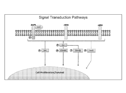
[0039] Figure 3 shows an exemplary illustration of signal transduction
pathway used by
the present invention.
CA 02795362 2012-10-02
WO 2011/133477 PCT/US2011/032935
-16-
100401 Figure 4 shows an exemplary process flowchart of the present
invention. Live
tumor samples are typically obtained from a subject and then at least one
stimulation is
applied to trigger signal transduction events in the live tumor samples. Basel
levels and
stimulated levels of various mRNA. or proteins can be evaluated and then
functional
stratification can be determined.
[00411 Figure 5 shows an exemplary illustration for various
steps/equipments to apply
stimulations to live tumor samples of a subject.
[0042] Figure 6 shows functional stratification of several breast cancer
cell lines. The
upper left chambers are for AKT. The upper middle chambers are for Erk. The
upper right
chambers are for EGFR. The lower left chambers are for GSK3Beta. The lower
middle
chamber are for STAT3. The lower right chambers are for P70S6K.,
[0043] Figure 7 shows an exemplary cell line hierarchal clustering based on
functional
stratification.
[0044] Figures 8 shows exemplary correlations between monolayer cell line
and process
cell line (i.e., after simulation such as SnapPatlirm.
[0045] Figure 9 shows correlations between processed cell line and
xenograft for HCC-
1937.
[0046] Figure 10 shows correlations between processed cell line and
xenograft for MDA-
MB-231.
[0047] Figure 11 shows exemplary functional stratification and potential
drug correlation,
where drug sensitivity and induced fold change after stimulations are
illustrated.
[0048] Figure 12 shows relationship between functional stratification and
potential
therapeutic options.
100491 Figure 13 shows an exemplary illustration where potential drug
sensitivity
associated with functional signaling profiles of TNBC. The upper row includes
pAKT, pErk,
and pEGFR. The lower row includes pGSK, pSTAT3, and p70S6k.
CA 02795362 2012-10-02
WO 2011/133477 PCT/US2011/032935
- 17 -100501 Figure 14 shows an exemplary illustration where ex vivo
stratification and cellular
functional circuitry analysis is possible through drug inhibition, and for
example, on the
SnapPathTM system. This analysis includes pAKT, pErk, pGSK, p70S6k, pSTAT3,
and
pEGFR.
100511 Figure 15 shows exemplary melanoma functional signaling profiles
(modulation)
upon EGF stimulation. Protein levels are measured for pAKT, pERK, pGSK3,
p70S6K,
pSTAT, pEGFR in RPMI-7951, SK-MEL 2, SK-MEL 28, and SK-MEL 31 cells. Fold
changes are calculated for protein level before and after EGF stimulation.
100521 Figure 16 shows exemplary melanoma functional signaling profiles
(inhibition)
upon MEK inhibition by U0126. Protein levels are measured for pERK, pAKT,
pGSK3a/13,
p70S6K., pSTAT, pEGFR in RPM1-7951, SK-MEL 2, SK-MEL 28, and SK-MEL 31 cells.
100531 Figure 17 shows exemplary differentiation of of PLX-4032 resistant
cell line
RPM1-7951 through the induction of pErk following stimulation by TPA.
100541 Figure 18 shows exemplary pancreatic tumor functional signaling
profiles. All
samples except 10195 are examples of human pancreatic neuroendocrine tumors
(PanNETs).
10195 is a sample of a human pancreatic adenocarcinoma. The data reveal
differences in
functional profiles based on TPA stimulation using three different
phosphoprotein biomarkers
(p-ERK1/2, p-GSKa113, and p-STAT3).
100551 Figure 19 shows exemplary melanoma cell line functional signaling
profiles
following stimulation with TPA on the SnapPathTM instrument. Protein levels
measured
include pAKT, pErk, pGSK3, pP70S6k, pSTA.T3, and pEGFR.
100561 Figure 20 shows exemplary melanoma cell line functional signaling
profiles
following stimulation with EGF on the SnapPathTM instrument. Protein levels
measured
include pAKT, pErk, pGSK30, pP70S6k, pSTAT3, and pEGFR.
100571 Figure 21 shows exemplary melanoma cell line functional signaling
profiles
following inhibition with U0126 in the absence of EGF on the SnapPathTM
instrument.
Protein levels measured include pAKT, pErk, pGSK30, pP70S6k, pSTAT3, and
pEGFR.
CA 02795362 2012-10-02
WO 2011/133477 PCT/US2011/032935
-18-
100581 Figure 22 shows exemplary melanoma cell line functional signaling
profiles
following inhibition with U0126 in the presence of ECiF on the SnapPathrm
instrument.
Protein levels measured include pAKT, pErk, pGSK313, pP70S6k, pSTAT3, and
pEGFR.
100591 Figure 23 shows exemplary melanoma cell line functional signaling
profiles
following stimulation of PDGF-P on the SnapPathTM instrument. Protein levels
measured
include pAKT, pErk, pGSK3P, pP70S6k, pSTAT3, and pEGFR.
100601 Figure 24 shows exemplary melanoma cell line functional signaling
profiles
following stimulation of PDGF-13 and MEK inhibition by U0126 on the SnapPathTM
instrument. Protein levels measured include pAKT, pErk, pGSK30, pP70S6k,
pSTAT3, and
pEGFR.
[0061] Figure 25 shows exemplary kinetic curves of phosphoprotein inhibition
in SK-
MEL-28, a melanoma cell line, following treatment with a BRAF inhibitor (PLX-
4702) on the
SnapPathTM instrument. Protein levels measured include pAKT, pErk, pGSK313,
pP70S6k,
and pSTAT3
[0062] Figure 26 shows exemplary dose response curves of phosphoprotein
inhibition in
SK-MEL-28, a melanoma cell line following treatment with a BRAE' inhibitor
(PLX-4702) on
the SnapPathTm instrument. Protein levels measured include pAKT, pErk, pGSK33,
pP70S6k, and pSTAT3.
[0063] Figure 27 shows exemplary kinetic curves of phosphoprotein
inhibition in RPM1-
7951, a melanoma cell line, following treatment with a BRAF inhibitor (PLX-
4702) on the
SnapPathTM instrument. Protein levels measured include pAKT, pErk, pGSK313,
pP70S6k.,
and pSTAT3
[0064] Figure 28 shows exemplary dose response curves of phosphoprotein
inhibition in
RPM1-7951, a melanoma cell line, following treatment with a BRAF inhibitor
(PLX-4702) on
the SnapPathrm instrument. Protein levels measured include pAKT, pErk, pGSK3P,
pP70S6k, and pSTAT3.
[0065] Figure 29 shows exemplary kinetic curves of phosphoprotein inhibition
in SK-
MEL-31, a melanoma cell line, following treatment with a BRAF inhibitor (PLX-
4702) on the
CA 02795362 2012-10-02
WO 2011/133477 PCT/US2011/032935
- 19 -
SnapPathTm instrument. Protein levels measured include pAKT, pErk, pGSK313,
pP70S6k,
and pSTAT3
[0066] Figure 30 shows exemplary dose response curves of phosphoprotein
inhibition in
SK-MEL-31, a melanoma cell line, following treatment with a BRAF inhibitor
(PLX-4702)
on the SnapPathTM instrument. Protein levels measured include pAKT, pErk,
pGSK311,
pP70S6k, and pSTAT3.
100671 Figure 31 shows exemplary kinetic curves of phosphoprotein
inhibition in SK-
MEL-2, a melanoma cell line, following treatment with a BRAF inhibitor (PLX-
4702) on the
SnapPathTM instrument. Protein levels measured include pAk, pErk., pGSK30,
pP70S6k, and
pSTAT3
[0068] Figure 32 shows exemplary dose response curves of phosphoprotein
inhibition in
SK-MEL-2, a melanoma cell line, following treatment with a BRAF inhibitor (PLX-
4702) on
the SnapPathTM instrument. Protein levels measured include pAKT, pErk,
pGSK3[3,
pP70S6k, and pSTAT3.
[0069] Figure 33 shows exemplary melanoma cell line (SK-MEL-28) functional
signaling
profiles following stimulation of EGF on the SnapPathrm instrument. Protein
levels measured
include pAKT, pErk, pMEK.
100701 Figure 34 shows exemplary melanoma cell line (RPMI-7951) functional
signaling
profiles following stimulation of EGF as well as BRAF and ERK inhibition with
PLX-4702
and U0126 on the SnapPathTM instrument. Protein levels measured include pAKT,
pErk,
pMEK.
[0071] Figure 35 shows exemplary melanoma cell line (RPM1-7951) functional
sig;naling
profiles following stimulation of EGF as well as BRAF and ERK inhibition with
PLX-4702
and U0126 on the SnapPathTM instrument. Protein levels measured include pAKT,
pErk,
pMEK and pEGFR.
100721 Figure 36 shows exemplary melanoma cell line (RPMI-7951) functional
signaling
profiles following stimulation of PDGFp as well as BRAF and ERIC inhibition
with PLX-
4702 and U0126 on the SnapPath.TM instrument. Protein levels measured include
pAKT,
pErk, pMEK.
CA 02795362 2012-10-02
WO 2011/133477 PCT/US2011/032935
- 20 -
DETAILED DESCRIPTION OF THE INVENTION
[0073] Before the present composition, methods, and treatment methodology
are
described, it is to be understood that this invention is not limited to
particular compositions,
methods, and experimental conditions described, as such compositions, methods,
and
conditions may vary. It is also to be understood that the terminology used
herein is for
purposes of describing particular embodiments only, and is not intended to be
limiting, since
the scope of the present invention will be limited only in the appended
claims.
100741 As used in this specification and the appended claims, the singular
forms "a", "an",
and "the" include plural references unless the context clearly dictates
otherwise. Thus, for
example, references to "the method" includes one or more methods, and/or steps
of the type
described herein which will become apparent to those persons skilled in the
art upon reading
this disclosure and so forth.
[0075] Unless defined otherwise, all technical and scientific terms used
herein have the
same meaning as commonly understood by one of ordinary skill in the art to
which this
invention belongs. Although any methods and materials similar or equivalent to
those
described herein can be used in the practice or testing of the invention, the
preferred methods
and materials are now described
[0076] The invention provides a safe, effective, accurate, precise,
reproducible,
inexpensive, cost effective, efficient, fast and convenient method and
"cartridge-based"
system for collecting, handling and processing of cellular specimens ex vivo.
These methods
and cartridges can maintain viability of the samples during the process to
maintain bi.omarker
integrity, and optionally, evoking biomarkers such as phosphoproteins and
nucleic acid
molecules not present in original sample through ex vivo stimulation and/or
inhibition. The
invention provides fully integrated specimen and information management in a
complete
diagnostic cytology laboratory system and controlled conditions following
biopsy, which
minimizes variability between tests, minimizes the risk of biocontamination,
and minimizes
the effect of the sample preparation process itself on biomarker expression.
[0077] Embodiments of the present invention can be used to facilitate
targeted treatment of
diseases, and optionally also provide a tissue sample adequacy evaluation such
as a cell-count,
cell function and/or other connected analyses.
- -
100781 As one of skill in the art Will appreciate, the devices, systems,
kits and methods as
described herein provide numerous advantages in a clinical or research
setting. For example,
they can be used to provide rapid, near patient, biopsy processing without the
need to send the
specimen to a remote laboratory. They cart also be used to standardize and
automate biopsy
processing in a cost effective manner. T he present invention can provide more
detailed
molecular information about the cells than current pathological processes
allow which enables
greater sub-classifications of cells in a biopsy (e.g., cancer or disease
cells), optionally using
new c_vvivo biomarkers. Taken together, the advantages of the present
invention allow for a.
rapid diagnosis near the point of care and the subsequent creation of more
effective patient
specific treatment regimens.
[00791 It should be understood that the methods of the invention may be
performed alone
or in conjunction with the systems and devices set forth in US. Publication
No.
2009/01162853.
[0080] In one aspect, the invention provides molecular assays capable
diagnosis and/or
prognosis of a disease in a subject. In addition, the molecular assays of the
invention are
capable of both evaluating the sensitivity or resistance of a patient's
disease to an agent prior
to initiation of therapy and monitoring the therapy effects during treatment.
The diagnostic
assay directs therapy and determines prognosis of patients treated with
targeted therapies.
[0081] Accordingly, the invention provides a method for the diagnosis
and/or prognosis of
disease in a subject. The method includes determining the difference between a
basal level or
state of a molecule in a sample and the level or state of the molecule after
stimulation Of a
portion of the sample with a modulator ox vivo, wherein the difference is
expressed as a value
which is indicative of the presence, absence or risk of having a disease.
[0082] Exemplary modulators include, but are not limited to, physical,
biological, or
chemical modulators. Included in the term "modulators" are .stimulators and
inhibitors, such
as small molecules (e.g. erlotinib, gel-mint), or lapatanib), and antibodies
1.IERCEI)TIN4)). In one embodiment, the modulator is .an epidermal growth
factor receptor
(EGER) inhibitor or activator. As used herein, the term "EGER" refers to erbB
gene familv
products. II will be understood by those skilled in the art that the EGFR may
he a product of
ally crbB receptor encoded by any gene from the erbB gene family, and any homo-
and
heterodimers thai these molecules are known to loon. While erbB-1 product is
the main
CA 2795362 2017-08-31
CA 02795362 2012-10-02
WO 2011/133477 PCT/US2011/032935
- 22 -
receptor, the expression of which has been detected in previous studies, there
is reason to
believe that the cell lines and tumors tested herein also express other erbB
gene family
members. Lastly, the EGFR ligand or combination of ligand we used binds to
almost all of
the known EGFR receptor forms, and therefore our assay measures the effects
exerted by
those proteins. In another embodiment, the modulator is a combination of one
or more
modulators such as, for example, one or more of EGF, TGF-u, and Heregulin.
[0083] Thus, the quantitative or qualitative effect measured can be the
expression level of
a gene, such as, an immediate or delayed early gene family member. Suitable
immediate or
delayed early gene family members include, but are not limited to, FOS, JUN
and DUSP I-
28.
[00841 As used herein, the term "disease" is used broadly to refer to any
pathological
condition of a part, organ, or system of a subject resulting from various
causes, such as
infection, genetic defect, or environmental stress, and characterized by an
identifiable group
of signs or symptoms. Exemplary diseases include, but are not limited to,
stroke,
cardiovascular disease, chronic obstructive pulmonary disorder, myocardial
infarction,
congestive heart failure, cardiomyopathy, myocarditis, ischemic heart disease,
coronary artery
disease, cardiogenic shock, vascular shock, pulmonary hypertension, pulmonary
edema
(including cardiogenic pulmonary edema), cancer, pathogen-mediated disease,
pleural
effusions, rheumatoid arthritis, diabetic retinopathy, retinitis pigmentosa,
and retinopathies,
including diabetic retinopathy and retinopathy of prematurity, inflammatory
diseases,
restenosis, edema (including edema associated with pathologic situations such
as cancers and
edema induced by medical interventions such as chemotherapy), asthma, acute or
adult
respiratory distress syndrome (ARDS), lupus, vascular leakage, transplant
(such as organ
transplant, acute transplant or heterograft or homograft (such as is employed
in burn
treatment)) rejection; protection from ischemic or reperfusion injury such as
ischemic or
reperfusion injury incurred during organ transplantation, transplantation
tolerance induction;
ischemic or reperfusion injury following angioplasty; arthritis (such as
rheumatoid arthritis,
psoriatic arthritis or osteoarthritis); multiple sclerosis; inflammatory bowel
disease, including
ulcerative colitis and Crohn's disease; lupus (systemic lupus crythematosis);
graft vs. host
diseases; T-cell mediated hypersensitivity diseases, including contact
hypersensitivity,
delayed-type hypersensitivity, and gluten-sensitive enteropathy (Celiac
disease); Type I
diabetes; psoriasis; contact dermatitis (including that due to poison ivy);
:Hashimoto's
CA 02795362 2012-10-02
WO 2011/133477 PCT/US2011/032935
- 23 -
thyroiditis; Sjogren's syndrome; Autoimmune Hyperthyroidism, such as Graves'
disease;
.Addison's disease (autoimmune disease of the adrenal glands); autoimmune
polyglandular
disease (also known as autoimmune polyglandular syndrome); autoimmune
alopecia;
pernicious anemia; vitiligo; autoimmune hypopituatarism; Guillain-Barre
syndrome; other
autoimmune diseases; cancers, including those where kinases such as Src-family
kinases are
activated or overexpressed, such as colon carcinoma and thymorna, or cancers
where kinase
activity facilitates tumor growth or survival; glomerulonephritis, serum
sickness; uticaria;
allergic diseases such as respiratory allergies (asthma, hayfever, allergic
rhinitis) or skin
allergies; mycosis fungoides; acute inflammatory responses (such as acute or
adult respiratory
distress syndrome and ischemialreperfusion injury); dermatomyositis; alopecia
areata; chronic
actinic dermatitis; eczema; Behcet's disease; Pustulosis palmoplanteris;
Pyoderma
gangrenum; Sezary's syndrome; atopic dermatitis; systemic schlerosis; morphea;
peripheral
limb ischemia and isch.emic limb disease; bone disease such as osteoporosis,
osteomal.acia,
hyperparathyroidism, Paget's disease, and renal osteodystrophy;vascular leak
syndromes,
including vascular leak syndromes induced by chemotherapies or
immunomodulators such as
IL-2; spinal cord and brain injury or trauma; glaucoma; retinal diseases,
including macular
degeneration; vitreoretinal disease; pancreatitis; vascu.latides, including
vasculitis, Kawasaki
disease, thromboangiitis obliterans, Wegener's granulomatosis, and Behcet's
disease;
scleroderma; preeclampsia; thalassemia; Kaposi's sarcoma; and von Hippel
Lindau disease.
100851 In one embodiment, the disease is cancer. Exemplary cancers include,
but are not
limited to, colorectal, esophageal, stomach, lung, prostate, uterine, breast,
skin, endocrine,
urinary, pancreas, ovarian, cervical, head and neck, liver, bone, biliary
tract, small intestine,
hematopoietic, vaginal, testicular, anal, kidney, brain, eye cancer, leukemia,
lymphoma, soft
tissue, melanoma, and metastases thereof.
100861 in another embodiment, the disease is a pathogen-mediated disease.
Exemplary
pathogens include, but are not limited to, bacteria, fungi, viruses,
spirochetes, and parasites.
Exemplary viruses include, but are not limited to, Herpes simplex virus l (HSV
1), Herpes
simplex virus 2 (HSV2), respiratory syncytial virus, measles virus (MV), human
cytomegalovirus (FICMV), vaccinia virus, human inununodeficiency virus type 1
(HIV-l),
and hepatitis C virus (HCV).
CA 02795362 2012-10-02
WO 2011/133477
PCT/US2011/032935
- 24 -
[0087] As used
herein, the terms "sample" and "biological sample" refer to any sample
suitable for the methods provided by the present invention. A. sample of cells
used in the
present method can be obtained from tissue samples or bodily fluid from a
subject, or tissue
obtained by a biopsy procedure (e.g., a needle biopsy) or a surgical
procedure. Thus,
exemplary samples include, but are not limited to, a tissue sample, a frozen
tissue sample, a
biopsy specimen, a surgical specimen, a cytological specimen, a cell line, a
xenograft, a
tumor, a fine needle aspiration, whole blood, bone marrow, cerebral spinal
fluid, peritoneal
fluid, pleural fluid, lymph fluid, serum, plasma, amniotic fluid, mucus,
plasma, urine, chyle,
stool, sputum, perspiration, tears, semen, nipple aspirate, saliva, and any
combination thereof.
In certain embodiments, the sample can be a fraction of a blood sample such as
a peripheral
blood lymphocyte (PHIL) fraction. Methods for isolating P.131,s from whole
blood are well
known in the art. In addition, it is possible to use a blood sample and enrich
the small amount
of circulating cells from a tissue of interest, e.g., ovaries, breast, etc.,
using methods known in
the art.
100881 Fine needle aspiration (FNA) has demonstrated to be a robust and safe
method to
acquire tumor material in sufficient quantities to assess pharmacodynamic
endpoints in a
serial manner. In addition, preliminary evidence is provided suggesting that
this methodology
can be efficiently used in procuring tissue to reproduce in vitro conditions
and develop an ex
vivo molecular sensitivity and resistance assay. This approach has classically
drawn
considerable interest and the outcome and ultimate significance of a number of
these studies
has been the subject of recent reviews, Most studies analyzed whether cells
derived from a
sample of viable tumor tissue show a response when exposed to selected
therapeutic agents
under in vitro conditions. Thus, in one embodiment, the assay is based on fine
needle
aspiration of any lesion and processing the aspirated material for protein
and/or nucleic acid
analysis. Depending on the particular pharmaceutical agent used, the assay
allows for
determination of sensitivity of the lesion to treatment, effectiveness of
specific pathway
blockade, and monitoring of therapy effects at the molecular level. The assay
can be
performed with minimal morbidities and discomfort, and can be used for drug
sensitivity
assessment, dosing regimen, therapy effect measurement, and prognostication.
[0089] SnapPathmi Ex Vivo Biomarker Platform: Fine needle aspiration biopsies
(FNABs)
are a minimally-invasive method for sampling human tumors that is widely used
in the United
States. Historically FNAB samples have provided adequate material for
microscopic
CA 02795362 2012-10-02
WO 2011/133477 PCT/US2011/032935
- 25 -
examination, however the successful development and use of targeted cancer
drugs will also
require biomarker information derived from these clinical samples.
100901 While ex vivo bionurkers have been used successfully in various
clinical trials
using manual live tissue manipulation at the patient's bedside. Ex vivo tests
are not clinically
feasible unless an automated, rapid processing device, such as SnapPatkrm
exists. The ability
to interrogate live tumor cells with novel ex vivo biomarker tests to
determine the most
effective cancer treatment for individual patients is the promise of the
SnapPathTM biomarker
platform.
100911 SnapPathTM benchtop units will utilize automated fluidic
technologies to process
and manipulate live tumor biopsy samples, within uniquely designed insertible
cartridges. In
the SnapPathTM system, radiologists will deposit (FN.A) biopsy samples into a
SnapPaLhTM
cartridge immediately after the needle is removed from the patient. Cartridges
will then be
rapidly delivered to pathology where the SnapPathTM platform will be located,
in a process
similar to that required for lymphoma samples processed by flow cytometry.
100921 The SnapPathTM biomarker platform is being developed with a $2.3
million Fast-
Track SBIR contract from the National Cancer Institute. In the NCI's contract
award, the
agency stated that the company's SnapPathTM technology presented an
"innovative" FNA
biopsy approach and instrument that was "extremely responsive" to the NCI's
contract
announcement which expressed interest in "biopsy instruments and devices that
preserve
molecular profiles in tumors," including those that will "create an entirely
new diagnostic
area" and "enable individualized molecular therapy of solid tumors based on
accurate
information about signal transduction pathways, molecular drug targets and
biomarkers." The
NCI also recently stated that technologies focusing on ex vivo diagnostics and
ex vivo tissue-
analysis are a "priority" for the 'NCI's SBIR. Phase II Bridge Award program.
10093) The term "subject" as used herein refers to any individual or
patient to which the
subject methods are performed. Generally the subject is human, although as
will be
appreciated by those in the art, the subject may be an animal. Thus other
animals, including
mammals such as rodents (including mice, rats, hamsters and guinea pigs),
cats, dogs, rabbits,
farm animals including cows, horses, goats, sheep, pigs, etc., and primates
(including
monkeys, chimpanzees, orangutans and gorillas) are included within the
definition of subject.
CA 02795362 2012-10-02
WO 2011/133477 PCT/US2011/032935
- 26 -
In addition, the term "subject" may refer to a culture of cells, where the
methods of the
invention are performed in vitro to assess, for example, efficacy of a
therapeutic agent.
100941 As used herein, the terms "molecule" or "biomolecule" refer to any
organic
molecule in a living organism. Exemplary biomolecules include, but are not
limited to,
peptides, lipids, nucleic acids, m.etabolites, and carbohydrates. In one
embodiment, the
biomolecule is a peptide, such as a protein, or a nucleic acid molecule. The
terms
"polypeptide," "peptide" and "protein" are used interchangeably herein to
refer to two or
more amino acid residues joined to each other by peptide bonds or modified
peptide bonds,
i.e., peptide isosteres. The terms apply to amino acid polymers in which one
or more amino
add residue is an artificial chemical mimetic of a corresponding naturally
occurring amino
acid, as well as to naturally occurring amino acid polymers, those containing
modified
residues, and non-naturally occurring amino acid polymer.
100951 The term "nucleic acid molecule" is used broadly herein to mean a
sequence of
deoxyribonucleotides or ribonucleotides that are linked together by a
phosphodiester bond.
As such, the term "nucleic acid molecule" is meant to include DNA and RNA,
which can be
single stranded or double stranded, as well as DNA/RNA hybrids. Furthermore,
the term
"nucleic acid molecule" as used herein includes naturally occurring nucleic
acid molecules,
which can be isolated from a cell, for example, a particular gene of interest,
as well as
synthetic molecules, which can be prepared, for example, by methods of
chemical synthesis
or by enzymatic methods such as by the polymerase chain reaction (PCR), and,
in various
embodiments, can contain nucleotide analogs or a backbone bond other than a
phosphodiester
bond.
100961 As used herein, the term "EGFR modulator" refers to a compound or drug
that is a
biological molecule or a small molecule that directly or indirectly modulates
EGFR. activity or
the EGFR signal transduction pathway. Compounds or drugs as used herein is
intended to
include both small molecules and biological molecules. Direct or indirect
modulation
includes activation or inhibition of EGFR activity or the EGFR signal
transduction pathway.
Inhibition refers to inhibition of the binding of EGFR to an EGFR ligand
including, for
example, EGF. In addition, inhibition can also refer to inhibition of the
kinase activity of
EGFR.
CA 02795362 2012-10-02
WO 2011/133477 PCT/US2011/032935
- 27 -
[0097] EGFR modulators include, for example, EGFR specific ligands, small
molecule
EGFR inhibitors, and EGFR monoclonal antibodies. In one aspect, the EGFR
modulator
inhibits EGFR activity and/or inhibits the EGFR signal transduction pathway.
In another
aspect, the EGFR modulator is an EGFR antibody that inhibits EGFR activity
and/or inhibits
the EGFR signal transduction pathway.
[0098] EGFR modulators include biological molecules or small molecules.
Biological
molecules include all lipids and polymers of monosaccharides, amino acids, and
nucleotides
having a molecular weight greater than 450. Thus, biological molecules
include, for example,
oligosaccharides, polysaccharides, oligopeptides, polypeptides, peptides,
proteins,
oligonucleotides, and polynucleotides. Oligonucleotides and polynucleotides
include, for
example, DNA and RNA. Biological molecules further include derivatives or
combination of
any of the molecules described above. For example, derivatives of biological
molecules
include lipid and glycosylation derivatives of oligopeptides, polypeptides,
peptides, and
proteins.
[0099] In addition to the biological molecules discussed above, the EGFR
modulators
useful in the invention may also be small molecules. Any molecule that is not
a biological
molecule can be considered herein to be a small molecule. Some examples of
small
molecules include organic compounds, organometallic compounds, salts of
organic and
organometallic compounds, saccharides, amino acids, and nucleotides. Small
molecules
further include molecules that would otherwise be considered biological
molecules, except
their molecular weight is not greater than 450. Thus, small molecules may be
lipids,
oligosaccharides, oligopeptides, and oligonucleotides and their derivatives,
having a
molecular weight of 450 or less.
[0100] It is noted that small molecules are merely called small molecules
because they
typically have molecular weights less than 450. Small molecules include
compounds that are
found in nature as well as synthetic compounds. In one embodiment, the EGFR
modulator is
a small molecule that inhibits the growth of tumor cells that express EGFR. In
another
embodiment, the EGFR modulator is a small molecule that inhibits the growth of
refractory
tumor cells that express EGFR. Numerous small molecules have been described as
being
useful to inhibit EGFR and are well known in the art.
CA 02795362 2012-10-02
WO 2011/133477 PCT/US2011/032935
-28-
101011 The invention also includes specialized microarrays, e.g.,
oligonucleotide
microarrays or cDNA microarrays, comprising one or more biomarkers, showing
expression
profiles that correlate with either sensitivity or resistance to one or more
stimulations, for
example EGFR modulators. Such microarrays can be employed in in vitro assays
for
assessing the expression level of the biomarkers in the test cells from tumor
biopsies, and
determining whether these test cells are likely to be resistant or sensitive
to stimulations, for
example EGFR modulators. Cells or live tissue samples from a subject can be
isolated and
exposed to one or more of stimulations, for example the EGFR modulators.
Following
application of nucleic acids isolated from both untreated and treated cells to
one or more of
the specialized microarrays, the pattern of gene expression of the tested
cells can be
determined and compared with that of the biomarker pattern from the control
panel of cells
used to create the biomarker set on the microarray. Based upon the gene
expression pattern
results from the cells that underwent testing, it can be determined if the
cells show a resistant
or a sensitive profile of gene expression.
101021 The invention also includes kits for determining or predicting
whether a patient
would be susceptible or resistant to a treatment that includes one or more
stimulations, for
example EGFR modulators. The patient may have a cancer or tumor such as, for
example, a
breast cancer or tumor. Such kits would be useful in a clinical setting for
use in testing a
patient's cancer samples, for example, to determine or predict if the
patient's tumor or cancer
will be resistant or sensitive to a given treatment or therapy. The kit
includes a suitable
container that includes one or more microarrays, e.g., oligonucleoti.de
microarrays or cDNA.
microarrays, that include those biomarkers that correlate with resistance and
sensitivity to
stimulations, for example EGFR modulators, particularly EGFR inhibitors; one
or more
stimulations, for example EGFR modulators for use in testing cancer samples or
cells from a
patient; and instructions for use. In addition, kits contemplated by the
invention can further
include, for example, reagents or materials for monitoring the expression of
biomarkers of the
invention at the levels of rnRNAs or proteins, using other techniques and
systems practiced in
the art such as, for example, RT-PCR assays, which employ primers designed on
the basis of
one or more of the biomarkers, immunoassays, such as enzyme linked
immunosorbent assays
(ELISAs), imm.unoblotting, e.g., Western blots, or in situ hybridization, and
the like.
CA 02795362 2012-10-02
WO 2011/133477 PCT/US2011/032935
- 29 -
[0103] In one embodiment, the protein is a post-translationally modified
protein, where the
protein is modified by one or more of phosphorylation, acetylation, amidation,
rnethylation,
nitrosylation, fatty acid addition, lipid addition, glycosylation, and
ubiquitination.
[0104] In another embodiment, the methods further include exposing the
sample to one or
more therapeutic agents or combination thereof. For solid tumor or other
cancer applications,
the therapeutic agents can include a targeted pharmaceutical agent such as,
for example,
antitumor monoclonal antibodies, e.g. trasttrzurnab (Herceptint), cetuximab
(Erbituxt),
bevacizumab (AvastinO) and rituximab (Rituxane andlor Mabtherae), and small
molecule
inhibitors, e.g., gefitinib (Iressa0), or erlotinib (Tarcevae) or cytotoxic
chemotherapy agents.
[0105] Exemplary chemotherapeutic agents also include, but are not limited
to,
antimetabolites, such as methotrex ate, DNA cross-linking agents, such as
cisplatinicarboplatin; alkylating agents, such as canbusil; topoisomerase I
inhibitors such as
dactinomicirn microtubule inhibitors such as taxol (paclitaxol), and the like.
Other
chemotherapeutic agents include, for example, a vinca alkaloid, mitomycin-type
antibiotic,
bleomycin-type antibiotic, antifolate, colchicine, demecoline, etoposide,
taxane, anthracycline
antibiotic, doxorubicin, daunorubicin, carminomycin, epirubicin, idarubicin,
mithoxanthrone,
4-dimethoxy-dattnomycin, 11-deoxydaunorubicin, I3-deoxydaunorubicin,
adriamycin-14-
benzoate, adtiamycin-14-octanoate, adriamycin-14-naphthaleneacetate,
amsacrine,
carmustine, cyclophosphamide, cytarabine, etoposide, lovastatin, melphalan,
topetecan,
oxalaplatin, chlorambucil, metbtrexate, lomustine, thioguanine, asparaginase,
vinblastine,
vindesine, tamoxifen, or mechlorethamine. While not wanting to be limiting,
therapeutic
antibodies include antibodies directed against the HER2 protein, such as
trastuzumab;
antibodies directed against growth factors or growth factor receptors, such as
bevacizumab,
which targets vascular endothelial growth factor, and OSI-774, which targets
epidermal
growth factor; antibodies targeting integrin receptors, such as Vitaxin (also
known as MEDI-
522), and the like. Classes of anticancer agents suitable for use in
compositions and methods
of the present invention include, but are not limited to: 1) alkaloids,
including, microtubule
inhibitors (e.g., Vincristine, Vinblastine, and Vindesine, etc.), microtubule
stabilizers (e.g.,
Paclitaxel [Taxol], and Docetaxel, Taxotere, etc.), and chromatin function
inhibitors,
including, topoisomerase inhibitors, such as, epipodophyllotoxins (e.g.,
Etoposide [VP-16],
and Teniposide [VM-26], etc.), and agents that target topoisomerase I (e.g.,
Camptothecin and
lsirinotecan [CPT-11], etc.); 2) covalent DNA-binding agents [allcylating
agents], including,
CA 02795362 2012-10-02
WO 2011/133477
PCT/US2011/032935
- 30 -
nitrogen mustards (e.g., Mechlorethamine, Chlorambucil, Cyclophosphamide,
Ifosphamide,
and Busulfan [Myleran], etc.), nitrosoureas (e.g., Carmustine, Lomustine, and
Semustine,
etc.), and other alkylating agents (e.g., Dacarbazine, Hydroxymethylmelamine,
Thiotepa, and
Mitocycin, etc.); 3) noncovalent DNA-binding agents [antitumor antibiotics],
including,
nucleic acid inhibitors (e.g., Dactinomycin [Actinomycin 13], etc.), anthracyc
lines (e.g.,
Daunorubicin [Daunornycin, and Cerubidine], Doxorubicin [Adriamycin], and
Idarubicin
[Idamycin], etc.), arithracenediones (e.g., anthracycline analogues, such as,
[Mitoxantrone],
etc.), bleomycins (Blenoxane), etc., and plicamycin (Mithramycin), etc.; 4)
antimetabolites,
including, antifolates (e.g., Methotrexate, Folex, and Mexate, etc.), purine
antimetabolites
(e.g., 6-Mercaptopurine [6-MP, Purinethol], 6-Thioguanine [6-TG],
Azathioprine, Acyclovir,
Ganciclovir, Chlorodeoxyadenosine, 2-Chlorodeoxyadenosine [CdA], and 2'-
Deoxycoformycin [Pentostatin], etc.), pyrimidine antagonists (e.g.,
fluoropyrimidines [e.g., 5-
fluorouracil (Adrucil), 5-fluorodeoxyuridine (Fdlird) (Floxuridine)] etc.),
and cytosine
arabinosides (e.g., Cytosar [ara-C] and Fludarabine, etc.); 5) enzymes,
including, L-
asparaginase; 6) hormones, including, glucocorticoids, such as, antiestrogens
(e.g.,
Tamoxifen, etc.), nonsteroidal antiandrogens (e.g., Flutamide, etc.), and
aromatase inhibitors
(e.g., anastrozole [Arirnidex], etc.); 7) platinum compounds (e.g., Cisplatin
and Carboplatin,
etc.); 8) monoclonal antibodies conjugated with anticancer drugs, toxins,
and/or
radionuclides, etc.; 9) biological response modifiers (e.g., interferons
[e.g., IFN-a, etc.] and
interleukins [e.g., IL-2, etc.], etc.); 10) adoptive immunotherapy; 11)
hematopoietic growth
factors; 12) agents that induce tumor cell differentiation (e.g., all-trans-
retinoic acid, etc.); 13)
gene therapy techniques; 14) antisense therapy techniques; 15) tumor vaccines;
16) therapies
directed against tumor metastases (e.g., Batimistat, etc.); and 17) inhibitors
of anOogenesis.
Thus, in one embodiment, the therapeutic regimen is a administration of
cisplatin in
combination with paclitaxel.
101061 In another
aspect, the invention provides a method of predicting the effect of an
agent or combination of agents. The method includes determining the difference
between a
basal level or state of a molecule in a sample and the level or state of the
molecule after
stimulation of a portion of the sample with a modulator ex vivo, wherein the
difference in the
basal level or state of the molecule expressed as a value is indicative of a
positive or negative
effect of the agent. In one embodiment, the agent interacts directly with the
molecule in the
sample. In another embodiment, the effect is the activation or inhibition of a
cellular pathway
selected from the group consisting of a metabolic pathway, a replication
pathway, a cellular
CA 02795362 2012-10-02
WO 2011/133477 PCT/US2011/032935
- 31 -
signaling pathway, an oncogenic signaling pathway, an apoptotic pathway, and a
pro-
angiogenic pathway.
101071 In another aspect, the invention provides a method of screening test
agents for an
effect on a molecule. Thus, the effects of the presence or absence of a test
agent can also be
determined by detecting an ex vivo biomarker, for example, a post-
translationally modified
protein, ions, or enzymes. The method includes contacting a sample containing
the molecule
or molecules with the test agent ex vivo, then determining a difference
between a basal level
or state of the molecule in the sample and the level or state of the molecule
after stimulation
of a portion of the sample with a modulator ex vivo; wherein a difference in
the basal level or
state of the molecule before and after contacting with the test agent is
indicative of an effect
on the molecule.
101081 Suitable test agents include, but are not limited to, one or more of
the following:
small molecule chemical, a chemotherapeutic agent, a hormone, a protein, a
peptide, a
peptidomimetic, a protein, an antibody, a nucleic acid, an RNAi molecule, and
an antisense
molecule. In one embodiment, the administration of a test agent can be
followed by
measuring a quantitative or qualitative effect on a target ex vivo biomarker
or biomolecule of
the dispersed or distributed cell.
101091 In another embodiment, it can be determined if the test agent
affects the expression
of one or more markers, wherein the presence, absence, or relative degree of
such expression
is indicative of the susceptibility of the cells to a selected pharmaceutical
agent. These
markers can include a wide array of ex vivo biomarkers such as niRNA, a
microRNA, cDNA.,
a protein, a phosphoprotein, a posttranslational modification of a protein, or
a modification of
histone or DNA packaging. For example, the marker can be mRNA or cDNA for an
early
response gene (e.g., FOS or JUN) associated with susceptibility to a
pharmaceutical agent.
The presence, absence, or relative degree of expression of combinations of
markers in the
presence of a test reagent can be indicative of the susceptibility of the
cells to a selected test
reagent, such as a pharmaceutical agent.
101101 For certain analytical methods, the test agent can be a detectable
agent. The
detectable agent can be used individually or as conjugated or otherwise
connected to another
compound (e.g., a detectable agent conjugated to an antibody). Suitable
detectable agents
include, but are not limited to, an enzyme, fluorescent material, luminescent
material,
=
- -
bioluminescent material, radioactive material, positron emitting metal using a
positron
emission tomography, or a nonradioactive paramagnetic metal ion.
[01111 Once disease is established and a treatment protocol is initiated,
the methods of the
invention may be repeated on a regular basis to evaluate whether the level or
intensity of
symptoms related to the disease in the subject begins to approximate that
which is observed in
a normal subject. The results obtained from successive assays may be used to
show the
efficacy of treatment over a period ranging from several days to months.
Accordingly, the
invention is also directed to methods for monitoring the course of a subject's
therapy. The
method includes determining the difference between a basal level or state of a
molecule in a
sample and the level or state of the molecule after stimulation of a portion
of the sample with
a modulator ex vivo, optionally prior to, simultaneously with or following a
course of therapy;
wherein the diffe;rence in the basal level or state of the molecule expressed
as a value. is
indicative of a positive or negative treatment. Thus, a positive treatment is
indicative of the
subject being a responder to the course of therapy. Likewise, a negative
treatment is
indicative of the subject having resistance to the course of therapy.
101121 In one embodiment, the method may further include comparing the
level of the.
signs and symptoms related to the disease prior to and during therapy, where a
lessening of
the signs and symptoms of disease indicates the efficacy of the therapy.
'Therefore, one
skilled in the art will be able to recognize and adjust the therapeutic
approach as needed.
[01131 The methods described herein may be performed with or without a
cartridge as
described in U.S. Publication No. 2009/0162853. An
advantage of the methods and devices herein is that the test agent can be
added at the point of
care and/or can come preloaded in specified wells of the cartridge. This
allows the testing of
ex vino biomarh-ers, optionally near the point of care, using live cells.
These methods and
devices can be used with specific test agents to manipulate samples ex vivo to
facilitate the
development of novel predictive hiomarkers, monitor and determine cellular
sensitivity to
specific pharmaceutical agents, and other uses that one of skill in the art
will appreciate,
10114 For example, a sample of a solid tumor from a patient can be
disaggregated,
distributed, and then tested against a panel of currently available cancer
therapeutics at the
point of care. The samples can then be stabilized and/or fixed if necessary
and analyzed.
Depending on the results for each lest agent, the physician can quic.k.ly
determine which
CA 2795362 2017-08-31
CA 02795362 2012-10-02
WO 2011/133477 PCT/US2011/032935
- 33 -
therapeutics will be most effective on the individual patient's tumor at or
near the point of
care. This personalized medicine provides numerous benefits, in particular,
the use of
targeted cancer therapeutics and regimens in a rapid, cost effective manner.
101151 Embodiments of the invention are directed to analyzing the
distributed cells (e.g.,
cancer cells) by administering at least one agent to produce a measurable
quantitative or
qualitative effect on a target ex vivo biomarker or biomolecule. The
quantitative or qualitative
effect can be the activation or inhibition of a cellular pathway. Exemplary
cellular pathways
include, but are not limited to, a metabolic pathway, a replication pathway, a
cellular
signaling pathway, an oncogenic signaling pathway, an apoptotic pathway, and a
pro-
angiogenic pathway. For example, the quantitative or qualitative effect can be
a measurement
of an agonistic or antagonistic effect on a G-protein coupled receptor or a
receptor tyrosine
kinase, such as, epidermal growth factor receptor (EGFR) and the downstream
pathways.
101161 In various embodiments, the cells and/or molecules are analyzed
using one or more
methods selected from an array, enzyme-linked inununosorbent assay (ELISA),
multiplex,
bioplex, luminex, mass spectrometry, flow cytometry, Northern blot, Southern
blot, Western
blot, and radioimmunoassay (RI A). In other embodiments, cells and/or
molecules are
analyzed using any apparatus known in the art for analyzing nucleic acids. In
other
embodiments, the difference between a basal level or state of a molecule in a
sample from a
subject and the level or state of the molecule after stimulation of a portion
of the sample with
a modulator ex vivo is determined using a computer.
101171 In another aspect, the invention provides a method for
stratification of patients
based on responsiveness to a therapeutic agent or therapeutic regimen. The
method includes
determining the difference between a basal level or state of a molecule in a
sample from a
subject and the level or state of the molecule after stimulation of a portion
of the sample with
a modulator ex vivo; wherein the difThrence in the basal level or state of the
molecule
expressed as a value is indicative of a positive or negative response to a
therapeutic agent or
therapeutic regimen, Thus, a positive response is indicative of the subject
being a responder
to the course of therapy. Likewise, a negative response is indicative of the
subject having
resistance to the course of therapy.
CA 02795362 2012-10-02
WO 2011/133477 PCT/US2011/032935
- 34 -
[0118] As described herein, the cells processed using the present invention
can be prepared
and stabilized in a number of ways to permit a wide array of cellular analyses
to be performed
on them. For example, the cells can be prepared for nucleic acid analysis,
protein analysis,
and/or analyzed using live cellular probes.
[0119] For nucleic acid analysis, a stabilizing reagent such as RN.Alater ,
RNA. Protect
Cell Reagent (both available from Qiagen), or ethanol can be added to the
cells. The
stabilized cells can then be optionally lysed or have the nucleic acid of
interest otherwise
extracted. The extracted and purified nucleic acid can then be analyzed, for
example, using
PCR techniques.
[0120] In some embodiments, the methods described herein yield nucleic acid
molecules
for further analysis. For these samples, following dispersion and optional
enrichment, the
nucleic acids can be stabilized or extracted (optionally) to yield high
quality and quantity
nucleic acid molecules. This can be done, for example, by lysin.g the desired
cells following
exposure to a test agent and then obtaining cDNA using reverse transcriptase
and DNA
primers. The DNA primers can include nonspecific primer complementary to poly
A, e.g.
oligo(dT)1 2-18 or a specific primer complementary to a mRNA transcript of
interest. As one
of skill in the art will appreciate, the cells can be lysed using a variety of
methods, such as,
chemical or mechanical means.
[0121] Optionally, the cells can be stabilized with reagents to detect
and/or preserve
biomarker information, e.g., using reverse transcriptase and DNA primer to
obtain cDNA
transcripts, preparing RNA, DNA. and protein for down stream molecular
analysis.
[0122] For protein or nucleic acid analysis, either whole cells or lysed
cells can be used.
Intact whole cells can be fixed and stabilized with a polymer, such that the
sample adheres to
the isolated chamber, for example, a glass slide. These samples can then be
subjected to
analysis, for example, immunohistochemical (IHC) analysis. Lysed or otherwise
ruptured
cells can be used in assays such as Western Blots and may not require
stabilization or fixation.
[0123] Slide preparation for morphological review by a pathologist and
protein analysis by
IBC can be an output of the methods described herein. Accordingly, the cells
can also be
prepared, optionally using polymers, on glass slides for analysis of
morphology and/or
immunohistoch.emistry.
CA 02795362 2012-10-02
WO 2011/133477 PCT/US2011/032935
-35-
101241 Live cellular probe analysis can involve adding a molecular probe
(such as
MitoTracker8) at any point in the method of processing the cells where the
cells are alive.
This addition of the live cell probe should be made prior to fixing or
otherwise allowing the
cells to die. For example, such a probe can be added before or after cellular
stabilization but
prior to cellular fixation.
[0125] In some embodiments, the cells can be stabilized and fixed by any
suitable means
that will permit subsequent molecular analysis and detection of markers.
Generally,
crosslinking fixatives such as formalin are not preferred but may be present
in small amounts
that will not interfere with subsequent analysis. Where the biomarker is
expression of a
particular gene or genes, in one embodiment the cells are lysed and exposed to
reverse
transcriptase and suitable primers, so as to generate cDNA transcripts of mRNA
transcripts in
the cells. This facilitates subsequent analysis, as cDNA. is less subject to
degradation than
mRNA.
[0126] In some embodiments, 1 x 10⁴ or more cells are processed to
stabilize any or all of
the following: RNA, DNA, protein, and/or phosphoproteins.
[0127] In some embodiments, the cells can be fixed after processing. Any
suitable means
of fixation can be used, for example, air drying techniques, adding a compound
such as
alcohol, e.g., a fixative comprising a lower alkanol, e.g., methanol or
ethanol, adding
formalin, adding an RNase inhibitor, adding agarose, adding polyethylene
glycol, adding poly
1-lysine, or adding one or more chelator or antioxidant. In some embodiments,
the fixative
includes agarose, polyethylene glycol, octylphenoxy-polyethylene glycol, poly-
1.-lysine,
reagent alcohol and water.
101281 in another aspect, the methods of the present invention include a
method for
preparing solid tissue cells from a subject, e.g., solid tumor cells from an
animal or human
subject having a solid tumor, e.g., for detennination of sensitivity of the
cells to a selected
targeted pharmaceutical agent. An example method can include the steps of: (a)
obtaining
solid tissue comprising desired cells from the subject; (b) dispersing (e.g.,
using shear forces)
the tissue into single live cells ancllor aggregates of not more than 100 live
cells, e.g., 10 to
100 cells; (c) enriching the sample, e.g. removing contaminating materials
from the live cells;
(d) distributing the live cells into test aliquots in isolated chambers; (e)
exposing the live cells
to one or more test reagents; and (f) treating the cells with a fixative
and/or stabilizing agent
CA 02795362 2012-10-02
WO 2011/133477 PCT/US2011/032935
- 36 -
(e.g., an agent stabilizing RNA, DNA, proteins and/or phosphoproteins) to fix
the tumor cells
and/or marker for further analysis; wherein the fixation of the tumor cells
and/or the marker is
completed within four hours of removal of the tissue from the subject in an
automated or
manual fashion.
[0129] Another embodiment of the invention provides a method of testing
cells wherein
solid tumor cells are removed from a mammal (e.g., a human patient), and while
most of the
cells, e.g., at least 65% of the cells, e.g., at least 75% of the cells are
viable and have not
replicated outside the body, exposing all or a portion of the cells ex vivo to
one or more test
reagents, and stabilizing the cells, optionally with a fixative (e.g., a
polymer) that can preserve
biomarker information including cellular DNA, RNA, proteins, and/or
phosphoproteins.
These biomarkers can be tested using molecular analyses known to one of skill
in the art or
using the novel ex vivo biomarker tests disclosed herein.
[0130] The following examples are provided to further illustrate the
advantages and
features of the present invention, but are not intended to limit the scope of
the invention.
While they are typical of those that might be used, other procedures,
methodologies, or
techniques known to those skilled in the art may alternatively be used.
EXAMPLE 1
Functional Signal Profiles of Phosohoprotein Array
101311 Figure 1 is a bar chart that summarizes data derived from a
phosphoprotein array
that contains 29 different phosphoproteins. The data are derived from 3 breast
cancer cell
lines that have been treated with EGF. The bars represent up-regulation of the
phosphoprotein relative to the basal state without EGF stimulation. Note that
these cell lines
display very different sets of up-regulated phosphoproteins upon EGF
stimulation giving
information about the signal transduction networks of these cells.
101321 Using raw data similar to that provided herein, an algorithm will be
used to create a
"profile" for each tumor. For example, the level of each individual
phosphoprotein will be
assigned a "score" between 0, low, medium and high, based on previously
determined cut
values. Then the scores from each analyzed protein within a tumor will be
assembled into a
group termed the functional signaling profile. Each profile will provide
information about the
functional status of the tumor cell which can then be used to predict the
targeted drug
CA 02795362 2012-10-02
WO 2011/133477 PCT/US2011/032935
- 37 -
sensitivity/resistance of the tumor. An example of such functional signaling
profiles for a set
of five breast cancer cell lines is shown in Figures 2A and 2B.
EXAMPLE 2
Functional Stratification of Breast Carcinoma Cells
Enables Predictive Therapeutic Strategies
[0133] Most targeted therapies still lack effective predictive biomarkers.
A major
limitation of the existing classes of biomarkers is the lack of functional
information about the
signal transduction networks targeted by molecularly targeted drugs. The
present invention
provides a functional assay based on ex vivo biomarkers produced by live tumor
cells. The
profile is elicited by short-term epidermal growth factor (E(iF) stimulation
in the presence or
absence of a MEK inhibitor. The resultant changes in signal transduction
phosphoprotein
levels are used to create functional signaling profiles that stratify tumor
cell lines into
functional groups. This functional signaling profile is feasible by an
automated platform that
is amenable to tumor biopsy processing.
[0134] Breast Cancer Cell lines (BT-474, MDA-MD-231, SKBR3, HCC-1937, BT-20,
T47D, MC:F-7, BT-549) are propagated and removed from the plate by gentle
scraping to
simulate a FNA biopsy sample. Following removal, the cells were placed on the
SnapPathTM
live-tumor-cell processing platform (BioMarker Strategies, LLC) to evoke ex
vivo
biomarkers. SnapPathTM disperses the sample, enriches for tumor cells,
aliquots into test
wells, and applies ex vivo stimulation by EGF (200 ng/m1) in the presence or
absence of the
MEK inhibitor, U0126 (1 AM). Cell lysates are then analyzed using the BioPlex
platform for
the following phosphoprotein.s: p-Erk 1/2, p-AKT, p-EGFR, p-Stat3 (BioRad).
Functional
profiles are generated for each cell line based on the levels of
phosphoproteins.
101351 Functional signaling profiles of breast cancer cell lines stimulated
with EGF in the
presence of U0126 reveal distinct functional groups that enabled the
stratification. Two
functional groups are identified based on pAKT phosphorylation levels: one
group displays
variable, but low levels of p-AKT inhibition, whereas another group shows
unanticipated up-
regulation of p-AK.T. This second group may be resistant to MEK inhibition but
sensitive to
the combination of MEK/AKT inhibition. Two other functional groups are
identified based
on pEGFR phosphorylation levels: one group displays variable, but low p-EGFR
CA 02795362 2012-10-02
WO 2011/133477 PCT/US2011/032935
- 38 -
whereas the other group shows unanticipated up-regulation of p-EGFR. This
second group
may be resistant to MEK inhibition, but sensitive to combined MEK/EGFR
101361 Functional signaling profiles of human cancers reveal unique details
of signal
transduction networks that permit stratification of tumors unavailable through
traditional
biomarkers. These profiles may correlate with targeted drug sensitivity or
resistance and may
yield successful companion diagnostics, including combination therapies of
targeted agents.
Such fiinctional profiles can be reproducibly elicited from small numbers of
tumor cells on an
automated platform, suggesting that this approach to predictive tests is
possible for human
tumor biopsy samples.
EXAMPLE 3
Stratification of Breast Cancers Based ou Functional
Phosphoprotein Simnaling Profiles Elicited from Live Tumor Cells
[0137] Abnormal signal transduction networks are frequent targets of
existing and
emerging molecularly targeted agents (MTAs). Unfortunately, most predictive
biomarkers to
guide therapeutic selection are based on indirect assessment of signal
transduction through
DNA mutations or transcriptional profiles rather than dynamic assessment of
signal
transduction proteins themselves. Classification of breast cancer based on
functional
signaling profiles derived from a set of signaling phosphoproteins induced
upon growth factor
stimulation of live breast cancer cells is likely to provide a more accurate
system for MTA
selection than indirect methods utilizing fixed or frozen tissue.
[0138] This example provides demonstration for stratification of multiple
breast cancer
model systems based on functional signaling profiles elicited from live tumor
cells in
response to ex vivo stimuli. Breast cancer cell lines (MCF-7, HCC-1937, MDA-MB-
231,
BI474, and SKBR3) are exposed to either vehicle (control) or stimulated with
200 ng/ml
epidermal growth factor (EGF) for 5 minutes then lysed and proteins extracted.
The signal
transduction pathway involving EGF is shown in Figure 3. Mean Fluorescence
Intensity
(MFI) levels of six phosphoproteins (pEGFR, pErk, pAKT, pP70S6K, pGSK313, and
pSTA.T3) are determined in sextuplet using a multiplexed bead-immunoassay
(BioPlex,
BioRad) and a modulation score (MS), defined as the log 2 (WI stimulated /WI
control),
calculated for each. The sample acquisition and processing methods are shown
in Figures 4
and 5. Scores are ranked by percentile relative to the median (0.66) and inter-
quartile range
CA 02795362 2012-10-02
WO 2011/133477 PCT/US2011/032935
- 39 -
(IQR) (1.54). Moderate responders are classified as those with MS between the
75th
percentile (2.20) and the 75th percentile plus the IQR (3.74). High responders
are those MS >
3.74. Low responders are those MS falling between the IQR and 75th percentile
(1.54-2.20)
whereas non responders are classified as MS < 1.54.
[0139] Functional stratification of breast cancer cell lines tested are
shown in Figure 6, and
the cell line hierarchal clustering based on functional stratification is
shown in Figure 7. EGF
stimulation results in high levels of EGFR-phosphorylation in all cells except
BT474, which
responds moderately (2.57). MS for pErk are high in MCF-7 cells (3.92),
moderate in HCC -
1937 (2.89) and none for the other lines tested. Moderate STAT-3
phosphorylation is
observed in only MCF-7cells (2.34) whereas low pAKT MS are observed in only
SKBR3
(1.78). All other markers across the five cell lines tested are non responders
(< 1.54), with
pGSK3f3 and pP70S6K yielding MS < 1.0 for all five cell lines. Interestingly,
the relative MS
rank order of all six proteins differed across each cell line suggesting
further opportunity for
stratification.
[0140] Correlations between monolayer cell lines and SanpPathTM processed
cell lines are
shown in Figure 8, and the processed cell line clustering is shown in Table 1.
This example
provides that SnapPathTM Enables Functional Stratification in Cell Lines and
Xenografts.
Correlations between SnapPathTM Processed Cell Line and Xenograft are shown in
Figure 9
(HCC-1937) and Figure 10 (MDA-MB-231).
I Table 1. SnapPathTM Processed Cell Line Clustering
BT-20 BT-474 BT-549 HCC- MCF-7 MDA- SKBR3 T-47D
1937 MB-231
BT-20' 0.33 0.93 0.90 0.18 0.96 0.15 0.62
BT-474 0.05 0.69 0.99 0.06 0.67 0.91
BT-549 0.78 -0.31 0.98 0.31 0.49
HCC- 0.22 0.78 0.55 0.90
1937
MCF-7 0.24 0.19 0.50
MDA- 0.17 0.46
MB-231
SKBR3 0.77
T-47D
CA 02795362 2012-10-02
WO 2011/133477 PCT/US2011/032935
-40-
101411 Figure 11 shows relationship between functional stratification and
potential drug
correlation, where drug sensitivity and induced fold change after stimulations
are illustrated.
Figure 12 shows relationship between functional stratification and potential
therapeutic
options. Different breast cancer cell lines display unique functional
phosphoprotein signaling
profiles, thereby providing a mechanism for stratifying tumors based on
individual signal
transduction pathway activation.
[0142] Figure 13 shows an illustration where potential drug sensitivity
associated with
functional signaling profiles of TNBC. The upper row includes pAKT, pErk, and
pEGFR.
The lower row includes pGSK, pSTAT3, and p70S6k. Figure 14 shows an
illustration where
ex vivo stratification and cellular functional circuitry analysis is possible
through drug
inhibition on the SnapPathTM system. This analysis also includes pAKT, pErk,
pGSK,
p70S6k, pSTAT3, and pEGFR.,
[0143] The SnapPathTM system is an automated platform capable of evoking
function
signaling profiles from cell lines and xenograft tumors. Functional signaling
profiles elicited
from the SnapPathTM system can be correlated to drug sensitivity and
resistance data
providing the foundation for a predictive diagnostic platform.
101441 The present invention further provides a method for evaluating a
physiological
function or toxicity of an agent, compound, a medicament, a poison or the like
by using
various cells obtained by the methods described herein.
EXAMPLE 4
Melanoma Functional SknalinE Profiles
[0145] It has been recognized for several years that melanoma develops through
complex
and heterogeneous interactions of several molecular pathways that control
cellular
proliferation, survival and apoptosis.
[0146] In particular the RAS-RAF-MEK -ER.K. pathway seems to play an important
role.
Approximately 20% of melanomas contain a mutation in NRAS and another 66%
contain a
mutation in BRAF. In addition to their roles in melanoma pathogenesis, these
molecular
defects have proven to be useful drug targets. For example, the RAF inhibitor,
PLX-4032,
has displayed a remarkable response rate in phase I and II clinical trials.
Unfortunately, both
CA 02795362 2012-10-02
WO 2011/133477
PCT/US2011/032935
-41 -
primary and acquired resistance invariably emerges in patients treated with
such RAF
inhibitors.
101471 Surprisingly, this resistance has not been attributed to the known
mechanism of
secondary mutations in the drug binding domain of the target protein. Instead,
patients appear
to either re-activate the MAPK. pathway or utilize an alternate bypass
signaling mechanism.
Since simple DNA analysis for mutations cannot resolve these resistance
mechanisms, a
functional assay is an ideal approach to identify resistance and predict
appropriate targeted
therapy (Soon, Soon et al. The Ochsner journal 2010;10(2):93-98; McMahon, M:
Parsing out
the complexity of RAF inhibitor resistance. Pigment Cell & Melanoma Research.
Article first
published online: 12 JAN 2011).
101481 Table 2 summarizes the genotype and phenotype of melanoma cell lines
used to
elicit fitnction signaling profiles, which represent the spectrum of actual
human melanoma
samples.
101491 As shown in Figures 15-17 and 19-36, functional signaling profiles
can distinguish
and stratify melanoma samples based on differences in their signal
transduction circuitry.
Such profiles can be generated by comparing basal levels of various proteins
(including pErk,
pAKT, pP70S6k, pGSK3D, pEGFR and STAT3) to levels upon exposure of cells to
various
agents (including EGF, TPA, other growth factors). In addition, perturbing
signal
transduction networks by exposing the melanoma cells to various agents (such
as MEK
inhibitors, BRAF inhibitors, etc.) can reveal additional functional
information, including the
elucidation of drug resistance mechanisms and oncogene bypass mechanisms.
Taken
together, such functional signaling profiles can form the foundation for
prognostic, predictive,
pharrnacodynamic, or monitoring tests.
Table 2. Melanoma cells genotypes and phenotypes for drug resistance
Melanoma cells Genotype PLX-4032
Phenotype
SK-ME1.-31 BRAF-wt, RAS-wt Resistant
SK-MEL-28 BRAF-mut (V600E) Sensitive
SK-MEL-2 NRAS-mut Unknown
BRAF-mut (V600E)
RPMI-7951Resistant
COT Amplification
CA 02795362 2012-10-02
WO 2011/133477 PCT/US2011/032935
-42 -
[0150] Figures 15-17 and 19-36 also demonstrate several specific examples
of functional
signaling profile features that distinguish melanoma samples and correlate
with drug
sensitivity or resistance. For example, Figure 15 shows that the SK-MEL-31 and
RMPI-7951
cell lines display the highest induction of pEGFR. upon EGF stimulation.
Surprisingly, these
two cell lines also display resistance to the BRAF inhibitor PLX-4032. Basal
levels of
various proteins can also distinguish melanomas. For example, SK-MEL-31, SK.-
MEL-28,
SK-MEL-2 and RPMI-7951 cell lines display different basal levels of pERK
(1816, 3880,
1948 and 776 avg. MFI, respectively) and pAKT. Additionally, Figure 16 shows
that MEK
inhibition by 110126 also demonstrates unique functional circuitry of each
cell line, including
unanticipated enhancement of collateral pathways, such as those marked by pERK
and
pEGFR. Figure 17 shows differentiation of PLX-4032 resistant cell line RPM1-
7951 through
the induction of pErk following stimulation by TPA. Additionally, Figures 19-
34
demonstrate the ability distinguish melanoma cell lines based on modulation
with TPA., .EGF,
PDGF13, or inhibition with PLX-4702 or U0126.
101511 Figures 19-36 demonstrate the ability distinguish melanoma cell
lines based on
modulation with TPA EGF, PDGFP, or inhibition with PLX-4702 or U0126. For
example
Figures 19 and 20 show evoked functional signaling profiles from four
different melanoma
cell lines modulated with TPA and EGF. As seen in Figure 20. RPMI-7951 and SK-
MEL-31
have differentiating levels of pEGFR following stimulating with EGF,
101521 Figures 21 and 22 demonstrate the impact of MEK inhibition by U0126 in
the
absence (Figure 21) and presence (Figure 22) of EGF modulation in four
melanoma cell lines.
In the absence of EGF stimulation and MEK inhibition in SK-MEL-28 cells pErk
is inhibited
while pEGFR is activated. In SK-MEL-31 cells, pErk is inhibited as well at a
comparable
level though pAkt is upregulated following inhibition. Comparable trends are
also
demonstrated in Figure 22.
101531 Figures 23 and 24 demonstrate the impact of PDGFP stimulation on
melanoma cell
lines as well as MEK inhibition of PDGFT1 stimulation. PDGF13 stimulation
activated pPDGF
in RPMI-7951 cell lines uniquely compared to other melanoma cell lines. MEK
inhibition
reduces pErk by approximately 50% in SK-MEL-21, SK-MEL-28 and RPMI-7951 cell
lines.
pErk is not impacted by MEK inhibition in SK-MEL-2 cell lines.
CA 02795362 2012-10-02
WO 2011/133477 PCT/US2011/032935
-43 -101541 Figures 33 and 34 demonstrate the impact of EGF stimulation on SK-
MEL-28 cell
lines in the presence of BRAF inhibitor PLX-4702 and MEK inhibitor 1.10126.
:PLX
inhibition plus EGF stimulation reduced pERk expression though not as
significantly as MEK
inhibition. pMEK was also increased in the presence of the MEK inhibitor and
EGF.
101551 Figure 35 demonstrates the impact of EGF stimulation and PLX-4072 or
MEK.
inhibition in RPMI-795 I cells. pEGFR is dramatically increased follow all EGF
modulation
while pErk is decreased following MEK inhibition.
101561 Figure 36 demonstrates PDGFR13 activation of RPMI-7951 cell in the
absence or
presence of MEK inhibitor, U0126, or BRAF inhibitor, PLX-4702. pMEK is
downregulated
in the presence of PLX-4702, while MEK inhibition appears less effective.
EXAMPLE 5
Functional Signaling Profiles of Pancreas Cancer Cells
101571 Pancreatic neuroendocrine tumors (PancNETs) are the second most common
tumor
of the pancreas, although they most likely represent a heterogeneous group of
related tumors.
The malignant potential of PancNETs varies widely and cannot be predicted
based on
microscopic analysis or standard immunohistochemical tests, such as those for
proliferation
rates. Functional signaling profiles offer an opportunity to identify tumors
with worse
prognosis, as well as the possibility of identifying molecular features that
would enable the
prediction of appropriate targeted therapy.
101581 As shown in Figure 18, functional signaling profiles can distinguish
and stratify
pancreatic tumor samples based on differences in their signal transduction
circuitry. Such
profiles can be generated by comparing basal levels of various proteins
(including pErk,
.pAKT, pP70S6k, pCiSK.313, pEGFR and pSTAT3) to levels upon exposure of cells
to various
agents (including EGF, TPA, other growth factors, etc.). In addition,
perturbing signal
transduction networks by exposing the melanoma cells to various agents (such
as MEK
inhibitors, mTOR inhibitors, etc.) can reveal additional functional
information, including the
elucidation of drug resistance mechanisms and oncogene bypass mechanisms.
Taken
together, such functional signaling profiles can form the foundation for
prognostic, predictive,
pharrnacodynamic, or monitoring tests.
CA 02795362 2012-10-02
WO 2011/133477 PCT/US2011/032935
-44 -
[0159] Figure 18 also shows several functional signaling profile features
that distinguish
pancreatic tumor samples and correlate with drug sensitivity or resistance.
These studies
utilized actual human tumor samples from individuals with pancreatic
neuroendocrine tumors
or pancreatic adenocarcinoma. For example, Figure 18 shows that the four
PancNETs can be
distinguished by their functional profiles as determined by induction of pERK
and pGSK
upon TPA. stimulation. Surprisingly, the sample with the most distinctive
functional profile
(10189; 10x pERK induction) was the only tumor that was metastatic. This
suggests that such
a functional profile can provide prognostic information about pancreatic
tumors. Basal levels
of various proteins can also distinguish pancreatic tumors. Functional
profiles based on
perturbation by agents such as drugs, including mTOR inhibitors, provide
additional
information, some of which can form the basis for predictive tests.
[0160] Although the invention has been described with reference to the
above examples, it
will be understood that modifications and variations are encompassed within
the spirit and
scope of the inventionõkccordingly, the invention is limited only by the
following claims.