Note: Descriptions are shown in the official language in which they were submitted.
ULTRASONIC METHODS AND DEVICES FOR DENTAL TREATMENT
INVENTOR
Cristian Scurtescu
TECHNICAL FIELD
This disclosure relates to ultrasound stimulation and more specifically, to
methods and devices for applying ultrasound stimulation in dental treatment.
BACKGROUND
It is generally known that both healthy support of teeth (i.e. a high tooth-
root
length to tooth-crown height ratio), and an increased capacity to withstand
occlusal forces (i.e. a high volume of alveolar bone capable of supporting a
tooth
root) are important factors in dental wellbeing. Unfortunately, dental trauma
or
orthodontic treatment (for example, wearing orthodontic braces) may cause
shortening or "resorption" of tooth root and/or alveolar bone, thereby
resulting in
a major cause of tooth mobility and/or loss. For instance, in cases of tooth
root
resorption, where the tooth-root to tooth-crown ratio may be adversely
affected,
increased tooth mobility can be observed and splinting of the impacted "loose"
teeth may be required. In addition, severe cases of root resorption may lead
to
tooth loss. In severe cases of alveolar bone resorption, where the volume and
height of alveolar bone supporting the tooth root is greatly reduced, complete
tooth loss may arise and the insertion of a dental implant may be required.
Unfortunately, the body's efficacy of repairing tooth root resorption can
depend
upon the degree and extent (surface area) of damaged root, and can result in
ankylosis where the bone attaches directly to the root surface. Further,
implants
for lost teeth prove difficult, particularly in
1
CA 2795679 2017-09-05
CA 02795679 2012-10-05
WO 2011/134071
PCT/CA2011/000498
circumstances where the implant must be inserted into the severely resorbed
alveolar bone.
Several non-invasive therapeutic methods for healing dental tissue are
known, such as, for example, techniques using electrical stimulation, pulsed
electromagnetic field, or low intensity pulsed ultrasound. For instance,
ultrasound devices have been used in an attempt to treat orthodontically-
induced tooth root resorption in humans, to stimulate dental tissue formation
and to enhance teeth eruption. It is known that the efficacy of ultrasound
treatments may depend upon the pulse duration and intensity applied. Indeed,
where suitable levels of ultrasound are applied, it is known that ultrasound
pulses can be effective for enhancing dental tissue healing, and for treating
declining tooth-root to tooth-crown ratio (known as the "tooth to root ratio
problem").
Current ultrasound devices, however, can be bulky or cumbersome, requiring
that a dentist positions the device on a patient's tooth or an orthodontic
bracket. Alternatively, some devices may need to be custom-made according
to the specific dimensions of the patient's tooth crown in order to ensure
positioning of the device on an individual tooth.
In addition to the foregoing application difficulties, typical ultrasound
devices
do not provide more than one ultrasound emitter (transducer), and thus may
only emit ultrasound to a single tooth at a time, and from one direction.
Attempts have been made to utilize ultrasound "trays", which are capable of
propagating ultrasound over a larger treatment area, however, such trays are
often manufactured from a stiff material which can be uncomfortable for
patients.
Current ultrasound devices, such as the "trays", typically lack accurate
control
means for maintaining or adjusting the intensity of ultrasound being emitted,
making it difficult to control the level of ultrasound that is applied to a
i 2
CA 02795679 2012-10-05
WO 2011/134071
PCT/CA2011/000498
treatment area. This lack of control further prevents the ability to monitor
and
regulate the amount of ultrasound applied to an individual tooth, and to
selectively treat individual teeth or groups of teeth as desired. In addition
to a
lack of control, current devices also lack accurate feedback means for sensing
or measuring the ultrasound received at the treatment area, including the
amount (intensity) of transmitted waves that pass through the tooth or bone
being treated. As such, even in circumstances where ultrasound emitters may
be provided on both sides of a tooth simultaneously, interference would likely
be created inside the bone or tooth, affecting treatment results and leading
to
unpredictable treatment outcomes.
Moreover, current ultrasound devices, lack the ability to monitor and measure
the quality of contact (coupling) between ultrasound emitters and the dental
tissue to be treated. This absence of a monitoring ability results in a user
not
knowing when the device is improperly positioned or not functioning properly.
Control and regulation of ultrasound emission, simultaneous feedback, and
monitoring of ultrasound emission and the coupling of the emitters to dental
tissue, may provide means of determining and varying the treatment protocol
for individual patients depending upon the thickness/density/shape of their
individual treatment area. It is known that different thicknesses will
necessitate
different propagation paths for the ultrasound waves, which can affect the
intensity of the waves received at the treatment area due to internal
interference and absorption.
Therefore, there is a need for an ultrasound device and method for use of
ultrasound that is easy and comfortable for patients to use, and that provides
improved control and regulation means (including feedback means) for
delivering an effective and accurate intensity of ultrasound to specific
treatment areas. Such a device or method for use of ultrasound may be
applicable for a variety of dental treatments, including, but not limited to:
improved jaw bone and alveolar bone remodeling; improved healing following
3
CA 02795679 2012-10-05
WO 2011/134071
PCT/CA2011/000498
oral surgery or dental implanting, acceleration of orthodontic tooth movement;
acceleration of tooth root remodeling; repair of tooth root resorption;
acceleration of repair to jaw and alveolar bone fractures due to wisdom teeth
extraction; treatment of tooth sensitivity at the root or crown level;
reduction of
gingiva infections, and improved healing of gingivitis and periodontitis,
including healing after gingival flap surgery (a procedure used to treat
periodontitis) and reduce pain or inflammation associated with oral surgery.
SUMMARY
Devices and methods for ultrasonic dental treatment are described, wherein
the device and method may provide an intra-oral attachment having a flexible
array of cooperative ultrasound transducers . The array can contain individual
ultrasonic transducers that can perform both functions of emitting and
sensing. The transducer emitters and transducer sensors can have the ability
to interchange their functions, and emission from each transducer can be
independently controlled by an external source controller. The transducers
can cooperate in providing an ultrasound treatment. More specifically, an
ultrasound system is provided comprising: an ultrasound transducer sensor
array operable to emit or sense ultrasound, wherein the timing and intensity
of
emission may be controlled by an electronic controller based on a feedback
signal from the sensors, a controller operatively coupled to the sensors and
emitters and operable to transmit the feedback signal from the sensors and
emitters to the controller; and a housing for carrying the transducer arrays
and
to position the sensor and the emitter arrays proximate the treatment area is
provided. In addition the ultrasound system can also have the ability to sense
coupling to a treatment tissue. This ability to sense proper coupling can
improve the efficacy of the treatment.
Broadly stated, in some embodiments, a system is provided for use in emitting
ultrasound to a dental area, the system comprising: an intra-oral dental
attachment for providing ultrasound emissions to the dental area; the dental
attachment comprising at least one flexible array of cooperative ultrasound
4
CA 02795679 2012-10-05
WO 2011/134071
PCT/CA2011/000498
transducers for emitting ultrasound and sensing at least one stimulus, and a
matching layer disposed between the at least one flexible array and the dental
area; and external controlling means for controlling the ultrasound emissions,
the external controlling means being in communication with the dental
attachment.
Broadly stated, in some embodiments, an intra-oral dental attachment for an
ultrasound system is provided comprising: at least one flexible array of
cooperative ultrasound transducers for emitting ultrasound and sensing at
least one stimulus; a matching layer disposed between the at least one
flexible array and a dental area; and a housing for containing the at least
one
flexible array and the matching layer, where the housing positions the at
least
one flexible array of ultrasound transducers in a manner to provide ultrasound
emissions to the dental area.
Broadly stated, in some embodiments, a method of ultrasound dental
treatment is provided comprising: providing an ultrasound dental system for
dental treatment; applying ultrasound to a dental treatment area; sensing at
least one stimulus; providing feedback based on the sensing; and adjusting
application of ultrasound in response to the feedback; whereby a dental
condition is treated.
Broadly stated, in some embodiments, a method of accelerating orthodontic
treatment using ultrasound is provided, the method comprising the steps of:
providing an ultrasound dental system for accelerating orthodontic treatment;
applying ultrasound to a dental treatment area; sensing at least one stimulus;
providing feedback based on the sensing; and adjusting application of
ultrasound in response to the feedback; whereby an orthodontic treatment is
accelerated.
5
CA 02795679 2012-10-05
WO 2011/134071
PCT/CA2011/000498
BRIEF DESCRIPTION OF THE DRAWINGS
Figures 1 is a block diagram of an embodiment of an ultrasonic dental system;
Figures 2 is a block diagram of an embodiment of an external electronics
controller of the system shown in Figure 1;
Figure 3A is a perspective view of an embodiment of an ultrasonic dental
attachment with an embedded connector placed on a dental cast;
Figure 3B is a top view of an embodiment of the ultrasonic dental attachment
of Figure 3A placed beside the dental cast;
Figure 3C is a perspective view of an embodiment of the ultrasonic dental
attachment with an external connector;
Figure 3D is a bottom view of an embodiment of the ultrasonic dental
attachment with an external connector;
Figure 3E is a perspective view of an embodiment of an ultrasonic dental
attachment for the treatment of both dental arches;
Figure 4A is a horizontal cross-section view of the ultrasonic dental
attachment shown in Figure 3A through horizontal plane AA';
Figure 4B is a horizontal cross-section view of the ultrasonic dental
attachment shown in Figure 3B through horizontal plane BB';
Figure 5A is a vertical cross-section view of the ultrasonic dental attachment
shown in Figure 3A through points AA';
Figure 56 is a vertical cross-section view of the ultrasonic dental attachment
shown in Figure 3B through points BB';
6
CA 02795679 2012-10-05
WO 2011/134071
PCT/CA2011/000498
Figure 5C is a vertical cross-section view of the ultrasonic dental attachment
shown in Figure 3A through points AA' where the ultrasonic dental attachment
has been modified to accommodate wire braces;
Figure 5D is a vertical cross-section view of the ultrasonic dental attachment
shown in Figure 3B through points BB' where the ultrasonic dental attachment
has been modified to accommodate wire braces;
Figure 5E is a vertical cross-section view of the ultrasonic dental attachment
shown in Figure 3A through points AA' where the ultrasonic dental attachment
has been modified to accommodate a clear orthodontic aligner or retainer;
Figure 5F is a vertical cross-section view of the ultrasonic dental attachment
shown in Figure 3B through points BB' where the ultrasonic dental attachment
has been modified to accommodate a clear orthodontic aligner or retainer;
Figure 5G is a vertical cross-section view of the ultrasonic dental attachment
shown in Figure 3A through points AA' where the ultrasonic dental attachment
has been modified for treatment of both a tooth crown and a tooth root;
Figure 5H is a vertical cross-section view of the ultrasonic dental attachment
shown in Figure 3B through points BB' where the ultrasonic dental attachment
has been modified for treatment of both a tooth crown and a tooth root;
Figure 51 is a vertical cross-section view of the ultrasonic dental attachment
shown in Figure 3A through points AA' where the ultrasonic dental attachment
has been modified to accommodate a soft bite pad;
Figure 5J is a vertical cross-section view of the ultrasonic dental attachment
shown in Figure 3B through points BB' where the ultrasonic dental attachment
has been modified to accommodate a soft bite pad;
7
CA 02795679 2012-10-05
WO 2011/134071
PCT/CA2011/000498
Figure 5K is a vertical cross section view of the ultrasonic dental attachment
shown in Figure 3E through points EE and placed over teeth, where the
ultrasonic dental attachment has been modified to fit both dental arches
(maxilla and mandible);
Figure 5L is a vertical cross section view of the ultrasonic dental attachment
shown in Figure 5K when not placed over teeth;
Figure 5M is a vertical cross section view of the ultrasonic dental attachment
shown in Figure 5K modified to emit ultrasound from only one of either lingual
or buccal sides only;
Figure 5N is a vertical cross section view of the ultrasonic dental attachment
shown in Figure 5M when not placed over teeth;
Figure 50 is a vertical cross section view of the ultrasonic dental attachment
shown in Figure 5M, modified to emit ultrasound to both teeth roots and teeth
crowns;
Figure 5P is a vertical cross section view of the ultrasonic dental attachment
shown in Figure 50 when not placed over teeth;
Figure 6A is a cross section close-up view of an embodiment of an ultrasound
transducer;
Figure 6E3 is a cross section close-up view of a further embodiment of an
ultrasound transducer;
Figure 6C is a cross section close-up view of a further embodiment of an
ultrasound transducer;
8
CA 02795679 2012-10-05
WO 2011/134071
PCT/CA2011/000498
Figure 7A is a partial rear view of an embodiment of an array of ultrasound
transducers;
Figure 76 is a partial rear view of a further embodiment of an array of
ultrasound transducers;
Figure 8 is a block diagram of an embodiment of a circuitry interface with an
ultrasonic dental attachment;
Figure 9 is an electrical schematic of an embodiment of a circuitry interface
with the dental attachment;
Figure 10 is a block diagram of an embodiment of a circuit to drive multiple
ultrasound transducers sequentially;
Figure 11A is a schematic diagram outlining a manufacturing method of an
embodiment of the ultrasonic dental system;
Figure 116 is a close up view of figure 11A, and outlines an embodiment of
connecting pads and an embodiment of a connector;
Figure 12A is a front view of an embodiment of an external electronics
controller; and
Figure 126 is a rear view of an embodiment of an external electronics
controller.
DETAILED DESCRIPTION
Ultrasonic methods and devices for dental treatment are described. The
methods and devices can be used to replace, prevent, enhance, or accelerate
treatments of tooth roots, tooth crowns, periodontal ligaments, alveolar bones
and jaw bones. In addition, the methods and devices can be used to improve
9
CA 02795679 2012-10-05
WO 2011/134071
PCT/CA2011/000498
(increase) the speed and success of other dental treatments such as dental
implants osseointegration, healing of alveolar bone fractures due to
extractions, alveolar bone modifications (remodeling) due to orthodontic
appliances, or periodontal treatments.
Referring now to Figure 1, ultrasonic dental system 1 can include an external
electronic controller 2, an ultrasonic dental attachment 3, and an external
base station 4. External base station 4 can be a personal computer that can
connect to the external electronic controller 2 though temporary,
bidirectional
communication, connection 6. Temporary connection 6 can be made through
a wired means (for example, a cable) or a wireless means (for example, radio,
infrared, or magnetic). External base station 4 can use a software application
to interact with the external electronic controller 2.
External base station 4 can be used to program the ultrasonic dental system
1, download and read recorded treatment data and ensure treatment
compliance, service or repair ultrasonic dental system 1, or charge the
battery
of external electronics controller 2 for instance by providing electrical
power
from the USB port of the personal computer. Battery of external electronics
controller 2 could also be charged by means of a plug-in adapter (not shown).
External electronic controller 2 can be connected to ultrasonic dental
attachment 3 through a fixed, bidirectional communication, connection 5.
Fixed connection 5 can be a flexible multi wire cable.
Ultrasonic dental system 1 can also include a storage/travel box (not shown)
to store ultrasonic dental attachment 3. The storage/travel box can also
include a tray and solution for cleaning, disinfection and storage.
Referring now to Figure 2, external electronics controller 2 can be made using
off-the-shelf electronic components, custom designed printed circuit board(s),
and custom developed firmware. External electronics controller 2 can include
CA 02795679 2012-10-05
WO 2011/134071
PCT/CA2011/000498
a processing unit 7, a dental attachment interface 8, a user interface 9, a
power supply 10, and a voltage regulator 11.
Processing unit 7 can be microcontroller such as an AVR 8-bit microcontroller,
for example ATmega 2560, and can also include auxiliary memory 12.
Interface 8 can connect external electronics controller 2 to ultrasonic dental
attachment 3 through connection 5. Interface 8 can also include driver
circuitry 13, coupling sensing circuitry 14, transmission sensing circuitry
15,
and switching circuitry 16, for ultrasonic dental attachment 3. User interface
9
can include a display or touch screen 17, light emitting diodes (LEDs) 18,
user
buttons 19, and one or more communication ports 20. Communication ports
can be connected with the external base station 4 through temporary
connection 6. Power supply 10 can be a battery (rechargeable or not-
rechargeable), a charger for the battery, or a wall plug-in electric adapter.
15 Communication ports 20 can also include charging features for power
supply
10.
External electronic controller 2 can connect wirelessly or wired to another
electronic device such as smart phone (not shown). The smart phone may act
20 as some of the components of the external electronic controller 2 such
as the
user interface 9. In this case the external electronic controller 2 could be a
module that attaches to the smart phone for example, and the smart phone
can use an application software program to power and control the external
electronic controller 2 which can control the ultrasonic dental attachment 3.
Referring now to Figures 3A, 3B, 30, 3D, 4A, and 4B, ultrasonic dental
attachment 3 can include interior ultrasound transducers 23 on the lingual
side of a patients teeth 22 and exterior ultrasound transducers 24 on the
buccal side of teeth 22. There can be sixteen teeth on each dental arch
(mandible and maxilla) and there can be one interior transducer 23 on the
lingual side of each tooth 22. In some embodiments one transducer can
cover more than one tooth. In some embodiments, more than one transducer
11
CA 02795679 2012-10-05
WO 2011/134071
PCT/CA2011/000498
can cover the buccal side of a tooth and/or the lingual side of a tooth. In
some embodiments, not all teeth are covered. Sixteen interior transducers 23
on the lingual side of each dental arch can form a flexible array of
transducers. In some embodiments, this array can be linear. In some
embodiments, the array can comprise cooperative ultrasound tranducers
which can cooperate during ultrasound treatment. There can be one exterior
transducer 24 on the buccal side of each tooth 22 and there can be sixteen
exterior transducers 24 on the buccal side of each dental arch forming a
flexible array of transducers. In some embodiments, this array can be linear.
Flexible enclosure 25 can encase transducers 23, 24 and can cover the crown
and root of the tooth. Flexible enclosure 25 can be made of plastic polymers
such as polypropylene, copolyester or ethyl vinyl acetate (EVA). In one
embodiment, two separate ultrasonic dental attachments 3 can be used
interchangeably or simultaneously for the mandible and maxilla.
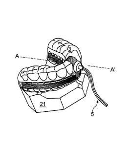
Referring now to Figure 3E, in another embodiment, two arches (one for
mandible and one for maxilla) can be formed together into an ultrasonic dental
attachment 3 that can treat both dental arches. As illustrated in figure 3E,
ultrasonic dental attachment 3 can contain four flexible arrays of ultrasound
transducers 23, 24: one array for maxillary buccal side, one for the maxillary
lingual side, one for the mandible buccal side, and one for mandible lingual
side. The ultrasonic dental attachment 3 can have orifices in the occlusion
(bite section) of the dental attachment 3 to allow patient breathing.
Dental cast 21 is used for illustrating how the ultrasonic dental attachment 3
can fit on patient teeth 22. Ultrasonic dental attachment 3 can be similar to
a
mouthguard that can be heated in hot water and when bitten by a user can be
fixed into a shape which follows the shape of the user's teeth. If a new shape
of the ultrasonic dental attachment 3 is required (for instance in the case of
orthodontic treatment where the teeth can change their position), then the
patient can reheat the ultrasonic dental attachment 3 in hot water and bite it
again to imprint the new shape or positions of the teeth 22. Professional
12
CA 02795679 2012-10-05
WO 2011/134071
PCT/CA2011/000498
alignment or adjustment of the position of the device is not necessarily
required. The patient can bite down on ultrasonic dental attachment 3 in
order to keep it positioned well on the teeth 22 during treatment and ensure
the placement is consistent with each use.
Connection 5 is shown as a cable which can connect ultrasonic dental
attachment 3 to external electronics controller 2. In some embodiments,
connection 5 can include wires and embedded connector 5a water-sealed
inside the ultrasonic dental attachment (Figure 3A, B, E), or external
connector 5b (Figures 3C and 3D) as an extension of the ultrasonic dental
attachment 3. Connectors 5a or 5b can connect transducers 23, 24 from
ultrasonic dental attachment 3 to the external electronics controller 2 as
desired through connection cable 5.
In addition, the connectors 5a and 5b can be permanently attached or can be
disconnected when cleaning, replacing or servicing of intra-oral attachment 3
is required.
Referring now to Figures 5A and 5B, tooth 22 can include crown 26 and root
27.
Tooth 22 can be connected through periodontal ligaments 28 to alveolar bone
29. Gums, or gingiva 30,31, can envelope alveolar bone 29 on the buccal
side 30 and on the lingual side 31 of tooth 22.
In one embodiment, ultrasound waves 32 can be propagated from the buccal
side transducer 24 through flexible enclosure 25, buccal side gums 30,
alveolar bone 29, periodontal ligaments 28, tooth root 27, and can continue
propagation through periodontal ligaments 28, alveolar bone 29, lingual side
gums 31, flexible enclosure 25 on the lingual side of tooth 22 and finally can
enter the lingual transducer 23 where ultrasound wave 32 can be converted
into an electric signal.
13
CA 02795679 2012-10-05
WO 2011/134071
PCT/CA2011/000498
Ultrasonic dental attachment 3 can use coatings or layers between gums 30,
31 and transducers 23, 24 that can behave as antireflection layers for the
ultrasound waves 32 at an operating frequency. The thickness of the coatings
can be an odd multiple of quarter wavelengths of an ultrasound wave 32 in
that material. This thickness can allow improved coupling of ultrasound
waves 32 from the emitter to the tissues and from the tissues to the sensor
and also can reduce the reflections back to the emitter or sensor which can
cause noise in ultrasonic dental system 1 and wave interference that can
affect treatment outcomes.
Flexible enclosure 25 can be made of flexible materials such as
polypropylene, copolyester or ethyl vinyl acetate (EVA) which can be
thermally formed, injection molded, deposited, or applied over and around
transducers 23, 24 in order to seal them from the external factors such as the
saliva from the patient or humidity from the environment. Such layers of
flexible materials can have thickness of less than 1mm while maintaining good
strength and sealant properties.
In this example, buccal side transducer 24 can emit ultrasonic waves, while
the lingual side transducer 23 can receive and sense ultrasonic waves 32,
although it would be appreciated that the opposite could also occur. In this
scenario, transducer 24 works as an emitter and transducer 23 as a
transmission sensor. In order to expose the tooth root 27 or crown 26 to
uniform ultrasonic treatment (uniform ultrasonic intensity), the transducers
23,
24 from the buccal and lingual side can interchange their dual function of
emitting and sensing. For instance, during a further step in treatment,
transducer 23 can emit ultrasound waves 32 and transducer 24 can sense the
transmitted ultrasound waves 32. In this way ultrasonic waves can equally
expose tooth 22 from both sides.
When multiple ultrasound emitters are used at the same time in proximity to
each other, wave interference can occur which can reduce the dental
14
CA 02795679 2012-10-05
WO 2011/134071
PCT/CA2011/000498
treatment outcome or can also cause tissue damage. The amplitude and
location of wave interference patterns can be difficult to predict and control
as
each patient has a unique dental structure. Ultrasonic dental system 1 can be
configured so that transducers 23, 24 will not emit ultrasound waves 32 at the
same time. As such, ultrasonic dental system 1 can avoid the interference of
the ultrasonic waves 32 inside tissues 27, 28, 29, 30, 31.
In one embodiment, transducer 23, 24 can cover the entire length (or a large
portion) of root 28, from the gum-crown interface to the tip of the root. By
using a transducer that covers the root 28, it can be possible to treat dental
problems located at any point of root 28 including its tip, or treat the
alveolar
bone 29 all around the root and its tip. Applications can include healing
dental
implants, root resorption, periodontitis, and accelerating alveolar bone
remodeling.
The area and shape of transducers 23, 24 can vary from tooth to tooth and
from buccal side to the lingual side of a tooth 22. Transducers 23, 24 can
have different shapes (rectangular, trapezoids, ovals, circular, etc), with
different widths, heights, or radii. In some embodiments, the width of
transducers 23, 24 can be similar with the width of a tooth crown 26, while
the
height can be similar with the length of the root 27. As the width of tooth 22
and the length of root 27 varies from tooth to tooth (for example incisors
have
a smaller crown 26 width but a longer root 27 than a molar), transducers 23,
24 can have different widths and heights.
Referring now to Figures 5C and 5D, a further embodiment of ultrasonic
dental attachment 3 can also be configured to accommodate orthodontic
braces 56 that are on a patient's tooth 22. Ultrasonic dental attachment 3 can
have a cavity 57 in flexible enclosure 25 to allow the brackets and wires from
the orthodontic braces to fit inside. Alternatively, cavity 57 can be an
orifice
through the flexible enclosure 25. A patient can wear both braces and
ultrasonic dental attachment 3 at the same time. In one embodiment,
CA 02795679 2012-10-05
WO 2011/134071
PCT/CA2011/000498
ultrasonic dental attachment 3 does not contact the brace brackets or the
brace wires as to not affect the way the orthodontic forces are applied by the
orthodontic braces to the tooth. Where ultrasonic dental attachment 3 can be
made of a soft material and can contact crown 26 such that uniform force can
.. be applied in all directions as to not influence the orthodontic brace
forces.
In figures 5C and 5D the orthodontic braces are illustratively located on the
buccal side of the teeth however, the braces can also be located on the
lingual side of the teeth, as required on the particular patient treatment and
.. type of braces chosen. As a result, cavity 57 can also be located on the
lingual side or on both sides of flexible enclosure 25, as required to
accommodate the location of the braces.
Referring now to Figures 5E and 5F, a further embodiment of ultrasonic dental
.. attachment 3 can be designed to be used with clear aligners or retainers
58. A
larger gap between flexible enclosure 25 and tooth crown 26 can allow
aligners or retainers 58 to fit inside the ultrasound dental attachment 3.
Referring now to Figures 5G and 5H, a further embodiment of ultrasonic
.. dental attachment 3 can be designed to treat both tooth root 27 and crown
26.
A second array of emitters and a second array of sensors can be in parallel
above the emitters and sensors which cover the length of root 27 and which
will cover the crown 26 partially or totally. Transducers 24 and 59 can be
placed on the buccal side and transducers 23 and 60 can be placed on the
.. lingual side. Ultrasound transducers 59 and 60 can be used for the
treatment
of tooth crown afflictions such as stimulating new dentine formation to help
repair deep cavities or to treat tooth sensitivity to cold, hot, or sweet.
In alternative embodiments, transducers 23, 24 can also cover the length of
.. the crown 26 of tooth 22 (transducers 23, 24 could cover both the root and
the
crown at the same time, in their entirety or only portions thereof). By using
an
ultrasound transducer that covers also the crown 26, ultrasonic dental system
16
CA 02795679 2012-10-05
WO 2011/134071
PCT/CA2011/000498
1 can treat crown problems such as deep cavities that can require ultrasound
stimulation to enhance the pre-dentine formation from inside the tooth to
potentially avoid a root canal. By exposing the entire tooth 22 (root 27 and
crown 26) to ultrasonic waves ultrasonic dental system 1 can treat the entire
tooth surface for sensitivity to cold, hot, or sweet, by stimulating the
entire
tooth interior which can lead to depositing of additional dentin in the areas
that
cause the sensitivity.
Ultrasonic Dental System can also help treat dental afflictions located at the
gum line, such as reducing gingiva infections, inflammation or pain, or
helping
accelerate healing after gingival surgical interventions (such as gingival
flap
surgery, dental implant or surgical tooth extractions)
Referring now to Figures 51 and 5J, a further embodiment of ultrasonic dental
attachment 3 can be designed to have a general form of a dental tray. In
some embodiments, the interior of the tray (facing tooth crown 26) can be
filled with soft bite pad 25a which can be made of a malleable material. As an
example, soft bite pad 25a can be made of silicone. Therefore when the
patient bites attachment 3, soft bite pad 25a can reshape and accommodate
tooth crowns 26. If the position of the teeth change over time (such as during
orthodontic treatment), soft bite pad 25a can allow continuous fit over tooth
crowns 26. An embodiment of the ultrasonic dental attachment 3 can
accommodate any type of orthodontic appliance (for example, wire braces
and clear orthodontic aligners). As illustrated in figure 5J, soft bite pad
25a
can recover its original shape when not bitten.
Referring now to Figures 5K and 5L, in some further embodiments, ultrasonic
dental attachment 3 can be designed to fit both dental arches (maxilla and
mandible). Some embodiments can deliver ultrasonic treatment selectively to
tooth roots 27 from both dental arches (maxilla and mandible) and from both
lingual and buccal directions as desired, while using a single external
electronics controller 2.
17
CA 02795679 2012-10-05
WO 2011/134071
PCT/CA2011/000498
Referring now to Figures 5M and 5N, some embodiments of ultrasonic dental
attachment 3 can be designed to fit both dental arches (maxilla and
mandible), and deliver the ultrasonic treatment selectively to tooth roots 27
from both dental arches (maxilla and mandible) from one direction only
(lingual or buccal) using a single external electronics controller 2.
Referring now to Figures 50 and 5P, some further embodiments of ultrasonic
dental attachment 3 can be designed to fit both dental arches (maxilla and
mandible), deliver the ultrasonic treatment selectively to tooth roots 27 and
tooth crowns 26 from both dental arches (maxilla and mandible) from one
direction only (lingual or buccal), using a single external electronics
controller
2.
Figures 5K, 5L, 5M, 5N, 50 and 5P illustrate examples of ultrasonic dental
attachments that can treat both dental arches (maxilla and mandible): from
both lingual and buccal directions (Figure 5K and 5L), from one direction only
(Figure 5M and 5N), and can also treat tooth crowns (Figure 50 and 5P). The
embodiments in Figures 5M, 5N, 50 and 5P can be made to attach and emit
ultrasound to the lingual side of the teeth or to the buccal side of the
teeth, as
required for treatment. For instance, people wearing customized orthodontic
appliances such as space closing springs or temporary anchorage screws,
some embodiments of the ultrasonic dental attachment may physically
interfere with the springs or anchorage screws and it is desired to use an
ultrasonic dental attachment that has the ultrasonic transducers on the side
opposite of the springs or screws. In addition, Figures 5K, 5L, 5M, 5N, 50 and
5P illustrate examples where the soft bite pad 25a can be used, but orifices
57 (as shown in figure 5D) can also be used, or extra space for clear aligners
58 (as shown in figure 5E) could also be used, or a tighter fit as illustrated
in
figure 5A could also be used, or any combination of the above.
18
CA 02795679 2012-10-05
WO 2011/134071
PCT/CA2011/000498
To allow for good coupling of the ultrasonic waves to teeth (crowns and
gums), in some embodiments, a coupling agent can be applied to the
tooth/gum contacting surface of ultrasonic dental attachment 3 when
treatment is to be applied. In some embodiments, the coupling agent can be
ultrasonic gel. In some embodiments, the coupling agent can be water or a
water-soaked substrate. It would be understood by a person skilled in the art
that any material which functions as a suitable coupling agent can be used.
In a further embodiment of ultrasonic dental attachment 3, soft membrane (not
shown) can delimit transducers 23, 24. A membrane can also separate the
area below gum and above gum, so ultrasonic dental attachment 3 can be
used for whitening purpose where a whitening gel will be applied or injected
separately only to cover the crown of the teeth. Further, in some
embodiments, a membrane can be used to indicate the area for ultrasonic gel
to be applied.
Referring now to Figure 6A, a cross section of an embodiment of ultrasound
transducer 24 which can emit ultrasound waves 32 is shown. Transducer 24
can include a piezoelectric material plate 33. Piezoelectric plate 33 can be
made of piezoelectric materials such as PZT (Lead-Zirconate-Titanate),
BaTiO3 (Barium Titanate) or PbNb206 (Lead Mataniobate). When
piezoelectric materials containing potentially hazardous materials (such as
lead) are utilized, piezoelectric plate 33 can be coated with a
humidity/moisture indicator substance. In the event there is a saliva leaking
into ultrasonic dental attachment 3, the indicator substance can change color
thereby alerting a patient to stop using the device. The thickness of
piezoelectric plate 33 can be constant and according to the acoustic velocity
of the piezoelectric material from which transducers 23, 24 are made, and by
the frequency at which transducers 23, 24 are operated at resonance. For
example, to drive transducers 23, 24 at resonance, the thickness of
piezoelectric plate 33 can be half a wavelength of the frequency of operation.
For example, piezoelectric plate 33 made of PZT and resonant at 1.5MHz can
19
CA 02795679 2012-10-05
WO 2011/134071
PCT/CA2011/000498
have a thickness in the order of 1.4mm. Driving transducers 23, 24 at
resonance can allow maximum power conversion efficiency from electrical
power to ultrasonic power.
Ultrasonic dental attachment 3 can further include acoustic impedance
matching layer 34 (which can be part of the overall device flexible enclosure
25) and air back layer 36. Transducer 24 can be glued or otherwise attached
to acoustic impedance matching layer 34 in order to provide that there is no
air gap in between. Air back layer 36 can be a foam layer (such foam tape, or
sputtered or deposited foam or an air gap). The foam can be made of flexible
material such as urethane and can have high air or neutral gas content.
Foam tapes with thickness on the order of 1mm or even less can be used in
order to obtain a compact (thin) transducer structure. Air back layer 36 can
also be created by applying a substance that can prevent flexible enclosure
25 material to stick to the back of the transducer 24. Due to the elastic
force
of flexible enclosure 25 material, when no glue is applied in between on
transducer 24, a very thin gap can form. This gap can act as an air back
reflector for the transducer 24.
Ultrasonic gel 35 can be used in-between acoustic impedance matching layer
34 and gum 30 which can allow a good coupling of ultrasonic waves 32 to the
gums 30 and alveolar bone 29. Acoustic impedance matching layer 34 can
have a thickness of odd multiples of quarter wavelengths of the ultrasound
waves at the operation frequency in the material from which the impedance
matching layer is made of. For example, the thickness can be a single
quarter wavelength of ultrasonic waves 32 which can allow minimal
absorption of the ultrasonic waves 32 when propagating through layer 34. For
example, if acoustic impedance matching layer 34 is made of materials such
as polypropylene, copolyester or ethyl vinyl acetate (EVA), then the thickness
of layer 34 can be on the order of 0.3-0.5mm. This thickness can vary with the
material used and its parameters.
CA 02795679 2012-10-05
WO 2011/134071
PCT/CA2011/000498
Matching layer 34 can be made of one material layer, or a combination of two
or more layers of different materials. For instance, a first layer of a harder
material like polypropylene or copolyester can be attached first to electrode
40
of transducer 33 in order to obtain a solid mechanical sealing, and a second
layer of a softer material such as EVA or silicon can be attached to provide
comfortable contact to gums 30 of patients. In this example, the thickness of
both layers can be chosen to allow maximum transmission and minimum
absorption. It would be understood by one skilled in the art that other
materials or combinations of materials than the one mentioned above could
also be used for the matching layer 34.
In some embodiments, a thin layer of water absorbent material such as foam
of fabric could be attached permanently or temporarily to matching layer 34 in
order to form a layer of water in-between matching layer 34 and gum 30. Such
embodiments could operate without ultrasonic gel but would require a patient
to imbibe the foam or fabric layer in water before use. The water absorbent
layer can come pre-imbibed in an aqueous solution in some embodiments.
The water absorbent layer can also have antibacterial properties or different
flavors. Moreover, the water absorbent layer could be a disposable
component that the patient can attach to the ultrasonic dental attachment 3
prior to use, and can discard it after application.
Ultrasonic dental attachment 3 can include a flexible cable 37 with parallel
wire traces. Flexible cable 37 can be a flat flexible cable (FFC) or flexible
printed circuit (FPC). Flexible cable 37 can contain individual wires 38
laminated between two dielectric films, where individual wires 38 can be flat
metal conductors. The thickness of a FFC can be in the order of 0.5mm while
the thickness of a FPC can be in the order of 0.1mm.
Ultrasonic dental attachment 3 can include a back electrode 39 and a front
electrode 40 on the piezoelectric plate 33. Back electrode 39 can cover the
majority of the back surface of the piezoelectric plate 33 with the exception
of
21
CA 02795679 2012-10-05
WO 2011/134071
PCT/CA2011/000498
a gap 45 and a small area electrode 40b. Gap 45 between electrodes 39, 40b
can allow electrical insulation between the two electrodes 39, 40b. Front
electrode 40 can cover the entire front surface of piezoelectric plate 33, and
can continue on the side (for example, on the bottom side) of plate 33 as
electrode 40a and can cover a small area (for example, a corner) of the back
surface of plate 33 as electrode 40b. Electrodes 40, 40a and 40b can be
electrically connected. In this way both electrodes 39, 40 can be accessible
on the same side (for example on the back side) of plate 33 to facilitate
connection to flexible cable 37. One wire 38 of flexible cable 37 can connect
to front electrode 40 by connecting to electrode 40b at electrical connection
41 . A second wire 38 of flexible cable 37 can connect to back electrode 39 at
electrical connection 42. Electrical connections 41 and 42 can be made by
soldering or by conductive glue. A window in the insulation layer of the
flexible
cable 37 can be opened in order to allow the connection to be made between
an individual conductor and the transducer electrode.
The total thickness of transducer 24 structure from gums 30 to the back of the
flexible enclosure 25 can be in the order of 3-5mm, depending of the materials
used for manufacturing. This thickness can allow patient comfort, while still
providing efficient transducer operation (for example, by having air back
reflector 36 and acoustic impedance matching layer 34 on the front) and the
treatment flexibility of emitting ultrasound waves 32 towards any or all teeth
in
a patient's mouth.
Referring now to Figure 6B, a cross section of a further embodiment of
ultrasound transducer 24 which can emit ultrasound waves 32 is shown. In
this example, air back layer 36 can be attached directly to back electrode 39
from back side of piezoelectric plate 33. Flexible cable 37 can be attached on
top of air back layer 36. Lateral connectors 44 and 43 can run from flexible
cable 37 in order to contact the flexible cable 37 to back electrode 39 and
electrode 40b. Electrode 40b can be connected through electrode 40a to front
electrode 40.
22
CA 02795679 2012-10-05
WO 2011/134071
PCT/CA2011/000498
Referring now to Figure 6C, a cross section of a further embodiment of
ultrasound transducer 24 which can emit ultrasound waves 32 is shown. In
this example front electrode 40 can be electrically connected to the back side
of piezoelectric plate 33 and electrode 40b by means of a through-hole via
40c. Through-hole 40c can be pre-made (pre-shaped) during transducer
manufacturing, or drilled using laser machining. Through-hole 40c can be
filled or plated with conductive material (for example by electroplating,
soldering, riveting, or application of conductive paint or epoxies).
The cross section of transducers from buccal side 24 and lingual side 23 can
be the same. Embodiments shown in Figures 6A, 66, and 6C can be used for
buccal transducers 24 as well as for lingual transducers 23, or a combination
of the three embodiments can be used within ultrasonic dental attachment 3.
In the embodiments shown figures 6A, 6B and 6C, piezoelectric material 33
can be replaced with a capacitive micro-machined ultrasonic transducer
(CMUT) array. In this case, air back layer 36 would not be required, as CMUT
emits unidirectional.
Figure 7A illustrates an embodiment of an array of ultrasound transducers 24
viewed from the back of transducers 24 facing away from the gums 30. This
embodiment illustrates the transducer structure of Figure 6A where the
flexible cable 37 can be located between the air back layer 36 and back
electrode 39. Figure 7A shows an array of transducers 24 which can be
interconnected using flexible cable 37 with individual wires 38. Only three
transducers 24 are illustrated for exemplification but it is understood that
there
can be several, for example sixteen, buccal transducers 24 which can be
connected in this manner. This transducer array configuration can also apply
to lingual transducers 23.
23
CA 02795679 2012-10-05
WO 2011/134071
PCT/CA2011/000498
It would be understood that while air back layer 36 is not illustrated in
figure
7A, this layer can be attached over flexible cable 37. Air back layer 26 can
be
cut in individual pieces to overlap the surface of each transducer 23 or 24,
or
be a long band that covers groups of transducers 23 or 24 or all
transducers23 or 24 of a flexible transducer array.
Figure 7A shows the pattern of electrodes 39, 40b as seen on the back of
piezoelectric plates 33. This electrode and connectivity configuration can
allow easy connectivity while using a single electrode pattern for all
transducers. Different electrode patterns for different transducers can be
utilized for cases were custom designed devices need to be manufactured.
Transducers 24 can use a common wire to connect to electrode 40b which
can be connected to front electrode 40 (as the ground electrode) at
connection point 41 of each transducer. Possible placement of flexible cable
37 and individual wires 38 and connections to the electrodes 39 and 40b are
shown. Connections 41, 42 can be made by soldering, or conductive glue or
epoxy. This transducer configuration can also apply to lingual transducers 23.
Referring now to Figure 7B, a further embodiment of an array of ultrasound
transducers 23 or 24 is shown. Figure 7B illustrates the transducer structure
from Figure 6B where air back layer 36 can be located between flexible cable
37 and back electrode 39. Air back layer 36 can be cut in individual pieces
for
each transducer, and can cover most of the piezoelectric plate 33 back
surface with the exception of an opening to allow connection 42 of individual
wire 38 to electrode 39, and connection 41 to another individual wire 38 to
electrode 40b. In other embodiments, air back layer 36 can be a long band
that can cover groups of transducers 23 or 24 or all transducers 23 or 24 of a
flexible transducer array.
24
CA 02795679 2012-10-05
WO 2011/134071
PCT/CA2011/000498
For clarity, for both figures 7A and 7B, air back layer 36 can be cut in
individual pieces and placed on the back of each transducer, or can be a
single piece that covers all transducers or groups of transducers of an array.
Figure 7B shows a further embodiment of how the array of transducers 24 can
be interconnected using flexible cable 37 with individual wires 38. Only three
transducers 24 are illustrated for exemplification but it is understood that
there
can be several, for example sixteen buccal transducers 24 which can be
connected in this manner. This transducer array configuration can also apply
to lingual transducers 23. A combination of the embodiments illustrated in
Figures 7A and 7B can be used together in ultrasonic dental attachment 3.
Figure 7B shows a pattern of electrodes 39 and 40b seen on the back of
piezoelectric plates 33. The illustrated embodiment can use a single common
wire connected to electrode 40b which can be connected to the front
electrode 40 (as the ground electrode) at connection point 41 of multiple
transducers. Possible placement of flexible cable 37 and individual wires 38
and connections to electrodes 39 and 40b are shown. Connections 41, 42
can be made by soldering, or conductive glue or epoxy.
In Figures 7A and 7B, back electrodes 39 can be connected to individual
wires 38 of flexible cable 37 while electrodes 40b (which can be connected to
front electrode 40) can be connected to a common wire ground. This common
rail wire can be connected to the ground of the external electronics
controller
2 through connection cable 5. In further embodiments, individual wires 38 can
be used for both electrodes (from the front and back of transducers 23, 24).
For clarity, individual ground wires can also be used for each transducer in
some embodiments.
In some embodiments, all buccal transducers 24 can be connected to a
common ground wire where all electrodes 40b (which can be connected to
front electrode 40) can be connected to a single common wire and all back
CA 02795679 2012-10-05
WO 2011/134071
PCT/CA2011/000498
electrodes 39 can be connected to a second common wire. This can allow
driving of all buccal transducers 24 at the same time by using only two wires.
Similar implementation can also be used for the lingual transducers 23.
Flexible cable 37 of the buccal transducers 24 and flexible cable 37 of the
lingual transducers 23 can be both connected through connector 5a or 5b to
cable 5 which can connect to external electronic controller 2. This connection
can be located in the front buccal side of the incisors as shown in Fig 3A,
3B,
3C, 3D, and 3E.
Referring now to Figure 8, circuitry interface 8 from Figure 2 is shown with
ultrasonic dental attachment 3 circuitry. Driver 13 can include at least one
radio frequency (RF) power amplifier 46 and at least one digitally controlled
voltage regulator 47. RF power amplifier 46 can be a Class E or Class F
switching amplifier. Voltage regulator 47 can be a variable voltage regulator
controlled by a digital potentiometer, where the digital potentiometer can be
controlled by the processing unit 7. Coupling sensing circuitry 14 can be
made of a current sense circuitry that can monitor the DC current supplied by
digitally controlled voltage regulator 47 to RF Power amplifier 46. The output
of coupling sensing circuitry 14 can be read by an Analogue to Digital
Converter (ADC) port of the processing unit 7 which can be a microcontroller.
Transmission sensing circuitry 15 can be a full-wave (or half-wave) rectifier
circuitry such as bridge rectifier or diode less rectifiers, followed by an
envelope detector. The output of transmission sensing circuitry 15 can be
read by an Analogue to Digital Converter (ADC) port of the processing unit 7
which can be a microcontroller. Switching circuitry 16 can be located in
external electronic controller 2 or in ultrasonic dental attachment 3, or a
portion in controller 2 and another portion in dental attachment 3. In some
embodiments, a portion of switching circuitry 16 could also be located on the
cable 5 or connector 5a or 5b.
26
CA 02795679 2012-10-05
WO 2011/134071
PCT/CA2011/000498
Figure 9 is an electrical schematic diagram which illustrates an embodiment of
the circuitry that can control transducer emitter 24 to generate ultrasonic
waves 32 and can also sense the degree which transducer emitter 24 is
coupled to tissue structure 48. The circuitry can also control transducer
sensor 23 which can sense the transmitted waves exiting dental tissue
structure 48. In order to drive multiple emitters and sensors, switching
circuitry 16 can be added.
In one embodiment, Vin1 can be the power supply coming from a power
source (for example, a rechargeable or non-rechargeable battery, a wall plug-
in adapter) Digitally controlled voltage regulator 47 can be made of a voltage
regulator with an adjustable voltage output Vout1. The voltage output Vout1
can be adjusted by a digital potentiometer R1 which can be connected to
processing unit 7 such as a microcontroller digital output. Processing unit 7
can supply input signal Vin3 to digital potentiometer R1 in order to adjust
the
resistor value which in turn, can adjust output voltage value Vout1. In some
embodiments, voltage regulator 47 can be a low-drop linear regulator LT3021
from Linear Technology.
Voltage regulator 47 can supply electrical power to an RF power amplifier 46.
RF power amplifier 46 can amplify a square wave digital signal Vin2
generated by the processing unit 7. Vin2 can be a megahertz frequency
signal which can be continuous or pulsed (with adjustable duty cycle),
depending on the treatment settings. RF power amplifier 46 can be a Class-E
power amplifier or a Class-F power amplifier. Either amplifier class can
amplify the input signal Vin2 to an AC signal with the peak-to-peak voltage
Vout 3 several times higher than the voltage rail Vout2. Voltage Vout2 =
Vout1 + Voltage drop over Rsense. Rsense can have a very small value (for
example, in the order of 0.0010hms) and therefore voltages Vout1 and Vout2
can be approximately equal.
27
CA 02795679 2012-10-05
WO 2011/134071
PCT/CA2011/000498
RF power amplifier 46 can drive piezoelectric transducer 24, which emits
ultrasonic waves 32 towards the dental tissue structure under ultrasonic
treatment 48. The dental structure 48 can be made of layers 30, 29, 28 and
31 as illustrated in Fig 5A.
When processing unit 7 adjusts the resistance value of the digital
potentiometer R1, Vout1 and Vout2 can change. This change can modify the
AC voltage (peak-to-peak voltage) Vout3 that drives the piezoelectric
transducer 24 and therefore can change the ultrasonic power level of the
ultrasonic waves 32 that are delivered to the tissue structure 48.
Piezoelectric transducer 23 can be placed on the opposite side of dental
structure 48 and can sense the amount of ultrasonic power that exits through
dental structure 48. The amount of ultrasonic power that is exiting dental
structure 48 can be an indicator of the amount of ultrasonic waves that were
absorbed by dental structure 48. The absorbed ultrasonic power can
stimulate the repair of dental tissue. Voltage Vout4 can be related to the
ultrasound power that passes from the emitter (in this example transducer 24)
to the sensor (in this example transducer 23).
The electrical signal generated by piezoelectric transducer 23 can be
connected to transmission sensing circuitry 15 which can condition the
electrical signal received and can output voltage Vout4 that can be read by
processing unit 7 (for instance an ADC port of the microcontroller).
Transmission sensing circuitry 15 can be made of a rectifier such as Schottky
diode bridge rectifier or non-diode rectifiers based on operational amplifiers
that can also have digitally adjustable gain. For example, a suitable Schottky
diode bridge rectifier can be the component MB12S from Micro Commercial
Components.
The DC current supplied to RF amplifier 46 can depend on the value of the
mechanical load of piezoelectric transducer 24. The electrical resistance
28
CA 02795679 2012-10-05
WO 2011/134071
PCT/CA2011/000498
value of the electrical impedance of piezoelectric transducer 24 can be low
when it is well attached/coupled to tissue , or higher when it is not coupled
to
tissue. This variation of the electrical resistance value of the transducer
can
modify the DC current that is supplied to RF amplifier 46 and which can flow
through the sensing resistor Rsense. The function of the coupling sensing
circuitry 14 can be to sense the variations in the DC current supplied to RF
amplifier 46. In this manner the amount or quality of the coupling of the
transducer 24 to the tissue under treatment 48 can be measured by the
controlling unit 7 and can alert the user. Poor coupling can indicate that
ultrasonic dental attachment 3 is not placed in the mouth, is placed
incorrectly
in the mouth, or that ultrasonic gel needs to be added. The coupling sensing
circuitry can alert the patient about the poor coupling so that corrective
measures are taken which lead to improved treatment outcomes.
Coupling circuitry 14 can be a current sensing circuit. Coupling circuitry 14
can sense the DC current that flows to RF amplifier 46 by the use of a sensing
resistor Rsense. The value of Rsense can be chosen to be very small (for
example, on the order of milliohms), which can lead to a negligible voltage
drop across the sensing resistor. Therefore, the sensing of the current can
have a negligible effect on the voltage Vout2 delivered to the RF amplifier 46
and on the AC voltage Vout3 that can drive the piezoelectric transducer 24.
The design of the current sensing circuit can be chosen to minimize the loss
over the sensing resistor. An amplified output voltage Vout5 (which is
dependent on the current through Rsense) can be provided to the ADC input
of processing unit 7.
The amplification gain of the coupling sensing circuitry 14 can be
configurable
with two resistors Rin and Rout. Commercial current sensing circuits which
include both the OP AMP and the bipolar transistor Q1 can be used, for
example, the Linear Technology low-cost current sense chip LT 6106.
29
CA 02795679 2012-10-05
WO 2011/134071
PCT/CA2011/000498
Processing unit 7 can receive output signal Vout4 coming from transmission
sensing circuitry 15 and voltage Vout5 coming from coupling sensing circuitry
14. The ADC ports of processing unit 7 can convert these voltages into digital
values. The firmware of processing unit 7 can then follow an operation
algorithm and can adjust digital signal Vin3 that controls the digital
potentiometer R1. Adjusting R1 can adjust in real-time the amplitude of the
voltage Vout3 which can modify the amplitude of ultrasonic waves 32 and
therefore modify the ultrasonic power emitted by a transducer 23 or 24. The
system can adjust the amplitude of Vout3 in order to compensate for losses
and absorption in the dental tissue structure 48.
Figure 10 shows a block diagram of one embodiment of a circuit and an
algorithm which can be used to drive multiple transducers sequentially in
ultrasonic dental system 1. In one embodiment, electrical signals 49 can drive
transducers 24 when working in ultrasound emitting mode and electrical
signals 50 can be sensed by transducers 23 when working in ultrasound
sensing mode. While Figure 10 demonstrates that five pairs of
sensor/emitters are being driven, this example can also extend to the
remaining pairs of sensor/emitters.
In the illustrated embodiment, the electrical signal from driver 13 can be a
continuous 1.5MHz signal which is switched to five channels 49 by switching
circuitry 16. Each individual signal 49 can be a 200microseconds burst of an
oscillating 1.5MHz signal, followed by 800microseconds when the 1.5MHz
signal is OFF. The cycle can repeat every 1000microseconds with
200microsenconds of the 1.5MHz signal ON and 800microseconds OFF. The
period when the 1.5MHz signal is ON does not overlap between the five
signals. Therefore, in a period of 1000 microseconds (or 1 millisecond) only
one transducer will emit at a time. This type of staggering can be used to
avoid interference between the five transducers that emit, although it would
be
understood that other values could be used to accomplish the same goal.
CA 02795679 2012-10-05
WO 2011/134071
PCT/CA2011/000498
Transducer 24 (in the emitting mode) can be driven by a signal of 1.5 MHz
that can be modulated at a 1KHz with 20% duty cycle. These parameters are
provided for examples only. Different MHz signals can be used other than
1.5MHz, different modulation signals other than 1KHz can be used, and duty
cycles different than 20% can also be used.
Each individual signal 50 coming from transducer 23 (in a sensing mode) can
be a delayed replica of emitted signal 49, but with the amplitude of the
oscillation decreased due to absorption of the ultrasound in the dental
tissue.
Only the amplitude will be reduced. The carrier frequency (for example here
1.5MHz), the modulation frequency (for example here 1KHz) and the duty
cycle (for example here 20%) can remain unchanged.
In this example five transducers 24 can emit ultrasound sequentially (one
after
another, not in the same time) and five transducers 23 can sense the
ultrasound. This can be done using a single driving circuitry 13, a single
sensing circuitry 14 and a single sensing circuitry 15. By the use of
switching
circuitry 16, transducers 24 can be switched to sensing mode and the
transducers 23 can be switched to emitting mode. In this way the ultrasonic
treatment can be delivered from either side of the tooth. Switching circuitry
16
can be made, for example, of one or more multiplexer/demultiplexer circuits
such as the analogue sixteen channel multiplexer/demultiplexer HCF4067
from STMicroelectronics.
In one example, a single transducer 24 can emit ultrasound and a single
transducer 23 (located on the other side of tooth 22) can sense ultrasound. In
addition, by using the switching circuitry 16, in some embodiments three
neighboring transducers 23 (centered on the other side of teeth to the emitter
transducer 24) can be connected together to sense at the same time. In this
manner three neighboring transducers 23 that are sensing can form a larger
sensing area and can receive more of the diverging and scattered ultrasound
waves coming from an emitting transducer 24. In this example, the 1.5 MHz
31
CA 02795679 2012-10-05
WO 2011/134071
PCT/CA2011/000498
signal from the processing unit 7 (microcontroller) can be switched so that
one
transducer 24 can emit from the buccal side, and three transducers 23 can
sense on the lingual side. This can increase the amplitude of the electrical
signal of the sensor transducers and improve the sensing capability of the
system. Alternatively, by using switching circuitry 16, one transducer 23
(from
the lingual side) can emit ultrasound and three transducers 24 (from the
buccal side) can sense ultrasound.
In a further example, teeth 22 with similar properties (thickness, length etc)
can be grouped together. For example, the four incisors= group one, the left
canine and the two left premolars = group two, the right canine and the two
right premolars = group three, the three left molars = group four, and the
three
right molars = group five. Each group can be driven at once, and the circuitry
shown in Figure 10 can therefore drive all the five groups sequentially.
For the cases where each individual tooth 22 has to be treated separately
from each other, then ultrasonic dental system 1 can be setup in the following
two ways: switching circuitry 16 can drive treatment of five teeth
sequentially
for the duration of the treatment (for example 20 minutes) and then can pass
to the next five teeth for another 20 minutes of treatment, or alternatively
interface circuitry 8 can have multiple blocks that each can drive treatment
of
five teeth sequentially.
Ultrasonic dental system 1 can have the ability to sense, in real-time, the
intensity of the ultrasound waves emitted to the tissue and can adjust this
intensity to the optimum desired range. This adjustment can be performed in
real-time, for each individual tooth or for all teeth at the same time, as
desired.
The effects of ultrasound on dental tissue and bone tissue can be dependent
on the intensity of the ultrasound used in treatment. Levels of intensity
lower
then an optimum level can result in poor tissue stimulation, while levels of
intensity higher then an optimum level can result in tissue damage. When an
32
CA 02795679 2012-10-05
WO 2011/134071
PCT/CA2011/000498
ultrasonic wave propagates through dental tissue it can be absorbed by the
tissue and the intended ultrasound intensity initially sent by an emitter can
be
reduced. As such, tissue further away from an emitter can be less stimulated.
Ultrasonic dental system 1 can provide uniform ultrasound treatment (uniform
ultrasonic intensity) to a treatment area by targeting ultrasound waves to a
treatment area from opposite sides of the treatment area at different times.
By emitting from the different directions at different times, wave
interference
can be reduced or avoided. For example, when ultrasound waves are emitted
from the buccal side of a treatment area for 200 microseconds followed by no
emission for 800 microseconds, and ultrasound is being emitted from the
lingual side of the treatment area for a different 200 microseconds and
followed by no emission for 800 microseconds, then within a total period of
1000 microseconds, a treatment area can receive 200 microseconds of
treatment from the lingual side and another 200 microseconds of treatment
from the buccal side without interference. In order to avoid wave
interference,
there can be no time of overlap between the two periods of ultrasound
emission. By treating both sides of a treatment area within a 1000
microsecond modulation period, a standard daily treatment time (for example,
twenty minutes) does not need to be increased.
Referring now to Figure 11A, one process for manufacturing ultrasonic dental
attachment 3 is shown, however other processes can be employed to
manufacture dental attachment 3. Combinations of the processes are also
contemplated.
A flat sheet 51 of flexible material is provided. Flat sheet 51 can be made of
polypropylene, copolyester or EVA. The thickness of flat sheet 51 can be a
quarter of an ultrasound wavelength. For example, for EVA material the
thickness of flat sheet 51 can be on the order of 0.3mm for ultrasonic waves
at the frequency of 1.5MHz. The exact value of the thickness of flat sheet 51
can depend on the specific properties of the material.
33
CA 02795679 2012-10-05
WO 2011/134071
PCT/CA2011/000498
An outline 52 of flat sheet 51 around the array of ultrasound transducers 23
and 24 can delimit the area of the acoustic impedance matching layer 34. The
end connecting pads 53 of flexible cable 37 for the buccal and lingual side
are
shown. In some embodiments, flexible cable 37 for both lingual and buccal
arrays can be separate pieces. In some embodiments, flexible cable 37 for
both of the lingual and buccal portions can be continuous (for example, one
piece).
Referring now to Figure 11B, a close up version of Figure 11A and
connections means are shown. Connecting pads 53 of flexible cable 37 of the
lingual and buccal transducers, can form an array of pads as part of flexible
cable 37. This array of pads 53 can connect to a second array of pads 53a.
The array of pads 53a can form the embedded connector 5a, which can
connect to cable 5 which can further connect the overall ultrasonic dental
attachment 3 to the external electronics controller 2. The array of pads 53
(of
flex cable 37) can be attached to array of pads 53a (of embedded connector
5a) in a temporary or permanent fashion, by use of conductive epoxy/glue or
soldering, or by any suitable mechanical attachment for example.
A first method of manufacturing can be outlined by the following steps:
Step 1: Attachment (by glue or by heat) of the array of buccal transducers 24
and lingual transducers 23 on flat sheet 51 on a flat surface in the pattern
shown in Figure 10. Front electrodes 40 of transducers 23, 24 can be facing
down to sheet 51.
Step 2: Interconnection of the transducers that form the buccal and lingual
array of transducers. If the transducer embodiment illustrated in Figures 6A
and 7A is used, connect flexible cable 37 to back electrodes 39 (connections
41 and 42 for each transducer 23 or 24) as illustrated in Figure 7A. Next, air
back layer 36 can be attached on top of flexible cable 37 as illustrated in
Figure 6A. Alternatively, the transducer embodiment illustrated in Figures 6B
and 7B can also be used. Steps 1 and 2 can be interchanged so that the
34
CA 02795679 2012-10-05
WO 2011/134071
PCT/CA2011/000498
array of transducers 23 and 24 can also be attached to flat sheet 51 after the
interconnections are realized.
Step 3: Cutting flat sheet 51 around the transducer arrays keeping a border of
few millimeters as illustrated by dashed line 52. The area encompassed by
dashed line 52 represents acoustic impedance matching layer 34 that can
come in contact with the gums.
Step 4: Positioning the two arrays of transducers (attached to the cut up of
sheet 51) on dental cast 21. Layer 34 of the transducer arrays can contact
and cover the tooth roots, as illustrated in Fig 3A.
Step 5: Forming a second flat sheet 51 over dental cast 21 and the
transducer arrays. The second flat sheet 51 can be applied by using vacuum
or pressure thermoforming, by coating with a liquid form of the flexible
material found in sheet 51 or by deposition (sputtering, spraying).
Step 6: Connection of the connecting pads 53 over the incisors to external
cable 5. The connection can be sealed with epoxy or another local
thermoforming or coating step.
Step 7: The edges of the second flat sheet 51 that was applied over the dental
cast and transducers can be trimmed around the bottom side of the
transducer array. A few millimeters of overlapping between the two flexible
materials can be kept in order to secure sealing of the internal components of
ultrasonic dental attachment 3.
This manufacturing method can ensure that layer 34 will have a well
controlled thickness of a quarter wavelengths, and this thickness will not be
altered during the manufacturing process.
A further method of manufacturing can be outlined by the following steps:
Step 1: Forming a layer of flat sheet 51 over dental cast 21 by using
thermoforming or by coating. The thickness of flat sheet 51 in the area next
to
the tooth roots can be a quarter of an ultrasound wavelength thick. If
thermoforming is used for this step, the thickness of layer 34 can be
controlled
by controlling the temperature of the flexible sheet during thermoforming.
CA 02795679 2012-10-05
WO 2011/134071
PCT/CA2011/000498
Step 2: Interconnecting the transducers that form the buccal and lingual array
of transducers. If the transducer embodiment illustrated in Figures 6A and 7A
is used, connect flexible cable 37 to back electrodes 39 (connections 41 and
42 for each transducer 23 or 24) as illustrated in Figure 7A. Next, air back
layer 36 can be attached on top of flexible cable 37 as illustrated in Figure
6A.
Alternatively, the transducer embodiment illustrated in Figures 6B and 7B can
be used.
Step 3: Attaching (using glue or thermal process) the two arrays of
transducers (buccal and lingual) on flat sheet 51 which can cover dental cast
21. Transducers can be positioned at the location of the tooth roots.
Step 4: A second flat sheet 51 can be placed over the transducer arrays
which are placed on the first flat sheet 51 that was placed over the dental
cast. The second flat sheet can be applied by using vacuum or pressure
thermoforming, by coating with a liquid form of the flexible material found in
sheet 51 or by deposition (sputtering, spraying).
Step 5: Connection of the connecting pads 53 over the incisors to external
cable 5. The connection can be sealed with epoxy or another local
thermoforming or coating step.
Step 6: The edges of flat sheets 51 can be trimmed around the bottom side of
the transducer array. A few millimeters of overlapping between the two
flexible
materials can be kept in order to secure sealing of the internal components of
ultrasonic dental attachment 3.
In some embodiments of the above manufacturing method, Step 1 can be
skipped, and the method commences at Step 2. Then the lingual and buccal
arrays of transducers can be placed on metal stands and thermoformed from
the side, so that one thermoforming process can completely seal around the
transducer array. The stands can be removed and the orifices sealed with
heat or glue. The thickness of the encapsulation layer can be controlled by
controlling the temperature during thermoforming. At Step 3 the lingual and
buccal arrays can be placed on a bare dental cast. Follow steps 4, 5 and 6 as
above.
36
CA 02795679 2012-10-05
WO 2011/134071
PCT/CA2011/000498
A further method of manufacturing can be outlined by the following steps:
Step 1: Interconnecting the transducers that form the buccal and lingual array
of transducers. If the transducer embodiment illustrated in Figures 6A and 7A
is used, flexible cable 37 can be connected to back electrodes 39
(connections 41 and 42 for each transducer 23 or 24) as illustrated in Figure
7A. Next, air back layer 36 can be attached on top of flexible cable 37 as
illustrated in Figure 6A. Alternatively, the transducer embodiment illustrated
in
Figures 6B and 7B can be used.
Step 2: Placing the two arrays of transducers in a negative injection mold
shell with the shape of dental cast 21. The position of the transducer arrays
inside the injection mold shell can be predetermined such as to form a layer
of
quarter of an ultrasound wavelength thickness between the transducer
surface and the gums 30,31.
Step 3: Connect connecting pads 53 to cable 5 at a location inside or outside
the shell.
Step 4: Fill (injection mold) the cast with melded or liquid form of the flat
sheet 51 material (for example, EVA, polypropylene, or copolyester).
Step 5: Cure and trim the interior part of the injection mold.
Dental cast 21 can be custom designed for each patient or can be a generic
shape. If a generic dental cast is used, then the patient can customize the
ultrasonic dental attachment 3 to their own teeth. The material of flat sheet
51
can be reshaped when heated (for instance in boiling water) and can be used
to perform this function by a patient at home. In some embodiments, a bite
pad 25a can be used to accommodate the shape of the user's teeth.
Referring now to Figures 12A and 12B, a front and rear view of an
embodiment of external electronics controller 2 are shown. The front panel of
external electronics controller 2 can have user interface elements such as
display 17 (LCD or touch screen) and push buttons 19 which can allow a user
37
CA 02795679 2012-10-05
WO 2011/134071
PCT/CA2011/000498
(patient or dentist) to operate and interact with ultrasonic dental system 1.
External electronics controller 2 may also include a speaker (not shown).
In some embodiments, a user can turn on/off the device using the button 19,
can receive information on the treatment status from display 17, and can be
alerted by display 17 and speaker if there is a malfunction or a low power
level. External electronics controller 2 and its interface can allow the
setting
of the ultrasonic dental system 1 prior to ultrasonic treatment. A user can
turn
on only the emitter-sensor pairs for the teeth that have to be treated and not
treat healthy teeth. In some embodiments external electronics controller 2 can
record treatment data which can be later verified by the user in order to
ensure treatment compliance and improve treatment outcomes.
External electronics controller 2 can be battery powered or powered from the
wall using a plug-in adapter. The rear panel of external electronics
controller
2 can provide access to battery 54. Battery compartment can be covered by
cover 55. The rear panel of external electronics controller 2 can also provide
access to a connection port such as USB (for connection to a computer) or
connector for power supply or battery charging.
In some embodiments of ultrasonic dental system 1, the electronics from the
external electronics controller 2 and battery can be fit inside the ultrasonic
dental attachment 3. In this embodiment, the user interface (display 17, push
buttons 19) can be kept outside the mouth and can communicate through a
cable or wirelessly to the intra-oral electronics. The intra-oral electronics
can
be a rigid or flexible printed circuit board (FPC) that can be placed on top
of
the teeth or on the buccal or lingual side of the teeth crowns and roots.
The thicknesses of the dental structures (gum, alveolar bone, periodontal
ligaments) can be at the order of the wavelength of the ultrasonic waves of
around 1mm in aqueous media. As such, the intensity of ultrasound waves
can be severely affected during propagation through these multilayer
38
CA 02795679 2012-10-05
WO 2011/134071
PCT/CA2011/000498
structures and can also be reflected when exiting into air or a sensor. The
use of anti-reflection type coatings on ultrasonic dental attachment 3 can
allow for desired coupling of the waves from emitter and sensor and
subsequently can allow for a more accurate power reading at the sensor. Use
of an anti-reflection coating can increase the energy that contacts and enters
the sensor and is absorbed and converted into electrical signal. Use of an
anti-reflection coating can also reduce or eliminate the reflection of waves
back onto the tooth. Our manufacturing methods solve this problem.
The surface of transducers 23, 24 and the interior of ultrasonic dental
attachment 3 can be coated with a compound that can change color when in
contact with water (for example, a humidity/moisture indicator material
similar
to the humidity indicator cards or stickers available on the market). For
example, if a crack forms in ultrasonic dental attachment 3 and water or
saliva
leak into it, then the user can observe a change in color and discontinue
using
ultrasonic dental attachment 3 .
In operation, ultrasonic dental system 1 can have ultrasonic dental attachment
3 connected to external electronic controller 2. External electronic
controller 2
can instruct a user on a treatment procedure and treatment status. Ultrasonic
dental attachment 3 can be placed inside the patient's mouth during an
ultrasonic treatment for a duration, for example of 20 minutes per day. In
some embodiments, external electronic controller 2 can generate, monitor and
adjust in real-time a dosage of ultrasound delivered to the desired area, can
display the treatment status and can record the treatment parameters. In
some embodiments, the transducers can be cooperative and can act in
cooperation when emitting and sensing ultrasound. When bitten, ultrasonic
dental attachment 3 can closely follow the shape of a dental arch allowing for
a patient to repeatedly place ultrasonic dental attachment 3 in the correct
position. This consistent positioning can ensure the appropriate delivery of
ultrasound to the desired area. A dental professional can diagnose a clinical
condition of a patient, and can indicate which teeth could benefit from
39
CA 02795679 2012-10-05
WO 2011/134071
PCT/CA2011/000498
ultrasound treatment. By accessing a menu section of the external electronic
controller 2, the user can set ON the emitter-sensor pairs for the teeth of
interest. The user can follow instructions displayed on external electronic
controller 2. Ultrasonic dental system 1 can treat different teeth or implants
requiring different dental treatments at the same time, on the same patient.
Interchangeable emitters and sensors can give an improved uniformity of
treatment and can improve patient outcome. Ultrasonic waves can be
alternatively emitted from either side of a tooth. In some embodiments,
ultrasonic waves are not emitted from both sides of a tooth at the same time
as this can lead to wave interference. For example, a first treatment can have
waves emitted from the lingual transducers and sensed by the buccal
transducers, while in a subsequent treatment the waves can be emitted by the
buccal transducers and sensed by the lingual transducers. By alternating the
side being treated, ultrasonic dental system 1 can ensure that the ultrasound
treatment is uniform on both sides which can lead to an improved patient
outcome.
In some embodiments, ultrasonic dental system 1 can provide a method to
accelerate the orthodontic tooth movement without applying any additional
force (cyclical and/or continuous) to a tooth crown. The application of
ultrasound dental treatment as described herein can result in accelerated
orthodontic tooth movement while not affecting the amount and direction of
the forces applied by the orthodontic appliance to the tooth crowns. The
application of ultrasound can affect the speed of tooth movement by
accelerating the processes involved in the alveolar bone remodeling around
the tooth roots. The use of ultrasonic dental system 1 can eliminate the need
for temporary anchorage devices for orthodontic tooth movement and space
closure as it selectively accelerate only the teeth of interest and not the
anchorage teeth. The use of ultrasonic dental system 1 can increase the
movement ratio between target tooth and anchorage tooth.
CA 02795679 2012-10-05
WO 2011/134071
PCT/CA2011/000498
In some embodiments, ultrasonic dental system 1 can include self-calibration
capabilities and self-adjusting coupling sensing. Ultrasonic dental system 1
can measure the electrical impedance of the ultrasonic dental device when in
the air (which can be the worst coupling scenario), and the electrical
impedance when in water (which can be the best coupling scenario). When
dental attachment 3 is placed in the mouth with ultrasonic gel, the electrical
impedance can be similar to the scenario in the water. Processing unit 7 can
then activate the ultrasonic treatment. If the dental attachment 3 is not
placed
in the mouth or gel was not used correctly, then the electrical impedance
reading can be different than the water reading, and closer to the in-air
reading. In some embodiments, processing unit 7 will not activate the
ultrasonic treatment and can alert the patient to use gel or place attachment
correctly in the mouth. As such, the deciding if dental attachment 3 is
correctly applied can be taken based on electrical impedance measurements
in the air and in the water. The two electrical impedance reference points can
vary with attachment 3 wearing or becoming physically damaged. To detect
these longer term changes, the microcontroller records and stores electrical
impedance measurements in the air and in the water periodically. If these
values vary from the previously stored values, it can indicate that the
ultrasonic attachment 3 properties have changed. If the difference between
values is not significant then processing unit 7 can adjust the driving
voltage
of the transducers based on an internal formula/model in order to compensate
and correct for the change in impendence and keep can the ultrasonic
intensity in the desired range, for example 30mW/cm2. In some embodiments,
if the difference is significantly greater than the pre-determined threshold,
processing unit 7 will not activate the ultrasonic treatment and can alert the
user to service the device.
Although a few embodiments have been shown and described, it will be
appreciated by those skilled in the art that various changes and modifications
might be made without departing from the spirit or scope of the invention.
Unless defined otherwise, all technical and scientific terms used herein have
41
CA 02795679 2012-10-05
WO 2011/134071
PCT/CA2011/000498
the same meaning as commonly understood to one of ordinary skill and the
art to which this invention belongs. In addition, the terms and expressions
used in the preceding specification have been used herein as terms of
description and not of limitation, and there is no intention in the use of
such
terms and expressions of excluding equivalents of the features shown and
described or portions thereof, it being recognized that the scope of the
invention is defined and limited only by the claims that follow.
42