Note: Descriptions are shown in the official language in which they were submitted.
ANTI-BACTERIAL APPLICATIONS OF POLY-IN-ACETYLGLUCOSAMINE
NANOFIBERS
1. FIELD
[0002] Described herein are compositions comprising shortened fibers of
poly-N-
acetylglucosamine and/or a derivative thereof ("sNAG nanofibers") and anti-
bacterial
applications of such compositions. The sNAG nanofibers may be formulated into
compositions
for the prevention and/or treatment of bacterial infections and diseases
associated with such
infections. Regimens employing such compositions are also described.
2. BACKGROUND
[0003] Currently, antibiotics are a standard therapy for bacterial
infections. However, some
individuals have an allergic reaction to certain antibiotics, others suffer
from side effects
associated with antibiotics, and the continued use of antibiotics often leads
to a reduction in their
efficacy. In addition, antibiotic therapy often leads to the emergence of
antibiotic-resistant
strains of bacteria. Accordingly, there is a continuing need for new anti-
bacterial agents that are
effective in fighting infection without generating resistance or reducing the
efficacy overtime.
There is a need for non-antibiotic anti-bacterial agents that can be used in
clinical settings, e.g.,
in the treatment of infectious diseases of the skin, digestive and respiratory
tract, and in wound
treatment.
[0004] Wound infection is one type of bacterial infection. Wound infection
is a major
complication, especially in patients with chronic disease such as diabetes or
during
immunosuppression. Such patients have disruptions in appropriate inflammatory
responses,
including the migration and recruitment of neutrophils and macrophages, which
predisposes
them to increased infection (Singer, A.J. and R.A. Clark, 1999, N Engl J Med
341(10): 738-46).
In addition, bacterial infection can lead to impairment of wound healing and
sepsis. Given the
ineffectiveness of many current antibiotic treatments and the increased
prevalence of antibiotic
resistant bacteria such as MRSA (Methyeillin-resistant S. aureus), new
clinical treatments are in
high demand.
1
CA 2796068 2017-10-18
CA 02796068 2012-10-10
WO 2011/130646 PCT/US2011/032709
3. SUMMARY
[0005] In one aspect, described herein are methods for treating and/or
preventing a bacterial
infection(s) and/or diseases associated with or caused by a bacterial
infection in a subject.
[0006] In certain embodiments, described herein are methods for treating a
bacterial
infection in a subject comprising topically administering a composition
comprising sNAG
nanofibers to a subject. In some embodiments, the subject is diagnosed with
the bacterial
infection or displaying one or more symptoms of the bacterial infection. The
methods of
diagnosis of bacterial infection and symptoms of bacterial infection are those
known in the art or
described herein. The bacterial infection may be a skin infection, a
gastrointestinal infection, a
respiratory infection, a urinary tract infection, a reproductive tract
infection, or infection of any
other organ or tissue in the body of the subject as described herein. In one
embodiment, the
infection is a nosocomial infection, an MRSA infection, a Pseudoinonas
infection, or a C.
dificule infection.
[0007] In certain embodiments, described herein are methods for treating
and/or preventing a
disease associated with a bacterial infection or a bacterial imbalance in a
subject comprising
topically administering a composition comprising sNAG nanofibers to the
subject. In one such
embodiment, the method involves treating and/or preventing a disease
associated with a bacterial
infection. In another embodiment, the method involves treating and/or
preventing a disease
associated with a bacterial imbalance, for example, an imbalance in bacterial
microbiota as
described herein. In certain embodiments, the methods involve treating an
existing bacterial
infection. In some of these embodiments, the subject to be treated is
diagnosed with a disease
associated with a bacterial infection or displays one or more symptoms of such
disease. In other
embodiments, the subject to be treated is diagnosed with a disease associated
with a bacterial
imbalance or displays one or more symptoms of such imbalance. The disease may
be a skin
disease, a gastrointestinal disease, a respiratory disease, a urinary tract
disease, a reproductive
tract disease, or disease of any other organ or tissue in the body of the
subject as described
herein. In some embodiments, the disease is a skin disease or a
gastrointestinal disease. In one
embodiment, the disease is associated with a nosocomial infection, an MRSA
infection, a
Pseudonzonas infection, or a C. dificule infection.
[0008] In some embodiments, described herein are methods for preventing a
bacterial
infection and/or a disease associated with a bacterial infection comprising
topically
2
CA 02796068 2012-10-10
WO 2011/130646 PCT/US2011/032709
administering a composition comprising sNAG nanofibers to a subject. In some
embodiments, a
composition comprising sNAG nanofibers is administered to a subject at high
risk of a bacterial
infection to prevent a disease associated with a bacterial infection. In
specific embodiments, a
composition comprising sNAG nanofibers is administered to a subject with a
wound or a subject
who has undergone a surgery. In one embodiment, the composition is
administered to an
immunocompromised subject. In some embodiments, a composition comprising sNAG
nanofibers is administered to a wound, where the wound is at high risk of
bacterial infection. In
certain embodiments, the wound is an open wound. The open wound may be a
gunshot wound, a
puncture wound, a laceration wound, an abrasion, a cut, a penetration wound, a
surgical wound,
or any other wound. In certain embodiments, the wound may be a puncture wound,
for example,
a puncture wound that is caused by a hemodialysis procedure or a
catheterization procedure. In
such embodiments, the subject to be treated may have been diagnosed with a
hemodialysis-
related or catheterization-related infection. In one embodiment, the bacterial
infection and/or the
disease associated with a bacterial infection to be prevented by a sNAG
composition is not in a
wound (e.g., an open wound) or is not associated with a wound. In one such
embodiment, the
bacterial infection and/or the disease associated with a bacterial infection
is not at the site of a
wound (e.g., not at the site of an open wound).
[0009] In some embodiments, described herein are methods for treating a
bacterially infected
wound in a subject, comprising topically administering a composition
comprising sNAG
nanofibers to the wound site in a subject. In some embodiments, the subject to
be treated is
diagnosed with a bacterial infection or displays one or more symptoms of the
bacterial infection.
In certain embodiments, the wound is an open wound. The open wound may be a
gunshot
wound, a puncture wound, a laceration wound, an abrasion, a cut, a penetration
wound, a
surgical wound, or any other wound. In certain embodiments, the wound may be a
puncture
wound, for example, a puncture wound that is caused by a hemodialysis
procedure or a
catheterization procedure. In such embodiments, the subject to be treated may
have been
diagnosed with a hemodialysis-related or catheterization-related infection.
[0010] Bacterial infections to be treated or prevented using the methods
described herein
include infections with bacteria of one or more of the following genuses:
Bordetella, Borrelia,
Brucella, Campylobacter, Chlamydia and Clamidophylia, Clostridium,
Corynebacterium,
Enterococcus, Escherichia, Francisella, Haemophilus, Helicobacter, ,
Legionella, Leptospira,
3
CA 02796068 2012-10-10
WO 2011/130646 PCT/US2011/032709
Listeria, Mycobacterium, Mycoplasma, Neisseria, Pseudomonas, Rickettsia,
Salmonella,
Shigella, Staphylococcus, Streptococcus, Treponema, Vibria, and Yersinia. In
some
embodiments, a sNAG composition may be used to treat and/or prevent a disease
associated with
an infection by bacteria from one or more of the listed genuses of bacteria,
or one or more
symptoms thereof.
[0011] Bacterial infections to be treated or prevented using the methods
described herein also
include infections with bacteria of one or more of the following species:
Bacillus anthracis,
Bordetella pertussis, Borrelia burgdorferi, Brucella abortus, Brucella canis,
Brucella melitensis,
Brucella suis, Campylobacterjejuni, Chlamydia pneumonia, Chlamydia
trachomatis,
Clamidophila psittaci, Clostridium botulinum, Clostridium difficule,
Clostridium perfringens,
Clostridium tetani, Corynebacterium diphtheriae, Enterococcu.s fttecalis,
Enterococcus faeciunz,
Escherichia coil, Francisella tularensis, Haemophilus influenae, Helicobacter
pylori, Legionella
pnettmphila, Leptospira pneumophila, Leptospira interrogans, Listeria
monocytogen es,
Mycobacterium lepme, Mycobacterium tuberculosis, Mycoplasma pneumoniae,
Neisseria
gonorrhoeae, Neisseria meningitides, Pseudomonas aeruginosa, Proteus
mirabilis, Rickettsia
rickettsii, Salmonella typhi, Salmonella typhimurium, Shigella sonnei,
Staphylococcus aureus,
Staphylococcus epidermidis, Staphylococcus saprophyticus; Streptococcus
agalactiae,
Streptococcus pneumonia, Streptococcus pyogenes, Treponema pallidum, Vibria
cholerae, and
Yersinia pestis. In some embodiments, a sNAG composition may be used to treat
and/or prevent
a disease associated with an infection by bacteria from one or more of the
listed species of
bacteria, or one or more symptoms thereof
[0012] In certain embodiments, the bacterial infection to be treated or
prevented using the
methods described herein is an MRSA infection, a Pseudomonas infection, or a
C. dificule
infection. In some embodiments, a sNAG composition may be used to treat and/or
prevent a
disease associated MRSA infection, a Pseudomonas infection, or a C. clificule
infection, or one
or more symptoms thereof symptom thereof.
[0013] In certain embodiments, the bacterial infection to be treated or
prevented using the
methods described herein is caused by bacteria that are known to one of
ordinary skill in the art
to be resistant to a standard anti-bacterial therapy, for example, resistant
to one or more
antibiotics. In one embodiment, the bacterial infection to be treated or
prevented using the
methods described herein is MRSA, e.g., a nosocomial MRSA. In some
embodiments, a sNAG
4
CA 02796068 2012-10-10
WO 2011/130646 PCT/US2011/032709
composition may be used to treat and/or prevent a disease associated with an
infection by
bacteria resistant to one or more antibiotics. In one embodiment, a sNAG
composition may be
used to treat and/or prevent a disease associated with MRSA, e.g., associated
with a nosocomial
MRSA.
[0014] The subject to be treated using the methods described herein may be
a mammal,
preferably a human. The subject can also be a livestock animal (e.g., a
chicken, a cow, a pig, a
goat) or a pet (e.g., a dog or a cat), or any other animal.
[0015] The sNAG nanofibers contemplated in the methods described herein may
be of
varying lengths, widths and molecular weights as described in Section 5.1,
infra. In certain
embodiments, the majority (and in certain embodiments, at least or more than
60%, 70%, 80%,
90%, 95% or 99%) of the sNAG nanofibers, or 100% of the sNAG nanofibers, are
between
about 1 to 15 Itm in length. In some embodiments, the majority (and in certain
embodiments, at
least or more than 60%, 70%, 80%, 90%, 95% or 99%) of the sNAG nanofibers, or
100% of the
sNAG nanofibers, are between about 2 to 10 lam, or 4 to 7 gm in length. The
sNAG nanofibers
of the described length can be obtained, for example, as described below in
Section 5.2, infra.
[0016] In certain embodiments, the sNAG nanofibers were produced by
irradiation, e.g.,
gamma irradiation, of poly-N-acetylglucosamine or a derivative thereof In some
embodiments,
the sNAG nanofibers are produced by irradiation of the poly-I3-1¨>4-N-
acetylglucosamine in the
form of dried fibers (e.g., at 500-2,000 kgy), or irradiation of the poly-13-
1¨>4-N-
acetylglucosamine in the form of wet fibers (e.g., at 100-500 kgy).
[0017] In certain embodiments, the sNAG nanofibers are derived from
microalgac. In
another embodiment, the sNAG nanofibers are not derived from crustaceans. In
yet another
embodiment, the sNAG nanofibers may be derived from microalgae, crustaceans
(e.g., shrimp),
fungus or any other source.
[0018] In one embodiment, the sNAG nanofibers comprise N-acetylglucosamine
monosaccharides and/or glucosamine monosaccharides, wherein more than 60%,
70%, 80%,
90%, 95%, or 99% of the monosaccharides of the sNAG nanofibers are N-
acetylglucosamine
monosachharides. In another embodiment, the sNAG nanofibers comprise N-
acetylglucosamine
monosaccharides and/or glucosamine monosaccharides, wherein more than 70% of
the
monosaccharides of the sNAG nanofibers are N-acetylglucosamine
monosachharides.
CA 02796068 2012-10-10
WO 2011/130646 PCT/US2011/032709
[0019] In certain embodiments, the sNAG nanofibers used in the methods
described herein
do not have an effect on bacterial growth or survival of Staphylococcus aureus
bacterial cultures
in vitro, or substantially have no effect on bacterial growth or survival of
Staphylococcus aureus
bacterial cultures in vitro. In some embodiments, the sNAG nanofibers reduce
bacterial growth
or survival of bacterial cultures in vitro by less than 1 log, 0.75 log, 0.5
log, 0.25 log, 0.2 log or
0.1 log, e.g., when Staphylococcus aureus bacterial cultures are
treated/incubated with the sNAG
nanofibers in vitro. The tests for the effect of sNAG nanofibers on bacterial
growth or survival
and the evaluation of the test results are described, for example, in Section
5.1, Example 2 (e.g.,
Section 6.2.2.5) and Figure 11E, infra.
[0020] In certain embodiments, the sNAG nanofibers used in the methods
described herein
are non-reactive in a biocompatibility test or tests. For example, the sNAG
nanofibers used in
the methods described herein may be non-reactive when tested in an elution
test, an
intramuscular implantation test, an intracutaneous test, or a systemic test.
In some embodiments,
the compositions described herein are non-reactive when tested in an elution
test, an
intramuscular implantation test, an intracutaneous test, or a systemic test.
In other embodiments,
the sNAG nanofibers used in the methods described herein have Grade 0 or Grade
1 when tested
in an elution test, an intramuscular implantation test, an intracutaneous
test, or a systemic test. In
yet another embodiment, the sNAG nanofibers used in the methods described
herein are at most
mildly reactive when tested in an elution test, an intramuscular implantation
test, an
intracutaneous test, or a systemic test. In one embodiment, the sNAG
nanofibers or
compositions comprising such nanofibers arc non-reactive as determined by an
intramuscular
implantation test. In certain embodiments, the compositions described herein
do not cause an
allergenic reaction or an irritation, e.g., at the site of application. In
other embodiments, the
compositions described herein cause at most a mild allergenic reaction or a
mild irritation, e.g.,
at the site of application.
[0021] The contemplated modes of administration of the compositions
described herein are
topical, e.g., topical on the skin; topical at the site of a wound, a surgery,
a bacterial infection, or
a symptom of an infection (e.g., a swelling); and topical to a body surface
such as the skin,
mucous membranes (e.g., vagina, anus, throat, eyes, ears), or the surface of
other tissues. In
certain embodiments, the sNAG nanofibers or compositions comprising such
nanofibers are
formulated as a dressing, a bandage, a mat, a spray, a liquid, a suspension, a
membrane, a
6
CA 02796068 2012-10-10
WO 2011/130646 PCT/US2011/032709
powder, an ointment, a cream, a paste, a suppository, or a gel. In some
embodiments, the sNAG
nanofibers or compositions comprising such nanofibers are formulated as a
cream, a gel, an
ointment, a membrane, a powder, a spray, or a suppository.
[0022] In another aspect, described herein are compositions for use in the
methods described
herein. In a specific embodiment, the compositions comprise sNAG nanofibers.
In certain
embodiments, the compositions described herein comprise sNAG nanofibers and
one or more
additional active ingredients useful in preventing and/or treating a bacterial
infection, a disease
associated with a bacterial infection, or a symptom thereof. In some
embodiments, the additional
active ingredient is an anti-bacterial agent. Such additional anti-bacterial
agent may be an
antibiotic. In another embodiment, such additional anti-bacterial agent is
zinc. In yet another
embodiment, the compositions described herein do not comprise an antibiotic.
In yet other
embodiments, the compositions described herein do not comprise any additional
anti-bacterial
agent. In one embodiment, the compositions described herein comprise the sNAG
nanofibers as
the only active ingredient and do not comprise any additional active
ingredients.
[0023] In specific embodiments, a composition comprises the sNAG nanofibers
and an
antibiotic. Examples of antibiotics that can be used in the compositions of
the invention include
microlides (e.g., erythromycin, azithromycin), aminoglycosides (e.g.,
amikacin, gentamicin,
neomycin, streptomycin), cephalosporins (e.g., cefadroxil, cefaclor,
cefotaxime, cefepime),
fluoroquinolones (e.g., ciprofloxacin, levofloxacin), penicillins (e.g.,
penicillin, ampicillin,
amoxicillin), tetracyclines (e.g., tetracycline, doxycycline), and/or
carbapenems (e.g.,
meropenem, imipenem). The sNAG nanofibers and agents described herein may be
used in such
compositions. In some embodiments, a composition comprises the sNAG nanofibers
and an
agent effective to treat or prevent or commonly used to treat or prevent an S.
aures infection,
MRSA infection, a Pseudomonas infection, or a C. dificule infection (e.g., an
antibiotic effective
against or commonly used against such infections).
[0024] In other embodiments, the compositions described herein are
administered in
conjunction with one or more additional anti-bacterial agents or any other
suitable therapy. In
some embodiments, the additional anti-bacterial agent or therapy is an
antibiotic (e.g., a standard
antibiotic therapy for the bacterial infection or a disease associated with a
bacterial infection to
be treated, as known in the art or described herein). In some embodiments, the
additional anti-
bacterial agent is an agent effective to treat or prevent or commonly used to
treat or prevent an S.
7
CA 02796068 2012-10-10
WO 2011/130646 PCT/US2011/032709
aures infection, MRSA infection, a Pseudomonas infection, or a C. dificule
infection (e.g., an
antibiotic effective against or commonly used against such infections). In
some embodiments,
the additional therapy is administered before, simultaneously with or after
administration of a
sNAG nanofiber composition. In yet another embodiment, the compositions
described herein are
not administered in conjunction with any other therapy, e.g., not administered
in conjunction
with an antibiotic.
3.1 Terminology
[0025] As used herein, the terms "sNAG nanofiber," "sNAG," -Taliderm," or
"Talymed"
(formerly known as "Taliderm") are used interchangeably to refer to shortened
fibers of poly-N-
acetylglucosamine and/or derivatives thereof.
[0026] As used herein, the term "about" means a range around a given value
wherein the
resulting value is the same or substantially the same (e.g., within 10%, 5% or
1%) as the
expressly recited value. In one embodiment, "about" means within 10% of a
given value or
range. In another embodiment, the term "about" means within 5% of a given
value or range. In
another embodiment, the term "about" means within 1% of a given value or
range.
[0027] As used herein, the terms "disease," "disorder" or "condition" are
used
interchangeably to refer to a medical condition in a subject. In a specific
embodiment, the
disease is the pathological state associated with or caused by a bacterial
infection.
[0028] As used herein, the term "bacterial infection" means the invasion
by, multiplication
and/or presence of bacteria in a cell or a subject.
[0029] As used herein, the numeric term "log" refers to logio.
[0030] As used herein, the terms "therapies" and "therapy" can refer to any
protocol(s),
method(s), compositions, formulations, and/or agent(s) that can be used in the
prevention and/or
treatment of a bacterial infection or a symptom or condition associated
therewith. In certain
embodiments, the term "therapy" refers to a sNAG nanofiber(s) or a
pharmaceutical composition
comprising a sNAG nanofiber(s). In other embodiments, the term "therapy"
refers to a therapy
other than a sNAG nanofiber(s) or a pharmaceutical composition comprising a
sNAG
nanofiber(s). In specific embodiments, an "additional therapy" and "additional
therapies" refer
to a therapy other than a sNAG nanofiber(s) or a pharmaceutical composition
comprising a
sNAG nanofiber(s). In a specific embodiment, the therapy includes use of a
sNAG nanofiber(s)
or pharmaceutical composition comprising a sNAG nanofiber(s) as an adjuvant
therapy; for
8
CA 02796068 2012-10-10
WO 2011/130646 PCT/US2011/032709
example, using a sNAG nanofiber composition in conjunction with a drug
therapy, such as an
antibiotic, and/or other therapies useful in treatment and/or prevention of a
bacterial infection or
a symptom or condition associated therewith.
[0031] As used herein, the term "effective amount" in the context of
administering a sNAG
nanofiber composition to a subject refers to the amount of a sNAG nanofiber
that results in a
beneficial or therapeutic effect. In specific embodiments, an "effective
amount" of a sNAG
nanofiber refers to an amount which is sufficient to achieve at least one,
two, three, four or more
of the following effects: (i) the clearance of a bacterial infection; (ii) the
eradication of one or
more symptoms associated therewith, (iii) the reduction of time required to
clear a bacterial
infection; (iv) the reduction or amelioration of the severity of a bacterial
infection and/or one or
more symptoms associated therewith; (v) the reduction in the duration of a
bacterial infection
and/or one or more symptoms associated therewith; (vi) the prevention or delay
of the generation
of a resistant strain or strains of bacteria or reduction of a number of
resistant strains of bacteria
generated; (vii) the prevention in the recurrence of a bacterial infection
and/or one or more
symptoms associated therewith; (viii) the reduction or elimination in the
bacterial cell
population; (ix) the reduction in the severity and/or duration of a condition
caused by or
associated with a bacterial infection; (x) the reduction in hospitalization of
a subject; (xi) the
reduction in hospitalization length; (xii) the increase in the survival of a
subject; (xiii) the
enhancement or improvement of the therapeutic effect of another therapy; (xiv)
a reduction in
mortality; (xv) the reduction or elimination in the spread of the bacteria
from one subject to
another subject, or one organ or tissue to another organ or tissue; (xvi) the
prevention of an
increase in the number of bacteria; (xvii) the prevention of the development
or onset of a
bacterial infection or one or more symptoms associated therewith; (xviii) the
reduction in the
number of symptoms associated with a bacterial infection; (xix) the reduction
in the duration
and/or severity of a condition caused by or associated with a bacterial
infection; (xx) the
inhibition or reduction in production of a bacterial toxin or toxins
associated with a bacterial
infection; (xxi) the stabilization or reduction of inflammation associated
with a bacterial
infection; (xxii) the induction of the expression of one or more defensin
proteins and/or defensin-
like proteins; (xxiii) the induction of the expression of one or more Toll-
like receptors; (xxiv) the
induction of the expression of one or more proteins that are beneficial for
clearance or reduction
in a bacterial infection or one or more symptoms associated therewith; (xxvi)
the reduction in
9
CA 02796068 2012-10-10
WO 2011/130646 PCT/US2011/032709
organ failure associated with a bacterial infection or a disease associated
therewith; (xxvii) the
prevention of the onset, development or recurrence of a condition caused by or
associated with a
bacterial infection; and/or (xxviii) improvement in quality of life as
assessed by methods well
known in the art, e.g., a questionnaire. In specific embodiments, an
"effective amount" of a
sNAG nanofiber refers to an amount of a sNAG nanofiber composition specified
herein, e.g., in
Section 5.6, infra.
[0032] As used herein, the term "elderly human" refers to a human 65 years
or older.
[0033] As used herein, the term "human adult" refers to a human that is 18
years or older.
[0034] As used herein, the term "human child" refers to a human that is 1
year to 18 years
old.
[0035] As used herein, the term "human infant" refers to a newborn to 1
year old year
human.
[0036] As used herein, the term "premature human infant" refers to a
newborn to 1 year old
year human who was born of less than 37 weeks gestational age (e.g., before 37
weeks, 36
weeks, 35 weeks, 34 weeks, 33 weeks, 32 weeks, 31 weeks, 30 weeks, 29 weeks,
28 weeks, or
less than 28 weeks of pregnancy).
[0037] As used herein, the term "human toddler" refers to a human that is 1
year to 3 years
old.
[0038] As used herein, the term "majority" refers to greater than 50%,
including, e.g., 50.5%,
51%, 55%, etc.
[0039] As used herein, the term "subject" and "patient" are used
interchangeably to refer to
an animal (e.g., cow, horse, sheep, pig, chicken, turkey, quail, cat, dog,
mouse, rat, rabbit, guinea
pig, etc.). In a specific embodiment, the subject is a mammal such as a non-
primate or a primate,
e.g., a human. In specific embodiments, the subject is a human. See Section
5.5, infra, for more
information concerning patients treated in accordance with the methods
provided herein.
[0040] As used herein, the term "low expression," in the context of
expression of a gene
(e.g., based on the level of protein or peptide produced by the gene) refers
to an expression that
is less than the "normal" expression of the gene. In a specific embodiment,
"low expression"
refers to expression of a gene that is less than 90%, less than 80%, less than
75%, less than 70%,
less than 65%, less than 60%, less than 55%, less than 50%, less than 45%,
less than 40%, less
than 35%, less than 30%, less than 25%, or less than 20% of the "normal"
expression of the
CA 02796068 2012-10-10
WO 2011/130646 PCT/US2011/032709
gene. In another specific embodiment, "low expression" refers to expression of
a gene that is
about 20-fold, about 15-fold, about 10-fold, about 5-fold, about 4-fold, about
3-fold, about 2-
fold, or about 1.5 fold less than the "normal" expression of the gene.
4. BRIEF DESCRIPTION OF FIGURES
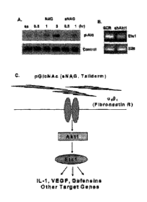
[0041] Figure 1. Nanofibers stimulate Akt 1 activation, an upstream
regulator of Etsl. (A)
Western blot analysis of phospho-Akt in response to NAG and sNAG stimulation
of serum
starved EC. (B) RT-PCR analysis of EC infected either with scrambled control
("SCR") or Aktl
shRNA lentiviruses and assessed for expression of Etsl and S26 as a loading
control. (C)
Schematic of a signal transduction pathway transducing a signal from sNAG
nanofibers to Aktl,
Etsl and Defensins.
[0042] Figure 2. Delayed wound healing in Aktl null animals is partially
rescued by
Taliderm treatment. (A) Representative images of wounded WT and AKT1 null mice
with and
without treatment of Taliderm. (B) H&E staining of representative mouse skin
sections from
day 3 wounds.
[0043] Figure 3. sNAG nanofibers stimulate cytokine and defensin expression
in primary
endothelial cells. (A) Immunohistochemisty of EC treated with or without sNAG
using an
antibody directed against a-defensin. (B) ELISA showing that nanofiber
treatment of EC results
in the secretion of a-defensins 1-3 (serum starved, treated with 5 jug/m1 or
10 glint sNAG).
[0044] Figure 4. sNAG nanofibers stimulate defensin expression in primary
endothelial
cells in an Aktl dependent manner. (A) and (B) Quantitative RT-PCR analyses of
serum starved
EC ("ss") treated with or without sNAG ("snag"), with or without PD98059 (MAPK
inhibitor,
"PD"), Wortmannin (PI3K inhibitor, "wtm") or infected with a scrambled control
("SCR"), or
Aktl ("AKT1") shRNA lentiviruses and assessed for expression of the genes
indicated.
[0045] Figure 5. sNAG nanofibers stimulate f3-defensin 3 expression in
mouse
keratinocytes. (A) Immunofluorescent staining with P-defensin 3 (visible as
bright staining in
the upper right hand panel; see, e.g., thick white arrows) and Involucrin
antibodies of paraffin
embedded mouse cutaneous wound sections from WT and Aktl null animals on Day
3. (B)
Quantification of 3-defensin 3 immunofluorescent staining using NIHImageJ
software
(TX=Taliderm; Akt1=Akt1 null). (C) Immunofluorescent staining of WT and Aktl
null treated
and untreated keratinocytes with P-Defensin 3 (visible as bright staining;
see, e.g., thick white
11
CA 02796068 2012-10-10
WO 2011/130646 PCT/US2011/032709
arrows) and TOPRO-3 (nuclei staining; see, e.g., thin white arrows). Notice
the increase in p-
Defensin 3 staining in WT and Aktl Taliderm treated wounds.
[0046] Figure 6. Aka dependent transcription factor binding sites.
Schematic of Aktl
dependent transcription factor binding sites. Using Genomatix software, 500 bp
upstream of the
transcription start site was analyzed for conserved sites on the mRNA of DEF1,
4, and 5 (ETS-
black ovals; FKHD-striped ovals; CREB-white ovals; NFKB-checkered ovals).
[0047] Figure 7. sNAG treatment results in expression and secretion of
defensins in vitro.
(A) RTPCR analysis of serum starved ("SS") primary endothelial cells treated
with sNAG
(50g/m1) for the times indicated and assessed for expression of P-defensin 3
and a-defensin 1.
(B) Immunofluorescent labeling of endothelial cells either scrum starved
(untreated) or treated
with sNAG nanofibers (10m/m1 for 5hrs). Antibodies are directed against a-
defensin 5 (FITC,
upper left hand panel), P-defensin 3 (Texas Red, upper right hand panel).
Nuclei are stained with
TOPRO-3 (Blue, lower left hand panel). Lower right hand panel represents
triple overlay. (C)
Immunofluoreseent labeling of keratinocytes (HaCat) that are either serum
starved (untreated) or
treated with sNAG nanofibers (1011g/m1 for 5 hours). Antibodies are directed
against a-defensin
(FITC, upper left hand panel), P-defensin 3 (Texas Red, upper right hand
panel). Nuclei are
stained with TOPRO-3 (Blue, lower left hand panel).
[0048] Figure 8. sNAG induced defensin expression is dependent on Aktl. (A)
Quantitative RT-PCR analyses using primers directed against a-defensin 1 from
total RNA
isolated from serum starved endothelial cells treated with or without sNAG for
3 hours, with or
without pretreatment with PD098059 ("PD")(50[LM), wortmannin ("WTM")(100nm).
Quantitation is relative to the S26 protein subunit. (B) Quantitation of P-
defensin 3 expression
from total RNA isolated from serum starved endothelial cells treated with or
without sNAG for 3
hours, with or without PD98059 (50pm), wortmannin (100nm) and shown as
relative to S26. (C)
Western Blot analysis of phospho-Akt in serum starved endothelial cells (SS)
stimulated with
sNAG for the times indicated. Line indicates where lanes have been removed (D)
Quantitative
RT-PCR analyses of serum starved endothelial cells infected with a scrambled
control (SCR) or
Aka shRNA lentiviruses, treated with or without sNAG and assessed for a-
defensin 4
expression. Quantitation is shown relative to S26. (E) Quantitation of P-
defensin 3 expression
from total RNA isolated from serum starved endothelial cells infected with a
scrambled control
(SCR) or Aktl shRNA lentiviruses, treated with or without sNAG. Quantitation
is shown
12
CA 02796068 2012-10-10
WO 2011/130646 PCT/US2011/032709
relative to S26. All experiments were done in at least triplicate and repeated
at least three
independent times and p values are shown.
[0049] Figure 9. sNAG induced defensin expression in vivo requires Aktl.
(A) Paraffin
embedded sections of cutaneous wounds harvested on day 3 post wounding from
both WT (n=3)
and Aka mice. Wounds were either untreated or treated with sNAG membrane.
Immunofluorescence was performed using antibodies directed against 13-defensin
3 (green,
visible as bright staining in the upper right hand panel; see, e.g., white
thick arrows), Involucrin
(Red), and Topro (Blue, nuclei staining; see, e.g., white thin arrows). (B)
Paraffin embedded
section from WT treated with sNAG harvested on day 3. Immunofluorescence was
performed
using antibodies directed against 13- defensin 3 (green, visible as bright
staining; sec, e.g., thick
white arrows), Involucrin (Red), and Topro (Blue, nuclei staining; see, e.g.,
thin white arrows).
This lower magnification (20x) is included to better illustrate the epidermal
layers expressing 13-
defensin 3. Scale bars = 50 um. (C) Quantitation of f3-defensin 3 expression
from paraffin
embedded sections was performed using NIH ImageJ software. Experiments were
repeated three
independent times and p values are shown.
[0050] Figure 10. sNAG treatment increases wound closure in wild type mice.
H&E
staining of wound tissue sections derived from C57B16 wild type animals either
untreated or
treated with sNAG membrane. The day post-wound is indicated to the left of
each panel. The
solid black line follows the keratinocyte cell layer indicating wound closure.
Black arrows
indicate the margin of the wound bed.
[0051] Figure 11. sNAG treatment reduces bacterial infection in an Aktl
dependent
manner. (A) Tissue gram staining of S. aureus infected wounds from WT mice. WT
mice were
wounded using a 4 mm biopsy punch. Immediately after wounding mice were
inoculated with 1
x 109 cfu/ml. 30 minutes post-infection, mice in the treated group were
treated with Taliderm.
Skin samples were taken 5 days post-treatment and sectioned for analysis.
Tissue gram staining
was performed. Dark purple staining indicates gram-positive bacteria and
neutrophils that have
engulfed bacteria. Sections under 20x amd 40x magnification are shown. (B)
Tissue gram
staining of paraffin embedded S. aureus infected wounds from WT and Aktl null
mice (n=3).
Infected wounds were either untreated or treated with sNAG membrane and wound
beds were
harvested on day 3 and day 5 for analysis. Dark purple staining indicates the
presence of gram
positive bacteria in the wound bed. Black arrows indicate examples of gram
positive staining.
13
CA 02796068 2012-10-10
WO 2011/130646 PCT/US2011/032709
Note the accumulation of positive staining in untreated WT that is lacking in
WT animals treated
with sNAG. Scale bars = 5011m. (C) CFUs derived from day 5 post wounding were
quantitated
from S. aureus infected wounds using both treated and untreated WT (n= 3) and
Aktl mice
(n=3). Wild type mice that were sNAG treated show a significant (p<.0 1)
decrease in bacteria
load in the wound beds as compared to Aktl null animals. All experiments were
repeated three
independent times and the p values are shown. (D) CFU quantitated from
infected wounds at
day 3 post wounding in a similar fashion described in (C). sNAG treatment of
infected wounds
shows a significant decrease in CFU of both WT and Aktl null animals on day 3,
but the WT
animals show an approximate 10 fold difference compared to a 2 fold difference
in Aktl
animals. (E) Quantitation of CFUs in S. aureus cultures that were either
untreated or treated with
various amounts of sNAG nanofibers. Each experiment was performed three
independent times
and p values are shown. (F) Tissue gram staining of S. aureus infected wounds
harvested on day
3 post wound from WT mice (n=3) that were treated with or without 13- defensin
3 peptide (1.0
uM). Note the decrease in gram positive staining in infected wounds that were
treated with 13-
defensin 3 peptide. (G) Quantitation of CFUs from S. aureus infected WT mice
(n=3) treated
with or without 13-defensin 3 peptide. Infected wounds that were treated with
peptide show a
significant decrease (p <.05) in CFU. Scale bars = 50.tm. Each experiment was
performed three
independent times and p values are shown.
[0052] Figure 12. Rapid induction of defensin expression by sNAG treatment
of S. aureus
infected wounds. (A) Paraffin embedded tissue sections from S. aureus infected
wounds,
harvested on day 3, were subjected to immunofluorescence using antibodies
directed against 13-
defensin 3 (green, visible as bright staining in the upper right hand panel
and in the lower panel
in the middle; see, e.g., thick white arrows), Tnvolucrin (red) to mark the
keratinocyte layer, and
Topro (blue, nuclei staining; see, e.g., thin white arrows) from both sNAG
treated WT (n=3) and
untreated WT mice (n=3). Non specific staining of keratin is indicated by the
no primary control
which was stained with secondary antibody only. Scale bar = 50[im. (B)
Quantitation of 13-
defensin 3 expression from paraffin embedded sections using NIH ImageJ
software. S. aureus
infected wounds that were treated with sNAG show a significant increase
(p<.05) in 13-defensin 3
staining. Experiments were repeated three independent times and p values are
shown.
[0053] Figure 13. Antibodies against 13-defensin 3 impedes antibacterial
effects of sNAG
treatment. (A) Tissue gram staining of paraffin embedded S. aureus infected
wounds treated
14
CA 02796068 2012-10-10
WO 2011/130646 PCT/US2011/032709
with sNAG from WT mice (n=3) that were harvested on Day 3. sNAG treated wounds
were
treated with either 13-defensin 3 antibody or isotype control goat IgG
antibody prior to sNAG
treatment. Representative images show increased accumulation gram positive
staining (black
arrows) in the wound beds of mice treated with an antibody directed against I3-
defensin 3. Scale
bar = 20 m. (B) Quantitation of CFUs from S. aureus infected WT mice treated
either 13-
defensin 3 antibody (n=3) or control IgG antibody (n=3) prior to sNAG
treatment. 13-defensin 3
application significantly increased (p <.05) CFU.
[0054] Figure 14. sNAG treatment reduces bacterial infection by Pseudomonas
aerugthosa.
Mice were wounded using a 4 mm biopsy punch, inoculated with 1.5 x 10⁹ cfu/ml
P. aeruginosa,
infected wounds were either untreated (n=6) or treated (n=6) with sNAG
membrane (n=6) 30
min post-infection, wound beds were harvested on day 3 for analysis, cultured
for 30 minutes,
plated, and CFUs of the untreated and treated infected wounds were
quantitated. sNAG treated
mice show a significant (p<.05) decrease in bacteria load in the wound beds as
compared to
untreated animals.
[0055] Figure 15. Effect of irradiation on chemical and physical structure
of pG1cNAc
fibers. (A) Correlation between molecular weight of pG1cNAc and irradiation
level/formulation
for irradiation. (B) Infrared (IR) spectrum of non-irradiated pG1cNAc slurry
(top line), pG1cNAc
slurry irradiated at 100 kGy (bottom line), and pG1cNAc slurry irradiated at
200 kGy (middle
line). (C) Scanning electron microscopic (SEM) analyses of pG1cNAc. (D)
Scanning electron
microscopic (SEM) analyses of sNAG.
[0056] Figure 16. pG1cNAc did not affect metabolic rate. For each time
period (i.e., at 24
and 48 hours), the identity for each of the four bars (from left to right) is
as follows: serum
starvation (SS), VEGF, and pG1cNAc (NAG) at 50 and 100 g/ml.
[0057] Figure 17. pG1cNAc protected human umbilical vein endothelial cell
(EC) from cell
death induced by serum deprivation. For each time period (i.e., at 24, 48 and
72 hours), the
identity for each of the five bars (from left to right) is as follows: serum
starvation (SS), VEGF,
and pG1cNAc (NAG) at 50, 100, and 250 g/ml.
[0058] Figure 18. sNAG induced marked increase in metabolic rate. Identity
for each of the
five bars (from left to right) is as follows: serum starvation (SS), VEGF, and
sNAG at 50, 100
and 200 g/ml.
CA 02796068 2012-10-10
WO 2011/130646 PCT/US2011/032709
[0059] Figure 19. sNAG did not protect EC from cell death induced by serum
deprivation.
For each time period (i.e., at 24 and 48 hours), the identity for each of the
five bars (from left to
right) is as follows: serum starvation (SS), VEGF, and sNAG at 50, 100 and 200
ug/ml.
5. DETAILED DESCRIPTION
[0060] The inventors have discovered that sNAG nanofibers decrease
bacterial infection of
cutaneous wounds infected with Staphylococcus aureus and Pseudomonas
aeurgino.sa. Without
being bound by any specific mechanism of action, data presented in Section 6.2
suggests that the
antibacterial effect of sNAG is not due to a direct interaction of sNAG with
the bacteria but is
due to downstream affects such as, for example, the regulation of defensins by
Aktl activation.
Specifically, data show that treatment of bacterial cultures with sNAG
nanofibers in vitro does
not affect bacterial count indicating that sNAG nanofibers do not directly
inhibit bacterial
growth. In a specific example described in Section 6.2.2.5 and illustrated in
Figure 11E, sNAG
nanofibers do not have a direct effect on growth or survival of Staphylococcus
aureus. The test
described in Section 6.2.2.5, infra, may be used to test the lack of a direct
effect of sNAG
nanofibers on bacterial growth or survival. In this example, S. aureus
cultures in solution were
treated with varying concentrations of sNAG nanofibers for three hours,
cultures were then
plated overnight at 37 C and bacterial CFU/ml determined. As shown in Figure
11E, no effect
on bacterial growth or survival was observed.
[0061] The inventors of the present invention have found that sNAG
nanofibers can
stimulate expression of defensins, which may boost the innate anti-bacterial
response. It is
widely accepted that defensins are important players in innate immunity and
function in anti-
bacterial activities. As demonstrated in the examples presented in Sections
6.1 and 6.2, infra, the
inventors of the present invention have found that sNAG nanofibers can
increase the expression
of both a- and 13.- type defensins in endothelial cells and I3-type defensins
in keratinocytes in vitro
and in a wound healing model in vivo.
[0062] Further, as demonstrated in the examples presented in Sections 6.1
and 6.2, infra, but
without being bound by any specific mechanism of action, Aktl appears to be
important for
sNAG-dependent defensin expression in vitro and in vivo, in a wound healing
model.
Consistently, sNAG treatment decreased bacterial infection of cutaneous wounds
infected with
Staphylococcus aureus in wild type control animals but not in similarly
treated Akt1 null
animals.
16
CA 02796068 2012-10-10
WO 2011/130646 PCT/US2011/032709
[0063] The inventors of this invention have also found that a number of
Toll-like receptors
can be up-regulated by sNAG treatment of human endothelial cells. Toll-like
receptors ("TLRs"
or "TLR") are highly conserved receptors that recognize specific molecular
patterns of bacterial
components leading to activation of innate immunity. Recent work has linked
human defensin
expression to TLR activation. In particular, stimulation of TLRs can lead to
increased defensin
synthesis. Thus, without being bound by any mechanism of action, sNAG
nanofibers may act as
a stimulator of innate immunity and bacterial clearance via the activation of
Aktl.
[0064] Accordingly, described herein is the use of sNAG nanofibers as a
novel method for
preventing and/or treating bacterial infections and diseases associated
therewith. In certain
embodiments, treatment of bacterial infections with sNAG nanofibers decreases
the bacterial
load in patients. In specific embodiments, the use of sNAG nanofibers enhances
wound closure
while simultaneously eradicating, decreasing or preventing bacterial infection
of the wound.
5.1 sNAG Nanofibers
[0065] Described herein are sNAG nanofiber compositions. The sNAG
nanofibers comprise
fibers of poly-N-acetylglucosamine and/or a derivative(s) thereof, the
majority of which are less
than 30 microns in length and at least 1 micron in length as measured by any
method known to
one skilled in the art, for example, by scanning electron microscopy ("SEM").
Such sNAG
nanofibers may be obtained, for example, as described herein.
[0066] In certain embodiments, the majority (and in certain embodiments, at
least 60%, 70%,
80%, 90%, 95%, 98%, 99%, 99.5%, 99.8%, 99.9%, or 100%, or between 55% to 65%,
55% to
75%, 65% to 75%, 75% to 85%, 75% to 90%, 80% to 95%, 90% to 95%, or 95% to
99%) of the
sNAG nanofibers are less than about 30, 25, 20, 15, 12, 10, 9, 8, 7, 6, 5, 4,
or 3 microns in
length, and at least 1 micron in length as measured by any method known to one
skilled in the
art, for example, by SEM. In specific embodiments, the majority (and in
certain embodiments,
at least 60%, 70%, 80%, 90%, 95%, 98%, 99%, 99.5%, 99.8%, 99.9%, or 100%, or
between
55% to 65%, 55% to 75%, 65% to 75%, 75% to 85%, 75% to 90%, 80% to 95%, 90% to
95%, or
95% to 99%) of the sNAG nanofibers are less than about 15 microns or less than
about 12
microns in length, and at least 1 micron in length as measured by any method
known to one
skilled in the art, for example, by SEM. In specific embodiments, all (100%)
of the sNAG
nanofibers are less than about 15 microns in length, and at least 1 micron in
length as measured
by any method known to one skilled in the art, for example, by SEM. In certain
embodiments,
17
CA 02796068 2012-10-10
WO 2011/130646 PCT/US2011/032709
the majority (and in certain embodiments, at least 60%, 70%, 80%, 90%, 95%,
98%, 99%,
99.5%, 99.8%, 99.9%, or 100%, or between 55% to 65%, 55% to 75%, 65% to 75%,
75% to
85%, 75% to 90%, 80% to 95%, 90% to 95%, or 95% to 99%) of the sNAG nanofibers
are equal
to or less than 14, 13, 12, 11, 10,9, 8 or 7 microns in length, and at least 1
micron in length as
measured by any method known to one skilled in the art, for example, by SEM.
In some
embodiments, the majority (and in certain embodiments, at least 60%, 70%, 80%,
90%, 95%,
98%, 99%, 99.5%, 99.8%, 99.9%, or 100%, or between 55% to 65%, 55% to 75%, 65%
to 75%,
75% to 85%, 75% to 90%, 80% to 95%, 90% to 95%, or 95% to 99%) of the sNAG
nanofibers
are between Ito 15, 2 to 15,2 to 14, Ito 12, 2 to 12, Ito 10, 2 to 10, 3 to
12, 3 to 10, Ito 9, 2 to
9, 3 to 9, 1 to 8, 2 to 8, 3 to 8, 4 to 8, 1 to 7, 2 to 7, 3 to 7, 4 to 7, 1
to 6, 1 to 5, 1 to 4, or 1 to 3
microns in length as measured by any method known to one skilled in the art,
for example, by
SEM.
[0067] In a specific embodiment, the majority (and in certain embodiments,
at least 60%,
70%, 80%, 90%, 95%, 98%, 99%, 99.5%, 99.8%, 99.9%, or 100%, or between 55% to
65%,
55% to 75%, 65% to 75%, 75% to 85%, 75% to 90%, 80% to 95%, 90% to 95%, or 95%
to
99%) of the sNAG nanofibers are about 8, 7, 6, 5, 4, 3 or 2 microns in length
as measured by any
method known to one skilled in the art, for example, by SEM. In another
specific embodiment,
the majority (and in certain embodiments, at least 60%, 70%, 80%, 90%, 95%,
98%, 99%,
99.5%, 99.8%, 99.9%, or 100%, or between 55% to 65%, 55% to 75%, 65% to 75%,
75% to
85%, 75% to 90%, 80% to 95%, 90% to 95%, or 95% to 99%) of the sNAG nanofibers
are
between about 2 to about 10 microns, about 3 to about 8 microns, or about 4 to
about 7 microns
in length as measured by any method known to one skilled in the art, for
example, by SEM. In
another specific embodiment, all (100%) of the sNAG nanofibers are between
about 2 to about
microns, about 3 to about 8 microns, or about 4 to about 7 microns in length
as measured by
any method known to one skilled in the art, for example, by SEM.
[0068] In certain embodiments, the sNAG nanofibers fibers are in a range
between 0.005 to 5
microns in thickness and/or diameter as determined by electron microscopy. In
specific
embodiments, the sNAG nanofibers are about 0.01, 0.02, 0.03, 0.04, 0.05, 0.06,
0.07, 0.08, 0.09,
0.1, 0.2, 0.25, 0.3, 0.35, 0.4, 0.45, 0.5, 0.55, 0.6, 0.65, 0.7, 0.75, 0.8,
0.85, 0.9, 1, 1.1, 1.2, 1.3,
1.4, 1.5, 1.6, 1.7, 1.8, 1.9, 2, 2.2, 2.4, 2.6, 2.8, 3 or 4 microns in
thickness and/or diameter on
average, or any range in between (e.g., 0.02 to 2 microns, 0.02 to 1 microns,
0.02 to 0.75
18
CA 02796068 2012-10-10
WO 2011/130646 PCT/US2011/032709
microns, 0.02 to 0.5 microns, 0.02 to 0.5 microns, 0.05 to 1 microns, 0.05 to
0.75 microns, 0.05
to 0.5 microns, 0.1 to 1 microns, 0.1 to 0.75 microns, 0.1 to 0.5 microns,
etc.). In specific
embodiments, the majority (and in certain embodiments, at least 60%, 70%, 80%,
90%, 95%,
98%, 99%, 99.5%, 99.8%, 99.9%, or 100%, or between 55% to 65%, 55% to 75%, 65%
to 75%,
75% to 85%, 75% to 90%, 80% to 95%, 90% to 95%, or 95% to 99%) of the sNAG
nanofibers
have a thickness or diameter of about 0.02 to 1 microns. In other specific
embodiments, the
majority (and in certain embodiments, at least 60%, 70%, 80%, 90%, 95%, 98%,
99%, 99.5%,
99.8%, 99.9%, or 100%, or between 55% to 65%, 55% to 75%, 65% to 75%, 75% to
85%, 75%
to 90%, 80% to 95%, 90% to 95%, or 95% to 99%) of the sNAG nanofibers have a
thickness or
diameter of about 0.05 to 0.5 microns. In specific embodiments, all (100%) of
the sNAG
nanofibers have a thickness or diameter of about 0.02 to 1 microns or about
0.05 to 0.5 microns.
In certain embodiments, the majority (and in certain embodiments, at least
60%, 70%, 80%,
90%, 95%, 98%, 99%, 99.5%, 99.8%, 99.9%, or 100%, or between 55% to 65%, 55%
to 75%,
65% to 75%, 75% to 85%, 75% to 90%, 80% to 95%, 90% to 95%, or 95% to 99%) of
the sNAG
nanofibers have a thickness or diameter of about 0.02 to 2 microns, 0.02 to 1
microns, 0.02 to
0.75 microns, 0.02 to 0.5 microns, 0.02 to 0.5 microns, 0.05 to 1 microns,
0.05 to 0.75 microns,
0.05 to 0.5 microns, 0.1 to 1 microns, 0.1 to 0.75 microns, or 0.1 to 0.5
microns.
[0069] In certain embodiments, the majority (and in certain embodiments, at
least 60%, 70%,
80%, 90%, 95%, 98%, 99%, 99.5%, 99.8%, 99.9%, or 100%, or between 55% to 65%,
55% to
75%, 65% to 75%, 75% to 85%, 75% to 90%, 80% to 95%, 90% to 95%, or 95% to
99%) of the
sNAG nanofibers are between 1 and 15 microns in length and have a thickness or
diameter of
about 0.02 to 1 microns.
[0070] In certain embodiments, the molecular weight of the sNAG nanofibers
is less than
100kDa, 90kDa, 80kDa, 75kDa, 70 kDa, 65 kDa, 60kDa, 55kDa, 50kDa, 45 kDA,
40kDa,
35kDa, 30kDa, or 25kDa. In certain embodiments, the majority (and in certain
embodiments, at
least 60%, 70%, 80%, 90%, 95%, 98%, 99%, 99.5%, 99.8%, 99.9%, or 100%, or
between 55%
to 65%, 55% to 75%, 65% to 75%, 75% to 85%, 75% to 90%, 80% to 95%, 90% to
95%, or 95%
to 99%) of the sNAG nanofibers have a molecular weight of less than 100kDa,
90kDa, 80kDa,
75kDa, 70 kDa, 65 kDa, 60kDa, 55kDa, 50kDa, 45 kDA, 40kDa, 35kDa, 30kDa, or
25kDa. In
other embodiments, the majority (and in certain embodiments, at least 60%,
70%, 80%, 90%,
95%, 98%, 99%, 99.5%, 99.8%, 99.9%, or 100%, or between 55% to 65%, 55% to
75%, 65% to
19
CA 02796068 2012-10-10
WO 2011/130646 PCT/US2011/032709
75%, 75% to 85%, 75% to 90%, 80% to 95%, 90% to 95%, or 95% to 99%) of the
sNAG
nanofibers have a molecular weight between about 5kDa to 100kDa, about 10kDa
to 100kDa,
about 20kDa to 100kDa, about 10kDa to 80kDa, about 20kDa to 80kDa, 20kDa to
75kDa, about
25kDa to about 75kDa, about 30kDa to about 80kDa, about 30kDa to about 75kDa,
about 40kda
to about 80kDa, about 40kDa to about 75kDa, about 40kDa to about 70kDa, about
50kDa to
about 70kDa, or about 55kDa to about 65kDa. In one embodiment, the majority
(and in certain
embodiments, at least 60%, 70%, 80%, 90%, 95%, 98%, 99%, 99.5%, 99.8%, 99.9%,
or 100%,
or between 55% to 65%, 55% to 75%, 65% to 75%, 75% to 85%, 75% to 90%, 80% to
95%,
90% to 95%, or 95% to 99%) of the sNAG nanofibers have a molecular weight of
about 60kDa.
[0071] In certain embodiments, 1% to 5%, 5% to 10%, 5% to 15%, 20% to 30%
or 25% to
30% of the sNAG nanofibers are deacetylated. In some embodiments, 1%, 5%,
10%,15%, 20%,
25%, or 30% of the sNAG nanofibers are deacetylated. In other embodiments,
less than 30%,
25%, 20%, 15%, 10%, 5%, 4%, 3%, 2% or 1% of the sNAG nanofibers are
deacetylated. In
some embodiments, equal to or more than 1%, 5%, 10%, 15%, 20%, 25%, 30%, 35%,
40%,
45%, 50%, 55%, 60%, 65%, 70%, 75%, 80%, 85%, 90%, 95% or 99%, or all (100%),
of the
sNAG nanofibers are deacetylated. In other embodiments, less than 1%, 5%, 10%,
15%, 20%,
25%, 30%, 35%, 40%, 45%, 50%, 55%, 60%, 65%, 70%, 75%, 80%, 85%, 90%, 95%,
99%, or
100% of the sNAG nanofibers are deacetylated.
[0072] In certain embodiments, 70% to 80%, 75% to 80%, 75% to 85%, 85% to
95%, 90%
to 95%, 90% to 99% or 95% to 100% of the sNAG nanofibers are acetylated. In
some
embodiments, 70%, 75%, 80%, 85%, 90%, 95%, 98%, 99% or 100% of the sNAG
nanofibers
are acetylated. In other embodiments, more than 70%, 75%, 80%, 85%, 90%, 95%,
98%, 99%,
99.5% or 99.9% of the sNAG nanofibers are acetylated. In some embodiments,
equal to or more
than 1%, 5%, 10%, 15%, 20%, 25%, 30%, 35%, 40%, 45%, 50%, 55%, 60%, 65%, 70%,
75%,
80%, 85%, 90%, 95% or 99%, or all (100%), of the sNAG nanofibers are
acetylated. In other
embodiments, less than 1%, 5%, 10%, 15%, 20%, 25%, 30%, 35%, 40%, 45%, 50%,
55%, 60%,
65%, 70%, 75%, 80%, 85%, 90%, 95%, 99%, or 100% of the sNAG nanofibers are
acetylated.
[0073] In some embodiments, the sNAG nanofibers comprise at least one
glucosamine
monosaccharide, and may further comprise at least 10%, 20%, 30%, 40%, 50%,
60%, 70%,
80%, 90%, 95% or 99% of the N-acetylglucosamine monosaccharides. In other
embodiments,
the sNAG nanofibers comprise at least one N-acetylglucosamine monosaccharide,
and may
further comprise at least 10%, 20%, 30%, 40%, 50%, 60%, 70%, 80%, 90%, 95% or
99% of
glucosamine monosaccharides.
[0074] In one aspect, the sNAG nanofibers increase the metabolic rate of
serum-starved
human umbilical cord vein endothelial cells ("EC") in a MTT assay. A MTT assay
is a
laboratory test and a standard colorimetric assay (an assay which measures
changes in color) for
measuring cellular proliferation (cell growth). Briefly, yellow MTT (3-(4,5-
Dimethylthiazol-2-
y1)-2,5-diphenyltetrazolium bromide, a tetrazole) is reduced to purple
formazan in the
mitochondria of living cells. This reduction takes place only when
mitochondrial reductase
enzymes are active, and therefore conversion can be directly related to the
number of viable
(living) cells. The metabolic rate of cells may be determined by other
techniques commonly
known to the skilled artisan.
[0075] In another aspect, the sNAG nanofibers do not rescue apoptosis of
serum-starved EC
in a trypan blue exclusion test. A trypan blue exclusion test is a dye
exclusion test used to
determine the number of viable cells present in a cell suspension. It is based
on the principle that
live cells possess intact cell membranes that exclude certain dyes, such as
trypan blue, Eosin, or
propidium, whereas dead cells do not. The viability of cells may be determined
by other
techniques commonly known to the skilled artisan.
[0076] In certain embodiments, compositions comprising the sNAG nanofibers
are
described, wherein the sNAG nanofibers increase the metabolic rate of serum-
starved human
umbilical cord vein endothelial cells in a MTT assay and/or do not rescue
apoptosis of serum-
starved human umbilical cord vein endothelial cells in a trypan blue exclusion
test. In some
embodiments, the sNAG nanofibers increase the metabolic rate of scrum-starved
human
umbilical cord vein endothelial cells in a MTT assay and do not rescue
apoptosis of serum-
starved human umbilical cord vein endothelial cells in a trypan blue exclusion
test.
[0077] In a specific embodiment, the sNAG nanofibers are biocompatible.
Biocompatibility
may be determined by a variety of techniques, including, but not limited to
such procedures as
the elution test, intramuscular implantation, or intracutaneous or systemic
injection into animal
subjects. Such tests are described in U.S. Patent No. 6,686,342 (see, e.g.,
Example 10).
21
CA 2796068 2017-10-18
[0078] In certain embodiments, the sNAG nanofibers used in the methods
described herein
are non-reactive in a biocompatibility test or tests. For example, the sNAG
nanofibers used in
the methods described herein may be non-reactive when tested in an elution
test, an
intramuscular implantation test, an intracutaneous test, and/or a systemic
test. In other
embodiments, the sNAG nanofibers used in the methods described herein have
Grade 0 or Grade
1 test score when tested in an elution test, an intramuscular implantation
test, an intracutaneous
test, or a systemic test. In yet another embodiment, the sNAG nanofibers used
in the methods
described herein are at most mildly reactive when tested in an elution test,
an intramuscular
implantation test, an intracutaneous test, and/or a systemic test. In certain
embodiments, the
compositions described herein do not cause an allergenic reaction or an
irritation. In other
embodiments, the compositions described herein cause at most a mild allergenic
reaction or a
mild irritation, e.g., at the site of application. The relevant tests and
evaluation of test results are
described in, e.g., U.S. Patent No. 6,686,342 and in Section 6.8, infra.
[0079] In a specific embodiment, the sNAG nanofibers are non-reactive when
tested in an
intramuscular implantation test. In one aspect, an intramuscular implantation
test is an
intramuscular implantation test ¨ ISO 4 week implantation, as described in
Section 6.8.3, infra.
In certain embodiments, the sNAG nanofibers display no biological reactivity
as determined by
an elution test (Elution Test Grade = 0). In some embodiments, the sNAG
nanofibers have a test
score equal to "0" and/or are at most a negligible irritant as determined by
intracutaneous
injection test. In some embodiments, the sNAG nanofibers elicit no intradermal
reaction (i.e.,
Grade I reaction) in Kligman test and/or have a weak allergenic potential as
determined by
Kligman test.
[0080] In certain aspects, the sNAG nanofibers are immunoneutral (i.e.,
they do not elicit an
immune response).
[0081] In some embodiments, the sNAG nanofibers are biodegradable. The sNAG
nanofibers preferably degrade within about I day, 2 days, 3 days, 5 days, 7
days (1 week), 8
days, 10 days, 12 days, 14 days (2 weeks), 17 days, 21 days (3 weeks), 25
days, 28 days (4
weeks), 30 days, 1 month, 35 days, 40 days, 45 days, 50 days, 55 days, 60
days, 2 months, 65
days, 70 days, 75 days, 80 days, 85 days, 90 days, 3 months, 95 days, 100 days
or 4 months after
administration or implantation into a patient.
22
CA 2796068 2019-01-30
CA 02796068 2012-10-10
WO 2011/130646 PCT/US2011/032709
[0082] In certain embodiments, the sNAG nanofibers do not cause a
detectable foreign body
reaction. A foreign body reaction, which may occur during wound healing,
includes
accumulation of exudate at the site of injury, infiltration of inflammatory
cells to debride the
area, and the formation of granulation tissue. The persistent presence of a
foreign body can
inhibit full healing. Rather than the resorption and reconstruction that
occurs in wound healing,
the foreign body reaction is characterized by the formation of foreign body
giant cells,
encapsulation of the foreign object, and chronic inflammation. Encapsulation
refers to the firm,
generally muscular collagen shell deposited around a foreign body, effectively
isolating it from
the host tissues. In one embodiment, treatment of a site (e.g., a wound or a
site of a bacterial
infection in a wound) with the sNAG nanofibers does not elicit a detectable
foreign body
reaction in 1 day, 3 days, 5 days, 7 days, 10 days or 14 days after treatment.
In one such
embodiment, treatment of a site (e.g., a wound) with the sNAG nanofibers does
not elicit a
foreign body encapsulations in 1 day, 3 days, 5 days, 7 days, 10 days or 14
days after treatment.
[0083] In some embodiments, the sNAG nanofibers (i) comprise fibers,
wherein majority of
the fibers are between about 1 and 15 microns in length, and (ii) (a) increase
the metabolic rate
of serum-starved EC in a MTT assay and/or do not rescue apoptosis of serum-
starved EC in a
trypan blue exclusion test, and (b) are non-reactive when tested in an
intramuscular implantation
test. In certain embodiments, the sNAG nanofibers (i) comprise fibers, wherein
majority of the
fibers are between about 1 and 12 microns in length, and (ii) (a) increase the
metabolic rate of
serum-starved EC in a MTT assay and/or do not rescue apoptosis of serum-
starved EC in a
trypan blue exclusion test, and (b) arc non-reactive when tested in an
intramuscular implantation
test. In certain embodiments, the sNAG nanofibers (i) comprise fibers, wherein
majority of the
fibers are between about 4 and 7 microns in length, and (ii) (a) increase the
metabolic rate of
serum-starved EC in a MTT assay and/or do not rescue apoptosis of serum-
starved EC in a
trypan blue exclusion test, and (b) are non-reactive when tested in an
intramuscular implantation
test.
[0084] In certain embodiments, the sNAG nanofibers do not have a direct
effect on the
growth or survival of bacteria, such as S. aureus, as determined by one
skilled in the art. In other
embodiments, sNAG nanofibers do not have a direct effect on the growth or
survival of bacteria,
such as S. aureus, as determined by the methods set forth in Section 6.2.2.5,
infra. In some
embodiments, the sNAG nanofibers do not have a direct effect in vitro on
bacterial growth or
23
CA 02796068 2012-10-10
WO 2011/130646 PCT/US2011/032709
survival. In one embodiment, the sNAG nanofibers do not have a direct effect
(e.g., in vitro) on
growth or survival of gram-negative bacteria. In another embodiment, the sNAG
nanofibers do
not have a direct effect (e.g., in vitro) on growth or survival of gram-
positive bacteria. In yet
another embodiment, the sNAG nanofibers do not have a direct effect (e.g., in
vitro) on growth
or survival of either gram-positive or gram-negative bacteria. In some
embodiments, the sNAG
nanofiber or a sNAG nanofiber composition does not bind bacteria (e.g., gram-
positive bacteria,
gram-negative bacteria, or both types of bacteria). In some embodiments,
incubation of a
bacterial culture with the sNAG nanofibers (e.g., 50-500 jig of sNAG
nanofibers) in vitro does
not reduce bacterial load in 10 minutes, 30 minutes, 1 hour, 2 hours, 3 hours,
6 hours, 12 hours,
24 hours, 48 hours, 72 hours or 96 hours of incubation (wherein the bacterial
culture may be
gram-positive and/or a gram-negative). In some embodiments, incubation of a S.
aureus culture
with sNAG nanofibers (e.g., about 80ng-300ng, or about 100-200 jig of sNAG
naofibers) in
vitro does not reduce bacterial load in 2 hours, 3 hours, 6 hours or 24 hours
of incubation. In yet
other embodiments, the sNAG nanofibers reduce bacterial growth or survival in
vitro by less
than 1 log, 0.9 log, 0.8 log, 0.75 log, 0.7 log, 0.6 log, 0.5 log, 0.4 log,
0.3 log, 0.25 log, 0.2 log,
0.1 log, 0.05 log, or 0.025 log, for example, when Staphylococcus aureus
bacterial cultures are
treated/incubated with the sNAG nanofibers in vitro. In some embodiments, the
sNAG
nanofibers reduce bacterial growth or survival in vitro by less than 1 x 10⁴,
2 x 10⁴, 3 x 10⁴, 4 x
104, 5 x 10⁴, 6 x 10⁴, 7 x 10⁴, 8 x 10⁴, 9 x 10⁴, or 10 x 104 cfu/ml, for
example, when
Staphylococcus aureus bacterial cultures are treated/incubated with the sNAG
nanofibers in
vitro. The tests of the effect of sNAG nanofibers on bacterial growth or
survival and the
evaluation of the test results are described, for example, in Example 2 (e.g.,
Section 6.2.2.5) and
Figure 11E, infra.
[0085] In some embodiments, the sNAG nanofibers (i) comprise fibers,
wherein majority of
the fibers are between about 1 and 15 microns, 1 and 12 microns, or 4 and 7
microns in length,
(ii) do not have an effect on bacterial growth or survival of Staphylococcus
aureus bacterial
cultures in vitro, and (iii) are non-reactive when tested in a
biocompatibility test (e.g., an
intramuscular implantation test).
[0086] In certain embodiments, the sNAG nanofibers induce a certain pattern
of gene
expression (RNA or protein expression as determined by, e.g., RT-PCR,
microarray or ELISA)
in a cell, tissue or organ treated with or exposed to a sNAG nanofiber
composition. Specifically,
24
CA 02796068 2012-10-10
WO 2011/130646 PCT/US2011/032709
in some embodiments, the sNAG nanofibers or a composition comprising the sNAG
nanofibers
induce expression of one or more defensin proteins, one or more defensin-like
proteins, and/or
one or more Toll-like receptors. In yet other embodiments, the sNAG nanofibers
or a
composition comprising the sNAG nanofibers induce expression of one or more
proteins that are
known to have an anti-bacterial effect.
T00871 In certain embodiments, the sNAG nanofibers or a composition
comprising the sNAG
nanofibers induce expression of one or more a-defensins (e.g., DEFAl(i.e., a-
defensin 1),
DEFA1B, DEFA3, DEFA4, DEFA5, DEFA6), one or more I3-defensins (e.g., DEFB1
(i.e., 13-
defensin 1), DEFB2, DEFB4, DEFB103A, DEFB104A, DEFB105B, DEFB107B, DEFB108B,
DEFB110, DEFB112, DEFB114, DEFB118, DEFB119, DEFB123, DEFB124, DEFB125,
DEFB126, DEFB127, DEFB128, DEFB129, DEFB131, DEFB136), and/or one or more 0-
defensins (e.g., DEFT1P). In some embodiments, the sNAG nanofibers or a
composition
comprising the sNAG nanofibers induce expression of one or more of DEFA1,
DEFA3, DEFA4,
DEFA5, DEFB1, DEFB3, DEFB103A, DEFB104A, DEFB108B, DEFB112, DEFB114,
DEFB118, DEFB119, DEFB123, DEFB124, DEFB125, DEFB126, DEFB128, DEFB129 and
DEFB131. In certain embodiments, the sNAG nanofibers or a composition
comprising the
sNAG nanofibers induce expression of one or more Toll receptors (e.g., TLR1,
TLR2, TLR3,
TLR4, TLR5, TLR6, TLR7, TLR8, TLR9, TLR10, TLR11, and/or TLR12). In other
embodiments, the sNAG nanofibers or a composition comprising the sNAG
nanofibers induce
expression of one or more of IL-1, CEACAM3, SPAG11, SIGIRR (IL1-like
receptor), IRAK1,
IRAK2, IRAK4, TBK1, TRAF6 and IKKi. In some embodiments, the sNAG nanofibers
or a
composition comprising the sNAG nanofibers induce expression of one or more of
TRAK2,
STGIRR, TLR1, TLR2, TLR4, TLR7, TLR8, TLR10 and TRAF6. In one embodiment, the
sNAG nano fibers or a composition comprising the sNAG nano fibers induce
expression of at
least one of the above-listed gene products.
[0088] In some embodiments, the sNAG nanofibers or a composition comprising
the sNAG
nanofibers induce expression of one or more of the above-listed genes in the
amount equal to or
more than about 0.25 fold, 0.5 fold, 1 fold, 1.5 fold, 2 fold, 2.5 fold, 3
fold, 3.5 fold, 4 fold, 4.5
fold, 5 fold, 6 fold, 7 fold, 8 fold, 9 fold, 10 fold, 12 fold, 15 fold or 20
fold as compared to the
level of expression of the one or more of the above-listed genes in a cell,
tissue or organ of a
subject before treatment with the sNAG nanofibers (e.g., a known average level
of expression of
CA 02796068 2012-10-10
WO 2011/130646 PCT/US2011/032709
the one or more of the above-listed genes). In some embodiments, the sNAG
nanofibers or a
composition comprising the sNAG nanofibers induce expression of one or more of
the above-
listed genes in the amount equal to or more than about 10%, 25%, 50%, 75%,
100%, 125%,
150%, 175%, 200%, 225%, 250%, 275%, 300%, 350%, 400%, 450%, 500%, 550%, 600%,
650%, 700%, 750%, 800%, 900% or 1000% the level of expression of the one or
more of the
above-listed genes in a cell, tissue or organ of a subject before treatment
with the sNAG
nanofibers (e.g., a known average level of expression of the one or more of
the above-listed
genes).
[0089] In some embodiments, the sNAG nanofibers but not long poly-N-
acetylglucosamine,
chitin and/or chitosan induce expression of the one or more genes listed
above, as determined by
a method known to one skilled in the art, or described herein. In some of
these embodiments,
long poly-N-acetylglucosamine, chitin and/or chitosan do not induce expression
of the one or
more genes listed above or induce lower level (e.g., more than 1.25 fold, 1.5
fold, 2 fold, 2.5
fold, 3 fold, 3.5 fold, 4 fold, 4.5 fold, 5 fold, 6 fold, 7 fold, 8 fold, 9
fold, or 10 fold lower) of
expression of the one or more genes listed above as compared to the level of
expression of the
one or more genes listed above induced by the sNAG nanofibers, as determined
by a method
known to one skilled in the art, or described herein.
[0090] In certain embodiments, the sNAG nanofibers or a composition
comprising the sNAG
nanofibers induce a gene expression profile that is consistent with, similar
to, about the same as,
or equivalent to one or more gene expression profiles demonstrated in Tables
I, II, III, V, VIII
and IX, Sections 6.2-6.5, infra. In some embodiments, the sNAG nanofibers or a
composition
comprising the sNAG nanofibers induce expression of one or more of the genes
shown to be
upregulated by sNAG treatment in Tables I, TT, III, V, VIII and IX, Sections
6.2-6.5, infra. In
some embodiments, the sNAG nanofibers or a composition comprising the sNAG
nano-fibers
induce expression of the majority or all of the genes shown to be upregulated
by sNAG treatment
in Tables I, II, III, V, VIII and IX, Sections 6.2-6.5, infra. In some of
these embodiments, gene
expression levels are measured at 1 hour, 2 hours, 4 hours, 5 hours, 6 hours,
8 hours, 10 hours,
12 hours, 14 hours, 16 hours, 18 hours, 20 hours, 24 hours, 48 hours, 3 days
or 5 days after
treatment of a cell, tissue or organ with a sNAG nanofiber composition by a
method known to
one skilled in the art, or described herein.
26
[0091] In certain embodiments, the sNAG nanofibers or a composition
comprising the sNAG
nanofibers induce a gene expression profile that differs from the profile
induced by long poly-N-
acetylglucosamine polymers or fibers. In specific embodiments, a gene
expression profile
induced by the sNAG nanofibers is consistent with, similar to, about the same
as, or equivalent
to that shown in Tables I, II, III, V, VIII and IX, Sections 6.2-6.5, infra,
whereas gene expression
profile induced by long poly-N-acetylglucosamine polymers or fibers is
consistent with, similar
to, about the same with, or equivalent to that shown in Table VIII and/or IX,
Section 6.5, infra.
In other embodiments, the sNAG nanofibers or a composition comprising the sNAG
nanofibers
induce a gene expression profile that differs from the gene expression profile
induced by chitin
or chitosan.
[0092] In a specific embodiment, the sNAG nanofibers are obtained by
irradiating poly-N-
acetylglucosamine and/or a derivative thereof. See Section 5.1.1, infra,
regarding poly-N-
acetylglucosamine and derivatives thereof and Section 5.2, infra, regarding
methods for
producing the sNAG nanofibers using irradiation. Irradiation may be used to
reduce the length
of poly-N-acetylglucosamine fibers and/or poly-N-acetylglucosamine derivative
fibers to form
shortened poly-13-1-4-N-acetylgulcosamine fibers and/or shortened poly-N-
acetylglucosamine
derivative fibers, i.e. sNAG nanofibers. Specifically, irradiation may be used
to reduce the
length and molecular weight of poly-N-aeetylglucosamine or a derivative
thereof without
disturbing its microstructure. The infrared spectrum (IR) of sNAG nanofibers
is similar to, about
the same as, or equivalent to that of the non-irradiated poly-I3-1¨>4-N-
acetylgulcosamine or a
derivative thereof.
[0093] In one embodiment, the sNAG nanofibers are not derived from chitin
or chitosan.
Whereas in another embodiment, the compositions described herein may be
derived from chitin
or chitosan, or the sNAG nanofibers may be derived from chitin or chitosan.
5.1.1 Poly-N-Aeetylglueosamine and Derivatives Thereof
[0094] U.S. Patent Nos. 5,622,834; 5,623,064; 5,624,679; 5,686,115;
5,858,350; 6,599,720;
6,686,342; 7,115,588 and U.S. Patent Pub. 2009/0117175 describe the poly-N-
acetylglucosamine
and derivatives thereof, and methods of producing the same. In some
embodiments, the
poly-N-acetylglucosamMe has a 0-1--44 configuration. In other embodiments, the
poly-N-
acetylglucosamine has a a-1-4 configuration.
27
CA 2796068 2017-10-18
The poly-N-acetylglucosamine and derivatives thereof may be in the form of a
polymer or in the
form of a fiber.
[0095] Poly-N-acetylglucosamine can, for example, be produced by, and may
be purified
from, microalgae, preferably diatoms. The diatoms which may be used as
starting sources for
the production of the poly-N-acetylglucosamine include, but are not limited to
members of the
Coscinodiscus genus, the Cyclotella genus, and the Thalassiosira genus. Poly-N-
acetylglucosamine may be obtained from diatom cultures via a number of
different methods,
including the mechanical force method and chemical/biological method known in
the art (see,
e.g., U.S. Patent Nos. 5,622,834; 5,623,064; 5,624,679; 5,686,115; 5,858,350;
6,599,720;
6,686,342; and 7,115,588). In certain embodiments, the poly-N-
acetylglueosamine is not
derived from one or more of the following: a shell fish, a crustacean, an
insect, a fungi or
yeasts.
100961 In one embodiment, poly- l -1-4-N-acetylglucosamine is derived from
a process
comprising a) treating a microalgae comprising a cell body and a poly-
acetylglucosamine polymer fiber with a biological agent (such as hydrofluoric)
capable of
separating the N-acetylglucosamine polymer fiber from the cell body for a
sufficient time so that
the poly- 0 -1-4-N-acetylglucosamine polymer fiber is released from the cell
body; b)
segregating the poly- fl -1-,-4-N-acetylglucosamine polymer fiber from the
cell body; and c)
removing contaminants from the segregated poly-13 -1-'4-N-acetylglucosamine
polymer fiber,
so that the poly- 13 - 1-.4-N-acetylglucosamine polymer is isolated and
purified.
[0097] In other embodiments, the poly- 13 -1 - 4 -N -acetylg lu c o s amine
may be derived from
one or more of the following: a shell fish, a crustacean, an insect, a fungi
or yeasts. In certain
embodiments, the compositions described herein do not comprise chitin or
chitosan.
[0098] One or more of the monosaccharide units of the poly-N-
acetylglucosamine may be
deacetylated. In certain embodiments, 1% to 5%, 5% to 10%, 5% to 15%, 20% to
30% or 25%
to 30% of the poly-N-acetylglucosamine is deacetylated. In some embodiments,
1%, 5%,
10%,15%, 20%, 25%, or 30% of the poly-N-acetylglucosamine is deacetylated. In
other
embodiments, less than 30%, 25%, 20%, 15%, 10%, 5%, 4%, 3%, 2% or 1% of the
poly-N-
acetylglucosamine is deacetylated. In some embodiments, equal to or more than
1%, 5%, 10%,
15%, 200/u, 25%, 30%, 35%, 40%, 45%, 50%, 55%, 60%, 65%, 70%, 75%, 80%, 85%,
90%,
28
CA 2796068 2017-10-18
95% or 99%, or all (100%), of the poly-N-acetylglucosamine is deacetylated. In
other
embodiments, less than 1%, 5%, 10%, 15%, 20%, 25%, 30%, 35%, 40%, 45%, 50%,
55%, 60%,
65%, 70%, 75%, 80%, 85%, 90%, 95%, 99%, or 100% of the poly-N-
acetylglucosamine is
deacetylated.
[0099) In certain
embodiments, a poly-N-acetylglucosamine composition comprises 70% to
80%, 75% to 80%, 75% to 85%, 85% to 95%, 90% to 95%, 90% to 99% or 95% to 100%
of
acetylated glucosamine (i.e., N-acetylglucosamine) monosaccharides. In some
embodiments, a
poly-N-acetylglucosamine composition comprises 70%, 75%, 80%, 85%, 90%, 95%,
98%, 99%
or 100% of acetylated glucosarnine (i.e., N-acetylglucosamine)
monosaccharides. In other
embodiments, a poly-N-acetylglucosamine composition comprises more than 70%,
75%, 80%,
85%, 90%, 95%, 98%, 99%, 99.5% or 99.9% of the acetylated glucosamine. In some
embodiments, a poly-N-acetylglucosamMe composition comprises equal to or more
than 1%,
5%,10%, 15%, 20%, 25%, 30%, 35%, 40%, 45%, 50%, 55%, 60%, 65%, 70%, 75%, 80%,
85%,
90%, 95% or 99%, or all (100%), of the acetylated glucosamine. In other
embodiments, a poly-
N-acetylglucosamine composition comprises less than 1%, 5%, 10%, 15%, 20%,
25%, 30%,
35%, 40%, 45%, 50%, 55%, 60%, 65%, 70%, 75%, 80%, 85%, 90%, 95%, 99%, or 100%
of the
acetylated glucosamine.
[001001 In some embodiments, a poly-N-a.cetylglucosamine composition comprises
at least
one glucosamine monosaccharide, and may further comprise at least 10%, 20%,
30%, 40%,
50%, 60%, 70%, 80%, 90%, 95% or 99% of N-acetylgluc,osamine monosaccharides.
In other
embodiments, a poly-N-acetylglucosamine composition comprises at least one N-
acetylglucosamine monosaccharide, and may further comprise at least 10%, 20%,
30%, 40%,
50%, 60%, 70%, 80%, 90%, 95% or 99% of glucosamine monosaccharides.
[001011 Derivatives of poly-N-acetylglucosamine may also be used in a
composition
described herein. Derivatives of poly-N-acetylglucosamine and methods of
making such
derivatives are described in U.S. Patent No. 5,623,064 (see, e.g., Section
5.4).
Derivatives of poly-N-acetylglucosamine may
include, but are not limited to, partially or completely deacetylated poly-N-
acetylglucosamine, or
its deacetylated derivatives. Further, poly-N-acetylglucosamine may
bederivanzed by being
sulfated, phosphorylated and/or nitrated. Poly-N-acetylglucosamine derivatives
include, e.g.,
sulfated poly-N-acetylglucosamine derivatives, phosphorylated poly-N-
acetylglucosamine
29
CA 2796068 2019-01-30
derivatives, or nitrated poly-N-acetylglucosamine derivatives. Additionally,
one or more of the
monosaccharide units of the poly-N-acetylglucosamine may contain one or more
sulfonyl groups
one or more 0-acyl groups. In addition, one or more of the monosaccharides of
the deacetylated
poly-N-acetylglucosamine may contain an N-acyl group. One or more of the
monosaccharides
of the poly-N-acetylglucosamine or of its deacetylated derivative, may contain
an 0-alkyl group.
One or more of the monosaccharide units of the poly -N-acetylglucosamine may
be an alkali
derivative. One or more of the monosaccharide units of the deacetylated
derivative of poly-N-
acetylglucosamine may contain an N-alkyl group. One or more of the
monosaccharide units of
the deacetylated derivative of poly-N-acetylglucosamine may contain at least
one deoxyhalogen
derivative. One or more of the monosaccharide units of the deacetylated
derivative of poly- N-
acetylglucosamine may form a salt. One or more of the monosaccharide units of
the
deacetylated derivative of poly-N-acetylglucosamine may form a metal chelate.
In a specific
embodiment, the metal is zinc. One or more of the monosaccharide units of the
deacetylated
derivative of poly-N-acetylglucosamine may contain an N-alkylidene or an N-
arylidene group.
In one embodiment, the derivative is an acetate derivative. In another
embodiment, the
derivative is not an acetate derivative. In one embodiment the poly-N-
acetylglucosamine or
deacetylated poly-N-acetylglucosamine is derivatized with lactic acid.
Wherein, in another
embodiment, the derivative is not derivatized with lactic acid.
5.2 Methods of Making sNAG Nanofibers
[00102] The poly-N-acetylglucosamine polymers or fibers, and any derivatives
of poly-N-
acetylglucosamine polymers or fibers described above, can be irradiated as dry
polymers or
fibers or polymer or fiber membranes. Alternatively, poly-N-acetylglucosarnine
polymers or
fibers, and any derivatives of poly-N-acetylglueosamine polymers or fibers
described above, can
be irradiated when wet. The methods of making sNAG nanofibers by irradiation
and the sNAG
nanofibers so produced have been described in U.S. Patent Pub. No.
2009/0117175.
[00103] In certain embodiments, the poly-N-acetylglucosamine polymers or
fibers are
formulated into a suspension/slurry or wet cake for irradiation. Irradiation
can be performed
prior to, concurrently with or following the formulation of the polymers or
fibers into its final
formulation, such as a dressing. Generally, the polymer or fiber content of
suspensions/slurries
and wet cakes can vary, for example from about 0.5 mg to about 50 mg of
polymer or fiber per 1
CA 2796068 2017-10-18
CA 02796068 2012-10-10
WO 2011/130646 PCT/US2011/032709
ml of distilled water are used for slurries and from about 50 mg to about 1000
mg of polymer or
fiber per 1 ml of distilled water are use for wet cake formulations. The
polymer or fiber may
first be lyophilized, frozen in liquid nitrogen, and pulverized, to make it
more susceptible to
forming a suspension/slurry or wet cake. Also, the suspensions/slurries can be
filtered to remove
water such that a wet cake is formed. In certain aspects, the polymer or fiber
is irradiated as a
suspension comprising about 0.5 mg, 1 mg, 2 mg, 3 mg, 4 mg, 5 mg, 6 mg, 7 mg,
8 mg, 9 mg, 10
mg, 12 mg, 15 mg, 18 mg, 20 mg, 25 mg or 50 mg of polymer or fiber per ml of
distilled water,
or any range in between the foregoing embodiments (e.g., 1-10 mg/ml, 5-15
mg/ml, 2-8 mg/ml,
20-50 mg/ml, etc.). In other aspects, the polymer or fiber is irradiated as a
wet cake, comprising
about 50-1,000 mg polymer or fiber per 1 ml of distilled water. In specific
embodiments, the wet
cake comprises about 50, 100, 200, 300, 400, 500, 600, 700, 800, 900 or 1000
mg of polymer or
fiber per 1 ml distilled water, or any range in between (e.g., 100-500 mg/ml,
300-600 mg/ml, 50-
1000 mg/ml, etc.).
[00104] The irradiation is preferably in the form of gamma radiation, e-beam
radiation, or x-
rays. Two sources of irradiation are preferred: radioactive nuclides and
electricity. In specific
embodiment, the radioactive nuclides are cobalt-60 and cesium-137. Both of
these nuclides emit
gamma rays, which are photons containing no mass. The gamma rays have energies
from 0.66
to 1.3 MeV. Using electricity, electrons are generated and accelerated to
energies up to 10 MeV
or higher. When irradiating polymers or fibers to reduce their size, a
consideration to take into
account is that the depth of penetration of materials with densities similar
to water by 10 MeV
electrons is limited to about 3.7 cm with one-sided exposure or about 8.6 cm
with two-sided
exposure. Depth of penetration decreases at lower electron energies. Electron
energy can be
converted to x-rays by placing a metal (usually tungsten or tantalum) target
in the electron beam
path. Conversion to x-rays is limited to electrons with energies up to 5 MeV.
X-rays are photons
with no mass and can penetrate polymers or fibers similar to gamma rays. There
is only about
8% efficiency in the conversion of electron energy to x-ray energy. High
powered electron beam
machines are needed in x-ray production facilities to account for the low
conversion efficiency.
[00105] In a specific embodiment, the irradiation is gamma irradiation.
[00106] The absorbed dose of radiation is the energy absorbed per unit weight
of product,
measured in gray (gy) or kilogray (kgy). For dried polymers or fibers, the
preferred absorbed
31
CA 02796068 2012-10-10
WO 2011/130646 PCT/US2011/032709
dose is about 500-2,000 kgy of radiation, most preferably about 750-1,250 kgy
or about 900-
1,100 kgy of radiation. For wet polymers or fibers, the preferred absorbed
dose is about 100-500
kgy of radiation, most preferably about 150-250 kgy or about 200-250 kgy of
radiation.
[00107] The dose of radiation can be described in terms of its effect on the
length of the
polymers or fibers. In specific embodiments, the dose of radiation used
preferably reduces the
length of the polymer or fiber by anywhere from about 10% to 90% of the
starting length of the
polymer or fiber, respectively. In specific embodiments, the average length is
reduced by about
10%, by about 20%, by about 30%, by about 40%, by about 50%, by about 60%, by
about 70%,
by about 80%, or by about 90%, or any range in between (e.g., 20-40%, 30-70%,
and so on and
so forth). Alternatively, the dose of radiation used preferably reduces the
length of the polymer
or fiber to anywhere from 1 to 100 microns. In specific embodiments, and
depending on the
starting fiber length, the average length of the polymer or fiber is reduced
to less than about 15
microns, less than about 14 microns, less than about 13 microns, less than
about 12 microns, less
than about 11 microns, less than about 10 microns, less than about 8 microns,
less than about 7
microns, less than about 5 microns, less than about 4 microns, less than about
3 microns, less
than 2 microns, or less than 1 microns. In certain embodiments, the length of
the majority (and in
certain embodiments, at least 60%, 70%, 80%, 90%, 95%, 98%, 99%, 99.5%, 99.8%,
99.9%, or
100%, or between 55% to 65%, 55% to 75%, 65% to 75%, 75% to 85%, 75% to 90%,
80% to
95%, 90% to 95%, or 95% to 99%) of the polymers or fibers is reduced to no
greater than about
20 microns, no greater than about 15 microns, no greater than about 12
microns, no greater than
about 10 microns, no greater than about 8 microns, no greater than about 7
microns, or no greater
than about 5 microns. In certain embodiments, irradiation of the polymers or
fibers reduces the
length of the majority (and in certain embodiments, at least 60%, 70%, 80%,
90%, 95%, 98%,
99%, 99.5%, 99.8%, 99.9%, or 100%, or between 55% to 65%, 55% to 75%, 65% to
75%, 75%
to 85%, 75% to 90%, 80% to 95%, 90% to 95%, or 95% to 99%) of the fibers to
anywhere
between about Ito 20 microns, between about 1 to 15 microns, between about 2
to 15 microns,
between about 1 to 12 microns, between about 2 to 12 microns, between about 1
to 10 microns,
between about 2 to 10 microns, between about 1 to 8 microns, between about 2
to 8 microns,
between about 1 to 7 microns, between about 2 to 7 microns, between about 3 to
8 microns,
between about 4 to 7 microns, between about 1 to 5 microns, between about 2 to
5 microns,
32
CA 02796068 2012-10-10
WO 2011/130646 PCT/US2011/032709
between about 3 to 5 microns, between about 4 to 10 microns, or any ranges
between the
foregoing lengths, which are also encompassed.
[00108] The dose of radiation can also be described in terms of its effect on
the molecular
weight of the polymer or fiber. In specific embodiments, the dose of radiation
used preferably
reduces the molecular weight of the polymer or fiber by anywhere from about
10% to 90% of the
starting weight of the polymer or fiber. In specific embodiments, the average
molecular weight
is reduced by about 10%, by about 20%, by about 30%, by about 40%, by about
50%, by about
60%, by about 70%, by about 80%, or by about 90%, or any range in between
(e.g., 20-40%, 30-
70%, and so on and so forth). Alternatively, the dose of radiation used
preferably reduces the
molecular weight of the polymer or fiber to anywhere from 1,000 to 1,000,000
daltons. In
specific embodiments, and depending on the starting molecular weight, the
average molecular
weight of the polymer or fiber is reduced to less than 1,000,000 daltons, less
than 750,000
daltons, less than 500,000 daltons, less than 300,000 daltons, less than
200,000 daltons, less than
100,000 daltons, less than 90, 000 daltons, less than 80,000 daltons, less
than 70,000 daltons, less
than 60,000 daltons, less than 50,000 daltons, less than 25,000 daltons, less
than 10,000 daltons,
or less than 5,000 daltons. In certain embodiments, the average molecular
weight is reduced to
no less than 500 daltons, no less than 1,000 daltons, no less than 2,000
daltons, no less 3,500
daltons, no less than 5,000 daltons, no less than 7,500 daltons, no less than
10,000 daltons, no
less than 25,000 daltons, no less than 50,000 daltons, no less than 60, 000
daltons or no less than
100,000 daltons. Any ranges between the foregoing average molecular weights
are also
encompassed; for example, in certain embodiments, irradiation of the polymer
or fiber reduces
the average molecular weight to anywhere between 10,000 to 100,000 daltons,
between 1,000
and 25,000 daltons, between 50,000 and 500,000 daltons, between 25,000 and
100,000 daltons,
between 30,000 and 90,000 daltons, between about 40,000 and 80,000 daltons,
between about
25,000 and 75,000 daltons, between about 50,000 and 70,000 daltons, or between
about 55,000
and 65,000 daltons and so on and so forth. In certain embodiments, irradiation
of the polymers
or fibers reduces the molecular weight of the majority and in certain
embodiments, at least 60%,
70%, 80%, 90%, 95%, 98%, 99%, 99.5%, 99.8%, 99.9%, or 100%, or between 55% to
65%,
55% to 75%, 65% to 75%, 75% to 85%, 75% to 90%, 80% to 95%, 90% to 95%, or 95%
to
99%) of the fibers to anywhere between about 20,000 and 100,000 daltons, about
25,000 and
75,000 daltons, about 30,000 and 90,000 daltons, about 40,000 and 80,000
daltons, about 50,000
33
CA 02796068 2012-10-10
WO 2011/130646 PCT/US2011/032709
and 70,000 daltons, or about 55,000 and 65,000 daltons. In certain
embodiments, irradiation of
the polymers or fibers reduces the molecular weight of the majority and in
certain embodiments,
at least 60%, 70%, 80%, 90%, 95%, 98%, 99%, 99.5%, 99.8%, 99.9%, or 100%, or
between
55% to 65%, 55% to 75%, 65% to 75%, 75% to 85%, 75% to 90%, 80% to 95%, 90% to
95%, or
95% to 99%) of the fibers to about 60,000 daltons.
[00109] Following irradiation, slurries can be filtered and dried, and wet
cakes can be dried, to
form compositions (e.g., dressings and other compositions described herein)
that are useful in the
practice of the invention.
5.3 Compositions Comprising sNAG Nanofibers
[00110] The sNAG nano-Fibers may be formulated in a variety of compositions
for topical
administration as described herein.
[00111] A composition comprising the sNAG nanofibers may be formulated as a
cream, a
membrane, a film, a liquid solution, a suspension, a powder, a paste, an
ointment, a suppository,
a gelatinious composition, an aerosol, a gel, or a spray. In one embodiment, a
composition
comprising the sNAG nanofibers is formulated as an ultra-thin membrane. In
some
embodiments, a composition comprising the sNAG nanofibers is formulated as a
dressing, a mat,
or a bandage. Solid formulations suitable for solution in, or suspension in,
liquids prior to
administration are also contemplated. It is also possible that such
compositions are incorporated
in or coated on implantable devices, such as orthopedic implants (for hip,
knee, shoulder; pins,
screws, etc.), cardiovascular implants (stents, catheters, etc.) and the like
where the antibacterial
activity would be of benefit.
[00112] A composition comprising the sNAG nanofibers may include one or more
of
pharmaceutically acceptable excipients. Suitable excipients may include water,
saline, salt
solution, dextrose, glycerol, ethanol and the like, or combinations thereof.
Suitable excipients
also include starch, glucose, lactose, sucrose, gelatin, malt, rice, flour,
chalk, silica gel, sodium
stearate, glycerol monostearate, oil (including those of petroleum, animal,
vegetable or synthetic
origin, such as peanut oil, soybean oil, mineral oil, sesame oil and the
like), talc, sodium
chloride, dried skim milk, propylene, glycol and the like. In addition, a
composition comprising
the sNAG nanofibers may include one or more of wetting agents, emulsifying
agents, pH
buffering agents, and other agents. The sNAG nanofiber compositions may also
be incorporated
34
CA 02796068 2012-10-10
WO 2011/130646 PCT/US2011/032709
in a physiologically acceptable carrier, for example in a physiologically
acceptable carrier
suitable for topical application. The term "pharmaceutically acceptable" means
approved by a
regulatory agency of the Federal or a state government or listed in the U.S.
Pharmacopeia or
other generally recognized pharmacopeia for use in animals, and more
particularly in humans.
Examples of suitable pharmaceutical carriers are described in "Remington's
Pharmaceutical
Sciences" by E.W. Martin.
[00113] The final amount of the sNAG nanofibers in a composition may vary. For
example,
the amount of the sNAG nanofibers in a composition (e.g., prepared for
administration to a
patient) may be greater than or equal to about 50%, about 60%, about 70%,
about 75%, about
80%, about 85%, about 90%, about 95%, about 98%, or about 99% weight by
volume. In one
embodiment, the amount of the sNAG nanofibers in a composition is about 95%,
about 98%,
about 99, or about 100%. Also, the amount of the sNAG nanofibers in a
composition (e.g.,
prepared for administration to a patient) may be about 50%-100%, about 60%-
100%, about 70%-
100%, about 75%-100%, about 80%-100%, about 90%-100%, about 95%-100%, about
70%-
95%, about 75%-95%, about 80%-95%, about 90%-95%, about 70%-90%, about 75%-
90%, or
about 80%-90% weight/volume. A composition may comprise more than 30%, 40%,
50%, 60%,
70%, 75%, 80%, 90%, 95% or 99% solution of the sNAG nanofibers.
[00114] A sNAG nanofiber composition may be formulated into a wound dressing.
In certain
embodiments, a sNAG nanofiber composition is formulated as a wound dressing in
the form of a
barrier, a membrane, or a film. Alternatively, a sNAG nanofiber composition
may be added to
dressing backings, such as barriers, membranes, or films. A barrier, membrane,
or film can be
supplied in a variety of standard sizes, which can be further cut and sized to
the area being
treated. The backing can be a conventional dressing material, such as a
bandage or gauze to
which a polymer or fiber is added or coated on, prior to application to the
patient. Alternatively,
the sNAG nanofibers can be formulated as a barrier, membrane, or film made out
of strings,
microbeads, microspheres, or microfibrils, or the composition can be
formulated as a barrier-
forming mat. In certain embodiments, at least 75%, at least 85%, at least 90%,
or at least 95% of
a dressing is composed of the sNAG nanofibers. In certain aspects, a dressing
does not contain
a conventional dressing material such as a gauze or bandage. In such
embodiments, the sNAG
nanofiber itself is formulated as a wound dressing.
[00115] A composition comprising the sNAG nanofibers may further comprise any
suitable
natural or synthetic polymers or fibers. Examples of suitable polymers or
fibers include
cellulose polymers, xanthan, polyaramides, polyamides, polyimides,
polyamide/imides,
polyamidehydrazides, polyhydrazides, polyimidazoles, polybenzoxazoles,
polyester/amide,
polyester/imide, polycarbonate/amides, polycarbonate/imides,
polysulfone/amides, polysulfone
imides, and the like, copolymers and blends thereof. Other suitable classes of
polymers or fibers
include polyvinyledene fluorides and polyacrylonitriles. Examples of these
polymers or fibers
include those described in U.S. Patent Nos. RE 30,351; 4,705,540, 4,717,393;
4,717,394;
4,912,197; 4,838,900; 4,935,490; 4,851,505; 4,880,442; 4,863,496; 4,961,539;
and European
Patent Application 0 219 878. The polymers or fibers can include at least one
of either of
cellulose polymers, polyamides, polyaramides, polyamide/imides or polyimides.
In certain
embodiments, the polymers or fibers include polyaramides, polyester, urethan
and
polytetrafluoroethylene. In one embodiment, the compositions described herein
comprise
more than one type of polymer (e.g., the sNAG nanofiber and cellulose).
[00116] In certain aspects, the sNAG nan.ofiber is the only active ingredient
in a composition.
[00117] In other embodiments, a composition comprises one or more additional
active
ingredients, e.g., to promote an anti-bacterial effect and/or healing (e.g.,
wound healing). In
some embodiments, the additional active ingredient is one or more anti-
bacterial agents (e.g., an
antibiotic, a defensin peptide, a defensin-like peptide, or a Toll-receptor-
like peptide), or a
growth factor. In specific embodiments, the additional active ingredient is a
growth factor such
as one or more of PDGF-AA, PDGF-AB, PDGF-BB, PDGF-CC, PDGF-DD, FGF-1, FGF-2,
FGF-5, FGF-7, FGF-10, EGF, TGF-a, (HB-EGF), amphiregulin, epiregulin,
betacellulin,
neuregulins, epigen, VEGF-A, VEGF-B, VEGF-C, VEGF-D, VEGF-E, placenta growth
factor
(PLGF), angiopoietin-1, angiopoietin-2, IGF-I, IGF-II, hepatocyte growth
factor (HGF), and
macrophage-stimulating protein (MSP). In other embodiments, the additional
active ingredient
is an agent that boost the immune system, a pain relief agent, or a fever
relief agent.
[00118] In certain embodiments, the additional active ingredient is an
antibiotic of one of the
following classes of antibiotics: microlides (e.g., erythromycin,
azithromycin), aminoglycos ides
(e.g., amikacin, gen.tamicin, neomycin, streptomycin), cephalosporins (e.g.,
cefadroxil, cefaclor,
cefotaxime, cefepime), fluoroquinolones (e.g., ciprofloxacin, levofloxacin),
penicillins (e.g.,
36
CA 2796068 2017-10-18
CA 02796068 2012-10-10
WO 2011/130646 PCT/US2011/032709
penicillin, ampicillin, amoxicillin), tetracyclines (e.g., tetracycline,
doxycycline), and
carbapenems (e.g., meropenem, imipenem). In some specific embodiments, the
additional active
ingredient is one or more of vancomycin, sulfa drug (e.g., co-
trimoxazole/trimethoprim-
sulfamethoxazole), tetracycline (e.g., doxycycline, minocycline), clindamycin,
oxazolidinones
(e.g., linezolid), daptomycin, teicoplanin, quinupristinIdalfopristin
(synercid), tigecycline, allicin,
bacitracin, nitrofurantoin, hydrogen peroxide, novobiocin, netilmicin,
methylglyoxal, bee
defensin-1, tobramycin, chlorhexidine digluconate, chlorhexidine gluconate,
levofloxacin, zinc,
and silver. In some embodiments, a composition comprises the sNAG nanofibers
and an
additional active ingredient effective to treat or prevent or commonly used to
treat or prevent an
S. aures infection, MRSA infection, a Pseudomonas infection, or a C. dificule
infection (e.g., an
antibiotic effective against or commonly used against such infections).
[00119] A sNAG nanofiber composition may contain collagen, although in certain
aspects a
sNAG nanofiber composition does not contain collagen.
[00120] In certain embodiments, a sNAG nanofiber composition does not comprise
any
additional therapy. In certain embodiments, a sNAG nanofiber composition does
not comprise
any additional anti-bacterial agent, a defensin peptide, a defensin-like
peptide, a Toll-receptor-
like peptide, or a growth factor. In some embodiments, a sNAG nanofiber
composition does not
comprise an antibiotic. In yet other embodiments, a sNAG nanofiber composition
may comprise
an additional therapy (e.g., an antibiotic). In one such embodiment, the
additional therapy (e.g.,
an antibiotic) is not encapsulated, immobilized or formulated in the sNAG
nanofibers.
[00121] In other aspects, a sNAG nanofiber composition does not comprise a
significant
amount of protein material. In specific embodiments, the protein content of a
sNAG nanofiber
composition is no greater than 0.1%, 0.5% or 1% by weight. In other
embodiments, the protein
content of the composition is undetectable by Coomassie staining.
[00122] In one embodiment, zinc is also included in a sNAG nanofiber
composition. In
addition to its antimicrobial properties, zinc also plays a role in wound
healing (see Andrews et
al., 1999, Adv Wound Care 12:137-8). The zinc is preferably added in the form
of a salt, such as
zinc oxide, zinc sulphate, zinc acetate or zinc gluconate.
37
CA 02796068 2012-10-10
WO 2011/130646 PCT/US2011/032709
5.4 Anti-Bacterial Uses of sNAG Compositions
[00123] A wide variety of bacterial infections and diseases associated
therewith may be
treated and/or prevented by the administration of the sNAG nanofiber
compositions described
herein (see, e.g., Sections 5.4.1 and 5.4.2, infra). In one embodiment the
compositions described
herein are bacteriostatic. In another embodiment, the compositions described
herein are
bactericidal. In an embodiment, the compositions described herein may be used
to treat and/or
prevent infections by Gram-positive bacteria and/or any diseases associated
therewith. In
another embodiment, the compositions described herein may be used to treat
and/or prevent
infections by Gram-negative bacteria and/or any diseases associated therewith.
In yet another
embodiment, the compositions described herein may be used to treat and/or
prevent infections by
both Gram-negative bacteria and Gram-positive bacteria and/or any diseases
associated
therewith.
[00124] Bacterial infections that may be treated and/or prevented using
compositions
described herein include infections by bacteria of the Aquaspirillum family,
Azospirillum family,
Azotobacteraceae family, Bacteroidaceae family, Bartonella species,
Bdellovibrio family,
Campylobacter species, Chlamydia species (e.g., Chlainydia pneumoniae),
clostridium,
Enterobacteriaceae family (e.g., Citrobacter species, Edwardsiella,
Enterobacter aerogenes,
Erwinia species, Escherichia coli, Hafnia species, Klebsiella species,
Morganella species,
Proteus vulgaris, Providencia, Salmonella species, Serratia marcescens, and
Shigella flexneri),
Gardinella family, Haenzophilus influenzae,Halobacteriaceae family,
Helicobacter family,
Legionallaceae family, Listeria species, Methylococcaceae family, mycobacteria
(e.g.,
Mycobacterium tuberculosis), Neisseriaceae family, Oceanospirillum family,
Pasteurellaceae
family, Pneurnococcu.s species, Pseudomonas species, Rhizobiacecie family,
Spirillum family,
Spirosomaceae family, Staphylococcus (e.g., methicillin resistant
Staphylococcus aureus and
Staphylococcus pyrogenes), Streptococcus (e.g., Streptococcus enteritidis,
Streptococcus fasciae,
and Streptococcus pneumoniae), Vampirovibr Helicobacter family, and/or
Vampirovibrio
family. In a specific embodiment, diseases caused by or associated with
infections by such
bacteria may also be prevented and/or treated using the compositions described
herein.
[00125] Bacterial infections that may be treated and/or prevented using
compositions
described herein also include infections by bacteria of the following genuses:
Bordetella,
Borrelia, Brucella, Campylobacter, Chlamydia and Clanzidophylia, Clostridium,
38
CA 02796068 2012-10-10
WO 2011/130646 PCT/US2011/032709
Corynebacterium, Enterococcus, Escherichia, Francis ella, Haemophilus,
Helicobacter,
Legion ella, Leptospira, Listeria, Mycobacterium, Mycoplasma, Neisseria,
Pseudomonas,
Rickettsia, Salmonella, Shigella, Staphylococcus, Streptococcus, Treponema,
Vibria, and/or
Yersinia. In a specific embodiment, diseases caused by or associated with
infections by such
bacteria may also be prevented and/or treated using the compositions described
herein.
[00126] Bacterial infections that may be treated and/or prevented using
compositions
described herein include infections by bacteria of the following species:
Bacillus anthracis,
Bordetella pertussis, Borrelia burgdorferi, Bruce/la abortus, Bruce/la canis,
Bruce/la melitensis,
Brucella suis, Campylobacter jejuni, Chlamydia pneumonia, Chlamydia
trachomatis,
Clamidophila psittaci, Clostridium botulinum, Clostridium difficule,
Clostridium perfringens,
Clostridium tetani, Cognebacterium diphtheriae, Enterococcu.s fttecalis,
Enterococcus faeciunz,
Escherichia coli, Francisella tularensis, Haemophilus influenae, Helicobacter
pylori, Legionella
pneumphila, Leptospira pneumophila, Leptospira interrogans, Listeria
monocytogen es,
Mycobacterium leprae, Mycobacterium tuberculosis, Mycoplaszna pneumonicle,
Neisseria
gonorrhoeae, Neisseria meningitides, Pseudomonas aeruginosa, Proteus
mirabilis, Rickettsia
rickettsii, Salmonella typhi, Salmonella typhimurium, Shigella sonnei,
Staphylococcus aureus,
Staphylococcus epidernzidis, Staphylococcus saprophyticus, Streptococcus
agalactiae,
Streptococcus pneumonia, Streptococcus pyogenes, Treponema pallidum, Vibria
cholerae,
and/or Yersinia pest/s. In a specific embodiment, diseases caused by or
associated with
infections by such bacteria may also be prevented and/or treated using the
compositions
described herein.
[00127] In some embodiments, the compositions described herein may be used to
treat and/or
prevent infections by aerobic bacteria and/or diseases associated therewith.
In other
embodiments, the compositions described herein may be used to treat and/or
prevent infections
by anaerobic bacteria and/or diseases associated therewith.
[00128] In certain embodiments, the compositions described herein are used to
treat and/or
prevent Pseudomonas aeruginosa infections. Pseudomonas is a gram-negative
aerobic bacteria
found in soil, water, other moist environments, plants and animals, clinical
isolates of which
produce the blue-green pigment pyocyanin and a characteristic sweet ordor.
Pseudomonas
aeruginosa is known to cause urinary tract infections, pneumonia, respiratory
system infections,
dermatitis, soft tissue infections, bacterimia, bone and joint infections,
gastrointestinal infections
39
CA 02796068 2012-10-10
WO 2011/130646 PCT/US2011/032709
and a variety of systemic infections. It is known to be an important cause of
infections,
particularly in patients with burns, patients with cystic fibrosis, patients
who are
immunosuppressed (e.g., AIDS and cancer patients), and in patients who have
been hospitalized
for longer than 1 week. It is a frequent cause of nosocomial infections such
as but not limited to
pneumonia, urinary tract infections and bacterimia. Any one or all of these
infections may be
prevented and/or treated by the compositions described herein.
[00129] In some embodiments, the compositions described herein are used to
treat and/or
prevent Staph infections, and particularly, Staphylococcus aureus infections.
Use of a sNAG
nanofiber composition in this embodiment and other embodiments described
herein may
preclude the generation of resistant organisms as well as allow for the
antibiotic-independent
clearance of a bacterial infection.
[00130] In certain embodiments, the compositions described herein may be used
to combat
bacteria that are resistant to one or more anti-bacterial agents. For example,
the compositions
described herein may be used to treat bacteria that are resistant to one or
more antibiotics, for
example resistant to conventional antibiotics such as MRSA (methicillin-
resistant
Staphylococcus aureus), VRSA (Vancomycin-resistant S. aureus), VRE (Vancomycin-
resistant
Enterococus), Penicillin-resistant Enterococcus, PRSP (Penicillin-resistant
Streptococcus
pneumonia), isoniazid/rifampin-resistant Mycobacterium tuberculosis and other
antibiotic-
resistant strains of bacteria (e.g., resistant strains of E. coli, Salmonella,
Campylobacter, and
Streptococci). In one embodiment, the compositions disclosed herein may be
used to treat
multiple drug resistant bacteria.
[00131] In some specific embodiments, the compositions described herein may be
used to
treat and/or prevent Methicillin-resistant Staphylococcus aureus ("MRSA"; it
may also be called
multidrug-resistant Staphylococcus aureus or oxacillin-resistant
Staphylococcus aureus
("ORSA")). MRSA is any strain of Staphylococcus aureus that has developed
resistance to beta-
lactam antibiotics, which include but are not limited to the penicillins
(penicillin, methicillin,
dicloxacillin, nafcillin, oxaeillin, etc.) and the cephasosporins. Some of the
known strains of
MRSA include EMRSA15 and EMRSA16 (also known as MR5A252), which are resistant
to
erythromycin and ciprofloxacin; CC8 (also known as 5T8:USA300); ST1:USA400;
ST8:USA500; ST59:USA1000; 5T93 strains; ST80 strains; and 5T59 strains. MRSA
is
responsible for a number of infections in humans. MRSA is a serious helath
concern, causing
CA 02796068 2012-10-10
WO 2011/130646 PCT/US2011/032709
approximately 50% of health-care associated staph infections. In the U.S.,
more than 94,000
people develop serious MRSA infection and about 19,000 die from infection each
year.
Especially prevalent MRSA is in hospitals; where risk factors for MRSA
infection include prior
antibiotic exposure (e.g., quino lone antibiotics), admission to an intensive
care unit, surgery and
exposure to an MRSA-colonized patient. Patients with open wounds,
immunocompromised
patients (due to, e.g., HIV/AIDS, cancer, transplant procedure, severe
asthma), young children
(e.g., human infant and human toddler), and the elderly (e.g., elderly human)
are at high risk of
developing an MRSA infection. Higher risk rates for MRSA infection are also
observed in
injection drug users, persons with diabetes, patients with dermatologic
conditions, patients with
invasive devices (e.g., intravascular catheters), and health care workers and
other people who
spend time in confined spaces (prison inmates, soldiers, patients in long-term
healthcare
facilities, such as nursing homes). S. aureus most commonly colonizes the
anterior nares (the
nostrils), although the rest of the respiratory tract, opened wounds,
intravenous catheters and
urinary tract are also potential sites for infection. Most of community-
associated MRSA
infections are localized to the skin and soft tissue. The initial symptoms of
MRSA include red
bumps that resemble pimples, spider bites or boils that may be accompanied by
fever and rashes;
the bumps may later develop into pus-filled boils. Common manifestations of
community-
associated MRSA are skin infections such as necrotizing fasciitis or
pyomyositis, necrotizing
pneumonia, infective endocarditis, bone or joint infections. Some MRSA leads
to sepsis and
toxic shock syndrome, which may be due to toxins carried by such strains
(e.g., PVL, PSM).
MRSA may cause cellulitis. Any of the above-listed or known in the art strains
of MRSA,
patients diagnosed with MRSA, symptoms of MRSA, patient populations at risk of
MRSA
and/or diseases associated with MRSA may be treated with the compositions
described herein.
In some embodiments, the compositions described herein prevent onset or
development of one or
more of the symptoms of MRSA, or reduce duration and/or severity of one or
more of these
symptoms (e.g., symptoms described herein).
[00132] The compositions described herein may be used as bactericidal agents
to kill or
damage unwanted bacteria. For example, the compositions described herein may
be used to treat
established bacterial infections, prophylactically for the prevention of
bacterial infections, or
administered topically to areas of a subject that are susceptible to infection
or to areas of the
41
CA 02796068 2012-10-10
WO 2011/130646 PCT/US2011/032709
body that are likely sites for bacterial growth (e.g., the gums, open wounds,
bed sores, and
vaginal or groin areas).
[00133] In certain embodiments, the compositions described herein reduce
bacterial growth
and/or bacterial survival by more than about 0.1 log, 0.2 log, 0.25 log, 0.3
log, 0.4 log, 0.5 log,
0.6 log, 0.7 log, 0.75 log, 0.8 log, 0.9 log, 1 log, 1.25 log, 1.5 log, 1.75
log, 2 log, 2.25 log, 2.5
log, 2.75 log, 3 log, 3.25 log, 3.5 log, 3.75 log, 4 log, 4.5 log, 5 log, 5.5
log, 6 log, 6.5 log, 7 log,
7.5 log, 8 log, 8.5 log, 9 log, 9.5 log, 10 log, 10.5 log, 11 log, 11.5 log,
12 log, 12.5 log, 13 log,
13.5 log, 14 log, 14.5 log, or 15 log of colony forming units (CFU)/mL. In
certain embodiments,
the compositions described herein reduce bacterial growth and/or bacterial
survival by about 0.2
log to 15 log, 0.2 log to 10 log, 0.2 log to 5 log, 0.5 log to 15 log, 0.5 log
to 10 log, 0.5 log to 5
log, 0.5 log to 3 log, 1 log to 15 log, I log to 12 log, 1 log to 10 log, 1
log to 7 log, 1 log to 5 log,
1 log to 3 log, 1.5 log to 5 log, 2 log to 15 log, 2 log to 10 log, 2 log to
Slog, 3 log to 15 log, 3
log to 10 log, 3 log to 5 log, 4 log to 10 log, 2 log to 8 log, 3 log to 8
log, 4 log to 8 log, 2 log to
7 log, 3 log to 7 log, 2 log to 6 log of colony forming units (CFU)/mL, and
any value in between
these values. In certain embodiments, the compositions described herein reduce
bacterial growth
and/or bacterial survival by equal to or more than 1 x 10¹⁰, 0.5 x 10¹¹, 1 x
1011, 1.5 x 1011,2 x
1011, 2.5 x 10",3 x 1011, 4 x 10¹¹, 5 x 10¹¹, 7 x 10¹¹, 1 x 10¹², 1.5 x 10¹²,
2 x 10¹², 3 x 10¹², 5 x
1012, 7 x 10¹², 8 x 10¹², 1 x 10¹³, 1.5 x 10", or 2 x 10¹³ (CFU)/mL, or any
range of values in
between these values. In some embodiments, such reduction in bacterial growth
and/or survival
is achieved in less than about 30 minutes, 1 hour, 2 hours, 3 hours, 4 hours,
5 hours, 6 hours, 7
hours, 8 hours, 9 hours, 10 hours, 11 hours, 12 hours, 15 hours, 18 hours, 20
hours, 22 hours, 24
hours, 36 hours, 48 hours, 60 hours, 72 hours, one day, two days, three days,
four days, five
days, seven days, ten days, one week, two weeks, three weeks, four weeks, 1
month or 2 months
after treatment of a bacterial infection with a single application/dose or
multiple
application/doses of a sNAG nanofiber composition.
[00134] 'In one embodiment, the infection to be treated with a sNAG nanofiber
composition
is not a viral infection, a fungal infection, a parasite infection, or an
yeast infection.
[00135] A variety of diseases or disease conditions associated with bacterial
infections may be
treated and/or prevented with the sNAG nanofiber compositions described herein
(see, e.g.,
Section 5.4.2, infra). In one embodiment, methods for treating an existing
bacterial infection or
a disease associated with a bacterial infection are contemplated.
42
CA 02796068 2012-10-10
WO 2011/130646 PCT/US2011/032709
[00136] In some embodiments, the compositions described herein may be used to
treat
wounds (see Section 5.4.1, infra). In specific embodiments, the compositions
described herein
are used to treat bacterially infected wounds. In other embodiments, the
compositions described
herein are used to prevent bacterial infection of wounds. In some embodiments,
the
compositions described herein are used to treat bacteria known to be
associated with wound
infections, and specifically used to treat Staphylococcus aureus/MRSA,
Streptococcus
pyrogenes, Enterococci and/or Psudomonas aeuruginosa infections, and diseases
associated with
such infections.
[00137] In other embodiments, the compositions described herein are not used
to treat
wounds. In one embodiment, the compositions described herein arc not used to
treat chronic
wounds. In another embodiment, the compositions described herein are not used
to treat burn
wounds. In yet another embodiment, the compositions described herein are not
used to treat
surgical wounds. In one embodiment, the compositions described herein are not
used to treat
chronic wounds, burn wounds and surgical wounds. In another embodiment, the
compositions
described herein are not used to treat a wound and/or a burn. In yet another
embodiment, the
compositions described herein are not used to treat un-infected wounds.
[00138] In some embodiments, the compositions described herein are not used to
treat a
bacterially infected wound. In another embodiment, the compositions described
herein are not
used to treat a bacterial infection associated with or caused by a wound. In
some embodiments,
the compositions described herein are not used to treat bacteria known to be
associated with
wound infections such as Staphylococcus aureusIMRSA, Streptococcus pyrogenes,
Enterococci
and/or Psudomonas aeuruginosa.
[00139] In some embodiment, the compositions described herein may be used to
treat or
prevent a variety of bacterial infections and diseases caused by or associated
with bacterial
infections, which are not associated with a wound (see Section 5.4.2, infra).
[00140] In certain embodiments, treatment of a disease associated with a
bacterial infection
comprises administration of one of the compositions described herein to a
subject or a population
of subjects to treat the disease or to obtain a beneficial or therapeutic
effect. In specific
embodiments, such treatment achieves one, two, three, four, five or more of
the following effects
in a subject or a population of subjects: (i) reduction or amelioration of the
severity of a disease
or a symptom associated therewith; (ii) reduction of the duration of a disease
or a symptom
43
CA 02796068 2012-10-10
WO 2011/130646 PCT/US2011/032709
associated therewith; (iii) prevention of the progression of a disease or a
symptom associated
therewith; (iv) regression of a disease or a symptom associated therewith; (v)
prevention of the
development or onset of a symptom associated therewith; (vi) prevention of the
recurrence of a
symptom associated therewith; (vii) prevention or reduction of the spread of a
disease from the
subject or population of subjects to another subject or population of
subjects; (viii) reduction in
organ failure associated with a disease; (ix) reduction of the incidence of
hospitalization; (x)
reduction of the hospitalization length; (xi) an increase the survival; (xii)
elimination of a
disease; (xiii) enhancement or improvement of the prophylactic or therapeutic
effect(s) of
another therapy; (xiv) improvement in quality of life as assessed by methods
well known in the
art, e.g., a questionnaire; (xv) reduction of the number of symptoms of a
disease; and/or (xvi)
reduction in mortality. In some embodiments, treatment comprises any therapy
using
compositions described herein.
[00141] In some embodiments treatment of a bacterial infection comprises
administration of
one of the compositions described herein to a subject or a population of
subjects to treat the
bacterial infection or a symptom of a bacterial infection. In specific
embodiments, such
treatment achieves one, two, three, four, five or more of the following
effects in a subject or a
population of subjects: (i) the clearance of a bacterial infection; (ii) the
eradication of one or
more symptoms associated with a bacterial infection, (iii) the reduction of
time required to clear
a bacterial infection; (iv) the reduction or amelioration of the severity of a
bacterial infection
and/or one or more symptoms associated therewith; (v) the reduction in the
duration of a
bacterial infection and/or one or more symptoms associated therewith; (vi) the
prevention or
delay of the generation of a resistant strain or strains of bacteria or
reduction of a number of
resistant strains of bacteria generated; (vii) the prevention in the
recurrence of one or more
symptoms associated therewith; (viii) the reduction or elimination in the
bacterial cell population
(such as reduction in bacterial counts, e.g., in a biological sample of a
patient, as measured by
CFU/mL or a log reduction by one of the methods known in the art or described
herein); (ix) the
reduction in hospitalization of a subject; (x) the reduction in
hospitalization length; (xi) the
increase in the survival of a subject; (xii) the enhancement or improvement of
the therapeutic
effect of another therapy; (xiii) a reduction in mortality; (xiv) the
reduction or elimination in the
spread of the bacteria from one subject to another subject, or one organ or
tissue to another organ
or tissue; (xv) the prevention of an increase in the number of bacteria; (xvi)
the prevention of the
44
CA 02796068 2012-10-10
WO 2011/130646 PCT/US2011/032709
development or onset of one or more symptoms associated therewith; (xvii) the
reduction in the
number of symptoms associated with a bacterial infection; (xviii) the
inhibition or reduction in
production of a bacterial toxin or toxins associated with a bacterial
infection; (xix) the
stabilization or reduction of inflammation associated with a bacterial
infection; (xx) the reduction
in organ failure associated with a bacterial infection or a disease associated
therewith; and/or
(xxi) improvement in quality of life as assessed by methods well known in the
art, e.g., a
questionnaire.
[00142] In certain embodiments, administration of the compositions described
herein to a
subject results in one or more of the following: (i) the induction of the
expression of one or more
defensin proteins and/or defensin-like proteins; (ii) the induction of the
expression of one or
more Toll-like receptors; and/or (iii) the induction of the expression of one
or more proteins that
are beneficial for clearance or reduction of a bacterial infection or one or
more symptoms
associated therewith.
[00143] In certain embodiments, prevention of a bacterial infection comprises
administration
of one of the compositions described herein to a subject or a population of
subjects to achieve
one or more of the following effects: (i) the inhibition of the development or
onset of a bacterial
infection, or a symptom associated therewith; and/or (ii) the inhibition of
the recurrence of a
bacterial infection, or a symptom associated therewith.
[00144] In other embodiments, prevention of a bacterial infection comprises
administration of
one of the compositions described herein to a subject or a population of
subjects to prevent a
disease associated with a bacterial infection. In specific embodiments, such
prevention achieves
one or more of the following effects in a subject or a population of subjects:
(i) the inhibition of
the development or onset of a disease associated with a bacterial infection or
a symptom thereof;
and/or (ii) the inhibition of the recurrence of a disease associated with a
bacterial infection or a
symptom associated therewith.
5.4.1 Treatment or Prevention of Bacterial Infection in Wounds
[00145] In certain embodiments, the sNAG nanofiber compositions described
herein may be
useful for treating a wide variety of bacterially infected wounds affecting
any tissue of the body
or preventing infection of wounds at risk of becoming infected with bacteria.
[00146] There are two types of wounds, open and closed. Open wounds arc
classified
according to the object that caused the wound. For example, incisions or
incised wounds
CA 02796068 2012-10-10
WO 2011/130646 PCT/US2011/032709
(including surgical wounds) are caused by a clean, sharp-edged object such as
a knife, a razor or
a glass splinter. Lacerations are irregular wounds caused by a blunt impact to
soft tissue which
lies over hard tissue (e.g., laceration of the skin covering the skull) or
tearing of skin and other
tissues such as caused by childbirth. Abrasions or grazes are superficial
wounds in which the
topmost layer of the skin (the epidermis) is scraped off Puncture wounds are
caused by an
object puncturing the skin, such as a nail or needle. Penetration wounds are
caused by an object
such as a knife entering the body. Gunshot wounds are caused by a bullet or
similar projectile
driving into (e.g., entry wound) and/or through the body (e.g., exit wound).
In a medical context,
all stab wounds and gunshot wounds are considered open wounds. Open wounds
also include
burn wounds induced by thermal, chemical, or electrical injury. Closed wounds
include
contusions (more commonly known as a bruise, caused by blunt force trauma that
damages
tissue under the skin), hematoma (also called a blood tumor, caused by damage
to a blood vessel
that in turn causes blood to collect under the skin), and crushing injuries
(caused by a great or
extreme amount of force applied over a long period of time).
[00147] In certain embodiments, the compositions described herein are used to
treat a
bacterial infected open wound or prevent a bacterial infection in an open
wound. In certain
embodiments, the compositions described herein may be used to treat or prevent
a bacterial
infection of a gunshot wound, a puncture wound and/or a penetration wound. In
certain
embodiments, the compositions described herein may be used to treat or prevent
a post-operative
bacterial infection, a surgical site bacterial infection, a catheter-related
bacterial infection or a
hemodialysis-related bacterial infection. In yet another embodiment, the
compositions described
herein are not used to treat or prevent a bacterial infection in an open
wound, a gunshot wound, a
puncture wound and/or a penetration wound. In certain embodiments, the
compositions
described herein are not used to treat or prevent a post-operative bacterial
infection, a surgical
site bacterial infection, a catheter-related bacterial infection or
hemodialysis-related bacterial
infection.
[00148] In some embodiments, the wound is a chronic wound. Chronic wound can
be any
wound that fails to heal properly, including a surgical wound (e.g., a skin
graft donor site), a
cutaneous ulcer (e.g., a diabetic ulcer, a venous stasis ulcer, a leg ulcer,
an arterial insufficiency
ulcer, or a pressure ulcer), or a burn wound. In one embodiment, the
compositions described
herein are used to treat or prevent chronic wound infections (e.g. an
infection associated with a
46
CA 02796068 2012-10-10
WO 2011/130646 PCT/US2011/032709
diabetic ulcer, a venous stasis ulcer, a leg ulcer, an arterial insufficiency
ulcer, a pressure ulcer, a
surgical wound, or a burn). In yet another embodiment, the compositions
described herein are
not used to treat or prevent chronic wound bacterial infections (e.g. not used
to prevent a
bacterial infection associated with a diabetic ulcer, a venous stasis ulcer, a
leg ulcer, an arterial
insufficiency ulcer, a pressure ulcer, a surgical wound, or a burn).
[00149] In certain embodiments, the compositions described herein are used to
treat or
prevent nosocomial bacterial infections. Of the nosocomial bacterial
infections, surgical wound
bacterial infections predominate; with statistics showing up to 8% of all
surgical patients. The
direct cost of these types of infections is approximately 4.5 billion dollars
per year. Many of
hospital-contracted bacteria developed resistance against antibiotics, and
thus non-antibiotic-
based treatments are desired. Use of the sNAG compositions described herein in
a hospital
setting could defray much of the cost and markedly reduce the production of
antibiotic resistant
species. In yet another embodiment, the compositions described herein are not
used to treat or
prevent nosocomial bacterial infections, such as surgical bacterial
infections.
[00150] In one embodiment, the compositions described herein may be used to
treat or
prevent bacterial infections in bleeding wounds (e.g., bleeding surface
wounds). In one
embodiment, the compositions described herein may be used to treat a gunshot
wound, a
puncture wound, a penetration wound or a surgical wound in order to treat or
prevent bacterial
infection in such wound. In yet another embodiment, the compositions described
herein are not
used to treat or prevent bacterial infections in bleeding wounds (e.g.,
bleeding surface wounds).
[00151] The compositions described herein may be useful for treating or
preventing a
bacterial infection in cutaneous wounds, such as wounds affecting the
epidermal and dermal
layers of the skin, as well as injuries to the cornea and epithelia-lined
organs, in order to treat or
prevent a bacterial infection in such wounds. The wounds may be caused by a
wide variety of
physical trauma, including cuts, abrasions, burns, chemical exposure, surgical
procedures (e.g.,
surgical incisions, skin grafting). In one embodiment, the compositions
described herein may be
used for treating corneal and sclera wounds, including wounds which affect the
epithelial layer,
stromal layer and endothelial layers of the eye in order to treat or prevent a
bacterial infection in
such wounds. In yet another embodiment, the compositions described herein are
not used to
treat or prevent bacterial infections in cutaneous wounds.
47
[00152] In some embodiments, the compositions described herein may be used to
treat a
wound in a patient diagnosed with a bacterial infection. In certain
embodiments, where the
compositions described herein are used to treat a bacterial infected wound, a
wound is
determined to be bacterially infected by a test or an assay for the presence
of a bacterial antigen.
In one embodiment, a wound culture is performed to detect a bacterial
infection in the wound of
a patient. In yet other embodiments, a wound is determined to be infected due
to the presence of
one or more symptoms of bacterial infection.
[00153] In other embodiments, the compositions described herein may be used to
treat a
wound in a patient when a patient displays one or more of the symptoms of
bacterial infection
such as: a wound is slow to heal; heat, redness and/or swelling at the site of
the wound;
tenderness at the site of the wound; drainage of fluid or pus at the site of
the wound; and/or fever.
Symptoms of wound bacterial infection include but are not limited to locali7ed
erythema,
localized pain, localized heat, cellulitis, oedema, abscess, discharge which
may be viscous,
discolored and purulent, delayed of wound healing, discoloration of tissues
both within and/or at
the wound margins, friable, bleeding granulation tissue, abnormal smell coming
from the wound
site, unexpected pain and/or tenderness at the site of dressing change,
lymphangitis (i.e., a red
line originating from the wound and leading to swollen tender lymph glands
draining the affected
area), and wound breakdown associated with wound pocketing/bridging at base of
wound (i.e., a
wound develops strips of granulation tissue in the base as opposed to a
uniform spread of
granulation tissue across the whole of the wound bed). In some embodiments,
the compositions
described herein prevent the onset or development of one or more of the above-
listed symptoms,
or reduce duration and/or severity of one or more of these symptoms.
[00154] In one embodiment, the compositions described herein may be used for
wound
healing and treatment of a wound bacterial infection, or for wound healing and
prevention of a
wound bacterial infection. In one embodiment, the compositions described
herein are used to
enhance wound healing while concurrently treating or preventing a wound
bacterial infection.
.Effects of the sNAG nanofiber compositions on wound healing and some of the
uses of the
sNAG nanofibers in wound healing applications have been described in U.S.
Patent Pub, No.
2009/0117175 (see, e.g., Example 2).
48
CA 2796068 2019-01-30
CA 02796068 2012-10-10
WO 2011/130646 PCT/US2011/032709
5.4.2 Treatment or Prevention of Other Bacterial Infections
[00155] In certain embodiments, the sNAG nanofiber compositions described
herein may be
used to treat and/or prevent bacterial infections of the skin,
gastrointestinal tract, respiratory
tract, urinary tract, reproductive tract, blood, throat, ears, eye, sinus or
any other organ or tissue
of the body. In another embodiment, the sNAG nanofiber compositions described
herein may be
used to treat and/or prevent skin conditions, gastrointestinal conditions,
respiratory conditions
and/or conditions of any other organ or tissue associated with a bacterial
infection. In some
embodiments, the sNAG nanofiber compositions described herein are applied
topically on the
skin, mouth, ear, eye, anus or groin areas of a patient to treat or prevent a
bacterial infection. In
some embodiments, the compositions described herein are used to treat and/or
prevent a bacterial
infection of an organ or tissue of the body that is not at the site of a
wound, and/or is not
associated with or caused by a wound.
[00156] In certain embodiments, the sNAG nanofiber compositions described
herein are used
to treat an existing bacterial infection. For example, such compositions may
be used to treat a
subject diagnosed with a bacterial infection by a test or an assay, such as
one of the tests
described herein or known in the art. Alternatively, such compositions may be
used to treat a
subject displaying one or more symptoms of a bacterial infection or a disease
associated with a
bacterial infection, such as one or more symptoms of a bacterial infection
known to a skilled
artisan (e.g., determined by a treating doctor/physician to be a symptom of a
bacterial infection)
and/or described herein.
[00157] In certain embodiments, the compositions described herein are used to
treat a
condition associated with an imbalance in bacterial microbiota, or a condition
associated with an
abnormal or altered bacterial microbiota. For example, such compositions may
be used to treat a
skin condition in a patient whose skin bacterial microbiota differs from that
in control subjects
(e.g., subjects with no symptoms of the skin condition). In other examples,
such compositions
may be used to treat an intestinal condition (or a condition of any other
tissue or organ) in a
patient whose intestinal bacterial microbiota (or microbiota of the other
tissue or organ) differs
from that in control subjects (e.g., subjects with no symptoms of the
intestinal condition).
[00158] In other embodiments, the compositions described herein may be used to
treat any
disease known to be associated with or exacerbated by a bacterial infection
(e.g., acne). In one
49
CA 02796068 2012-10-10
WO 2011/130646 PCT/US2011/032709
embodiment, the compositions described herein may be used to treat a cystic
fibrosis patient
infected by P. aeruginosa.
[00159] In some embodiments, the compositions described herein are effective
against toxins
secreted or excreted by bacteria. In one embodiment, the compositions
described herein may be
used to inhibit/reduce a bacterial toxin (and/or a condition or symptom caused
by a bacterial
toxin), for example toxins produced by Bacillus anthracis, Clostridium
difficile,
Corynebacterium diphtheria, Pseudomonas aeruginosa; endotoxins, and/or the
cytolysins. Some
defensins are able to inhibit bacterial toxins, including those produced by
Bacillus anthracis,
Clostridium difficile, Corynebacterium diphtheria, Pseudomonas aeruginosa, and
the cytolysins
(cndotoxins produced by Gram-positive bacteria that lyse red blood cells).
Given these functions
of defensins, activation of pathways resulting in defensin expression and
secretion may allow for
the antibiotic-independent clearance of bacterial infection, and thus avoid
the generation of
bacterial resistance.
[00160] In certain embodiments, the compositions described herein may be used
to treat
and/or prevent one or more bacterial infections of the skin, or diseases of
the skin associated with
a bacterial infection. In some embodiments, the compositions described herein
are used to treat
localized skin infections and/or diffuse skin infections. In some embodiments,
the compositions
described herein are used to treat or prevent skin infections or diseases of
the skin associated
with bacterial infections affecting the epidermis, dermis, and/or subcutaneous
(hypodermis)
tissues of the skin. In some of these embodiments, the affected layers of the
skin include one or
more layers of the epidermis (i.e., stratum basalc, stratum spinosum, stratum
granulosum,
stratum licidum, and stratum corneum), one or more types of tissues of the
dermis (i.e., collagen,
elastic tissue, and reticular fibers), one or more layers of the dermis (i.e.,
the upper, papillary
layer and the lower reticular layer); and/or one or more types of tissue of
the hypodermis (i.e.,
fat, elastin and connective tissue). In an embodiment, the compositions
described herein are
used to treat or prevent bacterial infections on the skin surface. In another
embodiment, the
compositions described herein are used to treat or prevent a Staphylococcus
("Staph") infection
of the skin and/or a Streptococcus ("Strep") infection of the skin. In yet
another embodiment,
the compositions described herein are used to treat Staphylococcus albus
and/or Staphylococcus
aureus infection of the skin. In some embodiments, the compositions described
herein are used
to treat or prevent cellulitis, impetigo, folliculitis, erythrasma,
carbuncles, furuncles, abscesses,
CA 02796068 2012-10-10
WO 2011/130646 PCT/US2011/032709
erysipelas, and/or a cutaneous anthrax. In another embodiment, the
compositions described
herein are used to treat or prevent cellulitis. Cellulitis affects the deeper
dermis and
subcutaneous tissues, and usually affects the face, arms, and legs, and almost
always occurs due
to a break in the skin that leads to a bacterial infection. The symptoms of
cellulitis include one
or more of: swelling of the skin around the break in the skin, pain,
tenderness, outward signs of
blistering, red lines between the lymph nodes, fever, and chills. In another
embodiment, the
composition described herein are used to treat or prevent a bacterial
infection of the skin at the
hair follicles (e.g., folliculitis). The symptoms of folliculitis include
swelling, pustules
surrounding the hair, hard nodules, and pain. In another embodiment, the
compositions
described herein may be used to treat or prevent acne. Appearance of acne is
frequently a
symptom of a bacterial infection. In one embodiment, the compositions
described herein are
used to treat or prevent dermatitis associated with or caused by a bacterial
infection. In some
embodiments, the compositions described herein prevent the onset or
development of one or
more of the above-listed symptoms, or reduce duration and/or severity of one
or more of these
symptoms.
[00161] In certain embodiments, the compositions described herein may be used
to treat
and/or prevent one or more intestinal/digestive bacterial infections or
gastrointestinal diseases
associated with bacterial infections. The common forms of intestinal bacterial
infection include
salmonella, shigella, E. coli, Clostridium, Staphylococcus, Listeria, and
Yersinia. These bacteria
cause diarrhea and inflammation of the stomach and intestines, also known as
gastroeneteritus.
Symptoms of an intestinal bacterial infection include but arc not limited to
abdominal cramps
and pain, bloody feces, loss of appetite, nausea sometimes accompanied by
vomiting, fever, and
diarrhea. In some embodiments, the compositions described herein are used to
treat a patient
displaying one or more symptoms of food poisoning, which is often associated
with a bacterial
infection.
[00162] In certain embodiments, the compositions described herein may be used
to treat a
disease associated with a Staph infection (e.g., a Staphylococcus aureus
infection). In some of
these embodiments, the disease is a Staph infection of the skin, nose, mouth,
and/or genital area.
In some embodiments, the disease is pneumonia, meningitis, endocarditis, toxic
shock syndrome,
and/or septicemia. In an embodiment, the compositions described herein may be
used to treat
Staph bacteria resistant to one or more antibiotics, for example, methicillin
resistant Staph
51
CA 02796068 2012-10-10
WO 2011/130646 PCT/US2011/032709
auretts ("MRSA"). In certain embodiments, the compositions described herein
are administered
to a subject diagnosed with a Staph infection, or to a subject displaying one
or more symptoms of
a Staph infection (e.g., presence of one or more of: small red bumps, crusty
red bumps, pus
filled bumps or abscess, boils, styes in the eyes, blisters and/or red scabby
skin, such as red
scabby skin around the nose and mouth, or symptom/s of a Toxic Shock
Syndrome). In some
embodiments, the compositions described herein prevent the onset or
development of one or
more of the above-listed symptoms, or reduce duration and/or severity of one
or more of these
symptoms.
[00163] In certain embodiments, the compositions described herein may be used
to treat or
prevent a cold associated with a bacterial infection. For example, the
compositions described
herein may be used to treat or prevent a cold that persists despite the use of
standard pain relief
medications. In one embodiment, the compositions described herein are used to
treat or prevent
a bacterial infection of sinus, ear or throat. The symptoms of such bacterial
infections include
localized pain and swelling. Bacterial infection in the sinus may lead to
nasal discharge and
acute pain in parts of the face or forehead. In one embodiment, the
compositions described
herein are used to treat strep throat (Streptococcus pyogenes). In some
embodiments, the
compositions described herein prevent the onset or development of one or more
of the above-
listed symptoms, or reduce duration and/or severity of one or more of these
symptoms.
[00164] In some embodiments, the compositions described herein may be used to
treat or
prevent a genital, urinary tract or anal bacterial infection, or a disease of
urinary or reproductive
tract associated with a bacterial infection. In some of these embodiments, the
compositions
described herein may be used to treat a sexually transmitted disease
associated with a bacterial
infection. Symptoms of such infections include but are not limited to painful
urination, cloudy
discharge, and/or pain during intercourse. In some of these embodiments, the
compositions
described herein are used to treat or prevent one or more of syphilis,
gonorrhea, clamydia, and
trichomonaisis. In one embodiment, the compositions described herein are used
to treat
Chlamidya. In another embodiment, the described herein are used to treat
gonorrhea.
Symptoms of gonorrhea include localized pelvic pain, itching and irritation,
painful urination, a
thick yellow or green discharge, bleeding between menstrual periods. In one
embodiment, the
compositions described herein are used to treat or prevent a bacterial
vaginosis infection.
Symptoms of bacterial vaginosis include vaginal discharge, odor, vaginal
itching, and abdominal
52
CA 02796068 2012-10-10
WO 2011/130646 PCT/US2011/032709
pain. In some embodiments, the compositions described herein prevent the onset
or
development of one or more of the above-listed symptoms, or reduce duration
and/or severity of
one or more of these symptoms.
[00165] In other embodiments, the compositions described herein may be used to
treat or
prevent a respiratory tract infection (e.g., a bacterial infection of the
lungs), or a respiratory
disease associated with a bacterial infection. In some embodiments, such
compositions are used
to treat or prevent upper respiratory tract infections. In one embodiment,
such compositions are
used to treat or prevent tuberculosis. Tuberculosis is caused by mucobacterium
tuberculosis, and
it is a highly infectious disease that is spread from person to person by
sneezing or saliva. Thus,
the compositions described herein may be used to treat not only subjects
diagnosed with
tuberculosis or displaying symptoms of tuberculosis, but also individuals in
contact with such
subjects (e.g, family members, caretakers or medical personnel). Symptoms of
tuberculosis
include coughing blood, excessive weight loss, fatigue, loss of appetite and
persistent fever. In
some embodiments, the compositions described herein prevent the onset or
development of one
or more of the above-listed symptoms, or reduce duration and/or severity of
one or more of these
symptoms. In one embodiment, the compositions described herein are used to
treat pneumonia
and/or a Streptococcus pneuinoniae infection. In another embodiment, such
compositions are
used to treat bronchitis. In one embodiment, the compositions described herein
are used to treat
Moraxella catarrhalis, Streptococcus pneumonia and/or Haemophilus influenza.
[00166] In some embodiments, the compositions described herein are used to
treat or prevent
bacterial infections of a mucosal surface (e.g., oral mucosa), or a
disease/condition of mucosal
surface associated with a bacterial infection. In one embodiment, the
compositions described
herein are used to treat or prevent bacterial infections of the oral cavity.
For example, such
compositions may be used to treat or prevent conditions associated with
bacterial infections in
the mouth such as gingivitis, caries, and/or tooth decay. In one embodiment,
the compositions
described herein may be used in oral hygiene products.
[00167] In one embodiment, the compositions described herein are used to treat
a bacterial
infection of the ear, such as middle ear infection, or a disease associated
with such infection. In
one embodiment, a composition described herein is used to treat otitis media
caused by a
bacterial infection.
53
CA 02796068 2012-10-10
WO 2011/130646 PCT/US2011/032709
[00168] In some embodiments, the compositions described herein are used to
treat or prevent
bacterial infections of implanted prosthesis, such as hearts valves and
catheters. In some
embodiments, the compositions described herein are used to treat animal bites,
for example cat
or dog bite, in order to prevent a bacterial infection.
[00169] The compositions described herein may be used to treat or prevent a
variety of
diseases associated with a bacterial infection including but not limited to
leprosy (Hansen's
disease), cholera, anthrax (e.g., cutaneous antrhax, pulmonary anthrax,
gastrointestinal anthrax),
pertussis, granuloma inguinale, bacterial vaginosis, gonorrhea, ophthalmia
neonatorum, septic
arthritis, syphilis, congenital syphilis, whooping cough, mycobacterium avium
complex,
mcliodosis, leptospirosis, tetanus, scarlet fever, strcp infections, invasive
group A Streptococcal
disease, Streptococcal Toxic shock syndrome, meningococcal disease,
bacterimia, strep throat,
Typhoid fever type salmonellosis, dysentery, colitis, salmonellosis with
gastroenteritis and
enterocolitis, bacillary dysentery, amebic dysentery, shigellosis, diphtheria,
cutaneous diphtheria,
respiratory diphtheria, Legionnaires' disease, tuberculosis, latent
tuberculosis, hemophilus
influenzae B, typhoid fever, vibrio parahaemolyticus, vibrio vulnificus,
vibrio, yersiniosis,
Whipple's disease, acute appendicitis, meningitis, encephalitis, impetigo,
cellulitis, carbuncle,
boil, acne, sepsis, septicemia, pneumonia, mycoplasma pneumonia, meningococcal
disease,
meningitis, Waterhouse-Friderichsen syndrome, ptomaine food poisoning, Staph
food poisoning,
Toxic shock syndrome, necrotizing pneumonia, septicemia, acute infective
endocarditis, an
infection of sweat glands (e.g., Hidradenitis suppurativa), a bacterial
disease transmitted by a tick
(e.g., Rocky Mountain Spotted Fever, Lyme disease), botulism, plague (e.g.,
bubonic plague,
pneumonic plague), tularemia, brucellosis, acute enteritis, nongonococcal
urethritis,
lymphogranuloma venerium, trachoma, inclusion conjunctivitis of the newborn,
psittacosis,
pseudomembranous colitis, gas gangrene, acute food poisoning, diarrhea,
traveller's diarrhea,
diarrhea in infants, hemorrhagic colitis, hemolytic-uremic syndrome,
bronchitis, listeriosis,
anaerobic cellulitis, peptic ulcer, Pontiac fever, cystitis, endometritis,
otitis media, sinusitis,
streptococcal pharyngitis, rheumatic fever, erysipelas, puerperal fever,
necrotizing fasciitis,
nosocomial infections, pseudomonas infection, and/or cat scratch disease.
5.5 Patient Populations
[00170] In certain embodiments, a sNAG nanofiber composition described herein
may be
administered to a naïve subject, i.e., a subject that does not have a
bacterial infection. In one
54
CA 02796068 2012-10-10
WO 2011/130646 PCT/US2011/032709
embodiment, a composition described herein is administered to a naive subject
that is at risk of
acquiring a bacterial infection.
[00171] In one embodiment, a sNAG nanofiber composition described herein may
be
administered to a patient who has been diagnosed with a bacterial infection.
In another
embodiment, a composition described herein may be administered to a patient
who displays one
or more symptoms of a bacterial infection.
[00172] In certain embodiments, the compositions described herein are
administered to
patients diagnosed with a bacterial infection. In certain embodiments, a
patient is diagnosed with
a bacterial infection prior to administration of a composition described
herein. For example, the
compositions described herein may be administered to a patient when a
bacterial antigen is
detected in a biological sample taken from the patient. In one embodiment, a
biological sample
is obtained from the site or area to be treated by the compositions described
herein or an area to
which the compositions described herein are to be administered. In one
embodiment, a swab is
used to collect cells or pus from the site of the suspected infection to
detect a bacterial infection.
In another embodiment, a fluid is aspirated from the suspected site of an
infection (e.g., a wound)
to detect a bacterial infection. In yet another embodiment, a tissue biopsy is
performed to detect
a bacterial infection. In an embodiment where the suspected site of an
infection is a wound, a
wound culture may be performed to detect a bacterial infection. In another
embodiment, the
biological sample is obtained from blood, urine, sputum or feces of the
patient. In some
embodiments, a blood or a urine test may be performed to detect a bacterial
infection (e.g., when
a bacterial infection is suspected to have spread into the blood or other
tissues/organs). In some
embodiments, the collected sample (e.g., cells, tissues or fluid) is tested
using DNA detection
methods such as PCR for presence of one or more types of bacteria. In other
embodiments,
immunofluorescence analysis, serology, culture (e.g., blood agar culture), or
any other test
known and/or practiced in the art may be used for laboratory diagnosis of
bacterial infection.
[00173] In other specific embodiments, the compositions described herein may
be
administered to a patient diagnosed with or displaying one or more symptoms of
a disease
associated with a bacterial infection. In certain embodiments, a patient is
diagnosed with a
disease associated with a bacterial infection or displays one or more symptoms
of a disease
associated with a bacterial infection prior to administration of a composition
described herein. A
disease associated with a bacterial infection may be diagnosed by any method
known to a skilled
CA 02796068 2012-10-10
WO 2011/130646 PCT/US2011/032709
artisan, including evaluation of the patient's symptoms and/or detection of a
bacterial antigen in
a biological sample of the patient (e.g., as described above). In one example,
the compositions
described herein may be administered to a patient diagnosed with a disease
associated with a
bacterial infection by a treating physician or another medical professional.
In another example, a
patient may use the compositions described herein upon detection of one or
more symptoms of a
disease associated with a bacterial infection.
[00174] In certain embodiments, a composition described herein is administered
to a patient
who has been diagnosed with an infection, e.g., the bacterial infection by
Bacillus anthracis,
Bordetella pertussis, Borrelia burgdorferi, Brucella abortus, Brucella canis,
Brucella melitensis,
Brucella suis, Campylobacter jejuni, Chlamydia psittaci, Chlanzydia pneumonia,
Chlamydia
trachonzatis, Clanzidophila psittaci, Clostridium botulinum, Clostridiunz
difficule, Clostridium
perfringens, Clostridium tetani, Corynebacterium diphtheriae, Enterococcus
faecalis,
Enterococcus faecium, Escherichia coli, Francisella tularensis, Haernophilus
influenzae,
Helicobacter pylori, Legionella pneumphila, Leptospira pneumophila, Leptospira
interrogans,
Listeria monocytogenes, Moraxella catarrhalis, Mycobacterium leprae,
Mycobacterium
tuberculosis, Mycoplasma pneumoniae, Neisseria gonorrhoeae, Neisseria
meningitides,
Psetidomonas aeruginosa, Proteus mirabilis, Pneumocystis jiroveci, Rickettsia
rickettsii,
Salmonella typhi, Salmonella typhimurium, Shigella sonnei, Staphylococcus
aureus,
Staphylococcus epidernzidis, Staphylococcus saprophyticus, Streptococcus
agalactiae,
Streptococcus pneumonia, Streptococcus pyogenes, Treponema pallidum, Vibria
cholerae,
Yersinia pestis and/or any other bacterial infection described herein or known
in the art. In one
embodiment, a composition described herein is administered to a patient who
has been diagnosed
with the bacterial infection by MRSA or Pseudomonas aeruginosa.
[00175] In certain embodiments, a composition described herein is
administered to a patient
who has been diagnosed with a disease associated with bacterial infection,
e.g., leprosy
(Hansen's disease), cholera, anthrax (e.g., cutaneous antrhax, pulmonary
anthrax,
gastrointestinal anthrax), pertussis, granuloma inguinale, bacterial
vaginosis, gonorrhea,
ophthalmia neonatorum, septic arthritis, syphilis, congenital syphilis,
whooping cough,
mycobacterium avium complex, meliodosis, leptospirosis, tetanus, scarlet
fever, strep infections,
invasive group A Streptococcal disease, Streptococcal Toxic shock syndrome,
meningococcal
disease, bacterimia, strep throat, Typhoid fever type salmonellosis,
dysentery, colitis,
56
CA 02796068 2012-10-10
WO 2011/130646 PCT/US2011/032709
salmonellosis with gastroenteritis and enterocolitis, bacillary dysentery,
amebic dysentery,
shigellosis, diphtheria, cutaneous diphtheria, respiratory diphtheria,
Legionnaires' disease,
tuberculosis, latent tuberculosis, hemophilus influenzae B, typhoid fever,
vibrio
parahaemolyticus, vibrio vulnificus, vibrio, yersiniosis, Whipple's disease,
acute appendicitis,
meningitis, encephalitis, impetigo, cellulitis, carbuncle, boil, acne, sepsis,
septicemia,
pneumonia, mycoplasma pneumonia, meningococcal disease, meningitis, Waterhouse-
Friderichsen syndrome, ptomaine food poisoning, Staph food poisoning, Toxic
shock syndrome,
necrotizing pneumonia, septicemia, acute infective endocarditis, an infection
of sweat glands
(e.g., Hidradenitis suppurativa), a bacterial disease transmitted by a tick
(e.g., Rocky Mountain
Spotted Fever, Lyme disease), botulism, plague (e.g., bubonic plague,
pneumonic plague),
tularemia, brucellosis, acute enteritis, nongonococcal urethritis,
lymphogranuloma venerium,
trachoma, inclusion conjunctivitis of the newborn, psittacosis,
pseudomembranous colitis, gas
gangrene, acute food poisoning, diarrhea, traveller's diarrhea, diarrhea in
infants, hemorrhagic
colitis, hemolytic-uremic syndrome, bronchitis, listeriosis, anaerobic
cellulitis, peptic ulcer,
Pontiac fever, cystitis, endometritis, otitis media, sinusitis, streptococcal
pharyngitis, rheumatic
fever, erysipelas, puerperal fever, necrotizing fasciitis, nosocomial
infections, pseudomonas
infection, and/or cat scratch disease.
[00176] In some embodiments, a composition described herein is administered to
a patient
with a bacterial infection before symptoms of the infection manifest or before
symptoms of the
infection become severe (e.g., before the patient requires treatment or
hospitalization). In some
embodiments, a composition described herein is administered to a patient with
a disease after
symptoms of the disease manifest or after symptoms of the disease become
severe (e.g., after the
patient requires treatment or hospitalization).
[00177] In some embodiments, a subject to be administered a composition
described herein is
an animal. In certain embodiments, the animal is a bird. In certain
embodiments, the animal is a
canine. In certain embodiments, the animal is a feline. In certain
embodiments, the animal is a
horse. In certain embodiments, the animal is a cow. In certain embodiments,
the animal is a
mammal, e.g., a horse, swine, mouse, or primate, preferably a human. In some
embodiments, the
animal is a pet or a farm animal.
[00178] In certain embodiments, a subject to be administered a composition
described herein
is a human adult. In certain embodiments, a subject to be administered a
composition described
57
CA 02796068 2012-10-10
WO 2011/130646 PCT/US2011/032709
herein is a human adult more than 50 years old. In certain embodiments, a
subject to be
administered a composition described herein is an elderly human subject.
[00179] In certain embodiments, a subject to be administered a composition
described herein
is a premature human infant. In certain embodiments, a subject to be
administered a composition
described herein is a human toddler. In certain embodiments, a subject to be
administered a
composition described herein is a human child. In certain embodiments, a
subject to be
administered a composition described herein is a human infant. In certain
embodiments, a
subject to whom a composition described herein is administered is not an
infant of less than 6
months old. In a specific embodiment, a subject to be administered described
herein is 2 years
old or younger.
[00180] In yet other embodiments, a composition described herein may be
administered to a
patient who is at risk (e.g., at high risk) of developing a bacterial
infection. Patients that are at
high risk of developing a bacterial infection include but are not limited to
the elderly (e.g.,
human elderly) and immunocompromised. In some embodiments, a composition
described
herein is administered to a patient at risk of developing a bacterial
infection, such as but not
limited to an immunosuppressed patient, (e.g., as a result of cancer treatment
or a transplantation
procedure), a human child, a premature human infant, an elderly human, a
person with diabetes,
a person diagnosed with cancer, a patient who has been treated with a course
of traditional
antibiotics, a patient who has undergone a surgery, and or a patient with a
wound. In some
embodiments, the compositions described herein may be administered
prophylactically to
patients who are at risk for developing a bacterial infection, e.g., those
with compromised
immune systems due to, for example, age, malnourishment, disease,
chemotherapy, those who
have been treated with a course of traditional antibiotics, or those who have
a wound (e.g., an
open wound). In other embodiments, a patient at risk of developing a bacterial
infection is an
HIV/AIDS patient, a cancer patient, a patient who has undergone a transplant
procedure, a
patient with asthma (e.g., severe asthma), a drug user, a patient with a
dermatologic conditions, a
patients with an invasive device (e.g., an intravascular catheter), a health
care workers or a
patient who spends time in a confined facility (e.g., a prison inmate, a
soldier, a patient in a long-
term healthcare facility such as a nursing home, etc.). A patient to be
administered a
composition described herein may also be a patient with a chronic obstructive
pulmonary
disorder (COPD), emphysema, rhinitis, bronchitis, larynghitis, tonsillitis,
and/or cystic fibrosis.
58
CA 02796068 2012-10-10
WO 2011/130646 PCT/US2011/032709
[00181] In certain embodiments, a composition described herein is administered
to a patient
who has not been diagnosed with a viral infection (e.g., HIV/AIDS), a fungal
infection, or an
yeast infection. In certain embodiments, a composition described herein is
administered to a
patient who does not belong to one or more of the following patient groups: an
immunocompromised patient, a cancer patient, an HIV/AIDS patient, a patient
with asthma, a
patient who has undergone a transplant procedure, a patient who has undergone
a surgery, and or
a patient with a wound. In one embodiment, the patient to be administered a
composition
described herein does not have a wound (e.g., a chronic wound, or an open
wound due, e.g., to a
surgery or battlefield trauma).
[00182] In certain embodiments, a subject to be administered a composition
described herein
is a subject with no or low level of expression of one or more defensin
peptides or a
mutation/deletion in a gene or genes encoding one or more defensin peptides.
In some
embodiments, a subject to be administered a composition described herein is a
subject with no or
low or altered level of expression of one or more a-defensins (e.g., DEFA1,
DEFA1B, DEFA3,
DEFA4, DEFA5, DEFA6), one or more 13-defensins (e.g., DEFB1, DEFB2, DEFB4,
DEFB103A, DEFB104A, DEFB105B, DEFB107B, DEFB108B, DEFB110, DEFB112,
DEFB114, DEFB118, DEFB119, DEFB123, DEFB124, DEFB125, DEFB126, DEFB127,
DEFB128, DEFB129, DEFB131, DEFB136), and/or one or more 0-defensins (e.g.,
DEFT1P).
In some embodiment, a subject to be administered a composition described
herein is a subject
with no or low or altered level of expression of one or more of DEFA1, DEFA3,
DEFA4,
DEFA5, DEFB1, DEFB3, DEFB103A, DEFB104A, DEFB108B, DEFB112, DEFB114,
DEFB118, DEFB119, DEFB123, DEFB124, DEFB125, DEFB126, DEFB128, DEFB129 and
DEFB131. In certain embodiments, a subject to be administered a composition
described herein
is a subject with no or low or altered level of expression of one or more Toll
receptors (e.g.,
TLR1, TLR2, TLR3, TLR4, TLR5, TLR6, TLR7, TLR8, TLR9, TLR10, TLR11, and/or
TLR12). In yet other embodiments, a subject to be administered a composition
described herein
is a subject with no or low or altered level of expression of one or more of
IL-1, CEACAM3,
SPAG11, SIGIRR (ILI-like receptor), IRAK1, IRAK2, IRAK4, TBK1, TRAF6 and IKKi.
In
some embodiments, a subject to be administered a composition described herein
is a subject with
no or low or altered level of expression of one or more of IRAK2, SIGIRR,
TLR1, TLR2, TLR4,
TLR7, TLR8, TLR10 and TRAF6. A low level of expression of a gene is a level
that is lower
59
CA 02796068 2012-10-10
WO 2011/130646 PCT/US2011/032709
(e.g., more than 1.25 fold, 1.5 fold, 2 fold, 2.5 fold, 3 fold, 3.5 fold, 4
fold, 4.5 fold, 5 fold, 6
fold, 7 fold, 8 fold, 9 fold, 10 fold lower) than the normal level of
expression, wherein the
normal level of expression is the level of expression is considered normal in
the species to which
the subject belongs by a skilled artisan and/or the level of expression in the
majority of the
subjects of the same species. An altered level of expression of a gene is a
level that differs (e.g.,
by more than 20%, 25%, 30%, 50%, 75%, 100%, 150%, 200%, 250%, 300%) from the
normal
level of expression, wherein the normal level of expression is the level of
expression is
considered normal in the species to which the subject belongs by a skilled
artisan and/or the level
of expression in the majority of the subjects of the same species. Wherein the
"normal"
expression of one or more defensin genes is: (i) the average expression level
known to be found
in subjects not displaying symptoms or not diagnosed with the disease or
infection to be treated;
(ii) the average expression level detected in three, five, ten, twenty, twenty-
five, fifty or more
subjects not displaying symptoms or not diagnosed with the disease or
infection to be treated;
and/or (iii) the level of expression detected in a patient to be administered
a composition
described herein before the onset of the disease or infection.
5.6 Modes of Administration of sNAG Nanofiber Compositions
[00183] In certain embodiments, methods are described herein for treating or
preventing a
bacterial infection or a disease associated with a bacterial infection,
wherein a composition
comprising the sNAG nanofibers is topically administered to a patient in need
of such treatment.
In some embodiments, a sNAG nanofiber composition is applied topically to
tissue or organ
which has an increased risk of a bacterial infection or disease.
[00184] In some embodiments, an effective amount of the sNAG nanofibers and/or
a sNAG
nanofiber composition is adminsitered to a subject.
[00185] In some embodiments, a composition comprising the sNAG nanofibers is
administered topically to the site of the bacterial infection in a patient or
to the site affected by a
disease associated with bacterial infection. In yet other embodiments, a
composition comprising
the sNAG nanofibers is administered topically to the site and around the site
of the bacterial
infection in a patient or to the site affected by a disease associated with
bacterial infection. In yet
other embodiments, a composition comprising sNAG nanofibers is applied in
proximity to the
site of the bacterial infection in a patient or in proximity to the site
affected by a disease
CA 02796068 2012-10-10
WO 2011/130646 PCT/US2011/032709
associated with bacterial infection. In yet another embodiment, a composition
comprising the
sNAG nanofibers is administered topically to the site at high risk of a
bacterial infection.
[00186] The sNAG nanofiber compositions described herein may be administered
by any of
the many suitable means of topical administration which are well known to
those skilled in the
art, including but not limited to topically to the skin, topically to any
other surface of the body
(e.g., mucosal surface), by inhalation, intranasally, vaginally, rectally,
buccally, or sublingually.
The mode of topical administration may vary depending upon the disease to be
treated or
prevented. The sNAG nanofiber compositions can be formulated for the various
types of topical
administration.
[00187] In one embodiment, a composition comprising sNAG nanofibers is applied
to the skin
of a patient. For example, such composition may be applied topically to the
skin of a patient for
treating and/or preventing a bacterial infection of the skin or a disease of
the skin that is
associated with a bacterial infection.
[00188] In another embodiment, a composition described herein may be applied
topically to a
mucosal surface of a patient. For example, such composition may be applied
topically to oral
mucosa for treating and/or preventing a bacterial infection of the mouth or
gums or a disease of
the mouth or gums that is associated with a bacterial infection.
[00189] In some embodiments, a composition comprising sNAG nanofibers is
applied to the
wound in a patient. For example, such composition may be applied topically
directly to site of
the wound or in proximity to the site of the wound of a patient for treating
and/or preventing a
bacterial infection of the wound or a disease associated with a bacterial
infection of the wound.
In one such embodiment, the wound is a bacterially infected wounds, for
example, as diagnosed
by one of the methods described herein. The wound may be any one of the types
of wounds
described herein. In yet other embodiments, a composition comprising sNAG
nanofibers is not
applied to a wound in a patient, or is not applied to a bacterially infected
wound in a patient.
[00190] In some embodiments, a composition described herein may be applied
topically to a
genital, urinal or anal surface/area of a patient. For example, such
composition may be applied
topically to genital, urinal or anal surface/area for treating and/or
preventing genital, urinal or
anal bacterial infections or a disease of such tissues that is associated with
a bacterial infection.
[00191] The above-listed methods for topical administration may include
administration of the
sNAG nanofiber in the form of a cream, an ointment, a gel, a liquid solution,
a membrane, a film,
61
CA 02796068 2012-10-10
WO 2011/130646 PCT/US2011/032709
a spray, a paste, a powder or any other formulation described herein or known
in the art. The
sNAG nanofiber may also be applied in a dressing or a bandage, for example to
treat localized
infections/conditions on the skin of a patient.
[00192] In some embodiments, a composition described herein may be applied as
a spray into
the oral cavity and/or respiratory system of a patient. For example, such
composition may be
applied as a spray for treating and/or preventing bacterial infection of the
mouth, nose, gums,
throat or lungs or a disease/condition of the mouth, nose, gums, throat or
lungs that is associated
with a bacterial infection. In one such embodiment, the composition may be
formulated to be
administered as an inhaler.
[00193] In some embodiments, a composition described herein may be applied as
a
suppository in the rectum, vagina or urethra of a patient. For example, such
composition may be
applied as a suppository for treating and/or preventing bacterial infection of
the digestive tract,
urinary tract or reproductive tract or a disease of such tissues that is
associated with a bacterial
infection.
[00194] In another embodiment, a composition described herein may be applied
at the site of
a surgical procedure. For example, such composition may be sprayed, applied as
a cream,
ointment, gel, membrane, or powder, or coated on the surface of the tissue or
organ to be
subjected to a surgical procedure or that has been subjected to the surgical
procedure. In one
embodiment, a composition described herein is applied at the site of the
surgical incision, at the
site of the excised tissue, or at the site of surgical stitches or sutures.
Such administration of a
composition described herein may prevent a post-surgical infection. For
example, a composition
described herein may be used during or after a surgical procedure which is
known to pose high
risk of a bacterial infection. Surgical procedures that are known to pose high
risk of a bacterial
infection include bowel resection, gastrointestinal surgical procedures,
kidney surgery, etc. A
composition described herein may be applied at the site of any of the above-
listed or other
surgical procedures.
[00195] In yet other embodiments, a composition described herein may be coated
on a device,
for example an oral hygiene product, a catheter, a surgical instrument or
another product, to be
used in or inserted into a patient, in order to prevent a bacterial infection
in a patient.
[00196] In some embodiments, methods contemplated herein include a step that
includes
detection/diagnosis of a bacterial infection in a patient. In some
embodiments,
62
CA 02796068 2012-10-10
WO 2011/130646 PCT/US2011/032709
detection/diagnosis involves a test or assay for one or more bacteria or
bacterial antigens in a
biological sample of the patient. In other embodiments, diagnosis involves
assessing whether the
patient has one or more symptoms of a bacterial infection or a disease
associated with a bacterial
infection.
[00197] The compositions described herein may exhibit sustained release
properties and/or
may be administered in a formulation resulting in a sustained release of such
compositions. In
some embodiments, the sNAG nanofibers biodegrade over time as described in
Section 5.1,
supra, and these properties of sNAG nanofibers may lead to or contribute to
sustained release of
the compositions described herein. In yet other embodiments, the compositions
described herein
are formulated to display sustained release capabilities using any methods
known in the art. The
compositions described herein may exhibit sustained release over a time period
equal to or more
than about 6 hours, 12 hours, 18 hours, 24 hours (1 day), 2 days, 3 days, 5
days, 7 days (1 week),
days, 14 days (2 weeks), 3 weeks or 4 weeks after administration of the
composition to the
patient.
[00198] Contemplated treatment regimes include a single dose or a single
application of a
sNAG nanofiber composition (e.g., of a cream, a membrane or a dressing), or a
regiment of
multiple doses or multiple applications of a sNAG nanofiber composition. A
dose or an
application may be administered hourly, daily, weekly or monthly. For example,
a dose of a
sNAG nanofiber composition may be administered once a day, twice a day, three
times a day,
four times a day, five times a day, every 3 hours, every 6 hours, every 12
hours, every 24 hours,
every 48 hours, every 72 hours, once a week, 2 times a week, 3 times a week,
every other day,
once in 2 weeks, once in 3 weeks, once in 4 weeks, or once a month.
[00199] A sNAG nanofiber composition may be administered for a duration equal
to or
greater than 1 week, 2 weeks, 3 weeks, 1 month, 2 months, 3 months, 4 months,
5 months, 6
months, 9 months, 1 year, 1.5 years, 2 years, 2.5 years, 3 years, 4 years, 5
years, 7 years, 10 years
or more. In one such embodiment, a sNAG nanofiber composition does not cause
any side
effects or causes only mild side effects during the duration of the treatment.
In one such
embodiment, a sNAG nanofiber composition does not lose its effectiveness or
does not cause
generation of resistant strains of bacteria in response to the treatment. In
another embodiment, a
sNAG nanofiber composition does not cause irritation (e.g., moderate or severe
irritation) or
allergy (e.g., moderate or severe allergy).
63
CA 02796068 2012-10-10
WO 2011/130646 PCT/US2011/032709
[00200] Concentration of the sNAG nanofiber in a composition may vary. In
general, an
effective amount of the sNAG nanofiber is used. A effective amount may be an
amount
sufficient to achieve one or more of the effects described herein, for example
an amount effective
to reduce or eradicate a bacterial infection, or reduce or eradicate one or
more symptoms of a
bacterial infection. For example, a composition may comprise about 0.2 to 20
mg/cm2 of the
sNAG nanofibers per dose/application of the composition in a form suitable for
topical delivery
to a patient. In certain embodiments, a composition described herein comprises
about 0.25 to 20
mg/cm2, about 0.5 to 20 mg/cm2, about 1 to 20 mg/cm2, about 1 to 15 mg/cm2,
about 1 to 12
mg/cm2, about 1 to 10 mg/cm2, about 1 to 8 mg/cm2, about 1 to 5 mg/cm2, about
2 to 8 mg/cm2,
or about 2 to 6 mg/cm2 of the sNAG nanofibcrs per dose/application of the
composition in a
form suitable for topical delivery to a patient.
5.7 Combination Therapy
[00201] The sNAG nanofiber compositions may be administered in conjunction
with other
therapies such as substances that boost the immune system, antibacterial
agents (e.g., an
antibiotic), defensin peptides, defensin-like peptides, pain relief therapy
(e.g., an analgesic),
fever relief therapy, and/or other agents or drugs known to be effective
against or commonly
used for treatment and/or prevention of bacterial infections or diseases
associated with bacterial
infections.
[00202] In some embodiments, a composition described herein is administered in
conjunction
with an additional anti-bacterial agent, for example an antibiotic. In one
such embodiment, a
composition described herein may be used to treat a bacterial infection or a
disease associated
with a bacterial infection in conjunction with a standard therapy commonly
used to treat such
bacterial infection or such disease. In one embodiment, a composition
described herein may be
administered to a patient diagnosed with or displaying symptoms of a bacterial
infection or a
disease associated with a bacterial infection in conjunction with a standard
anti-bacterial agent
(e.g., an antibiotic) known to be effective against such bacterial infection
or such disease.
[00203] In certain embodiments, a composition described herein is administered
in
conjunction with an antibiotic of one of the following classes of antibiotics:
microlides (e.g.,
erythromycin, azithromycin), aminoglycosides (e.g., amikacin, gentamicin,
neomycin,
streptomycin), ccphalosporins (e.g., ccfadroxil, ccfaclor, ccfotaximc,
ccfcpime),
fluoroquinolones (e.g., ciprofloxacin, levofloxacin), penicillins (e.g.,
penicillin, ampicillin,
64
CA 02796068 2012-10-10
WO 2011/130646 PCT/US2011/032709
amoxicillin), tetracyclines (e.g., tetracycline, doxycycline), and carbapenems
(e.g., meropenem,
imipenem). In some embodiments, a composition described herein is administered
in
conjunction with an agent (e.g., an antibiotic) effective to treat or prevent
or commonly used to
treat or prevent an S. aures infection, MRSA infection, a Pseudomonas
infection, or a C. dificule
infection.
[00204] In a specific embodiment, a composition described herein is
administered in
conjunction with one or more of vancomycin, sulfa drug (e.g., co-
trimoxazole/trimethoprim-
sulfamethoxazole), tetracycline (e.g., doxycycline, minocycline), clindamycin,
oxazolidinones
(e.g., linezolid), daptomycin, teicoplanin, quinupristinidalfopristin
(synercid), tigecycline, allicin,
bacitracin, nitrofurantoin, hydrogen peroxide, novobiocin, nctilmicin,
methylglyoxal, and bee
defensin-I. A composition described herein may also be administered in
conjunction with a
dressing comprising one or more of hydrogen peroxide, tobramycin,
chlorhexidine digluconate,
chlorhexidine gluconate, levofloxacin, and silver. In one embodiment, a
composition described
herein is administered with one or more of the listed agents to treat or
prevent a S. aureu.s'
infection, and particularly, an MRSA infection.
[00205] In some embodiments, the compositions described herein are
administered before
(e.g., 1 minute, 15 minutes, 30 minutes, 1 hour, 2 hours, 3 hours, 6 hours, 12
hours, 24 hours or
more before, or any time period in between), simultaneously with, or after
(e.g., 1 minute, 15
minutes, 30 minutes, 1 hour, 2 hours, 3 hours, 6 hours, 12 hours, 24 hours or
more after, or any
time period in between) administration of another therapy. For a example, such
compositions
maybe administered before, simultaneously with or after administration of an
anti-bacterial agent
(e.g., an antibiotic).
[00206] In some of these embodiments, the compositions described herein may be
administered to a patient to treat or prevent a bacterial infection or a
disease associated with
bacterial infection after the patient has undergone a course of treatment of
the bacterial infection
with another anti-bacterial agent (e.g., an antibiotic). In some embodiments,
the compositions
described herein may be administered to a patient who has developed resistance
to one or more
anti-bacterial agents (e.g., an antibiotic). In one embodiment, the
compositions described herein
may be administered to a patient who has undergone a course of treatment with
an antibiotic
(e.g., an antibiotic standardly used for the treatment of such bacterial
infection) and developed
resistance to such antibiotic.
CA 02796068 2012-10-10
WO 2011/130646 PCT/US2011/032709
[00207] However, in certain embodiments, a sNAG nanofiber composition is
administered
alone. In one such embodiment, a sNAG nanofiber composition is not
administered with any
other therapies, for example, it is not administered with an immunomodulator,
an antibacterial
agent (e.g., an antibiotic), a defensin peptide, a defensin-like peptide, a
pain relief therapy (e.g.,
an analgesic), or a fever relief therapy. In one embodiment, a sNAG nanofiber
composition is
not administered in conjunction with an antibiotic. In certain embodiments,
sNAG nanofiber
compositions are not administered in conjunction with an anti-viral agent, an
anti-fungal agent or
an anti-yeast agent.
5.8 Kits
[00208] A pharmaceutical pack or kit which comprises any of the above-
described sNAG
compositions is also contemplated. The pack or kit may comprise one or more
containers filled
with one or more ingredients comprising the compositions described herein. The
composition is
preferably contained within a sealed, water proof, sterile package which
facilitates removal of
the composition without contamination. Materials from which containers may be
made include
aluminum foil, plastic, or another conventional material that is easily
sterilized. The kit can
contain material for a single administration or multiple administrations of
the composition,
preferably wherein the material for each administration is provided in a
separate, waterproof,
sterile package.
[00209] In another embodiment, a container having dual compartments is
provided. A first
compartment contains any of the above-described sNAG compositions, while the
second
compartment contains another active agent such as another anti-bacterial
agent. In the field or
the clinic, the composition in the first compartment can be readily combined
with the agent in the
second compartment for subsequent administration to a patient.
[00210] Additionally, a kit designed for emergency or military use can also
contain disposable
pre-sterilized instruments, such as scissors, scalpel, clamp, tourniquet,
elastic or inelastic
bandages, or the like.
[00211] Optionally associated with such kit or pack can be a notice in the
form prescribed by
a governmental agency regulating the manufacture, use or sale of
pharmaceuticals or biological
products, which notice reflects approval by the agency of manufacture, use or
sale for human
administration. For example, a kit can comprise a notice regarding FDA
approval and/or
instructions for use.
66
CA 02796068 2012-10-10
WO 2011/130646 PCT/US2011/032709
[00212] The kits encompassed herein can be used in the above applications and
methods.
6. EXAMPLES
6.1 Example 1: sNAG Nanofibers From a Marine Diatom Promote Wound
Healing and
Defensin Expression via an Aktl/Etsl-Dependent Pathway
[00213] This example demonstrates that sNAG nanofibers promote cutaneous wound
healing
and expression of defensins, and that the Aktl¨>Ets1 pathway plays a central
role in the
regulation of cutaneous wound healing by sNAG nanofibers.
6.1.1 Materials and Methods
[00214] sNAG/Taliderm nanofibers are produced and supplied by Marine Polymer
Technologies and formed into suitable patches for wound treatment. Wildtype
C57 Black and
Aka null mice were housed at the Medical University of South Carolina animal
facilities.
Wildtype and Aktl null mice, ages ranging from eight to 12 weeks, were
anesthetized with 50%
pure oxygen and 50% isoflurane gas. Immediately before wounding, Nair Hair
Removal Lotion
was applied to their dorsum to remove any unwanted hair. A dorsal 4mm circular
area of skin
was removed using an excision biopsy punch. Taliderm was placed onto each
wound at day 0 or
wounds were left untreated. At days 1,3,5, and 7 the wounds were photographed,
measured, and
excised using an 8 mm biopsy punch to ensure complete removal of the wound and
surrounding
skin. Wildtype and Aktl null wounds with and without Taliderm treatment were
embedded in
paraffin in preparation for H&E and immunofluorescent staining.
[00215] Paraffin-embedded sections were sectioned and placed on microscope
slides for
staining. Slides were washed with xylenes to remove paraffin and rehydrated
through a series of
graded alcohols. The sections were then incubated in 0.1% Triton x100 for
permeabilization.
Sections were incubated in a boiling Antigen Retrival solution. 1% Animal
serum was used for
blocking before incubating in the primary goat antibody, I3-defensin 31:400
dilution. The
sections were then incubated in the primary antibody overnight at 4 in a
humidity chamber. An
immunofluorescent secondary Donkey a-goat 488 antibody 1:200 dilution was
used, followed by
nuclear staining with TOPRO-3. Images were captured using confocal microscopy.
[00216] Hematoxylin and eosin staining was used to visualize basic structures
such as the
epidermis, dermis, muscle, and blood vessels and to determine the orientation
and approximate
location in the wound. H&E staining was also used to begin to identify which
cell types are
stimulated by Taliderm in an Aktl-independent manner.
67
CA 02796068 2012-10-10
WO 2011/130646 PCT/US2011/032709
[00217] Other materials and methods are described in figure descriptions and
in the results
section below, and performed in accordance with the methods known in the art.
6.1.2 Results
[00218] sNAG Nanofibers stimulate Akt 1 activation, an upstream regulator of
Etsl. Figure
1A shows a Western blot analysis of phospho-Akt in response to NAG and sNAG
stimulation of
serum starved EC. Figure 1B shows RT-PCR analysis of EC infected either with
scrambled
control or Aktl shRNA lentiviruses and assessed for expression of Etsl and S26
as a loading
control. Figure 1C: illustrates a signal transduction pathway transducing a
signal from sNAG
nanofibers to Aktl, Etsl and Defensins.
[00219] Delayed wound healing in Aktl null animals is partially rescued by
Taliderm (sNAG)
treatment. Figure 2A shows representative images of wounded WT and AKT1 null
mice with
and without treatment of Taliderm. Figure 2B shows H&E staining of
representative mouse
skin sections from day 3 wounds. H&E staining of wildtype and Aktl wound
exicisons indicate
a Taliderm depdendent increase in keratinocyte proliferation and migration.
The dashed lines
indicate the area of keratinocyte proliferation across the wound margin. In
both the wildtype and
Aka treated wounds there is an an evident increase in reepithelization across
the wound margin
compared to the wildytpe and Aktl control. This indicates that Taliderm
increases kertainoctye
recruitment indepdent of the Aktl pathway. Although Taliderm induces a
complete
reepithlization of the epidermis across the wound margin, there is still
substanial lack of
revascularization in the underlying tissue compared to the wildtype. This is
evident by
substantial hemorrhaging and infiltration of red blood cells in the Aktl
aminals.
[00220] sNAG nanofibers stimulate cytokine and defensin expression in primary
endothelial
cells. Figure 3A shows immunohistochemisty of EC treated with or without sNAG
using an
antibody directed against a-defensin. Figure 3B presents ELISA showing that
nanofiber
treatment of EC results in the secretion of a-defensins 1-3.
[00221] sNAG nanofibers stimulate defensin expression in primary endothelial
cells in an
Aktl dependent manner. Figures 4A and 4B show quantitative RT-PCR analyses of
serum
starved EC treated with or without sNAG, with or without PD98059 (MAPK
inhibitor),
Wortmannin (PI3K inhibitor) or infected with a scrambled control or Aktl shRNA
lentiviruses
and assessed for expression of the genes indicated.
68
CA 02796068 2012-10-10
WO 2011/130646 PCT/US2011/032709
[00222] sNAG nanofibers stimulate 11-defensin 3 expression in mouse
keratinocytes. Figure
5A shows immunofluorescent staining with 13-defensin 3 and Involucrin
antibodies of paraffin
embedded mouse cutaneous wound sections from WT and Aktl null animals on Day
3. A
cutaneous wound healing model was developed in both WT and Aktl null mice to
assess the
effects of Taliderm in vivo. These findings show that I3-defensin 3 expression
increases in
Taliderm treated animals in an Aktl-dependent manner. The ability of Taliderm
to increase
defensin expression in a healing wound has important implications for treating
and controlling
wound infection. Figure 5B shows quantification of -defensin 3
immunofluorescent staining
using NIHImageJ software. Figure 5C shows immunofluorescent staining of WT and
Aktl null
treated and untreated keratinocytes withr3-Defensin 3 and TOPRO-3. Notice the
increase in
green 0-Defensin 3 staining in WT and Aktl Taliderm treated wounds. The
immunofluorescent
labeling of wound sections illustrates that Taliderm treated wounds show an
increase in 3-
defensin 3 expression in an Aktl dependent manner. Although the Akt I treated
wounds show a
reasonable increase in 13-defensin 3, the wildtype treated wounds illustrate a
more remarkable
increase. This indicates that 13-defensin 3 expression is not only increased
by application of the
nanofiber, but is at least partially dependent on the Aktl pathway. 13-
defensin 3 expression seems
limited to the keratinocytes indicating this expression is keratinocyte
specific.
[00223] Aktl dependent transcription factor binding sites. Figure 6 shows
schematic of Aktl
dependent transcription factor binding sites. Using Genomatix software, 500 bp
upstream of the
transcription start site was analyzed for conserved sites on the mRNA of DEF1,
4, and 5.
6.1.3 Conclusions
[00224] The provided data show that sNAG nanofiber stimulation of Etsl results
from the
activation of Aktl by these nanofibers. Nanofiber treatment resulted in marked
increases in the
expression of genes involved in cellular recruitment, such as IL-1 (a known
Etsl target), VEGF
and several defensins (133, al, a4, and a5), small anti-microbial peptides
recently shown to act as
chemoattractants. Both pharmacological inhibition of the PI3K/Akt1 pathway and
Aktl
knockdown using shRNAs resulted in decreased expression of these chemotactic
factors. Aktl
null mice exhibited a delayed wound healing phenotype that is partially
rescued by Taliderm
nanofibers. Taliderm treated wounds also showed an increase in defensin
expression that is Aktl
dependent.
69
CA 02796068 2012-10-10
WO 2011/130646 PCT/US2011/032709
[00225] The increase of P-defensin 3 expression and keratinocyte proliferation
in Taliderm
treated wounds demonstrates the beneficial use of Taliderm as an effective
wound healing
product. Taliderm acts to increase anti-microbial peptide expression in
keratinocytes in an Aktl
dependent manner suggesting the essential role of Aktl in the function of sNAG
nanofibers.
This correlates with the results from other studies in the laboratory (Buff,
Muise-Helmericks,
unpublished) that inhibition of the PI3K/Akt1 pathway and Aktl knockdown using
shRNAs
results in decreased expression of these chemotactic factors.
[00226] Although the increased expression of P-defensin 3 is Aktl -
dependent, H&E
staining of 8 mm wound excisions (Figure 2B) indicated that Taliderm acts
independent of Aktl
in wound reepithelization. Even though the new keratinocytes span the entire
wound margin, the
underlying tissue did not demonstrate the same stimulation in vascular growth.
This indicates
the that absence of Aktl is responsible for leaky blood vessels and the large
amount of floating
red blood cells in the dermis. This suggests that Taliderm is dependent on the
Aktl pathway for
an increase in vascularization.
[00227] In summary, (i) sNAG nanofibers (Taliderm) increase wound healing in
part by
stimulating angiogenesis; (ii) sNAG nanofibers treatment of endothelial cells
activate an
Aktl/Etsl dependent pathway leading to changes in cell motility and cytokine
secretion; (iii)
Taliderm treated wounds show increased expression of P-defensin 3 in an AKT1
dependent
manner; (iv) treatment of Aktl null animals with Taliderm partially rescues
the phenotype,
leading to markedly increased keratinocyte proliferation/migration; and (v)
bioinformatics
analysis indicates that ETS1 is likely involved in the sNAG activated pathway
leading to
increased wound healing and cytokine secretion.
[00228] Taken together these findings suggest a central role of the Aktl
¨>Ets1 pathway in the
regulation of cutaneous wound healing by sNAG nanofibers and support the use
of these
nanofibers as a novel and effective method for enhancing wound healing.
6.2 Example 2: sNAG Nanofibers Increase Defensin Expression, Increase
Kinetics of
Wound Closure, and Have an Indirect Defensin-Dependent Anti-Bacterial Effect.
[00229] This example demonstrates that sNAG nanofibers have a potent anti-
bacterial effect
against Staphylococcus aureus in vivo, which is indirect and defensin-
dependent. This example
also shows that sNAG nanofibers induce expression of defensins in vitro in
keratinocytes and
CA 02796068 2012-10-10
WO 2011/130646 PCT/US2011/032709
endothelial cells and in vivo in cutaneous wounds, in an Akt-1 dependent
manner, and increase
the kinetics of wound closure.
6.2.1 Materials and Methods
[00230] Tissue Culture, Pharmacological Inhibition, ELISA: Human umbilical
cord vein EC
(Lonza) were maintained at 37 with 5% CO2 in endothelial basal medium 2
(Lonza).
Endothelial basal medium 2 (EBM2) was supplemented with EC growth medium 2
SingleQuots
as described by Lonza procedures and 1% penicillin/streptomycin (Invitrogen).
Serum starvation
was performed at 80-90% confluency in EBM2 supplemented with 0.1% fetal calf
serum (Valley
Biomedical) for 24 hours followed by stimulation with highly purified pG1cNAc
(504ml)
nanofibers (sNAG) in sterile water (provided by Marine Polymer Technologies,
Inc., Danvers,
Mass., USA). The pG1cNAc diatom-derived nanofibers used in this study are
short
biodegradable fibers derived from a longer form (NAG), and have an average
length of 4-71Am
and a polymer molecular weight of approximately 60,000Da. For inhibition using
PD098059
(50[iM) or wortmannin (100nM), cells were pre-treated for 45 minutes prior to
3 hour
stimulation with sNAG (50pg/m1).
[00231] Statistical Analysis: Each quantitative experiment was performed at
least in triplicate
at least three independent times. All statistical analyses were performed
using Microsoft Excell
to calculate means, standard deviations and student t-test
[00232] Lentiviral Infection: Mission shRNA lentiviral constructs directed
against Aktl were
purchased from Sigma/Aldrich. A scrambled pLK0.1 shRNA vector was purchased
from
Addgene. Lentiviruses were propagated in 293T cells, maintained in DMEM
supplemented as
above. Lentiviral production was performed using psPAX2 and pMD2.G packaging
vectors
purchased from Addgene using the protocol for producing lentiviral particles
from Addgene. For
infection of target cells, 7.5 X 10⁵ cells were plated on 100 mm2 plates and
allowed to incubate
overnight. The next day, cells were transduced using a final concentration of
1 pg/m1polybrene
and either scrambled control or Aktl shRNA lentiviruses. After transduction,
endothelial cells
were serum starved overnight and stimulated with sNAG (50g/m1) for 3 hours.
All infections
were monitored for appropriate knockdown by RT-PCR.
[00233] RT-PCR: For semi-quantitative RT-PCR, RNA was extracted with RNAsol
(Teltest,
Inc.) following manufacturer's instructions. cDNA was synthesized from 2 1..tg
total RNA with a
Superscript First Strand Synthesis Kit (Invitrogen), using Oligo(dT) following
the
71
CA 02796068 2012-10-10
WO 2011/130646 PCT/US2011/032709
manufacturer's instructions. PCR reactions contained equal amounts of cDNA and
1.25 itiM of
the appropriate primer pair (Sigma- Proligo, St. Louis, MO, USA). All primer
sequences used in
these analyses are as follows:
Aktl F 5' GAGGCCGTCAGCCACAGTCTG 3'
Aktl R 5' ATGAGCGACGTGGCTATTGTG 3'
13-Defensin3 F 5' GTGGGGTGAAGCCTAGCAG 3'
13-Defensin 3 R 5' TTTCTTTCTTCGGCAGCATT 3'
a-Defensinl F 5' CACTCCAGGCAAGAGCTGAT 3'
a-Defensinl R 5' TCCCTGGTAGATGCAGGTTC 3'
S26 F 5' CTCCGGTCCGTGCCTCCAAG 3'
S26 R 5' CAGAGAATAGCCTGTCTTCAG 3'
[00234] Cycling conditions were: 94 C for 5 min; 30-35 cycles of 94 C for 1
min, 55-65 C
(based on primer T.) for 1 min, 72 C for 1 min; 72 C for 7 min and cooled to 4
C. Cycle
number was empirically determined to be within the linear range of the assay
for each primer
pair used. All semi-quantitative RT-PCR was performed with the ribosomal
protein subunit S26
primers as internal controls. Products were visualized on a BioRad Molecular
Imaging System
(Hercules, CA, USA). Real time PCR was performed using a Brilliant CYBR green
QPCR kit in
combination with an Mx3000P Real-Time PCR system both purchased from
Stratagene. Primers
detecting the ribosomal subunit S26 were used as internal controls.
[00235] Excisional Wound Healing Model: Wild Type C57B1/6 and Aktl-!- [43]
were used in
all experiments. The Aktl null animals were created using an insertional
mutagenesis strategy at
the translational start site that blocks expression of the entire protein.
Wounding was performed
on anesthetized adult male mice between 8-12 weeks old. Two full thickness
cutaneous wounds
were created using a 4mm biopsy punch (Miltex), to create two identical wounds
on each flank.
Mice were anesthetized using an 02/Isoflurane vaporizing anesthesia machine
(VetEquip, Inc.).
Isofluranc was used at 4% for induction; 2% for surgery. Prior to surgery hair
was removed by
depilation and the area was washed and sterilized using 70% ethanol. Wounds
were either treated
with sNAG membrane moistened with distilled water or left untreated. On days 3
and 5 animals
were euthanized and entire wounds were harvested including the surrounding
skin using an 8
mm biopsy punch (Miltex). Wounds were fixed in 4% paraformaldehyde overnight
at 4 ,
embedded in paraffin, and sectioned for analysis.
72
CA 02796068 2012-10-10
WO 2011/130646 PCT/US2011/032709
[00236] Hematoxylin and Eosin Staining (H&E): All H&E staining was performed
in the
Histology Core Facility at the Medical University of South Carolina,
Department of
Regenerative Medicine and Cell Biology. Briefly, sections were cleared in
xylene, rehydrated
through a series of graded alcohols, placed in Hematoxylin followed by acid
alcohol. Samples
were then placed in ammonia water, rinsed in ethanol and exposed to Eosin
before dehydrating
through graded alcohols and clearing in xylene. Sections were mounted using
Cytoseal-XYL
(Richard-Allan Scientific). H&E sections were visualized using an Olympus BX40
microscope
(4x objective lens, 0.13) and captured using an Olympus Camera (Model DP25)
and DP2-BSW
acquisition software.
[00237] Bacterial Inoculation, Tissue Gram Staining, Colony Forming Unit
Quantitation:
Male mice between 8-12 weeks were wounded as described above. Single colonies
of
Staphylococcus aureus (ATCC 25923) were picked and cultured overnight at 37
and adjusted to
an absorbance of 0D600= 0.53. One mL of S. aureus was spun at 10,000rpm, re-
suspended in
sterile PBS, and 15u1 was used to innoculate each wound. sNAG membranes were
applied to the
treated group thirty minutes post inoculation. Mice were euthanized on day 3
and 5 post
wounding and wounds were harvested using an 8 mm biopsy punch. One wound per
animal was
fixed overnight in 4% paraformaldehyde at 4 C and the other wound was cultured
and plated on
LB media without antibiotic for bacterial quantitation (see below). Wounds for
tissue gram
staining were embedded in paraffin and sectioned. Sections were cleared in
xylene and
rehydrated through a series of alcohol and were stained using a tissue gram
stain (Sigma-
Aldrich) by procedures described by the manufacturer.
[00238] For culturing, wound sections were placed in 0.5m1 bacterial media an
incubated for
30 min at 37oC while shaking. Colony forming units (CFU) were quantitated
using a dilution
series plated overnight at 37 C. Number of colonies per plate/per dilution
were counted and
CFU/ml were calculated.
[00239] To determine CFU/ml from sNAG treated bacterial cultures, S. aureus
cultures in
solution were treated with varying concentrations of sNAG (10u1 and 20 ul of
10.8 mg/ml
sNAG) for three hours. Cultures were then plated overnight at 37 and CFU/ml
were determined.
[00240] fl-defensin 3 Peptide Application: Three test concentrations (1.004,
2.5uM, 5.004)
of biologically active human fl-defensin 3 peptide (Peptide Institute, Inc.)
were tested for their
effect on bacterial growth in the infected wound healing model described
above. Each
73
CA 02796068 2012-10-10
WO 2011/130646 PCT/US2011/032709
concentration negatively affected bacterial growth so the lowest concentration
was chosen for
analyses. After each wound was infected with S. auretts,10u1 of peptide was
applied. After three
days, wounds were harvested, embedded for sectioning and gram staining, or
cultured for
CFU/ml quantitation as described above.
[00241] fl-defensin 3 Antibody Blockade: Wild Type male mice were wounded and
infected
with 15u1 of S. aureus as described above. After inoculation, one wound was
treated with
0.2ug/mL of13-defensin 3 antibody (Santa Cruz) while the other was treated
with 0.2ug/mL of
normal goat IgG control antibody (Santz Cruz). sNAG membranes were applied to
all mice after
antibody treatment on day 0. Antibody was applied every 24 hours. Mice were
euthanized on day
3 and wounds were harvested using an 8 mm biopsy punch. Wounds were fixed
overnight in 4%
paraformaldehyde at 4 C, embedded in paraffin, sectioned, and analyzed using
tissue gram stain.
CFU/ml quantitation was performed from wounds harvested on day 3 as described
above.
[00242] Itninunolluoresence, Microscopy: Paraffin embedded tissue sections
were re-
hydrated through xylene and a series of graded alcohols. Sections were treated
with 0.01%
Triton-X100 and subjected to antigen retrieval using antigen unmasking
solution (Vector
Laboratories) in a pressure cooker for 5min and allowed to cool. Skin sections
were labeled with
13-defensin 3 goat polyclonal antibody (Santa Cruz), involucrin rabbit
polyclonal antibody (Santa
Cruz), and TO-PRO 3-iodide (Molecular Probes). Sections were incubated in
primary antibody
overnight at 4 and appropriate secondary immunofluorescent antibodies
(Invitrogen) for 1 hour
at room temperature. Control sections for each antibody were stained without
primary antibody.
Tissue sections were visualized using an Olympus FluroView laser scanning
confocal
microscope (Model IX70) and captured at ambient temperature using an Olympus
camera
(Model FV5-ZM) and Fluoview 5.0 acquisition software. All tissue sections were
imaged using
60x oil immersion lens (Olympus Immersion Oil)
[00243] HUVECs were either serum starved or treated with sNAG for 5 hours in
culture and
stained with antibodies directed against a-defensin 5 (FITC),13-defensin 3
(Texas Red), or
TOPRO 3 (Blue). Images were taken using immunofluorescent microscopy. Cell
culture
defensin expression was visualized using a Zeiss Axiovert 100M confocal
microscope and was
captured at ambient temperature, using water as the medium, using LSM 510
camera (Zeiss
Fluor 63xW/1.2A objective).
74
CA 02796068 2012-10-10
WO 2011/130646 PCT/US2011/032709
[00244]
Western Blot Analysis: Endothelial cells were serum starved prior to
stimulation with
sNAG (50[tUml) for a given time course. Cells were then lysed and subjected to
Western blot
analysis. The antibodies used for Western blot analysis are as follows: anti-
p85 subunit of PI3K
and phosphospecific Akt antibody (Cell Signaling Technologies).
6.2.2 Results
6.2.2.1. Keratinocytes and Endothelial Cells Express and Secrete Defensins
When Stimulated With sNAG
[00245] This example demonstrates that sNAG treatment modulates the expression
of
defensins, small anti-microbial peptides that are part of the innate immune
response.
[00246] To investigate the affect of sNAG treatment on defensin expression in
vitro, primary
human umbilical vein endothelial cells in culture were used. Endothelial cells
express both a-
type and 13-type defensins when stimulated with sNAG. As shown in Figure 7A
endothelial
cells treated with sNAG show an up-regulation of13-defensin 3 and a-defensin 1
mRNA
expression within 1 hour of stimulation. Similar up-regulation of a-defensin 4
and 5 by sNAG
treatment was also observed (data not shown). Custom gene arrays containing
over 25 different
defensin genes were used to confirm the expression of the a-type defensins in
primary
endothelial cells and the I3-type defensins in keratinocytes. sNAG stimulation
of endothelial
cells was shown to increase the expression specifically of a-defensins 1, 4
and 5 and 13-defensin
3. Additionally, sNAG stimulation of human keratinocytes increased expression
of 13-defensin
like genes, several of which are listed in Table 1. These findings suggest
that at least three a-
defensin genes and I3-defensin 3 are expressed in primary endothelial cells
and multiple 13-
defensin genes are expressed in primary keratinocytes in response to sNAG
stimulation.
Table I: Gene array analysis reveals numerous defensin genes upregulated by
sNAG
Fold
HUVEC Gene Name Keratinocyte Gene
Name Fold Change
Change
a-defensin 1 +1.36 I3-defensin 1
+1.4
a-defensin 4 +2.74 I3-defensin 126 +1.73
a-defensin 5 +2.46 0-defensin 105B +2.55
13-defensin 1 +2.19 13-defensin 123 +1.65
13-defensin 4 +3.06 13-defensin 129 +1.46
CA 02796068 2012-10-10
WO 2011/130646 PCT/US2011/032709
[00247] To test whether the sNAG-dependent defensin expression also occurred
on the
protein level, sNAG stimulated endothelial cells were subjected to
immunofluorescence using
antibodies directed against both a and 13 defensins. As shown in Figure 7B,
both I3-defensin 3
and a- defensin 5 are up-regulated upon sNAG stimulation in this cell type.
However,
stimulation of primary human keratinocytes (HaCat) with sNAG did not cause
increased
expression of a- defensin but does cause an increase in the expression of -
defensin 3 (Figure
7C). Taken together, these experiments suggest that sNAG stimulation results
in an up-
regulation of defensin peptides in both primary keratinocytes and primary
endothelial cells.
6.2.2.2. sNAG-Dependent Defensin Expression Requires Aktl
[00248] Previously published data show that sNAG stimulation of primary
endothelial cells
results in increased inte grin activation, Etsl expression and MAP kinase
activation. (Vournakis,
J.N., et al., 2008, J Vasc Res. 45(3):222-32.) Findings position Aktl upstream
of Etsl in
endothelial cells and in Drosophila. (Lavenburg, K.R., et al., 2003, FASEB
J.17(15): 2278-80.)
To begin to determine the signaling pathway responsible for the expression of
defensins,
endothelial cells were serum starved and pre-treated with pharmacological
inhibitors directed
against PI3K (wortmannin) or MAP kinase (PD098059) prior to sNAG stimulation.
Quantitative
real time PCR analysis shows that a-defensin 1 mRNA levels are greatly
diminished after
inhibition of either the PI3K/Akt pathway or the MAP kinase pathway (Figure
8A). RT-PCR
analysis of I3-defensin 3 also shows that levels are decreased by the
inhibition of these pathways
as well (Figure 8B). sNAG treatment of endothelial cells for a short time
course leads to
phosphorylation of ALL a standard indicator of its activation (Figure 8C). To
confirm that
Aktl is indeed required for defensin expression, lentiviral delivery of shRNA
directed against
Aktl was used. Quantitative RT-PCR of serum starved endothelial cells infected
with scrambled
(SCR) control or Aktl shRNA followed with sNAG treatment confirms that Aktl
expression is
required for sNAG-dependent a-defensin expression (Figure 8D). Since 13-
defensins are known
to be expressed in epithelial cells, lentiviral delivery of shRNA directed
against Aktl was used in
human keratinocytes (HaCat). sNAG treatment of serum starved keratinocytes
infected with
scrambled (SCR) control leads to a significant increase in I3-defensin 3
expression that is
abrogated by Aktl knockdown (Figure 8E). These results illustrate that sNAG
treatment
activates Aktl in endothelial cells and strongly suggest that sNAG-dependent
defensin
expression requires Aktl in both endothelial cells and keratinocytes.
76
CA 02796068 2012-10-10
WO 2011/130646 PCT/US2011/032709
6.2.2.3. sNAG Treatment of Cutaneous Wounds Increase Defensin Expression
In Vivo
[00249] To confirm the dependence of Aktl for the expression of defensins in
vivo, wild type
and Aka null animals were used in an excisional wound healing model. Although
most
mammalian leukocytes express a-defensins (human, rabbit, rat, and hamster),
mouse leukocytes
do not express a-defensins. Therefore, J3-defensin expression in these mouse
models was
focused on. Treatment of cutaneous wounds with a dried form of sNAG, a thin
biodegradable
membrane, for three days results in a statistically significant increase in 13-
defensin 3 expression
in keratinocytes of wild type animals (Figure 9A). Involucrin (Watt, F.M.,
1983, J Invest
Dermatol. 81(1 Suppl):100s-3s) staining (red) was used to mark the
keratinocyte cell layers and
show that the expression of f3-defensin 3 is confined to the epidermal layer.
To assess if sNAG-
dependent defensin expression is dependent on Aktl, a similar assay was
performed using an
Aktl null animal model. Wounds from Akt1 null mice treated with sNAG membranes
show a
markedly reduced induction of 13-defensin 3 expression (Figure 9A). To better
visualize the
epidermal layers that are expressing f3-defensin 3, Figure 9B shows a
representative image of a
sNAG treated wild type wound harvested on day 3. sNAG treatment of cutaneous
wounds
induced 13-defensin 3 expression mainly in the suprabasal layers of skin
(Figure 9B).
Quantitative analyses shown in Figure 9C shows an approximate 5-fold increase
in 13-defensin 3
expression in sNAG treated wild type animals and that Aktl is required for
this increase.
6.2.2.4. sNAG Treatment Increases the Kinetics of Wound Closure in WT
Animals
[00250] Previous results have shown an increased kinetics of wound closure in
diabetic mouse
models in response to sNAG treatment. sNAGs were tested for a similar affect
in wild type
animals. Excisional wounds were created in wild type animals which were either
treated with
the membrane form of sNAG or left untreated. Tissue sections were taken at 1,
3 and 5 days
post wounding and subjected to H&E staining. As shown in Figure 10, sNAG
treatment of wild
type wounds results in complete closure, as visualized by the solid line, at
day 3 post wounding.
This occurs two days earlier than in the control wounds. Aktl null animals
display a delay in
wound closure; these animals do not fully close the wound until 7 days post
wounding. The
delay in wound closure in the Aktl null animals is not rescued by sNAG
treatment (data not
shown). These findings suggest that sNAG not only induces defensin expression
but also
increases wound healing kinetics in wild type mice and may be a novel and
effective therapeutic.
77
CA 02796068 2012-10-10
WO 2011/130646 PCT/US2011/032709
6.2.2.5. sNAG is an Effective Antimicrobial Against S.Aureus
[00251] Defensin peptides are known to possess antimicrobial properties that
are active
against gram-positive and gram-negative bacteria. Since treatment of
endothelial cells with
sNAG increases defensin expression (both a- and I3-type) and treatment of
cutaneous wounds
with sNAG dramatically increases 13- defensin 3 expression in vivo, the
antimicrobial efficacy of
sNAG treatment in bacterially infected wounds was assessed.
[00252] To determine if sNAG decreases bacterial load in cutaneous wounds,
wild type and
Aktl null animals were subjected to cutaneous wound healing, followed by
infection with
Staphylococcus aureus. Infected wounds were either treated with sNAG or left
untreated for 3
and 5 days post infection. As shown by the tissue gram staining in Figures 11A
and 11B, wild
type animals treated with sNAG show a significant reduction in gram positive
staining by day 5
post wounding as compared with untreated wounds. In contrast, gram stained
tissue derived
from untreated wounds in Aktl null animals at 5 days post wounding show an
accumulation of
neutrophils which stain gram positive (Figure 11B), indicating a potential
lack of bacterial
clearance in these animals that is not rescued by sNAG treatment. These
findings suggest that
Aka null animals have a defect in immune clearance mechanisms which is not
rescued by sNAG
treatment.
[00253] To quantitate sNAG-specific bacterial changes in colony forming units
(CFU),
infected wounds from both wild type and Aktl null mice either sNAG treated or
untreated were
harvested and cultured. As shown in Figure 11C, at 5 days post wounding
bacterial number is
markedly reduced (10-fold) in wild type animals treated with sNAG. However,
although the
number of bacteria detected in the Aktl null animals is reduced in comparison
to wild type,
sNAG treatment had a little effect on absolute bacterial number in the Aktl
null animals. At 3
days post-infection (Figure 11D), there is a similar 10-fold decrease in CFU
in sNAG treated
wild type mice as compared to untreated controls. The sNAG treated Aktl null
animals show a
2-fold decrease in CFU as compared to untreated Aktl null animals. In general,
the Aktl null
animals have a lower bacterial load per wound which may be reflective of an
Akt 1 -dependent
effect on other processes in addition to defensin expression. These findings
suggest that sNAG
treatment results in a marked reduction in bacterial load in infected
cutaneous wounds in wild
type mice but not in Aktl null mice, suggesting the possibility that defensins
are mediating the
anti-bacterial response.
78
CA 02796068 2012-10-10
WO 2011/130646 PCT/US2011/032709
[00254] To show that the antibacterial effect of sNAG treatment is not due to
a direct effect of
the nanofibers on bacterial growth or on their survival, S. aureus bacterial
cultures were treated
in solution with different amounts of sNAG, for 3 hours and colony forming
units were
determined. As shown in Figure 11E, sNAG treatment had no direct effect on the
growth of S.
aureus, indicating that sNAG is not directly inhibiting bacterial growth and
may then be working
via the up-regulation of defensins.
6.2.2.6. Application of Defensin Peptide Mimics the sNAG Antibacterial Effect
[00255] To determine whether addition of defensin peptide can block bacterial
infection
similarly to that shown for sNAG treatment, wild type mice were wounded and
inoculated with
S. aureus as described above and then treated with biologically active human
J3-defensin 3
peptide (1.0pm) for three days. Tissue biopsies were stained using a tissue
gram stain and CFU
was quantitated. Figure 11 F-G shows the results of these experiments.
Infected mice treated
with f3-defensin 3 peptide have a decreased bacterial load, an approximate 7.5
fold decrease in
viable bacteria (Figure 11G), similar to that shown in wild type mice treated
with sNAG.
[00256] One of the mechanisms by which defensin expression is induced is
through
stimulation by bacterial LPS, possibly through the activation of Toll like
receptors. (Selsted,
M.E. and A.J. Ouellette, 2005, Nat Immunol. 6(6):551-7.) To test whether
bacterial infection
alone is able to induce 13-defensin expression within the time periods tested,
expression of 13-
defensin was assessed in infected wounds from wild type animals after three
days post
wounding. As shown in Figure 12A, bacterial infection alone does not induce
the expression of
13-defensin within 3 days of infection, as is shown with sNAG treatment.
However, in wild type
animals, sNAG treatment of infected wounds causes approximate 3- to 5-fold
increase in the
expression of 13-defensin within a similar time period (Figure 12B). These
findings suggest that
sNAG treatment rapidly induces the expression of defensin expression resulting
in marked
bacterial clearance in S. aureus infected wounds.
6.2.2.7. Antibodies Directed Against fl-Defensin 3 Block the Antibacterial
Effect of sNAG
[00257] Since defensins are secreted proteins, the inventors hypothesized that
antibodies
directed against 13- defensin 3 may be able to block the antibacterial
activities. To test this
hypothesis, wounds were created, infected with S. aureus and treated with sNAG
as described
above. The wounds were either treated with a 13-defensin 3 antibody or an
isotype control; one
79
CA 02796068 2012-10-10
WO 2011/130646 PCT/US2011/032709
application each day for three days. Wound sections were obtained and stained
for gram positive
bacteria. As shown in Figure 13A, sections derived from wounds treated with P-
defensin
antibody have more gram positive bacteria than those treated with isotype
control antibodies. E
ach section shown was derived from the wound area directly under the scab.
Quantitation of
CFU in these wounds shows that neutralization of f3-defensin 3 prior to sNAG
treatment in S.
aureus infected wounds results in a significant increase in bacteria. Animals
that were treated
with an IgG isotype control show an approximate 5-fold reduction in viable
bacteria (Figure
13B). Taken together, these results suggest that sNAG treatment not only
results in the increased
kinetics of wound healing but also promotes an endogenous anti-bacterial
response and supports
the use of this nanofiber as novel therapy to enhance wound healing while
concurrently
decreasing wound infection.
6.2.3 Conclusions
[00258] The findings presented here demonstrate that a marine diatom derived
nanofiber,
sNAG, may be used as a novel and effective method to enhance wound healing
while
concurrently decreasing wound infection. The data demonstrates that this FDA
approved
material, which is presently used for hemostasis, stimulates the expression of
both a-type and 0-
type defensins in primary endothelial cells and an up-regulation of the 3-type
in primary
keratinocytes.
[00259] Defensins are an essential component of the innate immune system.
These peptides
possess anti-microbial properties that are active against gram-positive and
negative bacteria,
fungi, and many viruses. Defensins are small (3-4 kDa), cysteine-rich cationic
peptides found in
mammals, insects, and plants that are classified into different families (a,
p, and 0) based on their
pattern of disulfide bonding. a-defensins are thought to be specific to
neutrophils, are found in
very high concentrations (comprising approximately 5-7% of the total cellular
protein) (Ganz, T.
and R.I. Lehrer, 1994, Curr Opin Immunol. 6(4):584-9), and are secreted during
anti-microbial
responses (Ganz, T., 1987, Infect Immun. 55(3):568-71). It has also been shown
that rabbit
alveolar macrophages possess a-defensins in levels comparable to rabbit
neutrophils. (Ganz, T.,
et al., 1989, J Immunol. 143(4):1358-65.) P-defensins are found in epithelial
cell types such as
keratinocytes, mucosal epithelial cells (Harder, J., et al., 1997, Nature
387(6636):861; and
Harder, J., et al., 2001, J Biol Chem. 276(8):5707-13) , oral cavity tissues
and salivary secretions
(Mathews, M., et al., 1999, Infect Immun. 67(6):2740-5), and kidney where they
can be up-
CA 02796068 2012-10-10
WO 2011/130646 PCT/US2011/032709
regulated in response to infectious or inflammatory stimuli (Ganz, T. and R.I.
Lehrer, 1994, Curi-
Opin Immunol. 6(4):584-9). Human 13-defensin 1 (hDEFB1) is one of the most
important
antimicrobial peptides in epithelial tissues. Defensin expression and
secretion could be
extremely important for creating wound therapeutics. The anti-microbial action
by defensins is
considered part of innate immunity and is non-specific and broad spectrum.
Therefore acquired
bacterial resistance, as seen wih the overuse of antibiotics, is not an issue.
[00260] The data presented here also demonstrate that both in vitro and in
vivo Aktl is
required for defensin expression. sNAG treatment decreases Staph aureus
infection of cutaneous
wounds in wild type control animals but not in similarly treated Aktl null
animals. It is also
important to note that sNAG stimulation of wild type cutaneous wounds results
in an increased
kinetics of wound closure. Antibody blockade of f3-defensin results in a
reduction in the sNAG-
antibacterial activity. Taken together these findings suggest a central role
for Aktl in the
regulation of defensin expression that is responsible for the clearance of
bacterial infection and
that sNAG treatment activates these pathways in wild type animals.
[00261] The data that suggests that sNAG treatment of infected wounds could
drastically
decrease bacterial load in patients, at least in part, by the induction of
defensin expression.
Staphylococcus aureus is a bacterium frequently found colonizing the skin and
in the nose. It is
still a common cause of nosocomial infections, often causing postsurgical
wound infections. S.
aureus infections in hospitals have plagued healthcare workers for years and
the widespread
usage of antibiotics for treatment has lead to antibiotic resistant strains.
The data presented
herein shows that treatment of Staph infected wounds with sNAG dramatically
decreased the
bacterial load. For example, the lack of dark purple gram staining in the
treated WT mice in
Figures 11A and 11B indicates that the S. aureus infection has been cleared
from these wounds.
Both the in vitro and in vivo data provides strong evidence for the use of
Taliderm/sNAG in the
treatment of wounds to decrease bacterial infection and therefore enhance
wound healing.
[00262] Control experiments indicate that the antibacterial effect of sNAG is
not due to a
direct interaction of the material with the bacteria but is due to downstream
affects such as the
regulation of defensins by Aktl activation. It is widely accepted that
defensins are important
players in innate immunity and function in antimicrobial activities. Most of
the evidence for
their function is the direct killing of bacteria by in vitro mixing
experiments with purified
defensin peptides (Selsted, M.E. and A.J. Ouellette, 2005, Nat Immunol.
6(6):551-7) or in
81
CA 02796068 2012-10-10
WO 2011/130646 PCT/US2011/032709
similar experiments as shown in Figure 11 with direct application of the
purified active peptide.
The data here show that an induction of defensin expression in wild type
animals using a topical
application of sNAG results in an antibacterial response. It has recently been
shown that
transgenic mouse models expressing the human defensin 5 gene are resistant to
S. typhimurium,
an infection that results in death of wild-type animals (Salzman, N.H., et
al., 2003, Nature
422(6931):522-6) again suggesting the importance of defensins in the
regulation of the
antimicrobial response.
[00263] It has been accepted that the a-subtype of defensins are specifically
expressed in
neutrophils, whereas the I3-type defensins are epithelial in origin. I3-type
defensin expression
induced in response to sNAG in human keratinocytes both in culture and in the
cutaneous wound
healing model was detected. The in vivo data illustrates that 0-defensin 3 is
mainly expressed in
the suprabasal layers after treatment with sNAG. This is consistent with
previous data which
localized human -defensin 2 to the spinous and granular layers of the skin.
(Oren, A., et al.,
2003, Exp Mol Pathol. 74(2):180-2.) The skin is in constant contact with
injury and infection
and functions not only as a mechanical barrier but also maintains the ability
to mount an active
defense against infection. The expression of J3- defensin in the outer layers
of skin supports their
role in cutaneous innate immunity. However, the data show that sNAG
specifically stimulates
the expression of three different a-defensins (1, 4 and 5) in endothelial
cells. This is shown by
RT-PCR, gene array analysis, immunofluorescence and ELISA (data not shown).
The
interaction between endothelial cells and leukocytes in tissue repair is one
of the initial and most
important steps in wound healing. The process of extravasation of leukocytes
from the
vasculature is initiated by chemotactic factors, therefore; it is interesting
that a-defensins are
induced by sNAG and may contribute to the necessary neutrophil/endothelial
cellular
interactions. More recently, it has come to light that defensins exhibit
biological activities
beyond the inhibition of microbial cells, including their contribution to the
adaptive immune
response by exhibiting chemotactic activity on dendritic (Hubert, P., et al.,
2007, FASEB J.
21(11):2765-75) and T cells, monocytes, and macrophages (Garcia, JR., et al.,
2001, Cell Tissue
Res. 306(2):257-64) and keratinocytes (Niyonsaba, F., et al., 2007, J Invest
Dermatol.
127(3):594-604). Previous work shows that human beta defensins 1 and 2 have
the ability to
chemoattract immature dendritic cells and T cells through the CC-chemokine
receptor 6 (CCR6)
(Yang, D., et al., 1999, Science 286(5439):525-8), and that human beta
defensin 2 can
82
CA 02796068 2012-10-10
WO 2011/130646 PCT/US2011/032709
chemoattract TNFa treated neutrophils via the CCR6 receptor (Niyonsaba, F., H.
Ogawa, and I.
Nagaoka, 2004, Immunology 111(3):273-81). Human -defensin 2 and 3 have also
been shown
to induce chemotaxis by interacting with CCR2, a receptor expressed on
macrophages,
monocytes, and neutrophils. (Rohrl, J., et al., 2010, J Immunol, 2010.)
Interestingly, the data
show that sNAG treatment induces both a and -defensin expression in
endothelial cells. Taken
together, the recent data suggest that defensins may mediate wound healing not
only by their
antimicrobial properties, but also by being chemotactic for other cell types
necessary for proper
healing. However, application of -defensin 3 alone did not result in an
increase in wound
closure (data not shown) implying that topical application of a single
defensin does not sustain
the cellular interactions required for increased chemo attraction, cellular
recruitment and wound
closure.
[00264] The in vivo data using both wild type and Aktl knockout animals
confirms the
requirement for Aktl in sNAG-induced f3-defensin 3 expression. Since mouse
leukocytes do not
express a- defensins like most other mammalian leukocytes (Ganz, T., 2004, C R
Biol.
327(6):539-49) in vivo a-defensin staining of infiltrating immune cells was
not possible.
Treatment of airway epithelial cells in vitro with alpha defensins 1-3 causes
a dose and time-
dependent increased cell migration that requires activation of PI3K and MAPK
pathways.
(Aarbiou, J., et al., 2004, Am J Respir Cell Mol Biol. 30(2):193-201.) sNAG
stimulation of
endothelial cells has been shown to result in the activation of MAPK
(Vournakis, J.N., et al.,
2008, J Vase Res. 45(3):222-32) and in data presented here, pharmacological
inhibition of MEK
also inhibits the expression of the defensins in vitro. These findings suggest
that both pathways
impinge on the regulation of defensin expression by sNAG, however, Aktl
ablation results in a
marked reduction of its expression both in vitro and in vivo. In myeloid
cells, 13-defensin 1
expression is controlled at the level of transcription, in part, by the Ets-
family member PU.1.
(Yaneva, M., et al., 2006, J Immunol. 176(11):6906-17; and Ma, Y., Q. Su, and
P. Tempst, 1998,
J Biol Chem. 273(15):8727-40.) PU.1 is a downstream target of Aktl in the B-
cell lineage.
(Rieske, P. and J.M. Pongubala, 2001, J Biol Chem. 276(11):8460-8.) In primary
endothelial
cells it has been shown that Aktl is upstream of Etsl both in vitro and in
vivo during Drosophila
tracheal development. (Lavenburg, K.R., et al., 2003, FASEB J. 17(15):2278-
80.) sNAG
stimulation of endothelial cells results in increased expression of Etsl
(probably through Aktl)
83
CA 02796068 2012-10-10
WO 2011/130646 PCT/US2011/032709
which is required for the migration of endothelial cells. (Vournakis, J.N., et
at., 2008, J Vasc
Res. 45(3):222-32.)
[00265] Thus far, sNAG treatment has resulted in a series of downstream
activities;
hemostasis, cell migration, cell proliferation, increased wound closure, and
as described here,
stimulation of the innate immune response resulting in anti-bacterial
functions.
[00266] Given the dramatic increase of diabetic patients within the population
who present
with chronic wounds and complications due to wound infection, new clinical
treatments are in
high demand. Here, marine derived pG1cNAc nanofibers are described that not
only increase the
kinetics of wound healing but act to stimulate innate immunity thus providing
anti-bacterial
activity. The obvious importance of these observations is the application to
nosocomial
infections. Of the nosocomial infections, surgical wound infections
predominate; with statistics
showing up to 8% of all surgical patients. The direct cost of these types of
infections is
approximately 4.5 billion dollars per year. Given that defensins are part of
the innate immune
system, activation of these pathways will preclude the generation of resistant
organisms as well
as allow for the antibiotic-independent clearance of bacterial infection. Use
of sNAG in a
hospital setting would defray much of the cost and markedly reduce the
production of antibiotic
resistant species. Taken together, these findings suggest that these marine
derived pG1cNAc
nanofibers will be highly beneficial in the clinical arena.
6.3 Example 3: sNAG is an Effective Antimicrobial Against Pseudomonas
aeruginosa
[00267] This example demonstrates that sNAG nanofibers have an anti-bacterial
effect against
Pseudoinonas aeruginosa in vivo.
[00268] Materials and Methods: Wild Type C57B1/6 male mice between 8-12 weeks
old
were wounded created using a 4mm biopsy punch (Miltex), to create two
identical wounds on
each flank. Mice were anesthetized using an 02/Isoflurane vaporizing
anesthesia machine
(VetEquip, Inc.). Isoflurane was used at 4% for induction; 2% for surgery.
Prior to surgery hair
was removed by Pseudontonas aeruginosa were picked and cultured overnight at
370 and
adjusted to an absorbance of 0D600= 0.53. Each wound was inoculated with
1.5x10⁹ cfu/wound
of P. aeruginosa. After 30 minutes post inoculation, wounds were either
treated with sNAG
membrane moistened with distilled water (test group, n=6) or left untreated
(control group, n=6).
On day 3 animals were euthanizcd and entire wounds were harvested including
the surrounding
skin using an 8 mm biopsy punch (Miltex). One wound per animal was fixed
overnight in 4%
84
CA 02796068 2012-10-10
WO 2011/130646 PCT/US2011/032709
paraformaldehyde at 4 C , embedded in paraffin, and sectioned for analysis,
and the other wound
was cultured and plated on LB media without antibiotic for bacterial
quantitation. For culturing,
wound sections were placed in 0.5m1 bacterial media an incubated for 30 min at
37 C while
shaking. Colony forming units (CFU) were quantitated using a dilution series
plated overnight at
37 C. Number of colonies per plate/per dilution were counted and CFU/ml were
calculated.
[00269] Results: The efficacy of sNAG treatment of wounds infected with gram
negative
bacteria was assessed. As shown in Figure 14, at 3 days post infection
bacterial number is
markedly reduced (more than 2 fold) in animals treated with sNAG in comparison
to untreated
animals. These findings suggest that sNAG treatment results in a marked
reduction in bacterial
load of gram negative bacteria, and specifically P. aeuruginosa, in infected
cutaneous wounds
(in addition to reduction in bacterial load of gram positive bacteria shown in
Example 2).
6.4 Example 4: sNAG Nanofibers Upregulate Expression of a Number of
Defensins and
Toll Receptor Genes.
[00270] This example demonstrates that a number of defensins and Toll-like
receptors are up-
regulated by sNAG treatment of human endothelial cells.
[00271] Materials and Methods: Human Chip probes were printed on epoxy slides.
HUVEC
cells were cultured as described in section 6.2, and treated with sNAG
nanofibers ("sNAG") for
hours. RNA was extracted with RNAsol (Teltest, Inc.) following manufacturer's
instructions,
amplified using Amino Allyl MessageAMPTm II aRNA amplification kit (Applied
Biosystems),
and labeled. The slides were prepared for hybridization with aRNA by soaking
in blocking
solution (Sigma Tris-buffered saline pH8.0, in 1000m1 dH20, 1% BS Aw/v, NaN3
to 0.05%) at
RT 0/N, then rinsed and dryed. Samples containing labeled target aRNA from
sNAG-treated
cells were hybridized with the slides (65u1/slide; denatured at 95 C for 5
min; hybridized for 48
hours at 37 C in 0.1% SDS and 5 X SSC and 1% BSA), rinsed and dryed. The
slides were
scanned and hybridization detected using Perkin-Elmer Scan Array equipment and
ScanArray
Express software V3.0, updated. To identify up-regulated genes, microarray
data was analyzed
using Agilent GeneSpring GX v.11 Bioinformation Data Analysis.
[00272] Genes of interest analyzed: IL-1, CEACAM3, SPAG11, defensins ("DEFA"=a-
defensin, and "DEFB"=13-defensin); Toll-like receptors ("TLR"), SIGIRR (Single
IG IL-1-
related receptor), and TRAF6 (TNF receptor associated factor 6). Positive
controls: 1433Z
(Tyrosine-3-monohydrogenase/tryptophan 5 monohydrogenase actition protein);
GAPD
CA 02796068 2012-10-10
WO 2011/130646 PCT/US2011/032709
(glyceraldehydes-3-phosphate dehydrogenase); RPL13A (Ribosomal protein L13a);
UBC
(Ubiquitin C); ACTB (Actin B).
[00273] Results: Results of the microarray gene chip analyses and Q-PCR
validation of
microarray results are presented in Tables II-VI below. Using a custom gene
chip it was
determined that a number of defensins and Toll-like receptors are up-regulated
by sNAG
treatment of human endothelial cells.
[00274] Toll-like receptors (TLRs) are highly conserved receptors that
recognize specific
molecular patterns of bacterial components leading to activation of innate
immunity.
Interestingly, Drosophila lack an adaptive immune system but are still
resistant to microbial
infections. (Imler, J.L. and J.A. Hoffmann, 2000, Curr Opin Microbiol, 3(1):16-
22.) This host
defense is the result of an innate immune system that provides protection by
synthesizing the
antimicrobial peptides dToll and 18-wheeler which are induced by TLRs.
(Lemaitre, B., et al.,
1996, Cell 86(6):973-83; and Williams, M.J., et al., 1997, EMBO J. 16(20):6120-
30.) Recent
work has also linked human defensin expression to TLR activation. Human13-
defensin 2 was
shown to be induced in airway epithelial cells in a TLR-2 dependent manner.
(Hertz, C.J., et al.,
2003, J Immunol. 171(12): p. 6820-6.) Toll-like receptor 4 has been shown to
mediate human 0-
defensin 2 inductions in response to Chlamydia pneumonia in monocytes. (Romano
Carratelli,
C., et al., 2009, FEMS Immunol Med Microbiol. 57(2):116-24.) Importantly, the
PI3K/Akt
pathway is a key component in TLR signal transduction, controlling cellular
responses to
pathogens. (Weichhart, T. and M.D. Saemann, 2008, Ann Rheum Dis. 67 Suppl
3:iii70-4.)
Since it is known that stimulation of TLRs can lead to increased defensin
synthesis, this work
suggests the potential for sNAG as a stimulator of innate immunity and
bacterial clearance via
the activation of Aktl .
Table II: List of some genes up-regulated in response to sNAG stimulation
Gene Function
IL-1 Pro-inflammatory cytokine involved in immune defence
CEACAM3 Cell adhesion molecule which directs phagocytosis of several bacterial
species
SPAG1 1 6-defensin-3 like molecule that exhibits antimicrobial properties
Defensins A series of defensins that exhibit antimicrobial activity
TLRs Toll-like receptors: important for stimulation of cellular responses
toward infection
GENE LIGAND/FUNCTION FOLD
INDUCTION
TLR1 Triacyl lipopeptides from bacteria and mycobacteria 7.6
86
CA 02796068 2012-10-10
WO 2011/130646 PCT/US2011/032709
TLR4 LPS, viral proteins, Hsp60 (Chlamydia) 5.064
TLR7 synthetic compounds 3.271
TLR8 synthetic compounds 2.067
TRAF6 Downstream signalling modulator 6.167
SIGRR IL-1 receptor related TLR modulator 5.895
Table III: Defensin Microarray Gene Expression
(HUVEC Response to sNAG lOughnl 5 hours)
Gene NWnskE0ij.gc.JD] 44.!137C nornlOWi(forcl)
D107A_HUMAN [H300005354] 4.2 (2.6
to 5.2)
DEFA4 [H200000646] 4.2
(3.243 to 4.946)
DEFA5 [H200005803] 4.8
(3.664 to 6.123)
DEFB1 [H200004191] 2.7 (1.7
to 3.7)
DEFB103A [H300008014] 9.8 (7.4
to 12.5)
DEFB118 [H200017001] 2.7
(1.502 to 4.779)
DEFB119 [H300002796] 6.2 (4.68
to 8.04)
DEFB123 [H300009262] 8.9
(7.791 to 11.1)
DEFB124 [H300001942] 3.8 (1.6
to 5.1)
DEFB126 [H200012496] 9.2
(8.286 to 10)
DEFB129 [H300005026] 5.2
(4.338 to 6.277)
ACTB_HUMAN [H300006234] 6.8
(6.603 to 7.284)
GAPD [H200007830] 16.9
(12.81 to 21.13)
RPL13A [opHsVO4TC000041] 9.4
(7.311 to 12.01)
UBC [H200014214] 7.2
(5.789 to 9.979)
433Z _HUMAN
[opHsg04TC000038] 0.6 (0.4
to 0.844)
87
CA 02796068 2012-10-10
WO 2011/130646 PCT/US2011/032709
Table IV: DEFCB3 Microarray Gene Validation
(AB Prism 7000; sNAG (l0ug/m1), HUVEC for 5 h)
TagMan Relative OCR Fold Change Calculations (AM method)
= AAC = Fold difference
ACt t In DEFB3
Sample DEFB3 1433z DEFB3 - ACttreated -
relative to
1433z ACt untreated
untreated
untreated 37.41 0.74 14.71 0.26 22.7 0.78 0.00 0.78 1.4
(1.22-1.7)
treated 40.30 1.0 17.84 0.07 22.46 1.0 -0.24 1.0 1.8(1.24-
2.36)
Table V: Toll-Like Receptors Microarray Gene Expression
Gene Name [Oligo ID] Fold Change
SIG IRK ropHsV0400002471] 5.895 (3.916 to 7.920
.......... TLR1 [11300000701] 7.612 (3,796 to 11.33)
TLR4 [11200007406] 5.064(1.085 to 10.66)
TLR7 M2000083451 3.271(1.938 to 3.938)
TLR7 [H300006695] 2.2 (1.5 to 2.7)
TLR8 [H200016915] 2.067 (1.8 to 2.2)
TRAF6 [11200010465] 6.167 (s.2 to 7)
88
CA 02796068 2012-10-10
WO 2011/130646
PCT/US2011/032709
1433Z_HUMAN [opHsVO4TC0000381 0.573 (0.4 to 0.844)
89
Table VI: Real Time Q-PCR Gene Validation of TLR1 & 4
0
(HUVEC, lOug/m1 sNAG for 5 h)
o
,-
,-
i'= : ..
: ,
,-,
,
::
c...
o
o
AACt = ACt
i .6,
ACT = ACT sd -
Fold Fold
- :. test
Reference Reference up
: Target Target , TargetC7 2
: AACt sd - down Fold
Sample .: C ave CT sd (1433Z) CT (14.33Z) LT
(Starget + .::' sample(treated)
2 1/2 ::'sd
i ave
i T
(6.6.Ct+s i] 2
ave sd
sa)
1433zCT sreference ) Calibrator(untre i d)
ated)
. :: ::
= ::
%
11..R3.
:
: a
o
N)
--.1
to
untreated ::. 31.12 1.2 17.84 0.34 13.28 1.25 o
1.25 0.42 !: 2.37 0.83 at
0
0 !:
i! M
CO
NJ
treated ! 28.54 0.37 17.53 0.2 11.01 0.42 -2.27 :
0.42 3.60 : 6.46 : 5.03 o
H
iv
1
1-.
o
.
1
TL.R4 ... .
o
untreated 26.97 0.44 17.84 0.34 9.13 0.56 ii o
o.56 o.68 1.47 0.62
treated 25.04 0.38 17.53 0.2 7.52 0.43 -1.62
0.43 2.28 4.14 3.21
'TI
cn
=
cr
k....,
o
1-,
1-
o
c...)
tv..)
=-....1
0
`42,
CA 02796068 2012-10-10
WO 2011/130646 PCT/US2011/032709
6.5 Example 5: sNAG and Long Fiber NAG Differ in Their Gene Expression
Profiles.
[00275] This example demonstrates that sNAG nanofibers differ from long p-
GleNAc fibers
in their effect on gene expression, and specifically in their effect on
expression of some of the
defensins and Toll-like receptors.
[00276] Materials and Methods: Human Defensin Chip probes (concentration:
20uM,
quantity 18-20, solvent: SSC based spotting buffer) were printed on epoxy
slides using standard
techniques. HUVEC and HaCat cells were cultured as described in section 6.2,
and treated with
either long fibers ("LNAG") or sNAG nanofibers ("sNAG"), for 2 hours or 20
hours. RNA was
extracted with RNAsol (Teltest, Inc.) following manufacturer's instructions,
and amplified using
Amino Allyl MessageAMPTm II aRNA amplification kit (Applied Biosystems).
During RNA
amplification, aRNA from cells treated with LNAG and aRNA from cells treated
with sNAG
was differentially labeled with Cy3 or Cy5 fluorescent dyes. The slides were
prepared for
hybridization with aRNA by soaking in blocking solution (Sigma Tris-buffered
saline pH8.0, in
1000m1 dH20, 1%BSAw/v, NaN3 to 0.05%) at RT 0/N, then rinsed and dryed.
Samples
containing equal amounts of differentially labeled target aRNA from LNAG and
sNAG-treated
cells were mixed, hybridized with the slides (65u1/slide; denatured at 95 C
for 5 min; hybridized
for 48 hours at 37 C in 0.1% SDS amd 5 X SSC and 1% BSA), rinsed and dryed.
The following
exemplary graphs in Table VII illustrate experimental set up:
91
TABLE VII:
Labeling of aRNA
:
:i
20:0g total c
% aRNA
dye II labeled ;
%
260/280 for labeled
%
--,='
Name ng/ul conc. 260/280
1-
õ
r...,
'
% nm label used II 1001
Pmol/ul -- aRNA -- c
% (u)
c-,
%
% (u1) (20u1)
.....
õ
HaCat_e14d3_ctr 897.42 2.09 22.29 cy3 851.58 1.34 17031.6
HaCat _ e14d3 _LNAG100 1339.08 2.07 14.94 cy5 687.01
1.87 -- 13740.2
;,
HaCat _ e14d3 _sNAG100 1515.62 = 2.05 13.20 cy5 -- 519.15
1.93 10383 "7------- ;;
---------------------------------------------------------------------------
.,....._ .........
11 HUVEC_e18d4_ctr 1656.37 2.05 12.07 cy3 529.11 1.88
19577.07 ':: 37u1
HUVEC_e18d4_LNAG100 1078.63 2.07 18.54 cy5 760.26 1.9 15205.2 a
HUVEC e18d4 sNAG100 1447.87 .. cy5 1
... ':.=
206 1381 5 61757 184 123514
, 0
............................................................. -
..................................................... n)
-1
ko
01
0
,o Labeled aRNA Hybridization
al
N
CD
o
1 jip ::::::: . . . :.-: .:'. .f. =:;,:: ..
..: - :.:....-: F ..,..:,
To LAI :IR\ A ,:.Toti.il ,:
Itrii M...;::::::
20xSSC 1)11,0 :::::::::q..r=ii.:3:?,, ..., ....u*W:
i D...
Chip 1I)m 1-=
I.)
un $n! IL) zii2Nmii(le cone. ..n..v0I. ii si)s
0.1() - ............--
1--`
(IL!) (iigluol AR) li (.14.4.
0
1
.:3i"=;:i'":.'"ii?::
''''::;:;:;::=;:;;;;;',=;=;:;;;;;''''''''''':':'''''''''''':':''''''':':':''
.. 1--,
0
]!Actr .=-= - .,li 800 851.58 0.9 2 50
125.9 200 D1038 D1034
AL
NAG
....9
..
(Mix::;;;;..:ip 800 687.01 1.2 0 0
HaCat
Actr 800 l, 851.58 0.9 ii 2 50
125.5 200 01037 D1033
AsNAG100 (Mix 2) 800 519.15 1,5 1 0 0
1,51_
______________________________
v.,:,.,:,,:,:,:,:,...õ.õ,.,.:õ.,:õ.õ...,.,.,.,.,.,.,:,:,:,,:õõ,:.,...
800 529.11 2 50 125.4 200 D1036 D1032 *1:
iikittiffiaiiiitg!:!:!::!::!fiViii!Ol!::!: 800 760.26 1,1HUVEC
11 __________________________________________________________ 1-3
-C-
. ii cn
Vctr 800 529.11 , 5,f's 2 50 125.2
....... 200 D1035 D1031 w
14 c
_____________ VsNAG100 (Mix4) 800 617.57 1.3
1--,
C'
õ
c...)
3,J
-3
c
CA 02796068 2012-10-10
WO 2011/130646 PCT/US2011/032709
[00277] The slides were scanned and hybridization detected using Perkin-Elmer
Scan Array
equipment and ScanAn-ay Express software V3.0, updated. For each slide, Cy5,
Cy3 and
composite fluorescence was visualized. To identify up-regulated and down-
regulated genes
microarray data was analyzed using Agilent GeneSpring GX v.11 Bioinformation
Data Analysis.
Genes of interest analyzed: DEFA1, DEFA3, DEFA4, DEFA5, DEFA6, DEFB1,
DEFB013A,
DEFB104A, DEFB105B, DEFB108B, DEFB112, DEFB114, DEFB118, DEFB119, DEFB123,
DEFB124, DEFB125, DEFB126, DEFB127, DEFB128, DEFB129, DEFB131, and DEFB4
("DEFA"=a-defensin, and "DEFB"=fl-defensin); TLR1, TLR10, TL2, TLR3, TLR4,
TLR5,
TLR6, TLR7 and TLR8 ("TLR"=Toll receptor); SIGIRR (Single IG IL-1-related
receptor);
IRAK2 (IL-1 receptor-associated kinase 1); TRAF6 (TNF receptor associated
factor 6); D106A
(P-defensin 106), D107A (0-defensin 107). Negative controls: three random
sequences (1, 2, 3).
Positive controls: 1433Z (Tyrosine-3-monohydrogenase/tryptophan 5
monohydrogenase actition
protein); GAPD (glyceraldehydes-3-phosphate dehydrogenase); RPL13A (Ribosomal
protein
L13a); UBC (Ubiquitin C); ACTB (Actin B).
[00278] Results: Results of the microarray gene chip analyses are presented in
Tables VIII
and IX below. Table VIII shows gene expression in human umbilical vein
endothelial cells
("HUVEC") after 2h or 24 h exposure to either LNAG fibers or sNAG nanofibers.
Table IX
shows gene expression in human keratinocyte cell line (HaCat) after 2h or 24 h
exposure to
either LNAG fibers or sNAG nanofibers. The results demonstrate that gene
expression profile
induced by long poly-N-acetylglucosamine fibers ("LNAG") differs from the gene
expression
profile induced by sNAG nanofibers ("sNAG"). Specifically, LNAG and sNAG
differ in their
effect on expression of defensin genes and Toll receptor genes.
93
CA 02796068 2012-10-10
WO 2011/130646 PCT/US2011/032709
Table VIII: Microarray Defensin Gene Expression in Human Umbilical Vein
Endothelial Cells (HUVEC), Fold Change
:=:"'S'"':::4":::::::::ti2:h:, i i [2h,
N=:'":9]Irlin:., :. -..:".3w:tytili.F.Ip.:"IfIbli:::.:.:"..1.
Name :Name .1:::
.....LNAG] . SNAG] .. ... ... :..a.
4NAGI ,,.,..,SJIAG] ....õ11
,.õ., ,.:.,..,. ... .:.,..
1433Z HUMAN 0.039 0.329 1433Z HUMAN -0.046 -0.180
_ _
ACTB _ HUMAN -0.140 0.032 ACTB _ HUMAN 0.874 -0.413
,
D106A_HUMAN -1.376 -0.195 D106A HUMAN 1.107 0.522
_
D107A_HUMAN !! 1.825.!: 1.431 D107A _HUMAN -1.007 0.372
DEFA1 0.407 -1.107 DEFA1 -0.333 0.384
DEFA3 0.000 0.528 DEFA3 1.195 -2.335
DEFA4 -1.007 -0.123 DEFA4 0.496 V '''' '4.6304.
!!!
DEFA5 -0.863 __ 0.451 ___ DEFA5 ___ -0.287 -0.476
______
:::ffl::: !=::
DEFA6 ..1.969j! ------------------------ 0.805 DEFA6 ------ 0.333 -
1.402
:::::::::!=!===::::::: ..:*
DEFB1 0.315 1.441 DEFB1 :::*::: !!::4.933 0.413
DEFB103A ..... 4.426 : !... 1.486 DEFB103A 0.628
i!!....!!!!!!..õ.i1.348
,....
..
DEFB104A 1.296 L ........2260 DEFB104A 1.543: 0.344
:,:s,:::,::,:::,:.... ..................................... =======
DEFB105B 0.616 0.667 DEFB105B 0.723 -0.162
4.
DEFB108B !!! !!!!!!2.210.!! .. 0.441 , DEFB108B 0.351
:::: .1.8994.::
:õ:õõõ::::::... ....
DEFB112 ________ 0.000 -0.528 __ DEFB112 -0.862 1.107
,,- ------------------------------------------ _____,,_
DEFB114 ________ 0.000 0.667 ___ DEFB114 -0 862 !:::
1.799!!!!!
,:- ------------------------------------------ ___,_
DEFB118 -0.142 0.631 DEFB118 0.456 0.577
DEFB119 !! - 0.137 !: 1.472 DEFB119 0.808 -
1.530
!M i.4.
.:
DEFB123 ! .!!'=!:.:::::1.664 õõ 1 814 !.: DEFB123
0.390 -0.375
,:::;s:::.. i:::i.::i.::i ..,
4. 4...: .. -..,õ
DEFB124 1.242 !!. 1.533 DEFB124 1.113
1..357]]
.... ,
,
DEFB125 1.169 !: 1.969 !! DEFB125 1.269 -2.053
=:=.==
DEFB126 -0.064 0.801 ___ DEFB126 iii !1.818:.!!!! 0.385
DEFB127 !!. 1.723 0.000 DEFB127 0.000 1.085
::.:.::::: ::=.:
DEFB128 1602 .. .:
!!! . ]! -0.528
:::.... . DEFB128 0805 :: ::: :::=:=,=
,=,:::::
. * '2.238:
................ 1.528 , 0.407 DEFB129 ...A..936 -0.005
=,:a.1.....:...., .....:
DEFB131 -0.333 0.636 DEFB131 -0.723 -0.608
DEFB4 0.406 0.567 DEFB4 0.401 -0.190
GAPD 0.420 0.602 GAPD 0.616 0.324
IRAK2 -0.035 ____________ 1.106 IRAK2 _________ 1.084 __ 0.984
,:-
RPL13A 0.671 1.329 --- RPL13A 0.789 0.208
, =s
SIGIRR 0.358 !L.. 4.481.1 SIGIRR ::.....! 4.870.! -
0.050
õ..............., ..:,
TLR1 -0.194 1.089 TLR1 0.196 -0.631
TLR10 0.000 -0.333 TLR10 -0.528 0.644
..: = =======:, :.::.:. ========== = = =====
TLR2 0 653 2.07r TLR2 r........a.848:11r r,õ4
.49f
4.
TLR3 -0.528 1. -0.333 TLR3 4.484 -1.361
TLR4 ___________ 0.613 0 :,.073,. TLR4 616'V
0.634
:::::::::....::...= :::.
::::=-=:::: .:::::
TLR5 :!: 1723:: 1.181 TLR5 0.723 -0.417
94
TLR6 1 w 1.333 L 0.528 j TLR6 0.246 1 -0.4821
TLR7 ----pl1.274 TLR7 -a16.9.4. 0.199 1
1
TLR8 I -0.033 ___________ 0.843 __ TLR8 -0.37.1 i,õ. 1.219 J
TRAF6 74 : WI 0 472 TRAF6 0731 IVIEVE.641
UBC I -0.285 1 0.072 UBC -0.009 1 -0.265 j
..
Table IX: Mieroarray Defensin Gene Expression in Human Keratinocyte
Cell Line (HaCat), Fold Change
Name 2h, LNAG 2h, sNAG Name 20h, LNAG -20h, sNAG
1433Z 0.255 -0.282 1433Z 0.000 -0.205
GAPD 0.041 -0.191 GAPD 0.000 0.378
RPL13A -0532 0.698 . RPL13A 0.000 -1.187
UBC 0.136 -0.065 UBC ' 0.834 -0.023 '
ACTB 0.130 0.447 ACT13 0.333 0.988
._
Negative 0.000 0.000 Negative 0.000 0.000
Control Control
Negative 0.000 0.000 Negative 0.000 0.000
Control Control
Negative 0.000 0.000 Negative 0.000 0.000
Control Control
DEFB1 -0.647 1.390 DEFB1 -0333 -0.426
DEFB126 0.348 - 1.737 - DEFB126 1.000 0.744
DEFB129 0.382 1.464 DEFB129 -0.528 -0.931
6.6 Example 6: Effect of Irradiation on sNAG Membranes
[00279] Method of Preparation of sNAG Membrane. The sNAG membrane is derived
form .
microalgal pG1cNAc fibers produced as previously described (see Vournakis at
al. U.S. Patent
Nos. 5,623,064; and 5,624,679). Briefly, mieroalgae were cultured in unique
bioreactor conditions
using a defined growth media. Following the harvest of microalgae from high-
density cultures,
fibers were isolated via a stepwise separation and purification process
resulting in batches of
pure fibers suspended in water for injections (wfi). Fibers were formulated
into patches by
concentration
CA 2796068 2019-01-30
CA 02796068 2012-10-10
WO 2011/130646
PCT/US2011/032709
and oven drying, and were packaged and sterilized by gamma-irradiation. Fiber
dimensions
average 20-50nm x 1-2nm x 100jum. Batches of fibers were individually quality
controlled
using chemical and physical test parameters, and each batch met strict purity
criteria prior to
release. Final batches were required to be substantially free of proteins,
metal ions, and other
components. The fibers were then shortened by irradiation to produce sNAG
membranes.
Briefly, the starting material contained 60 g of pG1cNAc slurry at a
concentration of 1 mg/mt.
The concentration of the pG1cNAc slurry was confirmed by filtering 5 mL into a
0.2 um filter.
15 L of pG1cNAc slurry containing 15 g pG1cNAc was filtered until formation of
a wet cake.
The wake cake was then transferred into a foil pouch, which is a gamma
radiation compatible
container, and subjected to 200 kGy gamma radiation. Other irradiation
conditions were tested
for their effects on pG1cNAc compositions, as reflected in Figure 15A.
[00280] Effect of Irradiation on pGlclVilc Membranes. While irradiation
reduces the
molecular weight of pG1cNAc, irradiation did not disturb the microstructure of
the fibers.
pG1cNAc was irradiated under different conditions: as a dry, lyophilized
material; as a dry
membrane; as a concentrated slurry (30:70 weight by volume); and as a dilute
slurry (5 mg/ml).
A suitable molecular weight reduction (to a molecular weight of 500,000-
1,000,000 daltons) was
achieved at an irradiation dose of 1,000 kgy for dry polymer, and 200 kgy for
wet polymer
(Figure 15A).
[00281] The chemical and physical structure of the fibers was maintained
throughout
irradiation as verified by infrared (IR) spectrum (Figure 15B), elemental
assay, and scanning
electron microscopes (SEMs) analysis. Microscopic observation of irradiated
fibers showed a
decrease in the particle length (Figures 15C and 15D). The majority of the
fibers are less than
about 15 ym in length, with an average length of about 4 urn.
6.7 Example
7: sNAG Nanofibers and Long Form p-GleNAc Fibers Differ in Their
Effects on Metabolic Rate and Serum Deprivation of Umbilical Cord Vein
Endothelial Cells
[00282] Materials and Methods. Pooled, multiple-donor human umbilical cord
vein
endothelial cells (EC) (Cambrex) were maintained at 37 C with 5% CO2 in
endothelial basal
medium 2 (Cambrex) supplemented with EC growth medium 2 SingleQuots as
described by
Cambrex procedures. Serum starvation was performed at 80-90% confluency in
RPMI-1640
supplemented with 0.1% fetal calf serum (Gibco BRL) for 24 h followed by
stimulation with
96
CA 02796068 2012-10-10
WO 2011/130646 PCT/US2011/032709
VEGF 165 (20 ng/ml, R&D Systems) or with highly purified pGlcNAc nanofibers or
sNAG
nanofibers in sterile water (provided by Marine Polymer Technologies, Inc.,
Danvers, Mass.,
USA) with the amounts indicated in the figure descriptions. For cellular
proliferation/viability
assessment, 2 different assays were used: trypan blue exclusion by direct cell
counts using a
hemacytometer and an MTT [3-(4,5-dimethylthiazol-2y1)-2,5-diphenyltetrazolium
bromide]
assay in procedures described by the manufacturer (Promega).
[00283] Results¨pGleN.4c:
[00284] pGlcNAc did not affect metabolic rate. As shown in Figure 16, pGlcNAc
did not
result in a higher metabolic rate as measured by MTT assays, indicating that
this polymeric
material was not causing marked increases in cellular proliferation.
[00285] pGlcNAc Protected EC fi-om Cell Death Induced by Serum Starvation. To
test if
pGlcNAc fibers had a direct effect on EC, serum-starved EC cells were treated
with VEGF or
with different concentrations of pG1cNAc fibers. As shown in Figure 17 at 48 h
and 72 h after
serum starvation, as compared with the total number of cells plated (control),
there was about 2-
fold reduction in the number of cells after 48 h or 72 h. At 48 h, this
decrease in cell number was
rescued by the addition of VEGF or by the addition of pGlcNAc fibers at either
50 or 10011g/m1.
At 72 h, the decrease in cell number was rescued by the addition of VEGF or
largely rescued by
the addition of pGlcNAc fibers at 100 [tg/ml. These results indicated that
like VEGF, pGlcNAc
fiber treatment prevented cell death induced by serum deprivation.
[00286] Results¨.sNAG:
[00287] sNAG Induced Marked Increase in Metabolic Rate. As measured by MTT
assays,
sNAG at 50, 100 or 200[tg/m1 resulted in a higher metabolic rate of EC than
VEGF (Figure 18).
[00288] sNAG did not protect EC from cell death induced by serum deprivation.
To test if
sNAG fibers had a direct effect on EC, serum-starved EC cells were treated
with VEGF or with
different concentrations of sNAG fibers. As shown in Figure 19, at 48 h after
serum starvation,
as compared with the total number of cells plated (control), there was about 2-
fold reduction in
the number of cells. This decrease in cell number was rescued by the addition
of VEGF but not
by the addition of sNAG fibers at 50, 100 or 200 [tg/ml. These results
indicated that not like
VEGF, sNAG fiber treatment did not prevent cell death induced by serum
deprivation.
97
CA 02796068 2012-10-10
WO 2011/130646 PCT/US2011/032709
[00289] Conclusion: The above results demonstrate that sNAG, unlike long form
pG1cNAc,
increases the metabolic rate of serum-starved EC in a MTT assay and does not
rescue apoptosis
of serum-starved EC in a trypan blue exclusion test.
6.8 Example 8. Preclinical Testing of sNAG
6.8.1 Test Article
[00290] A test article comprising sNAG produced as previously described in
Section 6.2.1
supra. was utilized. The test article was supplied sterile by Marine Polymer
Technologies, Inc.
6.8.2 Biocompatibilitv Testing ¨ L929 MEM Elusion Test ¨ ISO 10993-5
[00291] Biocompatibility of the test article was tested in mouse fibroblast
L929 mammalian
cells. No biological reactivity (Grade 0) was observed in the L929 cells at 48
hours, post
exposure to the test article. The observed cellular response obtained from the
positive control
article (Grade 4) and negative control article (Grade 0) confirmed the
suitability of the test
system. Based on the criteria of the protocol, the test article is considered
non-toxic and meets
the requirements of the Elution Test, International Organization for
Standardization (ISO)
10993-5 guidelines. See Table X below.
98
Table X:
REACTIVITY
GRADES
Controls
Test Article
Time Medium Negative Positive
A B C A B C A B C A B C
0 Hours 0 0 0 0 0 0 0 0 0 0 0 0
24 Hours 0 0 0 0 0 0 0 0 0 3 3 3
48 Hours 0 0 0 0 0 0 0 0 0 4 4 4
Grade Reactivity Description of Reactivity Zone
0 None Discrete intracytoplasmic granules; no cell
lysis
Not more than 20% of the cells are round, loosely attached, and without
1 Slight
intracytoplasmic granules; occasional lysed cells are present
2 Mild Not more than 50% of the cells are round and devoid of
intracytoplasmic
granules; no extensive cell lysis and empty areas between cells
3 Moderate Not more than 70% of the cell layers contain
rounded cells or are lysed
4 Severe Nearly complete destruction of the cell layers
6.8.3 Intramuscular Implantation Test ¨ ISO ¨ 4 Week Implantation
6.8.3.1 Materials and Methods
[00292] To evaluate the potential of the test article to induce local toxic
effects, the
Intramuscular Implantation Test - ISO - 4 Week Implantation (-Intramuscular
Implantation
Test") was used. Briefly, the test article was implanted in the paravertebral
muscle tissue of
New Zealand White rabbits for a period of 4 weeks. The test article was then
evaluated
separately using two control articles: positive control SurgicelIm (Johnson
and Johnson, NJ)
and negative control High Density Polyethylene (Negative Control Plastic).
[00293] Preparation of Test and Control Articles. The test article measured
approximately
1 mm to in width and 10 mm in length. The two control articles were prepared.
The positive
control, SurgicelIm (C1), measured approximately 1 mm in width by 10 mm in
length and
was received sterile. Negative Control Plastic (C2), measured approximately 1
mm in width
by 10 mm in length and was sterilized by dipping in 70% ethanol.
99
Date Recue/Date Received 2021-06-23
CA 02796068 2012-10-10
WO 2011/130646 PCT/US2011/032709
[00294] Pre-Dose Procedure. Each animal was weighed prior to implantation. On
the day of
the test, the dorsal sides of the animals were clipped free of fur and loose
hair was removed by
means of a vacuum. Each animal was appropriately anesthetized. Prior to
implantation, the area
was swabbed with a surgical preparation solution.
[00295] Dose Administration. Four test article strips were surgically
implanted into each of
the paravertebral muscles of each rabbit, approximately 2.5 cm from the
midline and parallel to
the spinal column and approximately 2.5 cm from each other. The test article
strips were
implanted on one side of the spine. In a similar fashion, positive control
article strips (Surgicel)
were implanted in the contralateral muscle of each animal. Two negative
control strips
(Negative Control Plastic) were implanted caudal (toward the tail) to the test
article and to Cl
control implant sites on either side of the spine (total of four strips). A
total of at least eight test
article strips and eight of each control article strips are required for
evaluation.
[00296] Post-Dose Procedures. The animals were maintained for a period of 4
weeks. The
animals were observed daily for this period to ensure proper healing of the
implant sites and for
clinical signs of toxicity. Observations included all clinical manifestations.
At the end of the
observation period, the animals were weighed. Each animal was sacrificed by an
injectable
barbiturate. Sufficient time was allowed to elapse for the tissue to be cut
without bleeding.
[00297] Gross Observations. The paravertebral muscles in which the test or
control articles
were implanted were excised in toto from each animal. The muscle tissue was
removed by
carefully slicing around the implant sites with a scalpel and lifting out the
tissue. The excised
implant tissues were examined grossly, but without using excessive invasive
procedures that
might have disrupted the integrity of this tissue for histopathological
evaluation. The tissues
were placed in properly labeled containers containing 10% neutral buffered
formalin.
[00298] Histopathology. Following fixation in formalin, each of the implant
sites was excised
from the larger mass of tissue. The implant site, containing the implanted
material, was
examined macroscopically. Each site was examined for signs of inflammation,
encapsulation,
hemorrhaging, necrosis, and discoloration using the following scale:
0 = Normal
1 = Mild
2 = Moderate
100
CA 02796068 2012-10-10
WO 2011/130646 PCT/US2011/032709
3 = Marked
After macroscopic observation, the implant material was left in-situ and a
slice of tissue
containing the implant site was processed. Histologic slides of hematoxylin
and eosin stained
sections were prepared by Toxikon. The slides were evaluated and graded by
light microscopic
examination.
[00299] Pathological Assessment of the Effects of the Implant. The following
categories of
biological reaction were assessed by microscopic observation for each implant
site:
1. Inflammatory Responses:
a. Polymorphonuclear leukocytes
b. Lymphocytes
c. Eosinophils
d. Plasma cells
e. Macrophages
f. Giant cells
g. Necrosis
h. Degeneration
2. Healing Responses:
a. Fibrosis
b. Fatty Infiltrate
[00300] Each category of response was graded using the following scale:
0 = Normal
0.5 = Very Slight
1 = Mild
2 = Moderate
3 = Marked
[00301] The relative size of the involved area was scored by assessing the
width of the area
from the implant/tissue interface to unaffected areas which have the
characteristics of normal
tissue and normal vascularity. Relative size of the involved area was scored
using the following
scale:
0 = 0 mm, No site
101
CA 02796068 2012-10-10
WO 2011/130646 PCT/US2011/032709
0.5 = up to 0.5 mm, Very slight
1 = 0.6 - 1.0 mm, Mild
2 = 1.1 - 2.0 mm, Moderate
3 = > 2.0 mm, Marked
[00302] The Intramuscular Implantation Test was conducted based upon the
following
references:
1. ISO 10993-6, 1994, Biological Evaluation of Medical Devices ¨ Part 6: Tests
for Local
Effects After Implantation.
2. ISO 10993-12, 2002, Biological Evaluation of Medical Devices ¨ Part 12:
Sample
Preparation and Reference Materials.
3. ASTM F981-04, 2004, Standard Practice for Assessment of Compatibility of
Biomaterials for Surgical Implants with Respect to Effect of Materials on
Muscle and Bone.
4. ASTM F763-04, 2004, Standard Practice for Short Term Screening of Implant
Materials.
5. ISO/IEC 17025, 2005, General Requirements for the Competence of Testing and
Calibration Laboratories.
[00303] The results of the Intramuscular Implantation Test were evaluated
based upon the
following criteria:
1. Calculated Rating: For each implanted site, a total score is determined.
The average
score of the test sites for each animal is compared to the average score of
the control sites for that
animal. The average difference between test and control sites for all animals
is calculated and
the initial Bioreactivity Rating is assigned as follows:
0 - 1.5 No Reaction*
> 1.5 - 3.5 Mild Reaction
> 3.5 - 6.0 Moderate Reaction
> 6.0 Marked Reaction
* A negative calculation is reported as zero (0).
2. Modification of the Rating: The pathology observer reviews the calculated
level of
bioreactivity. Based on the observation of all factors (e.g., relative size,
pattern of response,
inflammatory vs. resolution), the pathology observer may revise the
Bioreactivity Rating.
Justification for the modification to the rating is presented in the narrative
report (A descriptive
102
narrative report regarding the biocompatibility of the test material is
provided by the
pathology observer).
6.8.3.2 Results
[00304] The results indicated that the test article was non-reactive when
implanted for 4
weeks (Bioreactivity Rating of 0.2) when compared to positive control
Surgicel; and non-
reactive (Bioreactivity Rating of 0.0) when compared to negative control High
Density
Polyethylene (Negative Control Plastic).
[00305] Clinical observation. Table XI below shows results of the macroscopic
evaluation of the test article and control implant sites indicated no
significant signs of
inflammation, encapsulation, hemorrhage, necrosis, or discoloration at the 4
week time
period. Some test sites and the majority of the positive control, Surgicel,
were not seen
macroscopically and serial sections were submitted for microscopic evaluation.
Table XI:
Macroscopic Observations
4 Week Implantation
Animal No. 60959
Control
Control
Tissue Size: Ti T2 T3 T4 TestC1-1 CI-2 C1-3 CI-4 CI C2-
1 C2-2 C2-3 C2-4 C2
Ave. Ave. Ave.
Inflammation 0 NSF 0 NSF 0 NSF NSF NSF NSF N/A -- 0 -- 0 -- 0 -- 0 -- 0
Encapsulation 0 NSF 0 NSF 0 NSF NSF NSF NSF N/A 0 0 0 0
Hemorrhage 0 NSF 0 NSF 0 NSF NSF NSF
NSF N/A 0 0 0 0 0
Necrosis 0 NSF 0 NSF 0 NSF NSF NSF
NSF N/A 0 0 0 0 0
Discoloration 0 NSF 0 NSF 0 NSF NSF NSF NSF N/A 0 0 0 0 0
Total 0 N/A 0 N/A N/A N/A N/A N/A -- 0 --
0 -- 0 -- 0
Animal No. 60961
T Control
Control
Tissue Size: Ti T2 T3 T4 est
C1-1 CI-2 C1-3 CI-4 Cl C2-1 C2-2 C2-3 C2-4 C2
Ave.
Ave. Ave.
Inflammation NSF NSF NSF NSF N/A NSF NSF NSF NSF N/A 0 0 NSF 0 0
Encapsulation NSF NSF NSF NSF N/A NSF NSF NSF NSF N/A 0 0 NSF 0
Hemorrhage NSF NSF NSF NSF N/A NSF NSF NSF NSF N/A 0 0 NSF 0 0
Necrosis NSF NSF NSF NSF N/A NSF NSF
NSF NSF N/A 0 0 NSF 0 0
Discoloration NSF NSF NSF NSF N/A NSF NSF NSF NSF N/A 0 0 NSF 0 0
Total N/A N/A N/A N/A N/A N/A N/A N/A 0
0 N/A 0
Animal No. 60968
T Control
Control
Tissue Size: Ti T2 T3 T4 est
CI-1 CI-2 C1-3 CI-4 Cl C2-1 C2-2 C2-3 C2-4 C2
Ave.
Ave. Ave.
Inflammation NSF NSF NSF NSF N/A NSF NSF NSF NSF N/A 0 0 0 0 0
Encapsulation NSF NSF NSF NSF N/A NSF NSF NSF NSF N/A 0 0 0 0
103
Date Recue/Date Received 2021-06-23
T est Control Control
Tissue Size: Ti T2 T3 T4 C1-1 C1-2 C1-3 C1-4 CI C2-1 C2-2
C2-3 C2-4 C2
Ave.
Ave. Ave.
Hemorrhage NSF NSF NSF NSF N/A NSF NSF NSF NSF N/A 0 0 0 0 0
Necrosis NSF NSF NSF NSF N/A NSF NSF NSF NSF N/A 0
0 0 0 0
Discoloration NSF NSF NSF NSF N/A NSF NSF NSF NSF N/A 0 0 0 0
0
Total N/A N/A N/A N/A N/A N/A N/A N/A 0
0 0 0
T = test site (representative sections were submitted for microscopic
assessment)
Cl = Surgicel (Due to the nature of the material, representative sections were
submitted for microscopic assessment)
C2 = Negative Control High Density Polyethylene (Negative Control Plastic)
Grading Scale
0 = no reaction 2 = moderate reaction NSF = No Site
Found
1 = mild reaction 3 = marked reaction N/A = Not Application
[00306] Implantation Site Observations (Microscopic). Table XII below shows
results of
the microscopic evaluation of the test article implant sites indicated no
significant signs of
inflammation, fibrosis, hemorrhage, necrosis, or degeneration as compared to
each of the
control article sites. The Bioreactivity Rating for the 4 week time period
(average of three
animals) was 0.2, (Cl - Surgicel) and 0.0 (C2 - Negative Control Plastic)
indicating no
reaction as compared to either of the control implant sites. The pathologist
noted there was a
moderate polymorphic and histiocytic (macrophages) infiltrate around the in
situ test article
that was not unexpected given the nature of the test material.
Table XII:
Microscopic Observations
4 Week Implantation
Animal No. 60959
Categories Test Sites" Control Sites
Reaction Ti T2 T3 C1-1 C1-2 C1-3 C1-4 C2-1 C2-2 C2-3 C2-4
Foreign Debris 0.0 0.0 0.0 0.0 0.0 0.0 0.0 0.0
0.0 0.0 0.0
Rel. Size oflnvolved area 0.5 0.5 0.5 0.5 0.5 0.5 0.5
0.5 0.5 0.5 0.5
* Polymorphs 0.0 0.5 0.5 0.0 0.0 0.0 0.0 0.0
0.0 0.0 0.0
* Lymphocytes 0.0 0.0 0.0 0.0 0.0 0.0 0.0 0.0
0.0 0.0 0.0
* Eosinophils 0.0 0.0 0.0 0.0 0.0 0.0 0.0 0.0
0.0 0.0 0.0
* Plasma Cells 0.0 0.0 0.0 0.0 0.0 0.0 0.0 0.0
0.0 0.0 0.0
* Macrophages 0.5 0.5 0.5 0.5 0.5 0.5 0.5 0.5
0.5 0.5 0.5
* Giant Cells 0.0 0.0 0.0 0.0 0.0 0.0 0.0 0.0
0.0 0.0 0.0
* Degeneration 0.5 0.5 0.5 0.5 0.0 0.5 0.0 0.0
0.0 0.0 0.0
* Necrosis 0.0 0.0 0.0 0.0 0.0 0.0 0.0 0.0
0.0 0.0 0.0
* Fibrosis 0.5 0.5 0.5 0.5 0.5 0.5 0.5 0.5
0.5 0.5 0.5
* Fatty Infiltrate 0.0 0.0 0.5 0.0 0.5 0.0 0.5 0.5
0.5 0.0 0.5
Total 1.5 2.0 2.5 1.5 1.5 1.5 1.5 1.5 1.5 1.0 1.5
T = Test Site
104
Date Recue/Date Received 2021-06-23
Cl = Surgicel
C2 = Negative Control High Density Polyethylene (Negative Control Plastic)
Animal Test Score(Average*) = 2.0
Animal Cl Score(Average*) = 1.5
Animal C2 Score(Average*) = 1.4
Animal Score (Average Test Score - Average Cl Score) = 0.5
Animal Score (Average Test Score - Average C2 Score) = 0.6
* Used in calculation of Bioreactivity Rating.
** No site found in T4.
Table XII:
Microscopic Observations (Cont.)
4 Week Implantation
Animal No. 60961
Categories Test Sites" Control Sites"
Reaction Tl T3 T4 C1-1 C1-3 C1-4 C2-1 C2-2 C2-3
Foreign Debris 0.0 0.0 0.0 0.0 0.0 0.0 0.0 0.0
0.0
Rel. Size of Involved area 0.5 0.5 0.5 0.5 0.5 0.5 0.5
0.5 0.5
* Polymorphs 0.0 0.0 0.5 0.5 0.0 0.5 0.5 0.5
0.5
* Lymphocytes 0.0 0.0 0.0 0.0 0.0 0.0 0.0 0.0
0.0
* Eosinophils 0.0 0.0 0.0 0.0 0.0 0.0 0.0 0.0
0.0
* Plasma Cells 0.0 0.0 0.0 0.0 0.0 0.0 0.0 0.0
0.0
* Macrophages 0.5 0.5 0.5 0.5 0.5 0.5 0.5 0.5
0.5
* Giant Cells 0.0 0.0 0.0 0.0 0.0 0.0 0.0 0.0
0.0
* Degeneration 0.5 0.5 0.5 0.5 0.5 0.5 0.5 0.5
0.5
* Necrosis 0.0 0.0 0.0 0.0 0.0 0.0 0.0 0.0
0.0
* Fibrosis 0.5 0.5 0.5 0.5 0.5 0.5 0.5 0.5
0.5
* Fatty Infiltrate 0.0 0.5 0.0 0.5 0.0 0.5 0.5 0.5
0.5
Total 1.5 2.0 2.0 2.5 1.5 2.5 2.5 2.5
2.5
T = Test Site
Cl = Surgical
C2 = Negative Control High Density Polyethylene (Negative Control Plastic)
Animal Test Score (Average*) 1.8
Animal Cl Score (Average*) 2.2
Animal C2 Score (Average*) 2.5
Animal Score (Average Test Score - Average Cl Score) = -0.4
Animal Score (Average Test Score - Average C2 Score) = -0.7
* Used in calculation of Bioreactivity Rating.
** No site found in T2, C1-2, and C2-4.
Table XII:
Microscopic Observations (Cont.)
4 Week Implantation
105
Date Recue/Date Received 2021-06-23
Animal No. 60968
Categories Test Sites Control Sites"
Reaction Tl T2 T3 T4 CI-1 CI-2 CI-3 C2-1 C2-2 C2-3 C2-4
Foreign Debris 0.0 0.0 0.0 0.0 0.0 0.0 0.0 0.0 0.0
0.0 0.0
Rel. Size of Involved area 0.5 0.5 0.5 0.5 0.5 0.5 0.5
0.5 0.5 0.5 0.5
* Polymorphs 0.0 0.5 0.0 0.5 0.0 0.0 0.0 0.5 0.5
0.0 0.5
* Lymphocytes 0.0 0.0 0.0 0.0 0.0 0.0 0.0 0.0 0.0
0.0 0.0
* Eosinophils 0.0 0.0 0.0 0.0 0.0 0.0 0.0 0.0 0.0
0.0 0.0
* Plasma Cells 0.0 0.0 0.0 0.0 0.0 0.0 0.0 0.0 0.0
0.0 0.0
* Macrophages 0.5 0.5 0.5 0.5 0.5 0.5 0.5 0.5 0.5
0.5 0.5
* Giant Cells 0.0 0.0 0.0 0.0 0.0 0.0 0.0 0.0 0.0
0.0 0.0
* Degeneration 0.5 0.5 0.5 0.5 0.5 0.5 0.5 0.5 0.5
0.5 0.5
* Necrosis 0.0 0.0 0.0 0.0 0.0 0.0 0.0 0.0 0.0
0.0 0.0
* Fibrosis 0.5 0.5 0.5 0.5 0.5 0.5 0.5 0.5 0.5
0.5 0.5
* Fatty Infiltrate 0.5 0.5 0.5 0.5 0.5 0.0 0.5 0.5
0.5 0.5 0.5
Total 2.0 2.5 2.0 2.5 2.0 1.5 2.0 2.5 2.5 2.0 2.5
T = Test Site
Cl = Surgicel
C2 = Negative Control High Density Polyethylene (Negative Control Plastic)
Animal Test Score (Average*) = 2.3
Animal Cl Score (Average*)= 1.8
Animal C2 Score (Average*) = 2.4
Animal Score (Average Test Score - Average Cl Score) = 0.5
Animal Score (Average Test Score - Average C2 Score) = 0.1
* Used in calculation of Bioreactivity Rating.
** No site found in C1-4.
Cl C2
Animal Score 60759 = 0.5 0.6
Animal Score 60961 = -0.4 -0.7
Animal Score 60968 = 0.5 -0.1
Bioreactivity Rating = 0.2 = No Reaction
Bioreactivity Rating = -0.1 = No Reaction
6.8.4 Intracutaneous Injection Test - ISO 10993-10
[00307] USP 0.9% Sodium Chloride for Injection (NaC1) and Cottonseed Oil (CSO)
extracts of the test article were evaluated for their potential to produce
irritation after
intracutaneous injection in New Zealand White rabbits. The test article sites
did not show a
significantly greater biological reaction than the sites injected with the
control article. Based
on the criteria of the protocol, the test article is considered a negligible
irritant and meets the
requirements of the ISO 10993-10 guidelines. Results are shown below in Table
XIII.
106
Date Recue/Date Received 2021-06-23
CA 02796068 2012-10-10
WO 2011/130646 PCT/US2011/032709
Table XIII:
Intracutaneous Test Skin Reaction Scores
NaCl Extract
Site Numbers
Animal # Vehicle Time Scoring (ER/ED)
'1-1 '1-2 '1-3 '1-4 '1-5 C-1 C-2 C-3 C-
4 C-5
0 hourst 0/0 0/0 0/0 0/0 0/0 0/0 0/0 0/0 0/0
0/0
24 hours 0/0 0/0 0/0 0/0 0/0 0/0 0/0 0/0 0/0
0/0
61917 NaC1
48 hours 0/0 0/0 0/0 0/0 0/0 0/0 010 0/0 0/0
0/0
72 hours 0/0 0/0 0/0 0/0 0/0 0/0 0/0 0/0 0/0
0/0
0 hourst 0/0 0/0 0/0 0/0 0/0 0/0 010 0/0 0/0
0/0
24 hours 0/0 0/0 0/0 0/0 0/0 0/0 0/0 0/0 0/0
0/0
61919 NaC1
48 hours 0/0 0/0 0/0 0/0 0/0 0/0 0/0 0/0 0/0
0/0
72 hours 0/0 0/0 0/0 0/0 0/0 0/0 010 0/0 0/0
0/0
Total 0.0 0.0
t = Immediately after injection, not used for the evaluation criteria.
Overall Mean Score' for Test Article = 0.0
Overall Mean Score' for Control Article = 0.0
Difference between Test Article and Control Article Overall Mean Score ¨ 0.0-
0.0 ¨ 0.0
C SO Extract
Site Numbers
Animal # Vehicle Time Scoring (ER/ED)
T-1 T-2 T-3 T-4 T-5 C-1 C-2 C-3 C-4
C-5
0 hourst 0/0 0/0 0/0 0/0 0/0 0/0 0/0 0/0 0/0
0/0
61917
24 hours 0/0 0/0 0/0 0/0 0/0 0/0 0/0 0/0 0/0
0/0
CSO
- 48 hours 0/0 0/0 0/0 0/0 0/0 0/0 010 0/0 0/0
0/0
72 hours 0/0 0/0 0/0 0/0 0/0 0/0 0/0 0/0 0/0
0/0
0 hourst 0/0 0/0 0/0 0/0 0/0 0/0 0/0 0/0 0/0
0/0
61919
24 hours 0/0 0/0 0/0 0/0 0/0 0/0 010 0/0 0/0
0/0
CSO
48 hours 0/0 0/0 0/0 0/0 0/0 0/0 0/0 0/0 0/0
0/0
72 hours 0/0 0/0 WO 0/0 0/0 0/0 010 0/0 0/0
0/0
Total 0.0 0.0
t = Immediately after injection, not used for the evaluation criteria.
Overall Mean Score' for Test Article = 0.0
Overall Mean Score' for Control Article = 0.0
Difference between Test Article and Control Article Overall Mean Score = 0.0-
0.0 = 0.0
ER = Erythema; ED=Edema; T = Test Sites; C = Control Sites
'Overall Mean Score ¨ Total erythema plus edema scores divided by 12
(2 animals x 3 scoring periods x 2 scoring categories)
6.8.5 Kligman Maximization Test ¨ ISO 10993-10
[00308] UPS 0.9% Sodium Chloride for Injection (NaC1) and Cottonseed Oil (CSO)
extracts
of the test article elicited no intradermal reaction in Hartley guinea pigs at
the challenge (0%
sensitization), following an induction phase. Therefore, as defined by the
scoring system of
Kligman, this is a Grade I reaction and the test article is classified as
having weak allergenic
potential. Based on the criteria of the protocol, a Grade I sensitization rate
is not considered
significant and the test article meets the requirements of the ISO 10993-10
guidelines. Results
are shown below in Table XIV.
107
Table XIV:
Skin Examination Data
Scores Percent
Allergenic
Group Animal # Sex Animals
Potential
Day 25 Day 26 Day 27 Sensitized
1 Male 0 0 0
2 Male 0 0 0
3 Male 0 0 0
4 Male 0 0 0
Test Article 5 Male 0 0 0 0% Weak
(NaC1 Extract) 6 Female 0 0 0
7 Female 0 0 0
8 Female 0 0 0
9 Female 0 0 0
10 Female 0 0 0
11 Male 0 0 0
12 Male 0 0 0
13 Male 0 0 0
14 Male 0 0 0
Test Article 15 Male 0 0 0
0% Weak
(CSO Extract) 16 Female 0 0 0
17 Female 0 0 0
18 Female 0 0 0
19 Female 0 0 0
20 Female 0 0 0
21 Male 0 0 0
22 Male 0 0 0
Negative
23 Female D 0 0 0% Weak
Control (NaCl)
24 Female 0 0 0
,
25 Female 0 0 0
26 Male 0 0 0
27 Male 0 0 0
Negative
28 Female 0 0 0 0% Weak
Control (CSO)
29 Female 0 0 0
30 Female 0 0 0
31 Male 2 1 0
32 Male 2 2 1
Positive Control
33 Female 3 2 1 100% Extreme
(DNCB)
34 Female 3 2 1
L 35 Female 3 3 =2
Sensitization Rate (%) Grade Class
0-8 I Weak
9-28 II Mild
29-64 III Moderate
65-80 IV Strong
81-100 V Extreme 1
The test results are interpreted based upon the percentage sensitization
observed.
[00309] Although the
foregoing invention has been described in some detail by way of
illustration and example for purposes of clarity of understanding, it will be
readily apparent
to those of ordinary skill in the art in light of the
108
CA 2796068 2017-10-18
CA 02796068 2012-10-10
WO 2011/130646 PCT/US2011/032709
teachings of this invention that certain changes and modifications may be made
thereto without
departing from the spirit or scope of the appended claims.
109