Note: Descriptions are shown in the official language in which they were submitted.
CA 02796267 2012-10-12
WO 2011/129893 PCT/US2011/000676
METHODS AND DEVICES FOR TREATING ATRIAL FIBRILLATION
CROSS-REFERENCE TO RELATED APPLICATIONS
[0001] This patent application claims priority under 35 U.S.C. 119(e) to U.S.
Provisional Patent Application No. 61/323,796 filed April 13, 2010, U.S.
Provisional Patent
Application No. 61/323,801 filed April 13, 2010, and U.S. Provisional Patent
Application No.
61/323,816 filed April 13, 2010, the disclosures of each of which is hereby
incorporated by
reference in its entirety.
BACKGROUND
[0002] It is well documented that atrial fibrillation, either alone or as a
consequence
of other cardiac disease, continues to persist as the most common cardiac
arrhythmia. Atrial
fibrillation may be treated using several methods, including administering
anti-arrhythmic
medications, and chemical and/or electrical cardioversion. Ablation of cardiac
tissue using
surgical techniques have also been developed for atrial fibrillation, such as
procedures for atrial
isolation and ablation of macroreentrant circuits in the atria. For example,
the MAZE III
procedure creates an electrical "maze" of non-conductive tissue in the atrium
that acts to prevent
the ability of the atria to fibrillate by creating incisions in certain
regions of atrial tissue. In
some cases, the MAZE III procedure may include the electrical isolation of the
pulmonary veins.
While the MAZE III procedure has shown some efficacy in treating medically
refractory atrial
fibrillation, additional devices and methods of treatment are desirable,
especially if they provide
advantages over existing techniques.
BRIEF SUMMARY
[00031 Described here are devices, systems, and methods for affecting tissue
within
a body to form a lesion. Some systems may comprise devices having tissue-
affecting elements
that are configured to be positioned on opposite sides of a tissue and
operated simultaneously to
form a lesion in the tissue between them. Some systems may also comprise
devices that guide
the advancement of the tissue-affecting elements to a target tissue region,
devices that locate and
secure tissue, devices that provide access to the target tissue, and/or
devices that may help
position the tissue-affecting elements on one or more surfaces of the target
tissue. The methods
described here may utilize one or more of these devices, and generally
comprise advancing a
1
CA 02796267 2012-10-12
WO 2011/129893 PCT/US2011/000676
first tissue-affecting device to a first surface of a target tissue, advancing
a second tissue-
affecting device to a second surface of the target tissue, and positioning the
first and second
devices so that a lesion may be formed in the tissue between them. In some
variations, the
devices, systems, and methods described here may be used to treat atrial
fibrillation by ablating
fibrillating tissue from an endocardial surface and an epicardial surface of
an atrium of a heart.
Methods of closing, occluding, and/or removing a portion of the target tissue
(e.g., the left atrial
appendage) are also described.
[0004] One variation of a system for affecting tissue within a body may
comprise a
first device and a second corresponding device. The first and second devices
may each comprise
an elongate member and one or more tissue-affecting elements. The one or more
tissue-
affecting elements of the second device may correspond to the tissue-affecting
elements of the
first device. In some variations, the first device may be configured to be
placed on a first surface
of a target tissue, and the second device may be configured to be placed on a
second surface of
the target tissue, where the second surface is opposite the first surface. The
first and second
devices may be configured to operate the tissue-affecting elements
simultaneously to form a
lesion in the target tissue at least partially therebetween.
[0005] Some variations of the first and second devices may comprise one or
more
magnetic components. Optionally, the first and second devices may also
comprise a longitudinal
lumen therethrough. The first and second devices may also comprise one or more
temperature
sensors. In certain variations, the first and second devices may have a first
delivery
configuration and a second deployed configuration, where the devices are
compressed in the
delivery configuration and expanded in the deployed configuration.
[0006] The first and second devices may each comprise one or more pre-shaped
curves in the second deployed configuration. In some variations, the pre-
shaped curves may
have varying radii of curvature, and/or may be spiral or funnel shaped. In
some devices, the
deployed configuration may comprise one or more curves in one or more planes,
and may
comprise a ring-structure coupled to an expandable net.
[0007] The tissue-affecting elements of the devices may affect tissue to form
a
lesion using any suitable mechanism. For example, the tissue-affecting
elements may ablate
tissue using cryogenic substances, high intensity focused ultrasound (HIFU),
radiofrequency
2
CA 02796267 2012-10-12
WO 2011/129893 PCT/US2011/000676
(RF) energy, lasers, heat, microwaves, and the like. Some tissue-affecting
elements may ablate
tissue using a combination of different mechanisms, as suitable for the target
tissue.
[00081 Methods of affecting tissue in a body are also described. One variation
of a
method comprises advancing and positioning a first tissue-affecting device to
a first surface of a
target tissue, advancing and positioning a second tissue-affecting device to a
second surface of a
target tissue, where the second surface is opposite the first surface,
positioning the first and
second tissue-affecting devices so that ablation energy may pass between them,
and operating
both devices simultaneously to form a lesion in the target tissue. In some
variations, advancing
the first device may comprise inserting a curved sheath at a location beneath
a sternum and
advancing the first device through the sheath. Optionally, the method may
comprise
withdrawing the first and second tissue-affecting devices after the lesion is
formed, as well as
verifying and assessing the lesion using fluoroscopic, electrical impedance,
and thermal imaging
techniques. In some variations of the method, the tissue-affecting devices may
comprise
magnetic components. Tissue-affecting devices may apply a variety of ablation
energies, for
example, cryogenic, high intensity focused ultrasound, laser energy,
radiofrequency energy, heat
energy and/or microwave energy. These methods may be used to ablate tissue of
the left atrium
as part of a procedure to treat atrial fibrillation, but may also be used to
target gastrointestinal
tissue, as well as cancerous cell masses.
[00091 Methods of forming a lesion in the tissue of a left atrium are also
described
here. One variation of a method may comprise advancing and positioning a first
tissue-affecting
device in the left atrium through a puncture or access site in a left atrial
appendage, advancing a
second tissue-affecting device to an external wall of the left atrium, where
the second device is
positioned opposite to the first device, operating both devices simultaneously
to form a lesion in
the atrial wall between them, and isolating the left atrial appendage.
Optionally, the method may
also comprise positioning the first and second devices with respect to each
other using one or
more magnetic components, and verifying and assessing the lesion using various
imaging
techniques (e.g. fluouroscopic, electrical impedance, and thermal imaging
techniques). In some
variations, the first tissue-affecting device may be advanced over a first
guide (e.g., guide wire)
into the left atrium to circumscribe the base of a pulmonary vein, and the
second tissue-affecting
device may be advanced over a second guide (e.g., guide wire) to circumscribe
the trunk of the
pulmonary vein on the external atrial wall. Additionally or alternatively, the
first guide wire and
3
CA 02796267 2012-10-12
WO 2011/129893 PCT/US2011/000676
the first tissue-affecting device may be advanced into the left atrium to
circumscribe the bases of
two or more pulmonary veins, while the second guide wire and the second tissue-
affecting
device may be advanced over the external atrial wall to circumscribe the
trunks of two or more
pulmonary veins. In some variations, isolating the left atrial appendage may
comprise
positioning an occlusion device comprising a rounded disc with one or more
grooves
circumscribing the outer perimeter of the disc, wherein the disc is sized and
shaped to be
constrained in an ostium or base of the left atrial appendage.
[0010] Also described here are kits for affecting tissue within a body. One
variation of a kit may comprise a first device with one or more tissue-
affecting elements and a
longitudinal lumen therethrough, where the first device has a first compressed
configuration and
a second expanded configuration, a second device with one or more tissue-
affecting elements
and a longitudinal lumen therethrough, where the first and second devices are
configured to
operate simultaneously to form a lesion that spans at least a portion of
tissue between them. In
some variations, the kit optionally comprises first and second devices as
described above, where
the first and second devices also comprise one or more magnetic components
and/or one or more
temperature sensors. In certain variations, the kit may also comprise a
closure member with an
elongate body and a distal snare, where the elongate body may comprise a
longitudinal lumen
therethrough, a piercing member that is configured to be advanced through the
lumen of the
elongate body, a first and second cannula, and a first and second guide wire.
BRIEF DESCRIPTION OF THE DRAWINGS
[0011] FIG. 1 depicts a heart with a partial cutaway in the left atrium.
[0012] FIG. 2 depicts one variation of a closure device.
[0013] FIG. 3A depicts an exploded view of one variation of an access device.
FIG. 3B depicts one variation of an assembled access device.
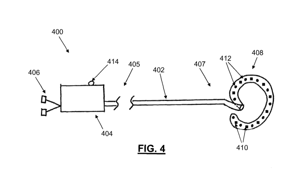
[0014] FIG. 4 depicts one variation of an endocardial ablation device.
[0015] FIG. 5 depicts one variation of an epicardial ablation device.
[0016] FIGS. 6A-6F depict side and front views of different ablation arrays
that
may be used with various ablation devices, including the devices shown in
FIGS. 4 and 5. FIGS.
4
CA 02796267 2012-10-12
WO 2011/129893 PCT/US2011/000676
6G and 6H depict variations of ablation arrays comprising temperature sensors.
FIG. 61 depicts
a partial cutaway of one variation of a temperature sensor that may be
encapsulated in an
alignment magnet of an ablation array.
[0017] FIG. 7 depicts one variation of an occlusion device comprising an
expandable element.
[0018] FIG. 8A depicts a flowchart that represents one variation of a method
for
ablating cardiac tissue from both the endocardial surface and epicardial
surface. FIG. 8B depicts
a flowchart that represents another variation of a method for ablating cardiac
tissue from both
the endocardial and epicardial surface.
[0019] FIGS. 9A-9D depict ablation patterns that may be formed by endocardial
and epicardial ablation of atrial wall tissue. FIGS. 9E-9G depict variations
of epicardial and
endocardial ablation arrays that comprise temperature sensor in various
configurations.
[0020] FIGS. 1OA-10S depict one example of an ablation method for ablating
tissue around the pulmonary veins, and for closing, and/or occluding, and/or
removing the left
atrial appendage. FIG. I OA schematically illustrates potential access sites
to the pericardial
space. FIGS. lOB-1OG schematically illustrate the use of a closure device to
locate and secure
the left atrial appendage. FIGS. 10H-10L schematically illustrate the
positioning of endocardial
and epicardial ablation devices. FIGS. I OM-ION depict the alignment of
endocardial and
epicardial arrays using magnetic components. FIGS. 100-10Q depict examples of
ablation
profiles that may form lesions that electrically isolate the tissue at or
around or within the
pulmonary veins. FIGS. IOR-IOS schematically illustrate the use of an
occlusion device to
occlude and isolate the left atrial appendage.
[0021] FIGS. 11A-11C depict one variation of a clip that may be used to secure
the
base or ostium of an atrial appendage.
[0022] FIGS. 12A and 12B depict various mechanisms by which an atrial
appendage may be occluded.
CA 02796267 2012-10-12
WO 2011/129893 PCT/US2011/000676
[0023] FIG. 13A depicts a flowchart that represents one variation of a method
for
ablating atrial wall tissue from an endocardial surface. FIG. 13B depicts a
flowchart that
represents another variation of a method for ablating tissue from an
endocardial surface.
[0024] FIGS. 14A and 14B depict ablation patterns that may be formed by
endocardial ablation of atrial wall tissue.
[0025] FIG. 15 depicts a flowchart that represents one variation of a method
for
ablating atrial wall tissue from an epicardial surface, comprising a procedure
to close, and/or
occlude, and/or remove the left atrial appendage.
[0026] FIGS. 16A and 16B depict ablation patterns that may be formed by
epicardial ablation of atrial wall tissue.
[0027] FIG. 17A depicts one variation of an access device. FIG. 17B depicts a
cross-sectional view of the access device of FIG. 17A taken along the lines
17B-17B. FIG. 17C
depicts one variation of a curved region of the device of FIG. 17A with a
plurality of slots. FIG.
17D depicts one example of how the access device of FIG. 17A may be used to
provide an
access path to the heart.
[0028] FIGS. 18A and 18B depict one variation of an occlusion device that may
be
used to position a closure element around a left atrial appendage.
[0029] FIGS. 19A-19F another example of devices and methods that may be used
to place a device at or around a tissue structure.
DETAILED DESCRIPTION
[0030] The system and methods described herein may be used to affect any
portion
of tissue within a body to form a lesion, and/or otherwise electrically
isolate a portion of tissue.
For illustrative purposes, these devices and methods are described in the
context of lesion
formation in the tissue of the left atrium for the treatment of atrial
fibrillation, and may include
the closure of the left atrial appendage. For example, methods for affecting
tissue to treat atrial
fibrillation may comprise accessing the pericardial space of the heart,
creating an access site
through the left atrial appendage (LAA), advancing a tissue-affecting device
intravascularly
and/or through the LAA to contact an endocardial surface of the left atrium,
advancing another
6
CA 02796267 2012-10-12
WO 2011/129893 PCT/US2011/000676
tissue-affecting device via the pericardial space to contact an epicardial
surface of the heart, and
affecting tissue from either or both the endocardial and epicardial surfaces.
In some variations, a
LAA access/exclusion device may be used to stabilize the LAA for the
advancement of devices
therethrough, as well maintain hemostasis by closing and/or opening the LAA
during and/or at
the conclusion of the procedure. While the systems and methods disclosed here
are described in
the context of affecting cardiac tissue, it should be understood that these
devices and methods
may be used to affect a variety of tissues, such as the skin, heart, liver,
etc., as well as to treat a
variety of conditions, including various cardiac deficiencies, tumors,
gastrointestinal
deficiencies, etc.
1. ANATOMY
[00311 FIG. 1 depicts a heart (100), with the cavity of the left atrium
partially cut
away to reveal a portion of the mitral valve (102) and pulmonary veins (104a),
(104b), (104c),
(104d). Both the right atrial appendage (106) and the left atrial appendage
(108) are shown,
located on the superior portion of the heart (100). The heart (100) is
enclosed by a pericardium
(not shown), which is filled with a fluid that may separate it from the heart.
The fluid-filled
space between the pericardium and the heart is the pericardial space. In a
heart affected by atrial
fibrillation, tissues associated with one or more of these anatomical
structures may pulsate
irregularly or asynchronously, and may cause the atrium to contract quickly
and/or irregularly.
Procedures for the treatment of atrial fibrillation may comprise the
electrical isolation of
arrhythmic cardiac tissue from other tissue regions. In some variations,
devices and methods for
treating atrial fibrillation may be directed towards the formation of lesions
in the right atrium
(e.g. in the proximity of the tricuspid valve annulus, the anterior limbus of
the fossa ovalis,
and/or the right atrial appendage), and/or lesions in the left atrium (e.g. in
proximity of the
pulmonary veins and/or LAA). For example, one variation of a method for
treating atrial
fibrillation in the left atrium may comprise the electrical isolation of
tissue(s) at or around or
within each of the pulmonary veins, and may optionally include the closure,
occlusion, and/or
removal of the left atrial appendage. While the devices and methods described
below may be
used to access, affect, and electrically isolate tissue in the left atrium,
similar devices and
methods may be used to treat any suitable portion(s) of the heart, e.g., the
right atrium, right
ventricle, left ventricle, etc.
7
CA 02796267 2012-10-12
WO 2011/129893 PCT/US2011/000676
II. DEVICES
Pericardial Access Device
[0032] In order to access certain portions of the heart, it may be useful to
place one
or guide elements in the pericardial space around the heart. Various devices
may be used to
provide access to the pericardial space for the placement of a guide element
into the pericardial
space for the advancement of subsequent devices to the heart. Some pericardial
access devices
may be configured to provide an access pathway from an initial access site
(e.g., a sub-thoracic
region, an intercostal region, etc.). For example, a pericardial access device
may comprise a
sheath with one or more curves, and one or more needles, guide elements,
tissue-piercing
elements, etc. to create a pathway through the pericardium to access the
pericardial.space. In
some variations, the one or more needles, guide elements, tissue-piercing
elements, etc. may be
sized and shaped to correspond with the one or more curves in the sheath. One
example of a
sheath with one or more curves is shown in FIGS. 17A and 17B. As shown there,
the sheath
(1702) may have a curved region (1706) between the proximal portion (1704) to
the distal
portion (1708). The proximal portion (1704) may be connected to a proximal
sheath actuator. A
sheath actuator may be used to advance the sheath, e.g., along a longitudinal
axis, to navigate the
distal portion of the sheath, and/or may be configured to cause the curved
region (1706) to bend.
A cross-section of the sheath (1702) is depicted in FIG. 17B. The sheath
(1702) may have one
or more longitudinal lumens therethrough, for example, a wire lumen (1710) and
an access
device lumen (1712). Other variations of a curved or bendable sheath may have
any desired
number of lumens therethrough, e.g. 2, 3, 5, 8, etc. The wire lumen (1710) may
be sized and
shaped for passing a wire therethrough. The access lumen (1712) may be sized
and shaped to
pass a pericardial access device therethrough, for example, any of the access
devices described
above. In some variations, sheaths may have additional lumens for inserting
other devices
therethrough, and/or as necessary for accommodating mechanisms that may be
used to control
the flexion of the curved region (1706).
[0033] The curved region (1706) may have one or more pre-shaped curves, and/or
may be flexible or bendable using a suitable actuating mechanism controlled by
the sheath
actuator at the proximal portion (1704). The curved region (1706) may serve to
generally orient
the sheath toward the heart upon insertion at an initial access point beneath
the sternum, and/or
may have a particular radius of curvature to help guide the sheath under the
rib cage to the heart.
8
CA 02796267 2012-10-12
WO 2011/129893 PCT/US2011/000676
In some variations, the curvature of the curved region (1706) may be locked or
fixed, e.g., the
curved region (1706) is first actuated to attain a desired degree of
curvature, then locked to retain
that desired curvature. Suitable locking mechanisms may include, for example,
maintaining the
tension of a wire that may be inserted through the wire lumen (1710), or
immobilizing the hinge
mechanisms to a desired configuration. A flexible or soft curved region may be
locked into
position by fixing the configuration (e.g., curvature, tension, etc.) of the
wire within the wire
lumen (1710). Some variations of a sheath may have a pre-shaped curve, where
the radius of
curvature is determined at the time of manufacture, and remains unchanged as
the sheath is used.
The radius of curvature of the curved region may be adjusted for sheaths that
are inserted at
different initial access points. For example, the radius of curvature of a
curved region of a
sheath to be inserted at an intercostal access site may be different from the
radius of curvature of
a curved region of a sheath to be inserted at a sub-thoracic access site.
[0034] The curved region (1706) may be made of a flexible or bendable
material,
or may be made of a substantially rigid material arranged in articulating
segments that allow for
the curved region (1706) to bend when actuated. The curved region (1706) may
be integrally
formed with the body of the sheath (1702), or may be separately formed and
attached to the
sheath (1702). For example, the curved region (1706) may be made of polymeric
tubing and/or
materials such as Pebax , nylon, fluoropolymers (e.g., PTFE, FEP),
polyethelene, Teflon ,
polyethylene terephthalate (PET), Tecothane , etc. In some variations, the
curved region (806)
may be made of a polymeric tube with reinforced stainless steel or nitinol.
Where the curved
region (1706) is made of a substantially rigid material, for example,
stainless steel, nickel
titanium, nitinol, cobalt alloys (e.g. nickel-cobalt, cobalt-nickel-chromium-
molybdenum), and/or
polymers such as PEEK, polyethylene (HDPE), polyimide, etc., the curved region
may be
slotted or segmented to allow bending to occur. In some variations, a curved
region (1707) may
have one or more slots (1705), as illustrated in FIG. 17C. In other
variations, the curved region
(1706) may comprise a plurality of segments, where the positioning of the
segments with respect
to each other is controlled by a wire or pivot mandrel. The segments may be
coupled together
via mechanical hinges and/or living hinges. Sheaths may also comprise multiple
curved regions,
where each of the curved regions may have the same or different radii of
curvature. For
example, one curved region may be made of a material with a selected
flexibility, while another
curved region may be made of a material with a different flexibility. Other
curved regions may
be slotted or segmented, as appropriate. Different curved regions may be
separated by a straight
9
CA 02796267 2012-10-12
WO 2011/129893 PCT/US2011/000676
portion of the sheath, or may be contiguous. A plurality of curved regions may
help to provide
additional maneuverability to navigate the distal portion of the sheath to the
targeted region of
the heart. Adjusting the tension on a wire through the wire lumen (1710) may
alter the curvature
of the curved region (1706). For example, increasing the wire tension may
cause bending of the
curved region (1706), while decreasing the wire tension may cause
straightening of the curved
region (1706).
[0035] FIG. 17D depicts one variation of a method of using the sheath (1702).
The
sheath (1702) may be inserted into the subject (1730) at a location beneath
the sternum (1722).
Prior to insertion, the sheath may be substantially straight, or may be
curved, as appropriate.
Once the sheath (1702) has been inserted, the curved region (1706) may be
adjusted in order to
bring the distal portion (1708) close to the surface of the heart (1720). For
example, the distal
portion (1708) may be navigated underneath the ribs (1728) towards the heart
(1720). Once the
distal portion (1708) of the sheath (1702) is in a desired location, e.g., an
anterior and/or slightly
lateral side of the heart, the curved region (1706) may be locked to retain
the curvature of the
curved region. The location of the distal portion (1708) may be monitored
using any suitable
imaging modality, for example, ultrasound, fluoroscopy, and the like. In some
methods, the
location of the distal portion (1708) may be monitored by tactile feedback.
[0036] An articulating sheath such as is shown and described above may be
useful
for accessing the heart (1720) where the abdomen (1724) of the subject (1730)
may limit the
angle at which the sheath (1702) may be positioned. Certain subject anatomy,
such as a smaller
abdomen (1724) may provide a large range of maneuverability for the sheath
(1702), while a
larger abdomen (1724) may limit the range of maneuverability for the sheath.
Providing one or
more curved regions may allow the heart to be more readily accessed where
subject anatomy
limits the range in which the sheath may be positioned. For example, providing
one or more
curved regions may help to reduce the force that may be required to position
the sheath (1702),
and may provide additional access paths to the heart in the event the
originally planned pathway
becomes unavailable.
Closure Device
[0037] Some methods for treating atrial fibrillation may comprise accessing an
endocardial surface of the left atrium through the LAA via the pericardial
space. Methods that
CA 02796267 2012-10-12
WO 2011/129893 PCT/US2011/000676
utilize the LAA as an entry port may also comprise closing and/or opening the
LAA during the
procedure (e.g., to advance devices therethrough) to maintain hemostasis.
Optionally, methods
may also comprise excluding the LAA at the conclusion of the procedure. Such a
device may be
used during the procedure to stabilize the LAA so that tissue-affecting
devices may be advanced
through the LAA into the left atrium, and may be used to at the conclusion of
the procedure to
permanently close off or otherwise occlude the LAA. One example of a device
that may be
capable of locating, securing, manipulating, stabilizing, closing and/or
excluding the LAA is
depicted in FIG. 2. Closure device (200) may comprise an elongate body (202),
a handle
portion (204) located at a proximal portion of the elongate body (202), an
extension (205)
located at a distal portion of the elongate body (202), and a distal looped
closure assembly (206)
distally coupled to the extension (205). While the closure device disclosed
below is described in
the context of locating, securing, manipulating, stabilizing and/or closing
the LAA, it should be
understood that the closure device may be used to act on any desirable tissue.
[00381 The elongate body (202) may have any appropriate shape, for example,
the
elongate body may be substantially straight (as depicted in FIG. 2), or may
have one or more
pre-formed curves and shapes. The elongate body (202) may have a suitable
cross-sectional
diameter and longitudinal length to facilitate navigating the closure device
(200) through the
vasculature to contact the LAA (or other target tissue). The elongate body
(202) may be made of
one or more flexible or rigid materials, as may be suitable for navigating
towards the target
tissue. In some variations, the elongate body may be steerable, and comprise
steering
mechanisms (such as mandrels, articulating and/or living hinges, cables, etc.)
that allow a user to
steer the elongate body using the handle portion (204). For example, the
elongate body may be
made of a single integral flexible material with one or more steering mandrels
embedded in the
side wall of the elongate body, such that bending the mandrel(s) would cause a
corresponding
deflection of the elongate body, which may help steer the elongate body
towards the target
tissue. Alternatively or additionally, the elongate body may be made of a
plurality of segments
that may be connected by articulating and/or living hinges. Each of the
plurality of segments
may be rigid or flexible. One or more mandrels may be coupled to each of the
plurality of
segments, and may be used to bend and steer the elongate body towards the
target tissue. The
elongate body may also comprise locking mechanisms so that after the elongate
body is steered
to a target location, it may be locked to retain a certain configuration to
maintain contact with
that target location. The elongate body (202) may comprise one or more working
channels (208)
11
CA 02796267 2012-10-12
WO 2011/129893 PCT/US2011/000676
to a target location, it may be locked to retain a certain configuration to
maintain contact with
that target location. The elongate body (202) may comprise one or more working
channels (208)
that extend from the proximal portion of the elongate body (202) to the distal
portion of the
elongate body. A variety of devices may be inserted through the working
channel in order to
manipulate a portion of tissue. Alternatively or additionally, the closure
device (200) may be
advanced over a guide element using the working channel (208).
[0039] As depicted in FIG. 2, the distal extension (205) may extend distally
beyond
the distal end of the elongate body (202). This may allow for additional
working space as may
be suitable for accessing the LAA. For example, the distal extension (205) may
extend distally
beyond the distal-most portion of the working channel (208) of the elongate
body. The length of
the distal extension (205) may be selected such that when the base of the LAA
is engaged by the
distal looped closure assembly (206), the tip of the LAA may be close to
(e.g., in contact with)
the distal-most portion of the working channel (208). This may allow devices
that are advanced
through the working channel (208) to directly contact and/or manipulate the
tip of the LAA once
it exits the working channel.
[0040] The distal extension (205) may be integrally formed with elongate body
(202), or separately formed and attached to the elongate body (e.g., by
welding, melding,
brazing, adhesives, etc.). The distal extension (205) may be made of rigid
and/or flexible
materials, and may be made of the same or different materials as the elongate
body. The
elongate body and/or distal extension may be made of polymeric materials such
as Pebax ,
polyethelyne, and/or other thermoplastic materials with various durometers or
densities, and/or
any polymers that may be tapered or graduated for varying degrees of
flexibility. Additionally
or alternatively, the elongate body and/or distal extension may be made of
metallic materials
such as nitinol, stainless steel, etc. The looped closure assembly (206),
distal extension (205),
elongate body (202), and/or portions thereof may comprise visualization
markers, such as
fluorescent markers, echogenic markers, and/or radiopaque markers, that permit
the closure
device to be visualized using a variety of imaging modalities. As with the
elongate body (202)
described above, the distal extension (205) may also be steerable. In some
variations, the distal
extension (205) may be steered independently from the elongate body, while in
other variations,
the distal extension (205) may be steered together with the elongate body. For
example, a
steering mandrel that may be used to steer the elongate body may also be
coupled to the distal
12
CA 02796267 2012-10-12
WO 2011/129893 PCT/US2011/000676
extension so that the extension may be steered in concert with the elongate
body. Alternatively,
there may be a first mandrel for the steering the elongate body and a second
mandrel for steering
the distal extension independently from the elongate body. Optionally, the
distal extension may
have one or more pre-shaped curves which may help to navigate the closure
device (200) to a
target tissue region.
[0041] In some variations, the distal extension (205) may comprise one or more
lumens that may extend from the distal-most end of the extension to the
proximal portion of the
closure device (e.g., to the handle portion). A lumen in the distal extension
may slidably retain a
portion of the looped closure assembly such that the dimensions of the loop
may be adjusted.
For example, the lumen of the distal extension (205) may slidably retain the
looped closure
assembly (206), which may comprise a distal loop (203). The distal loop (203)
may comprise a
snare loop and a suture loop that may be releasably coupled along the
circumference of the snare
loop. The distal loop (203) may be made of polymeric materials such as Pebax ,
and/or
metallic materials, such as nitinol, and/or any elastic, malleable,
deformable, flexible material.
The portion of the distal loop that extends outside of the extension, i.e.,
the external portion of
the distal loop, may be adjustable using an actuator at the proximal handle
portion. Adjusting
the length of the external portion of the distal loop (203) may help to snare
and/or close, or
release and/or open, a LAA or any anatomical protrusion. While the distal loop
(203) may have
the shape of a circle, it may also have other shapes, e.g., an ellipse, oval,
triangle, quadrilateral,
etc. In other variations, the looped closure assembly may be configured (e.g.
knotted, looped,
coiled, etc.) for other functions, such as locating and securing tissue. For
example, the looped
closure assembly may optionally comprise tissue graspers, hooks, or other such
tissue
engagement components that may help secure and retain a tissue portion.
[0042] The looped closure assembly (206) may have an expanded (e.g., open)
configuration, and a tightened (e.g., closed) configuration, where the
circumference of the loop
in the tightened configuration is smaller than in the expanded configuration.
For example, a
distal loop with an elliptical shape in the open configuration may have a
length along the minor
axis (e.g., the shortest dimension of the ellipse) from about 15 mm to about
50 mm, e.g., about
20 mm, and a length along the major axis (e.g., the longest dimension of the
ellipse) from about
15 mm to about 50 mm, e.g., about 40 mm. A distal loop in the closed
configuration may have a
diameter equivalent to about 5 mm to about 10 mm, e.g., 6 mm. The looped
closure assembly
13
CA 02796267 2012-10-12
WO 2011/129893 PCT/US2011/000676
(206) may be tightened or cinched to encircle and secure the LAA, and in some
variations, may
be able to close the LAA after it has been secured, if desirable. Optionally,
the looped closure
assembly (206) may be releasably coupled to the closure device such that after
the LAA is
encircled and secured by the distal loop (203), a knot or locking element may
be deployed to
retain the tension on the distal loop, which may then be released from the
closure device. For
example, a looped closure assembly may have a releasable suture loop that is
tightened over the
LAA and then released from the closure device. The tension on the suture loop
may be locked
so that the looped closure assembly may be proximally withdrawn from the
suture loop.
Optional closure elements, such as sutures, graspers, clips, staples, and the
like, may be included
with the looped closure assembly to help close the LAA. For example,
additional closure
features, e.g., graspers or staples, may be included at the tip of distal
extension (205) that may
act to secure the LAA. A looped closure assembly may also comprise one or more
energy
sources distributed along the length of the distal loop, where the energy
sources may be used to
ablate tissue or induce tissue fusion. Alternatively or additionally, the
looped closure assembly
(206) may be actuated in conjunction with other devices advanced through the
working channel
(208) to secure and position the closure device with respect to the LAA.
[00431 Various types of devices may be inserted through the working channel
(208) of the elongate body (202) as may be desirable. In some variations, a
vacuum device may
be inserted through working channel (208), while in other variations,
alignment devices, guide
elements, grasper devices, visualization devices, ablation devices, and/or
cutting devices may be
inserted through the working channel. Variations of the closure device may
have a multi-lumen
elongate body, where each lumen may be a working channel for one or more
different devices.
For example, the elongate body (202) may have multiple working channels for
the insertion of
different devices. Additionally or alternatively, the elongate body (202) may
comprise working
channels for the injection of liquid or gas fluids, as well as the application
of therapeutic and/or
chemical agents. The working channel (208) may have any cross-sectional shape
as may be
suitable for the devices to be inserted therethrough, for example, circular,
rectangular, etc.
[00441 The closure device (200) may comprise mechanisms to control the bending
and/or steering of the elongate body, as well as adjust the length of the
distal loop that extends
outside of the distal extension. For example, these functions may be
controlled by levers and/or
knobs at the handle portion (204). The handle portion (204) may comprise a
housing (214), a
14
CA 02796267 2012-10-12
WO 2011/129893 PCT/US2011/000676
loop actuator (212), and a working channel actuator (210). The housing (214)
may enclose at
least a portion of the actuators that control the use of the elongate body,
the looped closure
assembly, and the device in the working channel of the elongate body. For
example, loop
actuator (212) may regulate the tension on the distal loop of the looped
closure assembly, and
control the circumference of the external portion of the distal loop, e.g.
decrease it to encircle
and/or close the LAA, and increase it to release the LAA. In some variations,
the loop actuator
(212) may be a slider configured to adjust the circumference of the distal
loop (203). In
variations where the distal loop comprises a releasable suture loop, the loop
actuator may also
comprise a fob that initially couples the suture loop with the closure device
and may be pulled to
release the suture loop from the closure device. The working channel actuator
(210) may
comprise one or more buttons, sliders, levers, knobs, and the like that are
configured regulate the
operation of the device(s) in the working channel(s) of elongate body (202).
For instance, the
working channel actuator (210) may be a grasper actuator, and/or vacuum
actuator. Optionally,
handle portion (204) may also comprise one or more buttons, sliders, levers,
and/or knobs that
may be used to navigate the LAA access device through the vasculature to
access the LAA, for
example, by rotating, pulling, pushing, bending, or otherwise manipulating
steering mandrels.
Other features of a closure device and methods of use are described in U.S.
Pat. App. Serial No.
12/055,213 (published as U.S. Pub. No. 2008/0243183 Al), which is hereby
incorporated by
reference in its entirety. Another example of a closure device and methods of
use are described
in U.S. Pat. App. Serial No. 12/752,873, entitled "Tissue Ligation Devices and
Controls
Therefor," filed April 1, 2010, which is hereby incorporated by reference in
its entirety.
LAA Access Device
[0045] As described above, a variety of devices with different functions may
be
inserted through the working channel(s) of the elongate body of a closure
device to secure and/or
otherwise manipulate a portion of tissue. In procedures where access to an
internal tissue
structure may be desired (e.g. accessing a lumen of a hollow organ or vessel),
an access device
may be inserted through the working channel of the closure device after the
closure device has
been advanced at or near the target tissue (e.g., by advancing the closure
device over a guide
element). Access devices may create a way for the internal portion of a tissue
to be accessed
from outside the tissue. In some variations, access devices may create an
incision, puncture,
and/or opening, etc., which may be dilated to allow access to devices larger
than the initial
CA 02796267 2012-10-12
WO 2011/129893 PCT/US2011/000676
opening. Optionally, some variations of an access device may also comprise a
guide wire that
may be advanced into the created access site. One example of such a device is
shown in FIGS.
3A and 3B. FIG. 3A shows individual components of one variation of an access
device that may
be used to access a LAA or other tissue, and FIG. 3B shows the access device
of FIG. 3A fully
assembled. In this variation, LAA access device (300) comprises an access wire
(302), a
piercing wire (304), and an actuator portion (306). The access wire (302) may
comprise a lumen
(303) therethrough, where the lumen (303) may be sized and shaped for the
passage of a piercing
element therethrough, e.g. the piercing wire (304). The access wire (302) may
be made of a
metal alloy or one or more polymers that have mechanical properties suitable
for threading the
LAA access device (300) in the working lumen of a LAA stabilizing device and
for guiding the
piercing element. The access wire (302) may be made of a variety of materials,
including but
not limited to nitinol, stainless steel, as well as polymeric materials such
as polyethylene,
polypropylene and the like. The piercing wire (304) may be threaded through
the lumen (303)
of the access wire (302), and may comprise a piercing tip (308) at the distal
portion. The
piercing tip (308) may be used to create a puncture through the LAA.
Optionally, the piercing
wire (304) may comprise a lumen therethrough for the insertion of other
devices, such as a
catheter, guide wire, suture, and/or may be used for the infusion of fluids
(e.g. gas or liquid
fluids). In some variations, the piercing tip (308) may be a needle that is
attached to the distal
portion of the piercing wire (304). The piercing tip (308) may be separately
or integrally formed
with the piercing wire (304), and may have a lumen therethrough. The proximal
portion of
piercing wire (304) may be coupled with the actuator portion (306). The
actuator portion (306)
may be used to advance and/or withdraw and/or steer and/or rotate the piercing
wire (304), and
may also be used to maneuver the access wire (302) to access the LAA or other
target tissue.
The actuator portion (306) may be manual or mechanized, and may contain
ergonomic features
as appropriate, as well as electrical/mechanical interfaces to receive and
execute instructions
from a computing device or microcontroller. For example, the actuator portion
(306) may be
made of a metal alloy and/or one or more polymers that may be shaped to have
an ergonomic
geometry. The actuator portion (306) may be made of any materials that possess
sufficient
rigidity, flexibility, durability, etc., to engage and control the mechanisms
driving the use of the
closure device (200).
16
CA 02796267 2012-10-12
WO 2011/129893 PCT/US2011/000676
Corresponding Ablation Devices
[00461 Another example of a device that may be advanced through the working
channel(s) of the elongate body of a closure device is a tissue-affecting
device. Devices that
affect tissue may generally comprise one or more tissue-affecting elements,
arranged in various
patterns. In some variations, two or more tissue-affecting devices may be
positioned along a
target tissue, and used to affect the tissue in a desired pattern, where the
tissue-affecting
elements may be operated simultaneously or sequentially. In some variations,
the two or more
tissue-affecting devices may be placed across each other on opposite sides of
tissue such that the
tissue between them is affected. One example of a tissue-affecting device is
an ablation device.
Ablation devices may be provided for procedures that aim to ablate a portion
of tissue that is
abnormal, for example, cancerous tissue, or arrhythmic cardiac tissue. While
affecting tissue by
ablation is described in detail here, tissue may be affected in other ways,
including by excision,
occlusion, manipulation and the like. As described below, an ablation device
may be used to
ablate fibrillating atrial tissue, which may help to prevent the conduction of
the irregular or
asynchronous pulses in one tissue region to another tissue region.
[00471 In some variations, ablation devices may be used to create a lesion in
the
fibrillating atrial tissue. For the treatment of atrial fibrillation, one or
more tissue-affecting
devices, such as ablation devices, may be positioned on the endocardial
surface and/or the
epicardial surface of the left atrium. One example of an endocardial ablation
device that may be
inserted through a closure device is shown in FIG. 4. Endocardial ablation
device (400) may
comprise an elongate body (402), a handle portion (404), one or more ablation
source(s) (406),
and an ablation array (408). The elongate body (402) may be sized and shaped
to be inserted
through a working channel of a closure device, or any suitable guide cannula
or sheath. The
elongate body (402) may comprise one or more lumens therethrough, where the
lumens may be
configured to pass devices or fluids from the proximal handle portion (404) to
the ablation array
(408) at the elongate body distal portion. The elongate body may have any
number of pre-
formed curves for ease of navigation to the target tissue, and may optionally
be flexible and/or
steerable. While the elongate body (402) may be one continuous segment, other
variations of an
elongate body may be made of multiple articulating segments, and may be made
of one or more
flexible and/or rigid materials. In some variations, the elongate body may be
steerable via a
mechanism in handle portion (404), and as previously described for the closure
device. In
17
CA 02796267 2012-10-12
WO 2011/129893 PCT/US2011/000676
variations where the elongate body is passed through a portion of a closure
device, the curvature
and steerability of the elongate body (402) may correspond to the curvature
and steerability of
the closure device. This may help to inform a practitioner of the orientation
of the endocardial
ablation device with respect to the orientation of the closure device.
[0048] As shown in FIG. 4, the ablation array (408) may be located at the
distal
portion of elongate body (402). An ablation array may comprise one or more
tissue-affecting
elements that may be used for ablating and/or otherwise forming a lesion in
tissue. For example,
the ablation array (408) may comprise magnets (410) and ablation elements
(412) that may be
arranged, for example, along pre-formed curves or loops of the ablation array
(408). The
elongate body may also comprise magnets. The magnets may be of any suitable
type. For
example, the magnets may be rare-earth, electro-activated, or a multi-alloy
(e.g. iron, boron,
neodymium) magnets. The magnets may also have any suitable size or shape. More
generally,
the distal portion of an elongate body may have any open-shape or closed-shape
geometry, and
the magnetic and/or ablation elements may be arranged along the elongate body,
ablation array,
and/or on a structure at least partially enclosed within the perimeter of a
shaped distal portion of
the elongate body. In some variations, the ablation elements may themselves be
magnetic.
There may be any number of magnets (410) having any suitable configuration(s)
or pattern(s),
placed at any suitable location on the ablation array. For example, the
magnets (410) may be
arranged in a straight or curved line along the curvature of the ablation
array (408), as shown in
FIG. 4. Magnets may also be arranged along a length and a width of ablation
array. There may
be any number of ablation elements (412) as may be suitable to help ensure
that sufficient
ablation coverage of the target area is provided. For example, there may be 1,
2, 3, 5, 10, 12, 20,
etc. ablation elements. In general, ablation elements may be utilize any
mechanism and be in
any form that conveys the ablation energy/medium to the target tissue. For
example, cryo-
ablation elements may comprise conduits that may circulate a cryogenic
substance in conductive
proximity to the target tissue. Ablation elements may be electrodes that
ablate tissue via
radiation or heat energy. Alternatively or additionally, ablation elements may
be outlets or ports
that infuse substances that cause necrosis or apoptosis. For example, ablation
elements may
ablate tissue using one or more methods, such as cryo-ablation, heat ablation,
high intensity
focused ultrasound (HIFU) ablation, radiofrequency (RF) ablation, laser
ablation, or
combinations of the listed methods and/or any method that causes necrotic or
apoptotic cell
and/or tissue death. Some ablation arrays may comprise two or more different
types of ablation
18
CA 02796267 2012-10-12
WO 2011/129893 PCT/US2011/000676
elements, e.g., 2, 3, or 4 types of ablation elements. In some variations, an
ablation array may
comprise both RF and cryo-ablation elements. In some variations, an ablation
array may
comprise RF electrodes and HIFU electrodes. In still other variations, an
ablation array may
comprise laser emitters and RF electrodes. In some variations, an ablation
array may comprise
HIFU electrodes, RF electrodes, and cryo-ablation elements. The different
types of ablation
elements on an ablation array may be activated simultaneously and/or
sequentially in the course
of ablating tissue. Alternatively or additionally, ablation elements may be
sharp elements, e.g.
needles, that excise, cut, or pierce tissue, or any combination of the above.
For example, an
endocardial ablation device may comprise electrode ablation elements and
needle ablation
elements.
[0049] The shape of the ablation array (408) as shown in FIG. 4 is semi-
circular,
which may be suitable for circumscribing and ablating around a vascular
structure, such as a
pulmonary vein, however, other variations of ablation arrays may have other
shapes. For
example, an ablation array may have a planar structure with a length and a
width, with ablation
elements arranged along both the length and the width. An ablation array may
also be a one-
dimensional array, e.g., a linear structure, where the ablation elements are
arranged linearly
therealong. Indeed, ablation arrays may be any shape suitable for accessing
and contacting the
target tissue. For example, the semi-circular shape of ablation array (408)
may be suited for
circumscribing vascular structures, such as veins or arteries, and may be
configured to create
circular ablation patterns. Ablation arrays may also have a tapered region
that may be helpful in
accessing and contacting in the lumen of tubular structures, e.g., the inner
lumen of a vein. In
some variations, an ablation array may have a narrow undeployed configuration
and an
expanded deployed configuration. For example, an ablation array may be
constrained in a
sheath for delivery, and may expand into the deployed configuration by
removing the sheath. In
another variation, a curved ablation array may be retained in a straight
configuration by a
straightening mandrel for delivery, and may be expanded into the curved
deployed configuration
by removing the mandrel. Other variations will be described in detail below.
[0050] The ablation array (408) may be made from a flexible or shape-memory
material, such that it may be advanced to the target tissue in a substantially
straight
configuration, and may be deployed and contacted to tissue in a curved
configuration. In some
variations, the ablation array is made of a different material from the
remainder of the ablation
19
CA 02796267 2012-10-12
WO 2011/129893 PCT/US2011/000676
device (400), and may have different mechanical properties. For example, the
proximal portion
(405) of the elongate body may be made of a first material, while the distal
portion (407) and/or
the ablation array may be made of a second material. Examples of materials
that may be suitable
for the proximal portion (405) and/or the distal portion (407) of the elongate
body may include
metal alloys such as nickel titanium alloy, stainless steel, and/or any
polymers, such as
polyethylene, polyurethane, polypropylene, polytetrafluoroethylene, polyimide,
etc., and/or any
combinations thereof. In some variations, an ablation array may be integrally
formed with the
proximal portion of the ablation device, or may be attached via an
articulating hinge. The
ablation array may also be attached by other mechanical mechanisms, such as a
living hinge,
pivot joint, ball joint, etc, which may allow the ablation array to move with
respect to the
proximal portion of the ablation device (e.g., with two or more degrees of
freedom).
[00511 The handle portion (404) located at the proximal end of elongate body
may
comprise actuating elements that control the movement and/or action of the
elongate body and
ablation array. In some variations where endocardial ablation device (400) is
manually operated,
the handle (400) may be ergonomic, while in other variations where the device
is
mechanically/electrically operated, handle (400) may comprise an interface to
receive and
execute instructions from a computing device. The handle portion (404) may
comprise an
ablation array actuator (414), which may be used to regulate application of
ablation
energy/substances to the ablation array to the target tissue (e.g. frequency,
duty cycle,
magnitude/amplitude, etc.). Additionally, the handle portion (404) may
comprise an actuating
mechanism that controls the movement (e.g., bending, flexing, etc.) and
position of elongate
body (402). The handle portion (404) may also comprise an interface to the
ablation source(s)
(406), and provide a conduit or conduction pathway from the ablation source(s)
(406) to the
ablation array. For example, the ablation source (406) may comprise a
reservoir of cryogenic
substances (e.g., for cryo-ablation), which may be transported through a lumen
in the elongate
body (402) to the ablation array. Alternatively or additionally, the ablation
source (406) may
comprise a source of radioactive substances (e.g., radioactive seeds or
fluids), and/or a light
beam source (e.g., for laser ablation), and/or an ultrasound source (e.g., for
HIFU ablation),
and/or a radiofrequency source, and the like, which may be delivered or
transmitted from the
handle portion to the ablation array. In some variations, different ablation
sources may be used
together in the same ablation device.
CA 02796267 2012-10-12
WO 2011/129893 PCT/US2011/000676
[00521 Depending on the tissue to be ablated and the desired ablation pattern
(e.g.
lesion geometry and size) desired, a second ablation device may be provided,
where the second
ablation device corresponds to the first ablation device. A second ablation
device may increase
the tissue ablation area and/or may otherwise alter the ablation
characteristics of the first ablation
device (e.g. by constructive or destructive interference). For the purposes of
ablating tissue of a
left atrium, a second ablation device may be provided to help ensure that the
lesion formed by
ablating tissue spans at least portion of tissue that is between them. In the
treatment of atrial
fibrillation it may be desirable to electrically isolate the fibrillating
tissue from other tissues. In
some variations, the formation of a lesion that spans the entire thickness of
the atrial wall (e.g.,
from the endocardial surface to the epicardial surface) using one or more
ablation devices may
improve the electrical isolation of a portion of the atrial wall from other
portions of the heart.
Accordingly, in some variations, ablation devices may be placed on opposite
sides of a tissue
wall such that a lesion that spans a substantial portion of the tissue wall
between the ablation
devices may be formed. In some variations, positioning a first ablation device
on an interior
wall (endocardial surface) of the left atrium, and positioning a second
ablation device on an
exterior wall (epicardial surface) of the left atrium opposite to the first
ablation device, may help
form a lesion that spans at least a portion of the tissue between the first
and second ablation
devices. FIG. 5 illustrates one variation of an epicardial ablation device
(500) that may be used
with an endocardial ablation device to form a lesion in the left atrium. The
epicardial ablation
device (500) may comprise an elongate body (502), handle portion (504),
ablation source (506),
and an ablation array (508). As shown in FIG. 5, the ablation array (508) may
be located at the
distal portion of elongate body (502). The ablation array (508) may comprise
magnets (510) and
ablation elements (512) which may correspond to the magnets (410) and ablation
elements (412)
of the endocardial ablation device (400). The magnets of the epicardial and
endocardial ablation
devices attract each other when the ablation arrays are placed on opposite
sides of tissue, which
may act to align the epicardial and endocardial ablation devices. For example,
the magnets
(510) may be positioned on the epicardial ablation array (508) such that they
may be aligned
with the magnets (410) of the endocardial ablation array (408), e.g. magnets
(510) may
correspond to, or be mirror images of magnets (410). As with the magnets, the
ablation
elements (512) may correspond to, or be mirror images of the ablation elements
(412), or they
may be interlaced between the ablation elements (412). In some variations, the
alignment and
attraction of the magnets may position the endocardial and epicardial ablation
devices such that
21
CA 02796267 2012-10-12
WO 2011/129893 PCT/US2011/000676
the ablation elements of the ablation devices are aligned across from each
other. The shape of
the ablation array (508) as shown in FIG. 5 is semi-circular, however, other
variations of
ablation arrays may have any shape as may be suitable for accessing and
contacting the target
tissue. In some variations, the shape of ablation array (508) may be a mirror
image, or
complementary image, of the endocardial ablation array (408). For example, the
semi-circular
shape of the ablation array (508) may be suited for circumscribing vascular
structures, such as
veins or arteries. Other variations will be described in detail below. The
ablation array (508)
may be made from a flexible or shape-memory material, such that it may be
advanced to the
target tissue in a substantially straight configuration, and may be deployed
and contacted to
tissue in a curved configuration. For example, ablation array may be advanced
to, and contacted
with, an external wall of a vascular structure, e.g. artery, vein, heart
chamber, and/or atrial
appendage. The ablation elements of the endocardial array and the epicardial
array may be in
communication with each other, so that they may apply ablation energy in a
concerted or
programmed fashion.
[0053] The handle portion (504) located at the proximal end of elongate body
may
comprise actuating elements that control the movement and/or action of the
elongate body and
ablation array. In some variations where the endocardial ablation device (500)
is manually
operated, the handle (500) may be ergonomic, while in other variations where
the device is
mechanically/electrically operated, the handle (500) may comprise an interface
to receive and
execute instructions from a computing device. The handle portion (504) may
comprise an
ablation array actuator (514), which may be used to regulate application of
ablation
energy/substances to the ablation array to the target tissue (e.g. frequency,
duty cycle,
magnitude/amplitude, etc.). In some variations, the handle portion (504) may
be in
communication with the handle portion (404) of the endocardial ablation device
(400), such that
ablation energy from both ablation devices may be applied in-phase or out-of-
phase to form a
desired ablation wavefront and/or profile. Additionally, the handle portion
(504) may comprise
an actuating mechanism that controls the movement and position of elongate
body (502). The
handle portion (504) may also comprise an interface to ablation source(s)
(506), and provide a
conduit or conduction pathway from the ablation source(s) (506) to the
ablation array. For
example, the ablation source (506) may comprise a reservoir of cryogenic
substances (e.g., for
cryo-ablation), which may be transported through a lumen in the elongate body
(502) to the
ablation array. Alternatively or additionally, the ablation source (506) may
comprise a source of
22
CA 02796267 2012-10-12
WO 2011/129893 PCT/US2011/000676
radioactive substances (e.g., radioactive seeds or fluids), and/or a light
beam source (e.g., for
laser ablation), and/or an ultrasound source (e.g., for HIFU' ablation),
and/or a radiofrequency
source, and the like, which may be delivered or transmitted from the handle
portion to the
ablation array. In some variations, different ablation sources may be used
together in the same
ablation device.
Variations ofAblation Arrays
[0054] While the ablation devices depicted and described in FIGS. 4 and 5 are
shown as having a semi-circular shape, ablation arrays may have other
geometries. Ablation
and/or other tissue-affecting arrays may have a variety of geometries and
sizes as appropriate to
accommodate and contact the target anatomical structure. For instance,
ablation arrays with
various geometries may be suitable for contacting and ablating tissue,
especially vascular or
cardiac tissue. Several variations of ablation arrays are shown in FIGS. 6A-
6F. A side view and
front view of a spiral ablation array (600) inserted in the opening of a
vascular structure (603)
(e.g. pulmonary vein) is shown in FIGS. 6A and 6B, respectively. As shown
there, the spiral
ablation array (600) may be coupled to the distal portion of an elongate body
(602), where
ablation elements (604) and magnetic elements (606) are arranged throughout
the curves of the
array (600) such that they may contact the walls of the vascular structure
(603). FIGS. 6C and
6D depict a side view and a front view of a woven ablation array (610),
respectively. The
ablation array (610) may be attached at the distal portion of an elongate body
(612), and may
comprise a woven portion (615) and a rim (617) located along a distal
perimeter edge of the
woven portion. Ablation elements (614) and/or magnetic elements (616) may be
arranged
throughout the array, for example, along the rim (617) and/or on various
locations on the woven
portion (615). The size and shape of the woven portion (615) may be configured
to position the
ablation elements (614) and the magnetic elements (616) in order to
accommodate the geometry
of the target tissue (613), e.g., where the expanded size and shape of the
woven portion may be
bent, shaped, molded or otherwise constrained by the geometry of the target
tissue. The woven
portion (615) may be used as an ablation conduit or array, and may be arranged
to be in
proximity to target ablation tissue. Alternatively or additionally, the woven
portion (615) may
help stabilize the array (610) during ablation without occluding the pulmonary
vein. The woven
portion (615) may be constructed from various fibers, where the density of the
weave may be
adjusted according to the degree of perfusion desired. The fibers of the woven
portion may be
23
CA 02796267 2012-10-12
WO 2011/129893 PCT/US2011/000676
made of polypropylene, polyurethane, polyethylene, polytetrafluoroethylene, as
well as metal
alloys such as stainless steel and/or nickel titanium alloy. The woven portion
may be self-
expanding or mechanically expanded to fill the lumen or orifice of the
vascular structure, and
may be adjusted according to the size of the vascular structure. In some
variations, the woven
portion may be made of a shape-memory material so that the woven array (610)
may have a
compressed delivery configuration and an expanded deployed configuration. The
size of the
woven portion may be adjusted to ablate anatomical structures with dimensions
of about 8.0 mm
to about 30 mm, or about 12.0 mm to about 18.0 mm. Another variation of an
ablation array
(620) is shown in FIGS. 6E and 6F. As shown there, a tapered spiral ablation
array (620) may
be coupled to the distal portion of an elongate body (622), where ablation
elements and/or
magnetic elements may be arranged throughout the tapered spiral ablation array
(620). The
tapered spiral ablation array (620) may comprise a single continuous flexible
backbone that is
wound around the elongate body (622). Ablation elements may be distributed
along the length
of the backbone. In some variations, the backbone of the spiral ablation array
(620) may be a
wire that is electrically conductive, and may itself be capable of ablating
tissue without
additional ablation elements. The spiral ablation array (620) may have a first
collapsed
configuration shown in FIG. 6E, where the ablation array may be closely wound
around the
distal portion of the elongate body (622), e.g., with a tight radius of
curvature. The narrow
profile of the array in the collapsed configuration may help navigate the
array atraumatically
through narrow anatomical structures, and may also be inserted into folded or
creased tissue
structures. The ablation array (620) may be retained in its collapsed delivery
configuration by a
sheath that may be slidably disposed over the array (not shown), and/or by
retaining tension on
the backbone. FIG. 6F depicts a second expanded configuration of the tapered
spiral ablation
array (620), where removing the sheath and/or reducing the tension on the
backbone of the spiral
ablation array (620) may allow the backbone to loosen the radius of curvature
such that the
profile of the array expands. In some variations, expanding the ablation array
may act to dilate a
narrow tissue structure, e.g., open a folded or creased tissue structure,
enlarge a tissue lumen or
aperture for the insertion of additional devices, etc. In some variations, the
ablation array (620)
may help to maintain perfusion during ablation, and may be an alignment
reference point for
epicardial elements at various locations along the pulmonary veins. The
ablation and magnetic
elements may be arranged in any of the previously described configurations,
and may be
arranged to help stabilize the ablation device during the ablation procedure.
24
CA 02796267 2012-10-12
WO 2011/129893 PCT/US2011/000676
[0055] Any of the ablation arrays described above may optionally comprise one
or more temperature sensors. Temperature sensors may be used to measure the
ablation energy
that has been applied to a tissue, and may be used to evaluate the degree to
which tissue is
ablated. The measurement of temperature changes in the tissue during the
application of
ablation energy may be used to regulate the duration, power, and/or frequency
of the ablation
energy (e.g., by providing feedback information to the ablation array and/or
ablation array
controllers). Monitoring the temperature of the tissue during ablation may
also help prevent
excessive or harmful damage to peripheral tissues. The one or more temperature
sensors may be
thermocouples, thermsistors, thermal resistive sensors (RTD), and the like.
One example of an
ablation array with temperature sensors is depicted in FIG. 6G. Ablation array
(630) may
comprise an ablation array substrate or housing (634), one or more ablation
elements (not
shown), one or more alignment magnets (638) and one or more atraumatic
temperature sensors
(636) on the tissue-facing surface of the ablation array. The alignment
magnets (638),
temperature sensors (636), and ablation elements may be arranged in any
suitable configuration
on the tissue-facing surface of the ablation array, for example, the alignment
magnets (638) may
be arranged such that the ablation elements of two ablation arrays positioned
on opposite sides
of a tissue may attract each other to align the ablation elements of one
ablation array to the other.
The atraumatic temperature sensors (636) may be pressed into the tissue (632)
without
puncturing or piercing it to measure the temperature of the tissue. Another
example of an
ablation array with temperature sensors is depicted in FIG. 6H. Ablation array
(640) may
comprise an ablation array substrate or housing (644), one or more ablation
elements (not
shown), one or more alignment magnets (648) and one or more sharp temperature
sensors (646)
on the tissue-facing surface of the ablation array. The alignment magnets
(648), temperature
sensors (646), and ablation elements may be arranged in any suitable
configuration on the tissue-
facing surface of the ablation array, as previously described. The sharp
temperature sensors
(646) may be inserted into tissue (642) to measure the temperature of the
tissue at a certain depth
from the surface of the tissue (642). In some variations, the sharp
temperature sensors (646)
may pierce or puncture the tissue (642) in order to gain access to deeper
tissue regions. The
sharp temperature sensors (646) may also have a length that corresponds to the
thickness of the
tissue, and in some cases, may penetrate through the entire length of the
tissue. Temperature
sensors that penetrate through substantially the entire thickness of the
tissue may provide
temperature data across the entire span of the tissue, which may provide an
indication of the
CA 02796267 2012-10-12
WO 2011/129893 PCT/US2011/000676
uniformity of the tissue ablation, as well as provide information about the
temperature gradient
across the tissue. This may help improve the accuracy of the tissue
temperature measurement
that is fed back to the ablation array and/or ablation array controllers.
[0056] In the variations depicted in FIGS. 6G and 6H, the alignment magnets
and
temperature sensors are located adjacent to each other, however, in other
variations, the
alignment magnets and temperature sensors may be incorporated together in one
location. This
may help to reduce the overall size of the ablation array, which may reduce
the profile of the
array for ease of delivery to the target tissue site. For example, an ablation
array may have
alignment magnets that have a lumen sized and shaped for encapsulating a
temperature sensor.
FIG. 61 depicts a regions of one example of an ablation array (650) comprising
a housing (654),
a temperature sensor (656) and an alignment magnet (658) encapsulating the
temperature sensor.
The alignment magnet (658) may comprise a lumen (657) that is sized and shaped
for the
temperature sensor (656). The temperature sensor (656) (which may be an
atraumatic or tissue-
piercing or sharp temperature sensor) may protrude from the lumen (657), or
may be flush with
the opening of the lumen (657). In other variations, an ablation array may
comprise one or more
ablation elements that each comprise a lumen such that a temperature sensor
may be
encapsulated in the lumen. In still other variations, an ablation array may
comprise ablation
elements, alignment magnets, and/or temperature sensors that may be retracted
into a housing of
the ablation array. For example, during delivery of the ablation array to the
target tissue site, the
ablation elements, alignment magnets, and/or temperature sensors may be in a
first retracted
configuration, such that the profile of the ablation array is narrow. After
the ablation array has
been generally positioned at the target tissue site, the ablation elements,
alignment magnets,
and/or temperature sensors may be a second protracted configuration, where the
ablation
elements, alignment magnets, and/or temperature sensors are capable of
contacting the target
tissue for alignment, ablation, and/or measurement of temperature.
Occlusion Device
[0057] As described previously, some methods may include steps to help
maintain
hemostasis in the course of the procedure for the treatment of atrial
fibrillation. For example, in
procedures where access to an endocardial surface of the heart is gained using
the LAA as a port,
it may be desirable to close and/or exclude the LAA to maintain hemostasis
and/or help prevent
thrombosis. In some variations, a procedure for the treatment of atrial
fibrillation may also
26
CA 02796267 2012-10-12
WO 2011/129893 PCT/US2011/000676
include the temporary or permanent closure, and/or occlusion, and/or removal
of the left atrial
appendage. FIG. 7 illustrates one variation of an occlusion device (700) that
may be used with
the devices and methods described here. The occlusion device (700) may
comprise an elongate
body (702), an insert port (704) at a proximal portion of the elongate body,
and an expandable
member (706) at a distal portion of the elongate body. The elongate body (702)
may have one or
more lumens, for example, a guide wire lumen (708) sized and shaped for
passing a guide wire
therethrough. The elongate body (702) as shown in FIG. 7 may also comprise one
or more side
apertures (710) and imaging markers (712). Any number of side apertures (710)
may be
provided for infusion of any fluid substance, such as a contrast agent or
pharmacological agent
(e.g., heparin, antibacterial agent, etc.). The imaging markers (712) may be
radiopaque or
echogenic, etc., as appropriate for the imaging modality used to monitor the
position of the
occlusion device (700).
[0058] The elongate body (702) may be made from one or more rigid and/or
flexible materials. In some variations, the elongate body (702) may be
steerable. An insert port
may comprise one or more apertures for the insertion of fluids or devices
through the elongate
body (702). For example, the insert port (704) may comprise a guide wire
aperture (714) and a
fluid lumen (716). The guide wire aperture (714) and the fluid lumen (716) may
each have
independent lumens that may merge into one lumen at a bifurcation (717) of the
insert port
(704), or may each have separate lumens in the elongate body (702). The guide
wire aperture
(714) may be continuous with the guide wire lumen (708), and the fluid lumen
(716) may be
continuous with a cavity of the expandable member (706), such that the
introduction of fluid into
or out of the fluid lumen (716) may expand or constrict the expandable member.
Optionally, the
insert port (704) may also comprise actuation mechanisms for navigating and
adjusting the shape
of the elongate body (702), as well as control the motion of a guide wire, and
the expansion of
the expandable member (706).
[0059] The expandable member (706) may be any structure that comprises a first
small profile and a second larger profile, for example, a balloon or an
articulating polygonal
structure, e.g. rectangular prism or tetrahedron, and the like. The expandable
member (706) may
be sized and shaped to fit within the guide wire lumen (208) of the closure
device (200) so that it
may be advanced and/or withdrawn through the lumen (208). In some variations,
the
expandable element (706) may have a first collapsed configuration, and a
second expanded
27
CA 02796267 2012-10-12
WO 2011/129893 PCT/US2011/000676
configuration. For example, the rounded expandable element (706) shown in FIG.
7 may have a
diameter of about 15 mm to about 30 mm, e.g., 20 mm. The expandable member may
be made
of various materials, including polymeric and/or metallic materials. Examples
of polymers that
may be used in an expandable member may include materials such as latex,
silicone,
polyisoprene, polyethelene. Example of metals and/or metallic alloys that may
be used with an
expandable member may include nitinol, stainless steel, titanium and the like.
The expandable
member may be configured to either self expand or be mechanically expanded by
an actuator.
For example, the expandable member (706) may be transitioned from the small
profile to the
larger profile by introducing positive pressure or by a mechanical actuation.
In some variations,
a balloon expandable member may be urged into the larger configuration by
applying positive
fluid (gas or liquid) pressure into the lumen of the balloon. The expandable
member may have
any shape and size as appropriate for the target tissue. For example, a round
expandable balloon
may be used to occlude a vascular structure, such as a vein or an atrial
appendage, e.g. LAA.
[0060] Another variation of a device that may be used for occluding the LAA is
depicted in FIGS. 18A and 18B. As shown there, an occlusion device (1820) may
comprise
grooves (1822) in its deployed configuration. In some variations, the closure
device may be
deployed and positioned at the anatomical ostium of a left atrial appendage
(1800). However,
the occlusion device (1820) may be positioned at any desired location in the
heart. The
occlusion device (1820) may have a collapsed delivery configuration, which may
enable it to be
enclosed within a catheter or sheath and advanced through the vasculature
(e.g., from a
retrograde approach, or an antegrade transseptal approach) or through a port
in the LAA. The
occlusion device (1820) may be deployed into its expanded configuration after
it is positioned at
or near the ostium of the LAA. In some variations, the occlusion device may be
a rounded plate
or disc comprising one or more grooves circumscribing the outer perimeter.
Grooves (1822)
may be configured to interfit with a closure element (1810) (e.g., suture loop
or snare loop) of a
closure device as the circumference of the closure element is reduced, as
shown in FIG. 18B.
The occlusion device (1820) may be sized according to the desired degree of
closure of the left
atrial appendage (1800). Once the closure element (1810) has been secured and
decoupled from
the rest of the closure device (e.g., by cutting or detaching at a breakaway
point), the occlusion
device (1820) may be reverted to its collapsed configuration and withdrawn
from the ostium of
the left atrial appendage (1800). The devices and methods described above for
closing and/or
excluding the left atrial appendage may be included at the conclusion of a
procedure that uses
28
CA 02796267 2012-10-12
WO 2011/129893 PCT/US2011/000676
the left atrial appendage as an access site. This may be a more expedient
method of closing a
heart access site than other conventional methods, such as suture stitching,
which may be
substantially more time-consuming.
[0061] The above-described devices may be used to secure, ablate, and excise a
portion of tissue to help alleviate the symptoms of atrial fibrillation. For
example, the devices
above may be used to secure a LAA, ablate atrial tissue in the proximity of
the LAA and the
pulmonary veins, and to close, and/or occlude, and/or remove the LAA. While
the description
below provides methods of securing, ablating, and excising tissue of the left
atrium and/or LAA,
it should be understood that the methods may be used to perform similar
procedures on the right
atrium, as well as other vascular structures or organs. Similar methods may
also be used to
secure, ablate, and excise tissues and/or organs that have one or more
cavities therethrough, e.g.
stomach, intestine, bladder, etc., for a variety of indications.
III. METHODS
[0062] Methods for ablating tissue for the treatment of atrial fibrillation
may
generally comprise accessing targeted cardiac tissue regions, advancing
ablation arrays to the
targeted tissue regions, ablating the tissue regions, and withdrawing the
ablation arrays once the
desired degree of tissue ablation has been attained. Additionally, some
methods may include the
closure of the left and/or right atrial appendages, which may help reduce the
risk of thrombosis
and may help maintain hemostasis. Some variations of methods for tissue
ablation may
comprise ablating the tissue from an endocardial surface, an epicardial
surface, or both.
Ablation devices may access an endocardial surface of the left atrium
intravascularly, and/or
through the LAA via the pericardial space. Once the one or more ablation
devices have been
placed on the endocardial and/or epicardial surface(s), the ablation devices
may be activated
sequentially and/or simultaneously to achieve the desired degree of tissue
ablation. Ablation
array activation sequences may be repeated as may be desirable, and may
comprise applying
ablation energy pulses (from either or both of the endocardial and epicardial
ablation arrays) of
varying duration, frequency, duty cycle, power, intensity, etc. The ablation
array(s) may be re-
positioned to ablate tissue at various desired locations. Once all the desired
tissue regions have
been ablated, the ablation arrays may be withdrawn. In variations where the
LAA is used to
access the endocardial surface on the left atrium, the LAA may be closed
and/or excluded.
29
CA 02796267 2012-10-12
WO 2011/129893 PCT/US2011/000676
Epicardial and Endocardial Ablation
[0063] One variation of a method that may be used to electrically isolate
tissue in
the left atrium and/or LAA is depicted as a flowchart in FIG. 8A. Method (800)
may be used to
ablate tissue from both epicardial and endocardial surfaces using surgical,
intravascular and/or
other minimally invasive techniques (e.g., percutaneous, small incisions or
ports), and may be
used in stopped heart or beating heart procedures. The method (800) may
comprise gaining
access to the pericardial space (802), for example, using the access devices
described above.
Optionally, a device may be used to locate and stabilize the LAA (804), for
example, the closure
device (200) as described above and shown in FIG. 2. Once access into the
pericardial space
and to the LAA has been established, a device may be used to enter the LAA
(806), for example,
by creating a puncture in the LAA. Additional devices and methods of using the
LAA as an
access port to deliver devices into the heart (e.g., to contact and/or affect
an endocardial surface
of the heart) are described in U.S. Provisional Patent Application No.
61/323,816 filed April 13,
2010, which was previously incorporated by reference in its entirety, and U.S.
Patent
Application No. entitled "Methods and Devices for Accessing and Delivering
Devices to
a Heart," filed April 13, 2011, which is hereby incorporated by reference in
its entirety. Various
tissue regions in the left atrium (e.g., atrial wall tissue, tissue at or
around the base of the
pulmonary veins, tissue within the pulmonary veins, etc.) may be accessed from
an endocardial
side (810). Devices may be introduced into the left atrium through the LAA,
for example, an
endocardial ablation array may be positioned and placed in the left atrium
(812). An epicardial
ablation array may be aligned with the endocardial ablation array (814), for
example, based on
the position of the corresponding magnets on the endocardial and epicardial
ablation arrays. The
epicardial ablation array may be introduced to the epicardial surface of the
heart using the same
initial access site as the endocardial ablation array, or may be introduced
through a different
access site. For example, the endocardial ablation array may be introduced
through a right
intercostal site, while the epicardial ablation array may be introduced
through a left intercostal
site. Alternatively, the endocardial and epicardial ablation arrays may both
be introduced
through a left intercostal site, for example. Additional description of access
sites are described
below. The endocardial and epicardial ablation arrays may be positioned in
order to obtain a
particular ablation pattern, after which both ablation arrays may be activated
(816). For
example, the endocardial ablation array may circumscribe the base of the
pulmonary veins,
while the epicardial ablation array may circumscribe the trunk of the
pulmonary veins. After the
CA 02796267 2012-10-12
WO 2011/129893 PCT/US2011/000676
desired tissue regions have been ablated, the ablation devices may be removed
(818), and the
LAA may be occluded, closed, and/or removed (820). Once the LAA has been
decoupled from
the remainder of the left atrium, all devices may be retracted from the
surgical site (822).
[00641 As described previously, the endocardial side of the left atrium may be
accessed intravascularly and/or from the LAA via the pericardial space. The
access path into the
left atrium may be selected based on the targeted anatomical features in the
left atrium such that
the path length of the catheter and/or ablation devices may be reduced. The
access path may
also be selected to reduce the maneuvering, manipulating, bending, torquing,
etc. that may be
required to position the catheter and/or devices at the targeted tissue site
in the left atrium. For
example, an endocardial ablation device may access the left atrium using
either an intravascular
retrograde approach or an antegrade transseptal approach. Entering the left
atrium via an
intravascular antegrade transseptal approach may allow access to the left
pulmonary veins while
reducing the maneuvering, manipulating, bending, torquing, etc. of the distal
portion of the
device. Entering the left atrium via an intravascular retrograde approach may
allow access to the
right and left pulmonary veins while reducing the maneuvering, manipulating,
bending,
torquing, etc. of the distal portion of the device. Alternatively or
additionally, entering the left
atrium through the LAA via a pericardial approach may allow access to the
right pulmonary
veins without much maneuvering, manipulating, bending, torquing, etc. of the
distal portion of
the device. Any of these approaches may be used to position an endocardial
ablation device in
the left atrium. In some variations, a first endocardial ablation array may
enter the left atrium
through an intravascular approach, and a second endocardial ablation array may
enter the left
atrium through the LAA via a pericardial approach.
[00651 One example of a method (830) that comprises accessing the endocardial
surface of the left atrium both intravascularly and through the LAA via the
pericardial space is
depicted in FIG. 8B. As previously described, an access pathway may be created
to the
pericardial space (832). A LAA access/exclusion device may be used to locate
and stabilize the
LAA (834). Once access into the pericardial space and to the LAA has been
established, a
device may be used to create a LAA access site (836), e.g., by puncturing the
LAA, which may
allow a device to access the left atrium through the LAA. An intravascular
pathway to the left
atrium may also be attained by advancing a delivery catheter through the
vasculature into the left
atrium (838), e.g., using a retrograde or an antegrade transseptal approach.
Once the
31
CA 02796267 2012-10-12
WO 2011/129893 PCT/US2011/000676
intravascular and/or LAA access pathways into the left atrium have been
established, a first
endocardial ablation array may be advanced into the left atrium through the
LAA (840). The
first endocardial ablation array may be positioned at any desired tissue
region in the left atrium
(e.g., atrial wall tissue, tissue at or around the base of the pulmonary
veins, tissue within the
pulmonary veins, etc.), such as the tissue along the bases of the right
pulmonary veins (842).
The first endocardial ablation array may be activated to ablate tissue (844).
A second
endocardial ablation array may be advanced intravascularly through the
delivery catheter into the
left atrium (846). The second endocardial ablation array may be positioned at
any desired tissue
region in the left atrium (e.g., atrial wall tissue, tissue at or around the
base of the pulmonary
veins, tissue within the pulmonary veins, etc.), such as the tissue along the
bases of the left
pulmonary veins (848). The second endocardial ablation array may be activated
to ablate tissue
(850). An epicardial ablation array may be advanced via the pericardial space
to a location on
the outer surface of the heart (852), for example, a location corresponding to
either or both the
endocardial ablation arrays (854), and/or along tissue at or near one or more
pulmonary veins,
e.g., at or around the trunks of the pulmonary veins. Additional variations of
advancing and
positioning an epicardial device at around the trunks of the pulmonary veins
are described
below. In some variations, the endocardial ablation array(s) and the
epicardial ablation array
may be positioned opposite each other using alignment magnets. Once the
epicardial ablation
array is positioned at the desired location, the epicardical ablation array
may be activated to
ablate tissue (856). The positioning and activation of the epicardial and
endocardial ablation
arrays may be repeated as desired. After ablating the desired tissue regions,
the ablation arrays
may be removed (858). The LAA may be closed with the access/exclusion device
(860), and
then the access/exclusion device may be removed (862).
[0066] While the steps of the method (830) have been described in the sequence
as
depicted in FIG. 8B, it should be understood that the steps may take place in
an alternate
sequence, and certain steps may take place substantially simultaneously. For
example, the
delivery catheter may be advanced intravascularly into the left atrium (838)
before or after the
LAA access site is created (836). In some variations, the epicardial ablation
array may be
positioned on the epicardial surface of the heart (854) before either or both
of the endocardial
ablation arrays are positioned in the left atrium. The activation of the
ablation arrays may occur
sequentially or simultaneously. For example, the first or second endocardial
ablation array and
the epicardial ablation array may be activated simultaneously. Alternatively
or additionally, the
32
CA 02796267 2012-10-12
WO 2011/129893 PCT/US2011/000676
first and second ablation arrays and the epicardial ablation array may all be
activated
simultaneously, and/or the first and second ablation arrays may be activated
simultaneously
without activating the epicardial ablation array. In some cases, the
epicardial ablation array may
be activated without activating either or both of the first and second
ablation arrays. The method
(830) involves the use of two endocardial ablation arrays, but in other
variations, only one
endocardial ablation array may be used to ablate the left and/or right
pulmonary veins. The
single endocardial ablation array may be advanced intravascularly or through
the LAA, as may
be desirable.
[00671 The methods described above ablate the tissue of the left atrium and/or
pulmonary veins from both the endocardial and epicardial surfaces, either
simultaneously or
sequentially. Placement of the ablation arrays on both the endocardial and
epicardial surfaces
may help ablate atrial tissue from both sides. Ablating tissue simultaneously
from both sides
may help promote the formation of a lesion that spans a significant portion of
the thickness of
the tissue between the ablation arrays. A lesion that spans a significant
portion of atrial tissue
thickness may help to electrically isolate fibrillating tissue. The
application of ablation energy
(e.g., phase, magnitude, pulse sequence, etc.), type of ablation energy (e.g.,
radiofrequency,
laser, high intensity focused ultrasound, cryogenic agents, microwave energy,
heat energy, etc.),
and the shape and size of ablation arrays may be varied according to the
geometry of the tissue
and the ablation profile desired. For example, the endocardial ablation array
may ablate tissue
cryogenically, while the epicardial ablation array may ablate tissue with heat
energy.
Alternatively, the endocardial ablation array may ablate tissue using heat
energy, while the
epicardial ablation array may ablate tissue cryogenically. In other
variations, the endocardial
ablation array may ablate tissue using HIFU, while the epicardial ablation
array may ablate
tissue using microwaves. The type(s) of ablation energy used and the shape of
the ablation array
may be selected to limit ablation of non-target peripheral tissue.
[00681 While the methods and devices described here may be used to ablate
cardiac
tissue, it should be appreciated that the methods and devices described here
may be adapted to
ablate any tissue from any two tissue surfaces. For example, endocardial and
epicardial ablation
arrays may be adapted to ablate a tumor cell mass from one or more surfaces.
Endocardial and
epicardial ablation arrays may also be used to ablate tissue of a hollow organ
(e.g., stomach,
bladder, lungs, vascular structures, etc.) by positioning them opposite each
other on both the
33
CA 02796267 2012-10-12
WO 2011/129893 PCT/US2011/000676
inside and outside surfaces. When two ablation arrays are placed on opposite
sides of tissue,
they may ablate tissue therebetween in any variety of patterns, some of which
are shown in
FIGS. 9A-9D. These ablation patterns are described in the context of cardiac
structures,
however, it should be understood that these patterns may be formed in any
desirable tissue, as
described above. The ablation profile when using both endocardial and
epicardial arrays on
atrial tissue (900) may vary depending on the type of ablation energy (e.g.
cryo-ablation, high
intensity focused ultrasound, radiofrequency, laser, etc.). For example, as
depicted in FIG. 9A, a
first ablation array (908) may be placed on the endocardial surface (904) and
a second ablation
array (906) may be placed opposite the first ablation array (908) on an
epicardial surface (902)
of atrial tissue (900). Both the first and second ablation arrays (908, 906)
may be
simultaneously operated, where the first ablation array (908) and the second
ablation array (906)
may deliver ablation energy at substantially the same time. In some
variations, the ablation
arrays are operated to deliver ablation energy in-phase, out-of-phase, or at
an offset to form the
ablation pattern (910). FIG. 9B depicts an ablation pattern (920) that may
arise when epicardial
ablation array (916) delivers ultrasound ablation energy (e.g., HIFU) that may
be reflected off
endocardial array (918) back to epicardial array (916). Similarly, FIG. 9C
depicts an ablation
pattern (930) that may be formed when endocardial ablation array (928)
delivers ultrasound
ablation energy that may be reflected off epicardial array (926) back to
endocardial array (928).
FIG. 9D illustrates an ablation pattern (940) that may arise when both
endocardial ablation array
(938) and epicardial ablation array (936) reflect the ultrasound ablation
energy delivered by the
opposite array.
[0069] The ablation pattern created in the tissue may be monitored using one
or
more one or more temperature sensors on either or both the endocardial and
epicardial arrays.
For example, as depicted in FIG. 9E, epicardial ablation array (950) and
endocardial ablation
array (951) may both comprise one or more temperature sensors (952) and
alignment magnets
(954). Both the epicardial and endocardial ablation arrays may comprise
temperature sensors so
that a temperature change arising from activating the opposite ablation array
may be measured,
and may be used to indicate the progress of the ablation of tissue (953). In
some variations, a
temperature threshold may be set such that reaching or exceeding that
temperature will signal an
activated ablation array to deactivate. This may be used to prevent excessive
or harmful damage
to tissue (953). For example, the epicardial ablation array (950) may be
activated when the
endocardial ablation array (951) is not activated. The temperature sensors of
the endocardial
34
CA 02796267 2012-10-12
WO 2011/129893 PCT/US2011/000676
ablation array (951) may provide a temperature measurement as a feedback
signal to the
epicardial ablation array controller. For example, the duration, magnitude,
and other
characteristics of the ablation energy applied by the epicardial ablation
array may be regulated
based on the temperature measured by the temperature probe on the endocardial
surface of the
heart. The activation of the endocardial ablation array (951) may be similarly
regulated by
temperature feedback using the temperature sensors on the epicardial ablation
array. In other
variations, temperature sensors may only be provided on an ablation array on
one side of the
tissue, but not on the ablation array on the other side of the tissue. For
example, in the example
depicted in FIG. 9F, epicardial ablation array (960) may have one or more
temperature sensors
(962), while endocardial ablation array (961) may not have any temperature
sensors. Both the
epicardial and endocardial ablation arrays comprise one or more alignment
magnets (964) that
may be used to align the arrays with respect to each other across tissue
(954). The tissue (963)
may be clamped between the ablation arrays, such that the endocardial ablation
array (961) acts
as a support for the penetration of the temperature sensors of epicardial
ablation array (960).
The temperature sensors (962) may have a length that spans over a substantial
thickness of tissue
(963), which may allow the temperature of the middle of tissue (963) to be
measured. In some
variations, the temperature sensors of an ablation array may span the entire
depth of the tissue, as
depicted in FIG. 9G. As seen there, the temperature sensors (972) of the
epicardial ablation
array (970) may span the entire thickness of the tissue (973) and may, in some
cases, even
contact endocardial ablation array (971). This may allow the temperature
gradient across the
tissue (973) to be measured. For instance, it may be determined based on the
temperature
measurement if the tissue is ablated in a uniform manner, etc. Such data may
be fed back to an
ablation controller to adjust the power, intensity, frequency, magnitude, etc.
of the ablation
mechanism to attain the desired ablation pattern. As described previously, the
temperature
sensors may be atraumatic or may be tissue-piercing, as may be desirable.
[00701 One variation of a method for ablating tissue from both the endocardial
and
epicardial surfaces is depicted in FIGS. IOA-IOS. Access to the pericardial
space may be
attained in a variety of ways, some examples of which are shown in FIG. I OA.
Additional
examples are described in U.S. Provisional Patent Application No. 61/323,801
filed April 13,
2010, which was previously incorporated by reference in its entirety, and U.S.
Appl. No.
13/086,328, filed on April 13, 2011, entitled "Methods and Devices for
Pericardial Access,"
which is hereby incorporated by reference in its entirety. As shown in FIG. I
OA, a pericardial
CA 02796267 2012-10-12
WO 2011/129893 PCT/US2011/000676
sac (1002) encases the heart and LAA (1000). Access to the LAA (1000) may be
obtained from
an initial site located in between ribs, or below the rib cage (1004). For
example, the
pericardium may be accessed through a right intercostal site (1006), a left
intercostal site (1008),
or a sub-thoracic site (1010), below the costal cartilages. The pericardium
may also be accessed
from below the diaphragm. In some procedures, the pericardium may be accessed
from multiple
sites, for example, from both right intercostal (1006) and left intercostal
(1008) sites, the right
intercostal (1006) and sub-thoracic (1010) sites, and the left intercostal
(1008) and sub-thoracic
(1010) sites. Depending on the location of the tissue targeted by one or more
of the devices
described herein, the access sites may be selected such that the target tissue
region may be
readily accessed. For example, an access site may be chosen for a particular
target tissue region
such that the tissue region may be reached by an ablation device without acute
bending of the
device, and/or excessive device maneuvering, manipulating, bending, torquing,
etc. In some
variations, an access site may be selected to reduce the path length between
the initial entry site
and the target tissue. Pericardial access may be monitored and/or confirmed
using one or more
imaging techniques, for example, fluoroscopy, echocardiography, and endoscopy.
Once access
to the pericardium has been established and confirmed, an incision or needle
puncture may be
made in the pericardial sac (1002), where an incision size may be based in
part on the size of the
device used for entry (e.g., guide wire, cannula, or any of the devices
described here). In some
variations, a small incision or puncture may be initially made and
subsequently expanded by
dilators to enable entry of other devices. Entry of any device(s) into the
pericardial sac (1002)
may also be monitored and confirmed using one or more imaging techniques as
described above.
[0071] Various devices may be introduced into the epicardial space via an
incision
or puncture in the pericardium. FIG. I OB depicts a side view of the LAA
(1000) and the left
atrium (1003), encased by the epicardium (1001), myocardium (1005), and
pericardial sac
(1002). Within the cavity of the left atrium (1003), the bases of two
pulmonary veins (1007a)
and (1007b) may be seen. Devices may be advanced towards the LAA (1000) by
inserting guide
wire (1014) into a pericardial sac incision (1011). A guide cannula (1012) may
be advanced
over the guide wire (1014). A guide cannula (1012) and a guide wire (1014) may
be steerable
and/or pre-shaped according to a desired access route, for example, an access
route that enables
the penetration of LAA (1000) from between or under the rib cage. In some
variations, one or
more dilators may be used to insert and position the guide cannula (1012),
after which the one or
more dilators may be removed. In some variations, the guide wire (1014) may be
removed after
36
CA 02796267 2012-10-12
WO 2011/129893 PCT/US2011/000676
the guide cannula is positioned. Once in place, the guide cannula (1112) may
provide
navigational support and guidance to a LAA device, such as the LAA closure
device (200)
shown in FIG. 2. One method of localizing and stabilizing the LAA (1000) is
depicted in FIG.
IOC, where a LAA stabilizing device (1020) may be advanced via the guide
cannula (1012)
towards the LAA to contact the LAA. One variation of a LAA stabilizing device
(1020) may
contact the LAA (1000) by advancing a vacuum device (1022) through a looped
closure
assembly (1024). In this variation, the vacuum device (1022) may apply
negative pressure
which may draw a portion of the LAA (1000) into a collector, for example, one
or more lumens,
a basket, any woven semi-rigid structure, or a cup (1023), thereby securing
the LAA. Some
variations of the LAA stabilizing device (1020) may also comprise graspers.
Graspers may be
advanced through the looped closure assembly (1024) and such that they may
secure the wall of
the LAA (1000). Optionally, graspers may penetrate or pierce through the LAA
wall. After the
desired level of LAA stability is attained by activating the vacuum device
(1022) and/or
graspers, the looped closure assembly (1024) may be advanced over the LAA, and
closed over
the LAA. In some variations, the looped closure assembly may comprise a snare
loop and a
suture loop releasably coupled to the snare loop, where the snare loop and the
suture loop may
be separately tightened, and/or tightened in a coordinated fashion. The suture
loop may be
released and/or disengaged from the snare loop after the suture loop has been
tightened over the
neck of left atrial appendage. In some variations, the suture loop may be
released from the LAA
stabilizing device (1020) after being closed and locked around the LAA. In
some variations, the
looped closure assembly (1024) may be closed to secure/locate the LAA, and
then may be
opened to allow devices to be advanced therethrough, and then closed to
secure/locate the LAA.
The opening and closing of the looped closure assembly (1024) may help to
maintain hemostasis
during the procedure. Examples of a looped closure assembly and other
stabilization and closure
devices that may be used with the LAA stabilization device (1020), along with
other devices and
methods for ensnaring a LAA, are described in U.S. Pat. App. Serial No.
12/055,213 (published
as U. S. 2008/0243183 Al), which was previously incorporated herein by
reference in its
entirety.
[00721 FIG. I OD illustrates the proximal portion of the LAA stabilizing
device
(1020), which may comprise one or more ports, for example, a vacuum source
port (1021) and a
needle port (1030), and actuators (1028a) and (1028b). The vacuum source port
(1021) and the
needle port (1030) may comprise valves to regulate the passage of devices or
fluids through the
37
CA 02796267 2012-10-12
WO 2011/129893 PCT/US2011/000676
ports. Actuators (1028a) and (1028b) may activate the looped closure assembly
(1024) and the
vacuum device (1022), respectively. While the vacuum device (1022) is
activated (e.g. applying
negative or positive pressure), an access needle (1032) may be inserted into
the needle port
(1030). The LAA access device (300) as described above and depicted in FIG. 3
may be used
here. As seen in FIG. IOE, an access needle (1032) may be advanced through the
needle port
(1030), through the LAA stabilizing device, and through the vacuum device
(1022) to puncture
and enter the LAA (1000). Optionally, before or after the LAA is punctured by
the access
needle, looped closure assembly (1024) may be adjusted, e.g. closed or opened,
to control
bleeding and/or provide endocardial access to devices. Other hemostatic
devices (e.g., valves,
plugs, etc.) may be used at or near the needle puncture to control and/or
limit bleeding. Once
access needle (1032) has penetrated the LAA, a standard guide wire (1031) may
be advanced
into the LAA, and the access needle may be withdrawn. In some variations, the
access needle
(1032) may remain in the LAA and left atrium, to maintain the puncture in the
LAA and/or left
atrium. After the guide wire (1031) is inserted into the LAA and/or left
atrium, the vacuum
device (1022) may be removed, as shown in FIG. IOF.
[0073] Optionally, LAA stabilizing devices may comprise additional LAA
attachment features that may further secure the LAA after it has been
stabilized, for example, as
depicted in FIG. IOG. As shown there, a distal segment of a LAA stabilizing
device (1020')
may comprise a looped closure assembly (1024') and apertures (1025) through
which positive or
negative pressure may be applied. Negative pressure may be applied through
apertures (1025) to
draw the LAA towards the device, further securing and stabilizing it. In this
variation, negative
pressure may be applied to apertures (1025) after looped closure assembly
(1024') has
effectively encircled the LAA, which may help ensure that the LAA is fully
stabilized prior to
the insertion of access needle (1032). The position of looped closure assembly
(1024') after it
has encircled the LAA may be adjusted by applying positive pressure to the
apertures (to release
the LAA) and negative pressure (to secure the LAA). Alternatively, a distal
segment (1019) of
the LAA stabilizing device (1020') may be adapted to help looped closure
assembly (1024') to
engage and encircle the LAA. For example, the distal segment (1019) may be
advanced towards
the LAA. The looped closure assembly (1024') may then engage a tip portion of
the LAA, after
which negative pressure is applied to the distal-most aperture, while the
remaining apertures
remain pressure-neutral. Then, the distal segment (1019) may be advanced
towards the LAA,
and then the negative pressure in the distal-most aperture is released,
immediately followed by
38
CA 02796267 2012-10-12
WO 2011/129893 PCT/US2011/000676
the application of negative pressure on the second distal-most aperture. These
steps may be
repeated, where distal segment (1019) may effectively advance in a step-wise
fashion across the
LAA by sequentially applying and then releasing negative pressure on each of
the apertures
(starting from the distal-most aperture and moving proximally), until the
looped closure
assembly (1024') reaches the ostium of the LAA. Once the looped closure
assembly (1024')
reaches the ostium of the LAA, it may be cinched to secure the LAA, and
optionally, negative
pressure may be applied on all apertures (1025) to further secure the LAA.
[0074] Various devices may be advanced over the guide wire (1031) to access
the
internal portion of LAA (1000) and left atrium (1003). The guide wire (1031)
may be navigated
and controlled by actuator (1028c). Ablation devices may be advanced over the
guide wire
(1031) to ablate asynchronous tissue for the treatment of atrial fibrillation.
FIG. I OH depicts one
variation of an endocardial ablation device (1040) as it is advanced over
guide wire (1031),
through the wall of the LAA and into the left atrium. For example, the
endocardial ablation
device (400) as described above and depicted in FIG. 4 may be used here. In
some variations, an
endocardial ablation device may be advanced through the LAA to access the left
pulmonary
veins. Optionally, an endocardial ablation device may be advanced via an
intravascular
antegrade transseptal approach to access the right pulmonary veins. As
described previously, an
ablation device such as the endocardial ablation device (1040) may utilize any
tissue-affecting
mechanism to create a lesion in the target tissue. Examples of tissue-
affecting mechanisms
include cryo-ablation, radiofrequency (RF), ultrasound, microwave, laser, any
suitable type of
photo-ablation using light-activated agents that may trigger cellular
apoptosis, heat, localized
delivery of chemical or biological agents, and the like. In some variations of
an endocardial
ablation device, a source (1044) may be a reservoir of one or more cryogenic,
chemical, or
biological agents, and/or may be an energy source (e.g., laser light source,
pulse generator,
ultrasonic source, etc.) and may be located a proximal portion of the ablation
device (1040). A
conductive structure (1041) may provide a conduit for conveying the ablation
energy from the
source (1044) to the distal portion of ablation device (1040). For instance,
the conductive
structure (1041) may be a wire, fiber optic cable, lumen, channel,
microfluidic channel, etc.
[0075] Ablation array (1042) of the endocardial ablation device (1040) may be
integrally formed with the proximal portion of the ablation device (1040), or
may be attached via
an articulating hinge (1043). In some variations, an ablation array may
comprise ablation
39
CA 02796267 2012-10-12
WO 2011/129893 PCT/US2011/000676
elements and/or magnetic elements, as previously described above. The
endocardial ablation
device (1040) may have a first delivery configuration, where the ablation
array (1042) has a
narrow profile (as shown in FIG. 1 OH), and a second deployed configuration,
where the ablation
array (1042) assumes a wider profile (as shown in FIG. 101). In the delivery
configuration, the
ablation array (1042) may have a substantially straight linear geometry. In
the deployed
configuration, the ablation array (1042) may be expanded to have a curved
shape, such as a
semi-circular shape, to circumscribe the base of pulmonary vein (1007b). The
deployed
configuration of the ablation array may have any shape that may be configured
to accommodate
the anatomy of the target tissue to achieve a desired ablation profile. For
example, the ablation
array may have any of the shapes previously described and depicted in FIGS. 6A-
6F.
[0076] Once the ablation array (1042) of the endocardial ablation device is
positioned at a region of tissue in the left atrium, e.g. around the base of
pulmonary vein
(I 007b), an epicardial ablation device may be aligned and placed on the
epicardial surface of the
atrium (1003). A second guide cannula (1052) may be inserted in any of the
access sites
previously described and depicted in FIG. I OA, and may use the same or
different access point
from the first guide cannula (1012). The guide cannula (1052) may be advanced
to the
pericardial space as described previously, and once positioned and stabilized,
the guide wire
(1050) may be advanced through guide cannula (1052) to track around a target
tissue region, e.g.
the tissue region directly across where the endocardial ablation array (1042)
is positioned, as
shown in FIG. 1 OJ. Guide cannula (1052) may have one or more curves, and may
vary in
length, as suitable for the access site(s) used. Guide wire (1050) may
comprise a magnetic
component at its distal tip (not shown). The magnetic component may be of any
suitable type,
size, and shape, for example, the magnet may be a rare-earth, electro-
activated, or a multi-alloy
(e.g. iron, boron, neodymium) magnet. A guide wire with a magnetic distal tip
may facilitate the
navigation of the guide wire to the magnetic component(s) of the positioned
endocardial ablation
device. The epicardial ablation device may be navigated over the guide wire
(1050) and through
the guide cannula (1052) to the target site, e.g. at or around pulmonary vein
(1017b) which is
directly across from the base (1007b). In some variations, the epicardial
ablation device (500) as
described above and depicted in FIG. 5, may be used here. As with the
endocardial ablation
device, an ablation array (1062) may be attached to the distal portion of the
epicardial ablation
device (1060), as shown in FIGS. 10K and IOL. In some variations, an ablation
array may
comprise ablation elements and/or magnetic elements, as previously described
with respect to
CA 02796267 2012-10-12
WO 2011/129893 PCT/US2011/000676
ablation array (508). Similar to the endocardial ablation device (1040), the
epicardial ablation
device (1060) may have a delivery configuration that has a substantially
narrow profile, as seen
in FIG. 1OK, and a second deployed configuration, where ablation array (1062)
assumes a wider
profile, as seen in FIG. 1 OL. In the delivery configuration, the ablation
array (1062) may have a
substantially straight linear geometry. In the deployed configuration, the
ablation array (1062)
may have a curved shape, such as a semi-circular shape to circumscribe the
trunk of the
pulmonary vein (1017b), however, may be any shape to accommodate the anatomy
of the target
tissue to achieve a desired ablation profile. In the variation of method
described here, the tissue
around the pulmonary veins may be ablated both epicardially and endocardially.
According to
this variation, the shape of the deployed configuration of the epicardial
ablation device
corresponds with the shape of the deployed configuration of the endocardial
ablation device,
e.g., mirror-symmetric. Once the epicardial ablation device has assumed its
deployed
configuration, the guide wire (1050) may be withdrawn.
[00771 Endocardial and epicardial ablation devices may comprise alignment
features, which may help ensure a particular orientation of one ablation
device with respect to
another, and may also create an intimate contact between the ablation devices
and the tissue to
be ablated. In the variation of the ablation devices described here, the
attractive forces between
the magnets on one or both of the epicardial and endocardial ablation devices
may align the
devices to one another. FIGS. IOM and 1 ON show enlarged cross-sectional views
of the
endocardial ablation array (1042) and the epicardial ablation array (1062)
positioned across each
other, where the endocardial ablation array may circumscribe the base of a
pulmonary vein
within the cavity of the left atrium, and the epicardial ablation array may
circumscribe the trunk
of the same pulmonary vein on the outer surface of the left atrium. As shown
in FIG. 1 OM, the
epicardial ablation device may be advanced such that the epicardial ablation
array (1062) is
positioned approximately opposite the endocardial ablation array (1042), i.e.
around the
pulmonary vein (1017b) of the left atrium (1003), such as a left pulmonary
vein. Endocardial
magnetic components (1045) and epicardial magnetic components (1065) may
attract each other,
drawing the ablation arrays towards each other to form a stable contact with
the wall of the left
atrium, as shown in FIG. ION. The magnetic attraction between the ablation
arrays may
compress the wall of the left atrium against the ablation arrays, which may
improve the efficacy
of lesion formation in the atrial wall, which may reduce the magnitude of the
energy (or the
quantity of fluid) needed to ablate the tissue between the ablation arrays. In
some cases,
41
CA 02796267 2012-10-12
WO 2011/129893 PCT/US2011/000676
arranging the ablation arrays on both sides of the atrial wall may help form a
transmural lesion
that spans the entire thickness of the wall between the arrays.
[0078] While the devices and methods above are directed towards ablating
tissue
endocardially and epicardially to form an ablation pattern that circumscribes
the base of a
pulmonary vein, other ablation patterns and profiles may be also be used for
the treatment of
atrial fibrillation. Examples of other ablation patterns are schematically
illustrated in FIGS.
100-1OP. A cutaway of left atrium (1003) and LAA (1000) reveals the four
pulmonary veins
(1007a), (1007b), (1007c), and (1007d). FIG. 100 depicts one variation of an
ablation pattern
(1070) where each of the pulmonary veins are individually circumscribed. FIG.
I OP depicts
another ablation pattern (1071) where pairs of pulmonary veins are
circumscribed, i.e., (1007a)
and (1007c) are circumscribed by one lesion, and (1007b) and (1007d) are
circumscribed by
another lesion. Different pairs of pulmonary veins may be circumscribed
together, depending on
the profile of electrical isolation that is needed. The shape (e.g., number of
curves, radii of
curves, etc.) of the endocardial and epicardial ablation arrays may be
adjusted such that ablation
pattern (1071) may be obtained. For example, the endocardial and epicardial
ablation arrays
may have an elongated elliptical shape (e.g., where the length is
substantially greater than the
width) to attain the ablation pattern of FIG. l OP. FIG. I OQ depicts yet
another ablation pattern
(1072) where all pulmonary veins are circumscribed by a single lesion. In this
variation, the
endocardial and epicardial ablation arrays may be sized and shaped to
circumscribe all of the
pulmonary veins. In addition to the lesion patterns described in Figures 100-
10Q for pulmonary
vein isolation, the endocardial and/or epicardial ablation arrays may be used
to create linear
lesions through tissue of the left atrium (LA) including: the LA roof line
(e.g., along the
connection between the right superior pulmonary vein (1007b) and the left
superior pulmonary
vein (1007a)), the mitral valve isthmis line (e.g., along the connection
between left inferior
pulmonary vein (I007c) to the mitral valve annulus (1009)), and the posterior
LA line (e.g.,
along the connection between both sets of pulmonary veins across the posterior
LA). Other
ablation patterns and lesion geometries may be used to obtain a desired degree
and profile of
electrical isolation. While these ablation patterns have been described in the
context of
simultaneous ablation of tissue from both the endocardial and epicardial
surfaces, it should be
understood that these ablation patterns may also be attained by ablating
either the endocardial
surface or the epicardial surface. In general, any appropriate ablation
profile may be achieved
for any target tissue by adjusting the size and shape of the ablation arrays
on the ablation
42
CA 02796267 2012-10-12
WO 2011/129893 PCT/US2011/000676
devices. For example, to ablate a larger volume and/or area of tissue, a
smaller ablation array
(e.g. an array that ablates a volume of tissue smaller than the desired
ablation pattern) may apply
the ablation energy multiple times at different tissue regions. Alternatively,
a larger volume
and/or area of tissue may be ablated by an ablation array that is comparably
sized with the
desired ablation volume/area, and may be shaped according to the target
tissue. In this variation,
the ablation energy may only need to be applied once. While the ablation
regions around the
pulmonary veins have been described, additional ablation targets for the
treatment of atrial
fibrillation may include other anatomical regions. For example, other tissue
regions that may be
suitable non-pulmonary vein ablation targets may include the superior vena
cava (SVC), LA
posterior wall, crista terminalis, coronary sinus (CS), ligament of Marshall,
intrarterial septum,
and/or any other tissue regions that may trigger atrial fibrillation.
[0079] During and/or after tissue ablation, the progress of the ablation and
the
lesion size may be monitored and verified. Lesion formation may be monitored
functionally
and/or anatomically. For example, lesion formation may be monitored by heat
transfer
measurements, electrocardiography mapping, ejection fraction, local
electrogram amplitude
reduction and mapping, impedance tomography, ultrasound, fluoroscopy, and
other suitable
functional metrics or imaging modalities. Based on these measurements and
images, the rate,
size, and other characteristics of lesion formation may be modified, e.g., by
adjusting power and
wavelength of the ablation energy, to achieve the desired degree of electrical
isolation. In some
variations, lesion formation may be measured in terms of the change in the
tissue temperature
across the thickness of the tissue. For example, endocardial and epicardial
ablation arrays may
each comprise temperature sensors as previously described may be pressed into
the atrial wall
tissue to measure the temperature on either side of the atrial wall. In some
variations, either the
endocardial or the epicardial ablation array has a temperature probe, so that
the heat transfer
front from the other ablation array may be measured. The temperature probe may
also be a
separate device that is advanced to the desire target tissue region.
[0080] Once the desired portion of tissue has been ablated (e.g., verified
that a
lesion of a desired size and shape has been formed), the ablation devices and
positioning
catheters may be removed. The alignment feature that couples the endocardial
ablation array
(1042) with the epicardial ablation array (1062) may be deactivated, either
mechanically (e.g.,
by applying a force stronger than, and opposite to, the coupling force) or
electrically (e.g., by
43
CA 02796267 2012-10-12
WO 2011/129893 PCT/US2011/000676
turning off the electro-magnet). The endocardial ablation device (1040) and
the epicardial
ablation device (1060) may be removed sequentially or simultaneously, as may
be appropriate.
The endocardial guide wire (1031) may be kept in place to facilitate the
navigation of any
additional devices to the left atrium and/or LAA, however, in other
variations, the guide wire
(1031) may be removed.
[0081] Optionally, a method for the electrical isolation of tissue in the LAA
and/or
left atrium may comprise a step that electrically isolates the LAA. FIG. 1OR
depicts an
occlusion device (1080) that may be advanced over the guide wire (1031) via a
guide wire port
(1082) and through a working channel of the LAA stabilizing device (1020) to
access the
internal portion of the LAA (1000). For example, the occlusion device (700) as
described
previously and depicted in FIG. 7 may be used here. In some variations, an
occlusion device
may be configured to deliver contrast and/or therapeutic agents through the
guide wire port or an
infusion lumen that may extend along the occlusion device from the proximal
portion to distal
portion. The looped closure assembly (1024) may remain in a closed
configuration to stabilize
and localize the LAA. The distal portion of the occlusion device (1080) may
comprise an
expandable member (1086) which may have a collapsed delivery configuration
(shown in FIG.
IOR) and an expanded deployed configuration (shown in FIG. IOS). Optionally,
the distal
portion of the occlusion device (1080) may also comprise radioopaque and/or
echogenic markers
(1085) so that the position of the occlusion device may be detected by
imaging. Some variations
of an occlusion device may comprise side apertures that provide for the
infusion of a contrast
agent to enhance visualization of the occlusion device, or the infusion of
other agents, including
therapeutic agents such as heparin or other anticoagulants, saline, etc. The
expandable member
(1086) may be expanded by introducing a fluid, e.g. liquid or gas, via a fluid
lumen (1084), from
a pressurized fluid reservoir (1083). Alternatively or additionally, in other
variations of an
occlusion device, the expandable member may be mechanically dilated, e.g., by
actuating struts.
During or after the expansion of the expandable member (1086), the looped
closure assembly
(1024) may be further tightened around the LAA by actuating tab (1028d).
Tightening the
looped closure assembly (1024) around the neck of LAA (1000) may block the
exchange of any
substances between the LAA cavity and the left atrial cavity. In some
variations, tightening the
looped closure assembly may sever the LAA entirely, such that it is excluded
from the left
atrium. For example, a releasable suture loop and a snare loop of the looped
closure assembly
may be tightened to exclude the LAA, and the snare loop may be proximally
withdrawn from the
44
CA 02796267 2012-10-12
WO 2011/129893 PCT/US2011/000676
suture loop, e.g., after the suture loop is released from the looped closure
assembly by further
pulling on tab (1028d). The LAA may be extracted from the body by any suitable
method, for
example, by using negative pressure to secure the LAA into a collector or
tubular member,
which is then retracted out of the body. Optionally, a debrider may be used to
break the excised
LAA into smaller portions prior to extraction, which may be suitable for use
with a minimally
invasive procedure. In some variations, a chemical or enzyme agent may used
prior to
extraction to break down or soften the LAA for removal.
[0082] As described above, the neck of LAA may be encircled and cinched by a
suture snare, however, other mechanisms may be included to close and/or
occlude the LAA
cavity. As shown in FIGS. 11 A-11 C, a clip (1100) may be used to close the
LAA. Clip (1100)
may be advanced through a guide cannula and encircled around a LAA or LAA
neck.
Subsequently, a mandrel (1104) may be advanced through the guide cannula in
the direction of
arrow (1006) to urge a collet (1102) onto a clip neck (1103), as shown in FIG.
11 A. FIG. 11 B
depicts that the collet (1102) may continue to be urged in the direction of
arrow (1108), until it is
completely secured onto the clip (1100), and the LAA enclosed by the clip is
tightened. The
collet (1102) may be engaged onto the clip (1100) by snap-fit, press-fit, or
friction-fit. In some
variations, alternate closure mechanisms may be used, such as a cable tie with
a ratchet
mechanism, a Nitinol cable or loop, and the like. The clip (1100) may be made
of shape
memory material, such as a nickel titanium alloy, where in the unconstrained
configuration, the
neck (1103) naturally springs open, and the spring force engages and secures
collet (1102).
After the collet has secured and closed the clip, the mandrel (1104) may be
removed.
[0083] A variety of expandable members may be used to occlude and/or exclude
the LAA. For example, an inflatable expandable member, such as a balloon
similar to the
expandable member (1086), may be used to occupy the LAA cavity, preventing the
escape of, or
continuing development of, thrombi in the LAA. In another variation shown in
FIG. 12A,
expandable member (1200) in LAA (1000) may be filled with a hardening material
(1202), such
as thermal polymers, hydrogels, epoxy, and any suitable hardening materials.
The hardening
material may initially be a liquid or gel that may be delivered through a
lumen (1204) in the
occlusion device, and may solidify after being deposited in the expandable
member (1200)
within the LAA (1000). Alternatively or additionally, an expandable member may
be self-
expanding, as depicted in FIG. 12B. As shown there, an expandable element
(1210) may be an
CA 02796267 2012-10-12
WO 2011/129893 PCT/US2011/000676
ostial occluder that automatically expands once urged by mandrel (1212) into
the LAA cavity.
The expandable member may be made of one or more polymeric materials, for
instance,
polypropylene, polyurethane, polyethylene, polytetrafluoroethylene, and in
some variations, may
alternatively or additionally include one or more metal alloys such as
nitinol, stainless steel, etc.,
or any shape-memory material. A self-expanding expandable member may be an
enclosed
structure, such as a balloon, or a mesh-like structure. Mechanisms of self-
expansion include
shape-memory, thermal expansion, spring-action, and the like.
Endocardial Ablation
[00841 While some methods for the treatment of atrial fibrillation may ablate
tissue
in the left atrium both endocardially and epicardially, other variations of
ablating tissue in the
left atrium, and subsequently occluding and/or excising the LAA may be used.
One example of
a method that ablates an endocardial surface of a left atrium is shown in FIG.
13A. Method
(1300) may be used to ablate tissue from an endocardial surface using
surgical, intravascular
and/or other minimally invasive techniques (e.g., percutaneous, small
incisions or ports), and
maybe used in stopped heart or beating heart procedures. The method (1300) may
comprise
accessing the pericardial space (1302). Optionally, a device may be used to
locate and stabilize
the LAA (1304), for example, the closure device (200) as described above and
shown in FIG. 2.
Once access into the pericardial space and to the LAA has been established, a
device may enter
the LAA (1306) by creating a puncture in the LAA. Access to various tissue
regions in the left
atrium (e.g., atrial wall tissue, tissue at or around the base of the
pulmonary veins, tissue within
the pulmonary veins, etc.) from an endocardial side may be established (1310).
An endocardial
ablation array may be positioned and placed along an endocardial surface of
the left atrium
(1312). For example, the endocardial ablation array may circumscribe the
pulmonary veins to
obtain a particular ablation pattern. The endocardial ablation array may then
be activated
(1314). After the desired tissue has been ablated (e.g., atrial wall tissue,
tissue at or around the
base of the pulmonary veins, tissue within the pulmonary veins, etc.), the
ablation devices may
be removed (1316), and the LAA may be occluded, closed, and/or removed (1318).
Once the
LAA has been decoupled from the remainder of the left atrium, all devices may
be retracted
from the surgical site (1320). In some variations of methods for ablating
tissue in the left atrium,
the endocardial ablation device may be advanced intravascularly (e.g., from a
retrograde
46
CA 02796267 2012-10-12
WO 2011/129893 PCT/US2011/000676
approach, or an antegrade transseptal approach, etc.). Access to the left
atrium may be accessed
by any method or approach as may be suitable for contacting the targeted
tissue region.
[0085] While the method described above uses one endocardial ablation array
for
ablating the tissue of the left atrium from an endocardial side, other methods
may use two
endocardial ablation arrays. One example of a method that uses two endocardial
ablation arrays
for ablating atrial tissue on an endocardial side is depicted in FIG. 13B. As
previously
described, an access pathway is created to the pericardial space (1332). A LAA
access/exclusion
device may be used to locate and stabilize the LAA (1334). Once access into
the pericardial
space and to the LAA has been established, a device may be used to create a
puncture in the
LAA (1336), which may allow a device to access the left atrium through the
LAA. An
intravascular pathway to the left atrium may also be attained by advancing a
delivery catheter
through the vasculature into the left atrium (1338), e.g., using a retrograde
or an antegrade
transseptal approach. Once the intravascular and/or LAA access pathways into
the left atrium
have been established, a first endocardial ablation array may be advanced into
the left atrium
through the LAA (1340). The first endocardial ablation array may be positioned
at any desired
tissue region (e.g., atrial wall tissue, tissue at or around the base of the
pulmonary veins, tissue
within the pulmonary veins, etc.), such as along tissue at or around the bases
of the right
pulmonary veins (1342). The first endocardial ablation array may be activated
to ablate tissue
(1344). A second endocardial ablation array may be advanced intravascularly
through the
delivery catheter into the left atrium (1346). The second endocardial ablation
array may be
positioned along tissue at or around the bases of the left pulmonary veins
(1348). The second
endocardial ablation array may be activated to ablate tissue (1350). The
positioning and
activation of the first and second endocardial ablation arrays may be repeated
as desired. After
ablating the desired tissue regions, the ablation arrays may be removed
(1352). The LAA may
be closed with the access/exclusion device (1354), and then the
access/exclusion device may be
removed (1356).
[0086] While the steps of the method (1330) have been described in the
sequence
as depicted in FIG. 13B, it should be understood that the steps may take place
in an alternate
sequence, and certain steps may take place substantially simultaneously. For
example, the
delivery catheter may be advanced intravascularly into the left atrium (1338)
before or after the
LAA access site is created (1336). In some variations, the second ablation
array may be
47
CA 02796267 2012-10-12
WO 2011/129893 PCT/US2011/000676
advanced through the delivery catheter into the left atrium (1346) before the
first endocardial
ablation array is advanced through the LAA into the left atrium. The
activation of the ablation
arrays may occur sequentially or simultaneously. For example, the first or
second endocardial
ablation array may be activated simultaneously or sequentially.
[0087] Examples of ablation patterns that may be formed by endocardial
ablation
method (1300) are shown in FIGS. 14A and 14B. Ablation array (1402) is
positioned against
atrial wall (1400) on the endocardial side (1404). The ablation energy (1403)
may be any
mechanism of tissue ablation, as described previously. As depicted in FIG.
14B, the portion of
the atrial wall (1405) that is closest to ablation array may be ablated
relatively quickly, while the
portion of the atrial wall further from the ablation array, e.g. tissue near
the epicardial side
(1406), may not be ablated. To ablate tissue furthest from the ablation array
(1402), a longer
exposure to a greater quantity of ablation energy (1403) may be needed. For
example, to ablate
tissue closest to the epicardial side (1406), radiofrequency or cryogenic
delivery may need to be
increased, and laser energy and heat may need to be more intense. Additionally
or alternatively,
the ablation of tissue further from the ablation array (1402) may involve
increasing the exposure
time of tissue (1400) to the ablation energy (1403). The ablation depth
achieved an ablation
array may be regulated by adjusting more of the above-described factors, as
may be desirable.
For example, the depth of tissue that is ablated may be 5%, 10%, 25%, 40%,
50%, 60%, 75%,
80%, 95%, etc. of the thickness of the tissue wall. In some variations, closed
system or open
system irrigation may be included during the delivery of the energy source to
regulate the
ablation of tissue adjacent to the ablation array. As described previously a
temperature probe
may be used to measure temperature changes that may arise from tissue
ablation, which may
help to regulate the amount of ablation applied to a tissue region.
Epicardial Ablation
[0088] Ablation of tissue of the LAA and left atrium may be achieved by
epicardial
ablation. An example of a method (1500) for epicardial ablation is shown in
FIG. 15. Method
(1500) may be used to ablate tissue using surgical techniques or intravascular
techniques, and
may be used in stopped heart or beating heart procedures. As previously
described, an access
pathway may be created to the pericardial space (1532). An epicardial ablation
array may be
advanced via the pericardial space to the outer surface of the heart (1534).
The epicardial
ablation array may be positioned along tissue at or near the trunk of the
pulmonary veins (1536).
48
CA 02796267 2012-10-12
WO 2011/129893 PCT/US2011/000676
The epicardial ablation array may be activated to ablate tissue (1538).
Optionally, the epicardial
ablation array may be positioned and activated at different locations on the
outer surface of the
heart, as may be desirable. After ablating the desired tissue regions, the
ablation arrays may be
removed (1540). Optionally, a LAA access/exclusion device may be advanced to
the LAA via
the pericardial space (1542). The access/exclusion device may be used to
locate and stabilize the
LAA (1544). The LAA may be occluded or excised by the access/exclusion device
(1546), and
then the access/exclusion device may be removed (1548). Decoupling the LAA
from the
remainder of the left atrium may help reduce the risk of thrombosis or stroke
that may occur in
atrial fibrillation.
[0089] FIGS. 19A-19F depict another variation of an access device and method
that may be used to position a device on an epicardial surface of the heart,
e.g., around a tissue
structure such as a blood vessel or the LAA. Access device (1900) or a similar
device may be
used to place a guide element (1902) or other device around a tissue structure
(1904), such as a
blood vessel or the left atrial appendage. As shown there, access device
(1900) may comprise a
cannula (1906), a first guide (1908), and a second guide (1910). First (1908)
and second (1910)
guides each may comprise a lumen (1912) extending therethrough, and may
further comprise a
magnetic alignment element (1914) at a distal end thereof. First (1908) and
second (1910)
guides may be at least partially housed inside cannula (1906), and may be
configured to be
advanced out of a distal end of the cannula (1906). In some variations, first
(1908) and second
(1910) guides may be housed in a single lumen (not shown) of cannula (1906).
In other
variations, first (1908) and second (1910) guides may be housed in separate
lumens (e.g., a first
lumen and a second lumen, respectively). It should be appreciated that cannula
(1906) may
comprise any suitable number of lumens (e.g., one, two, or three or more).
[0090] Returning to the figures, cannula (1906) may be advanced to tissue
structure
(1904), as shown in FIG. 19A. In some variations, the tissue structure (1904)
may be the right
atrial appendage. Cannula (1906) may be advanced in any suitable manner. In
some variations,
cannula (1906) may be advanced over a guidewire (e.g., via one or more lumens
of the cannula
(1906). Additionally or alternatively, one or more portions of the cannula
(1906) may be
steerable. While shown in FIGS. 19A-19F as being a blood vessel (1905), tissue
structure
(1904) may be any suitable anatomical structure. In some variations, tissue
structure (1904) may
be the left atrial appendage.
49
CA 02796267 2012-10-12
WO 2011/129893 PCT/US2011/000676
[00911 Once cannula (1906) is positioned at or near the tissue structure
(1904), first
guide (1908) may be advanced out of the distal end of cannula (1906), as shown
in FIG. 19B.
As first guide (1908) is advanced out of the distal end of cannula (1906), it
may take on a curved
configuration. In some variations, the first guide (1908) has a pre-shaped
curved configuration,
which may be constrained when it is housed within cannula (1906). In other
variations, the first
guide (1908) may be steered or otherwise actuated to take on the curved
configuration. The first
guide (1908) may be advanced such that a distal portion of the guide (1908)
curves at least
partially around the tissue structure (1904), as depicted in FIG. 19B.
[00921 The second guide (1910) may then be advanced from the distal end of
cannula (1906), as depicted in FIG. 19C. As shown there, the second guide
(1910) may be
advanced toward and may engage the first guide (1908). For example, in
variations where the
first (1908) and second (1910) guides each comprise a magnetic alignment
element (1914), the
magnetic alignment elements (1914) of the first (1908) and second (1910)
guides may attract
each other and hold the distal ends of the two guides in place relative to
each other. In some
variations, the distal ends of first (1908) and second (1910) guides may be
positioned such that
the lumens (1912) of the two guides are aligned. In some of these variations,
the magnetic
alignment elements (1914) of each of the first (1908) and second (1910) guides
may hold the
lumens (1912) of the two guides in alignment.
[00931 Once the lumens (1912) of the first (1908) and second (1910) guides are
aligned, a guide element (1902) may be advanced through the lumen (1910) of
first guide (1908)
such that it exits the distal end of first guide (1908) and enters the lumen
of the second guide
(1910) (or vice versa). The guide element (1902) may then be advanced through
the second
guide (1910) (or the first guide (1908)) and the first (1908) and second
(1910) guides may be
withdrawn through the cannula, as shown in FIG. 19D. In some instances, both
ends (not
shown) of the guide element (1902) may extend out from a proximal end of the
cannula and/or
may extend outside of the body. In these variations, guide element (1902) may
be a wire, a
suture, yarn, strand, or the like. While FIGS. 19A-19D depict advancing a
guide element (1902)
through lumens (1912) of the first (1908) and second (1910) guides, it should
be appreciated that
in some variations, a tube or catheter may be advanced over the first (1908)
and second (1910)
guides to place the tube or catheter around the tissue structure (1904).
CA 02796267 2012-10-12
WO 2011/129893 PCT/US2011/000676
[0094] In some variations, the ends of the guide element (1902) may be pulled
proximally to cinch the distal exposed portion of guide element (1902) (e.g.,
the portion of guide
element extending from the distal end of cannula (1906)) around the tissue
structure (1904), as
shown in FIG. 19E. In variations where tissue structure (1904) is the left
atrial appendage (not
shown), cinching guide element (1902) around the left atrial appendage may act
to close the left
atrial appendage (temporarily or permanently). In variations where the left
atrial appendage is
used as an access port into the interior of the heart, as described
hereinthroughout, guide element
(1902) may be used to help provide hemostasis by temporarily closing the left
atrial appendage
around one or more devices placed through tissue of the left atrial appendage.
Additionally or
alternatively, in some variations, a knot, clip, or clamping structure (not
shown) may be
advanced over a portion of the guide element (1902) to hold the guide element
in place around
the tissue structure (1904). In variations where the guide element (1902) is
placed around the
left atrial appendage, the guide element (1902) may be used to close the left
atrial appendage (as
described immediately above). For example, a knot, clip, or clamping structure
may be
advanced over the guide element (1902) to hold it in place such that the left
atrial appendage is
held in a closed configuration. In some variations, the guide element may
comprise a releasable
suture loop, where cinching the guide element around the tissue structure
(1904) likewise
cinches the suture loop around the tissue structure (1905). Once the desired
level of tightening is
achieved, the suture loop may be released from the guide element, and the
guide element may be
retracted proximally. To secure the tension in the suture loop, a knot, clip
or other clamping
structure may be advanced through the cannula to lock the suture loop. In some
variations, a
suture-cutter or the like may be advanced over a portion the guide element
(1902) or suture loop
to sever at least a portion of the guide element (1902) or suture loop (e.g.,
the portions of guide
element located proximal to the knot, clip, or clamping structure.)
[0095] Additionally or alternatively, one or more devices may be advanced over
the guide element (1902) to place the device at or around the tissue structure
(1904). In some
variations, one or more ablation devices may be advanced over the guide
element, such as
ablation device (1918) shown in FIG. 19F. As shown there, ablation device
(1918) may
comprise one or more ablation elements (1920) and one or more magnetic
elements (1922), and
may be any of the ablation devices previously described. Additionally or
alternatively, access
device (1900) may also be used to place measurement electrodes, temperature
sensors, and the
like at or around the pulmonary veins, the LAA, and/or any tissue structure on
the epicardial
51
CA 02796267 2012-10-12
WO 2011/129893 PCT/US2011/000676
surface of the heart. The devices and methods depicted in FIGS. 19A-19F may be
used in
combination with any of the devices and methods previously described (e.g., in
combination
with the methods depicted in FIGS. 8A and 8B, FIG. 15, etc.).
[00961 Examples of ablation patterns that may be formed by epicardial ablation
method (1500) are shown in FIGS. 16A and 16B. Ablation array (1602) is
positioned against an
atrial wall (1600) on the epicardial side (1604). The ablation energy (1603)
may be any
mechanism of tissue ablation, as described above. The portion of atrial wall
tissue (1600) that is
closest to ablation array (1602) may be ablated relatively quickly, while
tissue further from the
ablation array, e.g. tissue near the endocardial side (1606), may not be
ablated. To ablate a
targeted tissue furthest from ablation array (1602), such as targeted tissue
(1607) depicted in
FIG. 16B, a longer exposure to a greater quantity of ablation energy (1603)
may be needed. For
example, to ablate tissue closest to the endocardial side (1606), ultrasound
and radio frequencies
may need to be increased, and laser energy and heat may need to be more
intense. Additionally
or alternatively, the ablation of tissue further from ablation array (1602)
may involve increasing
the exposure time of atrial wall (1600) to the ablation energy (1603).
Depending on the type of
ablation energy (1603) and/or the quality of the tissue (e.g., thermal energy
conductivity, etc.),
greater quantities of ablation energy may successfully ablate the targeted
tissue (1607) without
burning, charring, and/or coagulation of the tissue closest to the ablation
array. For example,
ultrasound ablation may be shaped and focused such that more energy is
delivered to the
targeted tissue (1607) than to the tissue on the epicardial side (1604). In
some variations, closed
system or open system irrigation may be included during the delivery of the
energy source to
limit the heating of tissue adjacent to the ablation array, while delivering
larger quantities of
energy to tissue further away from the ablation array. As described previously
a temperature
probe may be used to measure temperature changes that may arise from tissue
ablation, which
may help to regulate the amount of ablation applied to a tissue region.
IV. SYSTEMS
[0097) Also described herein are systems for affecting tissue within a body to
form
a lesion. In general, the systems may comprise devices that have one or more
tissue-affecting
elements, together with additional components that help to locate and secure
the target tissue.
For example, the system may comprise a first and second device, where each of
the devices
comprises an elongate member and one or more tissue-affecting elements. The
first and second
52
CA 02796267 2012-10-12
WO 2011/129893 PCT/US2011/000676
devices may be separate from each other, but have corresponding geometries and
sizes so that
operating the tissue-affecting elements may form a lesion in the tissue
between them. These
devices may have any geometry (e.g., size, number of curves, radii of
curvature, etc.), one or
more configurations (e.g., a delivery configuration and a deployed
configuration) and may apply
a variety of tissue-affecting mechanisms (e.g., cryogenic substances, lasers,
high intensity
focused ultrasound, radiofrequency energy, heat, microwave, etc.). The tissue-
affecting
elements for a given device may deliver a combination of one or more types of
tissue-affecting
mechanisms. The tissue-affecting elements may be any of the ablation elements
previously
described. Some devices may also comprise magnetic components so that the
attractive force
between the magnets may cause the first and second devices to be positioned in
a certain
orientation with respect to each other, e.g. opposite one another. Systems may
also include
actuators and controllers that regulate the application of the tissue-
affecting mechanisms. For
example, tissue-affecting elements may be configured to be operated
simultaneously, and/or
apply energy to the tissue in a pre-programmed manner. A controller may be
coupled to the
tissue-affecting elements to synchronize their operation temporally (e.g., to
affect tissue in-phase
or out-of-phase, synchronously or asynchronously) and spatially (e.g., to
affect one region of
tissue without affecting another, to affect one region of tissue from more
than one surface, etc.).
In some variations, a controller may be configured to receive temperature data
measured at the
target tissue site to regulate the operation of the tissue-affecting elements.
[00981 Some systems for affecting tissue within a body may include devices
that
aid in accessing and securing the tissue, as well as positioning the tissue-
affecting elements with
respect to the tissue. For example, some systems may comprise a closure device
(such as
described above) may be included to locate and secure target tissue, a
piercing member, one or
more guide cannulas, and one or more guide wires. These devices may be
configured to be
inserted through, or advanced over, each other, which may be desirable for
minimally invasive
procedures.
[00991 Although the foregoing invention has, for the purposes of clarity and
understanding been described in some detail by way of illustration and
example, it will be
apparent that certain changes and modifications may be practiced, and are
intended to fall within
the scope of the appended claims.
53